Embed Size (px)
Citation preview

Journal of Cellular Biochemistry 101:135–146 (2007)
Towards In Situ Tissue Repair: Human MesenchymalStem Cells Express Chemokine Receptors CXCR1,CXCR2 and CCR2, and Migrate Upon StimulationWith CXCL8 but not CCL2
Jochen Ringe,1,2* Sandra Strassburg,1,3 Katja Neumann,2 Michaela Endres,2 Michael Notter,4
Gerd-Rudiger Burmester,1 Christian Kaps,2 and Michael Sittinger1
1Tissue Engineering Laboratory, Department of Rheumatology, Charite-University Medicine Berlin,Berlin, Germany2TransTissue Technologies GmbH, Berlin, Germany3Wellcome Trust Centre for Cell-Matrix Research, University of Manchester, Manchester, UK4Department of Hematology and Oncology, Charite-University Medicine Berlin, Berlin, Germany
Abstract The recruitment of bone marrow CD34� mesenchymal stem- and progenitor cells (MSC) and theirsubsequent differentiation into distinct tissues is the precondition for in situ tissue engineering. The objective of this studywas to determine the entire chemokine receptor expression profile of human MSC and to investigate their chemotacticresponse to the selected chemokines CCL2, CXCL8 and CXCL12. Human MSC were isolated from iliac crest bone marrowaspirates and showed a homogeneous population presenting a typical MSC-related cell surface antigen profile (CD14�,CD34�, CD44þ, CD45�, CD166þ, SH-2þ). The expression profile of all 18 chemokine receptors was determined by real-time PCR and immunohistochemistry. Both methods consistently demonstrated that MSC express CC, CXC, C and CX3Creceptors. Gene expression and immunohistochemical analysis documented that MSC express chemokine receptorsCCR2, CCR8, CXCR1, CXCR2 and CXCR3. A dose-dependent chemotactic activity of CXCR4 and CXCR1/CXCR2 ligandsCXCL12 and CXCL8 (interleukin-8) was demonstrated using a 96-well chemotaxis assay. In contrast, the CCR2 ligandCCL2 (monocyte chemoattractant protein-1, MCP-1) did not recruited human MSC. In conclusion, we report that thechemokine receptor expression profile of human MSC is much broader than known before. Furthermore, for the first time,we demonstrate that human MSC migrate upon stimulation with CXCL8 but not CCL2. In combination with already knowndata on MSC recruitment and differentiation these are promising results towards in situ regenerative medicine approachesbased on guiding of MSC to sites of degenerated tissues. J. Cell. Biochem. 101: 135–146, 2007. � 2007 Wiley-Liss, Inc.
Key words: human mesenchymal stem cells; chemotaxis; chemokine IL-8; MCP-1; chemokine receptors; in situ tissuerepair
Regenerative medicine provides novel toolsfor the treatment of traumatic and degenerativediseases of skeletal tissues such as cartilage[Brittberg et al., 1994; Erggelet et al., 2003].
Bone marrow mesenchymal stem cells are veryprominent candidates for such cell therapyapproaches since they have a high expansioncapacity, avoid allogenic rejection after trans-plantation [Ryan et al., 2005], and show a highplasticity and therefore, have the potential toregenerate complex tissue defects [Barry andMurphy, 2004]. A few clinical studies have beenreported using MSC, for example osteoarthriticjoint repair [Wakitani et al., 2002].
Recent reports of the homing potential ofMSC have broadened the spectrum for clinicalapplications of these cells. MSC are able ofhoming to the bone marrow and migrate andengraft into several tissues following systemic
� 2007 Wiley-Liss, Inc.
Grant sponsor: Investitionsbank Berlin (IBB); Grantnumbers: 10020666, 10023712.
*Correspondence to: Dr. Jochen Ringe, Tissue EngineeringLaboratory, Charite-University Medicine Berlin, Tuchols-kystr. 2, 10117 Berlin, Germany.E-mail: [email protected]
Received 19 December 2005; Accepted 12 September 2006
DOI 10.1002/jcb.21172
infusion [Koc et al., 2000; Devine et al., 2001,2003]. Rat MSC injected intravenously orintraarterially migrate into neuronal tissueand reduce functional deficits of the brain afterstroke [Chen et al., 2001], whereas intravenousdelivery after myocardial infarction resulted inmigration and engraftment into the ischemicmyocardium [Barbash et al., 2003]. Goat MSC,delivered by intraarticular injection into theknee joint, are capable of engrafting in andrepair of damaged meniscus and cartilage[Murphy et al., 2003]. Damage to the subchon-dral bone results in the migration of MSC frombone marrow to the injured site and in sub-sequent formation of a fibrocartilage-like repairtissue [Steadman et al., 2001].
However, the homing, migration and engraft-ment factors and mechanisms are barely char-acterized. Molecules like BMPs [Fiedler et al.,2002], PDGFs [Fiedler et al., 2004], IGFs[Fiedler et al., 2006], and chemokines [Lutti-chaux et al., 2005; Sordi et al., 2005; Honczar-enko et al., 2006] are known, which showchemotactic activity on human MSC.
Chemokines are a family of small peptidesthat regulate proliferation, differentiation,chemotaxis, as well as other functions. Thechemokine super-family is divided into foursub-families: CXC, CC, C and CX3C chemo-kines, based upon the presentation of invariantcysteine (C) residues within the mature pep-tides. Chemokine receptors are classified as Gprotein-coupled receptors for CXC, CC, C orCX3C chemokines [Murphy et al., 2000].
A few studies with contradictory resultsdemonstrated different chemokine receptorprofiles of human MSC and a chemotactic effectof particular chemokines on these cells. A dosedependent effect of CXCL12 was observedin vitro [Sordi et al., 2005; Honczarenko et al.,2006], while other groups found no effect[Luttichaux et al., 2005]. In a rat model of nerveinjury, MSC transplanted into the lateralventricles of the brain migrated to the avulsedhypoglossal nucleus, where the expression ofCXCL12 and CX3CL1 was increased, under-lining a pivotal role of these molecules inhoming, migration and engraftment of MSC[Ji et al., 2004].
Therefore, the aim of this study was todemonstrate the entire chemokine receptorexpression profile of human MSC. Moreover,using a 96-multiwell plate chemotaxis assay, wehave investigated the dose-dependent response
of MSC to the CXCR1/CXCR2 ligand CXCL8(interleukin-8), the CXCR4 ligand CXCL12(SDF1a), and the CCR2 ligand CCL2 (MCP-1).The ulterior motive for carrying out thisstudy was to establish a basis for in situ tissuerepair based on MSC. We hypothesize that theapplication of chemokines will allow the recruit-ment of MSC from bone marrow to sites ofdegenerated tissues, such as bone, cartilage,myocard and brain, and their subsequentutilization for guided tissue repair. In situregenerative medicine approaches are of specialinterest since it is known that the homing andmigration capacity of MSC decreases duringin vitro expansion [Rombouts and Ploemacher,2003].
MATERIALS AND METHODS
Isolation and Culture of Human MSC
Human adult MSC were isolated from iliaccrest bone marrow aspirates of n¼ 12 normaldonors as already described [Haynesworthet al., 1992]. In brief, aspirates (3–5 ml) werewashed twice with phosphate buffered saline(PBS Biochrom, Berlin, Germany) and resus-pended in complete DME-medium (Biochrom)containing 10% fetal bovine serum (FBS, Lot40F7322K, GibcoBRL, Karlsruhe, Germany).Cells were purified using a percoll gradient(Biochrom) of a density of 1.073 g/ml, werewashed with PBS and then resuspended incomplete DME-medium. Cells were plated at adensity of 3� 105 cells/cm2 and cultured understandard cell culture conditions. Medium wasexchanged after 72 h and every 3 days there-after. Reaching 90% confluence, cells weredetached by the addition of a solution contain-ing 0.5% trypsin-EDTA (Biochrom) andreplated at a density of 5� 103 cells/cm2. Thestudy was approved by the ethical committee ofthe Charite-University Medicine Berlin.
Fluorescent Activated CellSorting (FACS) Analysis
FACS analysis was performed as describedearlier [Schmitt et al., 2003]. Cells (passages 2and 3) were washed in PBS/0.5%BSA priorincubation with titrated concentrations ofprimary staining reagents for 15 min on ice.Primary staining reagents unlabelled monoclo-nal mouse anti-human SH-2 (CD105, endoglin)was a gift from the German RheumatismResearch Center (DRFZ, Berlin, Germany),
136 Ringe et al.

whereas fluorescein isothiocyanate (FITC)labelled mouse anti-human CD44 and CD45,R-Phycoerythrin (PE) labelled mouse anti-human CD14 and CD166 (ALCAM) were pur-chased from BD-Pharmingen (Heidelberg, Ger-many). Monoclonal mouse anti-human CD34labelled to PE was purchased from MiltenyiBiotech (Bergisch Gladbach, Germany). Fordouble staining with SH-2, the following proce-dure was performed: cells were incubated withunlabeled SH-2, were washed and were incu-bated with biotinylated goat anti mouse IgG for10 min on ice. Subsequently, cells were washedagain and incubated with streptavidin coupledto cytochrome 5 (Cy5) together with FITC or PEantibodies specific for a second surface mole-cule, for example CD166, CD34 or CD45.Staining of other surface molecules was per-formed in a single step. Prior to the analysis in aLSR cytometer (Becton Dickinson, Heidelberg,Germany), cell samples were washed. Deadcells were stained with PI and then excluded.For analysis, CellQuest software (Becton Dick-inson) was used.
Gene Expression Profiling ofChemokine Receptors
Total RNA from 3 donors (passage 2) wasisolated using TRI Reagent LS (Sigma, Tauf-kirchen, Germany) following a protocol fromChomczynski [1993]. Subsequently, 5 mg totalRNA were reverse transcribed using the iScriptcDNA Synthesis Kit (BioRad, Munchen, Ger-many) according to the manufacturer’s instruc-tions. The relative expression level of thehousekeeping gene glyceraldehyde-3-phos-phate dehydrogenase (GAPDH) was used tonormalize chemokine receptor expression ineach sample in different concentrations. Semi-quantitative real-time PCR using the i-Cyclersystem (BioRad) was performed with 1 ml of thecDNA sample, using the SYBR Green PCR CoreKit (Applied Biosystems, Karlsruhe, Germany).PCR conditions for all 18 chemokine receptorswere hot start enzyme activation at 958C for10 min, 40 cycles of denaturation at 958C for 35 sand annealing of receptor specific oligonucleo-tides for 45 s, and finally 1 cycle at 958C for 30 sand at 608C for 30 s. Oligonucleotides used forPCR are given in Table I. The specificity ofoligonucleotides was verified by sequencing ofresulting PCR products (Medigenomix, Inc.,Martinsried, Germany). Relative quantitationof chemokine receptors was performed as
described [ABI Prism 7700, 1997] and is givenas a percentage of the GAPDH product.
Immunohistochemical Staining ofChemokine Receptors
For immunohistochemistry, the EnVi-sionTMþSystem, peroxidase (AEC) mouse kit(DAKO, Hamburg, Germany) was used. HumanMSC from 3 donors (passage 3) were seeded in 8-well chamber slides (Lab-Tek, Wiesbaden,Germany), cultured for 24 h, fixed with metha-nol/acetone (1:1), and then were incubated for30 min at 378C with unconjugated primarychemokine receptor antibodies or mouse-IgG1as control. Primary antibodies CCR1-CCR3,CCR5-7, CCR9, CXCR1-6 were purchased fromR&D-System (Wiesbaden, Germany), CCR4from BD-Pharmingen, CCR8, CCR10 andCX3CR from DPC Biermann (Bad Nauheim,Germany). Subsequently, horseradish peroxi-dase (HRP)-labeled secondary antibodies wereused for detection according to the manufac-turer’s protocol. MSC were counterstained withhaematoxylin. XCR expression was not deter-mined, since specific antibodies are not avail-able commercially.
Chemotaxis Assay
Chemotactic responses to CCL2 (monocytechemoattractant protein-1, MCP-1), CXCL8(interleukin-8, IL-8) and CXCL12 (stromalderived factor-1a, SDF-1a) were measured in96-multiwell format ChemoTx plates (Neuro-probe, Gaithersburg, more detailed info at http://www.neuroprobe.com) [Fiedler et al., 2002]with 8 mm polycarbonate membranes accordingto a protocol established in our group. CCL2 andCXCL8 were purchased from R&D-Systems andCXCL12 from BD-Pharmingen. Briefly, 3� 104
human MSC in 40 ml serum free DME-mediumwere seeded in triplicates in the upper wellseach. The lower wells were supplied with 1–103 nM recombinant CCL2, CXCL8 or CXCL12in 35 ml serum free DME-medium and thechambers were incubated for 20 h at 378C. Afterremoval of non-responding human MSC on topof the filter, cells that migrated through themembrane were fixed in ethanol/acetone,stained with Hemacolor (Merck, Darmstadt,Germany) and enumerated microscopically bycounting the number of stained cells in threerepresentative fields. Medium without chemo-kines (negative) and medium containing fetalcalf serum (positive) served as controls. To test
Chemokine Induced Migration of Human MSC 137

for CXCL8 and CXCL12-induced chemokinesis,in an additional experiment these chemokineswere added in same concentrations to both theupper and lower wells in triplicates [Zigmondand Hirsch, 1973].
RESULTS
Isolation, Culture and Flow Cytometric Analysisof Human MSC
Human bone marrow-derived cells consistedmostly of erythrocytes and nonadherent grow-ing haematopoietic cells. About day 3, cellsadhered and nonadherent cells were removeddue to the exchange of culture medium. Duringprimary cell culture, attached MSC stretchedand took shape of typical fibroblast-like cells.Morphologically, expanded MSC (P2, passage 2)presented a stable fibroblast-like phenotype(Fig. 1A). MSC cultures (P2) were assayedroutinely for the presence of MSC-related cellsurface antigens by flow cytometric analysis,and displayed a homogenous population. They
were uniformly positive for SH-2 (CD105,endoglin), for the activated leukocyte cell adhe-sion molecule ALCAM (CD166) (Fig. 1B, 97%SH-2/CD166 double positive), and the hyalur-onan receptor CD44 (data not shown). MSCwere negative for haematopoietic antigens suchas the lipopolysaccharide receptor CD14 (datanot shown) and the haematopoietic stem cellmarker CD34 (Fig. 1C). Most cultures alsostained negative for the leukocyte commonantigen CD45. However, some cultures wereslightly double positive for CD45/SH-2 (Fig. 1D,84% CD45�/SH-2þ).
Expression of Chemokine Receptors byHuman MSC
In this study, the entire chemokine receptorexpression profile of human MSC derived fromsix independent donors was determined on thelevel of gene expression (P2, n¼ 3) and immu-nohistochemistry (P3, n¼ 3). Semi-quantitativereal-time PCR and subsequent sequence analy-sis of the PCR products demonstrated that
TABLE I. Oligonucleotides Used for Chemokine Receptor Gene Expression Analysis
Subfamily Gene EMBL-database Oligonucleotide (50->30) Product size (b.p.)
CC CCR1 NM001295 For ACCATAGGAGGCCAACCCAAAATA 103Rev TCCATGCTGTGCCAAGAGTCA
CCR2 NM000647 For CTACCTTCCAGTTCCTCATTTTT 100Rev ACATTTACAAGTTGCAGTTTTCAGC
CCR3 NM178329 For TTTGTCATCATGGCGGTGTTTTTC 169Rev GGTTCATGCAGCAGTGGGAGTAG
CCR4 NM005508 For GAGAAGAAGAACAAGGCGGTGAAGA 200Rev GGATTAAGGCAGCAGTGAACAAAAG
CCR5 NM000579 For CAACCACAGGCAGCATTTAGCAC 147Rev GGCAGGCAGCATCTTAGTTTTTCAG
CCR6 NM031409 For CTGCCTGAACCCTGTGCTCTACG 171Rev TTATCTGCGGTCTCACTGGTCTGC
CCR7 NM001838 For GCCGAGACCACCACCACCTT 105Rev AGTCATTGCATCTGCTCCCTATCC
CCR8 NM005201 For AAGCCCCTGTGATGCGGAACT 123Rev CAGACCACAAGGACCAGGATGAC
CCR9 NM031200 For TATACAGCCAAATCAAGGAGGAATC 137Rev CATGACCACGAAGGGAAGGAAG
CCR10 NM016602 For GGGCTGGAGTCTGGGAAGTGC 183Rev ACGATGACGGAGACCAAGTGTGC
CXC CXCR1 NM000634 For CTGAGCCCCAAGTGGAACGAGACA 152Rev GCACGGAACAGAAGCTTTATTAGGA
CXCR2 NM001557 For CAATGAATGAATGAATGGCTAAG 118Rev AAAGTTTTCAAGGTTCGTCCGTGTT
CXCR3 NM001504 For CCCGCAACTGGTGCCGAGAAAG 148Rev AGGCGCAAGAGCAGCATCCACAT
CXCR4 NM003467 For ATCCCTGCCCTCCTGCTGACTATTC 231Rev GAGGGCCTTGCGCTTCTGGTG
CXCR5 NM032966 For TCCCCTCCTCACTCCCTTCCCATAA 224Rev CCTGCGGTTCCATCTGAGTGACATC
CXCR6 NM006564 For TTGTTTATAGCTTGCGCATTCTCAT 189Rev ATCCCCCTTGGTTTCAGCATTCTT
CX3C CX3CR NM001337 For ATAGATTCCCCATTGCCTCCTC 120Rev GGTTTTTCTATTTCCCTTACTGG
C XCR NM005283 For CATCATGACCATCCACCGCTACC 129Rev TCGAGGATGGAGGACAGGATGC
The table shows the sequences of oligonucleotides used to demonstrate the expression of all 18 CC, CXC, CX3C and C chemokinereceptors, the EMBL-database accession number of the sequence used for oligonucleotide design, and the real-time RT-PCR productsize in base pairs (b.p.).
138 Ringe et al.

human MSC express a much broader panel ofreceptors for chemokines of all four subfamiliesthan reported so far (Fig. 2). The relativeexpression level of the particular receptors
was calculated as a percentage of the expressionof the housekeeping gene GAPDH.
In general, the expression of all chemokinereceptors was very low and between 10�4% and
Fig. 1. Morphology and FACS analysis of human MSC. Humanmesenchymal stem- and progenitor cells (MSC) were isolatedfrom bone marrow aspirates using percoll density centrifugationand expanded in selected culture medium. A: During cell cultureMSC adhered to the culture plate, stretched and presented at leastup to passage 2 a stable fibroblast-like phenotype. B–D: Flow
cytometry demonstrates that MSC are positive for reactivity toMSC-related antigens SH-2 and ALCAM (activated leukocyte celladhesion molecule). They are negative for haematopoietic cellmarkers like CD34 and CD45 (84% CD45-/SH-2þ). Scale barcorresponds to 200 mm. [Color figure can be viewed in the onlineissue, which is available at www.interscience.wiley.com.]
Fig. 2. Semi-quantitative chemokine receptor gene expressionof human MSC. More than 50 human chemokines mediate theireffects through one or more of 18 chemokine receptors. Thesereceptors are classified as receptors for CC, CXC, C and CX3Cchemokines. Real-time RT-PCR expression analysis of threeindependent human MSC donors demonstrates a wide spectrumof chemokine receptors expressed by these cells. On mRNA level
human MSC express the chemokine receptors CCR2, CCR8,CXCR1-CXCR3 and XCR, receptors whose expression has notbeen reported so far. In general, the expression of all 18 receptorswas very low. The relative expression level of the particularreceptors was calculated as a percentage of the expression of thehousekeeping gene GAPDH.
Chemokine Induced Migration of Human MSC 139

10�1% of GAPDH. We detected the expression ofreceptors such as CCR1, CCR4, CCR6, CCR7,CCR9, CCR10, CXCR4, CXCR5, CXCR6 andCX3CR (Fig. 2), whose expression has alreadybeen reported for human MSC. Moreover, MSCexpressed CCR2, CCR8, XCR, CXCR1, CXCR2and CXCR3 (Fig. 2), chemokine receptors whoseexpression has not been reported so far. CCR2,CCR7, CCR8, CCR10, CXCR3, CXCR5 and XCRrepresented the strongest expressed receptors(about 10�2% of GAPDH). Generally, the profileshowed a variable expression of most of thereceptors.
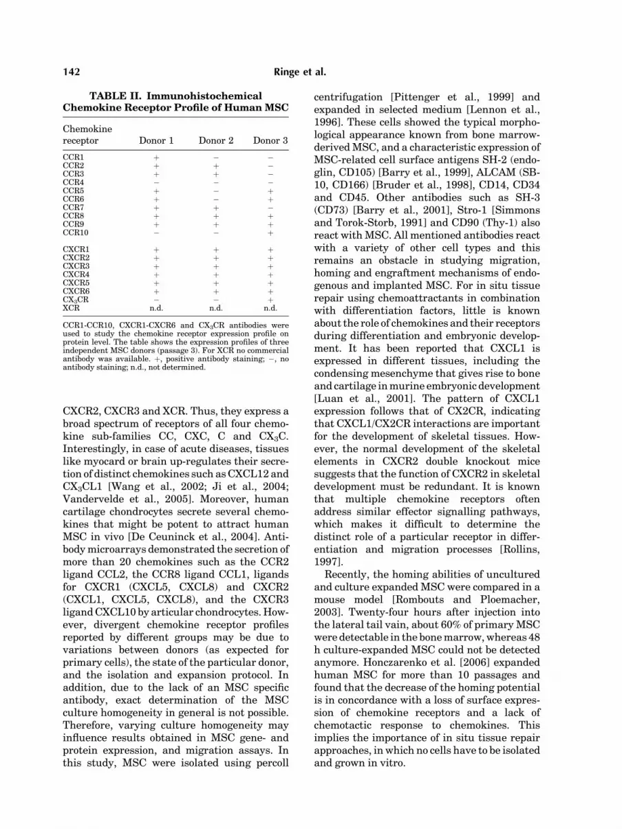
Immunohistochemical staining of MSCdemonstrated, that these cells also presentchemokine receptors for CC, CXC and CX3Cchemokines on the protein level (Fig. 3). Allthree MSC donors stained positive for all sixknown CXCR receptors confirming the qualita-tive expression of CXCR1, CXCR2 and CXCR3(Fig. 3) (Table II). CCR2 was expressed inabundance as shown by gene expression analy-sis, and was detected in two out of three donorimmunohistochemically. CCR8 and CCR9showed an expression independent of the donor,and CCR4 was not detected. The expression ofmost CC receptors varied between donors(Fig. 3) (Table II).
Dose-Dependent Chemotactic Effect of CCL2,CXCL8 and CXCL12
In our in vitro recruitment screen, a 96-wellplate chemotaxis assay using bone marrow-derived human MSC (passages 2 and 3) wasperformed to assess the functional activity ofthe chemokines CCL2, that binds to the CCR2receptor, of CXCL8, a ligand for CXCR1 andCXCR2, and of CXCL12, the ligand for thechemokine receptor CXCR4. In general, thenumber of cells migrated in the positive controls(FBS instead of chemokine) was reproduciblebetween 4� 103 and 5� 103. As shown inFigure 4, although human MSC express theCCR2 receptor in vitro its ligand CCL2 causedno chemotactic effect on these cells (Fig. 4AI–III). The number of migrating cells was lowerthan in the negative controls. The averagenumbers of cells that migrated in the negativecontrols are given by the two lines named UL(upper limit, average number of migrated cellsat 0 nM chemokine concentration plus standarddeviation) and lower limit (LL, average numberof migrated cells at 0 nM chemokine concentra-tion minus standard deviation). In contrast,
CXCL8 showed a dose-dependent chemotacticeffect on human MSC (Fig. 4BI–III). The dose-effect maximum was measured at 750 nM forthree of three MSC donors. Hundred nanomolarwas the minimum concentration of CXCL8 thatchemoattracts MSC. A dose-dependent activityof CXCL12 on MSC and the expression of itsreceptor CXCR4 have already shown before, butwith contradictory results. We verified theexpression of CXCR4 on the mRNA level(Fig. 2) and protein level (Table II), and inaddition showed that CXCL12 caused migrationof human MSC. Analysing three donors, thehighest response was measured using thischemokine in a concentration of 750 nM (onedonor) (Fig. 4CI) or between 250 and 1000 nM(two donors) (Fig. 4CII–III). Similar to CXCL8,100 nM was the minimum concentration ofCXCL12 that chemoattracts human MSC.
To show that the migratory response was dueto site-directed chemotaxis and not to chemo-kinesis, in one migratory assay CXCL8 andCXCL12 were added to both the upper and lowerwells, and therefore the concentration gradi-ents were eliminated. For 750 nM CXCL8 in thelower well, we found 1437� 11.3 migratedMSC. After elimination of the gradient, nomigrated cells were measured demonstratingchemotaxis but no chemokinesis. Using 500 nMCXCL12 in the lower well, we counted420� 80.7 migrated cells and in the chemokin-esis assay we measured 8� 11.3 migrated cells,again indicating chemotaxis but no chimokin-esis. The number of cells migrated in thenegative control was 21.4� 24.5.
DISCUSSION
Regenerative medicine approaches have pro-gressed from the ‘bench to bedside’. Beyondimplantation of cell suspensions [Brittberget al., 1994], advanced Tissue engineeringgrafts comprising cultured cells are clinicallyapplied. Due to a limited proliferation capacityof many adult organ specific cells and a loss offunctional quality during expansion in vitro,much attention has been drawn to stem cellssuch as MSC [Caplan and Bruder, 2001]. Inmost of the few MSC based clinical approaches,these cells were delivered as a cell suspensionfor cardiac or meniscal tissue repair (OsirisTherapeutics Inc., Baltimore) or for co-trans-plantation with haematopoietic stem cells in,for example haematologic malignancy patients
140 Ringe et al.

[Lazarus et al., 2005]. Such therapies arepromising because of the homing, migrationand engraftment potential of MSC [Koc et al.,2000].
With respect to our rationale to establish thebasis for in situ tissue repair based on biomater-ials, chemokines and human bone marrowCD34� MSC, here we reported their entire
chemokine receptor expression profile on themRNA and protein level. Recently, the expres-sion of CCR1, CCR7, CCR9, CXCR4-CXCR6[Honczarenko et al., 2006], CCR1, CCR4, CCR7,CCR9-CCR10, CXCR5 [Luttichaux et al., 2005],and CCR1, CCR7, CXCR4, CXCR6 and CX3CR[Sordi et al., 2005] has been reported. We foundthat MSC also express CCR2, CCR8, CXCR1,
Fig. 3. Immunohistochemical staining of the chemokine receptors CCR1-10, CXCR1-CXCR6 and CX3CR.Human mesenchymal stem- and progenitor cells (MSC) present distinct chemokine receptors for CC, CXCand CX3C chemokines also on the protein level (n¼3 donor) Interestingly, they stained positive for CCR2,CCR8, CXCR1, CXCR2 and CXCR3, receptors whose expression was unknown so far. IgG, negative control.[Color figure can be viewed in the online issue, which is available at www.interscience.wiley.com.]
Chemokine Induced Migration of Human MSC 141
CXCR2, CXCR3 and XCR. Thus, they express abroad spectrum of receptors of all four chemo-kine sub-families CC, CXC, C and CX3C.Interestingly, in case of acute diseases, tissueslike myocard or brain up-regulates their secre-tion of distinct chemokines such as CXCL12 andCX3CL1 [Wang et al., 2002; Ji et al., 2004;Vandervelde et al., 2005]. Moreover, humancartilage chondrocytes secrete several chemo-kines that might be potent to attract humanMSC in vivo [De Ceuninck et al., 2004]. Anti-body microarrays demonstrated the secretion ofmore than 20 chemokines such as the CCR2ligand CCL2, the CCR8 ligand CCL1, ligandsfor CXCR1 (CXCL5, CXCL8) and CXCR2(CXCL1, CXCL5, CXCL8), and the CXCR3ligand CXCL10 by articular chondrocytes. How-ever, divergent chemokine receptor profilesreported by different groups may be due tovariations between donors (as expected forprimary cells), the state of the particular donor,and the isolation and expansion protocol. Inaddition, due to the lack of an MSC specificantibody, exact determination of the MSCculture homogeneity in general is not possible.Therefore, varying culture homogeneity mayinfluence results obtained in MSC gene- andprotein expression, and migration assays. Inthis study, MSC were isolated using percoll
centrifugation [Pittenger et al., 1999] andexpanded in selected medium [Lennon et al.,1996]. These cells showed the typical morpho-logical appearance known from bone marrow-derived MSC, and a characteristic expression ofMSC-related cell surface antigens SH-2 (endo-glin, CD105) [Barry et al., 1999], ALCAM (SB-10, CD166) [Bruder et al., 1998], CD14, CD34and CD45. Other antibodies such as SH-3(CD73) [Barry et al., 2001], Stro-1 [Simmonsand Torok-Storb, 1991] and CD90 (Thy-1) alsoreact with MSC. All mentioned antibodies reactwith a variety of other cell types and thisremains an obstacle in studying migration,homing and engraftment mechanisms of endo-genous and implanted MSC. For in situ tissuerepair using chemoattractants in combinationwith differentiation factors, little is knownabout the role of chemokines and their receptorsduring differentiation and embryonic develop-ment. It has been reported that CXCL1 isexpressed in different tissues, including thecondensing mesenchyme that gives rise to boneand cartilage in murine embryonic development[Luan et al., 2001]. The pattern of CXCL1expression follows that of CX2CR, indicatingthat CXCL1/CX2CR interactions are importantfor the development of skeletal tissues. How-ever, the normal development of the skeletalelements in CXCR2 double knockout micesuggests that the function of CXCR2 in skeletaldevelopment must be redundant. It is knownthat multiple chemokine receptors oftenaddress similar effector signalling pathways,which makes it difficult to determine thedistinct role of a particular receptor in differ-entiation and migration processes [Rollins,1997].
Recently, the homing abilities of unculturedand culture expanded MSC were compared in amouse model [Rombouts and Ploemacher,2003]. Twenty-four hours after injection intothe lateral tail vain, about 60% of primary MSCwere detectable in the bone marrow, whereas 48h culture-expanded MSC could not be detectedanymore. Honczarenko et al. [2006] expandedhuman MSC for more than 10 passages andfound that the decrease of the homing potentialis in concordance with a loss of surface expres-sion of chemokine receptors and a lack ofchemotactic response to chemokines. Thisimplies the importance of in situ tissue repairapproaches, in which no cells have to be isolatedand grown in vitro.
TABLE II. ImmunohistochemicalChemokine Receptor Profile of Human MSC
Chemokinereceptor Donor 1 Donor 2 Donor 3
CCR1 þ � �CCR2 þ þ �CCR3 þ þ �CCR4 � � �CCR5 þ � þCCR6 þ � þCCR7 þ þ �CCR8 þ þ þCCR9 þ þ þCCR10 � � þ
CXCR1 þ þ þCXCR2 þ þ þCXCR3 þ þ þCXCR4 þ þ þCXCR5 þ þ þCXCR6 þ þ þCX3CR � � þXCR n.d. n.d. n.d.
CCR1-CCR10, CXCR1-CXCR6 and CX3CR antibodies wereused to study the chemokine receptor expression profile onprotein level. The table shows the expression profiles of threeindependent MSC donors (passage 3). For XCR no commercialantibody was available. þ, positive antibody staining; �, noantibody staining; n.d., not determined.
142 Ringe et al.

As shown here, CXCL8 and CXCL12 caused adose-dependent response on human MSC.Moreover, we demonstrated that the migratoryresponse was directional and clearly dependenton a CXCL8 and CXCL12 gradient confirming
that MSC movement was due to chemotaxis andnot to chemokinesis. This has already beenshown for CXCL12 and some other chemokines[Honczarenko et al., 2006]. In general, thenumber of migrated cells was low and donor
Fig. 4. Chemotactic response of human MSC to CCL2, CXCL8and CXCL12. Ninety six-multiwell plate chemotaxis assays wereperformed to assess the dose-dependent effect of CCL2 (MCP-1),CXCL8 (interleukin-8) and CXCL12 (SDF-1a) on human MSC.A(I–III): CCR2 caused no chemotactic response on MSC.B(I–III): In contrast, for the first time, a migratory effect of CXCL8on human MSC is shown here. Optimal concentration was 750and 100 nM was the minimum concentration of CXCL8 that
chemoattracts human MSC. C(I–III): In addition, CXCL12showed a dose-dependent activity on these cells. Concentrationsof about 250–1000 nM resulted in the highest response.(UL: upper limit, average number of migrated cells at 0 nMchemokine concentrationþ SD, LL: lower limit, average numberof migrated cells at 0 nM chemokine concentration—SD, SD:standard deviation).
Chemokine Induced Migration of Human MSC 143

and assay dependent. This was in concordancewith assays published before not only for MSC[Wang et al., 2002].
The importance of CXCL12 and its receptorCXCR4 is obvious. Knockout studies in nullmice on either CXCL12 or CXCR4 resulted inthe same phenotype, they die perinatally show-ing severe bone marrow failure [Zou et al.,1998]. It is assumed that the neonatal bonemarrow failure is a consequence of impairedmigration of stem cells, more precisely, CXCL12might be important for the movement ofprogenitor cells from fetal liver to bone marrowduring embryogenesis. The injection of CXCL12in the rat cerebral cortex area significantlyinduces the accumulation of MSC at this area invivo [Ji et al., 2004]. A very recent paper statedthat MSC express CXCR1 and CXCR2 onmRNA level [Lisignoli et al., 2006]. Althoughboth receptors are mainly known from acuteinflammation and innate immunity [Murphyet al., 2000], we have demonstrated theirexpression by human MSC using real-timePCR, immunohistochemistry and a functionalassay. As shown here, the inflammatory che-mokine CXCL8 (IL-8), which is known as apotent chemoattractant for neutrophils [Spa-naus et al., 1997], also caused a dose-dependentchemotactic response to MSC. Interestingly,MSC as well as other cells like chondrocytescontinuously secrete CXCL8 in vitro. A possiblemigratory effect of this chemokine on MSC invivo has to be investigated in future studies. Inchemotaxis assays using other cell types,CXCR2 was relatively non-selective for CXCL8when compared to other ligands such as CXCL1and CXCL5, whereas CXCR1 was highly selec-tive for CXCL8 [Lee et al., 1992; Loetscher et al.,1994].
Activated T-cells, monocytes and basophilsrepresent important targets for CCL2 (MCP-1)[Lahrtz et al., 1998]. Although expression of itsreceptor CCR2 was shown here, CCL2 causedno dose-dependent chemotactic response onhuman MSC. In rats, CCL2, not present innormal brain, is rapidly up-regulated followingmiddle cerebral artery occlusion, and promotesthe migration of injected bone marrow mesench-ymal cells to the sites of injury [Wang et al.,2002]. The in vivo role of CCL2 and otherchemokines, chemoattracting human MSCin vitro such as CCL3, CCL5, CCL17, CCL19,CCL21, CCL25, CCL28, CXCL10, CXCL12,CXCL13, CXCL16, CXCL13 and CX3CL1 [Lut-
tichaux et al., 2005; Sordi et al., 2005; Honczar-enko et al., 2006], have to be investigated inmore detail in further studies. For example, theobserved optimal concentrations for CXCL8 andCXCL12 were high and not physiological.Therefore, appropriate concentrations shouldbe further investigated in vivo.
In conclusion, we demonstrated that thechemokine receptor expression profile of normaldonor-derived human MSC is much broaderthan presented before. Moreover, we showedthat MSC dose-dependently respond to CXCL12(SDF-1a), and, for the first time, also to CXCL8(interleukin-8). In contrast, they do not migratefollowing stimulation with CCL2 (MCP-1).Although these promising results and thealready reported data represent the initial stepson the way towards new in situ tissue therapiesbased on chemokine induced MSC migrationinto distinct defect sites, clearly, the chemotac-tic activity of other chemokines and combina-tions thereof have to be analysed in vivo.Furthermore, since distinct chemokines andtheir receptors take part in, for exampleinflammatory processes, MSC-chemokine inter-actions need to be further investigated.
ACKNOWLEDGMENTS
We are also grateful to Alexander Loch(Charite-University Medicine Berlin) and RudiManz (Deutsches Rheumaforschungszentrum,Berlin) for supporting this study. We acknowl-edge Johanna Golla, Iris Leinhase, SamuelVetterlein and Claudia Hagedorn for excellenttechnical assistance.
REFERENCES
ABI Prism 7700. 1997. User Bulletin #2, ABI Prism 7700Sequence Detection System. Applied Biosystems. 1–36.
Barbash IM, Chouraqui P, Baron J, Feinberg MS, Etzion S,Tessone A, Miller L, Guetta E, Zipori D, Kedes LH,Kloner RA, Leor J. 2003. Systemic delivery of bonemarrow-derived mesenchymal stem cells to the infarctedmyocardium: Feasibility, cell migration, and body dis-tribution. Circulation 108:863–868.
Barry FP, Murphy JM. 2004. Mesenchymal stem cells:Clinical applications and biological characterization. IntJ Biochem Cell Biol 36:568–584.
Barry FP, Boynton RE, Haynesworth S, Murphy JM, ZaiaJ. 1999. The monoclonal antibody SH-2, raised againsthuman mesenchymal stem cells, recognizes an epitope onendoglin (CD105). Biochem Biophys Res Commun 265:134–139.
Barry F, Boynton R, Murphy M, Haynesworth S, Zaia J.2001. The SH-3 and SH-4 antibodies recognize distinct
144 Ringe et al.

epitopes on CD73 from human mesenchymal stem cells.Biochem Biophys Res Commun 289:519–524.
Brittberg M, Lindahl A, Nilsson A, Ohlsson C, Isaksson O,Peterson L. 1994. Treatment of deep cartilage defects inthe knee with autologous chondrocyte transplantation. NEngl J Med 331:889–895.
Bruder SP, Ricalton NS, Boynton RE, Connolly TJ, JaiswalN, Zaia J, Barry FP. 1998. Mesenchymal stem cellsurface antigen SB-10 corresponds to activated leukocytecell adhesion molecule and is involved in osteogenicdifferentiation. J Bone Miner Res 13:655–663.
Caplan AI, Bruder SP. 2001. Mesenchymal stem cells:Building blocks for molecular medicine in the 21stcentury. Trends Mol Med 7:259–264.
Chen J, Li Y, Wang L, Lu M, Zhang X, Chopp M. 2001.Therapeutic benefit of intracerebral transplantation ofbone marrow stromal cells after cerebral ischemia in rats.J Neurol Sci 189:49–57.
Chomczynski P. 1993. A reagent for the single-stepsimultaneous isolation of RNA, DNA and proteins fromcell and tissue samples. Biotechniques 15:532–534 , 536–537.
De Ceuninck F, Dassencourt L, Anract P. 2004. Theinflammatory side of human chondrocytes unveiled byantibody microarrays. Biochem Biophys Res Commun323:960–969.
Devine SM, Bartholomew AM, Mahmud N, Nelson M, PatilS, Hardy W, Sturgeon C, Hewett T, Chung T, Stock W,Sher D, Weissman S, Ferrer K, Mosca J, Deans R,Moseley A, Hoffman R. 2001. Mesenchymal stem cells arecapable of homing to the bone marrow of non-humanprimates following systemic infusion. Exp Hematol 29:244–255.
Devine SM, Cobbs C, Jennings M, Bartholomew A,Hoffman R. 2003. Mesenchymal stem cells distributeto a wide range of tissues following systemic infusioninto nonhuman primates. Blood 101:2999–3001.
Erggelet C, Sittinger M, Lahm A. 2003. The arthroscopicimplantation of autologous chondrocytes for the treat-ment of full-thickness cartilage defects of the knee joint.Arthroscopy 19:108–110.
Fiedler J, Roderer G, Gunther KP, Brenner RE. 2002.BMP-2, BMP-4, and PDGF-bb stimulate chemotacticmigration of primary human mesenchymal progenitorcells. J Cell Biochem 87:305–312.
Fiedler J, Etzel N, Brenner RE. 2004. To go or not to go:Migration of human mesenchymal progenitor cellsstimulated by isoforms of PDGF. J Cell Biochem93:990–998.
Fiedler J, Brill C, Blum WF, Brenner RE. 2006. IGF-I andIGF-II stimulate directed cell migration of bone-marrow-derived human mesenchymal progenitor cells. BiochemBiophys Res Commun 345:1177–1183.
Haynesworth SE, Goshima J, Goldberg VM, Caplan AI.1992. Characterization of cells with osteogenic potentialfrom human marrow. Bone 13:81–88.
Honczarenko M, Le Y, Swierkowski M, Ghiran I,Glodek AM, Silberstein LE. 2006. Human bone marrowstromal cells express a distinct set of biologicallyfunctional chemokine receptors. Stem Cells 24:1030–1041.
Ji JF, He BP, Dheen ST, Tay SS. 2004. Interactions ofchemokines and chemokine receptors mediate the migra-tion of mesenchymal stem cells to the impaired site in the
brain after hypoglossal nerve injury. Stem Cells 22:415–427.
Koc ON, Gerson SL, Cooper BW, Dyhouse SM, Haynes-worth SE, Caplan AI, Lazarus HM. 2000. Rapid hema-topoietic recovery after coinfusion of autologous-bloodstem cells and culture-expanded marrow mesenchymalstem cells in advanced breast cancer patients receivinghigh-dose chemotherapy. J Clin Oncol 18:307–316.
Lahrtz F, Piali L, Spanaus KS, Seebach J, Fontana A. 1998.Chemokines and chemotaxis of leukocytes in infectiousmeningitis. J Neuroimmunol 85:33–43.
Lazarus HM, Koc ON, Devine SM, Curtin P, Maziarz RT,Holland HK, Shpall EJ, McCarthy P, Atkinson K, CooperBW, Gerson SL, Laughlin MJ, Loberiza FR, Jr., MoseleyAB, Bacigalupo A. 2005. Cotransplantation of HLA-identical sibling culture-expanded mesenchymal stemcells and hematopoietic stem cells in hematologicmalignancy patients. Biol Blood Marrow Transplant 11:389–398.
Lee J, Horuk R, Rice GC, Bennett GL, Camerato T, WoodWI. 1992. Characterization of two high affinity humaninterleukin-8 receptors. J Biol Chem 267:16283–16287.
Lennon DP, Haynesworth SE, Bruder SP, Jailswal N,Caplan AI. 1996. Human and animal MesenchymalProgenitor Cells from Bone Marrow: Identifikation ofserum for optimal selection and proliferation. In VitroCell Dev Biol 32:602–611.
Lisignoli G, Cristino S, Piacentini A, Cavallo C, Caplan AI,Facchini A. 2006. Hyaluronan-based polymer scaffoldmodulates the expression of inflammatory and degrada-tive factors in mesenchymal stem cells: Involvement ofCd44 and Cd54. J Cell Physiol 207:364–373.
Loetscher P, Seitz M, Clark-Lewis I, Baggiolini M, Moser B.1994. Both interleukin-8 receptors independently med-iate chemotaxis. Jurkat cells transfected with IL-8R1 orIL-8R2 migrate in response to IL-8, GRO alpha and NAP-2. FEBS Lett 341:187–192.
Luan J, Furuta Y, Du J, Richmond A. 2001. Developmentalexpression of two CXC chemokines, MIP-2 and KC, andtheir receptors. Cytokine 14:253–263.
Luttichaux IV, Notohamiprodjo M, Wechselberger A,Peters C, Henger A, Seliger C, Djafarzadeh R, Huss R,Nelson PJ. 2005. Human adult CD34- progenitor cellsfunctionally express the chemokine receptors CCR1,CCR4, CCR7, CXCR5, and CCR10 but not CXCR4. StemCells Dev 14:329–336.
Murphy PM, Baggiolini M, Charo IF, Hebert CA, Horuk R,Matsushima K, Miller LH, Oppenheim JJ, Power CA.2000. International union of pharmacology. XXII.Nomenclature for chemokine receptors. Pharmacol Rev52:145–176.
Murphy JM, Fink DJ, Hunziker EB, Barry FP. 2003. Stemcell therapy in a caprine model of osteoarthritis. ArthritisRheum 48:3464–3474.
Pittenger MF, Mackay AM, Beck SC, Jaiswal RK, DouglasR, Mosca JD, Moorman MA, Simonetti DW, Craig S,Marshak DR. 1999. Multilineage potential of adulthuman mesenchymal stem cells. Science 284:143–147.
Rollins BJ. 1997. Chemokines. Blood 90:909–928.Rombouts WJ, Ploemacher RE. 2003. Primary murine MSC
show highly efficient homing to the bone marrow but losehoming ability following culture. Leukemia 17:160–170.
Chemokine Induced Migration of Human MSC 145

Ryan JM, Barry FP, Murphy JM, Mahon BP. 2005.Mesenchymal stem cells avoid allogeneic rejection. JInflamm (Lond) 2:8.
Schmitt B, Ringe J, Haupl T, Notter M, Manz R, BurmesterGR, Sittinger M, Kaps C. 2003. BMP2 initiates chondrogeniclineage development of adult human mesenchymal stemcells in high-density culture. Differentiation 71:567–577.
Simmons PJ, Torok-Storb B. 1991. Identification of stromalcell precursors in human bone marrow by a novelmonoclonal antibody, STRO-1. Blood 78:55–62.
Sordi V, Malosio ML, Marchesi F, Mercalli A, Melzi R,Giordano T, Belmonte N, Ferrari G, Leone BE, BertuzziF, Zerbini G, Allavena P, Bonifacio E, Piemonti L. 2005.Bone marrow mesenchymal stem cells express arestricted set of functionally active chemokine receptorscapable of promoting migration to pancreatic islets. Blood106:419–427.
Spanaus KS, Nadal D, Pfister HW, Seebach J, Widmer U,Frei K, Gloor S, Fontana A. 1997. C-X-C and C-Cchemokines are expressed in the cerebrospinal fluidin bacterial meningitis and mediate chemotacticactivity on peripheral blood-derived polymorphonuclearand mononuclear cells in vitro. J Immunol 158:1956–1964.
Steadman JR, Rodkey WG, Rodrigo JJ. 2001. Microfrac-ture: Surgical technique and rehabilitation to treatchondral defects. Clin Orthop 9:S362–S369.
Vandervelde S, van Luyn MJ, Tio RA, Harmsen MC. 2005.Signaling factors in stem cell-mediated repair ofinfarcted myocardium. J Mol Cell Cardiol 39:363–376.
Wakitani S, Imoto K, Yamamoto T, Saito M, Murata N,Yoneda M. 2002. Human autologous culture expandedbone marrow mesenchymal cell transplantation forrepair of cartilage defects in osteoarthritic knees.Osteoarthritis Cartilage 10:199–206.
Wang L, Li Y, Chen X, Chen J, Gautam SC, Xu Y, Chopp M.2002. MCP-1, MIP-1, IL-8 and ischemic cerebral tissueenhance human bone marrow stromal cell migration ininterface culture. Hematology 7:113–117.
Zigmond SH, Hirsch JG. 1973. Leukocyte locomotion andchemotaxis. New methods for evaluation, and demon-stration of a cell-derived chemotactic factor. J Exp Med137:387–410.
Zou YR, Kottmann AH, Kuroda M, Taniuchi I, Littman DR.1998. Function of the chemokine receptor CXCR4 inhaematopoiesis and in cerebellar development. Nature393:595–599.
146 Ringe et al.


