Embed Size (px)
Citation preview
Published: September 20, 2011
r 2011 American Chemical Society 1882 dx.doi.org/10.1021/tx200228c |Chem. Res. Toxicol. 2011, 24, 1882–1890
ARTICLE
pubs.acs.org/crt
Toxic Effects of Imidazolium Ionic Liquids on the Green Seaweed Ulvalactuca: Oxidative Stress and DNA DamageManoj Kumar, Nitin Trivedi, C. R. K. Reddy,* and Bhavanath Jha
Discipline of Marine Biotechnology and Ecology, Central Salt and Marine Chemicals Research Institute, Council of Scientific andIndustrial Research (CSIR), Gijubhai Badheka Marg, Bhavnagar 364021, India
’ INTRODUCTION
Ionic liquids (ILs) are a group of molten salts consisting ofasymmetrically substituted nitrogen or phosphorus cations togetherwith an organic or inorganic anion moiety. The unique physio-chemical properties of ILs, i.e., negligible vapor pressure, highthermal, chemical, and electrochemical stabilities and excellentsolvent properties, are attributed to their functionality as greensolvents.1 Nevertheless, the green credentials of these ILs have beenquestioned for use in aquatic environments as the water solubility ofmajority of ILs is significant.2 Since, the substitution of ILs forconventional solvents in a variety of chemical industries is on therise, there is a possibility that ILs may find their way into waterbodies. The release of ILs into aquatic environments througheffluent discharge or accidental spillage could result in waterpollution due to their poor biodegradability thereby negativelyimpacting the structure and function of the aquatic ecosystem.3,4
The 1-alkyl-3-methylimidazolium salts are some of the mostcommon ILs used in industrial applications5 and are reportedto have poor biodegradability except for octyl imidazolium andother ILs.4 Recently, there has been considerable effort todetermine the toxicity of ILs on both terrestrial and aquaticorganisms (see ref 2). The toxicological studies of ILs investi-gated so far have demonstrated that ILs cause membrane damageand disruption, coupled with increased generation of cellular
reactive oxygen species (ROS) manifesting the inhibition ofphotosynthesis leading to oxidative stress.2,6 However, thesource for ROS origin and the precise mechanisms of IL inducedtoxicity have largely remained unexplored. The evidence of ILstoxicity necessitated the need for undertaking a detailed inves-tigation addressing the effects of these compounds at variousbiological levels of organization.3,6�13 However, to date, there isno study investigating the toxic effects of ILs on seaweeds, whichare considered the main primary producers in coastal waters.These being an abundant component in coastal waters arerecognized as alternate promising sources for biofuel and chemi-cals with the potential to reduce greenhouse gas emissions andglobal warming.14 Any accidental release of ILs into an aquaticecosystem may cause grave environmental concerns by perturb-ing the food-web dynamics and ecosystem structure and func-tion. Seaweeds being of marine origin, the ILs could havedifferent effects according to the salinity of the environment.15
Therefore, IL toxicity mechanism based studies are of impor-tance to mitigate the impact of IL mediated water pollution.
In this study, for the first timewe report the toxicity and biologicaleffects of ILs belonging to 1-alkyl-3-methylimidazolium bromide
Received: May 31, 2011
ABSTRACT: The green credentials of ionic liquids (ILs) are being increasingly questioneddue to the growing evidence of their toxicity to aquatic ecosystems, although the mechanismsof toxicity are unknown. This study provides insights into the mechanism of toxicity andbiological effects of 1-alkyl-3-methylimidazolium bromide [Cnmim]Br (n = 4 to 16) on themarine macroalgaUlva lactuca. The cell viability of this alga during IL exposure was found to benegatively correlated to the chain length of the alkyl group. The IL ([C12mim]Br) exposuretriggers the generation of reactive oxygen species (ROS viz. O2
•�, H2O2, and OH•), damage of
the membrane and DNA, and inhibition of antioxidant systems in the alga. The enhancedproduction of ROS and lipid peroxidation in the alga subjected to LC50 concentration for 4days was largely attributed to lipoxygenase (LOX) activity coupled with the induction of twoLOX isoforms (∼80 kDa and ∼55 kDa). Pretreatment of the algal thallus with enzymeinhibitors such as diphenylene iodonium, sodium azide, cantharidin, and oxadiazoloquinoxalin-1-one, prior to [C12mim]Br exposure showed the regulation of ROS by the activation ofmembrane bound NADPH-oxidase and cytochrome oxidase. The IL exposure resulted in theaccumulation of n-3 and n-6 fatty acids at 0.5 LC50 concentration indicating the induction ofdesaturase enzymes. Furthermore, antioxidant enzyme activities such as superoxide dismutase(SOD), ascorbate peroxidase (APX), and glutathione reductase (GR) were enhanced by1.3�2.0-fold, while glutathione peroxidase (GSH-Px) diminished, together with a higher regeneration rate of reduced ascorbate andglutathione. The isoforms of antioxidant enzymes, namely, Mn-SOD (∼85 kDa), APX (∼125 and 45 kDa), and GR (∼135 kDa)regulated differentially to IL exposure. The comet assay performed for the first time for seaweeds revealed the significant inductionof DNA damage (>50�70% increase in % tail DNA over control) in alga exposed to g LC50 concentration.
1883 dx.doi.org/10.1021/tx200228c |Chem. Res. Toxicol. 2011, 24, 1882–1890
Chemical Research in Toxicology ARTICLE
salts with different alkyl-chain lengths on the green seaweed Ulvalactuca.This alga was selected as it has a worldwide distribution withthe potential to adapt and grow in diverse adverse environmentalconditions.16 We first determined the LC50 values of these ILs andtheir effect on ROS generation. Later, the regulation of enzymaticand nonenzymatic antioxidant systems, free fatty acids, damage tocell membranes and DNA, and the effects of different ROSscavengers were studied to elucidate the mechanism of IL toxicity.
’MATERIALS AND METHODS
Test Organism and Treatment of ILs. The vegetative thalli ofU. lactuca were collected from the intertidal region of the Veraval Coast(20� 540 N, 70� 220 E), Gujarat, India. Selected, clean juvenile thalli werebrought to the laboratory in a cool pack. In order to initiate unialgalculture, the thalli were cleaned manually with a brush in autoclavedseawater to remove epiphytes and any superficial foreign matter. Thecleaned thalli were acclimatized to laboratory conditions by culturing inaerated flat bottom, Erlenmeyer flasks in filtered natural seawater (NSW) of30 psu salinity supplementedwith provasoli enrichment seawatermedium17
and GeO2 (5 mg L�1) for 10 days. During the acclimatization period, the
mediumwas replenished on alternate days andmaintained under cool whitefluorescent tubes at an irradiance of 50μmol photonsm�2 s�1 with a 12:12h light/dark cycle at 22 ( 1 �C. For treatment, the acclimatized algalfragments were incubated in filtered NSW having 30 psu salinity with andwithout (control) ILs in aerated cultures.
The ILs used in this study included a series of 1-alkyl-3-methylimida-zolium bromide ([Cnmim]Br) salts with alkyl chains which ranged fromn = 4 to16, which were synthesized in the laboratory, as described in theliterature.18 The purities of ionic liquids were established to be >99%through proton nuclear magnetic resonance (1HNMR) spectroscopy byintegration of proton signals with respect to an internal standard oftetramethyl silane, and the NMR spectra were in agreement with theliterature data. The water content of the ionic liquids was determinedusing a coulometric Karl Fischer autotitrator (Mettler Toledo DL39)with Combi-Coulomat fritless Karl Fischer reagent (Merck) and agreedwithin 120�300 ppm range. The stock solutions of ILs (15 mM) wereprepared in distilled water and were found to be completely soluble. Fortreatment, the algal fragments were exposed to the tested concentrationswhile dissolving the required amount from the stock solutions in NSWhaving 30 psu salinity.Cell Viability Assay. A 2,3,5-triphenyltetrazolium chloride (TTC)
based method was used to determine the cell viability of the control andIL treated algal fragments according to Shiu and Lee.19 The TTC cellviability assay is based on the tetrazolium salt reduction to formazan bydehydrogenase respiratory enzymes and thus indicates the resilience ofthe mitochondrial component of the cell machinery when challengedwith IL-induced stress. After treatment, the algal fragments (50 mg freshweight) were incubated at 25 �C in 3 mL of NSW containing potassiumphosphate buffer (50 mM, pH 7.4) and 0.8% (w/v) TTC in darkness for16 h. Thereafter, the algal fragments were washed three times in NSW,and the intracellular insoluble red formazon was extracted twice with5mLof 95% ethanol at 80 �C for 20min. Extracts were combined, and theabsorbance was determined at A530. The cellular viability for IL treatedthalli was calculated as a percentage, while considering the absorbance ofthe control algal fragments with 100% cell viability. The viability percen-tages were plotted against the concentration tested for each IL to obtain alinear regression and for estimating the LC50. Each experiment was carriedout in triplicate, and the mean values of the LC50 were calculated.Determination, In Situ Localization of ROS, and Total
Chlorophyll Estimation. The O2•� production rate was measured
according to Liu et al.20 Samples (500 mg fresh weight) were homo-genized in 4 mL of 65 mM potassium phosphate buffer (pH 7.8) andcentrifuged at 5,000g for 10 min. The incubation mixture contained
0.9 mL of 65 mM potassium phosphate buffer (pH 7.8), 0.1 mL of10 mM hydroxylaminoniumchloride, and 1 mL of the supernatant. Afterincubation at 25 �C for 20 min, 17 mM sulphanilic acid and 7 mMα-naphthyl amine were added to the incubation mixture. After reactingat 25 �C for a further 20 min, the absorbance was read at 530 nm.A standard curve, with NaNO2 was used to calculate the production rateof O2
•�. For the estimation of H2O2, 100 mg fresh samples wereextracted in 200 μL of Na-acetate buffer (50mM, pH 6.5) and incubatedin reaction media containing 50 mM Na-acetate buffer, 1 mM 4-ami-noantipyrine, 1 mM 2, 4-dichlorophenol, 50 mM MnCl2, and 0.2 mMNADH for 24 h. The oxidation of aminoantipyrine was recorded at510 nm, and the absorbance was compared to the standard curveprepared with H2O2 in the same reaction mixture. Determination ofOH• production was performed based on the degradation of 2-deoxyr-ibose by OH• radicals.20 The O2
•� and H2O2 were visually detected inthe thalli using nitroblue tetrazolium (NBT) and 3,3-diaminobenzidine(DAB) as the substrates respectively, according to Kumar et al.21 Totalchlorophyll (chl a+b) was extracted in 80% acetone and estimated inaccordance with Kumar et al.22
Effect of ROS Inhibitors. To study the effect of inhibitors of ROSgeneration, the algal fragments were preincubated in NSW containingdiphenylene iodonium (DPI, NADPH oxidase inhibitor, 10 μM), sodiumazide (NaN3, peroxidase inhibitor, 1 mM), cantharidin (CANT, proteinphosphatase inhibitor, 10 μM), and oxadiazolo-quinoxalin-1-one (ODQ,guanylate cyclase inhibitor, 0.2 mM) for 24 h and then cultured in NSWsupplemented with a lethal dose of [C12mim]Br IL for 4 days. Thereafter,the thalli were washed with NSW twice and processed for ROS localiza-tion using NBT or DAB solutions.Protoplast Isolation and Comet Assay. Protoplast isolation
and purification were performed according to Gupta et al.23 Briefly,following IL treatment and washing, the algal fragments (300 mg FW)were chopped into small pieces of tissue (e1 mm thin) in naturalseawater (NSW) in two replicates, each with 150 mg of tissue. Thechopped tissues were then rinsed several times with NSW to removedebris. The tissues thus cleaned were incubated in an enzyme solutionconsisting of 0.5% dextron sulfate, 2% cellulase, 2% NaCl, and 0.8%mannitol in 50 mM MES buffer (pH 6.0) for 4�5 h on a rotary shaker(50 rpm) in the dark at 25 �C. The protoplasts were passed through amiracloth (Calbiochem Co., USA) and centrifuged at 300g for 10 min.The precipitates were suspended in saline phosphate buffer (PBS), atpH 7.4, and kept on ice at 4 �C. The protoplast yields were estimated bycounting the cells using a hemocytometer under an invertedmicroscope.
For the comet assay, the protoplast solution (50 μL) containingapproximately 1� 103 protoplasts was mixed with 50 μL of 1% low-meltingtemperature agarose (LMPA) dissolved in phosphate-buffered saline. An80 μL aliquot of the solution was layered onto a base slide, which wasprecoated with 1% agarose, and then covered with a coverslip. When theagarose gel solidified, the coverslip was gently slid off, and another agaroselayer (90μL, 0.5%LMPA) was layered while covering it with a new coverslipand then left for 10 min on a chilled metal plate in order for solidification ofthe agarose layer to occur. Following this, the coverslip was removed, and theslides were submerged in alkali lysis solution (2.5 M NaCl, 10 mM Trizma,and 100mMEDTA) at pH>13 overnight at 4 �C.Thereafter, the glass slidewas incubated in fresh, cold electrophoresis buffer (0.3MNaOH and 1mMEDTA, pH 13) in an horizontal electrophoresis tank (Model POWERPAC 200, Bio-Rad Co., USA) for 30 min at room temperature to allow forDNA unwinding. Electrophoresis was performed for 10 min at 25 V and300 mA in a chamber cooled in an ice bath. After electrophoresis, the glassslides were neutralized in 0.4 MTris�HCl (pH 7.5) buffer, washed twice indistilled water, and left overnight for drying at room temperature. The slideswere stained following the silver staining method.24 Stained slides wereexamined using an Olympus Microscope model-BX60 fitted with anOlympus-DP72 camera. The classification of comet category and their tailmeasurements were carried out according to Garcia et al.25
1884 dx.doi.org/10.1021/tx200228c |Chem. Res. Toxicol. 2011, 24, 1882–1890
Chemical Research in Toxicology ARTICLE
Analysis of Fatty Acids and Determination of Enzymaticand Nonenzymatic Antioxidants. Fatty acids from lipids wereconverted to the respective methyl esters and analyzed by GC-2010coupled with GCMS-QP2010.21 The water-soluble antioxidants such asglutathione (oxidized glutathione, GSSG; reduced glutathione, GSH)and ascorbate (oxidized ascorbate, DHA; reduced ascorbate, AsA) weredetermined as described by Wu and Lee.26 The activities of antioxidantenzymes, namely, superoxide dismutase (SOD), ascorbate peroxidase(APX), glutathione reductase (GR), and glutathione peroxidase (GSH-Px)were determined by the method of Wu and Lee26 and lipoxygenase(LOX) by the method of Kumar et al.22 The isoenzyme profile ofantioxidative enzymes (SOD, APX, GR, and GSH-Px) and LOX andtheir molecular mass were determined with 10 or 12% nondenaturingpolyacrylamide gels using their specific activity staining procedures.22
Data Analysis. Results are expressed as the mean of three replicateswith standard deviation. Statistical analyses were performed by one wayanalysis of variance (ANOVA). Significant differences among the meanvalues were determined by the least significant difference (LSD) at p< 0.05.
’RESULTS
LC50 Values. The LC50 values for seven types of 1-alkyl-3-methylimidazolium bromide ILs onU. lactuca exposed for 4 daysare presented in Table 1. The results demonstrated that the LC50
value decreased with the increasing alkyl-chain length andduration of exposure. It was observed that the ILs [C14mim]Brand [C16mim]Br were highly toxic to alga even at LC50 valuesand showed complete bleaching of cells indicating the deathof the tissue fragment within 36 h as inferred from the TTCcell viability assay. However, the fragments exposed to ILs-[Cnmim]Br (n = 12) were quite healthy and showed symptomsof bleaching only after 4 days at LC50 value, while at 2� LC50
concentration, they exerted significant damage to the cell mem-brane followed by bleaching at 4 days and eventually death after5 days of exposure, as confirmed by the TTC cell viability assay.Therefore, in the subsequent experiment the thallus fragmentswere treated with [C12mim]Br for 0.5 LC50, LC50, and 2� LC50
concentrations for a duration of 4 days only in order to examinethe effects on ROS generation, lipo-peroxidation of cell mem-branes (while measuring the MDA level by TBARS activityassay), and DNA (assessed by COMET assay). However, tostudy the regulation of the antioxidant system and free fatty acidcontents the exposure doses were restricted to 0.5 LC50 and LC50
only because at 2� LC50, excessive ROS generation and max-imum DNA damage was observed, which in turn reflected thetotal failure of the defense system of the cell and consequently thedeath of cells exposed to IL at 5 days.Generation, Histochemical Localization of ROS, and the
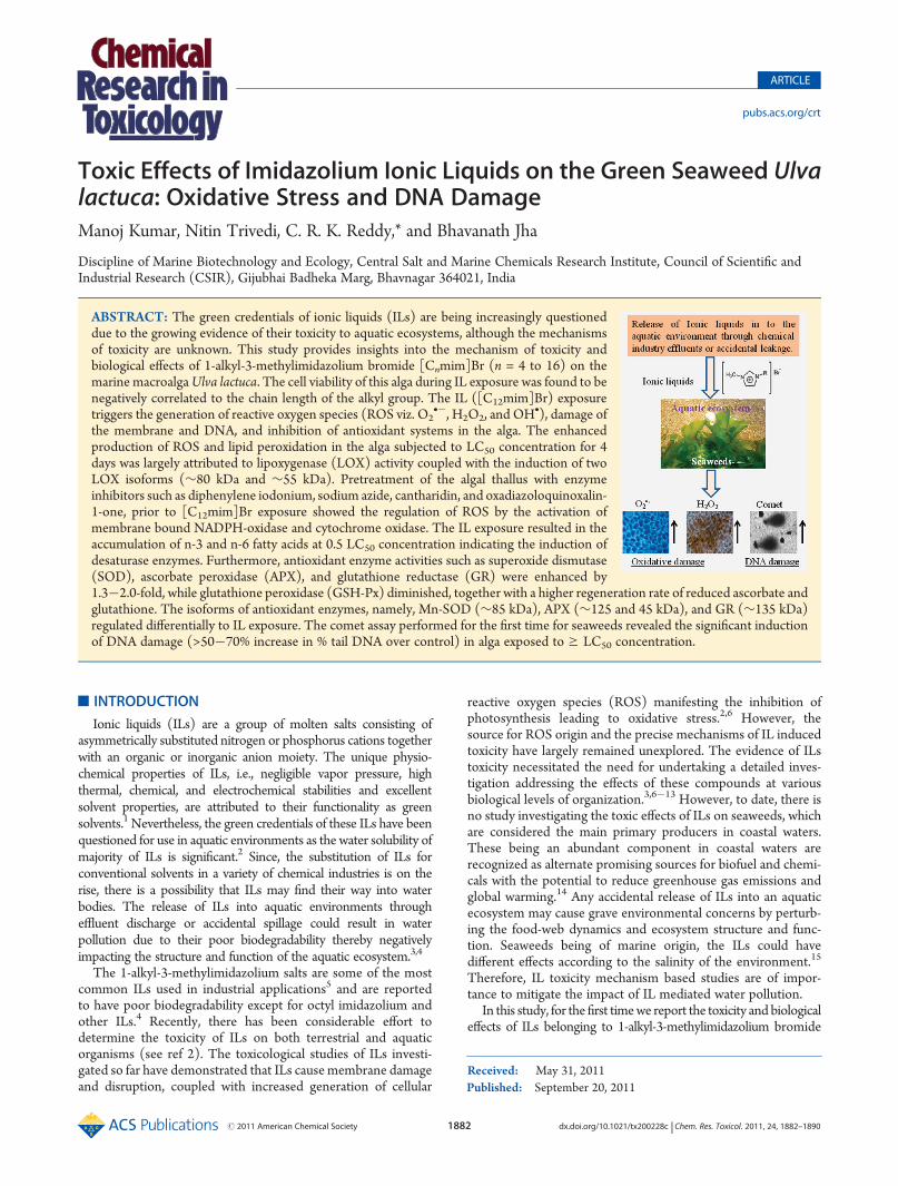
Effect of Their Inhibitors. The oxidative damage caused by[C12mim]Br IL was determined by measuring the lipid perox-idation (MDA level) and ROS (includingO2
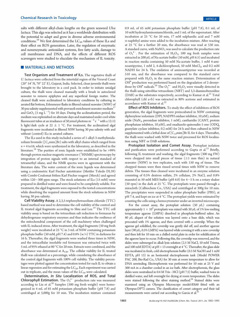
•�, OH•, andH2O2)content in Ulva (Figure 1). IL exposure to concentrations0.5 LC50, LC50, and 2� LC50 for 4 days inevitably inducedoxidative stress with a significant increase in membrane lipidperoxidation by 1.63-, 2.85-, and 4.20-fold, respectively, over thecontrol (3.08 nmol g�1 FW). Following the exposure to IL, theROS level was also elevated to surprisingly high levels in plantsby 1.25�5.25-fold, and accumulation was more evident in plantstreated with the sublethal and lethal doses of [C12mim]Br IL.The enhanced level of oxidative stress biomarkers resulted in asignificant decrease in total chlorophyll content by almost 21%(0.5 LC50), 43% (LC50), and 62% (2� LC50), respectively, whencompared with that of the control (16.16 μg 3 100 g�1 FW)(Figure 1). Histochemical staining (Figure 2A and B) employedfor in situ localization of O2
•� and H2O2 radicals made apparentthe distribution of reduced NBT dependent dense blue forma-zone and H2O2 dependent brown precipitate, respectively, allover the tissue in plants exposed to 0.5 LC50, LC50, and 2� LC50
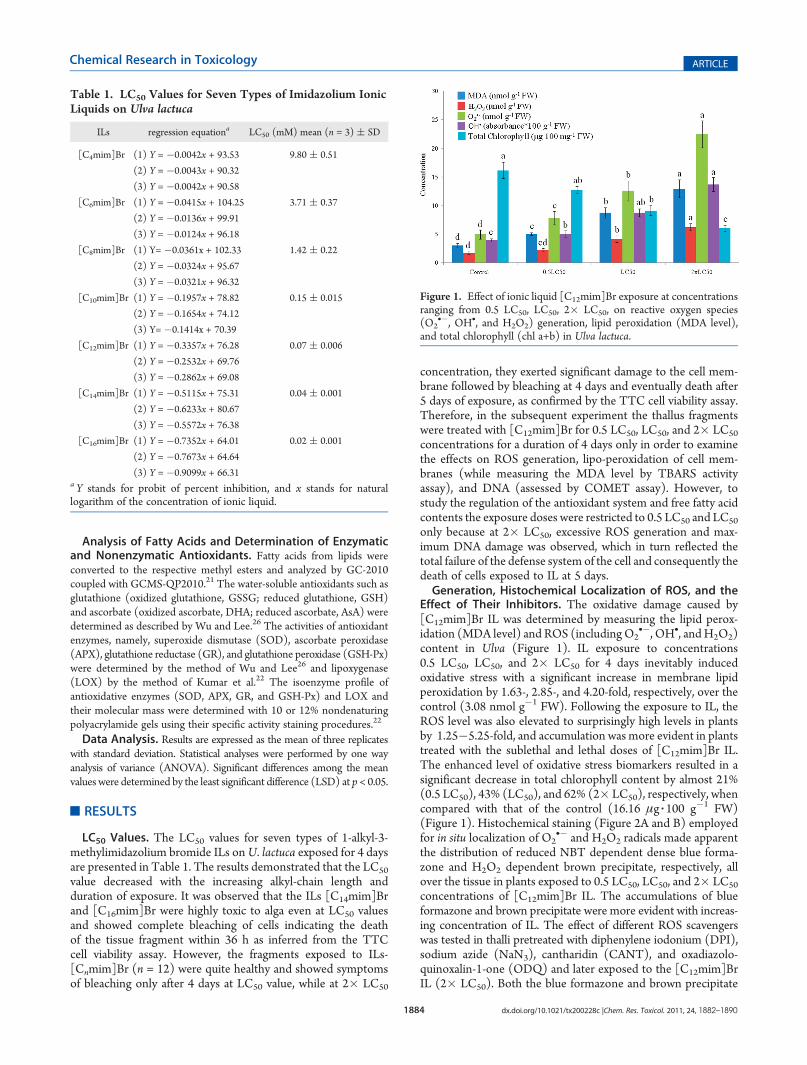
concentrations of [C12mim]Br IL. The accumulations of blueformazone and brown precipitate were more evident with increas-ing concentration of IL. The effect of different ROS scavengerswas tested in thalli pretreated with diphenylene iodonium (DPI),sodium azide (NaN3), cantharidin (CANT), and oxadiazolo-quinoxalin-1-one (ODQ) and later exposed to the [C12mim]BrIL (2� LC50). Both the blue formazone and brown precipitate
Table 1. LC50 Values for Seven Types of Imidazolium IonicLiquids on Ulva lactuca
ILs regression equationa LC50 (mM) mean (n = 3) ( SD
[C4mim]Br (1) Y = �0.0042x + 93.53 9.80 ( 0.51
(2) Y = �0.0043x + 90.32
(3) Y = �0.0042x + 90.58
[C6mim]Br (1) Y = �0.0415x + 104.25 3.71 ( 0.37
(2) Y = �0.0136x + 99.91
(3) Y = �0.0124x + 96.18
[C8mim]Br (1) Y= �0.0361x + 102.33 1.42 ( 0.22
(2) Y = �0.0324x + 95.67
(3) Y = �0.0321x + 96.32
[C10mim]Br (1) Y = �0.1957x + 78.82 0.15 ( 0.015
(2) Y = �0.1654x + 74.12
(3) Y= �0.1414x + 70.39
[C12mim]Br (1) Y = �0.3357x + 76.28 0.07 ( 0.006
(2) Y = �0.2532x + 69.76
(3) Y = �0.2862x + 69.08
[C14mim]Br (1) Y = �0.5115x + 75.31 0.04 ( 0.001
(2) Y = �0.6233x + 80.67
(3) Y = �0.5572x + 76.38
[C16mim]Br (1) Y = �0.7352x + 64.01 0.02 ( 0.001
(2) Y = �0.7673x + 64.64
(3) Y = �0.9099x + 66.31a Y stands for probit of percent inhibition, and x stands for naturallogarithm of the concentration of ionic liquid.
Figure 1. Effect of ionic liquid [C12mim]Br exposure at concentrationsranging from 0.5 LC50, LC50, 2� LC50, on reactive oxygen species(O2
•�, OH•, and H2O2) generation, lipid peroxidation (MDA level),and total chlorophyll (chl a+b) in Ulva lactuca.
1885 dx.doi.org/10.1021/tx200228c |Chem. Res. Toxicol. 2011, 24, 1882–1890
Chemical Research in Toxicology ARTICLE
formed due to the generation of O2•� and H2O2 radicals, res-
pectively, were almost abolished with DPI and NaN3 treatment(Figure 3A and B) and also reduced partially in plants pretreatedwith CANT and ODQ when compared with the plants treatedwith 2� LC50 concentration.DNA Damage. From the Comet assay, it was clearly evident
that the ionic liquid induced DNA damage responded in a dose-dependentmanner (Figure 4). Following exposure to ionic liquid[C12mim]Br, the number of comets belonging to categories 3(high damage, 45�70% of DNA in tail) and 4 (very highdamage, >70% of DNA in tail) increased significantly (p < 0.01)in plants treated with LC50 and 2� LC50 concentrations,respectively. However, the plants treated with the 0.5 LC50
concentration showed significantly less DNA damage with themajority of the comets falling into categories 1 (low damage,5�25% of DNA in tail) and 2 (medium damage, 25�45% ofDNA in tail).Fatty Acid, Enzymatic, and Nonenzymatic Antioxidants.
Table 2 summarizes the variation in fatty acid composition ofU. lactuca in response to IL-induced oxidative stress in terms ofthe percentage of total fatty acids (%TFA). Plants exposed to theIL (LC50) exhibited a significant decrease (p < 0.05) in n-3 and
n-6 polyunsaturated fatty acids (PUFAs), with a concomitantincrease in saturated fatty acids (SFAs). Following the IL (LC50)exposure, the content of SFAs, namely, palmitic, stearic, andbehenic acids increased by 60�70%, when compared to theircorresponding values of 24.42%, 3.05%, and 1.67% TFA in thecontrol, while the content of most of the PUFAs under thistreatment decreased considerably by∼50%. On the contrary, thecontents of both SFA and PUFAs in plants cultured with thesupplementation of IL (0.5 LC50) were found to be either similaror marginally higher than that of their contents in control, withthe exception of palmitic acid (which increased by 30%) anddocosahexaenoic acid contents (which decreased by 30%).Superoxide dismutase (SOD), ascorbate peroxidase (APX),
glutathione reductase (GR), and glutathione peroxidase (GSH-Px) were selected as biomarkers to determine the oxidative stresscaused by the IL on the enzymatic antioxidant defense system ofUlva. Exposure to IL (0.5 LC50) significantly enhances the enzymeactivities of SOD, APX, and GR by 1.3�1.8-fold (p < 0.01) overthe control activities with 87.45, 0.44, and 2.63 U mg�1 protein,respectively (Figure 5A). Exposure to even higher concentration(LC50) decreased the enzyme activities. It is noteworthy that theenzyme activity of GR, contrary to GSH-Px, was quite indifferent
Figure 2. Reactive oxygen species generation in Ulva lactuca following exposure to the ionic liquid [C12mim]Br for 4 days. (A) O2•� and (B) H2O2
radicals were detected with nitroblue tetrazolium (NBT) and 3,3-diaminobenzidine (DAB) staining, respectively. a�d inside figures represent thevarious treatments, viz. control, 0.5 LC50, LC50, and 2� LC50, respectively.
Figure 3. Effect of pretreatment by inhibitors (e.g., DPI, NaN3, CANT, and ODQ) on the production of reactive oxygen species in Ulva lactucafollowing exposure to the ionic liquid [C12mim]Br at 2� LC50 concentration. (A)H2O2 and (B)O2
•� radicals were detected with 3,3-diaminobenzidine(DAB) and nitroblue tetrazolium (NBT) staining, respectively.
1886 dx.doi.org/10.1021/tx200228c |Chem. Res. Toxicol. 2011, 24, 1882–1890
Chemical Research in Toxicology ARTICLE
Figure 4. Comets showing tails of different length induced by various concentrations of ionic liquid [C12mim]Br exposure inUlva lactuca: (a) control;(b) 0.5 LC50; (c) LC50; and (d) 2� LC50. A total of 60 comets were examined for each treatment with two replicates.
Table 2. Effect of [C12mim]Br Ionic Liquid on Fatty Acid Composition (% of Total Fatty Acids) in Ulva lactuca (Mean of ThreeIndependent Experiments ( SD)
fatty acids common name control 0.5 LC50 LC50
C 12:0 lauric acid 0.71 ( 0.09 a nd 0.48 ( 0.06 b
C 14:0 myristic acid 2.81 ( 0.19 a 2.21 ( 0.21 b 1.63 ( 0.02 c
C 15:0 pentadecanoic acid 0.92 ( 0.05 a 0.37 ( 0.06 c 0.54 ( 0.05 b
C 16:0 palmitic 24.42 ( 1.21 c 31.98 ( 2.25 b 40.51 ( 0.72 a
C 17:0 heptadecenoic acid 0.24 ( 0.00 a 0.17 ( 0.02 b 0.10 ( 0.00 c
C 18:0 stearic acid 3.05 ( 0.17 b 2.83 ( 0.19 b 4.82 ( 0.11 a
C 20:0 arachidic acid 0.15 ( 0.01 c 0.20 ( 0.02 b 0.35 ( 0.03 a
C 22:0 behenic acid 1.67 ( 0.01 b 1.80 ( 0.17 b 2.87 ( 0.16 a
C 24:0 lignoceric acid 0.27 ( 0.00ab 0.15 ( 0.01 b 0.30 ( 0.04 a
C 16:1(n-7) 9-hexadecenoic acid 1.51 ( 0.13 a 1.16 ( 0.16 c 1.23 ( 0.03 bc
C 16:1(n-9) 7-hexadecenoic acid 5.23 ( 0.62 a 4.01 ( 0.28 b 2.14 ( 0.21 c
C 17:1(n-7) heptadecenoic acid 0.71 ( 0.07 a 0.54 ( 0.09 b 0.79 ( 0.09 a
C 18:1(n-9) oleic acid 11.15 ( 0.29 a 11.94 ( 1.11 a 8.94 ( 0.54 b
C 18:2(n-6) linoleic acid 6.72 ( 0.64 a 5.95 ( 0.54 b 5.06 ( 0.11 c
C 18:3(n-6) γ-linolenic acid 1.10 ( 0.11 a 0.97 ( 0.10 b 0.50 ( 0.01 c
C 18:3(n-3) α-linolenic acid 19.08 ( 0.73 b 21.40 ( 0.74 a 14.08 ( 0.67 c
C 18:4(n-3) stearidonic acid 16.01 ( 0.88 a 13.49 ( 1.06 b 10.29 ( 0.45 c
C 20:3(n-6) dihomo- γ-linolenic acid 0.18 ( 0.01 a 0.15 ( 0.01 b 0.14 ( 0.01 b
C 20:4(n-6) arachidonic acid 0.84 ( 0.05 b 0.96 ( 0.06 a 0.56 ( 0.03 c
C 20:5(n-3) eicosapentaenoic acid 0.85 ( 0.07 b 1.17 ( 0.15 a 0.42 ( 0.05 c
C 22:6(n-3) docosahexaenoic 2.40 ( 0.14 a 1.68 ( 0.23 b 1.15 ( 0.05 c
ΣUFA/SFAa 1.92 ( 0.18 a 1.60 ( 0.14 b 0.89 ( 0.12 caUFA and SFA represent unsaturated and saturated fatty acids, respectively.
1887 dx.doi.org/10.1021/tx200228c |Chem. Res. Toxicol. 2011, 24, 1882–1890
Chemical Research in Toxicology ARTICLE
to IL toxicity and showed higher activity (25%) even at LC50 overthe control activity (0.46 U mg�1 protein). Furthermore, the ILtreatment markedly enhanced the lipoxygenase (LOX) activityby 1.9- and 3.0-fold (p < 0.01) in plants exposed to 0.5 LC50 andLC50 concentrations, respectively.Native PAGE analysis supported the spectrophotometric
measurements of enzymatic activities (Figure 5B). The apparenthigher activity of SOD observed in 0.5 LC50 treatments weresolely attributed toMn-SOD (SOD-1,∼150 kDa), confirmed byusing H2O2/KCN as inhibitors. In the control, one Fe-SOD andtwo Zn-SOD (SOD-3, ∼20 kDa, and -4, ∼35 kDa) wereobserved. Exposure to LC50 resulted in partial inhibition of Feand Zn-SOD isoforms. The enzyme APX showed differentialregulation in response to IL (LC50) with induction of APX-1(∼125 kDa) and APX-4 (∼45 kDa), partial inhibition of APX-3(∼65 kDa), and complete inhibition of APX-2 (∼100 kDa).However, the control and IL (0.5 LC50)-treated fragmentsexhibited only two APX isoforms (i.e., APX-2 and -3). Theactivity gel of GSH-Px showed only two isoforms GSH-Px-1(∼80 kDa) and GSH-Px-2 (∼55 kDa) in the control, while theirintensity got diminished with increasing IL doses (Figure 5B).Two forms of GR (GR-1, ∼180 kDa; GR-2, ∼135 kDa) were
visualized in a 10% activity staining gel at 0.5 LC50, while only asingle isoform (GR-1) was seen in the control. The exposure ofIL (LC50) markedly inhibited the activity of GR-1 with theinduction of the GR-2 isoform. Further, IL (LC50)-treatmentinduced two new isoforms of LOX (i.e., LOX-2 and -3) withmolecular weights of nearly∼80 kDa and∼55 kDa, respectively,in addition to an isoform with a molecular weight of ∼125 kDarecorded in the control. The content of water-soluble antiox-idants such as reduced ascorbate (AsA), oxidized ascorbate(DHA), reduced glutathione (GSH), and oxidized glutathione(GSSG) were greatly influenced by IL exposure (Table 3).The content of the reduced antioxidants increased significantlyby 2�3-fold at 0.5 LC50 concentration over the control butdecreased markedly at higher concentration (i.e., LC50). Todetermine the regeneration of these antioxidants, the ratios ofAsA/DHA and GSG/GSSG were calculated and found toincrease with values of 2.55 and 2.35 at 0.5 LC50 over theircorresponding values in the control of 1.69 and 1.58.
’DISCUSSION
ILs are stable salts with poor biodegradability and maymaintain their properties intact in contaminated water bodies.3
Figure 5. Effect of ionic liquid [C12mim]Br exposure on (A) the activity of antioxidative enzymes (U mg�1 protein) and (B) their isoforms in Ulvalactuca in various treatments, viz. control, 0.5 LC50, and LC50.
1888 dx.doi.org/10.1021/tx200228c |Chem. Res. Toxicol. 2011, 24, 1882–1890
Chemical Research in Toxicology ARTICLE
Their bioaccumulation in aquatic ecosystems may adverselyaffect the productivity of primary producers such as seaweedsthat might eventually bring about environmental perturbationsleading to the alteration of the delicate balance between thestructure and function of ecosystems. In the present study, thetoxicity mechanism of ILs was investigated for the first time usingthe most common green seaweed U. lactuca and was found to begreatly influenced by the alkyl chain lengths of ILs with thegreatest toxicity associated with the longer alkyl chains. Theresults showed that the toxicity of ILs with alkyl chains rangingfrom C12�C16 was 150�450-fold greater than that of the leasttoxic IL (e.g., [C4mim]Br). In this study, the LC50 values for[C4, 6, 8, 10,12 mim]Br were found to be 9.80, 3.71, 1.42, 0.15, and0.07 mM, respectively. These values are quite higher than that ofLC50 values (1.02�0.008 μM) reported for the marine greenmicroalga Oocystis submarina and diatoms Cyclotella meneghini-ana and Skeletonema marinoi.7,27 The reason for these differencescould be mostly due to the physical structure ofU. lactuca havingan expanded blade of two cell thickness (distromatic) with a largecell size (20�50 μm) and complex polysaccharides of water-soluble ulvans, insoluble cellulose, and traces of xyloglucansand glucuronan as cell wall materials. Ulvan is mainly built ondisaccharide repeating units of sulfated rhamnose and glucuronicacid, iduronic acid, or xylose. The significance of cell size andthe cell wall components has also been highlighted for mediatingthe toxic action of ionic liquids.27,28 The adverse effects of[C14mim]Br and ([C16mim]Br ILs were apparent within in36 h of incubation in the algal tissues, which turned to pale whitefrom their original dark green color suggesting the damage of thephotosynthetic apparatus. This effect could be attributable tostress-induced damage to membranes or electron transportchains with the excessive generation of ROS, which could haveaffected the redox state of some enzymes (regulate or inhibit)involved in photosynthesis, in turn decreasing or inhibiting thephotosynthetic activity of the organism.29 The relationship of longeralkyl chain length IL with greater lipophilicity and cell membranedisruption in aquatic organisms has been well documented.30,31
The findings obtained in the present study provide clearevidence for the narrow tolerance of U. lactuca against ILs. Thehistochemical localization investigation showed considerableaccumulation of O2
•� and H2O2 together with greater mem-brane damage, inhibition of antioxidant systems, and DNAdamage in the alga following the exposure to [C12mim]Br for4 days, confirming the state of oxidative stress even at the LC50
concentration. These results are in agreement with the previousfindings6 reporting the production of ROS with the disturbanceof the antioxidant system of Daphnia magna. Superoxide radicalsinactivate several crucial enzymes pertaining to energy produc-tion and amino acid metabolism following Fenton chemistry. Inorder to get some insights into the sources of ROS productionand the signal transduction pathway involved in the macroalgalresponses to IL, the effect of various enzyme inhibitors were
studied. Results showed that DPI (specific inhibitor for NADPHoxidase) in particular totally eliminated the formation of bothNBT and DAB precipitates, thus strongly indicating that the ILinduced O2
•� and H2O2 production originated, at least in part,from plasma membrane bound NADPH oxidase. The possibleinvolvement of NADPH oxidase in H2O2 generation indicatesthat ILs may mediate the toxicity and oxidative stress in algae in amechanism similar to that reported for toxic heavy metals.32,33
Apart from NADPH oxidase, there are several other potentialenzymatic sources for ROS generation.34 In the present study,the thalli of U. lactuca also showed their susceptibility to NaN3
(a peroxidase and cytochrome oxidase inhibitor) with a signifi-cant inhibition of O2
•� andH2O2 production during IL exposureat its lethal concentration. These results possibly suggest theactivation of POD and the IL induced ROS production in Ulva.Furthermore, a partial accumulation of IL induced O2
•� andH2O2 was restricted by CANT (protein phosphatase inhibitor)and ODQ (guanylate cyclase inhibitor), which hinted that theinitial control point in ROS regulation is at the level of phos-phorylation/dephosphorylation of proteins together with theinvolvement of cyclic GMP for the production of ROS throughthe elevation of Ca2+ concentration; however, further in-depthinvestigations are required to elucidate these signaling pathways.
The volatile aldehyde-like MDA and specific LOX isoenzymesare suitable markers for membrane lipid peroxidation. As comparedwith the control, the Ulva thalli exposed to [C12mim]Br for 4 daysat LC50 concentration showed a considerable rise in theMDA levelover the control. The reason for an increased MDA level could beattributed to the incorporation of the alkyl chain of [C12mim]Brinto the polar head groups of the phospholipid bilayer, which in turnled to the disruption ofmembrane bound proteins.35 The enhancedMDA level was also positively correlated to the increased activity ofLOX. This enzyme generated singlet oxygen and superoxide anionswhile incorporating molecular oxygen into linoleic and linolenicfatty acids, to form lipid hydroperoxides. In the present study, theenhanced LOX activity, together with the induction of two newisoforms, i.e., LOX-2 and LOX-3, in the Ulva thalli treated with[C12mim]Br for 4 days, could be ascribed to the decreased levels ofC18 and C20 PUFAs, which had been utilized as the substrata forthe catalytic reaction. Moreover, the induced LOXs isoform couldalso be categorized to Type II lipoxygenases, as these isoforms wereinduced during LC50 of [C12mim]Br exposure when the content ofthe primary photosynthestic pigments (Chl a+b) were diminished.Type II lipoxygenases are known to be widespread in plants, andhave a neutral pH optimum and a strong tendency to show co-oxidation reactions (e.g., with chlorophyll, carotenoids, lipophilicvitamins, etc.) caused by free radicals liberated during the catalyticprocess.36 Recently, the inhibition of photosynthetic activity withdecreased chlorophyll contents in Pseudokirchneriella subcapitata,S. obliquus, and Chlorella ellipsoidea after exposure to imidazo-lium- and/or pyridinium-derived ILs at different concentrationsranging from 10 μM�10 mM has also been observed.10,30
Table 3. Effect of [C12mim]Br IL onWater-Soluble Antioxidantsa inUlva lactuca (Mean of Three Independent Experiments( SD)
treatments GSSG GSH GSH/GSSG DHA AsA AsA/DHA
control 0.31 ( 0.03 b 0.47 ( 0.10 bc 1.58 ( 0.45 b 0.48 ( 0.04 c 0.80 ( 0.11 c 1.69 ( 0.36 b
0.5 LC50 0.47 ( 0.07 a 1.08 ( 0.08 a 2.35 ( 0.39 a 0.65 ( 0.07 b 1.64 ( 0.14 a 2.55 ( 0.42 a
LC50 0.41 ( 0.07 a 0.35 ( 0.07 c 0.89 ( 0.28 c 0.80 ( 0.05 a 0.61 ( 0.05 b 0.76 ( 0.10 c
LSD (5%) 0.08 0.23 0.50 0.08 0.13 0.72aGSH and GSSG represent reduced and oxidized glutathione, respectively; AsA and DHA represent reduced and oxidized ascorbate, respectively.
1889 dx.doi.org/10.1021/tx200228c |Chem. Res. Toxicol. 2011, 24, 1882–1890
Chemical Research in Toxicology ARTICLE
The ability to adjust the membrane fluidity by modulating theunsaturated fatty acids content with enhanced activity of fattyacid desaturases is a sign of stress acclimatization. Fluidity inmembranes is required to maintain the diffusion of lipophiliccompounds and the activities of membrane bound enzymes.37 Aconsistent higher level of n-3 and n-6 PUFAs and a UFA/SFAratio at 0.5 LC50 concentration implies their role in modulationand protection of desaturase from fatty acid peroxidase activitiesduring oxidative stress. Enhanced desaturase activity and higherlipid unsaturation has also been demonstrated in U. lactuca as ameans of combating the cadmium induced oxidative stress.22 Onthe contrary, the declined n-6 PUFAs and UFA/SFA ratio atLC50 suggest the IL induced activation of fatty acid peroxidaseactivities including dioxygenases, peroxidases, and lipoxygenases,leading to ROS accumulation and loss of membrane integrity.
Among the relatively few reported mechanisms suggested forILs toxicity, the most common is membrane disruption.30,31
However, the specific component of the cells that regulatedtoxicity and/or is maximally affected by the ILs is still a topic fordebate. As an indispensible strategy for overcoming ROS attack,plants in general deploy detoxifying enzymes to control ROSlevels, thereby preventing the cells from oxidative stress. Of thedetoxifying enzymes, SOD plays a key role in scavenging ROS asit is the first line of defense against ROS. In this study, itis interesting to note that higher activities of SOD, particularlyMn-SOD in the treatments of [C12mim]Br 0.5 LC50, coincidedwith the higher activities of other antioxidant enzymes. Thissuggests a rapid breakdown of O2
•� radicals by SOD to keeptheir levels in control at the place of their generation and wassubsequently followed by the action of APX, GR, and GSH-Pxthat might have allowed green algae to effectively fight offoxidative stress. A similar increased SOD activity attributed toMn-SOD had also been established in U. lactuca and G. corticataconfirming its potential to scavenge O2
•� more efficiently.21,22
However, a significant decrease in SOD and APX activity inplants exposed to LC50 concentrations suggested the productionof ROS, to such an extent that overwhelmed the antioxidantsystem, leading to cell and tissue damage.
The specific responses of the APX isoforms, APX-3 and APX-4, in this treatment revealed their greater ability for H2O2
detoxification, while at the same time inhibition of APX-2 during[C12mim]Br LC50 exposure revealed its sensitivity to H2O2. Thehigher activity of APX compared to that of other enzymes (i.e.,GSH-Px and GR) involved in H2O2 detoxification could be dueto its ease of availability throughout the cell and higher substrateaffinity in the presence of AsA as a reductant.22 Furthermore,increased AsA content together with a higher regeneration rate inUlva treated with 0.5 LC50 would have modulated the genetranscription or may have acted as an antioxidant to impede theprocesses regulated through ROS-mediated signaling.38 A sig-nificantly higher activity of enzymatic and nonenzymatic anti-oxidative systems in U. lactuca has also been observed for itsbetter acclimatization to changing environments prevailing insubtidal habitats.22 On the one hand, a significant higher content(2.5�4.0-fold) of GSH during 0.5 LC50 exposure was main-tained via GR, while on the other, it was used to detoxify theH2O2 via GPX activity. Further, high GSH/GSSG ratios wouldenableUlva to maintain the redox state of cells while also keepingthe sulphydryl groups of soluble and membrane proteins in areduced state. Enhanced activity of either individual or combinedSOD, APX, GR, and GSH-Px enzymes together with AsA andGSH has also been observed in U. fasciata,39 Grateloupia turuturu,
Palmaria palmata,40 and Gracilaria corticata,41 when subject tooxidative stress resulting from sun-exposure and emersion, hypo/hyper-salinity, heavy metals, and/or other adverse environmentalconditions.
In this study, we used the Comet assay as a sensitive biologicalmarker for measuring DNA damage in cells exposed to oxidativestress representing the disproportion between free radical pro-duction and functions of the antioxidant system. For the firsttime, we demonstrated a dose-dependent induction of DNAdamage induced by [C12mim]Br in the protoplasts isolated fromU. lactuca which was most probably mediated through ROS,triggered by IL. The comets obtained from the group treated witha 2� LC50 concentration of [C12mim]Br showed complete DNAdamage with significant amounts of tail DNA as compared to thecontrol. The increase in SOD, APX, and GR activity togetherwith higher redox level of nonenzymatic antioxidants couldprevent the increase of IL-induced ROS level and consequentlylimit the IL-induced DNA damage in plants exposed to 0.5 LC50.
’AUTHOR INFORMATION
Corresponding Author*Tel: +91 278 256 5801/256 3805 ext. 614. Fax: + 91 278 2566970/256 7562. E-mail:[email protected].
Funding SourcesM.K. gratefully acknowledges the Council of Scientific andIndustrial Research (CSIR), NewDelhi, India, for Senior ResearchFellowship (SRF).
’ACKNOWLEDGMENT
We are especially grateful to Dr. R. I. Kureshy and her studentMrs. Nirali Pandya for kindly assisting us in the synthesis of ILs.We also thank to Dr. Alan T. Critchley (Acadian SeaplantsLimited, Canada) for editing the manuscript.
’ABBREVIATIONS
[Cnmim]Br, 1-alkyl-3-methylimidazolium bromide; NSW, nat-ural seawater; ROS, reactive oxygen species; SOD, superoxidedismutase; APX, ascorbate peroxidise; GR, glutathione reduc-tase; GSH-Px, gluthathione peroxidise; LOX, lipoxygenase; AsA,reduced ascorbate; DHA, oxidized ascorbate; GSH, reduced glu-tathione; GSSG, oxidized glutathione; SFA, saturated fatty acids;PUFA, polyunsaturated fatty acids; TTC, 2,3,5-triphenyltetrazo-lium chloride; NSW, natural seawater; NBT, nitroblue tetrazo-lium; DAB, 3,3-diaminobenzidine; DPI, diphenylene iodonium;CANT, cantharidin;ODQ, oxadiazolo-quinoxalin-1-one; LMPA,low melting point agarose
’REFERENCES
(1) Earle, M. J., and Seddon, K. R. (2000) Ionic liquids. Greensolvents for the future. Pure Appl. Chem. 72, 1391–1398.
(2) Pham, T. P. T., Cho, C.W., and Yun, Y. S. (2010) Environmentalfate and toxicity of ionic liquids: A review. Water Res. 44, 352–357.
(3) Romero, A., Santos, A., Tojo, J., and Rodriguesz, A. (2008)Toxicity and biodegradability of imidazolium ionic liquids. J. Hazard.Mater. 151, 268–273.
(4) Coleman, D., and Gathergood, N. (2010) Biodegradation stud-ies of ionic liquids. Chem. Soc. Rev. 39, 600–637.
(5) Plechkova, N. V., and Seddon, K. R. (2008) Applications of ionicliquids in the chemical industry. Chem. Soc. Rev. 37, 123–150.
(6) Yu, M., Wang, S. H., Luo, Y. R., Han, Y. W., Li, X. Y., Zhang, B. J.,and Wang, J. J. (2009) Effects of 1-alkyl-3-methylimidazolium bromide
1890 dx.doi.org/10.1021/tx200228c |Chem. Res. Toxicol. 2011, 24, 1882–1890
Chemical Research in Toxicology ARTICLE
ionic liquids on the antioxidant defense system of Daphnia magna.Ecotoxicol. Environ. Safe. 72, 1798–1804.(7) Latala, A., Stepnowski, P., Nedzi, M., and Mrozik, W. (2005)
Marine toxicity assessment of imidazolium ionic liquids: acute effects onthe Baltic algae Oocystis submarina and Cyclotella meneghiniana. Aquat.Toxicol. 73, 91–98.(8) Cho, C. W., Jeon, Y. C., Pham, T. P. T., Vijayaraghavan, K., and
Yun, Y. S. (2008) The ecotoxicity of ionic liquids and traditional organicsolvents on microalga Selenastrum capricornutum. Ecotoxicol. Environ.Safe. 71, 166–171.(9) Bernot, R. J., Kennedy, E. E., and Lamberti, G. A. (2005) Effects
of ionic liquids on the survival, movement, and feeding behavior of thefreshwater snail, Physa acuta. Environ. Toxicol. Chem. 24, 1759–1765.(10) Pham, T. P. T., Cho, C.W., Vijayaraghavan, K.,Min, J., and Yun,
Y. S. (2008) Effect of imidazolium-based ionic liquids on the photo-synthetic activity and growth rate of Selenastrum capricornutum. Environ.Toxicol. Chem. 27, 1583–1589.(11) Wang, X., Ohlin, C. A., Lu, Q., Fei, Z., Hu, J., and Dyson, P. J.
(2007) Cytotoxicity of ionic liquids and precursor compounds towardshuman cell line HeLa. Green Chem. 9, 1191–1197.(12) Wang, L. S., Wang, L., Wang, L., Wang, G., Li, Z. H., andWang,
J. J. (2009) Effect of 1-butyl-3-methylimidazolium tetrafluoroborate onthe wheat (Triticum aestivum L.) seedlings. Environ. Toxicol. 24, 296–303.(13) Pretti, C., Chiappe, C., Pieraccini, D., Gregori, M., Abramo, F.,
Monni, G., and Intorre, L. (2006) Acute toxicity of ionic liquids to thezebrafish (Danio rerio). Green Chem. 8, 238–240.(14) APPA (2010) TheAsianNetwork forUsingAlgae As aCO2 Sink,
The Asia Pacific Phycological Association, Newsletter (6), Dec 1, 2010.(15) Latala, A., Nedzi, M., and Stepnowski, P. (2010) Toxicity of
imidazolium ionic liquids towards algae. Influence of salinity variations.Green Chem. 12, 60–64.(16) Han, T., Kang, S. H., Park, J. S., Lee, H. K., and Brown, M. T.
(2008) Physiological responses of Ulva pertusa and U. armoricana tocopper exposure. Aquat. Toxicol. 86, 176–184.(17) Provasoli, L. (1968) Media and Prospects for the Cultivation of
Marine Algae, inCultures and Collection of Algae (Watanabe, A., andHattori,A., Eds.) pp 63�67, Japanese Society of Plant Physiologists, Tokyo.(18) Bonhote, P., Dias, A. P., Papageorgiou, N., Kalyanasundaram,
K., and Gratzel, M. (1996) Hydrophobic, highly conductive ambient-temperature molten salts. Inorg. Chem. 35, 1168–1178.(19) Shiu, C. T., and Lee, T.M. (2005)Ultraviolet-B-induced oxidative
stress and responses of the ascorbate�glutathione cycle in a marinemacroalga Ulva fasciata. J. Exp. Bot. 56, 2851–2865.(20) Liu, Y., Jiang, H., Zhao, Z., and An, L. (2010) Nitric oxide
synthase like activity-dependent nitric oxide production protects againstchilling-induced oxidative damage in Chorispora bungeana suspensioncultured cells. Plant Physiol. Biochem. 48, 936–944.(21) Kumar, M., Gupta, V., Trivedi, N., Kumari, P., Bijo, A. J., Reddy,
C. R. K., and Jha, B. (2011) Desiccation induced oxidative stress and itsbiochemical responses in intertidal red alga Gracilaria corticata(Gracilariales, Rhodophyta). Environ. Exp. Bot. 72, 194–201.(22) Kumar,M., Kumari, P., Gupta, V., Anisha, P. A., Reddy, C. R. K.,
and Jha, B. (2010) Differential responses to cadmium induced oxidativestress in marine macroalga Ulva lactuca (Ulvales Chlorophyta). Biome-tals 23, 315–325.(23) Gupta, V., Kumar, M., Kumari, P., Reddy, C. R. K., and Jha, B.
(2011) Optimization of protoplast yields from the red algae Gracilariadura (C. Agardh) J. Agardh and G. verrucosa (Huds.) Papenfuss. J. Appl.Phycol. 23, 209–218.(24) Nadin, S. B., Vargas-Roig, L. M., and Ciocca, D. R. (2011) A
silver staining method for single-cell gel assay. J. Histochem. Cytochem.49, 1183–1186.(25) Garcia, O., Romerob, I., Gonzalez, J., and Mandina, E. T.
(2007) Measurements of DNA damage on silver stained comets usingfree Internet software. Mutat. Res. 627, 186–190.(26) Wu, T. M., and Lee, T. M. (2008) Regulation of activity and
gene expression of antioxidant enzymes in Ulva fasciata Delile (Ulvales,Chlorophyta) in response to excess copper. Phycologia 47, 346–360.
(27) Latala, A., Nedzi, M., and Stepnowski, P. (2009) Toxicity ofimidazolium and pyridinium based ionic liquids towards algae. Chlorellavulgaris, Oocystis submarina (green algae) and Cyclotella meneghiniana,Skeletonema marinoi (diatoms). Green Chem. 11, 580–588.
(28) Sena, D. W., Kulacki, K. J., Chaloner, D. T., and Lamberti, G. A.(2010) The role of the cell wall in the susceptibility of Chlamydomonasreinhardtii to ionic liquids. Green. Chem. 12, 1066–1071.
(29) Dring, M. J. (2006) Stress resistance and disease in seaweed:The role of reactive oxygen metabolism. Adv. Bot. Res. 43, 175–207.
(30) Ma, J. M., Cai, L. L., Zhang, B. J., Hu, L. W., Li, X. Y., andWang,J. J. (2010) Acute toxicity and effects of 1-alkyl-3-methylimidazoliumbromide ionic liquids on green algae.Ecotoxicol. Environ. Safe. 73, 1465–1469.
(31) Kulacki, K. J, and Lamberti, G. A. (2008) Toxicity of imidazo-lium ionic liquids to freshwater algae. Green Chem. 10, 104–110.
(32) Romero-Puertas, M. C., Rodriguez-Serrano, M., Corpas, F. J.,Gomez, M., del Rio, L. A., and Sandlio, L. M. (2004) Cadmium inducedsubcellular accumulation of superoxide and H2O2 in pea leaves. PlantCell Environ. 27, 1122–1134.
(33) Hao, F., Wang, X., and Chen, J. (2006) Involvement of plasmamembraneNADPHoxidase in nickel-induced oxidative stress in roots ofwheat seedlings. Plant Sci. 170, 151–158.
(34) Mittler, R. (2002) Oxidative stress, antioxidants and stresstolerance. Trends Plant Sci. 7, 405–410.
(35) Couling, D. J., Bernot, R. J., Docherty, K. M., Dixon, J. K., andMaginn, E. J. (2006) Assessing the factors responsible for ionic liquidtoxicity to aquatic organisms via quantitative structure-property relation-ship modelling. Green Chem. 8, 82–90.
(36) Siedow, J. N. (1991) Plant lipoxygenase: structure and function.Plant. Mol. Biol. 42, 145–188.
(37) Khotimchenko, S. V., and Yakovleva, I. M. (2005) Lipidcomposition of the red alga Tichocarpus crinitus exposed to differentlevels of photon irradiance. Phytochemistry 66, 73–79.
(38) Foyer, C. H., and Noctor, G. (2005) Oxidant and antioxidantsignaling in plants: A re-evaluation of the concept of oxidative stress in aphysiological context. Plant Cell Environ. 28, 1056–1071.
(39) Sung, M. S., Hsu, Y. T., Wu, T. M., and Lee, T. M. (2009)Hypersalinity and hydrogen peroxide upregulation of gene expression ofantioxidant enzymes in Ulva fasciata against oxidative stress. Mar.Biotechnol. 11, 199–209.
(40) Liu, F., and Pang, S. J. (2010) Stress tolerance and antioxidantenzymatic activities in the metabolisms of the reactive oxygen species intwo intertidal red algae Grateloupia turuturu and Palmaria palmata.J. Exp. Mar. Biol. Ecol. 382, 82–87.
(41) Kumar, M., Kumari, P., Gupta, V., Reddy, C. R. K., and Jha, B.(2010) Biochemical responses of red alga Gracilaria corticata(Gracilariales, Rhodophyta) to salinity induced oxidative stress. J. Exp.Mar. Biol. Ecol. 391, 27–34.


