Embed Size (px)
Citation preview

Shwu-Fan Ma and Joe G. N. GarciaPatrick A. Singleton, Steven M. Dudek, RECEPTOR FAMILYROLE FOR HYALURONAN AND CD44 Vascular Barrier Regulation: NOVEL1-Phosphate Receptors Is Essential for Transactivation of SphingosineDevelopmental Biology:Molecular Basis of Cell and
doi: 10.1074/jbc.M603680200 originally published online September 8, 20062006, 281:34381-34393.J. Biol. Chem.
10.1074/jbc.M603680200Access the most updated version of this article at doi:
.JBC Affinity SitesFind articles, minireviews, Reflections and Classics on similar topics on the
Alerts:
When a correction for this article is posted•
When this article is cited•
to choose from all of JBC's e-mail alertsClick here
http://www.jbc.org/content/281/45/34381.full.html#ref-list-1
This article cites 50 references, 24 of which can be accessed free at
at University of Chicago Library on November 11, 2013http://www.jbc.org/Downloaded from at University of Chicago Library on November 11, 2013http://www.jbc.org/Downloaded from
Transactivation of Sphingosine 1-Phosphate Receptors IsEssential for Vascular Barrier RegulationNOVEL ROLE FOR HYALURONAN AND CD44 RECEPTOR FAMILY*
Received for publication, April 17, 2006, and in revised form, September 7, 2006 Published, JBC Papers in Press, September 8, 2006, DOI 10.1074/jbc.M603680200
Patrick A. Singleton, Steven M. Dudek, Shwu-Fan Ma, and Joe G. N. Garcia1
From the Department of Medicine, Pritzker School of Medicine, University of Chicago, Chicago, Illinois 60637
The role for hyaluronan (HA) and CD44 in vascular barrierregulation is unknown. We examined high and low molecularweight HA (HMW-HA, �1,000 kDa; LMW-HA, �2.5 kDa)effects on human transendothelial monolayer electrical resist-ance (TER). HMW-HA increased TER, whereas LMW-HAinducedbiphasicTERchanges ultimately resulting inECbarrierdisruption.HMW-HA induced the associationof theCD44s iso-form with, and AKT-mediated phosphorylation of, the barrier-promoting sphingosine 1-phosphate receptor (S1P1) withincaveolin-enriched lipid raft microdomains, whereas LMW-HAinduced brief CD44s association with S1P1 followed by sus-tained association of the CD44v10 isoform with, and Src andROCK 1/2-mediated phosphorylation of, the barrier-disruptingS1P3 receptor. HA-induced EC cytoskeletal reorganization andTER alterations were abolished by either disruption of lipid raftformation, CD44 blocking antibody or siRNA-mediated reduc-tions in expression of CD44 isoforms. Silencing S1P1, AKT1, orRac1 blocked the barrier enhancing effects of HA whereassilencing S1P3, Src, ROCK1/2, or RhoA blocked the barrier dis-ruption induced by LMW-HA. In summary, HA regulates ECbarrier function through novel differential CD44 isoform inter-action with S1P receptors, S1P receptor transactivation, andRhoA/Rac1 signaling to the EC cytoskeleton.
Endothelial cells (EC)2 constitute an inner lining of bloodvessels to regulate the interface between the blood and the ves-sel wall including vascular barrier regulation, passive diffusion,and active transport of substances from the blood, regulation ofvascular smoothmuscle tone, and blood clotting (1, 2). Disrup-tion of this semi-selective cellular barrier is a critical feature ofinflammation as well as an important contributing factor toatherosclerosis and tumor angiogenesis (3, 4). A number of bio-active agonists contribute to EC barrier regulation via direct
effects on the integrity of EC tight junctions, cell-cell, and cell-matrix adhesions. One important extracellular matrix compo-nent, hyaluronan (HA), and its cell surface receptor, CD44, hasbeen implicated in normal EC function and angiogenesis (5, 6).Hyaluronan (HA) is amajor glycosaminoglycan (GAG) com-
ponent of the extracellular matrix of many tissues. Structurally,highmolecular weight (HMW)HA (�500,000 daltons) is com-posed of repeating disaccharide units of D-glucuronic acid andN-acetylglucosamine, which exists as a random coil structurethat can expand in aqueous solutions (6, 7). Aqueous HA ishighly viscous and elastic, properties which contribute to itsspace filling and filtering functions (7) and is synthesized by atleast three hyaluronan synthases (HAS1,HAS2, andHAS3) (8).Studies utilizing HAS gene knock-out mice reveal that onlyHAS2 is required for viability with HAS2 deletion resulting inlethal abnormalities in cardiac development, which is rescuedby addition of exogenous HA (9, 10). Proinflammatory cyto-kines (TNF�, IL-1�) and LPS induce HA production in EC invitro (11) and increased HA levels are observed in bronchoal-veolar lavage fluid (BALF) from patients with inflammatorylung disorders such as pulmonary fibrosis, acute lung injury,and chronic obstructive pulmonary disease (12–15). Further,intratracheal administration of nebulized high MW HA hasbeen used to prevent injury in experimental emphysema (16).HA is degraded by hyaluronidases, under certain pathologi-
cal inflammatory conditions, to produce lower molecularweight fragments found in tissue injury and serum of patientswith certainmalignancies (17, 18). Further, lowMW fragmentsof HA (LMW, 1,350–4,500 Da) are potent inducers of angio-genesis in vitro and in vivo (19, 20). Six hyaluronidase genesencode Hyal-1,2,3,4, PHYAL1 (a pseudogene) and PH-20 withhigh MW HA and its fragments binding hyaladherin proteinsincluding CD44, a major HA receptor (5, 8).CD44 belongs to a family of transmembrane glycoproteins,
which are expressed in a variety of cells including EC (21, 22).Multiple CD44 isoforms result from extensive, alternative exonsplicing events (23, 24) with the alternative splicing oftenoccurring between exons 5 and 15 leading to a tandem inser-tion of one or more variant exons (v1-v10, or exon 6 throughexon 14) within themembrane proximal region of the extracel-lular domain (25, 26). The variable primary amino acidsequence of different CD44 isoforms is further modified byextensive N- and O-glycosylations and glycosaminoglycan(GAG) additions (5, 26). The extracellular domain of CD44,containing clusters of conserved basic residues, plays an impor-tant role in HA binding, whereas the cytoplasmic domain is
* This research was supported by the Ruth L. Kirschstein National ResearchService Award National Institutes of Health F32 HL68472. The costs of pub-lication of this article were defrayed in part by the payment of pagecharges. This article must therefore be hereby marked “advertisement” inaccordance with 18 U.S.C. Section 1734 solely to indicate this fact.
1 To whom correspondence should be addressed: Dept. of Medicine, Univer-sity of Chicago Pritzker School of Medicine, 5841 S. Maryland Ave., W604,Chicago, IL 60637. E-mail: [email protected].
2 The abbreviations used are: EC, endothelial cell; HMW, high molecularweight; LMW, low molecular weight; IL, interleukin; CHAPS, 3-[(3-cholami-dopropyl)dimethylammonio]-1-propanesulfonic acid; TER, transendothe-lial monolayer electrical resistance; HA, hyaluronan; HGF, hepatocytegrowth factor; PDGF, platelet-derived growth factor; S1P, sphingosine1-phosphate; VEGF, vascular endothelial growth factor; CEM, caveolin-en-riched microdomain; M�CD, methyl-�-cyclodextrin.
THE JOURNAL OF BIOLOGICAL CHEMISTRY VOL. 281, NO. 45, pp. 34381–34393, November 10, 2006© 2006 by The American Society for Biochemistry and Molecular Biology, Inc. Printed in the U.S.A.
NOVEMBER 10, 2006 • VOLUME 281 • NUMBER 45 JOURNAL OF BIOLOGICAL CHEMISTRY 34381 at University of Chicago Library on November 11, 2013http://www.jbc.org/Downloaded from

HA/CD44/S1P Receptor Signaling in Human EC
34382 JOURNAL OF BIOLOGICAL CHEMISTRY VOLUME 281 • NUMBER 45 • NOVEMBER 10, 2006 at University of Chicago Library on November 11, 2013http://www.jbc.org/Downloaded from

both structurally and functionally linked to cytoskeletal ele-ments and signaling molecules (5, 26). In particular, HA bind-ing to CD44 isoforms can activate several downstream eventsincluding the PI 3-kinase/AKTpathway, the tyrosine kinase Srcand the serine/threonine kinase, ROCK (22, 27, 28). The signal-ing properties of CD44 are required for a variety of cellularactivities including EC adhesion, proliferation, migration, andangiogenesis (5, 20–22, 26). The effects of HA and CD44 onhuman vascular barrier regulation, however, are unknown butare explored in the present study.The S1P family of receptors regulate a number of functions
common to CD44 including migration and angiogenesis andlikeCD44, localize to lipid rafts in activated EC (21, 29–31). S1Pbinds to the plasma membrane heptahelical S1P receptors 1(Edg1), 2 (Edg5), 3 (Edg3), 4 (Edg6), and 5 (Edg8) expressed in avariety of cell types including endothelium (4, 31, 32). HumanECexhibit high expression of S1P1 and S1P3with S1P1 signalingcoupled to the Gi pathway and Rac1 activation whereas S1P3signaling couples to theGi, Gq/11, andG12/13 pathways and acti-vates RhoA to a much greater extent than Rac1 (4, 33).In this study, we examined the effects of low MW HA
(�2,500 Da) and high MW HA (�1 million Da) on selectiveCD44 isoform-specific S1P receptor transactivation leading toEC barrier regulation. We further examined the role of caveo-lin-enriched microdomains (CEM) or lipid rafts and cytoskel-etal regulatory GTPases (i.e. RhoA and Rac1) on HA-inducedregulation of EC barrier integrity.
EXPERIMENTAL PROCEDURES
Cell Culture and Reagents—Human pulmonary artery ECwere obtained from Cambrex (Walkersville, MD) and culturedas previously described in EBM-2 completemedium (Cambrex)at 37 °C in a humidified atmosphere of 5% CO2, 95% air, withpassages 6–10 used for experimentation (4). Unless otherwisespecified, reagents were obtained from Sigma. Reagents forSDS-PAGE electrophoresis were purchased from Bio-Rad,Immobilon-P transfer membrane from Millipore (MilliporeCorp., Bedford, MA), and gold microelectrodes from AppliedBiophysics (Troy,NY). Rat anti-CD44 (IM-7, commondomain)antibody was purchased from BD Biosciences (San Diego, CA).Goat anti-CD44var (v3-v10) antibody and mouse anti-KDR(VEGF receptor 2) antibody were purchased from Chemicon,International (Temecula, CA). Rabbit anti-CD44v3, anti-CD44v6 and anti-CD44v10 antibody were purchased fromCal-biochem. Rabbit anti-caveolin-1, anti-flotillin-1, anti-laminA/C, anti-GRP75, anti-GRP 78, anti-GRASP65, anti-vimentin,anti-AKT1, anti-phosphothreonine (308) AKT, anti-phospho-serine (473) AKT, anti-ROCK1, anti-ROCK2, anti-p115 rho-GEF, and anti-Tiam1 antibodies were purchased from Santa
Cruz Biotechnology (Santa Cruz, CA). Rabbit anti-S1P1 recep-tor was purchased from Affinity Bioreagents (Golden, CO).Rabbit anti-phosphoserine and anti-phosphothreonine anti-bodies were purchased from Zymed Laboratories, Inc. (SouthSan Francisco, CA).Mouse antibodies were purchased for S1P3receptor (Exalpha Biologicals, Watertown, MA), RhoA, Rac1,pp60Src, and phosphotyrosine antibody (Upstate Biotechnol-ogy, Lake Placid, NY). Mouse anti-�-actin antibody and rabbitanti-phosphotyrosine (418) Src antibody were purchased fromSigma. Recombinant active Src, ROCK1, andROCK2were pur-chased from Upstate Biotechnology. Secondary horseradishperoxidase-labeled antibodies were purchased from Amer-sham Biosciences (Piscataway, NJ). Texas Red-conjugatedphalloidin was purchased from Molecular Probes (Eugene,OR).Preparation and Quantitation of Low and High MW HA—
The method of preparation is similar to that described previ-ously (19). For HMW-HA, 500 mg of rooster comb HA (�1-million Da polymers (34) was dissolved in distilled water andcentrifuged in an Ultrafree-MCTM Millipore 100,000 Da MWcutoff filter and the flow-through (less than 100,000 Da) wasdiscarded. For LMW-HA, 500 mg of rooster comb HA wasdigested with 20,000 units of bovine testicular hyaluronidasein digestion buffer (0.1 M sodium acetate, pH 5.4, 0.15 M NaCl)for 24 h, and the reaction stoppedwith 10% trichloroacetic acid.The resulting solution was centrifuged in an Ultrafree-MCTM
Millipore 5,000 DaMW cutoff filter and the flow-through (lessthan 5,000 Da) was dialyzed against distilled water for 24 h at4 °C in 500-Da cutoff Spectra-Por tubing (Pierce-Warriner,Chester, UK). Low and High MW HA were quantitated usingan ELISA-like competitive binding assay with a known amountof fixed HA and biotintylated HA-binding peptide (HABP) asthe indicator (35). In some cases, both Low and High MWHAwere subject to boiling, proteinase K (50 �g/ml) digestion, hya-luronidase SD digestion (100 milliunits/ml) or addition ofboiled (inactivated) hyaluronidase SD to test for possible pro-tein/lipid contaminants (36). LMW and HMW-HA with DNAstandards were run on 4–20% SDS-PAGE gels and stained withcombined Alcian blue and silver staining to further determineHA purity and size (37).Lipid Raft Isolation—Caveolin-enriched microdomain
known as lipid rafts were isolated from human lung EC as wepreviously described (29). Triton X-100-insoluble materialswere mixed with 0.6 ml of cold 60% OptiprepTM and overlaidwith 0.6 ml of 40–20% OptiprepTM and the gradients centri-fuged (35,000 rpm) in SW60 rotor for 12 h at 4 °C and differentfractions were collected and analyzed. In some cases, differentfractions were were analyzed for total cholesterol content using
FIGURE 1. Characterization of caveolin-enriched microdomains and CD44 expression in human pulmonary EC. A, EC were grown to confluency, serum-starved for 1 h and Triton X-100 soluble, Triton X-100 insoluble and OptiprepTM fractions were then prepared as described under “Experimental Procedures.”The 20% OptiprepTM fraction represents the CEM (Lipid Raft) fraction. The fractions were run on SDS-PAGE, transferred to nitrocellulose and immunoblottedwith anti-caveolin-1 (A, panel a), anti-flotillin-1 (A, panel b), anti-lamin A/C (A, panel c), anti-GRP75 (A, panel d), anti-GRP78 (A, panel e), anti-GRASP65 (A, panel f),anti-VEGF receptor (A, panel g), or anti-Vimentin (A, panel h) antibody. B, EC were grown to confluency, serum-starved for 1 h and Triton X-100 soluble, TritonX-100-insoluble and OptiprepTM fractions were then prepared as described under “Experimental Procedures.” The 20% OptiprepTM fraction represents the CEM(Lipid Raft) fraction. The fractions were analyzed for cholesterol content as described under “Experimental Procedures.” C, immunoblot analysis of EC lysateswith anti-CD44 (IM-7, common domain) antibody, anti-CD44var(v3-v10) antibody, anti-CD44v3 antibody, anti-CD44v6 antibody, or anti-CD44v10 antibodyindicates the presence of CD44(standard form), and CD44v10 immunoreactive bands. D, RT-PCR analysis using total CD44 and CD44v10-specific primers ontotal RNA isolated from human EC as described under “Experimental Procedures.” The presence of CD44s and CD44v10 RNA are indicated by arrows.
HA/CD44/S1P Receptor Signaling in Human EC
NOVEMBER 10, 2006 • VOLUME 281 • NUMBER 45 JOURNAL OF BIOLOGICAL CHEMISTRY 34383 at University of Chicago Library on November 11, 2013http://www.jbc.org/Downloaded from

HA/CD44/S1P Receptor Signaling in Human EC
34384 JOURNAL OF BIOLOGICAL CHEMISTRY VOLUME 281 • NUMBER 45 • NOVEMBER 10, 2006 at University of Chicago Library on November 11, 2013http://www.jbc.org/Downloaded from

the Amplex RedTM cholesterol assay kit (Invitrogen (MolecularProbes).Immunoprecipitation and Immunoblotting—Cellular mate-
rials associated within the 20% OptiprepTM fractions (lipid raftfraction) were incubated with IP buffer A (50 mM HEPES (pH7.5), 150 mM NaCl, 20 mM MgCl2, 1% Nonidet P-40 (NonidetP-40), 0.4mMNa3VO4, 40mMNaF, 50�M okadaic acid, 0.2mMphenylmethylsulfonyl fluoride, 1:250 dilution of Calbiochemprotease inhibitor mixture 3) or IP buffer B (50mMHEPES (pH7.5), 150 mM NaCl, 20 mM MgCl2, 1% Triton X-100, 0.1% SDS,0.4mMNa3VO4, 40mMNaF, 50�M okadaic acid, 0.2mM phen-ylmethylsulfonyl fluoride, 1:250 dilution of Calbiochem prote-ase inhibitor mixture 3) as indicated. The samples were thenimmunoprecipitated with anti-S1P1 receptor or anti-S1P3receptor IgG followed by SDS-PAGE in 4–15% polyacrylamidegels, transfer onto ImmobilonTM membranes, and developed
with specific primary and secondary antibodies. Visualizationof immunoreactive bands was achieved using enhanced chemi-luminescence (Amersham Biosciences).Total RNA Isolation—Total RNA was isolated using Trizol
LS (Invitrogen) followed by RNeasy column (Qiagen, Valencia,CA) for further purification.Reverse-transcriptase Polymerase Chain Reaction (RT-PCR)—
Transcript levels of selected CD44 isoforms were measuredusing SuperScript One-Step RT-PCR with Platinum Taq sys-tem (Invitrogen) according to the manufacturer’s protocol.Specific primer pairs were used as follows: for all CD44 iso-forms (C2A reverse primer: 5�-CCAAGATGATCAGCCAT-TCTGG-3�, GenBankTM L05422, and C13 Forward Primer:5�-AAGACATCTACCCCAGCAAC-3�, GenBankTM L05410)and for CD44v10-specific isoform (C2A reverse primer: 5�-CCAAGATGATCAGCCATTCTGG-3�, GenBankTM L05422,
FIGURE 2. Characterization of low and high MW HA-induced CD44-mediated regulation of human EC permeability. A, EC were plated on gold micro-electrodes, serum-starved for 1 h, and either untreated (Control) or treated with 1 nM, 10 nM, or 100 nM High MW HA. The TER tracing represents pooled data �S.E. from three independent experiments as described under “Experimental Procedures.” The arrow indicates the time of High MW HA addition. A, inset, bargraph inset demonstrates that pretreatment of High MW HA by boiling (column b) or proteinase K digestion (column c) have little effect on High MWHA-induced EC TER (column a). Treatment with hyaluronidase SD (column d) blocked the effects of High MW HA, which were reversed by treating the HA withboiled (inactivated) hyaluronidase SD (column e). B, EC were plated on gold microelectrodes, serum-starved for 1 h and either untreated (Control) or treatedwith 1, 10, or 100 nM Low MW HA. The arrow indicates the time of Low MW HA addition. The TER tracing represents pooled data � S.E. from three independentexperiments as described under “Experimental Procedures.” B, inset, bar graph inset demonstrates that pretreatment of Low MW HA by boiling (column b) orproteinase K digestion (column c) have little effect on Low MW HA-induced EC TER (column a). Treatment with hyaluronidase SD (column d) blocked the effectsof Low MW HA, which were reversed by treating the HA with boiled (inactivated) hyaluronidase SD (column e). C, graphical representation of TER at 1 h with noHA (Control) (column a), 1.0 �g/ml High MW HA (column b), 10 �g/ml High MW HA (column c), 100 �g/ml High MW HA (column d), 1.0 �g/ml Low MW HA (columne), 10 �g/ml Low MW HA (column f), or 100 �g/ml Low MW HA (column g). D, EC were grown to confluency, serum-starved for 1 h, and were either untreated(Control) or treated with 5 mM M�CD (a cholesterol depletion agent) for 1 h. The 20% OptiprepTM fraction representing the CEM (Lipid Raft) fractions werecollected and analyzed for cholesterol content as described under “Experimental Procedures.” E, graphical representation of percent inhibition of HA-inducedchange in EC permeability. EC were plated on gold microelectrodes, serum-starved for 1 h and either treated with 100 nM High MW HA � control (ratpreimmune) IgG (10 �g/ml), 100 nM High MW HA � anti-CD44 (IM-7) antibody (10 �g/ml), 100 nM High MW HA � vehicle (phosphate-buffered saline, pH 7.4),or 5 mM M�CD (a cholesterol depletion agent that abolishes CEM formation) � 100 nM High MW HA, 100 nM Low MW HA � control (rat preimmune) IgG (10�g/ml), 100 nM low MW HA � anti-CD44 (IM-7) antibody (10 �g/ml), 100 nM Low MW HA � vehicle (phosphate-buffered saline, pH 7.4), or 5 mM M�CD (acholesterol depletion agent that abolishes CEM formation) � 100 nM Low MW HA. The bar graphs represent pooled TER data � S.E. at 30 min after addition ofagonist from three independent experiments as described under “Experimental Procedures.”
FIGURE 3. Analysis of HA-induced CD44 isoform-specific interaction with and activation of S1P receptors in caveolin-enriched microdomains. A, ECwere grown to confluency, serum starved for 1 h and either untreated (Control) or treated with 100 nM of low or high MW HA for 5, 15 or 30 min and CEM (LipidRaft) fractions (20% Optiprep™ layer) were then prepared as described in under “Experimental Procedures.” The CEM fractions were run on SDS-PAGE,transferred to nitrocellulose, and immunoblotted with anti-Caveolin-1 (A, panel a), anti-CD44 (IM-7, common domain) (A, panel b), anti-CD44v10 (A, panel c),anti-S1P1 receptor (A, panel d), or anti-S1P3 receptor (A, panel e) antibody. Experiments were performed in triplicate with highly reproducible findings (repre-sentative data shown). B, EC were grown to confluency, serum-starved for 1 h, and either untreated (control) or treated with 100 nM of Low or High MW HA for5, 15, or 30 min and CEM fractions (20% Optiprep™ layer) were then prepared as described under “Experimental Procedures.” The CEM fractions weresolublized in IP buffer A (50 mM HEPES (pH 7.5), 150 mM NaCl, 20 mM MgCl2, 1% Nonidet P-40 (Nonidet P-40), 0.4 mM Na3VO4, 40 mM NaF, 50 �M okadaic acid,0.2 mM phenylmethylsulfonyl fluoride, 1:250 dilution of Calbiochem protease inhibitor mixture 3) and immunoprecipitated with either anti-S1P1 or anti-S1P3receptor antibody. The resulting immunobeads were run on SDS-PAGE, transferred to nitrocellulose, and immunoblotted with anti-CD44 (IM-7, commondomain) (B, panel a and c), anti-S1P1 receptor (B, panel b), or anti-S1P3 receptor (B, panel d) antibody. Experiments were performed in triplicate with highlyreproducible findings (representative data shown).
HA/CD44/S1P Receptor Signaling in Human EC
NOVEMBER 10, 2006 • VOLUME 281 • NUMBER 45 JOURNAL OF BIOLOGICAL CHEMISTRY 34385 at University of Chicago Library on November 11, 2013http://www.jbc.org/Downloaded from

HA/CD44/S1P Receptor Signaling in Human EC
34386 JOURNAL OF BIOLOGICAL CHEMISTRY VOLUME 281 • NUMBER 45 • NOVEMBER 10, 2006 at University of Chicago Library on November 11, 2013http://www.jbc.org/Downloaded from

and pv10 Forward Primer: GGTGGAAGAAGAGACCCAAA-3�, GenBankTM L05419). Amplicons were analyzed by 1.25%agarose gel electrophoresis in 1� Tris borate EDTA.Determination of Complex Formation between S1P1 Recep-
tor/CD44s and S1P3 Receptor/CD44v10—ECmonolayers wereserum-starved for 1 h, treated with High or LowMWHA (100nM) (5–30min) and subsequently solubilized in IP buffer A (seeabove). The sampleswere then immunoprecipitatedwith eitherrabbit anti-S1P1 receptor ormouse anti-S1P3 receptor antibodyfollowed by SDS-PAGE in 4–15% polyacrylamide gels andtransfer onto ImmobilonTM membranes (Millipore Corp.).After blocking nonspecific sites with 5% bovine serum albumin,the blots were incubatedwith either rat anti-CD44 (IM-7, com-mon domain) antibody, rabbit anti-S1P1 antibody, or mouseanti-S1P3 antibody followed by incubation with horseradishperoxidase-labeled goat anti-rabbit, goat anti-mouse, or goatanti-rat IgG. Visualization of immunoreactive bands wasachieved using enhanced chemiluminescence (AmershamBiosciences).Determination of Tyrosine/Serine/Threonine Phosphoryla-
tion of the S1P1 and S1P3 Receptor—Solubilized proteins in IPbuffer B (see above) were immunoprecipitated with either rab-bit anti-S1P1 receptor or mouse anti-S1P3 receptor antibodyfollowed by SDS-PAGE in 4–15% polyacrylamide gels andtransfer onto ImmobilonTM membranes (Millipore Corp.).After blocking nonspecific sites with 5% bovine serum albumin,the blots were incubated with either rabbit anti-S1P1 antibody,mouse anti-S1P3 antibody,mouse anti-phosphotyrosine, rabbitanti-phosphoserine antibody, or rabbit anti-phosphothreonineantibody followed by incubation with horseradish peroxidase-labeled goat anti-rabbit or goat anti-mouse IgG. Visualizationof immunoreactive bands was achieved using enhanced chemi-luminescence (Amersham Biosciences).Construction and Transfection of siRNA against S1P1, S1P3,
CD44, AKT1, Src, ROCK1, ROCK2, Rac1, and RhoA—ThesiRNA sequence(s) targeting human against S1P1, S1P3, CD44,Rac1, and RhoA were generated using mRNA sequences fromGenBankTM (gi:13027635, gi:38788192, gi:30353932, gi:62241010,gi:77415509, gi:4885582, gi:41872582, gi:29792301, gi:33876092,respectively). For each mRNA (or scramble), two targets wereidentified. Specifically, S1P1 target sequence 1 (5�-AAGCTACA-CAAAAAGCCTGGA-3�), S1P1 target sequence 2 (5�-AAAAAG-CCTGGATCACTCATC-3�), S1P3 target sequence 1 (5�-AAC-AGGGACTCAGGGACCAGA-3�), S1P3 target sequence2 (5�-AAATGAATGTTCCTGGGGCGC-3�), CD44 targetsequence 1 (5�-AATATAACCTGCCGCTTTGCA-3�), CD44target sequence 2 (5�-AAAAATGGTCGCTACAGCATC-3�),
AKT1 target sequence 1 (5�-AATTATGGGTCTGTAACCACC-3�),AKT1 target sequence2 (5�-AAATGAATGAACCAGATTC-AG-3�), Src target sequence 1 (5�-AAAATCGAACCTCAGTGG-CGG-3�), Src target sequence 2 (5�-AATCGAACCTCAGTGGC-GGCG-3�), ROCK1 target sequence 1 (5�-AAAAAATGGACAA-CCTGCTGC-3�), ROCK1 target sequence 2 (5�-AAGTGAATT-CGGATTGTTTGC-3�), ROCK2 target sequence 1 (5�-AATCT-GACTGAGGGGCGGGGA-3�), ROCK2 target sequence 2 (5�-AAGCCGGAGCTAGAGGCAGGC-3�), Rac1 target sequence 1(5�-AAAACTTGCCTACTGATCAGT-3�), Rac1 target sequence2 (5�-AACTTGCCTACTGATCAGTTA-3�), RhoA targetsequence 1 (5�-AAGAAACTGGTGATTGTTGGT-3�), RhoAtarget sequence 2 (5�-AAAGACATGCTTGCTCATAGT-3�),scrambled sequence 1 (5�-AAGAGAAATCGAAACCGAAAA-3�), and scramble sequence 2 (5�-AAGAACCCAATTAAGCGC-AAG-3�) were utilized. Sense and antisense oligonucleotides wereprovided by the Johns Hopkins University DNAAnalysis Facility.For construction of the siRNA, a transcription-based kit fromAmbion was used (SilencerTM siRNA construction kit). HumanlungECwere then transfectedwith siRNAusing siPORTamineTMas the transfection reagent (Ambion, TX) according to the proto-col provided by Ambion. Cells (�40% confluent) were serum-starved for 1 h followed by incubated with 3 �M (1.5 �M of eachsiRNA)of target siRNA(orscramble siRNAornosiRNA) for6h inserum-free media. The serum-containing media was then added(1% serum final concentration) for 42 h before biochemical exper-iments and/or functional assays were conducted.Rho Family Activation Assay—RhoA and Rac activities in
human lung EC were performed as described previously (38).
FIGURE 4. CD44, S1P1, and S1P3 silencing inhibits HA-induced endothelial cell barrier function. A, immunoblot analysis of siRNA-treated or untreatedhuman EC. Cellular lysates from untransfected (control, no siRNA), scramble siRNA (siRNA that does not target any known human mRNA), S1P1 siRNA, S1P3siRNA, or CD44 siRNA-transfection were analyzed using immunoblotting with anti-S1P1 antibody (A, panel a), anti-S1P3 antibody (A, panel b), anti-CD44 (IM-7)antibody (A, panel c), anti-Caveolin-1 antibody (A, panel d), or anti-actin antibody (A, panel e) as described under “Experimental Procedures.” Experiments wereperformed in triplicate each with similar results. Representative data are shown. B, EC were plated on gold microelectrodes and treated with scramble siRNA(Control), S1P1 receptor siRNA, S1P3 receptor siRNA, or CD44 siRNA for 48 h. EC were then serum-starved for 1 h followed by addition of 100 nM High MW HA.The arrow indicates the time of High MW HA addition. The TER tracing represents pooled data � S.E. from three independent experiments as described under“Experimental Procedures.” C, EC were plated on gold microelectrodes and treated with scramble siRNA (Control), S1P1 receptor siRNA, S1P3 receptor siRNA, orCD44 siRNA for 48 h. EC were then serum-starved for 1 h followed by addition of 100 nM Low MW HA. The arrow indicates the time of Low MW HA addition. TheTER tracing represents pooled data � S.E. from three independent experiments as described under “Experimental Procedures.” D, bar graph demonstrates theinhibitory effects of S1P1 receptor siRNA transfection of EC on S1P (the natural ligand for S1P1 receptor), HGF, PDGF, VEGF, ATP, and thrombin-induced maximalchange in TER (at least n � 3 for each condition).
TABLE 1Cortical actin quantitation
Cortical actin phalloidin staining/totalphalloidin staining
5 Mintreatment
30 Mintreatment
1. Control (Scramble siRNA) 8 � 0.4 8 � 0.42. High MWHA � Scramble siRNA 73 � 3.5 85 � 4.23. High MWHA � S1P1 Receptor siRNA 15 � 0.8 18 � 1.24. High MWHA � S1P3 Receptor siRNA 75 � 3.8 88 � 4.55. High MWHA � Src siRNA 74 � 3.6 84 � 4.36. High MWHA � AKT1 siRNA 22 � 1.2 25 � 1.07. High MWHA � ROCK 1/2 siRNA 71 � 3.2 83 � 3.98. High MWHA � RhoA siRNA 78 � 3.7 84 � 3.89. High MWHA � Rac1 siRNA 22 � 0.9 25 � 1.3
10. LowMWHA � Scramble siRNA 64 � 2.8 5 � 0.211. LowMWHA � S1P1 Receptor siRNA 14 � 0.7 3 � 0.112. LowMWHA � S1P3 Receptor siRNA 68 � 2.7 56 � 2.513. LowMWHA � Src siRNA 21 � 1.3 15 � 0.614. LowMWHA � AKT1 siRNA 65 � 2.4 52 � 2.115. LowMWHA � ROCK 1/2 siRNA 12 � 0.5 10 � 0.416. LowMWHA � RhoA siRNA 59 � 2.5 53 � 2.317. LowMWHA � Rac1 siRNA 18 � 0.8 4 � 0.1
HA/CD44/S1P Receptor Signaling in Human EC
NOVEMBER 10, 2006 • VOLUME 281 • NUMBER 45 JOURNAL OF BIOLOGICAL CHEMISTRY 34387 at University of Chicago Library on November 11, 2013http://www.jbc.org/Downloaded from

Measurement of TransEC Electrical Resistance (TER)—ECwere grown to confluence in polycarbonate wells containingevaporated gold microelectrodes, and TERmeasurements per-formed using an electrical cell substrate impedance sensing sys-tem (Applied Biophysics) as previously described (4). TER val-ues from each microelectrode were pooled at discrete timepoints and plotted versus time as the mean � S.E.In Vitro S1P Receptor Phosphorylation—The S1P receptor
phosphorylation reaction was carried out in 50 �l of the reac-tionmixture containing 40mMTris-HCl (pH 7.5), 2mMEDTA,1 mM dithiothreitol, 7 mMMgCl2, 0.1% CHAPS, 0.1 �M calycu-linA, 100�MATP, purified enzymes (i.e. 100ng of recombinantactive Src, ROCK1 or ROCK2) with or without immunopre-cipitated S1P1 or S1P3 receptor obtained from human pulmo-nary EC thatwere serum-starved for 1 h.After incubation for 30min at 30 °C, the reaction mixtures were boiled in SDS samplebuffer and subjected to SDS-PAGE. Immunoblots were per-formed using mouse anti-phosphotyrosine, rabbit anti-phos-phoserine, rabbit anti-phosphothreonine, rabbit anti-S1P1, ormouse anti-S1P3 antibody followed by incubation with horse-radish peroxidase-labeled goat anti-rabbit or goat anti-mouseIgG. Visualization of immunoreactive bands was achievedusing enhanced chemiluminescence.Immunofluorescence Microscopy and Cortical Actin Quantita-
tion—Polymerized actin rearrangement was assessed withTexas Red-conjugated phalloidin and analyzed using a NikonEclipse TE 300 microscope as we have described (4). Comput-er-recorded tiff images were analyzed with ImageQuantTMsoftware from Amersham Biosciences. A standardized averagegray value (SAGV) was generated for total phalloidin stainingversus cortical phalloidin staining for each cell (29). To calcu-late percent cortical actin staining, the following equation wasused: ((cortical actin SAGV � area) divided by (total actinSAGV � area)) � 100. At least ten cells per sample were ana-lyzed. Experiments were performed in triplicate.Statistical Analysis—Student’s t test was used to compare the
means of data from two ormore different experimental groups.Results are expressed as means � S.E.
FIGURE 5. Characterization of S1P receptor phosphorylation by AKT1,Src, ROCK1, and ROCK2. A, EC were grown to confluency, serum-starved for1 h and either untreated (Control) or treated with 100 nM of Low or High MWHA or 1 �M S1P for 5, 15, or 30 min and CEM fractions (20% Optiprep™ layer)were then prepared as described under “Experimental Procedures.” The CEMfractions were solubilized in IP buffer B (50 mM HEPES (pH 7.5), 150 mM NaCl,20 mM MgCl2, 1% Triton X-100, 0.1% SDS, 0.4 mM Na3VO4, 40 mM NaF, 50 �M
okadaic acid, 0.2 mM phenylmethylsulfonyl fluoride, 1:250 dilution of Calbio-chem protease inhibitor mixture 3) and immunoprecipitated with either anti-S1P1 or anti-S1P3 receptor antibody. The resulting immunobeads were run onSDS-PAGE, transferred to nitrocellulose and immunoblotted with anti-phos-photyrosine (A, panels a and e), anti-phosphoserine (A, panel f), anti-phospho-threonine (A, panels c and g), anti-S1P1 receptor (A, panel d), or anti-S1P3
receptor (A, panel h) antibody. Experiments were performed in triplicate withhighly reproducible findings (representative data shown). B, EC were grownto confluency, serum-starved for 1 h and either untreated (Control) or treatedwith 100 nM of low or high MW HA for 5, 15, or 30 min and CEM (Lipid Raft)fractions (20% Optiprep™ layer) were then prepared as described under“Experimental Procedures.” The CEM fractions were run on SDS-PAGE, trans-ferred to nitrocellulose, and immunoblotted with anti-phosphotyrosine(418)-Src (B, panel a), anti-Src (B, panel b), anti-phosphoserine (473)-AKT (B,panel c), anti-phosphothreonine (308)-AKT (B, panel d), anti-AKT (B, panel e),anti-ROCK1 (B, panel f), anti-ROCK2 (B, panel g), or anti-caveolin-1 (B, panel h)antibody. Experiments were performed in triplicate with highly reproduciblefindings (representative data shown). C, in vitro S1P receptor phosphorylationreaction was carried out in 50 �l of the reaction mixture containing 40 mM
Tris-HCl (pH 7.5), 2 mM EDTA, 1 mM dithiothreitol, 7 mM MgCl2, 0.1% CHAPS,0.1 �M calyculin A, 100 �M ATP, purified enzymes (i.e. 100 ng of recombinantactive Src, ROCK1, or ROCK2) with or without immunoprecipitated S1P1 orS1P3 receptor obtained from human pulmonary EC that were serum-starvedfor 1 h. After incubation for 30 min at 30 °C, the reaction mixtures were boiledin SDS sample buffer and subjected to SDS-PAGE. Immunoblots were per-formed using anti-phosphotyrosine (C, panels a and e), anti-phosphoserine(C, panels b and f), anti-phosphothreonine (C, panels c and g), anti-S1P1 (C,panel d), or anti-S1P3 (C, panel h) antibody.
HA/CD44/S1P Receptor Signaling in Human EC
34388 JOURNAL OF BIOLOGICAL CHEMISTRY VOLUME 281 • NUMBER 45 • NOVEMBER 10, 2006 at University of Chicago Library on November 11, 2013http://www.jbc.org/Downloaded from
RESULTS
Divergent Effects of Low and High Molecular Weight Hyalu-ronan on Human Lung Endothelial Cell Barrier Function:Role of Caveolin-enriched Microdomains (Lipid Rafts)—Initialexperiments examined the effects of low and high MW-HA onhuman lung EC barrier function and the role of CD44 and lipidrafts in this process. Lipid rafts isolated from human lung ECcontain specific markers (caveolin-1 and flotillin-1), areenriched in cholesterol and exclude other subcellular organellemarkers (Fig. 1, A and B). These results demonstrate the purityand specificity of our lipid raft isolation procedure. Next, RT-PCR and isoform-specific immunoblot analysis were per-formed to explore whether CD44 isoforms, a major cell surfaceHA receptor family, were present in human lung EC. Fig. 1, Cand D demonstrate that human pulmonary EC express at leasttwo major CD44 isoforms, CD44s (standard form, �85 kDa)and CD44v10 (�116 kDa).HMW-HA (�1 million Da) consistently produced a gradual
and sustained rise in transmonolayer electrical resistance(TER) in dose-dependent fashion whereas LMW-HA (�2,500Da) induced biphasic changes in TER with an initial rapidincrease in barrier enhancement followed by significant andprolonged barrier disruption (Fig. 2, A and B). The doseresponse was significant when comparing equal nanomolarconcentrations (but not equal concentrations in the range of1.0–100 �g/ml (Fig. 2C)) of Low and HighMWHA. Depletingcholesterol with methyl-�-cyclodextrin (M�CD) treatment(Fig. 2D) or using a pan-CD44 blocking antibody, which blocksHA binding to all CD44 isoforms, abolished both HMW- andLMW-HA-induced changes in TER (Fig. 2E). These resultsdemonstrate that cholesterol-enriched microdomains regu-late HA-mediated EC barrier function. Further, CD44 is themajor HA receptor responsible for HA-mediated EC barrieralterations.Wepreviously demonstrated thatCD44 localizes in activated
EC to specialized cholesterol- and caveolin-enriched lipid rafts,plasmamembranemicrodomains implicated in a variety of cel-lular functions including potocytosis, cholesterol, and calciumregulation as well as signal transduction (21, 22, 39). Lipid raftsare biochemically defined by insolubility in 4 °C Triton X-100and light buoyant density after discontinuous gradient centrif-ugation (40). Both HMW-HA and LMW-HA rapidly (5 min)recruit CD44s to the lipid raft fraction whereas LMW-HA pro-motes robust but delayed recruitment of CD44v10 (after 15min) (Fig. 3A).
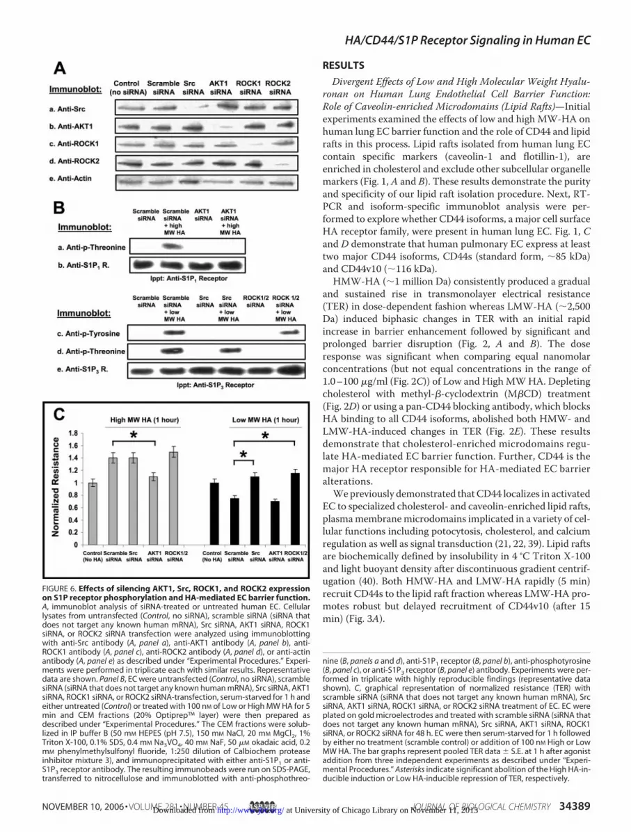
FIGURE 6. Effects of silencing AKT1, Src, ROCK1, and ROCK2 expressionon S1P receptor phosphorylation and HA-mediated EC barrier function.A, immunoblot analysis of siRNA-treated or untreated human EC. Cellularlysates from untransfected (Control, no siRNA), scramble siRNA (siRNA thatdoes not target any known human mRNA), Src siRNA, AKT1 siRNA, ROCK1siRNA, or ROCK2 siRNA transfection were analyzed using immunoblottingwith anti-Src antibody (A, panel a), anti-AKT1 antibody (A, panel b), anti-ROCK1 antibody (A, panel c), anti-ROCK2 antibody (A, panel d), or anti-actinantibody (A, panel e) as described under “Experimental Procedures.” Experi-ments were performed in triplicate each with similar results. Representativedata are shown. Panel B, EC were untransfected (Control, no siRNA), scramblesiRNA (siRNA that does not target any known human mRNA), Src siRNA, AKT1siRNA, ROCK1 siRNA, or ROCK2 siRNA-transfection, serum-starved for 1 h andeither untreated (Control) or treated with 100 nM of Low or High MW HA for 5min and CEM fractions (20% Optiprep™ layer) were then prepared asdescribed under “Experimental Procedures.” The CEM fractions were solub-lized in IP buffer B (50 mM HEPES (pH 7.5), 150 mM NaCl, 20 mM MgCl2, 1%Triton X-100, 0.1% SDS, 0.4 mM Na3VO4, 40 mM NaF, 50 �M okadaic acid, 0.2mM phenylmethylsulfonyl fluoride, 1:250 dilution of Calbiochem proteaseinhibitor mixture 3), and immunoprecipitated with either anti-S1P1 or anti-S1P3 receptor antibody. The resulting immunobeads were run on SDS-PAGE,transferred to nitrocellulose and immunoblotted with anti-phosphothreo-
nine (B, panels a and d), anti-S1P1 receptor (B, panel b), anti-phosphotyrosine(B, panel c), or anti-S1P3 receptor (B, panel e) antibody. Experiments were per-formed in triplicate with highly reproducible findings (representative datashown). C, graphical representation of normalized resistance (TER) withscramble siRNA (siRNA that does not target any known human mRNA), SrcsiRNA, AKT1 siRNA, ROCK1 siRNA, or ROCK2 siRNA treatment of EC. EC wereplated on gold microelectrodes and treated with scramble siRNA (siRNA thatdoes not target any known human mRNA), Src siRNA, AKT1 siRNA, ROCK1siRNA, or ROCK2 siRNA for 48 h. EC were then serum-starved for 1 h followedby either no treatment (scramble control) or addition of 100 nM High or LowMW HA. The bar graphs represent pooled TER data � S.E. at 1 h after agonistaddition from three independent experiments as described under “Experi-mental Procedures.” Asterisks indicate significant abolition of the High HA-in-ducible induction or Low HA-inducible repression of TER, respectively.
HA/CD44/S1P Receptor Signaling in Human EC
NOVEMBER 10, 2006 • VOLUME 281 • NUMBER 45 JOURNAL OF BIOLOGICAL CHEMISTRY 34389 at University of Chicago Library on November 11, 2013http://www.jbc.org/Downloaded from

HA/CD44/S1P Receptor Signaling in Human EC
34390 JOURNAL OF BIOLOGICAL CHEMISTRY VOLUME 281 • NUMBER 45 • NOVEMBER 10, 2006 at University of Chicago Library on November 11, 2013http://www.jbc.org/Downloaded from

Transactivation of S1P Receptors Are Involved in HA-medi-ated Lung Vascular Barrier Regulation in a CD44 Isoform-spe-cific Manner—We next explored whether HA induces physicaland/or functional associations between CD44 and S1P recep-tors, which may be involved in HA-mediated vascular barrierresponses. HMW-HA (100 nM) induced CD44s association inlipid rafts with S1P1, the known barrier-promoting S1P recep-tor (Fig. 3B). In contrast, LMW-HA initially recruited the S1P1receptor followed by recruitment of S1P3 receptors. Immuno-precipitation followed by immunoblotting from lipid raft frac-tions revealed that HMW-HA promotes S1P1 receptor associ-ation with CD44s. In contrast, LMW-HA (100 nM) induced aninitial CD44s association with S1P1 followed by CD44v10 asso-ciation with S1P3 receptor in lipid raft fractions. Both the spa-tially specific actin cytoskeletal reorganization and TER alter-ations evoked by HMW-HA and LMW-HA were abolished byeither M�CD (to inhibit lipid raft formation), by anti-CD44blocking antibody or by siRNAs specific for CD44 (Figs. 2 and 4,Table 1). Silencing S1P1 receptor blocked the EC barrierenhancing effects of High MWHAwhile silencing S1P3 recep-tor blocked the EC barrier disruptive effects of Low MW HA(Fig. 4). Consistent with HA-mediated S1P transactivation,HMW-HApromotedAKT1-mediated threonine phosphoryla-tion of S1P1 receptor whereas LMW-HA induced sequentialAKT1-mediated S1P1 and Src/ROCK1/2-mediated S1P3 recep-tor phosphorylation/activation (Figs. 5 and 6). These resultswere confirmed by using in vitro phosphorylation of S1P recep-tors with recombinant AKT1, Src, ROCK1 and ROCK2 (Fig.5C). Further, silencing AKT1 expression blocks HWM-HA-mediated EC barrier enhancement while silencing Src or bothROCK 1 and 2 expression blocks LMW-HA-mediated EC bar-rier disruption (Fig. 6C). Thus, low and high MWHA promotedifferential CD44 isoform-specific association with and activa-tion of S1P receptors in lipid rafts. Activation of S1P1 receptor isrequired for HA-induced EC barrier enhancement while S1P3receptor activation promotes barrier disruption.Role of RhoA and Rac1 Signaling on HA-induced EC Barrier
Function—Wehave previously demonstrated that theRho fam-ily GTPase, Rac1, regulates S1P-mediated EC barrier enhance-ment (4). We examined whether Rho family GTPases couldplay a role in the HA-specific regulatory responses and identi-fied that either LMW-HA (5min.) or HMW-HA (5, 15, 30min)
induced Rac1 activation in concert with recruitment of theRac1-specific exchange factor, Tiam1, to EC lipid rafts (Fig.7). Rac1 activation was inhibited by siRNA for S1P1 (but notS1P3) to reduce receptor expression. The HA-induced ECbarrier enhancement was inhibited by silencing Rac1 (butnot RhoA) expression. In contrast, LMW-HA (but notHMW-HA) recruited the RhoA exchange factor, p115 Rho-GEF, to EC lipid rafts at 15–30 min. and promoted RhoAactivation. LMW-HA-induced RhoA activation was inhib-ited by siRNA for S1P3 (but not S1P1) and LMW-HA-in-duced EC barrier disruption was inhibited by silencing RhoA(but not Rac1) expression.Finally, silencing either S1P1 or Rac1 expression attenuated
EC barrier-enhancing effects of HMW-HA and LMW-HAwhereas silencing S1P3 or RhoA expression diminished the ECbarrier-disruptive response to LMW-HA (Figs. 4 and 7). Theseresults suggest that HA promotes cytoskeletal reorganizationand EC barrier regulation through differential CD44 isoforminteraction with S1P receptors via RhoA/Rac1 signaling inlipid rafts. Transactivation of S1P1 receptor may represent acommon mechanism for receptor-mediated vascular barrierregulation.HA-induced, CD44, and S1P Receptor-dependent, Cytoskel-
etal Reorganization in EC—TER measurements of EC barrierfunction in vitro revealed that reduction in expression of eitherS1P1 or Rac1 attenuated the barrier-enhancing effects of lowand high MWHA; whereas reduction in S1P3 or RhoA expres-sion attenuated the delayed barrier-disruptive response to lowMWHAon EC (Figs. 4 and 7). As cytoskeletal reorganization isa fundamental element of virtually all EC barrier-regulatoryresponses, we compared phalloidin staining of HA- and S1P-challenged EC to visualize cellular F-actin localization (Fig. 8and Table 1). At early time points (5 min) both low and highMW HA induced prominent cortical actin ring formation,which was attenuated by reduction of S1P1 (but not S1P3),AKT, or Rac1 (but not RhoA) expression, findings similar tothat reported for S1P (4, 29). Low MW HA challenge for 30min., however, resulted in a loss of cortical actin staining withincreased F-actin stress fiber formation, whichwas significantlyattenuated by silencing S1P3 (but not S1P1) or RhoA (but notRac1) expression.
FIGURE 7. S1P receptor regulation of HA-induced RhoA/Rac1 signaling and EC permeability. A, EC were grown to confluency, serum-starved for 1 h and eitheruntreated (Control) or treated with 100 nM of Low or High MW HA for 5, 15, or 30 min and CEM fractions (20% Optiprep™ layer) were then prepared as described under“Experimental Procedures.” The CEM fractions were run on SDS-PAGE, transferred to nitrocellulose, and immunoblotted with anti-Tiam-1 (A, panel a), anti-p115RhoGEF (A, panel b), or anti-caveolin-1 (A, panel c) antibody. Experiments were performed in triplicate with highly reproducible findings (representative data shown).B, EC were treated with scramble siRNA (Control), S1P1 receptor siRNA, or S1P3 receptor for 48 h. EC were grown to confluency, serum-starved for 1 h and eitheruntreated (Control) or treated with 100 nM of High (B, panel a) or Low (B, panel b) MW HA for 5, 15, or 30 min. EC were then solublize in IP buffer A and incubated withp21-binding domain (PBD)-conjugated beads to bind activated (GTP-bound form) Rac1. The PBD bead-associated material was run on SDS-PAGE, transferred tonitrocellulose and immunoblotted with anti-Rac1 antibody. Experiments were performed in triplicate with highly reproducible findings (representative data shown).C, EC were treated with scramble siRNA (Control), S1P1 receptor siRNA or S1P3 receptor for 48 h. EC were grown to confluency, serum-starved for 1 h and eitheruntreated (Control) or treated with 100 nM of High (C, panel a) or Low (C, panel b) MW HA for 5, 15, or 30 min. EC were then solublized in IP buffer A and incubated withrho-binding domain (RBD)-conjugated beads to bind activated (GTP-bound form) RhoA. The RBD bead-associated material was run on SDS-PAGE, transferred tonitrocellulose, and immunoblotted with anti-RhoA antibody. Experiments were performed in triplicate with highly reproducible findings (representative data shown).D, immunoblot analysis of siRNA-treated or untreated human EC. Cellular lysates from untransfected (control, no siRNA), scramble siRNA (siRNA that does not targetany known human mRNA), RhoA siRNA, or Rac1 siRNA-transfection were analyzed using immunoblotting with anti-RhoA antibody (panel a), anti-Rac1 antibody (panelb), anti-caveolin-1 antibody (panel c), or anti-actin antibody (panel d) as described under “Experimental Procedures.” Experiments were performed in triplicate eachwith similar results. Representative data are shown. E, graphical representation of normalized resistance (TER) with scramble, RhoA, or Rac1 siRNA treatment of EC. ECwere plated on gold microelectrodes and treated with scramble siRNA (Control), RhoA siRNA or Rac1 siRNA for 48 h. EC were then serum starved for one hour followedby either no treatment (scramble control) or addition of 100 nM High or Low MW HA. The bar graphs represent pooled TER data � S.E. 1 h after agonist addition fromthree independent experiments as described under “Experimental Procedures.”
HA/CD44/S1P Receptor Signaling in Human EC
NOVEMBER 10, 2006 • VOLUME 281 • NUMBER 45 JOURNAL OF BIOLOGICAL CHEMISTRY 34391 at University of Chicago Library on November 11, 2013http://www.jbc.org/Downloaded from

Role of S1P1 Receptor as a Central Regulator of EC Permeabil-ity—We recently demonstrated that the S1P1 receptor regu-lates activated protein C (APC)/endothelial cell protein Creceptor (EPCR)-mediated EC barrier protection againstedemagenic agents such as thrombin (41). As silencing the S1P1receptor reduces the barrier enhancement induced by HA, andboth LMW-HA and HMW-HA promote transactivation ofS1P1 receptor during the EC barrier-enhancing stages of theseagonists, we next explored whether S1P1 receptor serves as acentral regulator of EC barrier function (Fig. 4D). Reductions inS1P1 receptor expression significantly modulated the barrier-regulatory effects of human lung EC challenged with HGF,PDGF, VEGF, or ATP (4, 42). In contrast, thrombin, a knownEC barrier-disruptive agent, was unaffected by S1P1 receptorsilencing, suggesting that the S1P1 receptor serves as a criticaland central regulator of EC barrier function.
DISCUSSION
Agents that exhibit the capacity to restore barrier integrityafter periods of increased vascular permeability have obvioustherapeutic applications in diverse inflammatory syndromes aswell as in conditions such as tumor angiogenesis and athero-sclerosis (4, 42). We explored the effects of HA, a major glycos-aminoglycan, which exists inmultipleMW forms (6, 43), on ECpermeability, We found that the high MW form (�1,000,000Da) promotes increased EC barrier integrity in vitro and pro-pose that high MW HA plays an important role in providing aprotective barrier between endothelial cells and underlying vas-culature in vivo. In contrast, hyaluronan fragments of �2,500Da (low MW HA), previously shown to be angiogenic (19),induces a biphasic effect on EC permeability with a brief, bar-rier-enhancing phase followed by a prolonged barrier-disrup-tive phase (Fig. 9).CD44 is highly likely to be important in lung disease as
CD44�/�mice develop lung fibrosis, inflammatory cell recruit-ment, and hyaluronan fragment accumulation at sites of lunginjury (44). Both highMWHA and its fragments bind to CD44,however, high and low MW HA evoke highly specific cellular
functions. The high MW HA induces CD44s-mediated trans-activation (AKT-dependent threonine phosphorylation) ofS1P1 receptor and consequent Rac1 signaling leading to corticalactin thickening and barrier enhancement in human pulmo-nary EC. Silencing S1P1 receptor, AKT1 or Rac1 reverses thebarrier-protective effects of high MW HA. In contrast, lowMW HA promotes CD44v10-mediated transactivation (Src-dependent tyrosine phosphorylation and ROCK1/2-dependentthreonine phosphorylation) of S1P3 receptor and RhoA signal-ing leading to EC barrier disruption. Silencing S1P3 receptor,Src, ROCK1/2, or RhoA reverses the barrier disruptive effectsof low MW HA. We previously demonstrated that CD44v10promotes RhoA activation with consequent Rho kinase(ROCK) activity (22) which regulates Ca2� signaling and EC
FIGURE 8. HA-induced EC cortical actin rearrangement. EC were serum-starved for 1 h and either untreated (Control), or treated with 100 nM High (A)or Low (B) MW HA for 5 or 30 min. Cells were then fixed and stained withTRITC-phalloidin (to visualize F-actin) and analyzed using fluorescent micros-copy. These observations are representative of the entire cell monolayer andwere reproduced in multiple independent experiments (at least n � 3 foreach condition).
FIGURE 9. Schematic diagrams illustrating the effects of LMW-HA andHWM-HA on EC barrier function. A, high MW HA binds to CD44s (A-1), whichpromotes recruitment of CD44s to CEM (lipid rafts) (A-2). This promotes AKT1-mediated transactivation (threonine phosphorylation) of the S1P1 receptor inCEM (A-3), Tiam1 recruitment to lipid rafts (A-4) and Rac1 activation (A-5) leadingto cytoskeletal reorganization/cortical actin formation (A-6) and increased ECbarrier function (A-7). B, initially, Low MW HA induces a signal transduction cas-cade as shown in A. However, after �15–30 min, Low MW HA ligates CD44v10(B-1), which promotes recruitment of CD44v10 to CEM (lipid rafts) (B-2). This pro-motes Src-mediated transactivation (tyrosine phosphorylation) and ROCk1/2-mediated transactivation (threonine phosphorylation) of the S1P3 receptor inCEM (B-3), RhoGEF recruitment to lipid rafts (B-4), and RhoA activation (B-5) lead-ing to cytoskeletal reorganization/decreased cortical actin (B-6), and decreasedEC barrier function (B-7).
HA/CD44/S1P Receptor Signaling in Human EC
34392 JOURNAL OF BIOLOGICAL CHEMISTRY VOLUME 281 • NUMBER 45 • NOVEMBER 10, 2006 at University of Chicago Library on November 11, 2013http://www.jbc.org/Downloaded from
migration. In the present study, lowMWHAwas found to be apotent inducer of CD44v10 signaling, S1P3 receptor activationand RhoA-mediated EC barrier disruption. The effects of Ca2�
signaling on HA-induced EC barrier regulation are currentlyunder investigation.Caveolin-enriched microdomains or lipid rafts, are impor-
tant plasma membrane microdomains that regulate numerousEC functions (45, 46). CD44 localization in lipid rafts is impor-tant forHA-mediated signaling (21, 30) as cholesterol depletionblocks both low and high MW HA-induced EC barrierresponses. Further, CD44 isoform-specific activation of S1Preceptors occurs in lipid rafts indicating that these microdo-mains play an important role in HA-induced EC functions. Wehave previously demonstrated that PI3 kinase and Rac1 signal-ing from lipid rafts are important for S1P1 receptor-mediatedEC barrier enhancement (29). The ability to potentially targetthesemicrodomains as ameans of drug delivery for edemagenicstates has significant promise.HA/CD44-mediated stimulation of Src (pp60Src, c-Src
tyrosine kinase) activity has been shown to regulate cytoskel-etal function (28). In agreement with our results, researchershave reported that activation of Src promotes cytoskeletal-mediated EC barrier disruption (47–49). In particular, Srcregulates EC contraction and vascular permeability (48).Inhibition of Src reduces edema and stabilizes a VEGF recep-tor 2/cadherin complex after myocardial infarction (50). Therole of Src activation on receptor tyrosine kinases and adhe-sion proteins in EC barrier function are currently beinginvestigated in our laboratory.Actin cytoskeletal reorganization plays a key role in EC barrier
regulatory responses to a variety of agents (3, 4).We observed thatHA promotes cytoskeletal reorganization and EC barrier regula-tion via differential CD44 isoform interaction with S1P receptorsandRhoA/Rac1 signaling in lipid rafts. Inparticular, highMWHAinducescortical actin ring formationwhile lowMWHAtreatmentofECfor15or30minpromotesactin stress fiber formation.Theseresults demonstrate the requirement for S1P1 receptor transacti-vation in agonist-induced EC barrier enhancement, and thepotential for S1P1 activation to represent a common mechanismfor receptor-mediated vascular barrier regulation.
REFERENCES1. Luscher, T. F., and Barton, M. (1997) Clin. Cardiol. 20, II-3–102. Pearson, J. D. (1991) Radiology 179, 9–143. Dudek, S. M., and Garcia, J. G. (2001) J. Appl. Physiol. 91, 1487–15004. Garcia, J. G., Liu, F., Verin, A. D., Birukova, A., Dechert, M. A., Gerthoffer,
W. T., Bamberg, J. R., and English, D. (2001) J. Clin. Investig. 108, 689–7015. Turley, E. A., Noble, P. W., and Bourguignon, L. Y. (2002) J. Biol. Chem.
277, 4589–45926. Toole, B. P. (2004) Nat. Rev. Cancer 4, 528–5397. Scott, J. E., and Heatley, F. (2002) Biomacromolecules 3, 547–5538. Csoka, A. B., Frost, G. I., and Stern, R. (2001)Matrix Biol. 20, 499–5089. Camenisch, T. D., Spicer, A. P., Brehm-Gibson, T., Biesterfeldt, J., Augus-
tine, M. L., Calabro, A., Jr., Kubalak, S., Klewer, S. E., and McDonald, J. A.(2000) J. Clin. Investig. 106, 349–360
10. Camenisch, T. D., Schroeder, J. A., Bradley, J., Klewer, S. E., and Mc-Donald, J. A. (2002) Nat. Med. 8, 850–855
11. Mohamadzadeh, M., DeGrendele, H., Arizpe, H., Estess, P., andSiegelman, M. (1998) J. Clin. Investig. 101, 97–108
12. Bensadoun, E. S., Burke, A. K., Hogg, J. C., and Roberts, C. R. (1996)Am. J.Respir. Crit. Care Med. 154, 1819–1828
13. Dentener, M. A., Vernooy, J. H., Hendriks, S., and Wouters, E. F. (2005)Thorax 60, 114–119
14. Nettelbladt, O., andHallgren, R. (1989)Am. Rev. Respir. Dis. 140, 1028–103215. Teder, P., and Heldin, P. (1997) Am. J. Respir. Cell Mol. Biol. 17, 376–38516. Cantor, J. O., and Turino, G. M. (2004) Chest 125, 288–29217. Stern, R. (2003) Glycobiology 13, 105R–115R18. Tammi, M. I., Day, A. J., and Turley, E. A. (2002) J. Biol. Chem. 277,
4581–458419. Slevin, M., Kumar, S., and Gaffney, J. (2002) J. Biol. Chem. 277,
41046–4105920. Toole, B. P., Wight, T. N., and Tammi, M. I. (2002) J. Biol. Chem. 277,
4593–459621. Singleton, P. A., and Bourguignon, L. Y. (2004)Exp. Cell Res. 295, 102–11822. Singleton, P. A., and Bourguignon, L. Y. (2002)CellMotil. Cytoskeleton 53,
293–31623. Lokeshwar, V. B., Iida, N., and Bourguignon, L. Y. (1996) J. Biol. Chem.
271, 23853–2386424. Hirano, H., Screaton, G. R., Bell,M. V., Jackson, D. G., Bell, J. I., andHodes,
R. J. (1994) Int. Immunol. 6, 49–5925. Gee, K., Kryworuchko, M., and Kumar, A. (2004) Arch. Immunol. Ther.
Exp. (Warsz) 52, 13–2626. Bourguignon, L. Y., Zhu, D., and Zhu, H. (1998) Front. Biosci. 3,
d637–d64927. Bourguignon, L. Y., Singleton, P. A., Zhu,H., andDiedrich, F. (2003) J. Biol.
Chem. 278, 29420–2943428. Bourguignon, L. Y., Zhu,H., Shao, L., andChen, Y.W. (2001) J. Biol. Chem.
276, 7327–733629. Singleton, P. A., Dudek, S. M., Chiang, E. T., and Garcia, J. G. (2005) Faseb
J. 19, 1646–165630. Oliferenko, S., Paiha, K., Harder, T., Gerke, V., Schwarzler, C., Schwarz,
H., Beug, H., Gunthert, U., and Huber, L. A. (1999) J. Cell Biol. 146,843–854
31. Spiegel, S., and Milstien, S. (2003) Nat. Rev. Mol. Cell. Biol. 4, 397–40732. Pyne, S., and Pyne, N. (2000) Pharmacol. Ther. 88, 115–13133. Waeber, C., Blondeau, N., and Salomone, S. (2004) Drug News Perspect.
17, 365–38234. Bourguignon, L. Y., Singleton, P. A., Diedrich, F., Stern, R., and Gilad, E.
(2004) J. Biol. Chem. 279, 26991–2700735. Pogrel, M. A., Low, M. A., and Stern, R. (2003) J. Oral Sci. 45, 85–9136. Calabro, A., Hascall, V. C., and Midura, R. J. (2000) Glycobiology 10,
283–29337. Min, H., and Cowman, M. K. (1986) Anal. Biochem. 155, 275–28538. Ren, X. D., Kiosses, W. B., and Schwartz, M. A. (1999) EMBO J. 18,
578–58539. Minshall, R. D., Sessa, W. C., Stan, R. V., Anderson, R. G., andMalik, A. B.
(2003) Am. J. Physiol. Lung Cell Mol. Physiol. 285, L1179–L118340. Harder, T., and Simons, K. (1997) Curr. Opin. Cell Biol. 9, 534–54241. Finigan, J. H., Dudek, S. M., Singleton, P. A., Chiang, E. T., Jacobson, J. R.,
Camp, S. M., Ye, S. Q., and Garcia, J. G. (2005) J. Biol. Chem. 280,17286–17293
42. Dudek, S.M., Jacobson, J. R., Chiang, E. T., Birukov, K. G.,Wang, P., Zhan,X., and Garcia, J. G. (2004) J. Biol. Chem. 279, 24692–24700
43. Konttinen, Y. T., Li, T. F., Mandelin, J., Ainola, M., Lassus, J., Virtanen, I.,Santavirta, S., Tammi, M., and Tammi, R. (2001) J. Pathol. 194, 384–390
44. Teder, P., Vandivier, R. W., Jiang, D., Liang, J., Cohn, L., Pure, E., Henson,P. M., and Noble, P. W. (2002) Science 296, 155–158
45. Gratton, J. P., Bernatchez, P., and Sessa, W. C. (2004) Circ. Res. 94,1408–1417
46. Harris, T. J., and Siu, C. H. (2002) Bioessays 24, 996–100347. Criscuoli, M. L., Nguyen, M., and Eliceiri, B. P. (2005) Blood 105,
1508–151448. Mucha, D. R., Myers, C. L., and Schaeffer, R. C., Jr. (2003) Am. J. Physiol.
Heart Circ. Physiol. 284, H994–H100249. Weis, S., Cui, J., Barnes, L., and Cheresh, D. (2004) J. Cell Biol. 167,
223–22950. Weis, S., Shintani, S., Weber, A., Kirchmair, R., Wood, M., Cravens, A.,
McSharry, H., Iwakura, A., Yoon, Y. S., Himes, N., Burstein, D., Doukas, J.,Soll, R., Losordo, D., and Cheresh, D. (2004) J. Clin. Investig. 113, 885–894
HA/CD44/S1P Receptor Signaling in Human EC
NOVEMBER 10, 2006 • VOLUME 281 • NUMBER 45 JOURNAL OF BIOLOGICAL CHEMISTRY 34393 at University of Chicago Library on November 11, 2013http://www.jbc.org/Downloaded from


