Embed Size (px)
Citation preview

Transcriptome of Extracellular Vesicles Released byHepatocytesFelix Royo1, Karin Schlangen2, Laura Palomo1, Esperanza Gonzalez1, Javier Conde-Vancells1,
Agustin Berisa3, Ana M. Aransay2, Juan M. Falcon-Perez
1 Metabolomics Unit, CIC bioGUNE, CIBERehd, Derio, Spain, 2 Genome Analysis Platform, CIC bioGUNE, CIBERehd, Derio, Spain, 3 R&D and Innovation Department, FAES
FARMA S.A., Leioa, Spain, 4 IKERBASQUE, Basque Foundation for Science, Bilbao, Spain
Abstract
The discovery that the cells communicate through emission of vesicles has opened new opportunities for betterunderstanding of physiological and pathological mechanisms. This discovery also provides a novel source for non-invasivedisease biomarker research. Our group has previously reported that hepatocytes release extracellular vesicles with proteincontent reflecting the cell-type of origin. Here, we show that the extracellular vesicles released by hepatocytes also carryRNA. We report the messenger RNA composition of extracellular vesicles released in two non-tumoral hepatic models:primary culture of rat hepatocytes and a progenitor cell line obtained from a mouse foetal liver. We describe differentsubpopulations of extracellular vesicles with different densities and protein and RNA content. We also show that the RNAcargo of extracellular vesicles released by primary hepatocytes can be transferred to rat liver stellate-like cells and promotetheir activation. Finally, we provide in vitro and in vivo evidence that liver-damaging drugs galactosamine, acetaminophen,and diclofenac modify the RNA content of these vesicles. To summarize, we show that the extracellular vesicles secreted byhepatocytes contain various RNAs. These vesicles, likely to be involved in the activation of stellate cells, might becomea new source for non-invasive identification of the liver toxicity markers.
Citation: Royo F, Schlangen K, Palomo L, Gonzalez E, Conde-Vancells J, et al. (2013) Transcriptome of Extracellular Vesicles Released by Hepatocytes. PLoSONE 8(7): e68693. doi:10.1371/journal.pone.0068693
Editor: Marinus F.W. t e Pas, Wageningen UR Livestock Research, The Netherlands
Received March 16, 2013; Accepted June 3, 2013; Published July 11, 2013
Copyright: � 2013 Royo et al. This is an open-access article distributed under the terms of the Creative Commons Attribution License, which permitsunrestricted use, distribution, and reproduction in any medium, provided the original author and source are credited.
Funding: This work was supported by grants from the Fondo de Investigaciones Sanitarias (Institute of Health Carlos III PS09/00526 and to JMF); Program‘‘Ramon y Cajal’’ of Spanish Ministry (to JMF); Centro de Investigacion Biomedica en Red en el Area tematica de Enfermedades Hepaticas y Digestivas (CIBERehd) isfunded by the Institute of Health Carlos III. Research infrastructure was supported by the Department of Industry, Tourism and Trade of the Government of theAutonomous Community of the Basque Country (Etortek Research Programs 2010/2013) and the Innovation Technology Department of the Bizkaia County. Thefunders had no role in study design, data collection and analysis, decision to publish, or preparation of the manuscript.
Competing Interests: Agustin Berisa is affiliated to a company and has participated in the work by performing the drug administration to the rats, and thesubsequent extraction of the serum from them. His participation in the current work does not alter the authors’ adherence to all the PLOS ONE policies on sharingdata and materials.
* E-mail: [email protected]
Introduction
In recent years, intercellular transference of active macromo-
lecules mediated by cell-released extracellular vesicles (EVs) has
become a recognized key regulatory mechanism in a growing
number of biological processes including development, cancer,
immunity and inflammation [1,2,3]. There are various mechan-
isms of formation of these vesicles, creating a complex repertory of
EVs [4]. The vesicles can be formed by outward budding from the
plasma membrane giving origin to so-called microparticles,
microvesicles, shedding particles or ectosomes [5]. Inward
budding of the membrane of endocytic organelles generates the
multivesicular bodies which contain another kind of vesicles, called
exosomes. Exosomes are released to extracellular media by the
fusion of multivesicular bodies to the plasma membrane [4].
Independently of their biogenesis, EVs carry lipids, proteins, and
nucleic acids: both coding and non-coding RNAs [6,7]. A wide
range of cells, of either tumoral or non-tumoral origin, can release
EVs to the culture media [8]. These EVs can be then captured by
other cells that will accept their cargo and, as a consequence, will
undergo modifications according to the encoded signals
[9,10,11,12,13]. EVs have been also detected in biological fluids
such as blood, urine, and ascitic fluid [14]. Thus, they have the
potential to release their cargo in both paracrine and long distance
manner; this feature is being widely exploited in non-invasive
disease biomarker discovery.
The extensive proteomic analysis of EVs in different cellular
systems have found some common as well as cell-type-specific
proteins [14]. In contrast, vesicle RNA content has not been
examined so widely, and in particular, very little is known about
vesicle messenger RNAs (mRNAs). Initial studies of mast [13] and
glioma [12] cells have shown the presence of both messenger and
microRNAs in the EVs. The subsequent research concentrated
mostly on the microRNAs, probably because of their direct
involvement in epigenetic regulatory processes [15,16].
Our group has previously reported that hepatic non-tumoral
cells and primary culture of hepatocytes can release EVs, including
exosomes [17]. We also performed the proteome analysis of EVs,
finding a large number of enzymes involved in endogenous and
xenobiotic metabolism [17,18]. These findings suggest that the
vesicles might be involved in response to stress conditions in the
liver. In the current work, we characterized the messenger RNAs
present in the EVs released in two hepatic cellular models using
microarray technology. We found that RNA content depends on
PLOS ONE | www.plosone.org 1 July 2013 | Volume 8 | Issue 7 | e68693

the cellular model used. We compared these different RNA sets to
other, already catalogued, messenger RNAs from various non-
tumoral cell types. As a result, we obtained a set of 223 RNAs
associated mainly with hepatocyte functions. Remarkably, we
found that some of those RNAs are enriched in the EVs in
comparison with the intracellular transcriptome. This result
suggests the existence of a regulated sorting mechanism to control
loading of specific transcripts into the EVs. We also validated some
of the RNAs by quantitative real-time PCR (qPCR) and
demonstrated that the RNA component is incorporated into and
affects the recipient cells. Finally, using in vitro and in vivo liver
disease models, we demonstrated that the hepatic RNA-containing
EVs are a suitable biological source for non-invasive biomarker
discovery.
Materials and Methods
Reagents, Cell CultureAll media and reagents for tissue culture were purchased from
GIBCO (Life Technologies Inc.). MLP29 is a murine liver
progenitor cell line [19]. The cell line called 8B is a rat
myofibroblastic hepatic stellate cell line (HSC) [20]. Analytical
grade reagents were mostly acquired from Sigma-Aldrich (St.
Louis, MO). Mouse monoclonal antibodies were purchased from
several vendors: anti-AIP1, anti-flotillin (clone 18) from BD
Biosciences (Mountain View, CA, USA), and anti-Tsg101 (clone
4A10) from Abcam (Cambridge, UK). Armenian hamster anti-
mouse Cd81 (clone Eat2) was purchased from Serotec (Oxford,
UK).
Animal ProceduresAll animal experimentation was conducted in accordance with
Spanish guidelines for the care and use of laboratory animals, and
protocols approved by the CIC bioGUNE ethical review
committee (Permit Number: P-CBG-CBBA-3610). All surgery
was performed under anaesthetic gas Isoflurane (IsoFLO, Abbott
Laboratories), and all efforts were made to minimize suffering. Rat
hepatocytes (RH) were obtained by liver perfusion of nine-week-
old healthy Sprague-Dawley male rats. For in vivo model of liver
damage, rats received an intraperitoneal injection of 1 g/kg/5 ml
of D(+)-galactosamine hydrochloride (GalN, Sigma-Aldrich). A
control group of animals received the same volume of saline
solution. Blood samples from each animal were drawn 18 h after
the injection, and the sera were transferred to fresh Eppendorf
tubes and stored at 280uC.
Production and Purification of EVsTo obtain EVs, MLP29 cells or fresh RH suspension were
plated in non-collagenized (MLP29) or collagen-coated (RH) 150 -
mm dishes, at 15–30 million cells per dish. Cells were cultured in
complete DMEM medium [Dulbecco’s modified Eagle medium
supplemented with 10% (v/v) foetal bovine serum (FBS), 0.1 mg/
ml streptomycin, and 100 units/ml penicillin (GIBCO, Life
Technologies Inc.)] for 24 h (MLP29 cells) or for 4 h (RH), at
37uC and 5% of CO2. The cells were washed twice with
Dulbecco’s modified phosphate-buffered saline (PBS) and in-
cubated for 48 h (MLP29 cells) or 36 h (RH) in 25 mM HEPES-
containing complete DMEM medium (contaminating vesicles
were first removed by overnight centrifugation at 1100006g [21]).
In cases of hepatotoxic drug treatment, the drugs were added to
the vesicle-depleted media. The concentrations employed were:
10 mM galactosamine (Sigma-Aldrich), 10 mM acetaminophen
(Sigma-Aldrich), or 400 mM diclofenac (Sigma-Aldrich). After
incubation, media were collected, and EVs were isolated as
previously described [22]. Briefly, culture supernatant was
centrifuged at 15006g for 10 min to remove lifted cells and
cellular debris. The resultant supernatant was filtered through
0.22 mm-pore filters, followed by ultracentrifugation at 100006g
and 1000006g for 30 min and 60 min, respectively. The resulting
pellets were resuspended in PBS, pooled, and again ultracentri-
fuged at 100,0006g for 60 min. The final pellet of EVs was
resuspended in PBS to between 1/700-th and 1/2000-th of the
original volume of the culture supernatant, and the aliquoted
solution was stored at 280uC. To purify the EVs further, sucrose
density gradients ranging from 2.0 to 0.25 M sucrose in HEPES
were prepared [21]. When indicated, the purification was
performed using Exoquick (SBI System Biosciences) in accordance
with the manufacturer’s protocol. Briefly, samples of serum or cell
media were pre-cleared by centrifugation at 3000 g for 30 min
and then mixed with 1 ml of Exoquick per 1 ml of media or 63 ml
per 250 ml of serum. After that, the samples were incubated at 4uCovernight and centrifuged at 15006g for 30 min.
EVs Capture and Hepatic Stellate Cell Line ActivationAssays
Capture experiments were conducted using the myofibroblastic
rat HSC 8B [20]. The cells were incubated in 6-well plates at 50
000 cells per well for 12 h, and then the medium was replaced
with OptiMEM (GIBCO, Life Technologies Inc.) supplemented
with 2% FBS. To standardize assays, total protein concentration
from EV preparations was measured by Bradford assay (Bio-Rad
Laboratories Inc). An aliquot (5 mg) of EVs was diluted in 1 ml
OptiMEM-2% FBS and filtered through filters with 0.22 mm pore
diameter before incubation with the cells for the indicated time
periods. As control, EVs were treated with RNase as described
below. At the end of incubation periods, cells were washed three
times with PBS, then scraped in PBS and centrifuged at 10006g
for 10 min. Cell pellets were kept frozen at 280uC until RNA
extraction and qPCRs were performed as described below.
To follow the capture of EVs through immunofluorescence,
80 ug of RH-derived EVs were labelled with DiI Vibrant-cell
labelling solution (Life Technologies, Inc). The label reaction took
place in 1 ml of PBS with 5 ul of cell labelling solution for 1 h at
37uC. After the reaction, EVs were recovered using a sucrose-
cushion as described in [23]. When indicated, EVs were treated
with Tx-100 and RNase as described below. Labelled EVs were
given to 8B cells cultured on glass covers, in the same amount and
condition than described above. As a negative control, the same
amount of label solution without EVs was subjected to sucrose-
cushion purification, and added to cells.
RNAse Protection AssaysTo test the ability of EVs to protect against RNase activity,
crude free human RNA (hRNA) was mixed with EVs preparations
and RNase A (100 ng/ml), and incubated for 15 min at 37uC, with
or without 0.1% Tx-100.
Electron MicroscopyFor cryo-electron microscopy, EV preparations were directly
adsorbed onto glow-discharged holey carbon grids (QUANTI-
FOIL, Germany). Grids were blotted at 95% humidity and rapidly
plunged into liquid ethane with the aid of VITROBOT
(Maastricht Instruments BV, The Netherlands). Vitrified samples
were imaged at liquid nitrogen temperature using a JEM-2200FS/
CR transmission cryo-electron microscope (JEOL, Japan)
equipped with a field emission gun and operated at an acceleration
voltage of 200 kV.
Transcriptome of Hepatocyte EVs
PLOS ONE | www.plosone.org 2 July 2013 | Volume 8 | Issue 7 | e68693

Nanoparticle Tracking Analysis (NTA)Size distribution within EV preparations was analyzed by
measuring the rate of Brownian motion using a NanoSight LM10
system (NanoSight, Amesbury, U.K.), which is equipped with
a fast video capture and particle-tracking software. NTA post-
acquisition settings were kept constant for all samples, and each
video was analyzed to give the mean, mode, and median vesicle
size, and an estimate of the concentration [24].
RNA Isolation, Characterization, and Gene ExpressionArrays
Total RNA isolation was achieved by two different methods.
Total RNAs for microarray hybridization were extracted using
TRIzol reagent (Life Technologies, Inc), and their integrity, size
and quantification were evaluated in RNA 6000 Pico Chips with
a Bioanalyzer (Agilent Technologies). Subsequently, cRNAs were
prepared using 0.3–0.5 ug of total RNA obtained from 250
millions of cells, according to manufacturers’ recommendations
and hybridized on MouseWG-6 v2.0 Expression BeadChips
(Illumina, Inc) in case of the murine MLP29 RNA, and on
Genechips Rat Genome 230 2.0 [Rat230_2] (AffymetrixH) for the
rat RH RNA. For array validation, characterization, gradients,
and cell capture, RNA was isolated using RNeasy columns
(Quiagen, Inc).
Reverse Transcriptase, PCR, and qPCRcDNA was synthesized from 0.1–0.5 mg of RNA using the
Quanta cDNA Supermix (Quanta Inc) following the manufac-
turer’s recommendations. To amplify gene fragments, PCR was
performed according to the manufacturer’s protocol for Taq
polymerase (Biotools). For relative quantification of genes, qPCR
was performed in duplicates using SYBR Green PCR Master Mix
((Bio-Rad Laboratories Inc) ) in an iCycler thermocycler (Bio-Rad
Laboratories Inc). Primers were designed using Primer3 [25], and
the sequences are listed in Table S1. The efficiency of each set of
primers was determined by serial dilution of the template cDNA.
The theoretical value obtained with the iCycler software was
10065% (Efficiency (%) = 1006[10(21/slope) –1]; Bio-Rad
Laboratories Inc).
To confirm the array results, three independent samples were
analyzed by qPCR for MLP29 and fold-change in gene expression
calculated using Rplp0 as reference gene, according to the formula:
2 - DDCt. In the experiments examining the stellate cell activation,
we calculated the increase in nitric oxide synthase 2 (Nos2)
transcript levels after EVs treatment using 28S rRNA gene as
a reference. Since no amplification for Nos2 was observed in the
untreated cells, mRNA level in EVs +0.1% Tx-100 samples was
taken as a control value.
Western Blot AnalysisThe protein concentration in the preparations was determined
by Bradford protein assay, using Bovine Serum Albumin (BSA) as
standard. Samples were incubated for 5 min at 37uC, 65uC and
95uC and separated on 4–12% pre-casted gels from Life
Technologies, Inc. After the transfer to nitrocellulose membranes,
the samples were blocked overnight (5% milk and 0.05% Tween-
20 in PBS), and the primary antibody was added for 1 h, followed
by PBS wash and the application of secondary HRP-conjugated
antibody. All proteins were detected under non-reducing condi-
tions. Chemiluminescence detection of bands was performed
using ECL Plus reagent (GE Healthcare, Buckinghamshire,
UK).
Gene Expression Array Data AnalysesRaw expression data were log2-transformed and quartile-
normalized using the lumi tool [26] in R/Bioconductor statistical
computing environment (http://www.bioconductor.org/). The
data for the probes with a detection p-value higher or equal to
0.01 were excluded. The data detected with p-value lower than
0.01 in at least one array were accepted as significant. Thus, the
data for the probes that did not pass the test were filtered out, and
the genes they represented were considered to be ‘‘not expressed’’.
For the detection of differentially expressed genes, a linear
model was fitted to the data and empirical Bayes moderated t-
statistics were calculated using the limma package [27] from
Bioconductor. P-values were adjusted using Benjamini and
Hochberg’s False Discovery Rate (FDR) Method.
To assess biological processes affected, the identified transcripts
were submitted to Ingenuity Pathway Analysis (IPA) program to
perform functional analysis or canonical pathway analysis.
Functional analyses identify functions that are significantly
enriched in the data set. Right-tailed Fisher’s Exact Test was
used to calculate p-values for determining the probability of each
biological function assigned to the data set to be due to chance
alone. Ingenuity-defined canonical pathways that were most
significantly enriched in the dataset were also reported. In this
case, Fisher’s Exact Test was used to calculate the probability that
the association between the genes in the dataset and the canonical
pathways can be explained by chance alone.
Statistical AnalysisTo calculate statistical correlation between qPCR and array
data, r2 coefficient of correlation and the associated p-value were
calculated using Prism5 (GraphPad Software Inc.). Using the same
software, ANOVA analysis with Bonferroni post-hoc test was
performed to calculate the differences between Nos2 expression
levels after different cell treatments. One asterisk denotes statistical
significance (p,0.05).
Results
EVs Released by Non-tumoral Hepatic Cells Carry theirOwn Transcriptome
First, we looked for RNA in EVs purified from two non-tumoral
hepatic cellular models – a murine cell line MLP29 and primary
culture of rat hepatocytes – as described previously [17]. Cryo-
electron microscopy and nanoparticle tracking (NTA) analyses
revealed that both cell types contained heterogeneous EV
populations with diameters between 50 and 500 nm although
the majority were smaller than 250 nm (Figure 1A–B). RNA
from these EVs preparations was extracted using TRIzol reagent,
which performs well with this type of samples [28]. Bioanalyzer
analysis of the extracts (Figure 1C–D) clearly showed that EVs
released by non-tumoral hepatocytes contain RNA transcripts of
different lengths and a reduced amount of the ribosomal fraction,
in agreement with the data obtained from other cell types
[12,13,29,30,31,32]. The amount of RNA relative to protein
content associated with EVs was higher in the murine MLP29
cellular model than in the primary culture of rat hepatocytes, with
average values of 3 and 0.5 ng of RNA per mg of protein,
respectively. Roughly, a million MLP29 cells produced 0.9 ng (SD
+/20.67, n = 3) of RNA and the same number of primary RH
cells yielded 1.2 ng (SD +/20.60, n = 3).
Next, we characterized the transcripts found in these vesicles by
performing a microarray expression analyses as described in
Materials and Methods, using the Illumina MouseWG-6 v2.0
Expression BeadChip for the murine MLP29 cells and Affymetrix
Transcriptome of Hepatocyte EVs
PLOS ONE | www.plosone.org 3 July 2013 | Volume 8 | Issue 7 | e68693

GeneChip Rat Genome 230 2.0 [Rat230_2] for the rat model
(primary hepatocyte culture). These microarray analyses were
deposited at the MIAME-express repository with ID number E-
MEXP-3740. A total of 6512 and 1336 transcripts were above the
significance threshold in the murine and rat models, respectively;
the sets shared 682 transcripts. The analysis of the molecular
functions represented by the transcriptome of each cell model
reflects the expected discrepancy due to their different cellular
origins. While EVs from adult primary hepatocytes are enriched in
transcripts related to metabolism (lipids, small molecules, vitamins)
and energy production, EVs from the embryonic liver-derived cell
line are enriched in transcripts that encode proteins associated
with cell death, cell cycle, and cellular organization (Figure 2).
The analysis of the canonical pathways provided by IPA program
also confirms enrichment in transcripts linked to metabolic
pathways in primary hepatocyte-derived EVs in comparison with
EVs from MLP29 cell line. The MLP29 EVs are enriched in
transcripts from proliferation-related pathways, including mTOR
and PI3K/AKT signalling (Figure 2).
The gene expression at the whole genome level (microarrays)
was only characterized for one sample per condition. To confirm
the observed expression level trends in a set of transcripts, we
performed qPCR for RNAs from three independent EV
preparations of each model. The transcripts were chosen by their
distinct intensity signal in the RH data (primary hepatocyte,
Tables S1 and S2) or by their differential enrichment in EVs
compared to intracellular RNA content (MLP29 cell line,
Figure 3A, Table S3). The Ct values in the qPCR analysis for
each cellular model correlated (r2 = 0.52, p = 0.005 and r2 = 0.41,
p = 0.02 for primary hepatocytes and MLP29 cells, respectively)
with the tendencies of the intensity signal measured in the
microarray expression analysis (Figure S1). For the MLP29 cell
line, the microarray analysis also included the comparison with the
intracellular transcripts present in those cells. Thus, 8856
transcripts were detected above the significance threshold in these
cells, and among them, 6345 were detected also within MLP29-
released EVs. Out of these common transcripts, 1673 were under-
represented, while 1930 were significantly enriched in EVs (fold-
change value higher than 2, p,0.001). To confirm the fold-
changes for some genes (see Table S1), qPCR for cells and EVs
were carried out. The results showed a high correlation (r2 = 0.77,
p = 0.0001) with the drift observed in the microarray expression
analysis (Figure 3B).
To perform subsequent experiments, a subset of genes was
chosen according to their enrichment or abundance in EVs. In the
case of MLP29, the subset includes acidic (leucine-rich) nuclear
phosphoprotein 32 family, member B (Anp32b), neuroepithelial
cell transforming 1 (Net1), and trafficking protein, kinesin binding
2 (Trak2). In the case of EVs derived from RH, we followed
albumin (Alb) and retinol binding protein 4, plasma (Rbp4).
Moreover, due to the relevance in the biological context of the
liver function, we also followed cytochrome P450, family 2,
subfamily d, polypeptide 1 (Cyp2d1),cytochrome P450, family 2,
subfamily e, polypeptide 1 (Cyp2e1), and guanine nucleotide
binding protein (G protein), beta polypeptide 2-like 1 (Gnb2l).
Figure 1. Characterization of EVs. Characterization of EVs from MLP29 (A, C) and rat primary hepatocytes (RH) (B, D). The NTA analyses of twoindependent samples for each cell type show more heterogeneous vesicle populations released by primary culture of hepatocytes. Cryo-TEM picturesshow membrane vesicles of different sizes (insets A–B). Bioanalyzer profiles of total RNA extracted by RNeasy from both cell types are similar witha wide distribution on size and also the presence of a reduced amount of ribosomal RNAs (C–D).doi:10.1371/journal.pone.0068693.g001
Transcriptome of Hepatocyte EVs
PLOS ONE | www.plosone.org 4 July 2013 | Volume 8 | Issue 7 | e68693
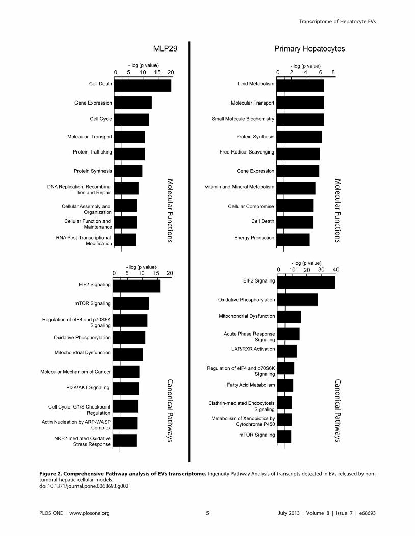
Figure 2. Comprehensive Pathway analysis of EVs transcriptome. Ingenuity Pathway Analysis of transcripts detected in EVs released by non-tumoral hepatic cellular models.doi:10.1371/journal.pone.0068693.g002
Transcriptome of Hepatocyte EVs
PLOS ONE | www.plosone.org 5 July 2013 | Volume 8 | Issue 7 | e68693

When our results are compared with other transcriptomes
reported for EVs of non-tumoral cellular models such as mast cells
[13], cardiomyocytes [33], and endothelial cells [34], 43
transcripts appear to be common to all those lists (Table S4),
and 223 are unique to our analysis (Table S5). IPA analysis of
common transcripts for all EV types (Figure 4A) showed a shared
background in protein synthesis and transcription machinery. In
addition, this analysis showed that the transcripts detected only in
the EVs from cell lines of hepatic origin are involved in fatty acid
metabolism and ethanol degradation (Figure 4B).
EV Protection Against RNase Activity and the Presence ofFull Coding Regions of Transcripts
The RNase protection feature reported for EVs [13] was also
confirmed for MLP29- and primary RH-derived EVs. This was
demonstrated by the PCR amplification in RNase-treated EVs
obtained for Anp32b and Net1 (enriched in MLP29-released EVs)
or Alb and Cyp2d1 (highly represented in primary RH-released
EVs) transcripts (lanes 2 Figure 5A–B). The presence of active
RNase in the sample was confirmed by the addition of exogenous
total human mRNA; the amplification of the gene encoding
hSOD protein was achieved only in the RNase-untreated samples
(lanes 1, 4, Figure 5A–B). The permeabilization of EVs using
detergent showed that the EV RNAs are susceptible to RNase
activity (lanes 3, Figure 5 A–B). To judge the integrity of
transcripts from EVs, we examined the coding region of Anp32b
gene in both cell models (Figure S2). We were able to amplify
a fragment containing the full coding sequence for the protein,
confirming the presence of full-length transcripts in these EVs.
Primary Hepatocytes Release a HeterogeneousPopulation of EVs
To examine the vesicle subpopulations in EVs preparations,
sucrose gradients were used as previously described [35]. The
protein markers associated with exosomes lay mainly between 1.09
and 1.21 g/ml fractions, matching the most abundant transcripts
identified in MLP29-released EVs (Figure 6 A). In contrast, the
profiles of primary RH-derived EVs were heterogeneous, in
agreement with NTA analysis (Figure 1B), and different
subpopulations could be defined (Figure 6B). A Cd81-containing
fraction with density of 1.13 g/ml carried detectable amounts of
Cyp2e1, Gapd, and Anp32b, but not Alb transcripts. Another
population of vesicles enriched in Alb and Gadph transcripts was
detected in a fraction with density of 1.19 g/ml. This fraction also
contained high levels of Cd81 and Flotillin, and low, although
detectable, levels of the exosomal protein marker Tsg101. The
third EV population, with density of 1.23 g/ml, was enriched in
Tsg101 protein and contained detectable levels of Cd81 and
Flotillin proteins. This population also contained Cyp2e1, but not
Alb or Gadph transcripts. Other populations containing Gadph were
also observed (Figure 6B). Overall, these results showed
differences between EVs released by different cell types and
revealed the complex composition of EVs released by primary
hepatocytes.
Activation of Hepatic Stellate Cells Mediated by RNA-containing EVs from Primary Hepatocytes
Liver contains various cell types that need to be finely
coordinated in order to respond to different stimuli and stress
conditions that continuously assault this essential organ [36].
HSCs belong to a liver-specific cell type with a major role in
fibrosis, an integrated response to liver injury and stressful
conditions [37]. To study the potential role of RNA carried in
EVs released by hepatocytes in the activation of stellate cells, we
incubated HSC 8B cells with EVs from primary cultured
hepatocytes (Figure 7). First, to examine the incorporation of
RNA from primary RH-derived EVs, we used the liver-specific
transcript Alb coding for Albumin, which is not expressed in 8B
cells [38]. The PCR amplification of this transcript could be
observed only in 8B cells incubated with intact hepatocyte-derived
EVs (lane 2, Figure 7A). This result strongly suggests a genetic
transfer through EVs. Furthermore, we were able to amplify Alb
transcript up to 24 hours after the incubation with EVs, which
indicates that the RNA is not degraded immediately. Next, we
evaluated the effect of these RH-derived EVs on the activation of
the stellate-like cell line. As previously described [38], functional
activation of 8B cells can be assessed by the measurement of
intracellular levels of the transcript coding for nitric oxide synthase
2 (Nos2), which is considered a marker of activated HSC [39,40].
The transcription of Nos2 in 8B cells was clearly increased by
incubation with hepatocyte-derived EVs (Figure 7B), confirming
stellate-activating properties of these vesicles. Importantly, this
effect was partly mediated by the RNA carried by the EVs; pre-
treating EVs with RNase in presence of Tx-100 inhibited this
Figure 3. Comparison between cellular and EVs transcriptomes in MLP29 cellular model. (A) Plot of fluorescent intensities for MLP29 EVsand cells reveals a group of genes enriched or underrepresented in EVs. (B) Plot shows the correlation of the fold changes estimated by microarrayanalysis and the fold change calculated by qPCR for a set of genes.doi:10.1371/journal.pone.0068693.g003
Transcriptome of Hepatocyte EVs
PLOS ONE | www.plosone.org 6 July 2013 | Volume 8 | Issue 7 | e68693

process (Figure 7B). It is noticeable that pre-treatment of EVs
with Tx-100 also reduces the induction on Nos2. This effect may
be explained by a reduction in the uptake efficiency, as showed in
Supplemental Figure 3.
RNA Cargo of Hepatocyte-Derived EVs Constitutesa Source for Diagnostic Purposes
In recent years, the number of studies examining the possible
application of EVs in medical diagnosis has increased greatly [41].
We analyzed by qPCR some transcripts from EVs released from
primary culture of hepatocytes treated with hepatotoxic drugs
galactosamine [42], acetaminophen [43], and diclofenac [44]
(Figure 8A). After treatment with these drugs, EVs were purified
from the culture media using Exoquick reagent (instead of
ultracentrifugation), a method more suitable for clinical practice.
As shown in Figure 8A, we found remarkable differences between
the RNA cargos of EVs released after different treatments. The
treatment with liver-damaging drugs clearly increased levels of
some RNAs in the culture media, such as Alb, Gnb2l, and Rbp4. In
the drug-treated hepatocytes, the amounts of Alb transcript
increased from 2 to more than 15-fold in comparison with
vehicle-treated hepatocytes. The levels of transcripts coding for
Gnb2l and Rbp4 proteins only increased significantly after
galactosamine and diclofenac treatments; the increase was more
pronounced in the latter case (Figure 8A). These results suggest
that RNA cargo isolated from hepatocyte-released EVs could serve
as a source of hepatotoxicity indicators in vitro. To complete this
study, RNAs obtained from EVs were analyzed as putative
indicators of liver injury in vivo. For this specific purpose, we
purified EVs from serum of rats treated with galactosamine
(controls treated with saline solution) using Exoquick reagent
(Figure 8B). EVs from the same volume of serum from each
animal were purified as described in Materials and Methods. As
shown in Figure 8B, the amplification of RNAs associated with
EVs, such as Alb, Gnb2l and Rbp4 was observed only in
galactosamine-injured animals, in agreement with the in vitro
results.
Discussion
The study of EVs as mediators of physiological and pathological
processes [11], therapeutics agents [45], and disease biomarkers
[46] has evolved rapidly in the recent years. The complexity of
their bioactive cargo, including proteins, RNA, microRNA, and
DNA, suggests multiple stages and mechanisms by which these
vesicles execute their functions. It is very important to characterize
the components of EVs in different cellular systems to understand
their functions and to elucidate the role of the RNA cargo in their
Figure 4. Comparison between the transcriptomes from MLP29 and RH’s EVs, and other published transcriptomes. Publishedtranscriptomes from endothelial, mesenchymal [13] and cardiomyocyte [33] EVs. The common genes among the five cell types (A) are enriched intranslation and ribosomal machinery categories. For the transcripts only detected MLP29 and RH (B), we found enrichment in pathways related tofatty acid metabolism and ethanol degradation.doi:10.1371/journal.pone.0068693.g004
Transcriptome of Hepatocyte EVs
PLOS ONE | www.plosone.org 7 July 2013 | Volume 8 | Issue 7 | e68693

activities. Early studies have shown that EVs released by mast cells
contain their own transcriptome formed by at least 1300 different
messenger RNAs [13]. Interestingly, this RNA cargo is modified
after exposure to oxidative stress, and the changes affect the ability
of recipient cells to handle oxidative stress [47]. RNA cargo has
been also characterized in EVs derived from cardiomyocytes; these
vesicles contain at least 1520 transcripts. Some of the transcripts
encode proteins involved in mitochondrial energy generation,
suggesting a metabolic role for these vesicles [33]. In EVs released
by endothelial progenitor cells, at least 298 different transcripts
were identified, and a role in the activation of angiogenic program
in quiescent endothelial cells has been proposed for these vesicles
[48]. De Jong and co-workers have reported the presence of at
least 1992 different transcripts in EVs released by microvascular
endothelial cells; they also demonstrated that the RNA cargo of
these vesicles changed under hypoxia or endothelial activating
conditions [34]. In EVs released by embryonic [49] or liver [50]
stem cells, 27 and 65 different transcripts, respectively, have been
detected and categorized. These transcripts are likely to be
involved in reprogramming haematopoietic progenitors [49] and
acceleration of hepatic regeneration [50]. These studies reveal the
complexity of the composition and functions of the RNA cargo of
EVs and highlight the need for further research on coding RNAs
in EVs released by non-tumoral cells. In the current work, we
characterized the coding RNA cargo of EVs released by primary
hepatocytes, detecting more than 1300 different transcripts. In
agreement with the results reported for mast cells [47], microvas-
cular endothelial [34] cells, and cardiomyocytes, we found
a significant enrichment in transcripts related to transcription,
response to oxidative stress, and energy production, indicating that
EVs could play a general role in these processes independently of
their cellular origin. In addition, we detected enrichment in gene
transcripts coding for proteins involved in lipid, small molecule
and xenobiotic metabolism, which are the functional processes of
hepatocytes. We also analyzed the RNA cargo in another non-
tumoral hepatocyte-like model, MLP29 cells, obtained from foetal
liver and presenting progenitor features [19]. In this model, we
detected the presence of more than 6500 different transcripts. We
found enrichment for the gene transcripts coding for regulatory
proteins belonging to various signalling pathways, such as EIF4,
mTOR, and PI3K/AKT, controlling cell cycle, cellular survival,
and proliferation. Thus, like in EVs obtained from primary
hepatocytes, the RNA cargo of MLP29-derived EVs reflects the
cellular origin of the vesicles; this feature could be useful for
diagnostics purposes. We also observed that, unlike the established
cell lines, primary cultures of hepatocytes release a heterogeneous
population of EVs carrying different RNA and protein cargos.
This phenomenon highlights the complexity of intercellular
communication mediated by EVs in the liver [36]. Maintaining
affinity of each target for a different vesicle subpopulation requires
complex cell systems to trigger specific responses. With more than
a thousand transcripts, either messenger or regulatory non-coding
RNAs, it is difficult to predict the possible effect of RNA cargo in
the recipient cells. In the present work, we showed that the
transcripts are captured and that the acceptor cells respond to the
RNA cargo. Previously published results have demonstrated that
EVs obtained from human liver stem cells accelerate regeneration
of the liver in vivo [50] and that this effect is RNA-dependent.
Here, we report that the Nos2 induction in HSC 8B, which has
been also observed in other studies [38], is partially dependent of
RNA cargo of hepatic EVs. The increase in Nos2 levels in HSC 8B
cells is a marker of cell activation. Nitric oxide synthase 2, the
product of Nos2 gene, is involved in triggering systemic vascular
responses associated with fibrotic and cirrhosis processes [39,40]
and also with regeneration [51]. It has been shown that EVs
released by liver stem cells accelerate liver regeneration [50]. The
activation of HSCs after liver injury has been described as
a transition from quiescent cells into a fibrogenic, proliferative and
contractile fibroblasts, which are critical in liver regeneration
(reviewed in [37,52]). Other studies have demonstrated the
activation of HSC by EVs derived from active HSC and T cells
[38,53]. Acute injury induces the release of EVs carrying
haematopoietic stem cell markers [54]; this gives some weight to
the hypothesis that to maintain its functionality, the liver interacts
with other tissues via circulating EVs [36].
Figure 5. RNase protection assay. Both MLP29 (A) and RH (B)transcripts can be detected even if RNase activity is present. We added1 mg of human RNA to 10 mg of EVs in PBS solution, and examined theextracted RNA samples for the presence of vesicular or human RNA(transcript SOD coding for human superoxide dismutase protein) (lane1). In the suspensions treated with RNase (lanes 2 and 3), the addedhuman RNA was degraded. The transcripts in EVs were only degraded ifdetergent had been added to the solution to disrupt the vesicularmembrane (lane 3). The detergent did not interfere with RNA extractionand subsequent PCR (lane 4).doi:10.1371/journal.pone.0068693.g005
Transcriptome of Hepatocyte EVs
PLOS ONE | www.plosone.org 8 July 2013 | Volume 8 | Issue 7 | e68693

Recently, blood-circulating EVs have become a subject of
growing interest as a minimally invasive resource for disease
diagnosis and treatment monitoring [46]. However, one of the
major challenges is to translate the basic research to the clinical
practice; to do that, we must be able to purify circulating EVs from
small volumes, using a methodology which can be applied in the
field. In our study, we tested the performance of Exoquick reagent
in obtaining fractions enriched in EVs (extracted from tissue
culture media and blood serum samples), with the purpose of
analyzing their RNA content. Our data show that Exoquick
reagent cannot precipitate naked RNA, and the RNA purified
from culture media using this reagent is resistant to RNase activity
Figure 6. Density fractionation of EVs. The figure shows sucrose gradients of EVs preparations from MLP29 (A) and RH (B). Aliquots of thesefractions were used for RNA extraction and protein extraction; the most abundant transcripts were found in the fractions containing typical exosomalmarkers (Tsg101 or Aip1). RH preparations showed more diversity, with vesicle populations fractionating at different densities.doi:10.1371/journal.pone.0068693.g006
Figure 7. Hepatic stellate cells capture hepatocyte-releasedEVs and become activated. HSC 8B cells capture EVs from RH andrespond to these stimuli by increasing Nos2 transcription. (A) Thecapture can be followed by detecting the presence of Alb, an RH-specific transcript, for up to 24 hours after capture. When EVs weretreated with RNase in the presence of detergent before incubation withthe cells, Alb transcript was not detected (6 h incubation time),indicating that the transcript must have been transferred from thehepatocyte-released EVs. (B) The activation of HSCs can be followed bythe expression of the protein nitric oxide synthase 2 (Nos2). Nos2 isexpressed at a very low level in HSC 8B (lanes 1 and 2), and clearlyexpressed if the cells are treated with EVs (lane 3 and 4), as previouslydescribed (31). After incubation with EVs pre-treated with RNase in thepresence of detergent, we do not detect Nos2 transcription (lane 5).HSC 8B cells incubated with EVs pre-treated only with detergentincrease the transcription of Nos2 (lane 6), although to lower levels thanthe cells treated with intact EVs (lanes 3 and 4). In the graph, error barsrepresent SD (n = 2),* denotes p,0.05 respect to control, lane 1.doi:10.1371/journal.pone.0068693.g007
Figure 8. RNA content of hepatic EVs could be useful inhepatotoxicity diagnostics. (A) Q-PCR analysis of the RNA cargo ofEVs released by hepatocytes after exposure to indicated drugs. (errorbars represent SD (n = 2), * denotes p,0.05 with respect to the control).(B) Q-PCR analysis of RNA cargo of EVs isolated from serum of untreated(U) rats and rats treated (T) with galactosamine (inducing acute liverinjury).doi:10.1371/journal.pone.0068693.g008
Transcriptome of Hepatocyte EVs
PLOS ONE | www.plosone.org 9 July 2013 | Volume 8 | Issue 7 | e68693
(Figure S4). These results confirm that Exoquick is suitable for
use in the diagnostic procedures designed to analyze EVs-
associated RNA. Several studies have proposed characterization
of EVs as a source of new disease biomarkers. Brodsky and co-
workers have shown that the levels of plasma-circulating EVs in
liver transplant patients dynamically change after surgery and
correlate with the clinical outcome, providing a marker of the
functional status of transplanted liver [55]. Moreover, the levels of
circulating liver-specific messenger RNAs for proteins such as
albumin, haptoglobin, and fibrinogen B can be also used as
indicators of liver injury in rats treated with hepatotoxicants (D-
galactosamine and acetaminophen) [56] although vesicular
association of those transcripts has not been examined. Our work
lends further support to the conclusions of that study; we
confirmed that the levels of Albumin transcript are increased in
EVs-enriched fractions from in vitro and in vivo liver injury models.
We also demonstrated that, by examining the levels of Alb and
Gnb2l transcripts, it is possible to distinguish between the
treatments used in this work: D-galactosamine, acetaminophen,
and diclofenac. Albumin levels were increased by all 3 treatments.
The levels of Gnb2l increased 5- and 20-fold after D-galactosamine
and diclofenac treatment, respectively, and acetaminophen
treatment had no effect on the levels of this transcript
(Figure 8A). Overall, these results suggest that some messenger
RNAs contained in EVs of hepatic origin could be added to the
repertoire of hepatic markers for detection of different liver
injuries.
In conclusion, the current work demonstrates the presence of
messenger RNAs in EVs released by non-tumoral hepatocytes and
shows that those messenger RNAs might play a role in the
activation of HSCs, the cells involved in the regulation of liver
inflammation and regeneration. Furthermore, this project brings
to light the complexity of the content of these vesicles as well as the
importance of vesicle cargo characterization in various EVs
populations, with functional and diagnostic perspectives in mind.
Supporting Information
Figure S1 Confirmation of the RNA identificationobtained by array hybridization. Array intensity trends
and obtained qPCR-Cts correlation for a group of genes. Each
point is the average of three independent qPCR experiments for
different EVs preparations vs. the array results for one sample
MLP29 (A) and one sample RH (B).
(TIF)
Figure S2 Integrity of the transcripts loaded in EVs.Amplification of the whole coding sequence of Anp32b protein
was achieved using RNAs from EVs derived from MLP29 and RH
cells as templates.
(TIF)
Figure S3 Treatment with Tx-100 reduces the efficiencyof EVs capture. (A) Stellate-like 8B cells were incubated with
fluorescently labelled-EVs derived from primary rat hepatocytes
either untreated or RNAse-treated with RNase in presence of Tx-
100 previous to the incubation. The capture was visualized after 6
hours by confocal microscopy. To highlight the differences,
representative images of the same field with low and high
brightness are showed for each condition. (B) 8B cells were
incubated in the absence or presence of RH-derived EVs that were
pre-treated as indicated. After 6-hours cells were recovered, RNA
extracted and subjected of RT-PCR to amplify Alb transcript.
Consistently with the phenomenon observed in (A), Tx-100 pre-
treatment (lane 4), reduce the presence of Alb transcript in the
recipient 8B cells. Pre-treatment of the EVs with Tx-100 and
RNase (lane 5) totally abrogated the detection of Alb transcript in
8B cells, as showed in Figure 7.
(TIF)
Figure S4 PCR amplifications of Net1, hSOD and TRAK2transcripts from samples obtained from cell mediausing Exoquick. (A) The transcripts obtained are resistant to
exogenous RNase activity in the cell media. (B) Exoquick does not
recover exogenous purified human RNA added to the cell culture.
(TIF)
Table S1 Primers employed for PCR and qPCR ampli-fications in the study.
(DOC)
Table S2 Most abundant transcripts (according tosignal intensity of the array) in EVs derived from RH.
(DOC)
Table S3 Top 50 enriched transcripts in MLP29 EVswhen compared to MLP29 cells.
(DOC)
Table S4 Transcripts only detected in MLP29 and RHafter comparison with other published EVs transcrip-tomes.
(DOC)
Table S5 Transcripts common to different publishedlist of EVs mRNA, MLP29 and RH.
(DOC)
Acknowledgments
We gratefully thank FAES FARMA for its support with rat experimen-
tation procedures and sample collection, Dr. E. Medico for providing the
MLP-29 cell line and CIC bioGUNE electron microscopy platform for the
support with TEM analysis of the samples.
Author Contributions
Conceived and designed the experiments: FR AMA JMF. Performed the
experiments: FR JCV EG AB LP. Analyzed the data: FR KS. Wrote the
paper: FR JMF.
References
1. Gutierrez-Vazquez C, Villarroya-Beltri C, Mittelbrunn M, Sanchez-Madrid F
(2013) Transfer of extracellular vesicles during immune cell-cell interactions.
Immunol Rev 251: 125–142.
2. Mathivanan S, Ji H, Simpson RJ (2010) Exosomes: extracellular organelles
important in intercellular communication. J Proteomics 73: 1907–1920.
3. Ohno SI, Ishikawa A, Kuroda M (2012) Roles of exosomes and microvesicles in
disease pathogenesis. Adv Drug Deliv Rev 2012: 00249–00249.
4. Simons M, Raposo G (2009) Exosomes–vesicular carriers for intercellular
communication. Curr Opin Cell Biol 21: 575–581.
5. Cocucci E, Racchetti G, Meldolesi J (2009) Shedding microvesicles: artefacts no
more. Trends Cell Biol 19: 43–51.
6. Kalra H, Simpson RJ, Ji H, Aikawa E, Altevogt P, et al. (2012) Vesiclepedia:
a compendium for extracellular vesicles with continuous community annotation.
PLoS Biol 10: e1001450.
7. Simpson RJ, Jensen SS, Lim JW (2008) Proteomic profiling of exosomes: current
perspectives. Proteomics 8: 4083–4099.
8. Bobrie A, Colombo M, Raposo G, Thery C (2011) Exosome secretion:
molecular mechanisms and roles in immune responses. Traffic 12: 1659–1668.
9. Mittelbrunn M, Gutierrez-Vazquez C, Villarroya-Beltri C, Gonzalez S,
Sanchez-Cabo F, et al. (2011) Unidirectional transfer of microRNA-loaded
exosomes from T cells to antigen-presenting cells. Nat Commun 2: 282.
Transcriptome of Hepatocyte EVs
PLOS ONE | www.plosone.org 10 July 2013 | Volume 8 | Issue 7 | e68693

10. Montecalvo A, Larregina AT, Shufesky WJ, Stolz DB, Sullivan ML, et al. (2012)
Mechanism of transfer of functional microRNAs between mouse dendritic cells
via exosomes. Blood 119: 756–766.
11. Peinado H, Aleckovic M, Lavotshkin S, Matei I, Costa-Silva B, et al. (2012)
Melanoma exosomes educate bone marrow progenitor cells toward a pro-
metastatic phenotype through MET. Nat Med 18: 883–891.
12. Skog J, Wurdinger T, van Rijn S, Meijer DH, Gainche L, et al. (2008)
Glioblastoma microvesicles transport RNA and proteins that promote tumour
growth and provide diagnostic biomarkers. Nat Cell Biol 10: 1470–1476.
13. Valadi H, Ekstrom K, Bossios A, Sjostrand M, Lee JJ, et al. (2007) Exosome-
mediated transfer of mRNAs and microRNAs is a novel mechanism of genetic
exchange between cells. Nat Cell Biol 9: 654–659.
14. Simpson RJ, Lim JW, Moritz RL, Mathivanan S (2009) Exosomes: proteomic
insights and diagnostic potential. Expert Rev Proteomics 6: 267–283.
15. Mittelbrunn M, Sanchez-Madrid F (2012) Intercellular communication: diverse
structures for exchange of genetic information. Nat Rev Mol Cell Biol 13: 328–
335.
16. O’Loughlin AJ, Woffindale CA, Wood MJ (2012) Exosomes and the emerging
field of exosome-based gene therapy. Curr Gene Ther 12: 262–274.
17. Conde-Vancells J, Rodriguez-Suarez E, Embade N, Gil D, Matthiesen R, et al.
(2008) Characterization and comprehensive proteome profiling of exosomes
secreted by hepatocytes. J Proteome Res 7: 5157–5166.
18. Conde-Vancells J, Gonzalez E, Lu SC, Mato JM, Falcon-Perez JM (2010)
Overview of extracellular microvesicles in drug metabolism. Expert Opin Drug
Metab Toxicol 6: 543–554.
19. Medico E, Mongiovi AM, Huff J, Jelinek MA, Follenzi A, et al. (1996) The
tyrosine kinase receptors Ron and Sea control ‘‘scattering’’ and morphogenesis
of liver progenitor cells in vitro. Mol Biol Cell 7: 495–504.
20. Greenwel P, Schwartz M, Rosas M, Peyrol S, Grimaud JA, et al. (1991)
Characterization of fat-storing cell lines derived from normal and CCl4-cirrhotic
livers. Differences in the production of interleukin-6. Lab Invest 65: 644–653.
21. Thery C, Regnault A, Garin J, Wolfers J, Zitvogel L, et al. (1999) Molecular
characterization of dendritic cell-derived exosomes. Selective accumulation of
the heat shock protein hsc73. J Cell Biol 147: 599–610.
22. Raposo G, Nijman HW, Stoorvogel W, Liejendekker R, Harding CV, et al.
(1996) B lymphocytes secrete antigen-presenting vesicles. J Exp Med 183: 1161–
1172.
23. Lamparski HG, Metha-Damani A, Yao JY, Patel S, Hsu DH, et al. (2002)
Production and characterization of clinical grade exosomes derived from
dendritic cells. J Immunol Methods 270: 211–226.
24. Dragovic RA, Gardiner C, Brooks AS, Tannetta DS, Ferguson DJ, et al. (2011)
Sizing and phenotyping of cellular vesicles using Nanoparticle Tracking
Analysis. Nanomedicine 7: 780–788.
25. Rozen S, Skaletsky H (2000) Primer3 on the WWW for general users and for
biologist programmers. Methods Mol Biol 132: 365–386.
26. Du P, Kibbe WA, Lin SM (2008) lumi: a pipeline for processing Illumina
microarray. Bioinformatics 24: 1547–1548.
27. Wettenhall JM, Smyth GK (2004) limmaGUI: a graphical user interface for
linear modeling of microarray data. Bioinformatics 20: 3705–3706.
28. Eldh M, Lotvall J, Malmhall C, Ekstrom K (2012) Importance of RNA isolation
methods for analysis of exosomal RNA: evaluation of different methods. Mol
Immunol 50: 278–286.
29. Chiba M, Kimura M, Asari S (2012) Exosomes secreted from human colorectal
cancer cell lines contain mRNAs, microRNAs and natural antisense RNAs, that
can transfer into the human hepatoma HepG2 and lung cancer A549 cell lines.
Oncol Rep 28: 1551–1558.
30. Hessvik NP, Phuyal S, Brech A, Sandvig K, Llorente A (2012) Profiling of
microRNAs in exosomes released from PC-3 prostate cancer cells. Biochim
Biophys Acta 1819: 1154–1163.
31. Huan J, Hornick NI, Skinner AM, Goloviznina NA, Roberts CT, et al. (2012)
RNA trafficking by acute myeloid leukemia exosomes. Cancer Res 2012: 13.
32. Kogure T, Lin WL, Yan IK, Braconi C, Patel T (2011) Intercellular nanovesicle-
mediated microRNA transfer: a mechanism of environmental modulation of
hepatocellular cancer cell growth. Hepatology 54: 1237–1248.
33. Waldenstrom A, Genneback N, Hellman U, Ronquist G (2012) Cardiomyocyte
microvesicles contain DNA/RNA and convey biological messages to target cells.PLoS ONE 7: e34653.
34. de Jong OG, Verhaar MC, Chen Y, Vader P, Gremmels H, et al. (2012)
Cellular stress conditions are reflected in the protein and RNA content ofendothelial cell-derived exosomes. Journal of Extracellular Vesicles; Vol 1 (2012)
incl Supplements.35. Thery C, Amigorena S, Raposo G, Clayton A (2006) Isolation and
characterization of exosomes from cell culture supernatants and biological
fluids. Curr Protoc Cell Biol Chapter 3: Unit 3 22.36. Royo F, Falcon-Perez JM (2012) Liver extracellular vesicles in health and
disease. Journal of Extracellular Vesicles; Vol 1 (2012) incl Supplements.37. Friedman SL (2000) Molecular regulation of hepatic fibrosis, an integrated
cellular response to tissue injury. J Biol Chem 275: 2247–2250.38. Witek RP, Yang L, Liu R, Jung Y, Omenetti A, et al. (2009) Liver cell-derived
microparticles activate hedgehog signaling and alter gene expression in hepatic
endothelial cells. Gastroenterology 136: 320–330 e322.39. DeLeve LD, Wang X, Hu L, McCuskey MK, McCuskey RS (2004) Rat liver
sinusoidal endothelial cell phenotype is maintained by paracrine and autocrineregulation. Am J Physiol Gastrointest Liver Physiol 287: G757–763.
40. Rockey DC, Chung JJ (1997) Regulation of inducible nitric oxide synthase and
nitric oxide during hepatic injury and fibrogenesis. Am J Physiol 273: G124–130.
41. Vlassov AV, Magdaleno S, Setterquist R, Conrad R (2012) Exosomes: currentknowledge of their composition, biological functions, and diagnostic and
therapeutic potentials. Biochim Biophys Acta 1820: 940–948.42. Keppler D, Lesch R, Reutter W, Decker K (1968) Experimental hepatitis
induced by D-galactosamine. Exp Mol Pathol 9: 279–290.
43. James LP, Mayeux PR, Hinson JA (2003) Acetaminophen-induced hepatotox-icity. Drug Metab Dispos 31: 1499–1506.
44. Castell JV, Gomez-Lechon MJ, Ponsoda X, Bort R (1997) The use of culturedhepatocytes to investigate the mechanisms of drug hepatotoxicity. Cell Biol
Toxicol 13: 331–338.
45. El-Andaloussi S, Lee Y, Lakhal-Littleton S, Li J, Seow Y, et al. (2012) Exosome-mediated delivery of siRNA in vitro and in vivo. Nat Protoc 7: 2112–2126.
46. Duijvesz D, Luider T, Bangma CH, Jenster G (2011) Exosomes as biomarkertreasure chests for prostate cancer. Eur Urol 59: 823–831.
47. Eldh M, Ekstrom K, Valadi H, Sjostrand M, Olsson B, et al. (2010) Exosomescommunicate protective messages during oxidative stress; possible role of
exosomal shuttle RNA. PLoS ONE 5: e15353.
48. Deregibus MC, Cantaluppi V, Calogero R, Lo Iacono M, Tetta C, et al. (2007)Endothelial progenitor cell derived microvesicles activate an angiogenic program
in endothelial cells by a horizontal transfer of mRNA. Blood 110: 2440–2448.49. Ratajczak J, Miekus K, Kucia M, Zhang J, Reca R, et al. (2006) Embryonic stem
cell-derived microvesicles reprogram hematopoietic progenitors: evidence for
horizontal transfer of mRNA and protein delivery. Leukemia 20: 847–856.50. Herrera MB, Fonsato V, Gatti S, Deregibus MC, Sordi A, et al. (2010) Human
liver stem cell-derived microvesicles accelerate hepatic regeneration inhepatectomized rats. J Cell Mol Med 14: 1605–1618.
51. Garcia-Trevijano ER, Martinez-Chantar ML, Latasa MU, Mato JM, Avila MA(2002) NO sensitizes rat hepatocytes to proliferation by modifying S-
adenosylmethionine levels. Gastroenterology 122: 1355–1363.
52. Hellerbrand C (2013) Hepatic stellate cells-the pericytes in the liver. PflugersArch.
53. Kornek M, Popov Y, Libermann TA, Afdhal NH, Schuppan D (2011) HumanT cell microparticles circulate in blood of hepatitis patients and induce fibrolytic
activation of hepatic stellate cells. Hepatology 53: 230–242.
54. Schmelzle M, Splith K, Andersen LW, Kornek M, Schuppan D, et al. (2013)Increased plasma levels of microparticles expressing CD39 and CD133 in acute
liver injury. Transplantation 95: 63–69.55. Brodsky SV, Facciuto ME, Heydt D, Chen J, Islam HK, et al. (2008) Dynamics
of circulating microparticles in liver transplant patients. J Gastrointestin Liver
Dis 17: 261–268.56. Wetmore BA, Brees DJ, Singh R, Watkins PB, Andersen ME, et al. (2010)
Quantitative analyses and transcriptomic profiling of circulating messengerRNAs as biomarkers of rat liver injury. Hepatology 51: 2127–2139.
Transcriptome of Hepatocyte EVs
PLOS ONE | www.plosone.org 11 July 2013 | Volume 8 | Issue 7 | e68693


