Embed Size (px)
Citation preview
membranes
Article
Transmigration across a Steady-State Blood–Brain BarrierInduces Activation of Circulating Dendritic Cells PartlyMediated by Actin Cytoskeletal Reorganization
Megha Meena 1 , Mats Van Delen 1, Maxime De Laere 1,2, Ann Sterkens 1,3, Coloma Costas Romero 1,Zwi Berneman 1,2 and Nathalie Cools 1,2,*
Citation: Meena, M.; Van Delen, M.;
De Laere, M.; Sterkens, A.; Costas
Romero, C.; Berneman, Z.; Cools, N.
Transmigration across a Steady-State
Blood–Brain Barrier Induces
Activation of Circulating Dendritic
Cells Partly Mediated by Actin
Cytoskeletal Reorganization.
Membranes 2021, 11, 700. https://
doi.org/10.3390/membranes11090700
Academic Editors: Marina Pinheiro
and Sandra Silva
Received: 2 June 2021
Accepted: 6 September 2021
Published: 13 September 2021
Publisher’s Note: MDPI stays neutral
with regard to jurisdictional claims in
published maps and institutional affil-
iations.
Copyright: © 2021 by the authors.
Licensee MDPI, Basel, Switzerland.
This article is an open access article
distributed under the terms and
conditions of the Creative Commons
Attribution (CC BY) license (https://
creativecommons.org/licenses/by/
4.0/).
1 Laboratory of Experimental Hematology, Vaccine & Infectious Disease Institute (VAXINFECTIO),Faculty of Medicine and Health Sciences, University of Antwerp, 2610 Wilrijk, Belgium;[email protected] (M.M.); [email protected] (M.V.D.);[email protected] (M.D.L.); [email protected] (A.S.); [email protected] (C.C.R.);[email protected] (Z.B.)
2 Center for Cell Therapy and Regenerative Medicine, Laboratory of Experimental Hematology,Antwerp University Hospital, 2650 Edegem, Belgium
3 Department of Dermatology, Antwerp University Hospital, 2650 Edegem, Belgium* Correspondence: [email protected]
Abstract: The central nervous system (CNS) is considered to be an immunologically unique site, inlarge part given its extensive protection by the blood–brain barrier (BBB). As our knowledge of thecomplex interaction between the peripheral immune system and the CNS expands, the mechanisms ofimmune privilege are being refined. Here, we studied the interaction of dendritic cells (DCs) with theBBB in steady–state conditions and observed that transmigrated DCs display an activated phenotypeand stronger T cell-stimulatory capacity as compared to non-migrating DCs. Next, we aimed togain further insights in the processes underlying activation of DCs following transmigration acrossthe BBB. We investigated the interaction of DCs with endothelial cells as well as the involvementof actin cytoskeletal reorganization. Whereas we were not able to demonstrate that DCs engulfmembrane fragments from fluorescently labelled endothelial cells during transmigration across theBBB, we found that blocking actin restructuring of DCs by latrunculin-A significantly impairedin vitro migration of DC across the BBB and subsequent T cell-stimulatory capacity, albeit no effecton migration-induced phenotypic activation could be demonstrated. These observations contributeto the current understanding of the interaction between DCs and the BBB, ultimately leading to thedesign of targeted therapies capable to inhibit autoimmune inflammation of the CNS.
Keywords: central nervous system; dendritic cells; blood–brain barrier; immune cells; endothelialcells; lymphocytes; transmigration; actin restructuring
1. Introduction
The blood–brain barrier (BBB) is a highly intricate, active interface between the circula-tion and the central nervous system (CNS), which restricts the free movement of pathogens,circulating immune cells, and biologically active factors between the bloodstream and thebrain microenvironment [1]. In doing so, the BBB plays a crucial role in the maintenance ofthe (immunological) homeostasis of the CNS, rendering the CNS an immune-privilegedand immunologically unique site [2,3]. Whereas the anatomical and functional basis ofBBB lies in the tight junctions formed between endothelial cells and their low pinocytoticactivity [4], these endothelial cells are in intimate contact with vascular cells (pericytesand vascular smooth muscle cells), glial cells consisting of microglia, astrocytes, and oligo-dendrocyte lineage cells, and neurons [5–7]. These components altogether maintain thestructure and integrity of the BBB [8]. The crosstalk and molecular signaling between thesedifferent cell types are collectively known as the neurovascular unit, which allows the BBB
Membranes 2021, 11, 700. https://doi.org/10.3390/membranes11090700 https://www.mdpi.com/journal/membranes
Membranes 2021, 11, 700 2 of 15
to properly perform its fundamental physiological functions. Other than that, pericytesplay a critical role during angiogenesis and regulating immune cell infiltration [9]. Inaddition, these cells have been shown to be important for regulating the formation of theBBB during development, as well as maintaining its function in adulthood and aging [10].Astrocytes, which ensheath almost 90% of brain microvasculature, are part of glial cellsand extend many branching cellular processes, including astrocytic end-feet [11,12]. Par-ticularly, astrocytic end-feet establish a close interaction with endothelial cells throughtransmembrane proteins anchoring, such as the water channel aquaporin-4 and the potas-sium channel KIR4.1 [13,14]. They also secrete angiopoietin-1 and angiotensin that restrictBBB permeability by supporting efficient organization of tight junctions [15].
To date, it is generally accepted that the CNS is under active surveillance rather thanfully immune quiescent [16–18]. Indeed, despite the presence of a physical barrier, interac-tions between the CNS and the peripheral immune system occur, and these are not limitedto pathology but also extend to homeostatic functions. In this context, peripheral immunecells cross the BBB and enter the steady-state CNS through mechanisms similar to thoseseen in peripheral organs, albeit at a lower rate [19]. On the other hand, due to its highlydynamic nature and sensitivity to pro-inflammatory stimuli, the BBB ensures enhancedrecruitment of immune cells to resolve local insults that would disrupt homeostasis andoptimal functioning of the CNS.
Of particular interest is the migration of dendritic cells (DCs) into and out of the CNS.These cells are involved in both immune-inducing and regulatory responses and are themost potent immune cells in terms of antigen presentation to and activation of T cells. Assuch, they critically regulate the balance between immunity and tolerance [20,21]. Thisalso explains why these cells play a pivotal role in the immunopathogenesis of severalneuroinflammatory disorders, including diseases such as multiple sclerosis (MS) [22,23],Alzheimer’s disease [24], and Parkinson’s disease [25]. In steady-state conditions, DCs arefound in low numbers in the meninges, choroid plexus, and cerebrospinal fluid (CSF) [26].In addition, they appear to migrate to the perivascular compartment, and stay in situ witha t1/2 of 5–7 days [27,28]. Previously, Zozulya et al. [29] found that the migration of murinein vitro bone marrow-derived DCs across a murine cerebral microvascular endothelialcell monolayer is regulated by CCL3. Importantly, transmigration of DCs upregulatedthe expression of costimulatory molecules and enhanced their T-cell stimulatory capacity.Nonetheless, the effects of BBB transmigration of DCs circulating in blood from humanvolunteers remains to be explored.
During the transmigration of leukocytes across the BBB, endothelial cells closelyinteract with migrating immune cells in various ways. For instance, endothelial cells (ECs)shred microparticles, which are known to affect a variety of immune cells. Indeed, it hasbeen reported that human brain microvascular endothelial cell-derived microparticlescould interact with and support the proliferation of T cells. Endothelial cell-derivedmicroparticles can express molecules important for antigen presentation and T cell co-stimulation, such as MHC class II and CD40, and consequently induce T cell activation [30].Moreover, it is also reported that endothelial cell-derived microparticles can specificallyinduce plasmacytoid dendritic cell maturation and their production of inflammatorycytokines [31]. In addition, Kedl et al. [32] demonstrated that migratory DCs acquireand present lymphatic endothelial cell-archived antigens during lymph node contraction.Subsequently, migratory DCs cross-present the lymphatic endothelial cell-archived antigensto circulating T cells. These findings prompted us to investigate the influence of thetransmigratory process on DCs and to investigate if DCs engulf endothelial fragments fromthe in vitro BBB endothelial cells while transmigrating, potentially resulting in an alteredphenotype and activation status.
Cell migration and cell–cell interactions require dynamic reorganization of the cell’sactin cytoskeleton [33]. Indeed, DC motility relies critically on the actin cytoskeleton, whichis, to an important extent, regulated by the actin-related protein 2/3 (ARP2/3) complex, anucleator of branched actin networks [34,35]. Consequently, loss of ARP2/3 stimulators
Membranes 2021, 11, 700 3 of 15
and upstream Rho family GTPases dramatically impairs DC migration [36]. Besides, studieshave shown that the DC actin cytoskeleton and in particular the F-actin network play arole in CD8+ T cell activation by DC and promotes DC-T cell adhesion by constrainingICAM-1 mobility [37,38]. In order to gain a deeper understanding of the process of DCmigration and to investigate whether any changes in the DC activation state followingtransmigration are a direct consequence of actin cytoskeleton restructuring, we studied theeffects of latrunculin-A pretreatment preceding migration. Latrunculin-A is an inhibitorof actin polymerization, which disrupts microfilament organization in cultured cells bybinding to monomeric G-actin in a 1:1 complex at sub micromolar concentrations [39].Latrunculin treatment leads to a complete depletion of the F-actin network in the cells andis therefore fit to study the importance of the cytoskeleton remodeling in migration-inducedDC activation.
Overall, in this study, we report differences in the activation state of human DCs follow-ing migration through a steady-state BBB. Next, we investigated whether this phenomenoncan be attributed to the interaction of DCs with endothelial cells while transmigrating andstudied the involvement of actin cytoskeleton remodeling. A clear understanding of thealterations caused in DC phenotype and function following their migration may advance thedevelopment of new therapies that intervene with the observed transmigration-mediatedactivation of DCs thereby modulating subsequent potentially autoreactive immune responses.
2. Material and Methods2.1. Cell Culture Model of Blood-Brain Barrier (BBB)
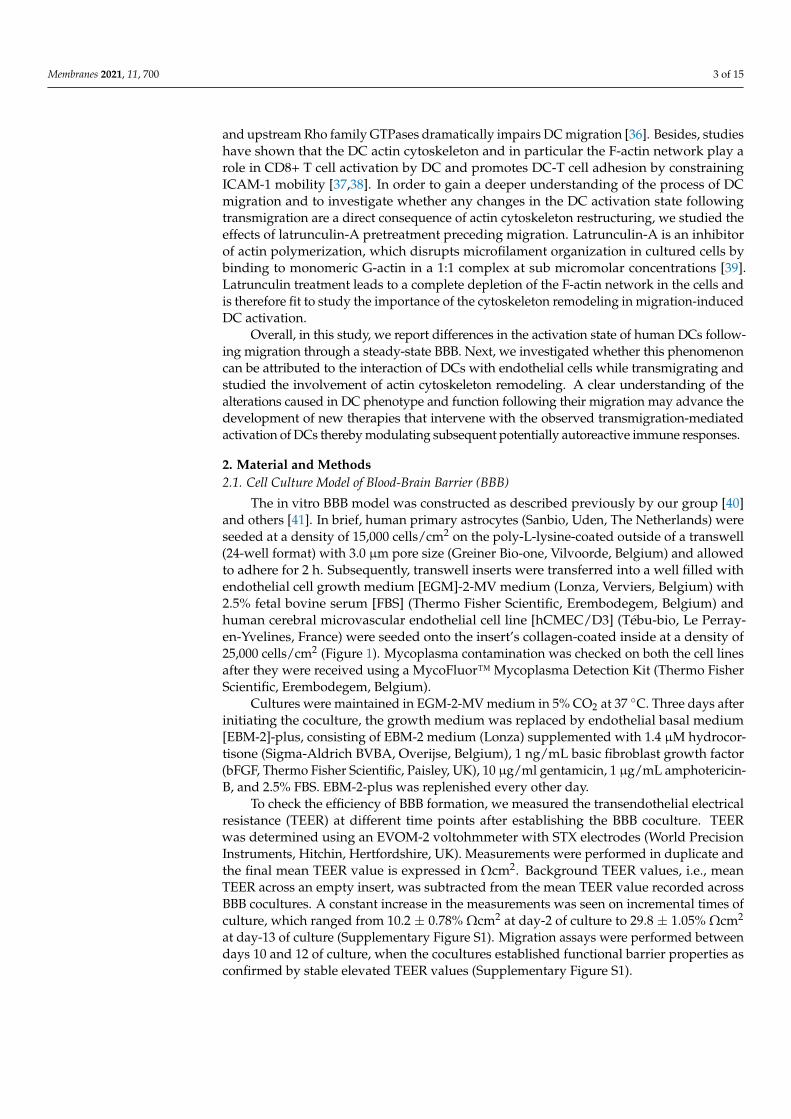
The in vitro BBB model was constructed as described previously by our group [40]and others [41]. In brief, human primary astrocytes (Sanbio, Uden, The Netherlands) wereseeded at a density of 15,000 cells/cm2 on the poly-L-lysine-coated outside of a transwell(24-well format) with 3.0 µm pore size (Greiner Bio-one, Vilvoorde, Belgium) and allowedto adhere for 2 h. Subsequently, transwell inserts were transferred into a well filled withendothelial cell growth medium [EGM]-2-MV medium (Lonza, Verviers, Belgium) with2.5% fetal bovine serum [FBS] (Thermo Fisher Scientific, Erembodegem, Belgium) andhuman cerebral microvascular endothelial cell line [hCMEC/D3] (Tébu-bio, Le Perray-en-Yvelines, France) were seeded onto the insert’s collagen-coated inside at a density of25,000 cells/cm2 (Figure 1). Mycoplasma contamination was checked on both the cell linesafter they were received using a MycoFluor™ Mycoplasma Detection Kit (Thermo FisherScientific, Erembodegem, Belgium).
Cultures were maintained in EGM-2-MV medium in 5% CO2 at 37 C. Three days afterinitiating the coculture, the growth medium was replaced by endothelial basal medium[EBM-2]-plus, consisting of EBM-2 medium (Lonza) supplemented with 1.4 µM hydrocor-tisone (Sigma-Aldrich BVBA, Overijse, Belgium), 1 ng/mL basic fibroblast growth factor(bFGF, Thermo Fisher Scientific, Paisley, UK), 10 µg/ml gentamicin, 1 µg/mL amphotericin-B, and 2.5% FBS. EBM-2-plus was replenished every other day.
To check the efficiency of BBB formation, we measured the transendothelial electricalresistance (TEER) at different time points after establishing the BBB coculture. TEERwas determined using an EVOM-2 voltohmmeter with STX electrodes (World PrecisionInstruments, Hitchin, Hertfordshire, UK). Measurements were performed in duplicate andthe final mean TEER value is expressed in Ωcm2. Background TEER values, i.e., meanTEER across an empty insert, was subtracted from the mean TEER value recorded acrossBBB cocultures. A constant increase in the measurements was seen on incremental times ofculture, which ranged from 10.2 ± 0.78% Ωcm2 at day-2 of culture to 29.8 ± 1.05% Ωcm2
at day-13 of culture (Supplementary Figure S1). Migration assays were performed betweendays 10 and 12 of culture, when the cocultures established functional barrier properties asconfirmed by stable elevated TEER values (Supplementary Figure S1).
Membranes 2021, 11, 700 4 of 15
Membranes 2021, 11, x 4 of 15
day-13 of culture (Supplementary Figure S1). Migration assays were performed between days 10 and 12 of culture, when the cocultures established functional barrier properties as confirmed by stable elevated TEER values (Supplementary Figure S1).
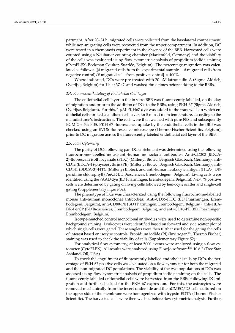
Figure 1. Schematic overview of the DC migration experiment using an in vitro model of the BBB. Endothelial cells were seeded on top of the 3.0 µm porous membrane in a 24-well transwell with astrocytes on the underneath. BBBs were maintained in culture for 10–12 days before the migration of DCs was studied. Abbreviations used: BBB—Blood–brain barrier, DC—Dendritic cells.
2.2. Cell Isolation Peripheral blood from healthy donors was obtained from buffy coats provided by
the Red Cross donor center (Red Cross-Flanders, Mechelen, Belgium), and peripheral blood mononuclear cells (PBMC) were isolated by density gradient centrifugation (Ficoll Pacque PLUS, GE Healthcare, Amsterdam, The Netherlands). The Pan-DC enrichment kit (Miltenyi biotech) was used to isolate DCs from the PBMC. From the remaining PBMC fraction, peripheral blood lymphocytes (PBLs) were depleted from CD14+ monocytes using CD14+ immunomagnetic selection (CD14 Reagent, Miltenyi Biotec, Bergisch Gladbach, Germany), according to the manufacturer’s instructions. The CD14-depleted cell fraction (i.e., peripheral blood lymphocytes (PBLs)) was cryopreserved in FBS supplemented with 10% dimethyl sulfoxide (DMSO, Sigma-Aldrich, Bornem, Belgium) and stored at −80 °C for later use in an allogeneic mixed leukocyte reaction (allo-MLR).
2.3. Migration Assay Transmigration of the isolated DCs was studied across steady-state BBB cocultures.
On the day of performing the migration assay, the cocultures were chemokine-coated by the addition of 2.5 ng/mL CCL4 (R&D systems, Bio-techne, Abingdon, UK) and CCL5 (R&D systems, Bio-techne, Abingdon, UK) to the upper transwell compartment for 1 h. Next, BBB cocultures were washed twice with Iscove’s Modified Dulbecco’s Medium (IMDM) (Thermo Fischer Scientific) supplemented with 1% human AB serum (hAB) (Thermo Fischer Scientific) to remove unbound chemokines. These BBB cocultures were then transferred to a new plate where the basolateral compartment contained 25 ng/mL CCL4 and 25 ng/mL CCL5 in IMDM, supplemented with 1% hAB serum. As compared to physiological levels [42–44], the used concentrations are relatively high, but the mentioned concentrations are well within the range for which maximal bioactivity was shown for these chemokines by the manufacturer (R&D systems, Bio-techne).
Figure 1. Schematic overview of the DC migration experiment using an in vitro model of the BBB.Endothelial cells were seeded on top of the 3.0 µm porous membrane in a 24-well transwell withastrocytes on the underneath. BBBs were maintained in culture for 10–12 days before the migrationof DCs was studied. Abbreviations used: BBB—Blood–brain barrier, DC—Dendritic cells.
2.2. Cell Isolation
Peripheral blood from healthy donors was obtained from buffy coats provided bythe Red Cross donor center (Red Cross-Flanders, Mechelen, Belgium), and peripheralblood mononuclear cells (PBMC) were isolated by density gradient centrifugation (FicollPacque PLUS, GE Healthcare, Amsterdam, The Netherlands). The Pan-DC enrichment kit(Miltenyi biotech) was used to isolate DCs from the PBMC. From the remaining PBMCfraction, peripheral blood lymphocytes (PBLs) were depleted from CD14+ monocytes usingCD14+ immunomagnetic selection (CD14 Reagent, Miltenyi Biotec, Bergisch Gladbach,Germany), according to the manufacturer’s instructions. The CD14-depleted cell fraction(i.e., peripheral blood lymphocytes (PBLs)) was cryopreserved in FBS supplemented with10% dimethyl sulfoxide (DMSO, Sigma-Aldrich, Bornem, Belgium) and stored at −80 Cfor later use in an allogeneic mixed leukocyte reaction (allo-MLR).
2.3. Migration Assay
Transmigration of the isolated DCs was studied across steady-state BBB cocultures.On the day of performing the migration assay, the cocultures were chemokine-coated by theaddition of 2.5 ng/mL CCL4 (R&D systems, Bio-techne, Abingdon, UK) and CCL5 (R&Dsystems, Bio-techne, Abingdon, UK) to the upper transwell compartment for 1 h. Next,BBB cocultures were washed twice with Iscove’s Modified Dulbecco’s Medium (IMDM)(Thermo Fischer Scientific) supplemented with 1% human AB serum (hAB) (ThermoFischer Scientific) to remove unbound chemokines. These BBB cocultures were thentransferred to a new plate where the basolateral compartment contained 25 ng/mL CCL4and 25 ng/mL CCL5 in IMDM, supplemented with 1% hAB serum. As compared tophysiological levels [42–44], the used concentrations are relatively high, but the mentionedconcentrations are well within the range for which maximal bioactivity was shown forthese chemokines by the manufacturer (R&D systems, Bio-techne).
Subsequently, 2 × 10⁵ enriched DCs resuspended in IMDM supplemented with1% hAB serum were added to the upper compartment. As a negative control, 2 × 10⁵ DCswere added to the upper compartment in the absence of chemokines in the basolateralcompartment. As a positive control, 2 × 10⁵ DCs were added directly to the lower com-
Membranes 2021, 11, 700 5 of 15
partment. After 20–24 h, migrated cells were collected from the basolateral compartment,while non-migrating cells were recovered from the upper compartment. In addition, DCwere tested in a chemotaxis experiment in the absence of the BBB. Harvested cells werecounted using a Neubauer counting chamber (Marienfeld, Germany) and the viabilityof the cells was evaluated using flow cytometric analysis of propidium iodide staining(CytoFLEX, Beckman Coulter, Suarlée, Belgium). The percentage migration was calcu-lated as follows: [(# migrated cells from the experimental sample − # migrated cells fromnegative control)/# migrated cells from positive control] × 100%.
Where indicated, DCs were pre-treated with 20 µM latrunculin-A (Sigma-Aldrich,Overijse, Belgium) for 1 h at 37 C and washed three times before adding to the BBBs.
2.4. Fluorescent Labeling of Endothelial Cell Layer
The endothelial cell layer in the in vitro BBB was fluorescently labelled, on the dayof migration and prior to the addition of DCs to the BBBs, using PKH-67 (Sigma-Aldrich,Overijse, Belgium). For this, 1 µM PKH67 dye was added to the transwells in which en-dothelial cells formed a confluent cell layer, for 5 min at room temperature, according to themanufacturer’s instructions. The cells were then washed with pure FBS and subsequentlyEGM-2 + 5% FBS. PKH-67 fluorescence uptake by the endothelial cells in the BBB waschecked using an EVOS fluorescence microscope (Thermo Fischer Scientific, Belgium),prior to DC migration across the fluorescently labeled endothelial cell layer of the BBB.
2.5. Flow Cytometry
The purity of DCs following pan-DC enrichment was determined using the followingfluorochrome-labelled mouse anti-human monoclonal antibodies: Anti-CD303 (BDCA-2)-fluorescein isothiocyanate (FITC) (Miltenyi Biotec, Bergisch Gladbach, Germany), anti-CD1c (BDCA-1)-phycoerythrin (PE) (Miltenyi Biotec, Bergisch Gladbach, Germany), anti-CD141 (BDCA-3)-FITC (Miltenyi Biotec), and anti-human leukocyte antigen (HLA-) DR-peridinin chlorophyll (PerCP, BD Biosciences, Erembodegem, Belgium). Living cells wereidentified using the 7AAD dye (BD Pharmingen, Erembodegem, Belgium). Next, % positivecells were determined by gating on living cells followed by leukocyte scatter and single-cellgating (Supplementary Figure S2).
The phenotype of DCs was characterized using the following fluorochrome-labelledmouse anti-human monoclonal antibodies: Anti-CD86-FITC (BD Pharmingen, Erem-bodegem, Belgium), anti-CD80-PE (BD Pharmingen, Erembodegem, Belgium), anti-HLA-DR-PerCP (BD Biosciences, Erembodegem, Belgium), and anti-CD40-PE (BD Pharmingen,Erembodegem, Belgium).
Isotype-matched control monoclonal antibodies were used to determine non-specificbackground staining. Leukocytes were identified based on forward and side scatter plot ofwhich single cells were gated. These singlets were then further used for the gating the cellsof interest based on isotype controls. Propidium iodide (PI) (Invitrogen™, Thermo Fischer)staining was used to check the viability of cells (Supplementary Figure S2).
For analytical flow cytometry, at least 5000 events were analyzed using a flow cy-tometer (CytoFLEX). All results were analyzed using FlowJo softwareTM 10.6.2 (Tree Star,Ashland, OR, USA).
To check the engulfment of fluorescently labelled endothelial cells by DCs, the per-centage of PKH-67 positive cells was evaluated on a flow cytometer for both the migratedand the non-migrated DC populations. The viability of the two populations of DCs wasassessed using flow cytometric analysis of propidium iodide staining on the cells. Thefluorescently labelled endothelial cells were harvested from the BBBs following DC mi-gration and further checked for the PKH-67 expression. For this, the astrocytes wereremoved mechanically from the insert underside and the hCMEC/D3 cells cultured onthe upper side of the membrane were homogenized with trypsin-EDTA (Thermo FischerScientific). The harvested cells were then washed before flow cytometric analysis. Further,
Membranes 2021, 11, 700 6 of 15
the percentage PKH-67 positive cells were analyzed on the FITC channel against a sidescatter plot on the flow cytometer.
2.6. Allogeneic Mixed Lymphocyte Reaction
To assess the allogeneic T cell-stimulatory capacity of DCs, DCs were coculturedwith allogeneic PBL in a 1:10 ratio. Non-stimulated responder PBL served as a negativecontrol, while allogeneic PBL stimulated with 1 µg/mL phytoheamagglutinin (PHA)(Sigma-Aldrich, Overijse, Belgium) were used as a positive control. Cocultures wereperformed in IMDM supplemented with 5% hAB serum at 37 C. After 5 days, the secretedlevel of IFN-γ in the cell culture supernatant was determined as a measure of T cellstimulatory capacity using a commercially available ELISA kit (PeproTech, East Windsor,NJ, USA).
2.7. Statistical Analyses
Data were analyzed using the Graphpad Prism software version 5.01 (Graphpad, SanDiego, CA, USA). The normality of data was checked using the Shapiro–Wilk normality test.For the comparison of 2 groups, a paired Student’s t-test or a Mann–Whitney U test wasused based on the normality of dataset. For comparing 3 groups or more, statistical analysiswas performed by one-way ANOVA, followed by Tukey’s multiple comparisons test or bya Kruskal–Wallis test in combination with Dunn’s multiple comparison test in case datawere not normally distributed. p-value < 0.05 was considered as statistically significant.
3. Results3.1. Dendritic Cells That Migrate across the BBB Are in a More Activated State ThanNon-Migrating Dendritic Cells
To investigate whether the migration of DCs through the in vitro model of the BBB af-fects their activation state, we investigated the phenotype and function of BBB-transmigrat-ing DCs as compared to non-migrating DCs. For this, DCs were selectively enriched,resulting in a pure DC population as determined by the expression of specific markers fordifferent DC subsets. This population consisted of 35.4 ± 5.46% of CD1c (BDCA-1)-positivemyeloid DCs, 4.8 ± 0.92% of CD141 (BDCA-3)-positive myeloid DCs, and 37.04 ± 2.88%of CD303 (BDCA-2)-positive plasmacytoid DCs, comprising a total of 84.75 ± 5.42% HLA-DR-positive DCs (Supplementary Figure S3). Cells had a viability of 89.23 ± 2.51%.
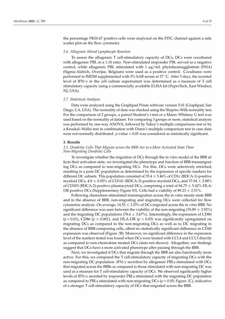
Following chemokine-stimulated transmigration across the in vitro steady-state BBBand in the absence of BBB, non-migrating and migrating DCs were collected for flowcytometric analysis. On average, 14.55 ± 3.25% of DCs migrated across the in vitro BBB. Nosignificant difference was seen between the viability of the non-migrating (76.89 ± 3.92%)and the migrating DC populations (78.6 ± 3.67%). Interestingly, the expression of CD80(p < 0.01), CD86 (p < 0.001), and HLA-DR (p < 0.05) was significantly upregulated onmigrating DCs as compared to the non-migrating DCs as well as to DC migrating inthe absence of BBB-composing cells, albeit no statistically significant difference in CD40expression was observed (Figure 2B). Moreover, no significant difference in the expressionlevel of the markers tested was found when DCs were treated with CCL4 and CCL5 directlyas compared to non-chemokine treated DCs (data not shown). Altogether, our findingssuggest that DCs have a more activated phenotype after passing through the BBB.
Next, we investigated if DCs that migrate through the BBB are also functionally moreactive. For this, we compared the T cell-stimulatory capacity of migrating DCs with thenon-migrating DC population. IFN-γ secretion by allogeneic PBLs stimulated with DCsthat migrated across the BBBs as compared to those stimulated with non-migrating DC wasused as a measure for T cell-stimulatory capacity of DCs. We observed significantly higherlevels of IFN-γ secreted by responder PBLs stimulated with the migrating DC populationas compared to PBLs stimulated with non-migrating DCs (p < 0.05; Figure 2C), indicativeof a stronger T cell-stimulatory capacity of DCs that migrated across the BBB.
Membranes 2021, 11, 700 7 of 15Membranes 2021, 11, x 7 of 15
Figure 2. BBB-transmigratory DCs display a more activated state as compared to their non-migrating counterparts and when compared to the migrating DCs in the absence of BBBs. (A) Representative figure showing the expression of co-stimulatory markers of migrating DCs (blue peak) and non-migrating dendritic cells (pink peak) as compared to their respective isotype controls (black). The count numbers of these cells are provided in Table S1 of the supplementary information (Table S1). (B) The expression of the activation markers CD80, CD86, HLA-DR, and CD40 on migrating vs. non-migrating DCs following a chemotaxis assay in an in vitro BBB model was evaluated along with the comparison against a control sample in the absence of BBB. Migrated DCs demonstrate a significantly higher expression of CD80, CD86, and HLA-DR as compared to non-migrated DCs (n = 6) and the DCs migrating in the absence of BBB (n = 3). (C) DCs that migrate across an in vitro model of the BBB demonstrate stronger T cell-stimulatory capacity as compared to non-migrating DCs, as shown in an allo-MLR (n = 11). (* p < 0.05; ** p < 0.01; *** p < 0.001).
Next, we investigated if DCs that migrate through the BBB are also functionally more active. For this, we compared the T cell-stimulatory capacity of migrating DCs with the non-migrating DC population. IFN-γ secretion by allogeneic PBLs stimulated with DCs that migrated across the BBBs as compared to those stimulated with non-migrating DC was used as a measure for T cell-stimulatory capacity of DCs. We observed significantly higher levels of IFN-γ secreted by responder PBLs stimulated with the migrating DC population as compared to PBLs stimulated with non-migrating DCs (p < 0.05; Figure 2C), indicative of a stronger T cell-stimulatory capacity of DCs that migrated across the BBB.
3.2. Dendritic Cells Do Not Take Up Membrane Fragments of Endothelial Cells Following Transmigration
Next, we studied if the migrating DCs capture membrane fragments from the endothelial cells while moving across the BBB and whether this contributed to the activation of the DCs. For this, we fluorescently labelled the endothelial cells in the in vitro transwell model of the BBB with the cell membrane labelling dye PKH-67 in order to check the interactions between endothelial cells and DCs. Membrane staining of the endothelial cell layer was confirmed using an EVOS fluorescence microscope prior to migration
CD86-FITC CD40-PECD80-PE HLA-DR-PerCP
(A)
(B) (C)
Figure 2. BBB-transmigratory DCs display a more activated state as compared to their non-migrating counterparts and whencompared to the migrating DCs in the absence of BBBs. (A) Representative figure showing the expression of co-stimulatorymarkers of migrating DCs (blue peak) and non-migrating dendritic cells (pink peak) as compared to their respective isotypecontrols (black). The count numbers of these cells are provided in Table S1 of the supplementary information (Table S1).(B) The expression of the activation markers CD80, CD86, HLA-DR, and CD40 on migrating vs. non-migrating DCsfollowing a chemotaxis assay in an in vitro BBB model was evaluated along with the comparison against a control sample inthe absence of BBB. Migrated DCs demonstrate a significantly higher expression of CD80, CD86, and HLA-DR as comparedto non-migrated DCs (n = 6) and the DCs migrating in the absence of BBB (n = 3). (C) DCs that migrate across an in vitromodel of the BBB demonstrate stronger T cell-stimulatory capacity as compared to non-migrating DCs, as shown in anallo-MLR (n = 11). (* p < 0.05; ** p < 0.01; *** p < 0.001).
3.2. Dendritic Cells Do Not Take up Membrane Fragments of Endothelial Cellsfollowing Transmigration
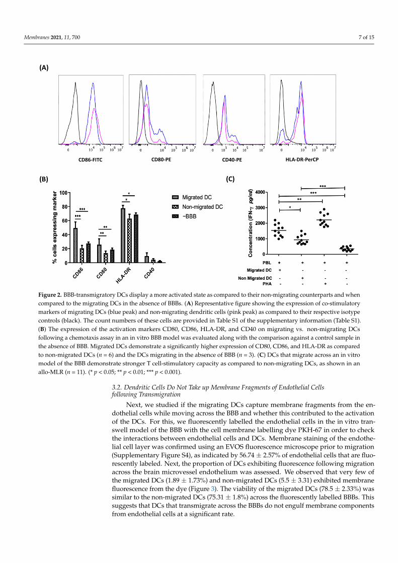
Next, we studied if the migrating DCs capture membrane fragments from the en-dothelial cells while moving across the BBB and whether this contributed to the activationof the DCs. For this, we fluorescently labelled the endothelial cells in the in vitro tran-swell model of the BBB with the cell membrane labelling dye PKH-67 in order to checkthe interactions between endothelial cells and DCs. Membrane staining of the endothe-lial cell layer was confirmed using an EVOS fluorescence microscope prior to migration(Supplementary Figure S4), as indicated by 56.74 ± 2.57% of endothelial cells that are fluo-rescently labeled. Next, the proportion of DCs exhibiting fluorescence following migrationacross the brain microvessel endothelium was assessed. We observed that very few ofthe migrated DCs (1.89 ± 1.73%) and non-migrated DCs (5.5 ± 3.31) exhibited membranefluorescence from the dye (Figure 3). The viability of the migrated DCs (78.5 ± 2.33%) wassimilar to the non-migrated DCs (75.31 ± 1.8%) across the fluorescently labelled BBBs. Thissuggests that DCs that transmigrate across the BBBs do not engulf membrane componentsfrom endothelial cells at a significant rate.
Membranes 2021, 11, 700 8 of 15
Membranes 2021, 11, x 8 of 15
(Supplementary Figure S4), as indicated by 56.74 ± 2.57% of endothelial cells that are fluorescently labeled. Next, the proportion of DCs exhibiting fluorescence following migration across the brain microvessel endothelium was assessed. We observed that very few of the migrated DCs (1.89 ± 1.73%) and non-migrated DCs (5.5 ± 3.31) exhibited membrane fluorescence from the dye (Figure 3). The viability of the migrated DCs (78.5 ± 2.33%) was similar to the non-migrated DCs (75.31 ± 1.8%) across the fluorescently labelled BBBs. This suggests that DCs that transmigrate across the BBBs do not engulf membrane components from endothelial cells at a significant rate.
Figure 3. DCs do not engulf endothelial cells while transmigrating across a steady-state BBB. Migrated and non-migrated DC were harvested after a 22-h migration assay across an intact in vitro BBB (n = 7). The expression of PKH-67 was assessed on the collected migrated and non-migrated DC populations. Only a minority of DCs were PKH-67 positive. Control samples were from the fluorescently labelled endothelial cells harvested from the BBB. (*** p < 0.001).
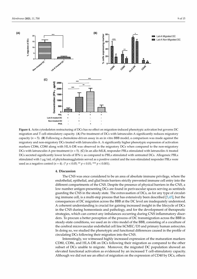
3.3. Actin Cytoskeleton Restructuring of DCs Has No Effect on Migration-Induced Phenotypic Activation But Governs DC Migration and T Cell-Stimulatory Capacity
For better understanding of the influence of DC cytoskeleton reorganization on DC activation and maturation along with the transmigratory capacities of DCs, we treated DCs with a cytoskeleton inhibitor latruncilin-A prior to their migration across the in vitro BBB. It was observed that the migration of DCs was severely disrupted upon treatment with latruncilin-A when compared to non-treated DCs (Figure 4A). The migratory capacity of latruncilin-A treated DCs (2.53 ± 0.77%) was significantly lower (p < 0.001) than that of untreated DCs (13.89 ± 4.17%). Besides, the viability of the migrated DC population treated with latruncilin-A (68.19 ± 1.82%) was similar to the viability of non-migrating latruncilin-A treated DCs (74.89 ± 3.16%) and to the non-treated cells (77.32 ± 4.22%). Additionally, DCs that were treated with latruncilin-A exhibited no significant differences in the expression levels of the co-stimulatory markers, CD80, CD86, HLA-DR, and CD40 as compared to non-treated DCs for both the migrated and non-migrated DC subsets. Nonetheless, the initially observed phenotypic differences between migrating and non-migrating DCs persisted after latruncilin-A treatment (Figure 4B). To test the effect of latruncilin-A on the T-cell stimulatory capacity of DCs, we performed an allo-MLR of PBLs stimulated with latruncilin-A treated and untreated DCs. We observed that the level
Figure 3. DCs do not engulf endothelial cells while transmigrating across a steady-state BBB. Mi-grated and non-migrated DC were harvested after a 22-h migration assay across an intact in vitroBBB (n = 7). The expression of PKH-67 was assessed on the collected migrated and non-migratedDC populations. Only a minority of DCs were PKH-67 positive. Control samples were from thefluorescently labelled endothelial cells harvested from the BBB. (*** p < 0.001).
3.3. Actin Cytoskeleton Restructuring of DCs Has No Effect on Migration-Induced PhenotypicActivation but Governs DC Migration and T Cell-Stimulatory Capacity
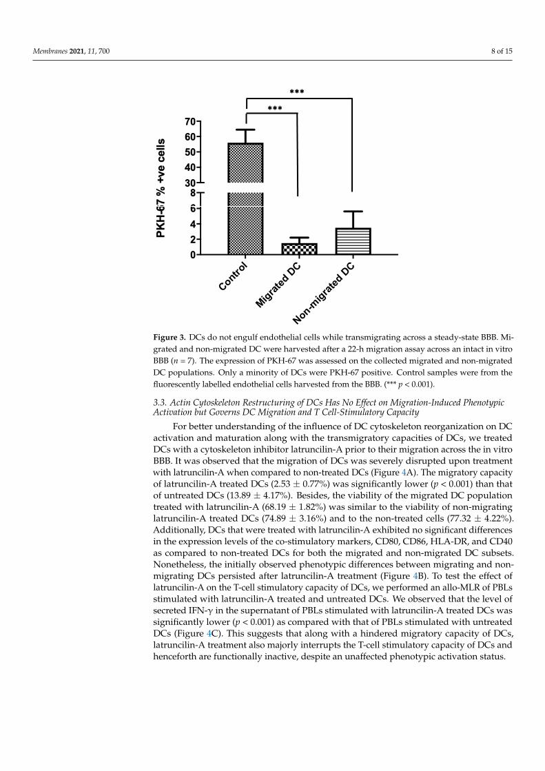
For better understanding of the influence of DC cytoskeleton reorganization on DCactivation and maturation along with the transmigratory capacities of DCs, we treatedDCs with a cytoskeleton inhibitor latruncilin-A prior to their migration across the in vitroBBB. It was observed that the migration of DCs was severely disrupted upon treatmentwith latruncilin-A when compared to non-treated DCs (Figure 4A). The migratory capacityof latruncilin-A treated DCs (2.53 ± 0.77%) was significantly lower (p < 0.001) than thatof untreated DCs (13.89 ± 4.17%). Besides, the viability of the migrated DC populationtreated with latruncilin-A (68.19 ± 1.82%) was similar to the viability of non-migratinglatruncilin-A treated DCs (74.89 ± 3.16%) and to the non-treated cells (77.32 ± 4.22%).Additionally, DCs that were treated with latruncilin-A exhibited no significant differencesin the expression levels of the co-stimulatory markers, CD80, CD86, HLA-DR, and CD40as compared to non-treated DCs for both the migrated and non-migrated DC subsets.Nonetheless, the initially observed phenotypic differences between migrating and non-migrating DCs persisted after latruncilin-A treatment (Figure 4B). To test the effect oflatruncilin-A on the T-cell stimulatory capacity of DCs, we performed an allo-MLR of PBLsstimulated with latruncilin-A treated and untreated DCs. We observed that the level ofsecreted IFN-γ in the supernatant of PBLs stimulated with latruncilin-A treated DCs wassignificantly lower (p < 0.001) as compared with that of PBLs stimulated with untreatedDCs (Figure 4C). This suggests that along with a hindered migratory capacity of DCs,latruncilin-A treatment also majorly interrupts the T-cell stimulatory capacity of DCs andhenceforth are functionally inactive, despite an unaffected phenotypic activation status.
Membranes 2021, 11, 700 9 of 15
Membranes 2021, 11, x 9 of 15
of secreted IFN-γ in the supernatant of PBLs stimulated with latruncilin-A treated DCs was significantly lower (p < 0.001) as compared with that of PBLs stimulated with untreated DCs (Figure 4C). This suggests that along with a hindered migratory capacity of DCs, latruncilin-A treatment also majorly interrupts the T-cell stimulatory capacity of DCs and henceforth are functionally inactive, despite an unaffected phenotypic activation status.
Figure 4. Actin cytoskeleton restructuring of DCs has no effect on migration-induced phenotypic activation but governs DC migration and T cell-stimulatory capacity. (A) Pre-treatment of DCs with latrunculin-A significantly reduces migratory capacity (n = 5). (B) Following a chemokine-driven assay in an in vitro BBB model, a comparison was made against the migratory and non-migratory DCs treated with latrunculin-A. A significantly higher phenotypic expression of activation markers CD86, CD80 along with HLA-DR was observed in the migratory DCs when compared to the non-migratory DCs with latrunculin-A pre-treatment (n = 5). (C) In an allo-MLR, responder PBLs stimulated with latruncilin-A treated DCs secreted significantly lower levels of IFN-γ as compared to PBLs stimulated with untreated DCs. Allogeneic PBLs stimulated with 1 µg/mL of phytoheamagglutinin served as a positive control and the non-stimulated responder PBLs were used as a negative control (n = 4). (* p < 0.05; ** p < 0.01; *** p < 0.001).
4. Discussion The CNS was once considered to be an area of absolute immune privilege, where the
endothelial, epithelial, and glial brain barriers strictly prevented immune cell entry into the different compartments of the CNS. Despite the presence of physical barriers in the CNS, a low number antigen-presenting DCs are found in perivascular spaces serving as sentinels guarding the CNS in the steady state. The extravasation of DCs, as for any type of circulating immune cell, is a multi-step process that has extensively been described [5,45], but the consequences of DC migration across the BBB at the DC level are inadequately understood. A coherent understanding is crucial for gaining increased insight in the lifecycle of DCs in the CNS during homeostasis and pathology, and for the development of therapeutic strategies, which can correct any imbalances occurring during CNS inflammatory disorders. To procure a better perception of the process of DC transmigration across the BBB in steady-state conditions, we used an in vitro model of the BBB, consisting of a coculture of the cerebral microvascular endothelial cell line hCMEC/D3 and primary human astrocytes. In doing so, we studied the phenotypic and
(A)
(C)
(B)
Non-trea
tedDC
Lat-Atreate
d DC
Lat-A treated DC + - - -
PHA - - + -Non-treated DC - + - -
PBL + + + +
CD86CD80
HLA-DR
CD400
20
40
60
80Lat-A Migrated DC
% c
ells
exp
ress
ing
mar
ker
Lat-A Non-Migrated DC***
**
*
Figure 4. Actin cytoskeleton restructuring of DCs has no effect on migration-induced phenotypic activation but governs DCmigration and T cell-stimulatory capacity. (A) Pre-treatment of DCs with latrunculin-A significantly reduces migratorycapacity (n = 5). (B) Following a chemokine-driven assay in an in vitro BBB model, a comparison was made against themigratory and non-migratory DCs treated with latrunculin-A. A significantly higher phenotypic expression of activationmarkers CD86, CD80 along with HLA-DR was observed in the migratory DCs when compared to the non-migratoryDCs with latrunculin-A pre-treatment (n = 5). (C) In an allo-MLR, responder PBLs stimulated with latruncilin-A treatedDCs secreted significantly lower levels of IFN-γ as compared to PBLs stimulated with untreated DCs. Allogeneic PBLsstimulated with 1µg/mL of phytoheamagglutinin served as a positive control and the non-stimulated responder PBLs wereused as a negative control (n = 4). (* p < 0.05; ** p < 0.01; *** p < 0.001).
4. Discussion
The CNS was once considered to be an area of absolute immune privilege, where theendothelial, epithelial, and glial brain barriers strictly prevented immune cell entry into thedifferent compartments of the CNS. Despite the presence of physical barriers in the CNS, alow number antigen-presenting DCs are found in perivascular spaces serving as sentinelsguarding the CNS in the steady state. The extravasation of DCs, as for any type of circulat-ing immune cell, is a multi-step process that has extensively been described [5,45], but theconsequences of DC migration across the BBB at the DC level are inadequately understood.A coherent understanding is crucial for gaining increased insight in the lifecycle of DCsin the CNS during homeostasis and pathology, and for the development of therapeuticstrategies, which can correct any imbalances occurring during CNS inflammatory disor-ders. To procure a better perception of the process of DC transmigration across the BBB insteady-state conditions, we used an in vitro model of the BBB, consisting of a coculture ofthe cerebral microvascular endothelial cell line hCMEC/D3 and primary human astrocytes.In doing so, we studied the phenotypic and functional differences caused in the profile ofcirculating DCs following their migration into the CNS.
Interestingly, we witnessed highly increased expression of the maturation markersCD80, CD86, and HLA-DR on DCs following their migration as compared to the othersubset of DCs unable to migrate. Moreover, the migrated DC population showed anelevated functional activation as evidenced by an increased T cell-stimulatory capacity.Although we did not see an effect of migration on the expression of CD40 by DCs, others
Membranes 2021, 11, 700 10 of 15
have demonstrated that CD40 plays a role in the migration process of CD4 T cells acrossbrain microvascular endothelial cells and increases adhesion of resting and activatedCD4+ T lymphocytes to endothelium [46]. Similar results were observed by Zozulya andcolleagues [29], who used an in vitro model of murine cerebral microvascular endothelialcell monolayers to show that CCL3 can stimulate the transmigration of mature bonemarrow-derived DCs in an MMP-dependent manner and equally witnessed DC activationupon transmigration.
Altogether, these results can plausibly demonstrate that DCs acquire a phenotypicand functionally active status post migration across a steady-state BBB in vitro. Whileimmunohistological studies investigating the maturation state of DCs in the CNS in situhave been conflicting [47–50], a study by Anandasabapathy et al. [27] in mice confirms thatin the steady-state CNS, DCs attain a mature phenotype and are capable of stimulating Tcells. While in steady-state conditions, this is not associated with autoimmune responses,this finding is of relevance in the context of CNS autoimmune disorders.
Based on previous reports, endothelial cells exchange antigens with DCs and shedmicroparticles, which lead to plasmacytoid DC maturation [30,31]. In line with this, wehypothesized that endothelial cells could directly lead to the maturation and activationof DCs by imparting their membrane particles to DCs during their migrating across theBBB. We observed that circulating DCs do not engulf any significant amount of membranefragments from the steady-state endothelial cells during their transmigratory process.
This is opposed to what has been reported previously [31,32]. For instance, Kedl andco-workers [32] demonstrated antigen exchange of viral antigens present on the lymphaticendothelial cell surface, following viral infection in mice, and DCs, and subsequent crosspresentation of these antigens by the DCs. Furthermore, others demonstrated upregulatedexpression of co-stimulatory markers, increased secretion of inflammatory cytokines, andenhanced ability to stimulate naive CD4 T-cell proliferation by plasmacytoid dendriticcells (pDC) following interaction with endothelial cells [31]. For this, microparticles, whichare complex vesicular structures shed by endothelial cells, are isolated after activatingthe endothelial cells, and incubated with human pDC. Subsequently, the uptake of thesemicroparticles by pDC was observed. In contrast, in current study, endothelial cells werenot activated, and engulfment of membrane fragments from steady-state endothelial cellsdirectly by the DCs when moving across the BBB was investigated. This could indicatethat transmigration of cells, and DCs in particular, occurs via different mechanisms in thesteady state versus in an inflammatory environment when endothelial cells are activated.In addition, dendritic cells may not directly engulf the membrane fragments from theendothelial cell layer but are activated by microparticles shed following the activationof the BBB. Hence, very few microparticles might be present in the steady culture ofBBB as a result of decreased membrane vesiculation of endothelial cells. Alternatively,fluorescent labelling of endothelial cells might not be sufficient for demonstrating theengulfment of cells or cellular particles by DCs since DCs might have internalized thefragments and presented on their membrane. Consequently, investigating the expressionof endothelial cell-specific molecules such as endothelial cell-derived antigens, chemerins,and markers like CD31 and CD99 on the transmigrating DCs could be valuable analysiswarranting further investigation of the exact mechanism driving DC activation followingtransmigration across a steady-state BBB.
Likely, endothelial cells induce DC activation via other mechanisms, which mightrequire direct cell–cell contact or could occur through the secretion of cytokines andsignaling molecules by endothelial cells. These mechanisms were not investigated here andstill need to be explored. In favor of this hypothesis are earlier studies, which describedthat a combination of fibroblast, endothelial, and epithelial cell-conditioned media canpromote DC maturation when added to the DC culture medium [51] and that the vascularendothelial growth inhibitor (VEGI), which is an anti-angiogenic cytokine produced byendothelial cells, can mediate DC maturation [52]. Other than this, endothelial cells canpromote attraction and subsequent maturation of DCs by several other factors in steady-
Membranes 2021, 11, 700 11 of 15
state and inflammatory conditions [53–56]. Moreover, there could also be the effects ofastrocytes from the BBB, which could lead to the activation of DCs following migration,as once DCs cross the blood vasculature, the first cellular structure they encounter are theendfeet or processes of astrocytes. Interestingly, it has been previously reported that humanCNS astrocytes could lead to the activation of B cells [57]. Furthermore, astrocytes have beenimplicated in playing a vital role in antigen presentation and naive T-cell activation [58].Hence, a direct role of astrocytes in the process of DC maturation and activation followingtransmigration through a BBB in steady-state and inflammatory conditions cannot beexcluded and still needs to be inspected.
The modulation of actin architecture is an essential and fundamental feature of bothDC migration and maturation. A previous report by Burns et al. [59,60] showed thatchanges in the DC actin cytoskeleton facilitate the transition from highly endocytic tissue-resident cells to migratory cells specialized for antigen presentation. This process involveschanges in the activation status of Rho GTPases and downstream actin regulatory proteins,and is known to downregulate antigen uptake and increase cell motility [61,62]. Similarly,others previously reported the importance of the actin cytoskeleton in lymphocyte acti-vation [63]. In particular, actin and microtubule meshwork are known to polarize andactivate T cells [64,65]. Additionally, the actin cytoskeleton is also known to play a role inthe regulation of B cell activation [66].
To investigate if changes in actin cytoarchitecture also result in increased maturationand activation of migratory DCs across a BBB, we treated immature circulating DCs withan actin-depolymerizing agent and a potent cytoskeleton inhibitor, latrunculin-A. The drugdid not affect the phenotypic activation of the migratory DCs as compared to the untreatedsubsets of cells, indicating that the increased phenotypic activation of DCs post migrationvia the BBB depends on factors other than the actin cytoskeleton restructuring of the cells.In contrast, the drug induced a significant decrease in the T cell-stimulatory capacityof treated DCs compared to the untreated DCs, along with highly disrupted migratorycapacity. The significantly lowered T cell stimulation by the latrunculin-A treated DCscould be an effect of the loss of cortical stiffness by the DCs upon treatment. This is basedon the findings from Blumenthal et al. [67], which showed that T cell priming is enhancedby maturation-dependent stiffening of the DC cortex. These findings can be used in thefuture to further unravel the effects of the cytoskeleton formation of DCs and its role intheir activation and maturation.
The current study also has some limitations. First, pericytes were not included in thein vitro model of the BBB used here. Nonetheless, previous reports have established thatpericytes play a critical role in the integration of endothelial and astrocyte function at theneurovascular unit, and in the regulation of the BBB in vitro [9,11,24]. Hence, includingpericytes in an in vitro model of the BBB could provide additional phenotypic advantagesin mimicking the in vivo BBB. Second, we incorporated a static in vitro model of the BBBin the current study, lacking unidirectional flow. Indeed, culturing the cells composingthe BBB under continuous flow generates shear stress and regulates the expression oftransporters and tight junctions, contributing towards effective barrier function. Recently,there have been some breakthrough studies in the development of a 3D organ-on-a-chipmodel of BBB [68] with a hollow channel in which a continuous monolayer of cells can begrown at the interphase between the lumen and the underlying endothelial cell matrix.Likewise, Wevers and colleagues [69] developed an in vitro model of the human BBBin a high-throughput microfluidic platform. This system is free of artificial membranes,accommodates fluid flow through the blood vessels, and allows fluid-phase samplingof molecules that penetrate the endothelial and matrix layers. Besides this, there havebeen other recent advancements in human brain organoid models and their applicationin modelling neurodevelopmental and neurodegenerative diseases [70–72]. Importantly,vascularized organoids, which more precisely mimic in vivo brain anatomy and physi-ology, may facilitate brain disease modelling. Ultimately, these models will lead to thedevelopment of efficient high-throughput screening and computer-aided drug design meth-
Membranes 2021, 11, 700 12 of 15
ods [73,74]. The recent advances in these novel methods have the potential to investigatehundreds of thousands of compounds per day hence reducing the cost, time, and effortrequired to develop new drugs. These systems comprise several steps including targetrecognition, compound management, reagent preparation, assay development, as wellas the screening itself for designing novel therapies for neurodegenerative diseases [74].This could be an important breakthrough driving the drug discovery arena for neurode-generative diseases and can provide novel insights in the underlying disease-associatedmechanisms supporting the design of targeted therapies in the future.
In conclusion, in this study, we have demonstrated that transmigration of DCs acrossan in vitro transwell model of the BBB results in the upregulation of the expression ofcostimulatory molecules and T cell-stimulatory capacity. Other than this, we showed thatendothelial cells do not impart their membrane particles to DCs that move across the BBB.Finally, we established that latrunculin-A severely disrupts the migration of DCs acrossthe BBB as well as the T cell-stimulatory capacity of DCs, while not affecting phenotypicmaturation as a consequence of transmigration. Understanding the underlying mechanismof the interaction between brain microvessel endothelial cells and DCs might lead to thedesign of targeted therapies to modify transmigration of DCs in the CNS and thus to inhibitperpetuating autoimmune inflammation of the CNS.
Supplementary Materials: The following are available online at https://www.mdpi.com/article/10.3390/membranes11090700/s1, Figure S1: The transendothelial electrical resistance (TEER) wasrecorded for the in vitro BBBs from day-2 of coculture. An incremental increase in the TEER valueswas seen over time. The last point of measurement was at day-12 and day-13 at which a significantlyhigher TEER value was observed as compared to day-2. This indicates a sufficiently confluent BBBformation, Figure S2. (A) Gating strategy for determination of purity of dendritic cells enriched usingthe Pan-DC enrichment kit. The cells were stained with 7AAD dye and a clear population of viablecells was observed and gated for further analysis. A forward and side scatter plot was generated fromthe viable cell population and leukocytes were gated for further analysis. A single cell populationwas additionally gated from the leukocytes which was then used for gating the % positive cells fortheir respective fluorophores based on the isotype controls. (B) Gating strategy for characterizationof dendritic cells following a migration assay. Leukocytes were identified based on forward and sidescatter plot of which single cells were gated. These singlets were then further used for the gating thecells of interest based on isotype controls. In these cells, the viability was additionally checked usingPI staining, Figure S3. Representative example of the different isolated circulating DC populationsfollowing magnetic Pan-DC enrichment as determined by flow cytometry. The cells were fluorescentlystained with anti-CD303-FITC, anti-CD303-PE (BDCA-2), anti-CD1c-PE (BDCA-1), anti-CD141-FITC(BDCA-3) and anti-HLA-DR-FITC, Figure S4. Fluorescent image of endothelial cells labelled with thePKH-67 dye as taken from an EVOS fluorescent microscope, Table S1. Number of events measured toacquire results depicted in Figure 2A in the manuscript ± error on the measurement, according toPoisson distribution of flow cytometry data.
Author Contributions: All authors have contributed substantially to this work, have approved themanuscript and agreed with its submission. Conceptualization, M.M. and N.C.; data curation, M.V.D.,A.S. and C.C.R.; formal analysis, M.M. and M.V.D.; methodology, M.M., M.D.L. and A.S.; validation,M.M., M.D.L. and N.C.; writing-original draft, M.M.; writing-review and editing, M.D.L. and N.C.;supervision, Z.B., N.C.; funding acquisition, N.C. All authors have read and agreed to the publishedversion of the manuscript.
Funding: This work was supported by BOF DOCPRO4 2015 grant no. 31967 of the Special ResearchFund (BOF) from the University of Antwerp, Belgium. Further support was provided through theMethusalem Funding Program from the University of Antwerp, and by the Belgian Charcot Foundation.
Institutional Review Board Statement: Not applicable.
Informed Consent Statement: Not applicable.
Data Availability Statement: Not applicable.
Membranes 2021, 11, 700 13 of 15
Acknowledgments: The authors would like to thank Isabel Pintelon and Zoë Embrechts of theLaboratory of Cell Biology and Histology of the University of Antwerp for their guidance and advicein performing the fluorescence microscopy.
Conflicts of Interest: The authors declare that they have no conflict of interest.
References1. Pachter, J.S.; de Vries, H.E.; Fabry, Z. The blood-brain barrier and its role in immune privilege in the central nervous system. J.
Neuropathol. Exp. Neurol. 2003, 62, 593–604. [CrossRef] [PubMed]2. Carson, M.J.; Doose, J.M.; Melchior, B.; Schmid, C.D.; Ploix, C.C. CNS immune privilege: Hiding in plain sight. Immunol. Rev.
2006, 213, 48–65. [CrossRef] [PubMed]3. Forrester, J.V.; McMenamin, P.G.; Dando, S.J. CNS infection and immune privilege. Nat. Rev. Neurosci. 2018, 19, 655–671.
[CrossRef] [PubMed]4. Stamatovic, S.M.; Keep, R.F.; Andjelkovic, A. V Brain endothelial cell-cell junctions: How to “open” the blood brain barrier. Curr.
Neuropharmacol. 2008, 6, 179–192. [CrossRef] [PubMed]5. Campos-Bedolla, P.; Walter, F.R.; Veszelka, S.; Deli, M.A. Role of the blood—Brain barrier in the nutrition of the central nervous
system. Arch. Med. Res. 2014, 45, 610–638. [CrossRef]6. Abbott, N.J.; Patabendige, A.A.K.; Dolman, D.E.M.; Yusof, S.R.; Begley, D.J. Structure and function of the blood—Brain barrier.
Neurobiol. Dis. 2010, 37, 13–25. [CrossRef]7. Kadry, H.; Noorani, B.; Cucullo, L. A blood–brain barrier overview on structure, function, impairment, and biomarkers of
integrity. Fluids Barriers CNS 2020, 17, 1–24. [CrossRef] [PubMed]8. Hawkins, B.T.; Davis, T.P. The blood-brain barrier/neurovascular unit in health and disease. Pharmacol. Rev. 2005, 57, 173–185.
[CrossRef]9. Török, O.; Schreiner, B.; Schaffenrath, J.; Tsai, H.-C.; Maheshwari, U.; Stifter, S.A.; Welsh, C.; Amorim, A.; Sridhar, S.; Utz, S.G.
Pericytes regulate vascular immune homeostasis in the CNS. Proc. Natl. Acad. Sci. USA 2021, 9, 118.10. Daneman, R.; Zhou, L.; Kebede, A.A.; Barres, B.A. Pericytes are required for blood–brain barrier integrity during embryogenesis.
Nature 2010, 468, 562–566. [CrossRef]11. Cabezas, R.; Ávila, M.; Gonzalez, J.; El-Bachá, R.S.; Báez, E.; García-Segura, L.M.; Jurado Coronel, J.C.; Capani, F.; Cardona-Gomez,
G.P.; Barreto, G.E. Astrocytic modulation of blood brain barrier: Perspectives on Parkinson’s disease. Front. Cell. Neurosci. 2014,8, 211. [CrossRef]
12. Correale, J.; Villa, A. Cellular elements of the blood-brain barrier. Neurochem. Res. 2009, 34, 2067–2077. [CrossRef] [PubMed]13. Kitchen, P.; Salman, M.M.; Halsey, A.M.; Clarke-Bland, C.; MacDonald, J.A.; Ishida, H.; Vogel, H.J.; Almutiri, S.; Logan, A.; Kreida,
S. Targeting aquaporin-4 subcellular localization to treat central nervous system edema. Cell 2020, 181, 784–799. [CrossRef]14. Sylvain, N.J.; Salman, M.M.; Pushie, M.J.; Hou, H.; Meher, V.; Herlo, R.; Peeling, L.; Kelly, M.E. The effects of trifluoperazine on
brain edema, aquaporin-4 expression and metabolic markers during the acute phase of stroke using photothrombotic mousemodel. Biochim. Biophys. Acta Biomembr. 2021, 1863, 183573. [CrossRef] [PubMed]
15. Wosik, K.; Cayrol, R.; Dodelet-Devillers, A.; Berthelet, F.; Bernard, M.; Moumdjian, R.; Bouthillier, A.; Reudelhuber, T.L.; Prat, A.Angiotensin II controls occludin function and is required for blood–brain barrier maintenance: Relevance to multiple sclerosis. J.Neurosci. 2007, 27, 9032–9042. [CrossRef] [PubMed]
16. Negi, N.; Das, B.K. CNS: Not an immunoprivilaged site anymore but a virtual secondary lymphoid organ. Int. Rev. Immunol.2018, 37, 57–68. [CrossRef] [PubMed]
17. Mrass, P.; Weninger, W. Immune cell migration as a means to control immune privilege: Lessons from the CNS and tumors.Immunol. Rev. 2006, 213, 195–212. [CrossRef]
18. Huber, A.K.; Irani, D.N. Is the concept of central nervous system immune privilege irrelevant in the setting of acute infection?Front. Oncol. 2015, 5, 99. [CrossRef] [PubMed]
19. Prinz, M.; Priller, J. The role of peripheral immune cells in the CNS in steady state and disease. Nat. Neurosci. 2017, 20, 136–144.[CrossRef]
20. Banchereau, J.; Briere, F.; Caux, C.; Davoust, J.; Lebecque, S.; Liu, Y.-J.; Pulendran, B.; Palucka, K. Immunobiology of dendriticcells. Annu. Rev. Immunol. 2000, 18, 767–811. [CrossRef]
21. Guermonprez, P.; Valladeau, J.; Zitvogel, L.; Théry, C.; Amigorena, S. Antigen presentation and T cell stimulation by dendriticcells. Annu. Rev. Immunol. 2002, 20, 621–667. [CrossRef]
22. Mohammad, M.G.; Hassanpour, M.; Tsai, V.W.W.; Li, H.; Ruitenberg, M.J.; Booth, D.R.; Serrats, J.; Hart, P.H.; Symonds, G.P.;Sawchenko, P.E.; et al. Dendritic cells and multiple sclerosis: Disease, tolerance and therapy. Int. J. Mol. Sci. 2013, 14, 547–562.[CrossRef] [PubMed]
23. Ludewig, P.; Gallizioli, M.; Urra, X.; Behr, S.; Brait, V.H.; Gelderblom, M.; Magnus, T.; Planas, A.M. Dendritic cells in braindiseases. Biochim. Biophys. Acta Mol. Basis Dis. 2016, 1862, 352–367. [CrossRef] [PubMed]
24. Cao, W.; Zheng, H. Peripheral immune system in aging and Alzheimer’s disease. Mol. Neurodegener. 2018, 13, 1–17.25. Koutsilieri, E.; Lutz, M.B.; Scheller, C. Autoimmunity, dendritic cells and relevance for Parkinson’s disease. J. Neural Transm. 2013,
120, 75–81. [CrossRef]
Membranes 2021, 11, 700 14 of 15
26. Meena, M.; Cools, N. On the road to new treatments for multiple sclerosis: Targeting dendritic cell migration into the centralnervous system. Neural Regen. Res. 2019, 14, 2088. [CrossRef]
27. Anandasabapathy, N.; Victora, G.D.; Meredith, M.; Feder, R.; Dong, B.; Kluger, C.; Yao, K.; Dustin, M.L.; Nussenzweig, M.C.;Steinman, R.M.; et al. Flt3L controls the development of radiosensitive dendritic cells in the meninges and choroid plexus of thesteady-state mouse brain. J. Exp. Med. 2011, 208, 1695–1705. [CrossRef]
28. Sie, C.; Perez, L.G.; Kreutzfeldt, M.; Potthast, M.; Ohnmacht, C.; Merkler, D.; Huber, S.; Krug, A.; Korn, T. Dendritic CellAccumulation in the Gut and Central Nervous System Is Differentially Dependent on $α$4 Integrins. J. Immunol. 2019,203, 1417–1427. [CrossRef]
29. Zozulya, A.L.; Reinke, E.; Baiu, D.C.; Karman, J.; Sandor, M.; Fabry, Z. Dendritic cell transmigration through brain microvesselendothelium is regulated by MIP-1$α$ chemokine and matrix metalloproteinases. J. Immunol. 2007, 178, 520–529. [CrossRef]
30. Wheway, J.; Latham, S.L.; Combes, V.; Grau, G.E.R. Endothelial microparticles interact with and support the proliferation of Tcells. J. Immunol. 2014, 193, 3378–3387. [CrossRef] [PubMed]
31. Angelot, F.; Seillès, E.; Biichlé, S.; Berda, Y.; Gaugler, B.; Plumas, J.; Chaperot, L.; Dignat-George, F.; Tiberghien, P.; Saas, P.; et al.Endothelial cell-derived microparticles induce plasmacytoid dendritic cell maturation: Potential implications in inflammatorydiseases. Haematologica 2009, 94, 1502. [CrossRef] [PubMed]
32. Kedl, R.M.; Lindsay, R.S.; Finlon, J.M.; Lucas, E.D.; Friedman, R.S.; Tamburini, B.A.J. Migratory dendritic cells acquire and presentlymphatic endothelial cell-archived antigens during lymph node contraction. Nat. Commun. 2017, 8, 1–15. [CrossRef]
33. Letort, G.; Ennomani, H.; Gressin, L.; Théry, M.; Blanchoin, L. Dynamic reorganization of the actin cytoskeleton. F1000Research2015, 4. [CrossRef] [PubMed]
34. Lei, W.; Omotade, O.F.; Myers, K.R.; Zheng, J.Q. Actin cytoskeleton in dendritic spine development and plasticity. Curr. Opin.Neurobiol. 2016, 39, 86–92. [CrossRef] [PubMed]
35. Swaney, K.F.; Li, R. Function and regulation of the Arp2/3 complex during cell migration in diverse environments. Curr. Opin.Cell Biol. 2016, 42, 63–72. [CrossRef]
36. Schachtner, H.; Weimershaus, M.; Stache, V.; Plewa, N.; Legler, D.F.; Höpken, U.E.; Maritzen, T. Loss of Gadkin affects dendriticcell migration in vitro. PLoS ONE 2015, 10, e0143883.
37. Ben-Shmuel, A.; Joseph, N.; Sabag, B.; Barda-Saad, M. Lymphocyte mechanotransduction: The regulatory role of cytoskeletaldynamics in signaling cascades and effector functions. J. Leukoc. Biol. 2019, 105, 1261–1273. [CrossRef]
38. Comrie, W.A.; Li, S.; Boyle, S.; Burkhardt, J.K. The dendritic cell cytoskeleton promotes T cell adhesion and activation byconstraining ICAM-1 mobility. J. Cell Biol. 2015, 208, 457–473. [CrossRef]
39. Pring, M.; Cassimeris, L.; Zigmond, S.H. An unexplained sequestration of latrunculin A is required in neutrophils for inhibitionof actin polymerization. Cell Motil. Cytoskeleton 2002, 52, 122–130. [CrossRef]
40. De Laere, M.; Sousa, C.; Meena, M.; Buckinx, R.; Timmermans, J.-P.; Berneman, Z.; Cools, N. Increased transendothelial transportof CCL3 Is insufficient to drive immune cell transmigration through the blood—Brain barrier under inflammatory conditionsin vitro. Mediators Inflamm. 2017, 2017. [CrossRef]
41. Weksler, B.B.; Subileau, E.A.; Perriere, N.; Charneau, P.; Holloway, K.; Leveque, M.; Tricoire-Leignel, H.; Nicotra, A.; Bourdoulous,S.; Turowski, P. Blood-brain barrier-specific properties of a human adult brain endothelial cell line. FASEB J. 2005, 19, 1872–1874.[CrossRef]
42. Mahad, D.J.; Howell, S.J.L.; Woodroofe, M.N. Expression of chemokines in the CSF and correlation with clinical disease activityin patients with multiple sclerosis. J. Neurol. Neurosurg. Psychiatry 2002, 72, 498–502.
43. Soleimani, M.; Soleymani, A.; Seyyedirad, N. Elevated CSF concentration of CCL3 and CCL4 in relapsing remitting multiplesclerosis patients. J. Immunoass. Immunochem. 2019, 40, 378–385. [CrossRef]
44. Matsushita, T.; Tateishi, T.; Isobe, N.; Yonekawa, T.; Yamasaki, R.; Matsuse, D.; Murai, H.; Kira, J. Characteristic cerebrospinalfluid cytokine/chemokine profiles in neuromyelitis optica, relapsing remitting or primary progressive multiple sclerosis. PLoSONE 2013, 8, e61835.
45. Mitroulis, I.; Alexaki, V.I.; Kourtzelis, I.; Ziogas, A.; Hajishengallis, G.; Chavakis, T. Leukocyte integrins: Role in leukocyterecruitment and as therapeutic targets in inflammatory disease. Pharmacol. Ther. 2015, 147, 123–135. [CrossRef]
46. Omari, K.M.; Dorovini-Zis, K. CD40 expressed by human brain endothelial cells regulates CD4+ T cell adhesion to endothelium.J. Neuroimmunol. 2003, 134, 166–178. [CrossRef]
47. Serafini, B.; Columba-Cabezas, S.; Di Rosa, F.; Aloisi, F. Intracerebral recruitment and maturation of dendritic cells in the onsetand progression of experimental autoimmune encephalomyelitis. Am. J. Pathol. 2000, 157, 1991–2002. [CrossRef]
48. Serafini, B.; Rosicarelli, B.; Magliozzi, R.; Stigliano, E.; Capello, E.; Mancardi, G.L.; Aloisi, F. Dendritic cells in multiple sclerosislesions: Maturation stage, myelin uptake, and interaction with proliferating T cells. J. Neuropathol. Exp. Neurol. 2006, 65, 124–141.[CrossRef] [PubMed]
49. Rezaie, P.; Corbisiero, V.; Male, D. Transient expression of MIDC-8 in the normal mouse brain. Neurosci. Lett. 2005, 377, 189–194.[CrossRef] [PubMed]
50. McMahon, E.J.; Bailey, S.L.; Miller, S.D. CNS dendritic cells: Critical participants in CNS inflammation? Neurochem. Int. 2006,49, 195–203. [CrossRef] [PubMed]
51. GanjiBakhsh, M.; Nejati, V.; Delirezh, N.; Asadi, M.; Gholami, K. Mixture of fibroblast, epithelial and endothelial cells conditionedmedia induce monocyte-derived dendritic cell maturation. Cell. Immunol. 2011, 272, 18–24. [CrossRef]
Membranes 2021, 11, 700 15 of 15
52. Tian, F.; Grimaldo, S.; Fugita, M.; Cutts, J.; Vujanovic, N.L.; Li, L.-Y. The endothelial cell-produced antiangiogenic cytokinevascular endothelial growth inhibitor induces dendritic cell maturation. J. Immunol. 2007, 179, 3742–3751. [CrossRef]
53. Weis, M.; Schlichting, C.L.; Engleman, E.G.; Cooke, J.P. Endothelial determinants of dendritic cell adhesion and migration: Newimplications for vascular diseases. Arterioscler. Thromb. Vasc. Biol. 2002, 22, 1817–1823. [CrossRef]
54. Moldenhauer, A.; Nociari, M.; Lam, G.; Salama, A.; Rafii, S.; Moore, M.A.S. Tumor necrosis factor alpha-stimulated endothelium:An inducer of dendritic cell development from hematopoietic progenitors and myeloid leukemic cells. Stem Cells 2004, 22, 144–157.[CrossRef]
55. Randolph, G.J.; Beaulieu, S.; Lebecque, S.; Steinman, R.M.; Muller, W.A. Differentiation of monocytes into dendritic cells in amodel of transendothelial trafficking. Science 1998, 282, 480–483. [CrossRef] [PubMed]
56. Methe, H.; Hess, S.; Edelman, E.R. Endothelial cell-matrix interactions determine maturation of dendritic cells. Eur. J. Immunol.2007, 37, 1773–1784. [CrossRef] [PubMed]
57. Touil, H.; Kobert, A.; Lebeurrier, N.; Rieger, A.; Saikali, P.; Lambert, C.; Fawaz, L.; Moore, C.S.; Prat, A.; Gommerman, J.; et al.Human central nervous system astrocytes support survival and activation of B cells: Implications for MS pathogenesis. J.Neuroinflamm. 2018, 15, 1–11. [CrossRef] [PubMed]
58. Cornet, A.; Bettelli, E.; Oukka, M.; Cambouris, C.; Avellana-Adalid, V.; Kosmatopoulos, K.; Liblau, R.S. Role of astrocytes inantigen presentation and naive T-cell activation. J. Neuroimmunol. 2000, 106, 69–77. [CrossRef]
59. Burns, S.; Hardy, S.J.; Buddle, J.; Yong, K.L.; Jones, G.E.; Thrasher, A.J. Maturation of DC is associated with changes in motilecharacteristics and adherence. Cell Motil. Cytoskelet. 2004, 57, 118–132. [CrossRef]
60. Burns, S.; Thrasher, A.J.; Blundell, M.P.; Machesky, L.; Jones, G.E. Configuration of human dendritic cell cytoskeleton by RhoGTPases, the WAS protein, and differentiation. Blood 2001, 98, 1142–1149. [CrossRef]
61. Garrett, W.S.; Chen, L.-M.; Kroschewski, R.; Ebersold, M.; Turley, S.; Trombetta, S.; Galán, J.E.; Mellman, I. Developmental controlof endocytosis in dendritic cells by Cdc42. Cell 2000, 102, 325–334. [CrossRef]
62. West, M.A.; Prescott, A.R.; Eskelinen, E.-L.; Ridley, A.J.; Watts, C. Rac is required for constitutive macropinocytosis by dendriticcells but does not control its downregulation. Curr. Biol. 2000, 10, 839–848. [CrossRef]
63. Penninger, J.M.; Crabtree, G.R. The actin cytoskeleton and lymphocyte activation. Cell 1999, 96, 9–12. [CrossRef]64. Billadeau, D.D.; Nolz, J.C.; Gomez, T.S. Regulation of T-cell activation by the cytoskeleton. Nat. Rev. Immunol. 2007, 7, 131–143.
[CrossRef] [PubMed]65. Beemiller, P.; Krummel, M.F. Mediation of T-cell activation by actin meshworks. Cold Spring Harb. Perspect. Biol. 2010, 2, a002444.
[CrossRef]66. Batista, F.D.; Treanor, B.; Harwood, N.E. Visualizing a role for the actin cytoskeleton in the regulation of B-cell activation. Immunol.
Rev. 2010, 237, 191–204. [CrossRef]67. Blumenthal, D.; Chandra, V.; Avery, L.; Burkhardt, J.K. Mouse T cell priming is enhanced by maturation-dependent stiffening of
the dendritic cell cortex. Elife 2020, 9, e55995. [CrossRef] [PubMed]68. Salman, M.M.; Marsh, G.; Kusters, I.; Delincé, M.; Di Caprio, G.; Upadhyayula, S.; De Nola, G.; Hunt, R.; Ohashi, K.G.; Gray, T.
Design and validation of a human brain endothelial microvessel-on-a-chip open microfluidic model enabling advanced opticalimaging. Front. Bioeng. Biotechnol. 2020, 8, 1077. [CrossRef]
69. Wevers, N.R.; Kasi, D.G.; Gray, T.; Wilschut, K.J.; Smith, B.; Van Vught, R.; Shimizu, F.; Sano, Y.; Kanda, T.; Marsh, G. A perfusedhuman blood–brain barrier on-a-chip for high-throughput assessment of barrier function and antibody transport. Fluids BarriersCNS 2018, 15, 1–12. [CrossRef]
70. Wang, H. Modeling neurological diseases with human brain organoids. Front. Synaptic Neurosci. 2018, 10, 15. [CrossRef]71. Di Lullo, E.; Kriegstein, A.R. The use of brain organoids to investigate neural development and disease. Nat. Rev. Neurosci. 2017,
18, 573–584. [CrossRef] [PubMed]72. Zhang, S.; Wan, Z.; Kamm, R.D. Vascularized organoids on a chip: Strategies for engineering organoids with functional
vasculature. Lab Chip 2021, 21, 473–488. [CrossRef] [PubMed]73. Salman, M.M.; Al-Obaidi, Z.; Kitchen, P.; Loreto, A.; Bill, R.M.; Wade-Martins, R. Advances in applying computer-aided drug
design for neurodegenerative diseases. Int. J. Mol. Sci. 2021, 22, 4688. [CrossRef] [PubMed]74. Aldewachi, H.; Al-Zidan, R.N.; Conner, M.T.; Salman, M.M. High-throughput screening platforms in the discovery of novel
drugs for neurodegenerative diseases. Bioengineering 2021, 8, 30. [CrossRef] [PubMed]


![Chapter [2] [One-Dimensional Steady Conduction Heat](https://img.pdfslide.net/doc/110x75/631f558d63f0eba19606d244/chapter-2-one-dimensional-steady-conduction-heat-.jpg)
