Embed Size (px)
Citation preview

Badr et al. BMC Immunology 2012, 13:32http://www.biomedcentral.com/1471-2172/13/32
RESEARCH ARTICLE Open Access
Treatment of diabetic mice with undenaturedwhey protein accelerates the wound healingprocess by enhancing the expression of MIP-1α,MIP-2, KC, CX3CL1 and TGF-β in wounded tissueGamal Badr1,2*, Badr M Badr6, Mohamed H Mahmoud1,5, Mohamed Mohany4, Danny M Rabah1,7 andOlivier Garraud3,8
Abstract
Background: Continuous diabetes-associated complications are a major source of immune system exhaustion andan increased incidence of infection. Diabetes can cause poor circulation in the feet, increasing the likelihood ofulcers forming when the skin is damaged and slowing the healing of the ulcers. Whey proteins (WPs) enhanceimmunity during childhood and have a protective effect on some immune disorders. Therefore, in this study, weinvestigated the effects of camel WP on the healing and closure of diabetic wounds in a streptozotocin (STZ)-induced type I diabetic mouse model.
Results: Diabetic mice exhibited delayed wound closure characterized by a significant decrease in an anti-inflammatory cytokine (namely, IL-10) and a prolonged elevation of the levels of inflammatory cytokines (TNF-α, IL-1β and IL-6) in wound tissue. Moreover, aberrant expression of chemokines that regulate wound healing (MIP-1α,MIP-2, KC and CX3CL1) and growth factors (TGF-β) were observed in the wound tissue of diabetic mice comparedwith control nondiabetic mice. Interestingly, compared with untreated diabetic mice, supplementation with WPsignificantly accelerated the closure of diabetic wounds by limiting inflammatory stimuli via the restoration ofnormal IL-10, TNF-α, IL-1β and IL-6 levels. Most importantly, the supplementation of diabetic mice with WPsignificantly modulated the expression of MIP-1α, MIP-2, KC, CX3CL1 and TGF-β in wound tissue compared withuntreated diabetic mice.
Conclusion: Our data demonstrate the benefits of WP supplementation for improving the healing and closure ofdiabetic wounds and restoring the immune response in diabetic mice.
Keywords: Cytokines, Diabetes mellitus, Inflammation, Wound healing, Whey protein
BackgroundType I diabetes (T1D) is a chronic autoimmune diseasecaused by the specific destruction of pancreatic β-cells,which produce insulin [1], Multiple complications areusually associated with diabetes mellitus [2]. Impairedwound healing represents a severe complication of thedisease, which could diminish physical activity and lead
* Correspondence: [email protected] Johara alibrahim center for cancer research, prostate cancer researchchair, College of Medicine, King Saud University, Riyadh, Saudi Arabia2Zoology Department, Faculty of Science, Assiut University, 71516, Assiut,EgyptFull list of author information is available at the end of the article
© 2012 Badr et al.; licensee BioMed Central LtCommons Attribution License (http://creativecreproduction in any medium, provided the or
to chronic wounds and limb amputation [3]. Previousstudies have reported that wound healing involves acomplex series of interactions between different celltypes, cytokine mediators, and the extracellular matrix.The phases of normal wound healing include hemo-stasis, inflammation, proliferation, and remodeling [4].Moreover, many factors that can affect wound healinginterfere with one or more phases in this process, thuscausing improper or impaired tissue repair. For instance,collagen deposition in acute wounds is impaired in T1D;possibly due to a decreased fibroblast proliferation [5].The capacity of a wound to heal depends in part on itsdepth, as well as on the overall health and nutritional
d. This is an Open Access article distributed under the terms of the Creativeommons.org/licenses/by/2.0), which permits unrestricted use, distribution, andiginal work is properly cited.
Badr et al. BMC Immunology 2012, 13:32 Page 2 of 9http://www.biomedcentral.com/1471-2172/13/32
status of the individual [6]. Cytokines such as IL-1α,IL-1β, IL-6, and TNF-α are thought to play importantroles in wound repair, including the stimulation of kera-tinocyte and fibroblast proliferation, the synthesis andbreakdown of extracellular matrix proteins, fibroblastchemotaxis, and immune response regulation [7]. Dia-betic individuals are more susceptible to both woundinfection and hyper-inflammation, which cannot bepathogenically separated from the elevated levels of pro-inflammatory cytokines, such as TNF-α and IL-6 [8].In addition, TNF-α dysregulation in mouse model ofdiabetic wounds impairs healing, which may involvethe enhanced apoptosis and decreased proliferation offibroblasts [9]. Moreover, overexpression of IL-10, ananti-inflammatory cytokine, decreases the inflammatoryresponse to injury, creating an environment conducivefor regenerative adult wound healing [10]. Several che-mokines that have been identified as regulators of spe-cific leukocyte accumulation at wound sites [11], such asmacrophage inflammatory proteins 1α and 2 (MIP-1α,MIP-2), are major chemoattractants for monocytes/macrophages and play key roles in macrophage infiltrationduring wound healing [12]. In particular, keratinocyte-derived chemokine (KC) and MIP-2 are believed to par-ticipate in the recruitment of neutrophils to sites ofinflammation in many tissues [13]. CX3CL1 is expressedas a soluble chemokine and as a membrane-bound formon the surface of inflamed endothelial cells, epithelial cells,macrophages, and vascular smooth muscle cells. The che-mokine CX3CL1 and its receptor CX3CR1 are both highlyinduced at wound sites and mediate skin wound healingby promoting macrophage and fibroblast accumulationand function [14]. Although CX3CL1 directly stimulatesangiogenesis, the local microvasculature also dependson several other growth factors, including transforminggrowth factor-β (TGF-β), which is produced by macro-phages in wounds [15] and functions in leukocyte chemo-taxis, fibroblast and smooth muscle cell mitogenesis andextracellular matrix deposition during granulation tissueformation [16].Protein is essential for the maintenance and repair of
body tissue. Depleted protein levels cause a decreasein collagen development, slowing the wound healingprocess. Adequate protein levels help to achieve optimalwound healing rates [17]. Camel whey proteins (WPs)are composed of a heterogeneous group of proteins thatinclude serum albumin, α lactalbumin, immunoglobulin,lactophorin and peptidoglycan recognition protein [18].Dietary whey supplementation is thought to increaseglutathione synthesis and cellular antioxidant defense[19]. Therefore, WP may be a therapeutic tool for oxida-tive stress-associated diseases [20]. In addition to itsimmunomodulatory properties and its ability to boostthe host defense systems [21], WP fractions are linked to
a range of bioactive functions, such as prebiotic effects,the promotion of tissue repair, the maintenance of intes-tinal integrity, the destruction of pathogens and theelimination of toxins [22]. In addition, a clear modula-tion of immune functions by several whey protein-derived products has been demonstrated in vitro andin vivo [23]. Our recent published study revealed thatthe supplementation of diabetic mice with WP rescuedfunctional, long-lived wound-resident macrophages andimproved the healing and closure of diabetic wounds[24]. Additionally, it was found that dietary supplemen-tation with WP enhances the normal inflammatoryresponses during wound healing in diabetic mice byrestoring the levels of oxidative stress and inflammatorycytokines [25]. Recent studies have shown that wheyincreases antioxidant activity in the body, combatsfatigue and inflammation, hastens healing, improvesstamina and may discourage related infections becauseof the immune system-enhancing and natural antibioticproperties of its components [26,27]. Nevertheless, thereare few studies that have investigated the influenceof WPs in wound healing. The aim of this study was toinvestigate the potential modulatory effects of the oraladministration of WPs on wound healing using a dia-betic mouse model.
MethodsPreparation of whey proteinsRaw camel milk was collected from healthy femalecamels (Majaheim) from the Riyadh area in Saudi Ara-bia. The milk was then centrifuged to remove the cream.The obtained skim milk was acidified to pH 4.3 using1 N HCl at room temperature and centrifuged at 10,000x g for 10 min to precipitate the casein. The resultingwhey, which contained the whey proteins, was saturatedwith ammonium sulfate to a final saturation of 80% toprecipitate the whey proteins. The precipitated wheyproteins were dialyzed against 20 volumes of distilledwater for 48 hr using a molecular porous membranewith a molecular weight cutoff of 6,000-8,000 kDa. Thedialysate containing undenatured whey proteins waslyophilized and refrigerated until use.
ChemicalsStreptozotocin (STZ) was obtained from Sigma Chem-ical Co., St. Louis, MO, USA. STZ was dissolved in cold0.01 M citrate buffer (pH 4.50) and was always freshlyprepared for immediate use (within 5 min).
Animals and experimental designA total of 60 sexually mature, 12-week-old male SwissWebster (SW) mice, each weighing 25-30 g, wereobtained from the Central Animal House of the Facultyof Pharmacy at King Saud University. All animal

Badr et al. BMC Immunology 2012, 13:32 Page 3 of 9http://www.biomedcentral.com/1471-2172/13/32
procedures were conducted in accordance with the stan-dards set forth in the Guidelines for the Care and Use ofExperimental Animals by the Committee for the Purposeof Control and Supervision of Experiments on Animals(CPCSEA). The study protocol was approved by theAnimal Ethics Committee of the Zoology Department,College of Science, King Saud University accordingto the Helsinki principles. All animals were allowed toacclimate to the metal cages inside a well-ventilatedroom for 2 weeks prior to experimentation. Animalswere maintained under standard laboratory condi-tions (temperature 23 °C, relative humidity 60-70% anda 12-hour light/dark cycle), fed a diet of standard com-mercial pellets and given water ad libitum. All mice werefasted for 20 hr prior to diabetes induction. Mice(n= 40) were rendered diabetic with an intraperitonealinjection (i.p.) of a single dose of STZ (60 mg/kg bodyweight) in 0.01 M citrate buffer (pH 4.5) [28]. Blood glu-cose levels were measured 3 consecutive days after STZinjection by cutting off the tip of the tail, squeezing itgently and using OneTouch Ultra (LifeScan, Paris,France). Mice were considered diabetic if glycemia washigher than 220 mg/dl with monitoring initial and finalglycemia during wound healing period (Badr, 2012).Mice in the control group (n= 20) were injected withthe vehicle alone (0.01 M citrate buffer, pH 4.5). Theanimals were divided into three experimental groups:group 1, control non-diabetic mice that were orally sup-plemented with distilled water (250 μl/mouse/day forone month by oral gavage); group 2, diabetic mice thatwere orally supplemented with distilled water (250 μl/mouse/day for one month by oral gavage); and group 3,diabetic mice orally supplemented with undenaturedWP (100 mg/kg body weight dissolved in 250 μl/day forone month by oral gavage). Therefore, the supplementedvolume for the 3 groups was constant and did notexceed 250 μl per dosage. The optimal dose of WPwas determined in our laboratory on the basis of theLD50 and several established parameters.
Excisional wound preparation and macroscopic examinationFollowing diabetes induction, mice in each group(n = 20) were wounded at the age of 12 weeks. Woundedmice in each group (n = 20) were divided into two sub-groups (n = 10 in each subgroup). Ten mice were usedfor the data presented in this study and the other 10animals from each group were used for the measure-ment of hydroxyproline content in the wound sites.After drying for 24 h at 120 °C, the amount of hydroxy-proline, a major constituent of collagen in skin woundsites, was measured to index collagen accumulation atthe wound site. All the diabetic animals in the diabeticgroup (n = 20) and in the WP-treated diabetic group(n = 20) were all responded to STZ and diabetes
induction was confirmed by monitoring the blood glu-cose levels throughout the experiment period. Wound-ing of mice was performed as described previously [29].Briefly, mice were anesthetized with a single i.p. injec-tion of ketamine (80 mg/kg body weight) and xylazine(10 mg/kg body weight). Blood samples were immedi-ately collected from orbital sinus in a non-heparinizedtube and centrifuged for 10 min at 3000 rpm to separatethe serum, then stored at 80 °C and later used to bio-chemical analyses. The hair on the back of each mousewas cut, and the back was subsequently cleansed with70% ethanol. Six full-thickness wounds (5 mm in diam-eter, 3-4 mm apart) were made on the back of eachmouse by excising the skin and the underlying pannicu-lus carnosus. The wounds were allowed to form a scab.
Wound closure measurementSkin biopsy specimens were obtained from the animalsat 4, 7, 10, and 13 days post-injury. At each time point,an area that included the scab, the complete epithelialand dermal compartments of the wound margins, thegranulation tissue, and parts of the adjacent muscle andsubcutaneous fat tissue was excised from each individualwound. Occasionally, a similar amount of skin was takenfrom the backs of non-wounded normal mice as a con-trol. Each wound site was photographed by single cam-era digital photogrammetry (SCP) at the indicated timeintervals to determine the wound areas. The woundarea was determined by wound measurement software(VeV MD, Vista Medical, Winnipeg, Manitoba, Canada).Total surface area yet to be healed was calculated bythe principal investigator who was blinded to group as-signment. Changes in wound area are expressed as thepercentage of the initial wound area. At the indicatedtime points, tissue from two wounds from each of tenanimals (n = 20 wounds) was harvested for RNA analysis,RT-PCR.
Blood analysisBlood glucose levels were determined using the Accu-Trend sensor (Roche Biochemicals, Mannheim, Ger-many). Serum insulin levels were analyzed by Luminex(Biotrend, Düsseldorf, Germany) according to the manu-facturer’s instructions.
Biochemical analysis of wound tissueAnalysis of wound tissue cytokine levelsWound tissues from 10 mice/group were pulverizedunder liquid nitrogen followed by the extraction ofprotein as described [30]. The levels of IL-1β, IL-6,IL-10 and TNF-α in tissue extracts were measuredusing commercially available ELISA kits (R&D Sys-tems, Wiesbaden, Germany) according to the manufac-turer’s instructions.

Badr et al. BMC Immunology 2012, 13:32 Page 4 of 9http://www.biomedcentral.com/1471-2172/13/32
Extraction of total RNA and RT-PCRTotal RNA was isolated from wounded skin samples(10 mice/group) using TRIzol reagent (Invitrogen LifeTechnologies, France) according to the manufacturer’sinstructions. Before reverse transcription, RNA was trea-ted with RNase-free DNase I following the manufac-turer’s protocol. cDNA was synthesized from 3 μg oftotal RNA using a Superscript III RT kit (Invitrogen LifeTechnologies, France). Unique primer sets for mouseMIP-1α, MIP-2, KC, CX3CL1, TGF-β1 and β-actin weredesigned (Table 1) based on sequences deposited withthe National Center for Biotechnology Information andwere synthesized by Invitrogen Life Technologies. PCRwas performed using 1 μl of cDNA in a reaction mixwith Taq polymerase (Invitrogen Life Technologies).PCR was found to be linear between 20 and 35 cycles,and PCR conditions were optimized to allow for asemiquantitative comparison of results. Bands separatedon ethidium bromide-stained agarose gels were quanti-tated from digital images using NIH Image AnalysisSoftware. The intensity of each product was normalizedto the intensity of the β-actin product and expressedrelative to the levels in injured skin from control non-diabetic mice.
Statistical analysisData were first tested for normality (using Anderson–Darling test) and for variances homogeneity prior to anyfurther statistical analysis. Data were normally distribu-ted and were expressed as the mean± standard error ofthe mean (SEM). Significant differences among groupswere analyzed using a one- or two-way ANOVA fol-lowed by Bonferroni’s multiple comparison tests usingPRISM statistical analysis software (GraphPad Software)and data was reanalyzed using a one- or two-way
Table 1 Sequences of primers used for RT-PCR
Transcript Sequences Productsize (bp)
MIP-1α (F) 5’-GCCCTTGCTGTTCTTCTCTGT-3’ 258
(R) 5’-GGCATTCAGTTCCAGGTCAGT-3’
MIP-2 (F) 5’-GAACAAAGGCAAGGCTAACTGA-3’ 204
(R) 5’-AACATAACAACATCTGGGCAAT-3’
KC (F) 5’-GTGTCCCCAAGTAACGGAGA-3’ 317
(R) 5’-TGCACTTCTTTTCGCACAAC-3’
CX3CLI (F) 5’GTTGGCTCCTGAGAGTGAGG-3’ 301
(R) 5’-CAAAATGGCACAGACATTGG-3’
TGF-β1 (F) 5’-CGGGGCGACCTGGGCACCATCCATGAC-3’ 405
(R) 5’-CTGCTCCACCTTGGGCTTGCGACCCA-3’
β-actin (F) 5’-TTCTACAATGAGCTGCGTGTGGC-3’ 456
(R) 5’-CTCATAGCTCTTCTCCAGGGAGGA
(F), Forward primer; (R), reverse primer.
ANOVA followed by Tukey’s post-test using SPSS soft-ware, version 17. Differences were considered statisti-cally significant at *P< 0.05, diabetic vs. control;+P< 0.05, diabetic +WP vs. control; #P< 0.05, diabetic +WP vs. diabetic.
ResultsAdministration of camel WP expedites wound closure indiabetic miceWe evaluated the macroscopic changes at skin-excisionwound sites in control mice, diabetic mice and diabeticmice supplemented with WP. Pictures were taken onday 0 immediately after the injury. The wound sitesexhibited a similar morphology in all 3 experimentalgroups on day 1 post-injury, and the wounds in the con-trol and diabetic mice supplemented with WP weresimilarly closed at 13 days post-injury. By contrast, thediabetic mice exhibited delayed wound closure. Changesin the diameters of wounded area throughout the experi-ment period were monitored in the 3 groups. Accumu-lated data from 10 individual mice in each group isexpressed as the mean percentage of wound closure ±SEM at each time point Figure 1. These results demon-strate that wound closure and healing were acceleratedin the diabetic mice supplemented with camel WP com-pared with untreated diabetic mice, which exhibiteddelayed wound closure. To optimize the parameters andconditions of the animal models during the experiments,blood glucose and insulin levels in the 3 groups of micewere monitored before and throughout the indicatedtime points post-injury (Table 2). Glucose levels in theWP-treated diabetic group were significantly lowerthan in the diabetic group and were higher than in thecontrol group. By contrast, insulin levels were signifi-cantly increased in the WP-treated diabetic group
Figure 1 Macroscopic changes at skin excisional wound sites.Changes in the percentage of wound closure at each timepoint compared with the original wound area (day 0) is shown.Accumulated data from 10 individual mice in each group isexpressed as the mean percentage of wound closure ± SEM ateach time point.

Badr et al. BMC Immunology 2012, 13:32 Page 5 of 9http://www.biomedcentral.com/1471-2172/13/32
compared with diabetic mice throughout the woundhealing period.
WP supplementation during diabetes restores the levelsof wound tissue cytokinesCytokines are secreted by specific immune cells, carrysignals locally between cells and are critical for thewound healing process. Therefore, we monitored thelevels of pro- and anti-inflammatory cytokines that con-trol immune cell function during wound healing in thethree groups of mice. Data from 10 individual mice fromeach group are shown in Figure 2. We observed that thelevel of pro-inflammatory cytokines (TNF-α, IL-1β andIL-6) peaked at 4 days post-injury. In diabetic mice, weobserved aberrant and significantly elevated levels ofTNF-α, IL-1β and IL-6 compared with control and WP-treated diabetic mice from 4 to 13 days post-injury,which indicates a prolonged pro-inflammatory phaseduring the healing of diabetic wounds. By contrast, thelevel of IL-10 was significantly reduced in diabetic micecompared with control and WP-treated diabetic mice atthe same time points. Thus, WP supplementation duringdiabetes significantly restored the levels of TNF-α, IL-1β,IL-6 and IL-10.
The effects of WP supplementation on the expression ofwound tissue chemokines and TGF-βThe expression levels of chemokines and growth factors,which play important roles in the process of woundhealing, were measured by RT-PCR. Excisional woundtissues were collected from the 3 groups of mice on days0, 4, 7, 10 and 13 post-injury. One representative
Table 2 Blood glucose and insulin levels
Blood glucose level (mg/dL)
Control mice Diabetic mice Diabetic +WP
Day 0 120 ± 11 360± 11.7* 280 ± 11.4 + #
Day 7 148 ± 13.8 370± 9.9* 301 ± 10.4 + #
Day 10 129 ± 11.6 345± 8.5* 280 ± 11.2 + #
Day 13 151 ± 12.4 377± 7.9* 257 ± 9.4 + #
Blood insulin level (ng/ml)
Control mice Diabetic mice Diabetic +WP
Day 0 4 ± 0.35 1.1 ± 0.2 2.4 ± 0.22 + #
Day 7 4.5 ± 0.4 1.9 ± 0.18 2.7 ± 0.19 + #
Day 10 3.9 ± 0.33 2.1 ± 0.2 2.2 ± 0.2+
Day 13 5 ± 0.45 1.5 ± 0.12 3 ± 0.25 + #
*P< 0.05 Diabetic vs control.{ P< 0.005 Diabetic +WP vs control.# P< 0.05 Diabetic +WP vs diabetic.The levels of blood glucose and insulin were monitored at the indicated timepoints post-injury. Representative results of 10 individual mice from eachgroup are shown as the mean ± SEM. *P< 0.05, diabetic vs. control; +P< 0.05,diabetic +WP vs. control; #P< 0.05, diabetic +WP vs. diabetic (ANOVA withTukey’s post-test).
Figure 2 Profile of pro- and anti-inflammatory cytokines inwound area. The levels of pro-inflammatory cytokines (TNF-α, IL-1βand IL-6) and an anti-inflammatory cytokine (IL-10) were measuredby ELISA in the wound tissues from the 3 groups of mice beforewounding (Day 0) and on the indicated days post-wounding. Theresults are expressed as the mean± SEM. *P< 0.05, diabetic vs.control; +P< 0.05, diabetic +WP vs. control; #P< 0.05, diabetic +WPvs. diabetic (ANOVA with Tukey’s post-test).

Badr et al. BMC Immunology 2012, 13:32 Page 6 of 9http://www.biomedcentral.com/1471-2172/13/32
experiment is shown for the expression of each MIP-1α,MIP-2, KC and TGF-β (i.e. one representative experi-ment for each gene expression (left page) and accumu-lated data in the bar graphs for each gene represented(right page) Figure 3. Day 0 represents one hour prior towound induction (non-wounded skin tissue). The datafrom 10 individual mice from each group reveal that thelevels of MIP-1α, MIP-2 and KC were significantly ele-vated for a prolonged period in the wound tissue of dia-betic mice compared with control mice. WP-treateddiabetic mice exhibited partially restored chemokinelevels in wound tissue compared with control and dia-betic mice (Figure 3 A, B, C). Similarly, the expressionof TGF-β peaked at 4 days post-injury, and in WP-supplemented diabetic mice, TGF-β levels were signifi-cantly decreased, especially at days 10 and 13 post-injury,when compared with diabetic mice (Figure 3 E). By con-trast, the levels of CX3CL1 were significantly reduced indiabetic mice when compared with control mice, and sup-plementation with WP partially restored these levels in dia-betic mice (Figure 3 D).
DiscussionAlthough it seems that the role of nutrition is well estab-lished in immune system functions and inflammatorydiseases, little is known about the role of nutritional sta-tus in normal physiological processes, such as cutaneouswound healing [6]. Impaired wound healing in diabeticpatients represents a severe complication of the diseaseand, more important, is an ongoing medical problemassociated with significant mortality [31]. Therefore, sev-eral attempts have been made to understand the under-lying defects in wound healing. In this study, wemonitored the macroscopic changes and percentage ofwound closure, which reflect the effects of wound con-traction and healing. We observed that macroscopicchanges and the rate of wound closure were significantlyenhanced in diabetic mice supplemented with WP whencompared with untreated diabetic mice. The acceleratedclosure of WP-treated diabetic wounds may be attri-buted to increased glutathione synthesis and cellularantioxidant defense [19]. Data obtained during theoptimization of the parameters and conditions of theanimal models during our investigation revealed thatdelayed wound repair in diabetic mice was associatedwith a significant increase in blood glucose levels and anobvious decrease in insulin levels, both of which werereversed by WP supplementation. Similarly, the additionof whey to meals has been observed to stimulate insulinrelease and reduce postprandial blood glucose excursionafter a lunch meal consisting of mashed potatoes andmeatballs in type 2 diabetic subjects [32]. Interestingly,we observed that WP treatment significantly decreasedthe elevated levels of pro-inflammatory cytokines (IL-1β,
IL-6, and TNF-α) and increased IL-10 in plasma andwound tissue. Thus, WP limits prolonged inflammation,and these data elucidate the mechanism underlying theenhanced immune response and may be a cause forimproving wound healing in WP-treated diabetic mice.These results are in agreement with those obtained byPeranteau et al., [10] who reported that overexpressionof IL-10, an anti-inflammatory cytokine, decreased theinflammatory response to injury, creating an environ-ment conducive for regenerative adult wound healing. Inaddition, a previous study that supports our resultsdemonstrated that lactoferrin can regulate the levels ofTNF-α and IL-6, which would decrease inflammationand mortality [33]. Several studies have focused on thecritical roles of chemokines, such as MIP-1α, MIP2, andKC, during tissue repair processes [34,35]. In the presentstudy, treatment of diabetic mice with WP may exertdifferent effects that obviously increased the expressionof MIP-1α, MIP2, and KC, and these effects may be par-ticipate in accelerating healing of diabetic wounds. Moriet al., [29] similarly demonstrated that several chemo-kines, such as MIP-1α and MIP2, have chemotacticactivities toward neutrophils and macrophages and thattheir expression can be upregulated by other pro-inflammatory cytokines, such as IL-1β. TGF-β playsan important role in wound repair by adding signalsimportant for the initiation of the healing cascade andby attracting macrophages and stimulating them tosecrete additional cytokines, including fibroblast growthfactor (FGF), platelet-derived growth factor (PDGF),TNF-α and IL-1 [36]. We also observed that WP treat-ment during diabetes decreased the expression of TGF-βin the wounded area and may be in turn promotingwound healing. Previous studies have demonstratedimprovements in wound healing by altering growth fac-tor and collagen expression [37]. CX3CL1 also contri-butes to wound healing by recruiting macrophages. Inthis study, impaired wound healing in diabetic mice wasaccompanied by a significant decrease in the levels ofCX3CL1, which was partially restored by WP supple-mentation. WP-induced CX3CL1 enhances phagocytosisand the immune response; thus, WP may be a promisingdrug candidate for immunomodulation in chronic dia-betic wounds. This study suggests that the oral adminis-tration of camel WP may be a new avenue for thetreatment of skin wounds in diabetic patients.
ConclusionsTaken together, the data presented in this studyexpand our knowledge of the benefits of WP supple-mentation in improving the healing and closure of dia-betic wounds, suggesting that WP may be a promisingdrug candidate for treating diabetic wounds and theirassociated complications.
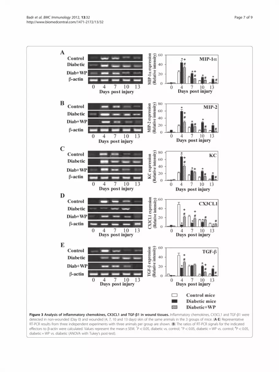
Figure 3 Analysis of inflammatory chemokines, CX3CL1 and TGF-β1 in wound tissues. Inflammatory chemokines, CX3CL1 and TGF-β1 weredetected in non-wounded (Day 0) and wounded (4, 7, 10 and 13 days) skin of the same animals in the 3 groups of mice. (A-E) RepresentativeRT-PCR results from three independent experiments with three animals per group are shown. (B) The ratios of RT-PCR signals for the indicatedeffectors to β-actin were calculated. Values represent the mean± SEM. *P< 0.05, diabetic vs. control; +P< 0.05, diabetic +WP vs. control; #P< 0.05,diabetic +WP vs. diabetic (ANOVA with Tukey’s post-test).
Badr et al. BMC Immunology 2012, 13:32 Page 7 of 9http://www.biomedcentral.com/1471-2172/13/32

Badr et al. BMC Immunology 2012, 13:32 Page 8 of 9http://www.biomedcentral.com/1471-2172/13/32
AbbreviationsIL: Interleukin; KC: Keratinocyte-derived chemokine; MIP-1α: MIP-2macrophage inflammatory proteins 1α and 2; STZ: Streptozotocin; TGF-β: Transforming growth factor-β; WPs: Whey proteins.
Competing interestsThe authors declare that they have competing interests.
AcknowledgmentsThis work was supported by the National Plan for Science and Technology(NPST) funded by King Abdulaziz City for Science and Technology (KACST)through project 10-BIO975-02. The authors declare that there are no conflictsof interest. The authors also acknowledge Dr. Ali Metwalli of the FoodScience Department, College of Agriculture and Food Science, King SaudUniversity, for the preparation of whey protein.
Author details1Princes Johara alibrahim center for cancer research, prostate cancer researchchair, College of Medicine, King Saud University, Riyadh, Saudi Arabia.2Zoology Department, Faculty of Science, Assiut University, 71516, Assiut,Egypt. 3Vice-Rectorate for Graduate Studies and Research-Visiting ProfessorProgram, King Saud University, Riyadh, Saudi Arabia. 4Zoology Department,College of Science, King Saud University, Riyadh, Saudi Arabia. 5Humannutrition Department, National Research Centre, Dokki, Cairo, Egypt.6Zoology Department, Faculty of Science, Menoufia University, Menoufia,Egypt. 7Department of Urology/Surgery, College of Medicine, King SaudUniversity, Riyadh, Saudi Arabia. 8EA3064—GIMAP, Université de Lyon,F-42023, Saint-Etienne (cedex 2), France.
Authors’ contributionsGB put the design of the study, carried out the immunological assays,prepared figures, drafted the manuscript and performed the statisticalanalysis. BMB carried out some immunological parameters and participatedin the analysis of the data. MHM was responsible for monitoring the foodand water consumption for the animal model throughout the experimentperiod and participated in drafting the manuscript. MM was responsible forthe animal model and participated in preparing the figures. OG participatedin the statistical analysis and drafting the manuscript. All authors read andapproved the final manuscript.
Authors’ informationDr Gamal Badr principle investigator of several research projects funded byKing Saud University, as well as head of a research group of Immunology. DrBadr is a leading and internationally respected immunologist. He hasobtained his master and PhD in Immunology from Faculty of Medicine, ParisXI University, France with a scholarship from the Egyptian government aswell as from Sidaction (France). He followed 2 years of significantpostdoctoral experience at University of Montreal, Canada with fellowshipfrom FRSQ. He awarded for the best oral presentation of Post-doctors in the10th Annual Conference of CHUM (18 December 2007), Montreal University,Canada. He hold his associate professor appointment in Immunology atAssiut University, Egypt as well as at King Saud University (January 2011).
Received: 24 March 2012 Accepted: 18 June 2012Published: 18 June 2012
References1. Maahs DM, Rewers M, Editorial: Mortality and renal disease in type 1
diabetes mellitus–progress made, more to be done. J Clin EndocrinolMetab 2006, 91:3757–375.
2. Thompson CS: Animal models of diabetes mellitus: relevance to vascularcomplications. Curr Pharm Des 2008, 14:309–324.
3. Lioupis C: Effects of diabetes mellitus on wound healing: an update. JWound Care 2005, 14:84–86.
4. Nithya V, Baskar A: A Preclinical Study on Wound Healing Activity ofLawsonia ulba Linn. Res J of Phytochemistry 2011, 5:123–129.
5. Black E, Vibe-Petersen J, Jorgensen LN, Madsen SM, Agren MS, Holstein PE,Perrild H, Gottrup F: Decrease of collagen deposition in wound repair intype 1 diabetes independent of glycemic control. Arch Surg 2003,138:34–40.
6. Ohura T, Nakajo T, Okada S, Omura K, Adachi K: Evaluation of effects ofnutrition intervention on healing of pressure ulcers and nutritionalstates (randomized controlled trial). Wound Repair Regen 2011, 19:330–336.
7. Werner S, Grose R: Regulation of wound healing by growth factors andcytokines. Physiol Rev 2003, 83:835–870.
8. Borst SE: The role of TNF-alpha in insulin resistance. Endocrine 2004,23:177–182.
9. Siqueira MF, Li J, Chehab L, Desta T, Chino T, Krothpali N, Behl Y, Alikhani M,Yang J, Braasch C, Graves DT: Impaired wound healing in mouse modelsof diabetes is mediated by TNF-alpha dysregulation and associated withenhanced activation of forkhead box O1 (FOXO1). Diabetologia 2010,53:378–388.
10. Peranteau WH, Zhang L, Muvarak N, Badillo AT, Radu A, Zoltick PW, LiechtyKW: IL-10 overexpression decreases inflammatory mediators andpromotes regenerative healing in an adult model of scar formation.J Invest Dermatol 2008, 128:1852–1860.
11. Christopherson K 2nd, Hromas R: Chemokine regulation of normal andpathologic immune responses. Stem Cells 2001, 19:388–396.
12. Dewald O, Zymek P, Winkelmann K, Koerting A, Ren G, Abou-Khamis T,Michael LH, Rollins BJ, Entman ML, Frangogiannis NG: CCL2/MonocyteChemoattractant Protein-1 Regulates Inflammatory Responses Critical toHealing Myocardial Infarcts. Circ Res 2005, 96:881–889.
13. Rich J, Lee JC: The pathogenesis of Staphylococcus aureus infection inthe diabetic NOD mouse. Diabetes 2005, 54:2904–2910.
14. Ishida Y, Gao JL, Murphy PM: Chemokine receptor CX3CR1 mediates skinwound healing by promoting macrophage and fibroblast accumulationand function. J Immunol 2008, 180:569–579.
15. Distler JH, Hirth A, Kurowska-Stolarska M, Gay RE, Gay S, Distler O:Angiogenic and angiostatic factors in the molecular control ofangiogenesis. Q J Nucl Med 2003, 47:149–161.
16. Werner S, Krieg T, Smola H: Keratinocyte-fibroblast interactions in woundhealing. J Invest Dermatol 2007, 127:998–1008.
17. Ord H: Nutritional support for patients with infected wounds. Br J Nurs2007, 16:1346–1348. 1350-2.
18. Kappeler SR, Heuberger C, Farah Z, Puhan Z: Expression of thepeptidoglycan recognition protein, PGRP, in the lactating mammarygland. J Dairy Sci 2004, 87:2660–2668.
19. Velioglu Ogünç A, Manukyan M, Cingi A, Eksioglu-Demiralp E, OzdemirAktan A, Süha Yalçin A: Dietary whey supplementation in experimentalmodels of wound healing. Int J Vitam Nutr Res 2008, 78:70–73.
20. Balbis E, Patriarca S, Furfaro A, Millanta S, Sukkar GS, Marinari MU, PronzatoAM, Cottalasso D, Traverso N: Whey proteins influence hepaticglutathione after CCl4 intoxication. Toxico Ind Heal 2009, 25:325–328.
21. Rusu D, Drouin R, Pouliot Y, Gauthier S, Poubelle PE: A bovine wheyprotein extract stimulates human neutrophils to generate bioactiveIL-1Ra through a NF-kappaB- and MAPK-dependent mechanism. J Nutr2010, 140:382–391.
22. Walzem RM, Dillard CJ, German JB: Whey Components: Millennia ofEvolution Create Functionalities for Mammalian Nutrition: What WeKnow and What We May Be Overlooking. CRC Cr Rev Food Sci 2002,4:353–375.
23. Krissansen GW: Emerging health properties of whey proteins and theirclinical implications. J Am Coll Nutr 2007, 26:S713–S723.
24. Badr G: Supplementation with Undenatured Whey Protein DuringDiabetes Mellitus Improves the Healing and Closure of Diabetic Woundsthrough the Rescue of Functional Long-lived Wound Macrophages.Cell Physiol Biochem 2012, 29:571–582.
25. Ebaid H, Salem A, Sayed A, Metwalli A: Whey protein enhances normalinflammatory responses during cutaneous wound healing in diabeticrats. Lipids Health Dis 2011, 10:235.
26. Weinberg ED: Antibiotic properties and applications of lactoferrin.Curr Pharm Des 2007, 13:801–811.
27. Engelmayer J, Blezinger P, Varadhachary A: Talactoferrin stimulates woundhealing with modulation of inflammation. J Surg Res 2008, 149:278–286.
28. Badr G, Bashandy S, Ebaid H, Mohany M, Sayed D: Vitamin Csupplementation reconstitutes polyfunctional T cells in streptozotocin-induced diabetic rats. Eur J Nutr 2011.
29. Mori R, Kondo T, Ohshima T, Ishida Y, Mukaida N: Accelerated woundhealing in tumor necrosis factor receptor p55-deficient mice withreduced leukocyte infiltration. FASEB J 2002, 16:963–74.
30. Matalka KZ, Tutunji MF, Abu-Baker M, Abu-Baker Y: Measurement ofprotein cytokines in tissue extracts by enzyme-linked immunosorbent

Badr et al. BMC Immunology 2012, 13:32 Page 9 of 9http://www.biomedcentral.com/1471-2172/13/32
assays: application to lipopolysaccharide-induced differential milieu ofcytokines. Neuroendocrinol Lett 2005, 26:231–236.
31. Reiber GE, Ledoux WR: The Evidence Base for Diabetes Care. InEpidemiology of diabetic foot ulcers and amputations: evidence for prevention.Edited by Williams R, Herman W, Kinmoth AL, Wareham NJ. Chichester: UK:Wiley; 2002:641–665.
32. Frid AH, Nilsson M, Holst JJ, Björck IM: Effect of whey on blood glucoseand insulin responses to composite breakfast and lunch meals in type 2diabetic subjects. Am J Clin Nutr 2005, 82:69–75.
33. Machnicki M, Zimecki M, Zagulski T: Lactoferrin regulates the release oftumour necrosis factor alpha and interleukin 6 in vivo. Int J Exp Pathol1993, 74:433–439.
34. Takamiya M, Fujita S, Saigusa K, Aoki Y: Simultaneous detections of 27cytokines during cerebral wound healing by multiplexed bead-basedimmunoassay for wound age estimation. J Neurotrauma 2007, 24:1833–44.
35. Lin Q, Fang D, Fang J, Ren X, Yang X, Wen F, Su SB: Impaired woundhealing with defective expression of chemokines and recruitment ofmyeloid cells in TLR3-deficient mice. J Immunol 2011, 186:3710–7.
36. Diegelmann RF, Evans MC: Wound healing: an overview of acute, fibroticand delayed healing. Front Biosci 2004, 9:283–9.
37. Pereira CT, Herndon DN, Rocker R, Jeschke MG: Liposomal gene transfer ofkeratinocyte growth factor improves wound healing by altering growthfactor and collagen expression. J Surg Res 2007, 139:222–8.
doi:10.1186/1471-2172-13-32Cite this article as: Badr et al.: Treatment of diabetic mice withundenatured whey protein accelerates the wound healing process byenhancing the expression of MIP-1α, MIP-2, KC, CX3CL1 and TGF-β inwounded tissue. BMC Immunology 2012 13:32.
Submit your next manuscript to BioMed Centraland take full advantage of:
• Convenient online submission
• Thorough peer review
• No space constraints or color figure charges
• Immediate publication on acceptance
• Inclusion in PubMed, CAS, Scopus and Google Scholar
• Research which is freely available for redistribution
Submit your manuscript at www.biomedcentral.com/submit


