Embed Size (px)
Citation preview
TREATMENT OF IDIOPATHIC CONGENITAL TALIPES EQUINO VARUS BY PONSETI METHOD
- A SHORT TERM FOLLOW UP STUDY
Dissertation submitted for
M.S. DEGREE EXAMINATION BRANCH – II ORTHOPAEDIC SURGERY
Department of Orthopaedics and traumatology Thanjavur Medical College,
Thanjavur.
TAMILNADU Dr. M.G.R. MEDICAL UNIVERSITY, CHENNAI, TAMIL NADU.
MARCH 2007
CERTIFICATE
This is to certify that this dissertation entitled
“TREATMENT OF IDIOPATHIC CONGENITAL TALIPES
EQUINO VARUS BY PONSETI METHOD – A SHORT
TERM FOLLOW UP STUDY” is a bonafide record work
done by . K. MANIVANNAN submitted as partial fulfillment
for the requirements of M.S. Degree Examinations
Branch II, Orthopaedic Surgery, MARCH 2007.
Date : Prof. Dr.K. Anbalagan M.S (Ortho)., D.Ortho., Professor & Head of the Department,
Department of Orthopaedics & Traumatology,
Thanjavur Medical College & Hospital,
Thanjavur.
ACKNOWLEDGEMENT
I have great pleasure in placing on record my deep sense
of gratitude and thanks to my Professor
DR. K. ANBALAGAN M.S. (Ortho)., D.Ortho, Professor and
Head of the Department of Orthopaedics and Traumatology,
Thanjavur Medical College and Hospital, Thanjavur for his
constant guidance, encouragement, and untiring help
throughout the preparation of this dissertation.
I express my profound gratitude to Professor
DR.R.RATHINASABAPATHY M.S.(Ortho)., D.Ortho and
Professor Dr.M.GULAM MOHIDEEN M.S. (Ortho)., D.Ortho for
their help, guidance and advice given to me in the preparation
of this study.
I owe much to Dr. P. Venkatesan M.S. (Ortho) who stood
by me in every step of this study and without whose guidance;
this study would not have been completed.
I equally thank Dr. V. Jayabalan, Dr. M.S. Manoharan,
Dr. K. Mahadevan, Dr. A. Barathy, Dr. S. Kumaravel and my
colleagues for their unstinted co-operation in doing this study.
I thank the DEAN Thanjavur Medical College and
Hospital, Thanjavur, for permitting me to utilize the hospital
facilities for my study.
I express my sincere thanks to all the PATIENTS, who have
co-operated in carrying out my study.

CONTENTS
1. INTRODUCTION 1
2. AIM 4
3. REVIEW OF LITERATURE 5
4. MATERIALS AND METHODS 36
5. OBSERVATIONS AND RESULTS 41
6. DISCUSSION 48
7. CONCLUSION 52
8. BIBLIOGRAPHY
9. APPENDICES
I. Pirani Severity scoring
II. Consent Proforma
III. Clinical Proforma
IV. Master Chart
1
Congenital talipus equinovarus probably the most common congenital
pathological condition is a descriptive term. The term was first described by
Hippocrates. It was Nicolas Andry in his “Orthopaedicia” described the term
“Pedis Equinal” which meant the foot resembling the foot of the horse. The
term “talipus equinovarus” is derived from latin : Talipus , a combination of
words- Talus ( ankle) and pes (foot) ; equinus meaning “horse like”(the heel
in plantar flexion) and varus meaning inverted and adducted.
The incidence of CTEV is approximately 1 – 1.4 cases per 1000 live
births 3. Boys are affected twice as often as girls 3.The etiology of club foot is
still obscure although too many theories have been proposed. A higher
incidence of CTEV was also noted in patients with a positive family
history 2,3.
The theories proposed in the etiology of CTEV are mechanical factors
in utero, neuromuscular defect, primary germ plasma defect, arrested fetal
devolopment, hereditary, etc;
Irrespective of the etiology, the pathoanatomic changes associated with
CTEV include ankle equinus, a calcaneum that is in equinus and inverted
position beneath the talus and the talar head prominence at the dorsolateral
midfoot, navicular medial and plantar to the talar head, cuboid medial and in
2
front of calcaneum, medial tilting of anterior part of talus, shortened talar
neck, narrow posterior ankle mortise, talar tilt out of ankle mortise 4.
The goal of treatment is to reduce or eliminate these deformities so that
the patient has a functional, painfree, plantigrade foot with good mobility and
without calluses and does not need to wear modified shoes.
The recommended treatment of CTEV ranges from non-operative
casting & stretching to complete peritalar surgical release and bony
procedures for neglected CTEV cases.
The methods of J.H.Kite 4, Ignacio V. Ponseti 6 and French
methods as described by Masse & Bensahel 7 are examples of non-operative
methods of correction of CTEV.
The technique of gradual and simultaneous correction of all
deformities of CTEV using manipulation and casting at weekly interval
described by Dr.Ignacio V. Ponseti has gained wide acceptance throughout
the world.
Ponseti opined different from others in that he described about the
interdependent movements of tarsal bones and considered the view that tarsal

3
joints move on a fixed axis of motion to be incorrect. He described the Kite’s
method of correction in which the abduction of calcaneus under the talus was
prevented by applying counter pressure over the calcaneocuboid joint as
“Kite’s error”. This is very essential in correction of heel varus as the
calcaneus cannot be everted unless it is fully abducted under the talus.
In this study, we have attempted to analyse the functional outcome
of Idiopathic clubfoot using Ponseti’s technique in children presenting to us
within the first two years of age without any prior treatment.
4
AIM
The aim of treatment in Idiopathic CTEV is to obtain:
Painless
Pliable
Plantigrade and
Cosmetically acceptable foot.
With various treatment modalities so far available for the treatment of
Idiopathic CTEV, we are not able to obtain a plantigrade foot with either after
single stage or multiple staged procedures. Most of the cases end up with stiff,
small and painful foot.
The present study is aimed at evaluating the functional outcome of
CTEV correction by Ponseti method at the end of initial correction and at six
months follows up.
5
HISTORICAL REVIEW
The earliest documentation of clubfoot comes from the ancient
Egyptian people. Paintings on the walls of their ancient tombs depict the
clubfoot deformity, and statue of diastrophic dwarf with a clubfoot can be
found in the Tutankhamen collection4.
Hippocrates was the first to start treatment in cases of CTEV as soon as
possible after birth, before bony deformities are established5. According to
him, intrauterine malposition was the cause and adaptive changes in
surrounding muscles and bones lead to articular malalignement 5.
Areaus, Pare and Fabrig recommended stretching of the foot by
specialized apparatus as early as 17th century 2.
In 1836 Mcguerin was the first to use plaster of paris in the treatment of
CTEV 2.
Surgical methods of management of CTEV was initiated by Little by
doing subcutaneous tenotomy of tendo achilles 2.
In 1857 Solly was the first to introduce a bony procedure – partial
cuboidectomy for correction of the deformity 2.
6
Dithrich forwarded the theory that primary failure was in the peripheral
nerve to the peroneal muscles and he found a sluggish electrical reaction in
the peroneii in premature infants with clubfoot.
In the 18th and 19th centuries, the general trend was to treat CTEV only
after the child has passed early infancy.Most preferred method was a single
stage correction of all the components of the deformity.
The current trend has reverted towards the non operative treatment of
Idiopathic CTEV as soon as possible after birth 3,4.
AETIOLOGY
The exact aetiology of this condition is not known, but deforming
forces are well understood 3. Various theories had been put forward with
regard to the aetiology of CTEV but none have succeeded in explaining the
same conclusively.
1. Mechanical pressure in utero :
This is an oldest theory described by Hippocrates. He believed that the
foot was held in a position of equinovarus by external uterine pressure leading
to development of CTEV.Oligohydromnios prevents fetal movements and
makes feet vulnerable to external pressure 4.
7
But when foot is forming i.e. first few months of pregnancy, fetus is floating in liquor amnii and result in uniform distribution of pressure. There was absence of increased incidence of CTEV in twin pregnancies in which the uterus was supposed to be overcrowded leading to unacceptability of this theory 4 . 2. Neuromuscular defect
Issac 9 proposed a prominent neurogenic factor in the causation of
CTEV. He believed that CTEV is a resistant form of Arthogryposis multiplex
congenita.He concluded that the anomalies observed at the time of dissection
were independent of immobilsation, stretching, relaxation of muscles and
were not influenced by previous treatment. He also proposed that fibrosis
observed in clubfoot muscle specimen should not be considered as a primary
aetiological factor 4.
3. Arrested development:
Heuter and Volkman regarded CTEV as an arrest of fetal development.
Bessel, Hagen opposed the theory of arrested fetal development as they said
that there is no such physiological stage in the development of fetus that
resembles CTEV4.
Mau wrote that the embryonic foot does not show the distortion of
bones about tarsal joints, which is found in CTEV 4.
8
A.Victoria Diaz said “embryonic foot position changes with
movements of talus and calcaneus due to growth spurt in distal tibia and
fibula. In fibular phase, foot goes into usual fetal position of equinovarus and
in tibial phase it is pronated into usual fetal position. Any arrest in the tibial
phase without growth spurt results in persistent equinovarus deformity” 10.
4. Blastemal defect in the devolopment of tarsal cartilage analogue:
Waisbord 11 described a defect in the cartilage analogue of tarsal bones
as cause of the deformity.Ponseti did not found any defect in the cartilage
analogue in specimen dissected by him 3.
Irani and Sherman 12 described a primary germ cell defect in head and
neck of talus, but were unable to explain germ cell defect in unilateral
clubfoot and correction of deformity by realigning the navicular and
calcaneus on talus without any correction in the talus 4.
5. Primary retracting fibrosis:
Zimmy et.al 3 showed fibroblast, cells resembling myofibroblast,
mast cells in fascia from medial and lateral side of clubfoot and speculated
that contractures of ligaments were due to myofibroblast like cells and
enhanced histamine released by mast cells.
9
Fukuhara observed myofibroblast like cells in the spring ligament
and speculated fibromatosis in the medial tarsal ligaments as the cause of
CTEV 11. Ippolito and Ponseti I.V. similarly described primary retracting
fibrosis as primary cause of CTEV deformity 14.
6. Hereditary and environmental factors:
The literature regarding inheritance pattern of CTEV is confusing.
There is confusing evidence for multifactorial aetiological factors including
environmental and genetic factors 15.
Pedigree studies have established that the disease is certainly not
inherited in a single autosomal dominant or autosomal recessive mendelian
fashion, although a mendelian component of inheritance cannot be fully
excluded 15. A genetic predisposition operating on a polygenic or in some
cases autosomal dominant basis was thought to manifest as Idiopathic CTEV
when a threshold for expression is exceeded. When the genetic predisposition
alone does not exceed the threshold, environmental factors may act alone or
synergistically to reduce the threshold for expression to the point at which
Idiopathic CTEV is manifest 15.
In CTEV, Palmer initially favoured the theory of autosomal
dominant gene with reduced penetrance as the cause of deformity but later he
supported the multifactorial system of inheritance 16.
10
Wynne Davis 16 concluded that a decreasing incidence of CTEV
disorder as the relationship of the parents become remote could be indicative
of dominant gene with reduced penetrance or multifactorial inheritance
system. However the manner in which the occurrence rate decreases is
suggestive of a multifactorial model.
A preponderance of the condition among those patient with first
degree relative affected increasing the frequency to 2.9 per 1000 live births is
highly suggestive of a heritable component 15.
Chung provided strong corroboration of the polygenic model of
inheritance in his study of incidence of CTEV according to race, conducted in
the population of Hawaii 16.
Syndromic CTEV has either Autosomal dominant i.e.
Craniocarpotarsal Dysplasia or Whistling face Syndrome, or Autosomal
recessive pattern i.e. Diastrophic Dwarfism 16. It is also associated with
Larson syndrome and Smith- Lemli- Opitz syndrome 16.
Gorlin R.J. 16 described clubfoot associated with X- linked
recessive pattern. He wrote on Pirre-Robin syndrome with congenital heart
malformation and CTEV.
11
7. Cytological abnormalities:
Cytological abnormality produces syndromes that include CTEV
with maternal unbalanced 6 : 11 translocation as reported by Clark 16. Insley
reported a case of association of CTEV with a deficiency of a part of long arm
of chromosome 18 16.
PATHOANATOMY
Antonio Scarpa was the first to describe the vivid anatomy of
CTEV.He described the twisting of the calcaneus and navicular around the
talus as “congenital dislocation of the talocalcaneonavicular joint” 3.
William Adams 17 called attention to abnormal shape of the head
and neck of talus, which he felt was secondarily due to the acquired
deformities i.e. adaptation to the altered position of the os calcis and
navicular, this deformed shape being result than cause of the deformity.
Evance D. said that the essential abnormality lies in the midtarsal
joints and other elements of the deformity were due to secondary adaptive
changes 4.
Attenbourough said “the fundamental deformity is plantar flexion of
the talus” 18.
12
Severity of the CTEV depends upon the degree of bony
displacements whereas the resistance to treatment is determined by the
rigidity of soft tissue contractures 3.
The adapted alteration in the shape of tarsal bones are acquired in
accordance of the Wolff’s law 4 i.e. every change in the use of static function
of bone causes a change in the internal form and architecture as well as
alteration in its external formation and function according to mathematical
law. The soft tissue contractures are acquired in accordance with law of
Davis 4 which states, “when ligaments and soft tissues are in a lax state, they
gradually will shorten”.
DEFORMITIES IN CTEV
The deformities in CTEV are:
1. Fore foot adduction
2. Hind foot varus
3. Hind foot equinus
4. Cavus
13
The foot in CTEV is always smaller in size than the normal foot in
cases of unilateral CTEV due to small muscle mass and connective tissue
fibrosis 3.
OSSEOUS DEFORMITIES
TALUS:
This is least displaced but most deformed bone in CTEV. As it has
no muscle attachments, it is forced into equinus by its articulations and
attachment to calcaneum and navicular 3. It appears to be subluxated
anteriorly out of ankle mortise.
Body of talus is wide anteriorly as only posterior part of trochlea
is in articulation with tibial plafond. Schiltz 4 observed that only the posterior
half was normal and having normal rounded contour. The anterior half was
wide, abnormal and important cause of limitation of dorsiflexion and
persistent equinus.The posterior part of the talus which was not covered with
cartilage is intra- articular.
The neck of the talus is directed plantarwards and medially 4. The
head –body angle as measured by Paturet is strikingly smaller in CTEV 11.
The neck is usually foreshortened and the usual constriction is absent.
14
The head of the talus is wedge shaped. It shows two articular
facets. The anterolateral surface is left uncovered by displaced navicular,
which extends over the talar neck. Talonavicular joint is oriented in a more
sagittal plane compared to normal coronal orientation of the facets 3. Three
facets on the inferior surface of the head appear as a single continuous flat
surface.Posterior concave facet of the body is less developed and shallow.
Medial surface is underdeveloped but congruent with oblique surface of
calcaneus.
CALCANEUS:
Calcaneus is involved in all the three deformities of CTEV i.e. equinus,
varus and adduction. Clinical deformity is due to abnormal position rather than
abnormal shape of the calcaneus. Posterior tuberosity is displaced upwards and
medially. Anterior end of the calcaneus is displaced downwards, medially and
inverted under the head of the talus 3. Sustentaculum tali is displaced medially
and underdeveloped. Medial surface is underdeveloped but congruent with
corresponding articular surface of the talus. Posterior facet is underdeveloped
while anterior and medial facets are flat and continuous. The longitudinal axis
of talus and calcaneus are parallel to each other.
15
NAVICULAR
This is most severely displaced bone in CTEV 4. It is grossly
medially displaced and adducted, inverted over the head of the talus. It is in
close contact with sustentaculam tali and medial malleolus. Medial tuberosity
of the navicular is large and provides large area of insertion for enlarged,
thickened tibialis posterior tendon. It is wedge shaped with wide dorsal and
narrow plantar lateral surface.
CUBOID
It is medially displaced and inverted in front of the calcaneus. It is
not as much medially displaced as the navicular. Only the medial part of
anterior part of the calcaneus articulates with the cuboid.
CUNEIFORMS AND METATARSALS
Cuneiform and Metatarsals are always adducted but are normal in
shape. 1st metatarsal is always in plantar flexion as compared to other
metatarsals and accounts for the cavus deformity in CTEV 3.
TIBIA
Lower end of tibia articulates only with posterior part of talus which
is devoid of articular cartilage. Tibia has half the amount of external rotation
as compared to normal foot 19. It has been the usual convention to suppose
16
that the tibia was medially rotated. This however has been challenged by
Swann et al.(1969), who demonstrated lateral rotation of the tibia which
indicated the need for a rotational osteotomy in some cases 52.
Lateral malleolus is displaced posteriorly 4. This brings the tendo
achilles in close relation to lateral malleolus mainly due to thickening of the
fascia enclosing peroneal tendons and the calcaneofibular ligament 20.
SOFT TISSUE ANATOMY
CTEV foot is always shorter than the normal foot 3. Reduction in the
girth and length of leg muscles is a common finding 3, 4, 10, 11, 14. Increase in
fibrous connective tissue in the muscles and tendon sheath is common finding
during dissection 3. Few authors have observed abnormalities in the insertion
of tendon during anatomical dissection and at surgery 5. Most authors have
found that the ligaments on the posterior and medial aspect of the tarsal joints
are thick and short 3, 10, and 14.
POSTERIOR CONTRACTURES
The contracture of tendo achilles, ankle capsule, subtalar capsule,
posterior talofibular ligament and calcaneofibular ligament prevents
correction of equinus deformity 5.
17
Posterior capsules of the ankle and the subtalar joints are thickened
and contracted. Posterior subtalar capsule contractures are more severe than
the posterior ankle contractures.
The Achilles tendon is always contracted and shortened. This
prevents downward extrusion of posterior tuberosity of the calcaneus, which
is necessary for dorsiflexion. Its calcaneal attachment is broader and wider. Its
insertion is more on the medial side of the calcaneal tuberosity as compared to
normal foot, resulting in varus position of the calcaneus. This medial
attachment must be divided while performing posterior release or
posteromedial soft tissue release, to aid in the correction of heel varus.
Posterior talofibular ligament and calcaneofibular ligaments
becomes thickened and shortened as per Law of Davis 4. This results in
prevention of movements of the fibula, which were very essential for normal
dorsiflexion at the ankle joint. This must be excised in posteromedial soft
tissue release to obtain good correction 20.
The dorsiflexion of the talus is prevented by the contracted and
shortened posterior capsule of the ankle and tight achilles tendon and
posterior talofibular ligament .These structures prevent the downward exit of
18
the back portion of the trochlea out of the ankle mortise, a prerequisite for the
dorsiflexion.
MEDIAL PLANTAR CONTRACTURE
(Tibialis Posterior tendon, Deltoid ligament, Spring ligament,
Talonavicular capsule)
The fibrosis of the above mentioned structures form a mass of
indistinguishable scar, which obscures the midtarsal, subtalar joints. It
maintains sustentaculum tali, medial malleolus and medial tuberosity of the
navicular in close proximity. This mass of scar tissue prevents the forward
and lateral migration of the navicular as well as lateral movement of anterior
end of the calcaneus. This fibrous tissue forms deep layer of Deltoid ligament,
which is located between contiguous surfaces of the medial malleolus and
medial articular surface of the talus 14.
The tibialis posterior tendon is short and its tendon sheath is thick
and hypertrophied. It has abnormal attachments to spring ligament,
sustentaculum tali and navicular. It has very wide insertion on navicular
tuberosity. It also blends with common mass of scar tissue, which maintains
sustentaculum tali, medial malleolus and medial tuberosity of the navicular in
close proximity.
19
Both flexor hallucis longus and flexor digitorum longus are short
and contracted which causes flexion contractures of the digits. The Master
knot of Henry 4 where these two structures cross is an important plantar
contracture that resists mobility of the navicular by virtue of its attachment to
the undersurface of the navicular.
Spring Ligament: This is an important structure, which supports talar
head on its plantar aspect. It is always contracted, short and inelastic. This is
because of the equinovarus deformity present in CTEV, which brings the
navicular in close relation to sustentaculum tali resulting in relaxation of the
spring ligament and subsequent contracture as per the law of Davis.
According to the law of Davis 4, the talonavicular joint capsule also
contracts, which is in a lax state due to the equinovarus position.
TALOCALCANEAL INTEROSSEOUS AND BIFURCATED
LIGAMENTS:
These are underdeveloped, stretched and thin 7. They are contracted
in cases of neglected CTEV in the older children 4. Release of these ligaments
during surgery may lead to over correction as a complication at a later date 4.
20
PLANTAR CONTRACTURES:
The contractures of abductor hallucis, intrinsic toe flexors and plantar
aponeurosis are more prominent in older children and are less prominent in
children less than four years of age 4.
Abductor hallucis is considered to be an important structure which
maintains persistent forefoot adduction. It has an accessory abnormal
attachment to tendon sheath of tibialis posterior, navicular and Master knot of
Henry.
Peroneal tendons are weak.Intrinsic toe flexors are shortened. Calf
muscles and extrinsic toe flexors are also shortened.
The ligaments on medial and posterior aspect of the ankle joint are
pulled into the joint by severe plantar flexion and varus displacement of talus.
There is marked thickening and shortening of the tibionavicular and plantar
calcaneonavicular ligament.
BIO-KINEMATICS:
The correction of the severe displacements of the tarsal bones in
CTEV requires a clear understanding of the functional anatomy of talus.
21
There are controversies regarding axis of motion of subtalar joints.
According to Farabuef, Virchow H, Huson and Siegler, there is no fixed axis
of motion of subtalar joint. This is in contrast to the concept by Hicks, Elfnan
and Inman which emphasis that subtalar joint moves around a fixed axis 3.
A better understanding of the tarsal mechanics in the normal foot was
given by Huson in his thesis “A functional and anatomical study of tarsus”.
He demonstrated that tarsal joints do not move as a single hinge but rotate
about a moving axis as in the case of the knee. Each joint has its own specific
motion pattern. These are described by means of discrete arcs, representing
the successive portion of a particular moving axis. This successive position is
followed by a fixed pattern which is characteristic for the joint concerned 6.
He described “Constrained Mechanism” in which motion of the
tarsal joints occur simultaneously. If one of the joint movements is blocked
the other joint movements also get blocked. The ligaments play an important
role as “Kinematic Constraints” of joints apart from their share in forced
transmission to support the elastic vault structure of the foot 3.
The concept of passage of axis of rotation from anteromedial to
posterolateral was given by Inman 21.
22
Seigler 3 described “Kinematic Coupling” as there is no separation
between the motion of the ankle joint and subtalar joint in living objects.
Motion of the foot shank complex in one direction occurs by the combined
motion of both joints. Contribution from ankle joint in dorsiflexion and
plantar flexion is more than that of subtalar joint while subtalar joint has more
contribution in inversion and eversion than that of ankle joint.Both joints
contribute equally in internal and external rotation.
Ponseti 53 gave a new concept to the kinematics around the talus.
He described that, in the clubfoot, the anterior portion of the calcaneus lies
beneath the head of the talus. This position causes varus and equinus
deformity of the heel. Attempts to push the calcaneus into eversion without
abducting will press the calcaneus against the talus and will not correct the
heel varus. Lateral displacement (abduction) of the calcaneus to its normal
relationship with the talus will correct the heel varus deformity of the
clubfoot.
He emphasized that the clubfoot deformity occurs mostly in the
tarsus. The tarsal bones, which are mostly made of cartilage, are in the most
extreme positions of flexion, adduction, and inversion at birth. The talus is in
severe plantar flexion, its neck is medially and plantarly deflected, and its
head is wedge shaped. The navicular is severely medially displaced, closed to
23
the medial malleolus and articulates with the medial surface of the head of the
talus. The calcaneus is adducted and inverted under the talus. No single axis
of motion (like a metered hinge) exists on which to rotate the tarsus whether
in a normal or a clubfoot. The tarsal joints are functionally interdependent.
The movement of each tarsal bone involves simultaneous shifts in the
adjacent bones. Joint motions are determined by the curvature of the joint
surfaces and by the orientation and structure of the binding ligaments. Each
joint has its own specific motion pattern. Therefore correction of the extreme
medial displacement and inversion of the tarsal bones in clubfoot necessitates
a simultaneous gradual lateral shift of the navicular, cuboid and calcaneus
before they can be everted into a neutral position 53.
TREATMENT
The spectrum of treatment options for CTEV is large .It ranges
from non-operative methods including manipulation, strapping, repeated
stretching and POP casting on one side to operative methods like soft tissue
surgery and bony procedure.
FORCIBLE MANIPULATION
The concept of forcible manipulation was first described by
Bruckner 5. Thomas did immediate forcible correction with a wrench and
application of a splint to hold the foot in corrected position 5. Forcible
24
corrections at one or two sittings was carried out by Lorenz using a modified
Thomas wrench and later used a padded pyramid correcting a deformity over
its apex.
Tubey was the first person to give details of the manipulation
technique. He advised abduction and eversion at talocalcaneonavicular,
calcaneocuboid joint with dorsiflexion of whole foot at ankle 5.
Harreustein feared damage to distal tibial and fibular epiphysis
during forcible manipulation 5.
SPLINT Pare advocated splint alone as a device to correct all or part of the
deformity 5. Scarpa used shoes to correct the deformity and emphasized that
varus should be converted into equinus. Trelat, Shaffer have described various
devices for manipulative correction.
In 1897, Gibnery practiced wrenching to convert the equinovarus
into equinovalgus.He then reduced the equinus by tenotomy and manual
force, immobilizing the foot in plaster of paris cast long enough for the bones
on the outer side to atrophy and for those on the inner side to hypertrophy 5.
25
Dennis Brown in 1934 gave a breakthrough by introducing metal
splint for the correction of the deformity 5.
Forcible manipulation has fallen to disrepute owing to the
stiffness of the joints, deformities of bones and spurious correction providing
a rocker bottom foot which developed following this form of treatment.
REPEATED STRETCHING:
The emphasis on treating newborn with CTEV was first given by
Hippocrates who advocated repeated manual correction and application of
strong bandages during manipulation. Over correction was considered to be
an essential part of the procedure 5.
Sofield departed from forcible manipulation and started using
elastic traction for the correction of the deformity 5. Brown supported this
principle and claimed that useful feet and leg can be obtained without use of
the force. He based his thoughtful account on three well known hypotheses:
continuous traction will gradually tire a muscle, a contracted muscle put on
stretch will gradually lengthen, if relaxed, will shorten and return to the
contracted state as per the Law of Davis 4. Hence over correction is a must.
26
J. Hiram Kite 2, 3, 5 was a strong advocator of non operative
treatment of clubfoot. His original technique consists of manipulation and
casting followed by wedging of the cast to correct individual deformities.
Later he advised repeated change of the whole cast with manipulative
stretching at each stage. He said “Whatever is gained without force is
achieved without harm”.
Jones and Lovett 6 said that: “In very young children it is probable
that every case can be cured without operation with the exception of a
possible tenotomy of the tendo achilles in the final stage after constantly
repeated manipulations by the parents carefully taught by the surgeon”.
PLASTER OF PARIS CASTS:
Guerin was the first to describe the use of plater of paris casts in
the treatment of CTEV 5. This was followed by Thomas, Jones, Litle,
Bradford and Lovett (1899) and Whitman (1910). Soule 5 practiced
manipulative reduction followed by retention in adhesive strapping
incorporating the strapped limb in plaster of paris cast (1930). Elmslie used
plater of paris casts without splinting. Trethowan and Dunn said that it is
practically impossible to maintain the correction by POP cast 5. Lord
introduced the above knee cast to avoid slipping and to aid in the correction of
inversion.
27
ADHESIVE STRAPPING:
It is not known who first described adhesive strapping to retain
the correction, but Whitman 5 was one of the most effective advocates of
adhesive strapping for correction of the deformity. Masse and Bensahel has
popularized this concept in recent times 1.
KITE’S METHOD:
The initial technique of Kite as described above was modified
by himself in which he advocated repeated stretching and applying a new cast
instead of wedge correction for individual deformities. After full correction,
Phleps splint is used for maintanence of CTEV correction 1, 2. This method
was derived from the concept three-point pressure, where manipulations are
done done by applying counter pressure over calcaneocuboid joint and
abduction of whole foot under the talus. Ponseti described this as ‘Kite’s
error’ as by applying counter pressure over calcaneocuboid joint he blocked
abduction of the calcaneus under the talus. This is very essential in the
correction of the heel varus as the calcaneus cannot be everted unless it is
fully abducted under the talus 3. Although this method is effective in most
cases, due to long duration of treatment, the practice changed and surgical
management is recommended for those patients with residual deformity after
three months of manipulation and casting 1.
28
FRENCH METHOD:
This nonoperative method of correcting CTEV was developed by
Masse and Bensahel in France in 1970 1. It is also known as “Functional
Method” of CTEV deformity correction. Followers of this method believe
that retraction of posterior tibial muscle and weak peroneal muscle are the
primary factors responsible for clubfoot. It consists of daily manipulation of
the newborn clubfoot, stimulation of weak peroneii, and temporary
immobilization with non-elastic adhesive strapping. Daily treatment is
continued for approximately two months and then sessions are progressively
reduced to three sessions per week for an additional six months, after which
strapping is continued until becomes ambulatory. Night time splinting is used
for an additional two to three years 1. In 1990 a continuous passive motion
machine was developed in France only for clubfoot treatment 23.
Manipulations are done on daily basis by the trained physiotherapist. Daily
two sittings of continuous passive motion for foot and ankle are advocated.
This treatment is very lengthy, expensive and a lot depends on the skill of the
physiotherapist. For those who still require surgery, the procedures are usually
restricted to posterior structures only.
This method fails to correct the deformity in a quarter of the
cases 1, 24. Parents’ compliance is very essential as daily visits to the clinic are
29
required for the treatment and if patient is living far from the hospital,
successful outcome becomes less likely.
PONSETI TECHNIQUE:
Ponseti published his first article on CTEV correction in The
Journal of Bone and Joint Surgery in March 1963 which was not widely
accepted. However his article in 1995 on the long term follow up of CTEV
cases by his technique created a new path in the treatment of CTEV by
nonoperative method 53.
It consists of serial manipulation and casting with gradual and
simultaneous correction of all deformities of CTEV. Manipulations and
casting are done at weakly intervals with POP immobilization. Equinus is the
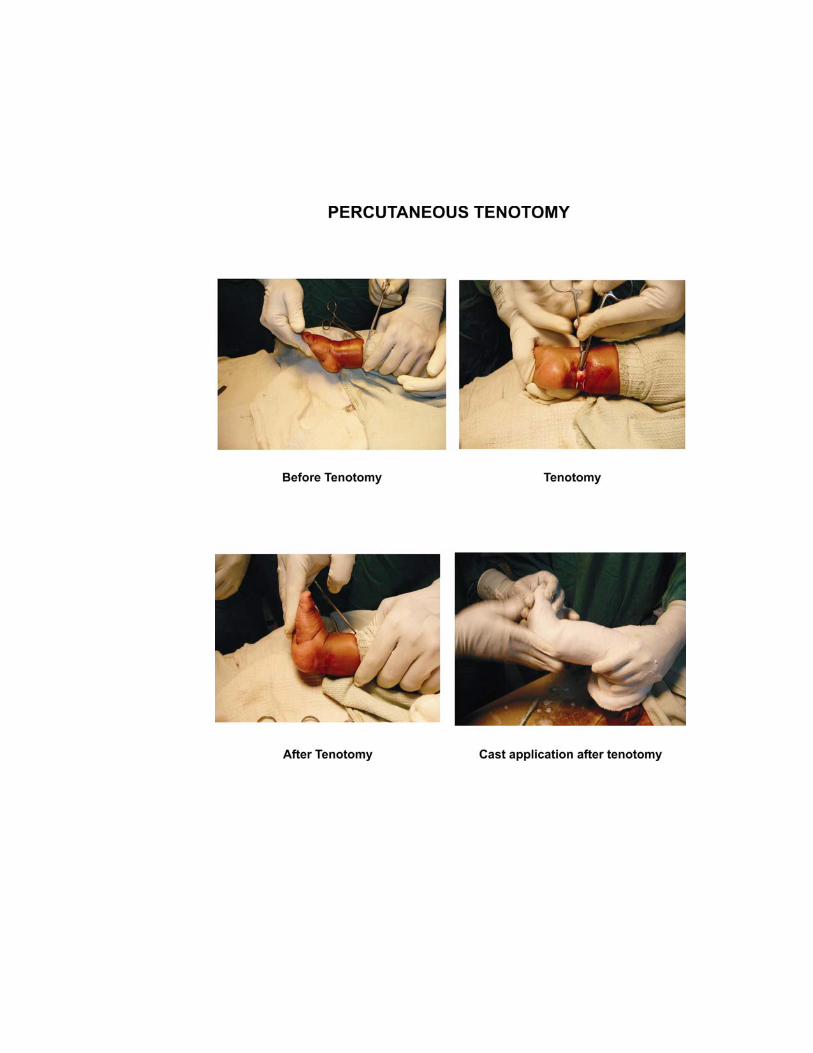
only residual deformity, which is to be corrected by percutaneus tenotomy of
tendo Achilles 7, 28, 29. This is followed by POP casting for three weeks. Then
the baby is subjected to bracing protocol which consists of open toe high-top
straight last shoes attached to a bar for full time for the first three months and
twelve hours at night and two to four hours in the middle of the day for a total
of fourteen to sixteen hours during each twenty four hour period 53.
30
SEQUENCE OF DEFORMITY CORRECTION IN PONSETI
TECHNIQUE :
CAVUS :
The first element of management is correction of the cavus
deformity by positioning the forefoot in proper alignment with the hindfoot.
The cavus which is the high medial arch is due to the pronation of the forefoot
in relation to the hindfoot. The cavus is always supple in newborns and
requires only supinating the forefoot to achieve a normal longitudinal arch of
the foot. The forefoot is supinated to the extent that visual inspection of the
plantar surface of the foot reveals a normal appearing arch – neither too high
nor too flat. Alignment of the forefoot with the hindfoot to produce a normal
arch is necessary for effective abduction of the foot to correct adductus and
varus.
MANIPULATION :
Location of the head of the talus:
The head of the talus is palpated in front of the lateral malleolus as its
lateral part is barely covered by the skin. The anterior part of the calcaneus is
felt beneath the talar head.
Stabilize the talus :
Stabilizing the talus provides a pivot point around which the foot is
abducted.
31
Manipulation of foot :
Next with the foot in supination and talus stabilized , the foot is
abducted as far as can be done without causing discomfort to the infant. The
correction is held with gentle pressure for about 60 seconds and then released.
Subsequent casts :
During this phase of treatment, the adductus and varus are fully
corrected. The equinus deformity gradually improves with correction of
adductus and varus. This is part of the correction because the calcaneus
dorsiflexes as it abducts under the talus. No direct attempt at equinus
correction is made until the heel varus is corrected.
Decision to perform tenotomy :
A major decision point in management is determining when
sufficient correction has been obtained to perform a percutaneous tenotomy to
gain dorsiflexion and to complete the treatment. This point is reached when
the anterior calcaneus can be abducted from underneath the talus. It has to be
confirmed that the foot is sufficiently abducted to safely bring the foot into 0
to 5 degrees of dorsiflexion before performing tenotomy. This abduction
allows the foot to be safely dorsiflexed without crushing the talus between the
calcaneus and the tibia .If the adequacy of the abduction is uncertain,
another cast or two is applied to be certain.
32
MAINTANENCE OF DEFORMITY CORRECTION:
The brace is applied immediately after the last cast is removed,
three weeks after tenotomy. The brace consists of open high-top straight last
shoes attached to a bar. For unilateral cases, the brace is set at sixty to seventy
degrees of external rotation on the clubfoot side and thirty to forty degrees of
external rotation on the normal side. In bilateral cases, it is set at seventy
degrees of external rotation on each side. The bar should be of sufficient
length so that the heels of the shoes are at shoulder width. The bar should be
bent five to ten degrees with convexity away from the child, to hold the feet in
dorsiflexion.
The brace should be worn full time (day and night) for the first three
months after the last cast was removed. After that the child should wear the
brace for twelve hours at night and two to four hours in the middle of the day
for a total of fourteen to sixteen hours during each twenty four hour period.
This protocol continues until the child is three to four years of age 53.
The rational behind this bracing is that the medial soft tissues
remain stretched out only if the brace is used after the casting. In the brace,
the knee are left free, so that the child can kick them straight to stretch the
gastrocnemius tendon. The abduction of the feet in the brace, combined with
the slight bend causes the feet to dorsiflex. This helps maintain the stretch on
the gastrocnemius muscle and Achilles tendon.
33
RELAPSE :
Relapse is detected when slight equinus and varus deformity of the
heel is observed, usually without increased cavus and adduction deformity of
the fore foot 4. Relapses are rare after five years and extremely rare after
seven years of age regardless of whether the deformity is fully corrected or
not3. Following are the guidelines described by Ponseti for treatment of
relapsed CTEV 3.
A. For correction of second or third relapses in children older than
two-and-half years of age, when tibialis anterior has a strong supinatory
action, transfer of tibialis anterior to third cuneiform is advocated.Transfer of
the tibialis anterior tendon averts further relapse, maintains the correction of
heel varus and thus greatly reduces need for medial release operation. The
tibialis anterior tendon should never be split so as not to loose its eversion
power, nor should it be transferred to fifth metatarsal or to the cuboid since
this may excessively evert the foot causing severe forefoot pronation and heel
valgus. To prevent bow stringing of tendon under the skin in front of the
ankle, the tendon must be left under the superior retinaculum.
B . Ligament and joint release surgeries are necessary only in few
cases. It should not be done before the age of six months. Ponseti advocates
sectioning of only tight ligaments to achieve proper alignment of bones, since

34
a perfect reduction is unattainable owing to the incongruity of the joint
surfaces and changes in the shape of the bones. Lengthening of tendon of
tibialis posterior was done by technique described by Coleman 40.
C . Cavo-varus is the commonest residual deformity of treated
CTEV, in which tarsus remained in some degree of varus while forefoot is
pronated. The plantar fascia becomes shortened and thickened, thus
aggravating the deformity 3. The rigidity of heel varus is assessed by
Coleman’s lateral block test 40. For correction of cavovarus deformity, if heel
varus corrects within five degrees of the neutral position with the Coleman’s
block test, following series of procedures advocated by Reginald R. Cooper 3
is used for best correction of the deformity-
1. Severence of plantar fascia percutaneously.
2. A small dorsolateral wedge of bone is resected from the base of the
first metatarsal.
3. Jones procedure.
4. The tendon of peroneus longus is severed in the plantar aspect of the
foot and sutured to the tendon of peroneus brevis.
5. Transfer of tendon of tibialis anterior to the third cuneiform.
6. Lengthening of the tendo achilles.
35
D . TRIPLE ARTHRODESIS :
This is a salvage procedure. This is to be done in patients at or
nearing the skeletal maturity. It is indicated when ankle joint motion is fairly
good but the tarsal joints are very rigid in supination 3, 4.
E . TALECTOMY :
It is indicated in severe cases of very stiff club foot with little or no
ankle motion that have relapsed after extensive tarsal release operation. It
gives satisfactory results when performed between ages of one to six years 3.
Talectomy can be done as a primary procedure in patients with severe club
foot and poor or absent leg muscles, who are suffering from arthrogryposis or
myelomeningocele.
Due to structural abnormalities of the talar bones and joints, a
clubfoot cannot be corrected fully and hence completely normal foot is
neither desirable nor expected 5, 7, 39, and 41.
36
Patients were selected from the Out Patient section of the Department
of Orthopaedics and Traumatology, Thanjavur Medical College , Thanjavur
for correction of Idiopathic CTEV using the Ponseti technique from October
2004 to November 2005 .Cases of Idiopathic CTEV of age upto 2 years were
selected. These patients were followed up in a prospective manner for a
period of six months.
INCLUSION CRITERIA :
1. Adduction,Supination and varus deformity of the foot with or
without wasting of calf muscles.
2. Age less than two years.
3. Virgin club foot.
EXCLUSION CRITERIA :
1. Postural club foot.
2. Syndromic club foot.
3. Neglected club foot.
4. Relapsed club foot.
Thirty four patients entered the study after explaining the study
protocol and the possible necessity for Achilles tenotomy and foot abduction
orthosis till the age of four years. Appropriate informed written consent was
obtained from the parents (Appendix II). Twenty eight patients (twenty three
37
unilateral and five bilateral CTEV) completed serial castings with or without
Achilles tenotomy and were given foot abduction orthosis and were followed
up for six months. Four patients who did not achieved the required degree of
correction at the end of ten castings were considered as failure cases and were
referred for posteromedial soft tissue release. Two patients were non –
compliant and dropped out in the middle of the treatment.
These patients are followed-up in a prospective manner for a period of
six months.
All infants were in the age group of four days to two years with a
mean age of presentation of 192 days. All were assessed for associated
syndromic pathology and only those infants with idiopathic CTEV were
included in the study. Before cast application every week the degree of
deformity was graded according to Pirani severity scoring system.
The Pirani severity scoring system consists of midfoot score and
hindfoot score with grading of the deformity between 0 and 3 in each
category. (Appendix I ).
There were twenty one male and eleven female infants included in
our study. Three male infants and two female infants had bilateral
deformities. Of the eighteen male patients with unilateral deformity, fourteen
38
infants had involvement of right foot and four had involvement of left foot.
Out of nine female infants with unilateral involvement, one had involvement
of left foot.
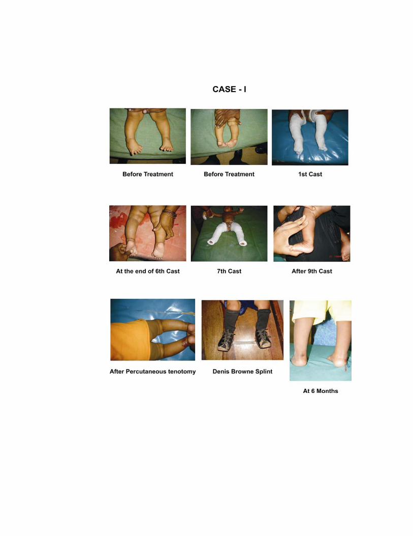
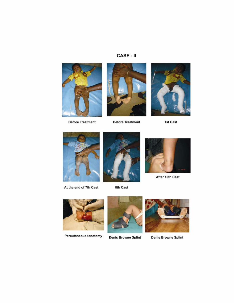
PONSETI TECHNIQUE
Initially a layer of cast padding was applied from groin to toe and
the surgeon held the foot in corrected position. An assistant applied the cast
using fast setting plaster in two sections. The first one comprised of below
knee plaster to hold the foot in corrected position. The next section consisted
of extending the cast above knee to convert it into a groin to toe plaster cast.
During this, the knee was held in 90 degree flexion. After application of the
cast the child was observed for about 30 minutes for any signs of limb
ischemia. The parents were educated about possible complications like
cyanosis, swelling, excess cry and the contact number in case of emergency
were provided. They were then advised to report for the next cast after 7 days.
The first cast was aimed at correcting the cavus deformity by
supinating the forefoot thereby bringing the forefoot in alignment with the
hindfoot.
In the second and subsequent casts, the foot in supination was
abducted while the surgeon applied counter- pressure on the head of the talus.
39
The calcaneus abducts by rotating and sliding under the talus. Simultaneously
it extends and everts thereby correcting the heel varus. To stretch the medial
tarsal ligaments fully, the foot was severely abducted to an angle of about 60
degrees. A maximum of 10 casts were fixed as endpoint for correction of
cavus, hindfoot varus and adduction deformity.
After correction of the above deformities, passive dorsiflexion of the
foot to 15 degree above neutral with the examiner applying a single finger
pressure was attempted; If achieved, a final cast was applied in the final
corrected dorsiflexed position for three weeks. If dorsiflexion more than 15
degrees was not possible, a percutaneous tenotomy of the tendo achilles was
done under general anaesthesia.After this tenotomy, the foot was placed in the
final corrected dorsiflexed position for three weeks.
After the last cast was removed, correction was maintained by using
Dennis-Browne splint. The brace was worn full time (day and night) for the
first three months after the last cast was removed. After that, the child should
wear the brace for 12 hours at night and 2-4 hours in the middle of the day for
a total of 14-16 hours during each 24-hour period. This protocol continues
until the child is 3-4 years of age.
40
The patients were reviewed at 14 days after application of Dennis- Brown
splint to assess the compliance of the parents. In subsequent visits patients
were reviewed once in three months. The parents were given contact numbers
and were advised to contact us regarding the maintanence of Dennis Browne
splint.
Statistical analysis
The results were analysed using SPSS 10 software.
T-Test paired samples analysis was done to find out the difference
between the means of values (before casting, after casting and follow up
castings).
41
In this study full correction of the deformity was obtained in thirty
three feet (23 unilateral and 5 bilateral CTEV). In this study, the end point for
castings was taken as ten casts. Percutaneous tenotomy was done, if needed,
once adequate abduction is achieved.
Out of 37 feet, 6 feet achieved full correction at the end of initial
casting without percutaneous tenotomy and 27 feet were fully corrected with
percutaneous tenotomy. Four feet were not corrected with Ponseti method and
were considered as failure cases. They were referred for posteromedial soft
tissue release. Two patients were non-compliant and dropped out in the
middle of the study.
The mean age at initiation of treatment for 32 patients (37 feet) was
192 days (range 4 days to 2 years).
The mean initial Pirani severity score for 37 feet was 4.30 (out of
maximum possible score of six). After full correction by ponseti technique
(with or without percutaneous tenotomy) the final mean score was found to
be 0.17 and the mean change in score was found to be 4.13. This was
analysed by the paired t test and the p value was <0.0005 which is significant.
The mean value of Pirani score at 6 months follow up was 0.11 which shows
42
a change of 4.19 from the initial score. This change also has a p value of
<0.0005 which is significant.
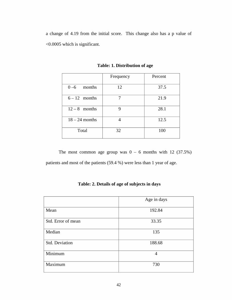
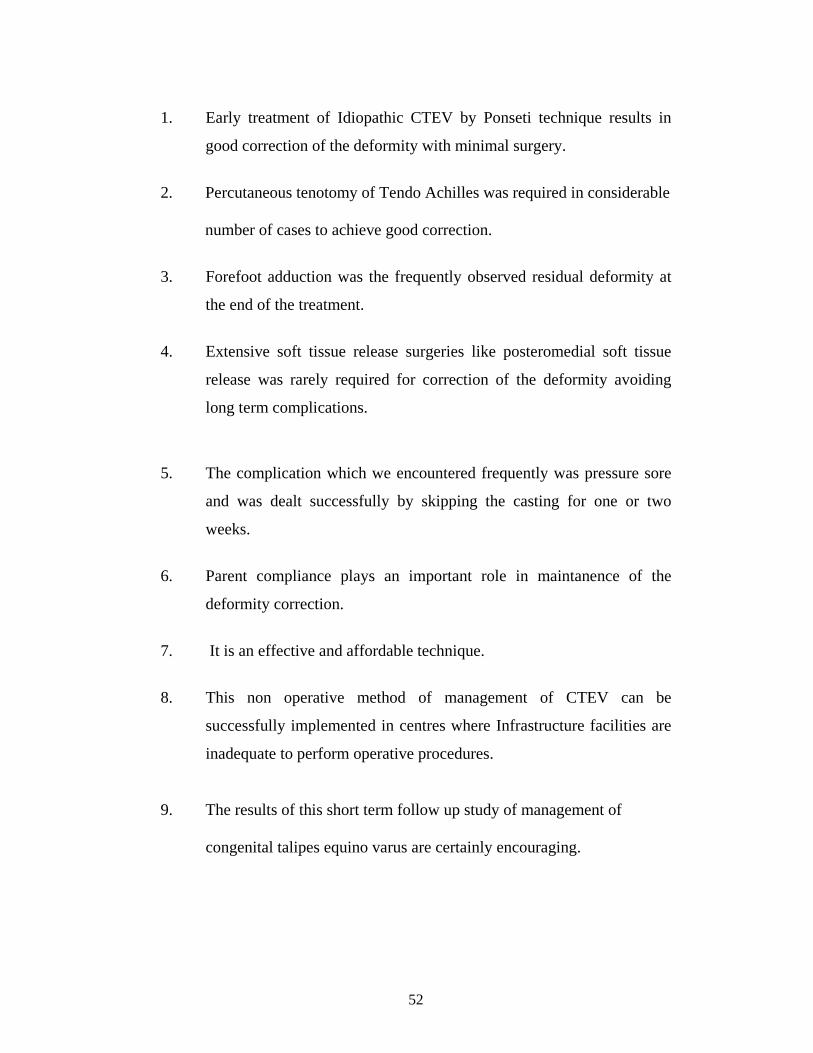
Table: 1. Distribution of age
Frequency Percent
0 –6 months 12 37.5
6 – 12 months 7 21.9
12 – 8 months 9 28.1
18 – 24 months 4 12.5
Total 32 100
The most common age group was 0 – 6 months with 12 (37.5%)
patients and most of the patients (59.4 %) were less than 1 year of age.
Table: 2. Details of age of subjects in days
Age in days
Mean 192.84
Std. Error of mean 33.35
Median 135
Std. Deviation 188.68
Minimum 4
Maximum 730
43
The minimum age – 4 days
The maximum age – 730 days (2 years).
The mean age at initiation of treatment for the 32 patients was 192 days
(range 4 days – 730 days).
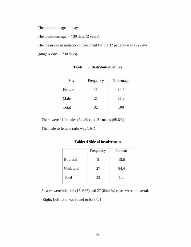
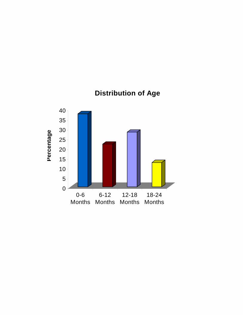
Table : 3. Distribution of Sex
There were 11 females (34.4%) and 21 males (65.6%).
The male to female ratio was 1.9: 1
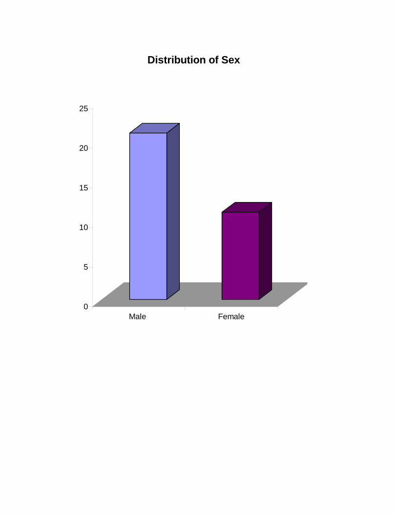
Table: 4 Side of involvement
Frequency Percent
Bilateral 5 15.6
Unilateral 27 84.4
Total 32 100
5 cases were bilateral (15. 6 %) and 27 (84.4 %) cases were unilateral.
Right: Left ratio was found to be 3.6:1
Sex Frequency Percentage
Female 11 34.4
Male 21 65.6
Total 32 100

44
Table 5 : Correlation between side and sex
Unilateral
Section
Bilateral Right Left
Male 3 14 4
Female 2 4 1
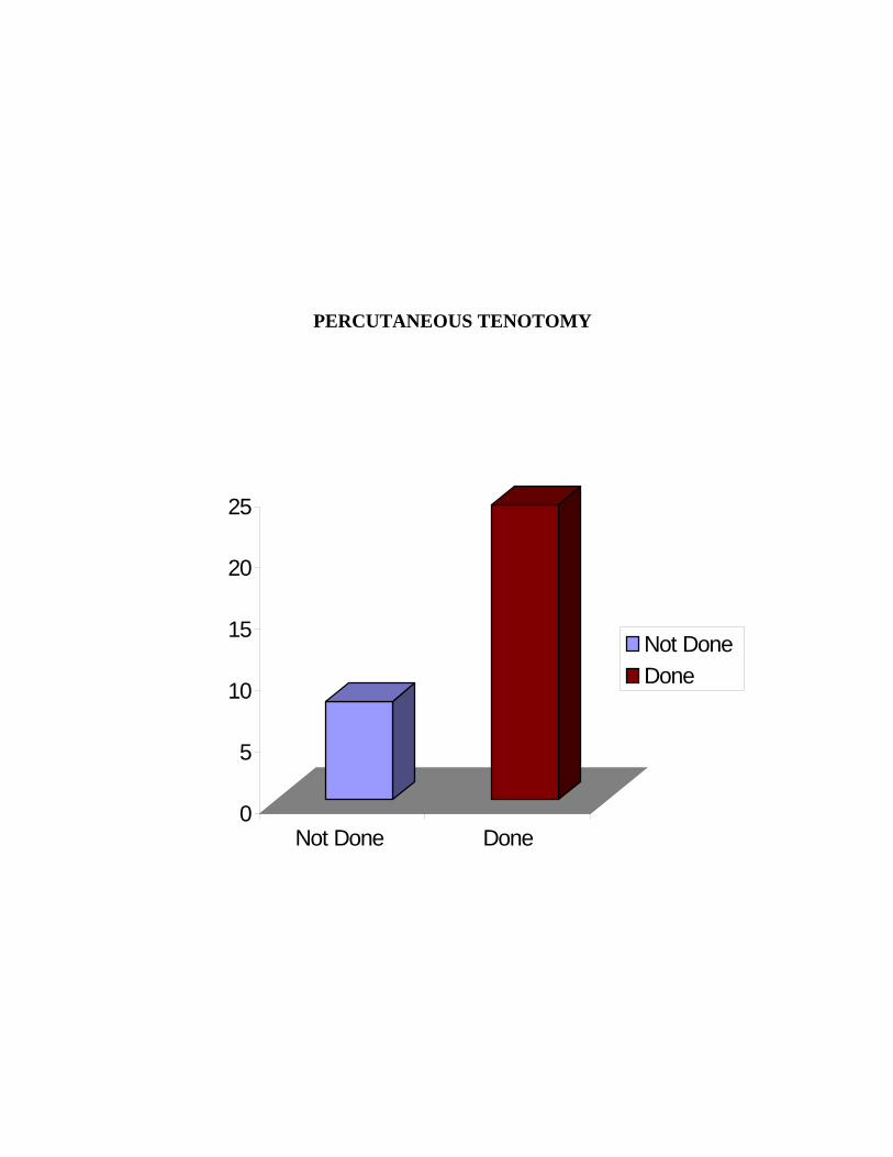
Table: 6. Details of Percutaneous tenotomy done
75 % of patients needed percutaneous tenotomy of tendo achilles at
the end of casting.
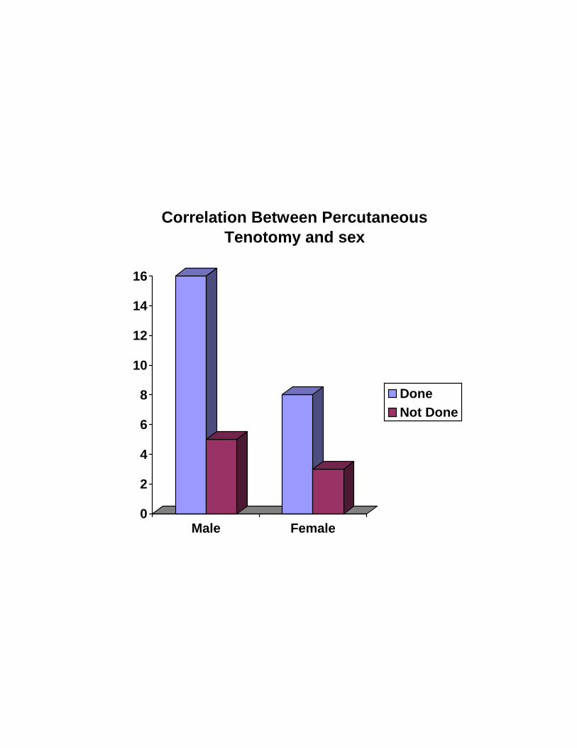
Table: 7. Correlation between Percutaneous tenotomy and sex
Sex Tenotomy
Female Male
Total
Done 8 16 24
Not done 3 5 8
Total 11 21 32
76 % of male patients and 72.7 % of female patients needed
percutaneous tenotomy.
Tenotomy Frequency Percent
Done 24 75
Not done 8 25
45
Table 8: Details of PMSTR done
Frequency Percent
Not done 28 87.5
Done 4 12.5
PMSTR: Postero Medial Soft Tissue Release.
Table 9: Details of Pirani score – Paired samples
Mean Pirani
Score N
Standard
Deviation
Standard
Error of mean
Before
Treatment
4.30 32 0.61 0.11 Pair I
After
Treatment
0.17 32 0.30 0.053
Before
Treatment
4.30 32 0.61 0.11 Pair II
At Follow up 0.11 32 0.21 0.037
After
Treatment
0.17 32 0.30 0.053 Pair III
At follow up 0.11 32 0.21 0.037
1. Mean Pirani score before treatment - 4.30 (range – 3.5 - 5)
2. Mean Pirani score after treatment - 0.17 (range – 0 – 0.5)
46
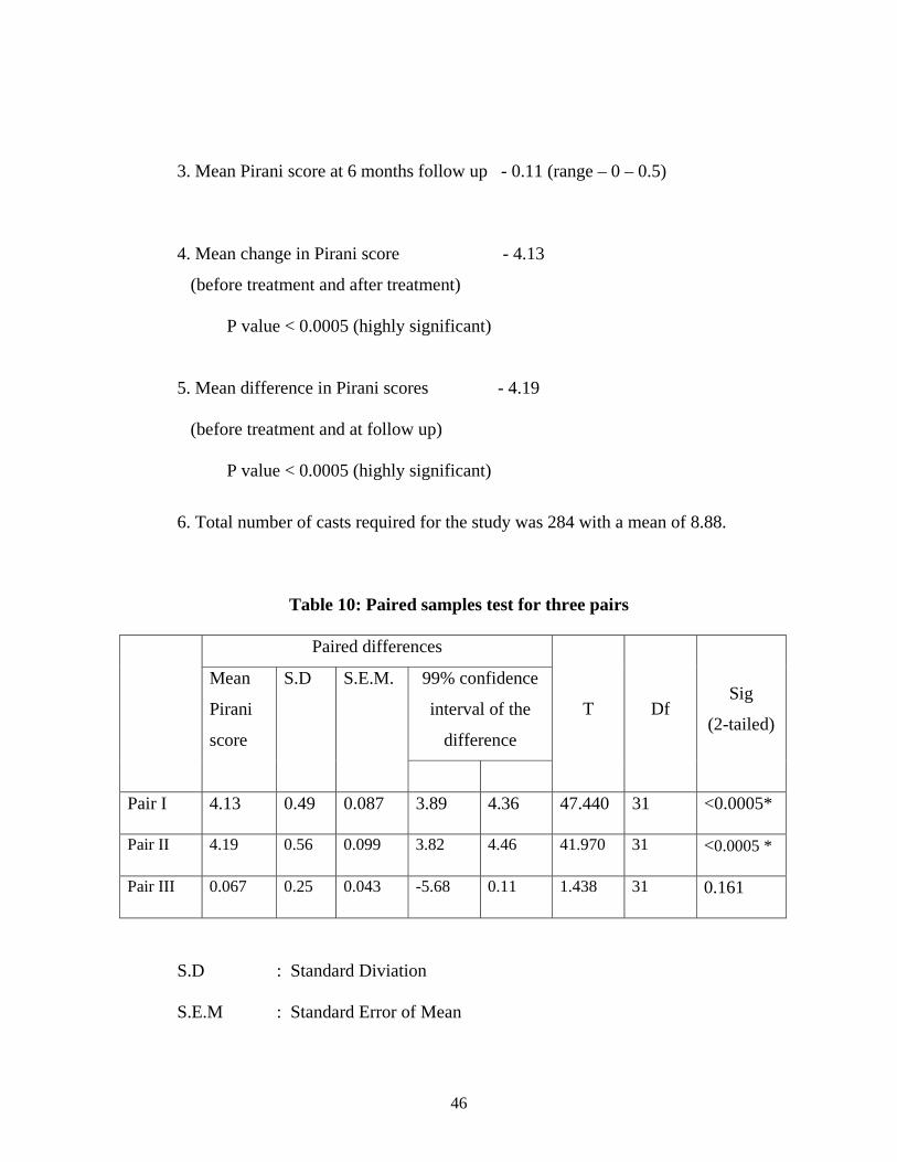
3. Mean Pirani score at 6 months follow up - 0.11 (range – 0 – 0.5)
4. Mean change in Pirani score - 4.13
(before treatment and after treatment) P value < 0.0005 (highly significant) 5. Mean difference in Pirani scores - 4.19
(before treatment and at follow up)
P value < 0.0005 (highly significant)
6. Total number of casts required for the study was 284 with a mean of 8.88.
Table 10: Paired samples test for three pairs
Paired differences
99% confidence
interval of the
difference
Mean
Pirani
score
S.D S.E.M.
T Df Sig
(2-tailed)
Pair I 4.13 0.49 0.087 3.89 4.36 47.440 31 <0.0005*
Pair II 4.19 0.56 0.099 3.82 4.46 41.970 31 <0.0005 *
Pair III 0.067 0.25 0.043 -5.68 0.11 1.438 31 0.161
S.D : Standard Diviation
S.E.M : Standard Error of Mean
47
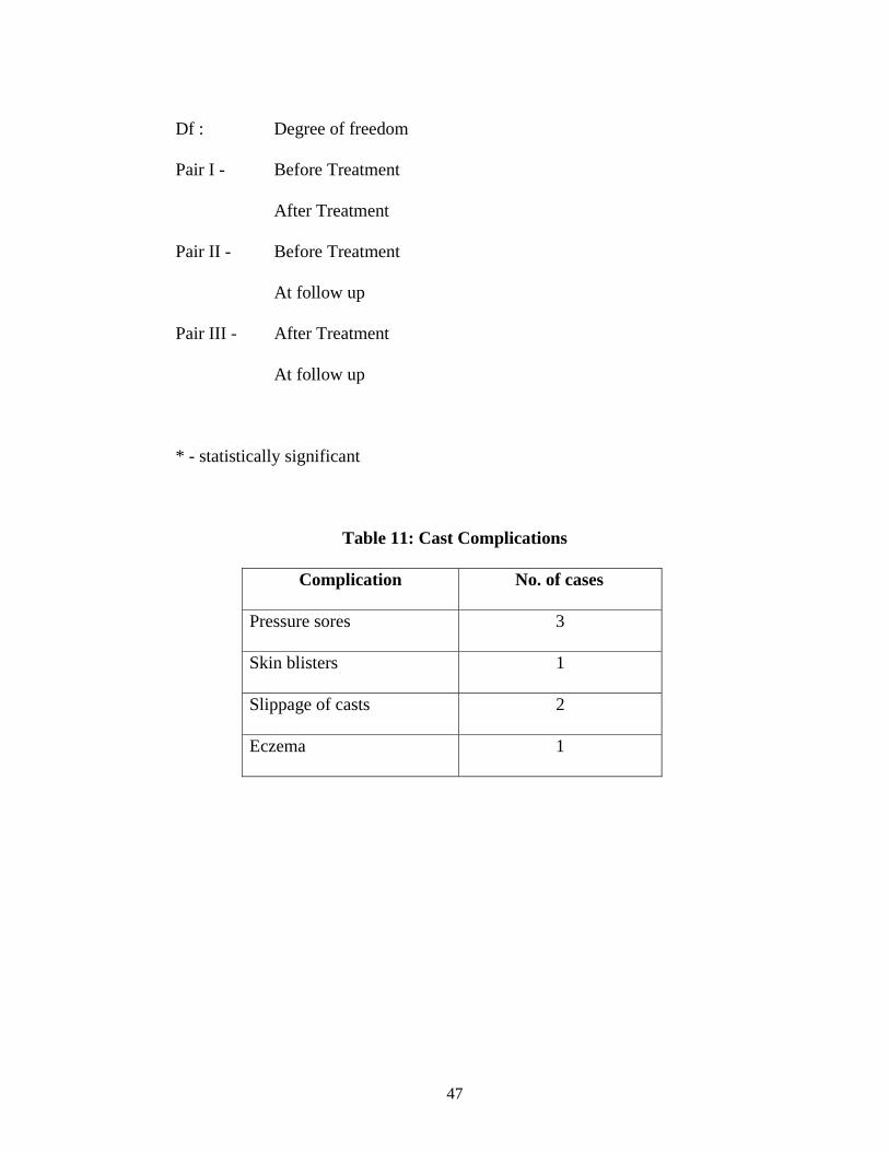
Df : Degree of freedom
Pair I - Before Treatment
After Treatment
Pair II - Before Treatment
At follow up
Pair III - After Treatment
At follow up
* - statistically significant
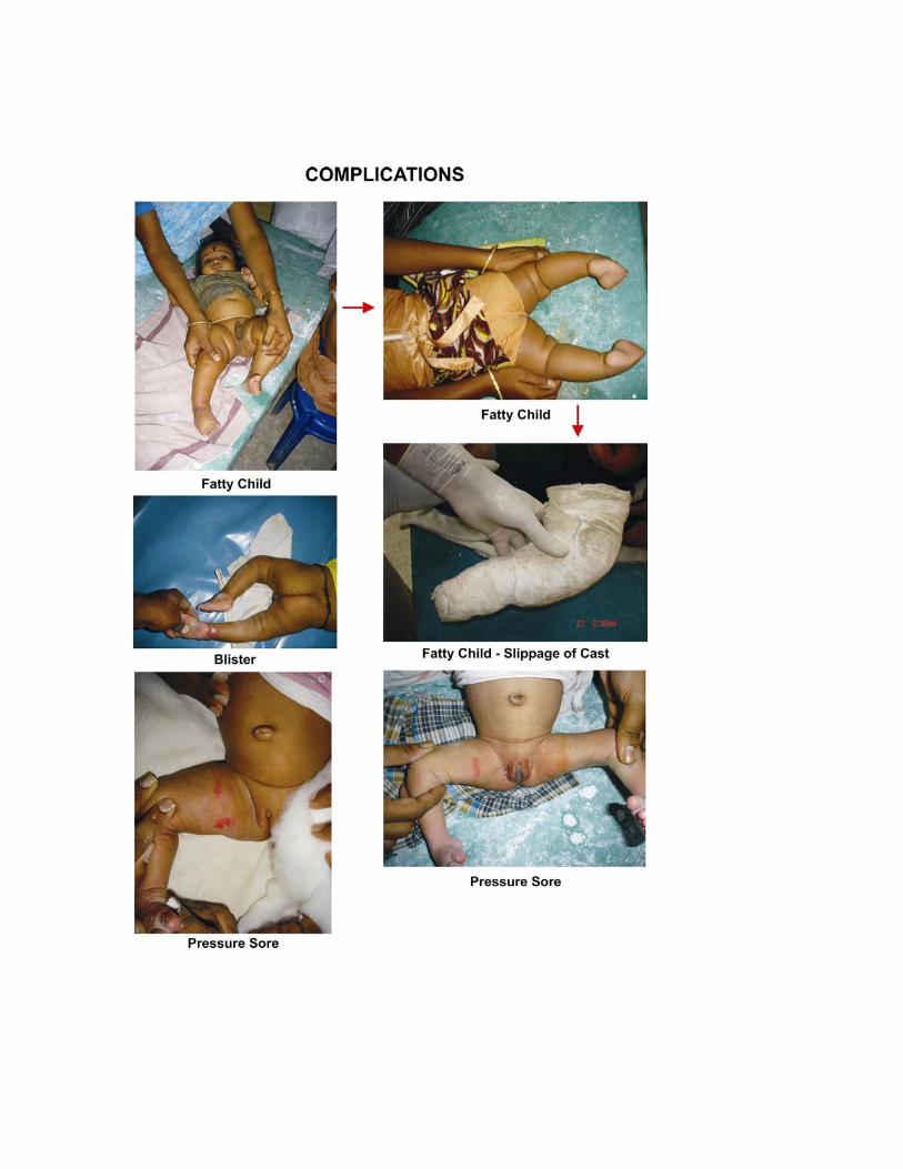
Table 11: Cast Complications
Complication No. of cases
Pressure sores 3
Skin blisters 1
Slippage of casts 2
Eczema 1
48
The treatment options for Congenital talipes equinovarus has gone a full
circle and reached the earlier concept of non -operative treatment, as it is
associated with improved results.
As it is evident from our study, the results of deformity correction are
better if treatment is started within first month of life and results are
statistically significant. Harold 30 and Porter 43 gave similar reports. The
visco-elastic properties of infant’s soft tissues respond to properly directed
mechanical stimuli with gradual remodeling of joint surfaces, resulting in
gradual and simultaneous correction of the deformities 3.
The sequence of deformity correction was most important to avoid
complications like Rocker-Bottom foot, persistent cavus and locking of the
calcaneus under the talus leading to persistent heel varus. Frick 38 has
emphasized on the importance of maximal forefoot supination in the initial
casting,failure of which results in persistent rigidity and incomplete correction
of the deformity.During manipulations the foot is never pronated in order to
prevent bean shaped deformity and incomplete correction of heel varus.
The fact that the navicular moves towards its normal position
following manipulation was confirmed by Kuhns in his study using
49
ultrasonography 44. Pirani confirmed similar results in clubfoot treated by
Ponseti method 46.
32 children with congenital clubfoot participated in the study. Total
number of clubfeet was 37. All the patients were of age 0 to 24 months
(range: 4 days to 2 years) at initial casting. Mean age of the group was192
days. Morcuende et al. had retrospectively analysed the records of 157
patients (256 clubfeet). In this study also all the patients were of the age group
0 to 24 months. There were 21 male children and 11 female children in the
present study and the male: female ratio is 1.9:1. Morcuende et al. reported a
male female ratio of 2.13: 1. The male preponderance found in this study is in
agreement with other studies.
The feet are evaluated using Pirani severity scoring system which was
easy to use and simple and fairly reproducible. In our study the scoring was
done by a faculty who was not involved in the study and casting was done by
the author throughout the period of study. The points in the Pirani scores are
allotted on the basis of inspection findings of the sole of the foot, lateral
border, posterior and medial creases, palpability of the talus and emptiness of
the heel as well as correctability of equinus.
50
In about twenty four patients (75%), percutaneous tenotomy of
tendoachilles was done in order to achieve complete correction. Ponseti him
self has observed that percutaneous tenotomy was needed in most of the
patients.
Though Ponseti advises tenotomy under local anaesthesia, we found
the child to be frightened and uncooperative to procedure under local
anaesthesia. Hence general anaesthesia was preferred and tenotomy was done
1.5 cm above the calcaneus with the foot held in maximum dorsiflexion by
the assistant. The tenotomy was performed with a size eleven surgical blade.
In our study, we observed that the earlier the child is started on
casting by ponseti technique, the results are better without any need for
surgery. In few of our infants, pressure sores developed due to the delicate
skin .However the pressure sores healed by skipping one week of casting and
then reapplying the cast a week later. One of our patient was very obese and
presented with frequent slipping of POP casts. Another patient developed
repeated eczema and was referred for posteromedial soft tissue release. We
also observed that as the child gets older, the prominence over the
calcaneocuboid junction in the lateral column prevents complete correction.
51
The most common residual deformity which occurred at the end of
treatment was forefoot adduction in about five patients who completed
Ponseti method of treatment with or without percutaneous tenotomy.
The rate of posteromedial soft tissue release can be drastically
reduced by using Ponseti technique and hence the complications of surgery
were avoided. Colburn reported similar finding following treatment of CTEV
by Ponseti method.
We found the following factors contributed to the success of CTEV
correction by Ponseti technique:
Earlier the child was started on treatment better were the results
The milder the severity of deformity
Strict adherence to the sequence of correction as advised by Ponseti.
Removal of the cast just before applying the subsequent cast.
Regular follow-up by the patients.
The compliance of the parents in maintaining the cast as well as the
Dennis Browne splint.
Absence of complications.
Our results were successful in 87.5 % of the patients with no major
adverse events and the results are certainly encouraging.
52
1. Early treatment of Idiopathic CTEV by Ponseti technique results in
good correction of the deformity with minimal surgery.
2. Percutaneous tenotomy of Tendo Achilles was required in considerable
number of cases to achieve good correction.
3. Forefoot adduction was the frequently observed residual deformity at
the end of the treatment.
4. Extensive soft tissue release surgeries like posteromedial soft tissue
release was rarely required for correction of the deformity avoiding
long term complications.
5. The complication which we encountered frequently was pressure sore
and was dealt successfully by skipping the casting for one or two
weeks.
6. Parent compliance plays an important role in maintanence of the
deformity correction.
7. It is an effective and affordable technique.
8. This non operative method of management of CTEV can be
successfully implemented in centres where Infrastructure facilities are
inadequate to perform operative procedures.
9. The results of this short term follow up study of management of
congenital talipes equino varus are certainly encouraging.
0
5
10
15
20
25
30
35
40
Perc
enta
ge
0-6Months
6-12Months
12-18Months
18-24Months
Distribution of Age
0
5
10
15
20
25
Male Female
Distribution of Sex
Side of Involvement
18%
18%
64%
Bilateral
Left
Right
0
5
10
15
20
25
Not Done Done
Not Done Done
PERCUTANEOUS TENOTOMY
0
2
4
6
8
10
12
14
16
Male Female
Correlation Between Percutaneous Tenotomy and sex
DoneNot Done
BIBLIOGRAPHY
1. Noonan K J, Richards B S- Nonsurgical management of Idiopathic
Clubfoot. Journal of American academy of orthopedic surgery, Nov-
Dec, 11(6) : 392-402, 2003.
2. Kite J. H. : The Clubfoot. Grune & Straton, New York, 11964.
3. Ponseti I V: Congential Clubfoot. Fundamentals of Treatment, Oxford
University Press, London, 1996.
4. Turco V J: Clubfoot, Churchil – Livingstone, New York, 1981.
5. Fripp & Shaw : Clubfoot, E & S Livingstone Ltd, London, 1967.
6. Jones R, Lowett R.W.: Orthopedic Surgery, 2nd edition, Oxford
University Press, London, 1929.
7. Ponseti I V: Treatment of Congenital Clubfoot – Journal of Bone &
Joint Surgery , Vol. 74-A, March, No.3, 448-454, 1992.
8. James Beaty : Campbell’s Operative Orthopedics-10th edition, edited
by Tarry S. Canale: international Edition, Mosby Publications, 2003.
9. Issac H, Handelsman J E, Bandenhorst M, Pickering A: The muscles in
Clubfoot – a histological, histochemical & electron microscopic study,
Journal of Bone & Joint Surgery, Vol.59-B, 465-472, 1977.
10. Diaz A V: Embryological contribution to aetiopathology of Idiopatic
Clubfoot- Journal of Bone & Joint Surgery, Vol, 61-B, 127, 1979.
11. Waisbrod Hanan: Congential Clubfoot; An anatomical study – Journal
of Bone & Surgery, Vol.55-B, 796-801, 1973.
12. Irani R. N. & Sherman M. S: Pathological anatomy of clubfoot-
Journal of Bone & Joint Surgery, Vol.45-A, 45, 1963.
13. Fukuhara K, Schollmeir G & Uhthoff H: The pathogenesis of the
Clubfoot; A histomorphic & immunohistochemical study of the
fetuses- Journal of Bone & Joint Surgery, Vol.76-B, 450-457, 1994.
14. Ippolito E. & Ponseti I, V: Congential clubfoot in Human fetuses; A
histological study – Journal of Bone & Joint Surgery, Vol.62-A, 8-22,
1980.
15. Simon Barker, David Chesney, Zosia Miedzybrodzka, Nicolla &
Maffulli: Genetics & Epidemology of Idiopathic Congenital Talipes
Equino Varus – Journal of Pediatric Orthopedics, 23: 265-272, 2003.
16. Henry R Cowell, Barry K Wein, Wilmington Delaware – Genetic
aspects of Clubfoot, Journal of Bone & Joint Surgery, Vol.62-A, Dec,
1381-1384, 1980.
17. Adams W: Clubfoot; Its Causes, Pathology & Treatment, 2nd edition,
Lindsay & Blakiston, 1973, Philadelphia.
18. Attenborough C.G: Congenital Talipes Equino Varus – Journal of
Bone & Joint Surgery, Vol. 48-B, 31-39, 1966.
19. Krishna M, Evans R, Taylor J F, Theis J C: Tibial torsion measured by
ultrasound in children with Talipes Equino Varus – Journal of Bone &
Joint Surgery, 73-B, 207-210, 1991.
20. Scoot W. A. Hosking S.W. Catterall A: Clubfoot; Observation on the
surgical anatomy of dorsiflexion – journal of Bone & Joint Surgery,
66-B, no.1, Jan. 71-76, 1984.
21. Inman V.T: The joints of the Ankle, Williams & Wilkins – Baltimore,
1976.
22. Lovell W.W. Bailey T. Prince C.J. Meehan P.L. The foot in Lovell W.
Winter: Pediatric Orthopaedics, edition 2, Philadelphia, PA:
Lippincott, 1986.
23. Dimeglio A. Bannet F. Marcall P. DeRosa V: Orthopedic treatment &
passive motion machine consequences for the surgical treatment of
Clubfoot- Journal of Pediatric Orthopedics, 5-B, 173-180, 1996.
24. Ponseti I.V.: Clubfoot management – Journal of Pediatric Orthopedics,
20, 699-700, 2000.
25. Sterling Laaveg, Ponseti I.V.: Long term results of treatment of
Congenital Clubfoot – Journal of Bone & Joint Surgery, 62-A, NO.1,
Jan. 23-31, 1980.
26. Douglas M. Cooper, Frederick R. Dietz : Treatment of Idiopathic
Clubfoot; A thirty years follow up note – Journal of Bone & Joint
Surgery, 77-A, 1477-1489, 1995.
27. Ippolito E, Farsetti P, Caterini R, Tudisco C: Long-term results
comparative results in patients with Congenital Clubfoot treated with
two different protocols – Journal of Bone & Joint Surgery, 85-A (7),
1286-1294, 2003.
28. Cowell H.R.: The management of Clubfoot – Journal of Bone & Joint
Surgery, 67-A, Sept., 991-992, 1985.
29. Ponseti I.V. & Smoley E.N.: Congenital Clubfoot; the results of
treatment-Journal of Bone & Joint Surgery, 45-A, 261-275, March
1963.
30. Harold A J & Walker C J: Treatment & prognosis in Congenital
Clubfoot – Journal of Bone & Joint Surgery, 65-B (1), 8-11, 1983.
31. Haruyasu Yamamoto, Kohtaro Furuya: Treatment of Congenital
Clubfoot with a modified Denis-Browne splint – Journal of Bone &
Joint Surgery, 72-B (3), May, 460-463, 1990.
32. Schlafly, Bruce, Butler, J.E., Siff S.J., Criswell A.R., Cain T.E.: The
appearance of the tarsal navicular after posteromedial release for
Clubfoot- Foot & Ankle, 5:222-237, 1985.
33. Wyne-Davies, Ruth: Talipes Equinovarus; A review of eighty four
cases after completion of the treatment – Journal of Bone & Joint
Surgery, 46-B, 464-476, 1964.
34. Green A.D.L., Lloyd Roberts A.G.: The results of early posterior
release in resistant Clubfeet; A long term review – Journal of Bone &
Joint Surgery, 67-B, 588-593, 1985.
35. Ryoppy S. Sairanen H.: Neonatal operative treatment of Clubfoot; A
preliminary report - Journal of Bone & Joint Surgery, 65-B, 320-325,
1983.
36. Simons G.W.: Correspondence – Journal of Bone & Joint Surgery, 68-
A, 151-152, Jan. 1986.
37. Coroll & Norris : Clubfoot in Lovell & Winters Pediatric Orthopedics,
Edited by R.T. Morrissy, 3rd edition, vol.2, 927-956, JB Lippincott,
Philadelphia, 1990.
38. Frick SL: The Ponseti method of treatment for congenital Clubfoot;
Importance of maximal forefoot supination in initial casting.
Orthopedics, Jan. 28 (1), 63-65, 2005.
39. Hutchins P.M. Foster B.K. Peterson D.C. Cole E.A.: Long term results
of early surgical release in clubfoot in clubfoot – Journal of Bone &
Joint Surgery, 67-B, 791-799, 1985.
40. Coleman S.S.: Complex foot deformities in children – Lea & Febiger,
Philadelphia.
41. John Herring: Tachdjian’s Pediatric Orthopedics, 3rd edition, Vol.2,
W.B. Sounders Company, Philadelphia.
42. Anja Van Compenhout, Guy Molenaers, Pierre Moeus, Guy Fabry:
Does functional treatment of Idiopathic Clubfoot reduce the indication
for the surgery? A call for widely accepted rating system – Journal of
Pediatric Orthopedics, part-B, 10:315-318, 2001.
43. Porter R.W. Congenial Talipes Equino Varus : Resolving & Resistant
Deformities – Journal of Bone & Joint Surgery, 69-B (5): 822-825,
1987.
44. Lawrence R. Kuhns, Khaldoun Koujok, Janette M, Clifford Craig :
Ultrasound of the navicular during the simulated Ponseti maneuver-
Journal of Pediatric Orthopedics, 23,, 243-245, 2003.
45. William Campbell’s : Operative Orthopedics, 9th edition Vol. 1.,
P:937-952, 1998.
46. Pirani Shafique, Laura Zeznik, David Hodges : Magnetic Resonance
Imaging study of the Congenital Clubfoot treated by Ponseti method-
Journal of Pediatric Orthopedics, 21:719-726, 2001.
47. Scher D.M. Feldman D.S. van Bosse H.J. Sala D.A. Lehman W.B.:
Predecting the need for tenotomy in the Ponseti method for correction
of clubfoot – Journal of Pediatric Orthopedics, July –Aug. 24(4): 349-
352, 2004.
48. Minkowitz B. Finkelstein B.I. Belicher M: Percutaneous Tendo
Achilles lengthening percutaneous tenotomy of Tendo Achilles in the
treatment of clubfoot deformity – Journal of Pediatric Orthopedics,
July-Aug. 24(4): 335-357, 2004.
49. Minkowitz B. Finkelstein B.I. Bleicher M: Percutaneous Tendo
Achilles lengthening with a large gauge needle: a modification of the
Ponseti technique for correction of Idiopathic Clubfoot – Journal of
Foot & Ankle Surgery, July – Aug. 24(4): 263-265, 2004.
50. John E. Herzenberg, Christof Radler, Noam Bor: Ponseti versus
traditional methods of casting for Idiopathic Clubfoot – Journal of
Pediatric orthopedics, 22:517-521, 2002.
51. Colburn M. Williams M: Evaluation of the treatment of Idiopathic
Clubfoot by using the Ponseti method – Journal of Foot & Ankle,
Surgery, Sept.-Oct. 42(5): 259-267, 2003.
52. Mercer’s Orthopedic surgery 9th edition – volume 1, Page 180, 2003.
53. Clubfoot – Ponseti Management – Second Edition – Global Help
Publication (downloaded from internet – Website : global-help.org.
54. Morcuende JA, Dolan L, Dietz F, Ponseti IV. Radical reduction in the
rate of extensive corrective surgery for clubfoot using the Ponseti
method. Pediatrics 113:376-380, 2004.

Consent Proforma
Title : Treatment of Idiopathic Congenital talipes equinovarus by Ponseti method –
Short term follow up study.
Aim : To evaluate the functional outcome of Idiopathic CTEV correction by Ponseti
method at the end of initial correction and at six months follow up.
Consent : I have been explained about the nature of the study and also about the nature
of child’s illness in my own vernacular language.
I here by give my consent for the inclusion of my child in the study
Signature
PROFORMA
Name :
Age :
Sex :
Address :
Consanguinity of Marriage :
Antenatal H/O :
Natal and Post Natal H/O :
Birth Order :
Immunisation :
Unilateral/Bilateral :
Family H/O :
Any previous treatment :
Assessment of the deformity using Pirani severity scoring system:
Before Treatment :
After Treatment :
At 6 months follow up :
Percutaneous Tenotomy done : YES / NO
Surgery (PMSTR) : Yes / No
Side Casting Percutaneous tenotomy Posteromedial soft
tissue release
Right
Left
Residual deformity at the
end of treatment :
Follow – up
Residual deformity :
Supple / Stiff foot :
Rocker Bottom Foot :
Pain :
Wasting of calf muscle :
Nonspecific complaints :

KEY TO MASTER CHART
M - Male
F - Female
Y - Yes
N - No
R - Right
L - Left
D.O - Dropped out cases
PT - Percutaneous Tenotomy
PMSTR - Posteromedial Soft tissue release.
PSS-FP - Pirani Severity Scoring at six months follow up.
Rx - Treatment
MON - Months
Pirani Severity score S.No. Name Age Sex Side UL/BL Before Rx After Rx
No. of Casting PT PMSTR PSS -
FP
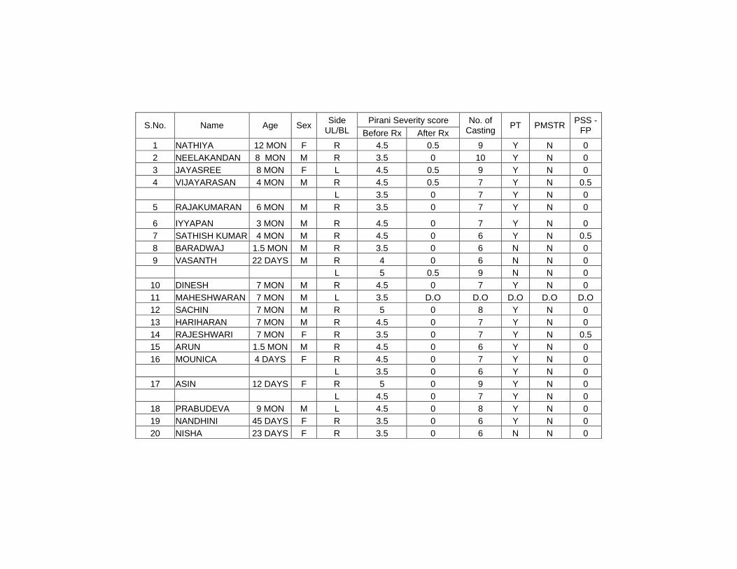
1 NATHIYA 12 MON F R 4.5 0.5 9 Y N 0 2 NEELAKANDAN 8 MON M R 3.5 0 10 Y N 0 3 JAYASREE 8 MON F L 4.5 0.5 9 Y N 0 4 VIJAYARASAN 4 MON M R 4.5 0.5 7 Y N 0.5 L 3.5 0 7 Y N 0 5 RAJAKUMARAN 6 MON M R 3.5 0 7 Y N 0
6 IYYAPAN 3 MON M R 4.5 0 7 Y N 0 7 SATHISH KUMAR 4 MON M R 4.5 0 6 Y N 0.5 8 BARADWAJ 1.5 MON M R 3.5 0 6 N N 0 9 VASANTH 22 DAYS M R 4 0 6 N N 0 L 5 0.5 9 N N 0
10 DINESH 7 MON M R 4.5 0 7 Y N 0 11 MAHESHWARAN 7 MON M L 3.5 D.O D.O D.O D.O D.O 12 SACHIN 7 MON M R 5 0 8 Y N 0 13 HARIHARAN 7 MON M R 4.5 0 7 Y N 0 14 RAJESHWARI 7 MON F R 3.5 0 7 Y N 0.5 15 ARUN 1.5 MON M R 4.5 0 6 Y N 0 16 MOUNICA 4 DAYS F R 4.5 0 7 Y N 0 L 3.5 0 6 Y N 0
17 ASIN 12 DAYS F R 5 0 9 Y N 0 L 4.5 0 7 Y N 0
18 PRABUDEVA 9 MON M L 4.5 0 8 Y N 0 19 NANDHINI 45 DAYS F R 3.5 0 6 Y N 0 20 NISHA 23 DAYS F R 3.5 0 6 N N 0
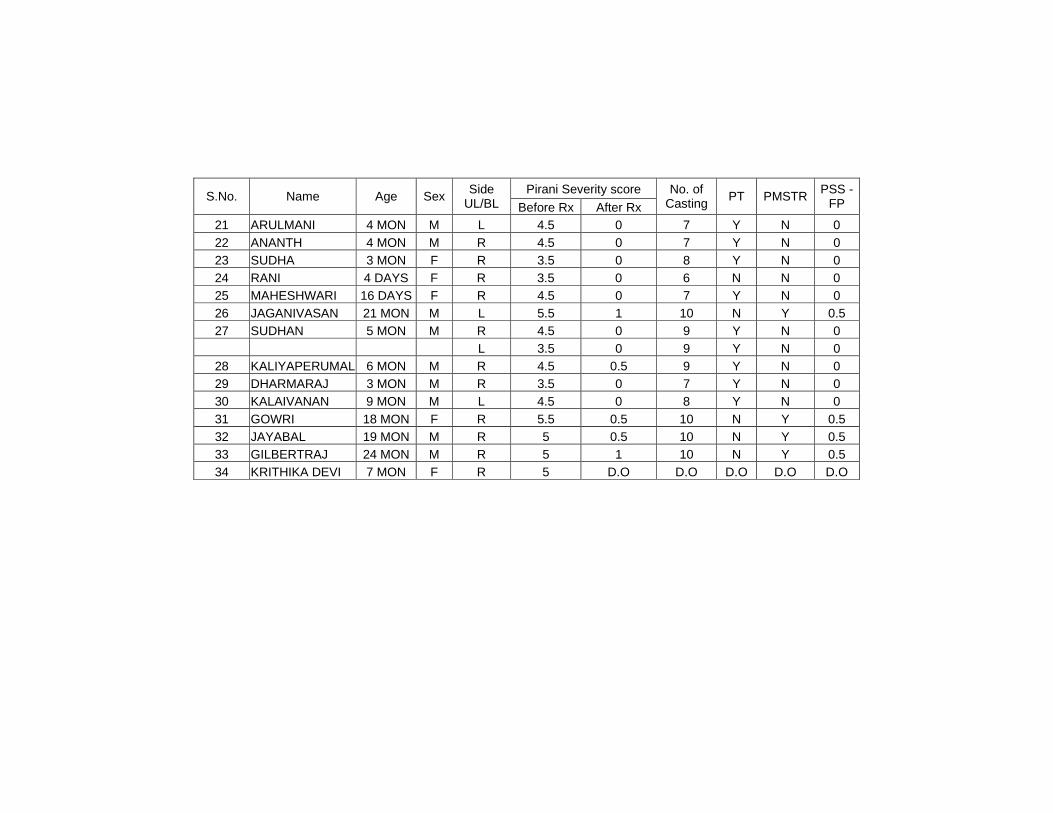
Pirani Severity score S.No. Name Age Sex Side
UL/BL Before Rx After Rx No. of
Casting PT PMSTR PSS - FP
21 ARULMANI 4 MON M L 4.5 0 7 Y N 0 22 ANANTH 4 MON M R 4.5 0 7 Y N 0 23 SUDHA 3 MON F R 3.5 0 8 Y N 0 24 RANI 4 DAYS F R 3.5 0 6 N N 0 25 MAHESHWARI 16 DAYS F R 4.5 0 7 Y N 0 26 JAGANIVASAN 21 MON M L 5.5 1 10 N Y 0.5 27 SUDHAN 5 MON M R 4.5 0 9 Y N 0 L 3.5 0 9 Y N 0
28 KALIYAPERUMAL 6 MON M R 4.5 0.5 9 Y N 0 29 DHARMARAJ 3 MON M R 3.5 0 7 Y N 0 30 KALAIVANAN 9 MON M L 4.5 0 8 Y N 0 31 GOWRI 18 MON F R 5.5 0.5 10 N Y 0.5 32 JAYABAL 19 MON M R 5 0.5 10 N Y 0.5 33 GILBERTRAJ 24 MON M R 5 1 10 N Y 0.5 34 KRITHIKA DEVI 7 MON F R 5 D.O D.O D.O D.O D.O


![[Argentine Consensus of Congenital Toxoplasmosis]](https://img.pdfslide.net/doc/110x75/634f5256eb0b18f1440ac9d0/argentine-consensus-of-congenital-toxoplasmosis.jpg)


