Embed Size (px)
Citation preview

The Knee xxx (2014) xxx–xxx
THEKNE-01910; No of Pages 6
Contents lists available at ScienceDirect
The Knee
Assessment of the lateral patellar facet in varus arthritis of the knee
Wenzel Waldstein a, Shari T. Jawetz b, Nadja A. Farshad-Amacker b, Christian Merle c,Tom Schmidt-Braekling a, Friedrich Boettner a,⁎a Adult Reconstruction & Joint Replacement Division, Hospital for Special Surgery, USAb Division of Magnetic Resonance Imaging, Hospital for Special Surgery, USAc Department of Orthopaedic and Trauma Surgery, University Hospital Heidelberg, Germany
⁎ Corresponding author at: 535 East 70th Street, NewYo774 2127; fax: +1 212 774 2286.
E-mail address: [email protected] (F. Boettner).
http://dx.doi.org/10.1016/j.knee.2014.05.0050968-0160/© 2014 Elsevier B.V. All rights reserved.
Please cite this article as: WaldsteinW, et al,10.1016/j.knee.2014.05.005
a b s t r a c t
a r t i c l e i n f oArticle history:
Received 19 November 2013Received in revised form 22 April 2014Accepted 14 May 2014Available online xxxxKeywords:Patellofemoral osteoarthritisUnicompartmental knee arthroplastyMRISkyline radiographsAnterior knee pain
Background: Lateral patellar arthritis has been associated with poor outcomes in unicompartmental kneearthroplasty. The current study correlates intraoperative findings with MRI imaging, skyline radiographs andthe presence of anterior knee pain.Methods: In 92 consecutive knees with varus arthritis, the patellofemoral compartment was assessed duringsurgery, on skyline radiographs and on MRI. Anterior knee pain was recorded on a visual-analog-scale. Intraop-erative assessmentwas based on the Outerbridge grading scale. Skyline radiographswere evaluated according tothe Ahlbäck grading scale; MRIs were assessed according to a modified Outerbridge grading scale.Results: There was an excellent correlation (rs=0.833; pb0.001) in the cartilage assessment of the lateralpatellar facet between MRI and surgery. A good correlation (rs=0.664; pb0.001) was seen betweenAhlbäck Grades and macroscopic Outerbridge Grades of the lateral patella. Ahlbäck Grades and MRI mod-ified Outerbridge Grades showed a good correlation (rs=0.643; pb0.001) for the lateral patella. Twelve
percent of knees (seven out of 60) with Ahlbäck Grade 0 or 1 and mild to moderate anterior knee painhad a macroscopic Outerbridge Grade of 3 on the lateral patella. None of these 60 knees had a full-thickness cartilage defect on MRI.Conclusion: Normal skyline radiographs in patients with mild to moderate anterior knee pain can rule outfull-thickness cartilage defects of the lateral patellar facet as observed during surgery and on MRI. TheMRI allows for the most accurate assessment of the patellofemoral joint and is warranted in all patientswith radiographic abnormalities or severe anterior knee pain.Level of evidence: Diagnostic study, Level II.© 2014 Elsevier B.V. All rights reserved.
1. Introduction
Knee osteoarthritis (OA) is one of the most common causes of painand disability [1–3]. Unicompartmental knee arthroplasty (UKA) canbe an effective long-term treatment option for end-stage medial com-partment arthritis [4] but its indications and contraindications remaincontroversial [5].
Many surgeons [6,7] adhere to the selection criteria as proposedby Kozinn and Scott [8] who generally consider the presence ofpatellofemoral arthritis as a contraindication formedial UKA. The recentliterature suggests that medial facet patellofemoral joint degenerationdoes not affect the outcome of medial UKA while caution is advised inpatients with lateral facet patellofemoral joint arthritis [9–12]. In thelatter, a significantly worse outcome was reported in the literature [9,
rk, NY 10021,USA. Tel.:+1212
Assessment of the lateral pate
10] and a total knee arthroplasty (TKA) might be the preferred treat-ment option [10]. McDonnell et al. [13] reported that preoperativeskyline radiographs can only detect advanced retropatellar arthritiswith reference to intraoperative findings but the study did not analyzethemedial and lateral patellar cartilage separately. Based on the currentliterature, the clinical value of skyline radiographs and magneticresonance imaging (MRI) in the preoperative assessment of thepatellofemoral joint is not clearly understood.
Anterior knee pain has also been proposed as a relative contraindica-tion for medial UKA [8,14,15]. However, the clinical value of anteriorknee pain as a predictor for patellofemoral cartilage degenerationremains questionable [13,16–18].
We therefore asked the following research questions: (1) How doMRI findings of the patellofemoral compartment correlate with visualintraoperative findings? (2) Does the assessment of the patellofemoralcompartment on skyline radiographs correlate with visual intraopera-tive findings? (3) Does the assessment of the patellofemoral compart-ment on skyline radiographs correlate with MRI findings? (4) Does
llar facet in varus arthritis of the knee, Knee (2014), http://dx.doi.org/
2 W. Waldstein et al. / The Knee xxx (2014) xxx–xxx
the severity of anterior knee pain predict the extent of cartilage damagein the patellofemoral compartment?
2. Patients and methods
2.1. Study cohort
The study prospectively enrolled 100 knees in 84 patients undergo-ing primary TKA for varus non-inflammatory arthritis of the kneebetween May 2010 and January 2012. The exclusion criteria weresecondary arthritis, and neutral or valgus alignment observed on hip-to-ankle AP standing radiographs. Each patient received a preoperativestandardized hip-to-ankle AP standing radiograph, a skyline radiographand a MRI of the knee. All images were stored in a generic DICOMformat.
Six patients were retrospectively excluded because not all radio-graphs or pain scores were on file, leaving 78 patients (92 knees; 45right knees and 47 left knees). There were 34 men and 44 women,who underwent 64 unilateral, 11 bilateral, and three staged bilateral pro-cedures. Themean age of the patientswas 67 years (range, 49–87 years),and their mean BMIwas 26 kg/m2 (range, 17–47 kg/m2). The studywasapproved by the institutional review board and all patients consentedto participate in the study. All procedures were in accordance with theethical standards of the review board and in line with the HelsinkiDeclaration of 1975, as revised in 2000.
2.2. Assessment of knee pain
Knee pain was assessed on a visual-analog-scale (VAS) from 0 to 10with 0 defined as “no pain” and 10 defined as “most severe pain”. Ante-rior knee pain was categorized into mild pain for patients with 0–3 onthe VAS, moderate pain for 4–7 on the VAS and severe pain for 8–10on the VAS, respectively. An independent investigator (MP) evaluatedthe pain in all patients before surgery. The investigator was provided aclear guideline for the data collection. Patients were asked about theirknee pain on the medial, anterior, lateral and posterior aspects of theknee. Additionally, knee pain during stair climbing, getting down thestairs, rising from a seated position and pain during walking wasrecorded.
2.3. Radiographic and MRI protocols
All radiographs were obtained utilizing standardized institutionalprotocols. Hip-to-ankle AP standing radiographs were corrected for ef-fects of magnification using a ruler. Skyline radiographs were obtainedin supine position with the knee flexed at 30–60°.
All subjects underwent MRI using 1.5 T or 3 T clinical scanners (GEHealthcare, Waukesha, WI) using either an eight channel phased arraytransmit receive coil (Invivo, Orlando, FL), a quadrature receive onlylower extremity coil (Invivo, Orlando, FL) or a three channel phasedarray receive only shoulder coil (USA Instruments Inc., Aurora, OH).Two-dimensional fast spin echo images were obtained in three planes.For those exams performed at 1.5 T, the repetition time (TR) was4000–5000 ms, echo time (TE) 34–40 ms, field of view (FOV) 140
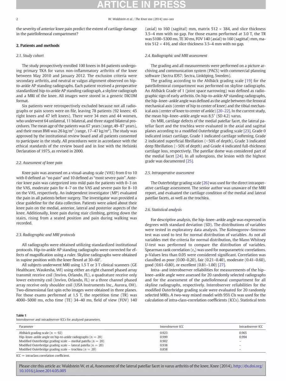
Table 1Interobserver and intraobserver ICCs for analyzed parameters.
Parameter
Ahlbäck grading scale (n = 92)Hip–knee–ankle angle on hip-to-ankle radiographs (n = 20)Modified Outerbridge grading scale — medial patella (n = 20)Modified Outerbridge grading scale — lateral patella (n = 20)Modified Outerbridge grading scale — trochlea (n = 20)
ICC = intraclass correlation coefficient.
Please cite this article as: WaldsteinW, et al, Assessment of the lateral pate10.1016/j.knee.2014.05.005
(axial) to 160 (sagittal) mm, matrix 512 × 384, and slice thickness3.5–4 mm with no gap. For those exams performed at 3.0 T, the TRwas5100–5300ms, TE 30ms, FOV140 (axial) to 160 (sagittal)mm,ma-trix 512 × 416, and slice thickness 3.5–4 mmwith no gap.
2.4. Radiographic and MRI assessment
The grading and all measurements were performed on a picture ar-chiving and communication system (PACS) with commercial planningsoftware (Sectra IDS7; Sectra, Linköping, Sweden).
The grading according to the Ahlbäck grading scale [19] for thepatellofemoral compartment was performed on skyline radiographs.An Ahlbäck Grade of 1 (joint space narrowing) was defined as radio-graphic sign of early arthritis. On hip-to-ankle AP standing radiographs,thehip–knee–ankle anglewasdefined as the angle between the femoralmechanical axis (center of hip to center of knee) and the tibial mechan-ical axis (center of knee to center of ankle) [20–22]. In the current study,the mean hip–knee–ankle angle was 8.5° (SD 4.2) varus.
On MRI, cartilage defects of the medial patellar facet, the lateral pa-tellar facet and the trochlea were evaluated in the axial and sagittalplanes according to a modified Outerbridge grading scale [23]. Grade 0indicated intact cartilage, Grade 1 indicated cartilage softening, Grade2 indicated superficial fibrillation (b50% of depth), Grade 3 indicateddeep fibrillation (N50% of depth) and Grade 4 indicated full-thicknesscartilage loss, respectively. The patellar dome was considered part ofthe medial facet [24]. In all subregions, the lesion with the highestgrade was documented [25].
2.5. Intraoperative assessment
TheOuterbridge grading scale [26]was used for the direct intraoper-ative cartilage assessment. The senior author was unaware of the MRIreport, and evaluated the cartilage condition of the medial and lateralpatellar facets, as well as the trochlea.
2.6. Statistical analysis
For descriptive analysis, the hip–knee–ankle angle was expressed indegrees with standard deviation (SD). The distributions of variableswere tested in exploratory data analysis. The Kolmogorov–Smirnovtest was used to test for normal distribution of variables. As not allvariables met the criteria for normal distribution, the Mann WhitneyU-test was performed to compare the distribution of variables.Spearman rank correlation (rs)was used for nonparametric correlations.p-Values less than 0.05 were considered significant. Correlation wasclassified as poor (0.00–0.20), fair (0.21–0.40), moderate (0.41–0.60),good (0.61–0.80), or excellent (0.81–1.00) [27].
Intra- and interobserver reliabilities for measurements of the hip–knee–ankle angle were assessed for 20 randomly selected radiographsand for the assessment of the patellofemoral compartment for allskyline radiographs, respectively. Interobserver reliabilities for themodified Outerbridge grading scale were evaluated for 20 randomlyselected MRIs. A two-way mixed model with 95% CIs was used for thecalculation of intra-class-correlation coefficients (ICCs). Statistical tests
Interobserver ICC Intraobserver ICC
0.923 0.9650.999 0.9940.902 –
0.938 –
0.858 –
llar facet in varus arthritis of the knee, Knee (2014), http://dx.doi.org/

Table 2Correlation of intraoperative cartilage assessment in the patellofemoral compartmentwith the preoperative assessment onMRI and on skyline radiographs for the entire cohort.
Parameters Intraoperative visual assessment
Lateral facet Medial facet Trochlea
MRI (rs) 0.833, p b 0.001 0.795, p b 0.001 0.691, p b 0.001Skyline radiographs (rs) 0.664, p b 0.001 0.329, p = 0.001 0.421, p b 0.001
3W. Waldstein et al. / The Knee xxx (2014) xxx–xxx
were carried out using SPSS 16.0 software for Windows (SPSS Inc.,Chicago, IL, USA).
3. Results
Excellent intra- and interobserver intra-class-correlation coefficients were seen for allparameters analyzed (Table 1).
For the lateral patellar facet, therewas an excellent correlation betweenMRImodifiedOuterbridge Grades and macroscopic Outerbridge Grades (Table 2). However, a full-thickness cartilage defect of the lateral patellar facet was macroscopically observed inonly one knee. In comparison, a modified Outerbridge Grade 4 defect (full-thickness)was recorded in 15 knees on MRI. For the medial patellar facet and the trochlea, therewere good correlations between the MRI modified Outerbridge assessment and themacroscopic Outerbridge assessment at the time of surgery (Tables 2 and 3).
A good statistical correlation could be demonstrated between Ahlbäck Grades onskyline radiographs and macroscopic Outerbridge Grades of the lateral patellar facet(Table 2). Out of 56 knees with an Ahlbäck Grade of 0, five knees (9%) had a macroscopicOuterbridge Grade of 3 on the lateral patellar facet. In one knee a macroscopic full-thickness cartilage defect of the lateral patellar facet was observed which was preopera-tively assessed as Ahlbäck Grade 3 (Table 4). Ahlbäck Grades moderately correlatedwith macroscopic Outerbridge Grades of the medial patellar facet. In four knees with anAhlbäck Grade of 0, therewas amacroscopic Outerbridge Grade of 4 of themedial patellarfacet. A fair correlation was observed between the Ahlbäck Grades and macroscopicOuterbridge Grades of the trochlea (Table 2).
Therewas a good correlation (rs= 0.643; p b 0.001) between the AhlbäckGrades andMRI modified Outerbridge Grades of the lateral patellar facet (Table 5). Of all knees withan Ahlbäck Grade of 0 on skyline radiographs, only one knee had a full-thickness cartilagedefect of the lateral patellar facet as assessed onMRI (Fig. 1, Table 5). Ahlbäck Grades onlyshowed a fair correlation (rs = 0.250; p= 0.016) withMRI modified Outerbridge Gradesof the medial patellar facet and a fair correlation (rs = 0.362; p b 0.001) with MRImodified Outerbridge Grades of the trochlea, respectively.
In the present cohort of knees with medial compartment arthritis, knee pain waspredominately located on the medial and anterior aspects of the knee. There were 28
Table 3Distribution of knees according to the modified Outerbridge grading scale for the lateral patellaOut of 15 knees assessed as a MRI modified Outerbridge Grade 4 on lateral patellar facet, only
MRI assessment Intraoperative visual assessment
MRI Modified Outerbridge grading scale Macroscopic Outerbridge grading scal
0 1
Grade 0 4 0Grade 1 6 3Grade 2 1 17Grade 3 0 0Grade 4 0 0
11 (12%) 20 (22%)
Table 4Distribution of knees according to the Ahlbäck grading scale on skyline radiographs (left) andthickness cartilage defect of the lateral patellar facet was observed at the time of surgery whic
Skyline radiographs Intraoperative visual assessment
Ahlbäck grading scale Outerbridge cartilage assessment of lateral facet
0 1
Grade 0 11 19Grade 1 0 1Grade 2 0 0Grade 3 0 0
11 (12%) 20 (22%)
Please cite this article as: WaldsteinW, et al, Assessment of the lateral pate10.1016/j.knee.2014.05.005
patients (30%) with mild anterior knee pain (AKP), 40 patients (44%) with moderateanterior knee pain and 24 patients (26%) with severe anterior knee pain, respectively.
Themacroscopic OuterbridgeGrades and theMRImodifiedOuterbridgeGrades of themedial patellar facet and of the trochlea did not correlate with any of the pain scores. Forthe lateral patellar facet, there was a fair correlation (rs = 0.234; p= 0.025) between themacroscopic Outerbridge Grades and pain rising from seated position. A fair correlationwas also observed between MRI modified Outerbridge Grades of the lateral patellarfacet and anterior knee pain (rs = 0.213; p = 0.041), pain getting down the stairs(rs = 0.270; p = 0.010), pain rising from seated position (rs = 0.331; p = 0.001) andpain during walking (rs = 0.208; p = 0.046), respectively.
Twelve percent of knees (seven out of 60)with an Ahlbäck Grade of 0 or 1 andmild tomoderate anterior knee pain had macroscopic fissuring to the level of the subchondralbone (Outerbridge Grade 3) on the lateral patellar facet. None of these 60 knees had afull-thickness cartilage defect of the lateral patellar facet on MRI. Fifty percent of knees(10 of 20) with an Ahlbäck Grade of 0 or 1 and severe anterior knee pain demonstrateda macroscopic Outerbridge Grade of 3 of the lateral patellar facet. Thirty percent ofthese knees (six out of 20) with Ahlbäck Grade 0 or 1 and severe anterior knee pain hadfull-thickness cartilage defects of the lateral patellar facet on MRI (Table 6).
4. Discussion
TheMRI assessment of the patellofemoral compartment is a sensitivemethod of predicting relevant patellofemoral cartilage defects as visuallyobserved at the time of surgery. Ahlbäck Grade 0 or 1 radiographicchanges in patients with mild to moderate anterior knee pain can ruleout full-thickness cartilage defects of the lateral patellar facet as assessedonMRI. Surgeons have to be concerned to encounter full-thickness later-al facet arthritis in patients with severe clinical symptoms (severe ante-rior knee pain) or severe radiographicfindings (AhlbäckGrades 2 and 3).
This is the first study to assess the medial and lateral patellar facets,as well as the trochlea, on MRI and at the time of surgery using theOuterbridge grading scale. The study shows that there is an excellentcorrelation between preoperative MRI assessment and intraoperativevisual assessment for the lateral patellar facet. MRI, however, is amore sensitive method. Out of 15 knees assessed as a MRI modifiedOuterbridge Grade 4 on lateral patellar facet, only one was graded as afull-thickness lesion during surgery. The remaining14kneeswere intra-operatively scored as a macroscopic Outerbridge Grade 3 (fissuring tothe level of the subchondral bone). Few authors have previously com-pared the assessment of cartilage lesions on MRI and subsequently at
r facet onMRI (left) and the intraoperative visual assessment (right) for the entire cohort.one was graded as a full-thickness lesion during surgery.
e
2 3 4 Total
0 0 0 4 (4%)1 0 0 10 (11%)15 2 0 35 (38%)17 11 0 28 (31%)0 14 1 15 (16%)33 (36%) 27 (29%) 1 (1%) 92 (100%)
the intraoperative visual assessment (right) for the entire cohort. In only one knee a full-h was preoperatively assessed as an Ahlbäck Grade 3.
2 3 4 Total
21 5 0 56 (61%)11 12 0 24 (26%)1 7 0 8 (9%)0 3 1 4 (4%)33 (36%) 27 (29%) 1 (1%) 92 (100%)
llar facet in varus arthritis of the knee, Knee (2014), http://dx.doi.org/

Table 5Distribution of knees according to the Ahlbäck grading scale on skyline radiographs (left) and the modified Outerbridge grading scale for the lateral patellar facet on MRI (right) for theentire cohort. Of all the kneeswith anAhlbäckGrade 0, one knee demonstrated amodifiedOuterbridgeGrade 4 of the lateral facet. All kneeswith Ahlbäck Grade 3 showedevidence of full-thickness defects on the lateral patella on MRI.
Skyline radiographs Magnetic resonance imaging
Ahlbäck grading scale Outerbridge cartilage assessment of lateral facet
0 1 2 3 4 Total
Grade 0 4 9 29 13 1 56 (60%)Grade 1 0 1 6 12 5 24 (26%)Grade 2 0 0 0 3 5 8 (10%)Grade 3 0 0 0 0 4 4 (4%)
4 (4%) 10 (11%) 35 (38%) 28 (31%) 15 (16%) 92 (100%)
4 W. Waldstein et al. / The Knee xxx (2014) xxx–xxx
surgery. In 1996, Disler et al. [28] reported that standard MR imagesmissed cartilage lesions as observed during arthroscopy. These findingmight be related to the lowerfield strength available in 1996 and cannotbe confirmed in the current study. Hurst et al. [29] recently performed astudy on 33 knees and suggested that abnormal preoperative MRIfindings did not have an influence on the outcome of UKA whenradiographic and clinical criteria for a UKA were met. However, Hurstet al. did not describe how the MRI was analyzed and what the authorsconsidered as an ‘abnormal’ MRI. The current study demonstrates thatMRI can detect full-thickness cartilage defects of the lateral patellarfacet that, if missed, lead to significantly worse outcomes of medialUKA [9,10].
Fig. 1. Corresponding imaging of the right patellofemoral compartment of a 75 year oldmale patient. (A) Standardized skyline radiograph with an Ahlbäck Grade of 0. (B) MRImodified Outerbridge Grade 4 (full-thickness cartilage defect) of the lateral patellarfacet. The 10 mm focal transverse full-thickness cartilage loss over the inferior margin ofthe lateral patellar facet was not seen on plain radiography.
Please cite this article as: WaldsteinW, et al, Assessment of the lateral pate10.1016/j.knee.2014.05.005
The study demonstrates that normal skyline radiographs can beused with some confidence to exclude severe cartilage degenerationof the lateral patellar facet as intraoperatively observed. However, foran accurate preoperative cartilage assessment of the medial patellarfacet and the trochlea, MRI is the preferred option. All macroscopicfull-thickness cartilage defects of the medial patellar facet were alsoassessed as modified Outerbridge Grade 4 on MRI.
The current study is furthermore thefirst to report on the correlationbetween lateral and medial patellar modified Outerbridge Grades asassessed onMRI andAhlbäckGrades as assessed on skyline radiographs.Several previous studies have investigated the accuracy of skyline radio-graphs to assess cartilage defects in the patellofemoral compartmentbut only one study looked at skyline radiographs in relation to MRI.Boegård et al. [30] compared measurements of minimal-joint-spacewidth on skyline radiographs with MRI detected cartilage lesions. Theauthors concluded that a minimal joint space width of b5 mm wasonly able to rule out 50% of MRI detected cartilage lesions. Unfortunate-ly, their study did not include a sensitivity and specificity table forradiographic joint space width measurements of the lateral and medialpatellofemoral compartments separately. Chang et al. [31] alsodiscussed that the evaluation of cartilage conditions through measure-ments of minimal joint space width on skyline radiographs can be inac-curate. Almost half of the knees (48%) studied had moderate to severearthritis at surgery despite a normal joint space width on skyline radio-graphs. Chang et al. measured joint space width in the lateral andmedi-al patellofemoral compartments but the macroscopic assessment ofpatellar articular cartilage did not differentiate between lateral andme-dial facets which might have contributed to the low sensitivity of plainradiography. As in the current study, McDonnell et al. [13] assessed thepatellofemoral compartment qualitatively according to the Ahlbäckgrading scale and defined arthritis as Grade 1 or higher. The authorsdemonstrated that skyline radiographs can be used to preoperativelyexclude themajority (90% sensitivity) of full-thickness patellar cartilagedefects as assessed at surgery. A separate analysis of lateral and medialpatellar facetswas not performed. The current study is thefirst to reporton the correlation between lateral and medial patellar modifiedOuterbridge Grades as assessed onMRI, and Ahlbäck Grades as assessedon skyline radiographs. The study demonstrates that skyline radio-graphs with no signs of arthritis (Ahlbäck Grade 0) can reliably andaccurately rule out full-thickness defects of the lateral patellar facet inpatients with medial compartment arthritis. These results are of highclinical relevance for surgeons performing unicompartmental kneearthroplasties as the entire lateral patellar facet may be difficult to visu-alize during surgery, especiallywith the use ofminimal invasive surgeryapproaches [32].
In the current study, patients with mild anterior knee pain did nothave full-thickness cartilage loss of the lateral patellar facet. Numerousstudies have reported on the weak or even missing correlation ofanterior knee pain with specific morphologic changes of patellofemoralcartilage [17,18,33,34]. Creamer et al. [16] summarized this as ‘pain isnot the same in all individuals with knee OA’. The study supportsthosefindings as only aweak correlation between lateral patellofemoral
llar facet in varus arthritis of the knee, Knee (2014), http://dx.doi.org/

Table 6Incidence ofMRImodifiedOuterbridge Grade 4 (full-thickness) cartilage defects for kneeswithmild,moderate and severe anterior knee pain in relation to the Ahlbäck Grades as assessedon skyline radiographs for the entire cohort (n = 92).With increasing Ahlbäck Grades and higher pain levels the incidence on full-thickness defects on the lateral patellar facet increases.
Incidence of lateral patellar facet full-thickness cartilage defects on MRI
Ahlbäck Grade 0 Ahlbäck Grade 1 Ahlbäck Grade 2 Ahlbäck Grade 3
Mild AKP (n) 0/20 0/8 0/0 0/0 0/28 (0%)Moderate AKP (n) 0/26 0/6 3/6 3/3 6/41 (15%)Severe AKP (n) 1/10 5/10 2/2 1/1 9/23 (39%)
AKP = anterior knee pain.The bold font is an indication that it's the sum of all knees with mild, moderate and severe knee pain, respectively.
5W. Waldstein et al. / The Knee xxx (2014) xxx–xxx
arthritis and anterior knee painwas observed. Nevertheless, there is stilla substantial number of orthopedic surgeons [6] who believe that ante-rior knee pain is a contraindication for UKA. Considering the present re-sults and those from literature, this point of view seems to generalizethe complex relationship of subjective pain and morphologic cartilagechanges in the patellofemoral compartment. The current study ana-lyzed patients with no or only mild anterior knee pain (VAS 0–3) inmore detail and could demonstrate that none of these patients hadfull-thickness cartilage defects over the lateral patellar facet on MRI orat the time of surgery. This finding is clinically relevant because it sug-gests that the absence of moderate or severe anterior knee pain canrule out advanced lateral patellofemoral degeneration in patients withmedial compartment arthritis. The results show that pain levels can behelpful in the workup for medial unicompartmental arthritis. In kneeswith Ahlbäck Grades 0 and 1 on skyline radiographs, only cases with se-vere anterior knee (VAS 8–10) pain had advanced lateral patellofemoraldegeneration on MRI. We would therefore recommend considering anMRI in patients presenting with severe anterior knee pain, even ifplain skyline radiographs do not show evidence of patellofemoralarthritis.
The study has the following limitations: First, skyline radiographswere obtained within a range of 30° to 60° of knee flexion. Davieset al. [35] described that the optimum knee flexion angle for skyline ra-diographs is 30°. However, they reported that skyline radiographs takenat 30° and 50° of knee flexion detected patellofemoral abnormalitiesequally well. Therefore, we believe that the technique, currently usedat our institution, is sufficient to make an accurate radiographic assess-ment of the patellofemoral compartment. Second, the study does notaim to assess the relevance of patellofemoral arthritis for the decisionof unicompartmental versus total knee arthroplasty but it analyzedthe complex association between radiographic imaging and anteriorknee pain.
Keeping these limitations in mind, normal skyline radiographs canrule out full-thickness cartilage defects of the lateral patellar facet asassessed. Anterior knee pain is helpful in the preoperative assessmentof the patellofemoral compartment and can serve as a predictor for car-tilage defects of the lateral patellar facet. MRI is the gold standard in thepreoperative assessment of the patellofemoral compartment andshould be considered in patients with severe anterior knee pain or inknees inwhich skyline radiographs cannot be assessedwith confidence.
Acknowledgments
We thank Jad Bou Monsef MD and Michelle Perna BS (MP) for theassistance with the data collection. The institution of the authors hasreceived funding from Smith & Nephew, Memphis, TN, USA. Smith &Nephew, Inc. was not involved in the design of the study, did not takepart in the data collection, analyses, interpretation of data or writingof the manuscript.
References
[1] Davis MA, Ettinger WH, Neuhaus JM, Mallon KP. Knee osteoarthritis and physicalfunctioning: evidence from the NHANES I Epidemiologic Followup Study. JRheumatol 1991;18:591–8.
Please cite this article as: WaldsteinW, et al, Assessment of the lateral pate10.1016/j.knee.2014.05.005
[2] Ettinger Jr WH, Fried LP, Harris T, Shemanski L, Schulz R, Robbins J. Self-reportedcauses of physical disability in older people: the Cardiovascular Health Study. CHSCollaborative Research Group. J Am Geriatr Soc 1994;42:1035–44.
[3] Verbrugge LM, Lepkowski JM, Konkol LL. Levels of disability among U.S. adults witharthritis. J Gerontol 1991;46:S71–83.
[4] Heyse TJ, Khefacha A, Peersman G, Cartier P. Survivorship of UKA in themiddle-aged.Knee 2012;19:585–91.
[5] Pandit H, Jenkins C, Gill HS, Smith G, Price AJ, Dodd CA, et al. Unnecessary contrain-dications for mobile-bearing unicompartmental knee replacement. J Bone Joint SurgBr 2011;93:622–8.
[6] Schindler OS, Scott WN, Scuderi GR. The practice of unicompartmental kneearthroplasty in the United Kingdom. J Orthop Surg (Hong Kong) 2010;18:312–9.
[7] Berger RA, Meneghini RM, SheinkopMB, Della Valle CJ, Jacobs JJ, Rosenberg AG, et al.The progression of patellofemoral arthrosis after medial unicompartmentalreplacement: results at 11 to 15 years. Clin Orthop Relat Res 2004:92–9.
[8] Kozinn SC, Scott R. Unicondylar knee arthroplasty. J Bone Joint Surg Am1989;71:145–50.
[9] Beard DJ, Pandit H, Gill HS, Hollinghurst D, Dodd CA, Murray DW. The influence ofthe presence and severity of pre-existing patellofemoral degenerative changes onthe outcome of the Oxford medial unicompartmental knee replacement. J BoneJoint Surg Br 2007;89:1597–601.
[10] Beard DJ, Pandit H, Ostlere S, Jenkins C, Dodd CA, Murray DW. Pre-operative clinicaland radiological assessment of the patellofemoral joint in unicompartmental kneereplacement and its influence on outcome. J Bone Joint Surg Br 2007;89:1602–7.
[11] Munk S, Odgaard A, Madsen F, Dalsgaard J, Jorn LP, Langhoff O, et al. Preoperativelateral subluxation of the patella is a predictor of poor early outcome of Oxfordphase-III medial unicompartmental knee arthroplasty. Acta Orthop 2011;82:582–8.
[12] LiddleAD, Pandit H, Jenkins C, PriceAJ, DoddCA, Gill HS, et al. Preoperative pain locationis a poor predictor of outcome after Oxford unicompartmental knee arthroplasty at 1and 5 years. Knee Surg Sports Traumatol Arthrosc 2013;21(11):2421–6.
[13] McDonnell SM, Bottomley NJ, Hollinghurst D, Rout R, Thomas G, Pandit H, et al. Sky-line patellofemoral radiographs can only exclude late stage degenerative changes.Knee 2011;18:21–3.
[14] Deschamps G, Chol C. Fixed-bearing unicompartmental knee arthroplasty. Patients'selection and operative technique. Orthop Traumatol Surg Res 2011;97:648–61.
[15] Tria A. Classical patient selection for unicondylar knee arthroplasty. In: Berend KR,Cushner FD, editors. Partial knee arthroplasty: techniques for optimal outcomes.Philadelphia, Pa.; London: Saunders; 2011. p. 11–7.
[16] Creamer P, Lethbridge-Cejku M, Hochberg MC.Where does it hurt? Pain localizationin osteoarthritis of the knee. Osteoarthritis Cartilage 1998;6:318–23.
[17] Han I, Chang CB, Lee S, Lee MC, Seong SC, Kim TK. Correlation of the condition ofthe patellar articular cartilage and patellofemoral symptoms and function inosteoarthritic patients undergoing total knee arthroplasty. J Bone Joint Surg Br2005;87:1081–4.
[18] Inaba Y, Numazaki S, Koshino T, Saito T. Provoked anterior knee pain in medialosteoarthritis of the knee. Knee 2003;10:351–5.
[19] Ahlback S. Osteoarthrosis of the knee. A radiographic investigation. Acta RadiolDiagn (Stockh) 1968(Suppl. 277):7–72.
[20] Marx RG, Grimm P, Lillemoe KA, Robertson CM, Ayeni OR, Lyman S, et al. Reliabilityof lower extremity alignment measurement using radiographs and PACS. Knee SurgSports Traumatol Arthrosc 2011;19:1693–8.
[21] Merle C, Waldstein W, Pegg E, Streit MR, Gotterbarm T, Aldinger PR, et al. Fem-oral offset is underestimated on anteroposterior radiographs of the pelvis butaccurately assessed on anteroposterior radiographs of the hip. J Bone JointSurg Br 2012;94:477–82.
[22] Moreland JR, Bassett LW, Hanker GJ. Radiographic analysis of the axial alignment ofthe lower extremity. J Bone Joint Surg Am 1987;69:745–9.
[23] Potter HG, Linklater JM, Allen AA, Hannafin JA, Haas SB. Magnetic resonance imagingof articular cartilage in the knee. An evaluation with use of fast-spin-echo imaging.J Bone Joint Surg Am 1998;80:1276–84.
[24] Peterfy CG, Guermazi A, Zaim S, Tirman PF, Miaux Y, White D, et al. Whole-OrganMagnetic Resonance Imaging Score (WORMS) of the knee in osteoarthritis. Osteoar-thritis Cartilage 2004;12:177–90.
[25] Vollnberg B, Koehlitz T, Jung T, Scheffler S, Hoburg A, Khandker D, et al. Prevalence ofcartilage lesions and early osteoarthritis in patients with patellar dislocation. EurRadiol 2012;22:2347–56.
[26] Outerbridge RE. The etiology of chondromalacia patellae. J Bone Joint Surg Br1961;43-B:752–7.
[27] Altman DG. Practical statistics for medical research. London: Chapman and Hall; 1991.[28] Disler DG, McCauley TR, Kelman CG, Fuchs MD, Ratner LM, Wirth CR, et al. Fat-
suppressed three-dimensional spoiled gradient-echo MR imaging of hyaline
llar facet in varus arthritis of the knee, Knee (2014), http://dx.doi.org/

6 W. Waldstein et al. / The Knee xxx (2014) xxx–xxx
cartilage defects in theknee: comparisonwith standardMR imaging andarthroscopy.AJR Am J Roentgenol 1996;167:127–32.
[29] Hurst JM, Berend KR, Morris MJ, Lombardi Jr AV. Abnormal preoperative MRI doesnot correlate with failure of UKA. J Arthroplasty 2013;28:184–6.
[30] Boegard T, Rudling O, Petersson IF, Sanfridsson J, Saxne T, Svensson B, et al. Joint-space width in the axial view of the patello-femoral joint. Definitions and compari-son with MR imaging. Acta Radiol 1998;39:24–31.
[31] Chang CB, Seong SC, Kim TK. Evaluations of radiographic joint space—do theyadequately predict cartilage conditions in the patellofemoral joint of the patientsundergoing total knee arthroplasty for advanced knee osteoarthritis? OsteoarthritisCartilage 2008;16:1160–6.
[32] Hamilton WG, Collier MB, Tarabee E, McAuley JP, Engh Jr CA, Engh GA. Incidenceand reasons for reoperation after minimally invasive unicompartmental kneearthroplasty. J Arthroplasty 2006;21:98–107.
Please cite this article as: WaldsteinW, et al, Assessment of the lateral pate10.1016/j.knee.2014.05.005
[33] Baert IA, Staes F, Truijen S, Mahmoudian A, Noppe N, Vanderschueren G, et al. Weakassociations between structural changes on MRI and symptoms, function andmuscle strength in relation to knee osteoarthritis. Knee Surg Sports TraumatolArthrosc 2013. http://dx.doi.org/10.1007/s00167-013-2434-y.
[34] Chang CB, Han I, Kim SJ, Seong SC, Kim TK. Association between radiological findingsand symptoms at the patellofemoral joint in advanced knee osteoarthritis. J BoneJoint Surg Br 2007;89:1324–8.
[35] Davies AP, Bayer J, Owen-Johnson S, Shepstone L, Darrah C, Glasgow MM, et al. Theoptimum knee flexion angle for skyline radiography is thirty degrees. Clin OrthopRelat Res 2004:166–71.
llar facet in varus arthritis of the knee, Knee (2014), http://dx.doi.org/


