Embed Size (px)
Citation preview

ORIGINAL PAPER
Two Byzantine Albanian icons: a non-destructivearchaeometric study
Chiara M. Franceschi & Enrico Franceschi & Dion Nole &
Stefano Vassallo & Lorenc Glozheni
Received: 4 June 2011 /Accepted: 1 July 2011 /Published online: 21 July 2011# Springer-Verlag 2011
Abstract The present work is part of a project aimed atstudying the pigments and painting techniques used byAlbanian iconographers to produce Byzantine and post-Byzantine icons dating from the fourteenth to thenineteenth centuries; the state of conservation of theseicons was also explored. Inorganic pigments are identifiedby means of X-ray fluorescence, reflectance spectropho-tometry and UV fluorescence analysis. These analysistechniques were performed to discriminate between pig-ments on the basis of their typical features. Moreover, thestudy of the optical properties of paintings is of fundamen-tal importance for correct restoration. This work enabled usto recognise the palette used in two artworks by anonymouspainters of the fourteenth and fifteenth centuries. Only threesmall samples were taken from the edge and the back of thewooden tables in order to achieve information on prepara-tory layers. Twelve to fourteen non-destructive measure-ments were made to characterise the palette used by the twoanonymous painters.
Keywords Byzantine icons . Albanian icons . X-rayfluorescence . UV fluorescence . Reflectancespectrophotometry
Introduction and research aims
The Museum of Medieval Art of Korçë (Albania) housesimportant artworks of various periods by renowned andanonymous artists. Amongst them are icons of the fourteenthand fifteenth centuries and masterpieces executed by leadingpainters, such as Onufri, Nicholas, Onufri Qiprioti, Konstantinthe Teacher, Konstantin Jeromonaku, Konstantin of Shpati,David of Selenica, and the Çetiri brothers, a family of paintersfrom Grabovë (Korçë district). The most important sixteenthcentury painter in this museum is Onufri, who was a greatexponent of the icon and fresco painting of this period. Hissignature was identified for the first time in the frescoes of St.Nicholas church in Shelcan (Elbasan district). His paintingscan also be found in many other churches in Elbasan, Berat,Kastoria (Greece), Zerze (Prilep-Macedonia) and elsewhere.
A few studies on Albanian post-Byzantine icons havebeen conducted in the last few years (Civici et al. 2005;Civici 2006; Franceschi et al. 2010), but this volume of workis very small compared to the large amount of unstudiedartworks in Albania that are in need of conservation.
This article reports the study of two Byzantine icons(Fig. 1), painted by anonymous Albanian artists of thefourteenth and fifteenth centuries with the tempera tech-nique on wooden panels. The first painting is St. Nicholas(39×27 cm, no. 6316) and is shown in Fig. 1a. Ananonymous artist painted it in the fourteenth century forthe church of Ascension in Mborje (Korçë district). Thesecond painting is St. George with scenes (89×62 cm, no.1633). An anonymous artist painted it in the fifteenth
C. M. Franceschi : E. Franceschi (*) :D. NoleDepartment of Chemistry and Industrial Chemistry,University of Genoa,Genoa, Italye-mail: [email protected]
C. M. Franceschie-mail: [email protected]
D. Nolee-mail: [email protected]
S. VassalloRestorer,Genoa, Italy
L. GlozheniNational Museum of Medieval Art,Korçë, Albania
Archaeol Anthropol Sci (2011) 3:343–355DOI 10.1007/s12520-011-0073-0
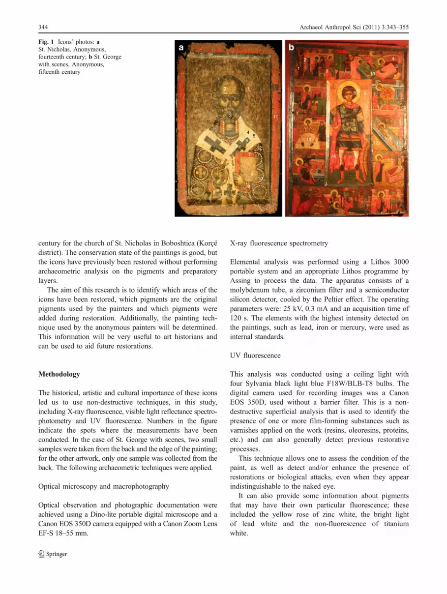
century for the church of St. Nicholas in Boboshtica (Korçëdistrict). The conservation state of the paintings is good, butthe icons have previously been restored without performingarchaeometric analysis on the pigments and preparatorylayers.
The aim of this research is to identify which areas of theicons have been restored, which pigments are the originalpigments used by the painters and which pigments wereadded during restoration. Additionally, the painting tech-nique used by the anonymous painters will be determined.This information will be very useful to art historians andcan be used to aid future restorations.
Methodology
The historical, artistic and cultural importance of these iconsled us to use non-destructive techniques, in this study,including X-ray fluorescence, visible light reflectance spectro-photometry and UV fluorescence. Numbers in the figureindicate the spots where the measurements have beenconducted. In the case of St. George with scenes, two smallsamples were taken from the back and the edge of the painting;for the other artwork, only one sample was collected from theback. The following archaeometric techniques were applied.
Optical microscopy and macrophotography
Optical observation and photographic documentation wereachieved using a Dino-lite portable digital microscope and aCanon EOS 350D camera equipped with a Canon Zoom LensEF-S 18–55 mm.
X-ray fluorescence spectrometry
Elemental analysis was performed using a Lithos 3000portable system and an appropriate Lithos programme byAssing to process the data. The apparatus consists of amolybdenum tube, a zirconium filter and a semiconductorsilicon detector, cooled by the Peltier effect. The operatingparameters were: 25 kV, 0.3 mA and an acquisition time of120 s. The elements with the highest intensity detected onthe paintings, such as lead, iron or mercury, were used asinternal standards.
UV fluorescence
This analysis was conducted using a ceiling light withfour Sylvania black light blue F18W/BLB-T8 bulbs. Thedigital camera used for recording images was a CanonEOS 350D, used without a barrier filter. This is a non-destructive superficial analysis that is used to identify thepresence of one or more film-forming substances such asvarnishes applied on the work (resins, oleoresins, proteins,etc.) and can also generally detect previous restorativeprocesses.
This technique allows one to assess the condition of thepaint, as well as detect and/or enhance the presence ofrestorations or biological attacks, even when they appearindistinguishable to the naked eye.
It can also provide some information about pigmentsthat may have their own particular fluorescence; theseincluded the yellow rose of zinc white, the bright lightof lead white and the non-fluorescence of titaniumwhite.
1 2
3 11
4 5
6
7
9 10
8
12
17 10
2
8 6 5
9
3
4
11 12
14
13
a bFig. 1 Icons’ photos: aSt. Nicholas, Anonymous,fourteenth century; b St. Georgewith scenes, Anonymous,fifteenth century
344 Archaeol Anthropol Sci (2011) 3:343–355

Reflectance spectrophotometry
Colour measurements were performed using a Minolta CM-2600 portable spectrophotometer equippedwith an integrativesphere inside the apparatus and a Xenon lamp to pulse thelight on the sample surface. Light is reflected by a pigmentwith an angle of 8°. It is captured by a silicon photodiode thatmeasures the colour spectrum between 360 and 740 nm withan interval of 10 nm. Colour coordinates are based on theCIEL*a*b* system using an illuminant D65 with an observerangle of 10°. In this system, L* represents colour lightnesswhile a* and b* are the coordinates of chromaticity.Coordinates +a* and −a* indicate red and green values while+b* and −b* indicate the yellow and blue values, respectively.
FTIR spectroscopy
Fourier transform infrared (FTIR) spectra were measuredby means of a Fourier transform infrared spectrometerSpectrum One System by Perkin Elmer that operates
between 7,800 and 350 cm−1, with Universal ATRequipment.
Scanning electron microscopy–energy dispersive system
Scanning electron microscopy (SEM) was conducted usinga Jeol 5600 LV scanning microscope equipped with anenergy dispersive X-ray spectroscopy (EDS) by Oxford.The elemental composition was determined using theprepared cross-sections in low vacuum.
Results and discussion
Firstly, we observed no evidence of an intermediate canvaslayer beneath the painted surfaces in these two icons.The inorganic pigments used for the paintings from thesetwo different periods were identified based on theirprincipal characteristic elements, their relative abundanceand by comparing their reflection spectra with the
Table 1 The counts of the main peaks of the elements detected in the analysed points of St Nicholas
Spot Colour P S K Ca Ti Mn Fe Ni Cu Zn Hg As Kβ Pb Lα Pb Lβ Sr
1 Preparative layer 12 4 18 684 2 16 124 4 3 6 2 19 14 175
2 Flesh 9 112 4 18 450 9 5 19 1,828 1,156 137
3 Brown 2 5 290 23 469 7 7 9 610 376 161
4 White 7 100 3 5 25 6 6 4,052 2,680 39
5 White-yellowish 7 22 5 62 7 30 4,752 3,259 20
6 Black 9 7 59 8 78 3 4,833 3,383
7 Red 27 9 134 12 121 5 3,073 133
8 Orange-red 5 17 467 6 26 465 20 7 7 21 23 167
9 Whitish 9 6 681 19 112 56 6 41 24 168
10 Blue 18 180 11 128 10 1,580 997 93
11 Red 7 28 9 153 14 121 6 2,339 66 33
12 Blue 9 179 15 231 4 2,400 1,668 80
The operating parameters were: 25 kV, 0.3 mA and an acquisition time of 120 s
Fig. 2 The occurrence of theprincipal elements in the 12analysed spots of St. Nicholas
Archaeol Anthropol Sci (2011) 3:343–355 345

literature data as well as with those of a pigment databasedeveloped in collaboration with the Soprintendenza per iBeni Architettonici e il Paesaggio della Liguria.
The results are discussed separately for each icon.
St. Nicholas, Anonymous, fourteenth century
X-ray fluorescence
The data obtained from the X-ray fluorescence (XRF)measurements were processed as discussed in the literature(Seccaroni and Moioli 2004). From these data, we wereable to detect the presence of various elements in thedifferent layers of the painting.
The experimental data obtained by X-ray fluorescence,specifically the counts measures for the main peak of eachelement for each analysed spot, are presented in Table 1.These data are visually summarised in Fig. 2 as a plot of theoccurrence of the principal elements in the differentanalysed spots. The principal peaks of arsenic and leadare As Kα (10.53 keV) and Pb Lα (10.50 keV), respec-tively. When arsenic and lead occur simultaneously in asample, these principal peaks are superimposed and cannotbe used for analysis. Consequently, when arsenic and leadoccurred in the same sample, we considered the counts ofKβ (11.73 keV) and Lβ (12.61 keV) respectively. The samepeak overlap occurs when sulphur (S Kα 2.31 keV) andlead (Pb Mα 2.35 keV) are present in the same sample. Inall cases, the counts of the peaks of the elements werenormalised for graphical presentation.
Titanium was found in four spots (1,2,4,8) and zinc insix (1,3,5,8,9,10), thereby indicating the occurrence ofrestoration interventions. In fact, these elements have tobe attributed to white ZnO (Kühn 1986) and TiO2 (Laver1997) pigments. These pigments have been used only sincethe nineteenth and twentieth centuries, respectively. It isimportant to note that from the experimental data we canexclude any correlation between titanium and iron (Helvig2007), suggesting the use of modern titanium white.
Visual investigation of painting’s surface permitted thedetection of extensive colour detachment (spots 1,9).From these data, we could determine the elementalcomposition of the preparative layer, which was foundto mainly contain phosphorus, sulphur, calcium, stron-tium and iron. This latter element is accompanied bymanganese and, in some cases, by nickel, whichindicates the use of a particular kind of ochre. Otherelements, such as copper, mercury and lead belong toremains of the upper layers.
The iron content could be derived from a successivepaint layer, or it could be due to the painter using apreparation layer coloured with ochre.
In the other spots it is noteworthy the presence of arsenic(spot 8) and mercury (spots 7,11)
The presence of phosphorus was also detected on theblack cross of the stole. This could be attributed to the useof bone/ivory black (Winter and West FitzHugh 2007).
Finally, the presence of lead in all the analysed spotscould be attributed to lead white used as a filler (Civici
Fig. 3 UV photograph of St. Nicholas
AB
G
B
C
BB
GF
F
D
D
E
E
C
C
C
A
Fig. 4 Comparison between visible light (top) and UV (bottom)images. Detail from St. Nicholas
346 Archaeol Anthropol Sci (2011) 3:343–355

2006) or as imprimatura, which is a second thick white orslightly coloured preparatory layer (Gettens et al. 1993a).
UV observation
Under UV illumination, areas containing cinnabar orvermillion become purple. Iron oxide presents a typicaldarkening behaviour, clearly showing the painting tech-nique used by the artist, with strokes of paint applied toform continuous layers as the painting base and thin strokesas overlayers (Fig. 3).
The presence of lead white is clearly evidenced by thestrong response to UV illumination (Aldrovandi et al. 1996;Fig. 4). The fresh, more or less recent varnish appears as blackareas, denoting restoration interventions (Aldrovandi 1999).
Based on Fig. 4, where a detailed image of St. Nicholascorresponding to a cross on the stole is shown, we can statethe following observations regarding the areas indicated bythe letters:
A) Areas where the preparatory layer is emerging due tothe loss of metal coating. The typical engraved lines ofthe preparatory drawing are clearly noticeable. The
presence of iron oxide (possibly due to bole residue)gives the layer a poor UV fluorescence.
B) Areas covered by a deep layer of very fluorescent leadwhite.
C) Cinnabar strokes that under UV radiation change to ared-purple tone.
D) Dark red colour formed mainly by iron oxides thatdarken under UV radiation.
E) The red colour appears in the background throughthe thin strokes of lead white used for ornamentaldesigns.
F) In the areas where the grey-blue colour was erased theunderlying preparatory layer comes to the surface.Under UV radiation the darkest areas reveal the zoneswhere the colour is preserved.
G) Strokes obtained with ochre that under UV lightbecome darker.
Reflectance spectrophotometric measurements
The reflectance Vis spectrophotometric investigations weremade on all the zones examined by X-ray fluorescence. The
Fig. 5 Reflectance curve refer-ring to spots 7 and 11 in St.Nicholas and processing reflec-tance spectra in the secondderivative (left), compared withthe curve typical for cinnabar(right)
Fig. 6 Reflectance curve refer-ring to spot 8 in St. Nicholasand processing reflectance spec-tra in the second derivative,compared with the curve typicalfor burnt umber
Archaeol Anthropol Sci (2011) 3:343–355 347

graph obtained by data processing in the first derivative(Bacci et al. 2003) or in the second derivative with oppositesign (Franceschi et al. 2010) loses the information ofbrightness (L*), but improves the reading of data, andhighlights the principal frequencies of most of the exam-ined reflectance colours. The method may allow for theidentification of the pigments that mainly contribute to theperception of colour. The technique, which offers the bestresults in the examination of mural paintings, where little orno paint binder is on the surface film, poses moredifficulties in easel painting, as in almost all the icons ofthe Korçë Museum, which are veiled with thick, unequaland misleading varnishes.
On the basis of the measurements made, the followingconclusions can be reached.
Red and brown
The hue range of red pigments varies from light orange todark red. The red inorganic pigments used in this icon werered ochre (Fe2O3) and cinnabar (HgS). In some cases redtonalities were obtained from appropriate mixtures of thesepigments with lead white. There is no evidence of the useof lakes. Iron-manganese-nickel oxide (brown earth) wasused to obtain the brown colour.
Figure 5 shows the results obtained by measuring twodifferent red spots of St. Nicholas, spot 7 and 11, andcomparing them with the spectrum of pure cinnabar(Gettens et al. 1993b).
The particular hue of spot 8, corresponding to the Saint’ssleeve, was obtained by using a layer of arsenic sulphide,likely orpiment, As2S3, on an underpaint of burnt umberand probably red ochre. See the results of XRF and thespectra comparison shown in Fig. 6 and some differences inthe curve shapes. It is known that orpiment, As2S3 and realgaroccur in icon paintings created between the 12th and 16thcenturies in some Orthodox countries (West FitzHugh 1997);our results confirm their use in Albanian icons as well.
Flesh tone and yellow
Considering the results of X-ray fluorescence andobserving Fig. 7, where a comparison between differentcurves is shown, we could assess that the flesh tone (spot2) was obtained by mixing lead white (mainly revealedby the presence of Pb by XRF measurements) withyellow ochre. In addition, the latter pigment was used tocolour the yellow stole highlights (spot 5). The reflec-tance curve of lead white, as occurs for other whitepigments, is not significant in the visible region;therefore the comparison with this pigment is hereomitted.
Blue
The anonymous fourteenth century artist used blue tonesto paint the stole, or polystavrion (“many crosses”), of St.Nicholas. Regarding the data reported in Table 1, ele-ments, such as lead, iron and calcium have been identifiedby X-ray fluorescence, but they do not make up the bluepigment composition. The optical microscope imagereported in Fig. 8 illustrates the technique used. The
Fig. 7 Reflectance curve refer-ring to spots 2 and 5 in St.Nicholas and processing reflec-tance spectra in the secondderivative, compared with thecurve typical for yellow ochre
Fig. 8 Detail of St. Nicholas with the enlargement of an area byoptical microscope
348 Archaeol Anthropol Sci (2011) 3:343–355

layer of black underpaint (possibly obtained by amixture of lead white and bone/ivory black as revealedby the presence of phosphorus) and the blue paint layerwere detected. The combination of blue highlights andblack underpaints is a technique used by painters of alleras. A priori we could not exclude the use of indigo(Schweppe 1997) or ultramarine (lapis lazuli) (Plesters1993) as the blue pigment. As well known, the strongblue colour of lapis lazuli is due to the presence of themineral lazurite (Na,Ca)8(SO4,S,Cl)2(AlSiO4)6. Then,elements characterising this latter compound by means ofXRF are Ca, S and Cl. The calcium can be confused with that
of the preparatory layer while the sulphur is covered by thelead signal. The absence of chlorine leads us to exclude theuse of ultramarine. The only possibility for the blue colourremains the use of indigo.
In Fig. 9a comparison between the typical reflectancecurves obtained for lapis lazuli and indigo, with theirsecond derivatives and the experimental curves obtained incorrespondence of spots 10 and 12 in St. Nicholas isshown. Based on the colour measurements, it was notpossible to obtain a reliable indication of the actual pigmentused for both spots but only a hypothesis on the possibleuse of indigo.
Fig. 9 Reflectance curve referring to lapis lazuli and indigo and processing reflectance spectra in the second derivative, compared with the curvesrelatives to spots 10 and 12 in St. Nicholas
Fig. 10 FTIR spectrum relative to the micro-sampling in St. Nicholas. The inorganic component mainly consists of CaCO3
Archaeol Anthropol Sci (2011) 3:343–355 349

Other remarks
In order to acquire information about the preparation andprotective layers of the backside of the wooden panel, atiny fragment was sampled from the backside of St.Nicholas and examined using the cross-section method.
The background
FTIR analysis of the sample (Fig. 10) revealed that theprotective layer is made up of calcium carbonate (peaks at1,794, 1,392, 1,088, 712 cm−1), while the paint preparation(Harrison et al. 2008) was obtained by using the morecommon gypsum (as it was revealed based on XRFmeasurements in spots 1 and 9). The presence of silicates
(peaks at 1,021 and 780 cm−1) can be attributed to the useof impure calcium carbonate or to the addition of colouredearth. The organic binder is almost completely convertedinto calcium oxalate (see FTIR spectrum of Fig. 10, peaksat 1,639–1,620, 1,323 and 780 cm−1), not allowing it to bereliably identified; only the C–H stretching peaks at 2,960–2,865 cm−1 are still present.
St. George with scenes, Anonymous, fifteenth century
X-ray fluorescence
The experimental data obtained for this painting based onX-ray fluorescence are shown in Table 2 and visuallysummarised in Fig. 11, where the occurrence of the
Table 2 The counts of the main peaks of the elements detected in the analysed points of St. George with scenes
Spot Colour S K Ca Ti Mn Fe Cu Zn Hg As Kα As Kβ Pb Lα Pb Lβ Sr
1 Dark red 7 174 58 3,440 136 19 190 28 423 275
2 Red 32 38 3 31 53 1,355 3,487 2,186
3 Dark red 10 8 168 22 30 5,067 32 90 27 401 59 138 90
4 Light red 7 61 4 7 548 18 9 44 156 23 3,292 2,140
5 Orange 6 10 9 29 7 156 5,266 3,797
6 Yellow 5 8 45 3 9 163 27 6,982 1,024 98 64
7 Yellow-orange 9 447 4 9 525 29 25 519 334
8 Brown 155 30 34 1,542 18 18 174 524 77 131 85
9 Green 11 5 109 2 7 261 6,462 456 312
10 Grey 8 47 37 31 3,480 2,300
11 Light-grey 3 58 38 32 3,380 2,135
12 Yellow-orange 16 300 64 1,205 15 7 73 47 32
13 White 15 285 45 24 1,773 1,177 20
14 Red 36 8 60 6 29 14 4,750 180
The operating parameters were: 25 kV, 0.3 mA and an acquisition time of 120 s
Fig. 11 The occurrence of theprincipal elements in the 14analysed spots of St. Georgewith scenes
350 Archaeol Anthropol Sci (2011) 3:343–355

principal elements is plotted. Also in this case, whenarsenic and lead are simultaneously present, we consideredthe counts of Kβ and Lβ, respectively. The counts of thepeaks of the elements have been normalised for graphicalpresentation; in the case of contemporary presence of As–Pb, the counts of As Kα were evaluated from As Kβmeasured value and the counts of Pb Lα were evaluatedfrom Pb Lβ measured value.
Many areas of the icon were restored, as shown by thewidespread presence of titanium (spots 3, 4, 6, 7, 8, 9, 12)and zinc (spots 3, 4, 5, 8, 12). UV reflection photographycan be used for better recognition of these areas.
It is important to notice that the occurrence of iron oxidepigments in this icon is not associated with the presence ofnickel but only of manganese in all analysed spots, exceptwhere they correspond to some light tones (spots 10, 11, 12,13). In particular, the painter probably used goethite, FeO(OH), as pigment for the yellow–orange colour. The total
absence of nickel indicates a different provenance of thesepigments compared to those of the St. Nicholas. Thepresence of phosphorus was not detected in this icon,showing another difference in the preparation techniquecompared to that of the St. Nicholas. Arsenic is present inseveral spots (1, 3, 4, 6, 8). Lead is revealed in all zones.Strontium is present only in two spots (12 and 13); sosuggesting a further difference between the preparationlayers of the two icons. A copper-based compound wasused to obtain the green hue.
The brown colour corresponding to spot 8 is obtained froma mixture of brown earth, red lead and realgar (or orpiment)with cinnabar red shades; in fact, the main elements detectedwere lead, iron, arsenic and mercury. Mercury is also presentin the red and dark red hues, revealing the use of cinnabar(spot 14) or the use of mixtures of different pigmentsincluding a lead based pigment (spot 2).
UV observation
The presence of lead white is limited to a few areas,evidenced by the strong response to UV illumination(Figs. 12 and 13). The more recent varnish appears asblack areas, denoting restoration interventions.
Consistently with the high lead intensity of some XRFmeasurements, such as the orange of spot 5 or the red mantel(spot 14), we think that the colours were obtained by mixturesof red lead and cinnabar with lead white. Still lead white wasused for the mantel or wall ornaments (see Fig. 14).
The high calcium intensity of the white cement (spot 13)showed the use of calcium carbonate too; this is confirmedby the UV photographs. Calcium carbonate was also usedto obtain the clear yellow-orange colour of the seat and thewall (spots 7, 12).
The following can be noted, as regards Figs. 13 and 14:
& The colour change in fluorescence between twodifferent yellows, i.e. the very fluorescent halo oforpiment (A) and the ochre used in the basement ofthe Saint (B) that becomes significantly darker.
& Areas containing cinnabar are less fluorescent evenwhen mixed with lead white (C).
Fig. 12 UV photograph of St. George with scenes
Fig. 13 Comparison betweenvisible light (left) and UV(right) images. Detail from St.George with scenes
Archaeol Anthropol Sci (2011) 3:343–355 351

& The strong fluorescence of the areas rich in lead white (D).& Varnish irregularities are highlighted (E).
Reflectance spectrophotometric measurements
On the basis of the reflectance spectrophotometric inves-tigations, the following conclusions can be drawn:
Brown
To produce brown and dark tonalities, the painter usedmanganese–iron oxides known as brown earth (spot 8) ormixed with red ochre (spots 1, 3). The complexity of thecolour is evidenced in Fig. 15, where the reflectance spectrumrelative to spot 8 (brown of the Saint’s hair) is shown,confirming the suggestion of the XRF data that manypigments contribute to the colour in the St. George withscenes icon.
Red
The hue range of red pigments varies from light orange todark red. The red inorganic pigments used in this icon werered ochre, red lead (Pb3O4), cinnabar and probably realgar.The variety of red tonalities was obtained from appropriatemixtures of these pigments and leads white.
The measurements conducted on a red–orange area of St.George with scenes (spot 5) were compared with the curves ofstandard pigments (Fig. 16). This comparison enabled us toattribute the presence of lead found with the method of X-ray fluorescence to red lead (West FitzHugh 1986).
Green
Taking into account the high counts of Cu obtained by XFRanalysis, the green colour could have been obtained by acopper-based pigment, such as malachite. Anyway, the
Fig. 14 Comparison betweenvisible light (left, black andwhite) and UV (right) images.Detail from a scene of the life ofthe Saint in St. George withscenes
Fig. 15 Brown colour reflec-tance curve for spot 8 in St.George with scenes and pro-cessing reflectance spectra in thesecond derivative, comparedwith the curve of burnt umber
352 Archaeol Anthropol Sci (2011) 3:343–355

significant differences between the curve of the greenpigment and the curve of malachite (Gettens and WestFitzHugh 1993; Bruno et al. 2008) seem to exclude thispossibility. Verdigris is another pigment most often foundin paintings from the thirteenth to the nineteenthcenturies (Kühn 1993), but spectrophotometric measurescannot assess its presence. Moreover, as a result ofvarnish darkening, the reflectance values are very low(Fig. 17).
Yellow
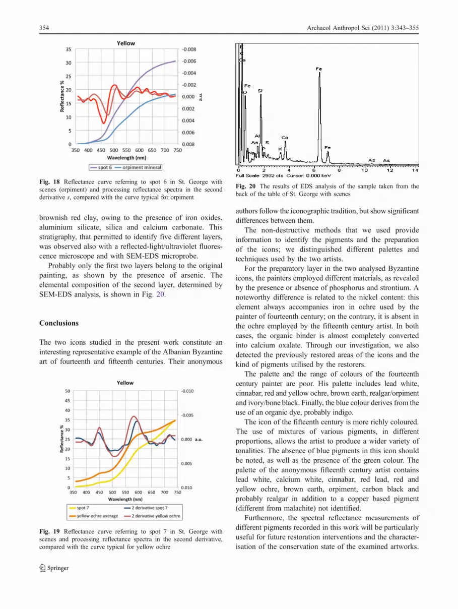
Two yellow pigments were suggested by X-ray fluores-cence measurements conducted on the spots investigated inthe icon of St. George with scenes. The painter usedorpiment, As2S3 (West FitzHugh 1997), to obtain theyellow for the Saint’s aureole. The other yellow pigment,revealed in spots 7 and 12, is yellow ochre (Helvig 2007).The spectral curves for the two pigments (orpiment andyellow ochre) shown in Figs. 18 and 19, respectively,confirmed this hypothesis.
Grey
The grey and light grey colours of the stones, representingthe terrible moments of the Saint’s torture, were obtained bymixing carbon black (or another organic black) with leadwhite (mainly revealed by XRF measurements).
Other remarks
Two tiny fragments were extracted from the edge and theback of the wooden panel; they were examined using thecross-section method. The situation found on St. Georgewith scenes is complex. The preparation exhibits a clearlydetectable darkening of cracks in the paint layer. Thisdarkening may be caused by simple alteration of thebinding protein but could also be caused by the presenceof deliberate pigmentation consisting of clayey material.The successive layers contain pigments, but we do notknow whether they are actually in connection with thecolours on the front. There is an interesting presence ofarsenic in the first reddish layer, composed, in fact, of
Fig. 16 Red colour reflectancecurve referring to spot 5 in St.George with scenes and pro-cessing reflectance spectra in thesecond derivative (left) com-pared with the spectra of somereference pigments (right)
Fig. 17 Dark green colour reflectance curve referring to spot 9 in St. George with scenes and processing reflectance spectra in the secondderivative, compared with malachite (middle) and verdigris (right) spectra
Archaeol Anthropol Sci (2011) 3:343–355 353
brownish red clay, owing to the presence of iron oxides,aluminium silicate, silica and calcium carbonate. Thisstratigraphy, that permitted to identify five different layers,was observed also with a reflected-light/ultraviolet fluores-cence microscope and with SEM-EDS microprobe.
Probably only the first two layers belong to the originalpainting, as shown by the presence of arsenic. Theelemental composition of the second layer, determined bySEM-EDS analysis, is shown in Fig. 20.
Conclusions
The two icons studied in the present work constitute aninteresting representative example of the Albanian Byzantineart of fourteenth and fifteenth centuries. Their anonymous
authors follow the iconographic tradition, but show significantdifferences between them.
The non-destructive methods that we used provideinformation to identify the pigments and the preparationof the icons; we distinguished different palettes andtechniques used by the two artists.
For the preparatory layer in the two analysed Byzantineicons, the painters employed different materials, as revealedby the presence or absence of phosphorus and strontium. Anoteworthy difference is related to the nickel content: thiselement always accompanies iron in ochre used by thepainter of fourteenth century; on the contrary, it is absent inthe ochre employed by the fifteenth century artist. In bothcases, the organic binder is almost completely convertedinto calcium oxalate. Through our investigation, we alsodetected the previously restored areas of the icons and thekind of pigments utilised by the restorers.
The palette and the range of colours of the fourteenthcentury painter are poor. His palette includes lead white,cinnabar, red and yellow ochre, brown earth, realgar/orpimentand ivory/bone black. Finally, the blue colour derives from theuse of an organic dye, probably indigo.
The icon of the fifteenth century is more richly coloured.The use of mixtures of various pigments, in differentproportions, allows the artist to produce a wider variety oftonalities. The absence of blue pigments in this icon shouldbe noted, as well as the presence of the green colour. Thepalette of the anonymous fifteenth century artist containslead white, calcium white, cinnabar, red lead, red andyellow ochre, brown earth, orpiment, carbon black andprobably realgar in addition to a copper based pigment(different from malachite) not identified.
Furthermore, the spectral reflectance measurements ofdifferent pigments recorded in this work will be particularlyuseful for future restoration interventions and the character-isation of the conservation state of the examined artworks.
Fig. 18 Reflectance curve referring to spot 6 in St. George withscenes (orpiment) and processing reflectance spectra in the secondderivative s, compared with the curve typical for orpiment
Fig. 19 Reflectance curve referring to spot 7 in St. George withscenes and processing reflectance spectra in the second derivative,compared with the curve typical for yellow ochre
Fig. 20 The results of EDS analysis of the sample taken from theback of the table of St. George with scenes
354 Archaeol Anthropol Sci (2011) 3:343–355

The latter technique has proved to be very useful forrecognising pigments not detectable by means of X-rayfluorescence.
Acknowledgements The authors are grateful to the Soprintendenzaper i Beni Architettonici e il Paesaggio della Liguria, Genoa, for thereflectance spectroscopy measurements aimed at setting up a pigmentspectral databank and to Dr. Kristaq Balli of the Muzeu Kombetar iArtit Mesjetar, Korçë, for the helpful discussions. Thanks are also dueto Mr. Carlo Narducci of the University of Pisa for carrying out theSEM-EDS analyses.
References
Aldrovandi A (1999) L’acquisizione fotografica della fluorescenzaUV: applicazione all’indagine dei dipinti antichi. OPD Restauro11:191–205
Aldrovandi A, Altamura ML, Cianfanelli MT, Riitano P (1996) Imateriali pittorici: tavolette campione per la caratterizzazionemediante analisi multispettrale. OPD Restauro 8:191–210
Bacci M, Casini A, Cucci C, Picollo M, Radicati B, Vervat M (2003)Non-invasive spectroscopic measurements on the Il ritratto dellafigliastra by Giovanni Fattori: identification of pigments andcolourimetric analysis. J Cult Herit 4:329–336
Bruno G, Picollo M, Radicati B, Bacci M (2008) Pigmenti Verdidall’antichità al XIX secolo: caratterizzazione non invasivamediante spettroscopia di riflettanza UV-Vis-NIR. In: Patron(ed) Colore e Arte: Storia e Tecnologia del Colore nei Secoli. Attidel III Convegno Nazionale di Archeometria (AIAr) (Firenze 28Febbraio–2 Marzo 2007). Bologna 2008
Civici N (2006) Non-destructive identification of inorganic pigmentsused in 16–17th century Albanian icons by total reflection X-rayfluorescence analysis. J Cult Herit 7:339–343
Civici N, Demko O, Clark RJH (2005) Identification of pigments usedon late 17th century Albanian icons by total reflection X-rayfluorescence and Raman microscopy. J Cult Herit 6:157–164
Franceschi E, Nole D, Vassallo S (2010) Icone Albanesi postBizantine: un approccio archeometrico, Paper presented at VICongresso Nazionale di Archeometria (AIAr), in Pavia, 15–18February 2010
Gettens RJ, West FitzHugh E (1993) Malachite and green verditer. In:Roy A (ed) Artists’ pigments. A handbook of their history and
characteristics, vol 2. Oxford University Press, New York, pp183–202
Gettens RJ, Kühn H, Chase WT (1993a) Lead white. In: Roy A (ed)Artists’ pigments. A handbook of their history and character-istics, vol 2. Oxford University Press, New York, pp 67–81
Gettens RJ, Feller R, Chase WT (1993b) Vermillion and cinnabar. In: RoyA (ed) Artists’ pigments. A handbook of their history and character-istics, vol 2. Oxford University Press, New York, pp 159–182
Harrison L, Cormack R, Cartwright CR, Ambers J (2008) An icon ofSt George: preparation for a portrait of a saint. In: Townsend JH,Doherty T, Heydenreich G, Ridge J (eds) Preparation forpainting. The artist’s choice and its consequences. ArchetypePublications, London, pp 14–21
Helvig K (2007) Iron oxide pigments: natural and synthetic. In: BerryBH (ed) Artists’ pigments. A handbook of their history andcharacteristics, vol 4. Publishing Office, National Gallery of Art,Washington, pp 39–109
Kühn H (1986) Zinc white. In: Feller RL (ed) Artists’ pigments. Ahandbook of their history and characteristics, vol 1. CambridgeUniversity Press, London, pp 169–186
Kühn H (1993) Verdigris and copper resinate. In: Roy A (ed) Artists’pigments. A handbook of their history and characteristics, vol 2.Oxford University Press, New York, pp 131–147
Laver M (1997) Titanium dioxide whites. In: West FitzHugh E (ed)Artists’ pigments. A handbook of their history and character-istics, vol 3. Oxford University Press, New York, pp 295–356
Plesters J (1993) Ultramarine blue, natural and artificial. In: Roy A (ed)Artists’ pigments. A Handbook of their history and characteristics,vol 2. Oxford University Press, New York, pp 37–65
Schweppe H (1997) Indigo and woad. In: West FitzHugh E (ed)Artists’ pigments. A handbook of their history and character-istics, vol 3. Oxford University Press, New York, pp 81–108
Seccaroni C, Moioli P (2004) Fluorescenza X. Prontuario per l’analisiXRF portatile applicata a superfici policrome. Nardini Editore,Firenze
West FitzHugh E (1986) Red lead and minium. In: Feller RL (ed)Artists’ pigments. A handbook of their history and character-istics, vol 1. Cambridge University Press, London, pp 109–140
West FitzHugh E (1997) Orpiment and realgar. In: West FitzHugh E (ed)Artists’ pigments. A handbook of their history and characteristics,vol 3. Oxford University Press, New York, pp 47–80
Winter J, West FitzHugh E (2007) Pigments based on carbon. In:Berry BH (ed) Artists’ pigments. A handbook of their history andcharacteristics, vol 4. Publishing Office, National Gallery of Art,Washington, pp 1–37
Archaeol Anthropol Sci (2011) 3:343–355 355


