Embed Size (px)
Citation preview

LUND UNIVERSITY
PO Box 117221 00 Lund+46 46-222 00 00
Haemophilus influenzae – typing, epidemiology and beta-lactam resistance
Månsson, Viktor
2019
Document Version:Publisher's PDF, also known as Version of record
Link to publication
Citation for published version (APA):Månsson, V. (2019). Haemophilus influenzae – typing, epidemiology and beta-lactam resistance. LundUniversity: Faculty of Medicine.
Total number of authors:1
General rightsUnless other specific re-use rights are stated the following general rights apply:Copyright and moral rights for the publications made accessible in the public portal are retained by the authorsand/or other copyright owners and it is a condition of accessing publications that users recognise and abide by thelegal requirements associated with these rights. • Users may download and print one copy of any publication from the public portal for the purpose of private studyor research. • You may not further distribute the material or use it for any profit-making activity or commercial gain • You may freely distribute the URL identifying the publication in the public portal
Read more about Creative commons licenses: https://creativecommons.org/licenses/Take down policyIf you believe that this document breaches copyright please contact us providing details, and we will removeaccess to the work immediately and investigate your claim.

VIK
TOR
MÅ
NSSO
N
Haem
ophilus influenzae – typing, epidemiology and beta-lactam
resistance 2019:4
Department of Translational Medicine
Lund University, Faculty of Medicine Doctoral Dissertation Series 2019:4
ISBN 978-91-7619-733-2ISSN 1652-8220
Haemophilus influenzae – typing, epidemiology and beta-lactam resistanceVIKTOR MÅNSSON
DEPARTMENT OF TRANSLATIONAL MEDICINE | LUND UNIVERSITY
Viktor Månsson is a medical doctor currently working at the Department of Infectious Diseases at Skåne University Hospital in Malmö. His interest in clinical microbiology and infectious diseases research started during his medical studies. This thesis includes studies on Haemophilus influenzae diagnostics, epidemiology and antimicrobial resistance.
978
9176
1973
32


1
Haemophilus influenzae – typing, epidemiology and beta-lactam
resistance
Viktor Månsson
DOCTORAL DISSERTATION by due permission of the Faculty of Medicine, Lund University, Sweden.
To be defended at the main lecture hall of the Pathology building, Jan Waldenströms gata 59, Malmö, 25 January 2019 at 13:00.
Faculty opponent Professor Niels Frimodt-Møller
Department of Clinical Microbiology, Rigshospitalet, University of Copenhagen, Copenhagen, Denmark

2
Organization LUND UNIVERSITY
Document name DOCTORAL DISSERTION
Date of issue: 25 January 2019
Author: Viktor Månsson Sponsoring organization
Title and subtitle: Haemophilus influenzae – typing, epidemiology and beta-lactam resistance
Abstract Haemophilus influenzae is a common cause of respiratory tract infections such as acute otitis media (AOM), exacerbations of chronic obstructive pulmonary disease (COPD) and pneumonia. The species is subdivided into encapsulated and non-encapsulated strains, designated type a-f and nontypeable H. influenzae (NTHi), respectively. Prior to introduction of polysaccharide-protein conjugate vaccines against H. influenzae type b (Hib) in childhood vaccination programmes this serotype frequently caused severe invasive infections in small children. Nowadays invasive disease by Hib is rare, but cases still occur. At present, NTHi is the dominating type to cause invasive disease and invasive NTHi disease appears to be increasing. Nontypeable H. influenzae disease severity traditionally has been considered largely host dependant. In parallel, non-beta-lactamase mediated beta-lactam resistance among NTHi is also increasing. In the first two studies of this thesis, we investigated capsule typing of H. influenzae by matrix-assisted laser desorption/ionization time-of-flight mass spectrometry (MALDI-TOF MS), a technology routinely used for bacterial species identification. Mass spectra of the different types of encapsulated H. influenzae were highly similar within each type and separable from each other. The differences in mass spectra relied on the clonal population structure of encapsulated H. influenzae, with conserved type specific genetic lineages. Mass spectra of NTHi were diverse, due to their genetic heterogeneity. Following construction of a comprehensive reference database, MALDI-TOF MS showed high accuracy for capsule typing of H. influenzae. In the third study of the thesis, a clonal group of NTHi with resistance against beta-lactam antibiotics was investigated. The clonal group accounted for one quarter of clinical respiratory H. influenzae isolates with non-beta-lactamase mediated beta-lactam resistance in the study region. Furthermore, patients infected by isolates of the clonal group had an increased risk of hospitalization compared to patients infected by other NTHi, indicating enhanced virulence traits. The clonal group was also found among invasive isolates. In the final study of the thesis we retrospectively compared benzylpenicillin, whose effect on H. influenzae is debated, to wide spectrum beta-lactams (WSBLs) as empirical treatment of H. influenzae lower respiratory tract infections in patients requiring hospitalization. Empirical treatment with benzylpenicllin was not associated with higher mortality or increased risk of hospital readmission compared to treatment with WSBLs. The early clinical response rate was, however, lower for patients receiving benzylpenicillin, which was attributed mainly to a lower response rate in patients infected with beta-lactamase producing isolates. In conclusion this thesis shows that MALDI-TOF MS can be efficiently used for rapid capsule typing of H. influenzae. The newly developed method can be valuable for typing of invasive H. influenzae isolates and for surveillance of Hib vaccination efficacy. The studied clonal group appears to harbour enhanced virulence traits. This indicates that bacterial factors may affect NTHi disease severity more than previously considered, and possibly contribute to the increased incidence of invasive NTHi disease. Finally, although the effect of benzylpenicillin on H. influenzae is debated, empirical treatment of lower respiratory tract infections of mild to moderate severity caused by H. influenzae with the agent appears safe.
Key words: Benzylpenicillin, beta-lactam resistance, beta-lactams, BLNAR, capsule typing, clonal, Haemophilus influenzae, Hib, MALDI-TOF MS, MLST, NTHi, serotyping, virulence
Classification system and/or index terms: Bacterial capsules, bacterial typing, beta-lactam resistance, epidemiology, Haemophilus influenzae, MALDI
Supplementary bibliographical information Language: English
ISSN and key title: 1652-8220 ISBN: 978-91-7619-733-2
Recipient’s notes Number of pages: 93 Price
Security classification
I, the undersigned, being the copyright owner of the abstract of the above-mentioned dissertation, hereby grant to all reference sources permission to publish and disseminate the abstract of the above-mentioned dissertation.
Signature Date 2018-12-10

3
Haemophilus influenzae – typing, epidemiology and beta-lactam
resistance
Viktor Månsson

4
Cover photo: Scanning electron microscopy picture of Haemophilus influenzae infecting lung tissue in an experimental mouse model. By Matthias Mörgelin, Colzyx AB, Lund, Sweden.
Copyright pp 1-93 Viktor Månsson
Paper I © American Society for Microbiology
Paper II © Centers for Disease Control and Prevention
Paper III © European Society of Clinical Microbiology and Infectious Diseases
Paper IV © The authors
Faculty of Medicine Department of Translational Medicine ISBN 978-91-7619-733-2 ISSN 1652-8220 Printed in Sweden by Media-Tryck, Lund University Lund 2018
Media-Tryck is an environmentallycertified and ISO 14001 certifiedprovider of printed material.Read more about our environmentalwork at www.mediatryck.lu.se
NO
RDIC
SWAN ECOLABE
L
1234 5678

5
Contents
List of papers .................................................................................................. 7
Abbreviations ................................................................................................. 8
Populärvetenskaplig sammanfattning .......................................................... 10
Introduction ............................................................................................................ 13
Brief history of the bacterium Haemophilus influenzae ............................... 13
Bacteriology of Haemophilus influenzae ..................................................... 14 Basic characteristics ............................................................................ 14 Pathogenesis and virulence .................................................................. 15 The polysaccharide capsule ................................................................. 19 Genetic variation and population structure .......................................... 20
Disease caused by Haemophilus influenzae ................................................. 23 Colonization and transmission ............................................................ 23 Respiratory tract infections .................................................................. 24 Invasive infections ............................................................................... 26
Antimicrobial susceptibility and resistance of Haemophilus influenzae ...... 30 Epidemiological cut-offs and clinical breakpoints .............................. 30 Beta-lactam susceptibility and resistance ............................................ 31 Non-beta-lactam susceptibility and resistance .................................... 37
MALDI-TOF MS in clinical microbiology .................................................. 38 Principle and function .......................................................................... 38 Use in clinical microbiology ............................................................... 39
The present investigation ........................................................................................ 43
Aims ............................................................................................................. 43
Methods, Results and Discussion ................................................................. 44 Paper I .................................................................................................. 44 Paper II ................................................................................................ 47 Paper III ............................................................................................... 53 Paper IV ............................................................................................... 59
Ethical considerations .................................................................................. 64
Concluding remarks and future perspectives .......................................................... 65
Acknowledgements ................................................................................................ 67
References .............................................................................................................. 69

6

7
List of papers
I. Viktor Månsson*, Fredrik Resman*, Markus Kostrzewa, Bo Nilson, Kristian Riesbeck. Identification of Haemophilus influenzae Type b Isolates by Use of Matrix-Assisted Laser Desorption Ionization-Time of Flight Mass Spectrometry. Journal of Clinical Microbiology 2015; 53(7):2215-24. *These authors contributed equally to the article.
II. Viktor Månsson, Janet R Gilsdorf, Gunnar Kahlmeter, Mogens Kilian, J
Simon Kroll, Kristian Riesbeck*, Fredrik Resman*. Capsule Typing of Haemophilus influenzae by Matrix-Assisted Laser Desorption/Ionization Time-of-Flight Mass Spectrometry. Emerging Infectious Diseases 2018; 24(3):443-52. *These senior authors contributed equally to the article.
III. Viktor Månsson, Dagfinn Skaare, Kristian Riesbeck, Fredrik Resman. The
spread and clinical impact of ST14CC-PBP3 type IIb/A, a clonal group of non-typeable Haemophilus influenzae with chromosomally mediated beta-lactam resistance – a prospective observational study. Clinical Microbiology and Infection 2017; 23(3):209 e1- e7.
IV. John Thegerström, Viktor Månsson, Kristian Riesbeck, Fredrik Resman.
Benzylpenicillin versus wide-spectrum beta-lactam antibiotics as empirical treatment of Haemophilus influenzae-associated lower respiratory tract infections in adults; a retrospective propensity score-matched study. European Journal of Clinical Microbiology Infectious Diseases 2018; 37(9):1761-75.
8
Abbreviations
AOM Acute otitis media
ATP Adenosine triphosphate
BLNAR Beta-lactamase negative ampicillin resistant
BLNAS Beta-lactamase negative ampicillin susceptible
BLPAR Beta-lactamase positive ampicillin resistant
BLPACR Beta-lactamase positive amoxicillin-clavulanate resistant
CAP Community-acquired pneumonia
CC Clonal cluster
CCI Charlson comorbidity index
CI Confidence interval
CLSI Clinical and Laboratory Standards Institute
COPD Chronic obstructive pulmonary disease
CRP C-reactive protein
ECM Extracellular matrix
ECOFF Epidemiological cut-off
ESBL Extended spectrum beta-lactamase
EUCAST European Committee on Antimicrobial Susceptibility Testing
hia Haemophilus influenzae adhesin
Hia Haemophilus influenzae type a
Hib Haemophilus influenzae type b
Hic Haemophilus influenzae type c
Hid Haemophilus influenzae type d
Hie Haemophilus influenzae type e
Hif Haemophilus influenzae type f
HMW High molecular weight protein
ICAM-1 Intercellular adhesion molecule-1

9
ICU Intensive care unit
IgA1 Immunoglobulin A1
IgD Immunoglobulin D
LOS Lipooligosaccharide
LPS Lipopolysaccharide
MALDI-TOF MS Matrix-assisted laser desorption/ionization time-of-flight mass spectrometry
MIC Minimum inhibitory concentration
MLEE Multilocus enzyme electrophoresis
MLST Multilocus sequence typing
MRSA Methicillin-resistant Staphylococcus aureus
MRSE Methicillin-resistant Staphylococcus epidermidis
MSP Main spectrum
NAD Nicotinamide adenine dinucleotide
NordicAST Nordic Committee on Antimicrobial Susceptibility Testing
NTHi Nontypeable Haemophilus influenzae
OMV Outer membrane vesicle
OR Odds ratio
PBP Penicillin-binding protein
PCA Principal component analysis
PCR Polymerase chain reaction
PcG Penicillin G
PcV Penicillin V
PCV Pneumococcal conjugate vaccine
PRP Polyribosylribitol phosphate
rPBP3 Resistance mediated by amino acid substitutions in PBP3
SAST Slide agglutination serotyping
ST Sequence type
WSBL Wide spectrum beta-lactam

10
Populärvetenskaplig sammanfattning
Haemophilus influenzae är en bakterie som lever i människans luftvägar. Ofta orsakar bakterien ingen skada hos personen som bär den, men ibland orsakar H. influenzae luftvägsinfektioner såsom öroninflammation hos barn, akut försämring hos patienter med kroniskt obstruktiv luftvägssjukdom (KOL) samt lunginflammation. Bakterien kan också orsaka allvarliga infektioner såsom blodförgiftning och hjärnhinneinflammation. Dessa infektioner kallas invasiva, vilket innebär att bakterier kan odlas fram från en normalt sett bakteriefri (steril) vävnad, som till exempel blod. H. influenzae delas in i olika typer. Dessa benämns typ a till f beroende på vilken sorts skyddande kapselmolekyl de producerar och omger sig med. Det finns även bakteriestammar som inte producerar någon kapsel. Dessa kallas icke-kapslade H. influenzae. För att särskilja de olika typerna finns olika metoder för kapseltypning.
Allvarliga infektioner har historiskt nästan uteslutande orsakats av H. influenzae typ b (Hib) och främst drabbat barn i tidig förskoleålder. Sedan barnvaccination mot Hib införts i stora delar av världen under 1990- och 2000-talet har sjukdom orsakad av Hib minskat drastiskt. Idag orsakas både luftvägssjukdom och invasiv sjukdom i huvudsak av de icke-kapslade stammarna. Invasiv sjukdom med icke-kapslade H. influenzae verkar öka i förekomst, men anledningen till detta är inte klarlagd. Sjukdom framkallad av dessa stammar har till stor del ansetts vara beroende av en ökad infektionskänslighet hos personen som drabbas. Enstaka fall av svår sjukdom orsakad av Hib förekommer dock fortfarande även i befolkningar med hög vaccinationstäckning. Det är därför viktigt att undersöka vilken kapseltyp invasiva bakteriestammar tillhör och på så sätt säkerställa att vaccinationsprogrammen mot Hib fungerar.
Infektioner orsakade av H. influenzae behandlas ofta med så kallade betalaktamantibiotika, men resistens mot denna typ av antibiotika ökar. I många fall behandlas patienter med infektioner innan det är säkerställt vilken bakterie som orsakar infektionen. Vid lunginflammation som kräver sjukhusvård rekommenderas ofta läkemedlet benzylpenicillin för sådan initial behandling. Benzylpenicillin har god effekt på pneumokockbakterier, som är den vanligaste orsaken till lunginflammation, men osäker effekt på H. influenzae.
I de första två studierna i denna avhandling undersöktes möjligheten att använda matrix-assisted laser desorption/ionization time-of-flight mass spectrometry (MALDI-TOF MS) för kapseltypning av H. influenzae. Vid denna typ av masspektrometri analyseras ett bakterieprovs proteininnehåll och ett så kallat massfingeravtryck genereras. Analysen går snabbt och tekniken används idag i klinisk diagnostik för artbestämning av bakterier från patientprover. Studierna visade att de kapslade kapseltyperna (a till f) hos H. influenzae hade olika typspecifika massfingeravtryck och gick att skilja åt med hjälp av dessa. En ny

11
automatiserad kapseltypningsmetod med MALDI-TOF MS skapades och visade goda resultat jämfört med den nuvarande metoden för kapseltypning.
I den tredje studien i avhandlingen undersöktes en särskild antibiotikaresistent ”klon” av icke-kapslad H. influenzae som tidigare har kopplats till invasiv sjukdom och ett sjukdomsutbrott på ett äldreboende. Studien visar att klonen var vanligt förekommande i Skåne åren 2010 till 2012. Bland patienter med luftvägssjukdom orsakad av klonen var risken för sjukhusvård större än hos patienter som drabbats av infektioner med andra bakteriestammar. Detta tyder på att klonen orsakade mer allvarlig sjukdom än andra bakteriestammar. Under den studerade tidsperioden orsakade klonen även två fall av blodförgiftning (även kallat sepsis).
I avhandlingens sista studie undersöktes initial behandling av lunginflammation som kräver sjukhusvård och som i ett senare skede visar sig vara orsakad av H. influenzae. Vi kunde inte se någon ökad dödlighet eller ökad förekomst av sjukhusåterinläggningar hos patienter som fått initial behandling med benzylpenicillin jämfört med de som fått behandling med andra antibiotika.
Sammanfattningsvis visar denna avhandling att kapseltypning av H. influenzae med MALDI TOF MS har god träffsäkerhet. Metoden är snabb, enkel och kan underlätta vaccinationsövervakning och snabb kapseltypning av invasiva bakteriestammar. Avhandlingen visar också att det finns icke-kapslade stammar av H. influenzae som verkar vara särskilt aggressiva och medför en högre risk att patienten drabbas av allvarlig sjukdom. Förekomsten av sådana kloner skulle delvis kunna förklara den ökning av allvarlig sjukdom orsakad av icke-kapslade H. influenzae som noterats de senaste åren. Resultaten indikerar också att svårighetsgraden av sjukdom orsakad av icke-kapslade stammar inte endast beror på patientens infektionskänslighet, utan att även bakteriens egenskaper har betydelse. Den sista studien visar att initial behandling med benzylpenicillin av luftvägsinfektioner av mild till måttlig grad som senare visar sig vara orsakade av H. influenzae inte verkar öka risken för allvarliga komplikationer. Detta tyder på att nuvarande behandlingsrekommendationer med benzylpenicillin inte behöver ändras av den anledningen.

12

13
Introduction
Brief history of the bacterium Haemophilus influenzae
Haemophilus influenzae is a bacterium commonly residing in the human respiratory tract. Its relationship to its host is complicated and multifaceted. The bacterium was first discovered in the late 19th century by the German bacteriologist Richard Pfeiffer. Pfeiffer isolated the bacterium from patients with influenza. He mistakenly believed that the bacterium was the cause of influenza as he found that the bacteria produced disease when inoculated in apes and rabbits, and named it Bacillus influenzae (1). During the 1918 to 1920 influenza pandemic, research on the influenza disease intensified. It was shown that specimens from patients with influenza which had been cleared from bacteria could transmit the disease, and that the bacterial infection observed by Pfeiffer was only secondary to another infective agent (2). Later the influenza virus was discovered, but H. influenzae, which the bacterium was renamed to in 1917, remains named after the disease (3, 4).
Although not the cause of influenza, the bacterium was still the cause of other severe diseases. Infections such as meningitis, epiglottitis and bacteraemia were common, especially in children (5). In the 1930s, bacteriologist Margaret Pittman classified the species into smooth and rough strains and concluded that these types of strains were encapsulated and non-encapsulated, respectively. She further divided smooth strains into types a and b, based on precipitation reactions with antisera (6). Since then, another four capsule types (designated c-f) have been discovered. Pittman also concluded that type b strains where the most common in specimens from meningitis patients (6). Correctly, H. influenzae type b (Hib) was the serotype which, almost exclusively, caused invasive disease in children during the 20th century. Because of this, H. influenzae research focused on Hib and the development of an effective vaccine. In the 1980s Hib became the first pathogen against which glycoconjugate vaccines were developed and licensed. Since then, Hib disease has diminished greatly in countries implementing childhood vaccination (5).
Besides this, the species has been involved in other major scientific breakthroughs, such as the discovery of penicillin by Alexander Fleming. By adding the newly discovered agent penicillin to culture medium, Fleming could separate H. influenzae from more penicillin susceptible bacterial species, such as Streptococcus pneumoniae (7). The difference in penicillin susceptibility between H. influenzae and S. pneumoniae is still of great importance regarding the treatment of respiratory

14
tract infections. Intriguingly, H. influenzae strain Rd was the first free-living organism to have its entire genome sequenced in 1995, which is another example of a significant role of the species in scientific breakthroughs (8). The achievement was a major advancement in the field of genomic research. The genome data of strain Rd obtained in 1995, and laboratory transformants of the strain, have been used in the studies of this thesis.
Following implementation of childhood Hib vaccination programmes, the spectrum of H. influenzae disease has shifted. Although the burden of Hib disease has decreased, H. influenzae is still one of the major bacterial pathogens causing disease in humans. Most disease is now caused by nontypeable H. influenzae (NTHi), the strains Pittman called rough, which lack a polysaccharide capsule (6). Furthermore, the patients currently most affected by severe disease caused by H. influenzae are newborns, the elderly and patients with immune system impairments.
Bacteriology of Haemophilus influenzae
Basic characteristics
H. influenzae is a small, facultatively anaerobic, Gram-negative bacterium of the Pasteurellaceae family. The bacteria are generally rod-shaped coccobacilli but are often pleomorphic (9).
The species requires nicotinamide adenine dinucleotide (NAD, V factor) and haemin (X factor) for growth (10). Both factors can be found in erythrocytes, hence the species’ generic name Haemophilus (blood loving). For the bacteria to utilize NAD the erythrocyte cell membrane must be disrupted, and NAD released. Special growth media, such as heat-treated red blood media (chocolate agar), is therefore needed for cultivation and standard blood agar is not sufficient.
The requirement of NAD and haemin for growth can be used to identify the species in the laboratory. More specifically it can be used to separate it from some other species of the Haemophilus genus. H. parainfluenzae is commonly found in the human airway, and requires only NAD for growth (11). To differentiate H. influenzae from the commensal H. haemolyticus the haemolytic ability of the latter can be used, although misclassifications are common (12). Matrix-assisted laser/desorption ionization time-of-flight mass spectrometry (MALDI-TOF MS) has been shown to reliably separate the three species excluding other more laborious tests (13-16).
H. influenzae is subdivided into encapsulated and non-encapsulated strains. Encapsulated strains possess one of six different types of capsule polysaccharides and are designated H. influenzae type a-f. Traditionally, typing was based on precipitation with specific antisera (serotyping). Strains which do not produce any

15
polysaccharide capsule cannot be serotyped and are therefore designated nontypeable H. influenzae (NTHi) (6).
Based on three biochemical tests (indole production, urease activity and ornithine decarboxylase activity), H. influenzae can be divided into different biotypes (17). This classification is useful for epidemiological typing, but its use has diminished since the development of other typing methods.
Pathogenesis and virulence
H. influenzae is both a colonizer and pathogen, specialized mainly on the respiratory tract. In order to colonize the respiratory tract, bacteria need to access and attach to the mucosa of the upper airway. There it must survive and, to cause disease, migrate to other parts of the body such as the middle ear, the lungs or the bloodstream. Importantly, most colonization with H. influenzae does not result in infection and clinical disease, similarly to other common bacterial species found in the respiratory tract such as S. pneumoniae and Moraxella catarrhalis (18, 19).
In most cases, disease occurs when the balance between the commensal bacteria and host defence mechanisms is disrupted. Immature or defect immune systems, defect structural barriers, chronic inflammation and prior viral infections, separate or in combination, allow bacteria to migrate and grow, which can lead to clinical disease (18, 20-23). For H. influenzae, the capacity to cause disease, and thus the impact of the factors mentioned above on disease development, varies between different strains. One major determinant of virulence is the presence of a polysaccharide capsule. Besides capsule production, H. influenzae has developed several other strategies to colonize and cause disease in humans. The molecular complexity of the disease process is increasingly understood. The subject has been reviewed several times recently and is only briefly reported on here (24-26).
Adherence to the respiratory epithelium
To attach to host epithelial cells and extracellular matrix (ECM), which is exposed if the epithelial cell layer is damaged, H. influenzae possesses several molecules with different effects and host targets (Figure 1).

16
Figure 1. Pathogen-host interactions of H. influenzae in the human respiratory tract. H. influenzae utilizes several adhesins to bind different host proteins which mediates attachment to the human epithelium and ECM of the respiratory tract. Adherence to the host tissue is essential for colonization and subsequent infection of the host. Vn, vitronectin. Image used with permission from the publisher (24).
Bacterial proteins which facilitate attachment to host tissue are most often incorporated in the outer membrane of the bacteria and are often multifunctional.
High molecular weight protein (HMW) 1 and 2 are adhesins expressed by approximately 75% of all NTHi strains (27, 28). These proteins bind to proteoglycans at the epithelial cell surface (29). Haemophilus influenzae adhesin (hia) is a similar adhesion protein, although with a different ligand, which most often is found in strains not expressing HMW 1 or 2 (28, 30). Proteins E and F are located

17
in the H. influenzae outer membrane, and confer adherence to epithelial cells and to the extracellular matrix of the respiratory tract by binding the major ECM components laminin and vitronectin (31-34). A similar effect is mediated by the protein P4 (also called lipoprotein e), which binds mainly fibronectin (also a component of the ECM), and to a lesser extent laminin (35). The Haemophilus adhesion and penetration protein (Hap) exhibits a comparable function by binding ECM components fibronectin, laminin and collagen IV (36). Several outer membrane proteins, including proteins P2 and P5, bind mucins, which are glycoproteins of the human nasopharyngeal mucus. If this is to advantage of the bacteria or the host is at present unclear (37). H. influenzae also expresses type IV pili, a protein structure important for bacterial motility (38). In addition, type IV pili confer attachment to the human mucosa by binding intercellular adhesion molecule-1 (ICAM-1) on epithelial cells (39, 40).
Immune system evasion and modulation
In the mucosa of the human respiratory epithelium, immunoglobulin A1 (IgA1) is continuously secreted and an important part of the local immune system. To counteract this most strains of H. influenzae produce an IgA protease, which cleaves human IgA1 (41). Higher IgA protease activity has been associated with disease causing strains, as opposed to commensal counterparts. The variation in activity was, however, large within the groups and the study sample relatively small (42). Different subtypes of IgA protease have been defined in H. influenzae, encoded by the genes igaA and igaB. Virtually all strains carry igaA while only some carry igaB. Strains carrying igaB are found in certain genetic groups of NTHi (43). Interestingly, carriage of igaB has been negatively associated with invasive disease and is instead more commonly found in strains isolated from samples derived from the respiratory tract (44). If this finding is attributed to igaB encoded IgA protease or other common characteristics of these strains is not currently known.
Furthermore, H. influenzae can disrupt the mucociliary clearance apparatus, which functions to remove bacteria and debris from the respiratory tract (45). This function is partly mediated via protein D, a surface protein ubiquitous for H. influenzae (46, 47). Protein D also functions as a hydrolase of glycerophosphodiesters and can thus aid the bacteria in the utilization of nutrients from the environment, such as choline from epithelial cells (48).
To protect itself from complement mediated killing, H. influenzae can bind C4b binding protein, which inhibits the classical pathway of the complement system (49). Furthermore, the proteins P4, P5, protein E and protein F have been shown to potentially protect bacterial cells from complement mediated clearance in vitro. This protection is mediated by the binding of host proteins, such as factor H and vitronectin, which inhibit complement activation (Figure 1) (33, 35, 50-52). Another ubiquitous protein of the outer membrane of H. influenzae is P6 (53). P6 is associated to the peptidoglycan layer of the bacterium and appears to protect against

18
complement mediated killing (54). Finally, a pro-inflammatory response is also mediated by P6 (55).
Lipooligosaccharide (LOS) is a glycolipid expressed by all H. influenzae. It is similar to lipopolysaccharide (LPS), which is typical for most other Gram-negative bacteria, but lacks the O-antigen. Lipooligosaccharide is an important part of the outer cell membrane and induces a pro-inflammatory response in host cells (56, 57). H. influenzae strains isolated from patients with chronic tonsillitis have the capacity to bind the B lymphocyte receptor immunoglobulin D (IgD). This results in a superantigen-dependent B lymphocyte activation with production of polyclonal antibodies not recognizing H. influenzae, and hence the immune system is potentially misled (58).
In addition to several other Gram-negative species, H. influenzae produces outer membrane vesicles (OMVs) which are budded off the outer membrane into the surroundings. The different functions of OMVs are not entirely clear, but similarly to LOS they induce a pro-inflammatory host response (59). It is possible that OMVs, containing LOS and other molecules of the periplasm and the outer membrane, act as decoys directing the host immune response away from the bacterial cell. Indeed, H. influenzae OMVs generate a similar unspecific response via the IgD receptor in B lymphocytes as do whole bacteria (60). Outer membrane vesicles from beta-lactamase producing H. influenzae have also been shown to protect Streptococcus pyogenes from amoxicillin in vitro (61). This OMV function may explain cases of penicillin treatment failure in respiratory tract infections primarily caused by susceptible Gram-positive species but where H. influenzae is also present. In other species OMVs have been shown to be highly multifunctional, and it is probable that they are for H. influenzae as well (62).
Biofilms are multicellular microbial communities, which also contain matrix material such as extracellular DNA. Biofilms protect the bacteria from the host immune system and antimicrobial agents. The type IV pili of H. influenzae appears to be involved in biofilm formation of the species (39). Another bacterial mechanism for avoiding the immune system is to invade and persist in host cells. Nontypeable H. influenzae has in fact been shown to remain viable for limited periods of time in both human epithelial and white blood cells (58, 63, 64). Immunoglobulin A proteases encoded by igaB appear to have a role in this persistence by inhibiting lysosomal activity of the human cells (65). The importance of biofilm and intracellular persistence in H. influenzae disease are not entirely clear, but these phenomena likely play an important role in persistent colonization and infections, such as recurring acute otitis media (AOM) and chronic obstructive pulmonary disease (COPD) (66).

19
The polysaccharide capsule
The polysaccharide capsule of H. influenzae is a major virulence factor. The function of the capsule is primarily to help the bacteria avoid phagocytosis by immune cells and opsonization by the complement system. This results in a higher capability to survive in human blood (67). In animal experiments of isogenic capsule transformants, certain encapsulated isolates showed markedly higher virulence. The type b transformant was shown to be the most virulent, followed by type a and type f (68). The increased experimental virulence of Hib is in agreement with the epidemiology of invasive H. influenzae disease.
The production of capsular polysaccharide is regulated by the cap gene locus. The locus consists of three distinct regions, of which regions I and III are common to all capsule types and region II is type specific (69) (Figure 2).
Figure 2. The H. influenzae cap locus. The H. influenzae cap locus consists of regions I to III, of which region II is serotype specific. Image adapted and used with permission from the publisher (70).
Region II contains genes involved in biosynthesis of the type specific capsular polysaccharide (71). Region I contains the bexABCD genes, which encode proteins involved in adenosine triphosphate (ATP)-driven transport of synthesized polysaccharide (72). In region III the genes hcsA and hcsB encode products which are essential for export of the capsule polysaccharide from the periplasm across the outer membrane (73). The Hib capsule consists of polyribosylribitol phosphate (PRP) and originally the increased virulence of Hib strains was attributed to the unique structure of this polysaccharide (74). It has, however, been shown that the cap locus in Hib strains often is duplicated and flanked on both sides by an insertion element, IS1016, effectively making the locus a composite transposon (75). In addition, one of the repeats contains an IS1016-bexA partial deletion rendering a defect bexA gene (75, 76). This configuration of the cap locus makes amplification of the locus by recombination and subsequent increased capsule polysaccharide production possible, which generates a thicker capsule (76, 77). However, the configuration also makes capsule deficient mutants, which are unable to export the capsule material to the cell surface, occur at high frequencies due to genetic recombination events eliminating the intact bexA gene (78). All Hib of genetic

20
division I, which was the clearly dominating genetic lineage to cause invasive disease during the 20th century, share this cap locus configuration (75, 76, 79). The high pathogenicity of Hib strains might in part be due to this configuration and the following capability to produce large amounts of capsule polysaccharide. A similar cap locus duplication with an IS1016-bexA partial deletion has been reported in virulent H. influenzae type a (Hia) strains of genetic division I, but not in types c, d, e or f (69, 80-83).
Typing of H. influenzae has traditionally been performed by slide agglutination serotyping (SAST) with serotype specific antisera (6). Using this technique, misidentifications are, however, common. Especially non-encapsulated strains are often misclassified as encapsulated, due to false positive reactions with antisera. The method is also user dependent (84-86). The genetic capsulation status of an isolate can also be determined by polymerase chain reaction (PCR), which is a highly accurate method. However, the method does not demonstrate the existence of an actual capsule phenotype. Presence of the cap locus is investigated with primers complementary to the bexA gene in region I of the cap locus (87). In bexA positive isolates, this is followed by PCR with type specific primers complementary to region II of the cap locus to determine the serotype (88, 89). Primers complementary to bexB have also been developed to facilitate the differentiation of previously encapsulated capsule deficient strains with the IS1016-bexA partial deletion from strains which lack the entire cap locus (often deemed true NTHi) (70).
Genetic variation and population structure
H. influenzae was at an early stage observed to be a heterogenous species, with variations in colony morphology, growth and metabolic properties (6).
According to the distributed genome hypothesis the total set of genes found in a bacterial species constitutes the supragenome. The supragenome consists of core genes, which are present in all strains of the species, and non-core genes, which are not present in all strains and can be laterally transferred between strains. Modern genomic studies have shown that the supragenome of H. influenzae may contain up to 6,000 genes, but that the core-genome only encompass about 1,500 genes (90, 91). Any single strain often carries between 1,700 and 2,100 genes (92). It is hypothesized that different H. influenzae strains exchange genetic material to adapt to environmental changes (90). The species is naturally competent with high DNA uptake in vitro (93, 94). DNA uptake and transformation varies greatly between different strains but is generally higher in resource scarce environments (95). Horizontal gene transfer has also been shown to occur between H. influenzae and H. haemolyticus (96).
Encapsulated H. influenzae can be divided into two major genetic divisions (division I and II) which contain separate genetic lineages representing strains of

21
specific capsule serotypes. This was first demonstrated by multilocus enzyme electrophoresis (MLEE) and has subsequently been shown with methods based on genetic sequencing, most notably multilocus sequence typing (MLST) which categorizes isolates into sequence types (STs) (97, 98). There are three major genetic groups of Hia and Hib isolates, divided between genetic division I and II. There is one group each for H. influenzae type c (Hic) and type d (Hid) (genetic division I) and H. influenzae type e (Hie) and type f (Hif) (genetic division II) (98). Hib of the ST6-related genetic lineage of genetic division I, which in general contain a duplicated cap locus with an IS1016-bexA partial deletion, was the dominating genetic group to cause invasive disease in the pre-vaccination era (75, 76, 79, 97). Within each serotype specific genetic lineage, strains are generally genetically conserved, although some genetic variations exist (98) (Figure 3).

22
Figure 3. Phylogenetic relationship of encapsulated H. influenzae. A minimum evolution tree based on concatenated MLST sequences. Different serotypes are genetically conserved and divided between major genetic divisions I and II. Isolates of Hia and Hib are found in both divisions. Tree in inset shows similar results based on MLEE analysis. Image used with permission from the publishers (98, 99).

23
NTHi make up a much more heterogenous entity than encapsulated H. influenzae and are found in both major genetic divisions of the species (100). Compared to encapsulated isolates, NTHi also appear more prone to genetic recombination events (101). However, studies using MLST and whole genome sequencing have separated NTHi into genetic groups which are designated clades. The clades, however, show a greater internal genetic variability compared to the genetic lineages of encapsulated H. influenzae (43, 100). Certain genetic groups of NTHi have been associated with expression of different virulence factors, but not to specific clinical infections (43, 100, 102). Several genes, although many with unknown function, have been shown to be more common in groups of commensal and pathogenic strains, respectively (91).
Disease caused by Haemophilus influenzae
Colonization and transmission
H. influenzae is exclusively found in humans, both as a commensal and pathogen, and no known animal reservoir exists. The bacterium is effectively transmitted between human hosts through infected respiratory droplets (103).
Asymptomatic carriage of NTHi in the respiratory tract is common, especially in children attending day-care facilities who on average are colonized in 30-40% of cases in different investigations (104-106). Colonization also occurs in adults (107). Carriage is dynamic, and strains are often exchanged between hosts (108). Colonization with a specific strain is often transient and the strain is in most cases cleared, and possibly replaced by another, within weeks or months (104, 105). Carriage of Hib strains is effectively reduced by vaccination and carriage of other encapsulated strains is relatively uncommon (104-106, 109).
The introduction of pneumococcal conjugate vaccines (PCVs), of which the 10-valent (PCV10) includes H. influenzae protein D as a carrier protein, may well have substantial effects on the prevalence of NTHi colonization in the population. It has been hypothesized that the vaccine including protein D might have an additional effect in eliciting an immune response against H. influenzae and decrease colonization. However, the implementation of pneumococcal vaccinations might also give NTHi a greater ecological niche by reducing pneumococcal carriage. Randomized controlled trials of PCV10 have shown no consistent impact on H. influenzae colonization compared to other PCVs and control vaccines (110-113). Cross-sectional studies comparing pre- and post-vaccination carriage have shown varied results with both decreased and increased H. influenzae colonization after PCV10 vaccination in different studies (114, 115). For the PCVs not including protein D (7-valent and 13-valent), cross sectional studies have shown higher H.

24
influenzae colonization rates in vaccinated compared to unvaccinated children (116, 117). In a longitudinal study, H. influenzae carriage in children increased in the years following inclusion of PCV7 in the childhood vaccination programme (118). A recent randomized controlled trial comparing PCV10 and PCV13 showed no significant difference in NTHi colonization up to 6 months post vaccination (119). Regarding the genetic population structure of H. influenzae, a British study indicated limited changes among strains isolated from children before and after change from PCV7 to PCV13 (120). To summarize, the findings are divergent regarding PCV10 but indicate that PCVs not including H. influenzae protein D might increase H. influenzae carriage.
Respiratory tract infections
H. influenzae is a colonizer of the respiratory tract, but also a cause of respiratory diseases, such as AOM, sinusitis, bronchitis, pneumonia and exacerbations and chronic infections in patients with COPD. Since both colonization and infection by H. influenzae is common, isolation of the species must be carefully evaluated regarding the clinical importance. The mucosal infections of H. influenzae are nowadays almost exclusively caused by NTHi and encapsulated isolates appear to be uncommon (121-123).
Otitis media
Acute otitis media is characterized by otalgia, irritability and fever. Diagnosis is made by otoscopy and the bacterial aetiology is preferably determined by analysis of middle ear fluid, sampled by tympanocentesis or by collection of otorrhea if spontaneous perforation of the tympanic membrane has occurred. Acute otitis media is one of the most common infections of childhood. By 1 year of age 23% of children has had at least one episode of AOM and by 3 years of age this number has increased to 60% (124). Otitis media is most often a bacterial infection or a viral-bacterial co-infection, but approximately 10-20% of episodes are considered to be of entirely viral aetiology (125). S. pneumoniae, NTHi and M. catarrhalis are the most common bacterial pathogens in AOM (124). Nontypeable H. influenzae account for almost all episodes of H. influenzae dependent AOM (122, 123). The initial efficacy study of PCV10 showed a significant reduction of AOM caused by NTHi (126). This was not the case in a more recent study that, however, reported low AOM and low NTHi colonization rates in both the PCV10 and the control group (112).
In a recent American study, 30% of middle ear fluid cultures of patients with AOM were positive for H. influenzae (124). The proportion of H. influenzae AOM cases have been similar in other studies, as shown in a recent meta-analysis (122). Interestingly, a surge of H. influenzae cases (60% of culture-positive cases) was noted in the last year (2016) of the American study. Simultaneously, cases caused

25
by S. pneumoniae decreased to about 20% (124). Although likely a result of natural variation, it is possible that this is a consequence of PCVs, which might give NTHi a greater ecological niche, as discussed above. Importantly, the children in the study were vaccinated with PCV7 and PCV13, neither of which includes H. influenzae antigens. This trend needs to be followed and verified in the future.
H. influenzae has been associated with recurrent AOM and is more commonly found in otitis prone children (122, 124). These cases likely depend on both host and bacterial factors, such as immunological deficiencies and biofilm formation capability, respectively.
The detection of bacterial pathogens may increase when molecular methods such as PCR are included for pathogen detection. In one study of recurring AOM genetic material from H. influenzae was found in more than 50% of culture-negative specimens (123). However, if the presence of genetic material should be taken into clinical concern in all cases, e.g. by H. influenzae targeted antibiotic therapy, is not certain considering the high frequency of asymptomatic NTHi carriage in children.
Community-acquired pneumonia
Community-acquired pneumonia (CAP) is a common disease worldwide, with an annual incidence of about 1% (127, 128). The disease is common early and late in life, with the highest incidence in the elderly population (127, 129). Many patients require hospital care and the mortality in Sweden is about 4% for patients treated at an infectious diseases unit (130). In a recent German study on CAP, a lower 30-day mortality in patients with H. influenzae caused CAP of 2% for hospitalized patients and 0.8% in total (including out-patients) was reported (131).
In clinical practice the aetiology of CAP is often not determined, as it is difficult to retrieve reliable specimens representing the microbial flora of the lower airways. In observational reports where only culture-based methods are used, aetiology is often only determined in approximately 30-40% of cases (129, 132-135). H. influenzae has been reported the causative pathogen of CAP in 6-14% of cases. After S. pneumoniae, identified in 17-20% of cases, it is often considered the second most common CAP pathogen although there is some variation between different studies and settings (132-135). An increase in pathogen detection to almost 90% has been reported in two studies using molecular identification methods (quantitative PCR) for bacterial and viral pathogens on airway samples, besides regular culture-based methods (136, 137). In these studies, H. influenzae was identified in about 10 and 40% of cases, respectively. It was often found in combination with other potential bacterial or viral pathogens. The large difference in H. influenzae associated cases between the studies might, besides an actual difference, in part be explained by different target genes and cut-offs for the quantitative PCR.
In Sweden and many other countries CRB-65, a score of clinical parameters including confusion or altered mental state, respiratory rate, blood pressure and age, is often used to assess CAP disease severity (130). Higher CRB-65 scores are

26
correlated to higher mortality (138, 139). The score is often used to guide empirical antibiotic treatment of patients with CAP. In Sweden, benzylpenicillin 3 g t.i.d. is recommended as standard empirical treatment of hospitalized patients with mild to moderate disease, i.e. CRB-65 scores ≤2/4. The recommended definitive treatment if H. influenzae is identified as the causative agent is amoxicillin 750 mg t.i.d. for susceptible isolates, amoxicillin-clavulanate 500 mg/125 mg t.i.d. or doxycycline 200 mg q.d. for isolates with beta-lactamase mediated beta-lactam resistance and doxycycline 200 mg q.d. for isolates with non-beta-lactamase mediated beta-lactam resistance (130).
Chronic obstructive pulmonary disease
Chronic obstructive pulmonary disease is characterized by pulmonary emphysema and chronic bronchitis. The most common cause of the disease is smoking, but it can also be caused by air pollution, occupational exposure to irritative agents or alpha-1 antitrypsin deficiency (140).
Patients with COPD are often colonized with bacteria in the lower respiratory tract, both during the stable phases of the disease and exacerbations. Acute exacerbations are often caused by bacterial infections and the acquisition of new bacterial strains (141). H. influenzae is the most common pathogen associated with exacerbations and is estimated to account for 20-30% of exacerbations (142). It has also been shown that H. influenzae colonizes up to 60% of COPD patients and appears to have a major role in disease progression and severity, also during the stable phase of the disease (143, 144). Colonizing NTHi strains can persist in patients with COPD for long periods of time. In persistent strains, phase variation by slipped-strand mispairing affecting genes involved in e.g. adhesion, iron acquisition and LOS biosynthesis occurs. Gene gain and loss, however, appears limited (145). Presence of the species in the lower airways in the stable phase of COPD has been associated with increased inflammation, reduced pulmonary function, structural pulmonary abnormalities and a lower quality of life (146-148). Importantly, these studies were observational and causality between H. influenzae colonization and the reported changes was not further investigated.
Invasive infections
Before introduction of Hib vaccines in childhood vaccination programmes
Severe disease caused by H. influenzae used to be clearly dominated by type b strains and affected mainly children below 5 years of age (5). Serum bactericidal activity against Hib in children was early shown to vary with age (149). Antibody levels against the Hib capsule were high in new born children due to maternal antibodies passing across the placenta. Levels subsequently decreased between 3 and 18 months of age, when levels successively started to rise again (21, 150). The

27
peak incidence of invasive disease coincided with the low antibody levels early in childhood (150-155). The most commonly reported invasive manifestations of Hib was meningitis, which accounted for approximately 50% of cases, followed by (in descending order) pneumonia, epiglottitis, septicaemia with no reported local infection, cellulitis and osteoarticular infections (5).
The incidence of Hib disease varied among different geographical regions. In Sweden the annual incidence of Hib meningitis was about 30/100,000 among children 0-4 years old and the incidence of all cases of invasive H. influenzae disease in the same age span 55/100,000 (156). In other Nordic countries, Hib disease rates were similar to those in Sweden (152, 157). Comparable incidences of invasive disease in small children (20-60/100,000) were also reported in other European countries (155, 158, 159). In North America invasive Hib disease was more common, sometimes with twice the incidences compared to European countries (151, 160, 161). Similar rates to those observed in Europe were reported from South America, Oceania, Japan and the Middle East (162-165). In Gambia the incidence of Hib meningitis was high, about 60/100,000 children under 5 years old (154). In Kenya and South Africa, the incidence of all invasive Hib disease cases in the same age group was 66/100,000 and 47/100,000, respectively (166, 167).
Several studies observed differences in susceptibility to Hib disease between different ethnic groups. In indigenous populations of North America, Hib disease incidences were much higher than the population average (150, 153, 160). Among Alaskan Inuit young children, the observed incidence was 705/100,000 (153). The indigenous people of Oceania were similarly more affected compared to people of European decent in the same countries (164). In the USA and South Africa, higher incidences of invasive disease were reported among African-American children and children of African descent, respectively (151, 161, 166).
Several environmental risk factors for invasive Hib disease exist. In pre-vaccination studies, attendance to day-care facilities, the presence of siblings and household crowding has been strongly associated with the disease (168, 169). Previous AOM and hospitalization were also associated with an increased risk, while breastfeeding after 6 months of age was protective (169). It is not clear if the observed differences in invasive Hib disease incidence between ethnic groups were due to genetic or environmental factors, or a combination of both.
Mortality from invasive Hib disease varied greatly between different countries in the pre-vaccination era. The mortality in Hib meningitis ranges from 4-37% in different reports from different countries (150, 154, 161-163, 166). In patients who recovered from Hib meningitis, neurological sequelae such as paresis, cognitive problems and impaired hearing were common (5, 150, 159, 163, 166).

28
Development and epidemiological effect of Hib vaccines
In 1939 it was shown that the serotype specific substance of Hib was a polysaccharide, which generated a highly specific immune response in immunized rabbits (170). The substance was later further characterized and identified as polyribosylribitol phosphate (PRP) (74). When injected in adult humans PRP generated an antibody response within two weeks. The bactericidal effect on Hib isolates of immunized serum increased due to antibodies directed to PRP, similarly to the sera of patients post Hib disease (21). In a large clinical trial, polysaccharide vaccines containing purified PRP showed antibody responses and protection against disease in children older than 18 months. No similar effects were observed among younger children, who have the greatest need of protection (171).
In the early 1980s, polysaccharide-protein conjugates were shown to induce bactericidal antibodies in mice in a T-cell dependent manner (172). It was subsequently shown that a PRP conjugate induce an antibody response in children below 18 months of age (173). In following clinical trials, different polysaccharide-protein conjugates were shown to be protective against invasive Hib disease in the same age group (174-177).
Hib conjugate vaccines to infants were first included in the national immunization programme of Finland, resulting in a dramatic decrease of severe Hib disease (178). Similar changes have been observed in other countries implementing vaccination in Europe (157, 179-181), North America (182-184), South America (185), Africa (167, 186), Asia (187), and Oceania (188). In Sweden, Hib conjugate vaccination has been included in the national vaccination programme for children since 1993 and Hib disease has declined dramatically (156). Contributing to the rapid decrease of invasive Hib disease, beside protection of the vaccines against disease, might be the potential of conjugate vaccines to decrease pharyngeal colonization, which consequently limits bacterial transmission (109).
Most countries now implement childhood Hib vaccination, and although coverage on a global basis is successively improving it is estimated that only 72% of eligible children receive full vaccination (189). In a report published in 2018, Hib was still estimated to cause 30,000 deaths in children under 5 years of age annually (190).
Hib vaccination failures have been described and more often occur in children with pre-existing medical conditions and immunoglobin deficiencies (191). In the United Kingdom a rise in invasive Hib disease was seen almost 10 years after vaccination was introduced which warranted a catch-up vaccination campaign. Contributing factors to the surge might have been omittance of a booster dose at age 12 months and the use of vaccinations containing PRP conjugate combined with specific acellular pertussis components, which may result in lower PRP antibody titres (192).

29
Current epidemiology
The epidemiology of invasive H. influenzae infections in countries implementing childhood vaccination is being increasingly studied. The current serotype distribution is considerably more diverse compared to the time before Hib vaccination. Several investigations in European and North American countries with Hib childhood vaccination programmes, concerning invasive H. influenzae disease in the late 1990s and first decade of the 21st century, show or indicate an increasing trend of invasive disease driven mainly by nontypeable strains (193-200). A few studies of the same time period, however, do not show this increase (201-204). The most recent and comprehensive studies support the notion that invasive NTHi disease is increasing compared to prior to the Hib vaccination era and is mainly affecting children below 1 year of age (especially neonates) and older adults (>65 years of age) (205-208). As these studies are observational and span over several years, many factors may contribute to the increased incidence. Besides an actual increased incidence, improved diagnostic procedures could be an important factor behind the increase. If the results from these studies reflect an actual increased incidence, the reason for this increase is not yet clear. Bacterial factors may contribute but also other factors, e.g. demographical changes and the increasing possibilities to treat chronic diseases. Current reports estimate the incidences of invasive NTHi disease in children less than 1 year old and adults more than 65 years old to 3 and 6/100,000 and 1 and 5/100,000, respectively (206, 207).
Increased incidences of invasive Hia disease in North and South America have also been reported (209-211). Children belonging to the indigenous population of North America are more affected by Hia disease than other groups (207, 210, 212-214). No similar trend has been observed among Australian indigenous children for Hia specifically, but for invasive H. influenzae in general (215, 216).
In a few post-Hib vaccination studies increased incidences and proportions of invasive Hie and Hif disease has been reported (193, 197, 217). Disease caused by Hic and Hid is only sporadically reported (206, 207).
Besides changes in serotype and age distribution, the post-vaccination era has also seen a notable shift in the clinical presentation of invasive H. influenzae disease. Septicaemia with no reported local infection and bacteraemic pneumonia are now the main clinical presentations, while the relative frequency of meningitis has decreased (206, 207). NTHi dominate all disease presentations, except for epiglottitis, which is still caused by Hib in a majority of cases (206). In contrast to disease caused by NTHi, Hia disease to a large extent resembles Hib in a clinical and epidemiological perspective, and childhood meningitis is a common presentation (209, 210, 214).

30
Nontypeable H. influenzae vaccine development
As NTHi now is the dominating cause of both respiratory and invasive H. influenzae disease research efforts are increasingly directed towards development of an effective NTHi vaccine. Opposite to Hib and the capsule polysaccharide PRP, no singular highly surface exposed and immunogenic antigen is present in NTHi. To be suitable for inclusion in a vaccine, an antigen needs to be ubiquitous, highly conserved, exposed on the bacterial cell surface and immunogenic. Due to the genetic heterogeneity of NTHi, finding antigens with these properties is challenging. Several of the bacterial molecules involved in NTHi pathogenesis have, however, been suggested as vaccine candidates (26, 218).
As NTHi in many cases only colonizes the host and is part of the normal flora of the upper respiratory tract, a presumptive vaccine should reasonably be designed and used for protection of certain populations, such as patients with COPD.
Currently, there is one ongoing clinical NTHi vaccine trial. The trial is randomized, placebo controlled and observer blind. It is targeted at patients with COPD and the primary endpoint is the frequency of moderate and severe acute exacerbations. The trial vaccine includes protein D (which is also included in PCV10), a fusion protein of protein E and type IV pilus subunit A, and UspA2 which is a ubiquitous surface protein of M. catarrhalis (26, 219).
Antimicrobial susceptibility and resistance of Haemophilus influenzae
Epidemiological cut-offs and clinical breakpoints
All bacterial species have a wild-type population, with a specific range of intrinsic activity of any antimicrobial agent. The most reliable method to determine the level of susceptibility or resistance against a specific antimicrobial agent of a bacterial isolate, is to determine the corresponding minimum inhibitory concentration (MIC). The MIC is the lowest concentration of an antimicrobial agent which inhibits growth of the bacteria. In clinical settings the faster and less laborious method of disc diffusion antimicrobial susceptibility testing is often used instead of precise MIC determination (220).
Based on the distribution of MICs to a specific antimicrobial agent in the bacterial population, an epidemiological cut-off (ECOFF) for the agent can be established. The ECOFF aims to distinguish isolates with and without any acquired resistance mechanisms (221).
For clinical breakpoints, which seek to advice on whether treatment with a specific agent will have an expected therapeutic effect, more variables need to be

31
considered, such as pharmacodynamic and pharmacokinetic properties, site of infection and agent dosing. Generally, for breakpoints to be set, results from clinical studies are also required (222). The clinical breakpoint for resistance thus is not necessarily the same as the ECOFF. In Sweden, clinical breakpoints set by the Nordic Committee on Antimicrobial Susceptibility Testing (NordicAST), which are based on recommendations from the European Committee on Antimicrobial Susceptibility Testing (EUCAST), are commonly used (223). In many countries, clinical breakpoints set by the Clinical and Laboratory Standards Institute (CLSI) are used. Importantly, clinical breakpoints for several antimicrobial agents differ between EUCAST and CLSI recommendations (224, 225).
Beta-lactam susceptibility and resistance
Structure and classes of beta-lactams
Beta-lactams are the most used antimicrobial agents globally today (226). The common structure essential for antibacterial activity is the beta-lactam ring. By binding to and halting the enzymatic activity of proteins involved in the synthesis of the peptidoglycan layer, an integral component of the bacterial cell wall, the beta-lactam agents exhibit their effect (227). The bacterial proteins bound by beta-lactams are designated penicillin-binding proteins (PBPs) and differ in number, size and function between different bacterial species (228, 229). The major beta-lactam classes used clinically today are penicillins, cephalosporins, carbapenems and monobactams. Differences in side structures confer different pharmacodynamic and pharmacokinetic properties, such as antibacterial spectrum and serum half-life, between different agents of the groups (Figure 4).

32
Figure 4. General chemical structure of different classes of beta-lactam antibiotics. The major classes of beta-lactam antibiotics are penicillins, cephalosporins, carbapenems and monobactams. Aztreonam is the only commercially available monobactam. The antibacterial effect of beta-lactams is mediated via the beta-lactam ring, a structure common to all beta-lactams. Side structures determine pharmacodynamic and pharmacokinetic properties. Image adapted and used with permission from the publisher (230).
Beta-lactam susceptibility of H. influenzae
Penicillins with extended spectrum, such as the aminopenicillins ampicillin and amoxicillin, have for long been among the first choices for treatment of infections caused by H. influenzae.
Phenoxymethylpenicillin (penicillin V, PcV), an oral narrow spectrum penicillin used to treat infections caused by S. pneumoniae, is not effective against H. influenzae (231). This difference in penicillin susceptibility was noted by Alexander Fleming already during the initial discovery of penicillin (7). For benzylpenicillin (penicillin G, PcG), a parenteral agent with similar antimicrobial spectrum as phenoxymethylpenicillin, the effect on wild-type H. influenzae is debated. The European Committee on Antimicrobial Susceptibility Testing have not set any clinical breakpoints, referring to a lack of evidence for clinical effect. Monte Carlo simulations of pharmacokinetic and pharmacodynamic data, however, suggest a possible effect on wild-type H. influenzae if adequate dosage is used (232). In a recent retrospective study, definite treatment of Haemophilus spp. bacteraemia with benzylpenicillin was associated with a higher mortality compared to treatment with

33
the cephalosporin cefuroxime or an aminopenicillin. Treatment of H. influenzae bacteraemia specifically (other Haemophilus spp. excluded) with benzylpenicillin was also associated to a higher mortality (233). Wild-type H. influenzae are susceptible to most cephalosporins and to carbapenems (234).
Beta-lactamase mediated beta-lactam resistance
For H. influenzae beta-lactam resistant isolates are divided based on the production of beta-lactamase, which degrades the antibiotic agent. Two different classes of beta-lactamases are known to cause resistance in H. influenzae, called TEM and ROB. Both beta-lactamases confer high levels of resistance to beta-lactams of the penicillin group (235, 236). Cefuroxime (second-generation cephalosporin), third-generation cephalosporins and carbapenems are not affected by the enzymes. The beta-lactamases of H. influenzae are inhibited by beta-lactamase inhibitors such as clavulanate (237).
The TEM, but not ROB, beta-lactamase gene is most often found on a large, conjugative plasmid which can be integrated to the chromosome or reside circular in the cytoplasm (234). The plasmid often carries resistance genes to other, non-beta-lactam antibiotics (238). Furthermore, both TEM and ROB beta-lactamase genes can reside on smaller, non-conjugative, circular plasmids (234). Globally, TEM beta-lactamases account for about 95% of all beta-lactamase positive H. influenzae (237). Notably, beta-lactamases resistant to beta-lactamase inhibitors have been found in H. parainfluenzae, a human commensal and close relative of H. influenzae (239). The risk of a similar development in, or transfer of genes encoding these types of beta-lactamases to, H. influenzae is worrying.
Non-beta-lactamase mediated beta-lactam resistance
In beta-lactam resistant isolates lacking beta-lactamase production, the only fully established mechanism of resistance is decreased affinity of beta-lactams to penicillin-binding protein 3 (PBP3) (240). Although decreased affinity of beta-lactams to other PBPs has also been observed in resistant isolates, the significance of this is unclear (241).
Decreased affinity of beta-lactams to PBP3 is mediated via mutations in the PBP3 encoding ftsI gene, resulting in amino acid substitutions in the transpeptidase region of the protein. The amino acid substitutions Arg517His (R517H) or Asn526Lys (N526K) are strongly associated with resistance (242). The acquisition of one of these substitutions is considered necessary for resistance development, even though a study employing site-directed mutagenesis indicates the need for complementary factors (243). The amino acid substitutions R517H and N526K may confer increased MICs against penicillins, cephalosporins and carbapenems, but mainly ampicillin and amoxicillin MICs become elevated. Still, a large proportion of isolates with these alterations remain susceptible to ampicillin and amoxicillin according to clinical breakpoints (121, 242-245). Besides these amino acid

34
alterations, an additional substitution, Ser385Thr (S385T), is associated with higher level beta-lactam resistance, which includes resistance to third-generation cephalosporins. The MICs of cephalosporins are further elevated if the Leu389Phe (L389F) amino acid substitution is present. Besides these key substitutions, many more have been described but their implications for resistance level and spectrum are however not entirely clear (242-244, 246).
The different PBP3 have been divided in groups, based on which resistance-mediating amino acid substitution(s) are present, and subgroups, based on additional associated amino acids substitutions (121, 242, 244, 245, 247, 248). Importantly, this is a genotypic characterization, and the phenotype, that is level of beta-lactam susceptibility, can vary within the groups (Table 1).
Table 1. Groups and subgroups of PBP3 with resistance mediating amino acid substitutions. Main PBP3 group (242, 244, 248)
Key amino acid substitution(s) Subgroups according to Dabernat (245)
PBP3 types according to Skaare (121, 247)
I R517H
II N526K a-d A-Q
III S385T, N526K
III-like S385T, R517H
III+ S385T, L389F, N526K
ÍII-like+ S385T, L389F, R517H
In addition to the already mentioned PBP3 amino acid substitutions, new resistance mediating PBP3 alterations have recently been reported. The amino acid substitution Tyr528His (Y528H) was recently described to independently confer increased MICs for ampicillin and amoxicillin (249). In Japan, isolates with the Y528H substitution and an additional amino acid insertion in PBP3 resistant against certain carbapenems have recently been reported (250, 251).
All isolates with key amino acid substitutions in PBP3 do not have MICs elevated above the level of resistance according to clinical breakpoints. Ampicillin resistance, and resistance to other beta-lactams, can also vary between isolates with the same PBP3 amino acid substitutions. Furthermore, amoxicillin resistant isolates with wild-type PBP3 and no evidence of beta-lactamase production have been reported (234, 252). This indicates that other factors than PBP3 amino acid substitutions have a role in non-beta-lactamase mediated resistance. One possible mechanism is frame shift insertions in the acrR gene. The acrR gene product represses expression of the AcrAB efflux pump in the outer membrane. This has been investigated in a study of isolates with PBP3 amino acid substitutions and very high ampicillin MICs. Transformation of strain Rd with the acrR gene from isolates with high-level resistance increased the MIC significantly (253). However, in another study of isolates with both PBP3 alterations and acrR mutations, acrR frame shifts and the level of ampicillin resistance was not clearly associated (244).

35
Another possible mechanism conferring or regulating levels of beta-lactam resistance is decreased beta-lactam affinity in other PBPs than PBP3. Studies of other PBPs have however not shown any distinct association between amino acid substitutions and levels of beta-lactam resistance (242, 253-255). Variations in PBP synthesis levels and decreased permeability of beta-lactams in the outer membrane are other possible mechanisms which may contribute to resistance.
Classification and terminology of beta-lactam resistant H. influenzae
Strains phenotypically resistant to penicillins by beta-lactamase production are often termed beta-lactamase positive ampicillin resistant (BLPAR) as ampicillin is the traditional first choice treatment of H. influenzae infections. Similarly, strains with non-beta-lactamase mediated resistance to beta-lactams are often termed beta-lactamase negative ampicillin resistant (BLNAR). Strains with both resistance mechanisms are designated beta-lactamase positive amoxicillin-clavulanate resistant (BLPACR) and susceptible strains beta-lactamase negative ampicillin susceptible (BLNAS) (234).
One problem with this terminology is that all strains with resistance defining PBP3 amino acid substitutions do not have ampicillin MICs elevated above the clinical breakpoints and are thus classified as BLNAS. Furthermore, clinical breakpoints for ampicillin vary between countries. Because of this, the term genetically (g) has been introduced as a prefix (e.g., gBLNAR) to separate strains genotypically categorized (based on PBP3 amino acid substitutions and presence of a beta-lactamase gene) from those phenotypically categorized (256). Another problem with this terminology is that it does not imply resistance against other beta-lactams than ampicillin, although it is common among isolates with non-beta-lactamase mediated resistance.
The term rPBP3 (resistance mediated by alterations in PBP3) has been suggested to denote isolates with confirmed resistance-mediating PBP3 amino acid substitutions, regardless of phenotype and presence of concurrent beta-lactamase production (121).
Beta-lactam susceptibility testing of H. influenzae
According to the current NordicAST protocol, primary screening for beta-lactam resistance in H. influenzae is made by disk diffusion using a benzylpenicillin 1 unit disk. Isolates with indication of resistance (zone diameter <12 mm) are tested for beta-lactamase production (223, 257). A common method for this is the use of nitrocefin, a chromogenic cephalosporin, which changes colour from yellow to red when hydrolysed by beta-lactamases. Beta-lactamase positive isolates are further evaluated by disk diffusion test with cefaclor or cefuroxime. If the test result indicates resistance the isolate likely harbours non-beta-lactamase mechanisms of resistance, besides the production of beta-lactamase. Beta-lactamase negative isolates with a benzylpenicillin zone diameter <12 mm are suspected to harbour

36
non-beta-lactamase mediated beta-lactam resistance, and evaluation with cefaclor or cefuroxime disk diffusion is not needed. Importantly, the different mechanisms of beta-lactam resistance occur independently (223).
Beta-lactamase positive isolates are reported resistant to ampicillin and amoxicillin, but susceptible to aminopenicillins in combination with beta-lactamase inhibitors (e.g. amoxicillin combined with clavulanate), cephalosporins and carbapenems. If non-beta-lactamase mediated resistance is present, all beta-lactam agents (penicillins, cephalosporins and carbapenems) which could be used for treatment in the current case, need to be tested, as the level of resistance in these isolates varies.
In NordicAST and EUCAST recommendations, the MIC for ampicillin resistance is set to >1 mg/L and for amoxicillin >2 mg/L (223, 224). In CLSI recommendations the breakpoint for ampicillin resistance is an MIC of ≥4 mg/L. No breakpoints are provided for amoxicillin, but for the combination amoxicillin-clavulanate the breakpoint for resistance is ≥8/4 mg/L (225). Beta-lactamase positive isolates often have MICs clearly above the ampicillin and amoxicillin breakpoints. Isolates with PBP3 amino acid substitutions, however, often have MICs near the breakpoints and the population is divided by them (121, 242, 244, 245). Moreover, MIC determination by gradient tests compared to broth microdilution in these isolates may underestimate MICs, which makes misclassification of resistant isolates as susceptible possible (257).
Epidemiology of beta-lactam resistance
Ampicillin resistance in H. influenzae was first described in 1974 (258). In a recent global survey encompassing the years 2004-2012, the prevalence of ampicillin resistance in H. influenzae was 21.7% according to CLSI breakpoints. Beta-lactamase production was present in 20.2 % of isolates and was thus the headmost resistance mechanism. Notably, beta-lactamase mediated resistance declined somewhat over the study period. Resistance levels vary between different regions, with the highest levels (about 25-28% of isolates beta-lactamase positive) in North America and Asia (259). In Sweden, levels of beta-lactamase producing clinical isolates have been stable around 10-20% for several years (260-262).
Non-beta-lactamase mediated beta-lactam resistance constitutes an increasing problem, although globally it is still in minority compared to beta-lactamase mediated resistance (259). In Japan where beta-lactam resistance levels are highest, non-beta-lactamase mediated resistance is present in about 60% of isolates and resistance to cephalosporins is common. The reason for this might be a comparatively high use of oral cephalosporins, which may increase selective pressure for this type of resistance (263, 264). In Europe, non-beta-lactamase mediated resistance levels are lower but rising, which has been shown in studies from Sweden, Spain, France and Norway (121, 260, 265, 266).

37
Clonal dissemination of isolates with non-beta-lactamase mediated resistance has been noted in several studies (121, 244, 247, 248, 256, 260, 267-270). To facilitate comparison of this type of studies, MLST in combination with ftsI gene sequencing has been suggested as a surveillance tool (121).
Non-beta-lactam susceptibility and resistance
Beta-lactam agents are often the first choice for treatment of H. influenzae infections. However, in some situations, such as patient allergy to beta-lactams or the occurrence of beta-lactam resistance, other antimicrobial agents may be used instead. Non-beta-lactam agents commonly used are folic acid metabolism inhibitors, tetracyclines and fluoroquinolones (234). The clinical effect of macrolide and ketolide agents on H. influenzae is disputed albeit the bacteria are susceptible in vitro. One study of AOM caused by H. influenzae shows a lower bacteriological cure rate for the macrolide azithromycin compared to amoxicillin-clavulanate (271). Another study showed similar, although not statistically significant, results (272). Importantly, many of the infections in these studies would likely heal spontaneously. While CLSI have set clinical breakpoints for some macrolide and ketolide agents EUCAST has not, referring to a lack of evidence for clinical effect (224, 225).
The folic acid inhibitors trimethoprim and sulfamethoxazole are used in combination to treat H. influenzae infections. The agents interfere with different enzymes involved in the production of tetrahydrofolate, which is important for bacterial metabolism and cell division. Resistance to the agents occurs by bacterial expression of modified enzymes, to which the antimicrobial agents have decreased affinity (234). In Sweden levels of resistance to trimethoprim-sulfamethoxazole between 2 and 20% have been reported (260).
Tetracyclines exert their antimicrobial effect by binding to the 30S subunit of the bacterial ribosome, which inhibits protein synthesis. Resistance against tetracyclines in H. influenzae is mediated via efflux pumps encoded by the tet(B) gene. Fluoroquinolones inhibit bacterial DNA replication and consequently bacterial reproduction. Resistance occurs by mutations in the genes encoding DNA gyrase and topoisomerase IV (234). Resistance to tetracyclines and fluoroquinolones is, however, relatively rare in Sweden (260).
38
MALDI-TOF MS in clinical microbiology
Principle and function
Matrix-assisted laser desorption/ionization time-of-flight mass spectrometry (MALDI-TOF MS) is a method for mass spectrometry combining the ionization technique MALDI with TOF mass analysis. MALDI is a soft ionization method, which refers to the limited fragmentation of the molecules this technique induces compared to other ionization techniques. The TOF mass analyser has a wide mass range, suitable for analysis of compounds including molecules of various sizes. These characteristics make the combination of these techniques suitable for mass analysis of biological specimens, such as microbiological samples (273).
Before MALDI-TOF MS analysis, the analyte is covered or mixed with a matrix consisting of a crystalline solid in a saturated organic solvent. The matrix aids in ionizing the analyte when it is irradiated by the laser of the MALDI instrument. In clinical microbiology the analyte is often whole cell bacteria taken directly from a culture plate. To improve the quality of results, different methods to extract the protein content from the bacterial cells can be applied. A commonly used method is ethanol and formic acid extraction, in which formic acid is used to disrupt the cell membrane to release the ribosomal proteins (274).
When the molecules of the analyte have been ionized by the instrument the ions are accelerated in an electrostatic field. Ions then drift in the TOF tube and, at the end of the tube, hit a detector. During drift, ions are separated according to mass, as their velocity is proportional to their mass to charge ratio (m/z). By measuring the ions time of flight, a spectrum of the m/z ratios is generated. As MALDI generally generates ions with a +1 electrical charge, a signal m/z ratio is often equivalent to the mass of a specific molecule. The spectrum generated represents the contents of the analyte within a specified mass range. In the case of microbial identification, the range of 2,000-20,000 Da is most commonly used. In clinical microbiology, the generated spectrum is often called the mass finger print of the analysed bacterial or fungal isolate. The workflow is presented in Figure 5 (273, 275).
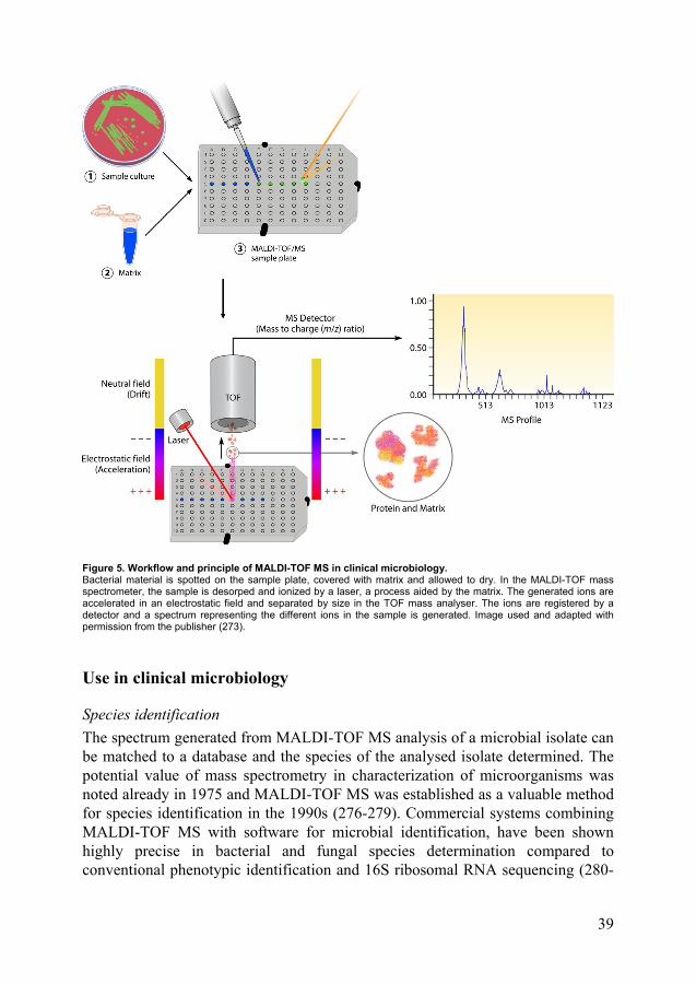
39
Figure 5. Workflow and principle of MALDI-TOF MS in clinical microbiology. Bacterial material is spotted on the sample plate, covered with matrix and allowed to dry. In the MALDI-TOF mass spectrometer, the sample is desorped and ionized by a laser, a process aided by the matrix. The generated ions are accelerated in an electrostatic field and separated by size in the TOF mass analyser. The ions are registered by a detector and a spectrum representing the different ions in the sample is generated. Image used and adapted with permission from the publisher (273).
Use in clinical microbiology
Species identification
The spectrum generated from MALDI-TOF MS analysis of a microbial isolate can be matched to a database and the species of the analysed isolate determined. The potential value of mass spectrometry in characterization of microorganisms was noted already in 1975 and MALDI-TOF MS was established as a valuable method for species identification in the 1990s (276-279). Commercial systems combining MALDI-TOF MS with software for microbial identification, have been shown highly precise in bacterial and fungal species determination compared to conventional phenotypic identification and 16S ribosomal RNA sequencing (280-

40
282). Due to its easy handling, swiftness and low per sample cost, MALDI-TOF MS is now a standard method for species identification in clinical microbiology laboratories (273, 281).
The technique was early shown to be potentially valuable in the difficult separation of species within the Haemophilus genus (283). More recent studies have demonstrated the instruments capability to differentiate H. influenzae from the commensals H. parainfluenzae and H. haemolyticus (13-16).
Subtyping
The possible use of MALDI-TOF MS for identification beyond the species level is intriguing, as it would allow the extraction of more diagnostic information without much increased use of time or resources. Several studies have been published suggesting the possibility to separate certain clones or groups of bacterial species. Typing by MALDI-TOF MS of the pathogen Clostridium difficile can be correlated to PCR ribotyping and used for surveillance of disease outbreaks. The method uses a different matrix and mass range (30,000-50,000 Da) than used for standard species identification to identify peaks correlating to S-layer proteins, which are usually analysed for ribotyping (284). Studies have also suggested the possible use of MALDI-TOF MS for the separation of different clonal groups of methicillin-resistant Staphylococcus aureus (MRSA) (285-289), extended spectrum beta-lactamase (ESBL)-producing and other subgroups of Enterobacteriaceae (290-292), and certain clones of Pseudomonas aeruginosa (293), among others.
Many studies, however, only compare a selected group of strains with known characteristics and lack testing and validation of the typing schemes on clinical isolates in a blinded fashion. There have also been concerns raised regarding the consistency and reproducibility of results from different studies (294, 295).
Antimicrobial susceptibility testing
MALDI-TOF MS has also been shown to be a possible tool for rapid detection of antimicrobial resistance.
The gene product of the psm-mec gene associated with the class A mec gene complex of MRSA and methicillin-resistant Staphylococcus epidermidis (MRSE) can be identified as a highly specific 2,415 m/z peak in MALDI-TOF MS analysis. This allows MALDI-TOF MS-based identification of MRSA and MRSE with a high positive predictive value (296, 297).
In another approach, the degradation products of carbapenems hydrolysed by carbapenemase-producing bacteria can be identified and the isolate classified as carbapenem susceptible or resistant. Variations of this method has been presented for Bacteroides fragilis, Pseudomonas aeruginosa, Enterobacteriaceae and Acinetobacter baumanni (298-303). These methods can, however, not identify non-enzymatic carbapenem resistance.

41
More general approaches to antimicrobial susceptibility testing using MALDI-TOF MS have also been investigated. In a recent study, droplets of blood culture specimens mixed with antibiotics were analysed by MALDI-TOF MS after a short incubation period. If the generated mass spectra could be used for successful species identification, the isolate of the culture was considered resistant to the added antibiotic. The method showed promising results compared to susceptibility testing by broth microdilution (304).

42

43
The present investigation
Aims
The aims of the thesis were:
• To investigate potential differences in MALDI-TOF mass spectra among different types of encapsulated H. influenzae and NTHi
• To investigate the capability of MALDI-TOF MS to separate Hib from non-type b H. influenzae.
• To develop a clinically applicable MALDI-TOF MS H. influenzae capsule typing method.
• To investigate the epidemiology and clinical characteristics of H. influenzae ST14CC-PBP3IIb/A, a clonal group of NTHi with non-beta-lactamase mediated beta-lactam resistance, in the county of Skåne.
• To compare benzylpenicillin to beta-lactams with established effect on H. influenzae as empirical treatment of lower respiratory tract infections caused by the species.

44
Methods, Results and Discussion
Paper I: Identification of H. influenzae type b by MALDI-TOF MS
Capsule typing of H. influenzae is traditionally made by conventional serotyping (SAST), but this method has limited specificity and misclassification of especially NTHi is common (6, 84-86). Typing with PCR is considered the gold standard and generates high quality results, but only addresses isolate genotype and not the phenotype (70, 87-89). Typing with PCR is also relatively time-consuming and laborious. Given the implementation of MALDI-TOF MS in many clinical laboratories for routine species identification and the genetically conserved capsule type specific lineages of H. influenzae, we sought to investigate the instruments capability to identify different capsule types of the species. In this study differences in mass spectra between Hib and the other capsule types of H. influenzae were investigated.
Methods
The culture collection of the study included Hib isolates (n=40) and H. influenzae isolates of all other capsule types (n=87). Most isolates were Swedish invasive isolates (n=114) but isolates from other countries, often representing rare capsule types, were also included. All isolates were capsule typed by PCR using bexB and type specific primers as previously described (70, 88, 89). Mass spectra were acquired for all isolates using a standard benchtop MALDI-TOF mass spectrometer. Mass spectra were manually analysed to investigate differences between Hib and non-Hib isolates. Selected isolates were further investigated by MLST.
Automatic Hib/non-Hib MALDI-TOF MS classification methods were constructed in ClinProTools and MALDI Biotyper using selected reference isolates. Classification methods relying on direct similarity with reference strains (MALDI Biotyper Main Spectra (MSPs) and subtyping MSPs) and methods classifying based on the absence or presence of specific peaks (ClinProTools) were constructed. Specially designed MALDI Biotyper MSPs, which included only two different mass peaks observed during manual analysis of mass spectra, were also constructed.
For validation of the methods isolates not included as reference isolates were used. Six spectra per isolate were acquired and classified by the methods. Isolates with ≥5/6 spectra classified as Hib were identified as Hib and isolates with ≤2/6 spectra classified as Hib were identified as non-Hib. If 3–4/6 spectra were classified as Hib, the isolate was identified as uncertain. To test the reproducibility of the results, mass spectra of 42 of the validation isolates (19 Hib and 23 non-Hib) were acquired on a different instrument and classified by the methods.
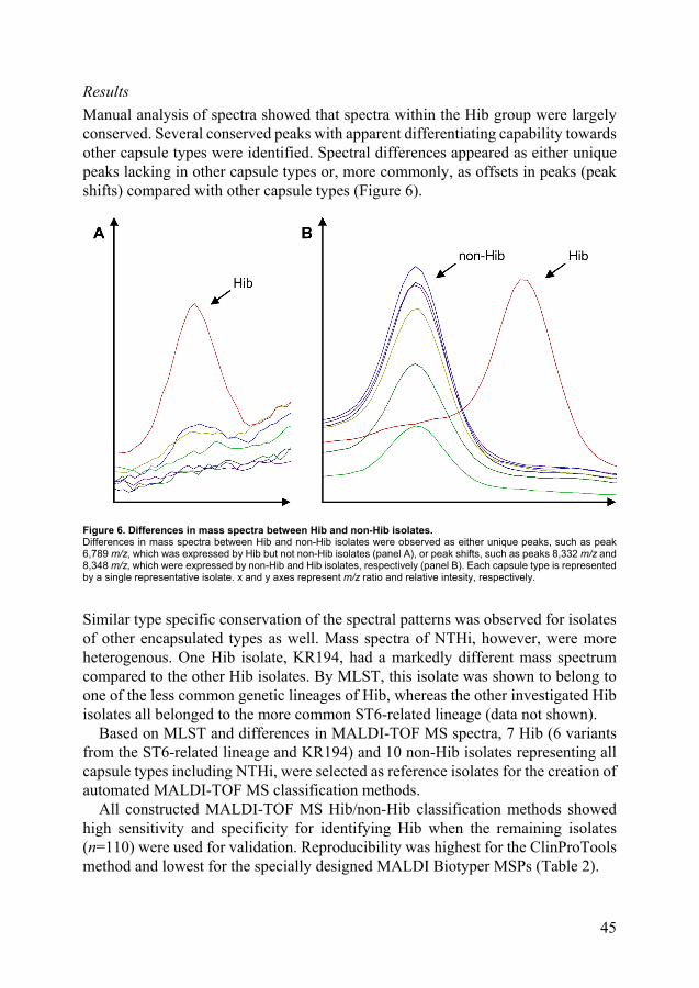
45
Results
Manual analysis of spectra showed that spectra within the Hib group were largely conserved. Several conserved peaks with apparent differentiating capability towards other capsule types were identified. Spectral differences appeared as either unique peaks lacking in other capsule types or, more commonly, as offsets in peaks (peak shifts) compared with other capsule types (Figure 6).
Figure 6. Differences in mass spectra between Hib and non-Hib isolates. Differences in mass spectra between Hib and non-Hib isolates were observed as either unique peaks, such as peak 6,789 m/z, which was expressed by Hib but not non-Hib isolates (panel A), or peak shifts, such as peaks 8,332 m/z and 8,348 m/z, which were expressed by non-Hib and Hib isolates, respectively (panel B). Each capsule type is represented by a single representative isolate. x and y axes represent m/z ratio and relative intesity, respectively.
Similar type specific conservation of the spectral patterns was observed for isolates of other encapsulated types as well. Mass spectra of NTHi, however, were more heterogenous. One Hib isolate, KR194, had a markedly different mass spectrum compared to the other Hib isolates. By MLST, this isolate was shown to belong to one of the less common genetic lineages of Hib, whereas the other investigated Hib isolates all belonged to the more common ST6-related lineage (data not shown).
Based on MLST and differences in MALDI-TOF MS spectra, 7 Hib (6 variants from the ST6-related lineage and KR194) and 10 non-Hib isolates representing all capsule types including NTHi, were selected as reference isolates for the creation of automated MALDI-TOF MS classification methods.
All constructed MALDI-TOF MS Hib/non-Hib classification methods showed high sensitivity and specificity for identifying Hib when the remaining isolates (n=110) were used for validation. Reproducibility was highest for the ClinProTools method and lowest for the specially designed MALDI Biotyper MSPs (Table 2).

46
Table 2. Evaluation of different Hib/non-Hib MALDI-TOF MS classfication methods. Classification method Hib
(n=33) Non-Hib (n=77)
Sensitivity (%)
Specificity (%)
Reproducibility (%)1
ClinProTools 100 98.7 97.6
Hib2 33 1
Uncertain3 0 0
Non-Hib4 0 76
Biotyper MSPs 100 98.7 90.5
Hib 33 0
Uncertain 0 1
Non-Hib 0 76
Biotyper subtyping MSPs 97 100 92.9
Hib 32 0
Uncertain 1 0
Non-Hib 0 77
Biotyper specially designed MSPs
81.8 100 76.2
Hib 27 0
Uncertain 3 0
Non-Hib 3 77 1. Defined as the percentage of isolates with mass spectra acquired at two different instruments (n=42) classified to the same group in both classifications. 2. ≥5/6 spectra classified as Hib. 3. 3–4/6 spectra classified as Hib. 4. ≤2/6 spectra classified as Hib.
Discussion
This study shows that Hib and non-Hib isolates of H. influenzae can be separated based on MALDI-TOF mass spectra. Furthermore, mass spectra were largely conserved within capsule types and other capsule types besides Hib appeared separable as well. Type specific differences observed in mass spectra were most often due to peak shifts, indicating amino acid variations in ribosomal proteins conserved within different capsule types. Unique peaks for Hib and other capsule types were also observed. Interestingly, one Hib isolate belonging to a different genetic lineage than the other Hib isolates had a markedly different mass spectrum. This indicates that the mass spectral differences between different capsule types are related to genetic differences between the isolates rather than differences in type specific capsule synthesis proteins, as expected.
Strengths of the study include the large and diverse culture collection and the careful selection and characterization of reference isolates used in the different Hib/non-Hib classification methods. The findings were robust, as indicated by the similar results obtained by the different Hib/non-Hib classification methods and the tests of reproducibility. One limitation in the study is that two genetic lineages of Hia and one of Hib were not represented. One Hib lineage included only one isolate, KR194, which was included as a reference isolate. The capability of MALDI-TOF MS to identify this lineage could hence not be tested.
In conclusion, this proof of concept study shows that MALDI-TOF MS can separate Hib from non-Hib isolates of H. influenzae with high accuracy.

47
Paper II: Capsule typing of H. influenzae by MALDI-TOF MS
In Paper I it was noted that other capsule types besides Hib appeared separable in a similar way as Hib, with conserved unique peaks and peak shifts compared to other capsule types. Furthermore, Hib strain KR194, which belonged to a different genetic lineage than the other Hib strains in the study, had a deviant MALDI-TOF mass spectrum. This indicated that differences in mass spectra were a result of differences in ribosomal protein content, mirroring genetic relatedness, rather than differences in capsule synthesis proteins themselves. Nontypeable isolates, as expected due to their high genetic variability, showed a much greater inter-strain variability in mass spectra compared to encapsulated isolates.
This study is a continuation of the work from Paper I. The aim was to construct and validate a clinically applicable capsule typing method including all capsule types based on MALDI-TOF MS analysis. Our view was that a large and comprehensive reference database encompassing all genetic lineages of encapsulated isolates would be a key factor for success. Furthermore, we investigated whether the observed differences in mass spectra between different capsule types were due to differences in capsule biosynthesis proteins or associated to the clonal population structure of encapsulated H. influenzae.
Methods
A culture collection comprising 258 H. influenzae from various geographical regions representing all capsule types including NTHi were included in the study. This evaluation set of isolates was used to further investigate the type specific differences in mass spectra observed in Paper I. Furthermore, isogenic capsule type a, b, c and d transformants originating from strain Rd were included (68, 305). A separate, second culture collection, consisting of Swedish invasive isolates (n=126), was used to validate the new MALDI-TOF MS capsule typing method developed.
Concatenated MLST sequences of all Hia and Hib isolates (of which there are more than one established genetic lineage) and several type c to f and nontypeable isolates were used to construct a maximum-likelihood phylogenetic tree.
Mass spectra of all isolates were acquired using a benchtop MALDI-TOF mass spectrometer. Six spectra per isolate were acquired. Isolates in the evaluation set and capsule transformants were investigated by principal component analysis (PCA) and biomarker analysis. Principal component analysis is a method to reduce factors when there is redundancy in the data. The analysis converts observations and correlated variables to principal components, which are ranked by the proportion of variance from the original data set they contain. Principal components, often the first three as they contain the most variance, can be used to easily visualize the data in a three-dimensional space. In biomarker analysis, biomarkers (m/z values) separating different groups in the data set are discovered.

48
A new MALDI Biotyper reference database for capsule typing was constructed with isolates from the evaluation set. The database was initially validated and supplemented using the remaining isolates in the evaluation set. After supplementation, a final validation was performed by classification of the separate culture collection of Swedish invasive isolates.
As all isolates were H. influenzae, high score values were expected for all MSPs regardless of capsule type. Therefore, each spectrum was classified according to the best matching MSP. For isolate classification, ≥5/6 spectra classified to the same type (a to f or NTHi) were required. If ≤4/6 spectra were classified to the same type, the isolate was classified as inconclusive. All MALDI-TOF MS typing results were compared to PCR capsule typing by bexB and type specific primers (70, 88, 89).
Results
Phylogenetic analysis of MLST data clearly illustrated the clonal population structure of encapsulated H. influenzae, with isolates of different capsule types belonging to different capsule type specific genetic lineages (Figure 7). All three described genetic lineages of Hib and two of the three major genetic lineages of Hia were represented. Isolates of the other capsule types belonged to their respective established genetic lineage, as expected. One isolate, KR1130, was typed as Hif according to PCR but did not sort with the other Hif isolates. This isolate was subsequently shown to harbour a non-expressed pseudogene cap locus, rendering a false positive PCR result. All capsule transformants belonged to the serotype d specific genetic lineage, as their parental strain Rd (98).
Figure 7. Phylogenetic analysis of H. influenzae isolates. The phylogentic relationship between isolates of the evaluation set in the study was investigated by MLST. All major genetic lineages of encapsulated H. influenzae, except the least common lineage of Hia, were represented in the collection. Isolate KR1130 did not belong to the established Hif lineage. Isolates included in the initial (1), and added to the supplemented (2), MALDI Biotyper typing database are indicated. A few NTHi which were misclassified in validation of the initial database could not be added to the database as they interfered with identification of Hie isolates. These isolates were included in the analysis and are indicated (*).

49

50
In the PCA NTHi formed a large heterogenous group, as expected (data not shown). Encapsulated isolates showed clustering according to capsule type and, for Hia and Hib isolates, genetic lineage. Isogenic capsule transformants clustered together, in proximity of Hid isolates, and were not distributed according to their respective capsule types (Figure 8).
Figure 8. Principal component analysis of mass spectra of encapsulated H. influenzae and isogenic capsule transformants. In the PCA, capsule type specific clustering was evident. Different genetic lineages of Hia and Hib isolates clustered separetaly. Isogenic capsule transformants clustered together and not with their respective capule type. Isolate KR1130 did not cluster with other Hif isolates.
The biomarker analysis revealed several mass peaks with separating capability between different capsule types and genetic lineages of Hia and Hib. The isogenic capsule transformants expressed similar peaks realtive to each other but differed in many peaks compared to wild-type isolates of the same capsule types (data not shown).
To construct a database for capsule typing in MALDI Biotyper, encapsulated isolates representing all capsule types and genetic lineages were selected (n=22). Nontypeable H. influenzae were included with the aim to include representatives of known genetic clades (n=9) (Figure 7) (100). Initial validation with the remaining isolates of the evaluation set (n=227) and capsule transformants (n=4) revealed perfect sensitivity for encapsulated isolates. Some NTHi, however, were misclassified as encapsulated which decreased specificity for encapsulated isolates.
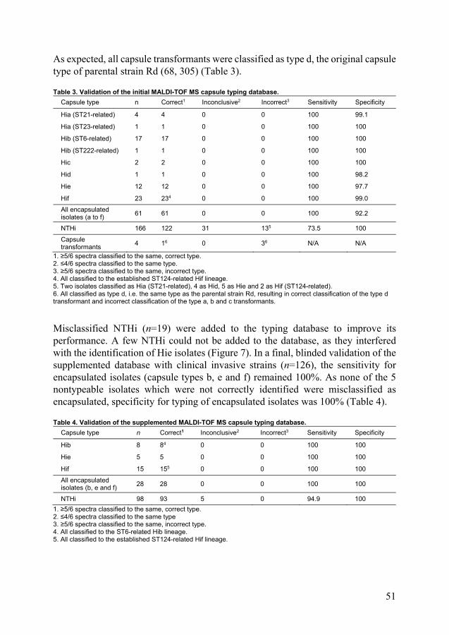
51
As expected, all capsule transformants were classified as type d, the original capsule type of parental strain Rd (68, 305) (Table 3).
Table 3. Validation of the initial MALDI-TOF MS capsule typing database.
Capsule type n Correct1 Inconclusive2 Incorrect3 Sensitivity Specificity
Hia (ST21-related) 4 4 0 0 100 99.1
Hia (ST23-related) 1 1 0 0 100 100
Hib (ST6-related) 17 17 0 0 100 100
Hib (ST222-related) 1 1 0 0 100 100
Hic 2 2 0 0 100 100
Hid 1 1 0 0 100 98.2
Hie 12 12 0 0 100 97.7
Hif 23 234 0 0 100 99.0
All encapsulated isolates (a to f)
61 61 0 0 100 92.2
NTHi 166 122 31 135 73.5 100
Capsule transformants
4 16 0 36 N/A N/A
1. ≥5/6 spectra classified to the same, correct type. 2. ≤4/6 spectra classified to the same type. 3. ≥5/6 spectra classified to the same, incorrect type. 4. All classified to the established ST124-related Hif lineage. 5. Two isolates classified as Hia (ST21-related), 4 as Hid, 5 as Hie and 2 as Hif (ST124-related). 6. All classified as type d, i.e. the same type as the parental strain Rd, resulting in correct classification of the type d transformant and incorrect classification of the type a, b and c transformants.
Misclassified NTHi (n=19) were added to the typing database to improve its performance. A few NTHi could not be added to the database, as they interfered with the identification of Hie isolates (Figure 7). In a final, blinded validation of the supplemented database with clinical invasive strains (n=126), the sensitivity for encapsulated isolates (capsule types b, e and f) remained 100%. As none of the 5 nontypeable isolates which were not correctly identified were misclassified as encapsulated, specificity for typing of encapsulated isolates was 100% (Table 4).
Table 4. Validation of the supplemented MALDI-TOF MS capsule typing database.
Capsule type n Correct1 Inconclusive2 Incorrect3 Sensitivity Specificity
Hib 8 84 0 0 100 100
Hie 5 5 0 0 100 100
Hif 15 155 0 0 100 100
All encapsulated isolates (b, e and f)
28 28 0 0 100 100
NTHi 98 93 5 0 94.9 100
1. ≥5/6 spectra classified to the same, correct type. 2. ≤4/6 spectra classified to the same type 3. ≥5/6 spectra classified to the same, incorrect type. 4. All classified to the ST6-related Hib lineage. 5. All classified to the established ST124-related Hif lineage.

52
Discussion
This study shows that following construction of a comprehensive database, MALDI-TOF MS can be utilized for capsule typing of H. influenzae.
The foremost strength of the study is the large collection of diverse strains collected at different times and from different regions, which ensure the robustness of the findings and the developed capsule typing method. By MLST, adequate coverage of different genetic lineages of encapsulated H. influenzae in the database was ensured. The database was furthermore carefully supplemented to reduce interference of the genetically heterogenous NTHi. Another major strength is the validation of the method using a collection of consecutive clinical isolates. One limitation is that no isolate in the culture collection belonged to the ST4-related genetic lineage of Hia and therefore could not be included in the typing database. For the least common genetic lineage of Hib (ST61-related) we only had access to one isolate which was included in the typing database.
Notably, isogenic capsule transformants clustered together in the PCA and showed similar peak patterns in biomarker analysis, confirming that differences in mass spectra is a result of general proteome differences related to genetic relationships rather than differences in capsule synthesis proteins. This notion is further supported by the separation of different genetic lineages of Hia and Hib isolates. The developed method relies on the clonal genetic population structure of encapsulated H. influenzae (97, 98). Each capsule type specific lineage is genetically relatively homogenous, which is reflected in the similarity of mass spectra of isolates within each lineage.
Compared to SAST and PCR typing, which identify the capsule polysaccharide and the cap locus, respectively, the indirect typing method of MALDI-TOF MS has both advantages and disadvantages. The diversity of NTHi constitutes a limitation of the typing method as complete coverage of the variations of NTHi of course cannot be guaranteed. Furthermore, a few NTHi were not added to the typing database as they interfered with identification of Hie isolates and a high sensitivity for encapsulated isolates was given priority to. These NTHi isolates were included in the phylogenetic analysis and shown to be closely related to the Hie genetic lineage (Figure 7). Due to the heterogeneity of NTHi, the specificity of the typing method for encapsulated isolates will not be perfect. This indicates that the method is mainly useful when the expected proportion of encapsulated isolates is substantial, such as in typing of isolates from sterile sites. Another potential drawback of the developed method is the possibility that new genetic lineages of encapsulated isolates, which the method would not identify, can occur. There is, however, little evidence that this has happened historically.
One advantage of MALDI-TOF MS typing is that identification of the genetic lineage of Hia and Hib isolates can be made simultaneously. Another advantage is that previously encapsulated capsule deficient strains, which might have lost their

53
capsule expression during infection or laboratory handling, are identified by the method (78, 305, 306).
A few studies have evaluated serotyping by MALDI-TOF MS of S. pneumoniae, for which there, similarly to H. influenzae, is a need of serotype surveillance. However, the results in these studies are less accurate compared to the accuracy for H. influenzae capsule typing in the current study (307, 308). The reason is likely the less clonal population structure of serotypes of S. pneumoniae, for which there exists more than 90 serotypes and serotype switching is not uncommon (309, 310). Subtyping by MALDI-TOF MS by separation of genetic lineages has also been investigated in other species, e.g. certain STs of E. coli and MRSA (285, 287, 288, 290, 292). For certain other species and subtyping the MALDI-TOF MS analysis is directed at certain proteins, such as the S-layer proteins of C. difficile (284).
In conclusion, Paper I and II show that MALDI-TOF MS can separate the different capsule types of H. influenzae. The capsule typing method developed in Paper II can become valuable as a rapid tool for typing of invasive H. influenzae and for surveillance of serotype distribution and Hib vaccination efficacy.
Paper III: Investigation on the epidemiology and clinical impact of a clonal group of NTHi with non-beta-lactamase mediated beta-lactam resistance
While the level of beta-lactamase mediated beta-lactam resistance has been relatively stable, non-beta-lactamase mediated resistance has increased in Europe during the last decades (121, 247, 260, 265, 266). Furthermore, isolates with non-beta-lactamase mediated resistance are often genetically related (121, 244, 247, 248, 256, 260, 267-270). Incidences of invasive NTHi disease appear to be increasing (205-208). The reason for this is not fully known, and NTHi disease severity has been considered largely host dependant (204). In a recent Swedish study however, one distinct clonal cluster (CC) of NTHi (ST14CC) with specific alterations in PBP3 (group IIb, type A (242, 245, 247)) was responsible for several cases of invasive disease (260). The same clonal group was also associated with a high hospitalization rate in a Norwegian study and has caused a disease outbreak at a Swedish nursing home (121, 311). Sequence type 14 (PBP3 amino acid substitutions not reported) has also been observed in Canada, Spain and Italy (267, 268, 312, 313).
The aim of this study was to investigate the epidemiology and clinical significance of this specific clonal group, denoted ST14CC-PBP3IIb/A, in Skåne, a county in southern Sweden.
Methods
For the years 2010-2012, all H. influenzae isolated from upper respiratory tract cultures in south Skåne (population approximately 500,000) and sterile sites from

54
all of Skåne (population approximately 1,200,000) were investigated. According to regional guidelines upper respiratory tract cultures are recommended in cases of recurrent AOM, treatment failure of AOM and CAP in adults. Cultures can also be obtained at the discretion of the referring physician.
The ST14CC-PBP3IIb/A clonal group was identified among isolates positive for screening of non-beta-lactamase mediated beta-lactam resistance by partial MLST. Initial screening of resistant isolates was made by sequencing of the adk gene, which appeared to have the highest discriminatory power for ST14 according to the H. influenzae MLST database (314). Isolates with adk 5 (specific adk allele of ST14) were further investigated by sequencing of recA and ftsI. In the H. influenzae MLST database all isolates with adk 5 and recA 5 are ST14 or single locus variants of ST14 (314). Deduced PBP3 amino acid sequences were compared with the amino acid sequence of H. influenzae Rd KW20 (GenBank accession number AAC22787). All isolates with adk 5, recA 5 and PBP3 group IIb, type A were considered part of the ST14CC-PBP3IIb/A clonal group. For confirmation, full MSLT was performed on a subgroup of randomly selected ST14CC-PBP3IIb/A isolates.
Minimum inhibitory concentrations for amoxicillin and cefotaxime were determined by gradient tests.
Cohorts of patients with disease caused by ST14CC-PBP3IIb/A, other NTHi with non-beta-lactamase mediated beta-lactam resistance and a control group of susceptible isolates, respectively, were identified. Data regarding age, sex, type of infection and hospitalization within five days of the culture date was retrieved from laboratory referrals and hospital records, and compared between the different cohorts. Significance in differences between groups were evaluated using the Pearson chi-squared test.
Results
Of respiratory tract isolates, 12.5% (400/3,192) were screening positive for non-beta-lactamase beta-lactam resistance and 360 were available for further typing. Of these isolates, 26.1% were identified as ST14CC-PBP3IIb/A. One isolate was excluded as it was encapsulated (capsule type e, PCR verified). Among invasive isolates, 13% (9/70) were screening positive for non-beta-lactamase mediated beta-lactam resistance. Of eight isolates available for typing two were ST14CC-PBP3IIb/A.
Although all ST14CC-PBP3IIb/A isolates had identical amino acid substitutions in PBP3, MICs determined by amoxicillin gradient test varied. Only 12% of non-beta-lactamase producing ST14CC/PBP3IIb/A isolates were classified as resistant according to EUCAST breakpoints. All but one ST14CC/PBP3IIb/A isolate were classified as susceptible to cefotaxime. Comparable proportions of other non-beta-lactamase producing beta-lactam resistant NTHi were classified as resistant to the tested agents (data not shown).
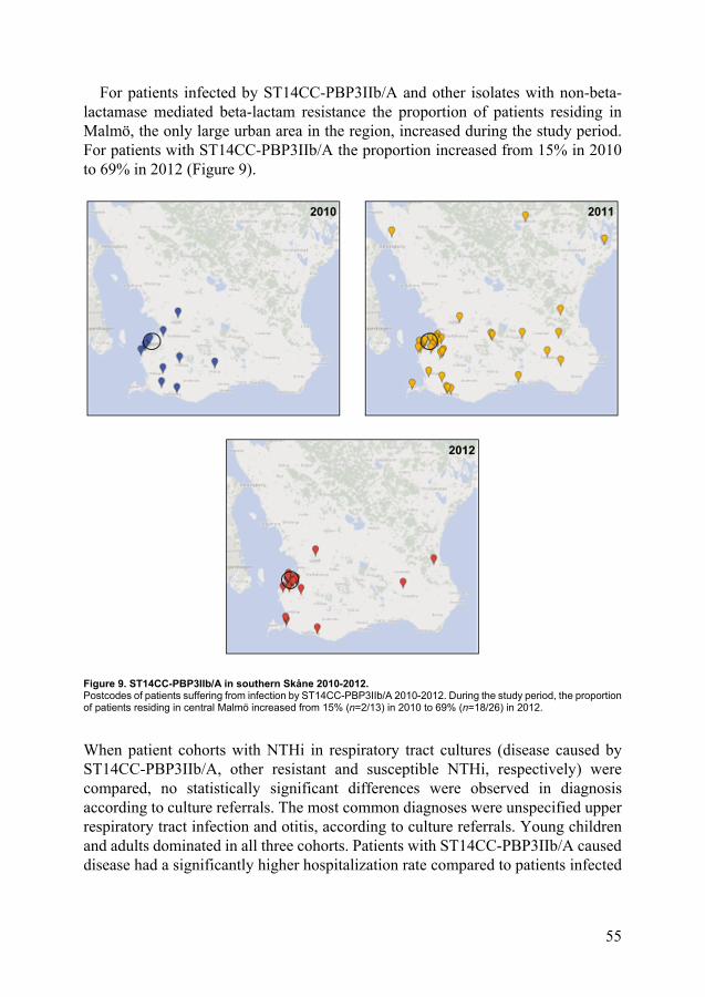
55
For patients infected by ST14CC-PBP3IIb/A and other isolates with non-beta-lactamase mediated beta-lactam resistance the proportion of patients residing in Malmö, the only large urban area in the region, increased during the study period. For patients with ST14CC-PBP3IIb/A the proportion increased from 15% in 2010 to 69% in 2012 (Figure 9).
Figure 9. ST14CC-PBP3IIb/A in southern Skåne 2010-2012. Postcodes of patients suffering from infection by ST14CC-PBP3IIb/A 2010-2012. During the study period, the proportion of patients residing in central Malmö increased from 15% (n=2/13) in 2010 to 69% (n=18/26) in 2012.
When patient cohorts with NTHi in respiratory tract cultures (disease caused by ST14CC-PBP3IIb/A, other resistant and susceptible NTHi, respectively) were compared, no statistically significant differences were observed in diagnosis according to culture referrals. The most common diagnoses were unspecified upper respiratory tract infection and otitis, according to culture referrals. Young children and adults dominated in all three cohorts. Patients with ST14CC-PBP3IIb/A caused disease had a significantly higher hospitalization rate compared to patients infected

56
by other non-beta-lactamase producing beta-lactam resistant NTHi and susceptible NTHi (Table 5). Furthermore, hospitalization appeared to more often be primarily related to the NTHi infection. The most common diagnosis for hospitalized ST14CC-PBP3IIb/A patients was pneumonia (data not shown).
All invasive disease caused by non-beta-lactamase producing beta-lactam resistant NTHi occurred in adults, of which 7 of 8 were above 65 years old. ST14CC-PBP3IIb/A accounted for two cases, one with pneumonia and one with cholangitis. All patients with disease caused by other non-beta-lactamase producing beta-lactam resistant NTHi suffered from pneumonia. One patient from each group died.

57
Tab
le 5
. C
lin
ical
dat
a o
f p
atie
nts
wit
h S
T14
CC
-PB
P3I
Ib/A
co
mp
ared
to
pat
ien
ts w
ith
oth
er n
on
-bet
a-la
ctam
ase
pro
du
cin
g b
eta-
lact
am r
esis
tan
t an
d s
usc
epti
ble
NT
Hi.
Pat
ient
co
hort
(n)
S
uspe
cted
dia
gnos
is/s
ite o
f inf
ectio
n1 (%
) A
ge in
yea
rs (
%)
H
ospi
tal-
ized
pa
tient
s (%
)
Uns
peci
fied
UR
I2
Ear
S
inus
E
ye
LRI3
Oth
er
Un-
know
n <
1 1-
5 6-
20
21-
65
≥66
ST
14C
C-
PB
P3I
Ib/A
(8
3)
17
(20
) 20
(2
4)
7 (8
) 12
(1
4)
9 (1
1)
0 18
(22
) 14
(1
7)
35
(42)
7
(8)
23
(28)
4 (5)
14
(17
)4
Oth
er
resi
stan
t N
TH
i (2
55)
70 (
28)
44
(17)
19
(7)
36
(1
4)
20 (
8)
4 (2
) 62
(24
) 36
(1
4)
78
(31)
21
(8
) 96
(3
8)
24
(9)
21
(8.
2)
Sus
cept
i-bl
e N
TH
i (1
92)
70
(36
) 29
(1
5)
6 (3
) 9
(5)
14 (
7)
4 (2
) 60
(31
) 25
(1
3)
74
(39)
24
(1
3)
56
(29)
13
(7
)
13 (
6.8)
1. A
ccor
ding
to c
ultu
re r
efer
ral.
2. U
pper
res
pira
tory
tra
ct in
fect
ion
3. L
ower
res
pira
tory
tra
ct in
fect
ion
4. S
tatis
tical
ly s
igni
fican
t di
ffere
nce
com
pare
d to
pat
ient
s w
ith o
ther
non
-bet
a-la
ctam
ase
prod
ucin
g be
ta-la
ctam
res
ista
nt N
TH
i (R
R 2
.0 (
95%
CI
1.1-
3.8)
, p=
0.02
5) a
nd
susc
eptib
le N
TH
i (R
R 2
.5 (
95%
CI
1.2-
5.0)
, p=
0.01
.

58
Discussion
This study demonstrates that a single clonal group NTHi accounted for one quarter of isolates with non-beta-lactamase mediated beta-lactam resistance in the investigated geographical region and spread dynamically during the study period. Furthermore, the clonal group was associated with a higher risk of patient hospitalization compared to both other resistant and susceptible isolates, indicating enhanced virulence properties.
The major strength of the study is the large and near-complete collection of isolates and cases studied. A potential limitation is the partial MLST scheme used to identify clonal isolates, which may result in minor differences in the number of isolates identified as ST14CC-PBP3IIb/A compared to full MLST. Furthermore, the disk used for initial screening for beta-lactam resistance in the clinical laboratory was changed from phenoxymethylpenicillin 10 µg to benzylpenicillin 1 unit during the study period (early 2011), which may affect the number of isolates classified as resistant (257). Given the short time period of the study data regarding temporal variations should be interpreted with caution.
The findings in this study agree well with previous reports which have associated this clonal group to disease outbreaks, invasive disease and high rates of hospitalization (121, 260, 311). The current study however includes a larger sample of both ST14CC-PBP3IIb/A strains and controls, making comparison of clinical data more reliable. Since publication of this study, ST14 (PBP3 amino acid substitutions not reported) has also been found to be the most common ST among invasive NTHi in an Italian surveillance study (315).
In a study of NTHi isolated from COPD patients, a specific clonal group (ST159) was identified in approximately 10% of cases (316). The clonal group (ST159) was, however, not associated with increased risk of COPD exacerbations or prolonged colonization. The study was smaller compared to the present study, including a total of 134 strains. No data on antimicrobial susceptibility or PBP3 amino acid substitutions was reported . In a study regarding the genetic population structure of NTHI including otitis media and commensal isolates, two different genetic clusters contained only disease causing and no commensal strains. The study however included no further clinical data (317). Other studies have linked genetically related groups of NTHi to different potential virulence factors, but not specific clinical infections or disease severity (43, 100, 102). Thus, previous studies have indicated that despite the large heterogeneity of NTHi isolates, clonal groups with specific virulence traits exist. The current study, in contrast to the mentioned studies on COPD and otitis media patients, included all respiratory tract isolates, regardless of patient diagnosis. We found no specific association to the type of infection for ST14CC-PBP3IIb/A, but instead an increased patient hospitalization rate, indicating increased clinical virulence.
In laboratory studies, increased invasion rates into epithelial cells have been shown in isolates with altered PBP3 (318, 319). There was however a high variation

59
within the group, and the altered PBP3 in itself does not appear to mediate the effect (319). Genotypic characterization was not performed in these studies and it is possible that clonal isolates with common virulence factors generated the observed difference in invasiveness.
Most ST14CC-PBPIIb/A isolates in the current study were classified as susceptible to both amoxicillin and cefotaxime. The number of phenotypically amoxicillin resistant isolates might be underestimated, as gradient tests were used for MIC determination (257). The results highlight the difficulties in classification of non-beta-lactamase producing beta-lactam resistant isolates and the range of amoxicillin and cefotaxime MICs within this clonal group with identical PBP3 underlines that other mechanisms than PBP3 alterations affect beta-lactam susceptibility.
In summary, although NTHi are genetically heterogenous, the investigated clonal group ST14CC-PBP3IIb/A appears to be widespread and associated with increased clinical virulence. This notion is supported by the current, previous and subsequent studies of the clonal group (121, 260, 311, 315).
Paper IV: Retrospective comparison of benzylpenicillin and wide-spectrum beta-lactams as empirical treatment of lower respiratory tract infections caused by H. influenzae In Sweden, benzylpenicillin is recommended as empirical treatment for mild to moderate CAP in adult patients requiring hospitalization (130). Considering benzylpenicillin’s debated effect on H. influenzae and the suggested increased proportion of cases of CAP caused by the species there is a need to assess the outcome of empirical treatment of benzylpenicillin on lower respiratory infections subsequently shown to be caused by H. influenzae (136, 232). This is further underlined by the increase in Europe in non-beta-lactamase mediated beta-lactam resistance, which might affect benzylpenicillin susceptibility (121, 247, 260, 265, 266). Importantly, in cases shown to be caused by H. influenzae definitive treatment with an agent with clinical breakpoints against the species should be given (130).
This retrospective study compared the outcomes of hospitalized patients with lower respiratory tract infections caused by H. influenzae who were empirically treated with either benzylpenicillin or a wide spectrum beta-lactam (WSBL) with established effect against H. influenzae.
Methods
Cases were identified from the records of the clinical microbiological laboratory in Skåne and included only patients ≥18 years of age. Two case definitions were used: [1] bacteraemia with H. influenzae and a lower respiratory tract infection during 1997-2016 or [2] pure culture of H. influenzae in respiratory tract samples in patients
60
hospitalized for lower respiratory tract infections during 2015-2016. Cases according to definition [2] were excluded if any other potential respiratory pathogen was recorded. Data was also retrieved regarding beta-lactam resistance, which is routinely investigated for.
Descriptive and clinical data was collected from patient records. Descriptive data included age and sex. Comorbidities were registered according to the Charlson comorbidity index (CCI) (320). Immunosuppression was registered and defined as ongoing primary immune deficiency or immunosuppressive therapy not denoted in the CCI. Clinical data recorded included sepsis severity score, maximum C-reactive protein (CRP) levels, CRB-65 score and admittance to an intensive care unit (ICU) (321). All cause 30-day mortality, 30-day hospital readmission rates and early clinical response rates were used as outcomes. Early clinical response was defined as no signs of fever, tachycardia, hypotension, hypoxemia or tachypnoea on day 4 after hospital admission (322). If complete data on all parameters regarding early clinical response was not available in records, they were complemented by an assessment whether a substantial general improvement in the patient’s condition had occurred.
Cases were sorted into patients receiving empirical treatment with benzyl-penicillin, any other beta-lactam antibiotic with established clinical breakpoints against H. influenzae and other empirical treatment regimens. Patients receiving more than one antimicrobial agent active against H. influenzae as empirical therapy were excluded. Outcomes were compared between the first two patient groups by logistic regression and comparison of propensity score matched cohorts. Multivariate logistic regression models were fitted using the purposeful selection algorithm (323).
Since the purposeful selection algorithm potentially risks to exclude covariates of intermediate value and since the study groups were assumed to be inherently different due to indication bias, a propensity-matched cohort was also created and analysed. Propensity scores were calculated in a logistic regression using treatment group (benzylpenicillin or a WSBL) as outcome and included categorized age, sex, ICU care, maximum levels of C-reactive protein (CRP), CCI, bacteraemia, immune suppression not denoted in the CCI and sepsis severity score as covariates. The CRB-65 scores were not included in propensity score calculation due to a high number of missing values. A 1-1 nearest neighbour matching without replacement with a caliper of 0.2 was performed and the balance in the covariates in the matched cohort was verified. Associations between treatment and outcomes in the matched cohort were assessed with full cohort logistic regression and conditional logistic regression on matched pairs.
Results
From blood and respiratory cultures 140 and 341 cases of lower respiratory tract infection caused by H. influenzae were identified, respectively. The median age was

61
75 years (interquartile range 66-84 years). The most common diagnosis was CAP (n=418, 87%). The all-cause 30-day mortality was 9%.
Empirical monotherapy treatment was given with benzylpenicillin in 199 cases and a WSBL in 213 cases. The most common WSBLs were cefotaxime (76%, n=162), piperacillin-tazobactam (8.5%, n=18) and cefuroxime (7.5%, n=16). Between the benzylpenicillin and WSBL treatment group there was significant differences (p≤0.05) regarding CCI, the proportions of patients given ICU care, the proportion of patients with bacteraemia and sepsis severity. The mortality was 5% and 13% in the benzylpenicillin and WSBL treatment group, respectively. In univariate logistic regression, WSBL treatment was associated with an increased mortality. The difference between treatment groups was however not significant in a multivariate model, adjusted for age, sepsis severity and CCI score (Table 6)
Similarly, when including only bacteraemic patients (n=120), a significantly increased mortality was noted for patients given empirical treatment with a WSBL in univariate regression (odds ratio (OR) 5.55, 95% confidence interval (CI) 1.20-25.7, p=0.028), but not in multivariate regression adjusted for age and CCI (OR 4.86, 95% CI 0.98-24, p=0.054).
No statistically significant differences regarding 30-day readmission rate in univariate or multivariate regression were observed. Although rates of early clinical response were high in both treatment groups, there was a significantly increased rate in the WSBL treatment group in multivariate regression (Table 6).
Table 6. Univariate and multivariate logisitc regression comparing WSBL treatment and benzylpenicillin treatment of H. influenzae lower respiratory tract infections.
Outcome WSBL treatment univariate OR1 (95% CI)
p WSBL treatment multivariate OR1 (95% CI)
p
30-day all cause mortality
2.59 (1.25-5.35) 0.010 2.03 (0.91-4.50)2 0.082
30-day readmission 1.23 (0.72-2.13) 0.45 1.16 (0.66-2.05)3 0.61
Early clinical response
1.28 (0.76-2.16) 0.35 2.28 (1.21-4.31)2 0.011
1. Benzylpenicillin treatment reference. 2. Adjusted for age, CCI category and sepsis severity. 3. Adjusted for age and CCI category.
The construction of a propensity score matched cohort resulted in the omission of many of the more severely ill patients (CRB-65≥3, ICU care, severe sepsis or septic shock). These patients were primarily found in the WSBL treatment group, which is reasonable as benzylpenicillin is not recommended as empirical therapy in these cases. The matched cohort consisted of 151 patients in each treatment group. Results regarding the different outcomes in the analyses of the propensity score matched cohort resembled those obtained from multivariate logistic regression. Treatment with a WSBL was associated with a higher, although not significantly higher, mortality (OR 2.14, 95% CI 0.93-4.92, p=0.075 and OR 1.89, 95% CI 0.84-4.23, p=0.12 for the full cohort and in conditional regression, respectively). Similarly, a

62
higher rate of early clinical response for patients treated with WSBLs was observed in this cohort both in full cohort logistic regression (OR 2.14, 95% CI 1.07-4.27, p=0.031) and conditional logistic regression (OR 2.5, 95% CI 1.20-5.21, p=0.014). There was no difference between treatment groups regarding hospital readmission.
Potential effect modification of treatment by isolates positive for beta-lactam resistance (beta-lactamase producing and non-producing) was investigated by comparison of odds ratios for the different outcomes stratified by treatment group and resistance mechanism between the propensity score matched cohorts. Significant effect modification was observed for beta-lactamase production regarding early clinical response and treatment group (interaction term for beta-lactamase × treatment group (benzylpenicillin as reference); β=3.12, 95% CI 0.82-5.43, p=0.008), with a lesser extent of early clinical response in the benzylpenicillin group compared to the WSBL group in patients infected by beta-lactamase producing isolates (Table 7).
Table 7. Effect modification by isolate beta-lactamase production on early clinical response in the propensity score matched cohort.
Isolate beta-lactamase production
Benzylpenicillin treatment WSBL treatment OR within strata of beta-lactamase production, (95% CI), p
n with early clinical response (%)
OR (95% CI), p
n with early clinical response (%)
OR (95% CI), p
No beta-lactamase production
109/125 (87.2%)
Reference 107/120 (89.2%)
1.21 (0.55-2.63), p=0.63
1.21 (0.55-2.63), p=0.63
Beta-lactamase production
10/21 (48%) 0.13 (0.05-0.36), p<0.001
25/26 (96%) 3.67 (0.46-29.0), p=0.22
27.5 (3.13-242), p=0.003
No similar effect modification was seen for non-beta-lactamase beta-lactam resistant isolates regarding early clinical response (data not shown). No significant effect modification was observed for any resistance mechanism regarding the other outcomes (data not shown).
Discussion
In this retrospective study of lower respiratory tract infections caused by H. influenzae, treatment with benzylpenicillin was not associated with a significantly increased risk of mortality or hospital readmission compared to treatment with a WSBL. Even though the rates of early clinical response were generally high, empirical treatment with benzylpenicillin was associated with a significantly lower rate of early clinical response compared to treatment with a WSBL. This difference was to a large extent explained by a lower response rate in patients infected with beta-lactamase producing isolates.
One major strength of this study is the relatively large size of the cohort, which was constructed in an un-biased way by identification of cases from culture findings.

63
The data has been thoroughly analysed and the results are robust, as shown by the similar results attained by both logistic regression and propensity score matching of the treatment groups.
One limitation of the study is the risk of indication bias due to its retrospective design. Possibly because of this, WSBL treatment was associated with worse outcomes in the crude models. The models were adjusted for risk factors of worse outcomes (such as comorbidities) using the purposeful selection algorithm to adjust for possible confounding, and this reduced the association. However, it still bordered significance, indicating possible residual confounding not adjusted for. Using another algorithm for variable selection more variables could have been included in the final models and possibly reduce this confounding. This would, however, have increased the risk of overfitting, especially as the numbers of the different outcomes were relatively low. Instead, the data was also investigated by creating a propensity score matched cohort based on regression analysis including several more variables. Furthermore, due to the limitations of diagnostic tools used for determination of CAP aetiology, it is not possible to know for sure that all cases included in the study had infections attributed to H. influenzae although the study method carefully tried to address this. Hopefully, implementation of modern molecular methods, such as quantitative PCR, can improve the diagnostic yield in the future (136, 137). Another limitation of the study is its limited power, due to the relatively low outcome rates. For mortality, which was 14% in the WSBL group, the study has power to detect an OR of approximately 2 (given α=0.05, β=0.8 and a two-sided test of significance).
Benzylpenicillin has been compared to treatment with WSBLs in patients with Haemophilus spp. bacteraemia in a previous retrospective study (233). In the study, definite (as opposed to empirical) treatment with benzylpenicillin compared to treatment with an aminopenicillin or cefuroxime was associated with an increased 30-day mortality for patients with H. influenzae bacteraemia. There are notable differences between the previous and the current study. The previous study investigated bacteraemic patients regardless of infection focus, as opposed to only respiratory tract infections. In the benzylpenicillin treatment group of the previous study, more than 30% had an unknown site of infection. The overall mortality was also considerably higher, 22% compared to 9%. Furthermore, several patients in the benzylpenicillin group hade polymicrobial infections and many patients received combination treatment. Adjustment for potential confounders was made only in separate bivariate regressions for each covariate. These factors, separate or in combination, could reasonably explain the previous study’s different results compared to the current.
In another recent retrospective study, treatment with phenoxymethylpenicillin or benzylpenicillin was compared to WSBL treatment in patients with CAP and a CRB-65 score ≤2 (134). The study found, similarly to the current study, no differences regarding 30-day or 90-day mortality between the treatment groups.

64
Treatment with WSBLs was associated with ICU care, possibly due to similar indication bias as in our study. The study included cases of pneumonia regardless of causative organism. H. influenzae accounted for 7.1% of cases in patients with CRB-65 score ≤1, and apparently differences in mortality among these patients were (at least) not large enough to significantly affect mortality in the different treatment groups in total.
Different dosing regimens might affect the effectiveness of benzylpenicillin. In this study, 1 g t.i.d. (40% of cases) and 3 g t.i.d. (49% of cases) were the most used regimens. Monte Carlo simulations calculated by EUCAST suggest markedly lower attainment rates of a time above a concentration of 1 mg/L (ECOFF for H. influenzae) for at least 30% or 40% of the dosing interval if the former dosing regimen is used (232). In patients receiving high dose (3 g t.i.d. or higher) and low dose (1 g t.i.d. or lower) treatment the mortality was 3% (n=4 of 116) and 8% (n=7 of 93), respectively. Statistical comparison between the groups was not made in the study due to a too small number of events.
The lower rate of early clinical response in patients infected by beta-lactamase producing isolates treated with benzylpenicillin is understandable as H. influenzae beta-lactamases are active against benzylpenicillin but not cefotaxime, piperacillin/tazobactam and cefuroxime, which were the most used agents in patients treated with WSBLs (234).
In conclusion, the results from this study suggest that empirical treatment with benzylpenicillin in patients with H. influenzae caused respiratory tract infections of mild to moderate severity is not associated with increased mortality or risk of hospital readmission. To fully determine optimal empirical treatment of these infections prospective randomized controlled trials are needed.
Ethical considerations
Papers I and II did not involve human subjects and no ethical permit was needed. Papers III and IV involved data on human subjects and ethical permits were required. The studies were approved by the Regional Ethical Review Board at Lund University (registration number 2014/533 and 2016/743 with addendum 18-391, respectively). All patient data in the studies was collected in retrospect and no interventions in the treatment given to patients were made. All data was anonymized and presented at group level in the published papers.

65
Concluding remarks and future perspectives
Despite the success of Hib childhood vaccination programmes, H. influenzae still is a major cause of disease in humans. With increasing incidences of invasive NTHi disease and remaining sporadic cases of invasive Hib disease, surveillance of invasive H. influenzae disease and Hib vaccination efficacy is still highly important. The capsule typing method developed in Paper II showed highly accurate results for encapsulated isolates. Future work will concern the further evaluation and implementation of the method in clinical laboratories, where the method can be valuable for rapid and cost-effective capsule type surveillance of invasive H. influenzae.
Typing of other bacterial species by MALDI-TOF MS is another research area which warrants further study. The possibility of typing by the instrument could be helpful in areas such as infection prevention and control. Although previous studies have investigated typing of bacterial strains with antimicrobial resistance, this form of MALDI-TOF MS typing is to a large extent not yet in clinical use. Development of clinically applicable typing methods in these cases could be highly useful.
As NTHi now are the most prevalent cause of both mucosal and invasive H. influenzae infections, future research will naturally focus on these isolates. The complex system of bacterial and host factors leading to NTHi disease is not fully understood. Paper III and previous studies have indicated ST14CC-PBP3IIb/A to be a clonal group of NTHi with enhanced virulence traits. The bacterial factors conferring this virulence have, however, not yet been investigated and poses an interesting area of further research. Given the rapid development of genetic sequencing methods and the possibilities to handle large amounts of genomic data, this could be a possible initial approach for investigation. Severe H. influenzae disease occurs only in a minority of patients colonized with the bacterium. Besides bacterial characteristics, host factors play a large role. It is, however, not entirely clear which factors contribute most to the risk of severe disease. Further investigations including both bacterial factors and patient risk factors are required to understand this interplay better. With increasing research focus on the development of an NTHi vaccine, studies in these areas could be of high value both for vaccine development and for information on which patients who would benefit most from such vaccination.

66
The increase in non-beta-lactam mediated beta-lactam resistance in H. influenzae is worrying. Although Paper IV provided no evidence for treatment failures with benzylpenicillin resulting in severe outcomes in patient with lower respiratory tract infections as a result of this kind of beta-lactam resistance, the issue should be taken seriously. Understanding this type of resistance is important to facilitate actions against further increase in resistance prevalence and could potentially be a source of new antimicrobial therapies.

67
Acknowledgements
I would like to express my sincere gratitude to all who have helped and supported me during my doctoral studies. In particular, I want to thank:
Kristian Riesbeck, my supervisor. You have guided and supported me throughout my studies. Without your enthusiasm and optimism my doctoral studies would never have started, and without your scientific skill and knowledge in the field they surely would not have been finished.
Fredrik Resman, my co-supervisor. We have worked closely during these years and you have been the architect of many of the studies. Your combination of the highest level of knowledge (almost regardless of subject), brightness and humbleness is inspiring.
Kristian and Fredrik, for not only being excellent supervisors but also reliable friends.
Johan Tham, my co-supervisor. Although your role in my doctoral studies was not formalized until late, you have been of great support and have provided insightful help. (You also got us tickets to NBA basketball in New Orleans which would have been enough in itself).
Bo Nilson, for introducing me to MALDI-TOF MS and for fruitful cooperation. John Thegerström, for letting me on board Paper IV and for pleasant
collaboration. Marta Brant and Emma Mattsson, for kindness and skillful help at the lab. Other current and previous members of the lab: Yu-Ching Su, Magnus Paulsson,
Birendra Singh, Tamim Al-Jubair, Nils Littorin, Corinna Richter, Fabian Uddén, Oskar Thofte, Sujinna Lekmeechai, Guillaume Manat, Birgitta Andersson, Ylva Mattisson, Victor Lindh, Maria Paul, Alfonso Felipe-Lopez, Ben Duell, Oindrilla Mukherjee, Kerstin Norrman, Petra Halang, Therese Nordström, Farshid Jalalvand and everyone else for making time in the lab so enjoyable.
My co-authors, Markus Kostrzewa, Dagfinn Skaare, Janet Gilsdorf, Gunnar Kahlmeter, Mogens Kilian and Simon Kroll, for invaluable contributions.
Per Björkman and Johan Malm, for constructive discussions during my half-time review.
The staff at Clinical Microbiology, Labmedicin Skåne for important help with strains, reagents and all other sorts of practicalities.

68
Colleagues during my internship, at the Department of Internal Medicine and at the Department of Infectious Diseases, all at Skåne University Hospital, Malmö, for friendship and encouragement.
Friends, for being precisely that. Bengt and Pernilla, my parents, and the rest of my family, Karl, Erik, Kersti,
Anders, Margareta, Petter, Anna, Oskar, Ebba, Alice, Annika, Simon, Cally and my grandmother Astrid, for limitless support.
Jenny, my wife, life companion and best friend, for being there both in good times and bad times. I love you and I am so happy and grateful for sharing this life with you.
69
References
1. Pfeiffer R. I.-Preliminary Communication on the Exciting causes of Influenza. Br Med J. 1892;1(1620):128.
2. Olitsky PK, Gates FL. Experimental Studies of the Naspharyngeal Secretions from Influenza Patients : I. Transmission Experiments with Nasopharyngeal Washings. J Exp Med. 1921;33(2):125-45.
3. Smith W, Andrewes CH, Laidlaw PP. A Virus Obtained form Influenza Patients. The Lancet. 1933;222(5732):66-8.
4. Winslow CE, Broadhurst J, Buchanan RE, Krumwiede C, Rogers LA, Smith GH. The Families and Genera of the Bacteria: Final Report of the Committee of the Society of American Bacteriologists on Characterization and Classification of Bacterial Types. J Bacteriol. 1920;5(3):191-229.
5. Peltola H. Worldwide Haemophilus influenzae type b disease at the beginning of the 21st century: global analysis of the disease burden 25 years after the use of the polysaccharide vaccine and a decade after the advent of conjugates. Clin Microbiol Rev. 2000;13(2):302-17.
6. Pittman M. Variation and type specificity in the bacterial species Haemophilus influenzae. J Exp Med. 1931;53(4):471-92.
7. Fleming A. On the Antibacterial Action of Cultures of a Penicillium, with Special Reference to their Use in the Isolation of B. influenzæ. Br J Exp Pathol. 1929;10(3):226-36.
8. Fleischmann RD, Adams MD, White O, Clayton RA, Kirkness EF, Kerlavage AR, et al. Whole-genome random sequencing and assembly of Haemophilus influenzae Rd. Science. 1995;269(5223):496-512.
9. Mandell GL, Douglas RG, Bennett JE, Dolin R. Mandell, Douglas, and Bennett's principles and practice of infectious diseases. New York: Elsevier/Churchill Livingstone; 2005.
10. Davis DJ. Food Accessory Factors (Vitamins) in Bacterial Culture with Especial Reference to Hemophilic Bacilli I. The Journal of Infectious Diseases. 1917;21(4):392-703.
11. Evans NM, Smith DD, Wicken AJ. Haemin and nicotinamide adenine dinucleotide requirements of Haemophilus influenzae and Haemophilus parainfluenzae. J Med Microbiol. 1974;7(3):359-65.
12. Murphy TF, Brauer AL, Sethi S, Kilian M, Cai X, Lesse AJ. Haemophilus haemolyticus: a human respiratory tract commensal to be distinguished from Haemophilus influenzae. J Infect Dis. 2007;195(1):81-9.
13. Frickmann H, Christner M, Donat M, Berger A, Essig A, Podbielski A, et al. Rapid discrimination of Haemophilus influenzae, H. parainfluenzae, and H.

70
haemolyticus by fluorescence in situ hybridization (FISH) and two matrix-assisted laser-desorption-ionization time-of-flight mass spectrometry (MALDI-TOF-MS) platforms. PLoS One. 2013;8(4):e63222.
14. Bruin JP, Kostrzewa M, van der Ende A, Badoux P, Jansen R, Boers SA, et al. Identification of Haemophilus influenzae and Haemophilus haemolyticus by matrix-assisted laser desorption ionization-time of flight mass spectrometry. Eur J Clin Microbiol Infect Dis. 2014;33(2):279-84.
15. Chen JHK, Cheng VCC, Wong CP, Wong SCY, Yam WC, Yuen KY. Rapid Differentiation of Haemophilus influenzae and Haemophilus haemolyticus by Use of Matrix-Assisted Laser Desorption Ionization-Time of Flight Mass Spectrometry with ClinProTools Mass Spectrum Analysis. J Clin Microbiol. 2017;55(9):2679-85.
16. Zhu B, Xiao D, Zhang H, Zhang Y, Gao Y, Xu L, et al. MALDI-TOF MS distinctly differentiates nontypable Haemophilus influenzae from Haemophilus haemolyticus. PLoS One. 2013;8(2):e56139.
17. Kilian M. A taxonomic study of the genus Haemophilus, with the proposal of a new species. J Gen Microbiol. 1976;93(1):9-62.
18. DeMuri GP, Gern JE, Eickhoff JC, Lynch SV, Wald ER. Dynamics of Bacterial Colonization With Streptococcus pneumoniae, Haemophilus influenzae, and Moraxella catarrhalis During Symptomatic and Asymptomatic Viral Upper Respiratory Tract Infection. Clin Infect Dis. 2018;66(7):1045-53.
19. Bae S, Yu JY, Lee K, Lee S, Park B, Kang Y. Nasal colonization by four potential respiratory bacteria in healthy children attending kindergarten or elementary school in Seoul, Korea. J Med Microbiol. 2012;61(Pt 5):678-85.
20. Maglione M, Bush A, Nielsen KG, Hogg C, Montella S, Marthin JK, et al. Multicenter analysis of body mass index, lung function, and sputum microbiology in primary ciliary dyskinesia. Pediatr Pulmonol. 2014;49(12):1243-50.
21. Schneerson R, Rodrigues LP, Parke JC, Jr., Robbins JB. Immunity to disease caused by Hemophilus influenzae type b. II. Specificity and some biologic characteristics of "natural," infection-acquired, and immunization-induced antibodies to the capsular polysaccharide of Hemophilus influenzae type b. J Immunol. 1971;107(4):1081-9.
22. Rosell A, Monso E, Soler N, Torres F, Angrill J, Riise G, et al. Microbiologic determinants of exacerbation in chronic obstructive pulmonary disease. Arch Intern Med. 2005;165(8):891-7.
23. Skevaki CL, Tsialta P, Trochoutsou AI, Logotheti I, Makrinioti H, Taka S, et al. Associations Between Viral and Bacterial Potential Pathogens in the Nasopharynx of Children With and Without Respiratory Symptoms. Pediatr Infect Dis J. 2015;34(12):1296-301.
24. Duell BL, Su YC, Riesbeck K. Host-pathogen interactions of nontypeable Haemophilus influenzae: from commensal to pathogen. FEBS Lett. 2016;590(21):3840-53.
25. Jalalvand F, Riesbeck K. Haemophilus influenzae: recent advances in the understanding of molecular pathogenesis and polymicrobial infections. Curr Opin Infect Dis. 2014;27(3):268-74.

71
26. Jalalvand F, Riesbeck K. Update on non-typeable Haemophilus influenzae-mediated disease and vaccine development. Expert review of vaccines. 2018;17(6):503-12.
27. St Geme JW, 3rd, Falkow S, Barenkamp SJ. High-molecular-weight proteins of nontypable Haemophilus influenzae mediate attachment to human epithelial cells. Proc Natl Acad Sci U S A. 1993;90(7):2875-9.
28. St Geme JW, 3rd, Kumar VV, Cutter D, Barenkamp SJ. Prevalence and distribution of the hmw and hia genes and the HMW and Hia adhesins among genetically diverse strains of nontypeable Haemophilus influenzae. Infect Immun. 1998;66(1):364-8.
29. Noel GJ, Love DC, Mosser DM. High-molecular-weight proteins of nontypeable Haemophilus influenzae mediate bacterial adhesion to cellular proteoglycans. Infect Immun. 1994;62(9):4028-33.
30. Barenkamp SJ, St Geme JW, 3rd. Identification of a second family of high-molecular-weight adhesion proteins expressed by non-typable Haemophilus influenzae. Mol Microbiol. 1996;19(6):1215-23.
31. Ronander E, Brant M, Janson H, Sheldon J, Forsgren A, Riesbeck K. Identification of a novel Haemophilus influenzae protein important for adhesion to epithelial cells. Microbes and infection. 2008;10(1):87-96.
32. Hallstrom T, Singh B, Resman F, Blom AM, Morgelin M, Riesbeck K. Haemophilus influenzae protein E binds to the extracellular matrix by concurrently interacting with laminin and vitronectin. J Infect Dis. 2011;204(7):1065-74.
33. Su YC, Jalalvand F, Morgelin M, Blom AM, Singh B, Riesbeck K. Haemophilus influenzae acquires vitronectin via the ubiquitous Protein F to subvert host innate immunity. Mol Microbiol. 2013;87(6):1245-66.
34. Jalalvand F, Su YC, Morgelin M, Brant M, Hallgren O, Westergren-Thorsson G, et al. Haemophilus influenzae protein F mediates binding to laminin and human pulmonary epithelial cells. J Infect Dis. 2013;207(5):803-13.
35. Su YC, Mukherjee O, Singh B, Hallgren O, Westergren-Thorsson G, Hood D, et al. Haemophilus influenzae P4 Interacts With Extracellular Matrix Proteins Promoting Adhesion and Serum Resistance. J Infect Dis. 2016;213(2):314-23.
36. Fink DL, Green BA, St Geme JW, 3rd. The Haemophilus influenzae Hap autotransporter binds to fibronectin, laminin, and collagen IV. Infect Immun. 2002;70(9):4902-7.
37. Reddy MS, Bernstein JM, Murphy TF, Faden HS. Binding between outer membrane proteins of nontypeable Haemophilus influenzae and human nasopharyngeal mucin. Infect Immun. 1996;64(4):1477-9.
38. Bakaletz LO, Baker BD, Jurcisek JA, Harrison A, Novotny LA, Bookwalter JE, et al. Demonstration of Type IV pilus expression and a twitching phenotype by Haemophilus influenzae. Infect Immun. 2005;73(3):1635-43.
39. Jurcisek JA, Bookwalter JE, Baker BD, Fernandez S, Novotny LA, Munson RS, Jr., et al. The PilA protein of non-typeable Haemophilus influenzae plays a role in biofilm formation, adherence to epithelial cells and colonization of the mammalian upper respiratory tract. Mol Microbiol. 2007;65(5):1288-99.

72
40. Novotny LA, Bakaletz LO. Intercellular adhesion molecule 1 serves as a primary cognate receptor for the Type IV pilus of nontypeable Haemophilus influenzae. Cell Microbiol. 2016;18(8):1043-55.
41. Kilian M, Mestecky J, Schrohenloher RE. Pathogenic species of the genus Haemophilus and Streptococcus pneumoniae produce immunoglobulin A1 protease. Infect Immun. 1979;26(1):143-9.
42. Vitovski S, Dunkin KT, Howard AJ, Sayers JR. Nontypeable Haemophilus influenzae in carriage and disease: a difference in IgA1 protease activity levels. JAMA. 2002;287(13):1699-705.
43. De Chiara M, Hood D, Muzzi A, Pickard DJ, Perkins T, Pizza M, et al. Genome sequencing of disease and carriage isolates of nontypeable Haemophilus influenzae identifies discrete population structure. Proc Natl Acad Sci U S A. 2014;111(14):5439-44.
44. Resman F, Manat G, Lindh V, Murphy TF, Riesbeck K. Differential distribution of IgA-protease genotypes in mucosal and invasive isolates of Haemophilus influenzae in Sweden. BMC Infect Dis. 2018;18(1):592.
45. Wilson R, Read R, Cole P. Interaction of Haemophilus influenzae with mucus, cilia, and respiratory epithelium. J Infect Dis. 1992;165 Suppl 1:S100-2.
46. Akkoyunlu M, Ruan M, Forsgren A. Distribution of protein D, an immunoglobulin D-binding protein, in Haemophilus strains. Infect Immun. 1991;59(4):1231-8.
47. Janson H, Carl n B, Cervin A, Forsgren A, Magnusdottir AB, Lindberg S, et al. Effects on the ciliated epithelium of protein D-producing and -nonproducing nontypeable Haemophilus influenzae in nasopharyngeal tissue cultures. J Infect Dis. 1999;180(3):737-46.
48. Forsgren A, Riesbeck K, Janson H. Protein D of Haemophilus influenzae: a protective nontypeable H. influenzae antigen and a carrier for pneumococcal conjugate vaccines. Clin Infect Dis. 2008;46(5):726-31.
49. Hallstrom T, Jarva H, Riesbeck K, Blom AM. Interaction with C4b-binding protein contributes to nontypeable Haemophilus influenzae serum resistance. J Immunol. 2007;178(10):6359-66.
50. Singh B, Jalalvand F, Morgelin M, Zipfel P, Blom AM, Riesbeck K. Haemophilus influenzae protein E recognizes the C-terminal domain of vitronectin and modulates the membrane attack complex. Mol Microbiol. 2011;81(1):80-98.
51. Barthel D, Singh B, Riesbeck K, Zipfel PF. Haemophilus influenzae uses the surface protein E to acquire human plasminogen and to evade innate immunity. J Immunol. 2012;188(1):379-85.
52. Langereis JD, de Jonge MI, Weiser JN. Binding of human factor H to outer membrane protein P5 of non-typeable Haemophilus influenzae contributes to complement resistance. Mol Microbiol. 2014;94(1):89-106.
53. Murphy TF, Bartos LC, Rice PA, Nelson MB, Dudas KC, Apicella MA. Identification of a 16,600-dalton outer membrane protein on nontypeable Haemophilus influenzae as a target for human serum bactericidal antibody. J Clin Invest. 1986;78(4):1020-7.

73
54. Murphy TF, Kirkham C, Lesse AJ. Construction of a mutant and characterization of the role of the vaccine antigen P6 in outer membrane integrity of nontypeable Haemophilus influenzae. Infect Immun. 2006;74(9):5169-76.
55. Berenson CS, Murphy TF, Wrona CT, Sethi S. Outer membrane protein P6 of nontypeable Haemophilus influenzae is a potent and selective inducer of human macrophage proinflammatory cytokines. Infect Immun. 2005;73(5):2728-35.
56. Khair OA, Devalia JL, Abdelaziz MM, Sapsford RJ, Tarraf H, Davies RJ. Effect of Haemophilus influenzae endotoxin on the synthesis of IL-6, IL-8, TNF-alpha and expression of ICAM-1 in cultured human bronchial epithelial cells. Eur Respir J. 1994;7(12):2109-16.
57. Choi J, Cox AD, Li J, McCready W, Ulanova M. Activation of innate immune responses by Haemophilus influenzae lipooligosaccharide. Clin Vaccine Immunol. 2014;21(5):769-76.
58. Singh K, Nordstrom T, Morgelin M, Brant M, Cardell LO, Riesbeck K. Haemophilus influenzae resides in tonsils and uses immunoglobulin D binding as an evasion strategy. J Infect Dis. 2014;209(9):1418-28.
59. Sharpe SW, Kuehn MJ, Mason KM. Elicitation of epithelial cell-derived immune effectors by outer membrane vesicles of nontypeable Haemophilus influenzae. Infect Immun. 2011;79(11):4361-9.
60. Deknuydt F, Nordstrom T, Riesbeck K. Diversion of the host humoral response: a novel virulence mechanism of Haemophilus influenzae mediated via outer membrane vesicles. J Leukoc Biol. 2014;95(6):983-91.
61. Schaar V, Uddback I, Nordstrom T, Riesbeck K. Group A streptococci are protected from amoxicillin-mediated killing by vesicles containing beta-lactamase derived from Haemophilus influenzae. J Antimicrob Chemother. 2014;69(1):117-20.
62. Jan AT. Outer Membrane Vesicles (OMVs) of Gram-negative Bacteria: A Perspective Update. Front Microbiol. 2017;8:1053.
63. Stepinska M, Olszewska-Sosinska O, Lau-Dworak M, Zielnik-Jurkiewicz B, Trafny EA. Identification of intracellular bacteria in adenoid and tonsil tissue specimens: the efficiency of culture versus fluorescent in situ hybridization (FISH). Curr Microbiol. 2014;68(1):21-9.
64. Clementi CF, Hakansson AP, Murphy TF. Internalization and trafficking of nontypeable Haemophilus influenzae in human respiratory epithelial cells and roles of IgA1 proteases for optimal invasion and persistence. Infect Immun. 2014;82(1):433-44.
65. Murphy TF, Kirkham C, Gallo MC, Yang Y, Wilding GE, Pettigrew MM. Immunoglobulin A Protease Variants Facilitate Intracellular Survival in Epithelial Cells By Nontypeable Haemophilus influenzae That Persist in the Human Respiratory Tract in Chronic Obstructive Pulmonary Disease. J Infect Dis. 2017;216(10):1295-302.
66. Swords WE. Nontypeable Haemophilus influenzae biofilms: role in chronic airway infections. Frontiers in cellular and infection microbiology. 2012;2:97.

74
67. Noel GJ, Hoiseth SK, Edelson PJ. Type b capsule inhibits ingestion of Haemophilus influenzae by murine macrophages: studies with isogenic encapsulated and unencapsulated strains. J Infect Dis. 1992;166(1):178-82.
68. Zwahlen A, Kroll JS, Rubin LG, Moxon ER. The molecular basis of pathogenicity in Haemophilus influenzae: comparative virulence of genetically-related capsular transformants and correlation with changes at the capsulation locus cap. Microb Pathog. 1989;7(3):225-35.
69. Kroll JS, Zamze S, Loynds B, Moxon ER. Common organization of chromosomal loci for production of different capsular polysaccharides in Haemophilus influenzae. J Bacteriol. 1989;171(6):3343-7.
70. Davis GS, Sandstedt SA, Patel M, Marrs CF, Gilsdorf JR. Use of bexB to detect the capsule locus in Haemophilus influenzae. J Clin Microbiol. 2011;49(7):2594-601.
71. Van Eldere J, Brophy L, Loynds B, Celis P, Hancock I, Carman S, et al. Region II of the Haemophilus influenzae Type b capsulation locus is involved in serotype-specific polysaccharide synthesis. Mol Microbiol. 1995;15(1):107-18.
72. Kroll JS, Loynds B, Brophy LN, Moxon ER. The bex locus in encapsulated Haemophilus influenzae: a chromosomal region involved in capsule polysaccharide export. Mol Microbiol. 1990;4(11):1853-62.
73. Sukupolvi-Petty S, Grass S, St Geme JW, 3rd. The Haemophilus influenzae Type b hcsA and hcsB gene products facilitate transport of capsular polysaccharide across the outer membrane and are essential for virulence. J Bacteriol. 2006;188(11):3870-7.
74. Crisel RM, Baker RS, Dorman DE. Capsular polymer of Haemophilus influenzae, type b. I. Structural characterization of the capsular polymer of strain Eagan. J Biol Chem. 1975;250(13):4926-30.
75. Kroll JS, Loynds BM, Moxon ER. The Haemophilus influenzae capsulation gene cluster: a compound transposon. Mol Microbiol. 1991;5(6):1549-60.
76. Kroll JS, Moxon ER. Capsulation and gene copy number at the cap locus of Haemophilus influenzae type b. J Bacteriol. 1988;170(2):859-64.
77. Corn PG, Anders J, Takala AK, Kayhty H, Hoiseth SK. Genes involved in Haemophilus influenzae type b capsule expression are frequently amplified. J Infect Dis. 1993;167(2):356-64.
78. Hoiseth SK, Connelly CJ, Moxon ER. Genetics of spontaneous, high-frequency loss of b capsule expression in Haemophilus influenzae. Infect Immun. 1985;49(2):389-95.
79. Kroll JS, Moxon ER, Loynds BM. An ancestral mutation enhancing the fitness and increasing the virulence of Haemophilus influenzae type b. J Infect Dis. 1993;168(1):172-6.
80. Kapogiannis BG, Satola S, Keyserling HL, Farley MM. Invasive infections with Haemophilus influenzae serotype a containing an IS1016-bexA partial deletion: possible association with virulence. Clin Infect Dis. 2005;41(11):e97-103.
81. Lima JB, Ribeiro GS, Cordeiro SM, Gouveia EL, Salgado K, Spratt BG, et al. Poor clinical outcome for meningitis caused by Haemophilus influenzae serotype

75
A strains containing the IS1016-bexA deletion. J Infect Dis. 2010;202(10):1577-84.
82. Giufre M, Cardines R, Mastrantonio P, Cerquetti M. Genetic characterization of the capsulation locus of Haemophilus influenzae serotype e. J Clin Microbiol. 2010;48(4):1404-7.
83. Satola SW, Schirmer PL, Farley MM. Genetic analysis of the capsule locus of Haemophilus influenzae serotype f. Infect Immun. 2003;71(12):7202-7.
84. LaClaire LL, Tondella ML, Beall DS, Noble CA, Raghunathan PL, Rosenstein NE, et al. Identification of Haemophilus influenzae serotypes by standard slide agglutination serotyping and PCR-based capsule typing. J Clin Microbiol. 2003;41(1):393-6.
85. Satola SW, Collins JT, Napier R, Farley MM. Capsule gene analysis of invasive Haemophilus influenzae: accuracy of serotyping and prevalence of IS1016 among nontypeable isolates. J Clin Microbiol. 2007;45(10):3230-8.
86. Falla TJ, Anderson EC, Chappell MM, Slack MP, Crook DW. Cross-reaction of spontaneous capsule-deficient Haemophilus influenzae type b mutants with type-specific antisera. Eur J Clin Microbiol Infect Dis. 1993;12(2):147-8.
87. van Ketel RJ, de Wever B, van Alphen L. Detection of Haemophilus influenzae in cerebrospinal fluids by polymerase chain reaction DNA amplification. J Med Microbiol. 1990;33(4):271-6.
88. Falla TJ, Crook DW, Brophy LN, Maskell D, Kroll JS, Moxon ER. PCR for capsular typing of Haemophilus influenzae. J Clin Microbiol. 1994;32(10):2382-6.
89. Lam TT, Elias J, Frosch M, Vogel U, Claus H. New diagnostic PCR for Haemophilus influenzae serotype e based on the cap locus of strain ATCC 8142. Int J Med Microbiol. 2011;301(2):176-9.
90. Hogg JS, Hu FZ, Janto B, Boissy R, Hayes J, Keefe R, et al. Characterization and modeling of the Haemophilus influenzae core and supragenomes based on the complete genomic sequences of Rd and 12 clinical nontypeable strains. Genome Biol. 2007;8(6):R103.
91. Eutsey RA, Hiller NL, Earl JP, Janto BA, Dahlgren ME, Ahmed A, et al. Design and validation of a supragenome array for determination of the genomic content of Haemophilus influenzae isolates. BMC Genomics. 2013;14:484.
92. National Center for Biotechnology Information, U.S. National Library of Medicine. Genome database, Haemophilus influenzae complete genomes [cited 13 Aug 2018]. Available from: https://www.ncbi.nlm.nih.gov/genome/genomes/165.
93. Goodgal SH, Herriott RM. Studies on transformations of Hemophilus influenzae. I. Competence. J Gen Physiol. 1961;44:1201-27.
94. Mell JC, Shumilina S, Hall IM, Redfield RJ. Transformation of natural genetic variation into Haemophilus influenzae genomes. PLoS Pathog. 2011;7(7):e1002151.
95. Maughan H, Redfield RJ. Extensive variation in natural competence in Haemophilus influenzae. Evolution. 2009;63(7):1852-66.
76
96. Sondergaard A, Witherden EA, Norskov-Lauritsen N, Tristram SG. Interspecies transfer of the penicillin-binding protein 3-encoding gene ftsI between Haemophilus influenzae and Haemophilus haemolyticus can confer reduced susceptibility to beta-lactam antimicrobial agents. Antimicrob Agents Chemother. 2015;59(7):4339-42.
97. Musser JM, Kroll JS, Moxon ER, Selander RK. Clonal population structure of encapsulated Haemophilus influenzae. Infect Immun. 1988;56(8):1837-45.
98. Meats E, Feil EJ, Stringer S, Cody AJ, Goldstein R, Kroll JS, et al. Characterization of encapsulated and noncapsulated Haemophilus influenzae and determination of phylogenetic relationships by multilocus sequence typing. J Clin Microbiol. 2003;41(4):1623-36.
99. Musser JM, Kroll JS, Granoff DM, Moxon ER, Brodeur BR, Campos J, et al. Global genetic structure and molecular epidemiology of encapsulated Haemophilus influenzae. Rev Infect Dis. 1990;12(1):75-111.
100. Erwin AL, Sandstedt SA, Bonthuis PJ, Geelhood JL, Nelson KL, Unrath WC, et al. Analysis of genetic relatedness of Haemophilus influenzae isolates by multilocus sequence typing. J Bacteriol. 2008;190(4):1473-83.
101. Connor TR, Corander J, Hanage WP. Population subdivision and the detection of recombination in non-typable Haemophilus influenzae. Microbiology. 2012;158(Pt 12):2958-64.
102. Erwin AL, Nelson KL, Mhlanga-Mutangadura T, Bonthuis PJ, Geelhood JL, Morlin G, et al. Characterization of genetic and phenotypic diversity of invasive nontypeable Haemophilus influenzae. Infect Immun. 2005;73(9):5853-63.
103. Murphy TV, Clements JF, Petroni M, Coury S, Stetler L. Haemophilus influenzae type b in respiratory secretions. Pediatr Infect Dis J. 1989;8(3):148-51.
104. Puig C, Marti S, Fleites A, Trabazo R, Calatayud L, Linares J, et al. Oropharyngeal colonization by nontypeable Haemophilus influenzae among healthy children attending day care centers. Microbial drug resistance (Larchmont, NY). 2014;20(5):450-5.
105. Dabernat H, Plisson-Saune MA, Delmas C, Seguy M, Faucon G, Pelissier R, et al. Haemophilus influenzae carriage in children attending French day care centers: a molecular epidemiological study. J Clin Microbiol. 2003;41(4):1664-72.
106. de Carvalho CX, Kipnis A, Thorn L, de Andrade JG, Pimenta F, Brandileone MC, et al. Carriage of Haemophilus influenzae among Brazilian children attending day care centers in the era of widespread Hib vaccination. Vaccine. 2011;29(7):1438-42.
107. Rawlings BA, Higgins TS, Han JK. Bacterial pathogens in the nasopharynx, nasal cavity, and osteomeatal complex during wellness and viral infection. Am J Rhinol Allergy. 2013;27(1):39-42.
108. Sa-Leao R, Nunes S, Brito-Avo A, Alves CR, Carrico JA, Saldanha J, et al. High rates of transmission of and colonization by Streptococcus pneumoniae and Haemophilus influenzae within a day care center revealed in a longitudinal study. J Clin Microbiol. 2008;46(1):225-34.

77
109. Murphy TV, Pastor P, Medley F, Osterholm MT, Granoff DM. Decreased Haemophilus colonization in children vaccinated with Haemophilus influenzae type b conjugate vaccine. J Pediatr. 1993;122(4):517-23.
110. van den Bergh MR, Spijkerman J, Swinnen KM, Francois NA, Pascal TG, Borys D, et al. Effects of the 10-valent pneumococcal nontypeable Haemophilus influenzae protein D-conjugate vaccine on nasopharyngeal bacterial colonization in young children: a randomized controlled trial. Clin Infect Dis. 2013;56(3):e30-9.
111. Hammitt LL, Ojal J, Bashraheil M, Morpeth SC, Karani A, Habib A, et al. Immunogenicity, impact on carriage and reactogenicity of 10-valent pneumococcal non-typeable Haemophilus influenzae protein D conjugate vaccine in Kenyan children aged 1-4 years: a randomized controlled trial. PLoS One. 2014;9(1):e85459.
112. Saez-Llorens X, Rowley S, Wong D, Rodriguez M, Calvo A, Troitino M, et al. Efficacy of 10-valent pneumococcal non-typeable Haemophilus influenzae protein D conjugate vaccine against acute otitis media and nasopharyngeal carriage in Panamanian children - A randomized controlled trial. Hum Vaccin Immunother. 2017;13(6):1-16.
113. Vesikari T, Forsten A, Seppa I, Kaijalainen T, Puumalainen T, Soininen A, et al. Effectiveness of the 10-Valent Pneumococcal Nontypeable Haemophilus influenzae Protein D-Conjugated Vaccine (PHiD-CV) Against Carriage and Acute Otitis Media-A Double-Blind Randomized Clinical Trial in Finland. J Pediatric Infect Dis Soc. 2016;5(3):237-48.
114. Hammitt LL, Akech DO, Morpeth SC, Karani A, Kihuha N, Nyongesa S, et al. Population effect of 10-valent pneumococcal conjugate vaccine on nasopharyngeal carriage of Streptococcus pneumoniae and non-typeable Haemophilus influenzae in Kilifi, Kenya: findings from cross-sectional carriage studies. Lancet Glob Health. 2014;2(7):e397-405.
115. Brandileone MC, Zanella RC, Almeida SCG, Brandao AP, Ribeiro AF, Carvalhanas TMP, et al. Effect of 10-valent pneumococcal conjugate vaccine on nasopharyngeal carriage of Streptococcus pneumoniae and Haemophilus influenzae among children in Sao Paulo, Brazil. Vaccine. 2016;34(46):5604-11.
116. Camilli R, Vescio MF, Giufre M, Daprai L, Garlaschi ML, Cerquetti M, et al. Carriage of Haemophilus influenzae is associated with pneumococcal vaccination in Italian children. Vaccine. 2015;33(36):4559-64.
117. Olwagen CP, Adrian PV, Nunes MC, Madhi SA. Evaluation of the association of pneumococcal conjugate vaccine immunization and density of nasopharyngeal bacterial colonization using a multiplex quantitative polymerase chain reaction assay. Vaccine. 2018;36(23):3278-85.
118. Spijkerman J, Prevaes SM, van Gils EJ, Veenhoven RH, Bruin JP, Bogaert D, et al. Long-term effects of pneumococcal conjugate vaccine on nasopharyngeal carriage of S. pneumoniae, S. aureus, H. influenzae and M. catarrhalis. PLoS One. 2012;7(6):e39730.
119. Pomat WS, van den Biggelaar AHJ, Wana S, Greenhill AR, Ford R, Orami T, et al. Safety and immunogenicity of pneumococcal conjugate vaccines in a high-risk

78
population: a randomised controlled trial of 10-valent and 13-valent PCV in Papua New Guinean infants. Clin Infect Dis. 2018.
120. Cleary D, Devine V, Morris D, Osman K, Gladstone R, Bentley S, et al. Pneumococcal vaccine impacts on the population genomics of non-typeable Haemophilus influenzae. Microb Genom. 2018.
121. Skaare D, Anthonisen IL, Caugant DA, Jenkins A, Steinbakk M, Strand L, et al. Multilocus sequence typing and ftsI sequencing: a powerful tool for surveillance of penicillin-binding protein 3-mediated beta-lactam resistance in nontypeable Haemophilus influenzae. BMC Microbiol. 2014;14:131.
122. Van Dyke MK, Pircon JY, Cohen R, Madhi SA, Rosenblut A, Macias Parra M, et al. Etiology of Acute Otitis Media in Children Less Than 5 Years of Age: A Pooled Analysis of 10 Similarly Designed Observational Studies. Pediatr Infect Dis J. 2017;36(3):274-81.
123. Pumarola F, Mares J, Losada I, Minguella I, Moraga F, Tarrago D, et al. Microbiology of bacteria causing recurrent acute otitis media (AOM) and AOM treatment failure in young children in Spain: shifting pathogens in the post-pneumococcal conjugate vaccination era. Int J Pediatr Otorhinolaryngol. 2013;77(8):1231-6.
124. Kaur R, Morris M, Pichichero ME. Epidemiology of Acute Otitis Media in the Postpneumococcal Conjugate Vaccine Era. Pediatrics. 2017;140(3).
125. Sillanpaa S, Kramna L, Oikarinen S, Sipila M, Rautiainen M, Aittoniemi J, et al. Next-Generation Sequencing Combined with Specific PCR Assays To Determine the Bacterial 16S rRNA Gene Profiles of Middle Ear Fluid Collected from Children with Acute Otitis Media. mSphere. 2017;2(2).
126. Prymula R, Peeters P, Chrobok V, Kriz P, Novakova E, Kaliskova E, et al. Pneumococcal capsular polysaccharides conjugated to protein D for prevention of acute otitis media caused by both Streptococcus pneumoniae and non-typable Haemophilus influenzae: a randomised double-blind efficacy study. Lancet. 2006;367(9512):740-8.
127. Jokinen C, Heiskanen L, Juvonen H, Kallinen S, Karkola K, Korppi M, et al. Incidence of community-acquired pneumonia in the population of four municipalities in eastern Finland. Am J Epidemiol. 1993;137(9):977-88.
128. Schnoor M, Hedicke J, Dalhoff K, Raspe H, Schafer T. Approaches to estimate the population-based incidence of community acquired pneumonia. J Infect. 2007;55(3):233-9.
129. Jain S, Self WH, Wunderink RG, Fakhran S, Balk R, Bramley AM, et al. Community-Acquired Pneumonia Requiring Hospitalization among U.S. Adults. N Engl J Med. 2015;373(5):415-27.
130. Athlin S, Lidman C, Lundqvist A, Naucler P, Nilsson AC, Spindler C, et al. Management of community-acquired pneumonia in immunocompetent adults: updated Swedish guidelines 2017. Infect Dis. 2018;50(4):247-72.
131. Forstner C, Rohde G, Rupp J, Schuette H, Ott SR, Hagel S, et al. Community-acquired Haemophilus influenzae pneumonia--New insights from the CAPNETZ study. J Infect. 2016;72(5):554-63.

79
132. Chalmers JD, Taylor JK, Singanayagam A, Fleming GB, Akram AR, Mandal P, et al. Epidemiology, antibiotic therapy, and clinical outcomes in health care-associated pneumonia: a UK cohort study. Clin Infect Dis. 2011;53(2):107-13.
133. Musher DM, Roig IL, Cazares G, Stager CE, Logan N, Safar H. Can an etiologic agent be identified in adults who are hospitalized for community-acquired pneumonia: results of a one-year study. J Infect. 2013;67(1):11-8.
134. Rhedin S, Galanis I, Granath F, Ternhag A, Hedlund J, Spindler C, et al. Narrow-spectrum β-lactam monotherapy in hospital treatment of community-acquired pneumonia: a register-based cohort study. Clin Microbiol Infect. 2017;23(4):247-52.
135. Roysted W, Simonsen O, Jenkins A, Sarjomaa M, Svendsen MV, Ragnhildstveit E, et al. Aetiology and risk factors of community-acquired pneumonia in hospitalized patients in Norway. Clin Respir J. 2016;10(6):756-64.
136. Gadsby NJ, Russell CD, McHugh MP, Mark H, Conway Morris A, Laurenson IF, et al. Comprehensive Molecular Testing for Respiratory Pathogens in Community-Acquired Pneumonia. Clin Infect Dis. 2016;62(7):817-23.
137. Johansson N, Kalin M, Tiveljung-Lindell A, Giske CG, Hedlund J. Etiology of community-acquired pneumonia: increased microbiological yield with new diagnostic methods. Clin Infect Dis. 2010;50(2):202-9.
138. Bauer TT, Ewig S, Marre R, Suttorp N, Welte T. CRB-65 predicts death from community-acquired pneumonia. J Intern Med. 2006;260(1):93-101.
139. Capelastegui A, Espana PP, Quintana JM, Areitio I, Gorordo I, Egurrola M, et al. Validation of a predictive rule for the management of community-acquired pneumonia. Eur Respir J. 2006;27(1):151-7.
140. Mannino DM, Buist AS. Global burden of COPD: risk factors, prevalence, and future trends. Lancet. 2007;370(9589):765-73.
141. Sethi S, Evans N, Grant BJ, Murphy TF. New strains of bacteria and exacerbations of chronic obstructive pulmonary disease. N Engl J Med. 2002;347(7):465-71.
142. Sethi S, Murphy TF. Infection in the pathogenesis and course of chronic obstructive pulmonary disease. N Engl J Med. 2008;359(22):2355-65.
143. Patel IS, Seemungal TA, Wilks M, Lloyd-Owen SJ, Donaldson GC, Wedzicha JA. Relationship between bacterial colonisation and the frequency, character, and severity of COPD exacerbations. Thorax. 2002;57(9):759-64.
144. Sethi S, Maloney J, Grove L, Wrona C, Berenson CS. Airway inflammation and bronchial bacterial colonization in chronic obstructive pulmonary disease. Am J Respir Crit Care Med. 2006;173(9):991-8.
145. Pettigrew MM, Ahearn CP, Gent JF, Kong Y, Gallo MC, Munro JB, et al. Haemophilus influenzae genome evolution during persistence in the human airways in chronic obstructive pulmonary disease. Proc Natl Acad Sci U S A. 2018;115(14):E3256-e65.
146. Bafadhel M, Haldar K, Barker B, Patel H, Mistry V, Barer MR, et al. Airway bacteria measured by quantitative polymerase chain reaction and culture in patients with stable COPD: relationship with neutrophilic airway inflammation,
80
exacerbation frequency, and lung function. Int J Chron Obstruct Pulmon Dis. 2015;10:1075-83.
147. Tufvesson E, Bjermer L, Ekberg M. Patients with chronic obstructive pulmonary disease and chronically colonized with Haemophilus influenzae during stable disease phase have increased airway inflammation. Int J Chron Obstruct Pulmon Dis. 2015;10:881-9.
148. Tufvesson E, Markstad H, Bozovic G, Ekberg M, Bjermer L. Inflammation and chronic colonization of Haemophilus influenzae in sputum in COPD patients related to the degree of emphysema and bronchiectasis in high-resolution computed tomography. Int J Chron Obstruct Pulmon Dis. 2017;12:3211-9.
149. Fothergill LD, Wright J. Influenzal Meningitis: The Relation of Age Incidence to the Bactericidal Power of Blood Against the Causal Organism. J Immunol. 1933;24(4):273-84.
150. Coulehan JL, Michaels RH, Hallowell C, Schults R, Welty TK, Kuo JS. Epidemiology of Haemophilus influenzae type B disease among Navajo Indians. Public Health Rep. 1984;99(4):404-9.
151. Murphy TV, Osterholm MT, Pierson LM, White KE, Breedlove JA, Seibert GB, et al. Prospective surveillance of Haemophilus influenzae type b disease in Dallas County, Texas, and in Minnesota. Pediatrics. 1987;79(2):173-80.
152. Takala AK, Eskola J, Peltola H, Makela PH. Epidemiology of invasive Haemophilus influenzae type b disease among children in Finland before vaccination with Haemophilus influenzae type b conjugate vaccine. Pediatr Infect Dis J. 1989;8(5):297-302.
153. Ward JI, Lum MK, Hall DB, Silimperi DR, Bender TR. Invasive Haemophilus influenzae type b disease in Alaska: background epidemiology for a vaccine efficacy trial. J Infect Dis. 1986;153(1):17-26.
154. Bijlmer HA, van Alphen L, Greenwood BM, Brown J, Schneider G, Hughes A, et al. The epidemiology of Haemophilus influenzae meningitis in children under five years of age in The Gambia, West Africa. J Infect Dis. 1990;161(6):1210-5.
155. Fogarty J, Moloney AC, Newell JB. The epidemiology of Haemophilus influenzae type b disease in the Republic of Ireland. Epidemiol Infect. 1995;114(3):451-63.
156. Garpenholt O, Silfverdal SA, Hugosson S, Fredlund H, Bodin L, Romanus V, et al. The impact of Haemophilus influenzae type b vaccination in Sweden. Scand J Infect Dis. 1996;28(2):165-9.
157. Jonsdottir KE, Steingrimsson O, Olafsson O. Immunisation of infants in Iceland against Haemophilus influenzae type b. Lancet. 1992;340(8813):252-3.
158. Spanjaard L, Bol P, Ekker W, Zanen HC. The incidence of bacterial meningitis in the Netherlands--a comparison of three registration systems, 1977-1982. J Infect. 1985;11(3):259-68.
159. Reinert P, Liwartowski A, Dabernat H, Guyot C, Boucher J, Carrere C. Epidemiology of Haemophilus influenzae type b disease in France. Vaccine. 1993;11 Suppl 1:S38-42.

81
160. Hammond GW, Rutherford BE, Malazdrewicz R, MacFarlane N, Pillay N, Tate RB, et al. Haemophilus influenzae meningitis in Manitoba and the Keewatin District, NWT: potential for mass vaccination. CMAJ. 1988;139(8):743-7.
161. Santosham M, Kallman CH, Neff JM, Moxon ER. Absence of increasing incidence of meningitis caused by Haemophilus influenza type b. J Infect Dis. 1979;140(6):1009-12.
162. Ferreccio C, Ortiz E, Astroza L, Rivera C, Clemens J, Levine MM. A population-based retrospective assessment of the disease burden resulting from invasive Haemophilus influenzae in infants and young children in Santiago, Chile. Pediatr Infect Dis J. 1990;9(7):488-94.
163. Zaki M, Daoud AS, ElSaleh Q, West PW. Childhood bacterial meningitis in Kuwait. J Trop Med Hyg. 1990;93(1):7-11.
164. Gilbert GL. Epidemiology of Haemophilus influenzae type b disease in Australia and New Zealand. Vaccine. 1991;9 Suppl:S10-3; discussion S25.
165. Sakata H. Invasive Haemophilus influenzae infections in children in Kamikawa subprefecture, Hokkaido, Japan, 1996-2005, before the introduction of H. influenzae type b vaccination. J Infect Chemother. 2007;13(1):30-4.
166. Hussey G, Hitchcock J, Schaaf H, Coetzee G, Hanslo D, van Schalkwyk E, et al. Epidemiology of invasive Haemophilus influenzae infections in Cape Town, South Africa. Ann Trop Paediatr. 1994;14(2):97-103.
167. Cowgill KD, Ndiritu M, Nyiro J, Slack MP, Chiphatsi S, Ismail A, et al. Effectiveness of Haemophilus influenzae type b Conjugate vaccine introduction into routine childhood immunization in Kenya. JAMA. 2006;296(6):671-8.
168. Cochi SL, Fleming DW, Hightower AW, Limpakarnjanarat K, Facklam RR, Smith JD, et al. Primary invasive Haemophilus influenzae type b disease: a population-based assessment of risk factors. J Pediatr. 1986;108(6):887-96.
169. Takala AK, Eskola J, Palmgren J, Ronnberg PR, Kela E, Rekola P, et al. Risk factors of invasive Haemophilus influenzae type b disease among children in Finland. J Pediatr. 1989;115(5 Pt 1):694-701.
170. Dingle JH, Fothergill LD. The Isolation and Properties of the Specific Polysaccharide of Type B Hemophilus Influenzae. The Journal of Immunology. 1939;37(1):53-63.
171. Peltola H, Kayhty H, Sivonen A, Makela H. Haemophilus influenzae type b capsular polysaccharide vaccine in children: a double-blind field study of 100,000 vaccinees 3 months to 5 years of age in Finland. Pediatrics. 1977;60(5):730-7.
172. Schneerson R, Barrera O, Sutton A, Robbins JB. Preparation, characterization, and immunogenicity of Haemophilus influenzae type b polysaccharide-protein conjugates. J Exp Med. 1980;152(2):361-76.
173. Eskola J, Kayhty H, Peltola H, Karanko V, Makela PH, Samuelson J, et al. Antibody levels achieved in infants by course of Haemophilus influenzae type B polysaccharide/diphtheria toxoid conjugate vaccine. Lancet. 1985;1(8439):1184-6.
174. Eskola J, Kayhty H, Takala AK, Peltola H, Ronnberg PR, Kela E, et al. A randomized, prospective field trial of a conjugate vaccine in the protection of

82
infants and young children against invasive Haemophilus influenzae type b disease. N Engl J Med. 1990;323(20):1381-7.
175. Black SB, Shinefield HR, Fireman B, Hiatt R, Polen M, Vittinghoff E. Efficacy in infancy of oligosaccharide conjugate Haemophilus influenzae type b (HbOC) vaccine in a United States population of 61,080 children. The Northern California Kaiser Permanente Vaccine Study Center Pediatrics Group. Pediatr Infect Dis J. 1991;10(2):97-104.
176. Santosham M, Wolff M, Reid R, Hohenboken M, Bateman M, Goepp J, et al. The efficacy in Navajo infants of a conjugate vaccine consisting of Haemophilus influenzae type b polysaccharide and Neisseria meningitidis outer-membrane protein complex. N Engl J Med. 1991;324(25):1767-72.
177. Booy R, Hodgson S, Carpenter L, Mayon-White RT, Slack MP, Macfarlane JA, et al. Efficacy of Haemophilus influenzae type b conjugate vaccine PRP-T. Lancet. 1994;344(8919):362-6.
178. Peltola H, Kilpi T, Anttila M. Rapid disappearance of Haemophilus influenzae type b meningitis after routine childhood immunisation with conjugate vaccines. Lancet. 1992;340(8819):592-4.
179. Zielen S, Ahrens P, Hofmann D. Efficacy of Hib vaccine. Lancet. 1994;344(8925):828.
180. Peltola H, Aavitsland P, Hansen KG, Jonsdottir KE, Nokleby H, Romanus V. Perspective: a five-country analysis of the impact of four different Haemophilus influenzae type b conjugates and vaccination strategies in Scandinavia. J Infect Dis. 1999;179(1):223-9.
181. Hargreaves RM, Slack MP, Howard AJ, Anderson E, Ramsay ME. Changing patterns of invasive Haemophilus influenzae disease in England and Wales after introduction of the Hib vaccination programme. BMJ. 1996;312(7024):160-1.
182. Scheifele DW. Recent trends in pediatric Haemophilus influenzae type B infections in Canada. Immunization Monitoring Program, Active (IMPACT) of the Canadian Paediatric Society and the Laboratory Centre for Disease Control. CMAJ. 1996;154(7):1041-7.
183. Murphy TV, White KE, Pastor P, Gabriel L, Medley F, Granoff DM, et al. Declining incidence of Haemophilus influenzae type b disease since introduction of vaccination. JAMA. 1993;269(2):246-8.
184. Bisgard KM, Kao A, Leake J, Strebel PM, Perkins BA, Wharton M. Haemophilus influenzae invasive disease in the United States, 1994-1995: near disappearance of a vaccine-preventable childhood disease. Emerg Infect Dis. 1998;4(2):229-37.
185. Ribeiro GS, Lima JB, Reis JN, Gouveia EL, Cordeiro SM, Lobo TS, et al. Haemophilus influenzae meningitis 5 years after introduction of the Haemophilus influenzae type b conjugate vaccine in Brazil. Vaccine. 2007;25(22):4420-8.
186. Adegbola RA, Secka O, Lahai G, Lloyd-Evans N, Njie A, Usen S, et al. Elimination of Haemophilus influenzae type b (Hib) disease from The Gambia after the introduction of routine immunisation with a Hib conjugate vaccine: a prospective study. Lancet. 2005;366(9480):144-50.
187. Sakata H, Adachi Y, Morozumi M, Ubukata K. Invasive Haemophilus influenzae infections in children in Kamikawa subprefecture, Hokkaido, Japan, 2006-2015:

83
The effectiveness of H. influenzae type b vaccine. J Infect Chemother. 2017;23(7):459-62.
188. McIntyre PB, Chey T, Smith WT. The impact of vaccination against invasive Haemophilus influenzae type b disease in the Sydney region. Med J Aust. 1995;162(5):245-8.
189. World Health Organization. Global and regional immunization profile, 2018 global summary. Available from: http://www.who.int/immunization/monitoring_surveillance/data/gs_gloprofile.pdf?ua=1.
190. Wahl B, O'Brien KL, Greenbaum A, Majumder A, Liu L, Chu Y, et al. Burden of Streptococcus pneumoniae and Haemophilus influenzae type b disease in children in the era of conjugate vaccines: global, regional, and national estimates for 2000-15. Lancet Glob Health. 2018;6(7):e744-e57.
191. Ladhani S, Heath PT, Slack MP, McIntyre PB, Diez-Domingo J, Campos J, et al. Haemophilus influenzae serotype b conjugate vaccine failure in twelve countries with established national childhood immunization programmes. Clin Microbiol Infect. 2010;16(7):948-54.
192. Ladhani SN. Two decades of experience with the Haemophilus influenzae serotype b conjugate vaccine in the United Kingdom. Clin Ther. 2012;34(2):385-99.
193. Resman F, Ristovski M, Ahl J, Forsgren A, Gilsdorf JR, Jasir A, et al. Invasive disease caused by Haemophilus influenzae in Sweden 1997-2009; evidence of increasing incidence and clinical burden of non-type b strains. Clin Microbiol Infect. 2011;17(11):1638-45.
194. Kastrin T, Paragi M, Kolman J, Cizman M, Kraigher A, Gubina M. Characterisation of invasive Haemophilus influenzae isolates in Slovenia, 1993-2008. Eur J Clin Microbiol Infect Dis. 2010;29(6):661-8.
195. Dworkin MS, Park L, Borchardt SM. The changing epidemiology of invasive Haemophilus influenzae disease, especially in persons > or = 65 years old. Clin Infect Dis. 2007;44(6):810-6.
196. Adam HJ, Richardson SE, Jamieson FB, Rawte P, Low DE, Fisman DN. Changing epidemiology of invasive Haemophilus influenzae in Ontario, Canada: evidence for herd effects and strain replacement due to Hib vaccination. Vaccine. 2010;28(24):4073-8.
197. Bajanca-Lavado MP, Simoes AS, Betencourt CR, Sa-Leao R. Characteristics of Haemophilus influenzae invasive isolates from Portugal following routine childhood vaccination against H. influenzae serotype b (2002-2010). Eur J Clin Microbiol Infect Dis. 2014;33(4):603-10.
198. MacNeil JR, Cohn AC, Farley M, Mair R, Baumbach J, Bennett N, et al. Current epidemiology and trends in invasive Haemophilus influenzae disease--United States, 1989-2008. Clin Infect Dis. 2011;53(12):1230-6.
199. Tsang RS, Sill ML, Skinner SJ, Law DK, Zhou J, Wylie J. Characterization of invasive Haemophilus influenzae disease in Manitoba, Canada, 2000-2006: invasive disease due to non-type b strains. Clin Infect Dis. 2007;44(12):1611-4.

84
200. Rubach MP, Bender JM, Mottice S, Hanson K, Weng HY, Korgenski K, et al. Increasing incidence of invasive Haemophilus influenzae disease in adults, Utah, USA. Emerg Infect Dis. 2011;17(9):1645-50.
201. Berndsen MR, Erlendsdottir H, Gottfredsson M. Evolving epidemiology of invasive Haemophilus infections in the post-vaccination era: results from a long-term population-based study. Clin Microbiol Infect. 2012;18(9):918-23.
202. Ladhani S, Slack MP, Heath PT, von Gottberg A, Chandra M, Ramsay ME. Invasive Haemophilus influenzae Disease, Europe, 1996-2006. Emerg Infect Dis. 2010;16(3):455-63.
203. Laupland KB, Schonheyder HC, Ostergaard C, Knudsen JD, Valiquette L, Galbraith J, et al. Epidemiology of Haemophilus influenzae bacteremia: a multi-national population-based assessment. J Infect. 2011;62(2):142-8.
204. Puig C, Grau I, Marti S, Tubau F, Calatayud L, Pallares R, et al. Clinical and molecular epidemiology of Haemophilus influenzae causing invasive disease in adult patients. PLoS One. 2014;9(11):e112711.
205. Desai S, Jamieson FB, Patel SN, Seo CY, Dang V, Fediurek J, et al. The Epidemiology of Invasive Haemophilus influenzae Non-Serotype B Disease in Ontario, Canada from 2004 to 2013. PLoS One. 2015;10(11):e0142179.
206. Whittaker R, Economopoulou A, Dias JG, Bancroft E, Ramliden M, Celentano LP. Epidemiology of Invasive Haemophilus influenzae Disease, Europe, 2007-2014. Emerg Infect Dis. 2017;23(3):396-404.
207. Soeters HM, Blain A, Pondo T, Doman B, Farley MM, Harrison LH, et al. Current Epidemiology and Trends in Invasive Haemophilus influenzae Disease-United States, 2009-2015. Clin Infect Dis. 2018.
208. Collins S, Litt D, Almond R, Findlow J, Linley E, Ramsay M, et al. Haemophilus influenzae type b (Hib) seroprevalence and current epidemiology in England and Wales. J Infect. 2018;76(4):335-41.
209. Bender JM, Cox CM, Mottice S, She RC, Korgenski K, Daly JA, et al. Invasive Haemophilus influenzae disease in Utah children: an 11-year population-based study in the era of conjugate vaccine. Clin Infect Dis. 2010;50(7):e41-6.
210. Bruce MG, Zulz T, DeByle C, Singleton R, Hurlburt D, Bruden D, et al. Haemophilus influenzae serotype a invasive disease, Alaska, USA, 1983-2011. Emerg Infect Dis. 2013;19(6):932-7.
211. Zanella RC, Bokermann S, Andrade AL, Flannery B, Brandileone MC. Changes in serotype distribution of Haemophilus influenzae meningitis isolates identified through laboratory-based surveillance following routine childhood vaccination against H. influenzae type b in Brazil. Vaccine. 2011;29(48):8937-42.
212. Rotondo JL, Sherrard L, Helferty M, Tsang R, Desai S. The epidemiology of invasive disease due to Haemophilus influenzae serotype a in the Canadian North from 2000 to 2010. Int J Circumpolar Health. 2013;72.
213. Eton V, Schroeter A, Kelly L, Kirlew M, Tsang RSW, Ulanova M. Epidemiology of invasive pneumococcal and Haemophilus influenzae diseases in Northwestern Ontario, Canada, 2010-2015. Int J Infect Dis. 2017;65:27-33.
214. Plumb ID, Lecy KD, Singleton R, Engel MC, Hirschfeld M, Keck JW, et al. Invasive Haemophilus influenzae Serotype a Infection in Children: Clinical

85
Description of an Emerging Pathogen-Alaska, 2002-2014. Pediatr Infect Dis J. 2018;37(4):298-303.
215. Menzies RI, Markey P, Boyd R, Koehler AP, McIntyre PB. No evidence of increasing Haemophilus influenzae non-b infection in Australian Aboriginal children. Int J Circumpolar Health. 2013;72.
216. Cleland G, Leung C, Wan Sai Cheong J, Francis J, Heney C, Nourse C. Paediatric invasive Haemophilus influenzae in Queensland, Australia, 2002-2011: Young Indigenous children remain at highest risk. J Paediatr Child Health. 2018;54(1):36-41.
217. Ladhani SN, Collins S, Vickers A, Litt DJ, Crawford C, Ramsay ME, et al. Invasive Haemophilus influenzae serotype e and f disease, England and Wales. Emerg Infect Dis. 2012;18(5):725-32.
218. Murphy TF. Vaccines for Nontypeable Haemophilus influenzae: the Future Is Now. Clin Vaccine Immunol. 2015;22(5):459-66.
219. ClinicalTrials.gov, U.S. National Library of Medicine. Trial identifier NCT03281876 [cited 27 Nov 2018]. Available from: https://clinicaltrials.gov/ct2/show/NCT03281876.
220. Matuschek E, Brown DF, Kahlmeter G. Development of the EUCAST disk diffusion antimicrobial susceptibility testing method and its implementation in routine microbiology laboratories. Clin Microbiol Infect. 2014;20(4):O255-66.
221. Turnidge J, Kahlmeter G, Kronvall G. Statistical characterisation of bacterial wild-type MIC value distributions and the determination of epidemiological cut-off values. Clin Microbiol Infect. 2006;12(5):418-25.
222. Mouton JW, Brown DF, Apfalter P, Canton R, Giske CG, Ivanova M, et al. The role of pharmacokinetics/pharmacodynamics in setting clinical MIC breakpoints: the EUCAST approach. Clin Microbiol Infect. 2012;18(3):E37-45.
223. Nordic Committee on Antimicrobial Susceptibility Testing. NordicAST breakpoint table, version 8.0, 2018. Available from: http://www.nordicast.org/brytpunktstabeller.
224. European Committee on Antimicrobial Susceptibility Testing. EUCAST breakpoint table, version 8.1, 2018. Available from: http://www.eucast.org/clinical_breakpoints/.
225. Clinical and Laboratory Standards Institute. M100-Performance Standards for Antimicrobial Susceptibility Testing, 28th edition, 2018. Available from: http://em100.edaptivedocs.net/Login.aspx.
226. Klein EY, Van Boeckel TP, Martinez EM, Pant S, Gandra S, Levin SA, et al. Global increase and geographic convergence in antibiotic consumption between 2000 and 2015. Proc Natl Acad Sci U S A. 2018;115(15):E3463-e70.
227. Kong KF, Schneper L, Mathee K. Beta-lactam antibiotics: from antibiosis to resistance and bacteriology. APMIS. 2010;118(1):1-36.
228. Spratt BG, Pardee AB. Penicillin-binding proteins and cell shape in E. coli. Nature. 1975;254(5500):516-7.
229. Georgopapadakou NH, Liu FY. Penicillin-binding proteins in bacteria. Antimicrob Agents Chemother. 1980;18(1):148-57.

86
230. Terico AT, Gallagher JC. Beta-lactam hypersensitivity and cross-reactivity. J Pharm Pract. 2014;27(6):530-44.
231. Garrod LP. Relative antibacterial activity of three penicillins. Br Med J. 1960;1(5172):527-9.
232. European Committee on Antimicrobial Susceptibility Testing. Benzylpenicillin: Rationale for the EUCAST clinical breakpoints, version 1.0, 2010. Available from: http://www.eucast.org/documents/rd/.
233. Thonnings S, Ostergaard C. Treatment of Haemophilus bacteremia with benzylpenicillin is associated with increased (30-day) mortality. BMC Infect Dis. 2012;12:153.
234. Tristram S, Jacobs MR, Appelbaum PC. Antimicrobial resistance in Haemophilus influenzae. Clin Microbiol Rev. 2007;20(2):368-89.
235. Medeiros AA, O'Brien TF. Ampicillin-resistant Haemophilus influenzae type B possessing a TEM-type beta-lactamase but little permeability barrier to ampicillin. Lancet. 1975;1(7909):716-9.
236. Rubin LG, Medeiros AA, Yolken RH, Moxon ER. Ampicillin treatment failure of apparently beta-lactamase-negative Haemophilus influenzae type b meningitis due to novel beta-lactamase. Lancet. 1981;2(8254):1008-10.
237. Farrell DJ, Morrissey I, Bakker S, Buckridge S, Felmingham D. Global distribution of TEM-1 and ROB-1 beta-lactamases in Haemophilus influenzae. J Antimicrob Chemother. 2005;56(4):773-6.
238. Leaves NI, Dimopoulou I, Hayes I, Kerridge S, Falla T, Secka O, et al. Epidemiological studies of large resistance plasmids in Haemophilus. J Antimicrob Chemother. 2000;45(5):599-604.
239. Garcia-Cobos S, Arroyo M, Campos J, Perez-Vazquez M, Aracil B, Cercenado E, et al. Novel mechanisms of resistance to beta-lactam antibiotics in Haemophilus parainfluenzae: beta-lactamase-negative ampicillin resistance and inhibitor-resistant TEM beta-lactamases. J Antimicrob Chemother. 2013;68(5):1054-9.
240. Parr TR, Jr., Bryan LE. Mechanism of resistance of an ampicillin-resistant, beta-lactamase-negative clinical isolate of Haemophilus influenzae type b to beta-lactam antibiotics. Antimicrob Agents Chemother. 1984;25(6):747-53.
241. Mendelman PM, Chaffin DO, Stull TL, Rubens CE, Mack KD, Smith AL. Characterization of non-beta-lactamase-mediated ampicillin resistance in Haemophilus influenzae. Antimicrob Agents Chemother. 1984;26(2):235-44.
242. Ubukata K, Shibasaki Y, Yamamoto K, Chiba N, Hasegawa K, Takeuchi Y, et al. Association of amino acid substitutions in penicillin-binding protein 3 with beta-lactam resistance in beta-lactamase-negative ampicillin-resistant Haemophilus influenzae. Antimicrob Agents Chemother. 2001;45(6):1693-9.
243. Osaki Y, Sanbongi Y, Ishikawa M, Kataoka H, Suzuki T, Maeda K, et al. Genetic approach to study the relationship between penicillin-binding protein 3 mutations and Haemophilus influenzae beta-lactam resistance by using site-directed mutagenesis and gene recombinants. Antimicrob Agents Chemother. 2005;49(7):2834-9.
244. Garcia-Cobos S, Campos J, Lazaro E, Roman F, Cercenado E, Garcia-Rey C, et al. Ampicillin-resistant non-beta-lactamase-producing Haemophilus influenzae in

87
Spain: recent emergence of clonal isolates with increased resistance to cefotaxime and cefixime. Antimicrob Agents Chemother. 2007;51(7):2564-73.
245. Dabernat H, Delmas C, Seguy M, Pelissier R, Faucon G, Bennamani S, et al. Diversity of beta-lactam resistance-conferring amino acid substitutions in penicillin-binding protein 3 of Haemophilus influenzae. Antimicrob Agents Chemother. 2002;46(7):2208-18.
246. Bae S, Lee J, Lee J, Kim E, Lee S, Yu J, et al. Antimicrobial resistance in Haemophilus influenzae respiratory tract isolates in Korea: results of a nationwide acute respiratory infections surveillance. Antimicrob Agents Chemother. 2010;54(1):65-71.
247. Skaare D, Allum AG, Anthonisen IL, Jenkins A, Lia A, Strand L, et al. Mutant ftsI genes in the emergence of penicillin-binding protein-mediated beta-lactam resistance in Haemophilus influenzae in Norway. Clin Microbiol Infect. 2010;16(8):1117-24.
248. Skaare D, Anthonisen IL, Kahlmeter G, Matuschek E, Natas OB, Steinbakk M, et al. Emergence of clonally related multidrug resistant Haemophilus influenzae with penicillin-binding protein 3-mediated resistance to extended-spectrum cephalosporins, Norway, 2006 to 2013. Euro Surveill. 2014;19(49).
249. Thegerstrom J, Matuschek E, Su YC, Riesbeck K, Resman F. A novel PBP3 substitution in Haemophilus influenzae confers reduced aminopenicillin susceptibility. BMC Microbiol. 2018;18(1):48.
250. Wajima T, Seyama S, Nakamura Y, Kashima C, Nakaminami H, Ushio M, et al. Prevalence of macrolide-non-susceptible isolates among beta-lactamase-negative ampicillin-resistant Haemophilus influenzae in a tertiary care hospital in Japan. J Glob Antimicrob Resist. 2016;6:22-6.
251. Kitaoka K, Kimura K, Kitanaka H, Banno H, Jin W, Wachino JI, et al. Carbapenem-Nonsusceptible Haemophilus influenzae with Penicillin-Binding Protein 3 Containing an Amino Acid Insertion. Antimicrob Agents Chemother. 2018;62(8).
252. Fluit AC, Florijn A, Verhoef J, Milatovic D. Susceptibility of European beta-lactamase-positive and -negative Haemophilus influenzae isolates from the periods 1997/1998 and 2002/2003. J Antimicrob Chemother. 2005;56(1):133-8.
253. Kaczmarek FS, Gootz TD, Dib-Hajj F, Shang W, Hallowell S, Cronan M. Genetic and molecular characterization of beta-lactamase-negative ampicillin-resistant Haemophilus influenzae with unusually high resistance to ampicillin. Antimicrob Agents Chemother. 2004;48(5):1630-9.
254. Straker K, Wootton M, Simm AM, Bennett PM, MacGowan AP, Walsh TR. Cefuroxime resistance in non-beta-lactamase Haemophilus influenzae is linked to mutations in ftsI. J Antimicrob Chemother. 2003;51(3):523-30.
255. Cherkaoui A, Diene SM, Renzoni A, Emonet S, Renzi G, Francois P, et al. Imipenem heteroresistance in nontypeable Haemophilus influenzae is linked to a combination of altered PBP3, slow drug influx and direct efflux regulation. Clin Microbiol Infect. 2017;23(2):118.e9-.e19.
256. Hotomi M, Fujihara K, Billal DS, Suzuki K, Nishimura T, Baba S, et al. Genetic characteristics and clonal dissemination of beta-lactamase-negative ampicillin-

88
resistant Haemophilus influenzae strains isolated from the upper respiratory tract of patients in Japan. Antimicrob Agents Chemother. 2007;51(11):3969-76.
257. Skaare D, Lia A, Hannisdal A, Tveten Y, Matuschek E, Kahlmeter G, et al. Haemophilus influenzae with Non-Beta-Lactamase-Mediated Beta-Lactam Resistance: Easy To Find but Hard To Categorize. J Clin Microbiol. 2015;53(11):3589-95.
258. Khan W, Ross S, Rodriguez W, Controni G, Saz AK. Haemophilus influenzae type B resistant to ampicillin. A report of two cases. JAMA. 1974;229(3):298-301.
259. Tomic V, Dowzicky MJ. Regional and global antimicrobial susceptibility among isolates of Streptococcus pneumoniae and Haemophilus influenzae collected as part of the Tigecycline Evaluation and Surveillance Trial (T.E.S.T.) from 2009 to 2012 and comparison with previous years of T.E.S.T. (2004-2008). Ann Clin Microbiol Antimicrob. 2014;13:52.
260. Resman F, Ristovski M, Forsgren A, Kaijser B, Kronvall G, Medstrand P, et al. Increase of beta-lactam-resistant invasive Haemophilus influenzae in Sweden, 1997 to 2010. Antimicrob Agents Chemother. 2012;56(8):4408-15.
261. Fleury C, Resman F, Rau J, Riesbeck K. Prevalence, distribution and transfer of small beta-lactamase-containing plasmids in Swedish Haemophilus influenzae. J Antimicrob Chemother. 2014;69(5):1238-42.
262. Tyrstrup M, Melander E, Hedin K, Beckman A, Molstad S. Children with respiratory tract infections in Swedish primary care; prevalence of antibiotic resistance in common respiratory tract pathogens and relation to antibiotic consumption. BMC Infect Dis. 2017;17(1):603.
263. Shiro H, Sato Y, Toyonaga Y, Hanaki H, Sunakawa K. Nationwide survey of the development of drug resistance in the pediatric field in 2000-2001, 2004, 2007, 2010, and 2012: evaluation of the changes in drug sensitivity of Haemophilus influenzae and patients' background factors. J Infect Chemother. 2015;21(4):247-56.
264. Ubukata K, Chiba N, Morozumi M, Iwata S, Sunakawa K. Longitudinal surveillance of Haemophilus influenzae isolates from pediatric patients with meningitis throughout Japan, 2000-2011. J Infect Chemother. 2013;19(1):34-41.
265. Garcia-Cobos S, Campos J, Cercenado E, Roman F, Lazaro E, Perez-Vazquez M, et al. Antibiotic resistance in Haemophilus influenzae decreased, except for beta-lactamase-negative amoxicillin-resistant isolates, in parallel with community antibiotic consumption in Spain from 1997 to 2007. Antimicrob Agents Chemother. 2008;52(8):2760-6.
266. Dabernat H, Delmas C. Epidemiology and evolution of antibiotic resistance of Haemophilus influenzae in children 5 years of age or less in France, 2001-2008: a retrospective database analysis. Eur J Clin Microbiol Infect Dis. 2012;31(10):2745-53.
267. Shuel ML, Tsang RS. Canadian beta-lactamase-negative Haemophilus influenzae isolates showing decreased susceptibility toward ampicillin have significant penicillin binding protein 3 mutations. Diagn Microbiol Infect Dis. 2009;63(4):379-83.

89
268. Shuel M, Hoang L, Law DK, Tsang R. Invasive Haemophilus influenzae in British Columbia: non-Hib and non-typeable strains causing disease in children and adults. Int J Infect Dis. 2011;15(3):e167-73.
269. Barbosa AR, Giufre M, Cerquetti M, Bajanca-Lavado MP. Polymorphism in ftsI gene and {beta}-lactam susceptibility in Portuguese Haemophilus influenzae strains: clonal dissemination of beta-lactamase-positive isolates with decreased susceptibility to amoxicillin/clavulanic acid. J Antimicrob Chemother. 2011;66(4):788-96.
270. Honda H, Sato T, Shinagawa M, Fukushima Y, Nakajima C, Suzuki Y, et al. Multiclonal Expansion and High Prevalence of beta-Lactamase-Negative Haemophilus influenzae with High-Level Ampicillin Resistance in Japan and Susceptibility to Quinolones. Antimicrob Agents Chemother. 2018;62(9).
271. Hoberman A, Dagan R, Leibovitz E, Rosenblut A, Johnson CE, Huff A, et al. Large dosage amoxicillin/clavulanate, compared with azithromycin, for the treatment of bacterial acute otitis media in children. Pediatr Infect Dis J. 2005;24(6):525-32.
272. Arguedas A, Soley C, Kamicker BJ, Jorgensen DM. Single-dose extended-release azithromycin versus a 10-day regimen of amoxicillin/clavulanate for the treatment of children with acute otitis media. Int J Infect Dis. 2011;15(4):e240-8.
273. Clark AE, Kaleta EJ, Arora A, Wolk DM. Matrix-assisted laser desorption ionization-time of flight mass spectrometry: a fundamental shift in the routine practice of clinical microbiology. Clin Microbiol Rev. 2013;26(3):547-603.
274. Drevinek M, Dresler J, Klimentova J, Pisa L, Hubalek M. Evaluation of sample preparation methods for MALDI-TOF MS identification of highly dangerous bacteria. Lett Appl Microbiol. 2012;55(1):40-6.
275. Bizzini A, Greub G. Matrix-assisted laser desorption ionization time-of-flight mass spectrometry, a revolution in clinical microbial identification. Clin Microbiol Infect. 2010;16(11):1614-9.
276. Anhalt JP, Fenselau C. Identification of bacteria using mass spectrometry. Anal Chem. 1975;47(2):219-25.
277. Krishnamurthy T, Ross PL, Rajamani U. Detection of pathogenic and non-pathogenic bacteria by matrix-assisted laser desorption/ionization time-of-flight mass spectrometry. Rapid Commun Mass Spectrom. 1996;10(8):883-8.
278. Holland RD, Wilkes JG, Rafii F, Sutherland JB, Persons CC, Voorhees KJ, et al. Rapid identification of intact whole bacteria based on spectral patterns using matrix-assisted laser desorption/ionization with time-of-flight mass spectrometry. Rapid Commun Mass Spectrom. 1996;10(10):1227-32.
279. Claydon MA, Davey SN, Edwards-Jones V, Gordon DB. The rapid identification of intact microorganisms using mass spectrometry. Nat Biotechnol. 1996;14(11):1584-6.
280. Mellmann A, Cloud J, Maier T, Keckevoet U, Ramminger I, Iwen P, et al. Evaluation of matrix-assisted laser desorption ionization-time-of-flight mass spectrometry in comparison to 16S rRNA gene sequencing for species identification of nonfermenting bacteria. J Clin Microbiol. 2008;46(6):1946-54.

90
281. Seng P, Drancourt M, Gouriet F, La Scola B, Fournier PE, Rolain JM, et al. Ongoing revolution in bacteriology: routine identification of bacteria by matrix-assisted laser desorption ionization time-of-flight mass spectrometry. Clin Infect Dis. 2009;49(4):543-51.
282. van Veen SQ, Claas EC, Kuijper EJ. High-throughput identification of bacteria and yeast by matrix-assisted laser desorption ionization-time of flight mass spectrometry in conventional medical microbiology laboratories. J Clin Microbiol. 2010;48(3):900-7.
283. Haag AM, Taylor SN, Johnston KH, Cole RB. Rapid identification and speciation of Haemophilus bacteria by matrix-assisted laser desorption/ionization time-of-flight mass spectrometry. J Mass Spectrom. 1998;33(8):750-6.
284. Rizzardi K, Akerlund T. High Molecular Weight Typing with MALDI-TOF MS - A Novel Method for Rapid Typing of Clostridium difficile. PLoS One. 2015;10(4):e0122457.
285. Camoez M, Sierra JM, Dominguez MA, Ferrer-Navarro M, Vila J, Roca I. Automated categorization of methicillin-resistant Staphylococcus aureus clinical isolates into different clonal complexes by MALDI-TOF mass spectrometry. Clin Microbiol Infect. 2016;22(2):161.e1-7.
286. Wolters M, Rohde H, Maier T, Belmar-Campos C, Franke G, Scherpe S, et al. MALDI-TOF MS fingerprinting allows for discrimination of major methicillin-resistant Staphylococcus aureus lineages. Int J Med Microbiol. 2011;301(1):64-8.
287. Sauget M, van der Mee-Marquet N, Bertrand X, Hocquet D. Matrix-assisted laser desorption ionization-time of flight Mass spectrometry can detect Staphylococcus aureus clonal complex 398. J Microbiol Methods. 2016;127:20-3.
288. Ostergaard C, Hansen SG, Moller JK. Rapid first-line discrimination of methicillin resistant Staphylococcus aureus strains using MALDI-TOF MS. Int J Med Microbiol. 2015;305(8):838-47.
289. Josten M, Reif M, Szekat C, Al-Sabti N, Roemer T, Sparbier K, et al. Analysis of the matrix-assisted laser desorption ionization-time of flight mass spectrum of Staphylococcus aureus identifies mutations that allow differentiation of the main clonal lineages. J Clin Microbiol. 2013;51(6):1809-17.
290. Novais Â, Sousa C, de Dios Caballero J, Fernandez-Olmos A, Lopes J, Ramos H, et al. MALDI-TOF mass spectrometry as a tool for the discrimination of high-risk Escherichia coli clones from phylogenetic groups B2 (ST131) and D (ST69, ST405, ST393). Eur J Clin Microbiol Infect Dis. 2014;33(8):1391-9.
291. Sachse S, Bresan S, Erhard M, Edel B, Pfister W, Saupe A, et al. Comparison of multilocus sequence typing, RAPD, and MALDI-TOF mass spectrometry for typing of β-lactam–resistant Klebsiella pneumoniae strains. Diagn Microbiol Infect Dis. 2014;80(4):267-71.
292. Lafolie J, Sauget M, Cabrolier N, Hocquet D, Bertrand X. Detection of Escherichia coli sequence type 131 by matrix-assisted laser desorption ionization time-of-flight mass spectrometry: implications for infection control policies? J Hosp Infect. 2015;90(3):208-12.

91
293. Cabrolier N, Sauget M, Bertrand X, Hocquet D. Matrix-assisted laser desorption ionization-time of flight mass spectrometry identifies Pseudomonas aeruginosa high-risk clones. J Clin Microbiol. 2015;53(4):1395-8.
294. Spinali S, van Belkum A, Goering RV, Girard V, Welker M, Van Nuenen M, et al. Microbial typing by matrix-assisted laser desorption ionization-time of flight mass spectrometry: do we need guidance for data interpretation? J Clin Microbiol. 2015;53(3):760-5.
295. Sauget M, Valot B, Bertrand X, Hocquet D. Can MALDI-TOF Mass Spectrometry Reasonably Type Bacteria? Trends Microbiol. 2017.
296. Rhoads DD, Wang H, Karichu J, Richter SS. The presence of a single MALDI-TOF mass spectral peak predicts methicillin resistance in staphylococci. Diagn Microbiol Infect Dis. 2016;86(3):257-61.
297. Schuster D, Josten M, Janssen K, Bodenstein I, Albert C, Schallenberg A, et al. Detection of methicillin-resistant coagulase-negative staphylococci harboring the class A mec complex by MALDI-TOF mass spectrometry. Int J Med Microbiol. 2018;308(5):522-6.
298. Burckhardt I, Zimmermann S. Using matrix-assisted laser desorption ionization-time of flight mass spectrometry to detect carbapenem resistance within 1 to 2.5 hours. J Clin Microbiol. 2011;49(9):3321-4.
299. Hrabák J, Walková R, Študentová V, Chudáčková E, Bergerová T. Carbapenemase Activity Detection by Matrix-Assisted Laser Desorption Ionization-Time of Flight Mass Spectrometry. J Clin Microbiol. 2011;49(9):3222-7.
300. Kempf M, Bakour S, Flaudrops C, Berrazeg M, Brunel JM, Drissi M, et al. Rapid detection of carbapenem resistance in Acinetobacter baumannii using matrix-assisted laser desorption ionization-time of flight mass spectrometry. PLoS One. 2012;7(2):e31676.
301. Johansson A, Nagy E, Soki J. Instant screening and verification of carbapenemase activity in Bacteroides fragilis in positive blood culture, using matrix-assisted laser desorption ionization--time of flight mass spectrometry. J Med Microbiol. 2014;63(Pt 8):1105-10.
302. Sauget M, Cabrolier N, Manzoni M, Bertrand X, Hocquet D. Rapid, sensitive and specific detection of OXA-48-like-producing Enterobacteriaceae by matrix-assisted laser desorption/ionization time-of-flight mass spectrometry. J Microbiol Methods. 2014;105:88-91.
303. Miltgen G, Plesiat P, Mille A, Chatelain P, Fournier D. Detection of carbapenemase activity in Pseudomonas aeruginosa by Matrix-Assisted Laser Desorption Ionization-Time of Flight Mass Spectrometry (MALDI-TOF MS). J Microbiol Methods. 2018;145:66-8.
304. Idelevich EA, Storck LM, Sparbier K, Drews O, Kostrzewa M, Becker K. Rapid direct susceptibility testing from positive blood cultures by the MALDI-TOF MS-based direct-on-target microdroplet growth assay. J Clin Microbiol. 2018.
305. Alexander HE, Leidy G. Determination of inherited traits of H. influenzae by desoxyribonucleic acid fractions isolated from type-specific cells. J Exp Med. 1951;93(4):345-59.

92
306. Tsang RS, Li YA, Mullen A, Baikie M, Whyte K, Shuel M, et al. Laboratory characterization of invasive Haemophilus influenzae isolates from Nunavut, Canada, 2000-2012. Int J Circumpolar Health. 2016;75:29798.
307. Nakano S, Matsumura Y, Ito Y, Fujisawa T, Chang B, Suga S, et al. Development and evaluation of MALDI-TOF MS-based serotyping for Streptococcus pneumoniae. Eur J Clin Microbiol Infect Dis. 2015;34(11):2191-8.
308. Pinto TC, Costa NS, Castro LF, Ribeiro RL, Botelho AC, Neves FP, et al. Potential of MALDI-TOF MS as an alternative approach for capsular typing Streptococcus pneumoniae isolates. Sci Rep. 2017;7:45572.
309. Jefferies JM, Smith A, Clarke SC, Dowson C, Mitchell TJ. Genetic analysis of diverse disease-causing pneumococci indicates high levels of diversity within serotypes and capsule switching. J Clin Microbiol. 2004;42(12):5681-8.
310. Wyres KL, Lambertsen LM, Croucher NJ, McGee L, von Gottberg A, Linares J, et al. Pneumococcal capsular switching: a historical perspective. J Infect Dis. 2013;207(3):439-49.
311. Andersson M, Resman F, Eitrem R, Drobni P, Riesbeck K, Kahlmeter G, et al. Outbreak of a beta-lactam resistant non-typeable Haemophilus influenzae sequence type 14 associated with severe clinical outcomes. BMC Infect Dis. 2015;15(1):581.
312. Puig C, Calatayud L, Marti S, Tubau F, Garcia-Vidal C, Carratala J, et al. Molecular epidemiology of nontypeable Haemophilus influenzae causing community-acquired pneumonia in adults. PLoS One. 2013;8(12):e82515.
313. Giufre M, Daprai L, Cardines R, Bernaschi P, Rava L, Accogli M, et al. Carriage of Haemophilus influenzae in the oropharynx of young children and molecular epidemiology of the isolates after fifteen years of H. influenzae type b vaccination in Italy. Vaccine. 2015;33(46):6227-34.
314. University of Oxford. Haemophilus influenzae MLST website [cited 1 May 2016]. Available from: http://pubmlst.org/hinfluenzae/.
315. Giufre M, Fabiani M, Cardines R, Riccardo F, Caporali MG, D'Ancona F, et al. Increasing trend in invasive non-typeable Haemophilus influenzae disease and molecular characterization of the isolates, Italy, 2012-2016. Vaccine. 2018;36(45):6615-22.
316. Murphy TF, Lesse AJ, Kirkham C, Zhong H, Sethi S, Munson RS, Jr. A clonal group of nontypeable Haemophilus influenzae with two IgA proteases is adapted to infection in chronic obstructive pulmonary disease. PLoS One. 2011;6(10):e25923.
317. LaCross NC, Marrs CF, Gilsdorf JR. Population structure in nontypeable Haemophilus influenzae. Infect Genet Evol. 2013;14:125-36.
318. Okabe T, Yamazaki Y, Shiotani M, Suzuki T, Shiohara M, Kasuga E, et al. An amino acid substitution in PBP-3 in Haemophilus influenzae associate with the invasion to bronchial epithelial cells. Microbiol Res. 2010;165(1):11-20.
319. Atkins NA, Kunde DA, Zosky G, Tristram SG. Genotypically defined beta-lactamase-negative ampicillin-resistant isolates of non-typable Haemophilus influenzae are associated with increased invasion of bronchial epithelial cells in vitro. J Med Microbiol. 2014;63(Pt 10):1400-3.

93
320. Charlson ME, Pompei P, Ales KL, MacKenzie CR. A new method of classifying prognostic comorbidity in longitudinal studies: development and validation. J Chronic Dis. 1987;40(5):373-83.
321. Levy MM, Fink MP, Marshall JC, Abraham E, Angus D, Cook D, et al. 2001 SCCM/ESICM/ACCP/ATS/SIS International Sepsis Definitions Conference. Intensive Care Med. 2003;29(4):530-8.
322. File TM, Jr., Wilcox MH, Stein GE. Summary of ceftaroline fosamil clinical trial studies and clinical safety. Clin Infect Dis. 2012;55 Suppl 3:S173-80.
323. Bursac Z, Gauss CH, Williams DK, Hosmer DW. Purposeful selection of variables in logistic regression. Source Code Biol Med. 2008;3:17.


Paper I


Identification of Haemophilus influenzae Type b Isolates by Use ofMatrix-Assisted Laser Desorption Ionization–Time of Flight MassSpectrometry
Viktor Månsson,a Fredrik Resman,a Markus Kostrzewa,b Bo Nilson,c,d Kristian Riesbecka
Clinical Microbiology, Department of Translational Medicine, Lund University, Lund, Swedena; Bruker Daltonics, Bremen, Germanyb; Clinical Microbiology, Labmedicin,Region Skåne, Swedenc; Division of Medical Microbiology, Department of Laboratory Medicine, Lund University, Lund, Swedend
Haemophilus influenzae type b (Hib) is, in contrast to non-type b H. influenzae, associated with severe invasive disease, such asmeningitis and epiglottitis, in small children. To date, accurate H. influenzae capsule typing requires PCR, a time-consumingand cumbersome method. Matrix-assisted laser desorption ionization–time of flight mass spectrometry (MALDI-TOF MS) pro-vides rapid bacterial diagnostics and is increasingly used in clinical microbiology laboratories. Here, MALDI-TOF MS was evalu-ated as a novel approach to separate Hib from other H. influenzae. PCR-verified Hib and non-Hib reference isolates were se-lected based on genetic and spectral characteristics. Mass spectra of reference isolates were acquired and used to generatedifferent classification algorithms for Hib/non-Hib differentiation using both ClinProTools and the MALDI Biotyper software.A test series of mass spectra from 33 Hib and 77 non-Hib isolates, all characterized by PCR, was used to evaluate the algorithms.Several algorithms yielded good results, but the two best were a ClinProTools model based on 22 separating peaks and subtypingmain spectra (MSPs) using MALDI Biotyper. The ClinProTools model had a sensitivity of 100% and a specificity of 99%, and theresults were 98% reproducible using a different MALDI-TOF MS instrument. The Biotyper subtyping MSPs had a sensitivity of97%, a specificity of 100%, and 93% reproducibility. Our results suggest that it is possible to use MALDI-TOF MS to differentiateHib from other H. influenzae. This is a promising method for rapidly identifying Hib in unvaccinated populations and for thescreening and surveillance of Hib carriage in vaccinated populations.
Haemophilus influenzae type b (Hib) has been, and in someregions still is, the dominating cause of severe invasive disease
associated with the species H. influenzae. More specifically, it is (orused to be) a feared cause of meningitis and epiglottitis in smallchildren (1, 2). The conjugate vaccines against Hib that were in-troduced in the early 1990s have resulted in a steep decline ininvasive Hib disease (1, 3–5). However, Hib still causes 5 to 10% ofinvasive H. influenzae disease in Sweden, and occasional cases offully vaccinated children with invasive Hib disease have been re-ported in several countries (6, 7). Hib is estimated to cause a sub-stantial number of infections and deaths among young childreneach year on a global basis (8). PCR is at present required foraccurate Hib capsule typing (9–11) but is a relatively time-con-suming and laborious method. Agglutination with antisera is analternative method used for capsular typing. Even though this is afaster method than PCR, studies have shown that its accuracy iscomparatively poor (12).
Early studies have suggested that the Hib population consistsof two genetically distinct clusters (13, 14). This population struc-ture was later confirmed using multilocus sequence typing(MLST), since practically all Hib isolates that had been submittedto the MLST database from all over the world could be sorted intoone of the two clusters (15). The dominant cluster comprises themain portion of Hib isolates and is centered on sequence type 6(ST6) in the MLST database. The second Hib cluster is compara-tively uncommon, and the predominant sequence type in thiscluster is ST93. The fact that there are two distinct Hib clusters isan important factor to take into account when investigatingmethods of identifying Hib that do not focus on capsule identifi-cation per se.
Matrix-assisted laser desorption ionization–time of flight mass
spectrometry (MALDI-TOF MS) has revolutionized clinical mi-crobiological diagnostics and is used nowadays to identify fungaland bacterial species, including H. influenzae (16, 17). The tech-nique has also been suggested to be a useful tool for various sub-typing of bacteria, testing of antimicrobial susceptibility, andidentification of virulent bacterial clones (18–21). However,MALDI-TOF MS is currently used only to identify H. influenzae atthe species level and has not yet been established for use in sub-typing.
The aim of this study was to investigate whether MALDI-TOFMS can be used to differentiate Hib from other H. influenzae cap-sular types and nontypeable (unencapsulated) H. influenzae(NTHi). The results demonstrate that MALDI-TOF MS can suc-cessfully be used to discriminate between Hib and non-b H. influ-enzae, but the choice of testing algorithm is essential to achieve
Received 18 January 2015 Returned for modification 14 March 2015Accepted 27 April 2015
Accepted manuscript posted online 29 April 2015
Citation Månsson V, Resman F, Kostrzewa M, Nilson B, Riesbeck K. 2015.Identification of Haemophilus influenzae type b isolates by use of matrix-assistedlaser desorption ionization–time of flight mass spectrometry. J Clin Microbiol53:2215–2224. doi:10.1128/JCM.00137-15.
Editor: A. J. McAdam
Address correspondence to Fredrik Resman, [email protected].
V.M. and F.R. contributed equally to the article.
Copyright © 2015, American Society for Microbiology. All Rights Reserved.
doi:10.1128/JCM.00137-15
July 2015 Volume 53 Number 7 jcm.asm.org 2215Journal of Clinical Microbiology

optimal results. MALDI-TOF MS can thus be used as a highlyvaluable cost- and time-effective methodology for typing of Hib.
MATERIALS AND METHODSBacterial isolates. The culture collection in this study comprised 127 iso-lates. The majority of the isolates (n � 114) were invasive H. influenzaeisolates collected in four Swedish metropolitan areas (Gothenburg, Lund,Malmö, and Stockholm) between 1997 and 2010. The remaining isolateswere collected from other countries, such as the United States, or wereobtained from culture collections (Culture Collection, University ofGothenburg [CCUG], Sweden, and the National Collection of Type Cul-tures [NCTC], Public Health England, London, United Kingdom). Iso-lates were identified as H. influenzae using routine clinical methods (he-min and NAD required for growth) and standard MALDI-TOF MSanalysis using the MALDI Biotyper software. All isolates were stored inglycerol at �80°C and cultured on chocolate agar plates at 37°C and 5%CO2 for 18 to 24 h before use.
Preparation of bacterial DNA. To prepare DNA, a few colonies weresuspended in 100 �l of distilled water and heated at 98°C for 10 min. Aftercentrifugation, the supernatant was collected and used as the template forPCR. To obtain high-quality sequencing results, extraction of genomicDNA was performed with the GenElute bacterial genomic DNA kit (Sig-ma-Aldrich, St. Louis, MO).
Capsular typing by PCR. All isolates in this study were capsule typedby PCR. To determine the presence of a capsule, PCR to detect the com-mon capsule transport complex (bexB) was performed as previously de-scribed (9). On isolates positive for bexB, capsule type-specific PCRs wereperformed as described previously (10, 11).
MLST. MLST was performed as described by Meats et al. (22) usingprimers available in the MLST database (www.mlst.net). The resultingPCR products were sequenced, analyzed using Geneious 7.1.3 (Biomat-ters, Auckland, New Zealand), and the STs of the isolates were determinedusing information in the MLST database.
MALDI-TOF MS data acquisition. Isolates were prepared for acqui-sition of spectra using a standard ethanol-formic acid extraction protocoldeveloped by the mass spectrometer manufacturer (Bruker Daltonics,Bremen, Germany). The bacterial pellets were allowed to dry completely(up to 1 h) following ethanol washing. The volume of formic acid andacetonitrile used (10 to 40 �l) was based on pellet size. Reference and testisolates were applied on 8 and 2 spots, respectively. Three spectra per spotwere acquired, giving 24 spectra per reference isolate and 6 spectra per testisolate.
Mass spectra of the reference and test isolates were acquired using amicroflex MALDI-TOF mass spectrometer with flexControl software(Bruker Daltonics, Bremen, Germany) using default settings (mass rangeof spectra, m/z 2,000 to 20,000 in linear positive-ionization mode). Eachspectrum consisted of 240 shots, divided into 40-shot increments. Forreproducibility testing of the generated classification methods, 19 Hib and23 non-Hib test isolates were randomly selected for repeated extractionand measurement on a different MALDI-TOF MS instrument (ultrafle-Xtreme; Bruker Daltonics) in another laboratory using default settings (atotal of 800 shots divided into 100-shot increments). Before each run, theinstruments were calibrated with Bacterial Test Standard (BTS) (BrukerDaltonics).
Manual analysis of mass spectra and selection of reference isolates.Manual analysis of mass spectra, using the ClinProTools 3.0 software(Bruker Daltonics), was performed to identify peak differences betweenHib and non-Hib isolates. Reference Hib isolates were selected from ascreen of 19 randomly chosen Hib isolates based on a manual analysis oftheir spectral profile and MLST. Reference isolates were chosen in order tocover the broadest possible range within the MLST Hib clusters. Isolatesnot chosen as reference isolates were included among the test isolates. Thenon-Hib reference isolates included all capsular types except Hib, as wellas five NTHi.
Hib/non-Hib classification methods. Four different methods wereeach tested in two separate ways, resulting in a total of 8 different classifi-cation algorithms, and these were evaluated for differentiation of Hib andnon-Hib. ClinProTools was used for the generation of classification mod-els using raw spectra of the reference isolates. The default settings wereused for spectrum preparation (resolution, 800; baseline subtraction, top-hat, 10% minimal baseline width; recalibration, 1,000 ppm maximal peakshift and 30% match to calibrant peaks), peak calculation (peak pickingon the total average spectrum; signal-to-noise ratio, 5), and peak selection(use all peaks). The models in ClinProTools were generated using threedifferent algorithms (genetic, supervised serial network, and quick classi-fier). The number of peaks to be included in the genetic algorithm variedbetween 5 and 25. This was considered a suitable range, based on manualanalysis for capsule-specific peak differences of the spectra of referenceisolates. The other two algorithms detect the optimal number of peaks tobe included automatically. The model with the best Hib/non-Hib differ-entiation (when analyzing 6 spectra per test isolate) was chosen for furthertesting with only two spectra per test isolate and reproducibility testing(see below).
The MALDI Biotyper 3.1 software was used to classify spectra usingthree different methods; regular main spectra (MSPs), subtyping MSPs,and specially designed MSPs. Before MSPs were created, the quality of thereference isolate spectra was checked using FlexAnalysis 3.4 (Bruker Dal-tonics). Smoothing and baseline subtraction were performed, and outlier(lacking a peak or having an extra peak) or low-quality (peaks outside a500-ppm range) spectra were removed. The remaining spectra, �20 ofeach isolate, were used to create the different types of MSPs in Biotyper.
Regular MSPs of Hib and non-Hib reference isolates were createdusing default settings (maximum, 70 peaks; mass range, m/z 3,000 to15,000). For the creation and testing of subtyping MSPs, the preprocess-ing, MSP creation, and subtyping MSP creation methods in Biotyper weremodified to use a wider mass range (m/z 2,500 to 15,500) and to allowmore peaks in the MSPs (maximum, 150 peaks). MSPs were created for allreference isolates. The subtyping MSPs of each Hib reference isolate werethen individually generated against the MSPs of the non-Hib isolates (nottogether with other Hib MSPs) to avoid Hib subtyping MSPs to differen-tiate between different Hib clones.
Specially designed MSPs of the Hib reference isolates were createdusing default mass range settings (m/z 3,000 to 15,000) but with an in-creased number of peaks allowed (maximum, 150 peaks). The MSP peaklists were modified to include only two peaks with masses of approxi-mately m/z 6,789 and m/z 8,348 (Fig. 1). These peaks were selected basedon a manual evaluation of spectra for their capacity to differentiate be-tween Hib isolates of the ST6 cluster and non-Hib isolates. H. influenzaestrain KR194 is not a part of the ST6 cluster and does not express thesepeaks.
Testing of different classification algorithms. Each method wastested in two different ways using 33 Hib and 77 non-Hib isolates (Table1). First, the methods were tested by classification of all six spectra ac-quired for each test isolate. Isolates with �5 of 6 spectra classified as Hibwere identified as Hib, and isolates with �2 of 6 spectra classified as Hibwere identified as non-Hib. Isolates with 3 to 4 of 6 spectra classified asHib were identified as uncertain. Second, to mimic the everyday clinicalstandard procedure, methods were tested using only the first spectrumacquired per spot, giving two spectra per test isolate. In this setting, iso-lates with �1 spectrum classified as Hib were identified as Hib, isolateswith no spectra classified as Hib were identified as non-Hib. The identi-fication category uncertain was not used.
ClinProTools models classify all spectra as either of the classes used togenerate the models (e.g., Hib or non-Hib). In MALDI Biotyper, scoresare calculated for each spectrum. When using regular MSPs, a spectrumwas classified as Hib if it had a Hib MSP as the best match (since all MSPswere of the same species, scores were expected to be high on all MSPs,regardless of the capsular type of the test isolates). When using subtypingMSPs, spectra were classified as Hib if the score was �2.0 for any Hib
Månsson et al.
2216 jcm.asm.org July 2015 Volume 53 Number 7Journal of Clinical Microbiology

subtyping MSP. When using specially designed MSPs, spectra were clas-sified as Hib if the score was �1.3 for any Hib MSP, as 1.301 was the scoreyielded by spectra expressing both peaks included in these MSPs.
A flowchart of the testing procedure of the different classificationmethods is presented in Fig. 2.
Analysis of test results. Sensitivity, specificity, and discriminatory ac-curacy (expressed as the area under the receiver operating characteristic[AU-ROC]) were calculated for each Hib/non-Hib classification algo-rithm based on the results generated on the microflex instrument. AU-ROC was calculated using SPSS Statistics 22.0 (IBM, Armonk, NY) basedon correct or false identification compared to PCR (golden standard). Allisolates identified as uncertain were considered misidentified in the anal-ysis. The reproducibility of each classification method was calculated asthe percentage of tested isolates that was sorted into the same category(Hib, uncertain, or non-Hib) in both measurements (microflex andultrafleXtreme).
RESULTSMLST analysis and selection of reference isolates. MLST wasperformed on 19 Hib isolates. Seven different STs were repre-sented in the group (Table 2). The isolates KR194, L2, KR211, andM17 belonged to new STs. All were single-allele variants frompreviously known STs. All isolates belonged to the worldwidedominant ST6 cluster, except KR194, which belonged to the es-tablished second cluster (single-allele variant from ST93). Bychoosing KR194 and 6 different variants from the ST6 cluster asreference isolates for MALDI-TOF MS classification, we aimed foroptimal coverage of known Hib clusters.
For the non-Hib reference group, one isolate of each non-Hibcapsular type (a, c, d, e, and f) and five NTHi were selected asreference isolates. Five NTHi isolates with different spectral andMLST patterns were chosen as references. Even though the refer-ence NTHi were genetically and spectrally heterogenous, we didnot cover the full and very wide range of NTHi. The focus of theselection was based on their spectral variation in key Hib peaks.The selected reference isolates are presented in Table 2.
Identification of Hib using MALDI-TOF MS. Four different
classification methods were tested in two different ways (Fig. 2).Sensitivity, specificity, AU-ROC, and reproducibility were calcu-lated for all classification algorithms. A full summary of the testresults is presented in Table 3.
Identification of Hib using ClinProTools. The best model inClinProTools was generated using the genetic algorithm and wasbased upon 22 peaks. Other models generated by the genetic algo-rithm achieved results close to those of the 22-peak model. Theresults from the 22-peak model (referred to as the ClinProToolsmodel) are presented below.
When 6 spectra per isolate were used, all Hib (n � 33) and98.7% of non-Hib (76 of 77) isolates were correctly identified. Thenon-Hib isolate KR183 was identified as Hib. All 6 spectra of allindividual Hib isolates were correctly classified, and all KR183spectra were incorrectly classified. Three non-Hib isolates eachhad one of six spectra that was incorrectly classified. The repro-ducibility was 97.6%, as all isolates except one Hib isolate (iden-tified as uncertain by the ultrafleXtreme) were sorted into thesame category by both instruments. When only two spectra perisolate were analyzed, the NTHi isolate M5 was incorrectly iden-tified as Hib according to our definitions (since one of the two firstspectra was incorrectly classified in this isolate) along with KR183.Here, all measured isolates were identified in the same category onthe second instrument, giving a reproducibility of 100%.
Identification of Hib using regular Biotyper MSPs. When us-ing 6 spectra, all Hib isolates were correctly classified on all spec-tra. A large portion (98.7%) of the non-Hib isolates were correctlyidentified. Several of the non-Hib isolates had one or two spectraincorrectly classified but only one isolate was incorrectly identifiedas uncertain, with four spectra classified as Hib. When repeated ona different instrument, 16 of 19 Hib and 22 of 23 non-Hib isolateswere identified to the same category, resulting in a reproducibilityof 90.5%.
When two spectra per isolate were analyzed, all Hib isolateswere correctly identified, with both spectra classified as Hib. Themajority (97.4% [75 of 77]) of the non-Hib isolates were correctlyidentified. When measured using the other instrument, all Hib(n � 19) and 21 of 23 non-Hib isolates were identified to the samecategory, resulting in a reproducibility of 95.2%.
Identification of Hib using Biotyper subtyping MSPs. Whenusing subtyping MSPs, a spectrum was classified as Hib if it hadany Hib subtyping MSP score of �2.0. When 6 spectra per isolatewere analyzed, 97.0% (32 of 33) of the Hib isolates were correctlyidentified. One isolate was classified as uncertain. All non-Hibisolates were correctly identified, and all non-Hib spectra (6 perisolate) had Hib subtyping scores of �2.0. When the analysis wasrepeated with the ultrafleXtreme instrument, the absolute scoreswere lower in general. However, relative differences in scores be-tween Hib and non-Hib isolates remained. The score cutoff onHib subtyping MSPs for Hib classification was consequently ad-justed for these spectra. The absolute reproducibility with thismethod was therefore low, but with the adjusted cutoff, the repro-ducibility was 92.9%, with 16 of 19 Hib and all non-Hib (n � 23)isolates were identified to the same category by the two instru-ments.
When using only two spectra per isolate, all Hib isolates andall non-Hib isolates were correctly identified. All isolates ex-tracted and measured by the other instrument were also cor-rectly identified, giving a reproducibility of 100% (implyingadjusted cutoff).
FIG 1 Illustration of peak m/z 8,332 and peak m/z 8,348 as potential differen-tiators between Hib and non-b H. influenzae. Non-Hib (Hia, Hic, Hid, Hie,Hif, and NTHi) isolates expressed a peak of approximately m/z 8,332. In con-trast, Hib isolates expressed an offset peak of approximately m/z 8,348. Eachcapsular type is represented by a single isolate. x and y axes show m/z values andintensity (in arbitrary units), respectively.
H. influenzae Type b and MALDI-TOF MS
July 2015 Volume 53 Number 7 jcm.asm.org 2217Journal of Clinical Microbiology

TABLE 1 Clinical isolates and laboratory reference strains used to test Hib/non-Hib classification
Isolate Capsular type Location Yr Site of isolation
HibClinical isolates
M58 b Malmö 1997 BloodM61a b Malmö 1997 BloodKR181a b Stockholm 1998 CSFb
G4a b Gothenburg 2002 BloodM41 b Malmö 2004 BloodM35a b Malmö 2004 BloodL40 b Lund 2004 BloodL2a b Lund 2004 BloodKR196 b Stockholm 2004 BloodKR192a b Stockholm 2004 BloodM31 b Malmö 2005 BloodL35a b Lund 2005 BloodM66 b Malmö 2006 BloodKR211 b Stockholm 2006 BloodKR209a b Stockholm 2006 BloodL58 b Lund 2007 BloodL63a b Lund 2008 BloodG32a b Gothenburg 2008 CSFM7a b Malmö 2009 BloodG26 b Gothenburg 2009 BloodS86a b Stockholm 2010 BloodS79 b Stockholm 2010 BloodM81a b Malmö 2010 CSFM75 b Malmö 2010 BloodG74a b Gothenburg 2010 CSFG72 b Gothenburg 2010 BloodG67a b Gothenburg 2010 Blood
Laboratory reference strainsCCUG 18095 (Eagan)a b Boston, MA 1968 CSFMinnAa b Minneapolis, MN 1979 CSFDL-42 b Dallas, TX Before 1984 Unknown850530a b Netherlands 1985 UnknownHK 691a b Netherlands Before 1985 UnknownHK 729 b United States Before 1988 CSF
Non-HibClinical isolates
L59a e Lund 2007 BloodKR228 e Stockholm 2008 BloodKR236 e Stockholm 2008 CSFKR138a e Unknown Unknown UnknownKR147 e Unknown Unknown UnknownKR569 e Malmö 2009 TonsilM63 f Malmö 1997 BloodKR179a f Stockholm 1998 CSFM54a f Malmö 1999 BloodKR187 f Stockholm 2000 CSFL45 f Lund 2005 BloodL44 f Lund 2005 BloodKR200 f Stockholm 2005 BloodM65 f Malmö 2006 BloodM29 f Malmö 2006 CSFL11a f Lund 2006 BloodKR210 f Stockholm 2006 BloodL50a f Lund 2007 BloodL19 f Lund 2007 BloodL18 f Lund 2007 BloodL16 f Lund 2007 Blood
(Continued on following page)
Månsson et al.
2218 jcm.asm.org July 2015 Volume 53 Number 7Journal of Clinical Microbiology

TABLE 1 (Continued)
IsolateCapsulartype Location Yr Site of isolation
M14 f Malmö 2008 BloodM12 f Malmö 2008 BloodM10a f Malmö 2008 BloodL64 f Lund 2008 punctateL22 f Lund 2008 BloodL21 f Lund 2008 BloodKR233a f Stockholm 2008 BloodG34 f Gothenburg 2008 BloodG22 f Gothenburg 2008 BloodG20 f Gothenburg 2008 BloodG19 f Gothenburg 2008 BloodG18a f Gothenburg 2008 BloodL29 f Lund 2009 BloodL25 f Lund 2009 BloodL24 f Lund 2009 BloodS78 f Stockholm 2010 BloodM60 NTc Malmö 1997 BloodM59 NT Malmö 1997 BloodKR170a NT Stockholm 1998 BloodG1a NT Gothenburg 1999 BloodKR183 NT Stockholm 2000 BloodM51a NT Malmö 2001 CSFG2a NT Gothenburg 2001 BloodM45 NT Malmö 2002 BloodM36 NT Malmö 2004 BloodL3a NT Lund 2004 BloodL17 NT Lund 2004 BloodKR189 NT Stockholm 2004 BloodKR201 NT Stockholm 2005 BloodL9 NT Lund 2006 BloodL8 NT Lund 2006 BloodL48 NT Lund 2006 BloodM23 NT Malmö 2007 BloodM21 NT Malmö 2007 BloodL61 NT Lund 2007 BloodL57a NT Lund 2007 BloodL56 NT Lund 2007 BloodL55a NT Lund 2007 BloodL20 NT Lund 2007 BloodG14 NT Gothenburg 2007 BloodM9 NT Malmö 2008 BloodM13 NT Malmö 2008 BloodKR223a NT Stockholm 2008 BloodG17 NT Gothenburg 2008 BloodM5 NT Malmö 2009 BloodM4a NT Malmö 2009 BloodM38 NT Malmö 2009 BloodL26 NT Lund 2009 BloodG40a NT Gothenburg 2009 Blood
Laboratory reference strainsCCUG 6881a a Unknown Before 1973 UnknownCCUG 7315 a United States 1941 Respiratory tractCCUG 4851a c United States 1942 SputumCCUG 4852 c New York, NY Before 1950 CSFNCTC 8470a d Netherlands 1937 ThroatCCUG 15521a e Unknown Before 1984 UnknownCCUG 6877 NT United States 1941 Unknown
a To test reproducibility, extraction and MALDI-TOF MS measurements were also performed on a different instrument in a second laboratory.b CSF, cerebrospinal fluid.c NT, nontypeable.
H. influenzae Type b and MALDI-TOF MS
July 2015 Volume 53 Number 7 jcm.asm.org 2219Journal of Clinical Microbiology

Identification of Hib using specially designed BiotyperMSPs. For all Hib reference isolates except KR194, which belongsto the less common of the two established genetic clusters of Hib,MSPs were generated. A score of �1.3 on any of the Hib MSPs wasrequired for Hib classification.
When 6 spectra per isolate were used, 81.8% (27 of 33) of theHib isolates were correctly identified. Three isolates were identi-fied as uncertain, and three isolates were identified as non-Hib. Allnon-Hib isolates (n � 77) were identified as non-Hib. When an-alyzed using the ultrafleXtreme instrument, only 9 of 19 Hib iso-lates were assigned to the same category as by the microflex instru-ment. However, all non-Hib isolates were correctly identified, andthe reproducibility was 76.2%.
When two spectra per isolate were used, 97.0% (32 of 33) of theHib isolates and 98.7% (76 of 77) of the non-Hib isolates werecorrectly identified. When the measurements were repeated withthe other instrument, 5 of 19 Hib isolates and 1 of 23 non-Hibisolates were assigned to a different category. Thus, 6 isolateschanged categories, and the reproducibility was 85.7%.
DISCUSSION
In the present study, we show that MALDI-TOF MS can be used todifferentiate Hib from non-b H. influenzae. All classification algo-rithms that were generated and tested identified Hib among the H.influenzae isolates, with variations in sensitivity and specificity.The ClinProTools model had the best and most robust results,performing similarly to or better than models based on this soft-ware in previous studies (23–25).
A major strength in our work is the generation, testing, andcomparison of several different classification algorithms to test
our hypothesis. The careful selection of genetically different refer-ence isolates is also important, especially since there have beenreports on increased genetic diversity within the ST6 cluster sincevaccination against Hib was introduced (26). Another strength ofthis study is the large selection of well-characterized isolates usedto validate the classification algorithms. The isolates were col-lected from several geographical areas during a wide time range toensure that the algorithms are applicable not just in a Scandina-vian setting. The robustness of the algorithms was validated byrepeated extraction and spectral acquisition on another MALDI-TOF MS instrument in a different laboratory.
One limitation of our study is that it has not been possible tofully evaluate the capacity of the different algorithms to identifyHib from the less common genetic cluster (represented by KR194among our reference isolates). None of our test isolates (neitherHib nor non-Hib) were fully similar to KR194 in its capsule type-defining peaks during manual analysis, indicating that this geneticcluster most likely was not represented among our test isolates.This is not surprising, since isolates from this cluster are uncom-mon both in Sweden and worldwide (22). A search in the MLSTdatabase (www.mlst.net) reveals that isolates from this cluster arefound mostly in Russia but also in a few other countries. In thesecountries, they still represent only a very small portion of Hibisolates. KR194 was, however, separable from the non-Hib isolatesin manual analysis, suggesting that identification of this clusterwith MALDI-TOF MS should be possible. It is likely that ourmethods do not identify the type b capsule itself but rather identifyproteins common to the Hib population due to the clonal rela-tionship of the isolates. It is therefore important to keep this in
FIG 2 Flowchart presenting the testing procedures of different Hib/non-Hib classification methods. The ClinProTools and MALDI Biotyper softwares were usedto separate Hib and non-Hib test isolates. In ClinProTools, the best model was chosen for further testing. In Biotyper, regular MSPs, subtyping MSPs, andspecially designed MSPs were used to classify test isolate spectra. Each method was tested with both 6 and 2 spectra per test isolate. Extraction and MALDI-TOFMS spectra acquisition were repeated on 19 Hib and 23 non-Hib isolates on a different MALDI-TOF MS instrument to test the reproducibility of the classificationmethods.
Månsson et al.
2220 jcm.asm.org July 2015 Volume 53 Number 7Journal of Clinical Microbiology

mind and perhaps regularly evaluate the sensitivity of MALDI-TOF MS as a capsule typing method.
Two of the classification algorithms, the ClinProTools modeland the Biotyper subtyping MSPs, showed better results. Thesealgorithms reliably separated Hib from non-Hib isolates, and theseparation could be fully or almost fully reproduced on anotherinstrument in another laboratory. Reproducibility of a classifica-tion algorithm is important, since it increases the possibility ofusing MALDI-TOF MS in clinical routine diagnostics. The use ofdifferent instrument models (i.e., microflex and ultrafleXtreme)should not affect the test results. Each MALDI-TOF MS instru-ment should, however, be individually calibrated for optimal out-come. The Biotyper subtyping MSPs had lower sensitivity than theClinProTools model when 6 spectra per isolates were used fortesting. Our requirements for Hib identification (�5 of 6 spectraclassified as Hib) were high, considering that the Hib isolate that
was not correctly identified in the original measurement had fourspectra classified as Hib, while no isolate in the non-Hib grouphad any spectra classified as Hib using our method. It was there-fore not surprising that the sensitivity for the Biotyper subtypingMSPs improved when using only two spectra per test isolate, whilethe specificity remained high.
Regular Biotyper MSPs performed somewhat less well in sep-arating Hib and non-Hib isolates, even though it cannot be ruledout that the results could be improved with optimized instrumentsettings. The slightly poorer performance, however, was not un-expected, as regular MSPs take all peaks of an isolate into accountwhen generating a score. Since this study investigated subtypingwithin one species, the spectral patterns as a whole were muchalike, increasing the risk of misidentification using regular MSPs.The advantage of the ClinProTools model and the Biotyper sub-typing MSPs is that spectral differences, rather than similarities,
TABLE 2 MLST of Hib isolates and reference isolates used for creation of Hib/non-Hib classification methods
IsolateCapsulartype
MLST results
Location YrSite ofisolationadk atpG frdB fucK mdh pgi recA ST
MLST of Hib isolatesa
G4 b 10 14 4 5 4 7 8 6 Gothenburg 2002 BloodG6b b 31 14 4 5 4 7 8 95 Gothenburg 2004 BloodG26 b 10 14 4 5 4 7 8 6 Gothenburg 2009 BloodG32 b 10 14 4 5 4 7 8 6 Gothenburg 2008 CSFKR169b b 10 14 4 5 4 7 8 6 Stockholm 1998 BloodKR181 b 31 14 4 5 4 7 8 6 Stockholm 1998 CSFKR191b b 10 14 5 7 57 7 8 120 Stockholm 2004 BloodKR194b b 6 30 23 9 33 29 7 New STc Stockholm 2004 BloodKR209 b 10 14 4 5 4 7 8 6 Stockholm 2009 BloodKR211 b 10 14 4 5 4 New 8 New STd Stockholm 2009 BloodL2 b 10 New 4 5 4 7 8 New STe Lund 2004 BloodL35 b 10 14 4 5 4 7 8 6 Lund 2005 BloodL40 b 10 14 4 5 4 7 8 6 Lund 2004 BloodL58 b 10 14 4 5 4 7 8 6 Lund 2007 BloodL62b b 10 14 4 5 4 7 8 6 Lund 2007 BloodL63 b 10 14 4 5 4 7 8 6 Lund 2008 BloodM16b b 10 14 4 1 4 7 8 206 Malmö 2007 BloodM17b b 10 14 5 7 4 New 8 New STd Malmö 2007 BloodM41 b 10 14 4 5 4 7 8 6 Malmö 2004 Blood
Non-Hib reference isolatesKR152f a 20 12 25 7 20 23 19 30 Unknown Unknown UnknownKR153f c 7 11 6 8 6 16 9 9 Unknown Unknown UnknownKR154f d 5 15 7 9 7 5 11 47 Unknown Unknown UnknownL13 e 18 6 3 7 10 28 12 18 Lund 2006 BloodM1 f 22 19 11 11 22 19 15 124 Malmö 2009 BloodG23 NTg 5 1 1 1 1 2 5 14 Gothenburg 2008 BloodG63 NT 14 7 13 7 17 13 17 57 Gothenburg 2010 BloodKR206 NT 11 2 15 8 28 26 3 34 Stockholm 2008 BloodL1 NT 1 1 New 14 9 14 3 New STh Lund 2004 BloodM15 NT 1 1 1 1 7 2 5 New STi Malmö 2008 Blood
a MLST was performed on 19 Hib isolates. The selection of Hib reference isolates was based on MLST and spectral characteristics. Hib not selected as reference isolates were used astest isolates for Hib/non-Hib classification methods. Non-Hib reference isolates and their characteristics are also presented.b Selected as Hib reference isolate.c Most similar to ST93 among previously known STs.d Most similar to ST53 among previously known STs.e Most similar to ST6 among previously known STs.f Unfortunately, for the Hia, Hic, and Hid reference isolates, the location, year, and tissue from which they were isolated are unknown.g NT, nontypeable.h Most similar to ST103 among previously known STs.i Most similar to ST3 among previously known STs.
H. influenzae Type b and MALDI-TOF MS
July 2015 Volume 53 Number 7 jcm.asm.org 2221Journal of Clinical Microbiology
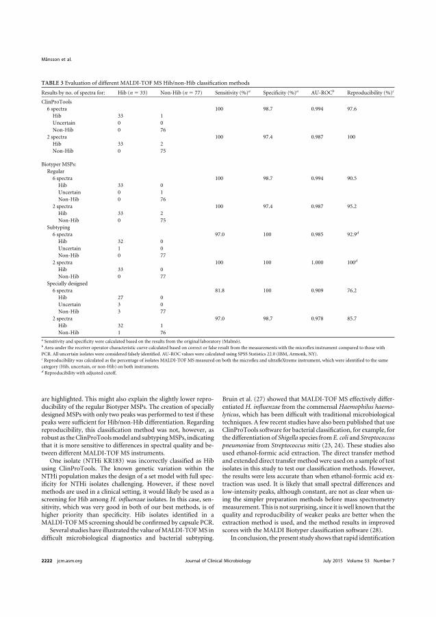
are highlighted. This might also explain the slightly lower repro-ducibility of the regular Biotyper MSPs. The creation of speciallydesigned MSPs with only two peaks was performed to test if thesepeaks were sufficient for Hib/non-Hib differentiation. Regardingreproducibility, this classification method was not, however, asrobust as the ClinProTools model and subtyping MSPs, indicatingthat it is more sensitive to differences in spectral quality and be-tween different MALDI-TOF MS instruments.
One isolate (NTHi KR183) was incorrectly classified as Hibusing ClinProTools. The known genetic variation within theNTHi population makes the design of a set model with full spec-ificity for NTHi isolates challenging. However, if these novelmethods are used in a clinical setting, it would likely be used as ascreening for Hib among H. influenzae isolates. In this case, sen-sitivity, which was very good in both of our best methods, is ofhigher priority than specificity. Hib isolates identified in aMALDI-TOF MS screening should be confirmed by capsule PCR.
Several studies have illustrated the value of MALDI-TOF MS indifficult microbiological diagnostics and bacterial subtyping.
Bruin et al. (27) showed that MALDI-TOF MS effectively differ-entiated H. influenzae from the commensal Haemophilus haemo-lyticus, which has been difficult with traditional microbiologicaltechniques. A few recent studies have also been published that useClinProTools software for bacterial classification, for example, forthe differentiation of Shigella species from E. coli and Streptococcuspneumoniae from Streptococcus mitis (23, 24). These studies alsoused ethanol-formic acid extraction. The direct transfer methodand extended direct transfer method were used on a sample of testisolates in this study to test our classification methods. However,the results were less accurate than when ethanol-formic acid ex-traction was used. It is likely that small spectral differences andlow-intensity peaks, although constant, are not as clear when us-ing the simpler preparation methods before mass spectrometrymeasurement. This is not surprising, since it is well known that thequality and reproducibility of weaker peaks are better when theextraction method is used, and the method results in improvedscores with the MALDI Biotyper classification software (28).
In conclusion, the present study shows that rapid identification
TABLE 3 Evaluation of different MALDI-TOF MS Hib/non-Hib classification methods
Results by no. of spectra for: Hib (n � 33) Non-Hib (n � 77) Sensitivity (%)a Specificity (%)a AU-ROCb Reproducibility (%)c
ClinProTools6 spectra 100 98.7 0.994 97.6
Hib 33 1Uncertain 0 0Non-Hib 0 76
2 spectra 100 97.4 0.987 100Hib 33 2Non-Hib 0 75
Biotyper MSPs:Regular
6 spectra 100 98.7 0.994 90.5Hib 33 0Uncertain 0 1Non-Hib 0 76
2 spectra 100 97.4 0.987 95.2Hib 33 2Non-Hib 0 75
Subtyping6 spectra 97.0 100 0.985 92.9d
Hib 32 0Uncertain 1 0Non-Hib 0 77
2 spectra 100 100 1.000 100d
Hib 33 0Non-Hib 0 77
Specially designed6 spectra 81.8 100 0.909 76.2
Hib 27 0Uncertain 3 0Non-Hib 3 77
2 spectra 97.0 98.7 0.978 85.7Hib 32 1Non-Hib 1 76
a Sensitivity and specificity were calculated based on the results from the original laboratory (Malmö).b Area under the receiver operator characteristic curve calculated based on correct or false result from the measurements with the microflex instrument compared to those withPCR. All uncertain isolates were considered falsely identified. AU-ROC values were calculated using SPSS Statistics 22.0 (IBM, Armonk, NY).c Reproducibility was calculated as the percentage of isolates MALDI-TOF MS measured on both the microflex and ultrafleXtreme instrument, which were identified to the samecategory (Hib, uncertain, or non-Hib) on both instruments.d Reproducibility with adjusted cutoff.
Månsson et al.
2222 jcm.asm.org July 2015 Volume 53 Number 7Journal of Clinical Microbiology

of Hib among H. influenzae is possible using a routine samplepreparation and MALDI-TOF MS analysis of H. influenzae iso-lates. After methodological optimization, this novel finding maybe implemented in clinical microbiology laboratories. H. influen-zae isolates identified by standard MALDI-TOF MS analysis couldthen be directly reanalyzed using the ethanol-formic acid extrac-tion procedure to identify Hib isolates. This would improve theefficiency of Hib identification and make rapid intervention pos-sible in cases of vaccine failure or poor vaccination coverage.MALDI-TOF MS could also be used as a surveillance tool in vac-cinated populations to monitor sustained vaccine efficacy and asan initial screening method of invasive H. influenzae isolates.
ACKNOWLEDGMENTS
We thank Åsa Johansson, Håkan Janson, and Gunnar Kahlmeter from theDepartment of Clinical Microbiology, Växjö, for fruitful discussions inthe initial process, and Søren Lehmann, Bruker Daltonics, for technicalsupport.
This work was supported by grants from the Alfred Osterlund, Annaand Edwin Berger, and Greta and Johan Kock foundations, O. E. och EdlaJohanssons, the Swedish Medical Research Council (grant K2015-57X-03163-43-4 [www.vr.se]), the Physiographical Society (Forssman’s Foun-dation), and Skåne County Council’s research and development founda-tion.
Markus Kostrzewa is an employee of Bruker Daltonics.
REFERENCES1. Peltola H. 2000. Worldwide Haemophilus influenzae type b disease at the
beginning of the 21st century: global analysis of the disease burden 25years after the use of the polysaccharide vaccine and a decade after theadvent of conjugates. Clin Microbiol Rev 13:302–317. http://dx.doi.org/10.1128/CMR.13.2.302-317.2000.
2. Jalalvand F, Riesbeck K. 2014. Haemophilus influenzae: recent advancesin the understanding of molecular pathogenesis and polymicrobial infec-tions. Curr Opin Infect Dis 27:268 –274. http://dx.doi.org/10.1097/QCO.0000000000000056.
3. Ishiwada N, Hishiki H, Nagasawa K, Naito S, Sato Y, Chang B, SasakiY, Kimura K, Ohnishi M, Shibayama K. 2014. The incidence of pediatricinvasive Haemophilus influenzae and pneumococcal disease in Chiba pre-fecture, Japan before and after the introduction of conjugate vaccines.Vaccine 32:5425–5431. http://dx.doi.org/10.1016/j.vaccine.2014.07.100.
4. Bajanca-Lavado MP, Simões AS, Betencourt CR, Sa-Leão R, PortugueseGroup for Study for Haemophilus influenzae Invasive Infection. 2014.Characteristics of Haemophilus influenzae invasive isolates from Portugalfollowing routine childhood vaccination against H. influenzae serotype b(2002–2010). Eur J Clin Microbiol Infect Dis 33:603– 610. http://dx.doi.org/10.1007/s10096-013-1994-6.
5. Resman F, Ristovski M, Ahl J, Forsgren A, Gilsdorf JR, Jasir A, KaijserB, Kronvall G, Riesbeck K. 2011. Invasive disease caused by Haemophilusinfluenzae in Sweden 1997–2009; evidence of increasing incidence andclinical burden of non-type b strains. Clin Microbiol Infect 17:1638 –1645.http://dx.doi.org/10.1111/j.1469-0691.2010.03417.x.
6. Schouls L, van der Heide H, Witteveen S, Zomer B, van der Ende A,Burger M, Schot C. 2008. Two variants among Haemophilus influenzaeserotype b strains with distinct bcs4, hcsA and hcsB genes display differ-ences in expression of the polysaccharide capsule. BMC Microbiol 8:35.http://dx.doi.org/10.1186/1471-2180-8-35.
7. Lepp T, Uhnoo I, Schölin A, Szirmai M, Lindstrand A, Tegnell A. 2014.The childhood immunization program in Sweden 2013, annual report.Public Health Agency of Sweden, Solna, Sweden. http://www.skane.se/Upload/Webbplatser/Smittskydd/Dokument/vaccinationst%C3%A4ckning/A%CC%8Arsrapport_barnvaccinationsprogrammet%202013%5B2%5D.pdf.
8. Watt JP, Wolfson LJ, O’Brien KL, Henkle E, Deloria-Knoll M, McCallN, Lee E, Levine OS, Hajjeh R, Mulholland K, Cherian T, Hib andPneumococcal Global Burden of Disease Study Team. 2009. Burden ofdisease caused by Haemophilus influenzae type b in children younger than5 years: global estimates. Lancet 374:903–911. http://dx.doi.org/10.1016/S0140-6736(09)61203-4.
9. Davis GS, Sandstedt SA, Patel M, Marrs CF, Gilsdorf JR. 2011. Use ofbexB to detect the capsule locus in Haemophilus influenzae. J Clin Micro-biol 49:2594 –2601. http://dx.doi.org/10.1128/JCM.02509-10.
10. Falla TJ, Crook DW, Brophy LN, Maskell D, Kroll JS, Moxon ER. 1994.PCR for capsular typing of Haemophilus influenzae. J Clin Microbiol 32:2382–2386.
11. Lam TT, Elias J, Frosch M, Vogel U, Claus H. 2011. New diagnostic PCRfor Haemophilus influenzae serotype e based on the cap locus of strainATCC 8142. Int J Med Microbiol 301:176 –179. http://dx.doi.org/10.1016/j.ijmm.2010.07.004.
12. Satola SW, Collins JT, Napier R, Farley MM. 2007. Capsule gene analysisof invasive Haemophilus influenzae: accuracy of serotyping and prevalenceof IS1016 among nontypeable isolates. J Clin Microbiol 45:3230 –3238.http://dx.doi.org/10.1128/JCM.00794-07.
13. Musser JM, Kroll JS, Moxon ER, Selander RK. 1988. Evolutionarygenetics of the encapsulated strains of Haemophilus influenzae. Proc NatlAcad Sci U S A 85:7758 –7762. http://dx.doi.org/10.1073/pnas.85.20.7758.
14. Musser JM, Kroll JS, Moxon ER, Selander RK. 1988. Clonal populationstructure of encapsulated Haemophilus influenzae. Infect Immun 56:1837–1845.
15. Erwin AL, Sandstedt SA, Bonthuis PJ, Geelhood JL, Nelson KL, UnrathWC, Diggle MA, Theodore MJ, Pleatman CR, Mothershed EA, SacchiCT, Mayer LW, Gilsdorf JR, Smith AL. 2008. Analysis of genetic relat-edness of Haemophilus influenzae isolates by multilocus sequence typing. JBacteriol 190:1473–1483. http://dx.doi.org/10.1128/JB.01207-07.
16. Bizzini A, Greub G. 2010. Matrix-assisted laser desorption ionizationtime-of-flight mass spectrometry, a revolution in clinical microbial iden-tification. Clin Microbiol Infect 16:1614 –1619. http://dx.doi.org/10.1111/j.1469-0691.2010.03311.x.
17. Emonet S, Shah HN, Cherkaoui A, Schrenzel J. 2010. Application anduse of various mass spectrometry methods in clinical microbiology. ClinMicrobiol Infect 16:1604 –1613. http://dx.doi.org/10.1111/j.1469-0691.2010.03368.x.
18. Demirev PA, Hagan NS, Antoine MD, Lin JS, Feldman AB. 2013.Establishing drug resistance in microorganisms by mass spectrometry. JAm Soc Mass Spectrom 24:1194 –1201. http://dx.doi.org/10.1007/s13361-013-0609-x.
19. Lange C, Schubert S, Jung J, Kostrzewa M, Sparbier K. 2014. Quanti-tative matrix-assisted laser desorption ionization–time of flight mass spec-trometry for rapid resistance detection. J Clin Microbiol 52:4155– 4162.http://dx.doi.org/10.1128/jcm.01872-14.
20. Christner M, Trusch M, Rohde H, Kwiatkowski M, Schluter H, WoltersM, Aepfelbacher M, Hentschke M. 2014. Rapid MALDI-TOF mass spec-trometry strain typing during a large outbreak of Shiga-toxigenic Esche-richia coli. PLoS One 9:e101924. http://dx.doi.org/10.1371/journal.pone.0101924.
21. Vogne C, Prod’hom G, Jaton K, Decosterd L, Greub G. 2014. A simple,robust and rapid approach to detect carbapenemases in Gram-negativeisolates by MALDI-TOF mass spectrometry: validation with triple quad-ripole tandem mass spectrometry, microarray and PCR. Clin MicrobiolInfect 20:O1106 –O1112. http://dx.doi.org/10.1111/1469-0691.12715.
22. Meats E, Feil EJ, Stringer S, Cody AJ, Goldstein R, Kroll JS, Popovic T,Spratt BG. 2003. Characterization of encapsulated and noncapsulatedHaemophilus influenzae and determination of phylogenetic relationshipsby multilocus sequence typing. J Clin Microbiol 41:1623–1636. http://dx.doi.org/10.1128/JCM.41.4.1623-1636.2003.
23. Khot PD, Fisher MA. 2013. Novel approach for differentiating Shigellaspecies and Escherichia coli by matrix-assisted laser desorption ionization–time of flight mass spectrometry. J Clin Microbiol 51:3711–3716. http://dx.doi.org/10.1128/JCM.01526-13.
24. Ikryannikova LN, Filimonova AV, Malakhova MV, Savinova T, Fili-monova O, Ilina EN, Dubovickaya VA, Sidorenko SV, Govorun VM.2013. Discrimination between Streptococcus pneumoniae and Streptococcusmitis based on sorting of their MALDI mass spectra. Clin Microbiol Infect19:1066 –1071. http://dx.doi.org/10.1111/1469-0691.12113.
25. Xiao D, Zhao F, Zhang H, Meng F, Zhang J. 2014. Novel strategy fortyping Mycoplasma pneumoniae isolates by use of matrix-assisted laserdesorption ionization–time of flight mass spectrometry coupled withClinProTools. J Clin Microbiol 52:3038 –3043. http://dx.doi.org/10.1128/JCM.01265-14.
26. Schouls LM, van der Ende A, van de Pol I, Schot C, Spanjaard L,VauteriVauterin P, Wilderbeek D, Witteveen S. 2005. Increase in genetic
H. influenzae Type b and MALDI-TOF MS
July 2015 Volume 53 Number 7 jcm.asm.org 2223Journal of Clinical Microbiology

diversity of Haemophilus influenzae serotype b (Hib) strains after intro-duction of Hib vaccination in The Netherlands. J Clin Microbiol 43:2741–2749. http://dx.doi.org/10.1128/JCM.43.6.2741-2749.2005.
27. Bruin JP, Kostrzewa M, van der Ende A, Badoux P, Jansen R, Boers SA,Diederen BM. 2014. Identification of Haemophilus influenzae and Hae-mophilus haemolyticus by matrix-assisted laser desorption ionization–
time of flight mass spectrometry. Eur J Clin Microbiol Infect Dis 33:279 –284. http://dx.doi.org/10.1007/s10096-013-1958-x.
28. Khot PD, Couturier MR, Wilson A, Croft A, Fisher MA. 2012. Opti-mization of matrix-assisted laser desorption ionization–time of flightmass spectrometry analysis for bacterial identification. J Clin Microbiol50:3845–3852. http://dx.doi.org/10.1128/JCM.00626-12.
Månsson et al.
2224 jcm.asm.org July 2015 Volume 53 Number 7Journal of Clinical Microbiology

Paper II

Emerging Infectious Diseases • www.cdc.gov/eid • Vol. 24, No. 3, March 2018 443
Encapsulated Haemophilus influenzae strains belong to type-specific genetic lineages. Reliable capsule typing re-quires PCR, but a more efficient method would be useful. We evaluated capsule typing by using matrix-assisted la-ser desorption/ionization time-of-flight (MALDI-TOF) mass spectrometry. Isolates of all capsule types (a–f and nontype-able; n = 258) and isogenic capsule transformants (types a-d) were investigated. Principal component and biomarker analyses of mass spectra showed clustering, and mass peaks correlated with capsule type-specific genetic lin-eages. We used 31 selected isolates to construct a capsule typing database. Validation with the remaining isolates (n = 227) showed 100% sensitivity and 92.2% specificity for encapsulated strains (a–f; n = 61). Blinded validation of a supplemented database (n = 50) using clinical isolates (n = 126) showed 100% sensitivity and 100% specificity for encapsulated strains (b, e, and f; n = 28). MALDI-TOF mass spectrometry is an accurate method for capsule typing of H. influenzae.
Haemophilus influenzae is subdivided into encapsulated strains, which express different serotypes of capsular
polysaccharide (designated types a–f), and nonencapsulat-ed strains, which are designated nontypeable H. influenzae (NTHi) (1). Since the introduction of conjugate vaccines against H. influenzae type b (Hib), a common cause of meningitis, epiglottitis, and sepsis in small children, the ep-idemiology of invasive H. influenzae disease has changed dramatically, with an increase in the diversity of serotypes responsible for illness.
Although the incidence of Hib disease has decreased in countries implementing childhood vaccination (2), invasive
disease caused by NTHi has become more prominent during the same period, especially among newborns and the elderly (3–6). In the postvaccination era, increasing incidences and outbreaks of invasive H. influenzae type a (Hia) infections have been reported in South and North America (7–10), par-ticularly among the indigenous populations in Canada and the United States (7,8,10). Studies have also suggested an increase in cases of invasive H. influenzae type e (Hie) and type f (Hif) disease (4,11,12). Hib vaccine failures have been described (13), and omission of booster dose(s) appears to re-sult in a rapidly increased incidence of invasive Hib disease (14,15), suggesting continued circulation of Hib isolates in the community. Globally, one third of eligible children still do not receive adequate vaccination (16).
Encapsulated H. influenzae strains are generally ge-netically clonal. This finding was first demonstrated by multilocus enzyme electrophoresis, by which encapsulated isolates could be separated into different genetic lineages that correspond to different capsule types (17). This clonal population structure has been confirmed by multilocus se-quence typing (MLST), which assigns isolates to differ-ent sequence types (STs), although some differences have been observed in the organization of different lineages (18). There are 3 known major genetic groups of Hia and Hib (18,19). Two genetic groups of Hia (related to ST21 and ST23) account for most Hia isolates in the MLST database (20). For Hib, ST6-related isolates account for most cases (17,18), whereas the second most common genetic group is related to ST222 (18,21). There is 1 known lineage each for serotypes c through f (18,19). In contrast, NTHi are geneti-cally heterogenous (19).
Capsule typing of H. influenzae has traditionally been performed by using slide agglutination with antisera (con-ventional serotyping), but incorrect results are common,
Capsule Typing of Haemophilus influenzae by Matrix-Assisted Laser Desorption/Ionization
Time-of-Flight Mass Spectrometry1
Viktor Månsson, Janet R. Gilsdorf, Gunnar Kahlmeter, Mogens Kilian, J. Simon Kroll, Kristian Riesbeck,2 Fredrik Resman2
RESEARCH
Author affiliations: Lund University, Malmö, Sweden (V. Månsson, K. Riesbeck, F. Resman); University of Michigan, Ann Arbor, Michigan, USA (J.R. Gilsdorf); Växjö Hospital, Växjö, Sweden (G. Kahlmeter); Aarhus University, Aarhus, Denmark (M. Kilian); Imperial College London, London, UK (J.S. Kroll)
DOI: https://doi.org/10.3201/eid2403.170459
1Preliminary results from this study were presented at the IDWeek 2016 Conference, October 26–30, 2016, New Orleans, Louisiana, USA.2These senior authors contributed equally to this article.

RESEARCH
444 Emerging Infectious Diseases • www.cdc.gov/eid • Vol. 24, No. 3, March 2018
and specificity for encapsulated isolates is low (22,23). De-termination of presence of the capsule gene complex (bexA or bexB) by PCR, followed by type-specific cap a–f PCRs has excellent sensitivity and specificity but is laborious and time-consuming (24–27). Because of limitations of current typing methods, typing might be delayed or not performed in clinical practice. However, rapidly obtained information on capsule type is still of interest for the treating clinician (5) and, in particular, for monitoring of capsule type dis-tribution and effectiveness of Hib vaccination programs, especially with respect to invasive disease.
Matrix-assisted laser desorption/ionization time-of-flight (MALDI-TOF) mass spectrometry is commonly used to identify bacterial and fungal species, including H. influ-enzae, by analyzing the composition of ribosomal proteins in a sample. It is a rapid and convenient method and has a low cost per sample (28). Recently, we have shown that MALDI-TOF mass spectrometry can separate Hib from non-b H. influenzae (29). In this study, we examined the capacity of MALDI-TOF mass spectrometry to perform full capsule typing of H. influenzae. This method would be valuable for first-line diagnostics of H. influenzae to iden-tify patients at risk for immunodeficiency or anatomic cere-brospinal fluid space defect, and to detect rapidly outbreaks caused by specific capsule types. It would also increase time and cost effectiveness of surveillance of H. influenzae epidemiology and Hib vaccination efficacy.
Materials and Methods
Bacterial IsolatesWe used 2 culture collections in this study (Figure 1). The first collection was an evaluation set of isolates used to con-struct a coherent reference database and was composed of
258 H. influenzae strains. It included isolates from 3 ma-jor clinical laboratories in Sweden (Malmö/Lund, Gothen-burg, and Stockholm) obtained in 1997–2011 but also a wide range of international strains from different countries, continents, and time periods (n = 41; online Technical Ap-pendix Table, https://wwwnc.cdc.gov/EID/article/24/3/17-0459-Techapp1.pdf). In addition, we included 4 isogenic capsule-transformed strains of types a (Rb–/a+:02), b (Rb+:02), c (Rb–/c+:02), and d (Rb–/d+:02) (30) in the study. These strains originate from strain Rd, a capsule-deficient type d strain (31). For validation of the new MALDI-TOF mass spectrometry typing method, we used a second col-lection composed of 126 bloodstream and cerebrospinal fluid H. influenzae isolates obtained in Sweden during 2010 and 2013–2016. All isolates were identified as H. influen-zae by using standard laboratory taxonomy techniques and were grown on chocolate agar plates overnight (18–24 h) in a humid atmosphere at 37°C containing 5% CO2 before any experiments were conducted.
PCR for Capsule Typing and MLSTWe prepared DNA by adding a few colonies of bacteria to distilled water. After heating at 98°C for 10 min, we centri-fuged each sample at 16,000 × g for 5–10 min and collected the supernatant. In a few instances, we extracted DNA by using the GenElute Bacterial Genomic DNA Kit (Sigma-Aldrich, St. Louis, MO, USA) according to the manufac-turer’s instructions. We performed capsule typing by PCR using bexB and type-specific cap primers for all isolates as described (24–26).
We performed PCR for MLST genes as described (18) and sequenced the resulting PCR products by using the forward primer and, if necessary for adequate sequence quality, the reverse primer. We trimmed and
Figure 1. Culture collections and methods used to investigate capsule typing of Haemophilus influenzae by MALDI-TOF mass spectrometry. An evaluation set of H. influenzae isolates of all capsule types from diverse geographic origins and time periods and isogenic capsule transformants (30) were used to investigate capsule type-specific differences in MALDI-TOF mass spectra. MLST was used to ensure adequate coverage of different genetic lineages of encapsulated H. influenzae. Reference isolates from the evaluation set (encapsulated and nonencapsulated) were selected to construct a new typing database in MALDI Biotyper. This database was tested with the remaining isolates in the set, and misclassified isolates were added to the database. The final supplemented database was blindly validated with a second culture collection that consisted of clinical invasive isolates. Hia, H. influenzae type a; Hib, H. influenzae type b; Hic, H. influenzae type c; Hid, H. influenzae type d; Hie, H. influenzae type e; Hif, H. influenzae type f; MALDI-TOF, matrix-assisted laser desorption/ionization time-of-flight; MLST, multilocus sequence typing; NTHi, nontypeable H. influenzae.
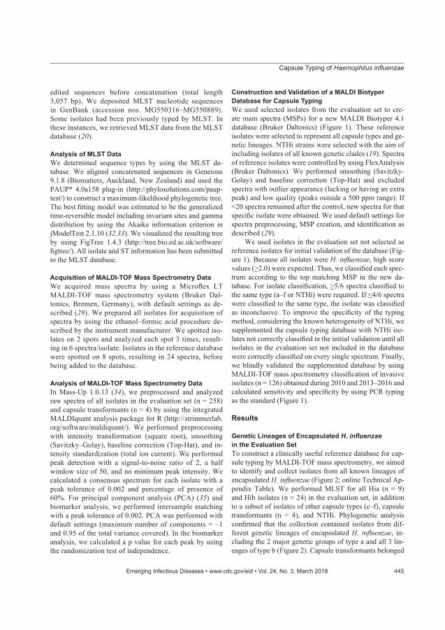
Emerging Infectious Diseases • www.cdc.gov/eid • Vol. 24, No. 3, March 2018 445
Capsule Typing of Haemophilus influenzae
edited sequences before concatenation (total length 3,057 bp). We deposited MLST nucleotide sequences in GenBank (accession nos. MG550316–MG550889). Some isolates had been previously typed by MLST. In these instances, we retrieved MLST data from the MLST database (20).
Analysis of MLST DataWe determined sequence types by using the MLST da-tabase. We aligned concatenated sequences in Geneious 9.1.8 (Biomatters, Auckland, New Zealand) and used the PAUP* 4.0a158 plug-in (http://phylosolutions.com/paup-test/) to construct a maximum-likelihood phylogenetic tree. The best fitting model was estimated to be the generalized time-reversible model including invariant sites and gamma distribution by using the Akaike information criterion in jModelTest 2.1.10 (32,33). We visualized the resulting tree by using FigTree 1.4.3 (http://tree.bio.ed.ac.uk/software/figtree/). All isolate and ST information has been submitted to the MLST database.
Acquisition of MALDI-TOF Mass Spectrometry DataWe acquired mass spectra by using a Microflex LT MALDI-TOF mass spectrometry system (Bruker Dal-tonics, Bremen, Germany), with default settings as de-scribed (29). We prepared all isolates for acquisition of spectra by using the ethanol–formic acid procedure de-scribed by the instrument manufacturer. We spotted iso-lates on 2 spots and analyzed each spot 3 times, result-ing in 6 spectra/isolate. Isolates in the reference database were spotted on 8 spots, resulting in 24 spectra, before being added to the database.
Analysis of MALDI-TOF Mass Spectrometry DataIn Mass-Up 1.0.13 (34), we preprocessed and analyzed raw spectra of all isolates in the evaluation set (n = 258) and capsule transformants (n = 4) by using the integrated MALDIquant analysis package for R (http://strimmerlab.org/software/maldiquant/). We performed preprocessing with intensity transformation (square root), smoothing (Savitzky–Golay), baseline correction (Top-Hat), and in-tensity standardization (total ion current). We performed peak detection with a signal-to-noise ratio of 2, a half window size of 50, and no minimum peak intensity. We calculated a consensus spectrum for each isolate with a peak tolerance of 0.002 and percentage of presence of 60%. For principal component analysis (PCA) (35) and biomarker analysis, we performed intersample matching with a peak tolerance of 0.002. PCA was performed with default settings (maximum number of components = –1 and 0.95 of the total variance covered). In the biomarker analysis, we calculated a p value for each peak by using the randomization test of independence.
Construction and Validation of a MALDI Biotyper Database for Capsule TypingWe used selected isolates from the evaluation set to cre-ate main spectra (MSPs) for a new MALDI Biotyper 4.1 database (Bruker Daltonics) (Figure 1). These reference isolates were selected to represent all capsule types and ge-netic lineages. NTHi strains were selected with the aim of including isolates of all known genetic clades (19). Spectra of reference isolates were controlled by using FlexAnalysis (Bruker Daltonics). We performed smoothing (Savitzky-Golay) and baseline correction (Top-Hat) and excluded spectra with outlier appearance (lacking or having an extra peak) and low quality (peaks outside a 500 ppm range). If <20 spectra remained after the control, new spectra for that specific isolate were obtained. We used default settings for spectra preprocessing, MSP creation, and identification as described (29).
We used isolates in the evaluation set not selected as reference isolates for initial validation of the database (Fig-ure 1). Because all isolates were H. influenzae, high score values (>2.0) were expected. Thus, we classified each spec-trum according to the top matching MSP in the new da-tabase. For isolate classification, >5/6 spectra classified to the same type (a–f or NTHi) were required. If <4/6 spectra were classified to the same type, the isolate was classified as inconclusive. To improve the specificity of the typing method, considering the known heterogeneity of NTHi, we supplemented the capsule typing database with NTHi iso-lates not correctly classified in the initial validation until all isolates in the evaluation set not included in the database were correctly classified on every single spectrum. Finally, we blindly validated the supplemented database by using MALDI-TOF mass spectrometry classification of invasive isolates (n = 126) obtained during 2010 and 2013–2016 and calculated sensitivity and specificity by using PCR typing as the standard (Figure 1).
Results
Genetic Lineages of Encapsulated H. influenzae in the Evaluation SetTo construct a clinically useful reference database for cap-sule typing by MALDI-TOF mass spectrometry, we aimed to identify and collect isolates from all known lineages of encapsulated H. influenzae (Figure 2; online Technical Ap-pendix Table). We performed MLST for all Hia (n = 9) and Hib isolates (n = 24) in the evaluation set, in addition to a subset of isolates of other capsule types (c–f), capsule transformants (n = 4), and NTHi. Phylogenetic analysis confirmed that the collection contained isolates from dif-ferent genetic lineages of encapsulated H. influenzae, in-cluding the 2 major genetic groups of type a and all 3 lin-eages of type b (Figure 2). Capsule transformants belonged

RESEARCH
446 Emerging Infectious Diseases • www.cdc.gov/eid • Vol. 24, No. 3, March 2018
Figure 2. Multilocus sequencing typing (MLST) of encapsulated and nonencapsulated Haemophilus influenzae isolates. MLST was performed on a subset of encapsulated isolates (n = 44) from the evaluation set, including all type a and type b isolates (n = 33). All major genetic lineages (indicated by colors), except the least common lineage of Hia (ST4-related), of encapsulated H. influenzae were represented in the collection, including the 2 more common lineages of Hia and all 3 lineages of Hib. An isolate typed by PCR as Hif (KR1130) with a nonexpressed pseudogene cap locus was also included. This isolate was not part of the established Hif lineage, and was initially suspected to be an outlier on the basis of differences from other Hif in matrix-assisted laser desorption/ionization time-of-flight (MALDI-TOF) mass spectra. All capsule transformants (n = 4) were ST47 (same as the parental strain Rd) (30,31) and were part of the Hid lineage. Nontypeable H. influenzae (NTHi; no color) included as reference isolates in the capsule typing databases (n = 28) were included in the analysis. NTHi in the evaluation set misclassified as type e (n = 5, indicated by asterisks) were also included. These isolates belonged to 3 separate genetic lineages, all related to the Hie lineage. Isolates included as references in MALDI-TOF mass spectrometry databases are indicated by arrows and numbers (1 for isolates in the original database and 2 for isolates added during supplementation of the database). Scale bar indicates nucleotide substitutions per site. Hia, H. influenzae type a; Hib, H. influenzae type b; Hic, H. influenzae type c; Hid, H. influenzae type d; Hie, H. influenzae type e; Hif, H. influenzae type f; ST, sequence type.

Emerging Infectious Diseases • www.cdc.gov/eid • Vol. 24, No. 3, March 2018 447
Capsule Typing of Haemophilus influenzae
to the known genetic lineage of Hid isolates (Figure 2) and were the same ST as the parental strain Rd (18). One isolate (KR1130) was typed by PCR as Hif (bexB- and cap f-pos-itive) but phylogenetically belonged to a lineage separate from all other Hif isolates. Thus, this isolate was not part of the established, ST124-related Hif lineage (Figure 2). The cap locus of this isolate was sequenced and found to be a nonexpressed pseudogene (data not shown).
MALDI-TOF Mass Spectrometry of Genetic Lineages of Encapsulated H. influenzaeWe performed PCA for all isolates in the evaluation set (n = 258) and the capsule transformants (n = 4). As expected, NTHi formed a large heterogeneous group, but cluster-ing of encapsulated isolates of the same capsule types was found (Figure 3, panel A). When PCA was performed on encapsulated isolates (n = 83) and capsule transformants (n = 4) only, the clustering became clearer and was particu-larly evident for Hib, Hie, and Hif isolates (Figure 3, panel B). Encapsulated isolates segregated in groups according to capsule type and, for Hia and Hib isolates, by genetic lineage according to MLST (Figure 2; 3, panel B). Cap-sule transformants were found as a separate group in close proximity of Hid isolates and not distributed according to
their respective capsule type (Figure 3, panel B). Isolate KR1130 did not cluster with Hif isolates of the ST124-re-lated lineage (Figure 3, panel B).
Biomarker analysis of encapsulated isolates and cap-sule transformants identified several peaks conserved with-in the different genetic lineages of capsule types, indicating the possibility of separating them on the basis of MALDI-TOF mass spectra (Figure 4). Capsule transformants ex-pressed similar peak patterns relative to each other but dif-fered in many peaks when compared with wild-type strains of the same capsule types.
Sensitivity and Specificity of Automated Capsule Typing by MALDI-TOF Mass SpectrometryAn initial capsule typing reference database was construct-ed in MALDI Biotyper. Encapsulated isolates (n = 22) representing all major genetic lineages of encapsulated H. influenzae were included (Figure 2). To ensure adequate coverage of potential variation within each lineage, mul-tiple reference isolates were chosen for each lineage (when possible) on the basis of geographic origin and variations in mass spectra. In addition, NTHi (n = 9) representing 8 of 10 known genetic clades of NTHi (19) were included in the database (Figure 2).
Figure 3. Principal component analysis (PCA) of matrix-assisted laser desorption/ionization time-of-flight mass spectra of encapsulated and nonencapsulated Haemophilus influenzae. A) PCA of all isolates (n = 258) of H. influenzae in the evaluation set representing all capsule types, which are color-coded according to capsule type and for Hia and Hib isolates by genetic lineage as shown by multilocus sequence typing (MLST) and capsule transformants (n = 4). The first 3 principal components (PC0, PC1, and PC2) are shown in 2-dimensional plots. Analysis showed the diversity of nontypeable H. influenzae (NTHi). Encapsulated isolates showed discrete clustering, which was further evaluated by PCA of encapsulated isolates separately. B) PCA of encapsulated isolates in the evaluation set (n = 83) and capsule transformants (n = 4) presented and color-coded as in panel A. Clustering of isolates on the basis of capsule type was evident, particularly for Hib, Hie, and Hif isolates. Different genetic lineages of the same capsule type (Hia and Hib) clustered separately. KR1130 (with a pseudogene type f cap locus) did not cluster with the other Hif isolates. Capsule transformants clustered together in proximity of Hid isolates, and not with their respective capsule type. Hia, H. influenzae type a; Hib, H. influenzae type b; Hic, H. influenzae type c; Hid, H. influenzae type d; Hie, H. influenzae type e; Hif, H. influenzae type f; ST, sequence type.

RESEARCH
448 Emerging Infectious Diseases • www.cdc.gov/eid • Vol. 24, No. 3, March 2018
Validation of the original database (n = 31) using the remaining isolates in the evaluation set (n = 227) showed 100% sensitivity for encapsulated isolates (Table 1), and every isolate was correctly classified on every spectrum. All capsule transformants were classified as type d, the original serotype of the parental strain Rd (31). No isolate matched KR1130, the isolate typed by PCR as Hif with a pseudogene cap locus.
A few NTHi were either inconclusively typed or mis-classified as encapsulated, resulting in reduced specificity for encapsulated isolates (Table 1). For this reason, we supplemented the capsule typing database with misclas-sified NTHi from the evaluation set until the database correctly classified all the remaining isolates in the evalu-ation set on every single spectrum. This modification re-sulted in an additional 19 NTHi being added to the MALDI
Biotyper database (Figure 2). When PCA was performed separately for NTHi in the evaluation set, it was evident that the supplemented database covered the heterogeneity of NTHi better than the original database (Figure 5). The same finding was evident from phylogenetic analysis (Fig-ure 2). Five NTHi were misclassified as Hie and could not be added to the database because they interfered with clas-sification of true Hie isolates and would decrease sensitiv-ity for Hie (Figure 2).
As a final performance test, we blindly validated the supplemented database (n = 50) by using a separate culture collection consisting of clinical invasive isolates from Sweden (n = 126) obtained during 2010 and 2013–2016. When we compared MALDI-TOF mass spectrom-etry capsule typing results with PCR capsule typing re-sults, all encapsulated isolates (types b, e, and f; n = 28)
Figure 4. Biomarker analysis of matrix-assisted laser desorption/ionization time-of-flight mass spectra of encapsulated Haemophilus influenzae. Analysis was performed on all encapsulated isolates in the evaluation set (n = 83) and capsule transformants (n = 4). Rows represent peaks (2,000–20,000 m/z in descending order), and columns represent groups of encapsulated H. influenzae. A total of 124 peaks with discriminatory power (p<0.05) between different capsule types and genetic lineages were identified. Peak expression is indicated by shades of black (black >75%, gray >25% but <75%, and white <25% of isolates in the group express the peak). Several peaks conserved within capsule types and genetic lineages with the possibility for separation were observed, as indicated by the mosaic of peak patterns. Capsule transformants showed similar peak patterns, and lacked many of the capsule type-specific peaks for their respective phenotypic capsule types. KR1130 expressed different peaks than Hif of the ST124-related lineage. Hia, H. influenzae type a; Hib, H. influenzae type b; Hic, H. influenzae type c; Hid, H. influenzae type d; Hie, H. influenzae type e; Hif, H. influenzae type f; ST, sequence type.

Emerging Infectious Diseases • www.cdc.gov/eid • Vol. 24, No. 3, March 2018 449
Capsule Typing of Haemophilus influenzae
were correctly classified on every single spectrum (Table 2). Of 98 NTHi, only 5 were not correctly classified. These isolates were all classified as inconclusive. Thus, no NTHi was incorrectly classified as encapsulated, and the resulting sensitivity and specificity of capsule typing was 100% in the final validation (Table 2).
DiscussionIn this study, we have shown that encapsulated H. influen-zae have different MALDI-TOF mass spectra that correlate with genetic lineages representing different capsule types. We have demonstrated that, after construction of a com-prehensive reference database, routine MALDI-TOF mass
Table 1. Validation of the original MALDI-TOF mass spectrometry capsule typing database (n = 31) by classification of the remaining 227 isolates in the evaluation set and 4 capsule transformants of Haemophilus influenzae* Capsule type No. No. correct† No. inconclusive‡ No. incorrect§ Sensitivity, % Specificity, % Hia, ST21-related 4 4 0 0 100 99.1 Hia, ST23-related 1 1 0 0 100 100 Hib, ST6-related 17 17 0 0 100 100 Hib, ST222-related 1 1 0 0 100 100 Hic 2 2 0 0 100 100 Hid 1 1 0 0 100 98.2 Hie 12 12 0 0 100 97.7 Hif 23 23¶ 0 0 100 99.0 All encapsulated isolates, a–f 61 61 0 0 100 92.2 NTHi 166 122 31 13# 73.5 100 Rb-negative capsule transformants 4 1** 0 3** NA NA *Hia, H. influenzae type a; Hib, H. influenzae type b; Hic, H. influenzae type c; Hid, H. influenzae type d; Hie, H. influenzae type e; Hif, H. influenzae type f; MALDI-TOF, matrix-assisted laser desorption/ionization time-of-flight; NA, not applicable; NTHi, nontypeable H. Influenzae; ST, sequence type. †>5/6 spectra classified to the same correct type. ‡<4/6 spectra classified to the same type. §>5/6 spectra classified to the same incorrect type. ¶All classified to the established ST124-related Hif lineage. #Two isolates classified as Hia (ST21-related), 4 as Hid, 5 as Hie, and 2 as Hif (ST124-related). **All classified as type d (i.e., the same type as the parental strain Rd) (30,31), resulting in correct classification of isolate Rb–/d+:02 and incorrect classification of isolates Rb+:02, Rb–/a+:02, and Rb–/c+:02.
Figure 5. Principal component analysis of matrix-assisted laser desorption/ionization time-of-flight mass spectra of NTHi in the original and supplemented databases. Isolates are color-coded according to database affiliation, and the first 3 principal components (PC0, PC1, and PC2) are shown in 2-dimensional plots. No clustering similar to that for encapsulated isolates was observed. NTHi reference isolates in the original capsule typing database (n = 9), representing different genetic clades, were evenly distributed in the group. Supplementing the reference database with another 19 isolates improved coverage of the heterogeneity of NTHi. NTHi, nontypeable H. influenzae; PCA, principal component analysis.

RESEARCH
450 Emerging Infectious Diseases • www.cdc.gov/eid • Vol. 24, No. 3, March 2018
spectrometry analysis has excellent capacity to identify type-specific genetic lineages associated with encapsulated H. influenzae and thereby can be used for capsule typing of H. influenzae.
Our study had several strengths. We analyzed a large collection of well-characterized strains collected at different times from various geographic regions to ensure the robust-ness of our findings. Using MLST, we ensured adequate coverage of the major genetic lineages of encapsulated H. influenzae in the MALDI-TOF mass spectrometry reference database. Moreover, the database was carefully evaluated and supplemented to ensure adequate coverage of the het-erogeneity of NTHi. We blindly validated the supplemented database to mimic an authentic clinical or epidemiologic sit-uation and demonstrated excellent sensitivity and specific-ity compared with conventional PCR-based typing. During construction of the capsule typing database, we identified several isolates previously typed by PCR or agglutination (by us or others) in which the MALDI-TOF mass spec-trometry results did not match the suggested capsule type. When we retyped these isolates by PCR, the capsule type suggested by MALDI-TOF mass spectrometry proved to be correct in all instances (except for the NTHi typed as Hie) (Figure 2), and isolates were reassigned to a new capsule type, further supporting the capacity of MALDI-TOF mass spectrometry for capsule typing.
Our study had some limitations. The first limitation reflects the limited availability of some rare variants. Our collection contained no Hia isolates belonging to the un-common ST4-related genetic group. For the ST61-related lineage of Hib, we had access to only 1 isolate, which was included in the reference database and thus not represent-ed in the test collection. However, our ST61 isolate was separable when mass spectra were analyzed by PCA and biomarker analysis, and no isolate was misclassified to this lineage in the initial or final validation of the typing da-tabases. Furthermore, we have demonstrated that identifi-cation of the ST222-related Hib lineage by MALDI-TOF mass spectrometry is possible (Table 1), which was not the case previously (29). The second potential limitation arises through the genetic heterogeneity of NTHi, making
adequate representation in the reference database a chal-lenge (19). This limitation was apparent during the initial evaluation of the typing method, when some NTHi were misclassified. To address this issue, we supplemented the database with 19 additional reference NTHi strains. The fi-nal validation of our typing method demonstrated excellent specificity for NTHi, but the sensitivity for identifying en-capsulated isolates remained unchanged. Because most in-vasive infections in countries implementing Hib vaccination are caused by NTHi, a high specificity is desirable (3–5).
MALDI-TOF mass spectrometry has proved valuable in subtyping several clinically relevant bacteria, including Clostridium difficile (36), methicillin-resistant Staphylo-coccus aureus (37,38), and enterohemorrhagic Escherichia coli (39). Subtyping generally relies on common genetic differences between isolates, reflected in the composition of the proteins measured. In our study, wild-type isolates of different capsule types could be separated, but isogenic capsule transformants could not. These isolates were clas-sified as type d, the original capsule type of the parental strain Rd. This finding confirms that capsule type identifi-cation is based on a proxy identification of genetic lineage, rather than identification of capsule biosynthesis-associat-ed proteins. Thus, our method is an indirect typing meth-od, as opposed to serotyping, which identifies the capsule polysaccharide, and PCR, which identifies the capsule gene complex directly.
Although there is little evidence that new lineages of encapsulated H. influenzae have appeared historically, novel lineages of encapsulated strains might appear and be missed by the method. Isolate KR1130 used in this study was initially suspected to represent such a lineage. How-ever, its cap locus was shown to be on a nonexpressed pseudogene. Only 1 other isolate of the same ST (ST184) is currently registered in the MLST database, and it is a nontypeable isolate. No other Hif strain in this study or the MLST database belongs to this genetic lineage (20).
One advantage of indirect capsule type identification by MALDI-TOF mass spectrometry is that determination of genetic lineage of encapsulated isolates can be made without further analysis. The method can also identify
Table 2. Validation of the supplemented MALDI-TOF mass spectrometry capsule typing database (n = 50) by classification of 126 invasive isolates of Haemophilus influenzae from Sweden* Capsule type No. tested No. correct† No inconclusive‡ No. incorrect§ Sensitivity, % Specificity, % Hib 8 8¶ 0 0 100 100 Hie 5 5 0 0 100 100 Hif 15 15# 0 0 100 100 All encapsulated isolates (b, e, and f) 28 28 0 0 100 100 NTHi 98 93 5 0 94.9 100 *Hib, H. influenzae type b; Hie, H. influenzae type e; Hif, H. influenzae type f; MALDI-TOF, matrix-assisted laser desorption/ionization time-of-flight; NTHi, nontypeable H. influenzae; ST, sequence type. †>5/6 spectra classified to the same correct type. ‡<4/6 spectra classified to the same type. §>5/6 spectra classified to the same incorrect type. ¶All classified to the ST6-related Hib lineage. #All classified to the established ST124-related Hif lineage.

Emerging Infectious Diseases • www.cdc.gov/eid • Vol. 24, No. 3, March 2018 451
Capsule Typing of Haemophilus influenzae
previously encapsulated capsule-deficient strains, which have lost parts or all of the cap locus, either during infec-tion or laboratory handling (40–42).
A concern regarding subtyping by MALDI-TOF mass spectrometry (43) is the potential need for special sample preparations, such as growth conditions and type of matrix. In several studies, differences in mass spectra between sub-types of various species were observed but no automated classification methods were was reported (43), which might limit general applicability. In this study, we used standard growth conditions, as well as routine ethanol–formic acid extraction and mass spectra acquisition protocols. The soft-ware used (MALDI Biotyper) also has the advantage of being a standard software used in clinical settings. These factors greatly increased the chance of clinical implementa-tion of our findings.
In conclusion, our study demonstrated that rapid cap-sule typing of H. influenzae by identification of capsule type-specific genetic lineages using routine MALDI-TOF mass spectrometry is possible and highly accurate. Af-ter further large-scale validation, this method has the po-tential for clinical and research use. With the increasing heterogeneity in capsule types of disease-causing H. in-fluenzae observed since Hib conjugate vaccines were in-troduced, the method can become a valuable tool in clinical diagnostic laboratories.
This study was supported by grants from the Foundations of Anna and Edwin Berger (to K.R.); the Royal Physiographical Society of Lund, Forssmans Foundation (to V.M.); the Swedish Medical Research Council (grant no. K2015-57X-03163-43-4; http://www.vr.se); and the Skåne County Council Research and Development Foundation (to K.R.).
About the AuthorDr. Månsson is a physician and doctoral student in clinical microbiology at the Riesbeck Laboratory, Lund University, Malmö, Sweden. His main research interests are respiratory tract infections, with special focus on H. influenzae diagnostics, epidemiology, and antimicrobial drug resistance.
References 1. Pittman M. Variation and type specificity in the bacterial
species Haemophilus influenzae. J Exp Med. 1931;53:471–92. http://dx.doi.org/10.1084/jem.53.4.471
2. Peltola H. Worldwide Haemophilus influenzae type b disease at the beginning of the 21st century: global analysis of the disease burden 25 years after the use of the polysaccharide vaccine and a decade after the advent of conjugates. Clin Microbiol Rev. 2000;13:302–17. http://dx.doi.org/10.1128/CMR.13.2.302-317.2000
3. Langereis JD, de Jonge MI. Invasive disease caused by nontypeable Haemophilus influenzae. Emerg Infect Dis. 2015;21:1711–8. http://dx.doi.org/10.3201/eid2110.150004
4. Resman F, Ristovski M, Ahl J, Forsgren A, Gilsdorf JR, Jasir A, et al. Invasive disease caused by Haemophilus influenzae in
Sweden 1997-2009; evidence of increasing incidence and clinical burden of non-type b strains. Clin Microbiol Infect. 2011;17:1638–45. http://dx.doi.org/10.1111/j.1469-0691.2010.03417.x
5. Ladhani S, Slack MP, Heath PT, von Gottberg A, Chandra M, Ramsay ME. European Union Invasive Bacterial Infection Surveillance participants. Invasive Haemophilus influenzae disease, Europe, 1996-2006. Emerg Infect Dis. 2010;16:455–63. http://dx.doi.org/10.3201/eid1603.090290
6. Whittaker R, Economopoulou A, Dias JG, Bancroft E, Ramliden M, Celentano LP. European Centre for Disease Prevention and Control Country Experts for Invasive Haemophilus influenzae Disease. Epidemiology of invasive Haemophilus influenzae disease, Europe, 2007-2014. Emerg Infect Dis. 2017;23:396–404. http://dx.doi.org/10.3201/eid2303.161552
7. Bender JM, Cox CM, Mottice S, She RC, Korgenski K, Daly JA, et al. Invasive Haemophilus influenzae disease in Utah children: an 11-year population-based study in the era of conjugate vaccine. Clin Infect Dis. 2010;50:e41–6. http://dx.doi.org/10.1086/ 651165
8. Bruce MG, Zulz T, DeByle C, Singleton R, Hurlburt D, Bruden D, et al. Haemophilus influenzae serotype a invasive disease, Alaska, USA, 1983-2011. Emerg Infect Dis. 2013;19:932–7. http://dx.doi.org/10.3201/eid1906.121805
9. Zanella RC, Bokermann S, Andrade AL, Flannery B, Brandileone MC. Changes in serotype distribution of Haemophilus influenzae meningitis isolates identified through laboratory-based surveillance following routine childhood vaccination against H. influenzae type b in Brazil. Vaccine. 2011;29:8937–42. http://dx.doi.org/10.1016/j.vaccine.2011.09.053
10. Rotondo JL, Sherrard L, Helferty M, Tsang R, Desai S. The epidemiology of invasive disease due to Haemophilus influenzae serotype a in the Canadian North from 2000 to 2010. Int J Circumpolar Health. 2013;72:72. http://dx.doi.org/10.3402/ ijch.v72i0.21142
11. Bajanca-Lavado MP, Simões AS, Betencourt CR, Sá-Leão R. Portuguese Group for Study of Haemophilus influenzae invasive infection. Characteristics of Haemophilus influenzae invasive isolates from Portugal following routine childhood vaccination against H. influenzae serotype b (2002-2010). Eur J Clin Microbiol Infect Dis. 2014;33:603–10. http://dx.doi.org/10.1007/ s10096-013-1994-6
12. Ladhani SN, Collins S, Vickers A, Litt DJ, Crawford C, Ramsay ME, et al. Invasive Haemophilus influenzae serotype e and f disease, England and Wales. Emerg Infect Dis. 2012;18:725–32. http://dx.doi.org/10.3201/eid1805.111738
13. Ladhani S, Heath PT, Slack MP, McIntyre PB, Diez-Domingo J, Campos J, et al.; Participants of the European Union Invasive Bacterial Infections Surveillance Network. Haemophilus influenzae serotype b conjugate vaccine failure in twelve countries with established national childhood immunization programmes. Clin Microbiol Infect. 2010;16:948–54. http://dx.doi.org/10.1111/ j.1469-0691.2009.02945.x
14. Ladhani SN. Two decades of experience with the Haemophilus influenzae serotype b conjugate vaccine in the United Kingdom. Clin Ther. 2012;34:385–99. http://dx.doi.org/10.1016/ j.clinthera.2011.11.027
15. Juarez MD, Rancaño C, Neyro S, Biscayart C, Katz N, Pasinovich M, et al. What’s happening with Haemophilus influenzae type B invasive disease in Latin America region? Argentina’s experience. In: Abstracts of IDWeek 2016, New Orleans, October 26–30. Abstract no. 768 [cited 2017 Dec 18]. https://idsa.confex.com/idsa/2016/webprogram/POSTER.html
16. World Health Organization. Global and regional immunization profile, 2016 global summary [cited 2017 Nov 9]. http://www.who.int/immunization/monitoring_surveillance/data/gs_gloprofile.pdf?ua=1
RESEARCH
452 Emerging Infectious Diseases • www.cdc.gov/eid • Vol. 24, No. 3, March 2018
17. Musser JM, Kroll JS, Moxon ER, Selander RK. Clonal population structure of encapsulated Haemophilus influenzae. Infect Immun. 1988;56:1837–45.
18. Meats E, Feil EJ, Stringer S, Cody AJ, Goldstein R, Kroll JS, et al. Characterization of encapsulated and noncapsulated Haemophilus influenzae and determination of phylogenetic relationships by multilocus sequence typing. J Clin Microbiol. 2003;41:1623–36. http://dx.doi.org/10.1128/JCM.41.4.1623-1636.2003
19. Erwin AL, Sandstedt SA, Bonthuis PJ, Geelhood JL, Nelson KL, Unrath WC, et al. Analysis of genetic relatedness of Haemophilus influenzae isolates by multilocus sequence typing. J Bacteriol. 2008;190:1473–83. http://dx.doi.org/10.1128/JB.01207-07
20. University of Oxford. Haemophilus influenzae MLST website [cited 2017 Nov 9]. http://pubmlst.org/hinfluenzae/
21. Myers AL, Jackson MA, Zhang L, Swanson DS, Gilsdorf JR. Haemophilus influenzae type b invasive disease in Amish children, Missouri, USA, 2014. Emerg Infect Dis. 2017;23:112–4. http://dx.doi.org/10.3201/eid2301.160593
22. LaClaire LL, Tondella ML, Beall DS, Noble CA, Raghunathan PL, Rosenstein NE, et al.; Active Bacterial Core Surveillance Team Members. Identification of Haemophilus influenzae serotypes by standard slide agglutination serotyping and PCR-based capsule typing. J Clin Microbiol. 2003;41:393–6. http://dx.doi.org/10.1128/JCM.41.1.393-396.2003
23. Satola SW, Collins JT, Napier R, Farley MM. Capsule gene analysis of invasive Haemophilus influenzae: accuracy of serotyping and prevalence of IS1016 among nontypeable isolates. J Clin Microbiol. 2007;45:3230–8. http://dx.doi.org/10.1128/JCM.00794-07
24. Falla TJ, Crook DW, Brophy LN, Maskell D, Kroll JS, Moxon ER. PCR for capsular typing of Haemophilus influenzae. J Clin Microbiol. 1994;32:2382–6.
25. Davis GS, Sandstedt SA, Patel M, Marrs CF, Gilsdorf JR. Use of bexB to detect the capsule locus in Haemophilus influenzae. J Clin Microbiol. 2011;49:2594–601. http://dx.doi.org/10.1128/JCM.02509-10
26. Lâm TT, Elias J, Frosch M, Vogel U, Claus H. New diagnostic PCR for Haemophilus influenzae serotype e based on the cap locus of strain ATCC 8142. Int J Med Microbiol. 2011;301:176–9. http://dx.doi.org/10.1016/j.ijmm.2010.07.004
27. van Ketel RJ, de Wever B, van Alphen L. Detection of Haemophilus influenzae in cerebrospinal fluids by polymerase chain reaction DNA amplification. J Med Microbiol. 1990;33:271–6. http://dx.doi.org/10.1099/00222615-33-4-271
28. Clark AE, Kaleta EJ, Arora A, Wolk DM. Matrix-assisted laser desorption ionization−time of flight mass spectrometry: a fundamental shift in the routine practice of clinical microbiology. Clin Microbiol Rev. 2013;26:547–603. http://dx.doi.org/10.1128/CMR.00072-12
29. Månsson V, Resman F, Kostrzewa M, Nilson B, Riesbeck K. Identification of Haemophilus influenzae type b isolates by use of matrix-assisted laser desorption ionization−time of flight mass spectrometry. J Clin Microbiol. 2015;53:2215–24. http://dx.doi.org/10.1128/JCM.00137-15
30. Zwahlen A, Kroll JS, Rubin LG, Moxon ER. The molecular basis of pathogenicity in Haemophilus influenzae: comparative virulence of genetically-related capsular transformants and correlation with changes at the capsulation locus cap. Microb Pathog. 1989;7:225–35. http://dx.doi.org/10.1016/ 0882-4010(89)90058-2
31. Alexander HE, Leidy G. Determination of inherited traits of H. influenzae by desoxyribonucleic acid fractions isolated from type-specific cells. J Exp Med. 1951;93:345–59. http://dx.doi.org/ 10.1084/jem.93.4.345
32. Darriba D, Taboada GL, Doallo R, Posada D. jModelTest 2: more models, new heuristics and parallel computing. Nat Methods. 2012;9:772. http://dx.doi.org/10.1038/nmeth.2109
33. Guindon S, Gascuel O, Rannala B. A simple, fast, and accurate algorithm to estimate large phylogenies by maximum likelihood. Syst Biol. 2003;52:696–704. http://dx.doi.org/ 10.1080/10635150390235520
34. López-Fernández H, Santos HM, Capelo JL, Fdez-Riverola F, Glez-Peña D, Reboiro-Jato M. Mass-Up: an all-in-one open software application for MALDI-TOF mass spectrometry knowledge discovery. BMC Bioinformatics. 2015;16:318. http://dx.doi.org/10.1186/s12859-015-0752-4
35. Kehrmann J, Wessel S, Murali R, Hampel A, Bange FC, Buer J, et al. Principal component analysis of MALDI TOF MS mass spectra separates M. abscessus (sensu stricto) from M. massiliense isolates. BMC Microbiol. 2016;16:24. http://dx.doi.org/10.1186/s12866-016-0636-4
36. Rizzardi K, Åkerlund T. High molecular weight typing with MALDI-TOF MS: a novel method for rapid typing of Clostridium difficile. PLoS One. 2015;10:e0122457. http://dx.doi.org/10.1371/journal.pone.0122457
37. Camoez M, Sierra JM, Dominguez MA, Ferrer-Navarro M, Vila J, Roca I. Automated categorization of methicillin-resistant Staphylococcus aureus clinical isolates into different clonal complexes by MALDI-TOF mass spectrometry. Clin Microbiol Infect. 2016;22:161.e1–7. http://dx.doi.org/10.1016/ j.cmi.2015.10.009
38. Josten M, Reif M, Szekat C, Al-Sabti N, Roemer T, Sparbier K, et al. Analysis of the matrix-assisted laser desorption ionization-time of flight mass spectrum of Staphylococcus aureus identifies mutations that allow differentiation of the main clonal lineages. J Clin Microbiol. 2013;51:1809–17. http://dx.doi.org/ 10.1128/JCM.00518-13
39. Ojima-Kato T, Yamamoto N, Suzuki M, Fukunaga T, Tamura H. Discrimination of Escherichia coli O157, O26 and O111 from other serovars by MALDI-TOF MS based on the S10-GERMS method. PLoS One. 2014;9:e113458. http://dx.doi.org/10.1371/journal.pone.0113458
40. Fothergill LD, Chandler CA, Spencer M. Observations on the dissociation of meningitic strains of H. influenzae. J Immunol. 1936;31:401–15.
41. Hoiseth SK, Connelly CJ, Moxon ER. Genetics of spontaneous, high-frequency loss of b capsule expression in Haemophilus influenzae. Infect Immun. 1985;49:389–95.
42. Tsang RS, Li YA, Mullen A, Baikie M, Whyte K, Shuel M, et al. Laboratory characterization of invasive Haemophilus influenzae isolates from Nunavut, Canada, 2000-2012. Int J Circumpolar Health. 2016;75:29798. http://dx.doi.org/10.3402/ijch.v75.29798
43. Sauget M, Valot B, Bertrand X, Hocquet D. Can MALDI-TOF mass spectrometry reasonably type bacteria? Trends Microbiol. 2017;25:447–55. http://dx.doi.org/10.1016/j.tim.2016.12.006
Address for correspondence: Viktor Månsson, Riesbeck Laboratory, Department of Translational Medicine, Lund University, Jan Waldenströms gata 59, 205 02 Malmö, Sweden; email: [email protected]

Page 1 of 2
Article DOI: https://doi.org/10.3201/eid2403.170459
Capsule Typing of Haemophilus influenzae by Matrix-Assisted Laser
Desorption/Ionization Time-of-Flight Mass Spectrometry
Technical Appendix
Technical Appendix Table. Isolates of Haemophilus influenzae used to develop a MALDI-TOF mass spectrometry typing database*
Isolate Other designation Capsule
type Location Year Tissue/infection
International CCUG6881 Smith a Unknown Before 1973 Unknown CCUG7315 NCTC8465 a USA 1941 Respiratory tract HK391 ATCC9327, NCTC8466 a USA 1942 CSF/nasal secretion HK643 Fin31 a Finland 1974–1976 Respiratory tract HK645 Harding a Boston, MA, USA 1981 CSF HK648 CDC78, E202 a Unknown Unknown Unknown HK649 CDC22, CDC D5361 a USA 1976 Blood KR152 a Sweden Unknown Unknown KR1141 a Angola 2016 Ear swab DL-42 b Dallas, TX, USA Before 1984 Unknown Eagan CCUG18095 b Boston, MA, USA 1968 CSF HK395 b USA Before 1954 Unknown HK691 A1132st b USA Before 1985 Unknown HK706 M1053 b USA 1979 Unknown HK714 M1062 b USA 1980 Unknown HK718 M1071 b USA 1941 Unknown HK727 M1084 b USA 1947 CSF HK729 M1077 b USA 1980 CSF Kansas2 b Missouri, USA 2014 Epiglottitis Kansas3 b Missouri, USA 2014 Osteomyelitis MinnA b Minneapolis, MN, USA 1979 CSF 850530 b The Netherlands 1985 Unknown CCUG4851 NCTC8469, ATCC9007 c USA 1942 Sputum CCUG4852 Ruggerio c New York, NY, USA Before 1950 CSF HK635 43/LA c Papua New Guinea 1980 Lung aspirate HK688 c Copenhagen, Denmark 1984 CSF KR153 c Sweden Unknown Unknown HK644 51/LA d Papua New Guinea 1979 Unknown KR154 d Sweden Unknown Unknown NCTC8470 d The Netherlands 1937 Respiratory tract CCUG15521 e Probably Sweden Before 1984 Unknown KR1142 e Unknown Unknown Unknown HK636 CDC54 e Unknown Unknown Unknown HK641 Hepke e Denver, CO, USA 1980 Unknown HK653 45/LA e Papua New Guinea 1979 Unknown KR138 A76/01 e Unknown Unknown Unknown KR147 A77/99 e Unknown Unknown Unknown CCUG15435 Shawn f Unknown Unknown Unknown CCUG6877 NT USA 1941 Unknown CCUG15519 NT Unknown Before 2001 Unknown HK224 NT Denmark Before 1976 Otitis media Sweden 11 b Gothenburg, Lund/Malmö,
Stockholm 1998–2007 Blood, CSF
8 e Lund/Malmö, Stockholm 2006–2009 Blood, CSF, respiratory tract

Page 2 of 2
Isolate Other designation Capsule
type Location Year Tissue/infection 26 f Gothenburg, Lund/Malmö,
Stockholm 1997–2009, 2011 Blood, CSF
172 NT Gothenburg, Lund/Malmö, Stockholm
1997–2011 Blood, CSF, respiratory tract
*ATCC, American Type Culture Collection; CCUG, Culture Collection of the University of Göteborg; CDC; US Centers for Disease Control and Prevention; CSF, cerebrospinal fluid; MALDI-TOF, matrix-assisted laser desorption/ionization time-of-flight; NCTC, National Collection of Type Cultures; NT, nontypeable.

Paper III


Original article
The spread and clinical impact of ST14CC-PBP3 type IIb/A, a clonalgroup of non-typeable Haemophilus influenzae with chromosomallymediated b-lactam resistanceda prospective observational study
V. Månsson 1, D. Skaare 2, K. Riesbeck 1, F. Resman 1, *
1) Riesbeck Laboratory, Clinical Microbiology, Department of Translational Medicine, Lund University, Malm€o, Sweden2) Department of Microbiology, Vestfold Hospital Trust, Tønsberg, Norway
a r t i c l e i n f o
Article history:Received 2 May 2016Received in revised form2 October 2016Accepted 4 November 2016Available online 13 November 2016
Editor: G. Lina
Keywords:Antimicrobial resistanceEpidemiologyGenotypingHaemophilus influenzaeHospitalizationMultilocus sequence typing
a b s t r a c t
Objectives: Increasing incidences of non-typeable Haemophilus influenzae (NTHi) with b-lactam resis-tance mediated through mutations in penicillin-binding protein 3 (BLNAR or rPBP3) have been observedin the past decades. Recently, an rPBP3 NTHi sequence type (ST) 14 with PBP3 type IIb/A caused a diseaseoutbreak in a nursing home in Sweden with severe outcomes, indicating increased bacterial virulence. Inthis prospective observational study, the epidemiology and clinical significance of the correspondingclonal group (ST14CC-PBP3IIb/A) in Skåne, Sweden was investigated.Methods: ST14CC-PBP3IIb/A isolates were identified through partial multilocus sequence typing andftsI(PBP3 gene)-sequencing of prospectively collected H. influenzae from patients with respiratory tract orinvasive infections (n¼ 3262) in 2010e2012. Data on type of infection, hospitalization and outcomeswere analysed, and the geographical distribution of isolates from different groups was investigated.Results: Isolates belonging to ST14CC-PBP3IIb/A constituted 26% (n¼ 94/360) of respiratory tract rPBP3NTHi. From mapping of patient addresses, a dynamic geographical spread was apparent during the studyperiod. Furthermore, patients with infections by ST14CC-PBP3IIb/A isolates had higher hospitalizationrates compared with patients infected with other rPBP3 NTHi (14/83 versus 21/255, p 0.025) and to agroup of patients infected with susceptible NTHi (14/83 versus 13/191, p 0.010). ST14CC-PBP3IIb/A iso-lates constituted 25% (n¼ 2/8) of invasive rPBP3 NTHi during the study period.Conclusions: Our investigation suggests that the ST14CC-PBP3IIb/A clonal group is associated withincreased clinical virulence, resistance to several antimicrobial agents, and is persistent over time. Weconclude that virulence varies between different NTHi, and that NTHi disease may be more dependent onbacterial factors than previously recognized. V. Månsson, CMI 2017;23:209.e1e209.e7© 2016 European Society of Clinical Microbiology and Infectious Diseases. Published by Elsevier Ltd. All
rights reserved.
Introduction
Haemophilus influenzae is a common colonizer of the humanrespiratory tract. The species is subdivided into encapsulated andnon-encapsulated (non-typeable) strains (NTHi) [1]. NTHi is oftencommensal but also a frequent cause of infections such as acuteotitis media, pneumonia and exacerbations of chronic obstructivepulmonary disease (COPD) [2,3]. Since the introduction of conju-gate vaccines against H. influenzae type b, studies suggestincreasing incidences of invasive NTHi disease, especially in theelderly [4,5].
Multilocus sequence typing (MLST) is a well-establishedmethod for typing of H. influenzae [6]. Classification of theH. influenzae population into different genetic clades by phyloge-netic analysis through MLST predicts carriage of virulence factorgenes [7,8]. Although specific sequence types (STs) have beenlinked to mucosal infections or to certain anatomical sites [9,10], noreport has linked NTHi disease severity to specific genetic clades orclonal complexes (CCs). Rather, it has been suggested that diseaseseverity may be largely host dependent [11,12].
b-lactam resistance in H. influenzae is mediated via b-lactamaseproduction (termed b-lactamase-positive ampicillin-resistant iso-lates; BLPAR) or by key alterations (Arg517/His or Asn526/Lys)in penicillin-binding protein 3 (PBP3) (termed b-lactamase-nega-tive ampicillin-resistant isolates; BLNAR) [1]. Current EuropeanCommittee on Antimicrobial Susceptibility Testing (EUCAST)
* Corresponding author. F. Resman, Riesbeck Laboratory, Department of Trans-lational Medicine, Lund University, Jan Waldenstr€oms gata 59, SE-205 02 Malm€o,Sweden.
E-mail address: [email protected] (F. Resman).
Contents lists available at ScienceDirect
Clinical Microbiology and Infection
journal homepage: www.cl inicalmicrobiologyandinfect ion.com
http://dx.doi.org/10.1016/j.cmi.2016.11.0061198-743X/© 2016 European Society of Clinical Microbiology and Infectious Diseases. Published by Elsevier Ltd. All rights reserved.
Clinical Microbiology and Infection 23 (2017) 209.e1e209.e7

breakpoints for ampicillin effectively split the population with keyPBP3 alterations in half, so the terms genetic BLNAR (gBLNAR) orrPBP3 (PBP3-mediated resistance) have been introduced to denotethe full population with key substitutions, and we will hence usethe term rPBP3 in this work [13,14]. PBP3 alterations have beenclassified into functional groups and subgroups by Ubukata,Dabernat, Skaare and their co-workers [15e17]. Although the pro-portion of BLPAR isolates has been relatively stable in the pastdecades, rPBP3 is an increasing problem in Europe, as incidenceshave risen, with group II rPBP3 isolates being particularly common[14,17e21]. In addition, rPBP3 isolates are often genetically related[14,17,19,21,22]. As a consequence, sequencing of the ftsI gene,encoding PBP3, has been suggested as an addition to the MLSTscheme [14].
A distinct clonal group of rPBP3 isolates, consisting of sequencetype (ST) 14 and related STs with PBP3 type IIb/A (aminoacid substitutions Asp350/Asn, Met377/Ile, Ala502/Val,Asn526/Lys, Val547/Ile and Asn569/Ser) [14,16,17] has beenhighlighted in recent studies in Scandinavia. This clonal group(ST14CC-PBP3IIb/A) caused an outbreak of respiratory tract in-fections in a nursing home in southern Sweden, resulting in severalhospitalizations and one fatality [23]. In addition, a previous studyon invasive rPBP3 isolates in Sweden revealed a cluster of invasivecases later confirmed to be caused by ST14CC-PBP3IIb/A isolates[19]. Furthermore, a Norwegian study indicated that ST14CC-PBP3IIb/A had the highest hospitalization rate of all STs in a 2007surveillance population [14].
Although ST14CC-PBP3IIb/A has been described in generalstudies on H. influenzae epidemiology, the recent outbreakprompted us to investigate the epidemiology and clinical signifi-cance of this clonal group. In this study we examined the preva-lence of ST14CC-PBP3IIb/A among respiratory and invasiveH. influenzae isolates in our geographical area. The epidemiologyand clinical presentation of isolates from this clonal group werecompared with those of other rPBP3 NTHi as well as those of b-lactam susceptible NTHi, with the hypothesis that this is a lineageof NTHi with increased virulence.
Materials and methods
Study setting
This study was carried out in Skåne, Sweden. During the years2010e2012, all H. influenzae isolated from upper respiratorytract cultures (nasopharyngeal swabs and secretions from themiddle ear, sinuses and conjunctivae) from patients with symptomsof respiratory tract infections referred to the Clinical MicrobiologyLaboratory at Skåne University Hospital, Malm€o, were prospectivelystored at e80�C. This laboratory receives clinical bacterial culturesfrom all health centres and hospitals in southern Skåne (populationapproximately 500 000). Furthermore, all hospitals in the regionshare the same electronic medical records system, allowing thor-ough epidemiological investigations. According to regional guide-lines, upper respiratory tract cultures are recommended in casesof recurrent acute otitis media, treatment failure of acute otitismedia and community-acquired pneumonia in adults. Cultures canalso be obtained at the discretion of the referring physician.
Invasive NTHi isolates (cultures from blood and cerebrospinalfluid) from 2010 to 2012, from Skåne county (population of approxi-mately 1 200 000), were also prospectively collected and studied.
Antimicrobial susceptibility testing and interpretation
All isolates were investigated for susceptibility to b-lactam an-tibiotics by EUCAST disc diffusion testing [24], a method
demonstrated to accurately identify rPBP3 isolates [25]. Resultswere interpreted according to the recommendations for the time ofisolation (www.eucast.org). Isolates were grown on Mül-lereHinton-fastidious agar and screened for b-lactam resistanceusing discs with phenoxymethylpenicillin (10 mg) or benzylpeni-cillin (1 U). Resistant isolates were examined by a nitrocefin b-lactamase test and b-lactamase non-producing isolates were pre-sumed to be rPBP3. b-lactamase-producing, cefaclor-resistant(30 mg disc) isolates were considered both b-lactamase-producingand rPBP3. All isolates with a phenotype suggesting PBP3-mediatedb-lactam resistance, both b-lactamase-producing and non-producing, were selected for further study (see Supplementarymaterial, Fig. S1). Not all recorded isolates could be retrieved, butapart from non-retrieval, no exclusion criteria were applied. Agroup of randomly selected, b-lactam-susceptible isolates (n¼ 193)were included as references in the study (Fig. 1).
Minimum inhibitory concentrations (MICs) for amoxicillin andcefotaxime were determined for respiratory isolates identified asST14CC-PBP3IIb/A, as well as for a randomly selected subset of non-ST14CC-PBP3IIb/A rPBP3 isolates (n¼ 95), by gradient tests (Etest,bioM�erieux, Marcy-l’�Etoile, France).
Culture conditions, preparation of bacterial DNA and PCR
Bacterial culturing and DNA preparation were performed aspreviously described [26]. PCR for MLST genes and the trans-peptidase region of the ftsI gene (encoding PBP3) was performed asdescribed previously [6,17]. Sequenced PCR products were analysedusing GENEIOUS R9 (Biomatters, Auckland, New Zealand).
Identification of ST14CC-PBP3IIb/A isolates
Following determination of the discriminatory power of eachMLST gene to identify ST14 (www.pubmlst.org), suspected rPBP3isolates were screened by sequencing of the adk gene. Isolates withadk 5 (specific adk allele of ST14) were further investigated bysequencing of recA and ftsI. In the MLST database (www.pubmlst.org), all isolates with adk 5 and recA 5 are ST14 or single locusvariants of ST14. Deduced PBP3 amino acid sequences werecompared with the amino acid sequence of H. influenzae Rd KW20(GenBank accession number AAC22787). Isolates with adk 5, recA 5and PBP3IIb/A [14] were considered part of the ST14CC-PBP3IIb/Aclonal group. To validate the method, full MLST was performedon a subset of ST14CC-PBP3IIb/A isolates (n¼ 9). Furthermore,resistance patterns of non-b-lactam antibiotics were compared,and the ftsI DNA sequences (nucleotides 1010e1719) werecompared with the previously reported PBP3IIb/A-encoding ftsIallele lambda-1 (EMBL: HG818630) associated with ST14 [14]. Thetyping process is outlined in Fig. 1.
Patient cohorts and patient data
Respiratory isolates. Since b-lactam resistance in itself could bea risk factor for treatment failure and more severe infections,clinical data were compared between patient cohorts with threegroups of isolates: (a) ST14CC-PBP3IIb/A, (b) other rPBP3 screeningpositive isolates and (c) b-lactam-susceptible isolates. If a patienthad sequential cultures of NTHi within 6 months belonging to thesame group, only the first was included in the patient cohortanalysis. Laboratory referrals were examined for patient age andsex, and the working diagnosis of the referring physician. Thisworking diagnosis could not, however, be validated and the infor-mation was treated accordingly. Medical records were investigatedfor information on hospital admission in any of the regional hos-pitals within 5 days of the culture date. For hospitalized patients,
V. Månsson et al. / Clinical Microbiology and Infection 23 (2017) 209.e1e209.e7209.e2

All respiratory H. influenzaeisolates 2010-2012 (n=3192)
Isolates with adk 5 (n=95)
ST14CC-PBP3IIb/A(n=94)
β-lactam resistant isolates (n=799)
rPBP3 isolates (n=400)
Available isolates (n=360)
Isolates with recA 5 and PBP3IIb/A (n=94)
Not resistant (n=2281)
Not available (n=34)Wrong species (n=5)Encapsulated (n=1)
β-lactamase mediatedresistance only (n=399)
Other PBP3 type (n=1)
Other rPBP3 NTHi (n=266)
=1)
Randomly selected reference group (n=193)
Other adk allele (n=265)
Not tested (n=112)
Fig. 1. Identification of ST14CC-PBP3IIb/A isolates. To identify ST14CC-PBP3IIb/A isolates, a simplified multilocus sequence typing (MLST) scheme (using only the adk and recA gene)was adopted in combination with ftsI gene sequencing and translation. All rPBP3 non-typeable Haemophilus influenzae (NTHi) isolates with adk 5 and recA 5 (the adk and recA allelesof ST14) and PBP3IIb/A were considered ST14CC-PBP3IIb/A. One isolate had adk 5 and recA 5, but a completely different PBP3 substitution pattern (Gly490/Glu, Asn526/Lys andAla530/Ser). All isolates with adk 5 and PBP3IIb/A also had recA 5. A randomly selected subset of ST14CC-PBP3IIb/A isolates (n¼ 9) were confirmed ST14 by full MLST, to validatethe typing scheme. Clonality of ST14CC-PBP3IIb/A isolates was further confirmed by coherent antibiotic susceptibility by disc diffusion testing for trimethoprim-sulfamethoxazole(R), nalidixic acid (S) and tetracycline (S) and analysis of ftsI sequences. Of b-lactam susceptible isolates (n¼ 193), about half (n¼ 91) were evaluated with the MLST-PBP3 typingscheme. Only three isolates had adk 5 and of these, all carried wild-type PBP3 and none had recA 5. Non-ST14CC-PBP3IIb/A rPBP3 isolates and the randomly selected b-lactamsusceptible isolates were used to identify control patient cohorts to evaluate the clinical impact of ST14CC-PBP3IIb/A.
Table 1Respiratory rPBP3 NTHi isolates and patients in the study region
Year (total respiratoryH. influenzaeisolates (n))
Incidence1 rPBP3 isolates2, 3 Patient cohorts4
0e5 6e20 21e65 66þ Total ST14CC-PBP3IIb/A (%) Other rPBP3 (%) Total ST14CC-PBP3IIb/A (%5) Other rPBP3 (%5) Susceptible6
2010 (1185) 2010 138 101 74 243 13 (21.3) 48 (78.7) 61 13 (22.4) 45 (77.6) 412011 (1062) 1453 140 118 83 216 48 (28.2) 122 (71.8) 170 40 (25.2) 119 (74.8) 752012 (945) 1319 117 94 89 190 33 (25.6) 96 (74.4) 129 30 (24.8) 91 (75.2) 662010-2012 (3,192) 1587 131 104 82 216 94 (26.1) 266 (73.9) 360 83 (24.6) 255 (75.4) 192
1 Yearly incidence of respiratory NTHi per 100 000 age group-sorted individuals.2 Of 400 suspected rPBP3 isolates, 34 were not available. Six isolates were excluded, five were of other species and one was encapsulated (type e, PCR-verified).3 Isolates obtained before and after 20 January 2011 were initially screened for b-lactam resistance with 10 mg phenoxymethylpenicillin and 1 U benzylpenicillin discs,
respectively.4 If the same patient had more than one respiratory culture of NTHi within 6 months, only the disease episode related to the first culture was included in the patient data
analyses, unless the separate isolates belonged to different groups (i.e. ST14CC-PBP3IIb/A, other rPBP3 or susceptible isolate).5 Per cent of patients with rPBP3 NTHi cultures.6 Identified from a randomly selected subset of susceptible isolates (n ¼ 193).
Table 2MICs of respiratory ST14CC-PBP3IIb/A and other rPBP3 NTHi for amoxicillin and cefotaxime
Type of NTHi Isolates(n)
b-lactamase (n) Amoxicillin Cefotaxime
Including b-lactamase producingisolates2
Excluding b-lactamase producingisolates
MIC50/MIC90 (mg/L) S/R3 (%) MIC50/MIC90 (mg/L) S/R3 (%) MIC50/MIC90(mg/L)
S/R3 (%)
ST14CC-PBP3IIb/A 94 5 2/4 84/16 1/4 88/12 0.064/0.125 99/1Other rPBP3 951 12 2/64 71/29 2/4 81/19 0.064/0.125 100/0
1 For cefotaxime, gradient tests were performed on 29 randomly selected isolates.2 As expected b-lactamase producing isolates had higher amoxicillin MICs than non-producing isolates (range 4e256 and 64e256 mg/L for ST14CC-PBP3IIb/A and other
rPBP3, respectively).3 Susceptibility according to EUCAST clinical breakpoint (S�/R>) for amoxicillin (2/2) and cefotaxime (0.125/0.125). All b-lactamase-producing isolates were classified as
resistant for amoxicillin.
V. Månsson et al. / Clinical Microbiology and Infection 23 (2017) 209.e1e209.e7 209.e3

information on the cause of hospitalization, final diagnosis (Inter-national Classification of Diseases 10th revision), recent antibiotictreatment, causal treatment and outcomes were collected.
Postcodes of patients in the three cohorts were registered andchanges in geographical distribution of cases during the studyperiod were analysed. Postcodes for patients with ST14CC-PBP3IIb/A infections were plotted on maps to illustrate the spread of theclonal group.
Invasive isolates. Invasive isolate patient data were analysedseparately for patient age, diagnosis and outcome.
Statistical analyses
Comparisons between the different patient cohorts and changesin geographical distribution of disease cases (categorical data) wereperformed using chi-square test. In the latter instance bycomparing the proportions of patients living in and outside centralMalm€o (the only large urban area in the region), comparing the first(2010) and last (2012) year of the study.
Ethical considerations
This study was approved by the Regional Ethics Committee atLund University (2014/533).
Results
ST14-PBP3A isolates in the study region
Respiratory isolates. During the study period, the yearly inci-dence of cultured H. influenzae from patients with respiratory tractsymptoms was 216/100 000 individuals, and was highest amongyoung children (Table 1). A total of 3192 respiratory tractH. influenzaewere registered. Susceptibility testing by disc diffusionscreening (Fig. 1) identified 400 (12.5%) likely rPBP3 isolates, ofwhich 360 were available for further typing (Table 1). Among b-lactam-susceptible isolates, a randomly selected group (n¼ 193)was included as reference.
When subjected to the MLST-PBP3 typing scheme (Fig. 1), 26.1%(n¼ 94) of all rPBP3 isolates belonged to ST14CC-PBP3IIb/A ac-cording to the typing scheme. In full MLST performed on a subset ofthese isolates (n¼ 9), all were confirmed ST14. All ST14CC-PBP3IIb/A isolates were resistant to trimethoprim-sulfamethoxazole,compared with 37.1% of other rPBP3 isolates (n¼ 266). OfST14CC-PBP3IIb/A isolates, 98% (n¼ 92) carried the ftsI allelelambda-1. In the two remaining isolates, PBP3Awas encoded by ftsIallele lambda-2 [14], with four single nucleotide polymorphismscompared with lambda-1. These isolates were also confirmed ST14by full MLST. As these isolates deviated slightly genetically, twoseparate analyses of patient data were made based upon inclusionand exclusion of the patients infected by these isolates. Of sus-ceptible isolates evaluated according to the typing scheme (n¼ 91),none were ST14 (Fig. 1).
The 94 ST14CC-PBP3IIb/A isolates were isolated from 83 (81when excluding the lambda-2-carrying isolates) individual pa-tients, while rPBP3 non-ST14CC-PBP3IIb/A isolates (n¼ 266) wereisolated from 255 patients (Table 1). Hence, ST14CC-PBP3IIb/Aconstituted 24.6% (alternatively 24.1% when excluding thelambda-2-carrying isolates) of non-redundant infections caused byrPBP3 NTHi during the study period. The b-lactam-susceptibleisolates were isolated from 192 individual patients (Table 1).
Invasive isolates. All invasive H. influenzae (n¼ 70) from2010e2012 were cultured from blood, no case of meningitis wasreported. Of 13% (n¼ 9) with phenotypes suggesting rPBP3, eightisolates were available for typing. Two (25%) were ST14CC-PBP3IIb/Ta
ble
3Descriptive
dataan
dge
ographical
distribution
ofpatients
withresp
iratoryNTH
iinfections
Patien
tco
hort
Patien
ts(n)
Susp
ecteddiagn
osis/siteof
infection1(%)
Age
(yea
rs)(%)
Sex(%)
Postco
de
available
(%)
Postco
de
incentral
Malm€ o(%)
Unsp
ecified
URI2
Ear
Sinus
Eye
LRI3
Other
Unkn
own
<1
1-5
6-20
21-65
�66
Female
Male
2010
2011
2012
ST14
CC-
PBP3
IIb/A
8317
(20)
20(24)
7(8)
12(14)
9(11)
018
(22)
14(17)
35(42)
7(8)
23(28)
4(5)
42(51)
41(49)
76(92)
2(15)
10(27)
18(69)
4
Other
rPBP3
255
70(28)
44(17)
19(7)
36(14)
20(8)
4(2)
62(24)
36(14)
78(31)
21(8)
96(38)
24(9)
140(55)
115(45)
249(98)
14(31)
49(43)
46(51)
5
Suscep
tible
192
70(36)
29(15)
6(3)
9(5)
14(7)
4(2)
60(31)
25(13)
74(39)
24(13)
56(29)
13(7)
107(56)
85(44)
183(95)
11(29)
31(42)
20(28)
1Accordingto
culture
referral.
2Upper
resp
iratorytractinfection.
3Lo
wer
resp
iratorytractinfection.
4Sign
ificantlyincrea
sed,c
ompared
to20
10(p
0.00
2).W
hen
lambd
a-2carryingST
14CC-PBP3
IIb/Aisolates
wereex
cluded
,thesign
ificantincrea
seremained
(p0.00
3).
5Sign
ificantlyincrea
sed,c
ompared
to20
10(p
0.02
9).
V. Månsson et al. / Clinical Microbiology and Infection 23 (2017) 209.e1e209.e7209.e4

A, both with ftsI allele lambda-1, according to theMLST-PBP3 typingscheme. These isolates were confirmed as ST14 by full MLST.
b-lactam-resistance among respiratory rPBP3 NTHi isolates
ST14CC-PBP3IIb/A isolates and control rPBP3 NTHi isolates hadcomparable MICs for amoxicillin and cefotaxime (Table 2). Eventhough the amoxicillin MIC ranges of b-lactamase non-producingisolates were narrow for both populations, both were divided bythe amoxicillin breakpoints. As expected, b-lactamase-producingisolates had higher MICs for amoxicillin (Table 2).
Patient data and clinical impact of ST14CC-PBP3IIb/A infections
Respiratory isolatesereferral data No statistically significantdifferences were observed between patients isolated with ST14CC-PBP3IIb/A, other rPBP3 or susceptible NTHi regarding suspectedclinical diagnosis, age or sex (Table 3). Young children (age 0e5years) and adults dominated all three cohorts. Nine cases ofneonatal infections, evenly spread between the groups, were noted(data not shown).
Geographical distribution. For ST14CC-PBP3IIb/A as well as forother rPBP3, theproportionsof infectedpatients residing in theurbanMalm€o region increased significantly over the study period (Table 3).The centralization of patient cases toMalm€owas, however, distinctlygreater for ST14CC-PBP3IIb/Awithan increaseof>400% (from2/13 in2010 to 18/26 cases in 2012), as visualized in Fig. 2, compared with65% for other rPBP3. No significant changes in geographical distri-bution were seen for the patient cohort with susceptible NTHi.
Hospital data. A significantly larger proportion of patientsinfected with ST14CC-PBP3IIb/A were hospitalized compared withpatients infected with other rPBP3 (14/83 versus 21/255, p 0.025)and susceptible strains (14/83 versus 13/192, p 0.010) (Table 4).When excluding cases caused by lambda-2-carrying ST14CC-PBP3IIb/A isolates, a significant difference was still observed(Table 4). Four individual patients (one repeat isolate) had in-fections caused by b-lactamase-producing ST14CC-PBP3IIb/A iso-lates, one of which was hospitalized.When a subgroup analysis wasperformed (including only b-lactamase-negative ST14CC-PBP3IIb/Aisolates), the difference compared with the non-ST14CC-PBPIIb/ArPBP3 cohort remained statistically significant (p 0.035). Patientswith infections caused by ST14CC-PBP3IIb/A were more oftenhospitalized primarily because of their respiratory tract infection,
Fig. 2. The spread of ST14CC-PBP3IIb/A in southern Skåne. Available postcodes of patients suffering from infections by ST14CC-PBP3IIb/A 2010 (n¼ 13), 2011 (n¼ 37) and 2012(n¼ 26), respectively, were plotted on maps. The proportion of patients with addresses in central Malm€o (the only large urban area in the region, indicated) increased during thestudy years. Patients residing in inner-city Malm€o constituted 15% (n¼ 2), 27% (n¼ 10) and 69% (n¼ 18) of cases in 2010, 2011 and 2012, respectively.
V. Månsson et al. / Clinical Microbiology and Infection 23 (2017) 209.e1e209.e7 209.e5

and they suffered less frequently from COPD compared with pa-tients infected by other rPBP3 NTHi. However, these differenceswere not statistically significant (Table 4). No hospitalized patientdied, but one patient each from the ST14CC-PBP3IIb/A and the otherrPBP3 NTHi cohort received intensive care. It is difficult to drawfirm conclusions on treatment effects of specific antibiotics in thislimited-size cohort, but five patients with rPBP3-related pneu-monia were treated with penicillins only (high-dose intravenousbenzylpenicillin and/or high-dose oral amoxicillin) with no recordof relapse. The corresponding isolates all had MICs for amoxicillinof 1e2 mg/L.
Invasive isolates. All cases of invasive rPBP3 NTHi diseaseoccurred in adults, of which seven were >65 years of age. The pa-tients with ST14-PBP3IIb/A disease (n¼ 2) suffered from cholangitisand pneumonia. The patients with other rPBP3 NTHi isolated fromblood (n¼ 6) had pneumonia. One patient from each group died.Due to the limited number of cases, no statistical analyses wereperformed.
Discussion
In this study we show that ST14CC-PBP3IIb/A accounted forapproximately 25% of respiratory rPBP3 NTHi in Skåne, Sweden2010e2012. We further demonstrate that ST14CC-PBP3IIb/A carriesa significantly higher patient hospitalization rate compared withother respiratory rPBP3 and susceptible NTHi; the clonal group isalso present among invasive isolates and has established itself in adensely populated urban area.
ST14CC-PBP3IIb/A was first recognized among Norwegian res-piratory isolates and was the most prevalent rPBP3 clone in a 2004surveillance study [17], and the fourth most prevalent in 2007 [14].In addition, the clonal group has been observed among Swedishinvasive isolates [19] and associated with pneumonia in Spain [27].Recently, it was reported to have caused a severe disease outbreakin a Swedish nursing home [23]. ST14 (PBP3 alterations not re-ported) has also been observed among invasive isolates in Canadaand Italy [28,29]. The number of previous studies performingMLST-PBP3 typing is limited, making it difficult to draw definitiveconclusions about the spread and clinical impact of ST14CC-PBP3IIb/A. The present study confirms that the clonal group iswidely established and persistent. Interestingly, ST14CC-PBP3IIb/Acases clustered in the urban Malm€o region during the study period.It is possible that this particular clonal group, in addition toincreased virulence, has an increased capacity of inter-individualspread that would be enhanced in a densely populated area. Thiswould be in line with the recently reported outbreak [23]. Isolatesin the clonal group are resistant to commonly used antimicrobials,and consistent with previous indications [14] a significantly higherhospitalization rate of ST14CC-PBP3IIb/A patients was observed inthe present study. The clonal group appears to be endemic inScandinavia [14,17,19], and possibly far beyond [27e29].
Two isolates in our ST14CC-PBP3IIb/A collection expressed PBP3encoded by the slightly different ftsI allele lambda-2, an allelepreviously reported to appear in other groups of rPBP3 NTHi [14].Horizontal gene transfer of the ftsI gene is a possible cause of thisdistribution, and has been shown in vitro [30]. Although theseisolates met the criteria of being part of the clonal group (ST14CCand PBP3IIb/A), we chose to reperform statistical analyses toconfirm significant differences between ST14CC-PBP3IIb/A andcontrol isolates, with these isolates excluded.
Notably, five ST14CC-PBP3IIb/A isolates had acquired b-lacta-mase genes, consistent with previous reports that the two major b-lactam-resistance mechanisms occur independently [21].
This study has several strengths. We have analysed a large andnear-complete collection of prospectively collected clinical isolatesTa
ble
4Hospitalized
patients
withresp
iratoryNTH
iinfections
Patien
tco
hort
Patien
ts(n)
Hospitalizationdata
Priordisea
seFinal
diagn
osis
Hospitalized
patients
1(%)
Med
ical
reco
rds
available
Hospitalization
primarily
relatedto
NTH
iinfection3(%)
Other
reason
for
hospitalization(%)
Med
ianhospital
time(day
s)Antibiotics
prior
tohospitalization(%)
Immuno-
compromised
4(%)
COPD
(%)
Pneu
mon
ia5
(%)
AOM
(%)
Other
infection(%)
ST14
CC-PBP3
IIb/A
8314
(17)
214
13(93)
1(7)
55(36)
03(21)
11(79)
2(14)
0Other
rPBP3
255
21(8.2)
1912
(63)
7(37)
43(16)
2(11)
6(32)
8(42)
046
(21)
Suscep
tible
192
13(6.8)
139(69)
4(31)
3,5
5(38)
01(8)
5(38)
1(8)
37(23)
1Admittedto
hospital
within
5day
sof
positivecu
lture.
2Statistically
sign
ificant,co
mpared
withpatients
infected
byother
rPBP3
andsu
scep
tibleNTH
i(p0.02
5,RR2.0(95%
CI1
.1e3.8)
and(p
0.01
0,RR2.5(95%
CI1
.2e5.0),respective
ly).Sign
ificantd
ifferencesremained
when
cases
causedby
lambd
a-2carryingisolates
wereex
cluded
(p0.04
2an
dp0.01
7forother
rPBP3
andsu
scep
tibleisolates,respective
ly).
3Hospitalized
dueto
distinct
bacterialinfectionrelatedto
thecu
lture
that
resp
onded
toad
equatean
tibiotic
trea
tmen
t.4Defi
ned
asneu
trop
enia,c
onge
nital
immunod
eficien
cyor
curren
tch
emotherap
y.5Rad
iograp
hically
confirm
ed.
6Includingthreech
ronic
obstructivepulm
onarydisea
se(COPD
)ex
acerba
tion
swithnosign
sof
pneu
mon
iaon
chestX-ray
.7Includingon
eCOPD
exacerba
tion
withnosign
sof
pneu
mon
iaon
chestX-ray
.
V. Månsson et al. / Clinical Microbiology and Infection 23 (2017) 209.e1e209.e7209.e6

from patients with respiratory tract and invasive infections,allowing a comprehensive epidemiological analysis. By investi-gatingmedical records, we have demonstrated an increased clinicalvirulence of ST14CC-PBP3IIb/A. The study also used the novelapproach of combining MLST and ftsI-typing to compare rPBP3strains between study groups [14]. A potential limitation of thestudy is the algorithm used to identify ST14CC-PBP3IIb/A isolates.However, in the MLST database (www.pubmlst.org), all isolateswith adk 5 and recA 5 are ST14, or single locus variants of ST14.Furthermore, in a study of 196 Norwegian clinical respiratory tractH. influenzae, all isolates expressing adk 5 and PBP3IIb/A were ST14(n¼ 11) or single locus variants of ST14 (n¼ 1) [14].
In conclusion, this study demonstrates the presence of a prev-alent, widespread and persistent clonal group of rPBP3 NTHi withincreased clinical virulence. The results call for continued epide-miological surveillance of NTHi, further studies regarding NTHivirulence mechanisms, and supports recent shifts in attitude to-wards NTHi as a pathogen capable of causing severe disease notonly in a host-dependent manner.
Transparency declaration
None of the authors have any conflict of interest to disclose.
Funding
This work was supported by grants from the Anna and EdwinBerger Foundation, the Royal Physiographic Society of Lund (For-ssman’s Foundation), Swedish Medical Research Council (grantnumber K2015-57X-03163-43-4, www.vr.se), and Skåne CountyCouncil’s research and development foundation.
Acknowledgements
We thank the Clinical Microbiology Laboratory at LabmedicinSkåne for providing clinical isolates.
Appendix A. Supporting information
Supplementary data related to this article can be found at http://dx.doi.org/10.1016/j.cmi.2016.11.006.
References
[1] Tristram S, Jacobs MR, Appelbaum PC. Antimicrobial resistance in Haemophilusinfluenzae. Clin Microbiol Rev 2007;20:368e89.
[2] Murphy TF, Faden H, Bakaletz LO, Kyd JM, Forsgren A, Campos J, et al. Non-typeable Haemophilus influenzae as a pathogen in children. Pediatr Infect Dis J2009;28:43e8.
[3] Sethi S, Murphy TF. Bacterial infection in chronic obstructive pulmonarydisease in 2000: a state-of-the-art review. Clin Microbiol Rev 2001;14:336e63.
[4] Resman F, Ristovski M, Ahl J, Forsgren A, Gilsdorf JR, Jasir A, et al. Invasivedisease caused by Haemophilus influenzae in Sweden 1997e2009; evidence ofincreasing incidence and clinical burden of non-type b strains. Clin MicrobiolInfect 2011;17:1638e45.
[5] Van Eldere J, Slack MP, Ladhani S, Cripps AW. Non-typeable Haemophilusinfluenzae, an under-recognised pathogen. Lancet Infect Dis 2014;14:1281e92.
[6] Meats E, Feil EJ, Stringer S, Cody AJ, Goldstein R, Kroll JS, et al. Characterizationof encapsulated and noncapsulated Haemophilus influenzae and determinationof phylogenetic relationships by multilocus sequence typing. J Clin Microbiol2003;41:1623e36.
[7] De Chiara M, Hood D, Muzzi A, Pickard DJ, Perkins T, Pizza M, et al. Genomesequencing of disease and carriage isolates of nontypeable Haemophilusinfluenzae identifies discrete population structure. Proc Natl Acad Sci U S A2014;111:5439e44.
[8] Erwin AL, Sandstedt SA, Bonthuis PJ, Geelhood JL, Nelson KL, Unrath WC, et al.Analysis of genetic relatedness of Haemophilus influenzae isolates by multi-locus sequence typing. J Bacteriol 2008;190:1473e83.
[9] Murphy TF, Lesse AJ, Kirkham C, Zhong H, Sethi S, Munson Jr RS. A clonalgroup of nontypeable Haemophilus influenzae with two IgA proteases isadapted to infection in chronic obstructive pulmonary disease. PLoS One2011;6:e25923.
[10] LaCross NC, Marrs CF, Gilsdorf JR. Population structure in nontypeable Hae-mophilus influenzae. Infect Genet Evol 2013;14:125e36.
[11] Erwin AL, Nelson KL, Mhlanga-Mutangadura T, Bonthuis PJ, Geelhood JL,Morlin G, et al. Characterization of genetic and phenotypic diversity of inva-sive nontypeable Haemophilus influenzae. Infect Immun 2005;73:5853e63.
[12] Puig C, Grau I, Marti S, Tubau F, Calatayud L, Pallares R, et al. Clinical andmolecular epidemiology of Haemophilus influenzae causing invasive disease inadult patients. PLoS One 2014;9(11):e112711.
[13] Hotomi M, Fujihara K, Billal DS, Suzuki K, Nishimura T, Baba S, et al. Geneticcharacteristics and clonal dissemination of beta-lactamase-negative ampi-cillin-resistant Haemophilus influenzae strains isolated from the upper respi-ratory tract of patients in Japan. Antimicrob Agents Chemother 2007;51:3969e76.
[14] Skaare D, Anthonisen IL, Caugant DA, Jenkins A, Steinbakk M, Strand L, et al.Multilocus sequence typing and ftsI sequencing: a powerful tool for surveil-lance of penicillin-binding protein 3-mediated beta-lactam resistance innontypeable Haemophilus influenzae. BMC Microbiol 2014;14:131.
[15] Ubukata K, Shibasaki Y, Yamamoto K, Chiba N, Hasegawa K, Takeuchi Y, et al.Association of amino acid substitutions in penicillin-binding protein 3 withbeta-lactam resistance in beta-lactamase-negative ampicillin-resistant Hae-mophilus influenzae. Antimicrob Agents Chemother 2001;45:1693e9.
[16] Dabernat H, Delmas C, Seguy M, Pelissier R, Faucon G, Bennamani S, et al.Diversity of beta-lactam resistance-conferring amino acid substitutions inpenicillin-binding protein 3 of Haemophilus influenzae. Antimicrob AgentsChemother 2002;46:2208e18.
[17] Skaare D, Allum AG, Anthonisen IL, Jenkins A, Lia A, Strand L, et al. Mutant ftsIgenes in the emergence of penicillin-binding protein-mediated beta-lactamresistance in Haemophilus influenzae in Norway. Clin Microbiol Infect2010;16:1117e24.
[18] Garcia-Cobos S, Campos J, Cercenado E, Roman F, Lazaro E, Perez-Vazquez M,et al. Antibiotic resistance in Haemophilus influenzae decreased, except forbeta-lactamase-negative amoxicillin-resistant isolates, in parallel with com-munity antibiotic consumption in Spain from 1997 to 2007. AntimicrobAgents Chemother 2008;52:2760e6.
[19] Resman F, Ristovski M, Forsgren A, Kaijser B, Kronvall G, Medstrand P, et al.Increase of beta-lactam-resistant invasive Haemophilus influenzae in Sweden,1997 to 2010. Antimicrob Agents Chemother 2012;56:4408e15.
[20] Dabernat H, Delmas C. Epidemiology and evolution of antibiotic resistance ofHaemophilus influenzae in children 5 years of age or less in France, 2001-2008:a retrospective database analysis. Eur J Clin Microbiol Infect Dis 2012;31:2745e53.
[21] Skaare D, Anthonisen IL, Kahlmeter G, Matuschek E, Natas OB, Steinbakk M,et al. Emergence of clonally related multidrug resistant Haemophilus influen-zae with penicillin-binding protein 3-mediated resistance to extended-spectrum cephalosporins, Norway, 2006 to 2013. Euro Surveill 2014;19(49).
[22] Barbosa AR, Giufre M, Cerquetti M, Bajanca-Lavado MP. Polymorphism in ftsIgene and {beta}-lactam susceptibility in Portuguese Haemophilus influenzaestrains: clonal dissemination of beta-lactamase-positive isolates withdecreased susceptibility to amoxicillin/clavulanic acid. J Antimicrob Chemo-ther 2011;66:788e96.
[23] Andersson M, Resman F, Eitrem R, Drobni P, Riesbeck K, Kahlmeter G, et al.Outbreak of a beta-lactam resistant non-typeable Haemophilus influenzaesequence type 14 associated with severe clinical outcomes. BMC Infect Dis2015;15:581.
[24] Matuschek E, Brown DF, Kahlmeter G. Development of the EUCAST diskdiffusion antimicrobial susceptibility testing method and its implementationin routine microbiology laboratories. Clin Microbiol Infect 2014;20:O255e66.
[25] Skaare D, Lia A, Hannisdal A, Tveten Y, Matuschek E, Kahlmeter G, et al.Haemophilus influenzae with non-beta-lactamase-mediated beta-lactamresistance: easy to find but hard to categorize. J Clin Microbiol 2015;53:3589e95.
[26] Mansson V, Resman F, Kostrzewa M, Nilson B, Riesbeck K. Identification ofHaemophilus influenzae type b isolates by use of matrix-assisted laserdesorption ionization-time of flight mass spectrometry. J Clin Microbiol2015;53:2215e24.
[27] Puig C, Calatayud L, Marti S, Tubau F, Garcia-Vidal C, Carratala J, et al. Mo-lecular epidemiology of nontypeable Haemophilus influenzae causingcommunity-acquired pneumonia in adults. PLoS One 2013;8:e82515.
[28] Shuel ML, Tsang RS. Canadian beta-lactamase-negative Haemophilus influ-enzae isolates showing decreased susceptibility toward ampicillin have sig-nificant penicillin binding protein 3 mutations. Diagn Microbiol Infect Dis2009;63(4):379e83.
[29] Giufre M, Daprai L, Cardines R, Bernaschi P, Rava L, Accogli M, et al. Carriage ofHaemophilus influenzae in the oropharynx of young children and molecularepidemiology of the isolates after fifteen years of H. influenzae type b vacci-nation in Italy. Vaccine 2015;33:6227e34.
[30] Takahata S, Ida T, Senju N, Sanbongi Y, Miyata A, Maebashi K, et al. Horizontalgene transfer of ftsI, encoding penicillin-binding protein 3, in Haemophilusinfluenzae. Antimicrob Agents Chemother 2007;51:1589e95.
V. Månsson et al. / Clinical Microbiology and Infection 23 (2017) 209.e1e209.e7 209.e7

The spread and clinical impact of ST14CC-PBP3 type IIb/A, a clonal group of non-typeable Haemophilus influenzae with chromosomally mediated β-lactam resistance—a prospective observational studySupplementary material.Fig. S1. Antibiotic susceptibility testing by disc diffusion.
The spread and clinical im
pact of ST14CC
-PBP3 type IIb/A, a clonal group
of non-typeable Haem
ophilus influenzae with chrom
osomally m
ediated β-lactam
resistance—a prospective observational study
Supplementary m
aterial.Fig. S1. A
ntibiotic susceptibility testing by disc diffusion.

Paper IV


ORIGINAL ARTICLE
Benzylpenicillin versus wide-spectrum beta-lactam antibioticsas empirical treatment of Haemophilus influenzae-associatedlower respiratory tract infections in adults; a retrospectivepropensity score-matched study
John Thegerström1,2& Viktor Månsson1
& Kristian Riesbeck1 & Fredrik Resman1,2
Received: 11 April 2018 /Accepted: 19 June 2018 /Published online: 30 June 2018# The Author(s) 2018
AbstractThere is consensus that definitive therapy for infections with H. influenzae should include antimicrobial agents with clinicalbreakpoints against the bacterium. In Scandinavia, benzylpenicillin is the recommended empirical treatment for community-acquired pneumonia (CAP) except in very severe cases. However, the effect of benzylpenicillin on H. influenzae infections hasbeen debated. The aim of this study was to compare the outcomes of patients given benzylpenicillin with patients given wide-spectrum beta-lactams (WSBL) as empirical treatment of lower respiratory tract H. influenzae infections requiring hospital care.We identified 481 adults hospitalized with lower respiratory tract infection by H. influenzae, bacteremic and non-bacteremic.Overall, 30-daymortality was 9% (42/481). Thirty-daymortality, 30-day readmission rates, and early clinical response rates werecompared in patients receiving benzylpenicillin (n = 199) and a WSBL (n = 213) as empirical monotherapy. After adjusting forpotential confounders, empirical benzylpenicillin treatment was not associated with higher 30-day mortality neither in a multi-variate logistic regression (aOR 2.03 for WSBL compared to benzylpenicillin, 95% CI 0.91–4.50, p = 0.082), nor in a propensityscore-matched analysis (aOR 2.14, 95% CI 0.93–4.92, p = 0.075). Readmission rates did not significantly differ between thestudy groups, but early clinical response rates were significantly higher in the WSBL group (aOR 2.28, 95% CI 1.21–4.31, p =0.011), albeit still high in both groups (84 vs 81%). In conclusion, despite early clinical response rates being slightly lower forbenzylpenicillin compared to WSBL, we found no support for increased mortality or readmission rates in patients empiricallytreated with benzylpenicillin for lower respiratory tract infections by H. influenzae.
Keywords Haemophilus influenzae . Benzylpenicillin . Beta-lactam antibiotics . Community-acquired pneumonia . Empiricalantibiotic treatment . Propensity score
Introduction
Haemophilus influenzae is considered the second most com-mon bacterial cause of community-acquired pneumonia
(CAP) after Streptococcus pneumoniae [1]. Surveillance datahave suggested an increased incidence of invasive infectionswith H. influenzae in recent years, and since the introductionof capsule type b polysaccharide conjugate vaccines, non-encapsulated strains (NTHi) dominate, followed by capsuletype f [2–4]. This has led to a shift in the clinical epidemiologyof severe H. influenzae infections, as most cases now presentas pneumonia in older adults or patients with comorbidities,most notably chronic obstructive pulmonary disease (COPD)[2, 3, 5]. With the widespread introduction of conjugatedpneumococcal vaccines, there is also concern that the propor-tion of CAP caused by H. influenzae may increase [6].Although this has yet to be confirmed, one recent study fromGreat Britain using molecular diagnostics showed it to be themost common agent in CAP, contributing to 40% of cases [7].
* John Thegerströ[email protected]
1 Riesbeck laboratory, Clinical Microbiology, Department ofTranslational Medicine, Faculty of Medicine, Lund University, JanWaldenströms gata 59, SE-205 02 Malmö, Sweden
2 Infectious Diseases Research Unit, Department of TranslationalMedicine, Faculty of Medicine, Lund University, Ruth Lundskogsgata 3, SE-205 02 Malmö, Sweden
European Journal of Clinical Microbiology & Infectious Diseases (2018) 37:1761–1775https://doi.org/10.1007/s10096-018-3311-x

In Scandinavian countries, high-dose benzylpenicillin(PcG) is still recommended as the empirical treatment for pa-tients with CAP requiring hospitalization, with the exceptionof patients with immunosuppression or very severe presenta-tion (CRB-65 > 2 or concomitant severe sepsis [8]). There is along tradition of benzylpenicillin treatment in Scandinavia,which remains effective against the majority of pneumococciand has limited collateral ecological effects compared to otherempirical alternatives [9].
The activity of benzylpenicillin againstH. influenzae has beendebated. Recommendations are based mainly on clinical experi-ence combined with theoretical assumptions from PK/PD simu-lations and time-killing experiments [10]. The EuropeanCommittee on Antimicrobial Susceptibility Testing (EUCAST)has not defined clinical breakpoints for benzylpenicillin, referringto insufficient evidence from clinical studies [11]. Moreover, inrecent years, the proportion ofH. influenzae isolateswith reducedsusceptibility to aminopenicillins due to alterations in penicillin-binding protein 3 (termed rPBP3) has increased, reaching 10–20% [12, 13]. In addition, a stable proportion (an additional 10–20%) of isolates expresses beta-lactamases [14, 15]. Both ofthese resistance mechanisms are likely to increase the risk oftreatment failure with use of empirical benzylpenicillin. The in-crease of these resistance mechanisms may makebenzylpenicillin a less viable option in the case of H. influenzaeCAP. One recent retrospective study from Denmark showed asignificantly higher 30-day mortality for patients receivingbenzylpenicillin as definitive treatment for H. influenzae bacter-emia, compared to those who received cephalosporins oraminopenicillins [16].
Considering the proposed increase in the proportion of H.influenzae as a cause of CAP and the current empirical treat-ment recommendation of benzylpenicillin, there is a need toassess the outcome of empirical benzylpenicillin treatment incases of lower respiratory tract infection byH. influenzae. Theprimary objective of this study was to compare the 30-daymortality between adults who had received benzylpenicillinas empirical treatment forH. influenzae lower respiratory tractinfections (with or without bacteremia) requiring hospital carewith individuals who had received empirical treatment withwide-spectrum beta-lactams (WSBL). As secondary objec-tives, 30-day readmission rates and estimations of early clin-ical response were compared between the two groups.
Materials and methods
Study population and setting
Patients with positive cultures ofH. influenzaewere identifiedat the clinical microbiology laboratories in Malmö and Lund,Sweden. The catchment area of these laboratories correspondsto Skåne county in southern Sweden (adult population of
1,045,792 in 2016 [17]), and healthcare was provided bySkåne University Hospital and surrounding regional hospitals.All blood and respiratory tract cultures sampled in the catch-ment area were analyzed in these two laboratories.
Case definitions and exclusion criteria
Two case definitions were applied: (1) bacteremia with H.influenzae in an individual ≥ 18 years of age 1997–2016 witha lower respiratory tract infection or (2) pure culture of H.influenzae from a respiratory tract sample in an individual hos-pitalized due to a lower respiratory tract infection 2015–2016.
Patients aged ≥ 18 years with positive blood cultures of H.influenzae between 1997 and 2016were identified through thelaboratories’ records. All individuals with positive blood cul-tures were included in the study if they had a concurrent re-spiratory tract infection, except for those with epiglottitis (inwhich the recommended treatment in the area is cefotaxim).Recurrent episodes of bacteremia were only recorded once.
Individuals above 18 years of age with positive cultures fromsputum or nasopharynx in Skåne county in 2015–2016were alsoidentified. All individuals with positive cultures were included inthe study if (1) they were admitted more than 24 h to a hospitalward due to a lower respiratory tract infection, (2) no other re-spiratory tract pathogen (see below) was present in any microbi-ological sample, (3) they had been diagnosed with lower respi-ratory tract infection (pneumonia or COPD exacerbation) at dis-charge from hospital, and (4) no alternative foci of infection wereidentified except the lower respiratory tract.
In cultures from sputum and nasopharyngeal swabs, concur-rent growth of Streptococcus pneumoniae, beta-haemolytic strep-tococci and Moraxella catharrhalis were regularly sought for,and if any of these pathogens were identified in a culture, thepatient was excluded. If a respiratory tract sample indicated thepresence of influenza virus, respiratory syncytial virus,Mycoplasma pneumoniae, Chlamydophila pneumoniae,Chlamydia psittaci, Legionella pneumophila, or Pneumocystisjirovecii through polymerase chain reaction (PCR), the patientwas excluded. Finally, patients were excluded if the presence ofStreptococcus pneumoniae or Legionella pneumophilawas dem-onstrated by urine antigen detection tests.
Microbiological methods and antimicrobialsusceptibility testing
All blood samples were cultured using the automatedBacTAlert system (bioMérieux, Marcy l’Etoile, France)(1997–December 2014) or the BACTEC system (BD diag-nostic systems, Sparks, MD) (December 2014–2016).Respiratory tract specimens from sputum and nasopharyngealswabs were cultured using standard microbiological tech-niques. Isolates were identified by typical colony morphologyon agar plates, through standard biochemical tests and by
1762 Eur J Clin Microbiol Infect Dis (2018) 37:1761–1775

MALDI Biotyper analysis (Bruker Daltonics, Bremen,Germany). Capsule typing was performed by PCR for H.influenzae isolated from blood. Isolates from respiratory tractsamples were not routinely capsule typed and were not saved(and thus not available for capsule typing at the time of thestudy).
Antimicrobial susceptibility testing was performed accordingto local laboratory guidelines until 2009, and thereafter accordingto the EUCAST algorithm [18], which is based upon a diskdiffusion screen of 1 U benzylpenicillin on fastidious MuellerHinton solid medium (MH-F) for identification of beta-lactamresistance. Beta-lactamase production was confirmed by a stan-dard nitrocefin assay in screening-positive cases. Gradient tests(Etest, bioMérieux, Marcy l’Etoile, France) were used to deter-mine the actual MICs of beta-lactam agents if the first disk dif-fusion screen was positive. Data on antimicrobial susceptibility,including beta-lactam resistance by rPBP3 and beta-lactamaseproduction, were obtained from laboratory records.
Clinical definitions
The following clinical descriptive patient data were recordedfrom hospital medical records: age, sex, immunosuppression,and comorbidities according to Charlson/Deyo comorbidity in-dex [19]. The study definition of immunosuppression was ongo-ing primary immune deficiency or immunosuppressive therapynot denoted in the Charlson/Deyo comorbidity index, in order toavoid overfitting in the statistical analysis. Regarding the present-ing infection, the following was recorded in addition to the anti-biotic treatment strategy: sepsis severity score (SCCM/ESICM/ACCP/ATS/SIS criteria [20]), the maximal concentration of C-reactive protein during hospitalization, the CRB-65 score, andadmittance to an intensive care unit. The following was recordedregarding the outcome of the infection: all-cause 30-day mortal-ity, readmission to hospital within 30 days from discharge, andearly clinical response. Early clinical response was defined as nosigns of fever, tachycardia, hypotension, hypoxemia, ortachypnea on day 4 following admission, according to FDAcriteria [21]. Since complete data on all parameters by day 4werenot always available, they were complemented by an evaluationof whether a substantial general improvement of the patient’scondition had occurred by day 4.
Patients were sorted into three groups according to type ofempirical antibiotic therapy: (1) patients receiving intravenous(i.v.) benzylpenicillin as empirical therapy, (2) patients receivingany other i.v. beta-lactam agent with clinical breakpoints againstH. influenzae, and (3) patients receiving other empirical treatmentregimens. Empirical treatmentwas defined as the initial antibioticagent the patient received upon admittance, prior to culture re-sults. Clinical outcomes were compared between groups 1 and 2.In the multivariate regressions and propensity score-matchedanalyses, all patients who received concomitant empirical
therapy with another antibiotic active against H. influenzae (flu-oroquinolone, tetracycline, aminoglycoside) were excluded.
Statistical analysis
Data were analyzed using Stata 14 (StataCorp, CollegeStation, TX) and SPSS statistics version 24 (IBM, Armonk,NY). The results were expressed as counts and percentages forcategorical variables and as medians and interquartile rangesfor continuous variables. Comparisons of baseline statisticsbetween the empirical treatment groups were assessed usingChi2-test for categorical variables, the Mann-Whitney U testor the Kruskal-Wallis test for continuous variables. P values≤ 0.05 were considered statistically significant. Univariate lo-gistic regressions were performed to establish associationsbetween outcomes (30-day all-cause mortality, 30-day read-mission, and early clinical response) and collected predictorsas well as covariates.
Multivariate logistic regression models were fitted for the de-fined outcomes. The main predictor was empirical antibiotictreatment (benzylpenicillin monotherapy versus i.v. WSBLmonotherapy). Considering the relatively low number of out-comes, to avoid over-fitting, all multivariate models were fittedusing the purposeful selection algorithm [22]. Briefly, the mainpredictor and all covariates with a p value of < 0.2 in the univar-iate analysis were included in a crudemodel. The least significantcovariate was step-wise removed from the model, unless theremoval changed the adjusted odds ratios by more than 20%,until only significant and strongly influential covariatesremained. A separate analysis was performed for 30-day mortal-ity on only bacteremic patients.
A propensity score-matched analysis was performed to assessthe effect of the two empirical treatment groups on 30-day mor-tality, 30-day readmission, and early clinical response. Propensityscores were calculated in a logistic regression using the treatmentgroup as outcome. The following variables were used as covar-iates in this regression: age (categorized), sex, ICU-care, maxi-mum CRP, CCI, bacteremia, immune suppression, and sepsisseverity score. A 1–1 nearest neighbormatchingwithout replace-ment was performed using the psmatch2 module [23], with acaliper of 0.2. The propensity scores were plotted graphicallyto verify spread and overlap, and balance in the covariates inthe matched cohort was verified (Table 5). Finally, the associa-tions between treatment and outcomes in the matched cohortwere assessed in two ways, with full-cohort logistic regressionas well as conditional logistic regression on matched pairs. In allanalyses, assessments were made only on individuals with com-plete outcome data for the respective outcome.
The potential effect modification of beta-lactamase produc-tion and rPBP3 on the associations between treatment groupand outcomes was investigated by stratifying the outcomesper treatment group and resistance mechanism in thepropensity-matched cohort and comparing the odds ratios
Eur J Clin Microbiol Infect Dis (2018) 37:1761–1775 1763

within each strata, including estimation of the beta-coefficient,95% confidence interval, and p value for the respective inter-action term in logistic regressions.
Results
A total of 214 unique episodes of bacteremia with H.influenzae were identified in the catchment area between1997 and 2016. Of these, 140 individuals had a lower respi-ratory tract infection (the vast majority had CAP, n = 135,96%). In addition, a total of 613 unique adult patients hadgrowth of H. influenzae in sputum or nasopharyngeal swabstaken at an emergency department in the catchment area be-tween 2015 and 2016. Of these, 419 were admitted to a hos-pital ward for more than 24 h. A total of 341 of these patients
had been diagnosed with a lower respiratory tract infection(the vast majority were CAP), while at the same time notmeeting any of the defined microbiological exclusion criteria.Thus, in total 481 individual cases were included for furtheranalysis (Fig. 1; Appendix Table 6).
Descriptive and demographic characteristicsof the crude study population
Out of the 481 included patients, 281 (58%) were women. Themedian age was 75 years (interquartile range (IQR), 66–84). Atotal of 25 patients (5.2%) met the study criteria of immuno-suppression, while the median Charlson comorbidity index(CCI), unadjusted for age, was 2 (IQR 1–3). One out of fourpatients in the study (n = 120) had COPD. The most common-ly confirmed diagnosis was pneumonia (n = 418, 87%)
Fig. 1 Cases included in the study. Flowchart summarizing the number ofincluded and excluded patients in the study as well as the reason forexclusion. Hi =Haemophilus influenzae. a, bSee appendix Table 6 for
diagnosis of the excluded patients. cSee appendix Table 7 for thespecific antibiotic agents administered in the different groups
1764 Eur J Clin Microbiol Infect Dis (2018) 37:1761–1775

followed by COPD exacerbation (n = 39, 8%). A total of 55patients (12%) had severe sepsis or septic shock upon admis-sion, whereas 30 (6%) had a CRB-65 score of > 2. ThemedianCRP level was 237 mg/L (IQR 157–318 mg/L), and 21 indi-viduals (4%) were admitted to an ICU. In total, 108 isolates(23%) were screening positive for beta-lactam resistancethrough rPBP3 in screening, and 65 (14%) were beta-lactamase positive.
Forty-two individuals died within 30 days after cultureswere being taken, resulting in an all-cause 30-day mortalityof 9%. The 30-day mortality in patients with concurrent bac-teremia was 12% (n = 17) and among patients with growth ofH. influenzae exclusively in respiratory tract samples 7% (n =25). The readmission rate 30 days after discharge was 15%(n = 72) for the whole study cohort, whereas 77 patients (16%)failed to meet the criteria of early clinical response on day 4.
Patients were sorted into three groups depending on whichempiric antibiotic regimen they had been given. The first group
comprised all patients treated with benzylpenicillin (n = 212,44%), the second group all patients treated with a WSBL an-tibiotic (total n = 230, 48%; cefotaxim (n = 175), piperacillin-tazobactam (n = 20), cefuroxime (n = 16), imipenem-cilastatin(n = 8), meropenem (n = 8), ampicillin (n = 2), ceftazidime(n = 1)), and the third group those who received alternativetreatment options (n = 39, 8%). A comparison of clinical char-acteristics between the three groups is presented in AppendixTable 8. Concomitant empirical therapy with another agentactive against H. influenzae was administered to 13 patients(6%) in the benzylpenicillin group (fluoroquinolone (n = 3),doxycycline (n = 1), aminoglycoside (n = 9)) and to 17 patients(7%) in the wide spectrum beta-lactam group (fluoroquinolone(n = 8), doxycycline (n = 3), aminoglycoside (n = 6)). Thus,199 patients received benzylpenicillin monotherapy and 213patients received WSBL monotherapy and were included inthe multivariate analysis and formed the basis for propensityscore matching (Fig. 1).
Table 1 Descriptive characteristics of the non-adjusted final cohort on which the logistic regressions are performed, including all individuals receivingempirical monotherapy with benzylpenicillin (PcG) or a wide-spectrum beta-lactam (WSBL). Significant p values are in italics
Covariate PcG, n = 199 WSBLa, n = 213 P Missing values (n)
Age, n (%) 0.50 –
0–40 years 12 (6.0) 11 (5.2)
40–60 years 11 (5.5) 20 (9.4)
60–80 years 103 (51.8) 104 (48.8)
> 80 years 73 (36.7) 78 (36.6)
Sex, n (%) Female 112 (56.3) 128 (60.1) 0.43 –
Maximum CRP, median (IQR) 250 (173–322) 241 (172–311) 0.53 4
ICU care, n (%) 3 (1.5) 12 (5.7) 0.024 2
CCI category (age not included), n (%) 0.001 4
0–1 82 (41.6) 69 (32.7)
2–3 87 (44.2) 84 (39.8)
4–5 24 (12.2) 35 (16.6)
> 5 4 (2.0) 23 (10.9)
Bacteremia, n (%) 48 (24.1) 72 (33.8) 0.031 –
Immune suppression, n (%) 10 (5.0) 13 (6.1) 0.63 4
Sepsis severity, n (%) 0.042 8
no SIRS 21 (10.6) 18 (8.7)
sepsis 162 (81.8) 158 (76.7)
severe sepsis 15 (7.6) 24 (11.7)
septic shock – 6 (2.9)
CRB-65, n (%) 0.12 108
0–1 112 (72.2) 92 (61.7)
2 35 (22.6) 43 (28.9)
3–4 8 (5.2) 14 (9.4)
Potential effect modifiers
Beta-lactamase, n (%) 29 (14.6) 30 (14.2) 0.92 2
rPBP3, n (%) 53 (26.6) 40 (19.0) 0.064 2
a i.v. WSBL antibiotics (monotherapy only): cefotaxim (n = 162), piperacillin-tazobactam (n = 18), cefuroxime (n = 16), imipenem-cilastatin (n = 7),meropenem (n = 7), ampicillin (n = 1), ceftazidime (n = 1)
Eur J Clin Microbiol Infect Dis (2018) 37:1761–1775 1765

Clinical outcome in the two treatment groupsaccording to uni- and multivariate logistic regressionanalysis
In the group receiving benzylpenicillin as empirical monother-apy, 11 patients (5%) died within 30 days after cultures weretaken. In the group receivingWSBLmonotherapy, 28 patients(13%) died. There was a significant difference between thetwo groups in terms of CCI, proportion of patients receivingICU care, proportion of patients with bacteremia, and sepsisseverity. Age, sex, maximal level of CRP, the proportion ofimmunosuppression, antimicrobial susceptibility to beta-lactam antibiotics, and CRB-65 score did not significantlydiffer between the two groups (Table 1).
To compare all-cause 30-day mortality between thegroup treated with benzylpenicillin and the group treatedwith WSBL, uni- and multivariate logistic regression wasperformed (Table 2). In the univariate analyses, a signifi-cantly increased 30-day mortality was seen in the grouptreated with WSBL compared to the benzylpenicillingroup. Increasing age, CCI score, sepsis severity, and
CRB-65 score were also associated with higher 30-daymortality. In the fitted multivariate model, adjusted forage, sepsis severity, and CCI-score, antibiotic treatmentwith WSBL was no longer significantly associated withincreased mortality (OR 2.03, 95%CI 0.91–4.50, p =0.082).
We also compared mortality in patients with only H.influenzae bacteremia treated with either benzylpenicillinor other intravenous beta-lactams in a multivariate logisticregression model (Appendix Table 9). Thirty-day mortal-ity was higher in the WSBL group, bordering on statisti-cal significance (age- and CCI-adjusted OR 4.86, 95% CIof 0.98–24, p = 0.054), compared to the benzylpenicillingroup.
There was no significant association between empiricalantibiotic treatment and 30-day readmission rates.Increasing age and CCI were both significantly associatedwith the risk of readmission in the multivariate regressionmodel (Table 3).
In contrast to 30-day mortality and 30-day readmissionrate, the proportion of cases with early clinical response (in
1766 Eur J Clin Microbiol Infect Dis (2018) 37:1761–1775
Table 2 Univariate and multivariate logistic regressions with 30-day mortality as outcome. Significant p values in the univariate regressions are initalics
Thirty-day mortality (n = 410)Events = 39
Univariate OR (95% CI) p Multivariate adjustedOR (95% CI), n = 399
p Missingvalues (n)
WSBL vs PcG (ref) empirical monotherapy 2.59 (1.25–5.35) 0.010 2.03 (0.91–4.50) 0.082 –
Age, continuousa 1.05 (1.02–1.08) 0.003 1.06 (1.03–1.10) 0.001 –
Sex, female vs male (ref) 0.73 (0.38–1.42) 0.35 –
Maximum CRP 1.00 (1.00–1.01) 0.21 4
ICU care 1.48 (0.32–6.81) 0.62 2
CCI category (age not included) 4
0–1 ref. cat – ref. cat –
2–3 1.53 (0.65–3.60) 0.33 0.94 (0.37–2.39) 0.89
4–5 2.17 (0.77–6.12) 0.15 1.06 (0.34–3.36) 0.92
> 5 6.64 (2.29–19.3) < 0.001 5.44 (1.69–17.5) 0.004
Bacteremia 1.79 (0.91–3.51) 0.093 –
Immune suppression 1.46 (0.41–5.16) 0.56 4
Sepsis severity 8
no ref. cat – ref. cat –
sepsis 1.53 (0.35–6.73) 0.57 1.40 (0.30–6.42) 0.67
severe sepsis 5.4 (1.08–26.9) 0.04 5.21 (0.96–28.3) 0.056
septic shock 18 (2.1–153) 0.008 28.1 (2.49–316) 0.007
CRB-65b 108
0-1 ref. cat –
2 3.5 (1.54–7.96) 0.003
3–4 6 (1.99–18.1) 0.001
a Used as a continuous variable due to no events in the youngest age groupbNot used in the multivariate analysis due to the number of missing values

which data could be obtained) was higher in the WSBL group(n = 170, 84%) compared with the benzylpenicillin group(n = 153, 81%). This difference was statistically significantin a multivariate regression model after adjustment for age,bacteremia, CCI, and sepsis severity (OR 2.28, 95% CI 1.21–4.31, p = 0.011) (Table 4).
Clinical outcomes in the two treatment groupsin the propensity score-matched cohort
To further adjust for potential confounders between the twotreatment groups, a propensity score-matched cohort was con-structed. The matched cohort consisted of 151 individualstreated with empirical benzylpenicillin monotherapy and 151individuals receiving empirical WSBL treatment. In thismatched cohort, 9 patients (6%) died within 30 days in thebenzylpenicillin group and 18 patients (12%) died in theWSBL group (Appendix Table 10). Table 5 shows the balancein covariates between the two groups. The propensity scorematching resulted in the omission of the most severely illpatients in the WSBL group, which is reasonable consideringthat these patients are not recommended empiricalbenzylpenicillin treatment.
Thirty-day mortality was still higher, but not significantlyhigher in theWSBL group when comparing the matched groupsboth in a full cohort logistic regression model (OR 2.14, 95% CI0.93–4.92, p= 0.075) and in a conditional regression model (OR1.89, 95%CI 0.84–4.23, p = 0.12). There were still no significantdifferences in 30-day readmission rates (Appendix Table 10).Early clinical response rates remained significantly higher inthe WSBL group both by full cohort logistic regression (OR2.14, 95% CI 1.07–4.27, p = 0.031) and by conditional logisticregression (OR 2.5, 95% CI 1.20–5.21, p= 0.014).
Effect modification
In order to evaluate any potential effect modification by beta-lactamase production and rPBP3, the odds ratios of the stratifiedoutcomes per treatment group and resistance mechanism in thepropensity-matched cohort were compared (Appendix Tables11and 12). The presence of a beta-lactamase was a significanteffect modifier of the association between treatment group andearly clinical response (the interaction term for beta-lactamase ×treatment group (benzylpenicillin as reference): β = 3.12, 95%CI 0.82–5.43, p = 0.008), explaining a substantial portion of thedifference in early clinical response. This was not the case for
Table 3 Univariate and multivariate logistic regressions with 30-day readmission as outcome. Significant p values in the univariate regressions are initalics
Thirty-day readmission (n = 369)Events = 63
Univariate OR(95% CI)
p Multivariate adjustedOR (95%CI), n = 369
p Missingvalues (n)
WSBL vs PcG (ref) empirical monotherapy 1.23 (0.72–2.13) 0.45 1.16 (0.66–2.05) 0.61 –
Age, continuousa 1.04 (1.01–1.06) 0.003 1.03 (1.01–1.06) 0.013 –
Sex, female vs male (ref) 0.95 (0.55–1.64) 0.84 –
Maximum CRP 1.00 (0.19–4.05) 0.87 2
ICU care 0.88 (0.32–6.81) 0.62 1
CCI category (age not included) –
0–1 ref. cat – ref. cat –
2–3 2.27 (1.14–4.55) 0.020 1.80 (0.88–3.68) 0.11
4–5 2.84 (1.20–6.72) 0.018 2.23 (0.92–5.39) 0.075
> 5 4.73 (1.62–13.8) 0.005 3.98 (1.32–12.0) 0.014
Bacteremia 1.44 (0.81–2.56) 0.21 –
Immune suppression 1.15 (0.37–3.55) 0.81 –
Sepsis severity 12
no SIRS ref. cat –
sepsis 1.26 (0.47–3.40) 0.65
severe sepsis 2.56 (0.73–8.93) 0.14
septic shock no events –
CRB-65b 93
0-1 ref. cat –
2 1.38 (0.67–2.84) 0.39
3–4 3.18 (1.09–9.28) 0.035
a Used as a continuous variable due to no events in the youngest age groupbNot used in the multivariate analysis due to the number of missing values
Eur J Clin Microbiol Infect Dis (2018) 37:1761–1775 1767

isolates with rPBP3 (the interaction term for rPBP3 × treatmentgroup (benzylpenicillin as reference): β = 0.74, 95% CI − 1.05–2.54, p= 0.42). Neither the associations between all cause 30-daymortality nor between 30-day readmission rates and treatmentgroup were significantly modified by the presence of either re-sistance mechanism.
Discussion
In the present study, the all-cause 30-day mortality of severelower respiratory tract infections caused by H. influenzae was9%. In an analysis adjusted for potential confounders, empiricalmonotherapy with benzylpenicillin was not significantly associ-ated with increased risk of mortality or readmission. Early clin-ical response rates were high in both treatment groups, but sig-nificantly higher in the group receiving empirical monotherapywith a WSBL in an adjusted analysis, a difference largely ex-plained by effect modification of benzylpenicillin treatment bybeta-lactamase-producing strains.
Previous studies have reported mortality rates between 8and 22% for Haemophilus bacteremia, results that are in goodagreement with our findings of 12% in the bacteremia cohort
[4, 5, 16]. Data on fatality rates of in-patients with pneumoniais scarce, with one recent study reporting a 30-day case fatalityratio of 2% [24]. This is lower than what we found in the non-bacteremia group (7%). The lower overall mortality in ourstudy (9%) supports the notion that lower respiratory tractinfections withH. influenzae are generally associatedwith lesssevere presentation compared with S. pneumoniae [1].
To the best of our knowledge, only one previous report hascompared treatment outcome of benzylpenicillin in severe infec-tions by H. influenzae [16]. That study was done in a retrospec-tive cohort of bacteremia cases with various foci of infection, inCopenhagen, Denmark. The authors found a significantly in-creased 30-day mortality when using benzylpenicillin as a defin-itive treatment for bacteremia. As for empirical treatment, theresults were not significant, but there was a trend towards highermortality in the benzylpenicillin group (p = 0.06). This study alsohad a higher overall case fatality rate, reaching 22%. These re-sults contrast our findings, where no such difference in outcomecould be shown between treatment groups, neither in the bacter-emia cohort nor in the overall study cohort. Since our studypopulation only comprised respiratory tract infections, and em-pirical, as opposed to definite, treatment with benzylpenicillin,the results are not fully comparable.
Table 4 Univariate andmultivariate logistic regressions with early clinical response as outcome. Significant p values in the univariate regressions are initalics
Early clinical response (n = 392) (events = 323) Univariate OR(95%CI)
p Multivariate, adjustedOR (95%CI), n = 385
p Missingvalues (n)
WSBL vs PcG (ref) empirical monotherapy 1.28 (0.76–2.16) 0.35 2.28 (1.21–4.31) 0.011 –
Age, continuousa 0.98 (0.96–1.00) 0.031 0.97 (0.95–0.99) 0.012 –
Sex, female vs male (ref) 1.02 (0.60–1.73) 0.93 –
Maximum CRP 0.99 (0.99–1.00) < 0.001 2
ICU care 0.17 (0.05–0.52) 0.002 –
CCI category (age not included) –
0–1 ref. cat – ref cat –
2–3 1.33 (0.73–2.45) 0.35 2.72 (1.30–5.70) 0.008
4–5 0.97 (0.44–2.11) 0.93 1.33 (0.54–3.27) 0.532
> 5 0.54 (0.21–1.36) 0.19 0.47 (0.16–1.37) 0.17
Bacteremia 0.28 (0.16–0.49) < 0.001 0.37 (0.20–0.69) 0.002 –
Immune suppression 2.09 (0.48–9.20) 0.33 –
Sepsis severity 7
no SIRS ref. cat – ref. cat
sepsis 0.31 (0.072–1.34) 0.12 0.27 (0.06–1.21) 0.087
severe sepsis 0.06 (0.013–0.30) 0.001 0.05 (0.010–0.27) < 0.001
septic shock 0.01 (0.001–0.18) 0.001 0.009 (0.001–0.15) 0.001
CRB-65b 98
0-1 ref. cat –
2 0.75 (0.37–1.51) 0.41
3–4 0.17 (0.06–0.43) < 0.001
a Used as a continuous variable due to no non-events in the youngest age groupbNot used in the multivariate analysis due to the number of missing values
1768 Eur J Clin Microbiol Infect Dis (2018) 37:1761–1775

In another recent, propensity score-matched study,benzylpenicillin and phenoxymethylpenicillin were comparedwith WSBL as empirical treatment of pneumonia with CRB-65 score ≤ 2 regardless of etiological agent [25]. No signifi-cant difference in mortality was seen between the two groups.
In contrast to 30-day mortality, there was a significantlylower chance of early clinical response among patients treatedwith benzylpenicillin in our study. However, the early clinicalresponse rate in the benzylpenicillin group was still above80%, and beta-lactamase production was found to be a signif-icant effect modifier. When comparing patients infected withnon-beta-lactamase-producing isolates in the propensity-matched cohort, early clinical response rates were 87% inthe benzylpenicillin group compared to 89% in the WSBLgroup (Appendix Table 11).
The strength of this study includes its substantial size of anunselected, population-based homogenous cohort of cases withH. influenzae respiratory tract infection and a thorough analysisof data. The risk of an indication bias in a retrospective analysisof antibiotic treatment is always substantial. To counter this, thetreatment groups were adjusted for known covariates that couldconfound the outcome association, including age, comorbidities,infection severity, and maximal level of CRP. However, the
retrospective design of the study still entails a risk for residualconfounding, supported by the counter-intuitive result that themortality was higher in the wide-spectrum treatment group, andthat this difference in outcome was bordering significance inmultivariate analysis even after adjusting for confounders. Onthe other hand, this type of study would be very challenging toperform prospectively, as the causative agent in CAP is rarelyknown at the start of treatment. Another limitation of this study isits limited power due to the relatively low mortality rate. Giventhe number of patients in the two treatment groups (n = 199 forthe benzylpenicillin group and n = 213 for the WSBL group), adefined α = 0.05 and β = 0.8, and a mortality of 14% in theWSBL group, this study has power to significantly detect anodds ratio of approximately 2, assuming a two-sided test ofequality.
We have not performed a statistical comparison betweenhigh-dose (3 g t.i.d or higher) and low-dose (1 g t.i.d. or less)benzylpenicillin treatment. Monte Carlo simulations calculatedby EUCAST suggest significantly lower target attainment ratesfor the latter regimen, which might lead to treatment failure [11].Therefore, current Swedish guidelines advocate the use of high-dose benzylpenicillin in CAP treatment [9]. In our cohort, thefraction of patients receiving high-dose versus low-dose
Table 5 Descriptive characteristics for the 8 covariates matched for in the propensity-matched cohort, based on individuals receiving empiricalmonotherapy with benzylpenicillin (PcG) or a wide-spectrum beta-lactam (WSBL), using a caliper of 0.2 (n = 302)
Covariate PcG, n = 151 WSBL, n = 151 p Missing values (n)
Age, n (%) 0.97 –
0–40 years 8 (5.3) 7 (4.6)
40–60 years 11 (7.3) 13 (8.6)
60–80 years 74 (49.0) 74 (49.0)
> 80 years 58 (38.4) 57 (37.8)
Sex, n (%) Female 95 (62.9) 91 (60.3) 0.64 –
Maximum CRP, median (IQR) 223 (142–306) 226 (167–304) 0.74 –
ICU care, n (%) 3 (2.0) 2 (1.3) 0.65 –
CCI category (age not included), n (%) 0.93 –
0–1 53 (35.1) 58 (38.4)
2–3 71 (47.0) 68 (45.0)
4–5 23 (15.2) 22 (14.6)
> 5 4 (2.7) 3 (2.0)
Bacteremia, n (%) 38 (25.1) 41 (27.2) 0.69 –
Immune suppression, n (%) 8 (5.3) 9 (6.0) 0.80 –
Sepsis severity, n (%) 0.63 –
no SIRS 13 (8.6) 17 (11.3)
sepsis 126 (83.4) 123 (81.5)
severe sepsis 12 (8.0) 10 (6.6)
septic shock – 1 (0.7)
CRB-65a, n (%) 0.96 79
0–1 82 (70.0) 73 (68.9)
2 28 (23.9) 27 (25.5)
3–4 7 (6.0) 6 (5.7)
a Not used for matching due to the number of missing values
Eur J Clin Microbiol Infect Dis (2018) 37:1761–1775 1769

treatment were quite similar (55% (116/212) versus 44% (93/212), appendix Table 8). There was a trend towards fewer casefatalities in the high-dose group (n = 4 versus n = 7), but thenumber of events was too small to allow further statisticalcomparison.
Almost all rPBP3 isolates in Europe are still considered sus-ceptible to third generation cephalosporins, whereas the suscep-tibility to aminopenicillins, and thus also to regularbenzylpenicillin remains a matter of controversy [26]. In ourstudy, the presence of rPBP3 isolates does not seem to haveany detrimental effect on the clinical outcome in thebenzylpenicillin group compared to patients treated withWSBL. Beta-lactamase production, in contrast to rPBP3, was asignificant effect modifier, associated with a risk of reduced earlyclinical response in the benzylpenicillin group. This is intuitive,since third generation cephalosporins are generally stable to beta-lactamases expressed by H. influenzae, whereas benzylpenicillinis not [14]. Thus, beta-lactamase expressionmaywell account forcases of treatment failure in this group. The proportion of beta-lactamase producing strains, however, has been stable over thepast decades, whereas the increase in rPBP3 isolates has beenworrisome [12, 13].
There has been debate on the optimal empirical treatmentof respiratory tract infections and CAP caused by H.influenzae. Although definitive therapy with an agent withclinical breakpoints against H. influenzae always should beused following a positive culture, the present study suggeststhat empirical benzylpenicillin treatment is not associated withhigher mortality in patients with mild to moderate lower re-spiratory tract infection caused by H. influenzae.
Funding information This work was supported by grants fromFoundation of Anna and Edwin Berger; the Swedish Medical ResearchCouncil (grant number K2015-57X-03163-43-4, http://www.vr.se);Forssman’s foundation (Royal Physiographic Society of Lund) andSkåne County Council’s research and development foundation. Thefunding bodies had no role in study design, sample collection, analysisof data or writing of the manuscript.
Compliance with ethical standards
Conflict of interest The authors declare that they have no conflict ofinterest.
Ethical approval The study design was reviewed and approved by theRegional Ethical Review Board in Lund, Sweden (Dnr 2016/743, adden-dum 18-391).
Research involving human participants and/or animals The study de-sign was reviewed and approved by the Regional Ethical Review Boardin Lund, Sweden (Dnr 2016/743, addendum 18-391). All proceduresperformed in studies involving human participants were in accordancewith the ethical standards of the institutional and/or national researchcommittee and with the Helsinki declaration and its later amendmentsor comparable ethical standards. This article does not contain any studieswith animals performed by any of the authors.
Informed consent Individual informed consent was waived in this ret-rospective observational study, but information about the study and itspurpose was announced in local and regional newspapers. The studydesign was approved by the regional ethical review board.
Appendix
Table 6 Patients excluded from the initial cohort due to other foci ofinfection or other diagnosis
Patients excluded from bacteremia group due to other foci of infection
Epiglottitis n = 14
Meningitis n = 11
Abdominal infection n = 8
Urinary Tract Infection n = 3
Arthritis n = 3
Peritoneal infection n = 2
Gynecological infection n = 2
Soft tissue infection n = 2
Aortitis n = 1
Unknown focus of infection n = 7
Incomplete medical records n = 21
Total n = 74
Patients excluded from the sputum/nasopharyngeal sample groupdue to other diagnosis
Other respiratory tract pathogens
Influenza virus n = 11
Streptococcus pneumoniae n = 6
Respiratory syncytial virus n = 4
Mycoplasma pneumoniae n = 3
β-hemolytic Streptococci n = 3
Legionella pneumonia n = 1
Pneumocystis jirovecii n = 1
Chlamydophila pneumoniae n = 1
Total n = 30
Other focus of infection than the respiratory tract (w or w/o otherpathogens)
Urinary tract infection n = 7
Escherichia coli bacteremia n = 4
Staphylococcus aureus bacteremia n = 3
Streptococcus mitis bacteremia n = 2
Pseudomonas aeruginosa bacteremia n = 1
Erysipelas n = 1
Clostridium difficile enteritis n = 1
Unknown focus of infection n = 9
Total n = 28
No signs of infection n = 20
Total n = 78
1770 Eur J Clin Microbiol Infect Dis (2018) 37:1761–1775

Table 7 Detailed description of the empirical antibiotic therapy given
Description of the empirical antibiotic treatment given:
Empiric antibiotic treatment (n, %) Group 1) Benzylpenicillin 212 (44%)
Total 212 (44%)
Group 2) Cefotaxime 175 (36%)
Cefuroxime 16 (3%)
Piperacillin-tazobactam 20 (4%)
Imipenem-cilastatin 8 (2%)
Meropenem 8 (2%)
Ampicillin 2 (< 1%)
Ceftazidime 1 (< 1%)
Total 230 (48%)
Group 3) Doxycycline 12 (2%)
Clindamycine 6 (1%)
Amoxicillin 5 (1%)
Erythromycine 3 (< 1%)
Ciprofloxacine 2 (< 1%)
Phenoximethyl penicillin 2 (< 1%)
Trimetoprim /
sulfamethoxazole 2 (< 1%)
Amoxicillin /
clavulanic acid 1 (< 1%)
Cloxacillin 1 (< 1%)
Levofloxacine 1 (< 1%)
Roxithromycine 1 (< 1%)
No antibiotic treatment 2 (< 1%)
Missing data 1 (< 1%)
Total 39 (8%)
Allergy to penicillin (n, %) No 461 (96%)
Yes 15 (3%)
Missing data 5 (1%)
Concomitant antibiotic treatment active against
H. influenzae in Benzylpenicillin group None 199 (94%)
(n, % within group)
Fluoroquinolone 3 (1%)
Doxycycline 1 (< 1%)
Aminoglycoside (1 dose) 9 (4%)
Total 212 (100%)
Concomitant antibiotic treatment active against
H. influenzae in wide-spectrum beta-lactam group None 213 (93%)
(n, % within group)
Fluoroquinolone 8 (4%)
Doxycycline 3 (1%)
Aminoglycoside (1 dose) 6 (3%)
Total 230 (100%)
Concomitant antibiotic treatment active against
H. influenzae in the miscellaneous group None 36 (92%)
(n, % within group)
Fluoroquinolone 1 (2%)
Doxycycline 1 (2%)
Aminoglycoside (1 dose) 1 (2%)
Eur J Clin Microbiol Infect Dis (2018) 37:1761–1775 1771

Table 7 (continued)
Description of the empirical antibiotic treatment given:
Total 39 (100%)
Dosage of benzylpenicillin 1 g t.i.d. 84 (40%)
(n, % within total group) 3 g t.i.d. 104 (49%)
1 g b.i.d. 9 (4%)
2 g t.i.d. 11 (6%)
1 g q.i.d. 1 (< 1%)
Missing data 3 (1%)
Total 212 (100%)
30-day mortality (no of casualties, % withindosage group)
in the benzylpenicillin group sorted by dosage
1 g t.i.d. 5 (6%)
3 g t.i.d. 4 (4%)
1 g b.i.d. 2 (22%)
Other dosages 0
Table 8 Descriptive characteristics of the crude cohort, including all individuals in the study, stratified on treatment group. Significant differences areitalics
Potential confounders PcG, n = 212 WSBLa, n = 230 Other/no antibioticn = 39
p Missingvalues (n)
Age n (%) 0.13 –0–40 years 12 (5.7) 11 (4.8) 6 (15.4)40–60 years 13 (6.1) 20 (8,7) 5 (12.8)60–80 years 110 (51.9) 115 (50.0) 18 (46.2)> 80 years 77 (36.3) 84 (36.5) 10 (25.6)
Sex n (%) Female 118 (55.7) 137 (59.6) 26 (66.7) 0.39 –MaxCRP (median) 251 242 127 < 0.001 4ICU care n (%) 5 (2.4) 15 (5.7) 1 0.082 2CCIcat (unadjusted for age) n (%) < 0.001 4
0–1 89 (42.4) 75 (32.9) 21 (53.9)2–3 90 (42.9) 90 (39.5) 11 (28.2)4–5 27 (12.9) 38 (16.7) 7 (18.0)> 5 4 (1.9) 25 (11.0) –
Bacteremia n (%) 53 (25.0) 78 (33.9) 9 (23.1) 0.082 –Immune suppression n (%) 11 (5.2) 14 (6.1) – 0.28 4Sepsis severity n (%) 0.12 11
no SIRS 22 (10.4) 18 (8.1) 5 (13.9)sepsis 170 (80.6) 172 (77.1) 28 (77.8)severe sepsis 19 (9.0) 25 (11.2) 2 (5.6)septic shock – 8 (3.6) 1 (2.8)
CRB65 n (%) 0.26 1230–1 119 (71.7) 100 (60.6) 12 (70.6)2 36 (21.7) 48 (29.1) 3 (17.7)3–4 11 (6.6) 17 (10.3) 2 (11.8)
Potential effect modifiersBeta-lactamase n (%) 32 (15.1) 31 (13.6) 2 (5.1) 0.25 2rPBP3 n (%) 56 (26.4) 44 (19.3) 8 (20.5) 0.19 2Low dose Benzyl-penicillinb n (%) 93 (45.6) – 3
aWide spectrum i.v. beta lactam antibiotics: cefotaxim (n = 175), piperacillin-tazobactam (n = 20), cefuroxime (n = 16), imipenem-cilastatin (n = 8),meropenem (n = 8), ampicillin (n = 2), ceftazidime (n = 1)b 1 g t.i.d. or lower
1772 Eur J Clin Microbiol Infect Dis (2018) 37:1761–1775

Table 9 Univariate and multivariate logistic regressions with outcome 30-day mortality in only bacteremic isolates receiving empirical monotherapywith an i.v. beta-lactam. Significant p values in the univariate regressions are in italics
Thirty-day mortality, (n = 120)Events = 16
Univariate odds ratio(95%CI)
P Multivariate adjustedOR (95%CI), n = 116
p Missingvalues (n)
WSBL vs PcG (ref) empirical monotherapy 5.55 (1.20–25.7) 0.028 4.86 (0.98–24) 0.054 –
Age, continuousa 1.05 (1.00–1.10) 0.004 1.06 (1.01–1.12) 0.025 –
Sex, female vs male (ref) 0.68 (0.24–1.95) 0.47 –
Maximum CRP 1.00 (1.00–1.01) 0.35 4
ICU care no events – 2
CCI category (age not included) 4
0–1 ref. cat – ref cat –
2–3 1.09 (0.27–4.33) 0.91 0.65 (0.15–2.83) 0.57
4–5 3.33 (0.63–17.6) 0.16 1.65 (0.26–10.3) 0.59
> 5 8.0 (1.51–42.4) 0.015 4.59 (0.74–28.6) 0.10
Immune suppression no events – 2
Sepsis severity 6
No ref. cat –
sepsis 0.72 (0.08–6.86) 0.78
severe sepsis 0.71 (0.06–8.40) 0.79
septic shock 3.33 (0.20–54) 0.40
CRB65b 47
0-1 ref. cat –
2 1.75 (0.39–7.8) 0.46
3–4 3.75 (0.68–21) 0.13
a Used as a continuous variable due to no events in the youngest age groupbNot used in the multivariate analysis due to the number of missing values
Table 10 Outcomes stratified by treatment group in the propensity-matched cohort
No n (%) logistic regression, OR (95%CI) conditional logistic regression, OR (95%CI)
Benzyl-penicillin WSBL Benzyl-penicillin WSBL p value Benzyl-penicillin WSBL p value
30-day all causemortality,(n = 300)
9/150(6.0%)
18/150(12.0%)
1 (ref) 2.14(0.93–4.92)
0.075 1 (ref) 1.89(0.84–4.23)
0.12
30-day all causehospitalreadmission,(n = 275)
24/143(16.8%)
21/132(15.9%)
1 (ref) 0.94(0.49–1.78)
0.85 1 (ref) 0.89(0.46–1.72
0.74
Early clinicalresponse atday 4 (n = 292)
119/146(81.5%)
132/146(90.4%)
1 (ref) 2.14(1.07–4.27)
0.031 1 (ref) 2.5(1.20–5.21)
0.014
Eur J Clin Microbiol Infect Dis (2018) 37:1761–1775 1773

Table 11 Potential effect modification of the treatment group by betalactamase production on the respective outcomes in the propensity-matchedcohort, balanced by covariates earlier described
Thirty-day all-cause mortality(n = 300)
Benzylpenicillin treatment Wide-spectrum betalactamasetreatment
OR within STRATA ofbeta-lactamase presence
n with outcome (%) OR (95% CI), p OR (95% CI), p
Isolates without betalactamase 8/129 (6.2%) 1 (ref) 16/125(12.8%)
2.22 (0.91–5.39),p = 0.078
2.22 (0.91–5.39),p = 0.078
Isolates with beta-lactamase 1/21 (4.7%) 0.76 (0.090–6.38),p = 0.80
2/25 (8.0%) 1.32 (0.26–6.60),p = 0.74
1.74 (0.15–20.6),p = 0.66
30-day all-cause readmission(n = 275)
Isolates without betalactamase 21/124 (16.9%) 1 (ref) 19/108(17.6%)
1.05 (0.53–2.07),p = 0.90
1.05 (0.53–2.07),p = 0.90
Isolates with beta-lactamase 3/19 (15.8%) 0.92 (0.25–3.44),p = 0.90
2/24 (8.3%) 0.45 (0.097–2.04),p = 0.30
0.48 (0.072–3.25),p = 0.46
Early clinical response on day 4(n = 292)
Isolates without betalactamase 109/125 (87.2%) 1 (ref) 107/120(89.2%)
1.21 (0.55–2.63),p = 0.63
1.21 (0.55–2.63),p = 0.63
Isolates with beta-lactamase 10/21 (47.6%) 0.13 (0.05–0.36),p < 0.001
25/26(96.1%)
3.67 (0.46–29.0),p = 0.22
27.5 (3.13–242),p = 0.003
Interaction term for betalactamase × treatment group (Benzyl-pc as ref) for 30-day mortality: β = − 0.24 (− 2.97–2.38), p = 0.86Interaction term for betalactamase × treatment group (Benzyl-pc as ref) for 30-day readmission: β = − 0.77 (− 2.79–1.25), p = 0.46Interaction term for betalactamase × treatment group (Benzyl-pc as ref) for early clinical response: β = 3.12 (0.82–5.43), p = 0.008
Table 12 Potential effect modification of the treatment group by rPBP3 on the respective outcomes in the propensity-matched cohort, balanced bycovariates earlier described
Thirty-day all-cause mortality(n = 300)
Benzylpenicillin treatment Wide-spectrum betalactamase treatment OR within STRATAof rPBP3
n with outcome (%) OR (95% CI), p OR (95% CI), p
Isolates without rPBP3 4/110 (3.6%) 1 (ref) 13/121 (10.7%) 3.19 (1.01–10.1),p = 0.048
3.19 (1.01–10.1),p = 0.048
Isolates with rPBP3 5/40 (12.5%) 3.79 (0.96–14.9),p = 0.06
5/29 (17.2%) 5.52 (1.38–22.1),p = 0.016
1.46 (0.38–5.6),p = 0.58
30-day all-cause readmission(n = 275)
Isolates without rPBP3 17/106 (16.0%) 1 (ref) 19/106 (17.9%) 1.14 (0.56–2.34),p = 0.72
1.14 (0.56–2.34),p = 0.72
Isolates with rPBP3 7/37 (18.9%) 1.22 (0.46–3.23),p = 0.69
2/26 (7.7%) 0.44 (0.094–2.02),p = 0.29
0.35 (0.068–1.88),p = 0.22
Early clinical response on day 4(n = 292)
Isolates without rPBP3 87/105 (82.9%) 1 (ref) 105/117(89.7%)
1.81 (0.83–3.96),p = 0.14
1.21 (0.55–2.63),p = 0.63
Isolates with rPBP3 32/41 (78.0%) 0.74 (0.30–1.80),p = 0.50
27/29 (93.1%) 2.79 (0.61–12.8),p = 0.19
3.80 (0.75–19.1),p = 0.11
Interaction term for rPBP3 × treatment group (Benzyl-pc as ref) for 30-day mortality: β = − 0.78 (− 2.55–0.99), p = 0.39Interaction term for rPBP3 × treatment group (Benzyl-pc as ref) for 30-day readmission: β = − 1.16 (− 2.97–0.65), p = 0.21Interaction term for rPBP3 × treatment group (Benzyl-pc as ref) for early clinical response: β = 0.74 (− 1.05–2.54), p = 0.42
1774 Eur J Clin Microbiol Infect Dis (2018) 37:1761–1775

Open Access This article is distributed under the terms of the CreativeCommons At t r ibut ion 4 .0 In te rna t ional License (h t tp : / /creativecommons.org/licenses/by/4.0/), which permits unrestricted use,distribution, and reproduction in any medium, provided you giveappropriate credit to the original author(s) and the source, provide a linkto the Creative Commons license, and indicate if changes were made.
References
1. Prina E, Ranzani OT, Torres A (2015) Community-acquired pneu-monia. Lancet 386(9998):1097–1108
2. European Centre for Disease Prevention and Control (2016)Annual Epidemiological Report—invasive Haemophilusinfluenzae disease. [Internet]. Stockholm:ECDC; 2016 [cited2018 Month 04]. Available from: http://ecdc.europa.eu/en/hea l th top ic s /haemoph i lus in f luenzae /Pages /Annua l /epidemiological/report2016.aspx
3. Langereis JD, de Jonge MI (2015) Invasive disease caused bynontypeable Haemophilus influenzae. Emerg Infect Dis 21(10):1711–1718
4. Resman F, Ristovski M, Ahl J, Forsgren A, Gilsdorf JR, Jasir A,Kaijser B, Kronvall G, Riesbeck K (2011) Invasive disease causedby Haemophilus influenzae in Sweden 1997-2009; evidence ofincreasing incidence and clinical burden of non-type b strains.Clin Microbiol Infect 17(11):1638–1645
5. Desmett AL, Sharma N, Myers JP (2014) Haemophilus speciesbacteremia in adults in the 21st century: review of 45 episodes overan 11-year period in a large community teaching hospital. Infect DisClin Pract 22(6):326–329
6. Camilli R, Vescio MF, Giufre M, Daprai L, Garlaschi ML,Cerquetti M, Pantosti A (2015) Carriage of Haemophilusinfluenzae is associated with pneumococcal vaccination in Italianchildren. Vaccine 33(36):4559–4564
7. Gadsby NJ, Russell CD, McHughMP, Mark H, Conway Morris A,Laurenson IF, Hill AT, Templeton KE (2016) Comprehensive mo-lecular testing for respiratory pathogens in community-acquiredpneumonia. Clin Infect Dis 62(7):817–823
8. Bauer TT, Ewig S, Marre R, Suttorp N, Welte T (2006) CRB-65predicts death from community-acquired pneumonia. J Intern Med260(1):93–101
9. Spindler C, Stralin K, Eriksson L, Hjerdt-Goscinski G, HolmbergH, Lidman C, Nilsson A, Ortqvist A, Hedlund J (2012) Swedishguidelines on the management of community-acquired pneumoniain immunocompetent adults—Swedish Society of InfectiousDiseases 2012. Scand J Infect Dis 44(12):885–902
10. Westh H, Frimodt-Moller N, Gutschik E, Bangsborg J (1992)Killing curve activity of ciprofloxacin is comparable to synergisticeffect of beta-lactam-tobramycin combinations againstHaemophilus species endocarditis strains. APMIS 100(9):856–860
11. The European Committee on Antimicrobial Susceptibility TestingBenzylpenicillin: Rationale for the clinical breakpoints, version 1.0,2010. http://www.eucast.org/
12. Resman F, Ristovski M, Forsgren A, Kaijser B, Kronvall G,Medstrand P, Melander E, Odenholt I, Riesbeck K (2012)Increase of beta-lactam-resistant invasive Haemophilus influenzaein Sweden, 1997 to 2010. Antimicrob Agents Chemother 56(8):4408–4415
13. Dabernat H, Delmas C (2012) Epidemiology and evolution of an-tibiotic resistance of Haemophilus influenzae in children 5 years ofage or less in France, 2001-2008: a retrospective database analysis.Eur J Clin Microbiol Infect Dis 31(10):2745–2753
14. Tristram S, Jacobs MR, Appelbaum PC (2007) Antimicrobial resis-tance in Haemophilus influenzae. ClinMicrobiol Rev 20(2):368–389
15. Van Eldere J, Slack MP, Ladhani S, Cripps AW (2014) Non-typeable Haemophilus influenzae, an under-recognised pathogen.Lancet Infect Dis 14(12):1281–1292
16. Thonnings S, Ostergaard C (2012) Treatment of Haemophilus bac-teremia with benzylpenicillin is associated with increased (30-day)mortality. BMC Infect Dis 12:153
17. Statistics Sweden, http://www.scb.se/18. 2018 The European Committee on Antimicrobial Susceptibility
Testing. Breakpoint tables for interpretation of MICs and zone di-ameters. Version 8.0. http://www.eucast.org/
19. Charlson ME, Pompei P, Ales KL, MacKenzie CR (1987) A newmethod of classifying prognostic comorbidity in longitudinal stud-ies: development and validation. J Chronic Dis 40(5):373–383
20. Levy MM, Fink MP, Marshall JC, Abraham E, Angus D, Cook D,Cohen J, Opal SM, Vincent JL, Ramsay G (2003) 2001 SCCM/ESICM/ACCP/ATS/SIS International Sepsis DefinitionsConference. Intensive Care Med 29(4):530–538
21. File TM Jr, Wilcox MH, Stein GE (2012) Summary of ceftarolinefosamil clinical trial studies and clinical safety. Clin Infect Dis55(Suppl 3):S173–S180
22. Bursac Z, Gauss CH,Williams DK, Hosmer DW (2008) Purposefulselection of variables in logistic regression. Source Code Biol Med3:17
23. Leuven E, Sianesi B (2003) PSMATCH2: Stata module to performfull Mahalanobis and propensity score matching, common supportgraphing, and covariate imbalance testing. http://ideas.repec.org/c/boc/bocode/s432001.html.
24. Forstner C, Rohde G, Rupp J, Schuette H, Ott SR, Hagel S,Harrison N, Thalhammer F, von Baum H, Suttorp N, Welte T,Pletz MW (2016) Community-acquired Haemophilus influenzaepneumonia—new insights from the CAPNETZ study. J Infect72(5):554–563
25. Rhedin S, Galanis I, Granath F, Ternhag A, Hedlund J, Spindler C,Naucler P (2017) Narrow-spectrum β-lactam monotherapy in hos-pital treatment of community-acquired pneumonia: a register-basedcohort study. Clin Microbiol Infect 23 (4):247–252
26. Skaare D, Anthonisen IL, Caugant DA, Jenkins A, Steinbakk M,Strand L, Sundsfjord A, Tveten Y, Kristiansen BE (2014)Multilocus sequence typing and ftsl sequencing: a powerful toolfor surveillance of penicillin-binding protein 3-mediated beta-lac-tam resistance in nontypeable Haemophilus influenzae. BMCMicrobiol 14
Eur J Clin Microbiol Infect Dis (2018) 37:1761–1775 1775


VIK
TOR
MÅ
NSSO
N
Haem
ophilus influenzae – typing, epidemiology and beta-lactam
resistance 2019:4
Department of Translational Medicine
Lund University, Faculty of Medicine Doctoral Dissertation Series 2019:4
ISBN 978-91-7619-733-2ISSN 1652-8220
Haemophilus influenzae – typing, epidemiology and beta-lactam resistanceVIKTOR MÅNSSON
DEPARTMENT OF TRANSLATIONAL MEDICINE | LUND UNIVERSITY
Viktor Månsson is a medical doctor currently working at the Department of Infectious Diseases at Skåne University Hospital in Malmö. His interest in clinical microbiology and infectious diseases research started during his medical studies. This thesis includes studies on Haemophilus influenzae diagnostics, epidemiology and antimicrobial resistance.
978
9176
1973
32