Embed Size (px)
Citation preview

Molecular Methods for Typing of Streptococcus agalactiae with Special Emphasis on the Development and Validation of a Multi-Locus Variable Number of Tandem Repeats Assay (MLVA)
Thesis for the degree of Philosophiae Doctor
Trondheim, June 2012
Norwegian University of Science and TechnologyFaculty of MedicineDepartment of Laboratory Medicine, Children’s and Women’s Health
Andreas Radtke

NTNUNorwegian University of Science and Technology
Thesis for the degree of Philosophiae Doctor
Faculty of MedicineDepartment of Laboratory Medicine, Children’s and Women’s Health
© Andreas Radtke
ISBN 978-82-471-3318-7 (printed ver.)ISBN 978-82-471-3319-4 (electronic ver.)ISSN 1503-8181
Doctoral theses at NTNU, 2012:28
Printed by NTNU-trykk

Norsk sammendrag avhandling: Norsk tittel: Molekylære metoder for typing av Streptococcus agalactiae med særlig vektlegging av utvikling og validering av et multi-locus variable number of tandem repeats assay (MLVA) Sammendraget: Streptococcus agalactiae eller gruppe B streptokokker (GBS) forårsaker livsfarlige infeksjoner hos nyfødte, gravide eller voksne med kroniske sykdommer. Den forårsaker også jurbetennelse i storfe. Typing av GBS gir innblikk i bakteriens epidemiologi og dens fylogenetiske slektskap. Ulike deler av bakteriene kan være mål for typingsmetoder. Eldre immunologiske metoder fokuserer ofte på overflateegenskaper som polysakkarid- eller proteinstrukturer. Nyere molekylære metoder benytter bakteriens genmateriale til typing. Studien undersøkte om molekylære metoder hadde potensiale til å gi en bedre oppløsning av en stammesamling. I detalj ble typingen av overflateproteiner med både immunologiske og molekylære metoder sammenlignet og en multi-locus variable number of tandem repeats assay (MLVA) ble utviklet og evaluert. Sistnevnte metode er basert på variabiliteten i repeterte områder i bakteriens genom. Sammenligning av sero- og genotyping av GBS overflateproteiner er kompleks på grunn av kryssreaksjoner mellom de ulike proteinene som er sammensatt fra "samme byggesett". Positive resultat for begge metoder ble funnet for 122 av 147 stammer. Av disse hadde 74 % overensstemmende resultater. Ikke overensstemmende resultater ble funnet for tre og delvis overensstemmede resultater for 29 stammer. Utvikling av en MLVA for GBS ble gjort gjennom analyse av publiserte, helgenomer for tre stammer som resulterte i testing av i alt 18 kandidatloci. Videre undersøkelser identifiserte fem loci som ble inkludert i studiens MLVA. En stammesamling av 126 stammer fra nyfødte ble delt inn i 70 grupper av MLVA metoden, noe som representerte en klart overlegen oppløsning sammenlignet med to referansemetoder. Videre ble metodens egnethet for typing av epidemiologisk relaterte stammer demonstrert ved å undersøke 187 stammer som hadde forårsaket jurbetennelse hos storfe. Stammene var samlet inn fra 34 gårder og det ble funnet 37 typer, stort sett en type per gård. På en gård som var representert med 48 stammer ble en forandring av et av MLVA områdene under innsamlingsperioden observert og kan gjenspeile stabiliteten av repeterte områder under in-vivo forhold. Oppsummert ble det vist at immunologiske og molekylære metoder viser overensstemmende eller delvis overensstemmende resultater i det store flertall av stammer. Molekylære metoder er overlegen i typingssammenheng siden det fører til mindre tvetydighet. MLVA metoden for GBS fungerte eksellent i studien og viste veldig god evne til å skille stammene i epidemiologisk relaterte grupper. Navn kandidat: Andreas Radtke Institutt: Institutt for laboratoriemedisin, barne- og kvinnesykdommer Veiledere:Kåre Bergh, Jan Egil Afset Finansieringskilde: Institutt for laboratoriemedisin, barne- og kvinnesykdommer; Laboratoriemedisinsk klinikk, St.Olavs hospital; Tine Meierier AS

Ovennevnte avhandling er funnet verdig til å forsvares offentlig for graden
PhD i molekylærmedisin Disputas finner sted i Auditoriet LA21, Laboratoriesenter, St.Olavs Hospital,
Trondheim. 6. juni 2012 , kl.12.15

3
Acknowledgements .................................................................................................................... 5 List of papers .............................................................................................................................. 7 Summary .................................................................................................................................... 9 Abbreviations ........................................................................................................................... 11 1 Introduction ........................................................................................................................... 13
1.1 Group B streptococci...................................................................................................... 13 1.1.1 Bacteriology of GBS................................................................................................ 13 1.2.2 Short history of GBS................................................................................................ 15
1.2 Major antigens of GBS................................................................................................... 16 1.2.1 Group B antigen ...................................................................................................... 16 1.2.2 Capsular polysaccharides ....................................................................................... 17 1.2.3 Surface proteins....................................................................................................... 18 1.2.4 Pili ........................................................................................................................... 22 1.2.5 Other virulence factors............................................................................................ 22
1.3 Genetics of GBS............................................................................................................. 23 1.4 Epidemiology of GBS .................................................................................................... 25
1.4.1 Infection and disease in humans ............................................................................. 25 1.4.2 Prevention of GBS disease ...................................................................................... 27 1.4.3 Infection in animals ................................................................................................. 29
1.5 Laboratory detection of GBS ......................................................................................... 31 1.6 Considerations regarding the typing of bacteria............................................................. 32 1.7 Typing of GBS ............................................................................................................... 37
1.7.1 Capsular polysaccharides ....................................................................................... 38 1.7.2 Surface proteins....................................................................................................... 38 1.7.3 Pulsed-field gel electrophoresis .............................................................................. 39 1.7.4 Multi-locus sequence typing .................................................................................... 39 1.7.5 Other typing methods used ...................................................................................... 40 1.7.6 Multi-locus variable number of tandem repeats assay ........................................... 40
2 Aims of the study .................................................................................................................. 45 3 Material and Methods............................................................................................................ 45
3.1 Strain collections ............................................................................................................ 45 3.2 Typing methods.............................................................................................................. 46 3.3 Analysis of results .......................................................................................................... 47
4 Results ................................................................................................................................... 47 4.1 Paper I ............................................................................................................................ 47 4.2 Paper II ........................................................................................................................... 48
4.2.1 Construction of a GBS-MLVA ................................................................................. 48 4.2.2 Comparison between MLVA, serotyping, and MLST .............................................. 50
4.3 Paper III.......................................................................................................................... 51 5 General discussion................................................................................................................. 52
5.1 Typing of surface proteins.............................................................................................. 53 5.2 MLVA typing of GBS.................................................................................................... 54 5.3 GBS and bovine mastitis ................................................................................................ 56 5.4 Limitations of the study.................................................................................................. 56
6 Conclusions ........................................................................................................................... 57 7 Future aspects ........................................................................................................................ 58 8 Referanser.............................................................................................................................. 59

4

5
AcknowledgementsThe present study has been carried out during the years 2007 through 2012 at the
Department of Laboratory Medicine, Children's and Women's Health at the Faculty of
Medicine of the Norwegian University of Science and Technology (NTNU) and at the
Department of Medical Microbiology of St. Olavs University Hospital. I am indebted to the
Department of Laboratory Medicine, Children's and Women's Health for accepting me as a
research fellow, supporting me with a PhD grant and providing the necessary facilities. Funds
from the collaboration organ between the Central Norway Regional Health Authority and the
Faculty of Medicine, NTNU supported this work. A grant was also received from Tine Dairy
BA. Both are thankfully acknowledged.
Many persons have been involved in this work and should be mentioned here. First of
all I will express my extreme gratitude to my supervisors Kåre Bergh and Jan Egil Afset.
Kåre's intention was to let me evolve into an independent researcher, guiding me with caution
and often anticipating the next step. His commitment extended to all levels of this research i.a.
from the planning of the study, details of laboratory work and to the extensive focus on details
of the writing process. His ability to uncover also tiny logic faults has not stopped to amaze
me. Without him I would not have been able to complete this work. Jan Egil has been present
throughout the study period and was always ready to discuss both details and general aspects
of the study in his kind and humble way. I am deeply thankful for all his helpful advices and
comments during all steps of this work.
Collaboration was essential for all three papers. The first paper is the result of
collaboration with the Gilbert group in Sydney. Fanrong Kong was my invaluable partner
Down Under for the practicalities of the realization of the laboratory work, analysis of results
and article writing. Gwendolyn Gilbert is acknowledged especially for her help during the
writing of the manuscript, in particular when we were forced to reduce the length of the
manuscript by one third. Throughout the whole process from planning to the writing of Paper
II Bjørn-Arne Lindstedt’s extensive knowledge of and experience with the MLVA method
were invaluable. For Paper III Torkjel Bruheim was responsible for collecting bovine GBS
strains over several years and having them ready for the serendipitous match of this collection
with our newly designed MLVA. It has been an immense pleasure both to work with and
getting acquainted to him.
Further I would like to express my gratitude to Johan Andreas Mæland and Lars
Bevanger who are guilty in raising my interest for GBS and ultimately invoked this work.

6
Johan Mæland also followed my development throughout this period and inspired me with his
tireless interest for GBS.
Randi Valsø Lyng has been an invaluable colleague with her extensive practical
knowledge and experience with GBS and its typing methods. Kirsti Løseth took a great load
of work from my shoulders by running many PCRs for Paper II. To my office mates Rooyen
Mavenyengwa and Håkon Bergseng I owe a big thank you for cheerful working days in the
“GBS office”.
I am indefinitely grateful for the support and commitment I received from my beloved
wife Maria throughout the study and especially in the hectic final phase of writing the thesis.
After a long wait we finally became parents to Helena last summer. With her indestructible
good mood and kindness Helena has been a wonderful inspirator and thus helped me to finish
this thesis.

7
List of papersI. Radtke A, Kong F, Bergh K, Lyng RV, Ko D, Gilbert GL. Identification of surface
proteins of group B streptococci: serotyping versus genotyping. Journal of
Microbiological Methods. 2009 Sep;78(3):363-5.
II. Radtke A, Lindstedt BA, Afset JE, Bergh K. Rapid multiple-locus variant-repeat assay
(MLVA) for genotyping of Streptococcus agalactiae. Journal of Clinical Microbiology.
2010 Jul;48(7):2502-8.
III. Radtke A, Bruheim, T, Afset JE, Bergh K. Multiple-locus variant-repeat assay (MLVA)
is a useful tool for molecular epidemiologic analysis of Streptococcus agalactiae strains
causing bovine mastitis. Veterinary Microbiology. 2012. Available online:
http://dx.doi.org/10.1016/j.vetmic.2011.12.034. In press.


9
SummaryStreptococcus agalactiae or group B streptococcus (GBS) is a commensal organism in
humans but can cause life threatening infection in susceptible hosts such as neonates,
pregnant women and non-pregnant adults with chronic illnesses. It is also a cause of mastitis
in bovines. Typing of GBS is performed to gain insight into the epidemiology and the
phylogeny of the organism. Numerous typing methods have been used over the past 80 years
reflecting the technical possibilities of their time. Over the past 20 years molecular methods
have become common.
Typing of GBS usually starts with the determination of the capsular polysaccharides
(CPS). Subtyping of strain variable surface proteins is performed by some investigators.
These proteins consist of the alpha-like proteins and the
This typing
has traditionally been performed by immunological methods.
Molecular typing methods have several advantages over serotyping, among them the
generation of more unambiguous results and they bypass the problem of immunological
cross-reactivity. More advanced molecular methods have the ability to differentiate strain
collections into many types. Examples of this are pulsed-field gel electrophoresis and multi-
locus sequence typing (MLST).
This study aimed at investigating the potential of molecular methods for better
resolution for the typing of GBS. Specifically the typing of surface proteins by immunological
and molecular methods was compared and a multi-locus variable number of tandem repeats
assay (MLVA) was developed and investigated.
Methods used in the study were serotyping of surface proteins, genotyping of surface
proteins and capsular polysaccharides, MLST and MLVA. The GBS strain collections used
consisted of 147 and 126 human strains in paper I and II, respectively, and 187 bovine strains
in paper III.
The comparison of sero- and genotyping of GBS surface proteins is complex due to
the mosaicism of the alpha-like proteins which results in cross-reactivity. Of the 147 isolates
used in paper I 24 and one were non-typable by sero- and genotyping, respectively. The two
methods produced congruent results in 73.8% of 122 strains which were typable by both
methods, discordant results in three and partially discordant results in 29 strains.
The construction of a MLVA was possible through in-silico screening of the genomes
of three fully sequenced strains followed by the construction and analysis of PCRs for 18

10
candidate loci. Five of these loci were selected for the proposed MLVA. The MLVA
generated clusters which corresponded well with those observed by the two other methods but
provided a considerably higher degree of diversity. The strain collection of 126 strains was
divided into 70 types by MLVA, 36 by MLST and 19 by the combination of CPS and surface
proteins. The strains were clustered into comparable groups.
To demonstrate the suitability of the MLVA method for high resolution typing of
epidemiologically related strains we investigated 187 bovine strains. The strains were
collected at 34 farms. MLVA analysis divided this strain collection into 37 types. In 29 farms
all GBS strains had identical MLVA profiles specific for each farm. In one farm represented
with 48 isolates, four MLVA variants with differences in one repeat locus were observed
during the collection period of almost three years. Similar variations were observed at four
other farms. This might reflect the stability of repeat loci under in vivo conditions.
In summary the study showed that typing of GBS surface proteins by immunological
and molecular methods provides concordant or partially concordant results for the large
majority of strains. Genotyping is superior to serotyping in this setting since it is able to type
almost all strains and leads to less ambiguity. The MLVA typing scheme for GBS designed as
part of the study performed excellently with very good discrimination. MLVA typed the
strains into epidemiological groups comparable to MLST and typing of CPS and surface
proteins. MLVA analysis of bovine GBS allocated a specific genotype to almost every farm
while isolates from one farm were always identical or closely related. Taken together the
results indicate that the MLVA is highly applicable for elucidating epidemiological
relationships in GBS.

11
AbbreviationsAFLP Amplified restriction fragment length polymorphism
Alp Alpha like protein
AMS Automated milking system
ATCC American Type Culture Collection
bp Base pair
CAMP-test Christie, Atkins, Munch-Petersen test
CC Clonal complex, group of highly related MLST types
CI Confidence interval
CPS Capsular polysaccharide
CRISPR Clustered regularly interspaced short palindromic repeats
FAT Fluorescence antibody test
GBS Group B streptococcus
IAP Intrapartum antibiotics prophylaxis
MLST Multi-locus sequence typing
MLVA Multi-locus variable number of tandem repeats assay
NVI National Veterinary Institute
PCR Polymerase chain reaction
PFGE Pulsed-field gel electrophoresis
SATR Streptococcus agalactiae tandem repeat
SLV Single locus variants
SNP Single-nucleotide polymorphisms
SSR Short sequence repeat
ST Sequence type (= MLST)
TR Tandem repeat, used as designation in the first part of Study II
VNTR Variable number of tandem repeats

12

13
1 IntroductionStreptococcus agalactiae or group B streptococcus (GBS) is a commensal organism in
humans but can cause life threatening infection in susceptible hosts such as neonates,
pregnant women and non-pregnant adults with chronic illnesses (Schuchat et al., 2006). It is
also a cause of mastitis in bovines (Keefe, 1997). Typing of GBS has provided many insights
into the epidemiology of the species. Serotyping of the capsular polysaccharides (CPS) has
uncovered high prevalence of serotype III in neonatal disease and the emergence of serotype
V in adult disease (Blumberg et al., 1996). The surface proteins of GBS have also been
studied extensively and used by some groups for adding further resolution to CPS typing.
Both GBS surface structures are important targets for the ongoing development of a capsule
based vaccine. Multi-locus sequence typing (MLST) which was introduced in 2003 revealed
predominance of sequence type 17 among newborns infected by serotype III strains and that
most GBS belong to four major clonal complexes (Jones et al., 2003). Although MLST and
pulsed-field gel electrophoresis (PFGE) are valuable typing methods they have some
drawbacks, being time-consuming, expensive and need expert personnel. Further MLST is not
a high resolution method and PFGE has limitations in the exchange and storage of results.
Multi-locus variable number of tandem repeat assays (MLVA) have in recent years been
proposed for several bacterial species as methods which are faster, cheaper and easier to
perform. They can have high resolution depending on the choice of loci and the numerical
results are easy to exchange and store.
1.1 Group B streptococci
1.1.1 Bacteriology of GBS
Streptococcus agalactiae is a species in the genus of Streptococcus. Streptococcus is
part of the family of Streptococcaceae in the order of Lactobacillales. The members of the
genus are facultative anaerobic, catalase-negative gram-positive cocci. Their metabolism is
mainly fermentative and lactic acid is the predominant end product (Whiley and Hardie,
2009). Streptococci divide in one plane and therefore occur as pairs or chains. Streptococci
may display beta-hemolytic, alpha-hemolytic and non-hemolytic reactions on blood agar.
Those with beta-hemolytic ability, i.e. to lysate erythrocytes completely in blood agar, were
subdivided by their reaction to specific antisera against their group-specific cell wall anchored
carbohydrate (Kilian, 2010). These tests were initially done with immunoprecipitation. The

14
classification of these streptococci was described by Rebecca Lancefield. In this classification
S. agalactiae is the only species belonging to the serogroup B (Lancefield, 1934). This has led
to a synonymous use of the term “group B streptococcus”, abbreviated GBS for the species.
Chains are formed by GBS usually with more than 4 cells. Chains can be long,
especially in liquid media and clinical material. Colonies of overnight cultures of GBS on
blood agar media tend to be >0.5 mm in diameter, typical for the large-colony-forming
streptococci of the pyogenic group and differentiating GBS from the minute-colony-forming
streptococci. The colonies are usually grayish-white but some strains can be pigmented from
yellowish to brick-red which is unique in the genus (Whiley and Hardie, 2009). A small zone
of complete hemolysis around the colonies is typical. The hemolysis is in some strains only
apparent when colonies are removed from the agar (Spellerberg and Brandt, 2011). The
hemolysin in GBS does not appear to be a major virulence factor and is not related to the
streptolysins of Streptococcus pyogenes (Weiser and Rubens, 1987). About 1% of GBS
- or non-hemolytic (Edwards and Nizet, 2011).
The cell wall of gram-positive bacteria has interlinked layers of peptidoglycan which
add up to a thickness of 15 to 30 nm. This structure gives the cell rigidity and is important in
controlling the intracellular turgor. Interspersed into this skeleton are secondary polymers
such as teichoic acid structures. Polysaccharides and surface proteins are bound into the
peptidoglycan and several of them protrude into the extracellular space often conferring
virulence as discussed below (Beveridge and Matias, 2006). Surface proteins are anchored to
the cell surface in different ways, the most common way is C-terminal anchoring including a
well conserved LPXTG motif which is positioned right at the outer surface of the cytoplasmic
membrane (Figure 1) (Fischetti, 2006).

15
Presumptive identification of streptococci from bovine mastitis was first made
possible in the 1920s by analysis of sodium hippurate hydrolysis (Ayers and Rupp, 1922) and
later by the above mentioned Lancefield typing system (Lancefield, 1933). The CAMP-test,
introduced in 1944 (Christie et al., 1944) became later a standard test for identification of
GBS. It uses the synergistic hemolytic effects of Staphylococcus aureus sphingomyelinase C
together with a GBS co-hemolysin. Today rapid tests, which usually include antisera from the
six most important Lancefield groups, are available from several suppliers, usually based on
latex agglutination.
1.2.2 Short history of GBS
Historically Nocard and Mollereau were in 1887 the first to report the isolation of
streptococci from bovine mastitis (Nocard and Mollereau, 1887). Several reports in the
Figure 1: Major surface structures of the cell wall of gram-positive bacteria. Linked to the surfaceof the peptidoglycan, many gram-positive organisms have polysaccharide structures that insome cases are used for their immunological classification. Surface proteins are linked by threemechanisms. (i) Lipoproteins have a lipid linked through a cysteine at the N terminus. (ii) C-terminal-anchored proteins are attached and stabilized in the peptidoglycan through a C-terminalcomplex containing an LPXTG motif. (Most surface proteins are anchored in this way.) (iii) Certainsurface proteins are attached through hydrophobic and/or charge interactions to the cell surface.(Some proteins are bound ionically to the lipoteichoic acid.) The teichoic acids (TA) are acommon feature of the gram-positive cell wall. TA is usually composed of a repeating carbohydrate-phosphate polymer linked through a phosphodiester linkage to the peptidoglycan. Lipoteichoic acid(LTA) is composed of a similar polymer linked to the cytoplasmic membrane through a fatty acid.Figure reprinted from (Fischetti, 2006) with permission from the publishers.

16
following years described streptococci as pathogens of mastitis, among them Lehmann and
Neumann in 1896 using the term Streptococcus agalactiae (Lehmann and Neumann, 1896).
In humans Hare and Colebrook noticed the difference between hemolytic streptococci isolated
from vaginal samples in parturient women with or without puerperal fever. The streptococci
of from parturient women without puerperal fever resembled those found in mastitis in cattle
(Hare and Colebrook, 1934). A closer investigation of these streptococci was made as
mentioned above by Lancefield in her extensive research on hemolytic streptococci
(Lancefield, 1933, 1934; Lancefield and Hare, 1935). This research provided the typing
system for hemolytic streptococci bearing her name which is based on the group specific
polysaccharides. Further research on GBS discovered the capsular polysaccharides (CPS).
Initially four types of CPS were demonstrated and found to be strain specific (Lancefield,
1934, 1938). This provided the first typing system for GBS.
The initial isolations of GBS were made in cases of bovine mastitis and at that time
this was seen as the main manifestation of GBS disease, despite a few reports linking GBS to
puerperal septicemia (Fry, 1938; Lancefield and Hare, 1935) or invasive disease in children
(Plummer, 1941; Wheeler and Foley, 1943).
The association of GBS to human disease was not properly recognized until the end of
the 1950s (Eickhoff et al., 1964) and became generally accepted through the 1960s (Finn and
Holden, 1970). This sudden emergence of GBS as human pathogen seems to be genuine
although other developments may have had an impact. The introduction of antibiotics through
the 1950s led to the disappearance of puerperal fever as the major problem of the perinatal
period. This may have led to increasing awareness for other diseases. Improvements in
laboratory technology may also have contributed (Ross, 1984). Before the introduction of
pasteurization cow milk contaminated with bovine GBS may have contributed to the
epidemiology of GBS in humans. Contemporary reviews of this development were written
e.g. by Finn and Holden (Finn and Holden, 1970), Jelínkoá (Jelínkoá, 1977) and Ross (Ross,
1984).
1.2 Major antigens of GBS
1.2.1 Group B antigen
GBS has two types of surface polysaccharides, the group B antigen and type specific
capsular polysaccharides of which ten have been described so far. The group antigen
encountered in all GBS consists of rhamnose, N-acetyl glucosamine, galactose and specific

17
for GBS glucitol (Madoff et al., 2006; Pritchard et al., 1981). These sugars form a tetra-
antennary structure protruding from the cell surface (Michon et al., 1987). Group B-specific
antibodies have shown opsonizing activity on bovine strains which often possess low levels of
CPS. On human strains which usually are highly encapsulated group B-specific antibodies are
not able to bind to the group B antigen. (Madoff et al., 2006; Marques et al., 1994).
1.2.2 Capsular polysaccharides
GBS is usually encapsulated by a polysaccharide capsule, which is a major virulence
determinant. Ten different of these capsular polysaccharides (CPS), designated Ia, Ib and II to
IX, have so far been identified. By serotyping about 5-10% of strains are found to be non-
typable, although the genetic information for CPS formation is almost always present when
the strains are typed by PCR (Kong et al., 2002; Slotved et al., 2003). The different
polysaccharides occur at different frequencies, with CPS types III and V as the most common
followed by Ia, II and recently IV (Diedrick et al., 2010; Madoff et al., 2006). There are,
however, geographical variations in the prevalence of CPS types, e.g. in Japanese women
types VI and VIII are predominant (Lachenauer et al., 1999). Also certain CPS types are
found more often in certain groups of patients, most notably, type III which causes up to 50%
of newborn infections (Diedrick et al., 2010; Edwards and Nizet, 2011).
The polysaccharides are built up of subunits of oligosaccharides with a backbone
structure and side chains as illustrated in Figure 2. The oligosaccharide subunits are composed
of four to seven monosaccharides depending on the serotype. Glucose, galactose, N-
acetylglucosamine, rhamnose and sialic acid are used in varying amounts (Madoff et al.,
2006). The subunits are repeated, usually 100 or more times (Rubens et al., 1987). The
composition and architecture of backbone and side chain varies, resulting in immunological
distinct CPS types. The polysaccharide capsule interferes with the deposition of complement
components on the bacterial surface. Sialic acid is present in all nine types that are
investigated while no data are available for the recently described CPS type IX (Slotved et al.,
2007). Sialic acid holds the terminal position of the side chain and is a pathogenicity factor in
itself because it inhibits the activation of the alternative complement pathway (Edwards et al.,
1982; Madoff et al., 2006; Marques et al., 1992; Wessels et al., 1989). Antibodies against CPS
can mediate type specific protective immunity.

18
1.2.3 Surface proteins
GBS express a variety of surface proteins, some of which are present in every strain such as
the Sip or the FbsA protein discussed below. Other proteins are found in some but not all
strains, and have been used for sero-subtyping purposes, most importantly the alpha-like
protein (Alp) group. A comprehensive overview of GBS surface proteins was published in
2005 (Lindahl et al., 2005).
1.2.3.1 Alps and other strain variable surface proteins
Of the GBS surface proteins, the strain variable alpha-like proteins have been most
extensively studied. The Alps include six known proteins Alp1, Alp2, Alp3,
Alp4 and R4. One of them is found in almost all GBS strains but only very rarely more than
one Alp is present. The nomenclature is incoherent. The prototype protein of the group
(Madoff et al., 2006). Strains which are positive for protein usually harbor
protein as well which is discussed below. Alp1
termed Epsilon by some authors (Creti et al., 2004; Puopolo and Madoff, 2003). Strains with
Alp1 usually do not poss originally detected in a bovine GBS strain,
occurs infrequently and has to our knowledge never been encountered in human strains. The
R4 protein was shown to be identical to the Rib protein, both designations are still in use
(Bevanger et al., 1995; Smith et al., 2004). The genes for all of these six Alps have been
sequenced, except for Alp4 for which only a partial sequence is available. The genes are
named bca for the protein, alp1 (or epsilon) for Alp1, alp2 (Alp2), alp3 (Alp3), alp4
Figure 2: Illustration of the structure of the oligosaccharide subunits of the GBS capsular polysaccharides. The subunits consist of a backbone with side chains. The subunits are usually repeated 100 times and more. Type III with a subunit with five monocaccharides, type V with seven are shown here.

19
(Alp4) and rib (R4). When tested immunologically some strains do not react with any of the
Alp antisera even if the genetic information for the protein is present in the genome. One of
these genes has been found in almost every GBS strain tested. The proportion of
immunologically non-typeable strains may depend on the strain collection, antisera used and
the immunological methods. The Alps are usually associated with certain capsular types, e.g.
CPS type Ia is usually found together with Alp1, type III with R4 and type V with Alp3
(Lindahl et al., 2005).
All Alps are constructed in a similar manner; they consist of a C-terminal end
containing the LPXTG motif, typical for the cell wall anchoring part of surface-anchored
proteins in gram-positive bacteria. Towards the N-terminal this is followed by a variable
region and then long tandem repeats of different length and repeat number. The bca gene
encoding A909 has nine completely identical
repeats of 246 nucleotides (Michel et al., 1992). These large repeats result in Western-Blot
patterns with a typical ladder formation (Madoff et al., 2006). It was shown strains
isolated from mothers and their newborn children that the strains from children had fewer
repeats than the strains from the mothers. The low repeat mutants were less efficiently
opsonized for phagocytic killing than the strains from mothers (Madoff et al., 1996; Madoff et
al., 2006). The N-terminal end is distinct for each protein, except for Alp2 and Alp3, and it
often harbors a specific antigenic site. Alps are built of interchangeable units in a mosaic
fashion (Lachenauer et al., 2000). For instance, R4 and Alp3 have nearly identical repeat
-termini
(Kvam et al., 2011). These similarities can probably explain the immunological cross-
reactivity observed. Cross-reactivity due to the mosaic structure however, makes the use of
these proteins for subtyping challenging. This is especially a problem for the Alp3 protein
which is thought to have no protein specific antigenic sites of its own, but reacts with antisera
against Alp2 and R4. Typing of this protein by immunological methods is therefore difficult.
Also Alp1 is difficult to distinguish by serotyping. The existence
of Alp1 was suspected through immunological studies and confirmed by sequencing in 1994
(Brady et al., 1988; Madoff et al., 2006). Studies published before the recognition of Alp1
would have reported strains containing
was due to strong immunological cross-reactivity. In a recent study an Alp1 specific antigenic
site has been described (Kvam et al., 2011).
Proteins of the Alp-family may also be found in other streptococci. The R28 protein in
Streptococcus pyogenes is nearly identical to the Alp3 protein of GBS. It might have arisen in

20
GBS and may later have been acquired by S. pyogenes by horizontal gene transfer
(Stalhammar-Carlemalm et al., 1999). In a Streptococcus dysgalactiae subsp. equisimilis
strain a chimeric protein of R4 and Alp2 was demonstrated (Creti et al., 2007).
mentioned above, the R3 and
the Z proteins ; often in CPS type Ib strains (Madoff
et al., 2006). It was first recognized as part of the C protein of which a trypsin resistant
component was found to be and a trypsin sensitive component was (Bevanger and
Maeland, 1979; Wilkinson and Eagon, 1971). is a 130-kDa membrane bound protein. The
bac is known, among others from the fully sequenced strain A909. It
does not have large repeats as those found in alpha-like proteins. has a domain in its N-
terminal half with high affinity to human IgA binding it in a non-immunological fashion.
Immunization of mice with this protein protected their neonatal pups from GBS infection
(Madoff et al., 1992). It has therefore been proposed as a conjugate component for a possible
conjugate vaccine (Madoff et al., 1994). The R3 protein was described in 1972 as one
of four R proteins found in streptococci (Wilkinson, 1972). Like the Alps described above, it
is also a trypsin resistant and ladder forming protein. It is usually found in combination with
an alpha-like protein. The sequence of the gene encoding R3 is not known. In most strain
collections a low prevalence of R3 was found (< 10%) but in two Zimbabwean collections it
was found in more than 20 % of all strains and in over 75% of serotype V strains
(Mavenyengwa et al., 2008; Moyo et al., 2002). R3 may therefore be more important in
certain geographical areas than in others. The Z protein was recently described and usually
occurred in strains which also carried the R3 protein (Mavenyengwa et al., 2009).
1.2.3.2 Other surface proteins
Several other surface proteins have been identified and tested for their involvement in
GBS virulence. The scpB-lmb composite transposon codes for C5a-peptidase and laminin-
binding protein. Homologues of this transposon were found in the genomes of S. pyogenes
and S. dysgalactiae subsp. equisimilis. The scpB-lmb genes are separated by a spacer region
of 164 bp. Alternatively an insertion element named IS1548 may be inserted into this
intergenic spacer or as a third possibility another element named GBSi1 might be found.
Interestingly these two inserted elements were shown to be associated with two CPS type III
clonal complexes (CC, as determined by multi-locus sequence typing discussed below).
IS1548 was always found in CC19 and GBSi1 always in CC17 (Granlund et al., 2001; Hery-
Arnaud et al., 2005). The C5a peptidase encoded by scpB cleaves the complement component
21
C5a which is a potent chemotaxin for polymorphonuclear leukocytes (Beckmann et al., 2002).
Further the peptidase mediates binding of GBS to human immobilized fibronectin and has
been shown to be involved in the invasion of epithelial cells by GBS (Cheng et al., 2002). The
lipoprotein encoded by lmb mediates binding of GBS to human laminin and thereby to
epithelial cells (Bröker and Spellerberg, 2004). The scpB and lmb genes seem to be harbored
by all human GBS, but is rarely found in bovine strains (Dmitriev et al., 2004).
The FbsA protein is a surface exposed protein which binds to human fibrinogen and is
therefore involved in the adhesion of GBS to human cells (Schubert et al., 2002). It has
typical features of a surface located protein with a signal peptide sequence at the N-terminus,
a cell wall spanning region and the typical anchoring motif LPXTG. Its most striking feature
however is the middle part consisting of tandem repeats of 48 bp which is highly variable in
number (Schubert et al., 2002). The number of repeats has been shown to have effects on the
binding efficiency of FbsA to fibrinogen. Those strains with lower repeat counts had the
highest efficiency in these experiments. Most of these strains belong to the clonal complex 17
(Rosenau et al., 2007).
The bibA gene encoding the BibA protein has been found in all GBS strains tested.
The protein harbors the LPXTG motif but surface exposure is only found in about half of the
strains tested. Surface exposure of the protein was associated with protection in mice
immunized with BibA. The protein confers resistance to phagocytic killing and confers
adhesion to host cells (Santi et al., 2007). The protein has been found in four allelic variants
which are associated with specific capsular types and MLST clonal complexes (Lamy et al.,
2006; Santi et al., 2009). One of the four variants is strongly associated with the CC17 and a
PCR assay has been developed based on bibA for presumptive identification of the CC (Lamy
et al., 2006).
The sip gene, encoding the surface immunogenic protein, is found in virtually all GBS
strains. Its sequence is highly conserved (Brodeur et al., 2000). It may therefore be used for
the detection of GBS by PCR (Bergh et al., 2004; Bergseng et al., 2007). Since immunization
of mice with the Sip protein produced protective antibodies against challenge with several
CPS types it is considered a vaccine candidate (Martin et al., 2002). The exact function of this
protein is still unknown.
The serine-rich repeat protein Srr-1 is secreted extracellular and transported to the cell
surface where it is heavily glycosylated. It was reported to promote colonization by enhancing
adhesion, also an enhanced penetration of the blood-brain barrier by GBS in mice was
reported (Sheen et al., 2011; van Sorge et al., 2009). Homologs of the protein exist in several

22
streptococcal species. A second serine-rich repeat protein Srr-2 has been described and seems
to be associated with the important CPS III, sequence type 17 (Seifert et al., 2006).
Hyaluronate lyase is encoded by hylB and is thought to be associated with cell
invasion. The insertion element IS1548 mentioned above can sometimes be found in several
copies in the genome and may be inserted into the hylB gene. These strains, many of which
are invasive, can not generate hyaluronate lyase and its importance as a virulence factor is
therefore doubtful since strains with or without the lyase seem to be equally virulent
(Sukhnanand et al., 2005; Yildirim et al., 2002).
1.2.4 Pili
Pili on the surface of bacteria are promoting adherence to epithelial cells. Other
functions may be to facilitate the formation of microcolonies and biofilms and to promote
transepithelial migration (Margarit et al., 2009). In GBS, pili were found in 2005 by a reverse
genetics approach (Dramsi et al., 2006; Lauer et al., 2005) which subsequently led to the
discovery of pili in S. pyogenes and S. pneumoniae (Barocchi et al., 2006; Mora et al., 2005).
Further studies in GBS showed three types of pili, termed 1, 2a and 2b. The encoding genes
for type 1 and 2a/2b are located at two different locations in the genome (Telford et al., 2006).
At least one of the three has been found in virtually all GBS strains tested and they often
appear in combinations. Certain pili or combinations seem to be associated with certain
serotypes. They have evoked protective immunity in mice and are therefore considered as
new candidates for a GBS vaccine (Margarit et al., 2009).
1.2.5 Other virulence factors
Several other virulence factors of which most are secreted have been described in
GBS. The CAMP factor used for presumptive identification of GBS has been shown to act as
a co-hemolysin together with the Staphylococcus aureus hemolysin sphingomyelinase. They
produce an enhanced hemolysis when cultured together on blood agar plates. CAMP factor
has the ability to produce pores in target cells. Its pathogenicity has, however, been
questioned (Hensler et al., 2008).
A cell-surface-associated protein (CspA) has been identified as an extracellular surface
associated protease. The protein can cleave human fibrinogen and selected chemotaxins
(Bryan and Shelver, 2009; Harris et al., 2003). CspA has been shown to be important for GBS
to be fully virulent.

23
The hemolysin responsible for GBS -hemolytic activity is -hemolysin/cytolysin
encoded by the cyl operon. It is a pore forming cytolysin. It is not related to the streptolysins
in S. pyogenes which are major virulence factors of that species. The occasional appearance of
non-hemolytic clinical isolates suggests that the hemolysin is not vital for the pathogenicity of
GBS. It has not enhanced GBS virulence in rats (Weiser and Rubens, 1987). More recent
studies found cytotoxic activity in the neuronal tissue of rats which might involve the protein
in the pathogenesis of meningitis (Reiß et al., 2011). Non-hemolytic strains seem to have a
insertion sequence in their cyl operon disturbing hemolysin production (Spellerberg et al.,
1999).
1.3 Genetics of GBS
In 1995 the genome of a Haemophilus influenzae strain was published (Fleischmann et
al., 1995), the first free-living organism to have its entire genome sequenced. This was
possible because of advances in sequencing technology, especially the chain termination
technology (Sanger et al., 1977) and the sophistication of this by fluorescence detection of
DNA fragments (Smith et al., 1986). This technology was afterwards used to sequence the
genome of other bacterial species. For the Streptococcus genus two genomes each of
Streptococcus pyogenes and Streptococcus pneumoniae were published in 2001 and 2002.
Later in 2002 two whole genome sequences of GBS were published by two independent
groups at the Pasteur institute (Glaser et al., 2002) and the TIGR institute (Tettelin et al.,
2002). Comparisons of the genomes of the three species showed that most of the proteins
found in the GBS genome had orthologs in at least one of the two other species. The
chromosomal order is highly conserved between GBS and S. pyogenes, underscoring the
relatedness of these two species (Glaser et al., 2002).
The Pasteur institute in Paris sequenced the strain NEM316 of CPS type III, Alp2
surface protein and multi-locus sequence type (MLST) 23. Strain 2603V/R (CPS type V,
protein R4, MLST110) was sequenced by the TIGR institute in Maryland, United States.
These genomes were sequenced by the shotgun procedure and afterwards assembled and
annotated. Both are somewhat atypical representatives of their serotypes, both with regard to
surface proteins and sequence types (as determined by multi-locus sequence typing discussed
below). NEM316 (ATCC12403) is a strain from before the Second World War and was given
to Lancefield by Colebrook according to the ATCC catalogue (LCG_Standards, 2011). It is
often referred to as an invasive neonatal strain, but this has been questioned (Sørensen et al.,

24
2010). Its surface protein is Alp2 instead of the expected R4 and the sequence type is ST23,
whereas a typical pathogenic type III strain would be expected to be ST17 or ST19. Further it
is lactose fermenting, which is more typical for bovine strains. The strain 2603V/R displays
the R4 protein instead of the more common Alp3 and it is of ST110 (clonal complex 19) and
not ST1 which would have been typical for a CPS type V strain. Capsule switching might be
responsible for the finding of CPS type V in 2603V/R (Davies et al., 2004). Another fully
assembled genome is published for strain A909 (CPS type Ia, pro ST7) (Tettelin
et al., 2005), also in this strain the presence of Alp1 more
typical for a Ia strain. Five additional strains were shotgun sequenced but not fully assembled
(strain 515 (Ia, epsilon, ST23), H36B (Ib, ST 6), 18RS21 (II, ST19), COH1 (III,
ST17) and CJB111 (V, ST1) (Tettelin et al., 2005). In 2010 an additional shotgun sequence
became available as part of the human microbiome project (ATCC13813, Ic, GeneBank
accession number: AEQQ00000000) and in 2011 the genome of a strain from bovine mastitis
assembled to only eight contigs was published (Richards et al., 2011).
GBS has a circular genome of around 2.2 mill bp with a low G+C content of ca. 35%,
typical for streptococci. The bovine strain sequenced by Richards et al. had a considerably
larger genome of 2.45 mill bp mainly due to insertion sequences (Richards et al., 2011). The
GBS genome contains about 2100 genes of which two-thirds have assigned biological roles
(Tettelin et al., 2002). While about 55% of the genes in the genome of strain NEM316 have
orthologes in the genome of S. pyogenes, most of the genes that do not have orthologes, are
clustered in 14 genomic islands initially described as putative pathogenicity islands (Glaser et
al., 2002). Further studies confirmed this assumption for four of these (Herbert et al., 2005).
Bacterial genomes have been divided into a conserved core genome and a more
variable accessory genome. The core genome consists of genes encountered in all strains of
the species and the accessory genome is the sum of all genes not present in all strains in
sequenced strains of a species. Together they represent the pan-genome of the species
(Tettelin et al., 2005). In a comparison between streptococcal genomes, the genome of GBS
has been found to have a larger pan-genome than the closely related S. pyogenes possibly
reflecting the adaption to a broader habitat by GBS (Lefebure and Stanhope, 2007). On the
other hand the core genome of GBS seems to be better conserved than that of S. pyogenes
with 18% and 37% putative recombinant genes, respectively. Because of the broader habitat
the size of the GBS pan-genome is thought to be less well estimated by the sequences of eight
genomes, a statement that is underscored by the bigger size of the recently sequenced bovine
strain with 183 genes specific to this strain (Richards et al., 2011).

25
1.4 Epidemiology of GBS
GBS is regularly found in humans as a colonizing organism without causing
symptoms. The main habitat of the bacterium is the gastrointestinal tract; this location leads to
a colonization of the female genitourinary tract (Edwards and Nizet, 2011). GBS from women
in childbearing age has been recovered at variable frequencies, but with recto-vaginal samples
and optimized culture techniques frequencies of 21-35% have often been reported in newer
studies (Bergseng et al., 2007; Madzivhandila et al., 2011; Mavenyengwa et al., 2010; Van
Dyke et al., 2009). In a recent review of colonization rates in Europe a range from 6.5%
(Turkey) to 36% (Denmark) was noted (Barcaite et al., 2008). Colonization with GBS in
pregnant women is intermittent in a considerable number of women. Late antenatal cultures
performed no longer than 5 weeks before delivery are therefore considered fairly accurate in
predicting the carrier status at delivery (Yancey et al., 1996). GBS can also be found in the
urethra and is an unusual cause of urinary tract infection. Other locations such as the oro-
pharynx and upper airways have been reported, however at much lower frequencies. During
delivery, the child of a colonized mother can become infected. Colonization in late pregnancy
is therefore a risk factor for newborn disease, and screening for GBS colonization is
performed in several countries. To determine the GBS colonization status it is recommended
to culture swabs collected from the lower vagina and rectum. The use of selective enrichment
broth is recommended and improves the chance of laboratory detection of GBS substantially
(Verani et al., 2010).
1.4.1 Infection and disease in humans
GBS is a colonizing organism in humans but can occur as an opportunistic pathogen.
Three patient groups can be separated: nonpregnant adults, newborn children and pregnant
women. About two-thirds of all invasive GBS cases in the USA in 2001 were encountered
among nonpregnant adults and the frequency of invasive diseases in this patient group seems
to increase further (Skoff et al., 2009), an observation also made in Norway (Figure 3). Most
of the patients in this group do have underlying diseases such as diabetes or malignancies
(Farley, 2001). The risk of GBS disease is also increasing with age. While septicemia without
identifiable focus is observed regularly in nonpregnant adults, syndromes such as skin and/or
soft tissue infection, pneumonia and septic arthritis are also common (Schuchat et al., 2006;

26
Skoff et al., 2009). Cases of necrotizing fasciitis are observed infrequently (Sendi et al.,
2008).
Among newborn children, GBS is a leading cause of invasive bacterial disease (Schuchat et
al., 2006). The neonatal patients are classified into two groups: those who become ill on days
0-6 of their life, referred to as early onset disease (EOD) and those affected on days 7-90 after
birth, referred to as late onset disease (LOD). Colonization of the newborn child is a
prerequisite of EOD. Vertical transmission of GBS from colonized mothers to their newborns
occurs in about 50 % of births (Edwards and Nizet, 2011). Transmission might occur by the
ascending route into the uterus, through translocation through intact membranes, through
ruptured membranes, or by contamination during passage through the birth canal. There is an
increased risk of colonization of the newborn if the mother is heavily colonized (Ferrieri et al.,
1977; Regan et al., 1996). Overall only few neonates develop invasive GBS disease; usually
less than one per 1000 life births in industrialized countries, however the incidence may be
higher in developing countries (Madhi et al., 2003). Most cases of invasive disease occur in
adults but the incidence is considerably higher in newborns. In Norway the annual incidence
rate for neonatal disease is 0.70/1000 for the period 2001-2010 (Folkehelseinstituttet, 2011).
Few newborns, usually between less than 0.5 to 1/1000 live births, will develop EOD.
The annual incidence rate in Norway for EOD for the years 2005-2010 is 0.42 (own data). In
the USA a prevention strategy has succeeded in reducing the incidence from around 2/1000
Figure 3: Number of cases of invasive group B streptococcal disease for three age groups as notified to the
Norwegian Surveillance System for Communicable Diseases (www.MSIS.no), years 1997-2011
0
20
40
60
80
100
120
140
160
1997 1998 1999 2000 2001 2002 2003 2004 2005 2006 2007 2008 2009 2010 2011
<11-39 år40+

27
live births to 0.34 in 2003-2005 (Phares et al., 2008). When considering a maternal
colonization rate of 25-33% and a newborn colonization rate of ca. 50%, about 12.5-17.5% of
newborns become carriers of GBS at birth and are at risk for EOD. Most fatal cases in
newborns occur in the EOD group. The case-fatality rate is reported to be 20% in infants
before 33 weeks of gestation and about 2-3% among full-term infants (Verani et al., 2010).
EOD will typically develop as septicemia, pneumonia, meningitis or other serious syndromes.
About 85% of EOD cases occur within 24 hours of birth. Most premature infants tend to be in
this group while babies with onset after 24 hours tend to be born at term (Edwards and Nizet,
2011).
About 20 to 40 % of neonatal cases have been classified as LOD but with successful
EOD prevention programs the proportion of LOD will necessarily increase. In the USA the
proportion has been reported to be around 50% (Phares et al., 2008). Among babies who
develop LOD 65% of cases may develop septicemia without identified focus (Edwards and
Nizet, 2011). Meningitis is a common presentation and found in 25% of cases. About 50% of
these cases suffer from long-term neurodevelopmental sequelae (Bedford et al., 2001). Risk
factors for LOD are less well defined than for EOD. GBS is acquired perinatally, from
community sources or nosocomially. Prematurity is the best recognized risk factor for LOD,
however in term infants, obvious risk factors can often not be identified (Edwards and Nizet,
2011; Jordan et al., 2008; Lin et al., 2003). A minority of cases might be associated to GBS in
breast milk (Gagneur et al., 2009; Kotiw et al., 2003).
Pregnant women may develop GBS related diseases during their pregnancy, childbirth
or the postpartum period (Schuchat et al., 2006). Urinary tract infection caused by GBS is
sometimes observed during pregnancy. More serious conditions such as chorioamnionitis or
bacteremia may occur. Studies found that such diseases accounted for 11% (Zangwill et al.,
1992) and 6.3% (Schrag et al., 2000) of all invasive GBS cases. Further, infections of the
mother may lead to premature delivery and low birth weight infants or late abortions
(Daugaard et al., 1988). Also GBS can be found after stillbirth and late abortion in autopsies
and may be considered a causative agent (Gibbs et al., 2004; McClure et al., 2010). GBS may
be found in breast milk with mastitis symptoms in about 21% of mastitis cases and in 10% of
controls (Kvist et al., 2008).
1.4.2 Prevention of GBS disease
Two main strategies for prevention of human GBS disease are focused upon in the
research community today: intrapartum antibiotic prophylaxis (IAP) and development of a

28
vaccine against GBS. It is well documented that vertical transmission of the agent in
newborns can be reduced by administration of intrapartum prophylactic antibiotics to the
mother (Verani et al., 2010). This strategy has succeeded in reducing the incidence of EOD in
the USA (Phares et al., 2008). Vaccination would have the potential to prevent LOD and adult
disease as well. Several candidate vaccines have been tested successfully and clinical trials
are underway (Heath, 2011).
Prevention of EOD in newborns is possible when antibiotics are given to the mother
during delivery, i.e. intrapartum antibiotic prophylaxis. Selection of pregnant women for IAP
follows two main strategies; either a risk-based approach or an approach based on screening
for recto-vaginal GBS colonization in pregnancy week 35-37. The risk-based strategy
includes a set of criteria known to increase the risk of EOD in newborns. The following risk
factors have been defined: GBS colonization, GBS bacteriuria as a marker of heavy
colonization or GBS urinary tract infections of the mother during pregnancy, preterm birth
before 37th week or low birth weight infants, prolonged rupture of membranes >18 hours,
temperature > 38ºC, and a previous infant with GBS disease (Verani et al., 2010). If any of
the risk factors is present the woman should receive IAP. The drug of choice in this setting is
intravenous penicillin, while clindamycin or erythromycin is used if penicillin is
contraindicated. IAP also reduces the frequency of invasive GBS disease in mothers (Phares
et al., 2008).
The screening- or culture-based strategy includes screening of all pregnant women for
GBS colonization in pregnancy week 35-37. All colonized women should then receive IAP.
This approach has been adopted in the USA through the 1990s with U.S. American national
guidelines describing these measures first released in 1996 and updated in 2002 and 2010
(Verani et al., 2010). This strategy has resulted in a considerable reduction in cases of EOD
from 1.7 cases per 1000 live births in the early 1990s to 0.5 in 1999 and 0.34-0.37 cases in
2003-2008 (Jordan et al., 2008; Verani et al., 2010). Most European countries chose to follow
the risk based strategy in the 1990s because of a generally lower incidence of EOD. In recent
years however the apparent success of the screening based strategy has caused countries like
France and Germany to adopt American guidelines. Other countries such as Norway and the
UK are following the risk based approach (Hordnes et al., 2010).

29
1.4.2.1 Vaccines
IAP prevents only EOD while vaccination is expected to prevent LOD as well. D
(Heath, 2011). A vaccine could be administered to adolescent females or late in pregnancy.
Also adults at risk (>65 years, diabetics) could be selected for targeted immunization.
Many antigens of GBS have been proposed as candidates for a vaccine. Most efforts
have concentrated on using CPS as immunoprophylactic antigens. The CPS type most
frequently encountered in neonatal disease is type III followed by types Ia, Ib, II and V.
Together they are responsible for about 96% of neonatal and 88% of adult cases in the USA
(Phares et al., 2008). A CPS vaccine should therefore include these antigens. Candidate
vaccines using CPS alone have shown poor immunogenicity. In newer candidate vaccines
CPS are therefore conjugated to protein carriers. Several proteins have been tested as
conjugates, e.g. tetanus toxoid or CRM197, a non-toxic diphtheroidal protein. GBS surface
proteins have been used successfully as conjugates with CPS in mice and have an obvious
attraction as they confer immunity on their own (Heath, 2011; Madoff et al., 1994). Other
targets than CPS for a GBS vaccine have been proposed. Several surface proteins of GBS
have been shown protective in animal studies, e.g. Sip (Brodeur et al., 2000) and C5a
peptidase (Santillan et al., 2008). Both proteins are present in all GBS strains and would have
obvious advantages in design over vaccines combined of several CPS types. The recent
discovery of pili has been a result of genome mining and has added another interesting group
of antigens to the list of GBS vaccine candidates (Margarit et al., 2009).
1.4.3 Infection in animals
GBS infection has been reported in several different animal species such as bovines,
dog, cat, goat, elephant, fish, crocodile and frog (Bishop et al., 2007). In veterinary literature
mastitis is studied mainly in domestic cattle, but other milk producing animals such as camels
or sheep can develop GBS mastitis (Linage and Gonzalo, 2008; Tibary et al., 2006). In other
animal species GBS may infect different organ systems resulting in outbreaks of
meningoencephalitis and septicemia in fish farms or outbreaks of necrotizing fasciitis on
crocodile farms (Bishop et al., 2007; Pereira et al., 2010).
1.4.3.1 Infection in cattle
In cattle, GBS is an obligate pathogen of the udder leading to acute mastitis or to
subclinical mastitis (McDonald, 1977). Other sites of infections in cattle are virtually
unknown in veterinary literature (Zadoks et al., 2011). Acute mastitis with fever and

30
inflammation of one or several quarters of the udder is usually readily diagnosed and treated.
Subclinical infection is at least equally common and does not have a high self-cure rate
(Keefe, 1997). It leads to an inflammation of the milk ducts and gradual scaring with
decreasing milk production. Animals with subclinical disease are thought to be the reservoir
for maintaining outbreaks in herds by going unnoticed and spreading the bacterium to other
individuals. In a Danish Study the udder was artificially infected with GBS strains obtained
from humans or bovines. Results suggested that the strain origin might have an influence on
the outcome. If the strain was a human colonizing strain it lead to acute mastitis with a
tendency towards self-cure. If the strain was of bovine origin the acute disease tended to be
milder, however self-cure was unusual (Jensen, 1982). Studies analyzing outbreaks of GBS
identify usually a single strain responsible for an outbreak in a herd, while different herds
have different strains (Barkema et al., 2009; Duarte et al., 2004; Zadoks et al., 2011).
Mastitis makes a serious economical impact on milk production. The annual costs of
mastitis were estimated to be around 245 million Norwegian kroner in 2000. This is a result of
the combined costs of diagnostics and treatment, discarded milk in the disease period, reduced
milk production afterwards and reduced prices because of elevated cell counts in the milk
(Østerås and Lystad, 2001). Previously, GBS was a major cause of mastitis in dairy cows,
especially in the pre-antibiotic era (Keefe, 1997). E.g. for Danish herds in the 1950s a
prevalence of GBS infected herds of 20-30% was observed (Jensen, 1980). Due to the high
prevalence eradication programs were instituted to control GBS mastitis. Measures included
education of farmers and infection control measures in stables. When GBS was found in a
herd, treatment of infected animals was instituted. In more severely affected herds all animals
were screened, subclinical infections were treated and finally animals which remained
infected after repeated treatment attempts were culled. Surveillance of mastitis is usually
carried out by screening the cell count in bulk milk. High cell counts indicate the presence of
leukocytes and inflammation and should trigger follow-up measures (Keefe, 1997;
McDonald, 1977). The efforts to eradicate bovine GBS mastitis succeeded in reducing the
incidence through the last 30-40 years, and mastitis by GBS was rarely encountered in
Scandinavia since the 1980s, e.g. Denmark had a herd prevalence of <2% through the 1980s
and 1990s (Agger et al., 1994). This success led to the abandonment of screening of bulk milk
for GBS in Norway in 1996.
During the last ten years a reemergence of GBS mastitis has been observed in several
Scandinavian countries, and in 2008 close to 6% of Danish herds were found positive
(Barkema et al., 2009; Katholm and Rattenborg, 2009; Zadoks et al., 2011). Newer

31
developments towards free stalls, bigger herds, milking robots and organic farming have been
suggested as contributing factors (Persson and Landin, 2009).
Given the close relationship between humans and cattle through history, the idea of a
mutual exchange of this common pathogen is obvious. The cohabitation of bovines and
humans on farms has given ample opportunity for GBS transmission between humans and
bovines in many situations including the ingestion of unpasteurized milk as a possible source
for transmission to humans. It is likely that there was a common ancestor. One of the most
important epidemiological questions of today is if an exchange of strains between human and
cattle still occurs or if human and bovine strains are distinct entities. The possibility of a more
easy clearance of human strains from infected udders may be an indication of separate entities
(Jensen, 1982). Several studies based on phenotypic markers concluded that GBS isolated
from bovines and humans are separate ecovars. This was based on tests such as hemolysis,
pigmentation and fermentation of salicin and lactose (Butter and de Moor, 1967; Finch and
Martin, 1984; Jelínkoá, 1977). These older reports have been supported in newer studies using
molecular methods. Most of these more recent studies argue that bovine GBS are distinct
from human GBS (Bohnsack et al., 2004; Martinez et al., 2000; Sukhnanand et al., 2005;
Sørensen et al., 2010). Also the first published genome of a bovine strain indicates that bovine
and human GBS represent distinctive lineages (Richards et al., 2011). Some evidence exists
for bovine ancestry of the human serotype III, ST17 strains often found in neonatal disease. It
was shown to be related to bovine ST61 strains (Bisharat et al., 2004; Hery-Arnaud et al.,
2007).
1.5 Laboratory detection of GBS
GBS grows readily on blood agar, in broth or blood culture vials. The detection of the
bacterium in samples from the vagina and/or rectum can be more challenging. The use of
enrichment broths such as Todd-Hewitt broth, with antimicrobial agents suppressing gram-
negative flora is therefore recommended for detection of colonization (Spellerberg and
Brandt, 2011; Verani et al., 2010). After enrichment in broth, subculture on blood agar plates
is used for further processing. The CAMP-test or latex agglutination tests reacting with the
group B antigen will usually lead to the identification of GBS (Spellerberg et al., 1999).
Lately chromogenic media have been introduced to alleviate the detection of GBS in
multibacterial samples. A color change in the presence of colonies of GBS facilitates
detection. Identification of non-hemolytic strains however may be challenging (Verani et al.,
2010).

32
Gene-based test such as probes or PCR have also been used to identify GBS. Real-
time PCR assays allow the rapid detection of GBS with high sensitivity. Several GBS genes
have been used as targets such as the cfb gene coding for the CAMP factor (Ke et al., 2000),
the sip gene (Bergh et al., 2004; Bergseng et al., 2007) or the pts1 gene (Uhl et al., 2005).
Although the sensitivity of such PCRs can be excellent even without previous enrichment
(Bergseng et al., 2007), American guidelines recommend the use of enrichment to maximize
sensitivity (Verani et al., 2010).
For screening purposes the use of enrichment broth and subsequent culture on agar
media results in a sample turnaround time of at least two days. Fast gene-based tests without
enrichment would theoretically allow screening of pregnant women in labor and give a more
correct diagnosis of colonization than screening in week 35-37. Several studies have
investigated the performance of gene-based tests without enrichment. Commercial PCR tests
designed for this purpose are available, e.g. the BD GeneOhm system (Becton Dickinson,
Trondheim, Norway) and the GeneXpert system (Cepheid Europe, Maurens-Scopont, France),
both using the cfb gene. With rapid processing, these systems may be able to detect the GBS
colonization status at delivery and thereby replace screening in weeks 35-37 by antepartum or
intrapartum screening. The tests have shown good sensitivity and specificity in some but not
all studies as summarized by Verani (Verani et al., 2010). Because of their variable
performance, the problem of turnaround time under obstetric routine conditions, eventual
delays in administration of antibiotics, costs and other unsolved problems, gene-based tests at
delivery are for the present regarded as supplemental to screening by culture and risk-based
approaches (Spellerberg and Brandt, 2011; Verani et al., 2010).
In cattle with acute mastitis milk from infected quarters of the udder should be
cultured and GBS or other bacteria causing mastitis can be identified by standard
microbiological methods. In subclinical mastitis individual or composite samples of all four
quarters should be cultured. The demonstration of GBS in bulk milk indicates infected
individuals in a herd and should result in follow up sampling (Keefe, 1997). Lately multiplex-
PCR systems have become commercially available which are able to diagnose several mastitis
pathogens in one assay, among them GBS (Koskinen et al., 2009).
1.6 Considerations regarding the typing of bacteria
An evaluation of the relatedness of bacterial isolates may be necessary in several
settings. For the microbiological routine laboratory or in a reference laboratory the need for

33
typing of bacteria will often arise when two or several samples are suspected to be
epidemiologically connected, e.g. in nosocomial or food-borne outbreaks. Another setting
might be the epidemiological surveillance of an infectious disease over time to follow disease
trends and designing possible ways of infection control. As part of a surveillance approach, it
is highly desirable to store typing results from outbreaks for comparison with future outbreaks
or other research. Typing methods which produce numerical, unambiguous results will
alleviate the exchange and comparison of results in scientific networks, e.g. through databases
accessible via the internet (van Belkum et al., 2007). Another possible application of typing
methods is the comparison of strains of a bacterial species in a single patient to differentiate
pathogenic from nonpathogenic or endogenic from exogenic strains. Finally, typing systems
may be used to determine the intraspecies population structure and lead to phylogenetic
hypotheses (Feil, 2004; Smith et al., 1993; van Belkum et al., 2007). Phylogenetic analysis
needs a careful selection of markers. Slowly evolving markers such as ribosomal or
housekeeping genes are thought to represent a better estimate of the phylogeny of species than
more quickly evolving genes of surface proteins or repeated sequences (Feil, 2004). In
research established methods or dedicated typing tools might be designed for epidemiological
comparisons, phylogenetic studies, study of virulence markers or other characteristics. The
methods used in the different situations sketched above will group the isolates according to
the discriminatory power of the method. The discriminatory power refers to the ability of a
method to assign a different type to two unrelated strains sampled randomly from the
population of a given species (Foxman et al., 2005; van Belkum et al., 2007).
In 2005 a publication which introduced the concept of a bacterial pan-genome used
GBS as an example organism (Tettelin et al., 2005). In this concept, as mentioned previously,
the pan-genome includes all genes ever found in strains of a given bacterial species. The pan-
genome is divided into a core genome and a dispensable or accessory genome. The core
genome consists of genes encountered in all strains of the species. These are housekeeping
genes, ribosomal genes and other genes necessary for the basic functions of the cell (Feil,
2004). The accessory genome contains all genes ever found in one or more strains of the
species, except for those of the core genome. The genes of the accessory genome usually
encode for accessory properties such as pathogenicity. The accessory genome will become
larger with each new sequenced strain. Based on the information from the eight strains in the
article by Tettelin et al. mathematical modeling extrapolated that the pan-genome of GBS will
increase by about 33 new genes (CI 22-42) with each new sequence becoming available. With
this concept the additional genome will increase while the core-genome will slowly decrease,

34
as genes previously classified as part of the core genome may be found to be lacking in some
strains as more genome sequences become available.
Typing methods are dependent on variations in the bacterial genome. The genetic
events leading to such variation are mainly recombination and point mutation (Bessen, 2010).
Recombination events are more frequent than mutations with rates of about nine to one (Feil
et al., 2001). Recombination events change stretches of sequences while point mutations may
affect only single nucleotides. Recombination is thought to be the main force of genetic
change in several streptococci, among them GBS (Bessen, 2010; Brochet et al., 2008;
Lefebure and Stanhope, 2007). Recombination and point mutations can occur throughout the
whole genome but at different frequencies. Conserved parts of the genome usually encode
proteins involved in processes which are essential for the functioning of the cell. Examples
are housekeeping genes or ribosomal proteins. Successful mutations in these genes are
uncommon since many mutational events may be lethal or leave the strain severely
disadvantaged and exposed to negative selection (Didelot and Falush, 2007).
Conserved genes constitute the stable backbone of the genome. Genetic islands in a
genome, on the other hand, are sequences which differ by a number of features from the
backbone of the genome. Genetic islands are usually identified by a different G+C content
than the core genome (Schmidt and Hensel, 2004). They are often thought to have been
acquired and integrated into the genome by horizontal gene transfer. Genomic islands may or
may not have retained features of movable genetic elements, but some, such as transposons,
integrases and genomic material from bacteriophages will usually be detectable. The most
integrated of these genetic islands may have lost all features of mobility and have become part
of the core genome of the species. Islands which have gone through such adaptation may have
contributed considerably to the fitness of the host organism (Hacker and Kaper, 2000).
Less integrated islands are usually the “hotspots” for variability. Mutations in these
regions may potentially have influence on cell functions, but are usually not essential for the
survival of the cell. Variability can also be favorable; it may lead to adaption to the host, or be
involved in the control of gene expression. Pathogenicity islands can either be considered as
genomic islands of their own, encoding virulence factors, or they may be part of larger
genomic islands (Hacker and Kaper, 2000). The conserved and variable parts of the genome
of bacteria described above have a different mutation rate, sometimes referred to as molecular
clock speed. Examples of genetic events leading to different rates of mutation are negative
selection as described above, horizontal gene transfer, homologous recombination or slipped
stream mispairing in repeated sequences. In the context of typing, genes with low mutation
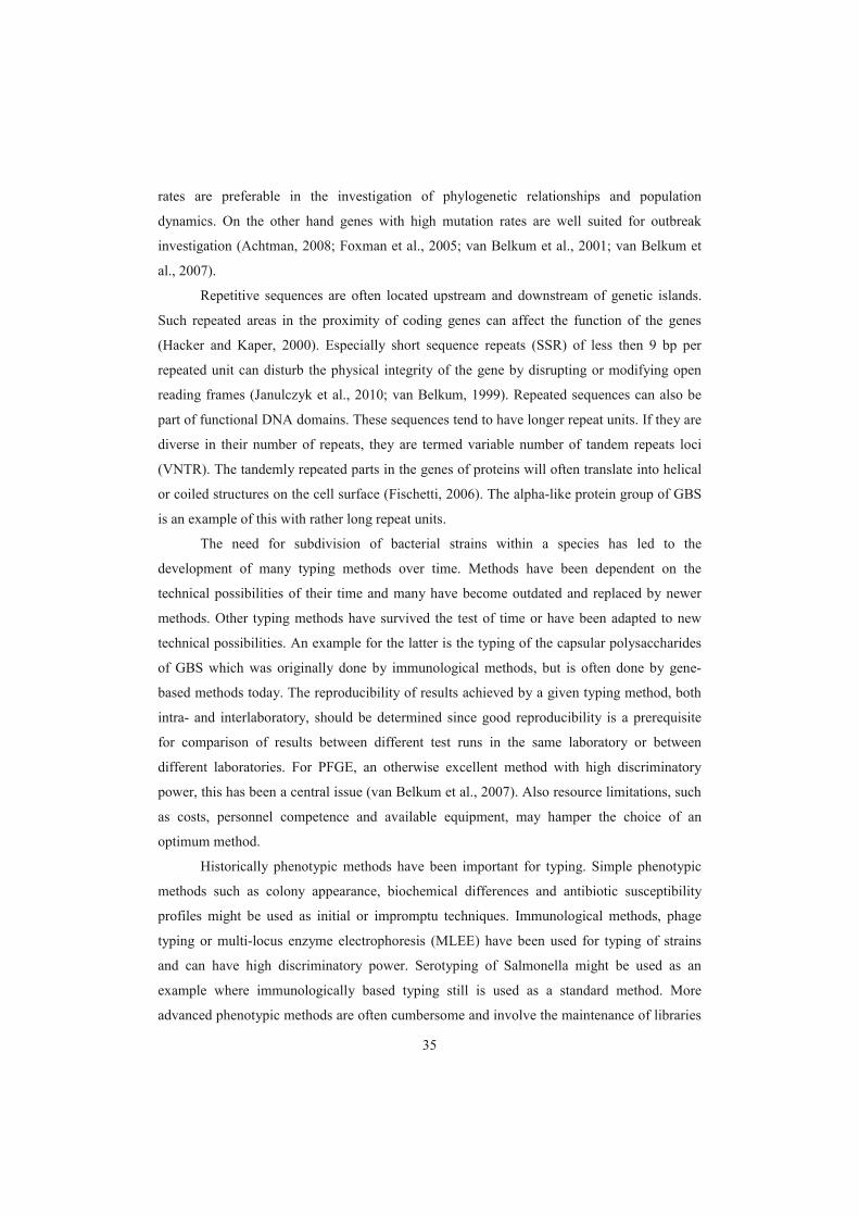
35
rates are preferable in the investigation of phylogenetic relationships and population
dynamics. On the other hand genes with high mutation rates are well suited for outbreak
investigation (Achtman, 2008; Foxman et al., 2005; van Belkum et al., 2001; van Belkum et
al., 2007).
Repetitive sequences are often located upstream and downstream of genetic islands.
Such repeated areas in the proximity of coding genes can affect the function of the genes
(Hacker and Kaper, 2000). Especially short sequence repeats (SSR) of less then 9 bp per
repeated unit can disturb the physical integrity of the gene by disrupting or modifying open
reading frames (Janulczyk et al., 2010; van Belkum, 1999). Repeated sequences can also be
part of functional DNA domains. These sequences tend to have longer repeat units. If they are
diverse in their number of repeats, they are termed variable number of tandem repeats loci
(VNTR). The tandemly repeated parts in the genes of proteins will often translate into helical
or coiled structures on the cell surface (Fischetti, 2006). The alpha-like protein group of GBS
is an example of this with rather long repeat units.
The need for subdivision of bacterial strains within a species has led to the
development of many typing methods over time. Methods have been dependent on the
technical possibilities of their time and many have become outdated and replaced by newer
methods. Other typing methods have survived the test of time or have been adapted to new
technical possibilities. An example for the latter is the typing of the capsular polysaccharides
of GBS which was originally done by immunological methods, but is often done by gene-
based methods today. The reproducibility of results achieved by a given typing method, both
intra- and interlaboratory, should be determined since good reproducibility is a prerequisite
for comparison of results between different test runs in the same laboratory or between
different laboratories. For PFGE, an otherwise excellent method with high discriminatory
power, this has been a central issue (van Belkum et al., 2007). Also resource limitations, such
as costs, personnel competence and available equipment, may hamper the choice of an
optimum method.
Historically phenotypic methods have been important for typing. Simple phenotypic
methods such as colony appearance, biochemical differences and antibiotic susceptibility
profiles might be used as initial or impromptu techniques. Immunological methods, phage
typing or multi-locus enzyme electrophoresis (MLEE) have been used for typing of strains
and can have high discriminatory power. Serotyping of Salmonella might be used as an
example where immunologically based typing still is used as a standard method. More
advanced phenotypic methods are often cumbersome and involve the maintenance of libraries

36
of antisera, standardization problems and problems of reproducibility. Such methods often
depend on specialized personnel for interpretation. Raman spectroscopy is a new promising
typing method which might be considered as a phenotypic method. It has the potential of
rapid, high throughput typing of bacteria without extensive expert hands-on work (Willemse-
Erix et al., 2009).
Methods based on the investigation of the genome of microbes have over the past 20
years in many settings replaced phenotypic methods and offer several advantages such as
usually better resolution and in many cases better reproducibility. Such molecular methods are
often technically advanced and may therefore be less convenient in resource limited settings
which is often the case in developing countries. A wide variety of typing methods has been
developed to investigate the diversity of bacterial genomes. Several methods use DNA
hybridization where immobilized DNA is probed with selective DNA molecules. Array
technology uses hybridization with a large number of targets and is able to screen all
identified genes in prokaryotic species. Fragment based methods use restriction enzymes and
analysis of the fragments by e.g. gel electrophoresis. Such methods might be combined with
PCR-amplification steps such as in amplified restriction fragment length polymorphism
(AFLP). PCR has been used in different typing methods, e.g. to amplify regions of known
variability such as in multi-locus variable number of tandem repeats assays (MLVA).
Methods based on sequencing of single or several loci in the genome are often used and have
become the most important typing methods in some bacterial species. MLST, discussed
below, is the main example of this (Pourcel and Vergnaud, 2011). A typing method targeting
the gene for Staphylococcus aureus protein A, spa, uses a combination of the size variability
of the gene due to VNTR and the variability of the sequence of the locus resulting in excellent
discriminatory power (Harmsen et al., 2003; Koreen et al., 2004).
The genomes of bacterial species have different levels of diversity. Some species are
considered monomorphic such as Bacillus anthracis, Yersinia pestis and the Mycobacterium
tuberculosis complex. These bacteria are thought to have emerged relatively recently as
pathogens in humans (Achtman, 2008). Typing of these species is challenging because most
of the genome is highly conserved. The sequencing of housekeeping genes did therefore
uncover little or no diversity and MLST was consequently not suited for these species
(Achtman, 2008). Several other methods have been developed for monomorphic species,
however the use of MLVA has gained general acceptance for this group of species. The
analysis of CRISPR (clustered regularly interspaced short palindromic repeats) loci as a

37
related method has been introduced for species, e.g. M. tuberculosis (Pourcel and Vergnaud,
2011).
The analysis of the whole genome of strains revealed that the methods mentioned
above are not able to describe the complete genetic diversity of a bacterial genome (Tettelin et
al., 2005). In contrast, whole-genome sequencing allows a much deeper understanding of the
evolution of a bacterial species either within the species or in comparison to other species.
Genetic sequencing has become accessible for more and more laboratories since the costs
have decreased dramatically. While sequencing itself has become affordable, the costs for the
subsequent bioinformatic work and necessary confirmatory experiments have not changed in
the same way. Therefore the assembly of a sequenced genome into contigs that allow
interpretation and comparison with other genomes is still a considerable challenge.
(Nagarajan et al., 2010).
1.7 Typing of GBS
Typing of GBS is done to follow the overall composition of GBS strains in a
geographic region, for research purposes or for outbreak investigations e.g. in hospitals or in
cattle herds. Typing of GBS starts usually with the determination of its capsular
polysaccharide and, in some centers, of the strain variable Alp surface proteins. In both cases
immunological methods have traditionally been used, while in later years, methods testing for
the gene-content of the CPS or proteins have become available. A method combining
multiplex PCR and reverse line blotting integrates typing of CPS, surface proteins and
antibiotic resistance genes in one assay (Kong et al., 2005; Zeng et al., 2006). Even if these
markers are combined, their discriminatory power is comparably low and therefore
supplementary methods have been developed of which pulsed-field gel electrophoresis
(PFGE) and multi-locus sequence typing (MLST) have been most frequently used in recent
years. PFGE is used as a tool for high resolution typing to elucidate epidemiological relations
between strains. Multi-locus sequence typing (MLST) was introduced for GBS in 2003 and is
well suited for phylogenetic typing to follow long term developments in strain collections but
might also be used for epidemiological typing (Jones et al., 2003). Both PFGE and MLST
require sophisticated equipment, well trained personnel and are expensive. Newer more
simple and less expensive methods have also been developed such as the MLVA described in
this work or analysis of sets of single-nucleotide polymorphisms (SNPs) (Honsa et al., 2008).

38
1.7.1 Capsular polysaccharides
The CPS antigens can be detected by antibodies raised in rabbits or mice. The classic
immunoprecipitation method was described in the 1930s (Lancefield, 1934). Fluorescent
antibody tests (FAT) (Bevanger and Maeland, 1977) or agglutination of latex particles
(Slotved et al., 2003) have also been used. The degree of encapsulation of GBS varies
between strains; especially bovine strains can have little or no capsule (Madoff et al., 2006).
Such strains tend to be difficult to type with immunological methods.
Horizontal transfer of genes that are relevant for the synthesis and assembly of the
CPS may occur en-bloc and cause a switching of CPS type (Cieslewicz et al., 2005; Luan et
al., 2005). This capsule switching may disrupt the phylogenetic relationship of a strain and
assign it to a serotype which is not in concordance with results of other typing methods such
as MLST. This has to be kept in mind when interpreting CPS typing results.
Several groups employ in-house methods for typing of CPS such as our group which
uses indirect fluorescence antibody test with antibodies raised in rabbits or mice. The Statens
Serum Institut in Copenhagen produces a commercially available latex based kit which is
widely used by reference laboratories (Afshar et al., 2011).
The sequences of the variable CPS type-specific regions have been available the last
ten years (Cieslewicz et al., 2005; Kong et al., 2002; Slotved et al., 2007). This made it
possible to design PCRs for CPS genotyping both as single or multiplex PCRs (Imperi et al.,
2009; Kong et al., 2005; Poyart et al., 2007). As mentioned above, PCR based typing showed
that strains which were found non-typable by immunological methods, did usually harbor the
genetic information for the synthesis of these polysaccharides. This problem seems to be
especially common in bovine strains (Zhao et al., 2006b).
1.7.2 Surface proteins
Some groups have also used the strain variable surface proteins for subtyping of CPS
types. The proteins typically appear in combination with certain CPS-types, e.g. III and R4 or
unusual CPS/protein combinations may be interesting
epidemiologically. Surface protein typing can be done by immunologic methods such as
immunoprecipitation or FAT. Several PCR-based methods for detection of the genes coding
for these proteins have been published. Multiplexed PCRs are a convenient approach (Creti et
al., 2004). As with CPS, immunologically non-typable strains usually harbor a surface protein
encoding gene (Imperi et al., 2009).

39
1.7.3 Pulsed-field gel electrophoresis
Pulsed-field gel electrophoresis (PFGE) has been used for typing of GBS since the
early 1990s (Fasola et al., 1993). The method is based on the macrorestriction of bacterial
DNA by an endonuclease into large fragments. These fragments are then separated by gel
electrophoresis where the electric field switches its direction, thereby allowing the large DNA
fragments to migrate through the gel. An image of the distribution of bacterial genomic
fragments in the gel is interpreted visually or by dedicated software. As for many other
bacterial species PFGE is a convenient method for typing of GBS. This method has however
some known drawbacks. The procedure is laborious and requires specific, expensive
equipment. It is difficult to obtain comparable images of the same strain in different
laboratories with PFGE. This requires rigorously standardized protocols and quality control
(van Belkum et al., 2007).
For PFGE analysis of GBS the restriction enzyme SmaI, which cleaves the DNA chain
relatively infrequently, is usually chosen. In an investigation of a broad selection of 78
Norwegian strains, Skjaervold et al. could distinguish 62 different types (Skjaervold et al.,
2004). In an article by Tenover on interpretation of PFGE, it is suggested that at least ten
fragments should be identifiable on the gel for sufficient discrimination (Tenover et al., 1995).
In GBS this is in our hands often not achieved as a number of strains only produce 7-9 bands.
Therefore PFGE with SmaI might be less discriminative for GBS compared to other species.
Other restriction enzymes might be added for additional resolution but this would add to the
complexity of the method.
1.7.4 Multi-locus sequence typing
In 2003 Jones et al. published a multi-locus sequence typing (MLST) system for GBS
(Jones et al., 2003). It is based on the sequencing of 400-500 bp parts of seven housekeeping
genes. Since mutations in these genes are usually not well tolerated, successful mutations
occur less often than in other parts of the genome. A web-based database is available for
comparison and registration of new GBS sequence types (http://pubmlst.org/sagalactiae/).
Over 570 different strains are registered in the database as of January 2012. Most of these
belong to clonal complexes (CC) of which four seem to be especially predominant: CC1,
CC17, CC19 and CC23 (Brochet et al., 2008). One of the most important findings of MLST
was the high prevalence of CC17 among strains from invasive neonatal disease. CC67 is often
found in strains from bovine mastitis (Sørensen et al., 2010).

40
Drawbacks of MLST are its comparable high costs, a sample processing time of 3-4
workdays and the need for a considerable amount of expert hands-on work. This has led to the
development of simpler methods which try to assign presumptive sequence types for the most
important types by PCR or SNP analysis (Honsa et al., 2008; Lamy et al., 2006).
1.7.5 Other typing methods used
Other less frequently used methods for typing of GBS include restriction fragment
length polymorphism analysis (RFLP) (Blumberg et al., 1992), ribotyping (Blumberg et al.,
1992; Huet et al.), multi-locus enzyme electrophoresis (MLEE) (Quentin et al., 1995), random
amplification of polymorphic DNA-analysis (RAPD) (Limansky et al., 1998; Zhang et al.,
2002) and amplified cps restriction polymorphism analysis (Manning et al., 2005).
1.7.6 Multi-locus variable number of tandem repeats assay
Repetitive sequences which may be located both in bacterial genes or intergenic
regions are common in prokaryotic genomes (Lindstedt, 2005). Repetitive sequences are
challenging for the genetic copying system of bacteria and deletion or insertion of one or
several repeats can occur. This leads to a different number of repeats in the bacterial
descendant and consequently a different size of the locus (Figure 4). The molecular model for
this phenomenon is termed slipped strand mispairing (Torres-Cruz and van der Woude, 2003).
A single gene locus with such variations is called a variable number of tandem repeats locus
(VNTR). If mispairing occurs it can be repaired by a DNA repair system but the efficiency of
these systems is variable (Caporale, 2003). As mentioned previously repeated sequences
found in intergenic regions are often polymorphic (Pourcel and Vergnaud, 2011). Repeats can
also be located in a promoter region of a gene where they can influence the strength of a
promoter and cause variation in gene expression (Martin et al., 2003; Willems et al., 1990).
Repeat count variations inside coding regions of genes have been shown to be involved in
adaption of bacteria to the host immune system as has been described for the surface proteins
of GBS. If the repeat sequence is not a multiple of 3 bp, changes in the repeat number will
lead to a frameshift and may result in alterations of gene functions. Other repeated sequences
are well conserved which might indicate that they are vital for the correct function of a gene
(Pourcel and Vergnaud, 2011). For typing purposes variations in the number of repeats results
in size variations of the locus which are detectable in PCR products.

41
Software such as the Tandem Repeats Finder (http://tandem.bu.edu/trf/trf.html) is
available for screening of genomes for repeated sequences (Benson, 1999). When more than
one sequenced genome is available the sequences can be compared and polymorphism in
VNTR loci uncovered in silico (Figure 4). Several VNTR loci can be combined into a multi-
locus variable number of tandem repeats assay, sometimes also called multi-locus variable
repeats assay (MLVA). A prerequisite for the design of a MLVA is the availability of at least
one fully sequenced genome of the targeted species. It is therefore not surprising that the first
MLVA published was for typing of Haemophilus influenzae, the first fully sequenced
bacterium (van Belkum et al., 1997).
The H. influenzae MLVA was described in 1997, about the same time as the first
MLST (Maiden et al., 1998). In the following years MLVA did not gain the same acceptance
as MLST. This situation changed somewhat when MLVAs were developed for monomorphic
species such as Bacillus anthracis and Yersinia pestis (Kattar et al., 2008; Keim et al., 2000;
Le Fleche et al., 2001). For typing of these species MLVA was an important improvement
since other typing methods were not able to uncover sufficient diversity or the results were
not considered to reflect the phylogeny of the species (Achtman, 2008). MLVA has gained
increasing acceptance for several enteropathogenic bacteria for which outbreak investigations
are often needed and where comparisons between laboratories are of great benefit, such as
Salmonella typhimurium or enterohemorragic E. coli (Lindstedt, 2011). Generally the MLVA
method is reproducible, rapid, and easier to perform and produces data that are easier to
analyze and share via databases than comparable methods such as PFGE or MLST.
Standardization of MLVA regarding choice of loci, laboratory protocols and nomenclature is
an important issue (Larsson et al., 2009; Lindstedt, 2011; Pourcel and Vergnaud, 2011).
MLST in contrast is a more standardized procedure where the choice of loci is usually given
and the underlying evolution of the housekeeping genes is known. This may explain the
broader acceptance of MLST compared to MLVA.

42
Technically MLVA is a fast and comparably straightforward typing method. A PCR
spanning the whole repeat locus is designed by locating suitable primer sites as close as
possible upstream and downstream of the repeated region. These flanking sequences should
be conserved. For a MLVA, several VNTR loci may be amplified simultaneously in a
multiplex PCR. Variable numbers of repeats will yield PCR products of different sizes and
this can be determined either by gel or capillary electrophoresis. When using gel
electrophoresis the results may be determined against reference strains with known numbers
of repeats. This method will need a certain repeat size since short sequence repeats (SSR)
smaller than approximately 9 bp will be difficult to distinguish on a gel and excludes
multiplexing of PCRs (Pourcel and Vergnaud, 2011). Capillary electrophoresis by a genetic
analyzer will generate a quite accurate estimate of the base pair count of the fluorescently
labeled PCR products. Therefore also small repeats of 6 or even 3 bp can be separated in a
MLVA using capillary electrophoresis. On the other hand, interpretation of PCR products
larger than the upper limit of the size standard used will be difficult. The base pair count
>1993465-1993889 Streptococcus agalactiae A909, complete genomeTTTCAATAGCTTTTTTAACCGCCAAGTTTCCGCTAGTATTAACCGATTTTTTGGTTGCTGGTTTGGCTTCTGGTTTAGCCTCTGGCTTAACGTCTGGTTTAACTTCTGGCTTAGCCTCTGGTTTGGCCTCTGGCTTAACGTCTGGTTTAACTTCTGGCTTAGCCTCTGGTCTGGCCTCTGGCTTAACGTCTGGTTTAATTTCTGGTTTAGCCTCTGGTTTGGCTTCTGGCTTAGCCTCTGGTTTAACTTCTGGCTTAACGTCTGGCTTAACGTCTGGCTTAACATCTGGTTTGGCTTCTGGTTTAACTTCTGGCTTAGCTTCTGGTTTGGCTACTTTTTTATCTTTTAATTCTAAACTTTGTAAAGCATCACGTAGCTTGTTAGCTAATTGATTTGCTTGATCAGTGGTAATAAATTTTCCCTCT
>2089975-2090315 Streptococcus agalactiae NEM316, complete genomeAATAGCTTTTTTAGCCGCCAAGTTTCCGCTAGTATTAACCGATTTTTTGGTTGCTGGTTTGGCCTCTGGTTTAGCTTCTAGCTTAGCCTCTGGTTTAGCTTCTGACTTAGCCTCTGGTTTAGCTTCTGGCTTAGCCTCTGGTTTAACGTCTGGTTTAGCTTCTGGCTTAACGTCTGGCTTAGCTTCTGGTTTGGCCTCTGGCTTAGCTTCTGGCTTAGCTTCTGGCTTAGCTTCTGGCTTAGCTTCTGGTTTGGCCTCTGGTTTGGCTTCTGGTTTGGCTACTTTTTTATCTTTTAATTCTAAACTTTGTAAAGCATCACGTAGCTTGTTAGCTAATTGAT
Figure 4: The same VNTR locus in two fully sequenced GBS strains. In strain A909 this locus has 23repeats, in strain NEM316 it has 19 repeats. The VNTR is used in our MLVA and designated SATR3. Underlined, primer sites. Note some sequence variation between single repeats.

43
generated by capillary electrophoresis allows an exact calculation of the repeat count. For this
the offset must be subtracted from the PCR product size and the result must be divided by the
repeat size. The offset consists of the primers and the sequences between the primers and the
start/end of the repeat sequence (Figure 5).
1.7.6.1 Analysis of MLVA results
Typing methods classify the individuals of a population into groups. Both the number
of groups and the size of each group generated by the method have to be taken into account to
make predictions on the discriminatory power of a method. Simpson’s index of diversity is a
statistical tool to calculate this (Simpson, 1949). By calculating Simpson’s index for different
typing methods used to type the same strain collection, the methods can be compared
regarding their ability to discriminate. The index should only be used when the strain
collections are epidemiologically unrelated (Hunter and Gaston, 1988). The modifications of
Simpson’s index described by Hunter and Gaston are especially useful in small strain
collections or if the diversity index is less than 0.90.
Comparison of VNTR loci or MLVA profiles may be done using different algorithms
suitable for the analysis of character data sets. Most often the distance coefficient used is a
categorical coefficient (Hamming’s distance), e.g. given a repeat copy number of five for a
locus the distance to a repeat copy number of six is considered equally distant as a repeat
count of nine. The generated distance coefficient can be used for clustering for which several
methods are available. In MLVA the unweighted pair group method using average linkages
(UPGMA) is often used, a relatively simple algorithm assuming that the rate of evolution is
repeated sequence
repeat singleoflengthoffset)Roffset(Fbpnumberrepeat
Examples:
SATR 2: fragment analysis result 225, offset 85, repeat length 18 = 8 repeats
SATR 3: Fragment analysis result 562, offset 92, repeat length 12 = 39 repeats
F primer R primer
F offset R offset
Figure 5: Illustration of the structure of a VNTR and flanking sequences. The offset is the primer plus the nucleotides between primer and start/end of the repeat sequence. Examples for the calculation of a repeat number from fragment analysisfor GBS-VNTR loci SATR2 and SATR3. F forward, R reverse, bp basepair count of repeated sequence.

44
constant in all branches of the tree (Dethlefsen et al., 2011; Pourcel and Vergnaud, 2011). If a
locus mutates in a plus or minus one repeat (stepwise) fashion other distance coefficients
should be chosen. The mutation pattern for each VNTR locus should be determined
experimentally (Vogler et al., 2007).

45
2 Aims of the studyThe general aim of the study was to investigate the potential of molecular methods to achieve
more discriminatory and robust typing of GBS. More specific aims were to:
study the relationship between results of serotyping and genotyping of GBS surface
proteins
develop a MLVA typing method for rapid, highly discriminatory genotyping of GBS
show the feasibility of and validate the developed MLVA method on a well
characterized strain collection
investigate the applicability of the MLVA in circumstances where epidemiologically
related strains are expected.
3 Material and Methods
3.1 Strain collections
Previously well characterized collections of human GBS strains were used for papers I and II,
while bovine GBS strains were investigated for paper III. In the study presented in paper I 147
strains were selected from a larger collection of strains from New Zealand and Australia. The
selection included all ten CPS types and the most common CPS types were represented in
higher proportions than the less common. The strains were in part (129 isolates) from invasive
cases, these were also used in a previous study (Zhao et al., 2008), and in part (18 isolates)
from antenatal, vaginal swabs. For paper II 126 strains were included. Of these, 113 were
invasive strains from neonates born in Norway which were routinely submitted to the national
GBS reference laboratory at St. Olavs University Hospital in Trondheim from other medical
microbiology laboratories nationwide. Additionally nine international reference strains, two
CPS type IX strains from a Danish collection and two colonizing strains from a Zimbabwean
collection were included. For paper III all bovine GBS isolates (n=148) identified by the
Trondheim section of the Norwegian Veterinary Institute between April 2007 and November
2010 were included. The samples were taken from individual udder quarters when considered
indicated by the referring veterinarian or farmer. Additionally 39 strains from the four other
sections of the Norwegian Veterinary Institute (Oslo, Sandnes, Bergen and Harstad) and from
the Tine Norwegian Dairies BA, Mastitis Laboratory in Molde were included.

46
3.2 Typing methods
Paper I was the result of cooperation between the Centre for Infectious Diseases and
Microbiology of the Westmead Institute in Sydney, Australia and the Norwegian national
reference laboratory for GBS at St. Olavs University Hospital in Trondheim, Norway. The
Australian group had established a gene-based typing method for GBS to detect the genes for
the strain variable surface proteins of the alpha-like protein (Alp) family bca (encoding the
protein), alp1 (Alp1), alp2 (Alp2), alp3 (Alp3), alp4 (Alp 4) and rib (R4/Rib). Additionally
encoding gene bac was analyzed. The method was based on multiplex PCR
followed by the demonstration of the PCR products by reverse line blot hybridization (Kong
and Gilbert, 2006; Zhao et al., 2006a). The group in Trondheim serotyped the same strains by
fluorescent antibody tests based on in-house antisera. Of the seven antisera used, sera reacting
with C were monoclonal antibodies raised in mice
(Bevanger et al., 1992; Kvam et al., 1999; Maeland et al., 2004; Naess et al., 1991) and sera
reacting with Alp2/3 common, Alp2 specific and R4 specific were polyclonal antibodies
raised in rabbits (Maeland et al., 2004; Moyo et al., 2001).
Paper II described the construction and assessment of a MLVA for GBS. All strains
used in the study were typed for CPS, surface proteins and MLST. For the Norwegian strains
the results for these three markers were known from a previous study (Bergseng et al., 2009).
The MLVA was developed by exploring the three fully sequenced and annotated genomes of
GBS reference strains A909, NEM316 and 2603V/R for repeated sequences using the
Tandem Repeats Finder program and other software. Eighteen candidate loci were identified
and tested by PCR and gel electrophoresis or using the Agilent 2100 Bioanalyzer with the
DNA 1000 chip (Agilent Technologies, Santa Clara, CA). This led to the selection of five
VNTR loci which were combined to a MLVA. In a further step a multiplex-PCR including all
five loci was constructed using the Qiagen multiplex-PCR kit (Qiagen, Hilden, Germany).
The forward primers for the five loci were fluorescently labeled and the nucleotide count of
the PCR product was estimated in a capillary electrophoresis instrument (ABI 3130xl Genetic
Analyzer; Applied Biosystems, Foster City, CA) using the LIZ1200 size standard. This
standard allows estimates of PCR product sizes up to 1200 bp. In paper III, the same MLVA
methods as in paper II was used except for a modification of the multiplex-PCR. Here the
SATR5 locus was amplified in a separate PCR reaction. In this paper a repeat count of zero
indicated that no PCR product was detected(Figure 1, Paper III).

47
3.3 Analysis of results
In paper II Simpson’s index of diversity was calculated. Phylogenetic trees were generated
with the Bionumerics software (Applied Maths, Sint-Martens-Latem, Belgium) version 6.0
(paper II) and 6.1 (paper III). Phylogenetic trees were generated using the Bionumerics
software. The algorithm used was the unweighted pair group method with arithmetic means
(UPGMA) and categorical values.
4 Results
4.1 Paper I
Classic serotyping and genotyping of the strain variable GBS surface proteins were compared
in a selected panel of 147 human strains. The comparison of the two methods employed is
complex and reflects the complicated immunology of these proteins. At least one surface
protein was detected by serotyping in 123 (83.7%) of the strains, while a surface protein
encoding gene was detected in all but one of the strains (146 strains, 99.3%). In total, 122 of
147 strains were typable by both antibody-based and molecular methods. Results were
concordant for 90 (73.8%) strains, partially concordant for 29 strains (23.8 %) and discordant
for three (2.5 %). When the serotyping result was C /Alp1, either bca or alp1 was present.
The bca gene was detected in 41 isolates and alp1 in 30. Of these 71 strains, 55 (77.5%) were
serotyped as C /Alp1. The monoclonal antibody used cannot distinguish between C and
Alp1. was identified in only nine of 31 isolates in which the corresponding gene, bac was
detected. The discordant results occurred mainly with the polyclonal antibodies detecting
Alp2 and Alp2/3 common (Table 1, Paper I). Ten of 24 non-serotypable strains contained rib
which means that the polyclonal antibodies did not recognize the protein in 22 of 32 strains
genotyped as rib. We detected R3, for which no corresponding gene has been identified, in
five isolates (3.4%).
The genotyping approach did very well in identifying the genes for surface
proteins. The problem of cross-reactivity which hampers the performance of serotyping is not
an issue in genotyping. Especially typing problems in both Alp3, which is an immunologic
chimera of Alp2 and R4 without known antigenic sites of its own, and of C and Alp1, which
are hardly distinguishable immunologically (Brady et al., 1988; Kvam et al., 2011), were not
encountered. The only non-typable strain by genotyping posed also difficulties in the
serotyping approach and no definite type could be assigned (result: Alp2 or 3).

48
4.2 Paper II
4.2.1 Construction of a GBS-MLVA
In this study a MLVA was constructed consisting of five VNTR loci in the genome of
GBS. The applicability of the MLVA was demonstrated on a collection of 126 well
characterized invasive strains. The construction of the MLVA involved several steps. An
initial in silico analysis of the three complete genomes of strains A909, NEM316, and
2603V/R identified 18 candidate loci which were analyzed further by designing PCR primers
and examining these in eleven selected strains. Seven loci were not found suitable for further
analysis and were therefore excluded. In six of these no or little diversity was found while
problems to obtain a PCR product led to the exclusion of the last. The remaining eleven loci
were tested in all strains. In this part of the study seven loci were found to have three or more
alleles. Of these the five most diverse were chosen. In each of the three loci designated
SATR1, TR9 and TR13 three different alleles were found among the 126 strains. Based on the
results of the calculation of Simpson’s index of diversity for each locus only SATR1 was
included in the MLVA (Table 2, Paper II). For each of the four other loci included in the
MLVA four or more alleles were detected.
The location of the five loci in the genome of the three reference strains 2603V/R,
NEM316 and A909 and the designation of the loci are given in Table 1. The five loci had
different characteristics which can be summarized briefly as follows:
1. SATR1 is a CRISPR-like region with 16 bp direct repeats and 44 bp spacer regions,
each repeat/spacer was therefore 60 bp long. We did only find three different alleles in
our collection of human strains and a fourth in our bovine collection, while a Russian
group encountered eight different types in their work with the same region
(Rozhdestvenskaya, 2008).
2. The repeats in the SATR2 locus had a repeat size of 18 bp. Nine different alleles were
found. Strains devoid of a PCR product were also considered as one type; this result
was encountered in 14 of the 126 strains.
3. The SATR3 locus had a repeat unit of 12 bp. In this strain collection 19 different
alleles were found, rendering SATR3 the second most diverse locus. The repeated
sequence is part of a predicted surface-anchored protein designated BibA (Santi et al.,
2007). Large PCR products, between 736 and 750 bp with the chosen primer set, were
encountered in 31 strains. Sequencing of PCR products demonstrated that the strains
with the largest products had had two copies of an insert of 216 bp. All but one of

49
these strains were of MLST clonal complex 17. This insert has also been proposed as a
target for a PCR for the rapid presumptive identification of ST17 (Lamy et al., 2006).
In 25 strains no PCR product was found, most of these were of serotype III/R4 and of
clonal complex 19.
4. Amplification of the SATR4 locus resulted in the smallest PCR products of the five
loci. The underlying repeat of 18 bp was quite degenerated; in some strains part or all
of the repeats consisted only of 15 bp. For convenience of typing all alleles were
assigned as times of 18 bp in the calculation of repeat numbers. A PCR product was
observed in all strains and five different types were present in this collection. The
repeat is located in the pcsB gene, encoding a protein found to be important for cell
wall separation (Reinscheid et al., 2001).
5. The SATR5 locus was the most diverse separating the strain collection into 24
different types. The underlying repeat is rather large with 48 bp and strains with more
than 20 repeats were found in some strains. Five strains had more than 21 repeats,
resulting in PCR products which exceeded the size standard limit of 1200 bp. In the
analysis they were considered as one type with the arbitrary repeat number of 50
although they showed diversity among each other by standard gel electrophoresis.
When the PCR product was equivalent with the offset (21 strains) the strain was
usually of MLST19. A result of six repeats was found in 22 strains, 18 of which
belonged to ST17. SATR5 is located in the gene encoding the fibrinogen binding
protein FbsA (Rosenau et al., 2007; Schubert et al., 2002).

50
4.2.2 Comparison between MLVA, serotyping, and MLST
MLVA analysis using the five proposed loci differentiated the 126 strains into 70 different
types, considerably more than MLST which differentiated them into 36 STs. The two largest
groups of strains with identical MLVA profiles consisted of 19 and 13 strains, respectively.
Of the former, 16 belonged to ST17 and 9 of the latter to ST19. All 27 of the ST17 strains in
this work clustered together but were resolved into 12 different MLVA types within this
cluster. Apart from the ST17 MLVA profile present in 16 strains, the eleven remaining
profiles were found in single strains only (Fig. 2, Paper II). Similarly, all 11 CPS III/ST19
strains were assigned to the same MLVA cluster and resolved into four MLVA types; 7 of the
strains had identical profiles. Most of the type V strains belonged to ST1 (21 out of 30 type V
Table 1: Location of the five MLVA Loci. Position and annotation in reference genomes.
2603V/R NEM316 A909 Note
SATR1 142905-143227
Hypothetical protein
SAG0134
142937-143139
Intergenic region
152958-153160
Hypothetical
protein SAK_0192
CRISPR
neighbouring
transposase
(sequence
inversed)
SATR2 666413-666676
Hypothetical protein
SAG0677
Not found 745915-746218
Hypothetical
protein SAK_0805
SATR3 2043303-2043801
Pathogenicity
protein SAG2063
2090028-2090347
Surface adhesion
protein gbs2018
1993422-1993889
Intergenic region
Upstream of a
neighbouring
translocase
(sequence
inversed),
designated BibA
SATR4 29490-29744
PcsB protein
29563-29817
PcsB protein
29486-29698
PcsB protein
Involved in cell
wall seperation
SATR5 1062519-1062709
Intergenic region
(no repeats)
1132283-1133330
Hypothetical protein
gbs1087
1118169-1119009
Fibrinogen binding
protein SAK_1142
Sequence
inversed, FbsA
protein

51
strains); 20 of these 21 were in a cluster consisting of 12 MLVA types, mostly because of
heterogeneity in SATR5.
4.3 Paper III
In this study 187 bovine GBS strains were investigated by genotyping of CPS and
surface proteins and by MLVA. The following CPS types were found: Ia (seven strains, 3.7
%), Ib (six strains, 3.2 %), II (three strains, 1.6 %), III (three strains, 1.6 %), IV (64 strains,
34.2 %), V (101 strains, 54.0 %), IX (one strain, 0.5 %) and two strains (1.1 %) were non-
typable. CPS type V and type IV accounted together for 88 % of the 187 strains, an
overrepresentation compared to other strain collections. Genes for five of the six strain
variable alpha-like proteins were found except the rarely encountered Alp4; additionally the
The combination of CPS and protein typing separated the strain
collection into twelve different types (Table 2, Paper III).
The MLVA was equally applicable on bovine strains as on human strains. The 187
strains were differentiated into 37 MLVA profiles (Figure 1, Paper III). In 29 of 34 farms all
GBS isolates on each farm had identical MLVA profiles, different from strains from other
farms. On the remaining five farms more than one profile was encountered (Table 3, Paper
III). Usually one profile predominated while a single strain with a divergent repeat number in
one locus was observed. One MLVA profile was found on three farms in the same
municipality. This was presumably the result of transmission due to cooperation between
these farms. Three other profiles were found on several farms, however these were located in
different counties and an epidemiological connection was regarded unlikely.
Automated milking systems (AMS) were used on 12 of the 34 farms (35%). Not
unexpectedly, those farms which had invested in AMS had bigger herd sizes (61.4 versus 27.7
cows). Of the 187 strains, 133 were provided by farms with AMS (71%). Also the AMS farms
provided 0.18 isolates per cow against 0.09 isolates per cow in non-AMS farms. On average,
11 strains were available from farms with AMS compared to 2.45 strains from those with
conventional milking.
One farm was represented with 48 samples collected over a period of almost three
years. Four different MLVA profiles were found among these with polymorphisms occurring
in SATR5 of 13, 15, 17 and 50 repeats, respectively. In the first part of the observation period
isolates with 17 repeats occurred in 19 samples (sampled until September 2009), replaced

52
later by isolates containing 13 repeats (in 27 samples, collected from August 2009 onwards).
The other two profiles were only found in single strains.
Seven of the MLVA profiles from cattle were also encountered in our recent study on
human strains (Radtke et al., 2010). In a sub-analysis the relatedness of bovine and human
strains was investigated. 47 of the bovine strains (representing at least one strain per farm and
MLVA profile) and seven human strains were examined for lactose fermentation, bacitracin
sensitivity, and presence of the scpB gene. In this analysis all but one of the bovine strains
fermented lactose, while none of the seven human strains did. The scpB gene was found in
about two-thirds of our bovine strains while other investigators found that a negative result
was strongly associated to bovine strains (Franken et al., 2001; Sørensen et al., 2010). Also
Bacitracin was less reliable in differentiating between bovine and human strains.
5 General discussionIn this work we investigated selected typing techniques for GBS, i.e. serotyping and
genotyping of strain variable surface proteins and the MLVA developed in this study. The
typing of bacteria within a species is usually done for surveillance, epidemiologic or research
purposes. Which methods are chosen will depend on the scientific problem and the methods
available. Different methods have different resolution; i.e. the ability to classify a strain
collection into groups. A basic typing method, e.g. the typing of the GBS polysaccharide
capsule, will separate GBS strains into only ten different groups, while the perhaps ultimate
typing technique of the future, whole genome sequencing, must be expected to characterize
almost no strains as identical. This will lead to an extreme version of the problem common for
high resolution methods such as MLVA or PFGE, i.e. how much divergence between results
can be accepted before strains are no longer considered related or closely related, a question
discussed by van Belkum (van Belkum et al., 2007) or for the case of PFGE by Tenover
(Tenover et al., 1995). If strains are obtained over a longer period of time, such as the strains
in our paper III which were sampled over three years, a greater divergence of strains should
be tolerated in the comparison of strains since some degree of mutation has to be expected
over time.
Technical advances continuously generate new microbiological typing methods which
have to be compared with established techniques in order to investigate their congruence and
to secure continuity in the typing of bacteria. Such comparisons will enable the scientific

53
community to discuss allocations of strains into concordant groups on the background of
different typing methods.
In this study we compared immunological serotyping of surface proteins with newer
genotyping (paper I). Further we aimed at and succeeded in developing a MLVA method for
GBS and compared its typing results with genotyping of capsular polysaccharides, surface
proteins and MLST (paper II) and applied it in an investigation of strains from GBS mastitis
in cattle in Norway (paper III).
5.1 Typing of surface proteins
The main objective of paper I was to investigate if immunological serotyping and
genotyping of GBS surface proteins produced concordant results. Ideally both methods should
show complete concordance. This however was not to be expected since non-typable strains
are regularly encountered in serotyping and immunological cross-reactivity is a well known
problem in typing of Alp proteins (Lindahl et al., 2005). A previous study addressed this
problem for a limited number of surface proteins (Persson et al., 2008).
The Department of Medical Microbiology at St. Olavs University Hospital in
Trondheim holds an extensive collection of antibodies against the strain variable surface
proteins of GBS. For the current work, this was paired with a thoroughly designed genotyping
approach for these surface markers by the contributors from Australia (Zhao et al., 2006a).
Genotyping is usually a straightforward test and identifies the gene for the protein in question
if the corresponding PCR is carefully designed. Apart from the Alps the study also targeted
two proteins which are not part of the alpha-like protein family:
is well known and usually part of typing of surface proteins, R3 is less often looked for and in
contrast to the others the genetic sequence is not known and therefore no PCR is available.
For typing of GBS surface proteins the study indicates that genotyping should be
preferred, as it leaves only few strains non-typable and gives usually unambiguous results.
Serotyping may be used as a supplementary method. Immunologically the phenomenon of
non-typability or cross-reactivity might reflect problems with the specificity of the antibodies
or other assay related problems. However, this could also reflect changes in the proteins such
as deletion of repeats or mutations at antigenic sites leading to immunologic variation.

54
5.2 MLVA typing of GBS
The method of multi-locus variable number of tandem repeats analysis has several
advantages compared to other typing methods. MLVA uses PCR assisted amplification of
repeat loci and size assessment of the PCR products by electrophoresis. These are fast and
comparably straightforward methods using less complicated laboratory protocols compared to
e.g. MLST or PFGE. The equipment needed for MLVA is available in many microbiological
laboratories, especially when gel electrophoresis is used. The results can be given in repeat
count, particularly if PCR product size is analyzed by fragment analysis. Fragment analysis by
capillary electrophoresis gives an estimate of the PCR product size in base pairs. This can be
computed into the repeat count which is easily exchangeable between laboratories (Figure 5).
Websites for exchange of these data have been established for several bacterial species. The
resolution achieved by the method is depending on the variability and number of VNTRs
used. A published MLVA has to be considered as a selection of possible loci and further
investigation of the genome of a bacterial species might find additional loci which can be
combined with previously described into an even more discriminating assay.
These characteristics render MLVA a tool which is especially well suited for
investigation of outbreaks. For phylogenetic typing or other issues, where less discriminative
assays might be preferred, MLVA might also have a place, particularly when databases are
accumulating many strains/types and consensus on what to consider as related strains has
been reached (Pourcel and Vergnaud, 2011). Although variable genetic areas are often
associated with repeats, the speed of mutation can be very different. Depending on the
intention of the analysis, selecting VNTRs for phylogenetic studies or the evaluation of long-
term epidemiology might be possible. A selection of slowly mutating loci can result in a
MLVA with characteristics closer to MLST. Even without such a selection the phylogenetic
trees in paper II showed that the strains are grouped with good congruence with the results of
the phylogenetic reference method MLST, in which clonal complexes are usually thought to
reflect phylogenetic linages. The proposed MLVA could theoretically be used to assign
presumptive clonal complexes of MLST.
In Paper II we investigated the stability of loci by passaging three strains 40 times in
vitro without observing variability. Findings made in paper III indicated that the stability of
VNTR loci in GBS and their mutation rate could be observed in cattle in vivo. In this study of
Norwegian bovine GBS we had the opportunity to investigate strains which were collected on
distinct farms over almost three years. We detected repeat count changes on five farms. On

55
four of these, changes appeared at only one locus, on the fifth farm with two available
samples, differences at two loci were detected. The most notable example for variability was
observed in the SATR5 locus at a farm represented in the study by 48 samples. We propose
that the term Single Locus Variants (SLV), by analogy to MLST, may be applicable for such
MLVA results.
The epidemiology of GBS in humans has to take into account the large proportion of
gastrointestinal carriers of the bacterium. Disease arises from the translocation of the
bacterium into usually sterile compartments such as blood, cerebrospinal fluid or synovial
fluid. The bacterium is ubiquitous among humans and outbreaks are not common. Small
outbreaks have occurred on maternity wards or in dialysis units; however they are usually
limited to a small number of cases (MacFarquhar et al., 2010). In animals on the other hand
outbreaks of GBS disease are common, especially as mastitis in cattle (Keefe, 1997; Zadoks
et al., 2011). The collection of strains used in paper III reflects an increased occurrence of
GBS as a cause of mastitis in Norwegian cattle in recent years. The MLVA performed
excellently in discriminating the 187 bovine strains into 37 different profiles. Almost every
farm had its own profile and a farm-specific profile was usually not encountered on other
farms, as mentioned above and in the paper. This is in accordance with the information that
animals are usually isolated at their farm without the possibility of direct contamination from
other farms. Farms with long lasting GBS problems might therefore be used to study the
microevolution of a GBS strain genetically. For the case of MLVA, this might give
information about the in vivo stability of the selected VNTR loci under immunological
pressure.
A French group has recently proposed a MLVA for GBS consisting of six VNTR loci
(Haguenoer et al., 2011). Interestingly three of their loci are also included in our MLVA. An
additional VNTR selected by them was investigated by us (TR9 in paper II), but not included
into our MLVA. Haguenoer et al. also compared their MLVA with MLST results. While our
paper II focuses on pediatric invasive strains, their selection of 186 strains was broader with
human invasive, human colonizing and bovine strains. Their MLVA was as well able to group
strains with good congruence to MLST results. The group emphasizes that they looked for
VNTRs which were present in all their strains. This was done to avoid problems “in terms of
resolution and reproducibility”. Their argument for this was that a negative result could occur
either if the repeat locus is lacking or the primers fail to anneal due to variable flanking
regions. In our opinion this only represents a problem if different primers are used by
different groups. A negative result is also a valid typing result of a VNTR locus. When using

56
different primer pairs for the same VNTR it should be ascertained that they achieve identical
results. An eventual cumulative MLVA from these two works should have consensus primer
sets for those loci which are present by both approaches.
5.3 GBS and bovine mastitis
The epidemiology of GBS infections in bovines is strikingly different from
epidemiology in humans. The route of GBS transmission into herds is not obvious.
Colonization of the gastrointestinal tract does not seem to occur and the udder is thought to be
the only location of GBS infection (Agger et al., 1994; Zadoks et al., 2011). Therefore
eradication of GBS from bovine herds might be possible and has been widely achieved
through infection control measures in the 1970s and 1980s in Scandinavia (Katholm, 2010b;
Zadoks et al., 2011). The question of evolution of human and bovine GBS is of importance in
this setting and for human disease. If bovines are a reservoir for human disease this should be
addressed in the discussion of prevention of human disease. If human strains infect bovines,
total eradication of bovine disease will be difficult to achieve. Several investigators
addressing the relationship of human and bovine GBS tend to conclude that they are separate
entities (see chapter 1.4.3). Experiments from the 1980s with artificial infections of bovine
udders suggest that human strains could cause symptomatic mastitis but might not lead to
subclinical infection (Jensen, 1982).
A reemergence of GBS mastitis in Scandinavian farming has been observed over the
last 10 years (Katholm, 2010a; Persson and Landin, 2009; Zadoks et al., 2011). This is best
documented in Denmark, but has also been observed in Norway and Sweden. Several possible
reasons for this have been mentioned. Herd size has increased and GBS eradication is more
complex on such farms. Another development involves free stalls instead of boxed stalls,
facilitating more physical contact between the animals. Finally automated milking systems
have been introduced and are suspected to be associated with the reemergence of GBS
mastitis (Persson and Landin, 2009).
5.4 Limitations of the study
In paper II we chose to compare our MLVA with MLST. Considering that MLVA is
foremost a tool for high resolution typing which is well suited for outbreak investigation, a
comparison of the method with PFGE might have been equally interesting. Usually PFGE

57
will result in a higher resolution compared to MLST and might therefore be better suited for a
comparison of epidemiological typing methods. As mentioned above, PFGE in GBS with the
restriction enzyme Sma1 has in our hands not given the resolution expected from this method.
The MLST method used here is well suited for phylogenetic comparisons and the most
common used method for this kind of questions in later years (Feil, 2004; Pourcel and
Vergnaud, 2011). MLVA and MLST clustered the strains into similar groups and MLVA
could be used to assign a presumptive clonal complex to a strain, given more experience with
MLVA.
The problem of mutation frequency and pattern of VNTRs was addressed both in
Paper II and III but a thorough study of these questions should be performed with dedicated in
vitro experiments. The investigation of farms with long lasting GBS problems might be an
alternative way to observe this in vivo.
In paper III the strains were submitted to the microbiological laboratory at the farmer’s
or veterinarian’s choice. It is therefore not known if the strain collection was representative
and definite conclusions about the extent of the problem in Norway could not be made.
However, GBS mastitis was only very rarely encountered in the last two decades and the
frequency of GBS isolations in the Trondheim laboratory of the Norwegian Veterinary
Institute was considerably higher than in the other regions. Further prospective studies should
be done. In particular such studies might compare farms with and without AMS.
6 Conclusions
Typing of GBS surface proteins by immunological and molecular methods provides
concordant or partially concordant results in the large majority of strains.
Genotyping is superior to serotyping since it is able to type almost all strains and leads
to less ambiguity.
A MLVA typing scheme for GBS was designed based on five VNTR loci.
The MLVA performed excellently with very good discrimination. MLVA typed the
strains into epidemiological groups comparable to MLST and typing of CPS and
surface proteins.
MLVA analysis of bovine GBS allocated a specific genotype to almost every farm
while isolates from one farm were always identical or closely related.

58
7 Future aspectsRoutine typing of GBS surface proteins should be performed by PCR. However, for
confirmation of ambiguous PCR results and for immunological studies serotyping should be
available as a supplementary method. The gene encoding the R3 protein should be identified.
The MLVA method presented in this work should be validated further and used for
typing of strain collections from other geographic regions. Although some sequencing of PCR
products from the five MLVA loci was done during the development of the method,
sequencing of further alleles is desirable for verification and further study of the diversity of
the loci. The method presented here should be compared with the method published by
Hagenouer et al. Ideally consensus primers for the three common loci should be agreed upon.
Further a common database on the internet with a consensus designations should be
established. The mutation pattern for each locus should be determined since this would have
implications for phylogenetic analysis.

59
8 ReferanserAchtman, M., 2008. Evolution, population structure, and phylogeography of genetically
monomorphic bacterial pathogens. Annu Rev Microbiol 62, 53-70.Afshar, B., Broughton, K., Creti, R., Decheva, A., Hufnagel, M., Kriz, P., Lambertsen, L.,
Lovgren, M., Melin, P., Orefici, G., Poyart, C., Radtke, A., Rodriguez-Granger, J., Sorensen, U.B.S., Telford, J., Valinsky, L., Zachariadou, L., members of the DEVANI Study Group, Efstratiou, A., 2011. International External Quality Assurance for Laboratory Identification and Typing of Streptococcus agalactiae (Group B Streptococci). J. Clin. Microbiol. 49, 1475-1482.
Agger, J.F., Priou, C., Huda, A., Aagaard, K., 1994. Risk factors for transmission of Streptococcus agalactiae infection between Danish dairy herds: a case control study. Vet Res 25, 227-234.
Ayers, S.H., Rupp, P., 1922. Differentiation of hemolytic streptococci from human and bovine sources by the hydrolysis of sodium hippurate. Journal of Infectious Diseases30, 388-399.
Barcaite, E., Bartusevicius, A., Tameliene, R., Kliucinskas, M., Maleckiene, L., Nadisauskiene, R., 2008. Prevalence of maternal group B streptococcal colonisation in European countries. Acta Obstetricia et Gynecologica Scandinavica 87, 260-271.
Barkema, H.W., Green, M.J., Bradley, A.J., Zadoks, R.N., 2009. Invited review: The role of contagious disease in udder health. J Dairy Sci 92, 4717-4729.
Barocchi, M.A., Ries, J., Zogaj, X., Hemsley, C., Albiger, B., Kanth, A., Dahlberg, S., Fernebro, J., Moschioni, M., Masignani, V., Hultenby, K., Taddei, A.R., Beiter, K., Wartha, F., von Euler, A., Covacci, A., Holden, D.W., Normark, S., Rappuoli, R., Henriques-Normark, B., 2006. A pneumococcal pilus influences virulence and host inflammatory responses. Proc Natl Acad Sci U S A 103, 2857-2862.
Beckmann, C., Waggoner, J.D., Harris, T.O., Tamura, G.S., Rubens, C.E., 2002. Identification of novel adhesins from Group B streptococci by use of phage display reveals that C5a peptidase mediates fibronectin binding. Infect Immun 70, 2869-2876.
Bedford, H., de Louvois, J., Halket, S., Peckham, C., Hurley, R., Harvey, D., 2001. Meningitis in infancy in England and Wales: follow up at age 5 years. BMJ 323, 533.
Benson, G., 1999. Tandem repeats finder: a program to analyze DNA sequences. Nucleic Acids Res 27, 573-580.
Bergh, K., Stoelhaug, A., Loeseth, K., Bevanger, L., 2004. Detection of group B streptococci (GBS) in vaginal swabs using real-time PCR with TaqMan probe hybridization. Indian J.Med.Res. 119 Suppl, 221-223.
Bergseng, H., Afset, J.E., Radtke, A., Loeseth, K., Lyng, R.V., Rygg, M., Bergh, K., 2009. Molecular and phenotypic characterization of invasive group B streptococcus strains from infants in Norway 2006-2007. Clinical Microbiology and Infection 15, 1182-1185.
Bergseng, H., Bevanger, L., Rygg, M., Bergh, K., 2007. Real-time PCR targeting the sip gene for detection of group B Streptococcus colonization in pregnant women at delivery. J.Med.Microbiol. 56, 223-228.
Bessen, D.E. 2010. Population Genetics of Streptococcus, In: Bacterial Population Genetics in Infectious Disease. John Wiley & Sons, Inc., 345-377.
Bevanger, L., Iversen, O.J., Naess, A.I., 1992. Characterization of the alpha antigen of the c proteins of group B streptococci (GBS) using a murine monoclonal antibody. APMIS100, 57-62.

60
Bevanger, L., Kvam, A.I., Maeland, J.A., 1995. A Streptococcus agalactiae R protein analysed by polyclonal and monoclonal antibodies. APMIS 103, 731-736.
Bevanger, L., Maeland, J.A., 1977. Type classification of group B streptococci by the fluorescent antibody test. Acta Pathol.Microbiol.Scand.[B] 85B, 357-362.
Bevanger, L., Maeland, J.A., 1979. Complete and incomplete Ibc protein fraction in group B streptococci. Acta Pathol.Microbiol.Scand.[B] 87B, 51-54.
Beveridge, T.J., Matias, V.R.F. 2006. Ultrastructure of Gram-Positive Cell Walls, In: Fischetti, V.A. (Ed.) Gram-Positive Pathogens. ASM Press, Washington DC, 3-11.
Bisharat, N., Crook, D.W., Leigh, J., Harding, R.M., Ward, P.N., Coffey, T.J., Maiden, M.C., Peto, T., Jones, N., 2004. Hyperinvasive neonatal group B streptococcus has arisen from a bovine ancestor. J Clin Microbiol 42, 2161-2167.
Bishop, E.J., Shilton, C., Benedict, S., Kong, F., Gilbert, G.L., Gal, D., Godoy, D., Spratt, B.G., Currie, B.J., 2007. Necrotizing Fasciitis in Captive Juvenile Crocodylus Porosus Caused By Streptococcus Agalactiae: An Outbreak and Review of the Animal and Human Literature. Epidemiology and Infection 135, 1248-1255.
Blumberg, H.M., Stephens, D.S., Licitra, C., Pigott, N., Facklam, R., Swaminathan, B., Wachsmuth, I.K., 1992. Molecular Epidemiology of Group B Streptococcal Infections: Use of Restriction Endonuclease Analysis of Chromosomal DNA and DNA Restriction Fragment Length Polymorphisms of Ribosomal RNA Genes (Ribotyping). Journal of Infectious Diseases 166, 574-579.
Blumberg, H.M., Stephens, D.S., Modansky, M., Erwin, M., Elliot, J., Facklam, R.R., Schuchat, A., Baughman, W., Farley, M.M., 1996. Invasive group B streptococcal disease: the emergence of serotype V. J Infect Dis 173, 365-373.
Bohnsack, J.F., Whiting, A.A., Martinez, G., Jones, N., Adderson, E.E., Detrick, S., Blaschke-Bonkowsky, A.J., Bisharat, N., Gottschalk, M., 2004. Serotype III Streptococcusagalactiae from bovine milk and human neonatal infections. Emerg Infect Dis 10, 1412-1419.
Brady, L.J., Daphtary, U.D., Ayoub, E.M., Boyle, M.D.P., 1988. Two Novel Antigens Associated with Group B Streptococci Identified by a Rapid Two-Stage Radioimmunoassay. Journal of Infectious Diseases 158, 965-972.
Brochet, M., Rusniok, C., Couve, E., Dramsi, S., Poyart, C., Trieu-Cuot, P., Kunst, F., Glaser, P., 2008. Shaping a bacterial genome by large chromosomal replacements, the evolutionary history of Streptococcus agalactiae. Proc Natl Acad Sci U S A 105, 15961-15966.
Brodeur, B.R., Boyer, M., Charlebois, I., Hamel, J., Couture, F., Rioux, C.R., Martin, D., 2000. Identification of Group B Streptococcal Sip Protein, Which Elicits Cross-Protective Immunity. Infect. Immun. 68, 5610-5618.
Bryan, J.D., Shelver, D.W., 2009. Streptococcus agalactiae CspA Is a Serine Protease That Inactivates Chemokines. Journal of Bacteriology 191, 1847-1854.
Bröker, G., Spellerberg, B., 2004. Surface proteins of Streptococcus agalactiae and horizontal gene transfer. International Journal of Medical Microbiology 294, 169-175.
Butter, M.N.W., de Moor, C.E., 1967. Streptococcus agalactiae as a cause of meningitis in the newborn, and of bacteraemia in adults. Antonie van Leeuwenhoek 33, 439-450.
Caporale, L.H., 2003. Natural selection and the emergence of a mutation phenotype: an update of the evolutionary synthesis considering mechanisms that affect genome variation. Annu Rev Microbiol 57, 467-485.
Cheng, Q., Stafslien, D., Purushothaman, S.S., Cleary, P., 2002. The Group B Streptococcal C5a Peptidase Is Both a Specific Protease and an Invasin. Infect. Immun. 70, 2408-2413.

61
Christie, R., Atkins, N.E., Munch-Petersen, E., 1944. A note on a lytic phenomenon shown by group B streptococci. Aust. J. Exp. Biol. Med. Sci. 22, 3.
Cieslewicz, M.J., Chaffin, D., Glusman, G., Kasper, D., Madan, A., Rodrigues, S., Fahey, J., Wessels, M.R., Rubens, C.E., 2005. Structural and Genetic Diversity of Group B Streptococcus Capsular Polysaccharides. Infect. Immun. 73, 3096-3103.
Creti, R., Fabretti, F., Orefici, G., von, H.C., 2004. Multiplex PCR assay for direct identification of group B streptococcal alpha-protein-like protein genes. Journal of Clinical Microbiology 42, 1326-1329.
Creti, R., Imperi, M., Baldassarri, L., Pataracchia, M., Alfarone, G., Orefici, G., 2007. Lateral transfer of alpha-like protein gene cassettes among streptococci: identification of a new family member in Streptococcus dysgalactiae subsp. equisimilis. Lett.Appl.Microbiol. 44, 224-227.
Daugaard, H.O., Thomsen, A.C., Henriques, U., Ostergaard, A., 1988. Group B streptococci in the lower urogenital tract and late abortions. Am J Obstet Gynecol 158, 28-31.
Davies, H.D., Jones, N., Whittam, T.S., Elsayed, S., Bisharat, N., Baker, C.J., 2004.Multilocus sequence typing of serotype III group B streptococcus and correlation with pathogenic potential. J Infect Dis 189, 1097-1102.
Dethlefsen, L., Lepp, P.W., Relman, D.A. 2011. Molecular Phylogenetic Analysis, In: Persing, D.H. (Ed.) Molecular Microbiology: Diagnostic Principles and Practice, Second Edition. ASM Press, Washington, 145-165.
Didelot, X., Falush, D., 2007. Inference of bacterial microevolution using multilocus sequence data. Genetics 175, 1251-1266.
Diedrick, M.J., Flores, A.E., Hillier, S.L., Creti, R., Ferrieri, P., 2010. Clonal analysis of colonizing group B Streptococcus, serotype IV, an emerging pathogen in the United States. J Clin Microbiol 48, 3100-3104.
-lmb intergenic Region as Criterion for Additional Classification of Human and Bovine Group B Streptococci. Acta Veterinaria Brno 73, 6.
Dramsi, S., Caliot, E., Bonne, I., Guadagnini, S., Prévost, M.-C., Kojadinovic, M., Lalioui, L., Poyart, C., Trieu-Cuot, P., 2006. Assembly and role of pili in group B streptococci. Molecular Microbiology 60, 1401-1413.
Duarte, R.S., Miranda, O.P., Bellei, B.C., Brito, M.A.V.P., Teixeira, L.M., 2004. Phenotypic and Molecular Characteristics of Streptococcus agalactiae Isolates Recovered from Milk of Dairy Cows in Brazil. J. Clin. Microbiol. 42, 4214-4222.
Edwards, M., Kasper, D., Jennings, H., Baker, C., Nicholson-Weller, A., 1982. Capsular sialic acid prevents activation of the alternative complement pathway by type III, group B streptococci. The Journal of Immunology 128, 1278-1283.
Edwards, M., Nizet, V. 2011. Group B Streptococcal Infections, In: Remington, J., Klein, J., Wilson, C., Nizet, V., Maldonado, Y. (Eds.) Infectious Diseases of the Fetus and Newborn Infant. Elsvier, Philadelphia.
Eickhoff, T.C., Klein, J.O., Daly, A.K., Ingall, D., Finland, M., 1964. Neonatal Sepsis and Other Infections Due to Group B Beta-Hemolytic Streptococci. N Engl J Med 271, 1221-1228.
Farley, M., 2001. Group B Streptococcal Disease in Nonpregnant Adults. Clinical Infectious Diseases 33, 556-561.
Fasola, E., Livdahl, C., Ferrieri, P., 1993. Molecular analysis of multiple isolates of the major serotypes of group B streptococci. J. Clin. Microbiol. 31, 2616-2620.
Feil, E.J., 2004. Small change: keeping pace with microevolution. Nat Rev Microbiol 2, 483-495.

62
Feil, E.J., Holmes, E.C., Bessen, D.E., Chan, M.S., Day, N.P., Enright, M.C., Goldstein, R., Hood, D.W., Kalia, A., Moore, C.E., Zhou, J., Spratt, B.G., 2001. Recombination within natural populations of pathogenic bacteria: short-term empirical estimates and long-term phylogenetic consequences. Proc Natl Acad Sci U S A 98, 182-187.
Ferrieri, P., Cleary, P.P., Seeds, A.E., 1977. Epidemiology of Group-B Streptococcal Carriage in Pregnant Women and Newborn Infants. J Med Microbiol 10, 103-114.
Finch, L.A., Martin, D.R., 1984. Human and bovine group B streptococci: two distinct populations. J Appl Bacteriol 57, 273-278.
Finn, P.D., Holden, F.A., 1970. Observations and comments concerning the isolation of group B beta-hemolytic streptococci from human sources. Can Med Assoc J 103, 249-252.
Fischetti, V.A. 2006. Surface Proteins on Gram-Positive Bacteria, In: Fischetti, V.A. (Ed.) Gram-Positive Pathogens. ASM Press, Washington DC, 12-25.
Fleischmann, R.D., Adams, M.D., White, O., Clayton, R.A., Kirkness, E.F., Kerlavage, A.R., Bult, C.J., Tomb, J.F., Dougherty, B.A., Merrick, J.M., et al., 1995. Whole-genome random sequencing and assembly of Haemophilus influenzae Rd. Science 269, 496-512.
Folkehelseinstituttet 2011. Meldesystem for infeksjonssykdommer, www.msis.no (The Norwegian Institute of Public Health).
Foxman, B., Zhang, L., Koopman, J., Manning, S., Marrs, C., 2005. Choosing an appropriate bacterial typing technique for epidemiologic studies. Epidemiologic Perspectives & Innovations 2, 10.
Franken, C., Haase, G., Brandt, C., Weber-Heynemann, J., Martin, S., Lammler, C., Podbielski, A., Lutticken, R., Spellerberg, B., 2001. Horizontal gene transfer and host specificity of beta-haemolytic streptococci: the role of a putative composite transposon containing scpB and lmb. Mol Microbiol 41, 925-935.
Fry, R.M., 1938. Fatal Infections by Hæmolytic Streptococcus Group B. The Lancet Volume 231, 3.
Gagneur, A., Hery-Arnaud, G., Croly-Labourdette, S., Gremmo-Feger, G., Vallet, S., Sizun, J., Quentin, R., Tande, D., 2009. Infected breast milk associated with late-onset and recurrent group B streptococcal infection in neonatal twins: a genetic analysis. Eur J Pediatr 168, 1155-1158.
Gibbs, R.S., Schrag, S., Schuchat, A., 2004. Perinatal infections due to group B streptococci. Obstet.Gynecol. 104, 1062-1076.
Glaser, P., Rusniok, C., Buchrieser, C., Chevalier, F., Frangeul, L., Msadek, T., Zouine, M., Couve, E., Lalioui, L., Poyart, C., Trieu-Cuot, P., Kunst, F., 2002. Genome sequence of Streptococcus agalactiae, a pathogen causing invasive neonatal disease. Mol.Microbiol. 45, 1499-1513.
Granlund, M., Michel, F., Norgren, M., 2001. Mutually Exclusive Distribution of IS1548 and GBSi1, an Active Group II Intron Identified in Human Isolates of Group B Streptococci. J. Bacteriol. 183, 2560-2569.
Hacker, J., Kaper, J.B., 2000. Pathogenicity islands and the evolution of microbes. Annu. Rev. Microbiol. 54, 641-679.
Haguenoer, E., Baty, G., Pourcel, C., Lartigue, M.-F., Domelier, A.-S., Rosenau, A., Quentin, R., Mereghetti, L., Lanotte, P., 2011. A multi locus variable number of tandem repeat analysis (MLVA) scheme for Streptococcus agalactiae genotyping. BMC Microbiology 11, 171.
Hare, R., Colebrook, L., 1934. The biochemical reactions of hæmolytic streptococci from the vagina of febeile and afebeile parturient women. The Journal of Pathology and Bacteriology 39, 429-442.

63
Harmsen, D., Claus, H., Witte, W., Rothganger, J., Turnwald, D., Vogel, U., 2003. Typing of methicillin-resistant Staphylococcus aureus in a university hospital setting by using novel software for spa repeat determination and database management. J Clin Microbiol 41, 5442-5448.
Harris, T.O., Shelver, D.W., Bohnsack, J.F., Rubens, C.E., 2003. A novel streptococcal surface protease promotes virulence, resistance to opsonophagocytosis, and cleavage of human fibrinogen. The Journal of Clinical Investigation 111, 61-70.
Heath, P.T., 2011. An update on vaccination against group B streptococcus. Expert Review of Vaccines 10, 685-694.
Hensler, M.E., Quach, D., Hsieh, C.-J., Doran, K.S., Nizet, V., 2008. CAMP factor is not essential for systemic virulence of Group B Streptococcus. Microbial Pathogenesis 44, 84-88.
Herbert, M., Beveridge, C., McCormick, D., Aten, E., Jones, N., Snyder, L., Saunders, N., 2005. Genetic islands of Streptococcus agalactiae strains NEM316 and 2603VR and their presence in other Group B Streptococcal strains. BMC Microbiology 5, 31.
Hery-Arnaud, G., Bruant, G., Lanotte, P., Brun, S., Picard, B., Rosenau, A., van der Mee-Marquet, N., Rainard, P., Quentin, R., Mereghetti, L., 2007. Mobile genetic elements provide evidence for a bovine origin of clonal complex 17 of Streptococcus agalactiae. Appl Environ Microbiol 73, 4668-4672.
Hery-Arnaud, G., Bruant, G., Lanotte, P., Brun, S., Rosenau, A., van der Mee-Marquet, N., Quentin, R., Mereghetti, L., 2005. Acquisition of Insertion Sequences and the GBSi1 Intron by Streptococcus agalactiae Isolates Correlates with the Evolution of the Species. J. Bacteriol. 187, 6248-6252.
Honsa, E., Fricke, T., Stephens, A.J., Ko, D., Kong, F., Gilbert, G.L., Huygens, F., Giffard, P.M., 2008. Assignment of Streptococcus agalactiae isolates to clonal complexes using a small set of single nucleotide polymorphisms. BMC.Microbiol. 8, 140.
Hordnes, K., Stray-Pedersen, B., Øian, P., Kirschner, R. 2010. Gruppe-B Streptokokker hos Gravide og Fødende, In: Sand, S., Bjørge, L., Henriksen, T., Øian, P., Hordnes, K. (Eds.) Veileder i fødselshjelp.
Huet, H., Martin, C., Geslin, P., Grimont, F., Quentin, R., Ribotyping of Streptococcus agalactiae strains isolated from vaginas of asymptomatic women. Research in Microbiology 144, 457-465.
Hunter, P.R., Gaston, M.A., 1988. Numerical index of the discriminatory ability of typing systems: an application of Simpson's index of diversity. Journal of Clinical Microbiology 26, 2465-2466.
Imperi, M., Pataracchia, M., Alfarone, G., Baldassarri, L., Orefici, G., Creti, R., 2009. A multiplex PCR assay for the direct identification of the capsular type (Ia to IX) of Streptococcus agalactiae. J Microbiol Methods 80, 212-214.
Janulczyk, R., Masignani, V., Maione, D., Tettelin, H., Grandi, G., Telford, J.L., 2010. Simple Sequence Repeats and Genome Plasticity in Streptococcus agalactiae. J. Bacteriol.192, 3990-4000.
Jelínkoá, J., 1977. Group B streptococci in the human population. Curr Top Microbiol Immunol 76, 38.
Jensen, N.E., 1980. Distribution of serotypes of group-B streptococci in herds and cows within an area of Denmark. Acta Vet Scand 21, 354-366.
Jensen, N.E., 1982. Experimental bovine group-B streptococcal mastitis induced by strains of human and bovine origin. Nordisk veterinaermedicin 34, 10.
Jones, N., Bohnsack, J.F., Takahashi, S., Oliver, K.A., Chan, M.S., Kunst, F., Glaser, P., Rusniok, C., Crook, D.W., Harding, R.M., Bisharat, N., Spratt, B.G., 2003. Multilocus sequence typing system for group B streptococcus. J Clin Microbiol 41, 2530-2536.

64
Jordan, H.T., Farley, M.M., Craig, A., Mohle-Boetani, J., Harrison, L.H., Petit, S., Lynfield, R., Thomas, A., Zansky, S., Gershman, K., Albanese, B.A., Schaffner, W., Schrag, S.J., 2008. Revisiting the need for vaccine prevention of late-onset neonatal group B streptococcal disease: a multistate, population-based analysis. Pediatr Infect Dis J 27, 1057-1064.
Katholm, J. 2010a. Streptococcus agalactiae - an increasing problem in Scandinavia (Videncentret for Landbrug).
Katholm, J. 2010b. The Surveillance Programme of Streptococcus agalactiae in Danish Dairy Herds 1989 -2008. In 5th IDF mastitis conference, Hillerton, J., ed. (Christchurch, New Zealand, Dunmore Pub., Wellington, N.Z), 752.
Katholm, J., Rattenborg, E., 2009. Overvågningen af B-streptokok infektion i danske malkekvægsbesætninger. Dansk Veterinærtidskrift 92, 8.
Kattar, M.M., Jaafar, R.F., Araj, G.F., Le Fleche, P., Matar, G.M., Abi Rached, R., Khalife, S., Vergnaud, G., 2008. Evaluation of a multilocus variable-number tandem-repeat analysis scheme for typing human Brucella isolates in a region of brucellosis endemicity. J Clin Microbiol 46, 3935-3940.
Ke, D., Menard, C., Picard, F.J., Boissinot, M., Ouellette, M., Roy, P.H., Bergeron, M.G., 2000. Development of Conventional and Real-Time PCR Assays for the Rapid Detection of Group B Streptococci. Clin Chem 46, 324-331.
Keefe, G.P., 1997. Streptococcus agalactiae mastitis: a review. Can.Vet.J. 38, 429-437.Keim, P., Price, L., Klevytska, A., Smith, K., Schupp, J., Okinaka, R., Jackson, P., Hugh-
Jones, M., 2000. Multiple-locus variable-number tandem repeat analysis reveals genetic relationships within Bacillus anthracis. J Bacteriol 182, 2928 - 2936.
Kilian, M. 2010. Streptococcus and Lactobacillus, In: Topley & Wilson's Microbiology and Microbial Infections. John Wiley & Sons, Ltd.
Kong, F., Gilbert, G.L., 2006. Multiplex PCR-based reverse line blot hybridization assay (mPCR/RLB)--a practical epidemiological and diagnostic tool. Nat.Protoc. 1, 2668-2680.
Kong, F., Gowan, S., Martin, D., James, G., Gilbert, G.L., 2002. Serotype identification of group B streptococci by PCR and sequencing. Journal of Clinical Microbiology 40,216-226.
Kong, F., Ma, L., Gilbert, G.L., 2005. Simultaneous detection and serotype identification ofStreptococcus agalactiae using multiplex PCR and reverse line blot hybridization. J.Med.Microbiol. 54, 1133-1138.
Koreen, L., Ramaswamy, S.V., Graviss, E.A., Naidich, S., Musser, J.M., Kreiswirth, B.N., 2004. spa typing method for discriminating among Staphylococcus aureus isolates: implications for use of a single marker to detect genetic micro- and macrovariation. J Clin Microbiol 42, 792-799.
Koskinen, M.T., Holopainen, J., Pyörälä, S., Bredbacka, P., Pitkälä, A., Barkema, H.W., Bexiga, R., Roberson, J., Sølverød, L., Piccinini, R., Kelton, D., Lehmusto, H., Niskala, S., Salmikivi, L., 2009. Analytical specificity and sensitivity of a real-time polymerase chain reaction assay for identification of bovine mastitis pathogens. Journal of Dairy Science 92, 952-959.
Kotiw, M., Zhang, G.W., Daggard, G., Reiss-Levy, E., Tapsall, J.W., Numa, A., 2003. Late-onset and recurrent neonatal Group B streptococcal disease associated with breast-milk transmission. Pediatr Dev Pathol 6, 251-256.
Kvam, A.I., Bevanger, L., Maeland, J.A., 1999. Properties and distribution of the putative R3 protein of Streptococcus agalactiae. APMIS 107, 869-874.

65
Kvam, A.I., Mavenyengwa, R.T., Radtke, A., Maeland, J.A., 2011. Streptococcus agalactiae Alpha-Like Protein 1 Possesses Both Cross-Reacting and Alp1-Specific Epitopes. Clin. Vaccine Immunol. 18, 1365-1370.
Kvist, L.J., Larsson, B.W., Hall-Lord, M.L., Steen, A., Schalen, C., 2008. The role of bacteria in lactational mastitis and some considerations of the use of antibiotic treatment. IntBreastfeed J 3, 6.
Lachenauer, C.S., Creti, R., Michel, J.L., Madoff, L.C., 2000. Mosaicism in the alpha-like protein genes of group B streptococci. Proc.Natl.Acad.Sci.U.S.A 97, 9630-9635.
Lachenauer, C.S., Kasper, D.L., Shimada, J., Ichiman, Y., Ohtsuka, H., Kaku, M., Paoletti, L.C., Ferrieri, P., Madoff, L.C., 1999. Serotypes VI and VIII predominate among group B streptococci isolated from pregnant Japanese women. J Infect Dis 179, 1030-1033.
Lamy, M.C., Dramsi, S., Billoet, A., Reglier-Poupet, H., Tazi, A., Raymond, J., Guerin, F., Couve, E., Kunst, F., Glaser, P., Trieu-Cuot, P., Poyart, C., 2006. Rapid detection of the "highly virulent" group B Streptococcus ST-17 clone. Microbes.Infect. 8, 1714-1722.
Lancefield, R.C., 1933. A Serological Differentiation of Human and Other Groups of Hemolytic Streptococci. J Exp Med 57, 571-595.
Lancefield, R.C., 1934. A Serological Differentiation of Specific Types of Bovine Hemolytic Streptococci (Group B). J Exp Med 59, 441-458.
Lancefield, R.C., 1938. Two Serological Types of Group B Hemolytic Streptococci with Related, but Not Identical, Type-Specific Substances. J Exp Med 67, 25-40.
Lancefield, R.C., Hare, R., 1935. The Serological Differentiation of Pathogenic and Non-Pathogenic Strains of Hemolytic Streptococci from Parturient Women. J Exp Med 61, 335-349.
Larsson, J.T., Torpdahl, M., Petersen, R.F., Sorensen, G., Lindstedt, B.A., Nielsen, E.M., 2009. Development of a new nomenclature for Salmonella typhimurium multilocus variable number of tandem repeats analysis (MLVA). Euro Surveill 14.
Lauer, P., Rinaudo, C.D., Soriani, M., Margarit, I., Maione, D., Rosini, R., Taddei, A.R., Mora, M., Rappuoli, R., Grandi, G., Telford, J.L., 2005. Genome Analysis Reveals Pili in Group B Streptococcus. Science 309, 105.
LCG_Standards 2011. LCG Standards.Le Fleche, P., Hauck, Y., Onteniente, L., Prieur, A., Denoeud, F., Ramisse, V., Sylvestre, P.,
Benson, G., Ramisse, F., Vergnaud, G., 2001. A tandem repeats database for bacterial genomes: application to the genotyping of Yersinia pestis and Bacillus anthracis. BMC Microbiol 1, 2.
Lefebure, T., Stanhope, M.J., 2007. Evolution of the core and pan-genome of Streptococcus: positive selection, recombination, and genome composition. Genome Biol 8, R71.
Lehmann, K.B., Neumann, R., 1896. Atlas und Grundrissder Bakteriologie.Limansky, A.S., Sutich, E.G., Guardati, M.C., Toresani, I.E., Viale, A.M., 1998. Genomic
Diversity among Streptococcus agalactiae Isolates Detected by a Degenerate Oligonucleotide-Primed Amplification Assay. Journal of Infectious Diseases 177, 1308-1313.
Lin, F.-Y.C., Weisman, L.E., Troendle, J., Adams, K., 2003. Prematurity Is the Major Risk Factor for Late-Onset Group B Streptococcus Disease. Journal of Infectious Diseases188, 267-271.
Linage, B., Gonzalo, C., 2008. Influence of an Intramammary Infusion at Drying-Off of Combined Penethamate Hydriodide, Benethamine Penicillin, and Framycetin Sulfate

66
on Intramammary Infections and Somatic Cell Counts in Dairy Sheep. Journal of Dairy Science 91, 3459-3466.
Lindahl, G., Stalhammar-Carlemalm, M., Areschoug, T., 2005. Surface proteins of Streptococcus agalactiae and related proteins in other bacterial pathogens. Clin Microbiol Rev 18, 102 - 127.
Lindstedt, B.-A., 2011. Genotyping of selected bacterial enteropathogens in Norway. International Journal of Medical Microbiology 301, 648-653.
Lindstedt, B.A., 2005. Multiple-locus variable number tandem repeats analysis for genetic fingerprinting of pathogenic bacteria. Electrophoresis 26, 2567-2582.
Luan, S.L., Granlund, M., Sellin, M., Lagergard, T., Spratt, B.G., Norgren, M., 2005. Multilocus sequence typing of Swedish invasive group B streptococcus isolates indicates a neonatally associated genetic lineage and capsule switching. Journal of Clinical Microbiology 43, 3727-3733.
MacFarquhar, J.K., Jones, T.F., Woron, A.M., Kainer, M.A., Whitney, C.G., Beall, B., Schrag, S.J., Schaffner, W., 2010. Outbreak of late-onset group B Streptococcus in a neonatal intensive care unit. American Journal of Infection Control 38, 283-288.
Madhi, S.A., Radebe, K., Crewe-Brown, H., Frasch, C.E., Arakere, G., Mokhachane, M., Kimura, A., 2003. High burden of invasive Streptococcus agalactiae disease in South African infants. Ann Trop Paediatr 23, 15-23.
Madoff, L., Michel, J., Gong, E., Kling, D., Kasper, D., 1996. Group B streptococci escape host immunity by deletion of tandem repeat elements of the alpha C protein. Proc Natl Acad Sci USA 93, 4131 - 4136.
Madoff, L.C., Michel, J.L., Gong, E.W., Rodewald, A.K., Kasper, D.L., 1992. Protection of neonatal mice from group B streptococcal infection by maternal immunization with beta C protein. Infect. Immun. 60, 4989-4994.
Madoff, L.C., Paoletti, C.L., Kasper, D.L. 2006. Surface structure of Group B Streptococci important in Human Immunity, In: Gram-positive pathogens. ASM Press, Washington DC, 169-185.
Madoff, L.C., Paoletti, L.C., Tai, J.Y., Kasper, D.L., 1994. Maternal immunization of mice with group B streptococcal type III polysaccharide-beta C protein conjugate elicits protective antibody to multiple serotypes. J Clin Invest 94, 286-292.
Madzivhandila, M., Adrian, P.V., Cutland, C.L., Kuwanda, L., Schrag, S.J., Madhi, S.A., 2011. Serotype Distribution and Invasive Potential of Group B Streptococcus Isolates Causing Disease in Infants and Colonizing Maternal-Newborn Dyads. PLoS ONE 6,e17861.
Maeland, J.A., Bevanger, L., Lyng, R.V., 2004. Antigenic determinants of alpha-like proteins of Streptococcus agalactiae. Clin.Diagn.Lab Immunol. 11, 1035-1039.
Maiden, M.C., Bygraves, J.A., Feil, E., Morelli, G., Russell, J.E., Urwin, R., Zhang, Q., Zhou, J., Zurth, K., Caugant, D.A., Feavers, I.M., Achtman, M., Spratt, B.G., 1998. Multilocus sequence typing: a portable approach to the identification of clones within populations of pathogenic microorganisms. Proc Natl Acad Sci U S A 95, 3140-3145.
Manning, S.D., Lacher, D.W., Davies, H.D., Foxman, B., Whittam, T.S., 2005. DNA Polymorphism and Molecular Subtyping of the Capsular Gene Cluster of Group B Streptococcus. J. Clin. Microbiol. 43, 6113-6116.
Margarit, I., Rinaudo, C.D., Galeotti, C.L., Maione, D., Ghezzo, C., Buttazzoni, E., Rosini, R., Runci, Y., Mora, M., Buccato, S., Pagani, M., Tresoldi, E., Berardi, A., Creti, R., Baker, C.J., Telford, J.L., Grandi, G., 2009. Preventing Bacterial Infections with Pilus-Based Vaccines: the Group B Streptococcus Paradigm. Journal of Infectious Diseases199, 108-115.
67
Marques, M.B., Kasper, D.L., Pangburn, M.K., Wessels, M.R., 1992. Prevention of C3 deposition by capsular polysaccharide is a virulence mechanism of type III group B streptococci. Infect Immun 60, 3986-3993.
Marques, M.B., Kasper, D.L., Shroff, A., Michon, F., Jennings, H.J., Wessels, M.R., 1994. Functional activity of antibodies to the group B polysaccharide of group B streptococci elicited by a polysaccharide-protein conjugate vaccine. Infect. Immun. 62, 1593-1599.
Martin, D., Rioux, S., Gagnon, E., Boyer, M., Hamel, J., Charland, N., Brodeur, B.R., 2002. Protection from Group B Streptococcal Infection in Neonatal Mice by Maternal Immunization with Recombinant Sip Protein. Infect. Immun. 70, 4897-4901.
Martin, P., van de Ven, T., Mouchel, N., Jeffries, A.C., Hood, D.W., Moxon, E.R., 2003. Experimentally revised repertoire of putative contingency loci in Neisseria meningitidis strain MC58: evidence for a novel mechanism of phase variation. Mol Microbiol 50, 245-257.
Martinez, G., Harel, J., Higgins, R., Lacouture, S., Daignault, D., Gottschalk, M., 2000. Characterization of Streptococcus agalactiae isolates of bovine and human origin by randomly amplified polymorphic DNA analysis. J Clin Microbiol 38, 71-78.
Mavenyengwa, R.T., Maeland, J.A., Moyo, S.R., 2008. Distinctive features of surface-anchored proteins of Streptococcus agalactiae strains from Zimbabwe revealed by PCR and dot blotting. Clin.Vaccine Immunol. 15, 1420-1424.
Mavenyengwa, R.T., Maeland, J.A., Moyo, S.R., 2009. A Putative Novel Surface-Exposed Streptococcus agalactiae (GBS) Protein Frequently Expressed by GBS from Zimbabwe. Clin Vaccine Immunol.
Mavenyengwa, R.T., Moyo, S.R., Nordbø, S.A., 2010. Streptococcus agalactiae colonization and correlation with HIV-1 and HBV seroprevalence in pregnant women from Zimbabwe. European Journal of Obstetrics & Gynecology and Reproductive Biology150, 34-38.
McClure, E.M., Dudley, D.J., Reddy, U.M., Goldenberg, R.L., 2010. Infectious causes of stillbirth: a clinical perspective. Clin Obstet Gynecol 53, 635-645.
McDonald, J.S., 1977. Streptococcal and staphylococcal mastitis. J Am Vet Med Assoc 170, 1157-1159.
Michel, J.L., Madoff, L.C., Olson, K., Kling, D.E., Kasper, D.L., Ausubel, F.M., 1992. Large, identical, tandem repeating units in the C protein alpha antigen gene, bca, of group B streptococci. Proc Natl Acad Sci U S A 89, 10060-10064.
Michon, F., Katzenellenbogen, E., Kasper, D.L., Jennings, H.J., 1987. Structure of the complex group-specific polysaccharide of group B Streptococcus. Biochemistry 26,476-486.
Mora, M., Bensi, G., Capo, S., Falugi, F., Zingaretti, C., Manetti, A.G., Maggi, T., Taddei, A.R., Grandi, G., Telford, J.L., 2005. Group A Streptococcus produce pilus-like structures containing protective antigens and Lancefield T antigens. Proc Natl Acad Sci U S A 102, 15641-15646.
Moyo, S.R., Maeland, J.A., Bergh, K., 2002. Typing of human isolates of Streptococcus agalactiae (group B streptococcus, GBS) strains from Zimbabwe. J.Med.Microbiol.51, 595-600.
Moyo, S.R., Maeland, J.A., Lyng, R.V., 2001. The putative R1 protein of Streptococcus agalactiae as serotype marker and target of protective antibodies. APMIS 109, 842-848.
Naess, A.I., Bevanger, L., Iversen, O.J., Maeland, J.A., 1991. Evaluation of monoclonal antibodies in serovar classification of group B streptococci (GBS). Brief report. APMIS 99, 1058-1060.

68
Nagarajan, N., Cook, C., Di Bonaventura, M., Ge, H., Richards, A., Bishop-Lilly, K., DeSalle, R., Read, T., Pop, M., 2010. Finishing genomes with limited resources: lessons from an ensemble of microbial genomes. BMC Genomics 11, 242.
Nocard, E., Mollereau, 1887. Sur une Mammite Contagieuse des Vaches Laitièsres. Annales de l'Institut Pasteur 1, 15.
Pereira, U.P., Mian, G.F., Oliveira, I.C.M., Benchetrit, L.C., Costa, G.M., Figueiredo, H.C.P., 2010. Genotyping of Streptococcus agalactiae strains isolated from fish, human and cattle and their virulence potential in Nile tilapia. Veterinary Microbiology 140, 186-192.
Persson, E., Berg, S., Bevanger, L., Bergh, K., Valso-Lyng, R., Trollfors, B., 2008. Characterisation of invasive group B streptococci based on investigation of surface proteins and genes encoding surface proteins. Clin Microbiol Infect 14, 66-73.
Persson, Y., Landin, H., 2009. Förändrat mastittpatogenmönster i robotgårder? Forskning Special - Svensk Mjölk.
Phares, C.R., Lynfield, R., Farley, M.M., Mohle-Boetani, J., Harrison, L.H., Petit, S., Craig, A.S., Schaffner, W., Zansky, S.M., Gershman, K., Stefonek, K.R., Albanese, B.A., Zell, E.R., Schuchat, A., Schrag, S.J., 2008. Epidemiology of invasive group B streptococcal disease in the United States, 1999-2005. JAMA 299, 2056-2065.
Plummer, H., 1941. A Serological and Biochemical Study of Hemolytic Streptococci. The Journal of Immunology 42, 91-107.
Pourcel, C., Vergnaud, G. 2011. Strain Typing Using Multiple Loci “Variable Number of Tandem Repeat” Analysis and Genetic Element CRISPR, In: Persing, D.H. (Ed.) Molecular Microbiology: Diagnostic Principles and Practice, Second Edition. ASM Press, Washington, 179-197.
Poyart, C., Tazi, A., Reglier-Poupet, H., Billoet, A., Tavares, N., Raymond, J., Trieu-Cuot, P., 2007. Multiplex PCR assay for rapid and accurate capsular typing of group B streptococci. J Clin Microbiol 45, 1985-1988.
Pritchard, D.G., Brown, G.B., Gray, B.M., Coligan, J.E., 1981. Glucitol is present in the group-specific polysaccharide of group B Streptococcus. Current Microbiology 5, 283-287.
Puopolo, K., Madoff, L., 2003. Upstream short sequence repeats regulate expression of the alpha C protein of group B Streptococcus. Mol Microbiol 50, 977 - 991.
Quentin, R., Huet, H., Wang, F., Geslin, P., Goudeau, A., Selander, R., 1995. Characterization of Streptococcus agalactiae strains by multilocus enzyme genotype and serotype: identification of multiple virulent clone families that cause invasive neonatal disease. J. Clin. Microbiol. 33, 2576-2581.
Radtke, A., Lindstedt, B.A., Afset, J.E., Bergh, K., 2010. Rapid multiple-locus variant-repeat assay (MLVA) for genotyping of Streptococcus agalactiae. J Clin Microbiol 48, 2502-2508.
Regan, J.A., Klebanoff, M.A., Nugent, R.P., Eschenbach, D.A., Blackwelder, W.C., Lou, Y., Gibbs, R.S., Rettig, P.J., Martin, D.H., Edelman, R., 1996. Colonization with group B streptococci in pregnancy and adverse outcome. VIP Study Group. Am J Obstet Gynecol 174, 1354-1360.
Reinscheid, D.J., Gottschalk, B., Schubert, A., Eikmanns, B.J., Chhatwal, G.S., 2001. Identification and molecular analysis of PcsB, a protein required for cell wall separation of group B streptococcus. J.Bacteriol. 183, 1175-1183.
Reiß, A., Braun, J.S., Jäger, K., Freyer, D., Laube, G., Bührer, C., Felderhoff-Müser, U., Stadelmann, C., Nizet, V., Weber, J.R., 2011. Bacterial Pore-Forming Cytolysins Induce Neuronal Damage in a Rat Model of Neonatal Meningitis. Journal of Infectious Diseases 203, 393-400.

69
Richards, V.P., Lang, P., Pavinski Bitar, P.D., Lefébure, T., Schukken, Y.H., Zadoks, R.N., Stanhope, M.J., 2011. Comparative genomics and the role of lateral gene transfer in the evolution of bovine adapted Streptococcus agalactiae. Infection, Genetics and Evolution 11, 1263-1275.
Rosenau, A., Martins, K., Amor, S., Gannier, F., Lanotte, P., van, d.M.-M., Mereghetti, L., Quentin, R., 2007. Evaluation of the ability of Streptococcus agalactiae strains isolated from genital and neonatal specimens to bind to human fibrinogen and correlation with characteristics of the fbsA and fbsB genes. Infect.Immun. 75, 1310-1317.
Ross, P.W., 1984. Occasional Review: Group-B Streptococcus—Profile of an Organism. Journal of Medical Microbiology 18, 139-166.
Rozhdestvenskaya, A., M. Bhide, J. Mikula jr., A. Dmitriev, I. Mikula 2008. Structural and sequence polymorphism of SAK0192 gene of bovine Streptococcus agalactiae strains as molecular epidemiological marker. In XVII Lancefield International Symposium on Streptococci & Streptococcal Diseases (Porto Heli, Greece), p321.
Rubens, C.E., Wessels, M.R., Heggen, L.M., Kasper, D.L., 1987. Transposon mutagenesis of type III group B Streptococcus: correlation of capsule expression with virulence. Proceedings of the National Academy of Sciences 84, 7208-7212.
Sanger, F., Nicklen, S., Coulson, A.R., 1977. DNA sequencing with chain-terminating inhibitors. Proc Natl Acad Sci U S A 74, 5463-5467.
Santi, I., Maione, D., Galeotti, C.L., Grandi, G., Telford, J.L., Soriani, M., 2009. BibA Induces Opsonizing Antibodies Conferring In Vivo Protection against Group B Streptococcus. Journal of Infectious Diseases 200, 564-570.
Santi, I., Scarselli, M., Mariani, M., Pezzicoli, A., Masignani, V., Taddei, A., Grandi, G., Telford, J.L., Soriani, M., 2007. BibA: a novel immunogenic bacterial adhesin contributing to group B Streptococcus survival in human blood. Molecular Microbiology 63, 754-767.
Santillan, D.A., Andracki, M.E., Hunter, S.K., 2008. Protective immunization in mice against group B streptococci using encapsulated C5a peptidase. Am J Obstet Gynecol 198,114 e111-116.
Schmidt, H., Hensel, M., 2004. Pathogenicity Islands in Bacterial Pathogenesis. Clin. Microbiol. Rev. 17, 14-56.
Schrag, S.J., Zywicki, S., Farley, M.M., Reingold, A.L., Harrison, L.H., Lefkowitz, L.B., Hadler, J.L., Danila, R., Cieslak, P.R., Schuchat, A., 2000. Group B streptococcal disease in the era of intrapartum antibiotic prophylaxis. N.Engl.J.Med. 342, 15-20.
Schubert, A., Zakikhany, K., Schreiner, M., Frank, R., Spellerberg, B., Eikmanns, B.J., Reinscheid, D.J., 2002. A fibrinogen receptor from group B Streptococcus interacts with fibrinogen by repetitive units with novel ligand binding sites. Mol Microbiol 46,557-569.
Schuchat, A., Balter, S., Fischetti, V.A., Novick, R.P., Ferretti, J.J., Portnoy, D.A., Rood, J.I. 2006. Epidemiology of Group B Streptococcal Infections, In: Gram-Positive Pathogens. ASM Press, Washington DC, 186-195.
Seifert, K.N., Adderson, E.E., Whiting, A.A., Bohnsack, J.F., Crowley, P.J., Brady, L.J., 2006. A unique serine-rich repeat protein (Srr-2) and novel surface antigen (epsilon) associated with a virulent lineage of serotype III Streptococcus agalactiae. Microbiology 152, 1029-1040.
Sendi, P., Johansson, L., Norrby-Teglund, A., 2008. Invasive group B Streptococcal disease in non-pregnant adults : a review with emphasis on skin and soft-tissue infections. Infection 36, 100-111.

70
Sheen, T.R., Jimenez, A., Wang, N.-Y., Banerjee, A., van Sorge, N.M., Doran, K.S., 2011. Serine Rich Repeat Proteins and Pili Promote Streptococcus agalactiae Colonization of the Vaginal Tract. Journal of Bacteriology.
Simpson, E.H., 1949. Measurement of Diversity. Nature 163, 688.Skjaervold, N.K., Bergh, K., Bevanger, L., 2004. Distribution of PFGE types of invasive
Norwegian group B streptococci in relation to serotypes. Indian J.Med.Res. 119 Suppl, 201-204.
Skoff, T.H., Farley, M.M., Petit, S., Craig, A.S., Schaffner, W., Gershman, K., Harrison, L.H., Lynfield, R., Mohle-Boetani, J., Zansky, S., Albanese, B.A., Stefonek, K., Zell, E.R., Jackson, D., Thompson, T., Schrag, S.J., 2009. Increasing Burden of Invasive Group B Streptococcal Disease in Nonpregnant Adults, 1990–2007. Clinical Infectious Diseases 49, 85-92.
Slotved, H.-C., Elliott, J., Thompson, T., Konradsen, H.B., 2003. Latex Assay for Serotyping of Group B Streptococcus Isolates. J. Clin. Microbiol. 41, 4445-4447.
Slotved, H.C., Kong, F., Lambertsen, L., Sauer, S., Gilbert, G.L., 2007. Serotype IX, a Proposed New Streptococcus agalactiae Serotype. Journal of Clinical Microbiology45, 2929-2936.
Smith, B.L., Flores, A., Dechaine, J., Krepela, J., Bergdall, A., Ferrieri, P., 2004. Gene encoding the group B streptococcal protein R4, its presence in clinical reference laboratory isolates & R4 protein pepsin sensitivity. Indian J Med Res 119 Suppl, 213-220.
Smith, J.M., Smith, N.H., O'Rourke, M., Spratt, B.G., 1993. How clonal are bacteria? Proceedings of the National Academy of Sciences 90, 4384-4388.
Smith, L.M., Sanders, J.Z., Kaiser, R.J., Hughes, P., Dodd, C., Connell, C.R., Heiner, C., Kent, S.B., Hood, L.E., 1986. Fluorescence detection in automated DNA sequence analysis. Nature 321, 674-679.
Spellerberg, B., Brandt, C. 2011. Streptococcus, In: Versalovic, J., Carroll, K.C., Funke, G., Jorgensen, J.H., Landry, M.L., Warnock, D.W. (Eds.) Manual of Clinical Microbiology. ASM Press, Washington.
Spellerberg, B., Pohl, B., Haase, G., Martin, S., Weber-Heynemann, J., Lütticken, R., 1999. Identification of Genetic Determinants for the Hemolytic Activity of Streptococcus agalactiae by ISS1Transposition. Journal of Bacteriology 181, 3212-3219.
Stalhammar-Carlemalm, M., Areschoug, T., Larsson, C., Lindahl, G., 1999. The R28 protein of Streptococcus pyogenes is related to several group B streptococcal surface proteins, confers protective immunity and promotes binding to human epithelial cells. Mol Microbiol 33, 208-219.
Sukhnanand, S., Dogan, B., Ayodele, M.O., Zadoks, R.N., Craver, M.P., Dumas, N.B., Schukken, Y.H., Boor, K.J., Wiedmann, M., 2005. Molecular subtyping and characterization of bovine and human Streptococcus agalactiae isolates. J Clin Microbiol 43, 1177-1186.
Sørensen, U.B.S., Poulsen, K., Ghezzo, C., Margarit, I., Kilian, M., 2010. Emergence and Global Dissemination of Host-Specific Streptococcus agalactiae Clones. mBio 1.
Telford, J.L., Barocchi, M.A., Margarit, I., Rappuoli, R., Grandi, G., 2006. Pili in gram-positive pathogens. Nat Rev Microbiol 4, 509-519.
Tenover, F.C., Arbeit, R.D., Goering, R.V., Mickelsen, P.A., Murray, B.E., Persing, D.H., Swaminathan, B., 1995. Interpreting chromosomal DNA restriction patterns produced by pulsed-field gel electrophoresis: criteria for bacterial strain typing. Journal of Clinical Microbiology 33, 2233-2239.
Tettelin, H., Masignani, V., Cieslewicz, M.J., Donati, C., Medini, D., Ward, N.L., Angiuoli, S.V., Crabtree, J., Jones, A.L., Durkin, A.S., Deboy, R.T., Davidsen, T.M., Mora, M.,

71
Scarselli, M., Ros, I., Peterson, J.D., Hauser, C.R., Sundaram, J.P., Nelson, W.C., Madupu, R., Brinkac, L.M., Dodson, R.J., Rosovitz, M.J., Sullivan, S.A., Daugherty, S.C., Haft, D.H., Selengut, J., Gwinn, M.L., Zhou, L., Zafar, N., Khouri, H., Radune, D., Dimitrov, G., Watkins, K., O'Connor, K.J., Smith, S., Utterback, T.R., White, O., Rubens, C.E., Grandi, G., Madoff, L.C., Kasper, D.L., Telford, J.L., Wessels, M.R., Rappuoli, R., Fraser, C.M., 2005. Genome analysis of multiple pathogenic isolates of Streptococcus agalactiae: implications for the microbial "pan-genome". Proc.Natl.Acad.Sci.U.S.A 102, 13950-13955.
Tettelin, H., Masignani, V., Cieslewicz, M.J., Eisen, J.A., Peterson, S., Wessels, M.R., Paulsen, I.T., Nelson, K.E., Margarit, I., Read, T.D., Madoff, L.C., Wolf, A.M., Beanan, M.J., Brinkac, L.M., Daugherty, S.C., Deboy, R.T., Durkin, A.S., Kolonay, J.F., Madupu, R., Lewis, M.R., Radune, D., Fedorova, N.B., Scanlan, D., Khouri, H., Mulligan, S., Carty, H.A., Cline, R.T., Van Aken, S.E., Gill, J., Scarselli, M., Mora, M., Iacobini, E.T., Brettoni, C., Galli, G., Mariani, M., Vegni, F., Maione, D., Rinaudo, D., Rappuoli, R., Telford, J.L., Kasper, D.L., Grandi, G., Fraser, C.M., 2002. Complete genome sequence and comparative genomic analysis of an emerging human pathogen, serotype V Streptococcus agalactiae. Proc.Natl.Acad.Sci.U.S.A 99, 12391-12396.
Tibary, A., Fite, C., Anouassi, A., Sghiri, A., 2006. Infectious causes of reproductive loss in camelids. Theriogenology 66, 633-647.
Torres-Cruz, J., van der Woude, M.W., 2003. Slipped-Strand Mispairing Can Function as a Phase Variation Mechanism in Escherichia coli. J. Bacteriol. 185, 6990-6994.
Uhl, J.R., Vetter, E.A., Boldt, K.L., Johnston, B.W., Ramin, K.D., Adams, M.J., Ferrieri, P., Reischl, U., Cockerill, F.R., 2005. Use of the Roche LightCycler Strep B Assay for Detection of Group B Streptococcus from Vaginal and Rectal Swabs. Journal of Clinical Microbiology 43, 4046-4051.
van Belkum, A., 1999. Short sequence repeats in microbial pathogenesis and evolution. Cellular and Molecular Life Sciences 56, 729-734.
van Belkum, A., Scherer, S., van Leeuwen, W., Willemse, D., van Alphen, L., Verbrugh, H., 1997. Variable number of tandem repeats in clinical strains of Haemophilus influenzae. Infect. Immun. 65, 5017-5027.
van Belkum, A., Struelens, M., de Visser, A., Verbrugh, H., Tibayrenc, M., 2001. Role of genomic typing in taxonomy, evolutionary genetics, and microbial epidemiology. Clin Microbiol Rev 14, 547-560.
van Belkum, A., Tassios, P.T., Dijkshoorn, L., Haeggman, S., Cookson, B., Fry, N.K., Fussing, V., Green, J., Feil, E., Gerner-Smidt, P., Brisse, S., Struelens, M., for the European Society of Clinical, M., Infectious Diseases Study Group on Epidemiological, M., 2007. Guidelines for the validation and application of typing methods for use in bacterial epidemiology. Clinical Microbiology and Infection 13, 1-46.
Van Dyke, M.K., Phares, C.R., Lynfield, R., Thomas, A.R., Arnold, K.E., Craig, A.S., Mohle-Boetani, J., Gershman, K., Schaffner, W., Petit, S., Zansky, S.M., Morin, C.A., Spina,N.L., Wymore, K., Harrison, L.H., Shutt, K.A., Bareta, J., Bulens, S.N., Zell, E.R., Schuchat, A., Schrag, S.J., 2009. Evaluation of Universal Antenatal Screening for Group B Streptococcus. New England Journal of Medicine 360, 2626-2636.
van Sorge, N.M., Quach, D., Gurney, M.A., Sullam, P.M., Nizet, V., Doran, K.S., 2009. The Group B Streptococcal Serine-Rich Repeat 1 Glycoprotein Mediates Penetration of the Blood-Brain Barrier. Journal of Infectious Diseases 199, 1479-1487.
Verani, J.R., McGee, L., Schrag, S.J., 2010. Prevention of perinatal group B streptococcal disease--revised guidelines from CDC, 2010. MMWR Recomm Rep 59, 1-36.

72
Vogler, A.J., Keys, C.E., Allender, C., Bailey, I., Girard, J., Pearson, T., Smith, K.L., Wagner, D.M., Keim, P., 2007. Mutations, mutation rates, and evolution at the hypervariable VNTR loci of Yersinia pestis. Mutat Res 616, 145-158.
Weiser, J.N., Rubens, C.E., 1987. Transposon mutagenesis of group B streptococcus beta-hemolysin biosynthesis. Infect Immun 55, 2314-2316.
Wessels, M.R., Rubens, C.E., Benedí, V.J., Kasper, D.L., 1989. Definition of a bacterial virulence factor: sialylation of the group B streptococcal capsule. Proceedings of the National Academy of Sciences 86, 8983-8987.
Wheeler, S.M., Foley, G.E., 1943. A Note on Non-Group-A Streptococci Associated with Human Infection. J Bacteriol 46, 391-392.
Whiley, R.A., Hardie, J.M. 2009. Genus I. Streptococcus Rosenbach 1884, 22, In: Vos, P., Garrity, G., Jones, D., Krieg, N.R., Ludwig, W., Rainey, F.A., Schleifer, K.-H., Whitman, W.B. (Eds.) Bergey's Manual of Systematic Bacteriology, Volume 3: The Firmicutes. Springer, Dordrecht, 655-711.
Wilkinson, H.W., 1972. Comparison of streptococcal R antigens. Appl.Microbiol. 24, 669-670.
Wilkinson, H.W., Eagon, R.G., 1971. Type-Specific Antigens of Group B Type Ic Streptococci. Infect. Immun. 4, 596-604.
Willems, R., Paul, A., van der Heide, H.G., ter Avest, A.R., Mooi, F.R., 1990. Fimbrial phase variation in Bordetella pertussis: a novel mechanism for transcriptional regulation. EMBO J 9, 2803-2809.
Willemse-Erix, D.F.M., Scholtes-Timmerman, M.J., Jachtenberg, J.-W., van Leeuwen, W.B., Horst-Kreft, D., Bakker Schut, T.C., Deurenberg, R.H., Puppels, G.J., van Belkum, A., Vos, M.C., Maquelin, K., 2009. Optical Fingerprinting in Bacterial Epidemiology: Raman Spectroscopy as a Real-Time Typing Method. Journal of Clinical Microbiology 47, 652-659.
Yancey, M.K., Schuchat, A., Brown, L.K., Ventura, V.L., Markenson, G.R., 1996. The accuracy of late antenatal screening cultures in predicting genital group B streptococcal colonization at delivery. Obstetrics & Gynecology 88, 811-815.
Yildirim, A.Ö., Fink, K., Lämmler, C., 2002. Distribution of the hyaluronate lyase encoding gene hylB and the insertion element is1548 in streptococci of serological group B isolated from animals and humans. Research in Veterinary Science 73, 131-135.
Zadoks, R.N., Middleton, J.R., McDougall, S., Katholm, J., Schukken, Y.H., 2011. Molecular epidemiology of mastitis pathogens of dairy cattle and comparative relevance to humans. J Mammary Gland Biol Neoplasia 16, 357-372.
Zangwill, K.M., Schuchat, A., Wenger, J.D., 1992. Group B streptococcal disease in the United States, 1990: report from a multistate active surveillance system. MMWR CDC Surveill Summ 41, 25-32.
Zeng, X., Kong, F., Wang, H., Darbar, A., Gilbert, G.L., 2006. Simultaneous detection of nine antibiotic resistance-related genes in Streptococcus agalactiae using multiplex PCR and reverse line blot hybridization assay. Antimicrob.Agents Chemother. 50, 204-209.
Zhang, G.W., Kotiw, M., Daggard, G., 2002. A RAPD-PCR genotyping assay which correlates with serotypes of group B streptococci. Letters in Applied Microbiology 35, 247-250.
Zhao, Z., Kong, F., Gilbert, G.L., 2006a. Reverse line blot assay for direct identification of seven Streptococcus agalactiae major surface protein antigen genes. Clin.Vaccine Immunol. 13, 145-149.
Zhao, Z., Kong, F., Martinez, G., Zeng, X., Gottschalk, M., Gilbert, G.L., 2006b. Molecular serotype identification of Streptococcus agalactiae of bovine origin by multiplex PCR-

73
based reverse line blot (mPCR/RLB) hybridization assay. FEMS Microbiol Lett 263, 236-239.
Zhao, Z., Kong, F., Zeng, X., Gidding, H.F., Morgan, J., Gilbert, G.L., 2008. Distribution of genotypes and antibiotic resistance genes among invasive Streptococcus agalactiae (group B streptococcus) isolates from Australasian patients belonging to different age groups. Clin Microbiol Infect 14, 260-267.
Østerås, O., Lystad, M.L. 2001. Helseutskrift - Økonomi, Helsetjenesten for storfe, T.N.M., ed. (Ås, Helsetjenesten for storfe, TINE Norske Meierier), 8.


Paper I

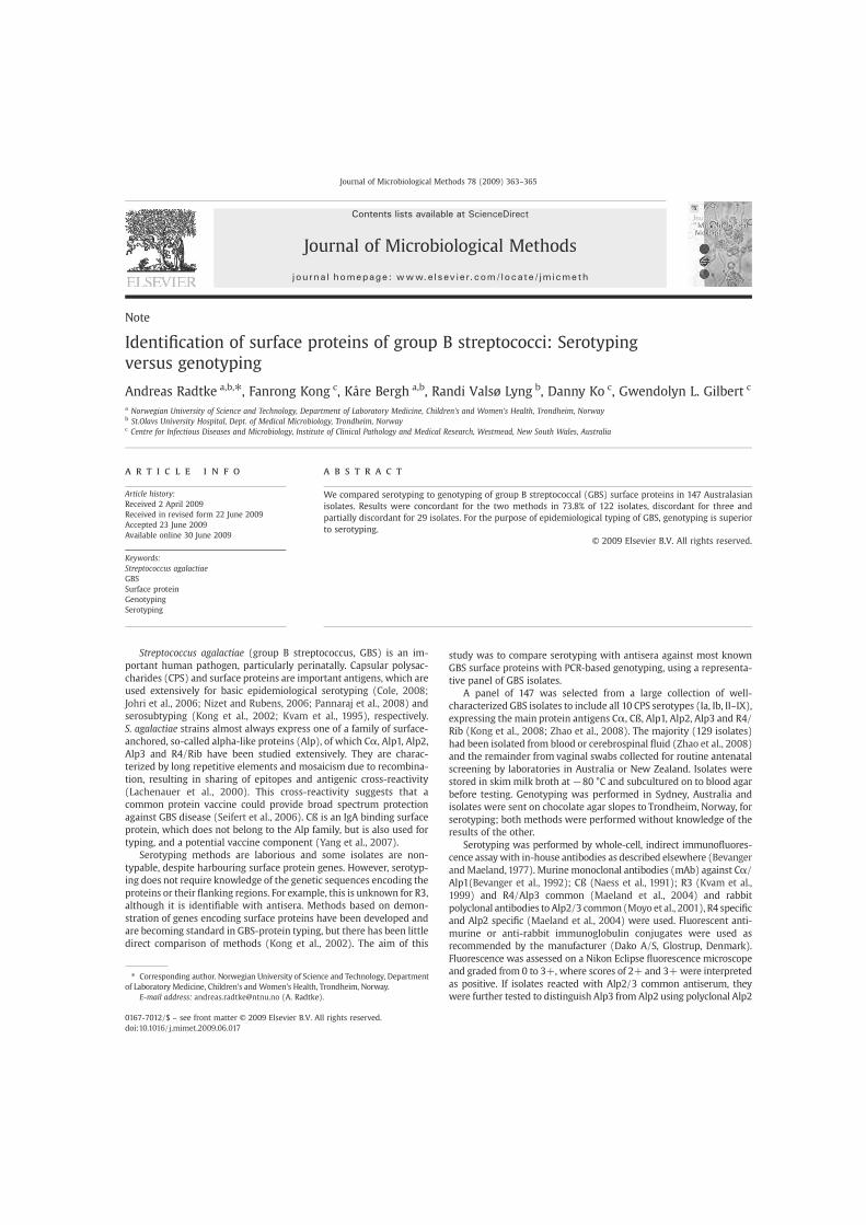
Note
Identification of surface proteins of group B streptococci: Serotypingversus genotyping
Andreas Radtke a,b,⁎, Fanrong Kong c, Kåre Bergh a,b, Randi Valsø Lyng b, Danny Ko c, Gwendolyn L. Gilbert c
a Norwegian University of Science and Technology, Department of Laboratory Medicine, Children's and Women's Health, Trondheim, Norwayb St.Olavs University Hospital, Dept. of Medical Microbiology, Trondheim, Norwayc Centre for Infectious Diseases and Microbiology, Institute of Clinical Pathology and Medical Research, Westmead, New South Wales, Australia
a b s t r a c ta r t i c l e i n f o
Article history:Received 2 April 2009Received in revised form 22 June 2009Accepted 23 June 2009Available online 30 June 2009
Keywords:Streptococcus agalactiaeGBSSurface proteinGenotypingSerotyping
We compared serotyping to genotyping of group B streptococcal (GBS) surface proteins in 147 Australasianisolates. Results were concordant for the two methods in 73.8% of 122 isolates, discordant for three andpartially discordant for 29 isolates. For the purpose of epidemiological typing of GBS, genotyping is superiorto serotyping.
© 2009 Elsevier B.V. All rights reserved.
Streptococcus agalactiae (group B streptococcus, GBS) is an im-portant human pathogen, particularly perinatally. Capsular polysac-charides (CPS) and surface proteins are important antigens, which areused extensively for basic epidemiological serotyping (Cole, 2008;Johri et al., 2006; Nizet and Rubens, 2006; Pannaraj et al., 2008) andserosubtyping (Kong et al., 2002; Kvam et al., 1995), respectively.S. agalactiae strains almost always express one of a family of surface-anchored, so-called alpha-like proteins (Alp), of which Cα, Alp1, Alp2,Alp3 and R4/Rib have been studied extensively. They are charac-terized by long repetitive elements and mosaicism due to recombina-tion, resulting in sharing of epitopes and antigenic cross-reactivity(Lachenauer et al., 2000). This cross-reactivity suggests that acommon protein vaccine could provide broad spectrum protectionagainst GBS disease (Seifert et al., 2006). Cß is an IgA binding surfaceprotein, which does not belong to the Alp family, but is also used fortyping, and a potential vaccine component (Yang et al., 2007).
Serotyping methods are laborious and some isolates are non-typable, despite harbouring surface protein genes. However, serotyp-ing does not require knowledge of the genetic sequences encoding theproteins or their flanking regions. For example, this is unknown for R3,although it is identifiable with antisera. Methods based on demon-stration of genes encoding surface proteins have been developed andare becoming standard in GBS-protein typing, but there has been littledirect comparison of methods (Kong et al., 2002). The aim of this
study was to compare serotyping with antisera against most knownGBS surface proteins with PCR-based genotyping, using a representa-tive panel of GBS isolates.
A panel of 147 was selected from a large collection of well-characterized GBS isolates to include all 10 CPS serotypes (Ia, Ib, II–IX),expressing the main protein antigens Cα, Cß, Alp1, Alp2, Alp3 and R4/Rib (Kong et al., 2008; Zhao et al., 2008). The majority (129 isolates)had been isolated from blood or cerebrospinal fluid (Zhao et al., 2008)and the remainder from vaginal swabs collected for routine antenatalscreening by laboratories in Australia or New Zealand. Isolates werestored in skim milk broth at−80 °C and subcultured on to blood agarbefore testing. Genotyping was performed in Sydney, Australia andisolates were sent on chocolate agar slopes to Trondheim, Norway, forserotyping; both methods were performed without knowledge of theresults of the other.
Serotyping was performed by whole-cell, indirect immunofluores-cence assaywith in-house antibodies as described elsewhere (Bevangerand Maeland,1977). Murine monoclonal antibodies (mAb) against Cα/Alp1(Bevanger et al., 1992); Cß (Naess et al., 1991); R3 (Kvam et al.,1999) and R4/Alp3 common (Maeland et al., 2004) and rabbitpolyclonal antibodies to Alp2/3 common (Moyo et al., 2001), R4 specificand Alp2 specific (Maeland et al., 2004) were used. Fluorescent anti-murine or anti-rabbit immunoglobulin conjugates were used asrecommended by the manufacturer (Dako A/S, Glostrup, Denmark).Fluorescence was assessed on a Nikon Eclipse fluorescence microscopeand graded from 0 to 3+, where scores of 2+ and 3+were interpretedas positive. If isolates reacted with Alp2/3 common antiserum, theywere further tested to distinguish Alp3 from Alp2 using polyclonal Alp2
Journal of Microbiological Methods 78 (2009) 363–365
⁎ Corresponding author. Norwegian University of Science and Technology, Departmentof Laboratory Medicine, Children's and Women's Health, Trondheim, Norway.
E-mail address: [email protected] (A. Radtke).
0167-7012/$ – see front matter © 2009 Elsevier B.V. All rights reserved.doi:10.1016/j.mimet.2009.06.017
Contents lists available at ScienceDirect
Journal of Microbiological Methods
j ourna l homepage: www.e lsev ie r.com/ locate / jmicmeth
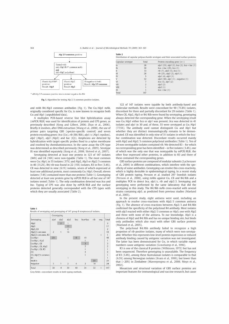
and mAb R4/Alp3 common antibodies (Fig. 1). The Cα/Alp1 mAb,originally considered specific for Cα, is now known to recognize bothCα and Alp1 (unpublished data).
A multiplex PCR-based reverse line blot hybridization assay(mPCR/RLB) was used for identification of protein and CPS genes, aspreviously described (Kong and Gilbert, 2006; Zhao et al., 2006).Briefly it involves, after DNA extraction (Wang et al., 2008), the use ofprimer pairs targeting GBS (species-specific control) and sevenprotein encoding genes: bca (Cα), rib (R4/Rib), alp1/ε (Alp1/epsilon),alp2 (Alp2), alp3 (Alp3) and bac (Cβ). Amplicons are detected byhybridization with target-specific probes fixed to a nylon membraneand resolved by chemiluminiscence. In the same assay the CPS typewas determined as described previously (Kong et al., 2005). SerotypeIX was identified separately (Kong et al., 2008; Slotved et al., 2007).
Serotyping detected at least one protein in 123 of 147 isolates(84%) and 24 (16%) were non-typable (Table 1). The most commonwere Cα/Alp1, in 55 isolates (37%) and Alp2, Alp3 or Alp2/3 commonin 46 (31.2%). R4/rib was found in 22 (15%) isolates, R3 in five (3.4%).Cß was detected in nine (6.1%) isolates, seven of which expressed atleast one additional protein, most commonly Cα/Alp1. Overall, elevenisolates (7.4%) contained more than one protein (Table 1). Genotypingdetected at least one protein gene by mPCR/RLB in all but one of 147isolates tested (Table 1). The only combination detected was bca andbac. Typing of CPS was also done by mPCR/RLB and the surfaceproteins detected generally corresponded with the CPS types withwhich they are usually associated (Table 2).
122 of 147 isolates were typable by both antibody-based andmolecular methods. Results were concordant for 90 (73.8%) isolates,discordant for three and partially discordant for 29 isolates (Table 1).When Cß, Alp2, Alp3 or R4/Rib were found by serotyping, genotypingalways detected the corresponding gene. When the serotyping resultwas Cα/Alp1 either bca or alp1 was present; bca was detected in 41isolates and alp1 in 30 and, of these, 55 were serotyped as Cα/Alp1(77.5%). The antibody used cannot distinguish Cα and Alp1 butwhether they are distinct immunologically remains to be demon-strated. Cß was identified in only nine of 31 isolates in which the bca/bac combination was detected. Discordant results occurred mainlywith Alp2 and Alp2/3 common polyclonal antibodies (Table 1). Ten of24 non-serotypable isolates contained rib. We detected R3 – for whichno corresponding gene has been identified – in five isolates (3.4%), oneof which was the only one that was nontypable by mPCR/RLB; theother four expressed other proteins, in addition to R3 and three ofthese contained the corresponding genes.
GBS surface proteins are composed ofmodular subunits (Lachenaueret al., 2000) in different combinations, which interfere with the spe-cificity of some antibodies. Genotyping can resolve this cross-reactivity,which is highly desirable in epidemiological typing. In a recent studyof GBS protein typing, Persson et al. studied 297 Swedish isolates(Persson et al., 2008), using mAbs against Cα, Cß and R4/Rib and amultiplex PCR to detect bca, alp1/ε, rib and alp2/3. Serotyping andgenotyping were performed by the same laboratory that did theserotyping in this study. The R4/Rib mAb cross-reacted with severalstrains containing alp3, as predicted from previous studies (Maelandet al., 2005).
In the present study, eight antisera were used, including anapproach to resolve cross-reactions with Alp2/3 common antisera(Fig. 1). The absence of cross-reactions between Alp2/3 and R4/Ribconfirmed the specificity of the polyclonal R4 antibody. Most isolateswith alp3 reacted with either Alp2/3 common or Alp3, one with Alp2and three with none of the antisera. To our knowledge, Alp3 is achimera of Alp2 and R4/Rib and has no unique binding site, but bindsonly antibodies which also react with other GBS surface proteins(Maeland et al., 2005).
The polyclonal R4/Rib antibody failed to recognize a highpropotion of rib-positive isolates, many of which were non-serotyp-able. Whether this represents low-level protein expression or reducedantibody binding caused by antigenic variation was not investigated.The latter has been demonstrated for Cα, in which variable repeatnumbers cause antigenic variation (Gravekamp et al., 1996).
R3 is one of the classical R proteins (Wilkinson, 1972) but has notbeen sequenced. Therefore genotyping is unavailable. The frequencyof R3 (3.4%), among these Australasian isolates is comparable to that(6.5%) among Norwegian isolates (Kvam et al., 1999), but lower thanthat (N20%) in Zimbabwe (Mavenyengwa et al., 2008; Moyo et al.,2002).
Mosaicism and structural variation of GBS surface proteins areimportant features for immunological and vaccine research, but cause
Fig. 1. Algorithm for testing Alp 2/3 common positive isolates.
Table 1Results of serotyping and genotyping of 147 group B streptococcal isolates.
Grey fields: concordant results in both typing methods.
Table 2Distribution of capsular polysaccharide serotypes and their associated surface proteins.
Capsular serotype Total Protein encoding gene (n=)
Ia 28 alp1 (23), alp2 (2), bca (2), bca+bac (1)Ib 30 bca+bac (29), bca (1)II 13 rib (7), alp3 (5), bca (1)III 28 rib (25), alp2 (2), alp3 (1)IV 5 alp1 (4), alp3 (1)V 32 alp3 (30), alp1 (1) , negative (1)VI 7 bca (5), alp1 (2)VII 2 alp3 (2)VIII 1 alp3 (1)IX 1 bca+bac (1)Total 147
364 A. Radtke et al. / Journal of Microbiological Methods 78 (2009) 363–365

discriminatory problems for epidemiological typing. We have shownthat GBS surface protein genotyping is superior, as almost all isolatesare typable and the results are unambiguous.
Acknowledgements
We thank Prof. Johan A. Maeland for help and discussions indesigning and realizing this work.
References
Bevanger, L., Maeland, J.A., 1977. Type classification of group B streptococci by thefluorescent antibody test. Acta Pathol.Microbiol.Scand.[B]. 85B, 357–362.
Bevanger, L., Iversen, O.J., Naess, A.I., 1992. Characterization of the alpha antigen of the cproteins of group B streptococci (GBS) using a murine monoclonal antibody. APMIS100, 57–62.
Cole, J.N.J.N., 2008. Human pathogenic streptococcal proteomics and vaccine develop-ment. Proteomics 2, 387–410.
Gravekamp, C., Horensky, D.S., Michel, J.L., Madoff, L.C., 1996. Variation in repeatnumber within the alpha C protein of group B streptococci alters antigenicity andprotective epitopes. Infect. Immun. 64, 3576–3583.
Johri, A.K., Paoletti, L.C., Glaser, P., Dua, M., Sharma, P.K., Grandi, G., Rappuoli, R., 2006.Group B Streptococcus: global incidence and vaccine development. Nat. Rev.Microbiol. 4, 932–942.
Kong, F., Gilbert, G.L., 2006. Multiplex PCR-based reverse line blot hybridization assay(mPCR/RLB) — a practical epidemiological and diagnostic tool. Nat. Protoc. 1,2668–2680.
Kong, F., Gowan, S., Martin, D., James, G., Gilbert, G.L., 2002. Molecular profiles ofgroup B streptococcal surface protein antigen genes: relationship to molecularserotypes. J. Clin. Microbiol. 40, 620–626.
Kong, F., Ma, L., Gilbert, G.L., 2005. Simultaneous detection and serotype identificationof Streptococcus agalactiae using multiplex PCR and reverse line blot hybridization.J. Med. Microbiol. 54, 1133–1138.
Kong, F.F., Lambertsen, L.M.L.M., Slotved, H.C.H.-C., Ko, D.D., Wang, H.H., Gilbert, G.L.G.L.,2008. Use of phenotypic and molecular serotype identification methods tocharacterize previously nonserotypeable group B streptococci. J. Clin. Microbiol.46, 2745–2750.
Kvam, A.I., Efstratiou, A., Bevanger, L., Cookson, B.D., Marticorena, I.F., George, R.C.,Maeland, J.A., 1995. Distribution of serovariants of group B streptococci in isolatesfrom England and Norway. J. Med. Microbiol. 42, 246–250.
Kvam, A.I., Bevanger, L., Maeland, J.A., 1999. Properties and distribution of the putativeR3 protein of Streptococcus agalactiae. APMIS 107, 869–874.
Lachenauer, C.S., Creti, R., Michel, J.L., Madoff, L.C., 2000. Mosaicism in the alpha-likeprotein genes of group B streptococci. Proc. Natl. Acad. Sci. U.S.A. 97, 9630–9635.
Maeland, J.A., Bevanger, L., Lyng, R.V., 2004. Antigenic determinants of alpha-likeproteins of Streptococcus agalactiae. Clin. Diagn. Lab. Immunol. 11, 1035–1039.
Maeland, J.A., Bevanger, L., Lyng, R.V., 2005. Immunological markers of the R4 protein ofStreptococcus agalactiae. Clin. Diagn. Lab. Immunol. 12, 1305–1310.
Mavenyengwa, R.T., Maeland, J.A., Moyo, S.R., 2008. Distinctive features of surface-anchored proteins of Streptococcus agalactiae strains from Zimbabwe revealed byPCR and dot blotting. Clin. Vaccine Immunol. 15, 1420–1424.
Moyo, S.R., Maeland, J.A., Lyng, R.V., 2001. The putative R1 protein of Streptococcusagalactiae as serotype marker and target of protective antibodies. APMIS 109,842–848.
Moyo, S.R., Maeland, J.A., Bergh, K., 2002. Typing of human isolates of Streptococcusagalactiae (group B streptococcus, GBS) strains from Zimbabwe. J. Med. Microbiol.51, 595–600.
Naess, A.I., Bevanger, L., Iversen, O.J., Maeland, J.A., 1991. Evaluation of monoclonalantibodies in serovar classification of group B streptococci (GBS). Brief report.APMIS 99, 1058–1060.
Nizet, V., Rubens, C.E., 2006. In: Fischetti, V.A., Novick, R.P., Ferretti, J.J., Portnoy, D.A.,Rood, J.I. (Eds.), Pathogenic mechanisms and virulence factors of group Bstreptococci, Vol. 2nd. ASM Press, Washington, DC, pp. 152–168.
Pannaraj, P.S., Kelly, J.K., Rench, M.A., Madoff, L.C., Edwards, M.S., Baker, C.J., 2008. AlphaC protein-specific immunity in humans with group B streptococcal colonizationand invasive disease. Vaccine 26, 502–508.
Persson, E., Berg, S., Bevanger, L., Bergh, K., Valso-Lyng, R., Trollfors, B., 2008.Characterisation of invasive group B streptococci based on investigation of surfaceproteins and genes encoding surface proteins. Clin. Microbiol. Infect. 14, 66–73.
Seifert, K.N., Adderson, E.E., Whiting, A.A., Bohnsack, J.F., Crowley, P.J., Brady, L.J., 2006.A unique serine-rich repeat protein (Srr-2) and novel surface antigen (epsilon)associated with a virulent lineage of serotype III Streptococcus agalactiae.Microbiology 152, 1029–1040.
Slotved, H.C., Kong, F., Lambertsen, L., Sauer, S., Gilbert, G.L., 2007. Serotype IX, aproposed new Streptococcus agalactiae serotype. J. Clin. Microbiol. 45, 2929–2936.
Wang, Y., Kong, F., Gilbert, G.L., Brown, M., Gao, W., Yu, S., Yang, Y., 2008. Use of amultiplex PCR-based reverse line blot (mPCR/RLB) hybridisation assay for the rapididentification of bacterial pathogens. Clin. Microbiol. Infect. 14, 155–160.
Wilkinson, H.W., 1972. Comparison of streptococcal R antigens. Appl. Microbiol. 24,669–670.
Yang, H.H., Madoff, L.C., Guttormsen, H.K., Liu, Y.D., Paoletti, L.C., 2007. Recombinantgroup B streptococcus Beta C protein and a variant with the deletion of itsimmunoglobulin A-binding site are protective mouse maternal vaccines andeffective carriers in conjugate vaccines. Infect. Immunol. 75, 3455–3461.
Zhao, Z., Kong, F., Gilbert, G.L., 2006. Reverse line blot assay for direct identification ofseven Streptococcus agalactiae major surface protein antigen genes. Clin. VaccineImmunol. 13, 145–149.
Zhao, Z., Kong, F., Zeng, X., Gidding, H.F., Morgan, J., Gilbert, G.L., 2008. Distribution ofgenotypes and antibiotic resistance genes among invasive Streptococcus agalactiae(group B streptococcus) isolates from Australasian patients belonging to differentage groups. Clin. Microbiol. Infect. 14, 260–267.
365A. Radtke et al. / Journal of Microbiological Methods 78 (2009) 363–365


Paper II


JOURNAL OF CLINICAL MICROBIOLOGY, July 2010, p. 2502–2508 Vol. 48, No. 70095-1137/10/$12.00 doi:10.1128/JCM.00234-10Copyright © 2010, American Society for Microbiology. All Rights Reserved.
Rapid Multiple-Locus Variant-Repeat Assay (MLVA) for Genotypingof Streptococcus agalactiae�
Andreas Radtke,1,2* Bjørn-Arne Lindstedt,3 Jan Egil Afset,1,2 and Kåre Bergh1,2
Norwegian University of Science and Technology, Department of Laboratory Medicine, Children’s and Women’s Health,Trondheim, Norway1; St. Olav’s University Hospital, Department of Medical Microbiology, Trondheim, Norway2; and
Norwegian Institute of Public Health, Division of Infectious Diseases Control, Oslo, Norway3
Received 4 February 2010/Returned for modification 8 April 2010/Accepted 15 May 2010
Several methods have been used for typing of Streptococcus agalactiae (group B streptococci [GBS]). Methodscurrently in use may provide inadequate resolution (e.g., typing of capsular polysaccharides and surfaceprotein) or are labor-intensive and expensive (e.g., multilocus sequence typing [MLST] or pulsed-field gelelectrophoresis). This work describes the construction and use of a multiple-locus variant-repeat assay(MLVA) on 126 well-characterized human GBS strains, consisting mostly of invasive Norwegian strains andinternational reference strains. Based on in silico whole-genomic analysis of the genomes of strains A909,NEM316, and 2603V/R, 18 candidate loci were selected and investigated by PCR. Eleven loci showed diversity,and the five most diverse loci were used for the construction of an MLVA, consisting of a multiplex PCRfollowed by fragment analysis with capillary electrophoresis. The assay generated clusters which correspondedwell with those observed by other methods. However, it provided a considerably higher degree of diversity, with70 different MLVA types compared to 36 types generated by MLST. Simpson’s index of diversity for the 5-locusMLVA was 0.963, compared to 0.899 for the MLST in this strain collection. MLVA results will generally beavailable within 2 days, which is usually faster than MLST. In our hands, MLVA of GBS represents a rapid,easy, and comparably inexpensive method for high-resolution genotyping of GBS.
Streptococcus agalactiae is the only species within the groupB streptococci (GBS) of Lancefield’s classification of hemolyticstreptococci. It is well recognized as a human pathogen, espe-cially for causing septicemia, meningitis, and other seriousinvasive disease in neonates; it is also associated with stillbirth.In addition, it is known to cause serious maternal infectionsand serious infections in elderly and immunocompromised pa-tients.
Different approaches for typing of GBS have been devel-oped through the years. Most commonly, GBS strains areassigned to one of the 10 capsular polysaccharide (CPS) types.Typing of surface proteins and analysis of the antibiotic resis-tance pattern can add further resolution to typing of pheno-typic features. Examination of CPS or surface proteins can bedone by immunological methods or, more recently, by detect-ing the corresponding genes by PCR (6, 8, 10). Multilocussequence typing (MLST) was developed to study the long-termevolutionary development of a species. The total number ofGBS sequence types (STs) is currently around 490 (http://pubmlst.org/sagalactiae/). Pulsed-field gel electrophoresis(PFGE) based on macrorestriction fragment analysis ofgenomic DNA is considered highly discriminatory. For GBS,the restriction enzyme SmaI is often used, generating only 8 to12 restriction fragments, which may be suboptimal with respectto discriminating capacity (2, 19, 21). Since PFGE is an image-based method, it may be difficult to compare results between
different laboratories. Both MLST and PFGE are labor-inten-sive techniques requiring experienced personnel and usuallyseveral days of work before results are available.
DNA loci consisting of repeated sequences are widespreadthroughout the genome of bacteria and have a tendency to varyin repeat number from strain to strain, so-called “variablenumber of tandem repeats” (VNTR). The combination of sev-eral VNTR loci in an assay (multiple-locus variant-repeat assay[MLVA]) has been shown in various bacterial species to gen-erate strain-specific profiles that can be compared, exchanged,and reproduced in different laboratories. The technique hasbeen employed successfully for the typing of an increasingnumber of bacterial species (23). The aim of the present studywas to explore the GBS genome for variably repeated geneticregions and to investigate the feasibility of an MLVA methodfor typing the bacterium.
MATERIALS AND METHODS
Selection, culture, and DNA extraction of GBS strains. A panel of 126 GBSstrains was selected to represent a broad range of CPS and surface proteins andsequence types. Most of these (n � 113) were invasive strains submitted to theNorwegian reference laboratory for GBS. They were also used in a previousstudy of invasive GBS in infants (3). In addition, nine international referencestrains were included, among them all eight fully sequenced strains. Two Zim-babwean colonizing strains and two Danish CPS type IX strains were alsoincluded. The distribution of CPS types was as follows: Ia, 12 strains; Ib, 10strains; II, 6 strains; III, 55 strains; IV, 9 strains; V, 30 strains; VI, 1 strain; andIX, 3 strains. Strains stored at �80°C were grown overnight on blood agar plates.For nucleic acid extraction, 4 to 5 colonies were added to 200 �l lysis solutioncontaining 12 �l (20 mg/ml) lysozyme (Sigma-Aldrich Corp., St. Louis, MO), 4.8�l (20 mg/ml) proteinase K (Sigma), 4.8 �l (10,000 U/ml) mutanolysin (Sigma),and 178.4 �l Tris-EDTA (TE) buffer and incubated at 37°C and 65°C for 15 mineach. DNA was purified on a Qiagen BioRobot M48 instrument using theMagAttractDNA Mini 48 kit (Qiagen, Hilden, Germany) and eluted in a volumeof 50 �l. The eluate was diluted 1:10 for PCR.
* Corresponding author. Mailing address: Andreas Radtke, Norwe-gian University of Science and Technology, Department of LaboratoryMedicine, Children’s and Women’s Health, Olav Kyrres gt. 9, 7489Trondheim, Norway. Phone: 47 725 73 253. Fax: 47 725 76 416. E-mail:[email protected].
� Published ahead of print on 26 May 2010.
2502

Detection of VNTRs, PCR protocols, and analysis of fragments. Genomeswere analyzed using the Tandem Repeats Finder program, version 4.00 (1). TheVariable Region Finder available at the Health Protection Agency website (http://www.hpa-bioinformatics.org.uk/variable_region_finder/index.html) was alsoused. Primers were designed using Oligo 6.71 software (Molecular Biology In-sights, Inc., Cascade, CO). The loci were consecutively termed TR1 throughTR18, while the five loci proposed for the subsequent MLVA were renamedSATR1 to SATR5 in the later phase of the study. Analysis of the repeats wasgenerally done with single PCRs in 25 �l of reaction mixture containing 50 �M(each) dATP, dCTP, dGTP, and dTTP; forward and reverse primers (0.5 �M);2.5 �l 10� PCR buffer with 15 mM MgCl and 0.75 U AmpliTaq Gold DNApolymerase (both of the latter by Applied Biosystems, Foster City, CA); a 1-�laliquot of the purified, diluted DNA template; and RNase-free water. The PCRconditions were 5 min at 95°C for activation of the polymerase and then 35 cyclesof 30 s at 95°C, 30 s at annealing temperature, and 60 s at 72°C. The annealingtemperature used initially was that proposed by the primer design software, butthe temperature was later changed to 55°C. The PCR was performed on an MJResearch PTC-200 instrument (MJ Research, Inc., Watertown, MA). Ampliconsize was determined with an Agilent 2100 Bioanalyzer (Agilent Technologies,Santa Clara, CA) using the DNA 1000 chip. Later, amplicon size was determinedby either 2% agarose gel electrophoresis for the VNTRs that showed only two orthree alleles or by fragment analysis with capillary electrophoresis. The Qiagenmultiplex PCR kit was used for the subsequent multiplex PCR. The manufac-turer’s recommendations were followed, except for the annealing temperature,which was set to 55°C. In addition, the concentrations of the individual primersin the primer mix were adjusted to 1.5 �M for SATR1 and -5, 1 �M for SATR2and -3, and 0.5 �M for SATR4, and the total PCR volume was reduced to 25 �l.
Fragment analysis was done on an ABI 3130xl genetic analyzer. Hi-Di form-amide, GeneScan 1200 LIZ size standard, and the PCR product were mixed (9plus 0.5 plus 0.5 �l). The standard protocol for fragment analysis with 36-cmcapillaries and POP7 polymer was used. The product sizes were analyzed usingthe GeneMapper 4.0 software. (All products used for fragment analysis werefrom Applied Biosystems.)
For analysis of the stability of the alleles, three strains (06/14, 06/123, and07/15) were passaged on blood agar plates 40 times over a period of 4 months.
Serotyping and MLST. Typing of CPS and surface proteins was done by PCR(6, 11, 24), except that immunofluorescence microscopy was used for the nineNorwegian strains collected in 2005 (4). MLST was performed as describedelsewhere (9). Results were available for all 126 of the strains. STs of most of theNorwegian strains were available from a previous study (3), whereas the ST ofthe international reference strains was available from a recent publication (22).
Analysis of results. For cluster analysis, BioNumerics 6.0 software (AppliedMaths, Sint-Martens-Latem, Belgium) was used. Simpson’s index of diversity wascalculated either via the VNTR DIversity and Confidence Extractor (V-DICE)software at the Health Protection Agency website (Health Protection Agency,
Colindale, London, United Kingdom; http://www.hpa-bioinfotools.org.uk/cgi-bin/DICI/DICI.pl) or manually (18). The number of repeats was determined bysubtracting the offset (the number of nucleotides between the primers and thestart/end of the repeat) and dividing the remaining number of nucleotides by therepeat length (Table 1).
Sequence verification. For DNA sequencing, loci were amplified in PCRs withunlabeled primers as described above. The PCR products were purified by usingExoSAP-IT (USB Corporation, Cleveland, OH) following the instructions of themanufacturer. BigDye 3.1 and BigDye terminator 3.1 (Applied Biosystems)reagents were used according to the manufacturer’s instructions for sequencingPCR and subsequent purification. Capillary electrophoresis of sequencing PCRproducts was performed on an ABI 3130xl genetic analyzer (Applied Biosys-tems). The resulting sequences were analyzed using Sequencher 4.2 software(Gene Codes Corporation, Ann Arbor, MI).
RESULTS
Identification of tandem repeats in GBS. The sequences ofthe three complete genomes of strains A909, NEM316, and2603 V/R were analyzed in silico using the Tandem RepeatsFinder program or the Variable Region Finder website. Can-didate loci were compared via the BLAST website with theeight published whole-genome sequences, namely, the threeabove mentioned and five not fully assembled and annotatedshotgun sequences (strains 515, H36B, 18RS21, COH1, andCJB111) (22). Most loci were excluded from further investiga-tion for reasons such as large size, poorly conserved repeats, orpoorly conserved flanking sequences. For the 18 remaining loci(Table 1), primers were designed. Their presence and theirsuitability for an MLVA were tested in 11 GBS strains. Sevenloci (marked with asterisks in Fig. 1) were found unsuitablebecause they did not show variability or were not amplifiedeven after several primer pairs were designed and tested.
The remaining 11 loci were investigated in all 126 strains.Four of the loci had four or more alleles, and three had threealleles, while the remaining four loci had two alleles in thisstrain collection. Six of the 11 diverse loci did not generate aPCR product in some strains (indicated as �1 repeats in Fig.1). Amplification of SATR4 and SATR5 generated a PCR
TABLE 1. Properties of 18 assumed variable genetic loci in Streptococcus agalactiae
Namea Strainb Position instrain genome
Repeat size and count inoriginal strain (offset size) Annotation in genome
TR1 (SATR1) 2603 V/R 143000 60 bp � 3 (139 bp) Hypothetical protein SAG0134, CRISPRTR2* A909 115500 9 bp � 4TR3 (SATR2) A909 746100 18 bp � 6 (85 bp) Hypothetical protein SAK_0805TR4* A909 1159300 18 bp � 2TR5* A909 1256100TR6 (SATR3) A909 1993600 12 bp � 23 (92 bp) Surface adhesion protein gbs2018 in NEM316TR7* A909 2071160TR8 (SATR4) NEM316 29700 18 bp � 3 (99 bp) pcsB proteinTR9 NEM316 107750 24 bp � 2 Chaperone protein, dnaJTR10* NEM316 476780TR11* NEM316 1547230TR12 (SATR5) A909 1118500 48 bp � 14 (161 bp) Putative fibrinogen-binding protein, SAK_1142TR13 2603 V/R 451800 45-bp insert upstream of alp proteinsTR14 NEM316 857700 53-bp insert, gene gbs0831TR15* NEM316 917600TR16 NEM316 1010700 66-bp insert, hypothetical protein gbs0966TR17 NEM316 1269700 45-bp deletion in A909, intergenetic between gbs1230 and ungTR18 NEM316 1624650 44-bp deletion, gbs1557
a *, not surveyed further after initial screening.b The strain column refers to the strain in which the locus was found originally.
VOL. 48, 2010 RAPID MLVA FOR GBS 2503

FIG. 1. Cluster analysis of 126 GBS strains using categorical values and the UPGMA (unweighted-pair group method using average linkages) algorithm,generated with Bionumerics 6.0 software. Numbers in the SATR1 to -5 columns indicate repeat count (�1 repeat, no PCR product). CPS, capsularpolysaccharide. The country column gives the origin of the strain: NO, Norway; INT, international reference strain; DK, Denmark; and ZW, Zimbabwe. Strainsmarked with asterisks were used in the initial analysis of 18 variable loci. There is an overlap of three strains between the two panels.
2504
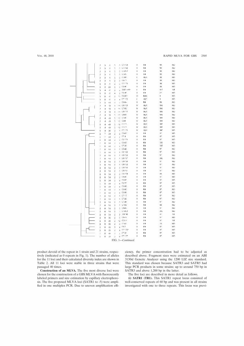
product devoid of the repeat in 1 strain and 21 strains, respec-tively (indicated as 0 repeats in Fig. 1). The number of allelesfor the 11 loci and their calculated diversity index are shown inTable 2. All 11 loci were stable in three strains that werepassaged 40 times.
Construction of an MLVA. The five most diverse loci werechosen for the construction of a GBSMLVA with fluorescentlylabeled primers and size estimation by capillary electrophore-sis. The five proposed MLVA loci (SATR1 to -5) were ampli-fied in one multiplex PCR. Due to uneven amplification effi-
ciency, the primer concentration had to be adjusted asdescribed above. Fragment sizes were estimated on an ABI3130xl Genetic Analyzer using the 1200 LIZ size standard.This standard was chosen because SATR3 and SATR5 hadlarge PCR products in some strains: up to around 750 bp inSATR3 and above 1,200 bp in the latter.
The five loci are described in more detail as follows.(i) SATR1 (TR1). This SATR1 repeat locus consisted of
well-conserved repeats of 60 bp and was present in all strainsinvestigated with one to three repeats. This locus was previ-
FIG. 1—Continued.
VOL. 48, 2010 RAPID MLVA FOR GBS 2505

ously described by Dmitriev et al. (7, 17) as consisting of 16-bpdirect repeats and 44-bp spacer regions.
(ii) SATR2 (TR3). The SATR2 repeats were well conserved,consisting of 18 bp, and the strains contained 3 to 15 repeats.All sequenced strains had an insert of 9 bp found betweenrepeats 2 and 3. The only strains having 15 repeats were thethree strains in this study of the recently described serotype IX(20). In 14 strains (11%), no PCR product was detected.
(iii) SATR3 (TR6). The SATR3 repeat locus gave compara-bly large products from 297 to 750 bp. The repeat unit of 12 bpwas quite well conserved. The repeat count was computed to18 to 54 repeats; however, sequencing demonstrated that thestrains with the largest amplicons had 18 repeats and an in-serted sequence of two 216-bp repeats. This combination wasfound in 33 strains (26.4%), all but one of which belonged toMLST clonal complex 17. A similar observation was made byLamy et al. (12), as discussed below. SATR3 was not amplifiedin 25 strains (20%), most of which were of serotype III/R4 andMLST clonal complex 19.
(iv) SATR4 (TR8). The SATR4 repeat gave the smallestPCR products, ranging between 99 and 168 bp. The underlyingrepeat of 18 bp was quite degenerated; in some strains, someor all of the repeats were only 15 bp. For convenience oftyping, all alleles are assigned as multiples of 18 bp in thecalculation of repeat numbers. A PCR product was observed inall 126 strains, and five different alleles were found. The repeatis located in the pcsB gene, encoding a protein which is im-portant for cell wall separation (15).
(v) SATR5 (TR12). The SATR5 locus is the most diverserepeat locus in this study, with 24 different alleles in the 126strains and a Simpson’s index of diversity of 0.913 (confidenceinterval [CI], 0.900 to 0.922). The repeat of 48 bp was quite wellconserved, and an amplification product was detected in all butone strain. Of 21 strains with a PCR product size equivalent to theoffset (0 repeats), 13 were of ST19. A result of six repeats wasfound in 22 strains, 18 of which belonged to ST17. Some strainshad an insert after the repeat locus, causing a variable offset.However, for typing purposes, the offset was set to 161 bp with theprimers chosen. The larger products of this locus, especially those
above 900 bp, did give weak signals in spite of attempts to opti-mize both multiplex PCR and fragment analysis. If SATR5 didnot yield a product in the multiplex PCR, it was repeated in asingle PCR. In five strains, the SATR5 locus was calculated to bemore than 1,400 bp by the GeneMapper software, although thiswas above the upper limit of the 1200 LIZ size standard andtherefore unreliable. When the five strains containing SATR5amplicons above 1,200 bp were analyzed by gel electrophoresis,they showed diversity. In the analysis, however, these five strainswere assigned a repeat count of 50. SATR5 is located in the geneencoding the fibrinogen binding protein FbsA (16).
Comparison between MLVA, serotyping, and MLST. MLVAanalysis with the five proposed loci resolved the 126 strains into 70different types with a Simpson’s index of diversity of 0.963, whileMLST separated them into 36 STs with an index of 0.899. Thetwo largest groups of identical MLVA profiles consisted of 19strains and 13 strains, most of which belonged to ST17 and ST19,respectively. These two MLST types are common subgroups ofCPS type III (Fig. 1). All 27 of the ST17 strains in this workclustered together but were resolved into 12 different MLVAtypes within this cluster; 16 of them had identical profiles (Fig. 2).Similarly, all 11 CPS III/ST19 strains belonged to the sameMLVA cluster and were resolved into four MLVA types; 7 ofthem had identical profiles. CPS type V has previously beendescribed as relatively homogenous, possibly due to a recent in-troduction of this group as a human pathogen (5). Most of thetype V strains belonged to ST1 (21 out of 30 type V strains); 20 ofthese 21 were in a cluster consisting of 12 MLVA types, mostlybecause of heterogeneity in SATR5.
A simpler MLVA analysis using SATR1 to -4 resulted in 43different types for the 126 strains (Simpson’s index of diversity,0.926), also corresponding very well with the clustering by theother typing methods.
DISCUSSION
This study identified VNTR loci in the genome of GBS thatare suitable for MLVA typing. MLVA is a relatively new typingmethod with several advantages compared to other methods.
TABLE 2. Primers, fluorescent dyes, and Simpson’s index of diversity for 11 VTNR loci in GBS
LocusSequence (5�33�)a
Diversityindexb 95% CI Kc max(pi)d
Forward primer Reverse primer
SATR5 (TR12) 6-FAM-AGTCACCTTGACTAGAGTG AATAAAATAGGTTTTAGAACTTGG 0.913 0.903–0.923 24 0.173SATR3 (TR6) VIC-CCGCCAAGTTTCCGCTAG AGCTAACAAGCTACGTGATGC 0.869 0.856–0.881 19 0.220SATR2 (TR3) PET-GTTGATAAAGTTGATGT
TCCGAGCCTTCTTCAACTATAGGTG 0.703 0.675–0.732 9 0.472
SATR1 (TR1) NED-ACTTGTGAAAGTGTATCGGT AAATCAGTGTTTTAACAGCAGC 0.643 0.626–0.661 3 0.465SATR4 (TR8) VIC-AAAGCATCTTTAATTCAGGCA CTGTGGCAGACTCAACTTGTG 0.633 0.608–0.658 5 0.504TR9 TGTCTAAGAAATACCATCCAG TTCAAAACTTAAATTAACACG 0.470 0.444–0.496 3 0.643TR13 TGTTTCTAAGAAAAAAGAGG ACCTTACATTTGATTACACC 0.470 0.444–0.496 3 0.643TR16 ATCAAAAAACCAGATACTGC CAATGCTTTAACCGAACCAG 0.455 0.431–0.478 2 0.651TR14 ATTATAGAGGACGTTACTCG CAACCATCCGACCTGTAATA 0.172 0.133–0.212 2 0.905TR18 ATGACAGTTGAACATGTGGA TACCATGTACCAACAGACTG 0.031 0.011–0.051 2 0.984TR17 AATTTGGTTCCGTTTGGCT ATGCTCGAAAGAAAAAGTCG 0.016 0.001–0.030 2 0.992
a Certain primers were labeled with the fluorescent dyes 6-FAM (6-carboxyfluorescein), VIC, PET, and NED, as indicated.b The diversity index (for VNTR data) is a measure of the variation of the number of repeats at each locus, ranging from 0.0 (no diversity) to 1.0 (complete diversity).
The most diverse loci are shown at the top.c K, number of different repeats present at this locus in a collection of 126 GBS strains.d max(pi), fraction of samples that have the most frequent repeat number in this locus (range, 0.0 to 1.0).
2506 RADTKE ET AL. J. CLIN. MICROBIOL.

Mainly, MLVA is easy to perform at moderate costs, andresults are in most cases available within 2 days, which willusually be more rapid than MLST or PFGE. Since MLVAgenerates numeric results, comparison of strains between dif-ferent laboratories may easily be done. In the present study, 18candidate loci were identified in the GBS genome by compu-tational studies. Initial laboratory analysis found seven of thesenot suitable. The remaining 11 loci were tested in 126 strains,and the five most diverse loci were selected for an MLVAbased on multiplex PCR and capillary electrophoresis for de-termination of the number of repeats.
The strain collection analyzed consisted of Norwegian inva-sive GBS strains and international reference strains which to-gether represented a wide selection of CPS types and STs. TheNorwegian strains, mostly from neonates, had been submittedto the national reference laboratory from hospitals throughoutthe country in the years 2006 and 2007. It is therefore reason-able to assume that there was no epidemiological associationbetween most patients. Based on the high discriminatorypower of the MLVA shown in this study, the method may well
be suited for outbreak investigation. This should be addressedin a study with epidemiologically related strains.
Typing with MLVA in this study did generally correspondwith the clustering of strains observed in CPS/surface proteintyping or MLST. The 5-locus MLVA provided a very highdegree of resolution, discriminating the 126 GBS strains into70 different MLVA types (Fig. 1). The discriminatory power ofthis MLVA is superior to those of both MLST and combinedCPS/protein typing, which resolved the strains into 36 and 19types, respectively. The strains could have been resolved fur-ther if SATR5 alleles above 1,200 bp had been differentiated,which could be necessary under certain circumstances. If re-quired, this will imply the use of other methods for exact sizeenumeration of SATR5 and consequently a delay of results.For SATR4, the diversity due to the degenerate character ofthe repeat should be kept in mind. In addition to the repeatcount, the fragment size may be considered, especially if theMLVA is used in epidemiological investigations. As an alter-native option for providing rapid results, a 4-locus MLVA,consisting of the loci SATR1 to -4 was also analyzed. Even this4-locus MLVA was capable of providing higher resolution thanMLST or CPS/protein typing. Conversely, some or all the sixloci mentioned in Table 2 but not included in the 5-locusMLVA analysis (e.g., TR9, TR13, and TR16) could be addedto the MLVA if an even higher degree of resolution is desir-able.
Some of the loci selected for the 5-locus MLVA have beeninvestigated previously for other purposes. SATR1 has beenaddressed by a Russian and Slovakian group (7, 17) who foundeight alleles in 112 bovine strains. Strictly speaking, this locusis a clustered regularly interspaced short palindromic repeat(CRISPR) but worked very well as component of this MLVA.SATR3 is located in the gene of surface adhesion proteingbs2018 previously analyzed by Lamy et al. (12). They de-scribed three alleles, one of which included the insert of 216 bpwith two repeats mentioned above and found it as well stronglycorrelated with ST17. GBS belonging to ST17 account for ahigh proportion of newborn infections (3, 9, 14); however,whether these GBS represent a hypervirulent cluster is underdiscussion (13). SATR5 is the most diverse repeat locus and isresponsible for a substantial part of the overall resolution ob-tained with this assay. Analyzed alone, it did cluster some ofthe MLST groups such as ST17 and ST19 well, while others(e.g., CPS type V/ST1) were more dispersed. On the otherhand, SATR5 resolved CPS-type V/ST1 into many differenttypes in the MLVA, while other typing techniques tend to givea much more homogeneous picture of this subgroup. InSATR2, the finding of 15 repeats was indicative for CPS typeIX. This was also observed in three additional type IX strainsnot included in this study. Results that were indicative of cer-tain sequence types were, e.g., the finding of 54 or 55 repeatsin SATR3 for ST17 (Fig. 2). Similarly, an ST19 could be ex-pected when SATR3 had no PCR product (�1 repeats) andSATR5 had 0 repeats.
To summarize, the present study identified VNTR loci in thegenome of Streptococcus agalactiae suitable for MLVA analy-sis. Five loci were selected for a multiplex PCR protocol fol-lowed by capillary electrophoresis, enabling a very discrimina-tory GBS typing assay. The 5-locus MLVA resolved a straincollection of 126 GBS strains considerably better than MLST
FIG. 2. MLVA cluster analysis of 27 ST17 strains included in thestudy using categorical values and the UPGMA algorithm, generatedwith Bionumerics 6.0 software. Numbers in the SATR1 to -5 columnsindicate the repeat count.
VOL. 48, 2010 RAPID MLVA FOR GBS 2507

and with less workload. This method is well suited for typing ofGBS, e.g., by national reference laboratories and for researchpurposes. Further work with this method would include refine-ment of the method, comparison with PFGE with epidemio-logically related strains, as well as analysis of other strain col-lections, such as non-Norwegian strains and noninvasive andanimal strains, and establishment of a website for internationalcollaboration and strain comparison.
ACKNOWLEDGMENTS
This work was supported by grants from the Clinic of LaboratoryMedicine, St. Olav’s University Hospital, Trondheim, and the CentralNorway Regional Health Authority. A.R. was supported by a Ph.D.grant from the Norwegian University of Science and Technology, De-partment of Laboratory Medicine, Children’s and Women’s Health.
Kirsti Løseth is gratefully acknowledged for expert laboratory work.
REFERENCES
1. Benson, G. 1999. Tandem repeats finder: a program to analyze DNA se-quences. Nucleic Acids Res. 27:573–580.
2. Benson, J. A., and P. Ferrieri. 2001. Rapid pulsed-field gel electrophoresismethod for group B streptococcus isolates. J. Clin. Microbiol. 39:3006–3008.
3. Bergseng, H., J. E. Afset, A. Radtke, K. Loeseth, R. V. Lyng, M. Rygg, and K.Bergh. 2009. Molecular and phenotypic characterization of invasive group Bstreptococcus strains from infants in Norway 2006–2007. Clin. Microbiol.Infect. 15:1182–1185.
4. Bevanger, L., and J. A. Maeland. 1977. Type classification of group B strep-tococci by the fluorescent antibody test. Acta Pathol. Microbiol. Scand. B85B:357–362.
5. Blumberg, H. M., D. S. Stephens, M. Modansky, M. Erwin, J. Elliot, R. R.Facklam, A. Schuchat, W. Baughman, and M. M. Farley. 1996. Invasivegroup B streptococcal disease: the emergence of serotype V. J. Infect. Dis.173:365–373.
6. Creti, R., F. Fabretti, G. Orefici, and C. von Hunelstein. 2004. MultiplexPCR assay for direct identification of group B streptococcal alpha-protein-like protein genes. J. Clin. Microbiol. 42:1326–1329.
7. Dmitriev, A. V., A. D. Shen, and A. A. Totolian. 2007. Streptococcus agalac-tiae sak0192 gene contains direct repeats and spacers that are genetic mark-ers for characterizing the strains. Mol. Gen. Mikrobiol. Virusol. 4:37–41. (InRussian.)
8. Imperi, M., M. Pataracchia, G. Alfarone, L. Baldassarri, G. Orefici, and R.Creti. 2010. A multiplex PCR assay for the direct identification of thecapsular type (Ia to IX) of Streptococcus agalactiae. J. Microbiol. Methods80:212–214.
9. Jones, N., J. F. Bohnsack, S. Takahashi, K. A. Oliver, M. S. Chan, F. Kunst,P. Glaser, C. Rusniok, D. W. Crook, R. M. Harding, N. Bisharat, and B. G.Spratt. 2003. Multilocus sequence typing system for group B streptococcus.J. Clin. Microbiol. 41:2530–2536.
10. Kong, F., and G. L. Gilbert. 2006. Multiplex PCR-based reverse line blothybridization assay (mPCR/RLB)—a practical epidemiological and diagnos-tic tool. Nat. Protoc. 1:2668–2680.
11. Kong, F., S. Gowan, D. Martin, G. James, and G. L. Gilbert. 2002. Serotypeidentification of group B streptococci by PCR and sequencing. J. Clin.Microbiol. 40:216–226.
12. Lamy, M. C., S. Dramsi, A. Billoet, H. Reglier-Poupet, A. Tazi, J. Raymond,F. Guerin, E. Couve, F. Kunst, P. Glaser, P. Trieu-Cuot, and C. Poyart. 2006.Rapid detection of the “highly virulent” group B Streptococcus ST-17 clone.Microbes Infect. 8:1714–1722.
13. Lin, F., V. Sintchenko, F. Kong, G. L. Gilbert, and E. Coiera. 2009. Com-monly used molecular epidemiology markers of Streptococcus agalactiae donot appear to predict virulence. Pathology 41:576–581.
14. Luan, S. L., M. Granlund, M. Sellin, T. Lagergard, B. G. Spratt, and M.Norgren. 2005. Multilocus sequence typing of Swedish invasive group Bstreptococcus isolates indicates a neonatally associated genetic lineage andcapsule switching. J. Clin. Microbiol. 43:3727–3733.
15. Reinscheid, D. J., B. Gottschalk, A. Schubert, B. J. Eikmanns, and G. S.Chhatwal. 2001. Identification and molecular analysis of PcsB, a proteinrequired for cell wall separation of group B streptococcus. J. Bacteriol.183:1175–1183.
16. Rosenau, A., K. Martins, S. Amor, F. Gannier, P. Lanotte, M. van derMee-Marquet, L. Mereghetti, and R. Quentin. 2007. Evaluation of the abilityof Streptococcus agalactiae strains isolated from genital and neonatal spec-imens to bind to human fibrinogen and correlation with characteristics of thefbsA and fbsB genes. Infect. Immun. 75:1310–1317.
17. Rozhdestvenskaya, A., M. Bhide, J. Mikula, Jr., A. Dmitriev, and I. Mikula.2008. Structural and sequence polymorphism of SAK0192 gene of bovineStreptococcus agalactiae strains as molecular epidemiological marker, p.p321. In XVII Lancefield International Symposium on Streptococci & Strep-tococcal Diseases, Porto Heli, Greece.
18. Simpson, E. H. 1949. Measurement of diversity. Nature 163:688.19. Skjaervold, N. K., K. Bergh, and L. Bevanger. 2004. Distribution of PFGE
types of invasive Norwegian group B streptococci in relation to serotypes.Indian J. Med. Res. 119(Suppl.):201–204.
20. Slotved, H. C., F. Kong, L. Lambertsen, S. Sauer, and G. L. Gilbert. 2007.Serotype IX, a proposed new Streptococcus agalactiae serotype. J. Clin.Microbiol. 45:2929–2936.
21. Tenover, F. C., R. D. Arbeit, R. V. Goering, P. A. Mickelsen, B. E. Murray,D. H. Persing, and B. Swaminathan. 1995. Interpreting chromosomal DNArestriction patterns produced by pulsed-field gel electrophoresis: criteria forbacterial strain typing. J. Clin. Microbiol. 33:2233–2239.
22. Tettelin, H., V. Masignani, M. J. Cieslewicz, C. Donati, D. Medini, N. L.Ward, S. V. Angiuoli, J. Crabtree, A. L. Jones, A. S. Durkin, R. T. Deboy,T. M. Davidsen, M. Mora, M. Scarselli, I. Ros, J. D. Peterson, C. R. Hauser,J. P. Sundaram, W. C. Nelson, R. Madupu, L. M. Brinkac, R. J. Dodson,M. J. Rosovitz, S. A. Sullivan, S. C. Daugherty, D. H. Haft, J. Selengut, M. L.Gwinn, L. Zhou, N. Zafar, H. Khouri, D. Radune, G. Dimitrov, K. Watkins,K. J. O’Connor, S. Smith, T. R. Utterback, O. White, C. E. Rubens, G.Grandi, L. C. Madoff, D. L. Kasper, J. L. Telford, M. R. Wessels, R. Rap-puoli, and C. M. Fraser. 2005. Genome analysis of multiple pathogenicisolates of Streptococcus agalactiae: implications for the microbial “pan-genome.” Proc. Natl. Acad. Sci. U. S. A. 102:13950–13955.
23. Yazdankhah, S. P., and B. A. Lindstedt. 2007. Variable number tandemrepeat typing of bacteria. Methods Mol. Biol. 396:395–405.
24. Zeng, X., F. Kong, J. Morgan, and G. L. Gilbert. 2006. Evaluation of amultiplex PCR-based reverse line blot-hybridization assay for identificationof serotype and surface protein antigens of Streptococcus agalactiae. J. Clin.Microbiol. 44:3822–3825.
2508 RADTKE ET AL. J. CLIN. MICROBIOL.


Paper III


Multiple-locus variant-repeat assay (MLVA) is a useful tool for molecularepidemiologic analysis of Streptococcus agalactiae strains causing bovinemastitis
Andreas Radtke a,b,*, Torkjel Bruheim c, Jan Egil Afset a,b, Kare Bergh a,b
aNorwegian University of Science and Technology, Department of Laboratory Medicine, Children’s and Women’s Health, Trondheim, Norwayb St. Olavs University Hospital, Department of Medical Microbiology, Trondheim, NorwaycNorwegian Veterinary Institute, Trondheim, Norway
1. Introduction
Bovine mastitis caused by Streptococcus agalactiae
(group B streptococci, GBS) is a serious problem to animalhealth and farm profitability. GBS mastitis can either bean acute or subclinical disease, the latter leading togradually diminishing milk production and risk ofinfecting other members of the herd. Since the 1960s
eradication programs have been used to control thisobligate udder pathogen and succeeded in reducing theincidence of GBS mastitis (Keefe, 1997; McDonald, 1977).Before year 2000 the prevalence of infected herds wasless than 2% in Scandinavia. Since then a reemergence ofGBS mastitis has been observed in Scandinavia; aprevalence rate of 5.8% was reported for Denmark in2008 (Katholm, 2010a,b; Zadoks et al., 2011). In Norwaythe Norwegian Veterinary Institute (NVI) observed anincreased frequency of GBS positive milk samples from0.09% in 2002 to 1.49% in 2010. The total number of milksamples analyzed was about 16,000 per year andconstant during the decade (Stale Sviland, NVI, personalcommunication).
Veterinary Microbiology xxx (2012) xxx–xxx
A R T I C L E I N F O
Article history:
Received 18 August 2011
Received in revised form 21 December 2011
Accepted 29 December 2011
Keywords:
Mastitis
Bovines
Group B streptococci
Streptococcus agalactiae
Multiple-locus variant number of tandem
repeats assay
MLVA
A B S T R A C T
Group B streptococci (GBS) were considered a major cause of mastitis in cattle until
preventive measures succeeded in controlling the disease in the 1970s and 1980s. During
the last 5–6 years an increasing number of cases have been observed in some Scandinavian
countries. A total of 187 GBS isolates from mastitis cases were collected from 119 animals
in 34 Norwegian farms in the period from April 2007 to November 2010. 133 (71%) of the
isolates were from farms with automated milking systems. The strains underwent typing
of capsular polysaccharides (CPS) and surface proteins, and were analyzed by multi-locus
variable repeat assay (MLVA) to investigate the epidemiological relationship of strains
within and between farms. The GBS strains were differentiated into 12 types by CPS and
surface protein analysis, with CPS types V (54%) and IV (34%) predominating. MLVA was
superior to CPS and protein typing for strain differentiation, resolving the 187 strains into
37 types. In 29 of 34 farms all GBS strains had identical MLVA profiles specific for each farm.
However, in one farm represented with 48 isolates, four MLVA variants with differences in
one repeat locus were observed during the almost 3-year long collection period. Similar
variations were observed at four other farms. This might reflect the stability of repeat loci
under in vivo conditions. Farms with automated milking systems were overrepresented in
this material. In conclusion, the five-loci MLVA allowed rapid high-resolution genotyping
of the bovine GBS strains within and between farms.
� 2012 Elsevier B.V. All rights reserved.
* Corresponding author at: Norwegian University of Science and
Technology, Department of Laboratory Medicine, Children’s and
Women’s Health, Olav Kyrres gt. 9, 7489 Trondheim, Norway.
Tel.: +47 725 73 253; fax: +47 725 76 416.
E-mail address: [email protected] (A. Radtke).
G Model
VETMIC-5588; No. of Pages 7
Please cite this article in press as: Radtke, A., et al., Multiple-locus variant-repeat assay (MLVA) is a useful tool formolecular epidemiologic analysis of Streptococcus agalactiae strains causing bovine mastitis. Vet. Microbiol. (2012),doi:10.1016/j.vetmic.2011.12.034
Contents lists available at SciVerse ScienceDirect
Veterinary Microbiology
jo u rn al ho m epag e: ww w.els evier .c o m/lo cat e/vetmic
0378-1135/$ – see front matter � 2012 Elsevier B.V. All rights reserved.
doi:10.1016/j.vetmic.2011.12.034

Basic typing of GBS usually determines the capsularpolysaccharide (CPS) type. This can be done by immuno-logic methods, but many bovine strains have been found tobe non-typable (Jensen and Aarestrup, 1996; Zhao et al.,2006). Genotyping studies using PCR, however, demon-strated the genes for CPS in almost all bovine isolates(Sørensen et al., 2010; Zhao et al., 2006). Surface proteintyping adds information to the basic typing. Newermolecular methods such as pulsed-field gel electrophor-esis (Baseggio et al., 1997; Merl et al., 2003), randomlyamplified polymorphic DNA analysis (Martinez et al.,2000; Pereira et al., 2010) and multi-locus sequence typing(Bisharat et al., 2004) are considerably more discrimina-tive.
Recently we described a multi-locus variant-repeatassays (MLVA) for GBS using five variable number oftandem repeat (VNTR) loci (Radtke et al., 2010). This MLVAwas more discriminatory than CPS and protein typing ormulti-locus sequence typing. The increased occurrence ofGBS-related mastitis in Norway prompted us to investigatethe applicability of MLVA in elucidating epidemiologicalrelationships.
2. Materials and methods
2.1. Selection, culture and DNA extraction of GBS strains
Milk samples from individual udder quarters weresubmitted to the Norwegian Veterinary Institute (NVI) fordiagnostic bacteriology when considered indicated by thereferring veterinarian or farmer. GBS isolates identified bythe NVI laboratory in Trondheim between April 2007 andNovember 2010 were stored at �80 8C. The other local NVIlaboratories (Oslo, Sandnes, Bergen and Harstad) and theTine Norwegian Dairies BA, Mastitis Laboratory in Moldewere asked for GBS isolates collected in the same period. Atotal of 187 GBS-isolates (148 from Central Norway, 39from other parts of Norway) were available for analysis.The 187 samples were collected from 34 farms; 14 farmswere represented by one isolate, 10 farms by 2–3 isolatesand further 10 farms had four or more isolates (range 4–48). An overview of herd sizes is given in Table 1. For 181strains isolated from 119 animals, information about theinfected cow was available. Of the 119 cows, 73 wererepresented with one sample, 35 with two, eight withthree, two with four and one with six samples. In the 46animals that had more than one sample available, thesewere either taken on the same day but from different teatsor taken on different occasions. Strains were cultured andsubcultured on blood agar plates containing 5% ox blood at35 8C in 5% CO2. Fermentation of lactose was examined inphenol red broth base supplemented with 0.3% meat
extract and 1% lactose (Sørensen et al., 2010). Tubes wereincubated for 2 days at 35 8C. Bacitracin resistance wasrecorded after overnight culture on blood agar plates usingbacitracin impregnated BBL Taxo Discs (Becton, Dickinsonand Company, Sparks, MD, USA). For nucleic acid extrac-tion, one colony was added to 200 ml lysis solutioncontaining 100 ml lysozyme (Sigma-Aldrich Corp., St.Louis, MO, USA; 20 mg/ml) and 100 ml TE buffer, andwas incubated at 37 8C for 15 min. DNA was purified fromthis lysate on a Qiagen BioRobot M48 instrument usingMagAttract DNA Mini 48 Kit (Qiagen, Hilden, Germany)and eluated in a volume of 100 ml.
2.2. Detection of VNTRs, PCR protocols and analysis of
fragments
Typing methods for the detection of genes encoding CPStypes Ia, Ib and II through IX and surface proteins Ca, Alp1,Alp2/3, Alp4, R4 and Cb were used as described elsewhere(Creti et al., 2004; Imperi et al., 2009; Zeng et al., 2006).
A five-loci MLVA was used as described previously(Radtke et al., 2010). Briefly, loci SATR1–4 were amplifiedin a multiplex-PCR with fluorescence labeled primersusing the Qiagen Multiplex PCR kit (Qiagen). Themanufacturer’s recommendation was followed exceptfor the annealing temperature which was set to 55 8C,and the concentrations of the individual primers in theprimer mix were adjusted to 2 mM for SATR1 and 1 mM forSATR2, 3 and 4, and the total PCR volume was reduced to25 ml. SATR5 was amplified in a separate PCR as describedpreviously (Radtke et al., 2010), using the same PCRconditions as for the multiplex-PCR. PCRs were performedon a MJ Research PTC-200 instrument (MJ Research, Inc.,Watertown, MA, USA).
Amplicon size was determined by fragment analysiswith capillary electrophoresis on an ABI 3130xl GeneticAnalyzer (Applied Biosystems, Foster City, CA, USA). Theproducts from the two PCR reactions were both diluted1:10 in water and mixed afterwards. Then 9 ml Hi-DiFormamide, 0.5 ml GeneScanTM 1200 LIZ Size Standard and0.5 ml of the mixed, diluted PCR products were blended.The standard protocol for fragment analysis with 36 cmcapillaries, POP7 polymer and the LIZ1200 standard wasused. The product sizes were analyzed using the Gene-Mapper 4.0 software (all products used for fragmentanalysis supplied by Applied Biosystems). In six strains theSATR5 amplicon was larger than 1200 bp, which is theupper limit of the size standard. For these strains, a repeatcount of 50 was assigned.
PCR for scpB was performed with previously publishedprimers (Dmitriev et al., 2004) under the followingconditions: 5 min at 95 8C, followed by 35 cycles of stepsof 95 8C, 55 8C and 72 8C at 30 s each, and a final elongationstep of 72 8C for 10 min. The PCR-products were analyzedwith an Agilent 2100 Bioanalyzer (Agilent Technologies,Santa Clara, CA, USA) using the DNA 1000 chip.
2.3. Cluster analysis
The BioNumerics 6.1 software (Applied Maths, Sint-Martens-Latem, Belgium) was used for cluster analysis,
Table 1
Herd size of 33 farms of 34 included in the study, data for one farm (Farm
ID: NS) were not available. Mean number of animals per farm and range
are calculated for all 33 farms, the farms with automated milking systems
(AMS) and the non-AMS farms, respectively.
Mean Range
All farms (n = 33) 39.9 7–93
AMS farms (n = 21) 61.4 46–93
Non-AMS farms (n = 12) 27.7 7–82
A. Radtke et al. / Veterinary Microbiology xxx (2012) xxx–xxx2
G Model
VETMIC-5588; No. of Pages 7
Please cite this article in press as: Radtke, A., et al., Multiple-locus variant-repeat assay (MLVA) is a useful tool formolecular epidemiologic analysis of Streptococcus agalactiae strains causing bovine mastitis. Vet. Microbiol. (2012),doi:10.1016/j.vetmic.2011.12.034

using categorical values and the UPGMA (unweighted-pairgroup method using arithmetic means) algorithm.
3. Results
In this study 187 bovine GBS strains were investigatedby genotyping of capsular polysaccharides, surface pro-teins and MLVA. Typing of the polysaccharide capsuleyielded the following results: Ia (seven strains, 3.7%), Ib(six strains, 3.2%), II (three strains, 1.6%), III (three strains,1.6%), IV (64 strains, 34.2%), V (101 strains, 54.0%), IX (onestrain, 0.5%) and two strains (1.1%) were non-typable. CPStype V and type IV together accounted for 88% of the 187strains. CPS type V strains represented an even higherproportion of strains from Central Norway where 94(63.5%) of 148 strains were CPS type V. From the farm(Farm ID: SS) where the two NT strains were isolated, twofurther strains (different cows, same timeframe) werefound to be CPS-type IV. All four strains had an identicalMLVA profile which may reflect a mutation in the CPScoding genes.
Genes for five of the six strain variable alpha-likeproteins were found except for the rarely encounteredAlp4; additionally the gene for Cb was detected. Thecombination of CPS and protein typing resulted in twelvedifferent types (Table 2).
The 187 strains were differentiated into 37 MLVAprofiles (Fig. 1). In 29 of 34 farms all GBS isolates on eachfarm had identical MLVA profiles, different from strainsfrom other farms. Four MLVA profiles, however, werefound in more than one farm (Fig. 1). For three of theseprofiles the respective farms were located in differentcounties and some of the samples had been collected atdifferent times. The occurrence of the fourth MLVA profilewas explained by the cooperation of the involved farms(see below). On five farms more than one MLVA profile wasencountered. Usually one profile predominated while asingle strain with a divergent repeat number in one locuswas observed (Table 3). On one farm (Farm ID: RSA)represented by two animals, the two strains encountered
were different in two loci. Such in-farm polymorphismswere detected in all the five VNTR loci surveyed here. In 42of 46 animals from which more than one sample wasavailable, the samples for each animal had identical MLVAprofiles. All four animals from which different MLVAprofiles were recovered, belonged to the same farmdiscussed below, and the samples were taken on differentoccasions.
Automated milking systems (AMS) were used on 12(35%) of the 34 farms. Herd size was known for all but oneof the farms with an average size of 39.9 animals (Table 1).Farms that had invested in AMS had bigger herd sizes (61.4versus 27.7 cows). Of the 187 strains, 133 (71%) werecollected on farms with AMS, with 0.18 isolates per cowcompared to 0.09 from non-AMS farms. On average, 11strains were available from farms with AMS compared to2.5 strains from farms with conventional milking.
One farm (Farm ID: EH) was represented with 48isolates from 22 cows, collected between December 2007and September 2010 (Fig. 2). Four different MLVA profileswere found among these strains with polymorphismsoccurring in SATR5 of 13, 15, 17 and 50 repeats,respectively. Two profiles predominated; in the first partof the observation period the SATR5 allele with 17 repeatsoccurred in 19 samples, until from September 2009onwards an allele with 13 repeats predominated (detectedin 27 strains). The other two SATR5 alleles were found onlyin one animal each (15 and 50 repeats); the latter was firstinfected with a 50 repeats strain and 2 month later withthe predominant 17 repeats strain. (The strain arbitrarilysized to 50 repeats had an estimated PCR product size thatwould translate to 23 repeats, but the product sizeexceeded the size standard used.) The other animal hadan infection with the 15 repeats strain and one and a halfyear later with the then predominant 13 repeats strain(Fig. 1). Further two cows were infected by the 17 repeatsstrain and later with the 13 repeats strain in a subsequentepisode. The time interval between their two infectionswas 18 and 9 months, respectively.
One cluster of 23 samples had an identical MLVA profilein 22 samples and a single locus variant in SATR3 in thelast. It originated from three farms located in the samemunicipality. In Farm NS several cases of GBS mastitis werediagnosed in the autumn of 2007. The farmer planned tomerge his farm with another, which had not encounteredproblems with GBS. In preparation of this all infected cattlewere treated, retested and culled if they remained infected.Thereafter an extended waiting period of almost 2 yearsfree of acute mastitis passed before the two farms merged.After the two had merged (Farm ID: MSM) however, theproblem with GBS infections reemerged and spread toanother cooperating farm (Farm ID: AS). This farm hadseveral cases of mastitis but only one GBS strain wasavailable for this study.
Seven of the bovine MLVA profiles were also encoun-tered in our recent study on human GBS strains (Radtkeet al., 2010). In a sub-analysis the relatedness of bovine andhuman strains was analyzed further. 47 of the bovineisolates (representing at least one strain per farm andMLVA profile) and seven human strains were examined forlactose fermentation, bacitracin sensitivity, and presence
Table 2
Distribution of capsular polysaccharide (CPS) and surface protein types
among 187 bovine GBS strains from 34 Norwegian farms. The number of
MLVA types and number of farms per CPS/surface protein type are also
given.
CPS Protein No. of
strains
No. of MLVA
types
No. of farms
Ia Alp2/3 2 2 1
Alp1 1 1 1
Non-typable 4 2 2
Ib Ca, Cb 6 1 1
II Alp2/3 2 1 1
Ca, Cb 1 1 1
III Alp2/3 3 3 3
IV Alp1 64 10 10
V R4, Cb 1 1 1
Alp2/3 100 14 13
IX Ca, Cb 1 1 1
Non-typable Alp1 2a (1) (1)
Sum 187 37 34a These two strains had an identical MLVA profile as two type IV, Alp1
strain. All four strains were from the same farm.
A. Radtke et al. / Veterinary Microbiology xxx (2012) xxx–xxx 3
G Model
VETMIC-5588; No. of Pages 7
Please cite this article in press as: Radtke, A., et al., Multiple-locus variant-repeat assay (MLVA) is a useful tool formolecular epidemiologic analysis of Streptococcus agalactiae strains causing bovine mastitis. Vet. Microbiol. (2012),doi:10.1016/j.vetmic.2011.12.034

100
80604020
MLVA
SA
TR1
SA
TR2
SA
TR3
SA
TR4
SA
TR5
1 0 19 3 50
1 0 19 3 15
1 3 19 3 20
1 3 19 3 5
1 0 40 3 8
4 15 43 3 50
4 15 43 3 16
2 8 37 3 11
2 0 0 3 50
3 8 0 3 19
3 8 0 3 50
3 8 0 3 17
3 8 0 3 13
3 8 0 3 15
3 8 39 3 19
3 8 39 3 50
3 8 39 3 10
3 8 39 3 7
3 8 33 3 18
3 3 0 3 10
3 8 0 2 6
3 4 39 1 8
3 0 39 1 8
3 3 39 1 8
3 4 40 1 15
3 4 40 1 15
3 4 34 1 15
3 4 40 3 10
3 4 40 3 18
3 4 40 3 0
3 4 40 3 14
3 4 40 2 14
1 4 40 3 14
1 4 19 3 14
1 4 23 1 16
1 4 23 1 14
1 4 22 1 3
1 15 43 1 9
CPS
III
Ia
III
Ia
Ia
Ia
Ia
V
V
V
V
V
V
V
V
V
V
V
V
V
II
IV
IV
IV
IV
NT
V
IV
IV
IV
IV
IV
IV
III
II
Ib
V
IX
Protein
Alp2/3
Alp2/3
Alp2/3
Alp2/3
Alp1
NT
NT
Alp2/3
Alp2/3
Alp2/3
Alp2/3
Alp2/3
Alp2/3
Alp2/3
Alp2/3
Alp2/3
Alp2/3
Alp2/3
Alp2/3
Alp2/3
Alp2/3
Alp1
Alp1
Alp1
Alp1
Alp1
Alp2/3
Alp1
Alp1
Alp1
Alp1
Alp1
Alp1
Alp2/3
Ca, Cß
Ca, Cß
R4, Cß
Ca, Cß
Strains (n=)
1
1
1
1
1
2
2
4
1
1
1
19
27
1
22
1
3
1
1
17
2
1
8
1
10
2
1
2
7
5
28
1
1
1
1
6
1
1
Farm ID
LN
RSA
ES
RSA
TA
HS
ØVS
JRM
RSD
MSM
EH
EH
EH
EH
MSM, NS, AS
TE
SOE
KJF
EK
SJ
KS
NB
NB
NB
AK, SS, ØE
SS
TS
AL
PR, SOH
VA
BP, BAS
BP
BP
SH
SJT
DES
KVS
FJ
Fig. 1. Cluster analysis of 187 bovine group B streptococci strains using categorical values and the UPGMA (unweighted-pair group method using average
likages) algorithm, generated with Bionumerics 6.1 software. Numbers in the SATR1–5 columns indicate repeat count. CPS, capsular polysaccharide;
Protein, surface protein; Strains (n=), number of strains in cluster; Farm ID, unique ID of each farm; NT, non-typable. CPS and surface proteins were inferred
from the gene content investigated by PCR.
A. Radtke et al. / Veterinary Microbiology xxx (2012) xxx–xxx4
G Model
VETMIC-5588; No. of Pages 7
Please cite this article in press as: Radtke, A., et al., Multiple-locus variant-repeat assay (MLVA) is a useful tool formolecular epidemiologic analysis of Streptococcus agalactiae strains causing bovine mastitis. Vet. Microbiol. (2012),doi:10.1016/j.vetmic.2011.12.034

of the scpB gene encoding C5a peptidase. All three tests hadpreviously been used to distinguish between bovine andhuman strains (Finch and Martin, 1984; Sørensen et al.,2010). All but one of the bovine strains fermented lactose,while none of the seven human strains did. The onlylactose negative bovine strain did not have a MLVA profileencountered among the human strains. All strains, bothbovine and human strains, were resistant to bacitracin.Gene content for the C5a peptidase (scpB) is common inhuman strains. All seven human strains contained the scpBgene, as did 35 (74.5%) of the 47 animal strains.
4. Discussion
The current study of 187 bovine GBS strains from 34farms showed that the MLVA method was capable ofdiscriminating the strains into 37 genotypes. MLVA as amethod for typing of GBS was found equally applicable onbovine as on human strains, a finding supported by arecently published second GBS-MLVA scheme whichincluded both human and bovine strains (Haguenoeret al., 2011). The discrimination in our study was achieveddespite the close relatedness of the majority of the strainsjudged from their CPS and surface protein profiles. As a rule
a herd was infected by one specific MLVA type notencountered in other herds. MLVA profiles found withinone herd were usually identical for all animals in that herd,with a possibility for single-locus variations in singleanimals (Table 3). An exception was a farm represented byonly two samples with variations in two loci. One mightspeculate if these two loci variants are the progenies of ahypothetical common ancestor. Another interestingexception was the farm represented with 48 samples,which had a switch of genotype during the collectionperiod, indicating that mutations could establish them-selves throughout a herd (Fig. 2). One may assume that twosingle locus variants of the outbreak strain with 13 and 17repeats in SATR5 predominated on this farm, while twoother single locus variants with 15 and 50 repeats wheredetected in only one isolate each. This indicates that herdswith persisting problems caused by GBS infections couldbe suitable for observation of the mutation pattern fordifferent VNTR loci in vivo. Such information would bevaluable to increase the quality of MLVA cluster analysis(Pourcel and Vergnaud, 2011; van Belkum et al., 2007). Inour recent study on human GBS strains using MLVA, threestrains were passaged 40 times in vitro without observingmutations. In this study of bovine strains with collectingperiods of up to 3 years, in-farm variations in repeatnumbers were detected in all five MLVA loci, demonstrat-ing some degree of instability of these repeated geneticloci. By analogy to MLST, the term single locus variants(SLV) might also be applicable to epidemiologically relatedisolates differing at one locus only.
CPS encoding genes were demonstrated in all but twostrains of this bovine GBS collection. Other studies usinggenotyping also found the genes for CPS in almost allstrains (Sørensen et al., 2010; Zhao et al., 2006). In thesetwo reports CPS-types Ia, II and III were predominant.Many veterinarian reports have used immunologicalmethods for CPS-typing resulting in many non-typablestrains, often above 50% (Martinez et al., 2000). Subsequentgenotyping of the strain collection used by Martinez et al.found the gene content for CPS in 99% of the strains (Zhaoet al., 2006). Merl et al. (2003) could not type CPS in 52 of79 bovine strains. A Norwegian study from 1981, beforethe emergence of CPS-type V, found that type III, Ic and IIwere predominant, and that at least 20% of the bovine
Table 3
Five farms on which more than one MLVA type was found. MLVA profile
(SATR1–5), number of cows and number of samples are given. In all cases
a mutation in the predominating MLVA profile is encountered. Alterations
are found in one or two (Farm ID: RSA) loci. Farm EH with 48 isolates is
discussed in more detail in Section 3.
Farm ID MLVA profile (repeats) Cows (n) Samples (n)
EH 3-8-0-3-17 11 19
3-8-0-3-15 1 1
3-8-0-3-50 1 1
3-8-0-3-13 13 27
PB 3-4-40-3-14 12 18
1-4-40-3-14 1 1
3-4-40-2-14 1 1
M-SM 3-8-39-3-19 13 18
3-8-0-3-19 1 1
NB 3-0-39-1-8 6 8
3-3-39-1-8 1 1
3-4-39-1-8 1 1
RSA 1-0-19-3-15 1 1
1-3-19-3-5 1 1
Fig. 2. Number of samples collected per month and the SATR5 repeat count for each sample are shown for a farm (Farm ID: EH) with 48 GBS samples
collected over almost 3 years. Note the shift of the predominating type in SATR5 around September 2009.
A. Radtke et al. / Veterinary Microbiology xxx (2012) xxx–xxx 5
G Model
VETMIC-5588; No. of Pages 7
Please cite this article in press as: Radtke, A., et al., Multiple-locus variant-repeat assay (MLVA) is a useful tool formolecular epidemiologic analysis of Streptococcus agalactiae strains causing bovine mastitis. Vet. Microbiol. (2012),doi:10.1016/j.vetmic.2011.12.034

strains were non-typable (Haug et al., 1981). In a Danishcollection of bovine strains from 1996 serotype III was themost prevalent, and 12 of 45 strains were non-typableusing immunological methods (Jensen and Aarestrup,1996; Katholm and Rattenborg, 2009). CPS antibodiesare usually raised after immunization with human GBSstrains. This might, at least in part, explain the highproportion of non-typable bovine strains by serotyping.
Capsular polysaccharide type V (54% of samples) and IV(34%) were overrepresented in our study compared toother publications. In cattle, type V has to our knowledgenot been described as an important and predominantsource for mastitis. Neither type IV has been reported as afrequent cause of mastitis. Type V has been encountered inincreasing numbers in human invasive disease since it wasfirst described in the early 1990s (Blumberg et al., 1996). Ithas established itself as the most common serotype ofinvasive GBS disease in adults in Norway (Bergseng et al.,2008). Lately, serotype IV has come into focus as anemerging pathogen in humans (Diedrick et al., 2010; Kielyet al., 2011), a development that has also been observed inNorway (own data, not shown). Based on the emergence oftypes IV and V in humans one could speculate abouthuman to cattle transmission as a possible route ofintroducing GBS into herds. Systematic research on thisimportant topic is scarce (Manning et al., 2010), andrelevant studies tend to conclude that bovine and humanstrains are distinct entities (Richards et al., 2011; Sørensenet al., 2010).
Herds in Norway tend to be isolated on their farm,largely without immediate contact with other herds.Insemination is usually done artificially and trading ofmilking cows is very limited. Some contact amongneighboring herds can occur during summer grazing.Transmission of GBS is therefore not very likely betweenfarms. The finding that almost every farm in this study hadits specific MLVA profile might support this.
On several of the farms with multiple samples, strainswith identical or single locus variant profiles wereencountered over a period of several years, indicatingsubclinical infection and transmission between the ani-mals of a herd, well known traits of GBS mastitis. Farmswith automated milking systems were overrepresentedand did provide 0.18 GBS isolates per animal against 0.09in farms with conventional milking. This could either beexplained by a more professional approach to infections,making AMS farms more prone to order a microbiologicalanalysis. However, an alternative explanation could be aproblem associated with AMS machines. Data from thecurrent study are not suited for a conclusive comparison ofGBS prevalence between AMS and conventional dairyfarms, due to the retrospective nature with possibly biasedsample collection. Prospective, controlled studies addres-sing this subject are warranted.
In conclusion, this five-loci MLVA can be regarded as atool well suited for rapid, highly discriminative genotypingof bovine GBS isolates, and was found to be equallyapplicable to bovine GBS as to human GBS strains. All butone farm had identical or single locus variant MLVAgenotypes found in all animals, which was distinct fromMLVA profiles from other farms. The increased occurrence
of GBS in Norwegian cattle reported in this study might inpart be connected to newer concepts and technology in thedairy industry, and the old principles for GBS infectioncontrol should be brought to mind again.
Acknowledgements
This work was supported by grants from the TineNorwegian Dairies BA, Clinic of Laboratory Medicine, St.Olavs University Hospital Trondheim and the CentralNorway Regional Health Authority. A.R. was supported bya Ph.D. grant form the Norwegian University of Science andTechnology, Department of Laboratory Medicine, Chil-dren’s and Women’s Health. Randi Valsø Lyng is gratefullyacknowledged for her expert laboratory work.
References
Baseggio, N., Mansell, P.D., Browning, J.W., Browning, G.F., 1997. Straindifferentiation of isolates of streptococci from bovine mastitis bypulsed-field gel electrophoresis. Mol. Cell. Probes 11, 349–354.
Bergseng, H., Rygg, M., Bevanger, L., Bergh, K., 2008. Invasive group Bstreptococcus (GBS) disease in Norway 1996–2006. Eur. J. Clin.Microbiol. Infect. Dis..
Bisharat, N., Crook, D.W., Leigh, J., Harding, R.M., Ward, P.N., Coffey, T.J.,Maiden, M.C., Peto, T., Jones, N., 2004. Hyperinvasive neonatal group Bstreptococcus has arisen from a bovine ancestor. J. Clin. Microbiol. 42,2161–2167.
Blumberg, H.M., Stephens, D.S., Modansky, M., Erwin, M., Elliot, J., Fack-lam, R.R., Schuchat, A., Baughman, W., Farley, M.M., 1996. Invasivegroup B streptococcal disease: the emergence of serotype V. J. Infect.Dis. 173, 365–373.
Creti, R., Fabretti, F., Orefici, G., von, H.C., 2004. Multiplex PCR assay fordirect identification of group B streptococcal alpha-protein-like pro-tein genes. J. Clin. Microbiol. 42, 1326–1329.
Diedrick, M.J., Flores, A.E., Hillier, S.L., Creti, R., Ferrieri, P., 2010. Clonalanalysis of colonizing group B streptococcus, serotype IV, an emergingpathogen in the United States. J. Clin. Microbiol. 48, 3100–3104.
Dmitriev, A., Suvorov, A., Shen, A.D., Yang, Y.H., 2004. Clinical diagnosis ofgroup B streptococci by scpB gene based PCR. Indian J. Med. Res.Suppl. 119, 233–236.
Finch, L.A., Martin, D.R., 1984. Human and bovine group B streptococci:two distinct populations. J. Appl. Bacteriol. 57, 273–278.
Haguenoer, E., Baty, G., Pourcel, C., Lartigue, M.-F., Domelier, A.-S., Rose-nau, A., Quentin, R., Mereghetti, L., Lanotte, P., 2011. A multi locusvariable number of tandem repeat analysis (MLVA) scheme forStreptococcus agalactiae genotyping. BMC Microbiol. 11, 171.
Haug, R.H., Gudding, R., Bakken, G., 1981. Serotyping and bacteriophagetyping of human and bovine group-B streptococci. J. Med. Microbiol.14, 479–482.
Imperi, M., Pataracchia, M., Alfarone, G., Baldassarri, L., Orefici, G., Creti, R.,2009. A multiplex PCR assay for the direct identification of thecapsular type (Ia to IX) of Streptococcus agalactiae. J. Microbiol.Methods 80, 212–214.
Jensen, N.E., Aarestrup, F.M., 1996. Epidemiological aspects of group Bstreptococci of bovine and human origin. Epidemiol. Infect. 117, 417–422.
Katholm, J., 2010a. Streptococcus agalactiae—An Increasing Problem inScandinavia Videncentret for Landbrug. , http://www.landbrugsinfo.dk/Kvaeg/Maelkekvalitet/Sider/Streptococcus-agalactiae-an-increasing-problem.aspx.
Katholm, J., 2010b. The surveillance programme of Streptococcus agalac-tiae in Danish dairy herds 1989–2008. In: Hillerton, J. (Ed.), 5th IDFmastitis conference, Christchurch, New Zealand, Dunmore Pub., Well-ington, NZ, p. 752.
Katholm, J., Rattenborg, E., 2009. Overvagningen af B-streptokok infektioni danske malkekvægsbesætninger. Dan. Veterinærtidskr. 92, 8.
Keefe, G.P., 1997. Streptococcus agalactiae mastitis: a review. Can. Vet. J.38, 429–437.
Kiely, R.A., Cotter, L., Mollaghan, A.M., Cryan, B., Coffey, A., Lucey, B., 2011.Emergence of group B streptococcus serotype IV in women of child-bearing age in Ireland. Epidemiol. Infect. 139, 236–238.
Manning, S.D., Springman, A.C., Million, A.D., Milton, N.R., McNamara, S.E.,Somsel, P.A., Bartlett, P., Davies, H.D., 2010. Association of Group B
A. Radtke et al. / Veterinary Microbiology xxx (2012) xxx–xxx6
G Model
VETMIC-5588; No. of Pages 7
Please cite this article in press as: Radtke, A., et al., Multiple-locus variant-repeat assay (MLVA) is a useful tool formolecular epidemiologic analysis of Streptococcus agalactiae strains causing bovine mastitis. Vet. Microbiol. (2012),doi:10.1016/j.vetmic.2011.12.034

Streptococcus colonization and bovine exposure: a prospective cross-sectional cohort study. PLoS One 5, e8795.
Martinez, G., Harel, J., Higgins, R., Lacouture, S., Daignault, D., Gottschalk,M., 2000. Characterization of Streptococcus agalactiae isolates ofbovine and human origin by randomly amplified polymorphic DNAanalysis. J. Clin. Microbiol. 38, 71–78.
McDonald, J.S., 1977. Streptococcal and staphylococcal mastitis. J. Am.Vet. Med. Assoc. 170, 1157–1159.
Merl, K., Abdulmawjood, A., Lammler, C., Zschock, M., 2003. Determina-tion of epidemiological relationships of Streptococcus agalactiae iso-lated from bovine mastitis. FEMS Microbiol. Lett. 226, 87–92.
Pereira, U.P., Mian, G.F., Oliveira, I.C., Benchetrit, L.C., Costa, G.M., Figueir-edo, H.C., 2010. Genotyping of Streptococcus agalactiae strains isolatedfrom fish, human and cattle and their virulence potential in Niletilapia. Vet. Microbiol. 140, 186–192.
Pourcel, C., Vergnaud, G., 2011. Strain typing using multiple loci variablenumber of tandem repeat analysis and genetic element CRISPR. In:Persing, D.H. (Ed.), Molecular Microbiology: Diagnostic Principles andPractice. 2nd edition. ASM Press, Washington, pp. 179–197.
Radtke, A., Lindstedt, B.A., Afset, J.E., Bergh, K., 2010. Rapid multiple-locusvariant-repeat assay (MLVA) for genotyping of Streptococcus agalac-tiae. J. Clin. Microbiol. 48, 2502–2508.
Richards, V.P., Lang, P., Pavinski Bitar, P.D., Lefebure, T., Schukken, Y.H.,Zadoks, R.N., Stanhope, M.J., 2011. Comparative genomics and the role
of lateral gene transfer in the evolution of bovine adapted Strepto-coccus agalactiae. Infect. Genet. Evol. 11, 1263–1275.
Sørensen, U.B.S., Poulsen, K., Ghezzo, C., Margarit, I., Kilian, M., 2010.Emergence and global dissemination of host-specific Streptococcusagalactiae clones. mBio 1.
van Belkum, A., Tassios, P.T., Dijkshoorn, L., Haeggman, S., Cookson, B.,Fry, N.K., Fussing, V., Green, J., Feil, E., Gerner-Smidt, P., Brisse, S.,Struelens, M., for the European Society of Clinical Microbiologyand Infectious Diseases Study Group on Epidemiological Markers,2007. Guidelines for the validation and application of typing meth-ods for use in bacterial epidemiology. Clin. Microbiol. Infect. 13,1–46.
Zadoks, R.N., Middleton, J.R., McDougall, S., Katholm, J., Schukken, Y.H.,2011. Molecular epidemiology of mastitis pathogens of dairy cattleand comparative relevance to humans. J. Mammary Gland Biol.Neoplasia 16, 357–372.
Zeng, X., Kong, F., Morgan, J., Gilbert, G.L., 2006. Evaluation of a multiplexPCR-based reverse line blot-hybridization assay for identification ofserotype and surface protein antigens of Streptococcus agalactiae. J.Clin. Microbiol. 44, 3822–3825.
Zhao, Z., Kong, F., Martinez, G., Zeng, X., Gottschalk, M., Gilbert, G.L., 2006.Molecular serotype identification of Streptococcus agalactiae of bovineorigin by multiplex PCR-based reverse line blot (mPCR/RLB) hybri-dization assay. FEMS Microbiol. Lett. 263, 236–239.
A. Radtke et al. / Veterinary Microbiology xxx (2012) xxx–xxx 7
G Model
VETMIC-5588; No. of Pages 7
Please cite this article in press as: Radtke, A., et al., Multiple-locus variant-repeat assay (MLVA) is a useful tool formolecular epidemiologic analysis of Streptococcus agalactiae strains causing bovine mastitis. Vet. Microbiol. (2012),doi:10.1016/j.vetmic.2011.12.034


Dissertations at the Faculty of Medicine, NTNU 1977
1. Knut Joachim Berg: EFFECT OF ACETYLSALICYLIC ACID ON RENAL FUNCTION 2. Karl Erik Viken and Arne Ødegaard: STUDIES ON HUMAN MONOCYTES CULTURED IN
VITRO 1978
3. Karel Bjørn Cyvin: CONGENITAL DISLOCATION OF THE HIP JOINT. 4. Alf O. Brubakk: METHODS FOR STUDYING FLOW DYNAMICS IN THE LEFT
VENTRICLE AND THE AORTA IN MAN. 1979
5. Geirmund Unsgaard: CYTOSTATIC AND IMMUNOREGULATORY ABILITIES OF HUMAN BLOOD MONOCYTES CULTURED IN VITRO
1980 6. Størker Jørstad: URAEMIC TOXINS 7. Arne Olav Jenssen: SOME RHEOLOGICAL, CHEMICAL AND STRUCTURAL
PROPERTIES OF MUCOID SPUTUM FROM PATIENTS WITH CHRONIC OBSTRUCTIVE BRONCHITIS
1981 8. Jens Hammerstrøm: CYTOSTATIC AND CYTOLYTIC ACTIVITY OF HUMAN
MONOCYTES AND EFFUSION MACROPHAGES AGAINST TUMOR CELLS IN VITRO 1983
9. Tore Syversen: EFFECTS OF METHYLMERCURY ON RAT BRAIN PROTEIN. 10. Torbjørn Iversen: SQUAMOUS CELL CARCINOMA OF THE VULVA.
1984 11. Tor-Erik Widerøe: ASPECTS OF CONTINUOUS AMBULATORY PERITONEAL
DIALYSIS. 12. Anton Hole: ALTERATIONS OF MONOCYTE AND LYMPHOCYTE FUNCTIONS IN
REALTION TO SURGERY UNDER EPIDURAL OR GENERAL ANAESTHESIA. 13. Terje Terjesen: FRACTURE HEALING AND STRESS-PROTECTION AFTER METAL
PLATE FIXATION AND EXTERNAL FIXATION. 14. Carsten Saunte: CLUSTER HEADACHE SYNDROME. 15. Inggard Lereim: TRAFFIC ACCIDENTS AND THEIR CONSEQUENCES. 16. Bjørn Magne Eggen: STUDIES IN CYTOTOXICITY IN HUMAN ADHERENT
MONONUCLEAR BLOOD CELLS. 17. Trond Haug: FACTORS REGULATING BEHAVIORAL EFFECTS OG DRUGS.
1985 18. Sven Erik Gisvold: RESUSCITATION AFTER COMPLETE GLOBAL BRAIN ISCHEMIA. 19. Terje Espevik: THE CYTOSKELETON OF HUMAN MONOCYTES. 20. Lars Bevanger: STUDIES OF THE Ibc (c) PROTEIN ANTIGENS OF GROUP B
STREPTOCOCCI. 21. Ole-Jan Iversen: RETROVIRUS-LIKE PARTICLES IN THE PATHOGENESIS OF
PSORIASIS. 22. Lasse Eriksen: EVALUATION AND TREATMENT OF ALCOHOL DEPENDENT
BEHAVIOUR. 23. Per I. Lundmo: ANDROGEN METABOLISM IN THE PROSTATE.
1986 24. Dagfinn Berntzen: ANALYSIS AND MANAGEMENT OF EXPERIMENTAL AND
CLINICAL PAIN. 25. Odd Arnold Kildahl-Andersen: PRODUCTION AND CHARACTERIZATION OF
MONOCYTE-DERIVED CYTOTOXIN AND ITS ROLE IN MONOCYTE-MEDIATED CYTOTOXICITY.
26. Ola Dale: VOLATILE ANAESTHETICS. 1987
27. Per Martin Kleveland: STUDIES ON GASTRIN. 28. Audun N. Øksendal: THE CALCIUM PARADOX AND THE HEART. 29. Vilhjalmur R. Finsen: HIP FRACTURES

1988 30. Rigmor Austgulen: TUMOR NECROSIS FACTOR: A MONOCYTE-DERIVED
REGULATOR OF CELLULAR GROWTH. 31. Tom-Harald Edna: HEAD INJURIES ADMITTED TO HOSPITAL. 32. Joseph D. Borsi: NEW ASPECTS OF THE CLINICAL PHARMACOKINETICS OF
METHOTREXATE. 33. Olav F. M. Sellevold: GLUCOCORTICOIDS IN MYOCARDIAL PROTECTION. 34. Terje Skjærpe: NONINVASIVE QUANTITATION OF GLOBAL PARAMETERS ON LEFT
VENTRICULAR FUNCTION: THE SYSTOLIC PULMONARY ARTERY PRESSURE AND CARDIAC OUTPUT.
35. Eyvind Rødahl: STUDIES OF IMMUNE COMPLEXES AND RETROVIRUS-LIKE ANTIGENS IN PATIENTS WITH ANKYLOSING SPONDYLITIS.
36. Ketil Thorstensen: STUDIES ON THE MECHANISMS OF CELLULAR UPTAKE OF IRON FROM TRANSFERRIN.
37. Anna Midelfart: STUDIES OF THE MECHANISMS OF ION AND FLUID TRANSPORT IN THE BOVINE CORNEA.
38. Eirik Helseth: GROWTH AND PLASMINOGEN ACTIVATOR ACTIVITY OF HUMAN GLIOMAS AND BRAIN METASTASES - WITH SPECIAL REFERENCE TO TRANSFORMING GROWTH FACTOR BETA AND THE EPIDERMAL GROWTH FACTOR RECEPTOR.
39. Petter C. Borchgrevink: MAGNESIUM AND THE ISCHEMIC HEART. 40. Kjell-Arne Rein: THE EFFECT OF EXTRACORPOREAL CIRCULATION ON
SUBCUTANEOUS TRANSCAPILLARY FLUID BALANCE. 41. Arne Kristian Sandvik: RAT GASTRIC HISTAMINE. 42. Carl Bredo Dahl: ANIMAL MODELS IN PSYCHIATRY.
1989 43. Torbjørn A. Fredriksen: CERVICOGENIC HEADACHE. 44. Rolf A. Walstad: CEFTAZIDIME. 45. Rolf Salvesen: THE PUPIL IN CLUSTER HEADACHE. 46. Nils Petter Jørgensen: DRUG EXPOSURE IN EARLY PREGNANCY. 47. Johan C. Ræder: PREMEDICATION AND GENERAL ANAESTHESIA IN OUTPATIENT
GYNECOLOGICAL SURGERY. 48. M. R. Shalaby: IMMUNOREGULATORY PROPERTIES OF TNF-α AND THE RELATED
CYTOKINES. 49. Anders Waage: THE COMPLEX PATTERN OF CYTOKINES IN SEPTIC SHOCK. 50. Bjarne Christian Eriksen: ELECTROSTIMULATION OF THE PELVIC FLOOR IN FEMALE
URINARY INCONTINENCE. 51. Tore B. Halvorsen: PROGNOSTIC FACTORS IN COLORECTAL CANCER.
1990 52. Asbjørn Nordby: CELLULAR TOXICITY OF ROENTGEN CONTRAST MEDIA. 53. Kåre E. Tvedt: X-RAY MICROANALYSIS OF BIOLOGICAL MATERIAL. 54. Tore C. Stiles: COGNITIVE VULNERABILITY FACTORS IN THE DEVELOPMENT AND
MAINTENANCE OF DEPRESSION. 55. Eva Hofsli: TUMOR NECROSIS FACTOR AND MULTIDRUG RESISTANCE. 56. Helge S. Haarstad: TROPHIC EFFECTS OF CHOLECYSTOKININ AND SECRETIN ON
THE RAT PANCREAS. 57. Lars Engebretsen: TREATMENT OF ACUTE ANTERIOR CRUCIATE LIGAMENT
INJURIES. 58. Tarjei Rygnestad: DELIBERATE SELF-POISONING IN TRONDHEIM. 59. Arne Z. Henriksen: STUDIES ON CONSERVED ANTIGENIC DOMAINS ON MAJOR
OUTER MEMBRANE PROTEINS FROM ENTEROBACTERIA. 60. Steinar Westin: UNEMPLOYMENT AND HEALTH: Medical and social consequences of a
factory closure in a ten-year controlled follow-up study. 61. Ylva Sahlin: INJURY REGISTRATION, a tool for accident preventive work. 62. Helge Bjørnstad Pettersen: BIOSYNTHESIS OF COMPLEMENT BY HUMAN ALVEOLAR
MACROPHAGES WITH SPECIAL REFERENCE TO SARCOIDOSIS. 63. Berit Schei: TRAPPED IN PAINFUL LOVE. 64. Lars J. Vatten: PROSPECTIVE STUDIES OF THE RISK OF BREAST CANCER IN A
COHORT OF NORWEGIAN WOMAN.

1991 65. Kåre Bergh: APPLICATIONS OF ANTI-C5a SPECIFIC MONOCLONAL ANTIBODIES FOR
THE ASSESSMENT OF COMPLEMENT ACTIVATION. 66. Svein Svenningsen: THE CLINICAL SIGNIFICANCE OF INCREASED FEMORAL
ANTEVERSION. 67. Olbjørn Klepp: NONSEMINOMATOUS GERM CELL TESTIS CANCER: THERAPEUTIC
OUTCOME AND PROGNOSTIC FACTORS. 68. Trond Sand: THE EFFECTS OF CLICK POLARITY ON BRAINSTEM AUDITORY
EVOKED POTENTIALS AMPLITUDE, DISPERSION, AND LATENCY VARIABLES. 69. Kjetil B. Åsbakk: STUDIES OF A PROTEIN FROM PSORIATIC SCALE, PSO P27, WITH
RESPECT TO ITS POTENTIAL ROLE IN IMMUNE REACTIONS IN PSORIASIS. 70. Arnulf Hestnes: STUDIES ON DOWN´S SYNDROME. 71. Randi Nygaard: LONG-TERM SURVIVAL IN CHILDHOOD LEUKEMIA. 72. Bjørn Hagen: THIO-TEPA. 73. Svein Anda: EVALUATION OF THE HIP JOINT BY COMPUTED TOMOGRAMPHY AND
ULTRASONOGRAPHY. 1992
74. Martin Svartberg: AN INVESTIGATION OF PROCESS AND OUTCOME OF SHORT-TERM PSYCHODYNAMIC PSYCHOTHERAPY.
75. Stig Arild Slørdahl: AORTIC REGURGITATION. 76. Harold C Sexton: STUDIES RELATING TO THE TREATMENT OF SYMPTOMATIC NON-
PSYCHOTIC PATIENTS. 77. Maurice B. Vincent: VASOACTIVE PEPTIDES IN THE OCULAR/FOREHEAD AREA. 78. Terje Johannessen: CONTROLLED TRIALS IN SINGLE SUBJECTS. 79. Turid Nilsen: PYROPHOSPHATE IN HEPATOCYTE IRON METABOLISM. 80. Olav Haraldseth: NMR SPECTROSCOPY OF CEREBRAL ISCHEMIA AND REPERFUSION
IN RAT. 81. Eiliv Brenna: REGULATION OF FUNCTION AND GROWTH OF THE OXYNTIC
MUCOSA. 1993
82. Gunnar Bovim: CERVICOGENIC HEADACHE. 83. Jarl Arne Kahn: ASSISTED PROCREATION. 84. Bjørn Naume: IMMUNOREGULATORY EFFECTS OF CYTOKINES ON NK CELLS. 85. Rune Wiseth: AORTIC VALVE REPLACEMENT. 86. Jie Ming Shen: BLOOD FLOW VELOCITY AND RESPIRATORY STUDIES. 87. Piotr Kruszewski: SUNCT SYNDROME WITH SPECIAL REFERENCE TO THE
AUTONOMIC NERVOUS SYSTEM. 88. Mette Haase Moen: ENDOMETRIOSIS. 89. Anne Vik: VASCULAR GAS EMBOLISM DURING AIR INFUSION AND AFTER
DECOMPRESSION IN PIGS. 90. Lars Jacob Stovner: THE CHIARI TYPE I MALFORMATION. 91. Kjell Å. Salvesen: ROUTINE ULTRASONOGRAPHY IN UTERO AND DEVELOPMENT IN
CHILDHOOD. 1994
92. Nina-Beate Liabakk: DEVELOPMENT OF IMMUNOASSAYS FOR TNF AND ITS SOLUBLE RECEPTORS.
93. Sverre Helge Torp: erbB ONCOGENES IN HUMAN GLIOMAS AND MENINGIOMAS. 94. Olav M. Linaker: MENTAL RETARDATION AND PSYCHIATRY. Past and present. 95. Per Oscar Feet: INCREASED ANTIDEPRESSANT AND ANTIPANIC EFFECT IN
COMBINED TREATMENT WITH DIXYRAZINE AND TRICYCLIC ANTIDEPRESSANTS. 96. Stein Olav Samstad: CROSS SECTIONAL FLOW VELOCITY PROFILES FROM TWO-
DIMENSIONAL DOPPLER ULTRASOUND: Studies on early mitral blood flow. 97. Bjørn Backe: STUDIES IN ANTENATAL CARE. 98. Gerd Inger Ringdal: QUALITY OF LIFE IN CANCER PATIENTS. 99. Torvid Kiserud: THE DUCTUS VENOSUS IN THE HUMAN FETUS. 100. Hans E. Fjøsne: HORMONAL REGULATION OF PROSTATIC METABOLISM. 101. Eylert Brodtkorb: CLINICAL ASPECTS OF EPILEPSY IN THE MENTALLY RETARDED. 102. Roar Juul: PEPTIDERGIC MECHANISMS IN HUMAN SUBARACHNOID HEMORRHAGE. 103. Unni Syversen: CHROMOGRANIN A. Phsysiological and Clinical Role.

1995 104. Odd Gunnar Brakstad: THERMOSTABLE NUCLEASE AND THE nuc GENE IN THE
DIAGNOSIS OF Staphylococcus aureus INFECTIONS. 105. Terje Engan: NUCLEAR MAGNETIC RESONANCE (NMR) SPECTROSCOPY OF PLASMA
IN MALIGNANT DISEASE. 106. Kirsten Rasmussen: VIOLENCE IN THE MENTALLY DISORDERED. 107. Finn Egil Skjeldestad: INDUCED ABORTION: Timetrends and Determinants. 108. Roar Stenseth: THORACIC EPIDURAL ANALGESIA IN AORTOCORONARY BYPASS
SURGERY. 109. Arild Faxvaag: STUDIES OF IMMUNE CELL FUNCTION in mice infected with MURINE
RETROVIRUS. 1996
110. Svend Aakhus: NONINVASIVE COMPUTERIZED ASSESSMENT OF LEFT VENTRICULAR FUNCTION AND SYSTEMIC ARTERIAL PROPERTIES. Methodology and some clinical applications.
111. Klaus-Dieter Bolz: INTRAVASCULAR ULTRASONOGRAPHY. 112. Petter Aadahl: CARDIOVASCULAR EFFECTS OF THORACIC AORTIC CROSS-
CLAMPING. 113. Sigurd Steinshamn: CYTOKINE MEDIATORS DURING GRANULOCYTOPENIC
INFECTIONS. 114. Hans Stifoss-Hanssen: SEEKING MEANING OR HAPPINESS? 115. Anne Kvikstad: LIFE CHANGE EVENTS AND MARITAL STATUS IN RELATION TO
RISK AND PROGNOSIS OF CANCER. 116. Torbjørn Grøntvedt: TREATMENT OF ACUTE AND CHRONIC ANTERIOR CRUCIATE
LIGAMENT INJURIES. A clinical and biomechanical study. 117. Sigrid Hørven Wigers: CLINICAL STUDIES OF FIBROMYALGIA WITH FOCUS ON
ETIOLOGY, TREATMENT AND OUTCOME. 118. Jan Schjøtt: MYOCARDIAL PROTECTION: Functional and Metabolic Characteristics of Two
Endogenous Protective Principles. 119. Marit Martinussen: STUDIES OF INTESTINAL BLOOD FLOW AND ITS RELATION TO
TRANSITIONAL CIRCULATORY ADAPATION IN NEWBORN INFANTS. 120. Tomm B. Müller: MAGNETIC RESONANCE IMAGING IN FOCAL CEREBRAL
ISCHEMIA. 121. Rune Haaverstad: OEDEMA FORMATION OF THE LOWER EXTREMITIES. 122. Magne Børset: THE ROLE OF CYTOKINES IN MULTIPLE MYELOMA, WITH SPECIAL
REFERENCE TO HEPATOCYTE GROWTH FACTOR. 123. Geir Smedslund: A THEORETICAL AND EMPIRICAL INVESTIGATION OF SMOKING,
STRESS AND DISEASE: RESULTS FROM A POPULATION SURVEY. 1997
124. Torstein Vik: GROWTH, MORBIDITY, AND PSYCHOMOTOR DEVELOPMENT IN INFANTS WHO WERE GROWTH RETARDED IN UTERO.
125. Siri Forsmo: ASPECTS AND CONSEQUENCES OF OPPORTUNISTIC SCREENING FOR CERVICAL CANCER. Results based on data from three Norwegian counties.
126. Jon S. Skranes: CEREBRAL MRI AND NEURODEVELOPMENTAL OUTCOME IN VERY LOW BIRTH WEIGHT (VLBW) CHILDREN. A follow-up study of a geographically based year cohort of VLBW children at ages one and six years.
127. Knut Bjørnstad: COMPUTERIZED ECHOCARDIOGRAPHY FOR EVALUTION OF CORONARY ARTERY DISEASE.
128. Grethe Elisabeth Borchgrevink: DIAGNOSIS AND TREATMENT OF WHIPLASH/NECK SPRAIN INJURIES CAUSED BY CAR ACCIDENTS.
129. Tor Elsås: NEUROPEPTIDES AND NITRIC OXIDE SYNTHASE IN OCULAR AUTONOMIC AND SENSORY NERVES.
130. Rolf W. Gråwe: EPIDEMIOLOGICAL AND NEUROPSYCHOLOGICAL PERSPECTIVES ON SCHIZOPHRENIA.
131. Tonje Strømholm: CEREBRAL HAEMODYNAMICS DURING THORACIC AORTIC CROSSCLAMPING. An experimental study in pigs
1998 132. Martinus Bråten: STUDIES ON SOME PROBLEMS REALTED TO INTRAMEDULLARY
NAILING OF FEMORAL FRACTURES.

133. Ståle Nordgård: PROLIFERATIVE ACTIVITY AND DNA CONTENT AS PROGNOSTIC INDICATORS IN ADENOID CYSTIC CARCINOMA OF THE HEAD AND NECK.
134. Egil Lien: SOLUBLE RECEPTORS FOR TNF AND LPS: RELEASE PATTERN AND POSSIBLE SIGNIFICANCE IN DISEASE.
135. Marit Bjørgaas: HYPOGLYCAEMIA IN CHILDREN WITH DIABETES MELLITUS 136. Frank Skorpen: GENETIC AND FUNCTIONAL ANALYSES OF DNA REPAIR IN HUMAN
CELLS. 137. Juan A. Pareja: SUNCT SYNDROME. ON THE CLINICAL PICTURE. ITS DISTINCTION
FROM OTHER, SIMILAR HEADACHES. 138. Anders Angelsen: NEUROENDOCRINE CELLS IN HUMAN PROSTATIC CARCINOMAS
AND THE PROSTATIC COMPLEX OF RAT, GUINEA PIG, CAT AND DOG. 139. Fabio Antonaci: CHRONIC PAROXYSMAL HEMICRANIA AND HEMICRANIA
CONTINUA: TWO DIFFERENT ENTITIES? 140. Sven M. Carlsen: ENDOCRINE AND METABOLIC EFFECTS OF METFORMIN WITH
SPECIAL EMPHASIS ON CARDIOVASCULAR RISK FACTORES. 1999
141. Terje A. Murberg: DEPRESSIVE SYMPTOMS AND COPING AMONG PATIENTS WITH CONGESTIVE HEART FAILURE.
142. Harm-Gerd Karl Blaas: THE EMBRYONIC EXAMINATION. Ultrasound studies on the development of the human embryo.
143. Noèmi Becser Andersen:THE CEPHALIC SENSORY NERVES IN UNILATERAL HEADACHES. Anatomical background and neurophysiological evaluation.
144. Eli-Janne Fiskerstrand: LASER TREATMENT OF PORT WINE STAINS. A study of the efficacy and limitations of the pulsed dye laser. Clinical and morfological analyses aimed at improving the therapeutic outcome.
145. Bård Kulseng: A STUDY OF ALGINATE CAPSULE PROPERTIES AND CYTOKINES IN RELATION TO INSULIN DEPENDENT DIABETES MELLITUS.
146. Terje Haug: STRUCTURE AND REGULATION OF THE HUMAN UNG GENE ENCODING URACIL-DNA GLYCOSYLASE.
147. Heidi Brurok: MANGANESE AND THE HEART. A Magic Metal with Diagnostic and Therapeutic Possibilites.
148. Agnes Kathrine Lie: DIAGNOSIS AND PREVALENCE OF HUMAN PAPILLOMAVIRUS INFECTION IN CERVICAL INTRAEPITELIAL NEOPLASIA. Relationship to Cell Cycle Regulatory Proteins and HLA DQBI Genes.
149. Ronald Mårvik: PHARMACOLOGICAL, PHYSIOLOGICAL AND PATHOPHYSIOLOGICAL STUDIES ON ISOLATED STOMACS.
150. Ketil Jarl Holen: THE ROLE OF ULTRASONOGRAPHY IN THE DIAGNOSIS AND TREATMENT OF HIP DYSPLASIA IN NEWBORNS.
151. Irene Hetlevik: THE ROLE OF CLINICAL GUIDELINES IN CARDIOVASCULAR RISK INTERVENTION IN GENERAL PRACTICE.
152. Katarina Tunòn: ULTRASOUND AND PREDICTION OF GESTATIONAL AGE. 153. Johannes Soma: INTERACTION BETWEEN THE LEFT VENTRICLE AND THE SYSTEMIC
ARTERIES. 154. Arild Aamodt: DEVELOPMENT AND PRE-CLINICAL EVALUATION OF A CUSTOM-
MADE FEMORAL STEM. 155. Agnar Tegnander: DIAGNOSIS AND FOLLOW-UP OF CHILDREN WITH SUSPECTED OR
KNOWN HIP DYSPLASIA. 156. Bent Indredavik: STROKE UNIT TREATMENT: SHORT AND LONG-TERM EFFECTS 157. Jolanta Vanagaite Vingen: PHOTOPHOBIA AND PHONOPHOBIA IN PRIMARY
HEADACHES 2000
158. Ola Dalsegg Sæther: PATHOPHYSIOLOGY DURING PROXIMAL AORTIC CROSS-CLAMPING CLINICAL AND EXPERIMENTAL STUDIES
159. xxxxxxxxx (blind number) 160. Christina Vogt Isaksen: PRENATAL ULTRASOUND AND POSTMORTEM FINDINGS – A
TEN YEAR CORRELATIVE STUDY OF FETUSES AND INFANTS WITH DEVELOPMENTAL ANOMALIES.
161. Holger Seidel: HIGH-DOSE METHOTREXATE THERAPY IN CHILDREN WITH ACUTE LYMPHOCYTIC LEUKEMIA: DOSE, CONCENTRATION, AND EFFECT CONSIDERATIONS.

162. Stein Hallan: IMPLEMENTATION OF MODERN MEDICAL DECISION ANALYSIS INTO CLINICAL DIAGNOSIS AND TREATMENT.
163. Malcolm Sue-Chu: INVASIVE AND NON-INVASIVE STUDIES IN CROSS-COUNTRY SKIERS WITH ASTHMA-LIKE SYMPTOMS.
164. Ole-Lars Brekke: EFFECTS OF ANTIOXIDANTS AND FATTY ACIDS ON TUMOR NECROSIS FACTOR-INDUCED CYTOTOXICITY.
165. Jan Lundbom: AORTOCORONARY BYPASS SURGERY: CLINICAL ASPECTS, COST CONSIDERATIONS AND WORKING ABILITY.
166. John-Anker Zwart: LUMBAR NERVE ROOT COMPRESSION, BIOCHEMICAL AND NEUROPHYSIOLOGICAL ASPECTS.
167. Geir Falck: HYPEROSMOLALITY AND THE HEART. 168. Eirik Skogvoll: CARDIAC ARREST Incidence, Intervention and Outcome. 169. Dalius Bansevicius: SHOULDER-NECK REGION IN CERTAIN HEADACHES AND
CHRONIC PAIN SYNDROMES. 170. Bettina Kinge: REFRACTIVE ERRORS AND BIOMETRIC CHANGES AMONG
UNIVERSITY STUDENTS IN NORWAY. 171. Gunnar Qvigstad: CONSEQUENCES OF HYPERGASTRINEMIA IN MAN 172. Hanne Ellekjær: EPIDEMIOLOGICAL STUDIES OF STROKE IN A NORWEGIAN
POPULATION. INCIDENCE, RISK FACTORS AND PROGNOSIS 173. Hilde Grimstad: VIOLENCE AGAINST WOMEN AND PREGNANCY OUTCOME. 174. Astrid Hjelde: SURFACE TENSION AND COMPLEMENT ACTIVATION: Factors
influencing bubble formation and bubble effects after decompression. 175. Kjell A. Kvistad: MR IN BREAST CANCER – A CLINICAL STUDY. 176. Ivar Rossvoll: ELECTIVE ORTHOPAEDIC SURGERY IN A DEFINED POPULATION.
Studies on demand, waiting time for treatment and incapacity for work. 177. Carina Seidel: PROGNOSTIC VALUE AND BIOLOGICAL EFFECTS OF HEPATOCYTE
GROWTH FACTOR AND SYNDECAN-1 IN MULTIPLE MYELOMA. 2001
178. Alexander Wahba: THE INFLUENCE OF CARDIOPULMONARY BYPASS ON PLATELET FUNCTION AND BLOOD COAGULATION – DETERMINANTS AND CLINICAL CONSEQUENSES
179. Marcus Schmitt-Egenolf: THE RELEVANCE OF THE MAJOR hISTOCOMPATIBILITY COMPLEX FOR THE GENETICS OF PSORIASIS
180. Odrun Arna Gederaas: BIOLOGICAL MECHANISMS INVOLVED IN 5-AMINOLEVULINIC ACID BASED PHOTODYNAMIC THERAPY
181. Pål Richard Romundstad: CANCER INCIDENCE AMONG NORWEGIAN ALUMINIUM WORKERS
182. Henrik Hjorth-Hansen: NOVEL CYTOKINES IN GROWTH CONTROL AND BONE DISEASE OF MULTIPLE MYELOMA
183. Gunnar Morken: SEASONAL VARIATION OF HUMAN MOOD AND BEHAVIOUR 184. Bjørn Olav Haugen: MEASUREMENT OF CARDIAC OUTPUT AND STUDIES OF
VELOCITY PROFILES IN AORTIC AND MITRAL FLOW USING TWO- AND THREE-DIMENSIONAL COLOUR FLOW IMAGING
185. Geir Bråthen: THE CLASSIFICATION AND CLINICAL DIAGNOSIS OF ALCOHOL-RELATED SEIZURES
186. Knut Ivar Aasarød: RENAL INVOLVEMENT IN INFLAMMATORY RHEUMATIC DISEASE. A Study of Renal Disease in Wegener’s Granulomatosis and in Primary Sjögren’s Syndrome
187. Trude Helen Flo: RESEPTORS INVOLVED IN CELL ACTIVATION BY DEFINED URONIC ACID POLYMERS AND BACTERIAL COMPONENTS
188. Bodil Kavli: HUMAN URACIL-DNA GLYCOSYLASES FROM THE UNG GENE: STRUCTRUAL BASIS FOR SUBSTRATE SPECIFICITY AND REPAIR
189. Liv Thommesen: MOLECULAR MECHANISMS INVOLVED IN TNF- AND GASTRIN-MEDIATED GENE REGULATION
190. Turid Lingaas Holmen: SMOKING AND HEALTH IN ADOLESCENCE; THE NORD-TRØNDELAG HEALTH STUDY, 1995-97
191. Øyvind Hjertner: MULTIPLE MYELOMA: INTERACTIONS BETWEEN MALIGNANT PLASMA CELLS AND THE BONE MICROENVIRONMENT

192. Asbjørn Støylen: STRAIN RATE IMAGING OF THE LEFT VENTRICLE BY ULTRASOUND. FEASIBILITY, CLINICAL VALIDATION AND PHYSIOLOGICAL ASPECTS
193. Kristian Midthjell: DIABETES IN ADULTS IN NORD-TRØNDELAG. PUBLIC HEALTH ASPECTS OF DIABETES MELLITUS IN A LARGE, NON-SELECTED NORWEGIAN POPULATION.
194. Guanglin Cui: FUNCTIONAL ASPECTS OF THE ECL CELL IN RODENTS 195. Ulrik Wisløff: CARDIAC EFFECTS OF AEROBIC ENDURANCE TRAINING:
HYPERTROPHY, CONTRACTILITY AND CALCUIM HANDLING IN NORMAL AND FAILING HEART
196. Øyvind Halaas: MECHANISMS OF IMMUNOMODULATION AND CELL-MEDIATED CYTOTOXICITY INDUCED BY BACTERIAL PRODUCTS
197. Tore Amundsen: PERFUSION MR IMAGING IN THE DIAGNOSIS OF PULMONARY EMBOLISM
198. Nanna Kurtze: THE SIGNIFICANCE OF ANXIETY AND DEPRESSION IN FATIQUE AND PATTERNS OF PAIN AMONG INDIVIDUALS DIAGNOSED WITH FIBROMYALGIA: RELATIONS WITH QUALITY OF LIFE, FUNCTIONAL DISABILITY, LIFESTYLE, EMPLOYMENT STATUS, CO-MORBIDITY AND GENDER
199. Tom Ivar Lund Nilsen: PROSPECTIVE STUDIES OF CANCER RISK IN NORD-TRØNDELAG: THE HUNT STUDY. Associations with anthropometric, socioeconomic, and lifestyle risk factors
200. Asta Kristine Håberg: A NEW APPROACH TO THE STUDY OF MIDDLE CEREBRAL ARTERY OCCLUSION IN THE RAT USING MAGNETIC RESONANCE TECHNIQUES
2002 201. Knut Jørgen Arntzen: PREGNANCY AND CYTOKINES 202. Henrik Døllner: INFLAMMATORY MEDIATORS IN PERINATAL INFECTIONS 203. Asta Bye: LOW FAT, LOW LACTOSE DIET USED AS PROPHYLACTIC TREATMENT OF
ACUTE INTESTINAL REACTIONS DURING PELVIC RADIOTHERAPY. A PROSPECTIVE RANDOMISED STUDY.
204. Sylvester Moyo: STUDIES ON STREPTOCOCCUS AGALACTIAE (GROUP B STREPTOCOCCUS) SURFACE-ANCHORED MARKERS WITH EMPHASIS ON STRAINS AND HUMAN SERA FROM ZIMBABWE.
205. Knut Hagen: HEAD-HUNT: THE EPIDEMIOLOGY OF HEADACHE IN NORD-TRØNDELAG
206. Li Lixin: ON THE REGULATION AND ROLE OF UNCOUPLING PROTEIN-2 IN INSULIN PRODUCING ß-CELLS
207. Anne Hildur Henriksen: SYMPTOMS OF ALLERGY AND ASTHMA VERSUS MARKERS OF LOWER AIRWAY INFLAMMATION AMONG ADOLESCENTS
208. Egil Andreas Fors: NON-MALIGNANT PAIN IN RELATION TO PSYCHOLOGICAL AND ENVIRONTENTAL FACTORS. EXPERIENTAL AND CLINICAL STUDES OF PAIN WITH FOCUS ON FIBROMYALGIA
209. Pål Klepstad: MORPHINE FOR CANCER PAIN 210. Ingunn Bakke: MECHANISMS AND CONSEQUENCES OF PEROXISOME
PROLIFERATOR-INDUCED HYPERFUNCTION OF THE RAT GASTRIN PRODUCING CELL
211. Ingrid Susann Gribbestad: MAGNETIC RESONANCE IMAGING AND SPECTROSCOPY OF BREAST CANCER
212. Rønnaug Astri Ødegård: PREECLAMPSIA – MATERNAL RISK FACTORS AND FETAL GROWTH
213. Johan Haux: STUDIES ON CYTOTOXICITY INDUCED BY HUMAN NATURAL KILLER CELLS AND DIGITOXIN
214. Turid Suzanne Berg-Nielsen: PARENTING PRACTICES AND MENTALLY DISORDERED ADOLESCENTS
215. Astrid Rydning: BLOOD FLOW AS A PROTECTIVE FACTOR FOR THE STOMACH MUCOSA. AN EXPERIMENTAL STUDY ON THE ROLE OF MAST CELLS AND SENSORY AFFERENT NEURONS
2003 216. Jan Pål Loennechen: HEART FAILURE AFTER MYOCARDIAL INFARCTION. Regional
Differences, Myocyte Function, Gene Expression, and Response to Cariporide, Losartan, and Exercise Training.

217. Elisabeth Qvigstad: EFFECTS OF FATTY ACIDS AND OVER-STIMULATION ON INSULIN SECRETION IN MAN
218. Arne Åsberg: EPIDEMIOLOGICAL STUDIES IN HEREDITARY HEMOCHROMATOSIS: PREVALENCE, MORBIDITY AND BENEFIT OF SCREENING.
219. Johan Fredrik Skomsvoll: REPRODUCTIVE OUTCOME IN WOMEN WITH RHEUMATIC DISEASE. A population registry based study of the effects of inflammatory rheumatic disease and connective tissue disease on reproductive outcome in Norwegian women in 1967-1995.
220. Siv Mørkved: URINARY INCONTINENCE DURING PREGNANCY AND AFTER DELIVERY: EFFECT OF PELVIC FLOOR MUSCLE TRAINING IN PREVENTION AND TREATMENT
221. Marit S. Jordhøy: THE IMPACT OF COMPREHENSIVE PALLIATIVE CARE 222. Tom Christian Martinsen: HYPERGASTRINEMIA AND HYPOACIDITY IN RODENTS –
CAUSES AND CONSEQUENCES 223. Solveig Tingulstad: CENTRALIZATION OF PRIMARY SURGERY FOR OVARAIN
CANCER. FEASIBILITY AND IMPACT ON SURVIVAL 224. Haytham Eloqayli: METABOLIC CHANGES IN THE BRAIN CAUSED BY EPILEPTIC
SEIZURES 225. Torunn Bruland: STUDIES OF EARLY RETROVIRUS-HOST INTERACTIONS – VIRAL
DETERMINANTS FOR PATHOGENESIS AND THE INFLUENCE OF SEX ON THE SUSCEPTIBILITY TO FRIEND MURINE LEUKAEMIA VIRUS INFECTION
226. Torstein Hole: DOPPLER ECHOCARDIOGRAPHIC EVALUATION OF LEFT VENTRICULAR FUNCTION IN PATIENTS WITH ACUTE MYOCARDIAL INFARCTION
227. Vibeke Nossum: THE EFFECT OF VASCULAR BUBBLES ON ENDOTHELIAL FUNCTION 228. Sigurd Fasting: ROUTINE BASED RECORDING OF ADVERSE EVENTS DURING
ANAESTHESIA – APPLICATION IN QUALITY IMPROVEMENT AND SAFETY 229. Solfrid Romundstad: EPIDEMIOLOGICAL STUDIES OF MICROALBUMINURIA. THE
NORD-TRØNDELAG HEALTH STUDY 1995-97 (HUNT 2) 230. Geir Torheim: PROCESSING OF DYNAMIC DATA SETS IN MAGNETIC RESONANCE
IMAGING 231. Catrine Ahlén: SKIN INFECTIONS IN OCCUPATIONAL SATURATION DIVERS IN THE
NORTH SEA AND THE IMPACT OF THE ENVIRONMENT 232. Arnulf Langhammer: RESPIRATORY SYMPTOMS, LUNG FUNCTION AND BONE
MINERAL DENSITY IN A COMPREHENSIVE POPULATION SURVEY. THE NORD-TRØNDELAG HEALTH STUDY 1995-97. THE BRONCHIAL OBSTRUCTION IN NORD-TRØNDELAG STUDY
233. Einar Kjelsås: EATING DISORDERS AND PHYSICAL ACTIVITY IN NON-CLINICAL SAMPLES
234. Arne Wibe: RECTAL CANCER TREATMENT IN NORWAY – STANDARDISATION OF SURGERY AND QUALITY ASSURANCE
2004 235. Eivind Witsø: BONE GRAFT AS AN ANTIBIOTIC CARRIER 236. Anne Mari Sund: DEVELOPMENT OF DEPRESSIVE SYMPTOMS IN EARLY
ADOLESCENCE 237. Hallvard Lærum: EVALUATION OF ELECTRONIC MEDICAL RECORDS – A CLINICAL
TASK PERSPECTIVE 238. Gustav Mikkelsen: ACCESSIBILITY OF INFORMATION IN ELECTRONIC PATIENT
RECORDS; AN EVALUATION OF THE ROLE OF DATA QUALITY 239. Steinar Krokstad: SOCIOECONOMIC INEQUALITIES IN HEALTH AND DISABILITY.
SOCIAL EPIDEMIOLOGY IN THE NORD-TRØNDELAG HEALTH STUDY (HUNT), NORWAY
240. Arne Kristian Myhre: NORMAL VARIATION IN ANOGENITAL ANATOMY AND MICROBIOLOGY IN NON-ABUSED PRESCHOOL CHILDREN
241. Ingunn Dybedal: NEGATIVE REGULATORS OF HEMATOPOIETEC STEM AND PROGENITOR CELLS
242. Beate Sitter: TISSUE CHARACTERIZATION BY HIGH RESOLUTION MAGIC ANGLE SPINNING MR SPECTROSCOPY
243. Per Arne Aas: MACROMOLECULAR MAINTENANCE IN HUMAN CELLS – REPAIR OF URACIL IN DNA AND METHYLATIONS IN DNA AND RNA
244. Anna Bofin: FINE NEEDLE ASPIRATION CYTOLOGY IN THE PRIMARY INVESTIGATION OF BREAST TUMOURS AND IN THE DETERMINATION OF TREATMENT STRATEGIES
245. Jim Aage Nøttestad: DEINSTITUTIONALIZATION AND MENTAL HEALTH CHANGES AMONG PEOPLE WITH MENTAL RETARDATION
246. Reidar Fossmark: GASTRIC CANCER IN JAPANESE COTTON RATS 247. Wibeke Nordhøy: MANGANESE AND THE HEART, INTRACELLULAR MR
RELAXATION AND WATER EXCHANGE ACROSS THE CARDIAC CELL MEMBRANE 2005
248. Sturla Molden: QUANTITATIVE ANALYSES OF SINGLE UNITS RECORDED FROM THE HIPPOCAMPUS AND ENTORHINAL CORTEX OF BEHAVING RATS
249. Wenche Brenne Drøyvold: EPIDEMIOLOGICAL STUDIES ON WEIGHT CHANGE AND HEALTH IN A LARGE POPULATION. THE NORD-TRØNDELAG HEALTH STUDY (HUNT)
250. Ragnhild Støen: ENDOTHELIUM-DEPENDENT VASODILATION IN THE FEMORAL ARTERY OF DEVELOPING PIGLETS
251. Aslak Steinsbekk: HOMEOPATHY IN THE PREVENTION OF UPPER RESPIRATORY TRACT INFECTIONS IN CHILDREN
252. Hill-Aina Steffenach: MEMORY IN HIPPOCAMPAL AND CORTICO-HIPPOCAMPAL CIRCUITS
253. Eystein Stordal: ASPECTS OF THE EPIDEMIOLOGY OF DEPRESSIONS BASED ON SELF-RATING IN A LARGE GENERAL HEALTH STUDY (THE HUNT-2 STUDY)
254. Viggo Pettersen: FROM MUSCLES TO SINGING: THE ACTIVITY OF ACCESSORY BREATHING MUSCLES AND THORAX MOVEMENT IN CLASSICAL SINGING
255. Marianne Fyhn: SPATIAL MAPS IN THE HIPPOCAMPUS AND ENTORHINAL CORTEX 256. Robert Valderhaug: OBSESSIVE-COMPULSIVE DISORDER AMONG CHILDREN AND
ADOLESCENTS: CHARACTERISTICS AND PSYCHOLOGICAL MANAGEMENT OF PATIENTS IN OUTPATIENT PSYCHIATRIC CLINICS
257. Erik Skaaheim Haug: INFRARENAL ABDOMINAL AORTIC ANEURYSMS – COMORBIDITY AND RESULTS FOLLOWING OPEN SURGERY
258. Daniel Kondziella: GLIAL-NEURONAL INTERACTIONS IN EXPERIMENTAL BRAIN DISORDERS
259. Vegard Heimly Brun: ROUTES TO SPATIAL MEMORY IN HIPPOCAMPAL PLACE CELLS
260. Kenneth McMillan: PHYSIOLOGICAL ASSESSMENT AND TRAINING OF ENDURANCE AND STRENGTH IN PROFESSIONAL YOUTH SOCCER PLAYERS
261. Marit Sæbø Indredavik: MENTAL HEALTH AND CEREBRAL MAGNETIC RESONANCE IMAGING IN ADOLESCENTS WITH LOW BIRTH WEIGHT
262. Ole Johan Kemi: ON THE CELLULAR BASIS OF AEROBIC FITNESS, INTENSITY-DEPENDENCE AND TIME-COURSE OF CARDIOMYOCYTE AND ENDOTHELIAL ADAPTATIONS TO EXERCISE TRAINING
263. Eszter Vanky: POLYCYSTIC OVARY SYNDROME – METFORMIN TREATMENT IN PREGNANCY
264. Hild Fjærtoft: EXTENDED STROKE UNIT SERVICE AND EARLY SUPPORTED DISCHARGE. SHORT AND LONG-TERM EFFECTS
265. Grete Dyb: POSTTRAUMATIC STRESS REACTIONS IN CHILDREN AND ADOLESCENTS
266. Vidar Fykse: SOMATOSTATIN AND THE STOMACH 267. Kirsti Berg: OXIDATIVE STRESS AND THE ISCHEMIC HEART: A STUDY IN PATIENTS
UNDERGOING CORONARY REVASCULARIZATION 268. Björn Inge Gustafsson: THE SEROTONIN PRODUCING ENTEROCHROMAFFIN CELL,
AND EFFECTS OF HYPERSEROTONINEMIA ON HEART AND BONE 2006
269. Torstein Baade Rø: EFFECTS OF BONE MORPHOGENETIC PROTEINS, HEPATOCYTE GROWTH FACTOR AND INTERLEUKIN-21 IN MULTIPLE MYELOMA
270. May-Britt Tessem: METABOLIC EFFECTS OF ULTRAVIOLET RADIATION ON THE ANTERIOR PART OF THE EYE
271. Anne-Sofie Helvik: COPING AND EVERYDAY LIFE IN A POPULATION OF ADULTS WITH HEARING IMPAIRMENT

272. Therese Standal: MULTIPLE MYELOMA: THE INTERPLAY BETWEEN MALIGNANT PLASMA CELLS AND THE BONE MARROW MICROENVIRONMENT
273. Ingvild Saltvedt: TREATMENT OF ACUTELY SICK, FRAIL ELDERLY PATIENTS IN A GERIATRIC EVALUATION AND MANAGEMENT UNIT – RESULTS FROM A PROSPECTIVE RANDOMISED TRIAL
274. Birger Henning Endreseth: STRATEGIES IN RECTAL CANCER TREATMENT – FOCUS ON EARLY RECTAL CANCER AND THE INFLUENCE OF AGE ON PROGNOSIS
275. Anne Mari Aukan Rokstad: ALGINATE CAPSULES AS BIOREACTORS FOR CELL THERAPY
276. Mansour Akbari: HUMAN BASE EXCISION REPAIR FOR PRESERVATION OF GENOMIC STABILITY
277. Stein Sundstrøm: IMPROVING TREATMENT IN PATIENTS WITH LUNG CANCER – RESULTS FROM TWO MULITCENTRE RANDOMISED STUDIES
278. Hilde Pleym: BLEEDING AFTER CORONARY ARTERY BYPASS SURGERY - STUDIES ON HEMOSTATIC MECHANISMS, PROPHYLACTIC DRUG TREATMENT AND EFFECTS OF AUTOTRANSFUSION
279. Line Merethe Oldervoll: PHYSICAL ACTIVITY AND EXERCISE INTERVENTIONS IN CANCER PATIENTS
280. Boye Welde: THE SIGNIFICANCE OF ENDURANCE TRAINING, RESISTANCE TRAINING AND MOTIVATIONAL STYLES IN ATHLETIC PERFORMANCE AMONG ELITE JUNIOR CROSS-COUNTRY SKIERS
281. Per Olav Vandvik: IRRITABLE BOWEL SYNDROME IN NORWAY, STUDIES OF PREVALENCE, DIAGNOSIS AND CHARACTERISTICS IN GENERAL PRACTICE AND IN THE POPULATION
282. Idar Kirkeby-Garstad: CLINICAL PHYSIOLOGY OF EARLY MOBILIZATION AFTER CARDIAC SURGERY
283. Linn Getz: SUSTAINABLE AND RESPONSIBLE PREVENTIVE MEDICINE. CONCEPTUALISING ETHICAL DILEMMAS ARISING FROM CLINICAL IMPLEMENTATION OF ADVANCING MEDICAL TECHNOLOGY
284. Eva Tegnander: DETECTION OF CONGENITAL HEART DEFECTS IN A NON-SELECTED POPULATION OF 42,381 FETUSES
285. Kristin Gabestad Nørsett: GENE EXPRESSION STUDIES IN GASTROINTESTINAL PATHOPHYSIOLOGY AND NEOPLASIA
286. Per Magnus Haram: GENETIC VS. AQUIRED FITNESS: METABOLIC, VASCULAR AND CARDIOMYOCYTE ADAPTATIONS
287. Agneta Johansson: GENERAL RISK FACTORS FOR GAMBLING PROBLEMS AND THE PREVALENCE OF PATHOLOGICAL GAMBLING IN NORWAY
288. Svein Artur Jensen: THE PREVALENCE OF SYMPTOMATIC ARTERIAL DISEASE OF THE LOWER LIMB
289. Charlotte Björk Ingul: QUANITIFICATION OF REGIONAL MYOCARDIAL FUNCTION BY STRAIN RATE AND STRAIN FOR EVALUATION OF CORONARY ARTERY DISEASE. AUTOMATED VERSUS MANUAL ANALYSIS DURING ACUTE MYOCARDIAL INFARCTION AND DOBUTAMINE STRESS ECHOCARDIOGRAPHY
290. Jakob Nakling: RESULTS AND CONSEQUENCES OF ROUTINE ULTRASOUND SCREENING IN PREGNANCY – A GEOGRAPHIC BASED POPULATION STUDY
291. Anne Engum: DEPRESSION AND ANXIETY – THEIR RELATIONS TO THYROID DYSFUNCTION AND DIABETES IN A LARGE EPIDEMIOLOGICAL STUDY
292. Ottar Bjerkeset: ANXIETY AND DEPRESSION IN THE GENERAL POPULATION: RISK FACTORS, INTERVENTION AND OUTCOME – THE NORD-TRØNDELAG HEALTH STUDY (HUNT)
293. Jon Olav Drogset: RESULTS AFTER SURGICAL TREATMENT OF ANTERIOR CRUCIATE LIGAMENT INJURIES – A CLINICAL STUDY
294. Lars Fosse: MECHANICAL BEHAVIOUR OF COMPACTED MORSELLISED BONE – AN EXPERIMENTAL IN VITRO STUDY
295. Gunilla Klensmeden Fosse: MENTAL HEALTH OF PSYCHIATRIC OUTPATIENTS BULLIED IN CHILDHOOD
296. Paul Jarle Mork: MUSCLE ACTIVITY IN WORK AND LEISURE AND ITS ASSOCIATION TO MUSCULOSKELETAL PAIN
297. Björn Stenström: LESSONS FROM RODENTS: I: MECHANISMS OF OBESITY SURGERY – ROLE OF STOMACH. II: CARCINOGENIC EFFECTS OF HELICOBACTER PYLORI AND SNUS IN THE STOMACH
2007 298. Haakon R. Skogseth: INVASIVE PROPERTIES OF CANCER – A TREATMENT TARGET ?
IN VITRO STUDIES IN HUMAN PROSTATE CANCER CELL LINES 299. Janniche Hammer: GLUTAMATE METABOLISM AND CYCLING IN MESIAL
TEMPORAL LOBE EPILEPSY 300. May Britt Drugli: YOUNG CHILDREN TREATED BECAUSE OF ODD/CD: CONDUCT
PROBLEMS AND SOCIAL COMPETENCIES IN DAY-CARE AND SCHOOL SETTINGS 301. Arne Skjold: MAGNETIC RESONANCE KINETICS OF MANGANESE DIPYRIDOXYL
DIPHOSPHATE (MnDPDP) IN HUMAN MYOCARDIUM. STUDIES IN HEALTHY VOLUNTEERS AND IN PATIENTS WITH RECENT MYOCARDIAL INFARCTION
302. Siri Malm: LEFT VENTRICULAR SYSTOLIC FUNCTION AND MYOCARDIAL PERFUSION ASSESSED BY CONTRAST ECHOCARDIOGRAPHY
303. Valentina Maria do Rosario Cabral Iversen: MENTAL HEALTH AND PSYCHOLOGICAL ADAPTATION OF CLINICAL AND NON-CLINICAL MIGRANT GROUPS
304. Lasse Løvstakken: SIGNAL PROCESSING IN DIAGNOSTIC ULTRASOUND: ALGORITHMS FOR REAL-TIME ESTIMATION AND VISUALIZATION OF BLOOD FLOW VELOCITY
305. Elisabeth Olstad: GLUTAMATE AND GABA: MAJOR PLAYERS IN NEURONAL METABOLISM
306. Lilian Leistad: THE ROLE OF CYTOKINES AND PHOSPHOLIPASE A2s IN ARTICULAR CARTILAGE CHONDROCYTES IN RHEUMATOID ARTHRITIS AND OSTEOARTHRITIS
307. Arne Vaaler: EFFECTS OF PSYCHIATRIC INTENSIVE CARE UNIT IN AN ACUTE PSYCIATHRIC WARD
308. Mathias Toft: GENETIC STUDIES OF LRRK2 AND PINK1 IN PARKINSON’S DISEASE 309. Ingrid Løvold Mostad: IMPACT OF DIETARY FAT QUANTITY AND QUALITY IN TYPE
2 DIABETES WITH EMPHASIS ON MARINE N-3 FATTY ACIDS 310. Torill Eidhammer Sjøbakk: MR DETERMINED BRAIN METABOLIC PATTERN IN
PATIENTS WITH BRAIN METASTASES AND ADOLESCENTS WITH LOW BIRTH WEIGHT
311. Vidar Beisvåg: PHYSIOLOGICAL GENOMICS OF HEART FAILURE: FROM TECHNOLOGY TO PHYSIOLOGY
312. Olav Magnus Søndenå Fredheim: HEALTH RELATED QUALITY OF LIFE ASSESSMENT AND ASPECTS OF THE CLINICAL PHARMACOLOGY OF METHADONE IN PATIENTS WITH CHRONIC NON-MALIGNANT PAIN
313. Anne Brantberg: FETAL AND PERINATAL IMPLICATIONS OF ANOMALIES IN THE GASTROINTESTINAL TRACT AND THE ABDOMINAL WALL
314. Erik Solligård: GUT LUMINAL MICRODIALYSIS 315. Elin Tollefsen: RESPIRATORY SYMPTOMS IN A COMPREHENSIVE POPULATION
BASED STUDY AMONG ADOLESCENTS 13-19 YEARS. YOUNG-HUNT 1995-97 AND 2000-01; THE NORD-TRØNDELAG HEALTH STUDIES (HUNT)
316. Anne-Tove Brenne: GROWTH REGULATION OF MYELOMA CELLS 317. Heidi Knobel: FATIGUE IN CANCER TREATMENT – ASSESSMENT, COURSE AND
ETIOLOGY 318. Torbjørn Dahl: CAROTID ARTERY STENOSIS. DIAGNOSTIC AND THERAPEUTIC
ASPECTS 319. Inge-Andre Rasmussen jr.: FUNCTIONAL AND DIFFUSION TENSOR MAGNETIC
RESONANCE IMAGING IN NEUROSURGICAL PATIENTS 320. Grete Helen Bratberg: PUBERTAL TIMING – ANTECEDENT TO RISK OR RESILIENCE ?
EPIDEMIOLOGICAL STUDIES ON GROWTH, MATURATION AND HEALTH RISK BEHAVIOURS; THE YOUNG HUNT STUDY, NORD-TRØNDELAG, NORWAY
321. Sveinung Sørhaug: THE PULMONARY NEUROENDOCRINE SYSTEM. PHYSIOLOGICAL, PATHOLOGICAL AND TUMOURIGENIC ASPECTS
322. Olav Sande Eftedal: ULTRASONIC DETECTION OF DECOMPRESSION INDUCED VASCULAR MICROBUBBLES
323. Rune Bang Leistad: PAIN, AUTONOMIC ACTIVATION AND MUSCULAR ACTIVITY RELATED TO EXPERIMENTALLY-INDUCED COGNITIVE STRESS IN HEADACHE PATIENTS

324. Svein Brekke: TECHNIQUES FOR ENHANCEMENT OF TEMPORAL RESOLUTION IN THREE-DIMENSIONAL ECHOCARDIOGRAPHY
325. Kristian Bernhard Nilsen: AUTONOMIC ACTIVATION AND MUSCLE ACTIVITY IN RELATION TO MUSCULOSKELETAL PAIN
326. Anne Irene Hagen: HEREDITARY BREAST CANCER IN NORWAY. DETECTION AND PROGNOSIS OF BREAST CANCER IN FAMILIES WITH BRCA1GENE MUTATION
327. Ingebjørg S. Juel : INTESTINAL INJURY AND RECOVERY AFTER ISCHEMIA. AN EXPERIMENTAL STUDY ON RESTITUTION OF THE SURFACE EPITHELIUM, INTESTINAL PERMEABILITY, AND RELEASE OF BIOMARKERS FROM THE MUCOSA
328. Runa Heimstad: POST-TERM PREGNANCY 329. Jan Egil Afset: ROLE OF ENTEROPATHOGENIC ESCHERICHIA COLI IN CHILDHOOD
DIARRHOEA IN NORWAY 330. Bent Håvard Hellum: IN VITRO INTERACTIONS BETWEEN MEDICINAL DRUGS AND
HERBS ON CYTOCHROME P-450 METABOLISM AND P-GLYCOPROTEIN TRANSPORT 331. Morten André Høydal: CARDIAC DYSFUNCTION AND MAXIMAL OXYGEN UPTAKE
MYOCARDIAL ADAPTATION TO ENDURANCE TRAINING 2008
332. Andreas Møllerløkken: REDUCTION OF VASCULAR BUBBLES: METHODS TO PREVENT THE ADVERSE EFFECTS OF DECOMPRESSION
333. Anne Hege Aamodt: COMORBIDITY OF HEADACHE AND MIGRAINE IN THE NORD-TRØNDELAG HEALTH STUDY 1995-97
334. Brage Høyem Amundsen: MYOCARDIAL FUNCTION QUANTIFIED BY SPECKLE TRACKING AND TISSUE DOPPLER ECHOCARDIOGRAPHY – VALIDATION AND APPLICATION IN EXERCISE TESTING AND TRAINING
335. Inger Anne Næss: INCIDENCE, MORTALITY AND RISK FACTORS OF FIRST VENOUS THROMBOSIS IN A GENERAL POPULATION. RESULTS FROM THE SECOND NORD-TRØNDELAG HEALTH STUDY (HUNT2)
336. Vegard Bugten: EFFECTS OF POSTOPERATIVE MEASURES AFTER FUNCTIONAL ENDOSCOPIC SINUS SURGERY
337. Morten Bruvold: MANGANESE AND WATER IN CARDIAC MAGNETIC RESONANCE IMAGING
338. Miroslav Fris: THE EFFECT OF SINGLE AND REPEATED ULTRAVIOLET RADIATION ON THE ANTERIOR SEGMENT OF THE RABBIT EYE
339. Svein Arne Aase: METHODS FOR IMPROVING QUALITY AND EFFICIENCY IN QUANTITATIVE ECHOCARDIOGRAPHY – ASPECTS OF USING HIGH FRAME RATE
340. Roger Almvik: ASSESSING THE RISK OF VIOLENCE: DEVELOPMENT AND VALIDATION OF THE BRØSET VIOLENCE CHECKLIST
341. Ottar Sundheim: STRUCTURE-FUNCTION ANALYSIS OF HUMAN ENZYMES INITIATING NUCLEOBASE REPAIR IN DNA AND RNA
342. Anne Mari Undheim: SHORT AND LONG-TERM OUTCOME OF EMOTIONAL AND BEHAVIOURAL PROBLEMS IN YOUNG ADOLESCENTS WITH AND WITHOUT READING DIFFICULTIES
343. Helge Garåsen: THE TRONDHEIM MODEL. IMPROVING THE PROFESSIONAL COMMUNICATION BETWEEN THE VARIOUS LEVELS OF HEALTH CARE SERVICES AND IMPLEMENTATION OF INTERMEDIATE CARE AT A COMMUNITY HOSPITAL COULD PROVIDE BETTER CARE FOR OLDER PATIENTS. SHORT AND LONG TERM EFFECTS
344. Olav A. Foss: “THE ROTATION RATIOS METHOD”. A METHOD TO DESCRIBE ALTERED SPATIAL ORIENTATION IN SEQUENTIAL RADIOGRAPHS FROM ONE PELVIS
345. Bjørn Olav Åsvold: THYROID FUNCTION AND CARDIOVASCULAR HEALTH 346. Torun Margareta Melø: NEURONAL GLIAL INTERACTIONS IN EPILEPSY 347. Irina Poliakova Eide: FETAL GROWTH RESTRICTION AND PRE-ECLAMPSIA: SOME
CHARACTERISTICS OF FETO-MATERNAL INTERACTIONS IN DECIDUA BASALIS 348. Torunn Askim: RECOVERY AFTER STROKE. ASSESSMENT AND TREATMENT; WITH
FOCUS ON MOTOR FUNCTION 349. Ann Elisabeth Åsberg: NEUTROPHIL ACTIVATION IN A ROLLER PUMP MODEL OF
CARDIOPULMONARY BYPASS. INFLUENCE ON BIOMATERIAL, PLATELETS AND COMPLEMENT

350. Lars Hagen: REGULATION OF DNA BASE EXCISION REPAIR BY PROTEIN INTERACTIONS AND POST TRANSLATIONAL MODIFICATIONS
351. Sigrun Beate Kjøtrød: POLYCYSTIC OVARY SYNDROME – METFORMIN TREATMENT IN ASSISTED REPRODUCTION
352. Steven Keita Nishiyama: PERSPECTIVES ON LIMB-VASCULAR HETEROGENEITY: IMPLICATIONS FOR HUMAN AGING, SEX, AND EXERCISE
353. Sven Peter Näsholm: ULTRASOUND BEAMS FOR ENHANCED IMAGE QUALITY 354. Jon Ståle Ritland: PRIMARY OPEN-ANGLE GLAUCOMA & EXFOLIATIVE GLAUCOMA.
SURVIVAL, COMORBIDITY AND GENETICS 355. Sigrid Botne Sando: ALZHEIMER’S DISEASE IN CENTRAL NORWAY. GENETIC AND
EDUCATIONAL ASPECTS 356. Parvinder Kaur: CELLULAR AND MOLECULAR MECHANISMS BEHIND
METHYLMERCURY-INDUCED NEUROTOXICITY 357. Ismail Cüneyt Güzey: DOPAMINE AND SEROTONIN RECEPTOR AND TRANSPORTER
GENE POLYMORPHISMS AND EXTRAPYRAMIDAL SYMPTOMS. STUDIES IN PARKINSON’S DISEASE AND IN PATIENTS TREATED WITH ANTIPSYCHOTIC OR ANTIDEPRESSANT DRUGS
358. Brit Dybdahl: EXTRA-CELLULAR INDUCIBLE HEAT-SHOCK PROTEIN 70 (Hsp70) – A ROLE IN THE INFLAMMATORY RESPONSE ?
359. Kristoffer Haugarvoll: IDENTIFYING GENETIC CAUSES OF PARKINSON’S DISEASE IN NORWAY
360. Nadra Nilsen: TOLL-LIKE RECEPTOR 2 –EXPRESSION, REGULATION AND SIGNALING 361. Johan Håkon Bjørngaard: PATIENT SATISFACTION WITH OUTPATIENT MENTAL
HEALTH SERVICES – THE INFLUENCE OF ORGANIZATIONAL FACTORS. 362. Kjetil Høydal : EFFECTS OF HIGH INTENSITY AEROBIC TRAINING IN HEALTHY
SUBJECTS AND CORONARY ARTERY DISEASE PATIENTS; THE IMPORTANCE OF INTENSITY,, DURATION AND FREQUENCY OF TRAINING.
363. Trine Karlsen: TRAINING IS MEDICINE: ENDURANCE AND STRENGTH TRAINING IN CORONARY ARTERY DISEASE AND HEALTH.
364. Marte Thuen: MANGANASE-ENHANCED AND DIFFUSION TENSOR MR IMAGING OF THE NORMAL, INJURED AND REGENERATING RAT VISUAL PATHWAY
365. Cathrine Broberg Vågbø: DIRECT REPAIR OF ALKYLATION DAMAGE IN DNA AND RNA BY 2-OXOGLUTARATE- AND IRON-DEPENDENT DIOXYGENASES
366. Arnt Erik Tjønna: AEROBIC EXERCISE AND CARDIOVASCULAR RISK FACTORS IN OVERWEIGHT AND OBESE ADOLESCENTS AND ADULTS
367. Marianne W. Furnes: FEEDING BEHAVIOR AND BODY WEIGHT DEVELOPMENT: LESSONS FROM RATS
368. Lene N. Johannessen: FUNGAL PRODUCTS AND INFLAMMATORY RESPONSES IN HUMAN MONOCYTES AND EPITHELIAL CELLS
369. Anja Bye: GENE EXPRESSION PROFILING OF INHERITED AND ACQUIRED MAXIMAL OXYGEN UPTAKE – RELATIONS TO THE METABOLIC SYNDROME.
370. Oluf Dimitri Røe: MALIGNANT MESOTHELIOMA: VIRUS, BIOMARKERS AND GENES. A TRANSLATIONAL APPROACH
371. Ane Cecilie Dale: DIABETES MELLITUS AND FATAL ISCHEMIC HEART DISEASE. ANALYSES FROM THE HUNT1 AND 2 STUDIES
372. Jacob Christian Hølen: PAIN ASSESSMENT IN PALLIATIVE CARE: VALIDATION OF METHODS FOR SELF-REPORT AND BEHAVIOURAL ASSESSMENT
373. Erming Tian: THE GENETIC IMPACTS IN THE ONCOGENESIS OF MULTIPLE MYELOMA
374. Ole Bosnes: KLINISK UTPRØVING AV NORSKE VERSJONER AV NOEN SENTRALE TESTER PÅ KOGNITIV FUNKSJON
375. Ola M. Rygh: 3D ULTRASOUND BASED NEURONAVIGATION IN NEUROSURGERY. A CLINICAL EVALUATION
376. Astrid Kamilla Stunes: ADIPOKINES, PEROXISOME PROFILERATOR ACTIVATED RECEPTOR (PPAR) AGONISTS AND SEROTONIN. COMMON REGULATORS OF BONE AND FAT METABOLISM
377. Silje Engdal: HERBAL REMEDIES USED BY NORWEGIAN CANCER PATIENTS AND THEIR ROLE IN HERB-DRUG INTERACTIONS
378. Kristin Offerdal: IMPROVED ULTRASOUND IMAGING OF THE FETUS AND ITS CONSEQUENCES FOR SEVERE AND LESS SEVERE ANOMALIES

379. Øivind Rognmo: HIGH-INTENSITY AEROBIC EXERCISE AND CARDIOVASCULAR HEALTH
380. Jo-Åsmund Lund: RADIOTHERAPY IN ANAL CARCINOMA AND PROSTATE CANCER 2009 381. Tore Grüner Bjåstad: HIGH FRAME RATE ULTRASOUND IMAGING USING PARALLEL
BEAMFORMING 382. Erik Søndenaa: INTELLECTUAL DISABILITIES IN THE CRIMINAL JUSTICE SYSTEM 383. Berit Rostad: SOCIAL INEQUALITIES IN WOMEN’S HEALTH, HUNT 1984-86 AND
1995-97, THE NORD-TRØNDELAG HEALTH STUDY (HUNT) 384. Jonas Crosby: ULTRASOUND-BASED QUANTIFICATION OF MYOCARDIAL
DEFORMATION AND ROTATION 385. Erling Tronvik: MIGRAINE, BLOOD PRESSURE AND THE RENIN-ANGIOTENSIN
SYSTEM 386. Tom Christensen: BRINGING THE GP TO THE FOREFRONT OF EPR DEVELOPMENT 387. Håkon Bergseng: ASPECTS OF GROUP B STREPTOCOCCUS (GBS) DISEASE IN THE
NEWBORN. EPIDEMIOLOGY, CHARACTERISATION OF INVASIVE STRAINS AND EVALUATION OF INTRAPARTUM SCREENING
388. Ronny Myhre: GENETIC STUDIES OF CANDIDATE TENE3S IN PARKINSON’S DISEASE
389. Torbjørn Moe Eggebø: ULTRASOUND AND LABOUR 390. Eivind Wang: TRAINING IS MEDICINE FOR PATIENTS WITH PERIPHERAL ARTERIAL
DISEASE 391. Thea Kristin Våtsveen: GENETIC ABERRATIONS IN MYELOMA CELLS 392. Thomas Jozefiak: QUALITY OF LIFE AND MENTAL HEALTH IN CHILDREN AND
ADOLESCENTS: CHILD AND PARENT PERSPECTIVES 393. Jens Erik Slagsvold: N-3 POLYUNSATURATED FATTY ACIDS IN HEALTH AND
DISEASE – CLINICAL AND MOLECULAR ASPECTS 394. Kristine Misund: A STUDY OF THE TRANSCRIPTIONAL REPRESSOR ICER.
REGULATORY NETWORKS IN GASTRIN-INDUCED GENE EXPRESSION 395. Franco M. Impellizzeri: HIGH-INTENSITY TRAINING IN FOOTBALL PLAYERS.
EFFECTS ON PHYSICAL AND TECHNICAL PERFORMANCE 396. Kari Hanne Gjeilo: HEALTH-RELATED QUALITY OF LIFE AND CHRONIC PAIN IN
PATIENTS UNDERGOING CARDIAC SURGERY 397. Øyvind Hauso: NEUROENDOCRINE ASPECTS OF PHYSIOLOGY AND DISEASE 398. Ingvild Bjellmo Johnsen: INTRACELLULAR SIGNALING MECHANISMS IN THE INNATE
IMMUNE RESPONSE TO VIRAL INFECTIONS 399. Linda Tømmerdal Roten: GENETIC PREDISPOSITION FOR DEVELOPMENT OF
PREEMCLAMPSIA – CANDIDATE GENE STUDIES IN THE HUNT (NORD-TRØNDELAG HEALTH STUDY) POPULATION
400. Trude Teoline Nausthaug Rakvåg: PHARMACOGENETICS OF MORPHINE IN CANCER PAIN
401. Hanne Lehn: MEMORY FUNCTIONS OF THE HUMAN MEDIAL TEMPORAL LOBE STUDIED WITH fMRI
402. Randi Utne Holt: ADHESION AND MIGRATION OF MYELOMA CELLS – IN VITRO STUDIES –
403. Trygve Solstad: NEURAL REPRESENTATIONS OF EUCLIDEAN SPACE 404. Unn-Merete Fagerli: MULTIPLE MYELOMA CELLS AND CYTOKINES FROM THE
BONE MARROW ENVIRONMENT; ASPECTS OF GROWTH REGULATION AND MIGRATION
405. Sigrid Bjørnelv: EATING– AND WEIGHT PROBLEMS IN ADOLESCENTS, THE YOUNG HUNT-STUDY
406. Mari Hoff: CORTICAL HAND BONE LOSS IN RHEUMATOID ARTHRITIS. EVALUATING DIGITAL X-RAY RADIOGRAMMETRY AS OUTCOME MEASURE OF DISEASE ACTIVITY, RESPONSE VARIABLE TO TREATMENT AND PREDICTOR OF BONE DAMAGE
407. Siri Bjørgen: AEROBIC HIGH INTENSITY INTERVAL TRAINING IS AN EFFECTIVE TREATMENT FOR PATIENTS WITH CHRONIC OBSTRUCTIVE PULMONARY DISEASE
408. Susanne Lindqvist: VISION AND BRAIN IN ADOLESCENTS WITH LOW BIRTH WEIGHT 409. Torbjørn Hergum: 3D ULTRASOUND FOR QUANTITATIVE ECHOCARDIOGRAPHY

410. Jørgen Urnes: PATIENT EDUCATION IN GASTRO-OESOPHAGEAL REFLUX DISEASE. VALIDATION OF A DIGESTIVE SYMPTOMS AND IMPACT QUESTIONNAIRE AND A RANDOMISED CONTROLLED TRIAL OF PATIENT EDUCATION
411. Elvar Eyjolfsson: 13C NMRS OF ANIMAL MODELS OF SCHIZOPHRENIA 412. Marius Steiro Fimland: CHRONIC AND ACUTE NEURAL ADAPTATIONS TO STRENGTH
TRAINING 413. Øyvind Støren: RUNNING AND CYCLING ECONOMY IN ATHLETES; DETERMINING
FACTORS, TRAINING INTERVENTIONS AND TESTING 414. Håkon Hov: HEPATOCYTE GROWTH FACTOR AND ITS RECEPTOR C-MET.
AUTOCRINE GROWTH AND SIGNALING IN MULTIPLE MYELOMA CELLS 415. Maria Radtke: ROLE OF AUTOIMMUNITY AND OVERSTIMULATION FOR BETA-CELL
DEFICIENCY. EPIDEMIOLOGICAL AND THERAPEUTIC PERSPECTIVES 416. Liv Bente Romundstad: ASSISTED FERTILIZATION IN NORWAY: SAFETY OF THE
REPRODUCTIVE TECHNOLOGY 417. Erik Magnus Berntsen: PREOPERATIV PLANNING AND FUNCTIONAL
NEURONAVIGATION – WITH FUNCTIONAL MRI AND DIFFUSION TENSOR TRACTOGRAPHY IN PATIENTS WITH BRAIN LESIONS
418. Tonje Strømmen Steigedal: MOLECULAR MECHANISMS OF THE PROLIFERATIVE RESPONSE TO THE HORMONE GASTRIN
419. Vidar Rao: EXTRACORPOREAL PHOTOCHEMOTHERAPY IN PATIENTS WITH CUTANEOUS T CELL LYMPHOMA OR GRAFT-vs-HOST DISEASE
420. Torkild Visnes: DNA EXCISION REPAIR OF URACIL AND 5-FLUOROURACIL IN HUMAN CANCER CELL LINES
2010 421. John Munkhaugen: BLOOD PRESSURE, BODY WEIGHT, AND KIDNEY FUNCTION IN
THE NEAR-NORMAL RANGE: NORMALITY, RISK FACTOR OR MORBIDITY ? 422. Ingrid Castberg: PHARMACOKINETICS, DRUG INTERACTIONS AND ADHERENCE TO
TREATMENT WITH ANTIPSYCHOTICS: STUDIES IN A NATURALISTIC SETTING 423. Jian Xu: BLOOD-OXYGEN-LEVEL-DEPENDENT-FUNCTIONAL MAGNETIC
RESONANCE IMAGING AND DIFFUSION TENSOR IMAGING IN TRAUMATIC BRAIN INJURY RESEARCH
424. Sigmund Simonsen: ACCEPTABLE RISK AND THE REQUIREMENT OF PROPORTIONALITY IN EUROPEAN BIOMEDICAL RESEARCH LAW. WHAT DOES THE REQUIREMENT THAT BIOMEDICAL RESEARCH SHALL NOT INVOLVE RISKS AND BURDENS DISPROPORTIONATE TO ITS POTENTIAL BENEFITS MEAN?
425. Astrid Woodhouse: MOTOR CONTROL IN WHIPLASH AND CHRONIC NON-TRAUMATIC NECK PAIN
426. Line Rørstad Jensen: EVALUATION OF TREATMENT EFFECTS IN CANCER BY MR IMAGING AND SPECTROSCOPY
427. Trine Moholdt: AEROBIC EXERCISE IN CORONARY HEART DISEASE 428. Øystein Olsen: ANALYSIS OF MANGANESE ENHANCED MRI OF THE NORMAL AND
INJURED RAT CENTRAL NERVOUS SYSTEM 429. Bjørn H. Grønberg: PEMETREXED IN THE TREATMENT OF ADVANCED LUNG
CANCER 430. Vigdis Schnell Husby: REHABILITATION OF PATIENTS UNDERGOING TOTAL HIP
ARTHROPLASTY WITH FOCUS ON MUSCLE STRENGTH, WALKING AND AEROBIC ENDURANCE PERFORMANCE
431. Torbjørn Øien: CHALLENGES IN PRIMARY PREVENTION OF ALLERGY. THE PREVENTION OF ALLERGY AMONG CHILDREN IN TRONDHEIM (PACT) STUDY.
432. Kari Anne Indredavik Evensen: BORN TOO SOON OR TOO SMALL: MOTOR PROBLEMS IN ADOLESCENCE
433. Lars Adde: PREDICTION OF CEREBRAL PALSY IN YOUNG INFANTS. COMPUTER BASED ASSESSMENT OF GENERAL MOVEMENTS
434. Magnus Fasting: PRE- AND POSTNATAL RISK FACTORS FOR CHILDHOOD ADIPOSITY
435. Vivi Talstad Monsen: MECHANISMS OF ALKYLATION DAMAGE REPAIR BY HUMAN AlkB HOMOLOGUES
436. Toril Skandsen: MODERATE AND SEVERE TRAUMATIC BRAIN INJURY. MAGNETIC RESONANCE IMAGING FINDINGS, COGNITION AND RISK FACTORS FOR DISABILITY

437. Ingeborg Smidesang: ALLERGY RELATED DISORDERS AMONG 2-YEAR OLDS AND ADOLESCENTS IN MID-NORWAY – PREVALENCE, SEVERITY AND IMPACT. THE PACT STUDY 2005, THE YOUNG HUNT STUDY 1995-97
438. Vidar Halsteinli: MEASURING EFFICIENCY IN MENTAL HEALTH SERVICE DELIVERY: A STUDY OF OUTPATIENT UNITS IN NORWAY
439. Karen Lehrmann Ægidius: THE PREVALENCE OF HEADACHE AND MIGRAINE IN RELATION TO SEX HORMONE STATUS IN WOMEN. THE HUNT 2 STUDY
440. Madelene Ericsson: EXERCISE TRAINING IN GENETIC MODELS OF HEART FAILURE 441. Marianne Klokk: THE ASSOCIATION BETWEEN SELF-REPORTED ECZEMA AND
COMMON MENTAL DISORDERS IN THE GENERAL POPULATION. THE HORDALAND HEALTH STUDY (HUSK)
442. Tomas Ottemo Stølen: IMPAIRED CALCIUM HANDLING IN ANIMAL AND HUMAN CARDIOMYOCYTES REDUCE CONTRACTILITY AND INCREASE ARRHYTHMIA POTENTIAL – EFFECTS OF AEROBIC EXERCISE TRAINING
443. Bjarne Hansen: ENHANCING TREATMENT OUTCOME IN COGNITIVE BEHAVIOURAL THERAPY FOR OBSESSIVE COMPULSIVE DISORDER: THE IMPORTANCE OF COGNITIVE FACTORS
444. Mona Løvlien: WHEN EVERY MINUTE COUNTS. FROM SYMPTOMS TO ADMISSION FOR ACUTE MYOCARDIAL INFARCTION WITH SPECIAL EMPHASIS ON GENDER DIFFERECES
445. Karin Margaretha Gilljam: DNA REPAIR PROTEIN COMPLEXES, FUNCTIONALITY AND SIGNIFICANCE FOR REPAIR EFFICIENCY AND CELL SURVIVAL
446. Anne Byriel Walls: NEURONAL GLIAL INTERACTIONS IN CEREBRAL ENERGY – AND AMINO ACID HOMEOSTASIS – IMPLICATIONS OF GLUTAMATE AND GABA
447. Cathrine Fallang Knetter: MECHANISMS OF TOLL-LIKE RECEPTOR 9 ACTIVATION 448. Marit Følsvik Svindseth: A STUDY OF HUMILIATION, NARCISSISM AND TREATMENT
OUTCOME IN PATIENTS ADMITTED TO PSYCHIATRIC EMERGENCY UNITS 449. Karin Elvenes Bakkelund: GASTRIC NEUROENDOCRINE CELLS – ROLE IN GASTRIC
NEOPLASIA IN MAN AND RODENTS 450. Kirsten Brun Kjelstrup: DORSOVENTRAL DIFFERENCES IN THE SPATIAL
REPRESENTATION AREAS OF THE RAT BRAIN 451. Roar Johansen: MR EVALUATION OF BREAST CANCER PATIENTS WITH POOR
PROGNOSIS 452. Rigmor Myran: POST TRAUMATIC NECK PAIN. EPIDEMIOLOGICAL,
NEURORADIOLOGICAL AND CLINICAL ASPECTS 453. Krisztina Kunszt Johansen: GENEALOGICAL, CLINICAL AND BIOCHEMICAL STUDIES
IN LRRK2 – ASSOCIATED PARKINSON’S DISEASE 454. Pål Gjerden: THE USE OF ANTICHOLINERGIC ANTIPARKINSON AGENTS IN
NORWAY. EPIDEMIOLOGY, TOXICOLOGY AND CLINICAL IMPLICATIONS 455. Else Marie Huuse: ASSESSMENT OF TUMOR MICROENVIRONMENT AND
TREATMENT EFFECTS IN HUMAN BREAST CANCER XENOGRAFTS USING MR IMAGING AND SPECTROSCOPY
456. Khalid S. Ibrahim: INTRAOPERATIVE ULTRASOUND ASSESSMENT IN CORONARY ARTERY BYPASS SURGERY – WITH SPECIAL REFERENCE TO CORONARY ANASTOMOSES AND THE ASCENDING AORTA
457. Bjørn Øglænd: ANTHROPOMETRY, BLOOD PRESSURE AND REPRODUCTIVE DEVELOPMENT IN ADOLESCENCE OF OFFSPRING OF MOTHERS WHO HAD PREECLAMPSIA IN PREGNANCY
458. John Olav Roaldset: RISK ASSESSMENT OF VIOLENT, SUICIDAL AND SELF-INJURIOUS BEHAVIOUR IN ACUTE PSYCHIATRY – A BIO-PSYCHO-SOCIAL APPROACH
459. Håvard Dalen: ECHOCARDIOGRAPHIC INDICES OF CARDIAC FUNCTION – NORMAL VALUES AND ASSOCIATIONS WITH CARDIAC RISK FACTORS IN A POPULATION FREE FROM CARDIOVASCULAR DISEASE, HYPERTENSION AND DIABETES: THE HUNT 3 STUDY
460. Beate André: CHANGE CAN BE CHALLENGING. INTRODUCTION TO CHANGES AND IMPLEMENTATION OF COMPUTERIZED TECHNOLOGY IN HEALTH CARE
461. Latha Nrugham: ASSOCIATES AND PREDICTORS OF ATTEMPTED SUICIDE AMONG DEPRESSED ADOLESCENTS – A 6-YEAR PROSPECTIVE STUDY

462. Håvard Bersås Nordgaard: TRANSIT-TIME FLOWMETRY AND WALL SHEAR STRESS ANALYSIS OF CORONARY ARTERY BYPASS GRAFTS – A CLINICAL AND EXPERIMENTAL STUDY
Cotutelle with University of Ghent: Abigail Emily Swillens: A MULTIPHYSICS MODEL FOR IMPROVING THE ULTRASONIC ASSESSMENT OF LARGE ARTERIES
2011 463. Marte Helene Bjørk: DO BRAIN RHYTHMS CHANGE BEFORE THE MIGRAINE
ATTACK? A LONGITUDINAL CONTROLLED EEG STUDY 464. Carl-Jørgen Arum: A STUDY OF UROTHELIAL CARCINOMA: GENE EXPRESSION
PROFILING, TUMORIGENESIS AND THERAPIES IN ORTHOTOPIC ANIMAL MODELS 465. Ingunn Harstad: TUBERCULOSIS INFECTION AND DISEASE AMONG ASYLUM
SEEKERS IN NORWAY. SCREENING AND FOLLOW-UP IN PUBLIC HEALTH CARE 466. Leif Åge Strand: EPIDEMIOLOGICAL STUDIES AMONG ROYAL NORWEGIAN NAVY
SERVICEMEN. COHORT ESTABLISHMENT, CANCER INCIDENCE AND CAUSE-SPECIFIC MORTALITY
467. Katrine Høyer Holgersen: SURVIVORS IN THEIR THIRD DECADE AFTER THE NORTH SEA OIL RIG DISASTER OF 1980. LONG-TERM PERSPECTIVES ON MENTAL HEALTH
468. MarianneWallenius: PREGNANCY RELATED ASPECTS OF CHRONIC INFLAMMATORY ARTHRITIDES: DISEASE ONSET POSTPARTUM, PREGNANCY OUTCOMES AND FERTILITY. DATA FROM A NORWEGIAN PATIENT REGISTRY LINKED TO THE MEDICAL BIRTH REGISTRY OF NORWAY
469. Ole Vegard Solberg: 3D ULTRASOUND AND NAVIGATION – APPLICATIONS IN LAPAROSCOPIC SURGERY
470. Inga Ekeberg Schjerve: EXERCISE-INDUCED IMPROVEMENT OF MAXIMAL OXYGEN UPTAKE AND ENDOTHELIAL FUNCTION IN OBESE AND OVERWEIGHT INDIVIDUALS ARE DEPENDENT ON EXERCISE-INTENSITY
471. Eva Veslemøy Tyldum: CARDIOVASCULAR FUNCTION IN PREECLAMPSIA – WITH REFERENCE TO ENDOTHELIAL FUNCTION, LEFT VENTRICULAR FUNCTION AND PRE-PREGNANCY PHYSICAL ACTIVITY
472. Benjamin Garzón Jiménez de Cisneros: CLINICAL APPLICATIONS OF MULTIMODAL MAGNETIC RESONANCE IMAGING
473. Halvard Knut Nilsen: ASSESSING CODEINE TREATMENT TO PATIENTS WITH CHRONIC NON-MALIGNANT PAIN: NEUROPSYCHOLOGICAL FUNCTIONING, DRIVING ABILITY AND WEANING
474. Eiliv Brenner: GLUTAMATE RELATED METABOLISM IN ANIMAL MODELS OF SCHIZOPHRENIA
475. Egil Jonsbu: CHEST PAIN AND PALPITATIONS IN A CARDIAC SETTING; PSYCHOLOGICAL FACTORS, OUTCOME AND TREATMENT
476. Mona Høysæter Fenstad: GENETIC SUSCEPTIBILITY TO PREECLAMPSIA : STUDIES ON THE NORD-TRØNDELAG HEALTH STUDY (HUNT) COHORT, AN AUSTRALIAN/NEW ZEALAND FAMILY COHORT AND DECIDUA BASALIS TISSUE
477. Svein Erik Gaustad: CARDIOVASCULAR CHANGES IN DIVING: FROM HUMAN RESPONSE TO CELL FUNCTION
478. Karin Torvik: PAIN AND QUALITY OF LIFE IN PATIENTS LIVING IN NURSING HOMES
479. Arne Solberg: OUTCOME ASSESSMENTS IN NON-METASTATIC PROSTATE CANCER 480. Henrik Sahlin Pettersen: CYTOTOXICITY AND REPAIR OF URACIL AND 5-
FLUOROURACIL IN DNA 481. Pui-Lam Wong: PHYSICAL AND PHYSIOLOGICAL CAPACITY OF SOCCER PLAYERS:
EFFECTS OF STRENGTH AND CONDITIONING 482. Ole Solheim: ULTRASOUND GUIDED SURGERY IN PATIENTS WITH INTRACRANIAL
TUMOURS 483. Sten Roar Snare: QUANTITATIVE CARDIAC ANALYSIS ALGORITHMS FOR POCKET-
SIZED ULTRASOUND DEVICES 484. Marit Skyrud Bratlie: LARGE-SCALE ANALYSIS OF ORTHOLOGS AND PARALOGS IN
VIRUSES AND PROKARYOTES 485. Anne Elisabeth F. Isern: BREAST RECONSTRUCTION AFTER MASTECTOMY – RISK OF
RECURRENCE AFTER DELAYED LARGE FLAP RECONSTRUCTION – AESTHETIC OUTCOME, PATIENT SATISFACTION, QUALITY OF LIFE AND SURGICAL RESULTS;

HISTOPATHOLOGICAL FINDINGS AND FOLLOW-UP AFTER PROPHYLACTIC MASTECTOMY IN HEREDITARY BREAST CANCER
486. Guro L. Andersen: CEREBRAL PALSY IN NORWAY – SUBTYPES, SEVERITY AND RISK FACTORS
487. Frode Kolstad: CERVICAL DISC DISEASE – BIOMECHANICAL ASPECTS 488. Bente Nordtug: CARING BURDEN OF COHABITANTS LIVING WITH PARTNERS
SUFFERING FROM CHRONIC OBSTRUCTIVE PULMONARY DISEASE OR DEMENTIA 489. Mariann Gjervik Heldahl: EVALUATION OF NEOADJUVANT CHEMOTHERAPY IN
LOCALLY ADVANCED BREAST CANCER BASED ON MR METHODOLOGY 490. Lise Tevik Løvseth: THE SUBJECTIVE BURDEN OF CONFIDENTIALITY 491. Marie Hjelmseth Aune: INFLAMMATORY RESPONSES AGAINST GRAM NEGATIVE
BACTERIA INDUCED BY TLR4 AND NLRP12 492. Tina Strømdal Wik: EXPERIMENTAL EVALUATION OF NEW CONCEPTS IN HIP
ARTHROPLASTY 493. Solveig Sigurdardottir: CLINICAL ASPECTS OF CEREBRAL PALSY IN ICELAND. A
POPULATION-BASED STUDY OF PRESCHOOL CHILDREN 494. Arne Reimers: CLINICAL PHARMACOKINETICS OF LAMOTRIGINE 495. Monica Wegling: KULTURMENNESKETS BYRDE OG SYKDOMMENS VELSIGNELSE.
KAN MEDISINSK UTREDNING OG INTERVENSJON HA EN SELVSTENDIG FUNKSJON UAVHENGIG AV DET KURATIVE?
496. Silje Alvestad: ASTROCYTE-NEURON INTERACTIONS IN EXPERIMENTAL MESIAL TEMPORAL LOBE EPILEPSY – A STUDY OF UNDERLYING MECHANISMS AND POSSIBLE BIOMARKERS OF EPILEPTOGENESIS
497. Javaid Nauman: RESTING HEART RATE: A MATTER OF LIFE OR DEATH – PROSPECTIVE STUDIES OF RESTING HEART RATE AND CARDIOVASCULAR RISK (THE HUNT STUDY, NORWAY)
498. Thuy Nguyen: THE ROLE OF C-SRC TYROSINE KINASE IN ANTIVIRAL IMMUNE RESPONSES
499. Trine Naalsund Andreassen: PHARMACOKINETIC, PHARMACODYNAMIC AND PHARMACOGENETIC ASPECTS OF OXYCODONE TREATMENT IN CANCER PAIN
500. Eivor Alette Laugsand: SYMPTOMS IN PATIENTS RECEIVING OPIOIDS FOR CANCER PAIN – CLINICAL AND PHARMACOGENETIC ASPECTS
501. Dorthe Stensvold: PHYSICAL ACTIVITY, CARDIOVASCULAR HEALTH AND LONGEVITY IN PATIENTS WITH METABOLIC SYNDROME
502. Stian Thoresen Aspenes: PEAK OXYGEN UPTAKE AMONG HEALTHY ADULTS – CROSS-SECTIONAL DESCRIPTIONS AND PROSPECTIVE ANALYSES OF PEAK OXYGEN UPTAKE, PHYSICAL ACTIVITY AND CARDIOVASCULAR RISK FACTORS IN HEALTHY ADULTS (20-90 YEARS)
503. Reidar Alexander Vigen: PATHOBIOLOGY OF GASTRIC CARCINOIDS AND ADENOCARCINOMAS IN RODENT MODELS AND PATIENTS. STUDIES OF GASTROCYSTOPLASTY, GENDER-RELATED FACTORS, AND AUTOPHAGY
504. Halvard Høilund-Kaupang: MODELS AND METHODS FOR INVESTIGATION OF REVERBERATIONS IN NONLINEAR ULTRASOUND IMAGING
505. Audhild Løhre: WELLBEING AMONG SCHOOL CHILDREN IN GRADES 1-10: PROMOTING AND ADVERSE FACTORS
506. Torgrim Tandstad: VOX POPULI. POPULATION-BASED OUTCOME STUDIES IN TESTICULAR CANCER
507. Anna Brenne Grønskag: THE EPIDEMIOLOGY OF HIP FRACTURES AMONG ELDERLY WOMEN IN NORD-TRØNDELAG. HUNT 1995-97, THE NORD-TRØNDELAG HEALTH STUDY
508. Kari Ravndal Risnes: BIRTH SIZE AND ADULT MORTALITY: A SYSTEMATIC REVIEW AND A LONG-TERM FOLLOW-UP OF NEARLY 40 000 INDIVIDUALS BORN AT ST. OLAV UNIVERSITY HOSPITAL IN TRONDHEIM 1920-1960
509. Hans Jakob Bøe: LONG-TERM POSTTRAUMATIC STRESS AFTER DISASTER – A CONTROLLED STUDY OF SURVIVORS’ HEALTH 27 YEARS AFTER THE CAPSIZED NORTH SEA OIL RIG
510. Cathrin Barbara Canto, Cotutelle with University of Amsterdam: LAYER SPECIFIC INTEGRATIVE PROPERTIES OF ENTORHINAL PRINCIPAL NEURONS
511. Ioanna Sandvig: THE ROLE OF OLFACTORY ENSHEATHING CELLS, MRI, AND BIOMATERIALS IN TRANSPLANT-MEDIATED CNS REPAIR

512. Karin Fahl Wader: HEPATOCYTE GROWTH FACTOR, C-MET AND SYNDECAN-1 IN MULTIPLE MYELOMA
513. Gerd Tranø: FAMILIAL COLORECTAL CANCER 514. Bjarte Bergstrøm: INNATE ANTIVIRAL IMMUNITY – MECHANISMS OF THE RIG-I-
MEDIATED RESPONSE 515. Marie Søfteland Sandvei: INCIDENCE, MORTALITY, AND RISK FACTORS FOR
ANEURYSMAL SUBARACHNOID HEMORRHAGE. PROSPECTIVE ANALYZES OF THE HUNT AND TROMSØ STUDIES
516. Mary-Elizabeth Bradley Eilertsen: CHILDREN AND ADOLESCENTS SURVIVING CANCER: PSYCHOSOCIAL HEALTH, QUALITY OF LIFE AND SOCIAL SUPPORT
517. Takaya Saito: COMPUTATIONAL ANALYSIS OF REGULATORY MECHANISM AND INTERACTIONS OF MICRORNAS
Godkjent for disputas, publisert post mortem: Eivind Jullumstrø: COLORECTAL CANCER AT LEVANGER HOSPITAL 1980-2004
518. Christian Gutvik: A PHYSIOLOGICAL APPROACH TO A NEW DECOMPRESSION ALGORITHM USING NONLINEAR MODEL PREDICTIVE CONTROL
519. Ola Storrø: MODIFICATION OF ADJUVANT RISK FACTOR BEHAVIOURS FOR ALLERGIC DISEASE AND ASSOCIATION BETWEEN EARLY GUT MICROBIOTA AND ATOPIC SENSITIZATION AND ECZEMA. EARLY LIFE EVENTS DEFINING THE FUTURE HEALTH OF OUR CHILDREN
520. Guro Fanneløb Giskeødegård: IDENTIFICATION AND CHARACTERIZATION OF PROGNOSTIC FACTORS IN BREAST CANCER USING MR METABOLOMICS
521. Gro Christine Christensen Løhaugen: BORN PRETERM WITH VERY LOW BIRTH WEIGHT – NEVER ENDING COGNITIVE CONSEQUENCES?
522. Sigrid Nakrem: MEASURING QUALITY OF CARE IN NURSING HOMES – WHAT MATTERS?
523. Brita Pukstad: CHARACTERIZATION OF INNATE INFLAMMATORY RESPONSES IN ACUTE AND CHRONIC WOUNDS
2012 524. Hans H. Wasmuth: ILEAL POUCHES 525. Inger Økland: BIASES IN SECOND-TRIMESTER ULTRASOUND DATING RELATED TO
PREDICTION MODELS AND FETAL MEASUREMENTS 526. Bjørn Mørkedal: BLOOD PRESSURE, OBESITY, SERUM IRON AND LIPIDS AS RISK
FACTORS OF ISCHAEMIC HEART DISEASE 527. Siver Andreas Moestue: MOLECULAR AND FUNCTIONAL CHARACTERIZATION OF
BREAST CANCER THROUGH A COMBINATION OF MR IMAGING, TRANSCRIPTOMICS AND METABOLOMICS
528. Guro Aune: CLINICAL, PATHOLOGICAL, AND MOLECULAR CLASSIFICATION OF OVARIAN CARCINOMA
529. Ingrid Alsos Lian: MECHANISMS INVOLVED IN THE PATHOGENESIS OF PRE-ECLAMPSIA AND FETAL GROWTH RESTRICTION. TRANSCRIPTIONAL ANALYSES OF PLACENTAL AND DECIDUAL TISSUE
530. Karin Solvang-Garten: X-RAY REPAIR CROSS-COMPLEMENTING PROTEIN 1 – THE ROLE AS A SCAFFOLD PROTEIN IN BASE EXCISION REPAIR AND SINGLE STRAND BREAK REPAIR
531. Toril Holien: BONE MORPHOGENETIC PROTEINS AND MYC IN MULTIPLE MYELOMA
532. Rooyen Mavenyengwa: STREPTOCOCCUS AGALACTIAE IN PREGNANT WOMEN IN ZIMBABWE: EPIDEMIOLOGY AND SEROTYPE MARKER CHARACTERISTICS
533. Tormod Rimehaug: EMOTIONAL DISTRESS AND PARENTING AMONG COMMUNITY AND CLINIC PARENTS
534. Maria Dung Cao: MR METABOLIC CHARACTERIZATION OF LOCALLY ADVANCED BREAST CANCER – TREATMENT EFFECTS AND PROGNOSIS
535. Mirta Mittelstedt Leal de Sousa: PROTEOMICS ANALYSIS OF PROTEINS INVOLVED IN DNA BASE REPAIR AND CANCER THERAPY
536. Halfdan Petursson: THE VALIDITY AND RELEVANCE OF INTERNATIONAL CARDIOVASCULAR DISEASE PREVENTION GUIDELINES FOR GENERAL PRACTICE
537. Marit By Rise: LIFTING THE VEIL FROM USER PARTICIPATION IN CLINICAL WORK – WHAT IS IT AND DOES IT WORK?
538. Lene Thoresen: NUTRITION CARE IN CANCER PATIENTS. NUTRITION ASSESSMENT: DIAGNOSTIC CRITERIA AND THE ASSOCIATION TO SURVIVAL AND HEALTH-RELATED QUALITY OF LIFE IN PATIENTS WITH ADVANCED COLORECTAL CARCINOMA
539. Berit Doseth: PROCESSING OF GENOMIC URACIL IN MAN AND MOUSE 540. Gro Falkenér Bertheussen: PHYSICAL ACTIVITY AND HEALTH IN A GENERAL
POPULATION AND IN CANCER SURVIVORS – METHODOLOGICAL, OBSERVATIONAL AND CLINICAL ASPECTS
541. Anne Kari Knudsen: CANCER PAIN CLASSIFICATION 542. Sjur Urdson Gjerald: A FAST ULTRASOUND SIMULATOR 543. Harald Edvard Mølmen Hansen: CARDIOVASCULAR EFFECTS OF HIGH INTENSITY
AEROBIC INTERVAL TRAINING IN HYPERTENSITIVE PATIENTS, HEALTHY AGED AND YOUNG PERSONS
544. Sasha Gulati: SURGICAL RESECTION OF HIGH-GRADE GLIOMAS 545. John Chr. Fløvig: FREQUENCY AND EFFECT OF SUBSTANCES AND PSYCHOACTIVE
MEDICATIONS THE WEEK BEFORE ADMISSION TO AN ACUTE PSYCHIATRIC DEPARTMENT
546. Kristin Moksnes Husby: OPTIMIZING OPIOID TREATMENT FOR CANCER PAIN – CLINICAL AND PHARMACOLOGICAL ASPECTS
547. Audun Hanssen-Bauer: X-RAY REPAIR CROSS-COMPLEMENTING PROTEIN 1 ASSOCIATED MULTIPROTEIN COMPLEXES IN BASE EXCISION REPAIR
548. Marit Saunes: ECZEMA IN CHILDREN AND ADOLESCENTS – EPIDEMIOLOGY, COURSE AND IMPACT. THE PREVENTION OF ALLERGY AMONG CHILDREN IN TRONDHEIM (PACT) STUDY, YOUNG-HUNT 1995-97
549. Guri Kaurstad: CARDIOMYOCYTE FUNCTION AND CALCIUM HANDLING IN ANIMAL MODELS OF INBORN AND ACQUIRED MAXIMAL OXYGEN UPTAKE
550. Kristian Svendsen: METHODOLOGICAL CHALLENGES IN PHARMACOEPIDEMIOLOGICAL STUDIES OF OPIOID CONSUMPTION
551. Signe Nilssen Stafne: EXERCISE DURING PREGNANCY 552. Marius Widerøe: MAGNETIC RESONANCE IMAGING OF HYPOXIC-ISCHEMIC BRAIN
INJURY DEVELOPMENT IN THE NEWBORN RAT – MANGANESE AND DIFFUSION CONTRASTS
553. Andreas Radtke: MOLECULAR METHODS FOR TYPING STREPTOCOCCUS AGALACTIAE WITH SPECIAL EMPHASIS ON THE DEVELOPMENT AND VALIDATION OF A MULTI-LOCUS VARIABLE NUMBER OF TANDEM REPEATS ASSAY (MLVA)


