Embed Size (px)
Citation preview
Ulceration as a Possible Link between Duodenogastric Reflux andNeoplasms in the Stomach of Rats
Kjell K. Øvrebø, M.D.,*,1 Steinar Aase, M.D.,† Ketil Grong, M.D.,* Asgaut Viste, M.D.,*Knut Svanes, M.D.,* and Halfdan Sørbye, M.D.‡
*Surgical Research Laboratory, Department of Surgery, University of Bergen, Haukeland Hospital, N-5021 Bergen, Norway;†Department of Pathology, Telemark Central Hospital, N-3710 Skien, Norway, and Department of Pathology,
Institute of Clinical Odontology, Faculty of Dentistry, University of Oslo, N-0317 Oslo, Norway; and‡Department of Oncology, University of Bergen, Haukeland Hospital, N-5021 Bergen, Norway
Journal of Surgical Research 107, 167–178 (2002)doi:10.1006/jsre.2002.6501
Submitted for publication October 5,
Background. Duodenogastric reflux predisposes togastric cancer. This study investigates whether ulcer-ation induced by duodenogastric reflux is associatedwith the development of neoplasms in the stomach.
Materials and methods. In a rat experiment, duode-nal fluid was directed into the corpus (jejunal reflux)or through the pylorus into the antrum (pyloric re-flux). Sham-operated animals served as controls. Theanimals were sacrificed after 24, 36, or 52 weeks.
Results. Ulcerations and neoplasms occurred morefrequently in the corpus than in the antrum. In thecorpus, ulceration was observed significantly more of-ten in animals with jejunal reflux (62, 55, and 53% at 24,36, and 52 weeks, respectively) than in animals withpyloric reflux (15, 21, and 30%). The incidence of neo-plasm in the corpus increased significantly with timefrom 38% at 24 weeks to 89% at 52 weeks in animalswith jejunal reflux and from 12 to 33% in animals withpyloric reflux. Ulceration and neoplasms shared loca-tion in the corpus adjacent to the gastrojejunostomyand by 24 weeks, all but one neoplasm in the jejunalreflux and one in pyloric reflux groups occurred adja-cent to ulceration. In the antrum, 37% of the animalshad a prepyloric ulceration after 24 weeks of pyloricreflux and only one of these animals had a neoplasm.By 52 weeks 20% of animals with pyloric reflux had aneoplasm that appeared in the prepyloric area.
Conclusions. Ulceration and neoplasm occurred atthe same sites in the stomach, and ulcerations pre-ceded the development of neoplasms in the antrumand very likely in the corpus. The results suggest that
1 To whom correspondence should be addressed at Surgical Re-
1; published online August 29, 2002
ulceration plays an important role in the genesis ofneoplasms in the stomach and that the vulnerability toduodenogastric reflux is more pronounced in the cor-pus than in the antrum mucosa. © 2002 Elsevier Science (USA)
Key Words: gastric mucosa; peptic ulcer; gastric mu-cosa erosion; gastric cancer; stomach neoplasms.
INTRODUCTION
Factors associated with an increased risk of gastriccancer are Helicobacter pylori (Hp) infection, smoking[1, 2], high salt intake and low consumption of rawvegetables and fresh fruit [3], and increased duodeno-gastric reflux [4, 5].
Hp has dominated the field of research on gastriccarcinogenesis in the past decade and Hp infection ispositively associated with distal gastric cancer [6].However, the incidence of distal gastric cancer de-clines, proximal gastric cancer remains stable [7], andcancer of the distal esophagus and cardia increases [8].The cardiac cancer is associated with gastroesophagealreflux disease, and this has renewed the interest induodenogastric reflux and its role in the carcinogenesisof the proximal stomach and cardia [9]. A fraction ofthe elderly population have been subjected to gastricsurgery for ulcer disease, which increases duodenogas-tric reflux and increases the risk of cancer in the prox-imal stomach [5].
Mucosal changes associated with increased risk ofgastric cancer are chronic atrophic gastritis, intestinalmetaplasia, and dysplasia, all of which have been
search Laboratory, Department of Surgery, University of Bergen,Haukeland Hospital, N-5021 Bergen, Norway. Fax: �47 55 97 27 61.E-mail: [email protected].
linked to Hp infection [10–13]. Epidemiological studiesindicate that a history of gastric ulcer is positivelyassociated with increased risk of gastric cancer [14,
200
167 0022-4804/02 $35.00
© 2002 Elsevier Science (USA)All rights reserved.

15], and ulceration appears adjacent to some forms ofearly gastric cancer [16, 17]. However, others have notconfirmed an increased risk of gastric cancer amongpatients with former gastric ulcer [18, 19]. Moreover,some authors consider the increased risk of gastriccancer in former gastric ulcer patients an epiphenom-enon of atrophic gastritis [20].
The mucosal changes by which duodenogastric refluxpredisposes to cancer are less clarified. Duodenogastricreflux induces reactive gastritis, and a distinction hasbeen sought between duodenogastric reflux and Hp-associated gastritis [10, 21]. In animal experimentswith duodenogastric reflux, a range of gastric mucosalchanges have been associated with gastric cancer de-velopment, including loss of specialized cells, hyperpla-sia of several elements of the mucosa, adenomatoushyperplasia, adenocystic glandular proliferation, ero-sions, and ulcers [22, 23]. Gastric adenocarcinomasusually appear after 32–40 weeks of duodenal reflux,only rarely within 20–24 weeks [22–24]. The mucosallesions and neoplasms occur close to the duodenal fluidpoint of entrance into the stomach [23, 25]. In a major-ity of animal studies, the antrum had been removedand only corpus mucosa was exposed to the duodenalfluid [23, 24, 26, 27]. To be able to compare tumordevelopment in the antrum and corpus, both of theregions must be adequately exposed to duodenal fluid.
Ulcerations may occur in the gastric mucosa adja-cent to a gastroenterostomy, the area where stumpcancers appear both in rodents and in humans [22, 23].The present study was designed to test whether pro-longed duodenogastric reflux to the corpus and theantrum induces gastric ulceration that precedes thedevelopment of a neoplasm in the stomach andwhether the response to duodenogastric reflux in themucosa is different in the corpus and antrum.
MATERIALS AND METHODS
Animals
277 Mole WIST male rats weighing 252.6 � 16 g (mean � SD) weresupplied by Møllergård Breeding (Ejby, Denmark). The rats werehoused in groups of four in MAK IV cages on aspen bedding and theanimals had ad libitum access to water and RM1-expanded diet(Special Diets Services, Witham, UK). The animal rooms maintained22 � 0.5°C, 50 � 10% relative humidity, and a 12/12-h light cycle.The experiment was performed according to “Principles of Labora-tory Animal Care” (NIH Publication No. 85-23 Rev. 1985) and ap-proved by the experimental animal board of the Norwegian Depart-ment of Agriculture.
Chemicals
Halothane (Fluothane, Zeneca Ltd. Macclesfield, Cheshire, UK),buprenorphin (Temgesic, Reckitt&Coleman Ltd. Hull, England,UK), and pentobarbital-Na (pentobarbital natrium) were purchasedfrom NMD (Norwegian Medicinal Depot ASA, Norway).
Animal Preparation
The animals were fasted for 12 h and operated while breathing1–2% Halothane added to a 50-50 blend of oxygen and nitrous oxidecirculated in a ventilated mask. The abdomen was opened in themidline and the stomach and the duodenum were exposed, carefullyavoiding damage to blood vessels and the vagus nerves. The surgicalprocedures are described below for each experimental group. Allanastomoses were performed with an extramucosal running suture(6/0 Vicryl, Ethicon, Norderstedt, Germany) inverting the mucosa.The abdominal wall was closed in layers with a running suture (4/0Vicryl) and the animals remained in an incubator at 37°C until theyregained mobility. For the following 2 days, each animal was housedalone and supplied ad libitum with carbohydrates and electrolytes(FK Salt balance, Lactamin, Stockholm, Sweden) dissolved in tapwater. Buprenorphin (0.05 mg � kg�1) was injected subcutaneouslybefore terminating the anesthesia, every 12 h during the first 2 days,and later on signs of discomfort. The animals were inspected dailyand weighed every 4th week. Animals showing signs of discomfort,malnutrition, or disease were killed. Those who died within the first2 weeks of the operation were replaced and those that died later wereexcluded. Only animals surviving until the planned time of sacrificeentered the evaluation.
The animals were sacrificed after 24, 36, or 52 weeks, according toa random, preplanned schedule. The animals received an intraperi-toneal injection of pentobarbital-Na (60 mg � kg�1) and were killed bysevering the inferior caval vein. The abdominal cavity was searchedfor metastases and the liver was cut through. Observed changeswere subjected to biopsy. The stomach was opened along the greatercurvature, pinned flat onto a corkboard and fixed in 70% (v/v) ethanolfor 48 h. All observed mucosa lesions were recorded and sampled. Inaddition, whole wall sections were taken out along the lesser curve ofthe antrum and pylorus, whereas in the corpus the sections wereselected along the line between the esophagus and the gastrojeju-nostomy or the greater curve. The samples were dehydrated andembedded in paraffin. Sections (4 �m thick) were stained with he-matoxylin and eosin for conventional light microscopy.
Microscopic Examination
A structured examination scheme was used for the histologicalevaluation. Cellular atypia was diagnosed at the presence of nuclearhyperchromasia, irregular arrangement of nuclei leading topseudostratification, increased nucleo-cytoplasmic ratio, nonunifor-mity of the nuclei, and disturbance of cellular polarity [28]. Dysplasiawas classified as low-grade or high-grade dysplasia according to thePadova classification [29].
Carcinoma was diagnosed when bizarre glands with severely dis-organized architecture and cellular atypia invaded into or beyondthe muscularis mucosae. The carcinomas were classified according toLauren’s classification [30].
Adenomatous tissue without cellular atypia found in the mucosaor below the muscularis mucosae, some of which had cystic glandulardilatation, was diagnosed as adenocystic glandular proliferation [22,23, 27]. Phenotypic normal glands or epithelium found below themuscularis mucosae were classified as displaced mucosa glands [22].
Several types of lesions could occur in one animal. The most severelesion classified each animal and served as the main dependentparameter (carcinoma worse than adenocystic glandular prolifera-tion worse than displaced mucosal glands). Only carcinoma andadenocystic glandular proliferation were classified as neoplasm.
The term mucosal erosion was used when the surface mucous celllayer, the foveolar epithelium, or the superficial part of the glandswas injured or absent. When the lesion extended through the mus-cularis mucosae or the mucosa glands were replaced by a granulationtissue, the lesion was classified as an ulcer. Ulceration was used asa common term for ulcer and erosion, and whenever present thelocation was recorded.
168 JOURNAL OF SURGICAL RESEARCH: VOL. 107, NO. 2, OCTOBER 2002

Experimental Groups
The animals were distributed at random into three groups (groupvariable) and distributed at random to sacrifice at 24, 36, or 52weeks, respectively (time variable). Figure 1 illustrates the surgicalprocedures. In the Control group, the gastrotomy was closed as asham operation. In the Jejunal reflux group, the duodenal fluidentered the corpus by a jejunal segment and drained through thepylorus. In the Pyloric reflux group, the duodenal fluid entered theantrum retrograde through the pylorus and drained through thegastro-jejunostomy.
Statistics
The lesions were categorized according to severity of histologicalchange. The worst ranked lesion served as the main dependentparameter in each animal and when the second or third ranked
lesion was analyzed; animals with preceding lesions were excluded.The data analyses were performed with SPSS Ver. 10.0 statisticalpackage. In one-way analysis of variance (ANOVA), weight was thedependent variable and the experimental groups (the surgical pro-cedure) or the times of sacrifice were the grouping factors. Newman–Keuls multiple-range tests were applied whenever justified by theanalysis of variance. Cross-tables with two-sided Pearson chi-squaretests were used for comparison of categorical variables. In accor-dance to Roscoe et al. [31], the average expected frequency (n/rc) inchi-square analyses was at least 6 for � � 0.05 and 10 for � � 0.01.A P value less than 0.05 was regarded statistically significant. Un-less otherwise indicated, data are presented with mean � SEM.
RESULTS
Animal Surveillance
Three control animals, six animals with jejunal re-flux, and three animals with pyloric reflux died or werekilled due to failure to thrive or malnutrition before theplaned sacrifice at 24, 36, and 52 weeks. Gastric tu-mors were not identified in these animals. Only ani-mals surviving until the planned time of sacrifice wereincluded in the analyses. Body weight increased signif-icantly with time in all groups (Fig. 2) and the weightgain was similar in the Jejunal and Pyloric refluxgroups but significantly lower than in the Controlgroup. The changes in body weight were similar amonganimals with or without tumors in the two interventiongroups. Microscopy sections from one animal were lost.
Macroscopic Findings
Local or distant metastases were not observed in theabdominal cavity. A moderate or severe dilatation ofthe duodenum was noted at sacrifice in 11% (10/91) ofanimals with pyloric reflux (6 animals by 24 week and4 by 52 weeks).
Animals with tumors judged macro- or microscopi-cally to be localized in the jejunum (13 in the jejunalreflux groups and 2 in the pyloric reflux group) weredefined as animals without stomach tumor in the anal-yses. In the corpus, the tumors were numerous, andlocated at or close to the gastrojejunostomy. In theantrum the tumors were few, appeared late, and werelocated in the prepyloric area of animals with pyloricreflux. The numbers of tumors increased with time.The location and time distribution of macroscopicallyidentified tumors in the stomach are summarized inTable 1.
Microscopy
Sutures were not visible at the anastomoses by thetime of sacrifice. However, microscopy revealed a gran-uloma surrounding a suture fragment in the oxynticmucosa of two controls and in one animal with jejunalreflux, all sacrificed after 24 weeks.
In addition to macroscopically visible tumors, themicroscopic examination also disclosed several muco-sal lesions that were not detected on macroscopic ex-
FIG. 1. Sketch of the surgical procedures. CP, the common bileduct and the pancreatic ducts. In all animals, an 8-mm gastrotomywas fashioned along the greater curvature starting 2 mm distally tothe border of the forestomach. In the Jejunal reflux group, a duode-nal segment with the common bile duct and pancreatic ducts wasisolated by dividing the duodenum proximally to the entrance of thebile and pancreatic ducts and the jejunum 5 cm distally to theduodeno-jejunal junction. The proximal part of this segment wasclosed blindly and the distal part was sutured end to side to the8-mm gastrotomy in the corpus. This procedure directed duodenalfluid into the corpus, and antegrade through antrum and pylorus. Inthe Pyloric reflux group, the jejunum was divided 2 cm distally to theduodeno-jejunal junction. The proximal end of the bowel was closedwith a suture and the distal end was sutured end to side to thegastrotomy in the corpus. This procedure directed the duodenal fluidretrograde through the pylorus into the antrum, and retrogradethrough the corpus to the gastrojejunostomy.
169ØVREBØ ET AL: ULCERATION AND NEOPLASM IN RAT STOMACH

amination. The results of the microscopic examinationare summarized in Table 2. Despite distension of theduodenum in 10 animals with pyloric reflux, indicatingsome obstruction to retrograde flow through the pylo-rus, mucosal lesions were found both in the corpus andin the antrum of 6 of these animals.
Adenocarcinoma
The cancers were all adenocarcinomas of intestinaltype (Lauren 1) and judged as possible invasive in 8%(3/38), invasive in the mucosa in 1 animal, and invasiveinto or beyond the submucosa in 89% (34/38) of theanimals. The adenocarcinomas were highly differenti-ated in 47% (18/38) (Fig. 3) and medium differentiatedin 53% (20/38) of the animals (Fig. 4).
Jejunal reflux increased the number of carcinomas inthe corpus when compared to the other groups anddoubled the incidence of carcinomas in the corpus be-tween 24 and 36 weeks of observation (Table 2). In theantrum, one carcinoma was diagnosed in each of theintervention groups after 52 weeks of observation. Theoverall frequency of carcinomas was higher in the cor-pus (20%, 35/177) than in the antrum (1%, 2/177) inanimals with duodenogastric reflux (P � 0.001).
Adenocystic Glandular Proliferation
Jejunal reflux increased the frequency of adenocysticglandular proliferation in the corpus (Fig. 5, Table 2).The frequency of adenocystic glandular proliferation inthe corpus increased with time, but the increase wasstatistically significant only in animals with pyloricreflux. In the antrum, only animals with pyloric refluxdeveloped adenocystic glandular proliferation (Fig. 6),and low-grade dysplasia was observed in one of thelesions at 24 weeks.
In animals with duodenogastric reflux, adenocysticglandular proliferation was more frequent in the cor-
TABLE 1
Incidence and Location of Visible Tumorsin the Stomacha
Time intervalsNo. of
animals
Number of animals with tumor (%)
Total Corpus Antrum
24 weeksControl 30 0Jejunal reflux 29 9 (31)*,** 9 (31)Pyloric reflux 32 1 (3) 1 (3)
36 weeksControl 29 1 (3) 1 (3)Jejunal reflux 29 22 (75)*,**,*** 22 (75)Pyloric reflux 30 5 (17) 3 (10) 2 (7)
52 weeksControl 28 0Jejunal reflux 28 25 (89)*,**,*** 25 (89)Pyloric reflux 30 5 (17)* 1 (3) 4 (14)
a Tumors at the jejunal side of the gastroenterostomy or in thejejunum close to the gastroenterostomy were excluded.
* P � 0.05, compared to control within each time interval(Pearson �2).
** P � 0.05, compared to the other intervention group withineach time interval (Pearson �2).
*** P � 0.05, compared to the same group at the 24-week timeinterval (Pearson �2).
FIG. 2. Body weight of the animals. *P � 0.01, Jejunal and Pyloric reflux groups different from Control group. †P � 0.01, within groupdifference, different from the preoperative weight and all other marked measurements.
170 JOURNAL OF SURGICAL RESEARCH: VOL. 107, NO. 2, OCTOBER 2002

pus (24%, 43/177) than in the antrum (6%, 11/177)(P � 0.001).
Displaced Mucosal Glands
In the corpus, mucosal glands or epithelium withregular growth patterns occurred below the muscularismucosae in a few animals, but there were no signs ofdysplasia. In the antrum of animals with pyloric reflux,glandular epithelium was observed below the muscu-laris mucosae at all time intervals and low-grade dys-plasia was seen in the glands of 2 animals at 24 weeks.The frequency of displaced mucosal glands was similarin the corpus (5%, 9/177) and in the antrum (7%, 12/177) of animals with duodenogastric reflux.
Ulceration and Neoplasm
This study evaluated only gastric ulcerations. Eigh-teen animals with ulceration only in the jejunum weredefined as animals without gastric ulceration in theanalyses. Six animals presented with ulcerations bothin the antrum and in the corpus. Table 3 summarizesthe observation of gastric ulcerations.
In the corpus, the ulcerations occurred usually closeto the gastrojejunostomy and were more frequentamong animals with jejunal reflux than among thosewith pyloric reflux. The fraction of animals with ulcer-ation in corpus remained fairly constant over time inanimals with jejunal reflux, whereas a nonsignificantincrease was noted among animals with pyloric reflux.In the antrum, the ulcerations occurred in the prepy-loric area of animals with pyloric reflux; however, thefrequency of ulceration fell with the duration of theduodenogastric reflux. Antral ulcerations were uncom-
mon in animals with jejunal reflux. The frequency ofulceration was higher in the corpus (39%, 69/177) thanin the antrum (15%, 27/177) of animals with duodeno-gastric reflux (P � 0.001).
Figure 7 shows some relationships between ulcer-ation and neoplasms (adenocarcinoma and adenocysticglandular proliferation taken together) in the corpus.After 24 weeks of jejunal reflux, 11 animals had aneoplasm, and all but one of these animals (91%) had aconcomitant ulceration. After 36 and 52 weeks of jeju-nal reflux the large majority of the animals had aneoplasm, and approximately half of them had an ul-ceration. The fraction of animals with ulceration butwithout a concomitant neoplasm fell from 28% by 24weeks to 7% at 52 weeks (P � 0.05). In animals withpyloric reflux, three of four (75%) neoplasms were as-sociated with ulceration at the 24-week interval. Laterthe incidence of neoplasm increased significantly.
In the antrum, almost all ulcerations and neoplasmswere found in animals with pyloric reflux (Fig. 8). Theonly neoplasm found at 24 weeks and three of five(60%) neoplasms found at 36 weeks occurred in anulcerated mucosa (Fig. 9). The number of ulcerationsfell, whereas the number of neoplasms increased sig-nificantly with time in animals with pyloric reflux.
DISCUSSION
In this study we found that duodenogastric refluxinduces ulceration and tumor in the corpus, morerarely in the antrum. The tumors were bulky neo-plasms, which by microscopy were classified as adeno-carcinoma or adenocystic glandular proliferation. Theneoplasms and ulcerations shared the same location
TABLE 2
Microscopy Findings in the Corpus and Antrum of the Stomach
No. ofanimals
AdenocarcinomaAdenocystic glandular
proliferationDisplaced mucosal
glandsa
Corpus (%) Antrum (%) Corpus (%) Antrum (%) Corpus (%) Antrum (%)
24 weeksControl 30 0 0 0 0 1 (3) 0Jejunal reflux 29 6 (21)*,** 0 5 (17)* 0 2 (7) 0Pyloric reflux 32 1 (3) 0 3 (9) 1 (3) 1 (3) 5 (16)*,**
36 weeksControl 29 1 (3) 0 0 0 1 (3) 0Jejunal reflux 29 13 (45)*,** 0 10 (34)*,** 0 1 (3) 0Pyloric reflux 29 3 (10) 0 2 (7) 5 (17)*,** 4 (14) 4 (14)*,**
52 weeksControl 28 0 0 3 (11) 0 0 0Jejunal reflux 28 12 (43)*,** 1 (4) 13 (46)* 0 0 0Pyloric reflux 30 0 1 (3) 10 (33)*** 5 (17)*,** 1 (3) 3 (10)
a Mucosal glands or epithelium with regular growth patterns below the muscularis mucosae.* P � 0.05, compared to control within each time interval (Pearson �2).
** P � 0.05, compared to the other intervention group within each time interval (Pearson �2).*** P � 0.05, compared to the same group at the 24-week time interval (Pearson �2).
171ØVREBØ ET AL: ULCERATION AND NEOPLASM IN RAT STOMACH


both in the corpus and in the antrum, close to theduodenal fluid site of entrance into the stomach. Earlyin the process, nearly all neoplasms appeared adjacentto an ulceration and the ulceration preceded the devel-opment of neoplasms in the antrum and very likely inthe corpus.
The anastomosis at the corpus of the stomach wasidentical in animals with pyloric and jejunal reflux.Nearly all of the suture material had disappeared 24weeks after the surgery, and it is therefore unlikelythat inflammation around suture material contributed
to the results. Moreover, body weight development wassimilar in animals with jejunal and pyloric reflux, in-dicating a similar nutritional status. Thus, the twointervention groups were good controls to one another.
Deaths or metastases from gastric neoplasms werenot observed in this study and are rare even amonganimals exposed to N-nitroso compounds [32]. Regularcriteria of malignancy are therefore not applicable, andmorphological features alone were used to indicatethat a mucosa lesion was malignant (Figs. 3 and 4).However, approximately half of the mucosa lesions
FIG. 3. Photomicrograph of tumor located in the corpus close to the gastrojejunostomy. The tumor infiltrated through the muscularismucosae into the submucosa. It is a well-differentiated adenocarcinoma with cellular atypia (�250).
FIG. 4. Photomicrograph of tumor from rat corpus mucosa. The tumor infiltrated through the muscularis mucosae into the submucosa.It is a medium differentiated adenocarcinoma with cellular atypia and abundant fibrosis and mucus (�250).
FIG. 5. Photomicrograph of tumor from rat corpus showing adenomatous tissue without cellular atypia, infiltrating through themuscularis mucosae into the submucosa. Some glands show marked dilatation ((A) �160, (B) �250). The muscularis mucosae is marked withan arrow. Diagnosis: Adenocystic glandular proliferation.
FIG. 6. Photomicrograph of antrum mucosa showing penetration of a gland through the muscularis mucosae (marked with an arrow) anda collection of glands without atypia below the muscularis mucosae (�160). Diagnosis: Adenocystic glandular proliferation.
FIG. 9. Photomicrograph of antrum mucosa showing deep erosion covered by a single layer of epithelial cells. A collection of irregularglands without cellular atypia is seen below the muscularis mucosae (�160). The muscularis mucosae is marked with an arrow. Diagnosis:Adenocystic glandular proliferation.
173ØVREBØ ET AL: ULCERATION AND NEOPLASM IN RAT STOMACH
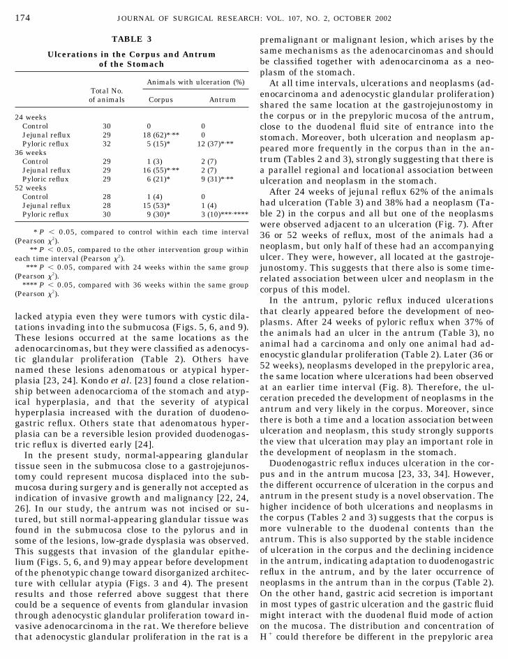
lacked atypia even they were tumors with cystic dila-tations invading into the submucosa (Figs. 5, 6, and 9).These lesions occurred at the same locations as theadenocarcinomas, but they were classified as adenocys-tic glandular proliferation (Table 2). Others havenamed these lesions adenomatous or atypical hyper-plasia [23, 24]. Kondo et al. [23] found a close relation-ship between adenocarcioma of the stomach and atyp-ical hyperplasia, and that the severity of atypicalhyperplasia increased with the duration of duodeno-gastric reflux. Others state that adenomatous hyper-plasia can be a reversible lesion provided duodenogas-tric reflux is diverted early [24].
In the present study, normal-appearing glandulartissue seen in the submucosa close to a gastrojejunos-tomy could represent mucosa displaced into the sub-mucosa during surgery and is generally not accepted asindication of invasive growth and malignancy [22, 24,26]. In our study, the antrum was not incised or su-tured, but still normal-appearing glandular tissue wasfound in the submucosa close to the pylorus and insome of the lesions, low-grade dysplasia was observed.This suggests that invasion of the glandular epithe-lium (Figs. 5, 6, and 9) may appear before developmentof the phenotypic change toward disorganized architec-ture with cellular atypia (Figs. 3 and 4). The presentresults and those referred above suggest that therecould be a sequence of events from glandular invasionthrough adenocystic glandular proliferation toward in-vasive adenocarcinoma in the rat. We therefore believethat adenocystic glandular proliferation in the rat is a
premalignant or malignant lesion, which arises by thesame mechanisms as the adenocarcinomas and shouldbe classified together with adenocarcinoma as a neo-plasm of the stomach.
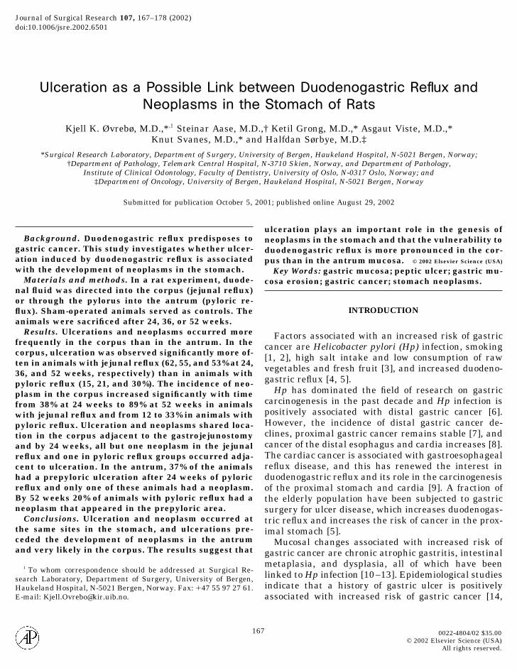
At all time intervals, ulcerations and neoplasms (ad-enocarcinoma and adenocystic glandular proliferation)shared the same location at the gastrojejunostomy inthe corpus or in the prepyloric mucosa of the antrum,close to the duodenal fluid site of entrance into thestomach. Moreover, both ulceration and neoplasm ap-peared more frequently in the corpus than in the an-trum (Tables 2 and 3), strongly suggesting that there isa parallel regional and locational association betweenulceration and neoplasm in the stomach.
After 24 weeks of jejunal reflux 62% of the animalshad ulceration (Table 3) and 38% had a neoplasm (Ta-ble 2) in the corpus and all but one of the neoplasmswere observed adjacent to an ulceration (Fig. 7). After36 or 52 weeks of reflux, most of the animals had aneoplasm, but only half of these had an accompanyingulcer. They were, however, all located at the gastroje-junostomy. This suggests that there also is some time-related association between ulcer and neoplasm in thecorpus of this model.
In the antrum, pyloric reflux induced ulcerationsthat clearly appeared before the development of neo-plasms. After 24 weeks of pyloric reflux when 37% ofthe animals had an ulcer in the antrum (Table 3), noanimal had a carcinoma and only one animal had ad-enocystic glandular proliferation (Table 2). Later (36 or52 weeks), neoplasms developed in the prepyloric area,the same location where ulcerations had been observedat an earlier time interval (Fig. 8). Therefore, the ul-ceration preceded the development of neoplasms in theantrum and very likely in the corpus. Moreover, sincethere is both a time and a location association betweenulceration and neoplasm, this study strongly supportsthe view that ulceration may play an important role inthe development of neoplasm in the stomach.
Duodenogastric reflux induces ulceration in the cor-pus and in the antrum mucosa [23, 33, 34]. However,the different occurrence of ulceration in the corpus andantrum in the present study is a novel observation. Thehigher incidence of both ulcerations and neoplasms inthe corpus (Tables 2 and 3) suggests that the corpus ismore vulnerable to the duodenal contents than theantrum. This is also supported by the stable incidenceof ulceration in the corpus and the declining incidencein the antrum, indicating adaptation to duodenogastricreflux in the antrum, and by the later occurrence ofneoplasms in the antrum than in the corpus (Table 2).On the other hand, gastric acid secretion is importantin most types of gastric ulceration and the gastric fluidmight interact with the duodenal fluid mode of actionon the mucosa. The distribution and concentration ofH� could therefore be different in the prepyloric area
TABLE 3
Ulcerations in the Corpus and Antrumof the Stomach
Total No.of animals
Animals with ulceration (%)
Corpus Antrum
24 weeksControl 30 0 0Jejunal reflux 29 18 (62)*,** 0Pyloric reflux 32 5 (15)* 12 (37)*,**
36 weeksControl 29 1 (3) 2 (7)Jejunal reflux 29 16 (55)*,** 2 (7)Pyloric reflux 29 6 (21)* 9 (31)*,**
52 weeksControl 28 1 (4) 0Jejunal reflux 28 15 (53)* 1 (4)Pyloric reflux 30 9 (30)* 3 (10)***,****
* P � 0.05, compared to control within each time interval(Pearson �2).
** P � 0.05, compared to the other intervention group withineach time interval (Pearson �2).
*** P � 0.05, compared with 24 weeks within the same group(Pearson �2).
**** P � 0.05, compared with 36 weeks within the same group(Pearson �2).
174 JOURNAL OF SURGICAL RESEARCH: VOL. 107, NO. 2, OCTOBER 2002

and at the gastrojejunostomy and affect the occurrenceof mucosal lesions. Wetscher et al. [35] found elevatedintragastric pH, gastrinemia, and hyperplasia of thegastric mucosa of rats with duodenogastric reflux, andKubo et al. [36] found gastrinemia in rats after vagotomy.Deveney et al. [37] found that gastrinemia exerted pro-tection against methyl-nitro-nitroso-guanidine (MNNG)-fortified carcinogenesis in the corpus of animals withduodenogastric reflux, and Tatsuta et al. [38, 39] re-duced the incidence of MNNG-induced adenocarcino-mas in the antrum of rats by repeated injections oftetragastrin. In animals with pyloric reflux, the trans-port of acid from the stomach into the duodenum isinhibited, and the antrum mucosa becomes exposed to
alkali, which could result in gastrinemia. Subsequently,the increased luminal pH and gastrinemia could con-tribute to the protection of the antral mucosa from theulceration. Several other hormones have been associ-ated with increased duodenogastric reflux [40, 41]. Dis-tension of the duodenal wall and increased pH in theduodenum could also result in hormonal changes thatinfluence the luminal contents of the antrum. How-ever, neither hormones nor luminal pH were examinedin the present study and their contribution to the dif-ferent incidences of ulceration and neoplasm in thejejunal and pyloric reflux models cannot be evaluated.
Due to the design of the study, a definitive causalrelationship cannot be established between ulceration
FIG. 7. The frequency of ulceration and neoplasm in the corpus mucosa. The frequencies of neoplasms are superimposed on the frequencyof animals with ulceration. *P � 0.05, the frequency of neoplasm (adenocarcinoma plus adenocystic glandular proliferation) different from24 weeks. †P � 0.05, the frequency of ulceration without neoplasm different from 24 weeks. Statistics by Pearson chi-square tests.
175ØVREBØ ET AL: ULCERATION AND NEOPLASM IN RAT STOMACH

and neoplasm. The possibility exists that the associa-tion is merely a consequence of a common etiologyeliciting two different kinds of response. However, asimilar sequence of events with ulceration and cancerwas observed by Kondo et al. [23] in rats subjected togastric resection. Moreover, the increased level ofN-nitroso compounds in resected stomachs is indica-tive of a milieu that favors the conversion of nitrite topossible carcinogenic substances [42], and Busby et al.[43] induced tumors in the stomach of rats with nitro-sated bile acids. An association between duodenogas-tric reflux and N-nitroso compound-induced gastric
carcinogenesis is therefore possible. Nishidoi et al. [44]found erosions in the mucosa at the gastroenterostomyof animals subjected to Bilroth II resection beforetreatment with the carcinogen MNNG, and erosionswere present in animals with carcinoma. Chronic ul-cers in rats increase the incidence of MNNG-inducedtumors in the corpus mucosa, and most tumors werelocated in the areas and margins of a previous ulcer-ation [45, 46]. Minimal erosions are observed overlyingmicroscopic or early gastric cancers in humans [16, 17].In a former study, we demonstrated that salt inducessuperficial injury of the mucosa that facilitates the
FIG. 8. The frequency of ulceration and neoplasm in the antrum mucosa. The frequencies of neoplasms are superimposed on thefrequency of animals with ulceration. *P � 0.05, the frequency of neoplasm (adenocarcinoma plus adenocystic glandular proliferation)different from 24 weeks. †P � 0.01, the frequency of ulceration without neoplasm different from 24 weeks. Statistics by Pearson chi-squaretests.
176 JOURNAL OF SURGICAL RESEARCH: VOL. 107, NO. 2, OCTOBER 2002

penetration of MNNG into the proliferating layer of thegastric mucosa in rats [47]. Cell proliferation is in-creased in the mucosa of the resected stomach, espe-cially close to the gastroenterostomy [27]. Moreover, inthe process of regeneration from ulceration the in-creased cell proliferation in the base and margin ofulceration may increase the number of cells at risk ofbeing hit by a carcinogen and thus render the mucosasusceptible to carcinogenesis [48, 49]. These observa-tions added to the present results strongly suggest thatlong-term reflux of duodenal contents into the stomachcauses mucosal injury and ulceration with increasedcellular proliferation and facilitated penetration of car-cinogens from the gastric lumen to the proliferatingcells, resulting in the development of neoplasms.
The association between corpus ulceration and neo-plasm observed in the present study is in accordancewith results of large epidemiological studies. A historyof gastric ulcer is positively associated with increasedrisk of gastric cancer [14, 15]. However, the epidemio-logical studies were unable to establish an associationbetween prepyloric ulcer and gastric cancer [14, 15].
CONCLUSION
Reflux of duodenal juice to the stomach of ratscaused ulcerations and neoplasms (carcinoma or ad-enocystic glandular proliferation) very often in the cor-pus, more rarely in the antrum. Ulceration and neo-plasm shared the same locations in the stomach, andearly in the process the ulceration preceded or ap-peared adjacent to a neoplasm. This supports the viewthat ulceration may play an important role in the gen-esis of neoplasms in the stomach.
ACKNOWLEDGMENTS
Dr. Øvrebø is a Research Fellow of the Norwegian Cancer Societyand this study was supported by grants from the Norwegian CancerSociety. We thank Inger Vikøyr, Anne Aarsand, Gry-Hilde Nilsen,and Cato Johnsen for skilled technical assistance.
REFERENCES
1. Tredaniel, J., Boffetta, P., Buiatti, E., Saracci, R., and Hirsch,A. Tobacco smoking and gastric cancer: Review and meta-analysis. Int. J. Cancer 72: 565, 1997.
2. Parsonnet, J., Friedman, G. D., Orentreich, N., and Vogelman,H. Risk for gastric cancer in people with CagA positive or CagAnegative Helicobacter pylori infection. Gut 40: 297, 1997.
3. Buiatti, E., Palli, D., Decarli, A., Amadori, D., Avellini, C.,Bianchi, S., Biserni, R., Cipriani, F., Cocco, P., Giacosa, A., et al.A case–control study of gastric cancer and diet in Italy. Int. J.Cancer 44: 611, 1989.
4. Caygill, C. P., Hill, M. J., Kirkham, J. S., and Northfield, T. C.Mortality from gastric cancer following gastric surgery for pep-tic ulcer. Lancet 1: 929, 1986.
5. Lundegardh, G., Adami, H. O., Helmick, C., Zack, M., and
Meirik, O. Stomach cancer after partial gastrectomy for benignulcer disease. N. Engl. J. Med. 319: 195, 1988.
6. Hansen, S., Melby, K. K., Aase, S., Jellum, E., and Vollset, S. E.Helicobacter pylori infection and risk of cardia cancer and non-cardia gastric cancer. A nested case-control study. Scand. J.Gastroenterol. 34: 353, 1999.
7. Hansen, S., Wiig, J. N., Giercksky, K. E., and Tretli, S. Esoph-ageal and gastric carcinoma in Norway 1958–1992: Incidencetime trend variability according to morphological subtypes andorgan subsites. Int. J. Cancer 71: 340, 1997.
8. Botterweck, A. A., Schouten, L. J., Volovics, A., Dorant, E., andvan Den Brandt, P. A. Trends in incidence of adenocarcinoma ofthe oesophagus and gastric cardia in ten European countries.Int. J. Epidemiol. 29: 645, 2000.
9. MacDonald, W. C., and Owen, D. A. Gastric carcinoma aftersurgical treatment of peptic ulcer: An analysis of morphologicfeatures and a comparison with cancer in the nonoperatedstomach. Cancer 91: 1732, 2001.
10. Sobala, G. M., O’Connor, H. J., Dewar, E. P., King, R. F., Axon,A. T., and Dixon, M. F. Bile reflux and intestinal metaplasia ingastric mucosa. J. Clin. Pathol. 46: 235, 1993.
11. Kokkola, A., Haapiainen, R., Laxen, F., Puolakkainen, P., Kivi-laakso, E., Virtamo, J., and Sipponen, P. Risk of gastric carci-noma in patients with mucosal dysplasia associated with atro-phic gastritis: A follow up study. J. Clin. Pathol. 49: 979, 1996.
12. Yoshimura, T., Shimoyama, T., Fukuda, S., Tanaka, M., Axon,A. T., and Munakata, A. Most gastric cancer occurs on the distalside of the endoscopic atrophic border. Scand. J. Gastroenterol.34: 1077, 1999.
13. Sipponen, P., Riihela, M., Hyvarinen, H., and Seppala, K.Chronic nonatropic (‘superficial’) gastritis increases the risk ofgastric carcinoma. A case–control study. Scand. J. Gastroen-terol. 29: 336, 1994.
14. Hansson, L. E., Nyren, O., Hsing, A. W., Bergstrom, R., Josef-sson, S., Chow, W. H., Fraumeni, J. F., Jr., and Adami, H. O.The risk of stomach cancer in patients with gastric or duodenalulcer disease. N. Engl. J. Med. 335: 242, 1996.
15. Molloy, R. M., and Sonnenberg, A. Relation between gastriccancer and previous peptic ulcer disease. Gut 40: 247, 1997.
16. Nagayo, T. Microscopical cancer of the stomach—A study onhistogenesis of gastric carcinoma. Int. J. Cancer 16: 52, 1975.
17. Shimoyama, S., Joujima, Y., Oohara, T., and Kaminishi, M.Dual roles of peptic ulcer in the carcinogenesis or extension ofearly gastric cancer. Ann. Surg. Oncol. 6: 495, 1999.
18. Hole, D. J., Quigley, E. M., Gillis, C. R., and Watkinson, G.Peptic ulcer and cancer: An examination of the relationshipbetween chronic peptic ulcer and gastric carcinoma. Scand. J.Gastroenterol. 22: 17, 1987.
19. Lee, S., Iida, M., Yao, T., Shindo, S., Okabe, H., and Fujishima,M. Long-term follow-up of 2529 patients reveals gastric ulcersrarely become malignant. Dig. Dis. Sci. 35: 763, 1990.
20. Testoni, P. A., Masci, E., Marchi, R., Guslandi, M., Ronchi, G.,and Tittobello, A. Gastric cancer in chronic atrophic gastritis.Associated gastric ulcer adds no further risk. J. Clin. Gastro-enterology 9: 298, 1987.
21. Sobala, G. M., King, R. F., Axon, A. T., and Dixon, M. F. Refluxgastritis in the intact stomach. J. Clin. Pathol. 43: 303, 1990.
22. Taylor, P. R., Mason, R. C., Filipe, M. I., Vaja, S., Hanley, D. C.,Murphy, G. M., Dowling, R. H., and McColl, I. Gastric carcino-genesis in the rat induced by duodenogastric reflux withoutcarcinogens: morphology, mucin histochemistry, polyamine me-tabolism, and labelling index. Gut 32: 1447, 1991.
23. Kondo, K., Kojima, H., Akiyama, S., Ito, K., and Takagi, H.Pathogenesis of adenocarcinoma induced by gastrojejunostomy
177ØVREBØ ET AL: ULCERATION AND NEOPLASM IN RAT STOMACH

in Wistar rats: Role of duodenogastric reflux. Carcinogenesis16: 1747, 1995.
24. Imai, T., Kobayasi, S., Rodrigues, M. A., de Camargo, J. L.,Ogawa, K., Iwata, H., and Tatematsu, M. Reduction of cellproliferative activities of gastric stump adenomatous hyperpla-sias after bile reflux diversion in rats. Carcinogenesis 14: 1765,1993.
25. Miwa, K., Hasegawa, H., Fujimura, T., Matsumoto, H., Miyata,R., Kosaka, T., Miyazaki, I., and Hattori, T. Duodenal refluxthrough the pylorus induces gastric adenocarcinoma in the rat.Carcinogenesis 13: 2313, 1992.
26. Mason, R. C., Taylor, P. R., Filipe, M. I., and McColl, I. Pancre-aticoduodenal secretions and the genesis of gastric stump car-cinoma in the rat. Gut 29: 830, 1988.
27. Miwa, K., Kamata, T., Miyazaki, I., and Hattori, T. Kineticchanges and experimental carcinogenesis after Billroth I and IIgastrectomy. Br. J. Surg. 80: 893, 1993.
28. Nagayo, T. Dysplasia of the gastric mucosa and its relation tothe precancerous state. Gann. 72: 813, 1981.
29. Rugge, M., Correa, P., Dixon, M. F., Hattori, T., Leandro, G.,Lewin, K., Riddell, R. H., Sipponen, P., and Watanabe, H.Gastric dysplasia: The Padova international classification.Am. J. Surg. Pathol. 24: 167, 2000.
30. Lauren, P. The two histological main types of gastric carci-noma: Diffuse and so-called intestinal type carcinoma. ActaPathol. Microbiol. Scand. 64: 31, 1965.
31. Roscoe, J. T., and Byars, J. A. An investigation of the restraintswith respect to sample size commonly imposed on the use of thechi-square statistic. J. Am. Stat. Assoc. 66: 755, 1971.
32. Tatsuta, M., Iishi, H., Baba, M., Narahara, H., Uedo, N., Yano,H., Yamamoto, R., Mukai, M., and Akedo, H. Induction bybombesin of peritoneal metastasis of gastric cancers induced byN-methyl-N�-nitro-N-nitrosoguanidine in Wistar rats. GastricCancer 4: 14, 2001.
33. Kaminishi, M., Sadatsuki, H., Johjima, Y., Oohara, T., andKondo, Y. A new model for production of chronic gastric ulcer byduodenogastric reflux in rats. Gastroenterology 92: 1913, 1987.
34. Azuma, T., Dojyo, M., Ito, S., Yamazaki, Y., Miyaji, H., Ito, Y.,Suto, H., Kuriyama, M., Kato, T., and Kohli, Y. Bile reflux dueto disturbed gastric movement is a cause of spontaneous gastriculcer in W/Wv mice. Dig. Dis. Sci. 44: 1177, 1999.
35. Wetscher, G. J., Hinder, R. A., Kretchmar, D., Stinson, R.,Perdikis, G., Smyrk, T., Klingler, P. J., and Adrian, T. E. Duo-denogastric reflux causes growth stimulation of foregut mucosapotentiated by gastric acid blockade. Dig. Dis. Sci. 41: 2166,1996.
36. Kubo, N., Noguchi, T., Takeno, S., Tohara, K., Uchida, Y., andShimoda, H. Injury to the gastric mucosa and cellular dynamicsin a rat model of duodenogastric reflux: The possible signifi-
cance of gastrin induction and a heat shock protein. Surg.Today 30: 999, 2000.
37. Deveney, C. W., Freeman, H., and Way, L. W. Experimentalgastric carcinogenesis in the rat: effects of hypergastrinemiaand acid secretion. Am. J. Surg. 139: 49, 1980.
38. Tatsuta, M., Itoh, T., Okuda, S., Taniguchi, H., and Tamura, H.Effect of prolonged administration of gastrin on experimentalcarcinogenesis in rat stomach induced by N-methyl-N�-nitro-N-nitrosoguanidine. Cancer Res. 37: 1808, 1977.
39. Tatsuta, M., Yamamura, H., Taniguchi, H., and Tamura, H.Gastrin protection against chemically induced gastric adeno-carcinomas in Wistar rats: Histopathology of the glandularstomach and incidence of gastric adenocarcinoma. J. Natl. Can-cer Inst. 69: 59, 1982.
40. Thomas, W. E., Ardill, J., and Buchanan, K. D. The hormonalchanges produced by duodenogastric reflux. Scand. J. Gastro-enterol. Suppl. 92: 44, 1984.
41. Wilson, P., Welch, N. T., Hinder, R. A., Anselmino, M., Her-rington, M. K., DeMeester, T. R., and Adrian, T. E. Abnormalplasma gut hormones in pathologic duodenogastric reflux andtheir response to surgery. Am. J. Surg. 165: 169, 1993.
42. Guadagni, S., Walters, C. L., Smith, P. L., Verzaro, R., Valenti,M., and Reed, P. I. N-Nitroso compounds in the gastric juice ofnormal controls, patients with partial gastrectomies, and gas-tric cancer patients. J. Surg. Oncol. 63: 226, 1996.
43. Busby, W. F., Jr., Shuker, D. E., Charnley, G., Newberne, P. M.,Tannenbaum, S. R., and Wogan, G. N. Carcinogenicity in rats ofthe nitrosated bile acid conjugates N-nitrosoglycocholic acidand N-nitrosotaurocholic acid. Cancer Res. 45: 1367, 1985.
44. Nishidoi, H., Koga, S., and Kaibara, N. Possible role of duode-nogastric reflux on the development of remnant gastric carci-noma induced by N-methyl-N�-nitro-N-nitrosoguanidine inrats. J. Natl. Cancer Inst. 72: 1431, 1984.
45. Takahashi, M., Shirai, T., Fukushima, S., Hahanouchi, M., andHirose, M. Effect of fundic ulcers induced by iodoacetamide ondevelopment of gastric tumors in rats treated with N-methyl-N�-nitro-N-nitrosoguanidine. Gann. 67: 47, 1976.
46. Wong, J., and Ong, G. B. Gastric carcinoma developing inchronic gastric ulcer in the rat treated with N-methyl-N1-nitro-N-nitrosoguanidine. J. Surg. Res. 29: 446, 1980.
47. Sorbye, H., Ovrebo, K., Gislason, H., Kvinnsland, S., andSvanes, K. Blood flow and mucoid cap protect against penetra-tion of carcinogens into superficially injured gastric mucosa ofrats. Dig. Dis. Sci. 40: 1720, 1995.
48. Li, H., and Helander, H. F. Hypergastrinemia increases prolif-eration of gastroduodenal epithelium during gastric ulcer heal-ing in rats. Dig. Dis. Sci. 41: 40, 1996.
49. Owens, D. M., Wei, S., and Smart, R. C. A multihit, multistagemodel of chemical carcinogenesis. Carcinogenesis 20: 1837,1999.
178 JOURNAL OF SURGICAL RESEARCH: VOL. 107, NO. 2, OCTOBER 2002


