Embed Size (px)
Citation preview
Significant number of papers has been published in re-cent years elucidating unknown or less-studied tubular structures of liver. These works particularly address the identification of reliable topography and architectonics of finest biliary branches – cholangioles and Herring’s ductuli and the stereomorphology of functional unit of the liver [7,12,13,18]. This should be explained by the fact that conventionally accepted models of liver functional unit, such as “classic lobule”, “secondary lobule”, “acinus”, etc., and their corresponding microcirculatory networks cannot adequately describe all manifestations of different clinical and experimental pathologies of liver and biliary system, especially taking into the consideration, that a mi-crocirculatory module of liver unlike the standard modules with three circulatory pools additionally includes the fourth component - bile circulation [11,18].
One of the pathological phenomena that trigger the investiga-tion of the functional morphology of liver lobule and especially the finest branches of biliary tract in depth is Ductular Reaction (DR). DR is a well-known phenomenon found in acute and chronic liver lesions in humans and rodents. Histologically DR is expressed by the increased amount of ductular profiles (DPs) [4,14]. DR developed due to acute biliary obstruction (“typical DR“) [6,23] is associated with increased mitotic ac-tivity of biliary epithelium and following bile ducts prolifera-tion which is evident from the second/third days after common bile duct Ligation (CBDL). DPs firstly develop in periportal areas and later gradually advance intralobularly [17,24,25, 27,28]. Their amount is increasing in time-depended manner after CBDL [3,8,10,20].
However, we have shown previously that number of DPs increase already several hours after CBDL in rats. Obvi-ously, this early DR could not be result of proliferation. Our studies have shown that this observation was due to increased biliary pressure caused by CBDL which leads to widening of periportal biliary plexus and intralobular bile ductuli (including Hering’s ductuli) with their subsequent visualization [1,2].
In present study, our attention was drawn to the ductular pro-files of unusual location directly attaching the tributaries of hepatic veins of different caliber – from large to sub-lobular and central veins. Despite the fact that the intralobular ductuli sometimes might be quite long and penetrate deep into the lobular parenchyma [13,18,19], the special search conducted in PubMed database could not yield any data regarding the above-mentioned paravenous biliary structures.
The aim of this work was to investigate the essence and origin of ductular profiles accompanying the tributaries of hepatic veins of different caliber.
Material and methods. animals and experimental Protocol. Male Wistar rats (n=28) weighing 250-300 g were subjected to CBDL (n=16) or sham operation (control) (n=12). Animals were anesthetized with ethyl ether. The common bile duct was ligated through midline laparotomy. Sham-operated animals underwent laparo-tomy without CBDL. Experimental protocols and use of animals were approved by the Institutional Animal Care Committee, in compliance with institutional guidelines from the European Commission and the National Insti-tutes of Health (USA) on the protection of animals used for experimental and other scientific purposes. The liver tissue was studied at 6, 12 and 24 hours after CBDL (4 animals in each group); Control samples were studied as well at 6, 12 and 24 hours after sham-operation (3 animals in each group); Dissected liver tissue samples (median lobe) were fixed in 10% neutral buffered for-malin, and used for routine histological and immuno-histochemical examination.
The contrast medium (Indian Ink) was injected through the catheter inserted into common bile duct toward liver tissue (proximal direction) in three animals from control group and four animals from CBDL group at 6th hour after sham operation and CBDL, respectively. The animals were killed immediately after intervention by deepening of anesthesia; dissected liver samples (median lobe) were fixed in 10% neutral buffered formalin and used for routine histology (Table 1).
Histology and immunohistochemistrySlices of 5 µm thickness obtained from 10% formalin fixed, paraffin embedded liver tissue samples were used for routine H&E and immunohistochemical staining. Primary antibodies against Ki67 (1:150) (ab16667, (Rabbit anti-rat) Abcam plc, Cambridge, UK), OV-6 (1:100) (MAB2020 (mouse anti-rat) R&D Systems, Inc), anti-CK7 (MBS570028, (mouse anti-rat) MyBioSource, SanDiego, USA) and anti-CK19 (ab77983, (mouse anti-rat) Abcam plc, Cambridge, UK) were used. Immunohistochemistry was performed using the standard streptavidin-biotin-immunoperoxidase method with DAB as chromogen (RE7280-K Novolink, Max Polymer Detection System, Leica Microsistems, Wetzlar, Germany), according to the manufacturer’s instructions. Sections were counter-stained with Hematoxylin.
UNKNOWN BILE DUCTULI ACCOMPANYING HEPATIC VEIN TRIBUTARIES (EXPERIMENTAL STUDY)
Kordzaia D., Jangavadze M.
i. Javakhishvili Tbilisi state University, a. natishvili institute of Morphology, Georgia

The average number of ductular profiles in cross-sectional areas was calculated in 20 appropriate fields for each sample: a) intralobularly; b) adjacently to central veins or to smallesttributaries of hepatic veins (sublobular veins) (<50 μm /); c) within the connective tissue surrounding the lobar branch of hepatic vein or its large tributaries (≥250 μm).
The classification of vein diameters was made in accor-dance with Morikawa et al., 2000 [16].
Liver from one rat was considered as a one sample. Data for the average number of ductular profiles were analyzed by one-way analysis of variances (one-way ANOVA). Time served as a factor for the average number of ductular profiles in ANOVA. Planned comparisons between the average numbers of ductular profiles were made by using t-tests.
Results and their discussion. CBDL during the 6 hours
reveals significant effect by the number of DPs in every investigated area (per large and small hepatic vein tribu-taries, intralobularly). On the 12th hour of the experiment only DPs surrounding the central and sublobular veins (the smallest tributaries of hepatic veins) (<50 μm) continued to increase in number (Table 2).
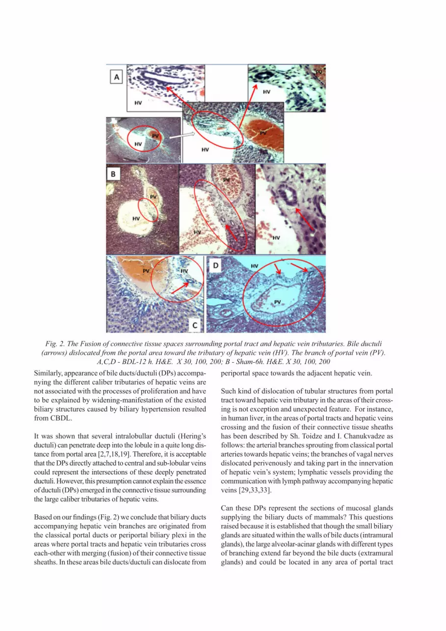
The diameters (calibers) of DPs located in the adventitia of the large tributaries of hepatic veins are varying from 10 to 30 mµ. Their different shapes indicate that ductuli may be situated transversely, tangentially or longitudinally towards the vein branches (Fig. 2 D; 3 A,B,C).
The diameters (calibers) and shape of DPs found imme-diately at central and sublobular veins are varying from 5 to 15 mµ and from circular - to ellipsoidal. They did not differ from other DPs (Fig. 1 A,B); Their epitheliocytes all are CK19 positive (Fig. 1 C), and OV6 positive (Fig. 1 D), but Ki-67 negative.
Table 1. distribution of experimental animals by surgery and methods
Group Sham – 6 h. Sham -12 h. sham-24 h. CBDL – 6 h. CBDL -12
h. CBDL – 24 h.
Number of ani-mals
3 3 3 3 4 4 4 4
Methods
H&E Ink + H&E H&E H&E H&E Ink +
H&E H&E H&E
OV 6 OV 6 OV 6 OV 6 OV 6 OV 6
CK 7 CK 7 CK 7 CK 7 CK 7 CK 7
CK 19 CK 19 CK 19 CK 19 CK 19 CK 19H&e – Hematoxylin and eosin
cBdl – common Bile duct ligation sham – sham operated group
Table 2. Number of ductular profiles (DP) in different areas and terms of experimentAreas
of calculation
Terms of experiment
Ductular Profiles in the con-nective tissue surrounding the lobar branch of hepatic vein or its large tributaries
(>/= 250 µm) (M ±SD)
Ductular Profiles sur-rounding the central veins and the smallest tributaries of hepatic
veins (sublobular veins) (< 50 μm/) (M ±SD)
Intralobulary(M ±SD)
Sham – 6 h. 1,75*± 0.7 0,05* ± 0.29 0.18*±0.04
CBDL- 6 h 3,45 ±0,9 0,5# ± 0,59 2.62 ±0.22
Sham – 12 h. 1,9* ±0.60 0.1* ± 0.40 0.08* ±0.04
CBDL- 12 h 3,85 ± 0,98 1, 35§ ± 0,65 2.65 ±0.25
Sham – 24 h. 1.8* ±0.46 0.09* ± 0,03 0.25* ±0.08
CBDL - 24 h 3.95± 0,95 1,45§ ± 0,59 2.79 ±0.20cBdl – bile duct ligated group; sham – sham operated group; M – Mean; sd – standard deviation;
* - Significant Difference with same data of corresponding BDL groups (p<0.05)§ - Significant Difference with same data for 6 hour (p<0.05)

Fig. 1. Ductular profiles (arrows) accompanying central and sub-lobular veins. CBDL-6 h. A, B - H&E. X 100 (A) and 200 (B). C – CK19. X 100 and 200; D – OV6. X 100 and 200
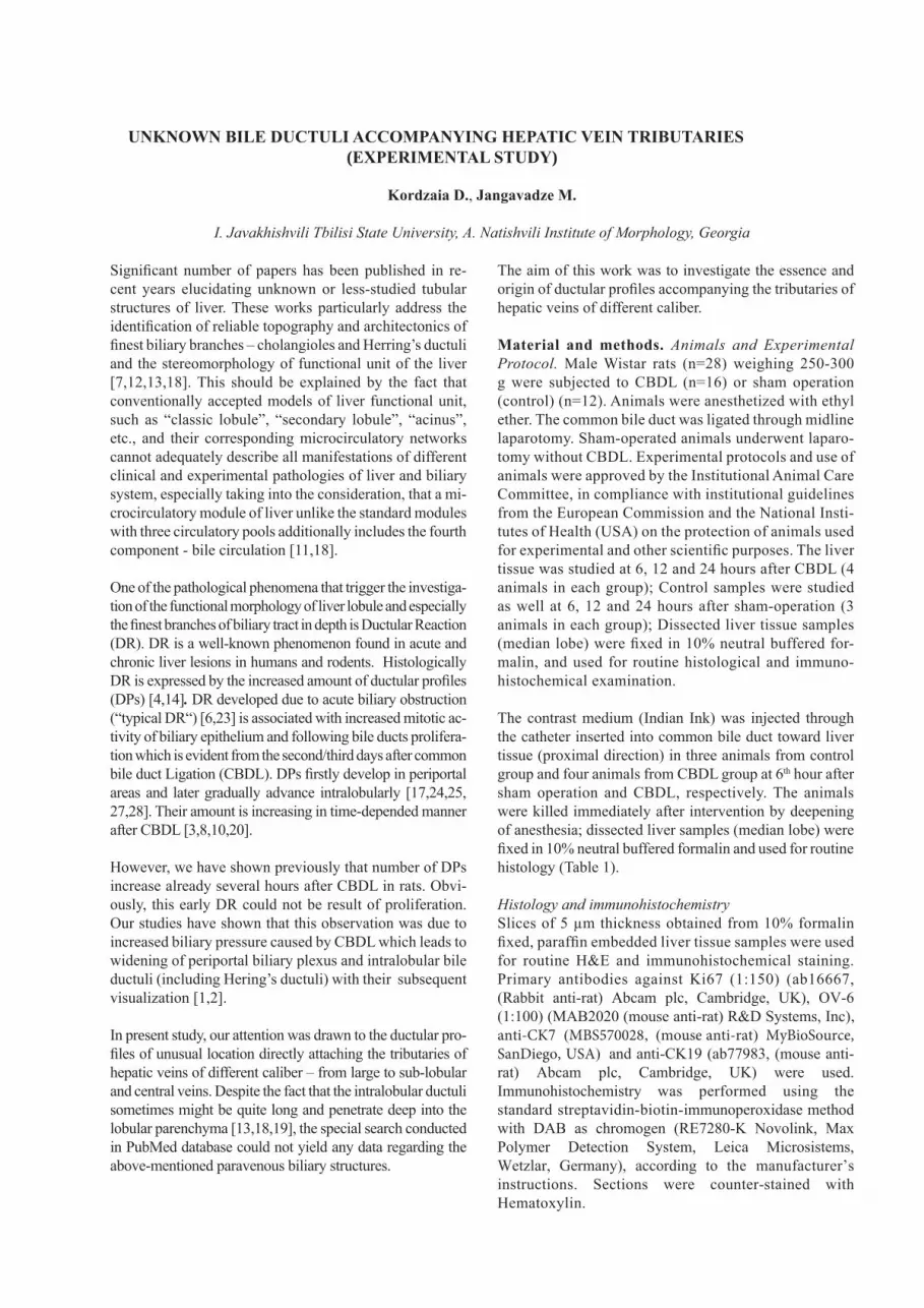
On the histological specimens of rat livers from both - con-trol and CBDL groups were revealed the sites of crossing of different size portal tracts and hepatic vein tributaries fea-tured by integration (fusion) of connective tissue sheaths of the above-mentioned structures. In these areas the perivas-cular capsule - the derivate of the liver sheath (Glisson’s sheath) - surrounding the portal triad extends toward the hepatic vein and includes it as a fourth component. Some of DPs located at these sites have a traditional, periportal location, while the others are dislocated (moved) towards the hepatic vein branch and situated within its connective tissue sheath (adventitia) (Fig. 2 A,B,C).
In the specimens prepared after the injection of contrast medium into the biliary system via CBD, the injected ink is found in both - portal and perivenous DPs as well as in bile canaliculi confirming the intercommunication and continuity of the lumina (beds) of these structures. At the same time, the lumina of blood vessels and sinusoids remain contrast medium-free (Fig. 3 D).
We have shown the ductular profiles accompanying the hepatic vein tributaries of different calibers in rats in norm and CBDL setting. Interstingly that appiarence of these
DPs are not related with proliferative activity. We found no analogue of this finding in the literature.
To understand the essence of bile ductuli/DPs accompa-nying hepatic vein tributaries of different diameter it is ultimately important to clarify if these structures represent the existing (but invisible and thus unknown) compartment of biliary tree or the newly formed constructions.
It is considered, that development of intralobular DPs after CBDL in rodents is caused by the proliferation of existed bile ducts (ductuli) and/or biliary transdifferen-tiation of hepatocytes and/or proliferation of oval cells with formation of ductule-like structures [6,23]. All these three mechanisms may develop from the 2nd -3rd day after CBDL and maintain during several weeks [9,21]. However, the number of DPs is increased at earliest stages of bile congestion - after 6 and 12 hours following CBDL, when the above-mentioned mechanisms were not initiated yet [19,20]. Thus, the DR observed at early stages following CBDL could be considered only as a result of widening and manifestation of already existed but in conventional condition invisible intralobullar biliary ductuli.
Similarly, appearance of bile ducts/ductuli (DPs) accompa-nying the different caliber tributaries of hepatic veins are not associated with the processes of proliferation and have to be explained by widening-manifestation of the existed biliary structures caused by biliary hypertension resulted from CBDL.
It was shown that several intralobullar ductuli (Hering’s ductuli) can penetrate deep into the lobule in a quite long dis-tance from portal area [2,7,18,19]. Therefore, it is acceptable that the DPs directly attached to central and sub-lobular veins could represent the intersections of these deeply penetrated ductuli. However, this presumption cannot explain the essence of ductuli (DPs) emerged in the connective tissue surrounding the large caliber tributaries of hepatic veins.
Based on our findings (Fig. 2) we conclude that biliary ducts accompanying hepatic vein branches are originated from the classical portal ducts or periportal biliary plexi in the areas where portal tracts and hepatic vein tributaries cross each-other with merging (fusion) of their connective tissue sheaths. In these areas bile ducts/ductuli can dislocate from
periportal space towards the adjacent hepatic vein.
Such kind of dislocation of tubular structures from portal tract toward hepatic vein tributary in the areas of their cross-ing is not exception and unexpected feature. For instance, in human liver, in the areas of portal tracts and hepatic veins crossing and the fusion of their connective tissue sheaths has been described by Sh. Toidze and I. Chanukvadze as follows: the arterial branches sprouting from classical portal arteries towards hepatic veins; the branches of vagal nerves dislocated perivenously and taking part in the innervation of hepatic vein’s system; lymphatic vessels providing the communication with lymph pathway accompanying hepatic veins [29,33,33].
Can these DPs represent the sections of mucosal glands supplying the biliary ducts of mammals? This questions raised because it is established that though the small biliary glands are situated within the walls of bile ducts (intramural glands), the large alveolar-acinar glands with different types of branching extend far beyond the bile ducts (extramural glands) and could be located in any area of portal tract
Fig. 2. The Fusion of connective tissue spaces surrounding portal tract and hepatic vein tributaries. Bile ductuli (arrows) dislocated from the portal area toward the tributary of hepatic vein (HV). The branch of portal vein (PV).
A,C,D - BDL-12 h. H&E. X 30, 100, 200; B - Sham-6h. H&E. X 30, 100, 200
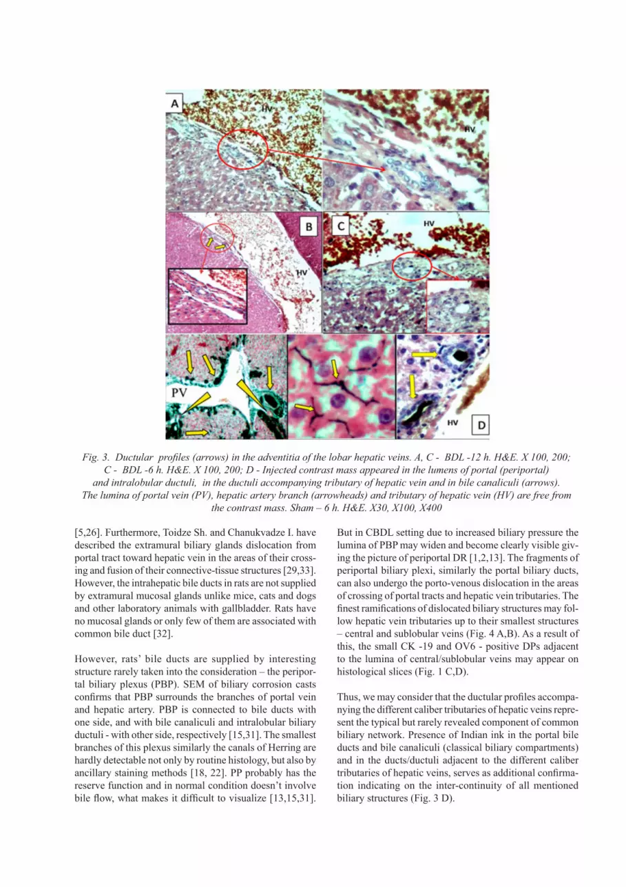
Fig. 3. Ductular profiles (arrows) in the adventitia of the lobar hepatic veins. A, C - BDL -12 h. H&E. X 100, 200; C - BDL -6 h. H&E. X 100, 200; D - Injected contrast mass appeared in the lumens of portal (periportal)
and intralobular ductuli, in the ductuli accompanying tributary of hepatic vein and in bile canaliculi (arrows). The lumina of portal vein (PV), hepatic artery branch (arrowheads) and tributary of hepatic vein (HV) are free from
the contrast mass. Sham – 6 h. H&E. X30, X100, X400
[5,26]. Furthermore, Toidze Sh. and Chanukvadze I. have described the extramural biliary glands dislocation from portal tract toward hepatic vein in the areas of their cross-ing and fusion of their connective-tissue structures [29,33]. However, the intrahepatic bile ducts in rats are not supplied by extramural mucosal glands unlike mice, cats and dogs and other laboratory animals with gallbladder. Rats have no mucosal glands or only few of them are associated with common bile duct [32].
However, rats’ bile ducts are supplied by interesting structure rarely taken into the consideration – the peripor-tal biliary plexus (PBP). SEM of biliary corrosion casts confirms that PBP surrounds the branches of portal vein and hepatic artery. PBP is connected to bile ducts with one side, and with bile canaliculi and intralobular biliary ductuli - with other side, respectively [15,31]. The smallest branches of this plexus similarly the canals of Herring are hardly detectable not only by routine histology, but also by ancillary staining methods [18, 22]. PP probably has the reserve function and in normal condition doesn’t involve bile flow, what makes it difficult to visualize [13,15,31].
But in CBDL setting due to increased biliary pressure the lumina of PBP may widen and become clearly visible giv-ing the picture of periportal DR [1,2,13]. The fragments of periportal biliary plexi, similarly the portal biliary ducts, can also undergo the porto-venous dislocation in the areas of crossing of portal tracts and hepatic vein tributaries. The finest ramifications of dislocated biliary structures may fol-low hepatic vein tributaries up to their smallest structures – central and sublobular veins (Fig. 4 A,B). As a result of this, the small CK -19 and OV6 - positive DPs adjacent to the lumina of central/sublobular veins may appear on histological slices (Fig. 1 C,D).
Thus, we may consider that the ductular profiles accompa-nying the different caliber tributaries of hepatic veins repre-sent the typical but rarely revealed component of common biliary network. Presence of Indian ink in the portal bile ducts and bile canaliculi (classical biliary compartments) and in the ducts/ductuli adjacent to the different caliber tributaries of hepatic veins, serves as additional confirma-tion indicating on the inter-continuity of all mentioned biliary structures (Fig. 3 D).

It is logical that dislocated biliary ductules accompanying hepatic vein tributaries and extending up to the smallest sublobular/central veins, should not be ended blindly. It is biologically “inexpedient”. Presumably, they should have the contact with bile canaliculi similarly with other intral-obular bile ductuli [2]. The bile canaliculi, in fact, may be the only structures from which bile should flow into these atypically located biliary ductuli located at the center of some lobuli. However, no similar communications are described yet in humans and/or other mammals. It is not excluded, that communication of bile canaliculi with atypi-cally located bile ductuli represent a specific feature of rat liver. For instance, inlets of sinusoids at different levels of hepatic venous tree (including sub-lobular and collecting veins) is described only in rat liver, while in humans and other mammals sinusoids drain only into the central veins [34]. It may be concluded that bile from the bile canaliculi in some lobuli of rat liver is drained in two directions: por-tal and caval. In portal areas bile is collected by classical interlobular bile ductuli, while centrolobularly it would be collected by “atypical”, “dislocated ductuli” which are originated from the sites of crossing of portal tracts and hepatic vein tributaries and fusion their connective-tissue sheaths (Fig. 4 A,B).
Apparently, these dislocated ductuli have the spare function and there is no active bile flow in their lumina,
which supports to their invisibility in normal conditions. Bile drainage via these ductuli should be “switched on” in settings of bile congestion and increased biliary pressure. This phenomenon may account for additional adaptative mechanism in bile congestion in rats, which biliary tree is not provided neither by gallbladder nor mucosal glands and therefore is more vulnerable to biliary hypertension.
REFERENCES
1. Azmaiparashvili E., Berishvili E., Jangavadze M., Kordzaia D. Study on the Origin of «Newductules» Appearing in the Rat Liver in Several Hours After Common Bile Duct Ligation. Acta Morphologica et Anthropologica 2012; 18:6.2. Azmaiparashvili E., Berishvili E., Kakabadze Z., Pilishvili O., Mikautadze E., Solomonia R., Jangavadze M., Kordzaia D. Ductular reaction at the early terms of common bile duct ligation in the rats. Acta Biologica Hungarica 2012; 63:321-332.3. Franchitto A., Onori P., Renzi A., Carpino G., Mancinelli R., Alvaro D., Gaudio E. Recent advances on the mechanisms regulating cholangiocyte proliferation and the significance of the neuroendocrine regulation of cholangiocyte pathophysiology. Annals of Translational Medicine 2012; 1.4. Desmet V.J. Ductal plates in hepatic ductular reactions. Hypoth-esis and implications. I. Types of ductular reaction reconsidered. Virchows Archiv. An International Journal of Pathology 2011; 458:251-259.
Fig.4. Schemes of bile circulation in areas, when hepatic vein tributaries are accompanied by bile ductuli. A, B – 1. branch of hepatic artery; 2. branch of portal vein; 3. portal bile duct; 4. central vein; 5. sub-lobular vein;
6. Periportal biliary plexus; 7. intralobular ductules (ductules of Hering); 8. dislocated bile ductules; 9. bile canaliculi; 10. sinusoid

5. Chanukvadze I.M. Surgical Anatomy of Main IntrahepaticPortal Tracts. Proc. Georgian Nat. Acad. Sci. Biomed. Series 2010; 36:9.6. Priester S., Wise C., Glaser S.S. Involvement of cholangiocyteproliferation in biliary fibrosis. World Journal of Gastrointestinal Pathophysiology 2010; 1:30-37.7. Dezso K., Paku S., Papp V., Turanyi E., Nagy P. Architecturaland immunohistochemical characterization of biliary ductules in normal human liver. Stem Cells and Development 2009; 8:1417-1422.8. Dirlik M., Canbaz H., Dusmez D., Apa M., Caglikulekci A.,Yaylak F., Balli E., Tamer L., Kanik A., Aydin S. The monitoring of progress in apoptosis of liver cells in bile duct-ligated rats. The Turkish Journal of Gastroenterology. The Official Journal of Turkish Society of Gastroenterology 2009; 20:247-256.9. Georgiev P., Jochum W., Heinrich S., Jang J.H., Nocito A.,Dahm F., Clavien P.A. Characterization of time-related changes after experimental bile duct ligation. The British Journal of Sur-gery 2008; 95:646-656.10. Gaudio E., Franchitto A., Pannarale L., Carpino G., AlpiniG., Francis H., Glaser S., Alvaro D., Onori P. Cholangiocytes and blood supply. World Journal of Gastroenterology: WJG 2006; 12:3546-3552.11. Sulaberidze G.D., Kardzvia D.D., Kikalishvili L.A.,Khomeriki T. Common intralobular microcirculatory module peculliarities in cholestasis in white rats. Georgian Medical News 2006; 100-105.12. Teutsch H.F. The modular microarchitecture of human liver.Hepatology 2005; 42:317-325.13. Roskams T.A., Theise N.D., Balabaud C., Bhagat G., BhathalP.S., Bioulac-Sage P., Brunt E.M., Crawford J.M., Crosby H.A., Desmet V., Finegold M.J., Geller S.A., Gouw A.S., Hytiroglou P., Knisely A.S., Kojiro M., Lefkowitch J.H., Nakanuma Y., Olynyk J.K., Park Y.N., Portmann B., Saxena R., Scheuer P.J., Strain A.J., Thung S.N., Wanless I.R., West A.B. Nomenclature of the finer branches of the biliary tree: canals, ductules, and ductular reac-tions in human livers. Hepatology 2004; 39:1739-1745.14. Roskams T.A., Libbrecht L., Desmet V.J. Progenitor cells indiseased human liver. Seminars in Liver Disease 2003; 23:385-396.15. Murakami T., Sato H., Nakatani S., Taguchi T., Ohtsuka A.Biliary tract of the rat as observed by scanning electron micros-copy of cast samples. Archives of Histology and Cytology 2001; 64:439-447.16. Morikawa H., Hachiya K., Mizuhara H., Fujiwara H., Nishi-guchi S., Shiomi S., Kuroki T., Kaneda K. Sublobular veins as the main site of lymphocyte adhesion/transmigration and adhesion molecule expression in the porto-sinusoidal-hepatic venous sys-tem during concanavalin A-induced hepatitis in mice. Hepatology 2000; 31:83-94.17. Demetris A.J., Sakamoto T., Liu Z., Yokomuro S., Ezure T.,Murase N., Blakolmer K. The ductular reaction in liver disease-emphasis on a type I response. in: W.E. Fleig (ed.). Normal and Malignant Liver Cell Growth, vol. 103c. Kluwer Academic Publication, Dordrecht; Boston: 1999; 141-155.18. Saxena R., Theise N.D., Crawford J.M. Microanatomy of thehuman liver-exploring the hidden interfaces. Hepatology 1999; 30:1339-1346.19. Theise N.D., Saxena R., Portmann B.C., Thung S.N., Yee H.,Chiriboga L., Kumar A., Crawford J.M. The canals of Hering and hepatic stem cells in humans. Hepatology 1999; 30:1425-1433.20. Alpini G., Glaser S.S., Ueno Y., Pham L., Podila P.V., CaligiuriA., LeSage G., LaRusso N.F. Heterogeneity of the proliferative
capacity of rat cholangiocytes after bile duct ligation. The Ameri-can Journal of Physiology 1998; 74:767-775.21. Roskams T., Desmet V. Ductular reaction and its diagnosticsignificance. Seminars in Diagnostic Pathology 1998; 15:259-269.22. Ekataksin W., Zou Z., Wake K., Chunhabundit P., Somana R.,Nishida J., McCuskey R. The hepatic microcirculatory subunits: an over-threecentury-long search for the missing link between an exocrine unit and an endocrine unit in mammalian liver lobules. in: P. M. Motta (ed.), Recent Advances in Microscopy of Cells, Tissues and Organs. University of Rome La Sapienza Press: Rome; 1997; 375-380.23. Desmet V., Roskams T., Van Eyken P. Ductular reaction in theliver. Pathology, Research and Practice 1995; 191:513-524.24. Burt A.D., MacSween R.N. Bile duct proliferation--its truesignificance? Histopathology 1993; 23:599-602.25. Marucci L., Baroni G.S., Mancini R., Benedetti A., JezequelA.M., Orlandi F. Cell proliferation following extrahepatic biliary obstruction. Evaluation by immunohistochemical methods. Jour-nal of Hepatology 1993; 17:163-169.26. Kordzaia D. Extrahepatic cholestasis. Ganatleba, Tbilisi:1990. 27. Shibayama Y. Factors producing bile infarction and bile ductproliferation in biliary obstruction. The Journal of Pathology 1990; 160:57-62.28. Slott P.A., Liu M.H., Tavoloni N. Origin, pattern, and mecha-nism of bile duct proliferation following biliary obstruction in the rat. Gastroenterology 1990; 99:466-477.29. Chanukvadze I.M. Structure and Connections of FibroseSheaths of Portal Complexes and Hepatic Veins. Collections of Scientific Works of Tbilisi State Medical Institute 1989; 1:11.30. Izraelashvili M.S. Topographic Anatomy of Lymphatic Ves-sels and Regional Lymph Nodes of Liver. The Applied Aspect. Collections of Scientific Works of Tbilisi State Medical Institute 1989; 1:20.31. Yamamoto K., Phillips M.J. A hitherto unrecognized bileductular plexus in normal rat liver. Hepatology 1984; 4:381-385.32. Andrews C.J., Andrews W.H. The relation between structureand function of bile ducts in man, some laboratory animals and the Adelie penguin. Quarterly Journal of Experimental Physiology and Cognate Medical Sciences 1979; 64:61-67.33. Toidze S.S., Chanukvadze I.M. Presented at the Transcauca-sian Conference of Morphologists, Baku: 1978.34. Gershbein L.L., Elias H. Observations on the anatomy of therat liver. The Anatomical Record 1954; 120:85-98.
SUMMARY
UNKNOWN BILE DUCTULI ACCOMPANYING HEPATIC VEIN TRIBUTARIES (EXPERIMENTAL STUDY)
Kordzaia D., Jangavadze M.
i. Javakhishvili Tbilisi state University, a. natishvili insti-tute of Morphology, Georgia
Studying Ductular reaction (DR) at early stages after com-mon bile duct ligation (CBDL) in rats we revealed some ductular profiles (DPs) of unusual location - accompanying

different caliber tributaries of hepatic veins (THV) includ-ing central and sub-lobular venules. We investigated the essence and genesis of these atypically located ductuli.
28 Wistar Rat livers were studied histologically and im-munohistochemically in norm and after 6, 12 and 24 hours of CBDL. Biliary system of part of the animals was pre-liminary injected by Indian Ink.
After CBDL the number of DPs including the ones accom-panying large and small THV was increased. The diameters of DPs found immediately at central and sublobular veins were varying from 5 to 15 mµ and of DPs located in the adventitia of the large THV are varying from 10 to 30 mµ. The cell of these DPs were CK19, CK7 and OV6 positive, but Ki-67 negative, what confirms their belonging to cho-langiocytes but denies their proliferative genesis.
In the sites of crossing of different size portal tracts (PT) and THV with integration of their connective tissue sheaths were revealed some biliary ducts/ductules dislocated from PT towards the THV and situated within their adventitia.
The Indian Ink injected via CBD was found in both - por-tal and perivenous DPs as well as in bile canaliculi, what confirms their inter-continuity.
The biliary ductules dislocated from PT toward THV may accompany hepatic venous pathways reversely up to cen-tral venules. These finest ductuli having spare function are likely to communicate with bile canaliculi.
Keywords: intrahepatic biliary system, ductular reaction, bile congestion, portal complex, perivascular sheath.


