Embed Size (px)
Citation preview
REVIEW Open Access
Untangling the origin and function ofgranulovacuolar degeneration bodies inneurodegenerative proteinopathiesVera I. Wiersma1,2, Jeroen J. M. Hoozemans3 and Wiep Scheper1,2*
Abstract
In the brains of tauopathy patients, tau pathology coincides with the presence of granulovacuolar degenerationbodies (GVBs) both at the regional and cellular level. Recently, it was shown that intracellular tau pathology causesGVB formation in experimental models thus explaining the strong correlation between these neuropathologicalhallmarks in the human brain. These novel models of GVB formation provide opportunities for future research intoGVB biology, but also urge reevaluation of previous post-mortem observations. Here, we review neuropathologicaldata on GVBs in tauopathies and other neurodegenerative proteinopathies. We discuss the possibility thatintracellular aggregates composed of proteins other than tau are also able to induce GVB formation. Furthermore,the potential mechanisms of GVB formation and the downstream functional implications hereof are outlined inview of the current available data. In addition, we provide guidelines for the identification of GVBs in tissue and cellmodels that will help to facilitate and streamline research towards the elucidation of the role of these enigmaticand understudied structures in neurodegeneration.
Keywords: Granulovacuolar degeneration bodies, Tau pathology, Neurodegenerative proteinopathies, Lysosomes
Neurodegenerative tauopathies, including Alzheimer’s dis-ease (AD) and frontotemporal lobar degeneration (FTLD)share a common neuropathological feature: the depositionof misfolded and hyperphosphorylated tau protein in mul-timeric, highly-ordered, filamentous aggregates in the cen-tral nervous system. The existence of familial forms ofFTLD that are caused by mutations in the tau-encodingMAPT gene underscores the key role of tau in diseasepathogenesis [49, 109, 121]. Indeed, also in sporadic tauo-pathy patients the accumulation of aggregated tau stronglycorrelates with neuronal loss and clinical symptoms, asdemonstrated by neuropathological studies [4, 34, 51, 98]
and more recently in living patients by tau positron emis-sion tomography [77]. Despite this strong association be-tween tau aggregation and neurodegeneration, themechanisms that connect the two are largely unknown.Here, we focus on an intracellular alteration that coincideswith the formation of tau aggregates: the emergence ofgranulovacuolar degeneration bodies (GVBs).GVBs were first observed in 1911 by Simchowicz, in
pyramidal neurons in the hippocampus of AD patients[120]. GVBs are membrane-delineated clear vacuolesthat harbor a dense core or “granule”. The GVB vacuoleand core measure between ~ 3 to ~ 5 μm and ~ 0.5 and~ 1.5 μm in diameter, respectively. The number of GVBsper cell is highly variable, ranging from a single one todozens. They are predominantly found in the neuronalcell body but can also be localized in the (proximal)neurite. Based on the basophilic and argentophilic prop-erties of the core, GVBs have traditionally been
© The Author(s). 2020 Open Access This article is licensed under a Creative Commons Attribution 4.0 International License,which permits use, sharing, adaptation, distribution and reproduction in any medium or format, as long as you giveappropriate credit to the original author(s) and the source, provide a link to the Creative Commons licence, and indicate ifchanges were made. The images or other third party material in this article are included in the article's Creative Commonslicence, unless indicated otherwise in a credit line to the material. If material is not included in the article's Creative Commonslicence and your intended use is not permitted by statutory regulation or exceeds the permitted use, you will need to obtainpermission directly from the copyright holder. To view a copy of this licence, visit http://creativecommons.org/licenses/by/4.0/.The Creative Commons Public Domain Dedication waiver (http://creativecommons.org/publicdomain/zero/1.0/) applies to thedata made available in this article, unless otherwise stated in a credit line to the data.
* Correspondence: [email protected] of Clinical Genetics, Amsterdam University Medical Centerslocation VUmc, Amsterdam, The Netherlands2Department of Functional Genomics, Center for Neurogenomics andCognitive Research, Vrije Universiteit (VU), De Boelelaan 1085, 1081 HVAmsterdam, The NetherlandsFull list of author information is available at the end of the article
Wiersma et al. Acta Neuropathologica Communications (2020) 8:153 https://doi.org/10.1186/s40478-020-00996-5
visualized using the routine histological hematoxylin andeosin (H&E) staining and by silver impregnation tech-niques [133]. Currently, GVBs are most commonly de-tected by immunolabeling of the core using specificmarkers (Table 1; Fig. 1). The presence of GVBs withincells constitutes the neuropathological hallmark knownas granulovacuolar degeneration (GVD).Since their discovery over a century ago, GVBs have
been enigmatic structures of unknown origin and func-tion. In 1980, a review on senile dementia stated: “In the
absence of an experimental model for the induction ofgranulovacuolar change, this characteristic cytologicalchange has been largely ignored, and at the present timeno hypothesis for induction is available” [11]. It took 4more decades to the publication of the first experimentalmodel for GVB formation in vitro, in which GVBs areinduced by the seeding of intracellular tau pathology[143]. This opens new avenues for future research intothe mechanism and function of GVB formation. How-ever, the causal relation between tau pathology and
Table 1 Box: Validation of GVB identity
Both the core and membrane of GVBs carry epitopes that can be used to confirm a GVB identity. Commonly used GVB core markers that consistentlydetect GVBs in human brain and experimental models are CK1δ (Fig. 1a-d) and CK1ε [33, 67, 143], CHMP2B [90, 143, 147], pPERK (Fig. 1c-f), peIF2αand pIRE1α [43, 67, 143] and pTDP-43 [47, 59, 79, 148]. In Supplementary Table 1, an overview of primary antibodies used to detect these commonGVB markers in tissue and culture is provided. Of these common GVB markers, the evidence for the localization of CK1 isoforms to human and experi-mental GVBs meets high standards of rigor. The presence of CK1δ and CK1ε in human and experimental GVBs has been shown using antibodies thatstain GVBs in a manner independent of phosphorylation – as phosphatase pre-treatment of human brain tissue did not affect GVB immunolabeling[33]. When comparing CK1δ to CK1ε staining in aged tau Tg mice, a high degree of overlap was found: in ~ 40% of GVB-bearing neurons, all GVBswere double immunopositive, with the great majority of remaining neurons showing between 50 and 99% of double-labeled GVBs [67]. In addition,CK1δ and CK1ε staining overlap with GVBs detected by H&E staining in serial brain sections [33]. Furthermore, using STED super-resolution micros-copy and/or immuno-EM the subcellular localization of CK1δ [33, 143] and CK1ε [74] in the GVB core has been confirmed at high resolution. More-over, fluorescently-tagged CK1δ localizes to GVBs in vitro [143]. Therefore, CK1δ is currently the only constituent of the GVB core of which thepresence has been confirmed directly, without the use of antibodies. In 100% [90]/98% [30] of CK1δ-positive neurons in the human brain, GVBs werealso labeled by CHMP2B. On the subcellular level, CK1δ and CHMP2B co-localize in the core of 82% of GVBs in the human brain [30]. The overlap be-tween the two GVB markers was somewhat lower in aged tau Tg mice, with CHMP2B immunolabeling being detected in 57% of neurons with CK1δ-positive GVBs and within those neurons 63% of GVBs being double positive for both CHMP2B and CK1δ [90]. In the human brain, CHMP2B stainingoverlaps with H&E staining after ethanol-mediated de-staining [146, 147] and mirrors H&E staining of GVBs in adjacent sections [147]. Furthermore,GVB counts based on CHMP2B and H&E staining strongly correlate [147]. Importantly, in cultured neurons CHMP2B does not only stain GVBs, but alsoyields a punctate staining pattern in control cells [143]. Therefore, in vitro GVB detection using CHMP2B requires co-staining with an additional GVBmarker. Also the UPR activation markers pPERK, peIF2α and pIRE1α localize to structures that are morphologically clearly recognizable as GVBs in thehuman and tau Tg mouse brain [43, 67, 82, 99] (Fig. 1). The localization of pPERK to human GVBs was also shown by immuno-EM [82]. pPERK immu-nolabeling highly co-localizes with CK1δ-positive GVBs in aged tau Tg mice: in ~ 55% of neurons, all GVBs were double-positive for both markers –which is a higher percentage than found for the co-localization between CK1δ and CK1ε (see above) in single GVBs – and in the other ~ 45% of neu-rons between 50 and 99% of GVBs were positive for both CK1δ and pPERK. Also in the human brain and cultured neurons, stainings for the UPR acti-vation markers overlap with CK1δ in GVBs [143] (Fig. 1), although the exact percentages of co-localization remain to be quantified. In conclusion,pPERK, peIF2α and pIRE1α are adequate markers of human and mouse GVBs. The Thal GVD neuropathological staging system [132] is based onimmunopositivity for CK1δ and CK1ε and in addition pTDP-43 [59]. pTDP-43 localizes to structures that morphologically resemble GVBs in the human[59, 79, 132] and mouse brain [148], but has so far not been tested in the in vitro GVB model. pTDP-43 staining in tissue overlaps with CK1δ- andCK1ε-immunolabeled GVBs [132, 148], although the co-localization was not quantified. However, in the human brain the distribution pattern of GVBsis similar when determining the Thal GVD stage using CK1δ, CK1ε or pTDP-43 [132]. Furthermore, no difference was found when quantifying the per-centage of GVB-positive neurons in the human brain using antibodies against CK1δ or the newly discovered GVB-localizing protein pMLKL of whichthe staining pattern overlaps with CK1δ- and pTDP-43-positive GVBs [68]. This indicates that also pMLKL is a suitable GVB marker in the human brain,whereas its use in experimental models remains to be validated. Although pPERK, peIF2α, pIRE1α and pTDP-43 (and pMLKL) are detected in GVBsusing phospho-specific rather than generic antibodies which could interfere with their functional interpretation (Table 3), they are reliable and con-sistent GVB markers across tissue from different species and in experimental models. The GVB membrane that surrounds the core and vacuole is posi-tive for LAMP1 in tissue and cells [30, 143] and can alternatively be detected using LIMP2 [143]. Although the global marker profile applies to mostGVBs, future studies should characterize in more detail whether differential marker positivity of individual cores identifies different GVB structures andfunctions. Based on the existing literature on GVBs in post-mortem tissue and experimental models, we propose refinement of the criteria for valid-ation of GVB identity in research settings, in order to systematize future post-mortem and experimental studies.
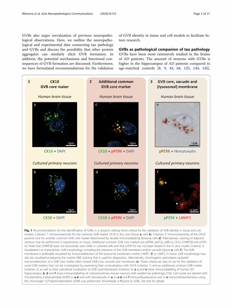
In the context of diagnostics, GVBs are typically detected by H&E staining. For research purposes, we here propose refinement of the criteria toidentify GVBs to aid future studies (Fig. 1). These guidelines are of use for the validation of GVB identity in tissue and cell models. We stronglyrecommend that the presence of a CK1δ-positive core is a prerequisite to classify an organelle as GVB (Fig. 1a, b) as CK1δ is one of the most consist-ent and rigorously validated GVB core markers in tissue and cells and its GVB localization is confirmed in an antibody-independent manner. Inaddition, immunopositivity of the CK1δ-labeled GVB core for at least one of the other common GVB core markers (pPERK, peIF2α, pIRE1α, CK1ε,pTDP-43 and CHMP2B) is recommended, demonstrated preferably by double immunolabeling (Fig. 1c, d) or by staining of adjacent tissue sections. Inaddition, confirmation of the distinctive GVB morphology, including the presence of the GVB membrane that surrounds core and vacuole, is recom-mended. In cells as well as tissue, this can be done by visualization of the GVB membrane by immunodetection of the lysosomal membrane proteinLAMP1 (Fig. 1f) or LIMP2. Alternatively, this staining could be replaced by morphological confirmation of membrane, core and vacuole on H&E stain-ing. In addition, the GVB membrane often becomes visible upon the chromogenic peroxidase-catalyzed detection of immunolabeling with a GVBcore marker – e.g. by means of the widely used chromogen 3,3′-diaminobenzidine (DAB) –, whereas upon fluorescent detection only the GVB core isshown (compare pPERK immunoreactivity in Fig. 1e with Fig. 1c, d, f). This may be due to deposition of the diffusible chromogenic product at mem-branes during the enzymatic signal amplification step. Therefore, as DAB signal may incorrectly suggest localization of a protein to both the GVB coreand membrane, fluorescent double labeling is advised to determine the subcellular localization of GVB proteins. Lastly, super-resolution microscopyand EM can be employed to study the morphology of immunolabeled GVBs at high resolution. In conclusion, we recommend three criteria for theassessment of GVB identity that includes immunoreactivity for CK1δ (criterion 1) and an additional common GVB marker (criterion 2) in the GVB coreand visualization of GVB membrane and/or vacuole (criterion 3) (Fig. 1).
Wiersma et al. Acta Neuropathologica Communications (2020) 8:153 Page 2 of 21
GVBs also urges reevaluation of previous neuropatho-logical observations. Here, we outline the neuropatho-logical and experimental data connecting tau pathologyand GVBs and discuss the possibility that other proteinaggregates can similarly elicit GVB formation. Inaddition, the potential mechanisms and functional con-sequences of GVB formation are discussed. Furthermore,we have formulated recommendations for the validation
of GVB identity in tissue and cell models to facilitate fu-ture research.
GVBs as pathological companion of tau pathologyGVBs have been most extensively studied in the brainsof AD patients. The amount of neurons with GVBs ishigher in the hippocampus of AD patients compared toage-matched controls [8, 9, 43, 68, 125, 144, 145].
Fig. 1 Recommendations for the identification of GVBs in a research setting: three criteria for the validation of GVB identity in tissue and cellmodels. Criterion 1: Immunoreactivity for the common GVB marker CK1δ in the core (tissue a, cells b). Criterion 2: Immunoreactivity of the CK1δ-positive core for another common GVB core marker determined by double immunolabeling (tissue c, cells d). Alternatively, staining of adjacentsections may be performed in experiments on tissue. Additional common GVB core markers are pPERK, peIF2α, pIRE1α, CK1ε, CHMP2B and pTDP-43. Note that CHMP2B does not exclusively stain GVBs in cultured cells and that pTDP-43 has not been tested in the in vitro model. Criterion 3:Visualization of characteristic GVB morphology, including the presence of the GVB membrane and/or vacuole (tissue e, cells f). The GVBmembrane is preferably visualized by immunodetection of the lysosomal membrane marker LAMP1 (f) or LIMP2. In tissue, GVB morphology mayalso be visualized employing the routine H&E staining that is used for diagnostics. Alternatively, chromogenic peroxidase-catalyzedimmunodetection of a GVB core marker often reveals GVB core, vacuole and membrane (e). These criteria are also of use for the validation ofnovel GVB markers that can be investigated by examining their co-localization with CK1δ (criterion 1) and an additional common GVB marker(criterion 2) as well as their subcellular localization to GVB core/membrane (criterion 3). a, c and e show immunolabeling of human ADhippocampus. b, d and f show immunolabeling of cultured primary mouse neurons with seeded tau pathology [143]. Cell nuclei are stained with4′,6-diamidino-2-phenylindole (DAPI) in a-d and with hematoxylin in e. In a-d and f immunofluorescence and in e immunohistochemistry usingthe chromogen 3,3′-diaminobenzidine (DAB) was performed. Arrowheads in f point to GVBs. See text for details
Wiersma et al. Acta Neuropathologica Communications (2020) 8:153 Page 3 of 21
Although GVBs are most often detected in hippocampalpyramidal neurons, GVBs are also found beyond the hip-pocampal formation. The distribution of GVBs follows astereotypical pattern through the brain, described by ascore of 1–5 according to the Thal GVD neuropatho-logical staging system [132]. In Thal GVD stages 2–5,GVBs are also detected outside of the hippocampus: firstin the entorhinal cortex, then in the temporal neocortex,hypothalamus and amygdala and eventually also infrontal and parietal cortical areas. The Thal GVD stageof AD cases is significantly higher than that of age-matched controls [68, 131, 132], indicating that in ADpatients more brain regions are affected by GVBs.Therefore, the GVB load in AD patients is increasedboth within and outside of the hippocampus.A striking correlation between GVBs and the presence
of AD-related tau pathology is observed in the humanbrain. In AD patients, the hippocampal formation is thehotspot for tau pathology as well as for GVBs. Withinthe hippocampus, the number of neurons with GVBssignificantly correlates with the number of neurons withtau pathology [43]. In accordance, the hippocampal GVBload in AD patients increases with the Braak stage forneurofibrillary tangle (NFT) pathology (NFT Braak stage[12, 13]) in which the severity of tau pathology in thehippocampus increments per stage [32, 43, 69, 81, 145].Therefore, a strong positive correlation exists betweenthe local tau pathology and GVB load in the AD hippo-campus. Furthermore, the distribution pattern of GVBsin the human brain [132] roughly follows that of AD-type tau pathology [12, 13]: both lesions first occur inand around the hippocampal formation before spreadingto other limbic and neocortical areas. Hence, GVBs andAD-related tau pathology follow a similar spatiotemporaldistribution pattern in the human brain. In line withthis, the Thal GVD and NFT Braak stage significantlycorrelate [68, 132]. Moreover, GVBs and tau pathologyco-localize at the single cell level in the AD brain: GVBsare typically detected in neurons that also exhibit taupathology [5, 43, 72, 81, 90, 111, 122, 135, 146]. There-fore, GVBs and tau pathology co-occur in the samebrain regions and in the same cells in AD.The strong association between GVBs and tau path-
ology extends beyond AD. Also in patients with the pri-mary tauopathy progressive supranuclear palsy (PSP), asignificant increase in the number of neurons with GVBsis found in different brain areas affected by tau path-ology, including the brainstem and midbrain [125]. Thisis in line with various descriptive studies reporting thepresence of GVBs in these disease-specific brain areas inPSP patients [16, 99, 118, 123]. Similar to what has beendescribed for AD, tau pathology is also detected in themajority of GVB-bearing cells in the pons of PSP pa-tients [125]. Furthermore, the amount of neurons with
GVBs was found to be significantly higher in patientswith the primary tauopathy Pick’s disease compared tocontrols in the hippocampal area, one of the brain re-gions affected in Pick’s disease [144]. Indeed, severalstudies have shown that GVBs and Pick bodies co-occurin the hippocampus of Pick’s disease cases, sometimes inthe same neurons [74, 94, 99, 111, 118, 134], in the ab-sence [111, 118, 134] or only low abundant presence[74] of AD-related NFTs, indicating a specific associ-ation of Pick’s type tau pathology with GVBs. Hence, inaddition to AD, also in primary tauopathy patients, theGVB frequency is increased and correlates with the pres-ence of tau pathology.Descriptive studies have additionally reported the pres-
ence of GVBs in the brains of patients with many otherdisorders that present with tau pathology. This includescases with FTLD caused by the MAPT mutationsG272V, P301L, L315R and P364S [97, 99, 124], pallido-ponto-nigral degeneration (PPND, caused by the N279KMAPT mutation) [118], corticobasal degeneration (CBD)[24, 105], argyrophilic grain disease [132], parkinsonismdementia complex of Guam [39, 40, 83, 118, 136], Downsyndrome [18, 55, 118, 126], pantothenate kinase-associated neurodegeneration (previously known asHallervorden-Spatz syndrome) [26], diffuse neurofibril-lary tangles with calcification [151], myotonic dystrophysubtypes [96, 115, 116], the lysosomal storage disordersNiemann-Pick disease type C [127] and Salla disease [6],normal pressure hydrocephalus [10], tuberous sclerosis[41], subacute sclerosing panencephalitis [86], menin-gioangiomatosis [106] and various type of brain tumors[15, 56, 71, 106, 110]. Therefore, independent of diseaseetiology, tau pathology and GVBs coincide in the dis-eased human brain.Interestingly, although the hippocampal GVB burden
increases with age in subjects without clinical symptoms[9, 31, 125, 144], GVBs are also observed in the brains ofyoung patients with tau pathology. For example, GVBswere found to accompany tau pathology/argentophilicNFTs in a 13-year-old patient with tuberous sclerosis[41], a 15-year-old patient with subacute sclerosingpanencephalitis [86], a 15-year-old patient with Downsyndrome [55], 34- and 41-year-old patients with differ-ent lysosomal storage disorders [6, 127] and young pa-tients with Fukuyama-type congenital musculardystrophy (age range: 14–34 years) [115]. This is furthercorroborated by the finding of GVBs in neoplastic neu-rons with tau pathology in patients with ganglion cell tu-mors (mean age: 44 years), whereas neither GVBs nortau pathology were detected in adjacent normal braintissue in the same patients [15]. Also in cortical neuronsentrapped by meningioangiomatosis – a benign tumor-like lesion/mass – GVBs and argentophilic NFTs co-occur in a subset of cases, including a 17-year-old
Wiersma et al. Acta Neuropathologica Communications (2020) 8:153 Page 4 of 21
patient [106]. Therefore, the concurrence of tau path-ology and GVBs in the human brain is independent ofage.A few studies have reported GVBs in tau transgenic
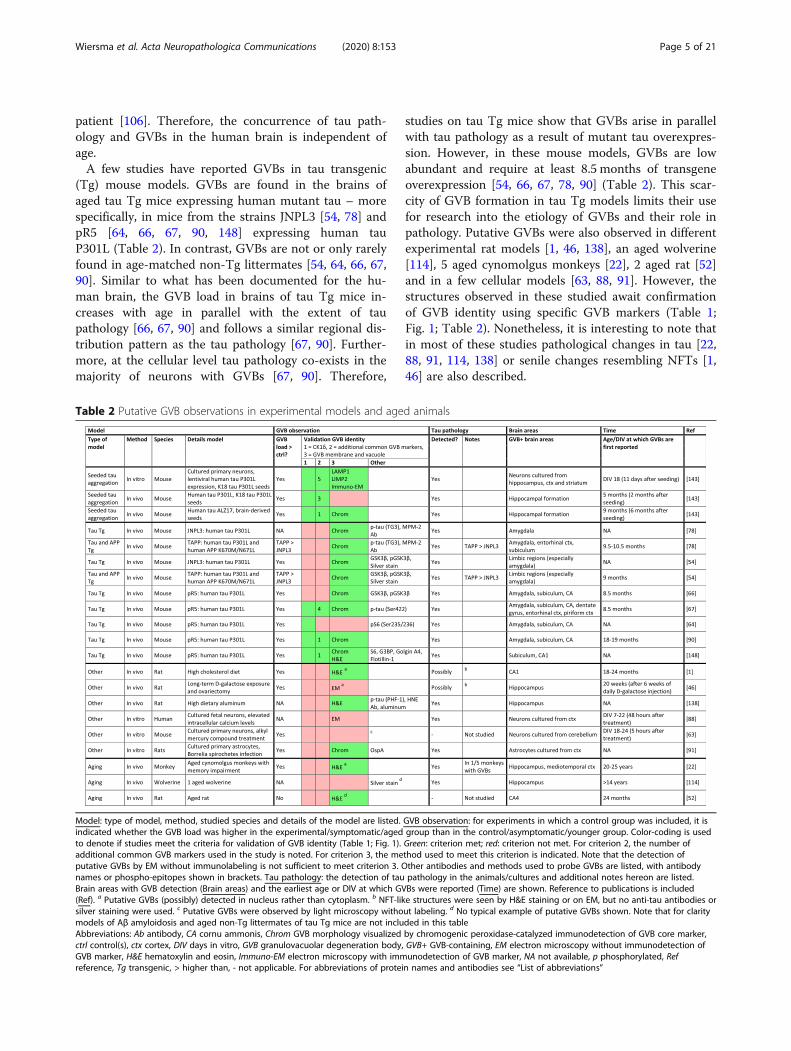
(Tg) mouse models. GVBs are found in the brains ofaged tau Tg mice expressing human mutant tau – morespecifically, in mice from the strains JNPL3 [54, 78] andpR5 [64, 66, 67, 90, 148] expressing human tauP301L (Table 2). In contrast, GVBs are not or only rarelyfound in age-matched non-Tg littermates [54, 64, 66, 67,90]. Similar to what has been documented for the hu-man brain, the GVB load in brains of tau Tg mice in-creases with age in parallel with the extent of taupathology [66, 67, 90] and follows a similar regional dis-tribution pattern as the tau pathology [67, 90]. Further-more, at the cellular level tau pathology co-exists in themajority of neurons with GVBs [67, 90]. Therefore,
studies on tau Tg mice show that GVBs arise in parallelwith tau pathology as a result of mutant tau overexpres-sion. However, in these mouse models, GVBs are lowabundant and require at least 8.5 months of transgeneoverexpression [54, 66, 67, 78, 90] (Table 2). This scar-city of GVB formation in tau Tg models limits their usefor research into the etiology of GVBs and their role inpathology. Putative GVBs were also observed in differentexperimental rat models [1, 46, 138], an aged wolverine[114], 5 aged cynomolgus monkeys [22], 2 aged rat [52]and in a few cellular models [63, 88, 91]. However, thestructures observed in these studied await confirmationof GVB identity using specific GVB markers (Table 1;Fig. 1; Table 2). Nonetheless, it is interesting to note thatin most of these studies pathological changes in tau [22,88, 91, 114, 138] or senile changes resembling NFTs [1,46] are also described.
Table 2 Putative GVB observations in experimental models and aged animals
Model: type of model, method, studied species and details of the model are listed. GVB observation: for experiments in which a control group was included, it isindicated whether the GVB load was higher in the experimental/symptomatic/aged group than in the control/asymptomatic/younger group. Color-coding is usedto denote if studies meet the criteria for validation of GVB identity (Table 1; Fig. 1). Green: criterion met; red: criterion not met. For criterion 2, the number ofadditional common GVB markers used in the study is noted. For criterion 3, the method used to meet this criterion is indicated. Note that the detection ofputative GVBs by EM without immunolabeling is not sufficient to meet criterion 3. Other antibodies and methods used to probe GVBs are listed, with antibodynames or phospho-epitopes shown in brackets. Tau pathology: the detection of tau pathology in the animals/cultures and additional notes hereon are listed.Brain areas with GVB detection (Brain areas) and the earliest age or DIV at which GVBs were reported (Time) are shown. Reference to publications is included(Ref). a Putative GVBs (possibly) detected in nucleus rather than cytoplasm. b NFT-like structures were seen by H&E staining or on EM, but no anti-tau antibodies orsilver staining were used. c Putative GVBs were observed by light microscopy without labeling. d No typical example of putative GVBs shown. Note that for claritymodels of Aβ amyloidosis and aged non-Tg littermates of tau Tg mice are not included in this tableAbbreviations: Ab antibody, CA cornu ammonis, Chrom GVB morphology visualized by chromogenic peroxidase-catalyzed immunodetection of GVB core marker,ctrl control(s), ctx cortex, DIV days in vitro, GVB granulovacuolar degeneration body, GVB+ GVB-containing, EM electron microscopy without immunodetection ofGVB marker, H&E hematoxylin and eosin, Immuno-EM electron microscopy with immunodetection of GVB marker, NA not available, p phosphorylated, Refreference, Tg transgenic, > higher than, - not applicable. For abbreviations of protein names and antibodies see “List of abbreviations”
Wiersma et al. Acta Neuropathologica Communications (2020) 8:153 Page 5 of 21
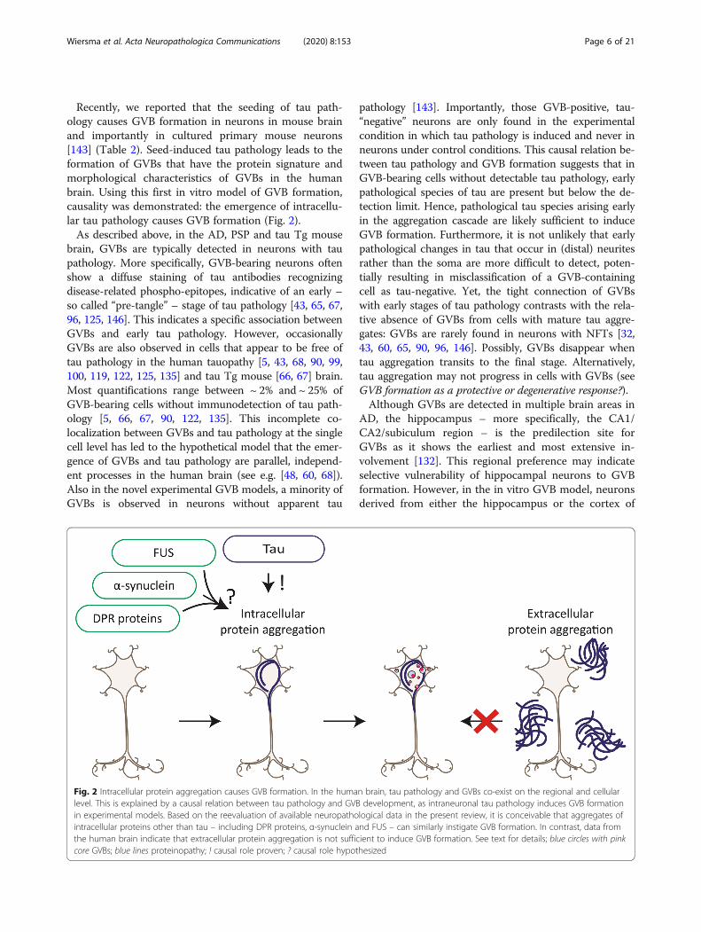
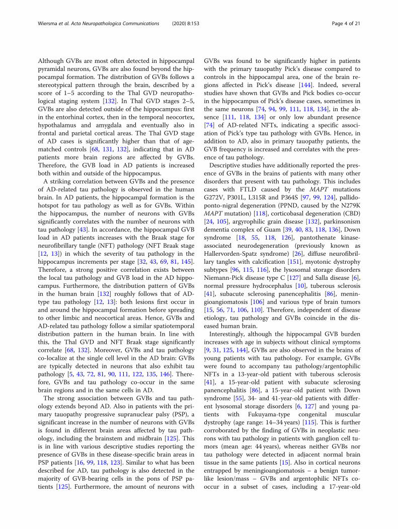
Recently, we reported that the seeding of tau path-ology causes GVB formation in neurons in mouse brainand importantly in cultured primary mouse neurons[143] (Table 2). Seed-induced tau pathology leads to theformation of GVBs that have the protein signature andmorphological characteristics of GVBs in the humanbrain. Using this first in vitro model of GVB formation,causality was demonstrated: the emergence of intracellu-lar tau pathology causes GVB formation (Fig. 2).As described above, in the AD, PSP and tau Tg mouse
brain, GVBs are typically detected in neurons with taupathology. More specifically, GVB-bearing neurons oftenshow a diffuse staining of tau antibodies recognizingdisease-related phospho-epitopes, indicative of an early –so called “pre-tangle” – stage of tau pathology [43, 65, 67,96, 125, 146]. This indicates a specific association betweenGVBs and early tau pathology. However, occasionallyGVBs are also observed in cells that appear to be free oftau pathology in the human tauopathy [5, 43, 68, 90, 99,100, 119, 122, 125, 135] and tau Tg mouse [66, 67] brain.Most quantifications range between ~ 2% and ~ 25% ofGVB-bearing cells without immunodetection of tau path-ology [5, 66, 67, 90, 122, 135]. This incomplete co-localization between GVBs and tau pathology at the singlecell level has led to the hypothetical model that the emer-gence of GVBs and tau pathology are parallel, independ-ent processes in the human brain (see e.g. [48, 60, 68]).Also in the novel experimental GVB models, a minority ofGVBs is observed in neurons without apparent tau
pathology [143]. Importantly, those GVB-positive, tau-“negative” neurons are only found in the experimentalcondition in which tau pathology is induced and never inneurons under control conditions. This causal relation be-tween tau pathology and GVB formation suggests that inGVB-bearing cells without detectable tau pathology, earlypathological species of tau are present but below the de-tection limit. Hence, pathological tau species arising earlyin the aggregation cascade are likely sufficient to induceGVB formation. Furthermore, it is not unlikely that earlypathological changes in tau that occur in (distal) neuritesrather than the soma are more difficult to detect, poten-tially resulting in misclassification of a GVB-containingcell as tau-negative. Yet, the tight connection of GVBswith early stages of tau pathology contrasts with the rela-tive absence of GVBs from cells with mature tau aggre-gates: GVBs are rarely found in neurons with NFTs [32,43, 60, 65, 90, 96, 146]. Possibly, GVBs disappear whentau aggregation transits to the final stage. Alternatively,tau aggregation may not progress in cells with GVBs (seeGVB formation as a protective or degenerative response?).Although GVBs are detected in multiple brain areas in
AD, the hippocampus – more specifically, the CA1/CA2/subiculum region – is the predilection site forGVBs as it shows the earliest and most extensive in-volvement [132]. This regional preference may indicateselective vulnerability of hippocampal neurons to GVBformation. However, in the in vitro GVB model, neuronsderived from either the hippocampus or the cortex of
Fig. 2 Intracellular protein aggregation causes GVB formation. In the human brain, tau pathology and GVBs co-exist on the regional and cellularlevel. This is explained by a causal relation between tau pathology and GVB development, as intraneuronal tau pathology induces GVB formationin experimental models. Based on the reevaluation of available neuropathological data in the present review, it is conceivable that aggregates ofintracellular proteins other than tau – including DPR proteins, α-synuclein and FUS – can similarly instigate GVB formation. In contrast, data fromthe human brain indicate that extracellular protein aggregation is not sufficient to induce GVB formation. See text for details; blue circles with pinkcore GVBs; blue lines proteinopathy; ! causal role proven; ? causal role hypothesized
Wiersma et al. Acta Neuropathologica Communications (2020) 8:153 Page 6 of 21
mice developed GVBs to a similar extent when chal-lenged with tau pathology [143]. Furthermore, GVBsalso formed in neurons cultured from the mouse stri-atum [143], a region devoid of GVBs in the AD brain[132]. Although it could be argued that primary mouseneurons in culture may lack characteristics present inthe context of the brain that determine their vulnerabil-ity or that species differences play a role, these experi-mental data argue against a regional preference for GVBdevelopment. Rather, they point to tau pathology as thekey determinant of the (extent of) GVB formation.Neuropathological observations indicate a strong con-nection with tau pathology, but also suggest that the ex-tent of GVB formation in tau-positive neurons maydisplay regional differences. In AD, GVBs are absentfrom the basal ganglia [132], but tau pathology in thisbrain area is also minor – with moderate NFT load onlyat the final Braak NFT stage VI [13]. In contrast, bothGVBs and tau pathology are found in the basal gangliaof patients with PSP [99, 118, 123]. This suggests thatindependent of the brain region, neurons form GVBswhen tau pathology is present. In agreement, the distri-bution patterns of tau pathology and GVBs in the AD[12, 13, 132] and tau Tg mouse [67, 90] brain largelyoverlap. However, in the earliest AD stages, there is aninteresting discrepancy as the entorhinal cortex is thepredilection site for tau pathology, but the CA1/CA2/subiculum region for GVBs [8, 12, 13, 132]. In addition,in the final AD stages, NFTs are abundant in the frontal,parietal and occipital neocortex, yet GVBs remain scarcein these areas [13, 132] Although precise quantificationof the tau and GVB load is needed to verify these obser-vations, it suggests a model where tau pathology is aprerequisite for GVB formation, but additional factorsmodulate the process and thereby underly selective vul-nerability of specific neuronal populations for GVB de-velopment. Another possibility is that yetuncharacterized differences in the kinetics or structureof tau pathology between brain regions/neuronal popula-tions dictate GVB formation.In addition to the frequent co-occurrence of tau pathology
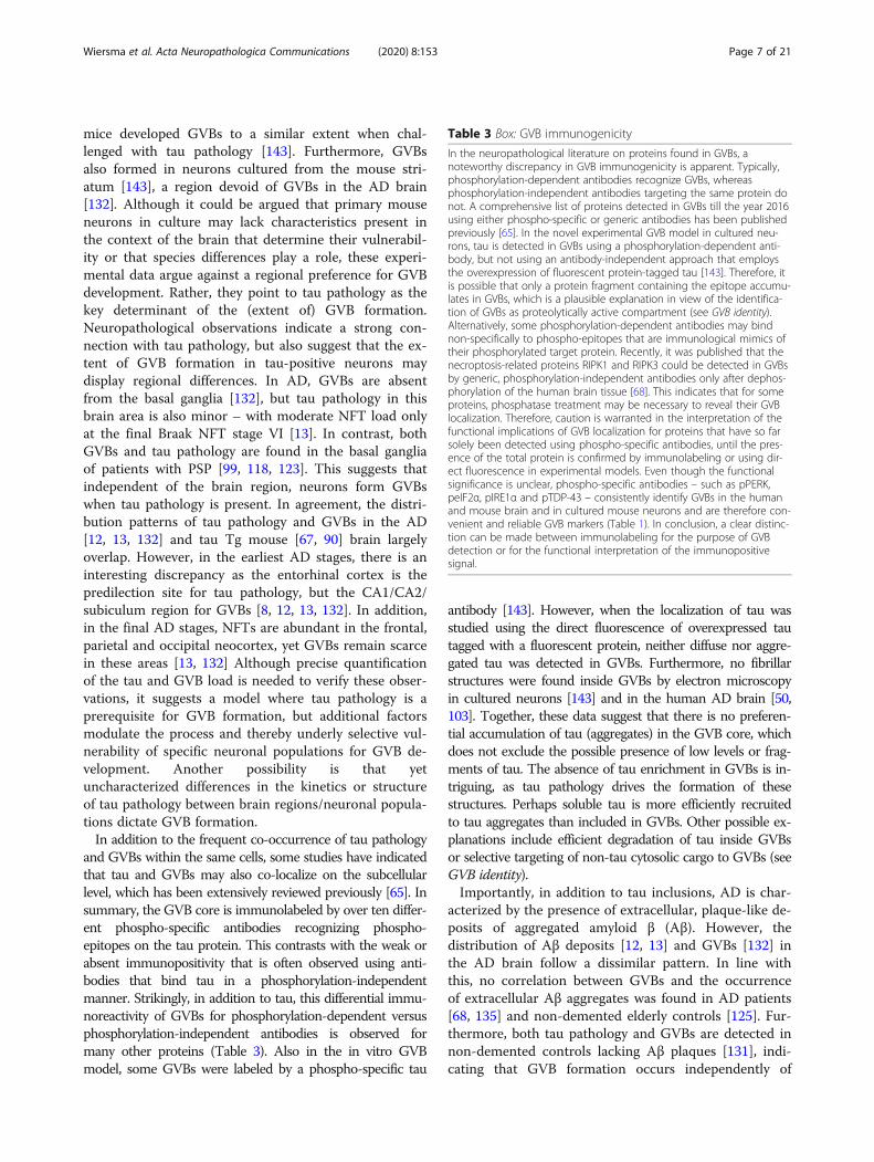
and GVBs within the same cells, some studies have indicatedthat tau and GVBs may also co-localize on the subcellularlevel, which has been extensively reviewed previously [65]. Insummary, the GVB core is immunolabeled by over ten differ-ent phospho-specific antibodies recognizing phospho-epitopes on the tau protein. This contrasts with the weak orabsent immunopositivity that is often observed using anti-bodies that bind tau in a phosphorylation-independentmanner. Strikingly, in addition to tau, this differential immu-noreactivity of GVBs for phosphorylation-dependent versusphosphorylation-independent antibodies is observed formany other proteins (Table 3). Also in the in vitro GVBmodel, some GVBs were labeled by a phospho-specific tau
antibody [143]. However, when the localization of tau wasstudied using the direct fluorescence of overexpressed tautagged with a fluorescent protein, neither diffuse nor aggre-gated tau was detected in GVBs. Furthermore, no fibrillarstructures were found inside GVBs by electron microscopyin cultured neurons [143] and in the human AD brain [50,103]. Together, these data suggest that there is no preferen-tial accumulation of tau (aggregates) in the GVB core, whichdoes not exclude the possible presence of low levels or frag-ments of tau. The absence of tau enrichment in GVBs is in-triguing, as tau pathology drives the formation of thesestructures. Perhaps soluble tau is more efficiently recruitedto tau aggregates than included in GVBs. Other possible ex-planations include efficient degradation of tau inside GVBsor selective targeting of non-tau cytosolic cargo to GVBs (seeGVB identity).Importantly, in addition to tau inclusions, AD is char-
acterized by the presence of extracellular, plaque-like de-posits of aggregated amyloid β (Aβ). However, thedistribution of Aβ deposits [12, 13] and GVBs [132] inthe AD brain follow a dissimilar pattern. In line withthis, no correlation between GVBs and the occurrenceof extracellular Aβ aggregates was found in AD patients[68, 135] and non-demented elderly controls [125]. Fur-thermore, both tau pathology and GVBs are detected innon-demented controls lacking Aβ plaques [131], indi-cating that GVB formation occurs independently of
Table 3 Box: GVB immunogenicity
In the neuropathological literature on proteins found in GVBs, anoteworthy discrepancy in GVB immunogenicity is apparent. Typically,phosphorylation-dependent antibodies recognize GVBs, whereasphosphorylation-independent antibodies targeting the same protein donot. A comprehensive list of proteins detected in GVBs till the year 2016using either phospho-specific or generic antibodies has been publishedpreviously [65]. In the novel experimental GVB model in cultured neu-rons, tau is detected in GVBs using a phosphorylation-dependent anti-body, but not using an antibody-independent approach that employsthe overexpression of fluorescent protein-tagged tau [143]. Therefore, itis possible that only a protein fragment containing the epitope accumu-lates in GVBs, which is a plausible explanation in view of the identifica-tion of GVBs as proteolytically active compartment (see GVB identity).Alternatively, some phosphorylation-dependent antibodies may bindnon-specifically to phospho-epitopes that are immunological mimics oftheir phosphorylated target protein. Recently, it was published that thenecroptosis-related proteins RIPK1 and RIPK3 could be detected in GVBsby generic, phosphorylation-independent antibodies only after dephos-phorylation of the human brain tissue [68]. This indicates that for someproteins, phosphatase treatment may be necessary to reveal their GVBlocalization. Therefore, caution is warranted in the interpretation of thefunctional implications of GVB localization for proteins that have so farsolely been detected using phospho-specific antibodies, until the pres-ence of the total protein is confirmed by immunolabeling or using dir-ect fluorescence in experimental models. Even though the functionalsignificance is unclear, phospho-specific antibodies – such as pPERK,peIF2α, pIRE1α and pTDP-43 – consistently identify GVBs in the humanand mouse brain and in cultured mouse neurons and are therefore con-venient and reliable GVB markers (Table 1). In conclusion, a clear distinc-tion can be made between immunolabeling for the purpose of GVBdetection or for the functional interpretation of the immunopositivesignal.
Wiersma et al. Acta Neuropathologica Communications (2020) 8:153 Page 7 of 21
extracellular Aβ aggregation. Interestingly, intracellularAβ has been detected inside GVBs [72] (see also GVBidentity). Furthermore, a higher GVB load is found inbigenic TAPP mice that express both human tau P301Land human mutant amyloid precursor protein (APP)K670M/N671L (APPSw) compared to JNPL3 mice carry-ing only the human tau P301L transgene [54, 78] (Table2). This higher GVB frequency is likely related to the ex-acerbated tau pathology in TAPP mice rather than thecomorbid Aβ pathology, as GVBs are absent in mousemodels of pure Aβ amyloidosis by overexpression ofAPPSw (Tg2576), APPSw,Ind (J20), APPSw/L/PS1M146L orAPPSw/PS1dE9 [54, 67, 90] – although the recent detec-tion of granular structures with an immunogenicity pro-file that partially overlaps with GVBs in APPSw/PS1dE9
mice requires further investigation [148]. Taken to-gether, the data indicate that neither in human nor inTg mouse brain, GVBs and extracellular Aβ pathologyare associated. The conclusion that tau rather than Aβpathology is the driver of GVB formation in AD is in ac-cordance with the presence of GVBs in many primarytauopathies.
GVB formation in “non-tau” neurodegenerativeproteinopathiesProtein aggregation is a common neuropathological charac-teristic of many neurodegenerative diseases. Although differ-ent proteins form the basis of aggregates in theseneurodegenerative proteinopathies, the biochemical mecha-nisms underlying their formation appear to overlap. There-fore, it is conceivable that aggregating proteins other thantau can also instigate GVB formation.In addition to tauopathies, the presence of GVBs has
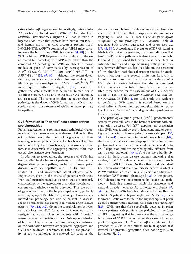
been studied in the brains of patients with other neuro-degenerative proteinopathies, including human priondiseases, α-synucleinopathies and TDP-43- and FUS-related FTLD and amyotrophic lateral sclerosis (ALS).Importantly, even in the brains of patients with these“non-tau” neurodegenerative diseases that are primarilycharacterized by the aggregation of another protein, con-current tau pathology can be observed. This tau path-ology is often found in the hippocampal region, probablyreflecting aging-/AD-related tau pathology. However, co-morbid tau pathology can also be present in disease-specific brain areas, for example in human prion diseasepatients [70, 112, 142]. Given the causal relation betweentau pathology and GVB formation, it is essential to in-vestigate tau co-pathology in patients with “non-tau”neurodegenerative proteinopathies. Only upon exclusionof tau pathology as a confounding factor, a conclusionon the correlation between other protein aggregates andGVBs can be drawn. Therefore, in Table 4, the probabil-ity of tau co-pathology is reviewed for each of the
studies discussed below. In this assessment, we have alsomade use of the fact that phospho-specific antibodiestargeting tau and TDP-43 (see GVBs as pathologicalcompanion of tau pathology; GVB identity; Table 3)recognize both protein aggregates and GVBs (see e.g.[47, 48, 59]). Accordingly, if p-tau or pTDP-43 staininglabels GVBs but not aggregates, this is an indication thattau/TDP-43 protein pathology is absent from those cells.It should be mentioned that detection is dependent onantibody titration and image acquiring settings that mayvary between different studies. In addition, the detectionof low abundant pathological protein species by quali-tative microscopy is a general limitation. Lastly, it isimportant to note that the extent of evidence of aGVB identity varies between the studies discussedbelow. To streamline future studies, we have formu-lated three criteria for the assessment of GVB identity(Table 1; Fig. 1 ; see Recommendations and conclud-ing remarks). In Table 4, for each study the evidenceto confirm a GVB identity is scored based on thenovel criteria. Below, neuropathological data on puta-tive GVBs in “non-tau” neurodegenerative proteinopa-thies is discussed.The pathological prion protein (PrPSc) predominantly
aggregates extracellularly in the brains of patients with hu-man prion diseases. For PrPSc deposits, no associationwith GVBs was found by two independent studies cover-ing the majority of human prion disease subtypes [135,142] (Table 4). Interestingly, in one of these cohorts [142],almost all prion disease cases displayed small neuritic tau-positive inclusions that are believed to be secondary toPrPSc deposition and are morphologically different fromAD-type tau pathology [70, 112]. GVBs were hardly ob-served in these prion disease patients, indicating thatmodest, distal PrPSc-related changes in tau are not associ-ated with GVB formation. On the other hand, abundantGVBs were observed in a prion disease patient in which aPRNP mutation led to an unusual Gerstmann-Sträussler-Scheinker (GSS) clinical phenotype [142]. In this patient,PrPSc deposition was accompanied by severe tau path-ology – including numerous tangle-like structures andneuropil threads – whereas Aβ pathology was absent [57,142]. Similarly, GVBs have been described in another fa-milial GSS patient with pre-tangles and NFTs [53]. Fur-thermore, GVBs were found in the hippocampus of priondisease patients with comorbid AD-related tau pathology[135]. GVBs are therefore specifically detected in priondisease patients with proximal tau pathology in the formof NFTs, suggesting that in these cases the tau pathologyis the cause of GVB formation. As neither extracellular de-posits of aggregated PrPSc nor of Aβ correlate with thepresence of GVBs in the human brain, it appears thatextracellular protein aggregation does not trigger GVBformation (Fig. 2).
Wiersma et al. Acta Neuropathologica Communications (2020) 8:153 Page 8 of 21
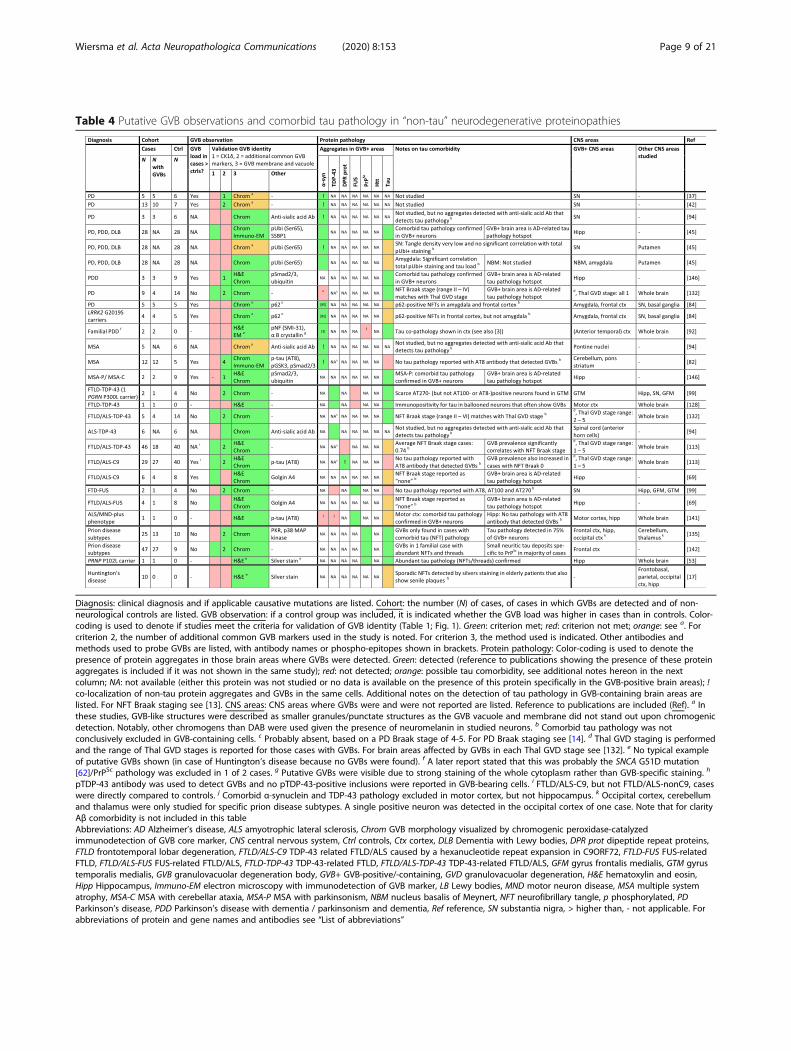
Table 4 Putative GVB observations and comorbid tau pathology in “non-tau” neurodegenerative proteinopathies
Diagnosis: clinical diagnosis and if applicable causative mutations are listed. Cohort: the number (N) of cases, of cases in which GVBs are detected and of non-neurological controls are listed. GVB observation: if a control group was included, it is indicated whether the GVB load was higher in cases than in controls. Color-coding is used to denote if studies meet the criteria for validation of GVB identity (Table 1; Fig. 1). Green: criterion met; red: criterion not met; orange: see a. Forcriterion 2, the number of additional common GVB markers used in the study is noted. For criterion 3, the method used is indicated. Other antibodies andmethods used to probe GVBs are listed, with antibody names or phospho-epitopes shown in brackets. Protein pathology: Color-coding is used to denote thepresence of protein aggregates in those brain areas where GVBs were detected. Green: detected (reference to publications showing the presence of these proteinaggregates is included if it was not shown in the same study); red: not detected; orange: possible tau comorbidity, see additional notes hereon in the nextcolumn; NA: not available (either this protein was not studied or no data is available on the presence of this protein specifically in the GVB-positive brain areas); !co-localization of non-tau protein aggregates and GVBs in the same cells. Additional notes on the detection of tau pathology in GVB-containing brain areas arelisted. For NFT Braak staging see [13]. CNS areas: CNS areas where GVBs were and were not reported are listed. Reference to publications are included (Ref). a Inthese studies, GVB-like structures were described as smaller granules/punctate structures as the GVB vacuole and membrane did not stand out upon chromogenicdetection. Notably, other chromogens than DAB were used given the presence of neuromelanin in studied neurons. b Comorbid tau pathology was notconclusively excluded in GVB-containing cells. c Probably absent, based on a PD Braak stage of 4-5. For PD Braak staging see [14]. d Thal GVD staging is performedand the range of Thal GVD stages is reported for those cases with GVBs. For brain areas affected by GVBs in each Thal GVD stage see [132]. e No typical exampleof putative GVBs shown (in case of Huntington’s disease because no GVBs were found). f A later report stated that this was probably the SNCA G51D mutation[62]/PrPSc pathology was excluded in 1 of 2 cases. g Putative GVBs were visible due to strong staining of the whole cytoplasm rather than GVB-specific staining. h
pTDP-43 antibody was used to detect GVBs and no pTDP-43-positive inclusions were reported in GVB-bearing cells. i FTLD/ALS-C9, but not FTLD/ALS-nonC9, caseswere directly compared to controls. j Comorbid α-synuclein and TDP-43 pathology excluded in motor cortex, but not hippocampus. k Occipital cortex, cerebellumand thalamus were only studied for specific prion disease subtypes. A single positive neuron was detected in the occipital cortex of one case. Note that for clarityAβ comorbidity is not included in this tableAbbreviations: AD Alzheimer’s disease, ALS amyotrophic lateral sclerosis, Chrom GVB morphology visualized by chromogenic peroxidase-catalyzedimmunodetection of GVB core marker, CNS central nervous system, Ctrl controls, Ctx cortex, DLB Dementia with Lewy bodies, DPR prot dipeptide repeat proteins,FTLD frontotemporal lobar degeneration, FTLD/ALS-C9 TDP-43 related FTLD/ALS caused by a hexanucleotide repeat expansion in C9ORF72, FTLD-FUS FUS-relatedFTLD, FTLD/ALS-FUS FUS-related FTLD/ALS, FTLD-TDP-43 TDP-43-related FTLD, FTLD/ALS-TDP-43 TDP-43-related FTLD/ALS, GFM gyrus frontalis medialis, GTM gyrustemporalis medialis, GVB granulovacuolar degeneration body, GVB+ GVB-positive/-containing, GVD granulovacuolar degeneration, H&E hematoxylin and eosin,Hipp Hippocampus, Immuno-EM electron microscopy with immunodetection of GVB marker, LB Lewy bodies, MND motor neuron disease, MSA multiple systematrophy, MSA-C MSA with cerebellar ataxia, MSA-P MSA with parkinsonism, NBM nucleus basalis of Meynert, NFT neurofibrillary tangle, p phosphorylated, PDParkinson’s disease, PDD Parkinson’s disease with dementia / parkinsonism and dementia, Ref reference, SN substantia nigra, > higher than, - not applicable. Forabbreviations of protein and gene names and antibodies see “List of abbreviations”
Wiersma et al. Acta Neuropathologica Communications (2020) 8:153 Page 9 of 21
Post-mortem studies have also investigated the pres-ence of GVBs in neurodegenerative proteinopathiescharacterized by the intracellular aggregation of proteinsother than tau, albeit generally in small cohorts (Table4). In a single study, GVBs were not found in differentbrain regions of 10 patients with Huntington’s disease[17]. In contrast to the tau-related FTLD variants, no as-sociation was found between GVBs and protein aggre-gates composed of FUS in different brains areas ofpatients with FUS-related FTLD [99]. In another study,scarce GVBs were found in the hippocampus of only 1out of 4 patients with FUS-related FTLD/ALS [69].Interestingly, in a case report, GVBs were found in themotor cortex and hippocampus of a patient with anALS-like phenotype and FUS inclusions [141]. The pres-ence of GVBs in the motor cortex of this case may beexplained by the detection of comorbid tau pathology inthe same motor neurons. However, in the hippocampus,the p-tau antibody AT8 labeled GVBs, but in the samecells tau aggregates were not reported [141]. These datafrom a single patient may point to tau-independent GVBinduction by FUS aggregates.GVBs are not associated with aggregates of TDP-43 in
different brain areas of patients with TDP-43-relatedFTLD [99] and also do not localize to neurons withTDP-43 inclusions in AD patients with comorbid TDP-43 pathology [59]. Furthermore, neither TDP-43 aggre-gates nor nuclear clearance of TDP-43 – a cellularchange that is often associated with cytoplasmic TDP-43aggregation – were observed in neurons with GVBs in alarge cohort of elderly brain donors [47]. Also in anter-ior horn cells with TDP-43 pathology in the spinal cordof ALS patients, no GVBs were detected [94]. These dataindicate that there is no association between TDP-43 ag-gregates and the presence of GVBs. In line with this, intwo studies that did describe GVBs in patients withTDP-43 proteinopathy [128, 132], GVB detection waslikely related to comorbid tau pathology. In the firststudy, GVBs were found in the hippocampus of TDP-43proteinopathy patients with NFT Braak stages rangingbetween II and VI, but not in a TDP-43 case with NFTBraak stage 0 [132]. Furthermore, GVBs in this studywere detected using an antibody against pTDP-43 thatrecognizes both aggregates and GVBs, but in neuronswith pTDP-43 immunoreactive GVBs, no proteininclusions were reported, suggesting that there is norobust TDP-43 aggregation in those cells. In the secondstudy, GVBs were found in ballooned neurons in themotor cortex of an FTLD/ALS patient with TDP-43inclusions [128]. However, also tau pathology was oftendetected in ballooned neurons and may explain theappearance of GVBs. Together, these data do not sup-port an association between TDP-43 aggregation andGVB formation.
Interestingly, GVBs were found to be more prevalentand more widely distributed in patients with TDP-43-related FTLD/ALS carrying a hexanucleotide repeat ex-pansion in C9ORF72 (FTLD/ALS-C9) compared toTDP-43-related FTLD/ALS patients without a C9ORF72repeat expansion and age-matched controls in an exten-sive study [113]. In line with this, low levels of GVBswere also reported in the hippocampus of 4 out of 6FTLD/ALS-C9 patients in another study [69]. Inaddition to TDP-43 inclusions, FTLD/ALS-C9 patientbrains contain aggregates composed of dipeptide repeat(DPR) proteins that result from non-canonical transla-tion of the expanded repeat region. In FTLD/ALS-C9patients, GVBs were frequently found in those neuronsthat also contained DPR inclusions [113]. On the otherhand, using a pTDP-43 or p-tau antibody in the samepatients, GVBs were clearly detected, yet no aggregateswere visible in the same neurons. These observationsstrengthen the idea that DPR protein rather than TDP-43 or tau aggregates are associated with the presence ofGVBs in this cohort. Furthermore, the GVB prevalencein FTLD/ALS-C9 cases was also increased when onlysubjects with the NFT Braak stage 0 were taken intoaccount [113]. Therefore, the presence of GVBs seemsspecifically related to the presence of DPR protein aggre-gates and not to concomitant tau pathology in theseFTLD/ALS-C9 patients.Additional studies have reported the presence of (pu-
tative) GVBs in brain areas and cells with α-synucleinaggregates in patients with multiple system atrophy(MSA) [82, 94, 146] and patients in the Lewy body dis-ease spectrum, including Parkinson’s disease [37, 42, 45,84, 94, 132] and Parkinson’s disease with dementia [45,92, 146], with both sporadic and genetic etiologies. Inter-estingly, GVBs are frequently found in cells with earlystages of α-synuclein aggregation in MSA patients [82],mirroring the specific association of GVBs with early taupathology (see GVBs as pathological companion of taupathology). In some of the α-synucleinopathy subjects,tau comorbidity was detected either at the single celllevel in GVB-carrying neurons [45, 146] or in the samebrain areas as the GVBs, as shown by the additionalimmunodetection of pathological tau [92] or based onthe NFT Braak staging of the cases [132]. Yet, concur-rent tau pathology does not seem to fully explain the de-tection of GVBs in all α-synucleinopathy cases. In MSApatients, although the GVBs were labeled by the p-tauantibody AT8, no tau pathology was noted in GVB-positive neurons [82]. Furthermore, GVBs were found inbrain areas specifically affected by α-synuclein pathologyin MSA patients, namely cerebellum, pons and striatum[82]. In line with this, GVBs – and smaller granules –were detected in α-synuclein aggregate-bearing neuronsin the substantia nigra of three patients with Parkinson’s
Wiersma et al. Acta Neuropathologica Communications (2020) 8:153 Page 10 of 21
disease [94]. As this brain area becomes mildly affectedby AD-related tau pathology only at NFT Braak stage V[13], AD-related tau co-pathology in all cases seems im-probable. However, the presence of comorbid tau path-ology cannot be fully excluded without analysis at thesingle cell level. In addition, several independent studieshave reported granular structures possibly representing(small) GVBs in cells with α-synuclein pathology indisease-specific brain areas, namely in the substantianigra of Parkinson’s disease [37, 42] and Lewy body dis-ease spectrum [45] patients and in the pontine nuclei ofMSA patients [94]. The GVB identity of these granulesremains to be validated.Taken together – and taking into consideration in-
complete evidence for GVB identity and tau pathologyas confounding factor (Table 4) – the limited neuro-pathological data available suggest that, in addition totau, also α-synuclein, DPR protein and possibly FUS ag-gregates may be able to elicit GVB formation in the hu-man brain (Fig. 2). GVB formation may thus be acommon response to intracellular protein aggregation.
GVB identityDetailing all proteins detected in GVBs is beyond thescope of this review and for an overview of proteins de-tected in GVBs up to 2016 the reader is here referred toa previous, excellent review [65] (for novel GVB-localizing proteins see below). Here, the molecular com-position of GVBs is briefly discussed in the light of re-cent discoveries in the human brain and experimentalmodels. In Table 1, common markers of GVBs in thehuman brain and experimental models and the level ofrigor to which these markers have been validated arediscussed and in Supplementary Table 1, primary anti-bodies for common GVB marker detection are listed.For other proteins localizing to GVBs discussed below,the extent of evidence confirming their GVB localization(e.g. the number of publications and the study of overlapwith common GVB markers) varies. Investigating themolecular composition of GVBs is important, as it pro-vides clues to the mechanism of their formation.GVBs were previously proposed to be an aberrant type
of autophagosome: a double-membraned autophagicintermediate [103]. This hypothetical GVB origin hasbeen widely referred to and over time turned into a def-inition of GVBs. However, neither GVBs in the humanbrain [30] nor experimental GVBs [143] are immunopo-sitive for the autophagosome membrane marker LC3.This is in agreement with ultrastructural analysis usingimmunoelectron microscopy in both post-mortem tissue[33, 74] and the in vitro GVB model [143] that did notshow a double membrane, as would be expected if GVBswere autophagosomes. In agreement with the presenceof a single limiting membrane, the GVB membrane is
positive for the lysosomal transmembrane proteinsLAMP1 [30, 143] (Fig. 1f) and LIMP2 [143]. Further-more, the lysosomal hydrolase CTSD is found in GVBsin the human brain [30, 94] and the in vitro GVB model[143]. Using the endocytic cargo and cathepsin substrateDQ-BSA – of which the fluorescence becomesdequenched upon proteolysis – it was shown that indeedmany GVBs in vitro are proteolytically active [143].Quantification of the DQ-BSA fluorescence intensity re-vealed that the proteolytic activity in GVBs is similar tothat of other degradative compartments in the sameneuron. In the human brain, GVBs showed stronger co-localization with LAMP1 than with CTSD [30]. This isin line with data from the in vitro GVB model that con-sistently shows a LAMP1/LIMP2-positive GVB mem-brane, but a range of CTSD and DQ-BSA intensityvalues in the population of GVBs [143], which could in-dicate variation in the degradative capacity between indi-vidual GVBs. The predominant somatic localization ofGVBs mimics the subcellular distribution of proteolytic-ally active lysosomes in neurons that are more abundantin the soma than the neuronal protrusions in contrast toearlier organelles in the endo- and autolysosomal path-ways [28, 150]. In conclusion, combined post-mortemand experimental data showing a single limiting mem-brane, the presence of lysosomal membrane and proteo-lytic proteins as well as degradative capacity thereforeidentify GVBs as active lysosomal structures, rather thanautophagosomes. GVBs can be distinguished fromphysiological lysosomal structures by the accumulationof a dense protein core, despite their proteolytic activity.The accumulation of endocytic and cytosolic cargo inGVBs indicates that both endolysosomal and autolysoso-mal pathways contribute to their content.The presence of the endocytic cargo DQ-BSA in GVBs
[143] shows that extracellular content can reach the de-gradative GVB lumen via the endolysosomal pathway. Inline with this, various other markers along the endolyso-somal pathway localize to the GVB core. Studies haveidentified multiple markers of late endosomes in GVBs,including Rab7 and M6PR in GVBs in the human brain[148] and CHMP2B in GVBs in the human and mousebrain and cultured cells [30, 90, 143, 147]. Also theCHMP2B-interacting protein VPS4a has – at low levels– been found in GVBs [90]. Some studies additionallyidentified the early and recycling endosome markerphosphorylated Rab10 (Table 3) [149], the recycling en-dosome marker Rab11 [148] and the early endosomemarker EEA1 [148] in human GVBs. However, in theGVB model in cultured neurons EEA1 was absent fromGVBs – whereas early endosomes were clearly detectedin a punctate staining pattern –, indicating a moreprominent involvement of late than early stage endoso-mal proteins in GVBs. In line with this, also the
Wiersma et al. Acta Neuropathologica Communications (2020) 8:153 Page 11 of 21
membrane-associated protein Flotillin-1 that localizes tothe plasma membrane as well as the membrane of lateendosomes and lysosomes, co-localizes with GVBs in thehuman and mouse brain [95, 148]. Flotillin-1 is addition-ally being used as an exosomal membrane marker,raising the possibility that some GVBs undergo exocyt-osis, although GVBs are typically not observed in associ-ation with the plasma membrane and to dateexperimental data supporting GVB release is lacking.A variety of proteins accumulates in the characteristic
GVB core. Human GVBs have been reported to containubiquitin [25, 45, 80, 103] and the autophagy receptorp62 [59, 84], although this is not consistently observed(ubiquitin: [25, 43, 80, 103]; p62: [30, 43, 113]). This in-dicates a connection with disturbed proteostasis, whichis further strengthened by the presence of protein factorsinvolved in proteostatic stress responses in the GVBcore. Human and experimental GVBs are immunoposi-tive for the phosphorylated forms of key proteins in theunfolded protein response (UPR), namely pIRE1α,pPERK (Fig. 1c-f) and its downstream target peIF2α [43,67, 143]. The phosphorylated state of these proteins isindicative of activation of the UPR: a cellular stress re-sponse initiated upon disturbances of the protein foldinghomeostasis in the endoplasmic reticulum (ER; Table 3).Also the UPR-induced ER-resident E3 ligase Hrd1 ispresent in GVBs [44]. In addition to UPR activationmarkers, other cellular stress-related proteins have beenfound in GVBs in the human brain, including key medi-ators in apoptotic signaling cascades, such as caspase-3[122, 126] and phosphorylated SAPK/JNK [74], and thephosphorylated/activated necrosome complex proteinsRIPK1, RIPK3 and MLKL (Table 3, the presence of totalRIPK and RIPK3 in GVBs was confirmed after tissue de-phosphorylation [68]). Furthermore, disease-associatedamyloidogenic proteins are detected in GVBs in post-mortem tissue. Despite the lack of association of extra-cellular Aβ plaque pathology with GVB occurrence, bothphosphorylated and non-phosphorylated Aβ have beendetected in the GVB core [72]. As discussed above, datafrom the in vitro GVB model indicate that tau does notaccumulate in GVBs albeit their frequent immunoposi-tivity for phospho-specific anti-tau antibodies (see GVBsas pathological companion of tau pathology; Table 3).Similarly, TDP-43 has been found in GVBs in the hu-man and mouse brain using phosphorylation-dependent[47, 59, 79, 148], but not generic anti-TDP-43 antibodies[47, 59, 79] (Table 3). Furthermore, FUS immunoreactiv-ity was detected in GVBs in the human – but not tau Tgmouse – brain [148]. Also various kinases that can con-tribute to pathological hyperphosphorylation of tau arefound in GVBs in the human brain, often in phosphory-lated/active form (Table 3). Tau kinases that have beenreported to localize in GVBs include GSK-3β [43, 76],
CDK5 [96], MARK3 and MARK4 [81], SAPK/JNK [74],PSKs [129], p38 MAP kinase [153], Syk [116], c-Abl [58]and CK1δ [33]. In addition to CK1δ (Fig. 1a, b), also thecasein kinase 1 isoforms CK1α and CK1ε have been de-tected in human [33] and human and experimentalGVBs [33, 67, 143], respectively. Interestingly, in thein vitro GVB model it was shown that green fluorescentprotein (GFP)-tagged CK1δ but not GFP-tau or GFPalone accumulates in GVBs [143]. This suggest that se-lective targeting or degradation mechanisms are at playin GVBs. Furthermore, a plethora of other proteins in-volved in various cellular processes have been found inGVBs (for a tabular overview of GVB-localizing proteinsdiscovered till 2016 see [65]; in our literature search, weadditionally came across publications reporting GVBlocalization in tissue for PKR [135], phosphorylatedp300 [5], PICALM [2], annexin2 [95], LRRK2 [95] andreticulon-3 [36] and publications after 2016 showed theGVB localization of TMEM230 [119], Dvl3 [93], rapsyn[93], APC [93], PrP [142], Golgin A4 [69], phosphory-lated (S65) ubiquitin [45], SSBP1 [45], nucleolin [38],SIL1 [73], NF-κB [145], GM130 [148], β-COP [148],matrin-3 [148], G3BP [148] and immunoreactivity ofGVBs for an anti-sialic acid antibody [94] (for phospho-specific antibodies see Table 3)). In conclusion, regard-less of the presence of proteolytic activity markers, theGVB core harbors a variety of proteins, including variousmarkers of cellular stress and potentially harmfulproteins.
Untangling the mechanism: GVB formation as alysosomal stress response to intracellular proteinaggregation?GVBs are lysosomal structures that form in response to(early) aggregates of tau and possibly other proteins. Insearch of the mechanism underlying protein aggregation-induced GVB formation, the cell type-specific appearanceof GVBs is of interest. In the human brain, GVBs havepredominantly been reported in neurons. Neuropatho-logical reports on GVBs in glia cells are scarce, but GVBshave been reported in glia with tau pathology in patientswith Pick’s disease and FTLD caused by MAPT mutations[99] and in oligodendrocytes with α-synuclein pathology –the so-called “glial cytoplasmic inclusions” or “Papp-Lan-tos bodies” – in MSA patients [82]. Contradictory datahave been reported on the presence of GVBs in glia withtau pathology in patients with PSP or a mixed PSP/CBDphenotype [99, 118, 135]. GVBs were not detected in gliain patients with the tauopathies aging-related tau astro-gliopathy [90], PPND and parkinsonism dementia com-plex of Guam [118]. Taken together, the much largerbody of literature on GVBs in neurons relative to that onGVBs in glia in the human brain suggests a neuron-selective, but not neuron-exclusive, occurrence of GVBs.
Wiersma et al. Acta Neuropathologica Communications (2020) 8:153 Page 12 of 21
Experimental GVB formation is also a neuron-selectiveprocess: GVBs form in cultured primary neurons, but notprimary astrocytes or HEK293 cells with seeded tau path-ology [143]. These data suggest that the neuronal predom-inance of GVBs in the human brain is caused by a celltype-specific response to tau pathology rather than by adifferent susceptibility of neurons and glia to develop taupathology. This indicates that GVBs are formed via amechanism that is more readily induced by intracellularprotein aggregation in neurons than in glia.The identification of GVBs as lysosomal structures in-
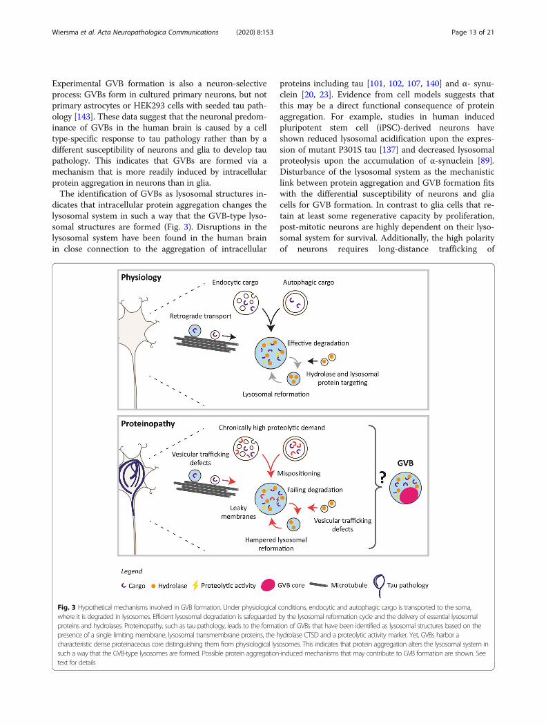
dicates that intracellular protein aggregation changes thelysosomal system in such a way that the GVB-type lyso-somal structures are formed (Fig. 3). Disruptions in thelysosomal system have been found in the human brainin close connection to the aggregation of intracellular
proteins including tau [101, 102, 107, 140] and α- synu-clein [20, 23]. Evidence from cell models suggests thatthis may be a direct functional consequence of proteinaggregation. For example, studies in human inducedpluripotent stem cell (iPSC)-derived neurons haveshown reduced lysosomal acidification upon the expres-sion of mutant P301S tau [137] and decreased lysosomalproteolysis upon the accumulation of α-synuclein [89].Disturbance of the lysosomal system as the mechanisticlink between protein aggregation and GVB formation fitswith the differential susceptibility of neurons and gliacells for GVB formation. In contrast to glia cells that re-tain at least some regenerative capacity by proliferation,post-mitotic neurons are highly dependent on their lyso-somal system for survival. Additionally, the high polarityof neurons requires long-distance trafficking of
Fig. 3 Hypothetical mechanisms involved in GVB formation. Under physiological conditions, endocytic and autophagic cargo is transported to the soma,where it is degraded in lysosomes. Efficient lysosomal degradation is safeguarded by the lysosomal reformation cycle and the delivery of essential lysosomalproteins and hydrolases. Proteinopathy, such as tau pathology, leads to the formation of GVBs that have been identified as lysosomal structures based on thepresence of a single limiting membrane, lysosomal transmembrane proteins, the hydrolase CTSD and a proteolytic activity marker. Yet, GVBs harbor acharacteristic dense proteinaceous core distinguishing them from physiological lysosomes. This indicates that protein aggregation alters the lysosomal system insuch a way that the GVB-type lysosomes are formed. Possible protein aggregation-induced mechanisms that may contribute to GVB formation are shown. Seetext for details
Wiersma et al. Acta Neuropathologica Communications (2020) 8:153 Page 13 of 21

(lysosomal) organelles and cargo. Therefore, neurons arelikely to respond differently to (protein aggregation-induced) lysosomal stress than glia cells. This is illus-trated by the finding that neurodegeneration is a prom-inent feature of lysosomal storage disorders (LSDs),which are caused by mutations in genes encoding ubi-quitously expressed lysosomal enzymes and other lyso-somal proteins [104]. Interestingly, case studies havereported the presence of GVBs in patients with the LSDsNiemann-Pick disease type C [127] and Salla disease [6].In these patients, also NFTs were detected in the GVB-containing brain areas. Therefore, although further studyof the LSD spectrum is warranted, this could indicatethat also in these disorders concomitant protein aggrega-tion is a prerequisite for the development of GVBs.As discussed above, the aggregation of proteins other than
tau may similarly elicit GVB formation. Consistent with thishypothesis, GVB formation could not be directly connectedto loss of the physiological function of tau, as acute disrup-tion of the microtubule network in cultured primary neuronsdoes not lead to GVB formation [143]. Therefore, a genericgain-of-function response of the lysosomal system to (early)intracellular protein aggregates appears to underlie the for-mation of GVBs (Fig. 3). Protein aggregates and cellularcomponents damaged by those aggregates will be targetedfor degradation, demanding an increase in the degradativecapacity of the lysosomal system. Persistent protein aggrega-tion may overload the lysosomal system and the protein ac-cumulation in the GVB core may therefore represent anoverwhelmed lysosome. This is in line with the possible de-tection of different amyloidogenic proteins and ubiquitin inGVBs. In addition to a direct overload of lysosomal proteoly-sis, protein aggregates may disrupt the integrity of the lyso-somal membrane, resulting in leakage of lysosomal enzymesto the cytosol. Indeed, in tauopathy patients the lysosomalhydrolase CTSD shows a diffuse cytoplasmic localization,whereas in controls CTSD is observed in punctate structurescorresponding to lysosomes [107]. It has been shown thattau aggregates impair the membrane integrity of artificialphospholipid vesicles [29] and that extracellularly suppliedtau aggregates have the ability to damage the membrane ofendocytic vesicles in cultured rat neurons [19], indicatingthat pathological forms of tau can rupture vesicular mem-branes. Lysosomal membrane permeabilization likely causesa drop in proteolytic capacity. In turn, this may interfere withthe process of lysosomal reformation, as efficient degradationof auto- and endolysosomal cargo is required for regener-ation of the lysosomal pool [152]. Retrograde axonal trans-port is essential for the maturation of lysosomes and therebyproper cargo degradation in neurons [27]. Another possibilitytherefore is that protein aggregates indirectly induce lyso-somal stress via the disruption of vesicular transport. Indeed,reduced axonal transport of lysosomes is observed in cul-tured mouse neurons with tau aggregates [35]. Also α-
synuclein aggregates disrupt vesicular protein trafficking, in-cluding the targeting of hydrolases to lysosomes early in thesecretory pathway, leading to lysosomal dysfunction in hu-man iPSC neurons [89]. Therefore, protein aggregation-induced trafficking defects can lead to mispositioning of lyso-somes and impaired cargo delivery. Protein aggregationtherefore not only imposes a chronically high proteolytic de-mand on a neuron, but may also lead to failing degradation,hampered lysosomal reformation and defective trafficking oflysosomes and their cargo. Any of these processes alone orin combination leads to a situation of stress in the lysosomalsystem that may in turn result in the appearance of theGVB-type lysosomal structure (Fig. 3).
GVB formation as a protective or degenerativeresponse?An outstanding question is whether GVB formation is aprotective or degenerative response to intracellular pro-tein aggregation (Fig. 4). GVBs predominate in cells withearly-stage protein aggregation, as shown for tau [43, 65,67, 96, 125, 146] and α-synuclein [82]. The emergenceof GVBs early in the protein aggregation process couldindicate a protective response, aimed to make neuronsmore resilient to proteostatic stress. Furthermore, theactive contribution of GVBs to the degradation of cellu-lar waste suggests that GVBs are protective structures,formed to restore proteostasis by increasing the degrada-tive capacity of the neuron. Lysosomal stress can triggerthe transcription factor EB (TFEB) response, which acti-vates a transcriptional program that promotes cellularclearance by enhancing lysosomal biogenesis and autoly-sosomal flux [21]. TFEB overexpression reduces the taupathology load in tau Tg mice [108, 139]. This demon-strates that lysosomal stress responses can have neuro-protective consequences in the context of proteinaggregation. The GVB-type lysosomal response mayhave a similar beneficial outcome. In favor of a protect-ive role, highly abundant GVBs were reported in acohort of cognitively healthy individuals over 100-years-of-age [31]. This may suggest that in healthy centenar-ians, a high GVB load is associated with protection fromneurodegeneration and clinical symptoms. In line withthis, phosphorylated/activated stress signaling proteinsinvolved in adaptive responses like UPR activation arefound in GVBs, which could indicate that a protectiveresponse is ongoing. Indeed, different downstream tar-gets of the UPR signaling cascades are upregulated inbrains of patients with neurodegenerative diseases [117].Although these observations are in agreement with UPRactivation in GVB-positive neurons, a functional involve-ment of the UPR in GVB formation has not been dem-onstrated (Table 3).Alternatively, the presence of GVBs in neurons with
early-stage protein aggregation [43, 65, 67, 82, 96, 125,
Wiersma et al. Acta Neuropathologica Communications (2020) 8:153 Page 14 of 21
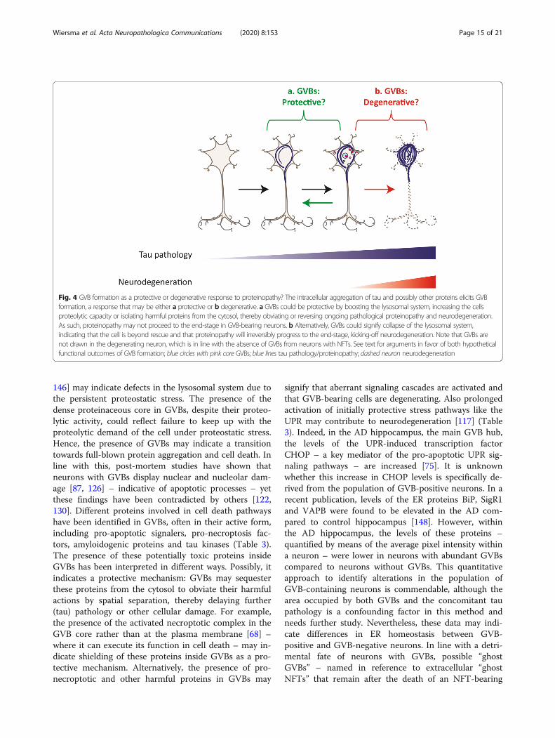
146] may indicate defects in the lysosomal system due tothe persistent proteostatic stress. The presence of thedense proteinaceous core in GVBs, despite their proteo-lytic activity, could reflect failure to keep up with theproteolytic demand of the cell under proteostatic stress.Hence, the presence of GVBs may indicate a transitiontowards full-blown protein aggregation and cell death. Inline with this, post-mortem studies have shown thatneurons with GVBs display nuclear and nucleolar dam-age [87, 126] – indicative of apoptotic processes – yetthese findings have been contradicted by others [122,130]. Different proteins involved in cell death pathwayshave been identified in GVBs, often in their active form,including pro-apoptotic signalers, pro-necroptosis fac-tors, amyloidogenic proteins and tau kinases (Table 3).The presence of these potentially toxic proteins insideGVBs has been interpreted in different ways. Possibly, itindicates a protective mechanism: GVBs may sequesterthese proteins from the cytosol to obviate their harmfulactions by spatial separation, thereby delaying further(tau) pathology or other cellular damage. For example,the presence of the activated necroptotic complex in theGVB core rather than at the plasma membrane [68] –where it can execute its function in cell death – may in-dicate shielding of these proteins inside GVBs as a pro-tective mechanism. Alternatively, the presence of pro-necroptotic and other harmful proteins in GVBs may
signify that aberrant signaling cascades are activated andthat GVB-bearing cells are degenerating. Also prolongedactivation of initially protective stress pathways like theUPR may contribute to neurodegeneration [117] (Table3). Indeed, in the AD hippocampus, the main GVB hub,the levels of the UPR-induced transcription factorCHOP – a key mediator of the pro-apoptotic UPR sig-naling pathways – are increased [75]. It is unknownwhether this increase in CHOP levels is specifically de-rived from the population of GVB-positive neurons. In arecent publication, levels of the ER proteins BiP, SigR1and VAPB were found to be elevated in the AD com-pared to control hippocampus [148]. However, withinthe AD hippocampus, the levels of these proteins –quantified by means of the average pixel intensity withina neuron – were lower in neurons with abundant GVBscompared to neurons without GVBs. This quantitativeapproach to identify alterations in the population ofGVB-containing neurons is commendable, although thearea occupied by both GVBs and the concomitant taupathology is a confounding factor in this method andneeds further study. Nevertheless, these data may indi-cate differences in ER homeostasis between GVB-positive and GVB-negative neurons. In line with a detri-mental fate of neurons with GVBs, possible “ghostGVBs” – named in reference to extracellular “ghostNFTs” that remain after the death of an NFT-bearing
Fig. 4 GVB formation as a protective or degenerative response to proteinopathy? The intracellular aggregation of tau and possibly other proteins elicits GVBformation, a response that may be either a protective or b degenerative. a GVBs could be protective by boosting the lysosomal system, increasing the cellsproteolytic capacity or isolating harmful proteins from the cytosol, thereby obviating or reversing ongoing pathological proteinopathy and neurodegeneration.As such, proteinopathy may not proceed to the end-stage in GVB-bearing neurons. b Alternatively, GVBs could signify collapse of the lysosomal system,indicating that the cell is beyond rescue and that proteinopathy will irreversibly progress to the end-stage, kicking-off neurodegeneration. Note that GVBs arenot drawn in the degenerating neuron, which is in line with the absence of GVBs from neurons with NFTs. See text for arguments in favor of both hypotheticalfunctional outcomes of GVB formation; blue circles with pink core GVBs; blue lines tau pathology/proteinopathy; dashed neuron neurodegeneration
Wiersma et al. Acta Neuropathologica Communications (2020) 8:153 Page 15 of 21
neuron – have been described in the aged human brain[47, 48]. Ghost GVBs were defined as deposits morpho-logically resembling GVBs but without a discernable liv-ing cell body or nucleus and were detected with an anti-tau or anti-pTDP43 antibody. This could indicate that atleast a population of neurons with GVBs degenerates.Future studies using additional GVB and cellularmarkers as well as higher resolution microscopy shouldconfirm the existence of ghost GVBs.The GVB load negatively correlates with neuronal
density in the AD hippocampus [7, 68]. Accordingly, theextent of GVBs is correlated with various measures ofcognitive decline [32, 47, 48, 61, 68, 132]. However,these correlations are the result of the causal relation be-tween tau pathology and GVB formation as in the same[32, 48, 68] and other [4, 34, 51, 98] studies tau path-ology correlates similarly with neurodegeneration andsymptom severity. Indeed, it was shown that GVBs areonly associated with dementia when measures of taupathology – the NFT Braak stage and cortical neuriticplaque pathology – are not controlled for in the analysis[48, 61]. The positive correlations between GVB loadand neuropathological and clinical measures of diseaseseverity can therefore be explained both by an activecontribution of GVBs to cell death and by activation ofthe GVB response as a cellular defense mechanism insurviving neurons (Fig. 4). Clearly, further experimentsare needed to unravel the functional implications ofGVB formation, where the study of experimental modelswill be instrumental.
Recommendations and concluding remarksAlthough their first description dates back more than acentury, GVBs are relatively underinvestigated. The re-cent discovery of tau pathology as the trigger for GVBformation has given an impulse to the GVB researchfield. This sets the stage for future research into themechanism and downstream consequences of GVB for-mation in experimental models, but the finding of caus-ality between tau pathology and GVB formation also hasimportant implications for the interpretation of neuro-pathological data. Without the exclusion of concurrenttau pathology in patients with non-tau neurodegenera-tive disorders, no conclusions on a relation betweenother protein aggregates and GVBs can be drawn.Therefore, future neuropathological studies shouldalways analyze the presence of comorbid tau pathologyin GVB-bearing cells. Here, important progress can bemade by studying the co-localization of GVBs and tauaggregates at the single cell level by double immunola-beling rather than using pathological staging. The likelypossibility that minute amounts of pathological tau ag-gregates are sufficient to trigger GVB formation impliesthat the exclusion of tau comorbidity in GVB-containing
neurons may be hampered by detection limitations. Tothis end, confocal microscopy is recommended that enablessingle cell analysis at high resolution. Furthermore, to reducethe probability of tau co-pathology, specifically those brainareas with disease-specific pathology should be studied. Im-portantly, definite proof of a causal relation between non-tauprotein aggregation and GVB formation can only be ob-tained in experimental models. Based on the current humanneuropathological data, α-synuclein and DPR protein aggre-gates are the prime candidates for putative non-tau GVB in-duction and they await experimental validation. Tostreamline future research, we have formulated recommen-dations for the validation of a GVB identity in tissue and cellmodels. These guidelines include immunopositivity for CK1δand at least one additional common GVB marker as well asproof of the characteristic GVB morphology (Table 1; Fig. 1).In Tables 2 and 4, previous studies on GVBs in non-tau neu-rodegenerative proteinopathies, experimental models andaged animals are aligned with these refined criteria. In thisrapidly changing field, potential new GVB markers are iden-tified continuously and their GVB localization can also berigorously validated following the proposed guidelines. Ourrecommendations for the identification of GVBs will help tosystematically identify GVBs and thereby facilitate future re-search towards the elucidation of the role of these enigmaticstructures in the pathogenesis of tauopathies and other neu-rodegenerative proteinopathies.
Supplementary informationSupplementary information accompanies this paper at https://doi.org/10.1186/s40478-020-00996-5.
Additional file 1: Supplementary Table 1. Overview of primaryantibodies commonly used to detect GVB.
AbbreviationsAb: Antibody; Aβ: Amyloid β; AD: Alzheimer’s disease; ALS: Amyotrophiclateral sclerosis; APC: Adenomatous polyposis coli; APP: Amyloid precursorprotein; APPSw: Mutant amyloid precursor protein K670M/N671L;APPSw,Ind: Mutant amyloid precursor protein K670M/N671L/V717F;APPSw/L: Mutant amyloid precursor protein K670M/N671L/V717I; α-syn: α-synuclein; AT8: Antibody recognizing tau phosphorylated at Ser202/Thr205;AT100: Antibody recognizing tau phosphorylated at Thr212/Ser214;AT270: Antibody recognizing tau phosphorylated at Thr181; BiP: Bindingimmunoglobulin protein; β-COP: Coatomer subunit β; CA: Cornu ammonis;c-Abl: Abelson tyrosine kinase; CBD: Corticobasal degeneration; CDK5: Cyclin-dependent kinase 5; CHMP2B: Charged multivesicular body protein 2B;CHOP: C/EBP homologous protein; CK1α: Casein kinase 1 α; CK1δ: Caseinkinase 1 δ; CK1ε: Casein kinase 1 ε; CTSD: Cathepsin D;C9ORF72: Chromosome 9 open reading frame 72; DAB: 3.3′-diaminobenzidine; DAPI: 4′,6-diamidino-2-phenylindole; DPR: Dipeptiderepeat; DQ-BSA: Self-quenched dye conjugate of bovine serum albumin;Dvl3: Segment polarity protein dishevelled homolog 3; EEA1: Earlyendosome antigen 1; eIF2α: Eukaryotic translation initiation factor 2α;EM: Electron microscopy; ER: Endoplasmic reticulum; FTLD: Frontotemporallobar degeneration; FTLD/ALS-C9: TDP-43 related frontotemporal lobardegeneration / amyotrophic lateral sclerosis caused by a hexanucleotiderepeat expansion in C9ORF72; FUS: Fused in sarcoma; GFP: Green fluorescentprotein; GSK-3β: Glycogen synthase kinase-3β; GSS: Gerstmann-Sträussler-Scheinker syndrome; GVBs: Granulovacuolar degeneration bodies;GVD: Granulovacuolar degeneration; G3BP: Ras-GTPase-activating protein-
Wiersma et al. Acta Neuropathologica Communications (2020) 8:153 Page 16 of 21
binding protein; HEK293: Human embryonic kidney 293 cells; Hrd1: E3 ligaseHMG-CoA reductase degradation protein 1; HNE: 4-hydroxy-2-nonenal;Htt: Huntingtin; H&E: Hematoxylin and eosin; iPSC: Induced pluripotent stemcell; IRE1α: Inositol-requiring enzyme 1α; LAMP1: Lysosome-associatedmembrane protein 1; LC3: Microtubule-associated protein 1A/1B-light chain3; LIMP2: Lysosomal integral membrane protein 2; LRRK2: Leucine-rich repeatkinase 2; LSD: Lysosomal storage disorder; MAPT: Microtubule-associatedprotein tau; MARK3: Microtubule affinity regulating kinase isoform 3;MARK4: Microtubule affinity regulating kinase isoform 4; MLKL: Mixed lineagekinase domain-like protein; MPM-2: Ab antibody recognizing mitoticphospho-epitopes; MSA: Multiple system atrophy; M6PR: Mannose 6-phosphate receptor; NF: Neurofilament; NF-κB: Nuclear factor-kappa B;NFT: Neurofibrillary tangle; OspA: Outer surface protein of Borreliaburgdorferi spirochete; p: Phosphorylated; PERK: Protein kinase R (PKR)-likeendoplasmic reticulum kinase; PGRN: Progranulin gene; PHF-1: Antibodyrecognizing tau phosphorylated at Ser396/Ser404; PICALM: Phosphatidylinositol-binding clathrin assembly protein; PKR: Double-stranded RNA-dependent protein kinase; PRKN: Parkin RBR E3 ubiquitinprotein ligase; PPND: Pallido-ponto-nigral degeneration; PRNP: Prion proteingene; PrP: Prion protein; PrPSc: PrP-Scrapie; PSKs: Prostate-derived sterile 20-like kinases; PSP: Progressive supranuclear palsy; PS1: Presenilin-1;PS1dE9: Mutant PS1 with deletion of exon 9; p300: E1A binding protein p300;p38: MAP kinase p38 mitogen-activated protein kinase; p62: Sequestosome1; Rab7: Ras-related protein 7; Rab10: Ras-related protein Rab10; Rab11: Ras-related protein 11; RIPK1: Receptor-interacting serine/threonine-proteinkinase 1; RIPK3: Receptor-interacting serine/threonine-protein kinase 3; SAPK/JNK: Stress-activated protein kinase/c-Jun N-terminal kinase; SigR1: Sigmareceptor 1; SIL1: Nucleotide exchange factor SIL1; SMI-31: Antibodyrecognizing phosphorylated neurofilament H and M; SNCA: α-synucleingene; SSBP1: Single stranded DNA binding protein 1; Syk: Spleen tyrosinekinase; S6 40S: Ribosomal subunit protein S6; TDP-43: TAR DNA-binding pro-tein 43; TFEB: Transcription factor EB; Tg: Transgenic; TG3: Antibodyrecognizing tau phosphorylated at Thr231 (also conformation-specific);TMEM230: Transmembrane protein 230; Ubi: Ubiquitin; UPR: Unfoldedprotein response; VAPB: Vesicle-associated membrane protein-associated pro-tein B; VPS4a: Vacuolar protein sorting-associated protein 4A
AcknowledgementsThe authors thank members of the Molecular Neurodegeneration group forstimulating discussions, ZonMW and Alzheimer Nederland for financialsupport (Memorabel grant #733050101) and scientists worldwide for theircontributions to the still increasing knowledge on GVBs.
Authors’ contributionsThis review was conceptualized and written by VIW, JJMH and WS. VIWgenerated tables and figures. The author(s) read and approved the finalmanuscript.
Competing interestsThe authors declare that they have no competing interests.
Author details1Department of Clinical Genetics, Amsterdam University Medical Centerslocation VUmc, Amsterdam, The Netherlands. 2Department of FunctionalGenomics, Center for Neurogenomics and Cognitive Research, VrijeUniversiteit (VU), De Boelelaan 1085, 1081 HV Amsterdam, The Netherlands.3Department of Pathology, Amsterdam University Medical Centers locationVUmc, Amsterdam, The Netherlands.
Received: 14 July 2020 Accepted: 14 July 2020
References1. Abo El-Khair DM, El-Safti FE-NA, Nooh HZ, El-Mehi AE (2014) A comparative
study on the effect of high cholesterol diet on the hippocampal CA1 areaof adult and aged rats. Anat Cell Biol 47:117–126. https://doi.org/10.5115/acb.2014.47.2.117
2. Ando K, Brion J-P, Stygelbout V, Suain V, Authelet M, Dedecker R et al (2013)Clathrin adaptor CALM/PICALM is associated with neurofibrillary tangles andis cleaved in Alzheimer’s brains. Acta Neuropathol 125:861–878. https://doi.org/10.1007/s00401-013-1111-z
3. Arima K, Mizutani T, Alim MA, Tonozuka-Uehara H, Izumiyama Y, Hirai S et al(2000) NACP/α-synuclein and tau constitute two distinctive subsets offilaments in the same neuronal inclusions in brains from a family ofparkinsonism and dementia with Lewy bodies: double-immunolabelingfluorescence and electron microscopic studies. Acta Neuropathol 100:115–121. https://doi.org/10.1007/s004010050002
4. Arriagada PV, Growdon JH, Hedley-Whyte ET, Hyman BT (1992)Neurofibrillary tangles but not senile plaques parallel duration and severityof Alzheimer’s disease. Neurology 42:631–639. https://doi.org/10.1212/wnl.42.3.631
5. Aubry S, Shin W, Crary JF, Lefort R, Qureshi YH, Lefebvre C et al (2015)Assembly and interrogation of Alzheimer’s disease genetic networks revealnovel regulators of progression. PLoS One 10:e012035. https://doi.org/10.1371/journal.pone.0120352
6. Autio-Harmainen H, Oldfors A, Sourander P, Renlund M, Dammert K, SimiläS (1988) Neuropathology of Salla disease. Acta Neuropathol 75:481–490.https://doi.org/10.1007/BF00687135
7. Ball MJ (1977) Neuronal loss, neurofibrillary tangles and granulovacuolardegeneration in the hippocampus with ageing and dementia - aquantitative study. Acta Neuropathol 37:111–118. https://doi.org/10.1051/epjconf/20147504003
8. Ball MJ (1978) Topographic distribution of neurofibrillary tangles andgranulovacuolar degeneration in hippocampal cortex of aging anddemented patients. A quantitative study. Acta Neuropathol 42:73–80.https://doi.org/10.1007/bf00690970
9. Ball MJ, Lo P (1977) Granulovacuolar degeneration in the ageing brain andin dementia. J Neuropathol Exp Neurol 36:474–487. https://doi.org/10.1097/00005072-197705000-00006
10. Ball MJ, Vis CL (1978) Relationship of granulovacuolar degeneration inhippocampal neurones to aging and to dementia in normal-pressurehydrocephalics. J Gerontol 33:815–824. https://doi.org/10.1093/geronj/33.6.815
11. De Boni U, Crapper McLachlan DR (1980) Senile dementia and Alzheimer’sdisease: a current view. Life Sci 27:1–14. https://doi.org/10.1016/0024-3205(80)90013-2
12. Braak H, Alafuzoff I, Arzberger T, Kretzschmar H, Tredici K (2006) Staging ofAlzheimer disease-associated neurofibrillary pathology using paraffinsections and immunocytochemistry. Acta Neuropathol 112:389–404. https://doi.org/10.1007/s00401-006-0127-z
13. Braak H, Braak E (1991) Neuropathological stageing of Alzheimer-relatedchanges. Acta Neuropathol 82:239–259. https://doi.org/10.1007/BF00308809
14. Braak H, Del Tredici K, Rüb U, de Vos RAI, Jansen Steur ENH, Braak E (2003)Staging of brain pathology related to sporadic Parkinson’s disease.Neurobiol Aging 24:197–211. https://doi.org/10.1016/S0197-4580(02)00065-9
15. Brat DJ, Gearing M, Goldthwaite PT, Wainer BH, Burger PC (2001) Tau-associated neuropathology in ganglion cell tumours increases with patientage but appears unrelated to ApoE genotype. Neuropathol Appl Neurobiol27:197–205. https://doi.org/10.1046/j.1365-2990.2001.00311.x
16. Brown J, Lantos P, Stratton M, Roques P, Rossor M (1993) Familialprogressive supranuclear palsy. J Neurol Neurosurg Psychiatry 56:473–476.https://doi.org/10.1136/jnnp.56.5.473
17. Bruyn GW, Roos RAC (1990) Senile plaques in Huntington’s disease: apreliminary report. Clin Neurol Neurosurg 92:329–331. https://doi.org/10.1016/0303-8467(90)90059-E
18. Burger PC, Vogel FS (1973) The development of the pathologic changes ofAlzheimer’s disease and senile dementia in patients with Down’s syndrome.Am J Pathol 73:457–476
19. Calafate S, Flavin W, Verstreken P, Moechars D (2016) Loss of Bin1 promotesthe propagation of tau pathology. Cell Rep 17:931–940. https://doi.org/10.1016/j.celrep.2016.09.063
20. Chu Y, Dodiya H, Aebischer P, Olanow CW, Kordower JH (2009) Alterationsin lysosomal and proteasomal markers in Parkinson’s disease: relationship toalpha-synuclein inclusions. Neurobiol Dis 35:385–398. https://doi.org/10.1016/j.nbd.2009.05.023
21. Cortes CJ, La Spada AR (2019) TFEB dysregulation as a driver of autophagydysfunction in neurodegenerative disease: molecular mechanisms, cellularprocesses, and emerging therapeutic opportunities. Neurobiol Dis 122:83–93. https://doi.org/10.1016/j.nbd.2018.05.012
22. Darusman HS, Agungpriyono DR, Kusumaputri VA, Sajuthi D, Schapiro SJ, Hau J(2019) Granulovacuolar degeneration in brains of senile cynomolgus monkeys.Front Aging Neurosci 11:50. https://doi.org/10.3389/fnagi.2019.00050
Wiersma et al. Acta Neuropathologica Communications (2020) 8:153 Page 17 of 21
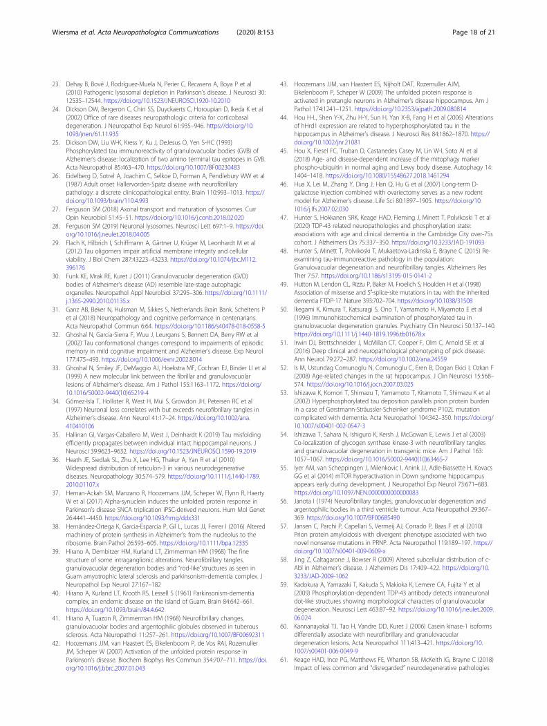
23. Dehay B, Bové J, Rodríguez-Muela N, Perier C, Recasens A, Boya P et al(2010) Pathogenic lysosomal depletion in Parkinson’s disease. J Neurosci 30:12535–12544. https://doi.org/10.1523/JNEUROSCI.1920-10.2010
24. Dickson DW, Bergeron C, Chin SS, Duyckaerts C, Horoupian D, Ikeda K et al(2002) Office of rare diseases neuropathologic criteria for corticobasaldegeneration. J Neuropathol Exp Neurol 61:935–946. https://doi.org/10.1093/jnen/61.11.935
25. Dickson DW, Liu W-K, Kress Y, Ku J, DeJesus O, Yen S-HC (1993)Phosphorylated tau immunoreactivity of granulovacuolar bodies (GVB) ofAlzheimer’s disease: localization of two amino terminal tau epitopes in GVB.Acta Neuropathol 85:463–470. https://doi.org/10.1007/BF00230483
26. Eidelberg D, Sotrel A, Joachim C, Selkoe D, Forman A, Pendlebury WW et al(1987) Adult onset Hallervorden-Spatz disease with neurofibrillarypathology: a discrete clinicopathological entity. Brain 110:993–1013. https://doi.org/10.1093/brain/110.4.993
27. Ferguson SM (2018) Axonal transport and maturation of lysosomes. CurrOpin Neurobiol 51:45–51. https://doi.org/10.1016/j.conb.2018.02.020
28. Ferguson SM (2019) Neuronal lysosomes. Neurosci Lett 697:1–9. https://doi.org/10.1016/j.neulet.2018.04.005
29. Flach K, Hilbrich I, Schiffmann A, Gärtner U, Krüger M, Leonhardt M et al(2012) Tau oligomers impair artificial membrane integrity and cellularviability. J Biol Chem 287:43223–43233. https://doi.org/10.1074/jbc.M112.396176
30. Funk KE, Mrak RE, Kuret J (2011) Granulovacuolar degeneration (GVD)bodies of Alzheimer’s disease (AD) resemble late-stage autophagicorganelles. Neuropathol Appl Neurobiol 37:295–306. https://doi.org/10.1111/j.1365-2990.2010.01135.x
31. Ganz AB, Beker N, Hulsman M, Sikkes S, Netherlands Brain Bank, Scheltens Pet al (2018) Neuropathology and cognitive performance in centenarians.Acta Neuropathol Commun 6:64. https://doi.org/10.1186/s40478-018-0558-5
32. Ghoshal N, García-Sierra F, Wuu J, Leurgans S, Bennett DA, Berry RW et al(2002) Tau conformational changes correspond to impairments of episodicmemory in mild cognitive impairment and Alzheimer’s disease. Exp Neurol177:475–493. https://doi.org/10.1006/exnr.2002.8014
33. Ghoshal N, Smiley JF, DeMaggio AJ, Hoekstra MF, Cochran EJ, Binder LI et al(1999) A new molecular link between the fibrillar and granulovacuolarlesions of Alzheimer’s disease. Am J Pathol 155:1163–1172. https://doi.org/10.1016/S0002-9440(10)65219-4
34. Gómez-Isla T, Hollister R, West H, Mui S, Growdon JH, Petersen RC et al(1997) Neuronal loss correlates with but exceeds neurofibrillary tangles inAlzheimer’s disease. Ann Neurol 41:17–24. https://doi.org/10.1002/ana.410410106
35. Hallinan GI, Vargas-Caballero M, West J, Deinhardt K (2019) Tau misfoldingefficiently propagates between individual intact hippocampal neurons. JNeurosci 39:9623–9632. https://doi.org/10.1523/JNEUROSCI.1590-19.2019
36. Heath JE, Siedlak SL, Zhu X, Lee HG, Thakur A, Yan R et al (2010)Widespread distribution of reticulon-3 in various neurodegenerativediseases. Neuropathology 30:574–579. https://doi.org/10.1111/j.1440-1789.2010.01107.x
37. Heman-Ackah SM, Manzano R, Hoozemans JJM, Scheper W, Flynn R, HaertyW et al (2017) Alpha-synuclein induces the unfolded protein response inParkinson’s disease SNCA triplication iPSC-derived neurons. Hum Mol Genet26:4441–4450. https://doi.org/10.1093/hmg/ddx331
38. Hernández-Ortega K, Garcia-Esparcia P, Gil L, Lucas JJ, Ferrer I (2016) Alteredmachinery of protein synthesis in Alzheimer’s: from the nucleolus to theribosome. Brain Pathol 26:593–605. https://doi.org/10.1111/bpa.12335
39. Hirano A, Dembitzer HM, Kurland LT, Zimmerman HM (1968) The finestructure of some intraganglionic alterations. Neurofibrillary tangles,granulovacuolar degeneration bodies and “rod-like”structures as seen inGuam amyotrophic lateral sclerosis and parkinsonism-dementia complex. JNeuropathol Exp Neurol 27:167–182
40. Hirano A, Kurland LT, Krooth RS, Lessell S (1961) Parkinsonism-dementiacomplex, an endemic disease on the island of Guam. Brain 84:642–661.https://doi.org/10.1093/brain/84.4.642
41. Hirano A, Tuazon R, Zimmerman HM (1968) Neurofibrillary changes,granulovacuolar bodies and argentophilic globules observed in tuberoussclerosis. Acta Neuropathol 11:257–261. https://doi.org/10.1007/BF00692311
42. Hoozemans JJM, van Haastert ES, Eikelenboom P, de Vos RAI, RozemullerJM, Scheper W (2007) Activation of the unfolded protein response inParkinson’s disease. Biochem Biophys Res Commun 354:707–711. https://doi.org/10.1016/j.bbrc.2007.01.043
43. Hoozemans JJM, van Haastert ES, Nijholt DAT, Rozemuller AJM,Eikelenboom P, Scheper W (2009) The unfolded protein response isactivated in pretangle neurons in Alzheimer’s disease hippocampus. Am JPathol 174:1241–1251. https://doi.org/10.2353/ajpath.2009.080814
44. Hou H-L, Shen Y-X, Zhu H-Y, Sun H, Yan X-B, Fang H et al (2006) Alterationsof hHrd1 expression are related to hyperphosphorylated tau in thehippocampus in Alzheimer’s disease. J Neurosci Res 84:1862–1870. https://doi.org/10.1002/jnr.21081
45. Hou X, Fiesel FC, Truban D, Castanedes Casey M, Lin W-I, Soto AI et al(2018) Age- and disease-dependent increase of the mitophagy markerphospho-ubiquitin in normal aging and Lewy body disease. Autophagy 14:1404–1418. https://doi.org/10.1080/15548627.2018.1461294
46. Hua X, Lei M, Zhang Y, Ding J, Han Q, Hu G et al (2007) Long-term D-galactose injection combined with ovariectomy serves as a new rodentmodel for Alzheimer’s disease. Life Sci 80:1897–1905. https://doi.org/10.1016/j.lfs.2007.02.030
47. Hunter S, Hokkanen SRK, Keage HAD, Fleming J, Minett T, Polvikoski T et al(2020) TDP-43 related neuropathologies and phosphorylation state:associations with age and clinical dementia in the Cambridge City over-75scohort. J Alzheimers Dis 75:337–350. https://doi.org/10.3233/JAD-191093
48. Hunter S, Minett T, Polvikoski T, Mukaetova-Ladinska E, Brayne C (2015) Re-examining tau-immunoreactive pathology in the population:Granulovacuolar degeneration and neurofibrillary tangles. Alzheimers ResTher 7:57. https://doi.org/10.1186/s13195-015-0141-2
49. Hutton M, Lendon CL, Rizzu P, Baker M, Froelich S, Houlden H et al (1998)Association of missense and 5′-splice-site mutations in tau with the inheriteddementia FTDP-17. Nature 393:702–704. https://doi.org/10.1038/31508
50. Ikegami K, Kimura T, Katsuragi S, Ono T, Yamamoto H, Miyamoto E et al(1996) Immunohistochemical examination of phosphorylated tau ingranulovacuolar degeneration granules. Psychiatry Clin Neurosci 50:137–140.https://doi.org/10.1111/j.1440-1819.1996.tb01678.x
51. Irwin DJ, Brettschneider J, McMillan CT, Cooper F, Olm C, Arnold SE et al(2016) Deep clinical and neuropathological phenotyping of pick disease.Ann Neurol 79:272–287. https://doi.org/10.1002/ana.24559
52. Is M, Ustundag Comunoglu N, Comunoglu C, Eren B, Dogan Ekici I, Ozkan F(2008) Age-related changes in the rat hippocampus. J Clin Neurosci 15:568–574. https://doi.org/10.1016/j.jocn.2007.03.025
53. Ishizawa K, Komori T, Shimazu T, Yamamoto T, Kitamoto T, Shimazu K et al(2002) Hyperphosphorylated tau deposition parallels prion protein burdenin a case of Gerstmann-Sträussler-Scheinker syndrome P102L mutationcomplicated with dementia. Acta Neuropathol 104:342–350. https://doi.org/10.1007/s00401-002-0547-3
54. Ishizawa T, Sahara N, Ishiguro K, Kersh J, McGowan E, Lewis J et al (2003)Co-localization of glycogen synthase kinase-3 with neurofibrillary tanglesand granulovacuolar degeneration in transgenic mice. Am J Pathol 163:1057–1067. https://doi.org/10.1016/S0002-9440(10)63465-7
55. Iyer AM, van Scheppingen J, Milenkovic I, Anink JJ, Adle-Biassette H, KovacsGG et al (2014) mTOR hyperactivation in Down syndrome hippocampusappears early during development. J Neuropathol Exp Neurol 73:671–683.https://doi.org/10.1097/NEN.0000000000000083
56. Janota I (1974) Neurofibrillary tangles, granulovacuolar degeneration andargentophilic bodies in a third ventricle tumour. Acta Neuropathol 29:367–369. https://doi.org/10.1007/BF00685490
57. Jansen C, Parchi P, Capellari S, Vermeij AJ, Corrado P, Baas F et al (2010)Prion protein amyloidosis with divergent phenotype associated with twonovel nonsense mutations in PRNP. Acta Neuropathol 119:189–197. https://doi.org/10.1007/s00401-009-0609-x
58. Jing Z, Caltagarone J, Bowser R (2009) Altered subcellular distribution of c-Abl in Alzheimer’s disease. J Alzheimers Dis 17:409–422. https://doi.org/10.3233/JAD-2009-1062
59. Kadokura A, Yamazaki T, Kakuda S, Makioka K, Lemere CA, Fujita Y et al(2009) Phosphorylation-dependent TDP-43 antibody detects intraneuronaldot-like structures showing morphological characters of granulovacuolardegeneration. Neurosci Lett 463:87–92. https://doi.org/10.1016/j.neulet.2009.06.024
60. Kannanayakal TJ, Tao H, Vandre DD, Kuret J (2006) Casein kinase-1 isoformsdifferentially associate with neurofibrillary and granulovacuolardegeneration lesions. Acta Neuropathol 111:413–421. https://doi.org/10.1007/s00401-006-0049-9
61. Keage HAD, Ince PG, Matthews FE, Wharton SB, McKeith IG, Brayne C (2018)Impact of less common and “disregarded” neurodegenerative pathologies
Wiersma et al. Acta Neuropathologica Communications (2020) 8:153 Page 18 of 21
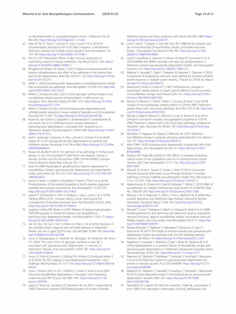
on dementia burden in a population-based cohort. J Alzheimers Dis 28:485–493. https://doi.org/10.3233/jad-2011-111268
62. Kiely AP, Asi YT, Kara E, Limousin P, Ling H, Lewis P et al (2013) A-synucleinopathy associated with G51D SNCA mutation: a link betweenParkinson’s disease and multiple system atrophy? Acta Neuropathol 125:753–769. https://doi.org/10.1007/s00401-013-1096-7
63. Kim SU (1971) Neurotoxic effects of alkyl mercury compound onmyelinating cultures of mouse cerebellum. Exp Neurol 32:237–246. https://doi.org/10.1016/0014-4886(71)90067-7
64. Klingebiel M, Dinekov M, Köhler C (2017) Analysis of ribosomal protein S6baseline phosphorylation and effect of tau pathology in the murine brainand human hippocampus. Brain Res 1659:121–135. https://doi.org/10.1016/j.brainres.2017.01.016
65. Köhler C (2016) Granulovacuolar degeneration: a neurodegenerative changethat accompanies tau pathology. Acta Neuropathol 132:339–359. https://doi.org/10.1007/s00401-016-1562-0
66. Köhler C, Dinekov M, Götz J (2013) Active glycogen synthase kinase-3 andtau pathology-related tyrosine phosphorylation in pR5 human tautransgenic mice. Neurobiol Aging 34:1369–1379. https://doi.org/10.1016/j.neurobiolaging.2012.11.010
67. Köhler C, Dinekov M, Götz J (2014) Granulovacuolar degeneration andunfolded protein response in mouse models of tauopathy and Aβ amyloidosis.Neurobiol Dis 71:169–179. https://doi.org/10.1016/j.nbd.2014.07.006
68. Koper MJ, Van Schoor E, Ospitalieri S, Vandenberghe R, Vandenbulcke M,von Arnim CAF et al (2020) Necrosome complex detected ingranulovacuolar degeneration is associated with neuronal loss inAlzheimer’s disease. Acta Neuropathol 139:463–484. https://doi.org/10.1007/s00401-019-02103-y
69. Kork F, Jankowski J, Goswami A, Weis J, Brook G, Yamoah A et al (2018)Golgin A4 in CSF and granulovacuolar degenerations of patients withAlzheimer disease. Neurology 91:e1799–e1808. https://doi.org/10.1212/WNL.0000000000006457
70. Kovacs GG, Budka H (2013) The spectrum of tau pathology in human priondisease. In: Zou W-Q, Gambetti P (eds) Prions and diseases: volume 2,animals, humans and the environment (ISBN: 9781461453383). SpringerScience+Business Media New York, pp 103–119
71. Kudo M (1986) Hypothalamic gangliocytoma. Selective appearance ofneurofibrillary changes, granulovacuolar degeneration, and argentophilicbodies. Acta Pathol Jpn 36:1225–1229. https://doi.org/10.1111/j.1440-1827.1986.tb02843.x
72. Kumar S, Wirths O, Stüber K, Wunderlich P, Koch P, Theil S et al (2016)Phosphorylation of the amyloid β-peptide at Ser26 stabilizes oligomericassembly and increases neurotoxicity. Acta Neuropathol 131:525–537.https://doi.org/10.1007/s00401-016-1546-0
73. Labisch T, Buchkremer S, Phan V, Kollipara L, Gatz C, Lentz C et al (2018)Tracking effects of SIL1 increase: taking a closer look beyond theconsequences of elevated expression level. Mol Neurobiol 55:2524–2546.https://doi.org/10.1007/s12035-017-0494-6
74. Lagalwar S, Berry RW, Binder LI (2007) Relation of hippocampal phospho-SAPK/JNK granules in Alzheimer’s disease and tauopathies togranulovacuolar degeneration bodies. Acta Neuropathol 113:63–73. https://doi.org/10.1007/s00401-006-0159-4
75. Lee JH, Won SM, Suh J, Son SJ, Moon GJ, Park U-J et al (2010) Induction ofthe unfolded protein response and cell death pathway in Alzheimer’sdisease, but not in aged Tg2576 mice. Exp Mol Med 42:386–394. https://doi.org/10.3858/emm.2010.42.5.040
76. Leroy K, Boutajangout A, Authelet M, Woodgett JR, Anderton BH, BrionJ-P (2002) The active form of glycogen synthase kinase-3β isassociated with granulovacuolar degeneration in neurons inAlzheimer’s disease. Acta Neuropathol 103:91–99. https://doi.org/10.1007/s004010100435
77. Leuzy A, Chiotis K, Lemoine L, Gillberg P-G, Almkvist O, Rodriguez-Vieitez Eet al (2019) Tau PET imaging in neurodegenerative tauopathies—still achallenge. Mol Psychiatry 24:1112–1134. https://doi.org/10.1038/s41380-018-0342-8
78. Lewis J, Dickson DW, Lin W-L, Chisholm L, Corral A, Jones G et al (2001)Enhanced neurofibrillary degeneration in transgenic mice expressingmutant tau and APP. Science 293:1487–1491. https://doi.org/10.1126/science.1058189
79. Lippa CF, Rosso AL, Stutzbach LD, Neumann M, Lee VM-Y, Trojanowski JQ(2009) Transactive response DNA-binding protein 43 burden in familial
Alzheimer disease and Down syndrome. Arch Neurol 66:1483–1488. https://doi.org/10.1001/archneurol.2009.277
80. Love S, Saitoh T, Quijada S, Cole GM, Terry RD (1988) Alz-50, ubiquitin andtau immunoreactivity of neurofibrillary tangles, pick bodies and Lewybodies. J Neuropathol Exp Neurol 47:393–405. https://doi.org/10.1097/00005072-198807000-00001
81. Lund H, Gustafsson E, Svensson A, Nilsson M, Berg M, Sunnemark D et al(2014) MARK4 and MARK3 associate with early tau phosphorylation inAlzheimer’s disease granulovacuolar degeneration bodies. Acta NeuropatholCommun 2:22. https://doi.org/10.1186/2051-5960-2-22
82. Makioka K, Yamazaki T, Fujita Y, Takatama M, Nakazato Y, Okamoto K (2010)Involvement of endoplasmic reticulum stress defined by activated unfoldedprotein response in multiple system atrophy. J Neurol Sci 297:60–65. https://doi.org/10.1016/j.jns.2010.06.019
83. Malamud N, Hirano A, Kurland LT (1961) Pathoanatomic changes inamyotrophic lateral sclerosis on Guam: special reference to the occurenceof neurofibrillary changes. Arch Neurol 5:401–415. https://doi.org/10.1001/archneur.1961.00450160051003
84. Mamais A, Manzoni C, Nazish I, Arber C, Sonustun B, Wray S et al (2018)Analysis of macroautophagy related proteins in G2019S LRRK2 Parkinson’sdisease brains with Lewy body pathology. Brain Res 1701:75–84. https://doi.org/10.1016/j.brainres.2018.07.023
85. Mamais A, Raja M, Manzoni C, Dihanich S, Lees A, Moore D et al (2013)Divergent α-synuclein solubility and aggregation properties in G2019SLRRK2 Parkinson’s disease brains with Lewy body pathology compared toidiopathic cases. Neurobiol Dis 58:183–190. https://doi.org/10.1016/j.nbd.2013.05.017
86. Mandybur TI, Nagpaul AS, Pappas Z, Niklowitz WJ (1977) Alzheimerneurofibrillary change in subacute sclerosing panencephalitis. Ann Neurol 1:103–107. https://doi.org/10.1002/ana.410010112
87. Mann DMA (1978) Granulovacuolar degeneration in pyramidal cells of thehippocampus. Acta Neuropathol 42:149–151. https://doi.org/10.1007/BF00690983
88. Mattson MP, Engle MG, Rychlik B (1991) Effects of elevated intracellularcalcium levels on the cytoskeleton and tau in cultured human corticalneurons. Mol Chem Neuropathol 15:117–142. https://doi.org/10.1007/bf03159951
89. Mazzulli JR, Zunke F, Isacson O, Studer L, Krainc D (2016) α-Synuclein-induced lysosomal dysfunction occurs through disruptions in proteintrafficking in human midbrain synucleinopathy models. Proc Natl Acad SciU S A 113:1931–1936. https://doi.org/10.1073/pnas.1520335113
90. Midani-Kurçak JS, Dinekov M, Puladi B, Arzberger T, Köhler C (2019) Effect oftau-pathology on charged multivesicular body protein 2b (CHMP2B). BrainRes 1706:224–236. https://doi.org/10.1016/j.brainres.2018.11.008
91. Miklossy J, Kis A, Radenovic A, Miller L, Forro L, Martins R et al (2006) Beta-amyloid deposition and Alzheimer’s type changes induced by Borreliaspirochetes. Neurobiol Aging 27:228–236. https://doi.org/10.1016/j.neurobiolaging.2005.01.018
92. Mizutani T, Inose T, Nakajima S, Kakimi S, Uchigata M, Ikeda K et al (1998)Familial parkinsonism and dementia with ballooned neurons, argyrophilicneuronal inclusions, atypical neurofibrillary tangles, tau-negative astrocyticfibrillary tangles, and Lewy bodies. Acta Neuropathol 95:15–27. https://doi.org/10.1007/s004010050761
93. Murata-Shinozaki Y, Takahashi T, Matsubara T, Maruyama H, Izumi Y,Matsumoto M (2017) The origins of rimmed vacuoles and granulovacuolardegeneration bodies are associated with the Wnt signaling pathway.Neurosci Lett 638:55–59. https://doi.org/10.1016/j.neulet.2016.12.014
94. Nagamine S, Yamazaki T, Makioka K, Fujita Y, Ikeda M, Takatama M et al(2016) Hypersialylation is a common feature of neurofibrillary tangles andgranulovacuolar degenerations in Alzheimer’s disease and tauopathy brains.Neuropathology 36:333–345. https://doi.org/10.1111/neup.12277
95. Nakamori M, Takahashi T, Nishikawa T, Yamazaki Y, Kurashige T, MaruyamaH et al (2013) Molecular markers for granulovacuolar degeneration arepresent in rimmed vacuoles. PLoS One 8:e80995. https://doi.org/10.1371/journal.pone.0080995
96. Nakamori M, Takahashi T, Yamazaki Y, Kurashige T, Yamawaki T, MatsumotoM (2012) Cyclin-dependent kinase 5 immunoreactivity for granulovacuolardegeneration. NeuroR 23:867–872. https://doi.org/10.1097/WNR.0b013e328358720b
97. Nasreddine ZS, Loginov M, Clark LN, Lamarche J, Miller BL, Lamontagne Aet al (1999) From genotype to phenotype: a clinical, pathological, and
Wiersma et al. Acta Neuropathologica Communications (2020) 8:153 Page 19 of 21
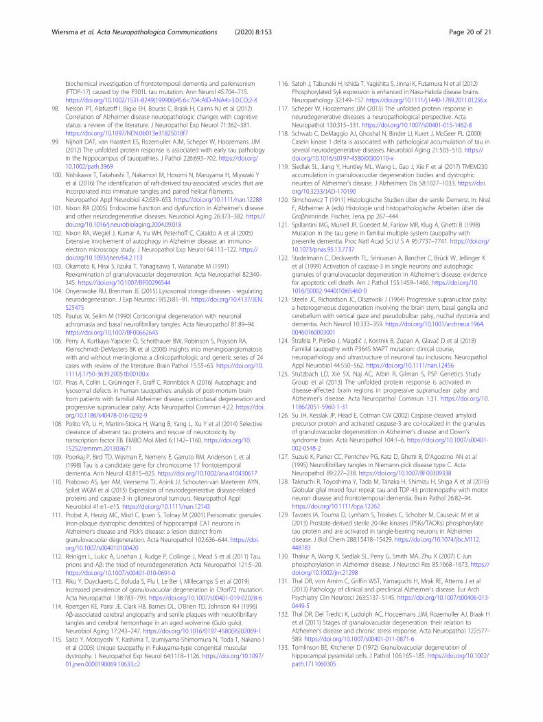
biochemical investigation of frontotemporal dementia and parkinsonism(FTDP-17) caused by the P301L tau mutation. Ann Neurol 45:704–715.https://doi.org/10.1002/1531-8249(199906)45:6<704::AID-ANA4>3.0.CO;2-X
98. Nelson PT, Alafuzoff I, Bigio EH, Bouras C, Braak H, Cairns NJ et al (2012)Correlation of Alzheimer disease neuropathologic changes with cognitivestatus: a review of the literature. J Neuropathol Exp Neurol 71:362–381.https://doi.org/10.1097/NEN.0b013e31825018f7
99. Nijholt DAT, van Haastert ES, Rozemuller AJM, Scheper W, Hoozemans JJM(2012) The unfolded protein response is associated with early tau pathologyin the hippocampus of tauopathies. J Pathol 226:693–702. https://doi.org/10.1002/path.3969
100. Nishikawa T, Takahashi T, Nakamori M, Hosomi N, Maruyama H, Miyazaki Yet al (2016) The identification of raft-derived tau-associated vesicles that areincorporated into immature tangles and paired helical filaments.Neuropathol Appl Neurobiol 42:639–653. https://doi.org/10.1111/nan.12288
101. Nixon RA (2005) Endosome function and dysfunction in Alzheimer’s diseaseand other neurodegenerative diseases. Neurobiol Aging 26:373–382. https://doi.org/10.1016/j.neurobiolaging.2004.09.018
102. Nixon RA, Wegiel J, Kumar A, Yu WH, Peterhoff C, Cataldo A et al (2005)Extensive involvement of autophagy in Alzheimer disease: an immuno-electron microscopy study. J Neuropathol Exp Neurol 64:113–122. https://doi.org/10.1093/jnen/64.2.113
103. Okamoto K, Hirai S, Iizuka T, Yanagisawa T, Watanabe M (1991)Reexamination of granulovacuolar degeneration. Acta Neuropathol 82:340–345. https://doi.org/10.1007/BF00296544
104. Onyenwoke RU, Brenman JE (2015) Lysosomal storage diseases - regulatingneurodegeneration. J Exp Neurosci 9(S2):81–91. https://doi.org/10.4137/JEN.S25475
105. Paulus W, Selim M (1990) Corticonigral degeneration with neuronalachromasia and basal neurofibrillary tangles. Acta Neuropathol 81:89–94.https://doi.org/10.1007/BF00662643
106. Perry A, Kurtkaya-Yapicier Ö, Scheithauer BW, Robinson S, Prayson RA,Kleinschmidt-DeMasters BK et al (2006) Insights into meningioangiomatosiswith and without meningioma: a clinicopathologic and genetic series of 24cases with review of the literature. Brain Pathol 15:55–65. https://doi.org/10.1111/j.1750-3639.2005.tb00100.x
107. Piras A, Collin L, Grüninger F, Graff C, Rönnbäck A (2016) Autophagic andlysosomal defects in human tauopathies: analysis of post-mortem brainfrom patients with familial Alzheimer disease, corticobasal degeneration andprogressive supranuclear palsy. Acta Neuropathol Commun 4:22. https://doi.org/10.1186/s40478-016-0292-9
108. Polito VA, Li H, Martini-Stoica H, Wang B, Yang L, Xu Y et al (2014) Selectiveclearance of aberrant tau proteins and rescue of neurotoxicity bytranscription factor EB. EMBO Mol Med 6:1142–1160. https://doi.org/10.15252/emmm.201303671
109. Poorkaj P, Bird TD, Wijsman E, Nemens E, Garruto RM, Anderson L et al(1998) Tau is a candidate gene for chromosome 17 frontotemporaldementia. Ann Neurol 43:815–825. https://doi.org/10.1002/ana.410430617
110. Prabowo AS, Iyer AM, Veersema TJ, Anink JJ, Schouten-van Meeteren AYN,Spliet WGM et al (2015) Expression of neurodegenerative disease-relatedproteins and caspase-3 in glioneuronal tumours. Neuropathol ApplNeurobiol 41:e1–e15. https://doi.org/10.1111/nan.12143
111. Probst A, Herzig MC, Mistl C, Ipsen S, Tolnay M (2001) Perisomatic granules(non-plaque dystrophic dendrites) of hippocampal CA1 neurons inAlzheimer’s disease and Pick’s disease: a lesion distinct fromgranulovacuolar degeneration. Acta Neuropathol 102:636–644. https://doi.org/10.1007/s004010100420
112. Reiniger L, Lukic A, Linehan J, Rudge P, Collinge J, Mead S et al (2011) Tau,prions and Aβ: the triad of neurodegeneration. Acta Neuropathol 121:5–20.https://doi.org/10.1007/s00401-010-0691-0
113. Riku Y, Duyckaerts C, Boluda S, Plu I, Le Ber I, Millecamps S et al (2019)Increased prevalence of granulovacuolar degeneration in C9orf72 mutation.Acta Neuropathol 138:783–793. https://doi.org/10.1007/s00401-019-02028-6
114. Roertgen KE, Parisi JE, Clark HB, Barnes DL, O’Brien TD, Johnson KH (1996)Aβ-associated cerebral angiopathy and senile plaques with neurofibrillarytangles and cerebral hemorrhage in an aged wolverine (Gulo gulo).Neurobiol Aging 17:243–247. https://doi.org/10.1016/0197-4580(95)02069-1
115. Saito Y, Motoyoshi Y, Kashima T, Izumiyama-Shimomura N, Toda T, Nakano Iet al (2005) Unique tauopathy in Fukuyama-type congenital musculardystrophy. J Neuropathol Exp Neurol 64:1118–1126. https://doi.org/10.1097/01.jnen.0000190069.10633.c2
116. Satoh J, Tabunoki H, Ishida T, Yagishita S, Jinnai K, Futamura N et al (2012)Phosphorylated Syk expression is enhanced in Nasu-Hakola disease brains.Neuropathology 32:149–157. https://doi.org/10.1111/j.1440-1789.2011.01256.x
117. Scheper W, Hoozemans JJM (2015) The unfolded protein response inneurodegenerative diseases: a neuropathological perspective. ActaNeuropathol 130:315–331. https://doi.org/10.1007/s00401-015-1462-8
118. Schwab C, DeMaggio AJ, Ghoshal N, Binder LI, Kuret J, McGeer PL (2000)Casein kinase 1 delta is associated with pathological accumulation of tau inseveral neurodegenerative diseases. Neurobiol Aging 21:503–510. https://doi.org/10.1016/s0197-4580(00)00110-x
119. Siedlak SL, Jiang Y, Huntley ML, Wang L, Gao J, Xie F et al (2017) TMEM230accumulation in granulovacuolar degeneration bodies and dystrophicneurites of Alzheimer’s disease. J Alzheimers Dis 58:1027–1033. https://doi.org/10.3233/JAD-170190
120. Simchowicz T (1911) Histologische Studien über die senile Demenz. In: NisslF, Alzheimer A (eds) Histologie und histopathologische Arbeiten über dieGroβhirnrinde. Fischer, Jena, pp 267–444
121. Spillantini MG, Murrell JR, Goedert M, Farlow MR, Klug A, Ghetti B (1998)Mutation in the tau gene in familial multiple system tauopathy withpresenile dementia. Proc Natl Acad Sci U S A 95:7737–7741. https://doi.org/10.1073/pnas.95.13.7737
122. Stadelmann C, Deckwerth TL, Srinivasan A, Bancher C, Brück W, Jellinger Ket al (1999) Activation of caspase-3 in single neurons and autophagicgranules of granulovacuolar degeneration in Alzheimer’s disease: evidencefor apoptotic cell death. Am J Pathol 155:1459–1466. https://doi.org/10.1016/S0002-9440(10)65460-0
123. Steele JC, Richardson JC, Olszewski J (1964) Progressive supranuclear palsy:a heterogeneous degeneration involving the brain stem, basal ganglia andcerebellum with vertical gaze and pseudobulbar palsy, nuchal dystonia anddementia. Arch Neurol 10:333–359. https://doi.org/10.1001/archneur.1964.00460160003001
124. Štrafela P, Pleško J, Magdič J, Koritnik B, Zupan A, Glavač D et al (2018)Familial tauopathy with P364S MAPT mutation: clinical course,neuropathology and ultrastructure of neuronal tau inclusions. NeuropatholAppl Neurobiol 44:550–562. https://doi.org/10.1111/nan.12456
125. Stutzbach LD, Xie SX, Naj AC, Albin R, Gilman S, PSP Genetics StudyGroup et al (2013) The unfolded protein response is activated indisease-affected brain regions in progressive supranuclear palsy andAlzheimer’s disease. Acta Neuropathol Commun 1:31. https://doi.org/10.1186/2051-5960-1-31
126. Su JH, Kesslak JP, Head E, Cotman CW (2002) Caspase-cleaved amyloidprecursor protein and activated caspase-3 are co-localized in the granulesof granulovacuolar degeneration in Alzheimer’s disease and Down’ssyndrome brain. Acta Neuropathol 104:1–6. https://doi.org/10.1007/s00401-002-0548-2
127. Suzuki K, Parker CC, Pentchev PG, Katz D, Ghetti B, D’Agostino AN et al(1995) Neurofibrillary tangles in Niemann-pick disease type C. ActaNeuropathol 89:227–238. https://doi.org/10.1007/BF00309338
128. Takeuchi R, Toyoshima Y, Tada M, Tanaka H, Shimizu H, Shiga A et al (2016)Globular glial mixed four repeat tau and TDP-43 proteinopathy with motorneuron disease and frontotemporal dementia. Brain Pathol 26:82–94.https://doi.org/10.1111/bpa.12262
129. Tavares IA, Touma D, Lynham S, Troakes C, Schober M, Causevic M et al(2013) Prostate-derived sterile 20-like kinases (PSKs/TAOKs) phosphorylatetau protein and are activated in tangle-bearing neurons in Alzheimerdisease. J Biol Chem 288:15418–15429. https://doi.org/10.1074/jbc.M112.448183
130. Thakur A, Wang X, Siedlak SL, Perry G, Smith MA, Zhu X (2007) C-Junphosphorylation in Alzheimer disease. J Neurosci Res 85:1668–1673. https://doi.org/10.1002/jnr.21298
131. Thal DR, von Arnim C, Griffin WST, Yamaguchi H, Mrak RE, Attems J et al(2013) Pathology of clinical and preclinical Alzheimer’s disease. Eur ArchPsychiatry Clin Neurosci 263:S137–S145. https://doi.org/10.1007/s00406-013-0449-5
132. Thal DR, Del Tredici K, Ludolph AC, Hoozemans JJM, Rozemuller AJ, Braak Het al (2011) Stages of granulovacuolar degeneration: their relation toAlzheimer’s disease and chronic stress response. Acta Neuropathol 122:577–589. https://doi.org/10.1007/s00401-011-0871-6
133. Tomlinson BE, Kitchener D (1972) Granulovacuolar degeneration ofhippocampal pyramidal cells. J Pathol 106:165–185. https://doi.org/10.1002/path.1711060305
Wiersma et al. Acta Neuropathologica Communications (2020) 8:153 Page 20 of 21
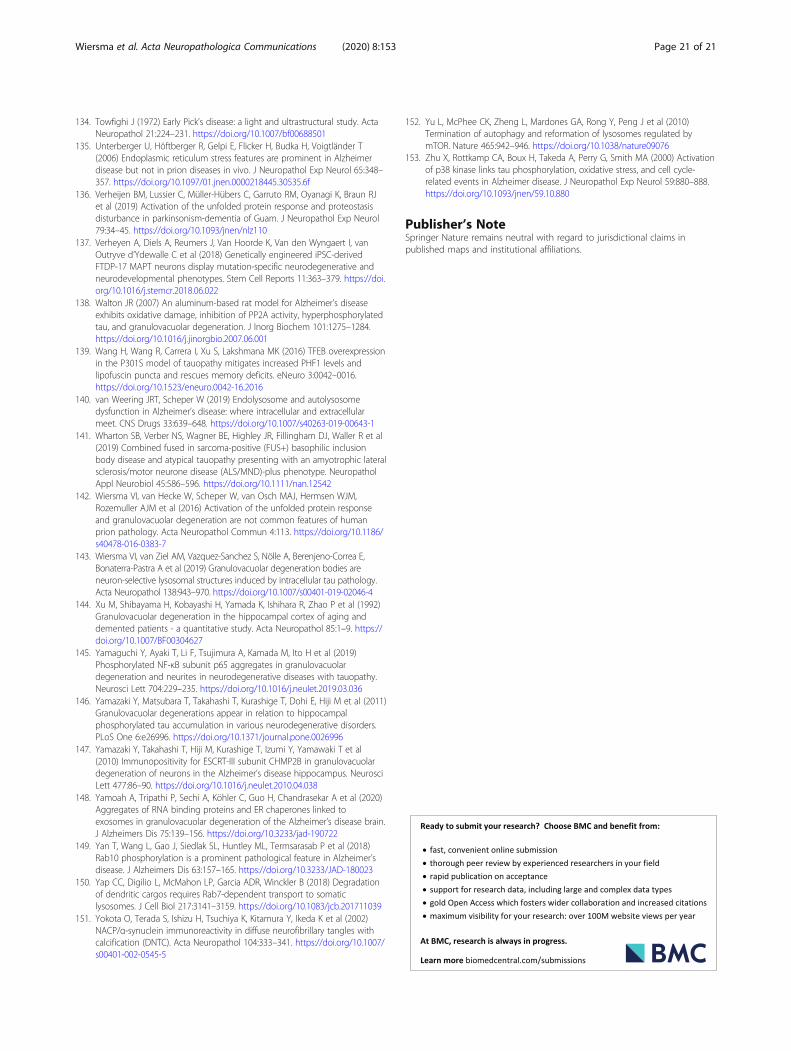
134. Towfighi J (1972) Early Pick’s disease: a light and ultrastructural study. ActaNeuropathol 21:224–231. https://doi.org/10.1007/bf00688501
135. Unterberger U, Höftberger R, Gelpi E, Flicker H, Budka H, Voigtländer T(2006) Endoplasmic reticulum stress features are prominent in Alzheimerdisease but not in prion diseases in vivo. J Neuropathol Exp Neurol 65:348–357. https://doi.org/10.1097/01.jnen.0000218445.30535.6f
136. Verheijen BM, Lussier C, Müller-Hübers C, Garruto RM, Oyanagi K, Braun RJet al (2019) Activation of the unfolded protein response and proteostasisdisturbance in parkinsonism-dementia of Guam. J Neuropathol Exp Neurol79:34–45. https://doi.org/10.1093/jnen/nlz110
137. Verheyen A, Diels A, Reumers J, Van Hoorde K, Van den Wyngaert I, vanOutryve d’Ydewalle C et al (2018) Genetically engineered iPSC-derivedFTDP-17 MAPT neurons display mutation-specific neurodegenerative andneurodevelopmental phenotypes. Stem Cell Reports 11:363–379. https://doi.org/10.1016/j.stemcr.2018.06.022
138. Walton JR (2007) An aluminum-based rat model for Alzheimer’s diseaseexhibits oxidative damage, inhibition of PP2A activity, hyperphosphorylatedtau, and granulovacuolar degeneration. J Inorg Biochem 101:1275–1284.https://doi.org/10.1016/j.jinorgbio.2007.06.001
139. Wang H, Wang R, Carrera I, Xu S, Lakshmana MK (2016) TFEB overexpressionin the P301S model of tauopathy mitigates increased PHF1 levels andlipofuscin puncta and rescues memory deficits. eNeuro 3:0042–0016.https://doi.org/10.1523/eneuro.0042-16.2016
140. van Weering JRT, Scheper W (2019) Endolysosome and autolysosomedysfunction in Alzheimer’s disease: where intracellular and extracellularmeet. CNS Drugs 33:639–648. https://doi.org/10.1007/s40263-019-00643-1
141. Wharton SB, Verber NS, Wagner BE, Highley JR, Fillingham DJ, Waller R et al(2019) Combined fused in sarcoma-positive (FUS+) basophilic inclusionbody disease and atypical tauopathy presenting with an amyotrophic lateralsclerosis/motor neurone disease (ALS/MND)-plus phenotype. NeuropatholAppl Neurobiol 45:586–596. https://doi.org/10.1111/nan.12542
142. Wiersma VI, van Hecke W, Scheper W, van Osch MAJ, Hermsen WJM,Rozemuller AJM et al (2016) Activation of the unfolded protein responseand granulovacuolar degeneration are not common features of humanprion pathology. Acta Neuropathol Commun 4:113. https://doi.org/10.1186/s40478-016-0383-7
143. Wiersma VI, van Ziel AM, Vazquez-Sanchez S, Nölle A, Berenjeno-Correa E,Bonaterra-Pastra A et al (2019) Granulovacuolar degeneration bodies areneuron-selective lysosomal structures induced by intracellular tau pathology.Acta Neuropathol 138:943–970. https://doi.org/10.1007/s00401-019-02046-4
144. Xu M, Shibayama H, Kobayashi H, Yamada K, Ishihara R, Zhao P et al (1992)Granulovacuolar degeneration in the hippocampal cortex of aging anddemented patients - a quantitative study. Acta Neuropathol 85:1–9. https://doi.org/10.1007/BF00304627
145. Yamaguchi Y, Ayaki T, Li F, Tsujimura A, Kamada M, Ito H et al (2019)Phosphorylated NF-κB subunit p65 aggregates in granulovacuolardegeneration and neurites in neurodegenerative diseases with tauopathy.Neurosci Lett 704:229–235. https://doi.org/10.1016/j.neulet.2019.03.036
146. Yamazaki Y, Matsubara T, Takahashi T, Kurashige T, Dohi E, Hiji M et al (2011)Granulovacuolar degenerations appear in relation to hippocampalphosphorylated tau accumulation in various neurodegenerative disorders.PLoS One 6:e26996. https://doi.org/10.1371/journal.pone.0026996
147. Yamazaki Y, Takahashi T, Hiji M, Kurashige T, Izumi Y, Yamawaki T et al(2010) Immunopositivity for ESCRT-III subunit CHMP2B in granulovacuolardegeneration of neurons in the Alzheimer’s disease hippocampus. NeurosciLett 477:86–90. https://doi.org/10.1016/j.neulet.2010.04.038
148. Yamoah A, Tripathi P, Sechi A, Köhler C, Guo H, Chandrasekar A et al (2020)Aggregates of RNA binding proteins and ER chaperones linked toexosomes in granulovacuolar degeneration of the Alzheimer’s disease brain.J Alzheimers Dis 75:139–156. https://doi.org/10.3233/jad-190722
149. Yan T, Wang L, Gao J, Siedlak SL, Huntley ML, Termsarasab P et al (2018)Rab10 phosphorylation is a prominent pathological feature in Alzheimer’sdisease. J Alzheimers Dis 63:157–165. https://doi.org/10.3233/JAD-180023
150. Yap CC, Digilio L, McMahon LP, Garcia ADR, Winckler B (2018) Degradationof dendritic cargos requires Rab7-dependent transport to somaticlysosomes. J Cell Biol 217:3141–3159. https://doi.org/10.1083/jcb.201711039
151. Yokota O, Terada S, Ishizu H, Tsuchiya K, Kitamura Y, Ikeda K et al (2002)NACP/α-synuclein immunoreactivity in diffuse neurofibrillary tangles withcalcification (DNTC). Acta Neuropathol 104:333–341. https://doi.org/10.1007/s00401-002-0545-5
152. Yu L, McPhee CK, Zheng L, Mardones GA, Rong Y, Peng J et al (2010)Termination of autophagy and reformation of lysosomes regulated bymTOR. Nature 465:942–946. https://doi.org/10.1038/nature09076
153. Zhu X, Rottkamp CA, Boux H, Takeda A, Perry G, Smith MA (2000) Activationof p38 kinase links tau phosphorylation, oxidative stress, and cell cycle-related events in Alzheimer disease. J Neuropathol Exp Neurol 59:880–888.https://doi.org/10.1093/jnen/59.10.880
Publisher’s NoteSpringer Nature remains neutral with regard to jurisdictional claims inpublished maps and institutional affiliations.
Wiersma et al. Acta Neuropathologica Communications (2020) 8:153 Page 21 of 21


