Embed Size (px)
Citation preview

Molecular Cell, Vol. 6, 637–648, September, 2000, Copyright 2000 by Cell Press
Structure and Function of Cdc6/Cdc18:Implications for Origin Recognitionand Checkpoint Control
in S phase. This variable timing implies that some pre-RCs persist after the G1/S transition. In addition, pre-RCs and/or active replication complexes appear to playroles in the checkpoint mechanisms that delay cell cycleprogression in the event of DNA damage or incomplete
Jinyu Liu,*‡ Cheryl L. Smith,*‡§ Deborah DeRyckere,*‖Kristen DeAngelis,* G. Steven Martin,*and James M. Berger*†
*Department of Molecular and Cell BiologyUniversity of California, Berkeley
DNA replication (Stewart and Enoch, 1996).Berkeley, California 94720The Cdc6 protein (Cdc18 in fission yeast) is critical
for pre-RC assembly. Cdc6/Cdc18 is recruited to repli-cation origins by ORC (Liang et al., 1995; Grallert and
Summary Nurse, 1996; Leatherwood et al., 1996). Once localizedat replication origins, Cdc6/Cdc18 helps to recruit and
Cdc6/Cdc18 is a conserved and essential component load Mcm factors onto DNA in a process that is believedof prereplication complexes. The 2.0 A crystal struc- to be ATP dependent (Coleman et al., 1996; Romanowskiture of an archaeal Cdc6 ortholog, in conjunction with et al., 1996; Donovan et al., 1997; Tanaka et al., 1997;a mutational analysis of the homologous Cdc18 pro- Perkins and Diffley, 1998; Weinreich et al., 1999). Dele-tein from Schizosaccharomyces pombe, reveals novel tion of Cdc6/Cdc18 leads not only to a block in DNAaspects of Cdc6/Cdc18 function. Two domains of replication but also to a loss of checkpoint control, whichCdc6 form an AAA1-type nucleotide binding fold that allows cells to enter mitosis without prior DNA replica-is observed bound to Mg•ADP. A third domain adopts tion (Kelly et al., 1993; Piatti et al., 1995). The checkpointa winged-helix fold similar to known DNA binding mod- function of Cdc6/Cdc18 is mediated by signaling path-ules. Sequence comparisons show that the winged- ways that block Cdc28/Cdc2 activation at mitosis, andhelix domain is conserved in Orc1, and mutagenesis it is dependent on the products of checkpoint controldata demonstrate that this region of Cdc6/Cdc18 is genes, such as the hus and rad genes of S. pomberequired for function in vivo. Additional mutational (Stewart and Enoch, 1996; Caspari and Carr, 1999).analyses suggest that nucleotide binding and/or hy- Archaea contain orthologs of several eukaryotic repli-drolysis by Cdc6/Cdc18 is required not only for pro- cation proteins, including Cdc6/Cdc18 and Mcms (Ed-gression through S phase, but also for maintenance gell and Doolittle, 1997; Baker and Bell, 1998; Leipe etof checkpoint control during S phase. al., 1999). The archaeal replication systems are thought
to function analogously to those of eukaryotes, but theyappear to be less complex. For example, archaea do
Introduction not contain any obvious homologs of the ORC complexother than Cdc6/Cdc18, which shares significant homol-
DNA replication is a highly coordinated biological pro- ogy with regions of certain ORC subunits, particularlycess. In all organisms, initiation of replication requires Orc1 (Bell et al., 1995; Quintana and Dutta, 1999). Thisthe sequential assembly of macromolecular complexes observation raises the question as to whether the arch-at replication origins. In eukaryotes, initiation starts with aeal Cdc6/Orc1 may play a dual role in both origin recog-a six-subunit assembly known as the origin recognition nition and Mcm recruitment.complex (ORC). ORC is an ATP-dependent protein com- The highest degree of sequence similarity betweenplex that binds specifically to replication origins (Bell Cdc6/Cdc18 proteins is encompassed within a nucleo-and Stillman, 1992; Diffley and Cocker, 1992; Klemm et tide binding domain of the Rossmann fold family. Cdc6/al., 1997; Lee and Bell, 1997) and triggers the assembly Cdc18 is a member of a large superfamily of ATPasesof prereplication complexes (pre-RCs) at the M to G1 known as ATPases associated with various cellular ac-transition. Components recruited to pre-RCs include tivities (AAA) proteins (Patel and Latterich, 1998; PerkinsCdc6, minichromosome maintenance factors (Mcms), and Diffley, 1998; Neuwald et al., 1999). Additional con-
served sequence elements of Cdc6/Cdc18 indicate thatand Cdc45 (for review, see Baker and Bell, 1998; Diffley,this protein specifically falls within the AAA1 subclass1998). Following assembly of pre-RCs, formation of ac-of proteins (Patel and Latterich, 1998; Neuwald et al.,tive replicative complexes is dependent on the activity1999), which includes Orc1, Orc4, Orc5, replication fac-of the cyclin-dependent kinase CDK1 (Cdc28 or Cdc2tor C (RFC), the bacterial g and d’ clamp-loader subunits,in budding and fission yeast) and the serine-threonineN-ethylmaleimide-sensitive factor (NSF), and the Clpkinase Cdc7/Hsk1 (Sclafani and Jackson, 1994; Masaifamily of chaperones. Nucleotide binding by AAA1 pro-et al., 1995; Diffley, 1998). The timing of this conversionteins is thought to elicit conformational changes thatis variable, with certain replication origins firing only latepromote remodeling of target proteins and protein com-plexes. While it appears that Cdc6/Cdc18 uses ATP for
† To whom correspondence should be addressed (e-mail: jmberger@ binding and loading Mcm proteins onto replication ori-uclink4.berkeley.edu) gins (Coleman et al., 1996; Romanowski et al., 1996;‡ These authors contributed equally to this work.
Donovan et al., 1997; Tanaka et al., 1997; Perkins and§ Present address: Department of Pathology, Stanford University,Diffley, 1998; Weinreich et al., 1999), it is not known howStanford, California 94305Cdc6/Cdc18 physically accomplishes this task. Like-‖ Present address: University of Colorado Health Sciences Center,
Denver, Colorado 80262 wise, the molecular basis on which Cdc6/Cdc18 binds

Molecular Cell638
Table 1. cdc18 Alleles
cdc18 Allele Altered Amino Acids Equivalent P. aerophilum Change Phenotype Permissive Temperature
cdc18–15 R172, E173 R22, E23 S/ckpt or Null* NCcdc18–16 E175, K176 Q25, L26 2C Arrest 308Ccdc18–17 E180, R184 D30, G34 2C Arrest 308Ccdc18–25 E265, E267 L115, V117 2C Arrest 308Ccdc18–37 D418, R420, K421 D239, R241, L242 2C Arrest NCcdc18–46 E521, D524 Q334, S337 2C Arrest NCcdc18–47 R536, R538, K540 E349, R351, N353 2C Arrest 308Ccdc18–51 K571, R572 E386, E387 2C Arrest NCcdc18–55 F303, E304, W305 I146, R147, L148 Null 208Ccdc18–56 N320, D323 H167, A170 Null NCcdc18–57 Y347, T348 Y193, T194 S/ckpt 258Ccdc18–58 Q479, Q480, K481 H289, E290, K291 Null NCcdc18–ts1 G193 to D P45 2C Arrest 308Ccdc18–DEAD D286, E287 D132, D133 S/ckpt NC
* Checkpoint phenotype at 308C; Null phenotype at 368C; NC, nonconditional. Wild-type alleles are listed in the supplemental data at http://www.molecule.org/cgi/content/full/6/3/637/DC1.
ORC and partakes in checkpoint control remains un- repressed; the remaining 14 nonviable mutants includedknown. seven that were temperature sensitive (Table 1).
To better understand the function of Cdc6/Cdc18, we To assess the replicative and checkpoint functionshave conducted an extensive mutational analysis of the of the nonviable mutants, cell cycle progression andS. pombe cdc181 gene. Three distinct phenotypes were nuclear morphology were assessed in mutant strainsobserved: (1) loss of both replication and checkpoint synchronized by S phase block and release, followedactivities, (2) impaired (but not abolished) replicative by incubation in thiamine to repress expression of wild-function and intact checkpoint response, or (3) impaired type cdc181 (Figures 1A and 1B). All strains regained areplicative ability with delayed loss of mitotic checkpoint 2C DNA content following the S phase block and release.control. In order to evaluate the physical basis for these A control cdc181 integrant strain maintained a 2C DNAeffects, we determined the structure of the Cdc6 or- content throughout subsequent cycles. In contrast, atholog from the archaeon Pyrobaculum aerophilum. The vector-integrant control displayed the cdc18 null pheno-protein exhibits a two-domain AAA1 module and is ob- type (Kelly et al., 1993), in which cells with 1C DNAserved complexed with Mg•ADP. Unexpectedly, the content first appeared, followed by “cut” cells with lessstructure reveals that Cdc6 contains a domain belonging than 1C DNA content and abnormal nuclear morpholo-to the winged-helix family of folds. This domain is con- gies due to cell division in the absence of DNA replica-served between Cdc6/Cdc18 and Orc1 members, and tion (Figures 1A and 1B). The mutant alleles fall intoit is structurally similar to several proteins known to three phenotypic classes.bind DNA. Mutations were identified throughout Cdc6/ The first phenotypic class comprises four mutantsCdc18, and biochemical function was deduced from the (alleles 15, 55, 56, and 58) that exhibit a loss of bothresulting phenotypes. replication and checkpoint control consistent with a null
phenotype; for one (allele 15), the null phenotype wasevident only at 368C (Figure 1; Table 1). All of the mutantResults and Discussionproteins were expressed at levels comparable to thoseof the wild-type protein (data not shown). None of theFission Yeast cdc18 Mutants Fall into Three Classesmutant alleles behaved as dominant negatives whenTo examine the functional effects of substitutionsexpressed at endogenous levels, although cdc18–56 re-throughout Cdc6/Cdc18, 61 alleles of S. pombe cdc181
duced viability to 5%–10% when overexpressed (datawere generated by site-directed mutagenesis. Of thesenot shown).alleles, 51 (alleles 1–18 and 20–52) were alanine-substi-
The second phenotypic class includes eight mutantstution mutations designed to replace two or more(alleles 16, 17, 25, 37, 46, 47, 51, and ts1) in whichcharged residues (K, R, D, or E) that cluster togethercells arrested with a 2C DNA content, an elongated cellwithin a 5 amino acid window. An additional eight ala-morphology, and a low septation index (Figure 1). More-nine-substitution mutations (alleles 53–59 and DEAD)over, the electrophoretic properties of chromosomalwere designed to disrupt residues that are highly con-DNAs from the arrested mutants indicated that DNAserved among Cdc6/Cdc18 family members. Two addi-replication intermediates are present at the 2C-arresttional alleles, ts1 (G139D) and ts2 (V298I), were alsopoint (data not shown), which in turn indicates S phasegenerated and represent the individual substitutionsarrest (Nasmyth and Nurse, 1981; Hennessy et al., 1991;found within the temperature-sensitive cdc18-K46 al-Waseem et al., 1992; DeRyckere et al., 1999). This phe-lele. Mutant cdc18 alleles under the control of thenotype is similar to that previously described for thecdc181 promoter were integrated into cdc18 cells thattemperature-sensitive K46 allele (Kelly et al., 1993) andcontained a thiamine-repressible, plasmid-borne copy ofresults from a late S phase arrest. To investigate thecdc181. Of the 61 mutants, 47 proved to be viable when
the expression of complementing wild-type cdc181 was role of checkpoint signaling in the 2C-arrest phenotype,

Structure and Function of Cdc6/Cdc18639
Figure 1. cdc18 Mutants Fall into ThreeClasses
(A) DNA content and septation indices ofcdc18 mutants. Each haploid mutant strain(see Experimental Procedures) was arrestedin S phase by addition of HU. After the equiva-lent of one cell cycle in the presence of HU,thiamine was added to repress the expres-sion of wild-type cdc181, and 30 min laterwashing the preparation in medium con-taining thiamine removed HU (arrow, 1B1/2HU). Cells were fixed before the addition ofHU (Exp, bottom), after HU arrest (HU), andat hourly intervals after removal of HU (t 5
1–7, indicated at the bottom left corner ofeach panel). DNA content was determined byflow cytometric analysis, and the septationindex is shown in the top right corner. Repre-sentative profiles are shown for the null, 2C-arrest, and S phase/checkpoint-deficientclasses of cdc18 mutants.(B) Nuclear morphology of cdc18 mutants.Cells were fixed 7 or 8 hr after the release fromHU arrest, stained with DAPI, and observedusing a fluorescence microscope. The per-centage of cells displaying the cut phenotype8 hr following release from the HU block isindicated.
we constructed cdc18 mutant and control strains car- was slightly delayed relative to that in the null mutants.These data, together with the finding that the cdc18–15rying the rad1–1 or hus1–14 allele (al-Khodairy and Carr,
1992; Rowley et al., 1992; al-Khodairy et al., 1994). When mutant has an S phase/checkpoint phenotype at 308Cand a null phenotype at 368C, suggest that S phase/expression of wild-type cdc181 was repressed in these
double-mutant strains, a portion of each population dis- checkpoint mutants may retain a level of function that isintermediate between that of null and 2C-arrest mutants.played an approximately 2C DNA content for a few
hours. Eventually, these cells accumulated a 1C and This intermediate level of function may be sufficient totransiently activate the checkpoint response but not toless-than-1C DNA content and maintained high septa-
tion indices (Figure 2A), indicative of ongoing cell divi- maintain it indefinitely.sion. The mitotic arrest in 2C-arrest mutants is thereforedependent on an intact rad-hus checkpoint-control Structure Determination of Cdc6
To determine the physical basis underlying the variouspathway (Kelly et al., 1993) and may arise from a partialloss of cdc181 function, with the persistence of unfired mutant phenotypes, we initiated structural studies of
Cdc6/Cdc18. PSI-BLAST (Altschul et al., 1997) searchespre-RCs leading to activation of the checkpoint-controlpathway. identified 17 orthologs of Cdc6/Cdc18 as well as certain
ORC subunits, particularly Orc1, in a wide range of eu-The third phenotypic class includes three mutants thatdisplayed a slow progression through S phase, accom- karyotes and archaea. Interestingly, only one Cdc6/
Cdc18/Orc1-like protein is found in each archaeal spe-panied by a transient decline in septation index (alleles57 and DEAD, and allele 15 at 308C). Unlike 2C-arrest cies, with the exception of Methanobacterium thermo-
autotrophicum, which has two such proteins.mutants, these strains ultimately underwent mitosis(Figure 1), indicative of a failure in the checkpoint re- The 45 kDa Pyrobaculum aerophilum Cdc6 protein
used for this study is 20% identical and 36% similarsponse during S phase. To further characterize thesemutants, we tested their ability to maintain the check- over 388 residues to S. pombe Cdc18. P. aerophilum
Cdc6 and other archaeal orthologs lack an N-terminalpoint function in the absence of DNA replication (Figure2B). Wild-type cells and 2C-arrest mutants exhibited extension that is present but not conserved in eukaryotic
Cdc6/Cdc18 proteins. The P. aerophilum cdc6 gene wasonly low levels of aberrant nuclear morphologies (2%and 13.5%, respectively) and indicated an intact check- cloned into and expressed using a T7 promoter-based
expression plasmid (pET28b) and the Cdc6 protein puri-point control mechanism. In contrast, S phase/check-point and null mutants entered into lethal mitoses much fied from E. coli. The protein exists as a monomer in
solution, as judged from its elution profile from a gel-more frequently (approximately 40%–60% abnormal nu-clear morphology, DNA content less than 1C), although filtration column, and exhibits ATPase activity (data not
shown).the onset of mitosis in the S phase/checkpoint mutants

Molecular Cell640
composed of residues 275–388. Domain I is a five-stranded parallel b sheet (b1-b5) sandwiched on oneside by helices a2-a3 and on the other by helices a4-a10.This domain exhibits a Rossmann-type fold commonlyfound in nucleotide binding proteins (Branden andTooze, 1991). Domain II is a small, anti-parallel four-helix bundle (a11-a14) that is linked to domain I by twoshort coil regions (residues 15–20 and 187–202). DomainIII is a five-helical bundle (a15-a19) backed on one sideby three b strands (b6-b8) and is coupled by a relativelypolar interface to domain II through helices a12 and a14on the side opposite domain I. A search of the proteinstructural database using DALI (Holm and Sander, 1996)reveals that a16-a18 and b6 -b8 of domain III belong tothe “winged-helix” (WH) family of folds.
The AAA1 Module of Cdc6 and Nucleotide BindingThe Cdc6 structure confirms earlier sequence compari-sons that suggested domains I and II together form anAAA1 module (Perkins and Diffley, 1998). The signatureGXXGXGK(T/S) (Walker-A motif) and D(D/E)XX (Walker-Bmotif) sequences reside on the b1/a3 and b3/a6 loops,respectively, of domain I. Other key regions implicatedin AAA1 function include the sensor I and II motifs ofthe nucleotide binding pocket (Guenther et al., 1997)that are found on the b4-a9 loop of domain I and a13of domain II, respectively. Overall, domains I and II ofCdc6 are each structurally similar to equivalent regionsin the other AAA1 proteins, including d9, NSF-D2, andHslU (Figure 4A) (Guenther et al., 1997; Lenzen et al.,1998; Yu et al., 1998; Bochtler et al., 2000).
Figure 2. Characterization of cdc18 MutantsTogether, domains I and II of Cdc6 generate a two-
(A) DNA content and septation indices of cdc18/rad1 double mu-lobed, cashew-shaped molecule. Cdc6 binds Mg•ADPtants. Thiamine was added to exponentially growing cultures of thein a deep cleft formed between the two domains, anddouble-mutant strains, and DNA content was determined by flowbinds both the nucleotide and metal ion using the con-cytometry at hourly intervals following addition of thiamine. DNA
content and septation indices are represented as in Figure 1A. served Walker and sensor motifs (Figure 4B). The nucle-(B) DNA content and cut phenotype of cdc18 mutants incubated otide binding mode is similar to that seen for NSF-D2with HU. Each haploid mutant strain was arrested in S phase with (Guenther et al., 1997; Lenzen et al., 1998; Yu et al.,HU. After the equivalent of one cell cycle in the presence of HU,
1998), except that in Cdc6, the base of the ADP is ori-thiamine was added to repress expression of wild-type cdc181, andented in an “anti” configuration with respect to the ri-after 30 min HU was removed by washing the preparation in medium
containing thiamine (arrow, 1B1/2HU). HU was added to the culture bose; NSF-D2 binds the “syn” nucleotide conformer.after cells first completed both S and M phase and were known to The base and ribose of ADP are contacted by groupsbe approaching the subsequent S phase (approximately 2/3 cell from the a1/a2 loop, the b5/a11 loop, and helices a11cycle). Cells were fixed before the addition of HU (Exp, bottom),
and a13. Critical contacts arise from multiple hydrogenafter the initial HU arrest (HU), and at hourly intervals upon thebinding interactions with the invariant Tyr-193 and con-second addition of HU (t 5 0–5). DNA content is represented as in
Figure 1A, and the percentage of cut cells is shown in the top right served Arg-205 residues; in particular, Arg-205 associ-corner of each panel. ates with the 29 -OH of the ribose, and this may explain
the preference of human Cdc6 for ATP over dATP (Her-big et al., 1999). The GXGK(T/S) region of the Walker-AP. aerophilum Cdc6 crystallized in the space groupmotif drapes five main-chain amide groups from Gly-54,P31, with two monomers per asymmetric unit. The struc-Thr-55, Gly-56, Lys-57, and Thr-58 around the b-phos-ture was determined using multiwavelength anomalousphate of the bound ADP; the b-phosphate is additionallydispersion (MAD) phasing, and it was refined at 2.0 Abound by a salt-bridge interaction with Lys-57. The Mg21resolution to a free R-value of 25.8% and a working Rion is coordinated by the hydroxyl of Thr-58, an oxygenfactor of 22.1% (Figure 3; Table 2). During model build-atom from the b-phosphate of ADP, and four water mole-ing, a region of electron density that could clearly becules in an octahedral configuration. Asp-132 of themodeled as Mg•ADP was observed in each monomerWalker-B region hydrogen bonds Thr-58 of the Walker-(Figure 3C).A motif as well as one of the Mg21-binding waters. Asp-The Cdc6 monomer has overall dimensions of 35 3133 of the Walker-B motif helps anchor the Mg21 coordi-50 3 90 A and is composed of three distinct domainsnation sphere by hydrogen bonding to two of the waters(Figure 3B); domain I spans residues 20–190, domain II
includes residues 1–19 and 191–275, and domain III is that bind the ion.

Structure and Function of Cdc6/Cdc18641
Figure 3. Structure of Cdc6
(A) Sequence alignment of the P. aerophilum, S. pombe, S. cerevisiae, and H. sapiens Cdc6/Cdc18 proteins. Nonconserved N-terminal regionsof the eukaryotic proteins have been removed for clarity. Vertical bars and dashes indicate the positions of insertions and gaps in the eukaryoticsequences, respectively. Magenta and dark-blue boxes on the P. aerophilum sequence denote residues that are invariant or highly conservedamong 17 different orthologs. Gray boxes denote (from N- to C-termini) the Walker-A, Walker-B, sensor-I, and sensor-II regions that arehallmarks of AAA1 proteins. Residues that lie within 4 A of the Mg•ADP moiety are marked by dots underneath the corresponding single-letter amino acid code. Residues in S. pombe cdc181 that were mutated for this study are indicated in bold and underlined: black, magenta,light-blue, and dark-blue colorings represent viable, null, 2C-arrest, and S phase/checkpoint mutations, respectively. The secondary-structuralelements of the protein are shown above the sequences by lines (coil), zigzags (a helices), and arrows (b strands) and are colored green, red,and gold to indicate domains I, II, and III, respectively. Strands and helices are labeled. See Methods for alignment parameters.(B) Stereographic ribbon representation of P. aerophilum Cdc6. Secondary-structural labels and colorings correspond to those in panel (A).The bound Mg•ADP is shown as black and magenta ball-and-stick.(C) Closeup of the nucleotide binding cleft. Experimental, density-modified MAD electron density (Fobs, aflat) for the Mg•ADP and coordinatingwaters is shown in gray. Contouring is at 1.4 s above the mean.(B) and (C) generated by RIBBONS (Carson, 1991).

Molecular Cell642
Table 2. Structure Determination Statistics
Data Collection
Native Peak Inflection Remote 1 Remote 2
Radiation A 0.9611 0.9793 0.9795 0.9840 0.9611Resolution A 20–2.0 20–2.5 20–2.5 20–2.5 20–2.5Completeness % (last shell) 98.5 (98.7) 97.2 (89.6) 97.3 (92.4) 98.2 (97.1) 97.8 (91.5)Rsym* % (last shell) 5.7 (28.1) 4.5 (9.5) 4.6 (10.5) 4.5 (12.8) 4.5 (12.5)Overall figure of merit 0.45 — — —
Structure Refinement (Native)
Resolution range (A) 20–2.0 Number reflections (overall) 97721Number nonhydrogen atoms 6482 Number reflections (test set) 8855Number protein atoms 6033 Rwork/Rfree** (%) 22.1/25.8Number ligand atoms 56 Rmsdbonds/angles 0.014 A/1.98
Number water atoms 393
* Rsym 5 SSj|Ij 2 ,I.|/SIj, where Ij is the intensity measurement for reflection j and ,I. is the mean intensity for multiply recorded reflections.** Rwork, free 5 S||Fobs| 2 |Fcalc||/|Fobs|, where the working and free R factors are calculated using the working and free reflection sets, respectively.The free reflections (8% of the total) were held aside throughout refinement.
Cdc18 Mutants and Molecular Function such as lethality or aberrant Mcm loading (Perkins andDiffley, 1998; Weinreich et al., 1999). Comparable muta-Previous mutational studies have provided important
information about the function of certain regions of tions in the human enzyme have been demonstrated todecrease or abolish ATP binding and hydrolysis in vitroCdc6/Cdc18, particularly regarding the ATP binding
pocket. For example, work on the budding yeast Cdc6 (Herbig et al., 1999). Mutations in the Walker-A and -Bboxes similarly affect the function of fission yeast Cdc18protein has shown that both the invariant lysine in the
Walker-A motif and the acidic groups of the Walker-B (see above; DeRyckere et al., 1999). As expected, ourCdc6/Cdc18 structure definitively shows that the con-region are important for Cdc6 function. Mutation of
these residues can lead to a broad array of defects, served lysine and acidic residues directly coordinate
Figure 4. Nucleotide Binding by Cdc6
A) Comparison of Cdc6 with various AAA1
proteins. NSF-D2, Cdc6, and d9 are shownin ribbon representation and colored cyan,green/red, and gold. Mg•ATP and Mg•ADPare shown bound to NSF-D2 and Cdc6, re-spectively, as black ball-and-stick. Structuralcomparisons between the AAA1 regions ofCdc6, NSF-D2, and d’ can be made by usingthe core regions with sequence similarity asan additional guide: domain I of Cdc6 has anoverall rmsd of 1.7 A and 1.8 A (spanning 100and 79 residues) to NSF-D2, and d9, respec-tively; domain II of Cdc6 superposes with theequivalent regions of NSF and d’ to 1.3 A and1.2 A rmsd over 25 and 21 amino acids. Globalrmsds spanning both domains are similar toindividual domain rmsds for NSF-D2 andCdc6 but are markedly different for cdc6 andd9 (2.0 A rmsd over 125 residues for NSF-D2compared to 2.5 A rmsd over 100 residuesfor d9).(B) Stereogram view of the nucleotide bindingregion. Secondary structure is shown as awhite coil. Residues within 4 A of boundMg•ADP are shown as gray ball-and-stickand are labeled; the one exception is His-167,which is part of the conserved sensor I motifbut lies 5 A away from the b-phosphategroup. ADP is colored as magenta ball-and-stick, and the Mg21 ion and coordinating wa-ters are shown as black and red spheres, re-spectively. Hydrogen bonds are shown asdashed lines. Backbone nitrogen atoms areshown as blue spheres and are exaggeratedin size for emphasis.(A) and (B) generated by RIBBONS (Carson,1991).

Structure and Function of Cdc6/Cdc18643
Figure 5. Positions of Mutations
Front (left) and side (right) views of the P.aerophilum Cdc6 monomer show the loca-tions of S. pombe cdc18 mutations. Null, 2C-arrest, and S phase/checkpoint phenotypesare magenta, cyan, or dark-blue spheres, re-spectively, and are labeled. Gray spheres in-dicate regions where alanine substitutionshad no apparent phenotype. Mg•ADP isshown as black and gray ball-and-stick, anddomains are colored as per Figure 3. Figuregenerated by RIBBONS (Carson, 1991).
the bound nucleotide and metal. Thus, the observed region on the surface of the Cdc6 protein (Figure 6).phenotypic effects of these mutations would be ex- Given that allele 56 shows modest dominant-negativepected to derive from defects in binding and hydrolysis behavior, the mutant protein encoded by this allele may(Perkins and Diffley, 1998; DeRyckere et al., 1999; Herbig retain the ability to interact with other components ofet al., 1999; Weinreich et al., 1999). the pre-RC but lose other functions, and this might result
In order to further correlate in vivo functions of Cdc6/ in the formation of nonfunctional pre-RCs.Cdc18 with its biochemical mechanism, we mapped our The eight 2C-arrest alleles contain mutations that areCdc18 mutagenesis data on the P. aerophilum Cdc6 scattered fairly widely about the protein and map tostructure. Sequence comparisons using all Cdc6/Cdc18 surface-exposed residues (Figure 5). Four are locatedorthologs were combined with iterative homology mod- in domain I (alleles 16, 17, 25, and ts1), with three of theeling of several different Cdc6/Cdc18 proteins to ensure four falling in or around helix a2 (alleles 16, 17, and ts1)that sequence divergence did not misguide such analy- and the fourth lying on helix a5, near the bottom of theses (see Experimental Procedures). Together, these protein (allele 25). Residing in domain II, the fifth 2C-data indicate that the effect of many of the different arrest mutant (allele 37) changes an invariant argininecdc18 alleles can be explained structurally. (Arg-420Arg-241Ala) in the sensor II region that resides near
Of the three sets of substitutions that lead to the null the bound ADP. The remaining three alleles (46, 47,phenotype, one (allele 55) maps to the hydrophobic core and 51) contain substitutions in residues that lie on theregion of domain I while another (allele 58) falls within surface of domain III and map to helices a18, a19, anda cluster of residues that tie together several secondary- strand b7, respectively. In general, the mutations associ-structural elements of domain III (Figure 5). The third ated with the 2C-arrest phenotype may indicate sur-(allele 56) substitutes residues that lie on the surface of
faces that are important for interacting with other factorsthe protein in the sensor-I region and includes the
(Figure 6). In addition, the Arg-420Arg-241Ala mutation inchange Asn-320His-167Ala (where Asn-320 and His-167the sensor II region (allele 37) may alter nucleotidecorrespond to S. pombe and P. aerophilum residues,binding or hydrolysis properties and impair catalyticrespectively) (Table 1); this residue is always polar, gen-function.erally asparagine, in Cdc6/Cdc18 orthologs. These data
The three cdc18 alleles that lead to the S phase/suggest that the null phenotype of alleles 55 and 58checkpoint phenotype show changes in highly con-probably arises from structural destabilization of Cdc18.served or invariant amino acids that are clusteredIn contrast, there are several possibilities for the effectaround the nucleotide binding environment (Figure 5).of allele 56. NSF-D2 contains a polar residue (serine) inOne of these (allele DEAD) alters the acidic groups ofthe position analogous to Asn-320His-167 that hydrogenthe Walker-B D(D/E)XX motif. The second (allele 57) re-bonds a water molecule associated with the g-phos-moves an invariant tyrosine (Tyr-347Tyr-193Ala) in the b5/phate of bound nucleotide (Lenzen et al., 1998; Yu eta11 loop that forms one of the domain I/II hinges. Theal., 1998). Thus, removal of Asn-320His-167 might impairthird mutant allele (15) falls in the other domain I/II hingenucleotide hydrolysis. Alternatively, the changes pres-
ent in allele 56 occur within a relatively well-conserved on the a1/a2 loop and changes an invariant arginine
Molecular Cell644
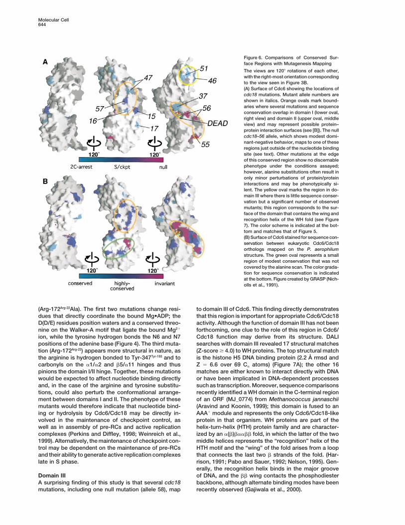
Figure 6. Comparisons of Conserved Sur-face Regions with Mutagenesis Mapping
The views are 1208 rotations of each other,with the right-most orientation correspondingto the view seen in Figure 3B.(A) Surface of Cdc6 showing the locations ofcdc18 mutations. Mutant allele numbers areshown in italics. Orange ovals mark bound-aries where several mutations and sequenceconservation overlap in domain I (lower oval,right view) and domain II (upper oval, middleview) and may represent possible protein–protein interaction surfaces (see [B]). The nullcdc18–56 allele, which shows modest domi-nant-negative behavior, maps to one of theseregions just outside of the nucleotide bindingsite (see text). Other mutations at the edgeof this conserved region show no discernablephenotype under the conditions assayed;however, alanine substitutions often result inonly minor perturbations of protein/proteininteractions and may be phenotypically si-lent. The yellow oval marks the region in do-main III where there is little sequence conser-vation but a significant number of observedmutants; this region corresponds to the sur-face of the domain that contains the wing andrecognition helix of the WH fold (see Figure7). The color scheme is indicated at the bot-tom and matches that of Figure 5.(B) Surface of Cdc6 stained for sequence con-servation between eukaryotic Cdc6/Cdc18orthologs mapped on the P. aerophilumstructure. The green oval represents a smallregion of modest conservation that was notcovered by the alanine scan. The color grada-tion for sequence conservation is indicatedat the bottom. Figure created by GRASP (Nich-olls et al., 1991).
(Arg-172Arg-22Ala). The first two mutations change resi- to domain III of Cdc6. This finding directly demonstratesthat this region is important for appropriate Cdc6/Cdc18dues that directly coordinate the bound Mg•ADP; the
D(D/E) residues position waters and a conserved threo- activity. Although the function of domain III has not beenforthcoming, one clue to the role of this region in Cdc6/nine on the Walker-A motif that ligate the bound Mg21
ion, while the tyrosine hydrogen bonds the N6 and N7 Cdc18 function may derive from its structure. DALIsearches with domain III revealed 17 structural matchespositions of the adenine base (Figure 4). The third muta-
tion (Arg-172Arg-22) appears more structural in nature, as (Z-score $ 4.0) to WH proteins. The top structural matchis the histone H5 DNA binding protein (2.2 A rmsd andthe arginine is hydrogen bonded to Tyr-347Tyr-193 and to
carbonyls on the a1/a2 and b5/a11 hinges and thus Z 5 6.6 over 69 Ca atoms) (Figure 7A); the other 16matches are either known to interact directly with DNApinions the domain I/II hinge. Together, these mutations
would be expected to affect nucleotide binding directly or have been implicated in DNA-dependent processessuch as transcription. Moreover, sequence comparisonsand, in the case of the arginine and tyrosine substitu-
tions, could also perturb the conformational arrange- recently identified a WH domain in the C-terminal regionof an ORF (MJ_0774) from Methanococcus jannaschiiment between domains I and II. The phenotype of these
mutants would therefore indicate that nucleotide bind- (Aravind and Koonin, 1999); this domain is fused to anAAA1 module and represents the only Cdc6/Cdc18-likeing or hydrolysis by Cdc6/Cdc18 may be directly in-
volved in the maintenance of checkpoint control, as protein in that organism. WH proteins are part of thehelix-turn-helix (HTH) protein family and are character-well as in assembly of pre-RCs and active replication
complexes (Perkins and Diffley, 1998; Weinreich et al., ized by an a(b)baabb fold, in which the latter of the twomiddle helices represents the “recognition” helix of the1999). Alternatively, the maintenance of checkpoint con-
trol may be dependent on the maintenance of pre-RCs HTH motif and the “wing” of the fold arises from a loopthat connects the last two b strands of the fold. (Har-and their ability to generate active replication complexes
late in S phase. rison, 1991; Pabo and Sauer, 1992; Nelson, 1995). Gen-erally, the recognition helix binds in the major grooveof DNA, and the bb wing contacts the phosphodiesterDomain III
A surprising finding of this study is that several cdc18 backbone, although alternate binding modes have beenrecently observed (Gajiwala et al., 2000).mutations, including one null mutation (allele 58), map

Structure and Function of Cdc6/Cdc18645
Figure 7. Cdc6 Domain III
(A) Ribbon diagram comparing the similar regions of Cdc6 domain III (right, gold) and histone H5 (left, blue). “HTH” and “W” designate thehelix-turn-helix and wing regions, respectively. Secondary-structural elements of Cdc6 correspond to those in (C).(B) One model for domain III function. Domain III (gold) is shown docked onto duplex DNA (gray stick). To generate the model, domain III wassuperposed on E2F as seen in the E2F/DNA cocrystal structure (Zheng et al., 1999). The rmsd between E2F and P. aerophilum Cdc6 domainIII is 2.4 A over 64 Ca positions. Amino acids known to be important for appropriate Cdc6 activity are shown as magenta (null mutants) orcyan (2C-arrest mutants) ball-and-stick. It is interesting to note that, much like origin sequences, the surface of this domain is not conservedamong Cdc6/Cdc18 orthologs. However, most of the observed mutations cluster on one side of the domain, and alleles 46 and 47 fall on ornear the putative DNA binding elements (see Figure 6).(C) ClustalX (Thompson et al., 1997) sequence alignment of the C-termini of Cdc6/Cdc18 and Orc1 orthologs. The secondary-structuralelements observed in Cdc6 are drawn below as cylinders (a helices), arrows (b strands), and coil (lines). The P. aerophilum cdc6 and S. pombecdc181 sequences are boxed in gray, while colors indicate regions of chemical conservation; for example, blue represents hydrophobicconservation, orange represents conservation of positively charged groups, etc.(A) and (B) generated by RIBBONS (Carson, 1991).
Possible roles for domain III might include mediating and it has recently been shown using electrophoreticmobility shift assays that Drosophila melanogaster Cdc6protein–protein interactions or regulating nucleotide
binding and hydrolysis through an as yet unknown can bind to DNA containing an amplification controlelement (D. Remus and M. Botchan, personal communi-mechanism. However, our mutagenesis data show that
amino-acid changes in the recognition helix of the HTH cation).Sequence alignments indicate that domain III, like do-(allele 46) or in or near the putative b7-b8 “wing” region
(allele 47) impair Cdc18 function, results which raise the mains I and II, is conserved across Cdc6/Cdc18 andOrc1 proteins (Figure 7C). This conservation may implypossibility that Cdc6/Cdc18 might contact DNA through
these motifs (Figure 7B). Others have also proposed that a similar functional role for domain III in both Cdc6/Cdc18 and Orc1 family members; since Orc1 is knownCdc6 may interact directly with DNA (Feng et al., 1998),

Molecular Cell646
strains, the mutant haploid strains carrying the pDAD112 plasmidto contact DNA (Lee and Bell, 1997), this region couldwere mated with either a hus1–14 leu1–32 ura4-D18 or a rad1–1be used as part of that activity in Orc1 if domain III wereleu1–32 ura4-D18 strain (al-Khodairy et al., 1994) (kindly provideda DNA binding element. It should be noted that whole-by Tamar Enoch). Haploid progeny were tested for thiamine sensitiv-
genome sequencing of numerous archaeal species has ity (to detect the mutant cdc18 allele) and for HU sensitivity (tothus far revealed no proteins related to ORC subunits detect the mutant hus or rad allele).
To isolate the cdc18-K46 allele by gap repair, the cdc181 openwith the exception of Cdc6/Cdc18, although archaea doreading frame was removed by partial digestion of the cdc181 geno-appear to contain defined replication origins (Myllykalliomic clone in pUR19 (Kelly et al., 1993) with MscI. The 8.7 kb fragmentet al., 2000). Given the particularly close relation be-was used to transform a cdc18-K46 leu1–32 ura4-D18 strain bytween Orc1 and Cdc6/Cdc18, it is tempting to speculateelectroporation, and ura1 transformants were selected. Plasmids
that archaea might utilize a Cdc6/Cdc18/Orc1 hybrid were rescued from ura1 transformants, purified, and sealed withthat acts in both origin recognition and Mcm recruit- T4 DNA ligase. Repaired plasmids were transformed into E. coli,
recovered, and identified by restriction digestion. The cdc18-K46ment. In this scenario, the winged-helix domain mightopen reading frames of two independently repaired plasmids werebe even more likely to act as a DNA-localization factor.sequenced, a process which revealed two mutations, Gly-193Aspand Val-298Ile, in the allele.
Conclusion Physiological characterization of mutant strains was carried outThe process of initiating DNA replication hinges on a using cells in exponential growth in minimal medium. Procedures
for flow cytometry, staining with DAPI, pulsed-field gel electrophore-series of proteins that specifically assemble on DNAsis of chromosomal DNAs, and immunoblotting with anti-Cdc18origins in a precisely determined manner. The Cdc6/antiserum have been previously described (Kelly et al., 1993; Nishi-Cdc18 protein is a key mediator of this process by serv-tani and Nurse, 1995; DeRyckere et al. 1999).
ing to bind ORC, helping to load Mcm replication factorsonto DNA, and coupling cell cycle signals to the replica-
Cloning, Expression, and Purification of P. aerophilum Cdc6tion machinery. This work reports data from a systematicThe P. aerophilum cdc6 encoding sequence was amplified by PCRmutational analysis of S. pombe cdc181 in combination and cloned into pET28b (Novagen). Expression of Cdc6 was carried
with the structure of a Cdc6 ortholog from the archaeon out in E. coli BL21(DE3) cells containing a rare tRNA-expressingP. aerophilum. Mutations in cdc181 generate three types plasmid, pSJS1244 (Kim et al., 1998). Cells were grown at 378C,
induced at A600 5 0.35 with 1 mM IPTG for 4 hr, harvested by centrifu-of phenotypic defect: complete loss of function, partialgation, resuspended in buffer A (50 mM Tris-HCl [pH 7.5], 1 mMfunction with intact checkpoint control, or partial func-EDTA, 1 mM EGTA, 10% glycerol, 1 mM pepstatin-A, 1 mM leupeptin,tion with loss of checkpoint control. The structure dem-1 mM PMSF, 1mM DTT), and flash-frozen dropwise in liquid nitrogen.onstrates that two domains of Cdc6 comprise an AAA1- Purification of Cdc6 was as follows: cells were lysed by sonication
type module capable of binding nucleotide, while a third and centrifuged. Clarified extract was heated to 658C for 15 min,domain belongs to the WH family of folds. This work chilled on ice for 15 min, then centrifuged. Cdc6 was purified using
anion-exchange chromatography (POROS HS-50, Perseptive Bio-provides a high-resolution view of a replication initiatorsystems), followed by gel filtration (Sephacryl S-300, Pharmacia).protein of the Cdc6/Orc1 class and has allowed us toPurified protein was concentrated, dialyzed against 10 mM Tris-HClmap the cdc18 mutations onto the Cdc6 structure and(pH 8.5), 150 mM NaCl and adjusted to 10 mg/ml final concentration.to thus identify regions of the protein that are critical Selenomethionine-substituted Cdc6 was prepared as the native pro-
for function. tein except that 10 mM DTT was used throughout the purification.Two surprising results were manifest from this work. Incorporation of selenium was verified by mass spectrometry.
One is that the ability of Cdc6/Cdc18 to bind and/orhydrolyze nucleotide may directly affect progression Crystallization and Data Collectionthrough S phase and the checkpoint control that regu- Cdc6 was crystallized by hanging-drop vapor diffusion. Protein (1.0lates entry into mitosis. This is consistent with the ml) was mixed with well solution (1.0 ml) containing 6% MPD, 40 mM
magnesium acetate, 20 mM Tris-HCl (pH 8.5) and equilibrating overhypotheses that the checkpoint function of Cdc6/Cdc181 ml of well solution. Crystals typically grew to approximately 0.3is related to its ability to assemble pre-RCs and thatmm3 over 2–3 days. The reproducibility of these crystals was sub-the nucleotide-dependent function of Cdc6/Cdc18 isstantially improved by the use of microseeding. For data collection,
required for the assembly and/or maintenance of pre- crystals were transferred to harvesting buffer (well solution 1 25%RCs that fire in mid- or late-S phase. The other surprising glycerol) for 30 min and flash-frozen in liquid nitrogen. Data werefinding is that Cdc6/Cdc18 and its close relative, Orc1, collected at the Advanced Light Source (ALS) Beamline 5.0.2 and
processed using DENZO and SCALEPACK (Otwinowski and Minor,contain a winged helix–type fold that our mutagenesis1997). The crystals belong to the space group P31 with unit celldata show is critical for appropriate Cdc6/Cdc18 func-dimensions a 5 b 5 132.02 A, c 5 82.20 A and a solvent contenttion in vivo. Future studies will include assessing theof approximately 70%. There are two Cdc6/Cdc18 monomers per
biological significance of the winged-helix domain, as asymmetric unit.well as correlating the phenotypic effects of amino acidsubstitutions in Cdc6/Cdc18 with their biochemical ef-
Model Building and Refinementfects on nucleotide binding and hydrolysis, Cdc6/Mcm The positions of all six selenium sites (excluding the N-terminalinteractions, ORC binding, and checkpoint control. methionines) were identified from MAD data using SOLVE (Terwil-
liger and Berendzen, 1999). Initial phases were greatly improved bydensity modification with NCS averaging (DM [Cowtan, 1994]). ForExperimental Procedureseach monomer, 379 of the 389 amino acids, a single ADP molecule,and a Mg21 were built into the resultant electron density maps usingFission Yeast Strains and Methods
Strains were constructed using standard genetic techniques (Mor- O (Jones and Kjeldgaard, 1997) and the six selenium sites as guides.No density was visible for seven amino acids in the C-terminal loopeno et al., 1991), and cdc18 mutant strains were derived as pre-
viously described (DeRyckere et al., 1999). Each mutant strain had (residues 355–361) or the N-terminal methionine. Refinement of theinitial model was performed first using the selenomethionine datathe genotype Dcdc18::ura41 leu11::cdc18-X ade6-M210 or -M216
ura4-D18 his3-D1 (pDAD112). To construct cdc18 double-mutant with both phase- and NCS-restraints. Later rounds of refinement

Structure and Function of Cdc6/Cdc18647
used native data and only NCS restraints. All refinement was per- eukaryotic origins of DNA replication by a multiprotein complex.Nature 357, 128–134.formed using REFMAC/ARP (Lamzin and Wilson, 1993; Murshudov
et al., 1997), except for 8% of the data during all stages that was Bell, S.P., Mitchell, J., Leber, J., Kobayashi, R., and Stillman, B.used to calculate Rfree. During the course of refinement, the model (1995). The multidomain structure of Orc1p reveals similarity to regu-was subjected to four rounds of manual rebuilding using sigmaA- lators of DNA replication and transcriptional silencing. Cell 83,weighted 2Fo-Fc and Fo-Fc maps and Ramachandran analyses as 563–568.guides. PROCHECK (Laskowski et al., 1993) analysis shows that
Bochtler, M., Hartmann, C., Song, H., Bourenkov, G., Bartunik, H.,the Cdc6 model has good overall geometry, with no residues fallingand Huber, R. (2000). The structures of HslU and the ATP-dependentin either generously allowed or disallowed regions of Ramachandranprotease HslU-HslV. Nature 403, 800–805.space. The observation that contacts between the two monomersBranden, C., and Tooze, J. (1991). Introduction to Protein Structure.are primarily polar and contain water molecules suggests that these(New York: Garland Publishing).interactions arise from crystal packing.Carson, M. (1991). Ribbons 2.0. J. Appl. Crystallogr. 24, 958–961.
Alignments and Homology Modeling Caspari, T., and Carr, A.M. (1999). DNA structure checkpoint path-Initial alignments and pairwise similarities of Cdc6/Cdc18-related ways in Schizosaccharomyces pombe. Biochimie 81, 173–181.genes were calculated using PSI-BLAST (Altschul et al., 1997). Sev-
Coleman, T.R., Carpenter, P.B., and Dunphy, W.G. (1996). The Xeno-enteen Cdc6/Cdc18 and nine Orc1 orthologs were uncovered bypus Cdc6 protein is essential for the initiation of a single round ofdatabase searching; searches using only residues found in domainDNA replication in cell-free extracts. Cell 87, 53–63.III from P. aerophilum Cdc6/Cdc18 (Ala-273-Arg-389) return onlyCowtan, K. (1994). ‘DM’: an automated procedure for phase im-Cdc6, Cdc18, and Orc1 homologs with significant E-values (averageprovement by density modification. Joint CCP4 and ESF-EACBMsimilarity approximately 34%). Multiple family alignments were per-Newsletter on Protein Crystallography 31, 34–38.formed using CLUSTAL-X (Thompson et al., 1997) with a Gonnet-
250 matrix. Alignments among P. aerophilum, S. pombe, D. melano- DeRyckere, D., Smith, C.L., and Martin, G.S. (1999). The role ofgaster, and S. cerevisiae Cdc6/Cdc18 were further refined as fol- nucleotide binding and hydrolysis in the function of the fission yeastlows: homology modeling between the archaeal and eukaryotic pro- cdc18(1) gene product. Genetics 151, 1445–1457.teins was performed using MODELLER (Sanchez and Sali, 1997) Diffley, J.F. (1998). Replication control: choreographing replicationand initial CLUSTAL-X alignments. The models were manually in- origins. Curr. Biol. 8, R771–R773.spected for inappropriately aligned regions as evidenced, for exam-
Diffley, J.F., and Cocker, J.H. (1992). Protein-DNA interactions at aple, by buried polar or charged residues. Poorly modeled regionsyeast replication origin. Nature 357, 169–172.were realigned by eye and then remodeled using MODELLER. ForDonovan, S., Harwood, J., Drury, L.S., and Diffley, J.F. (1997).the S. cerevisiae sequence, only three variable connector regionsCdc6p-dependent loading of Mcm proteins onto pre-replicativeneeded to be manually adjusted, while for the S. pombe and D.chromatin in budding yeast. Proc. Natl. Acad. Sci. USA 94, 5611–melanogaster sequences the original CLUSTAL-X output produced5616.a satisfactory model. As a final check, the modeled eukaryotic struc-
tures were compared with one another to ensure consistent thread- Edgell, D.R., and Doolittle, W.F. (1997). Archaea and the origin(s) ofing results. DNA replication proteins. Cell 89, 995–998.
Feng, L., Wang, B., and Jong, A. (1998). Saccharomyces cerevisiaeAcknowledgments Cdc6 stimulates Abf1 DNA binding activity. J. Biol. Chem. 273, 1298–
1302.The authors would like to thank M. Botchan, N. Cozzarelli, S. Gradia,
Gajiwala, K.S., Chen, H., Cornille, F., Roques, B.P., Reith, W., Mach,and J. L. Keck for critical reading of the manuscript. We thank K.B., and Burley, S.K. (2000). Structure of the winged-helix proteinGould and T. Enoch for strains; P. Nurse for aCdc18 antibodies;hRFX1 reveals a new mode of DNA binding. Nature 403, 916–921.D. King (R. Tjian Lab) for assistance with mass spectrometry; S.Grallert, B., and Nurse, P. (1996). The ORC1 homolog orp1 in fissionFitzgibbons and J. Miller for providing P. aerophilum sequence; andyeast plays a key role in regulating onset of S-phase. Genes Dev.the staff at Beamline 5.0.2 of the Advanced Light Source for help10, 2644–2654.with data collection. This work has been supported by the American
Cancer Society grant CB107 and National Institutes of Health grant Guenther, B., Onrust, R., Sali, A., O’Donnell, M., and Kuriyan, J.CA17542 (G. S. M.), by the G. Harold and Leila Y. Mathers Charitable (1997). Crystal structure of the d9 subunit of the clamp-loader com-Foundation (J. M. B.), and by the facilities of the University of Califor- plex of E. coli DNA polymerase III. Cell 91, 335–345.nia Cancer Research Laboratory. C. L. S. was supported by National
Harrison, S.C. (1991). A Structural taxonomy of DNA-binding do-Institutes of Health training grant GM07232 and D. D. by a predoc-
mains. Nature 353, 715–719.toral fellowship from the Howard Hughes Medical Institute.
Hennessy, K.M., Lee, A., Chen, E., and Botstein, D. (1991). A groupof interacting yeast DNA replication genes. Genes & Dev. 5, 958–969.ReferencesHerbig, U., Marlar, C.A., and Fanning, E. (1999). The Cdc6 nucleo-tide-binding site regulates its activity in DNA replication in humanal-Khodairy, F., and Carr, A.M. (1992). DNA repair mutants definingcells. Mol. Biol. Cell 10, 2631–2645.G2 checkpoint pathways in Schizosaccharomyces pombe. EMBO
J. 11, 1343–1350. Holm, L.L., and Sander, C. (1996). Mapping the protein universe.Science 273, 595–602.al-Khodairy, F., Fotou, E., Sheldrick, K.S., Griffiths, D.J., Lehmann,
A.R., and Carr, A.M. (1994). Identification and characterization of Jones, T.A., Zou, J.Y., Cowan, S.W., and Kjeldgaard, M. (1991).new elements involved in checkpoint and feedback controls in fis- Improved methods for building protein models in electron densitysion yeast. Mol. Biol. Cell 5, 147–160. maps and the location of errors in these models. Acta Crystallogr.
A. 47, 110–119.Altschul, S.F., Madden, T.L., Schaffer, A.A., Zhang, J., Zhang, Z.,Miller, W., and Lipman, D.J. (1997). Gapped BLAST and PSI-BLAST: Kelly, T.J., Martin, G.S., Forsburg, S.L., Stephen, R.J., Russo, A.,a new generation of protein database search programs. Nucleic and Nurse, P. (1993). The fission yeast cdc181 gene product couplesAcids Res. 25, 3389–3402. S-phase to START and mitosis. Cell 74, 371–382.Aravind, L., and Koonin, E.V. (1999). DNA-binding proteins and evo- Kim, R., Sandler, S., Gorldman, S., Yokota, H., Clark, A., and Kim,lution of transcription regulation in the archaea. Nucleic Acids Res. S.-H. (1998). Overexpression of archaeal proteins in Escherichia coli.27, 4658–4670. Biotech. Let. 20, 207–210.Baker, T.A., and Bell, S.P. (1998). Polymerases and the replisome: Klemm, R.D., Austin, R.J., and Bell, S.P. (1997). Coordinate bindingmachines within machines. Cell 92, 295–305. of ATP and origin DNA regulates the ATPase activity of the origin
recognition complex. Cell 88, 493–502.Bell, S.P., and Stillman, B. (1992). ATP-dependent recognition of

Molecular Cell648
Lamzin, V.S., and Wilson, K.S. (1993). Automated refinement of pro- Sanchez, R., and Sali, A. (1997). Evaluation of comparative proteinstructure modeling by MODELLER-3. Proteins Suppl 1, 50–58.tein models. Acta Crystallogr. D 49, 129–147.
Sclafani, R.A., and Jackson, A.L. (1994). Cdc7 protein kinase forLaskowski, R., MacArthur, M., Moss, D., and Thornton, J. (1993).DNA metabolism comes of age. Mol. Microbiol. 11, 805–810.PROCHECK: a program to check the stereochemical quality of pro-
tein structures. J. Appl. Crystallogr. 26, 283–291. Stewart, E., and Enoch, T. (1996). S-phase and DNA-damage check-points: a tale of two yeasts. Curr. Op. Cell. Biol. 8, 781–787.Leatherwood, J., Lopez-Girona, A., and Russell, P. (1996). Interac-
tion of Cdc2 and Cdc18 with a fission yeast ORC2-like protein. Tanaka, T., Knapp, D., and Nasmyth, K. (1997). Loading of an McmNature 379, 360–363. protein onto DNA replication origins is regulated by Cdc6p and
CDKs. Cell 90, 649–660.Lee, D.G., and Bell, S.P. (1997). Architecture of the yeast originTerwilliger, T., and Berendzen, J. (1999). Automated structure solu-recognition complex bound to origins of DNA replication. Mol. Celltion for MIR and MAD. Acta Crystallogr. D. 55, 849–861.Biol. 17, 7159–7168.
Thompson, J.D., Gibson, T.J., Plewniak, F., Jeanmougin, F., andLeipe, D., Aravind, L., and Koonin, E. (1999). Did DNA replicationHiggins, D.G. (1997). The CLUSTAL_X windows interface: flexibleevolve twice independently? Nucleic Acids Res. 27, 3389–3401.strategies for multiple sequence alignment aided by quality analysisLenzen, C.U., Steinmann, D., Whiteheart, S.W., and Weis, W.I. (1998).tools. Nucleic Acids Res. 25, 4876–4882.Crystal structure of the hexamerization domain of N-ethylmaleimide-Waseem, N.H., Labib, K., Nurse, P., and Lane, D.P. (1992). Isolationsensitive fusion protein. Cell 94, 525–536.and analysis of the fission yeast gene encoding polymerase d acces-
Liang, C., Weinreich, M., and Stillman, B. (1995). ORC and Cdc6psory protein PCNA. EMBO J. 11, 5111–5120.
interact and determine the frequency of initiation of DNA replicationWeinreich, M., Liang, C., and Stillman, B. (1999). The Cdc6p nucleo-in the genome. Cell 81, 667–676.tide-binding motif is required for loading mcm proteins onto chroma-
Masai, H., Miyake, T., and Arai, K. (1995). hsk11, a Schizosaccharo- tin. Proc. Natl. Acad. Sci. USA 96, 441–446.myces pombe gene related to Saccharomyces cerevisiae CDC7, is
Yu, R.C., Hanson, P.I., Jahn, R., and Brunger, A.T. (1998). Structurerequired for chromosomal replication. EMBO J. 14, 3094–3104.of the ATP-dependent oligomerization domain of N-ethylmaleimide
Moreno, S., Klar, A., and Nurse, P. (1991). Molecular genetic analysis sensitive factor complexed with ATP. Nat. Struct. Biol. 5, 803–811.of fission yeast Schizosaccharomyces pombe. In Guide to Yeast
Zheng, N., Fraenkel, E., Pabo, C.O., and Pavletich, N.P. (1999). Struc-Genetics and Molecular Biology, C. Guthrie and G.R. fink, eds. (San
tural basis of DNA recognition by the heterodimeric cell cycle tran-Diego: Academic Press), pp. 795–823.
scription factor E2F-DP. Genes Dev. 13, 666–674.Murshudov, G.N., Vagin, A.A., and Dodson, E.J. (1997). Refinementof macromolecular structures by the maximum-likelihood method. Protein Data Bank CoordinatesActa Crystallogr. D. 53, 240–255.
Myllykallio, H., Lopez, P., Lopez-Garcıa, P., Heilig, R., Saurin, W., Coordinates have been deposited with the Protein Data Bank atZivanovic, Y., Philippe, H., and Forterre, P. (2000). Bacterial mode the Research Collaboratory for Structural Bioinformatics databaseof replication with eukaryotic-like machinery in a hyperthermophilic under accession number 1FNN.archaeon. Science 288, 2212–2215.
Nasmyth, K., and Nurse, P. (1981). Cell division cycle mutants alteredin DNA replication and mitosis in the fission yeast Schizosaccharo-myces pombe. Mol. Gen. Genet. 182, 119–124.
Nelson, H.C.M. (1995). Structure and function of DNA-binding pro-teins. Curr. Opin. Genet. Dev. 5, 180–189.
Neuwald, A.F., Aravind, L., Spouge, J.L., and Koonin, E.V. (1999).AAA1: a class of chaperone-like ATPases associated with the as-sembly, operation, and disassembly of protein complexes. GenomeRes. 9, 27–43.
Otwinowski, Z., and Minor, W. (1997). Processing of X-ray diffractiondata collected in oscillation mode. In Methods Enzymology: Macro-molecular Crystallography, C.W.J. Carter and R.M. Sweet, eds. (Bos-ton: Academic Press), pp. 472–494.
Pabo, C., and Sauer, R.T. (1992). Transcription factors: structuralfamilies and principles of DNA recognition. Annu. Rev. Biochem. 61,1053–1095.
Patel, S., and Latterich, M. (1998). The AAA team: related ATPaseswith diverse functions. Trends Cell Biol. 8, 65–71.
Perkins, G., and Diffley, J.F. (1998). Nucleotide-dependent prerepli-cative complex assembly by Cdc6p, a homolog of eukaryotic andprokaryotic clamp-loaders. Mol. Cell 2, 23–32.
Piatti, S., Lengauer, C., and Nasmyth, K. (1995). Cdc6 is an unstableprotein whose de novo synthesis in G1 is important for the onsetof S phase and for preventing a ‘reductional’ anaphase in the bud-ding yeast Saccharomyces cerevisiae. EMBO J. 14, 3788–3799.
Quintana, D.G., and Dutta, A. (1999). The metazoan origin recogni-tion complex. Frontiers Biosci. 4, D805–D815.
Romanowski, P., Madine, M.A., and Laskey, R.A. (1996). XMCM7,a novel member of the Xenopus MCM family, interacts with XMCM3and colocalizes with it throughout replication. Proc. Natl. Acad. Sci.USA 93, 10189–10194.
Rowley, R., Subramani, S., and Young, P.G. (1992). Checkpoint con-trols in Schizosaccharomyces pombe: rad1. EMBO J. 11, 1335–1342.


