Embed Size (px)
Citation preview
Unveiling the degradative route of the V247Ma-sarcoglycan mutant responsible for LGMD-2D
Elisa Bianchini1, Marina Fanin2, Kamel Mamchaoui3, Romeo Betto4 and Dorianna Sandona1,∗
1Department of Biomedical Sciences and 2Department of Neurosciences, University of Padova, Padova 35131, Italy,3Institut de Myologie, UPMC UM76, INSERM U974, CNRS UMR 7215, Paris 6, France and 4Institute of Neuroscience,
Consiglio Nazionale delle Ricerche, Padova 35131, Italy
Received December 11, 2013; Revised February 17, 2014; Accepted February 19, 2014
Many membrane and secretory proteins that fail to pass quality control in the endoplasmic reticulum (ER) aredislocated into the cytosol and degraded by the proteasome. In applying rigid rules, however, quality controlsometimes discharges proteins that, even though defective, retain their function. The unnecessary removalof such proteins represents the pathogenetic hallmark of diverse genetic diseases, in the case of DF508mutant of cystic fibrosis transmembrane conductance regulator being probably the best known example.Recently, the inappropriate proteasomal degradation of skeletal muscle sarcoglycans (a, b, g and d) with mis-sense mutation has been proposed to be at the bases of mild-to-severe forms of limb girdle muscular dystrophy(LGMD) known as type 2D, 2E, 2C and 2F, respectively. The quality control pathway responsible for sarcoglycanmutant disposal, however, is so far unexplored. Here we reveal key components of the degradative route ofV247M a-sarcoglycan mutant, the second most frequently reported mutation in LGMD-2D. The disclosure ofthe pathway, which is led by the E3 ligases HRD1 and RFP2, permits to identify new potential druggable targetsof a disease for which no effective therapy is at present available. Notably, we show that the pharmacologicalinhibition of HRD1 activity rescues the expression of V247-a-sarcoglycan both in a heterologous cell modeland in myotubes derived from a LGMD-2D patient carrying the L31P/V247M mutations. This represents thefirst evidence that the activity of E3 ligases, the enzymes in charge of mutant fate, can be eligible for drug inter-ventions to treat sarcoglycanopathy.
INTRODUCTION
Up to one-third of newly synthesized proteins mature into theendoplasmic reticulum (ER). Continuous monitoring of ERfunctional status is essential for cellular homeostasis. In fact,occasionally, proteins fail to fold properly because of tran-scriptional and translational errors or the imbalanced produc-tion of complex subunits. In addition, gene mutations mightalso lead to the formation of misfolded/unfolded polypeptides.To avoid their potentially harmful accumulation, such aberrantproteins are (i) recognized by the quality control system,(ii) transported to ER extraction sites, (iii) polyubiquitinatedby ER-associated E3 ligases and (iv) retrotranslocated intothe cytoplasm where (v) degradation by the 26S proteasomeoccurs (1,2). These sequential processes, collectively called
ER-associated degradation (ERAD), are carried out by differ-ent specialized pathways depending on the position in theprotein of structural defects. In fact, defects exposed in ERluminal, transmembrane or cytosolic domains, identify mis-folded polypeptides as ERAD-L, ERAD-M or ERAD-C sub-strates (3). A number of ER luminal chaperones and lectinsinitially recognize and deliver ERAD substrates to membraneexport sites (4,5). Here, a multi-protein complex, led byER-associated E3 ubiquitin ligases, such as HRD1, gp78,TEB4 or RMA1, marks the misfolded protein with ubiquitin(6,7), an event also favoring the dislocation into the cytosol,where the 26S proteasome resides. The polyubiquitin chain,in fact, offers a handle to p97, an AAA-ATPase that eventuallyprovides the driving force to eradicate ERAD substrates fromthe membrane (8–10).
∗To whom correspondence should be addressed at: Department of Biomedical Sciences, University of Padova, Via U. Bassi 58/B, 35131 Padova, Italy.Tel: +39 0498276028; Fax: +39 0498276040; Email: [email protected]
# The Author 2014. Published by Oxford University Press.This is an Open Access article distributed under the terms of the Creative Commons Attribution Non-Commercial License (http://creativecommons.org/licenses/by-nc/3.0/), which permits non-commercial re-use, distribution, and reproduction in any medium, provided the original work is properly cited.For commercial re-use, please contact [email protected]
Human Molecular Genetics, 2014 1–13doi:10.1093/hmg/ddu088
HMG Advance Access published March 9, 2014 at U
niversita Degli Studi D
i Padova on March 24, 2014
http://hmg.oxfordjournals.org/
Dow
nloaded from

We have recently shown that several mutants ofa-sarcoglycanare eliminated by the ubiquitin–proteasome system (11).a-Sarcoglycan, together with b-, g- and d-sarcoglycan, forms anessential tetrameric complex of striated muscles (12). Sarcogly-cans are co-translationally translocated in the ER, where theymature and assemble to be eventually targeted to the cell mem-brane. Their correct assembly is crucial for a stable localizationinto the plasma membrane, alongside the dystrophin associatedprotein complex (13). Mutations in any one of the sarcoglycanscompromise complex stability, in fact, loss of the defectiveprotein is accompanied by the reduction or absence of the othersubunits. This leads to the development of mild-to-severe, world-wide rare, muscular dystrophies, collectively named sarcoglyca-nopathies and identified as LGMD-2C-F, according to thesarcoglycan involved (14). They are characterized by a progres-sive limb-girdle muscle weakness and wasting, with frequent re-spiratory insufficiency and in some cases cardiomyopathy (15).Sarcoglycanopathy clinical phenotype is very heterogeneous forage of onset, rate of progression and severity, often correlated tothe level of residual sarcoglycans. Most of the LGMD-2D cases,duetodefectsofthea-sarcoglycancodinggene,areassociatedwithmissense mutations that could lead to a misfolded protein thatbecomes an ERAD substrate (11,16,17). Loss or strong reductionof a-sarcoglycan usually causes the absence or reduction of theother subunits, thus depriving muscle fibers of a critical complex(18) and identifying ERAD action as the key pathogenetic mech-anism of LGMD-2D (12,16). Even though many disease-causingmissense mutations do not have functional consequences, becauseof the quality control rigid rules, these mutants are prematurelydegraded. However, treatments interfering with either ERAD orproteasomeattenuated the degradationof misfolded but ‘function-al’ sarcoglycans and allowed a significant fraction of the survivingprotein to reach the cell surface (11,16,17).a-Sarcoglycan is expected to be substrate of the ERAD-LM
pathway (19). In fact a-sarcoglycan is a type I, membrane-tethered protein with most disease-causing mutations locatedin the N-terminal domain (12), i.e. the region exposed to theER lumen during maturation. Yet, none of the ERAD compo-nents responsible for the removal of a-sarcoglycan mutantshave been identified.
To explore the ERAD pathway, we studied V247Ma-sarcoglycan, the second most frequently reported disease-causing mutant (Leiden Muscular Dystrophy pages at www.dmd.nl). Our results show that disposal of V247M a-sarcoglycanis operated by an ERAD-LM pathway comprising the E3 ligaseHRD1, its adaptor protein SEL1L, the E2 conjugase UBE2J1,and the dislocon components Derlin-1 and p97. An additionalE3 ligase, RFP2, also contributes to the disposal of V247Mmutant. Notably, the study demonstrates that the pharmacologicalinhibition of HRD1 ligase activity is effective in rescuing the ex-pression of a-sarcoglycan mutant both in a heterologous cellsystem and in myotubes derived from a LGMD-2D patient. Thisfinding represents a very important advancement for the develop-ment of a pharmacological therapy to treat sarcoglycanopathy, as,at present, no effective cure is available.
RESULTS
This study was carried out in HEK-293 cells, where wild-typea-sarcoglycan is transported to the cell membrane even in the
absence of the other sarcoglycan subunits (20,21). Importantly,in HEK-293 cells constitutively expressing the wild-type formsof b-, g- and d-sarcoglycan, most of transiently transfecteda-sarcoglycan mutants, like, for example, the V247M protein,is intercepted and degraded by the proteasome and only tracesof the sarcoglycan complex are recognizable at the plasma mem-brane (11), in this resembling the condition present in skeletalmuscle of LGMD-2D subjects (18). Here we utilized HEK-293cells in which only the V247M mutant of a-sarcoglycan wasstably transfected (hereafter named V247M cells). All V247Mcells express the mutant protein, although with slightly differentefficiency (Supplementary Material, Fig. S1B). Inhibition ofproteasome led to the accumulation in V247M cells of bothmature and deglycosylated forms of the mutant (SupplementaryMaterial, Fig. S1A and B). Moreover, after proteasomal block, afaint a-sarcoglycan signal became evident at the surface ofseveral not permeabilized cells (Supplementary Material,Fig. S1B). Thus, we took advantage of this simplified and effi-cient cell system to investigate the ERAD pathway of sarcogly-can mutants.
The E3 ubiquitin ligase HRD1 is required for thedisposal of V247M a-sarcoglycan mutant
In mammals, two ERAD-LM pathways have been reported sofar to process membrane-tethered proteins with luminaldefects, like a-sarcoglycan. One is built around the E3 ligaseHRD1 and the other to the ligase gp78 (19). Therefore, wefirst investigated, by RNA interference experiments, theputative involvement of these RING finger E3 ligases incontrolling the disposal of V247M a-sarcoglycan mutant. InV247M cells we demonstrated that downregulation of HRD1by a specific shRNA caused a 2.5-fold increase of V247Mprotein level, compared with cells transfected with thecontrol GFP shRNA (Supplementary Material, Fig. S2A). Bycontrast, the silencing of gp78 (Supplementary Material,Fig. S2B and C) had no effect on a-sarcoglycan mutantprotein level (Fig. 1A).
To validate these data, we utilized a mutated form of bothHRD1, called HRD1-C1A, and gp78, called RFM-gp78. Thesubstitution of critical RING domain residues (C291A inHRD1, C337S and C374S in gp78) exerts a dominant-negativeeffect on the activity of the two ligases (22,23) resulting in areduced disposal of their ERAD substrates. As expected, trans-fection of V247M cells with the inactive HRD1-C1A substan-tially increased the level of a-sarcoglycan mutant, whereas thepresence of the inactive gp78 had no effect (Fig. 1B).
Then, we examined whether HRD1 interacts with thea-sarcoglycan mutant by performing co-immunoprecipitation(IP) assays. Protein lysates from V247M cells transfected witheither wild-type HRD1, or the inactive form HRD1-C1A,were incubated with the antibody-specific for a-sarcoglycan.To prolong the short-lived association between the substrateand its E3 ligase, we utilized the HRD1-C1A inactive form.Indeed, due to RING finger motif disruption, substrate ubiquiti-nation is impaired and interaction is consequently protracted.On the other hand, in wild-type HRD1 overexpression experi-ments, a similar effect has been obtained by inhibiting prote-asome activity. The a-sarcoglycan-specific antibody was ableto immunoprecipitate the V247M mutant together with either
2 Human Molecular Genetics, 2014
at Universita D
egli Studi Di Padova on M
arch 24, 2014http://hm
g.oxfordjournals.org/D
ownloaded from

wild-type HRD1 or inactive HRD1-C1A (Fig. 1C), confirmingthe close interaction of this E3 ligase with the mutant protein.All together, these results show that the V247M mutant degrada-tive pathway is HRD1-dependent and gp78 dispensable.
The HRD1-associated proteins handling V247M mutant
In mammals, HRD1 complex is composed of several pro-teins that guarantee recognition, ubiquitination and retro-translocation into the cytosol of ERAD clients. A scaffoldingrole is played by SEL1L, an adaptor protein able to interactwith E3 and E2 enzymes, the ligase substrate (either directlyor by means of lectin-binding proteins), as well as with othercomponents of the complex (5,24).
Downregulation of SEL1L by a specific shRNA (Supplemen-tary Material, Fig. S2B) caused about a 3-fold increase ofV247M mutant level (Fig. 2A), suggesting that either thealtered stoichiometry of HRD1 complex might reduce the E3ligase activity (25), or the reduction of SEL1L might comprom-ise the nexus between the proteins recognizing the substrate, still
unknown for a-sarcoglycan, and HRD1 (5). Whatever is thecase, the result is the salvage of the V247M mutant. The involve-ment of the adaptor protein has also been confirmed by co-IPassays. In fact, as expected, from protein lysates of V247Mcells transfected with HRD1-C1A, both HRD1 and SEL1Lco-precipitated with the a-sarcoglycan mutant (Fig. 2B).
Crucial for the ubiquitination activity of the RINGfinger HRD1ligase is the association with the E2 ubiquitin conjugase. Two ERmembrane-associated E2 enzymes, UBE2J1 (aka UBC6e) andUBE2J2, and the soluble UBE2G2, have been identified to workin a concerted action with ER-associated E3 ligases (6). TheUBE2J1conjugase has been already demonstrated to work in con-junction with HRD1 in the disposal of misfolded MHC class Iheavy chain (26), whereas the UBE2G2 has been shown tosupport gp78 (27). Therefore, to verify whether UBE2J1 is partof the HRD1 complex disposing V247M mutant, we transfectedV247M cells with either wild-type UBE2J1 or its inactivemutant, HA-tagged UBE2J1-C91S (28). The inactive E2 conju-gase, because of a critical substitution in the ubiquitin conjugatingdomain, was unable to attach ubiquitin to the V247M mutant
Figure 1. E3 ubiquitin ligase HRD1 is required for the disposal of V247Ma-sarcoglycan mutant. (A) Transfection of shRNAs targeting the E3 ligases HRD1 and gp78was carried out in V247M cells. Depletion of HRD1, but not that of gp78, induced the accumulation of V247Ma-sarcoglycan mutant (a-SG-V247M) as revealed bythea-sarcoglycan-specific antibody; as loading control, blots were stained for b-actin. Densitometric analysis of at least four independent experiments is reported inthe graph. (B) Inactive variants of the two E3 ligases, HRD1-C1A and c-Myc-tagged gp78-RFM, or the empty vector (e.v.), were transfected with V247M cells. Blotswere stained with HRD1, cMyc, a-sarcoglycan and b-actin (as loading control) specific antibodies. Inactive HRD1, but not inactive gp78, induced 2-fold V247Mmutant accumulation as determined by densitometric analysis of at least four independent experiments. (C) HRD1 interacts with V247M a-sarcoglycan mutant.V247M cells were transfected with vectors encoding wild-type HRD1, inactive HRD1-C1A, or the e.v.; where indicated (+), to prevent mutant degradation prote-asome was inhibited by 10 mM MG132; proteins from naive HEK-293 cells (HEK) were used as negative control. Protein lysates were immunoprecipitated with amonoclonal antibody specific for a-sarcoglycan; IgG of the same isotype were used as negative IP control (see Supplementary Material, Fig. S3); blot was probedfor HRD1, a-sarcoglycan, and b-actin. In blot C: ng, non-glycosylated form of V247M a-sarcoglycan mutant (11); a, non-specific protein band recognized by theHRD1 antibody; asterisks, IgG whole molecule and IgG heavy chains; ∗∗P , 0.01; ∗∗∗P , 0.001.
Human Molecular Genetics, 2014 3
at Universita D
egli Studi Di Padova on M
arch 24, 2014http://hm
g.oxfordjournals.org/D
ownloaded from

resulting in a diminished degradation. On the other hand, overex-pression of wild-type UBE2J1 had no effects on the mutant level(Fig. 2C).
Then, we verified the interaction between UBE2J1 andV247M mutant, by co-IP experiments in protein lysates fromV247M cells transfected with either wild-type UBE2J1 or the in-active form UBE2J1-C91S. As a consequence of inactivation,the interaction between UBE2J1-C91S and its substrate is
prolonged, in that improving the co-IP assay. For the samepurpose, in cells transfected with either the empty vector or thewild-type form of UBE2J1, the proteasome was inhibited byMG132. Both the E2 conjugase and the mutated a-sarcoglycanwere present in the immunocomplexes recovered by thea-sarcoglycan-specific antibody (Fig. 2D). Thus, we concludedthat UBE2J1 acts in concert with HRD1 ligase in targetingV247M mutant to degradation.
Figure 2. SEL1L and UBE2J1 co-operates with HRD1 in the disposal of V247M a-sarcoglycan mutant. (A) Reduction of SEL1L, produced by the expression inV247M cells of a specific shRNA, permitted the accumulation of the a-sarcoglycan mutant; b-actin expression was probed as loading control. The graph showsthe densitometric analysis of at least four independent experiments. (B) V247M mutant interacts with both SEL1L and HRD1. Proteins solubilized from V247Mcells transfected with inactive HRD1-C1A or the e.v. were immunoprecipitated with a monoclonal antibody specific for a-sarcoglycan, IgG of the same isotypewere used as negative IP control (see Supplementary Material, Fig. S3). Blot was probed with SEL1L, HRD1, b-actin and a-sarcoglycan-specific antibodies.(C) The inactive UBE2J1 causes the increase of V247M mutant. Wild-type UBE2J1, the inactive variant HA-tagged UBE2J1-C91S or the e.v. were transfected inV247M cells. Blots were stained with either UBE2J1,b-actin anda-sarcoglycan-specific antibodies. Densitometric analysis of at least four independent experimentsis reported in the graph. (D) UBE2J1 interacts with V247M mutant. V247M cells were transfected with wild-type UBE2J1, inactive HA-tagged UBE2J1-C91S, or thee.v.; where indicated (+), to prevent mutant degradation, proteasome was inhibited by 10 mM MG132. Protein lysates were immunoprecipitated with a monoclonalantibody specific for a-sarcoglycan; IgG of the same isotype were used as negative IP control (see Supplementary Material, Fig. S3); blot was probed for b-actin,a-sarcoglycanand UBE2J1.HEK,naive HEK-293cells, used in C and D as negativecontrol; ng, non-glycosylated formof V247Ma-sarcoglycan mutant (11); asteriskin blot D, IgG heavy chains; ∗∗P , 0.01; ∗∗∗P , 0.001.
4 Human Molecular Genetics, 2014
at Universita D
egli Studi Di Padova on M
arch 24, 2014http://hm
g.oxfordjournals.org/D
ownloaded from

Dislocation from the ER of V247M mutant
To be degraded, ERAD substrates must cross the ER membraneand reach the cytosol, where proteasome resides. Diverse ERADfactors are involved in the dislocation process (included the E3ligase complexes), whereas the force to eradicate ubiquitinatedproteins from the ER is, in most cases, provided by the cytosolicAAA-ATPase p97 (8). In this context, Derlin-1 works in concertwith p97 and, because of its rhomboid architecture, by compres-sing and deforming the lipid bilayer, may help ERAD substratescrossing the membrane (29). Thus, we asked whether processingofa-sarcoglycan mutants also comprises these two ERAD com-ponents. Overexpression of wild-type GFP-tagged p97 (30) waswithout major effects on V247Ma-sarcoglycan expression, sug-gesting that the endogenous level of p97 is sufficient to fullyextract a-sarcoglycan mutant from the ER. On the contrary,the presence of the dominant-negative mutant p97-K524A, inwhich the K524A substitution in the Walker A domain inacti-vates the enzymatic activity (30), strongly reduced degradationof V247M mutant (Fig. 3A).
To verify the interaction between V247Ma-sarcoglycan, p97and, possibly, Derlin-1, we performed IP experiments by usingthe a-sarcoglycan-specific antibody in protein lysates fromV247M cells transfected with either wild-type or inactive p97.
Being the dominant-negative form of p97 unable to eradicatethe protein from the ER, a large amount of a-sarcoglycanmutant co-sedimented with the AAA-ATPase even from cellswhere the proteasome was fully active (Fig. 3B). Inhibition ofproteasome, however, by reducing mutant degradation, permit-ted the IP of V247M mutant also together with the wild-typeform of p97, indicating a close association between the two pro-teins during retrotranslocation.
In Figure 3B, endogenous Derlin-1, whose molecular weightis �29 kDa (29), has the same electrophoretic mobility ofa-sarcoglycan antibody light chains, making the interpretationof results difficult. However, it is evident that the intensity ofthe antibody light chain band is strongly increased in all immu-noprecipitated samples, but not in the negative control (HEK),where only the antibody is present. All together these datastrongly support the idea that Derlin-1 helps the extraction ofthe a-sarcoglycan mutant from the ER membrane, with thep97 ATPase providing the driving force for such operation.
V247M mutant interacts with the endogenous dislocon
To definitely demonstrate that V247Ma-sarcoglycan is the sub-strate of HRD1 complex, we performed an IP assay in 1%
Figure 3. The AAA-ATPase p97 and Derlin-1 are involved in the retro-translocation of V247Ma-sarcoglycan mutant. (A) Wild-type or a dominant-negative form ofp97 (p97 K524A), both GFP-tagged, or the e.v. was transfected in V247M cells. Blots were stained with p97, a-sarcoglycan and b-actin-specific antibodies.p97-specific antibody recognized both the ectopically expressed and endogenous forms of the protein. The dominant-negative form of p97 induced .2-foldV247M mutant accumulation, as determined by densitometric analysis of at least four independent experiments, whereas wild-type p97 had no effects. (B) p97and Derlin-1 interact with V247M mutant. V247M cells were transfected with wild-type p97, inactive p97 K524A, both GFP-tagged, or the e.v.; where indicated(+), to prevent mutant degradation, proteasome was inhibited with 10 mM MG132. Protein lysates were immunoprecipitated with a monoclonal antibody-specificfora-sarcoglycan ; IgG of the same isotype were used as negative IP control (see Supplementary Material, Fig. S3); blots were probed for p97, Derlin-1,a-sarcoglycanand b-actin. Naive HEK-293 cells (HEK) were used as negative control; ng, non-glycosylated form of V247M mutant protein (11); asterisks in blot B, IgG wholemolecule and IgG heavy and light chains; endo, endogenous p97; transf, transfected forms of p97; ∗∗∗P , 0.001.
Human Molecular Genetics, 2014 5
at Universita D
egli Studi Di Padova on M
arch 24, 2014http://hm
g.oxfordjournals.org/D
ownloaded from
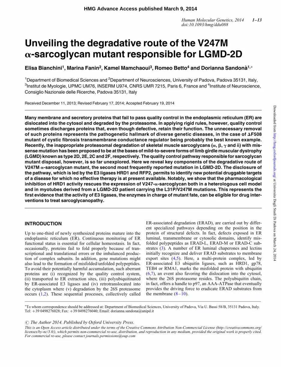
digitonin solubilized proteins from V247M cells with the anti-body specific for a-sarcoglycan. To prolong the associationamong HRD1 complex components and a-sarcoglycanmutant, we inhibited proteasome activity with MG132. Therecovered immunocomplexes contained all the endogenousERAD partners identified with the above described experiments,i.e. SEL1L, HRD1, UBE2J1 and Derlin-1 (Fig. 4).
The E3 ligase RFP2 contributes to V247M mutantdegradation
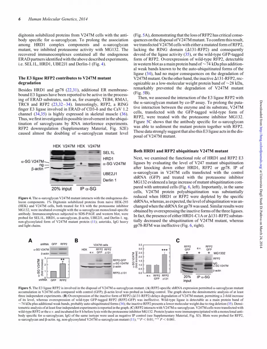
Besides HRD1 and gp78 (22,31), additional ER membrane-bound E3 ligases have been reported to be active in the process-ing of ERAD substrates, such as, for example, TEB4, RMA1,TRC8 and RFP2 (23,32–34). Interestingly, RFP2, a RINGfinger E3 ligase involved in ERAD of CD3-d and the CaV 1.2channel (34,35) is highly expressed in skeletal muscle (36).Thus, we first investigated its possible involvement in the ubiqui-tination of sarcoglycans by RNA interference experiments.RFP2 downregulation (Supplementary Material, Fig. S2E)caused almost the doubling of a-sarcoglycan mutant level
(Fig. 5A), demonstrating that the loss of RFP2 has critical conse-quences on the disposal of V247M mutant. To confirm this result,we transfected V247M cells with either a mutated form of RFP2,lacking the RING domain (D131-RFP2) and consequentlydevoid of the ligase activity (35), or the wild-type GFP-taggedform of RFP2. Overexpression of wild-type RFP2, detectablein western blot as a main protein band of �74 kDa plus addition-al weak bands known to be the auto-ubiquitinated forms of theligase (34), had no major consequences on the degradation ofV247M mutant. On the other hand, the inactiveD131-RFP2, rec-ognizable as a low-molecular weight protein band of �28 kDa,remarkably prevented the degradation of V247M mutant(Fig. 5B).
Then, we assessed the interaction of the E3 ligase RFP2 withthe a-sarcoglycan mutant by co-IP assay. To prolong the puta-tive interaction between the enzyme and its substrate, V247Mcells, transfected with the GFP-tagged wild-type form ofRFP2, were treated with the proteasome inhibitor MG132.Figure 5C shows that the antibody specific for a-sarcoglycanwas able to sediment the mutant protein together with RFP2.These data strongly suggest that also this E3 ligase acts in the dis-posal of V247M mutant.
Both HRD1 and RFP2 ubiquitinate V247M mutant
Next, we examined the functional role of HRD1 and RFP2 E3ligases by evaluating the level of V247 mutant ubiquitinationafter knocking down either HRD1, RFP2 or gp78. IP ofa-sarcoglycan in V247M cells transfected with the controlshRNA (GFP) and treated with the proteasome inhibitorMG132 evidenced a large increase of mutant ubiquitination com-pared with untreated cells (Fig. 6, left). Importantly, in the samecells, V247M protein polyubiquitination was substantiallyreduced when HRD1 or RFP2 were depleted by the specificshRNAs, whereas, as expected, the level of ubiquitination was un-changed when the shRNA for gp78 was used. Similar results wereobtained by overexpressing the inactive forms of the three ligases.In fact, the presence of either HRD1-C1A orD131-RFP2 substan-tially decreased the ubiquitination of V247M mutant, whereasgp78-RFM was ineffective (Fig. 6, right).
Figure 4. The a-sarcoglycan V247M mutant interacts with the endogenous dis-locon components. 1% Digitonin solubilized proteins from naive HEK-293(HEK) and V247M cells, both treated for 8 h with the proteasome inhibitorMG132, were incubated overnight with the a-sarcoglycan monoclonal-specificantibody. Immunocomplexes subjected to SDS-PAGE and western blot, wereprobed for SEL1L, HRD1, a-sarcoglycan, b-actin, UBE2J1, and Derlin-1. ng,non-glycosylated form of V247M mutant protein (11); asterisks, IgG heavyand light chains.
Figure 5. The E3 ligase RFP2 is involved in the disposal of V247M a-sarcoglycan mutant. (A) RFP2-specific shRNA expression permitted a-sarcoglycan mutantaccumulation in V247M cells compared with control (GFP); b-actin level was probed as loading control. The graph shows the densitometric analysis of at leastthree independent experiments. (B) Overexpression of the inactive form of RFP2 (D131-RFP2) delays degradation of V247M mutant, permitting a 2-fold increaseof its level, whereas overexpression of wild-type GFP-tagged RFP2 (RFP2-GFP) was ineffective. Wild-type ligase is detectable as a main protein band of�74 kDa plus additional weak bands, probably auto-ubiquitinated forms (34); the inactive RFP2 presents a lower molecular weight due to ring deletion (35). Densi-tometric analysis of at least four independent experiments is reported in the graph. (C) RFP2 interacts with V247Ma-sarcoglycan. V247M cells were transfected withwild-type RFP2 or the e.v. and incubated for 8 h before lysis with the proteasome inhibitor MG132. Protein lysates were immunoprecipitated with a monoclonal anti-body specific for a-sarcoglycan; IgG of the same isotype were used as negative IP control (see Supplementary Material, Fig. S3). Blots were probed for RFP2,a-sarcoglycan and b-actin. ng, non-glycosylated V247M a-sarcoglycan mutant (11); ∗∗P , 0.01; ∗∗∗ P , 0.001.
6 Human Molecular Genetics, 2014
at Universita D
egli Studi Di Padova on M
arch 24, 2014http://hm
g.oxfordjournals.org/D
ownloaded from

Pharmacological inhibition of HRD1 leads to V247Mmutant recovery
The ultimate goal of our work was the identification of newpharmacological targets for the treatment of sarcoglycanopathy.To this end, we tested two small molecules, LS-101 and LS-102,recently identified by high-throughput screening as strong inhi-bitors of the E3 ligase HRD1, and proposed as drug candidatesfor the treatment of Rheumatoid arthritis (37). Incubation ofV247M cells for 8 h with either 15 mM LS-101 or 20 mM
LS-102 led to a 3-fold increase of a-sarcoglycan mutant level,practically reaching that of the wild-type protein (Fig. 7A).Then, to evaluate the cytolocalization of the protein afterLS-102 treatment, we performed an experiment in whichV247M cells, grown on glass coverslips, were treated for 8 hwith either 10 mM LS-102 or its vehicle, dimethyl sulfoxide(DMSO). In permeabilized cells (Fig. 7B), it is well evidentthe intracellular increase of a-sarcoglycan mutant level afterincubation with LS-102, in comparison to DMSO treatment.Importantly, this short treatment induced the appearance ofsome of the rescued V247M mutant in the plasma membraneof several not permeabilized cells (see arrowheads in Fig. 7B).
Inhibition of HRD1 rescues sarcoglycan complex inprimary myotubes from a LGMD-2D patient
We finally tested the efficacy of the small molecules LS-101 andLS-102 in rescuing a-sarcoglycan mutant in skeletal musclecells derived from a heterozygous LGMD-2D patient carryingthe L31P and V247M substitutions on the a-sarcoglycanalleles. A small bioptic fragment from the LGMD-2D patient,received by the Telethon Genetic Biobank, was utilized toinduce satellite cell sprouting, growth and differentiation intomyotubes. At the fifth day of differentiation, increasing concen-tration of either LS101 or LS102, or the vehicle DMSO, wereadded to the myotubes. Forty-eight hours later, cells werelysed and the expression of different sarcoglycans was analyzedby western blot (Fig. 8A). For comparison, the total cell lysate
from 7-day differentiated control myotubes, derived fromimmortalized satellite cells of a healthy subject, was probedfor a-, b- and d-sarcoglycan and embryonic myosin heavychain expression. The latter marker was used to check the differ-entiation homogeneity of the different samples. It is well evidentthat, by inhibiting the HRD1 E3 ligase activity in the LGMD-2Dpatient myotubes, the amount of a-, b- and d-sarcoglycan sub-stantially increased. The effect is more pronounced for a- andd-sarcoglycan as their basal level, in untreated cell, is very lowor almost absent. It seems also clear that LS102, the more specificHRD1 inhibitor (37), produced a more effective, dose-dependent recovery of sarcoglycans. In parallel experiments,myoblasts from the LGMD-2D patient, were grown and differ-entiated on glass coverslips and treated with either 10 mM
LS-102 or 1‰ DMSO for 48 h. Thereafter, not permeabilizedmyotybes were probed with an antibody specific for an extracel-lular epitope of a-sarcoglycan. As positive control, myotubesderived from the healthy subject were also immunodecorated.Figure 8B shows thata-sarcoglycan is homogeneously expressedat the surface of all myotubes from the healthy subject, whereasonly traces of a-sarcoglycan staining are evident in untreatedmyotubes from the LGMD-2D patient. Instead, inhibition ofHRD1 activity with LS-102 induced a marked, although nothomogeneous localization of a-sarcoglycan mutant at the cellmembrane of almost all myotubes derived from the LGMD-2Dpatient (Fig. 8B).
DISCUSSION
Maturation and assembly of the skeletal muscle membrane sar-coglycan complex occur in the ER (13). However, little is knownabout the ER processing of the four sarcoglycans (a,b, g and d),how the hetero-tetrameric complex assembles, and, more im-portantly, which ER quality control mechanisms control thefate of misfolded or orphan subunits. Our lack of knowledge, par-ticularly about the latter point, is a serious limit to develop suit-able therapeutic intervention when the inappropriate disposal ofsarcoglycan mutants causes sarcoglycanopathy. Most of sarco-glycanopathy cases are in fact associated with missense muta-tions that can lead to a misfolded protein. However, manydisease-causing missense mutations are without functional con-sequences (12,17), nonetheless, the mutant is intercepted by thequality control system and delivered to the proteasome. The pre-mature degradation precludes the possibility for the mutated,but probably functional protein, to assemble with its partners,exit the ER, and move to the plasma membrane. The result is,de facto, a loss of function responsible for sarcoglycanopathy.
Importantly, recent evidence indicates that pharmacologicaltreatments interfering with either the initial maturation steps(17) or the ubiquitin–proteasome system (11) might prevent theremoval of some of the a-sarcoglycan mutants and promotetheir functional recovery. As at present, no effective cure is avail-able to treat sarcoglycanopathy, the final goal of our work was theidentification of new molecular targets potentially eligible for thepharmacological treatment of the disease. To this intent, wedecided to decipher the ERAD pathway controlling the disposalof V247M a-sarcoglycan. The results demonstrated that theV247M a-sarcoglycan mutant, a typical type I membraneprotein with the defect located in the luminal domain, follows
Figure 6. HRD1 and RFP2 are the E3 ligases responsible for the V247M mutantubiquitination. Left blot, V247M cells were transfected with the indicatedshRNAs. Right blot, V247M cells were transfected with the indicated inactiveforms of the E3 ubiquitin ligases (22,23,35). Eight hours before lysis, cellswere treated with either 10 mM MG132 (+), in order to block protein degradationthrough the proteasome, or the vehicle DMSO (2). Two hundred micrograms ofproteins solubilized in RIPA buffer were incubated overnight with thea-sarcoglycan monoclonal-specific antibody. After SDS PAGE, blots wereprobed with antibodies-specific to mono and polyubiquitinated proteins (IB ubi-quitin) and to a-sarcoglycan (IB a-SG).
Human Molecular Genetics, 2014 7
at Universita D
egli Studi Di Padova on M
arch 24, 2014http://hm
g.oxfordjournals.org/D
ownloaded from

the ERAD-LM pathway, and that twoE3ligases,HRD1andRFP2,are responsible for its ubiquitination and delivery to proteasome.
First, by using different and selective molecular approaches,we demonstrated that the naturally occurring V247M mutantof a-sarcoglycan is handled by the HRD1 complex. Inmammals, basic components of this complex are the E3 ligaseHRD1, the adaptor protein SEL1L, that exerts a fundamentalrole in presenting substrates to the ligase (19,24), and one E2conjugase, that working in concert with the RING finger E3ligase, links ubiquitin to the substrate (6). Our data demonstratethat HRD1, SEL1L and the E2 conjugase UBE2J1 interact withV247M mutant as they are co-IP by the a-sarcoglycan-specificantibody. These ERAD components play a key role in the dis-posal of V247M mutant, as either their knock down or the over-expression of specific inactive forms of such proteins led to
a-sarcoglycan mutant accumulation (Figs 1 and 2). The activityof the HRD1 complex is usually assisted by the cytosolicAAA-ATPase, p97, that provides the force to eradicate polyubi-quitinated proteins form the ER membrane (9,40), and byDerlin-1, that facilitates the extraction of substrates from themembrane (29). Accordingly, IP assays and experiments withdominant-negative forms, show that p97 and Derlin-1 are twofundamental ERAD players in the disposal of V247M mutant(Fig. 3).
Our results show that another E3 ligase, RFP2, cooperates withHRD1 in the processing of V247M mutant. This is not surprising,as emerging evidence demonstrates that two, or even more, E3ligases might assist the processing of certain ERAD substrates.For example, HRD1 cooperates with gp78 in the degradation ofmutant neuroserpin (41), whereas gp78 together with Trc8 operates
Figure 7. Rescue of V247M mutant in V247M cells after pharmacological inhibition of the E3 ligase HRD1. (A) V247M cells were treated for 8 h with either 15 mM
LS-101 or 20 mM LS-102, specific HRD1 ligase inhibitors (37). DMSO, the vehicle of HRD1 inhibitors, was used as negative control. Cells expressing wild-typea-sarcoglycan were used as positive control. b-actin level was used to normalize protein loading. Densitometric analysis of at least three independent experiments,in which the level of wild-typea-sarcoglycan was set as 100%, is reported in the graph. ∗P , 0.05; ∗∗P , 0.01. (B) V247M cells were treated for 8 h with the HRD1inhibitor LS102 (10 mM) or its vehicle DMSO (1‰). At the end of treatments, cells were either permeabilized with Triton X-100 or left intact (not permeabilized) andimmunodecorated with the monoclonal a-sarcoglycan-specific antibody. Localization of a-sarcoglycan was revealed with the secondary DyLight 488-conjugatedanti-mouse antibody. Insets are the transmitted light images of the same IF fields. All images, collected with a Leica SP2 confocal microscope, are at the same mag-nification. It is well evident that, after HRD1 inhibitor treatment, the intensity of the intracellulara-sarcoglycan signal increased, and that, in several not permeabilizedcells (arrowheads) a faint membrane stain became evident.
8 Human Molecular Genetics, 2014
at Universita D
egli Studi Di Padova on M
arch 24, 2014http://hm
g.oxfordjournals.org/D
ownloaded from

Figure 8. Inhibition of the E3 ubiquitin ligase HRD1induced sarcoglycan rescue in myotubes from a LGMD-2D patient. (A) Five-day differentiated myotubes of aLGMD-2D patient, carrying the L31P and V247M mutations on the a-sarcoglycan alleles, were incubated for 48 h with either DMSO, used as negative control, orincreasing concentration of LS-101 and LS-102, as indicated. At the end of incubation, myotubes were lysed and the expression of a-sarcoglycan (a-SG),b-sarcoglycan (b-SG) and d-sarcoglycan (d-SG) was analyzed by western blotting. To verify the differentiation homogeneity of the different samples the expressionof embryonic myosin heavy chain (eMHC) (38) was measured. By comparison, the expression of the same proteinswas evaluated in 7-day old myotubesderived from ahealthy subject (39). As loading control, the Ponceau red staining of the membranes is reported. (B) Myoblasts form the LGMD-2D patient were grown on glass cover-slips and differentiated for 7 days. At the fifth day of differentiation, either DMSO (negative control) or 10 mM LS102 were added. Two days later, not permeabilizedmyotubes were immunodecorated with the rabbit polyclonal antibody specific for an extracellular epitope ofa-sarcoglycan. As positive control 7-day differentiated,not permeabilized myotubes derived from a healthy subject, were immunolabeled with the same antibody. The primary antibody was visualized by a DyLight488-conjugated anti-rabbit secondary antibody. On the right of each fluorescence image, the transmitted light image of the same field is reported; in each imagesbars indicate 30 mm. Images were recorded with a Leica SP2 laser scanning confocal microscope at the same setting conditions.
Human Molecular Genetics, 2014 9
at Universita D
egli Studi Di Padova on M
arch 24, 2014http://hm
g.oxfordjournals.org/D
ownloaded from

the sterol-mediated degradation of 3-hydroxy-3-methylglutaryl co-enzyme A reductase (42), and the orphan CD3-d subunit is client ofboth gp78 and RFP2 E3 ligases (34). The latter ligase also mediatesthe degradation of CaV 1.2 channels (35) and regulates the expres-sion of the p53 tumor suppressor by degrading MDM2 (43). Con-sidering that RFP2 is highly expressed in skeletal muscle, as wellas in HEK-293 cells (36), we thought RFP2 as an additional E3ligase handling sarcoglycan. Knocking down experiments or in-active RFP2 overexpression confirmed this hypothesis (Figs. 5and 6). Interestingly, RFP2 ligase activity has been linked to theUBE2J1 conjugase (34), Derlin-1 and p97 (35), ERAD elementsalso working with the HRD1 ligase, suggesting thata-sarcoglycanmutants follow a common ERAD pathway converging on alterna-tive E3 ligases. In this light, it will be a stimulating challenge tounderstand the functional meaning of the cross-talk between ubi-quitin ligases.AsRFP2 isknowntoremoveorphansubunitsofmul-timeric complexes (34,35), our data are the first demonstration thatRFP2 also handles misfolded mutated proteins. However, asa-sarcoglycan is member of a tetramer, and our experiments havebeen carried out in cells expressing solely this protein, RFP2 role inV247Ma-sarcoglycandisposalmightbe linked to the propensityofthis ligase to take care of unassembled subunits. Additional experi-ments are needed to test this possibility.
In conclusion, by deciphering the ERAD pathway responsiblefor the processing of V247M mutant we have focused the intereston the two key elements of the pathway, HRD1 and RFP2. TheE3 ligases, for their fundamental role in driving substrate disposal,are emerging as significant druggable targets (44) for the treatmentof different diseases involving the ubiquitin–proteasome system(45). In this respect, our work, for the first time, addressed the activ-ity of an E3 ligase involved in the disposal of an a-sarcoglycanmutant causing LGMD-2D. Two small molecules, screened asinhibitors of HRD1 (37) were actually able to recover the expres-sion of the V247M mutant both in a cellular model (V247Mcells) and, remarkably, in primary skeletal muscle cells derivedfrom a LGMD-2D patient. In the cellular model, a short treatment(8 h) with both LS101 and LS102, produced a recovery of themutant that almost reached the level of wild-type a-sarcoglycanexpressed in the HEK293 cells (Fig. 7A) and permitted the appear-ance of the V247M-a-sarcoglycan on the cell surface. Moreover,inhibition of the ligase activity seems to block also the dislocationfrom the ER, as the recovered mutant is fully glycosylated, differ-ently to what happens when the proteasome is blocked, a treatmentthat produces the accumulation of both the mature and cytosolicdeglycosylated form of a-sarcoglycan (11). The availability ofprimary cells from a LGMD-2D patient, permitted us to test the ef-ficacy of the two small molecules not only in the myogenic cellbackground but, more importantly, directly on cells of a humansubject affected by the disease. The genetic analysis from thisLGMD-2D patient indicated the presence of a T.C substitutionat position 98 of one allele and a G.A substitution at position739 of the other allele of the SGCA gene leading to L31P andV247M missense mutations on a-sarcoglycan. The residuala-sarcoglycan expression in the skeletal muscle tissue, as deter-minedbywesternblottinganalysis,was�5%,whereasnoinforma-tion was available forb-,g- and d-sarcoglycan (data from TelethonGenetic Biobank). At 7 days of differentiation, in comparison withsarcoglycan expression in myotubes of a healthy subject, patientmyotubes show a very low level of both a- and b-sarcoglycanprotein, whereas d-sarcoglycan is almost undetectable. Both
LS101 and LS102 seems to be effective in rescuinga-sarcoglycan,with the second molecule causing a much more pronounced recov-ery of the protein, in a dose-dependent fashion. This is true not onlyfor the mutated a-sarcoglycan, but also for the wild-type partners,d-sarcoglycan and b-sarcoglycan, suggesting that the beneficialeffect of the treatments is extended to the entire sarcoglycancomplex. Even though our data cannot establish if both, or justone, of the two allelic forms of a-sarcoglycan has been recovered,the result is very promising. In fact, the central issue is that theamount of saveda-sarcoglycan should reach a level sufficient to as-semble with the other sarcoglycans which in turn, being no moreorphan subunits, are preserved from degradation. It is importantto underline that the severity of the disease phenotype is strictlyassociated with the residual level of sarcoglycans present inmuscle fibers (12,18), therefore, even a small overall sarcoglycansrescue is expected to ameliorate patient condition.
Despite still preliminary, our work has identified novel poten-tial druggable targets providing new hints for the search and/oroptimization of potent and selective inhibitors of the ERADpathway specifically involved ina-sarcoglycan mutant disposal.Moreover, the successful pharmacological intervention in both acellular model and in a LGMD-2D patient specimen can be con-sidered a proof of principle study on the development of a futuresarcoglycanopathy therapy, specifically targeting an E3 ubiqui-tin ligase.
MATERIALS AND METHODS
Cell culture, transfection, and treatments
HEK-293 and V247M cells, were grown in Dulbecco’s modifiedEagle’s medium (Sigma) supplemented with 10% fetal bovineserum (FBS) (Gibco) and maintained in a humidified atmospherecontaining 5% CO2 at 37 8C. V247M cells were generated byHEK-293 cell transfection with the mutated a-sarcoglycancDNA cloned in pcDNA3 and subsequent G418 selection.
Immortalized human myoblasts (39), from the ‘Human cellculture platform’ of the Myology Institute in Paris, weregrown in Skeletal Muscle Cell Growth Medium (Promocell)supplemented with 15% FBS (Gibco), named skeletal growthmedium (SGM). To start differentiation, myoblasts grown atconfluence were incubated with DMEM supplemented with2% horse serum (Euroclone), 10 mg/ml human recombinantinsulin (Sigma), 100 mg/ml human Apotransferrin (Sigma),named skeletal differentiating medium (SDM). Differentiationwas carried out for seven days.
For overexpression and RNAi experiments, V247M cellswere seeded at �50 000 cells/cm2 and transiently transfectedthe day after seeding with Transit293 (MirusBio) according tomanufacturer’s instruction. Cells were lysed either 48 or 72 h(RNAi assays) after transfection and subjected to direct westernblotting analysis or immunoprecipitation assay. MG132 (Sigma)or HRD1 inhibitors, kindly provided by Toshihiro Nakajima(Tokyo Medical University) (37), were added 8 h before V247Mcell lysis or 8–48 h before immunofluorescence staining.
Culture and differentiation of primary cells derivedfrom a human muscular biopsy
Thanks to the Telethon Genetic Bio-Bank facility we obtained asmall fragment of a frozen biopsy from a LGMD-2D patient
10 Human Molecular Genetics, 2014
at Universita D
egli Studi Di Padova on M
arch 24, 2014http://hm
g.oxfordjournals.org/D
ownloaded from

carrying two different missense mutations (V247M/L31P) onthe a-sarcoglycan alleles. The biopsy, washed twice, was incu-bated with SGM containing 25% human plasma from donors,in order to form a clot. A week later, cells started to spread offand the biopsy was cut into small pieces, transferred on gelatin-coated plastic wells and incubated in SGM. Myoblasts, spread-ing from the biopsy fragments, were expanded and subsequentlyused to carry out experiments. When at confluence, myoblastswere differentiated by changing SGM with SDM. Differenti-ation was carried out for 7 days. E3 ligase inhibitors wereadded 48 h before myotubes lysis.
Plasmids
V247M-a-sarcoglycan construct was generated as previouslydescribed (11). p97-GFP and p97 K524A-GFP mutant werekindly provided by Akira Kakizuka (Kyoto University); HRD1and HRD1-C1A mutant were obtained from Emanuel Wiertz(Leiden University); murine gp78 C337/374S RFM mutant wasdonated by Kazuhiro Nagata (Kyoto University); humangp78-HA and the plasmid expressing gp78-specific shRNA werekindly provided by Petek Ballar (Ege University); UBE2J1 andUBE2J1-C91S were from Hidde L. Ploegh (WIBR, Cambridge);RFP2 and D131-RFP2 mutant were obtained from GeraldW. Zamponi (University of Calgary). RFP2, originally cloned inpUC57 vector, was subcloned in the pEGFPN1 vector, in framewith GFP.
shRNA producing vectors
The target sequences for shRNA-mediated knock down are:GAGACAGTTTCAGATGATT for HRD1, CAGATAAAGT-GAAGGAATT for RFP2, GGCTATACTGTGGCTAGAA forSEL1L, GAAGTCGTGCTGCTTCATG for GFP, GACTACA-CAAATCAGCGATTT for LacZ, the last two used as controls.Complementary oligonucleotides comprising the sense and anti-sense sequences separated by a standard TTCAAGAGA loopstructure were purchased from MWG, annealed and cloned intothe pSUPER vector. All constructs were checked by sequencing.
Antibodies
The antibodies utilized were: mouse monoclonal a-sarcoglycanfrom DBA and home-made rabbit polyclonala-sarcoglycan anti-body (raisedagainst the peptide ‘CYDTLAPHFRVDW’mappingin the extracellular domain of the protein, see Supplementary Ma-terial, Fig. S4 for the characterization); HRD1, Derlin1, p97, GFPand UBE2J1 from Abcam; b-actin and SEL1L from SigmaAldrich; HA and Myc from Millipore; RFP2, gp78 and mouseIgG from Santa Cruz Biotechnology; mono and polyubiquitinatedconjugates from Enzo Life Science; embryonic myosin heavychain was a kind gift of Stefano Schiaffino (VIMM, Padova)(38); DyLight 488-conjugated anti-mouse and anti-rabbit anti-bodies from Jackson.
Immunoprecipitation and immunoblotting
For western blotting analysis, total protein homogenate wasobtained by lysing cells with 5% sodium deoxycholate solutionsupplemented with complete protease inhibitor (Roche). For
co-immunoprecipitation assays, cells were lysed with 50 mM
TRIS pH 7.4, 150 mM NaCl, 5 mM EDTA pH 7.4, 1% TritonX-100supplementedwithprotease inhibitorsand2 mM N-ethylma-leimide (NEM) (Sigma). Analyses were performed on the TritonX-100 soluble fraction. Where indicated, proteins were solubilizedin 1% digitonin or radioimmunoprecipitation assay (RIPA) buffersupplemented with protease inhibitors and 2 mM NEM (Sigma).Proteins were quantified by the bicinchoninic acid protein assaykit(ThermoScientific)accordingtomanufacturer’sinstructions.Forimmunoprecipitation assays, 200 mg of solubilized proteins wereincubated overnight at 48C with either the mouse monoclonala-sarcoglycan-specificantibodyorwithmouse IgGof thesameiso-type,asthenegativecontrol.Thefollowingday,ProteinG-magneticbeads (Millipore) were added and the mixture incubated 1 h in thesame condition. Beads were recovered and extensively washedwith lysis buffer and finally aspirated to dryness. After Laemmlibuffer addition, samples were analyzed by sodium dodecylsulfate(SDS-PAGE)andwesternblottingusingspecificantibodies.
Immunofluorescence
To visualize membrane-resident a-sarcoglycan, immunofluor-escence experiments were performed on intact cells. V247Mcells, seeded and grown on poly-lysine-treated glass coverslips,or human myoblasts seeded, grown and differentiated on gelatin/fibronectin-treated glass coverslips, at the end of specific drugtreatments, were chilled at 48C for 30 min, washed with coldPBS supplemented with Ca2+ and Mg2+. Cells were subsequent-ly incubated at 48C with either the mouse monoclonal or therabbit polyclonal antibody specific fora-sarcoglycan, recogniz-ing an extracellular epitope. After washing, cells were incubatedwith the specific DyLight 488-conjugated secondary antibody(Jackson). At the end of incubation, cells were washed andfixed with 4% PFA.
To visualize intracellular a-sarcoglycan, immunofluores-cence experiments were performed on permeabilized cells.V247M cells cultivated as above described, were first fixedwith 4% PFA and subsequently permeabilized with 0.5%Triton X-100 for 15 min. Incubation with primary and secondaryantibodies was performed as described above. Cells were thenexamined with a Leica SP2 confocal microscope.
Statistical analysis
Data from at least three independent experiments are expressedas mean+SEM. Statistical differences were determined bypaired two-tailed Student’s t-test or ANOVA (using GraphPadPrism 5).
SUPPLEMENTARY MATERIAL
Supplementary Material is available at HMG online.
AUTHORS’ CONTRIBUTIONS
E.B., R.B. and D.S. conceived and designed the experiments;M.F. and K.M. provided human specimens; E.B. and D.S. per-formed the experiments; E.B., R.B. and D.S. analyzed thedata; E.B., R.B. and D.S. wrote the paper.
Human Molecular Genetics, 2014 11
at Universita D
egli Studi Di Padova on M
arch 24, 2014http://hm
g.oxfordjournals.org/D
ownloaded from

ACKNOWLEDGEMENTS
Toshihiro Nakajima, Tokyo Medical University, is gratefullythanked for providing the HRD1 inhibitors. Vincent Moulyand the ‘Human cell culture platform’ of the Myology InstituteUniversite Pierre et Marie Curie, Paris 6 are gratefully thankedfor providing immortalized human myoblasts derived from ahealthy subject. Corrado Angelini and the Telethon GeneticBio-Bank facility, University of Padova are gratefully thankedfor providing the skeletal muscle biopsy of a LGMD-2Dpatient. Stefano Schiaffino is gratefully thanked for providingthe embryonic myosin heavy chain mouse monoclonal antibody.
Conflict of Interest statement. The authors declare no conflict ofinterest.
FUNDING
This work was supported by Association Francaise contre lesMyopathies (grant number 14999, 15969 to R.B.), Italian Tele-thon (grant number GEP12058 to D.S.) and institutional fundsfrom the Consiglio Nazionale delle Ricerche to R.B. and Univer-sity of Padova to D.S. Funding to pay the Open Access publica-tion charges for this article was provided by Italian Telethon.
REFERENCES
1. Smith, M.H., Ploegh, H.L. and Weissman, J.S. (2011) Road to ruin: targetingproteins for degradation in the endoplasmic reticulum. Science, 334,1086–1090.
2. Bagola, K., Mehnert, M., Jarosch, E. and Sommer, T. (2011) Proteindislocation from the ER. Biochim. Biophys. Acta., 1808, 925–936.
3. Carvalho, P., Goder, V. and Rapoport, T.A. (2006) Distinct ubiquitin–ligasecomplexes define convergent pathways for the degradation of ER proteins.Cell, 126, 361–373.
4. Hosokawa, N., Wada, I., Nagasawa, K., Moriyama, T., Okawa, K. andNagata, K. (2008) Human XTP3-B forms an endoplasmic reticulum qualitycontrol scaffold with the HRD1-SEL1L ubiquitin ligase complex and BiP.J. Biol. Chem., 283, 20914–20924.
5. Christianson, J.C., Shaler, T.A., Tyler, R.E. and Kopito, R.R. (2008) OS-9and GRP94 deliver mutant alpha1-antitrypsin to the Hrd1-SEL1L ubiquitinligase complex for ERAD. Nat. Cell. Biol., 10, 272–282.
6. Hirsch, C., Gauss, R., Horn, S.C., Neuber, O. and Sommer, T. (2009)The ubiquitylation machinery of the endoplasmic reticulum. Nature, 458,453–460.
7. Kostova, Z., Tsai, Y.C. and Weissman, A.M. (2007) Ubiquitin ligases,critical mediators of endoplasmic reticulum-associated degradation. Semin.Cell. Dev. Biol., 18, 770–779.
8. Ye, Y., Meyer, H.H. and Rapoport, T.A. (2001) The AAA ATPase Cdc48/p97 and its partners transport proteins from the ER into the cytosol. Nature,414, 652–656.
9. Flierman, D., Ye, Y., Dai, M., Chau, V. and Rapoport, T.A. (2003)Polyubiquitin serves as a recognition signal, rather than a ratchetingmolecule, during retrotranslocation of proteins across the endoplasmicreticulum membrane. J. Biol. Chem., 278, 34774–34782.
10. Hampton, R.Y. and Sommer, T. (2012) Finding the will and the way ofERAD substrate retrotranslocation. Curr. Opin. Cell Biol., 24, 460–466.
11. Gastaldello, S., D’Angelo, S., Franzoso, S., Fanin, M., Angelini, C., Betto, R.and Sandona, D. (2008) Inhibition of proteasome activity promotes thecorrect localization of disease-causing alpha-sarcoglycan mutants inHEK-293 cells constitutively expressing beta-, gamma-, anddelta-sarcoglycan. Am. J. Pathol., 173, 170–181.
12. Sandona, D. and Betto, R. (2009) Sarcoglycanopathies: molecularpathogenesis and therapeutic prospects. Expert Rev. Mol. Med., 11, e28.
13. Noguchi, S., Wakabayashi, E., Imamura, M., Yoshida, M. and Ozawa, E.(2000) Formation of sarcoglycan complex with differentiation in culturedmyocytes. Eur. J. Biochem., 267, 640–648.
14. Kirschner, J. and Lochmuller, H. (2011) Sarcoglycanopathies. Handb. Clin.
Neurol., 101, 41–46.15. Politano, L., Nigro, V., Passamano, L., Petretta, V., Comi, L.I., Papparella,
S., Nigro, G., Rambaldi, P.F., Raia, P., Pini, A. et al. (2001) Evaluation ofcardiac and respiratory involvement in sarcoglycanopathies. Neuromuscul.
Disord., 11, 178–185.16. Bartoli, M., Gicquel, E., Barrault, L., Soheili, T., Malissen, M., Malissen, B.,
Vincent-Lacaze, N., Perez, N., Udd, B., Danos, O. and Richard, I. (2008)Mannosidase I inhibition rescues the human alpha-sarcoglycan R77Crecurrent mutation. Hum. Mol. Genet., 17, 1214–1221.
17. Soheili, T., Gicquel, E., Poupiot, J., N’Guyen, L., Le Roy, F., Bartoli, M. andRichard, I. (2012) Rescue of sarcoglycan mutations by inhibition ofendoplasmic reticulum quality control is associated with minimal structuralmodifications. Hum. Mutat., 33, 429–439.
18. Barresi, R., Confalonieri, V., Lanfossi, M., Di Blasi, C., Torchiana, E.,Mantegazza, R., Jarre, L., Nardocci, N., Boffi, P., Tezzon, F. et al. (1997)Concomitant deficiency of beta- and gamma-sarcoglycans in 20alpha-sarcoglycan (adhalin)-deficient patients: immunohistochemicalanalysis and clinical aspects. Acta Neuropathol., 94, 28–35.
19. Bernasconi, R., Galli, C., Calanca, V., Nakajima, T. and Molinari, M. (2010)Stringent requirement for HRD1, SEL1L, and OS-9/XTP3-B for disposal ofERAD-LS substrates. J. Cell Biol., 188, 223–235.
20. Sandona, D., Gastaldello, S., Martinello, T. and Betto, R. (2004)Characterization of the ATP-hydrolysing activity of alpha-sarcoglycan.Biochem. J., 381, 105–112.
21. Draviam, R.A., Wang, B., Shand, S.H., Xiao, X. and Watkins, S.C. (2006)Alpha-sarcoglycan is recycled from the plasma membrane in the absence ofsarcoglycan complex assembly. Traffic, 7, 793–810.
22. Kikkert, M., Doolman, R., Dai, M., Avner, R., Hassink, G., van Voorden, S.,Thanedar, S., Roitelman, J., Chau, V. and Wiertz, E. (2004) Human HRD1 isan E3 ubiquitin ligase involved in degradation of proteins from theendoplasmic reticulum. J. Biol. Chem., 279, 3525–3534.
23. Morito, D., Hirao, K., Oda, Y., Hosokawa, N., Tokunaga, F., Cyr, D.M.,Tanaka, K., Iwai, K. and Nagata, K. (2008) Gp78 cooperates with RMA1 inendoplasmic reticulum-associated degradation of CFTRDeltaF508. Mol.
Biol. Cell, 19, 1328–1336.24. Mueller, B., Klemm, E.J., Spooner, E., Claessen, J.H. and Ploegh, H.L.
(2008) SEL1L nucleates a protein complex required for dislocation ofmisfolded glycoproteins. Proc. Natl. Acad. Sci. USA, 105, 12325–12330.
25. Iida, Y., Fujimori, T., Okawa, K., Nagata, K., Wada, I. and Hosokawa, N.(2011) SEL1L protein critically determines the stability of theHRD1-SEL1L endoplasmic reticulum-associated degradation (ERAD)complex to optimize the degradation kinetics of ERAD substrates. J. Biol.
Chem., 286, 16929–16939.26. Burr, M.L., Cano, F., Svobodova, S., Boyle, L.H., Boname, J.M. and Lehner,
P.J. (2011) HRD1 and UBE2J1 target misfolded MHC class I heavy chainsfor endoplasmic reticulum-associated degradation. Proc. Natl. Acad. Sci.
USA, 108, 2034–2039.
27. Chen, B., Mariano, J., Tsai, Y.C., Chan, A.H., Cohen, M. and Weissman,A.M. (2006) The activity of a human endoplasmic reticulum-associateddegradation E3, gp78, requires its Cue domain, RING finger, and anE2-binding site. Proc. Natl. Acad. Sci. USA, 103, 341–346.
28. Claessen, J.H., Mueller, B., Spooner, E., Pivorunas, V.L. and Ploegh, H.L.(2010) The transmembrane segment of a tail-anchored protein determines itsdegradative fate through dislocation from the endoplasmic reticulum.J. Biol. Chem., 285, 20732–20739.
29. Greenblatt, E.J., Olzmann, J.A. and Kopito, R.R. (2011) Derlin-1 is arhomboid pseudoprotease required for the dislocation of mutant alpha-1antitrypsin from the endoplasmic reticulum. Nat. Struct. Mol. Biol., 18,1147–1152.
30. Kobayashi, T., Tanaka, K., Inoue, K. and Kakizuka, A. (2002) FunctionalATPase activity of p97/Valosin-containing protein (VCP) is required for thequality control of endoplasmic reticulum in neuronally differentiatedmammalian PC12 cells. J. Biol. Chem., 277, 47358–47635.
31. Fang, S., Ferrone, M., Yang, C., Jensen, J.P., Tiwari, S. and Weissman, A.M.(2001) The tumor autocrine motility factor receptor, gp78, is a ubiquitinprotein ligase implicated in degradation from the endoplasmic reticulum.Proc. Natl. Acad. Sci. USA, 98, 14422–14427.
32. Hassink, G., Kikkert, M., van Voorden, S., Lee, S.J., Spaapen, R., van Laar,T., Coleman, C.S., Bartee, E., Fruh, K., Chau, V. and Wiertz, E. (2005) TEB4is a C4HC3 RING finger-containing ubiquitin ligase of the endoplasmicreticulum. Biochem. J., 388, 647–655.
12 Human Molecular Genetics, 2014
at Universita D
egli Studi Di Padova on M
arch 24, 2014http://hm
g.oxfordjournals.org/D
ownloaded from

33. Lee, J.P., Brauweiler, A., Rudolph, M., Hooper, J.E., Drabkin, H.A. andGemmill, R.M. (2010) The TRC8 ubiquitin ligase is sterol regulated andinteracts with lipid and protein biosynthetic pathways. Mol. Cancer Res., 8,93–106.
34. Lerner, M., Corcoran, M., Cepeda, D., Nielsen, M.L., Zubarev, R., Ponten,F., Uhlen, M., Hober, S., Grander, D. and Sangfelt, O. (2007) The RBCCgene RFP2 (Leu5) encodes a novel transmembrane E3 ubiquitin ligaseinvolved in ERAD. Mol. Biol. Cell, 18, 1670–1682.
35. Altier, C., Garcia-Caballero, A., Simms, B., You, H., Chen, L., Walcher, J.,Tedford, H.W., Hermosilla, T. and Zamponi, G.W. (2011) The Cavbetasubunit prevents RFP2-mediated ubiquitination and proteasomaldegradation of L-type channels. Nat. Neurosci., 14, 173–180.
36. Baranova, A., Hammarsund, M., Ivanov, D., Skoblov, M., Sangfelt, O.,Corcoran, M., Borodina, T., Makeeva, N., Pestova, A., Tyazhelova, T. et al.(2003) Distinct organization of the candidate tumor suppressor gene RFP2 inhuman and mouse: multiple mRNA isoforms in both species- andhuman-specific antisense transcript RFP2OS. Gene, 321, 103–112.
37. Yagishita, N., Aratani, S., Leach, C., Amano, T., Yamano, Y., Nakatani, K.,Nishioka, K. and Nakajima, T. (2012) RING-finger type E3 ubiquitin ligaseinhibitors as novel candidates for the treatment of rheumatoid arthritis.Int. J. Mol. Med., 30, 1281–1286.
38. Schiaffino, S., Gorza, L., Sartore, S., Saggin, L. and Carli, M. (1986)Embryonic myosin heavy chain as a differentiation marker of developinghuman skeletal muscle and rhabdomyosarcoma. A monoclonal antibodystudy. Exp. Cell Res., 163, 211–220.
39. Zhu, C.H., Mouly, V., Cooper, R.N., Mamchaoui, K., Bigot, A., Shay, J.W.,Di Santo, J.P., Butler-Browne, G.S. and Wright, W.E. (2007) Cellularsenescence in human myoblasts is overcome by human telomerase reversetranscriptase and cyclin-dependent kinase 4: consequences in aging muscleand therapeutic strategies for muscular dystrophies. Aging Cell, 6, 515–523.
40. Jentsch, S. and Rumpf, S. (2007) Cdc48 (p97): a “molecular gearbox” in theubiquitin pathway? Trends Biochem. Sci., 32, 6–11.
41. Ying, Z., Wang, H., Fan, H. and Wang, G. (2011) The endoplasmic reticulum(ER)-associated degradation system regulates aggregation and degradationof mutant neuroserpin. J. Biol. Chem., 286, 20835–20844.
42. Jo, Y., Lee, P.C., Sguigna, P.V. and DeBose-Boyd, R.A. (2011)Sterol-induced degradation of HMG CoA reductase depends on interplay oftwo Insigs and two ubiquitin ligases, gp78 and Trc8. Proc. Natl. Acad. Sci.
USA, 108, 20503–20508.
43. Joo, H.M., Kim, J.Y., Jeong, J.B., Seong, K.M., Nam, S.Y., Yang, K.H.,Kim,C.S., Kim, H.S., Jeong, M., An, S. and Jin, Y.W. (2011) Ret finger protein 2enhances ionizing radiation-induced apoptosis via degradation of AKT andMDM2. Eur. J. Cell Biol., 90, 420–431.
44. Goldenberg,S.J., Marblestone, J.G., Mattern, M.R. and Nicholson, B. (2010)Strategies for the identification of ubiquitin ligase inhibitors. Biochem. Soc.
Trans., 38, 132–136.
45. Guerriero, C.J. and Brodsky, J.L. (2012) The delicate balance betweensecreted protein folding and endoplasmic reticulum-associated degradationin human physiology. Physiol. Rev., 92, 537–576.
Human Molecular Genetics, 2014 13
at Universita D
egli Studi Di Padova on M
arch 24, 2014http://hm
g.oxfordjournals.org/D
ownloaded from


