Embed Size (px)
Citation preview
Upper Limb Effort Does Not Increase Maximal Voluntary MuscleActivation in Individuals with Incomplete Spinal Cord Injury
Helen J. Huang, M.S.1 and Daniel P. Ferris, Ph.D.1,2,31Department of Biomedical Engineering, University of Michigan, Ann Arbor, MI, USA2School of Kinesiology, University of Michigan, Ann Arbor, MI, USA3Department of Physical Medicine and Rehabilitation, University of Michigan, Ann Arbor, MI, USA
AbstractObjective—To determine the effect of upper limb effort on maximal lower limb muscle activationin individuals with incomplete spinal cord injury.
Methods—Fifteen individuals with incomplete spinal cord injury performed recumbent steppingusing different combinations of upper and lower limb efforts.
Results—There was no significant difference in active lower limb electromyography amplitudesregardless of whether the upper limbs were resting or exerting maximal effort. Upper limb effortincreased passive lower limb muscle activation and likewise, lower limb effort increased passiveupper limb muscle activation.
Conclusions—Upper limb effort did not increase lower limb muscle activation during active lowerlimb effort in individuals with incomplete spinal cord injury during recumbent stepping. Thissuggests that individuals with incomplete spinal cord injury cannot recruit additional lower limbmotor units using maximal volitional effort of their upper limbs.
Significance—Understanding how upper limb effort and movement influences lower limb muscleactivation patterns in incomplete spinal cord injury patients has implications for prescribing therapiesfor lower limb rehabilitation.
Keywordsneural coupling; rehabilitation; interlimb coupling; incomplete spinal cord injury; electromyography;locomotion
INTRODUCTIONUpper limb muscle activation can increase muscle activity in the passive lower limbs duringa rhythmic motor task (Ferris DP, et al., 2006). In previous studies, we examined neurologicallyintact subjects performing recumbent stepping on an exercise device that coupled motion ofthe upper and lower limbs. We found that increasing upper limb muscle activation through
© 2009 International Federation of Clinical Neurophysiology. Published by Elsevier Ireland Ltd. All rights reserved.Corresponding author: Helen J. Huang, Ph.D. Research Associate, Neuromechanics Laboratory, Department of Integrative Physiology,University of Colorado, Boulder, CO 80309-0354. Phone: +1.303.492.0926. Fax: +1.303.492.4009. [email protected]'s Disclaimer: This is a PDF file of an unedited manuscript that has been accepted for publication. As a service to our customerswe are providing this early version of the manuscript. The manuscript will undergo copyediting, typesetting, and review of the resultingproof before it is published in its final citable form. Please note that during the production process errors may be discovered which couldaffect the content, and all legal disclaimers that apply to the journal pertain.
NIH Public AccessAuthor ManuscriptClin Neurophysiol. Author manuscript; available in PMC 2010 September 1.
Published in final edited form as:Clin Neurophysiol. 2009 September ; 120(9): 1741–1749. doi:10.1016/j.clinph.2009.07.038.
NIH
-PA Author Manuscript
NIH
-PA Author Manuscript
NIH
-PA Author Manuscript
greater resistance (Huang HJ and Ferris DP, 2004) or higher movement frequency (Kao PCand Ferris DP, 2005) resulted in greater lower limb muscle electromyography amplitudes inpassively moving legs. The most likely explanation for the observed lower limb musclerecruitment with active upper limb exertion is an excitatory connection between upper limbmotor neurons and lower limb motor neurons involving the neural networks controllinglocomotion (Ferris DP, et al., 2006).
Clinically, it has been suggested that active upper limb movement during gait training can bebeneficial for rehabilitation (Behrman and Harkema 2000). When subjects with incompletespinal cord injury freely swing their arms, their lower limb muscle activity looks moresymmetric and has greater rhythmic bursts (Visintin M and Barbeau H, 1994). Kawashima andcolleagues recently showed that individuals with incomplete spinal cord injury using an uprightexercise device had improved muscle activation in passively moved lower limbs with passivearm swing compared to a stationary arm condition (Kawashima N, et al., 2008). These studiessupport the idea that reciprocal upper limb movement can enhance lower limb muscleactivation patterns and promote activity-dependent neural plasticity during gait rehabilitation(Ferris DP, et al., 2006). An unanswered question is whether active upper limb exertionprovides a means to increase lower limb muscle recruitment over what could be achievedwithout active upper limb exertion. Therapeutic interventions after incomplete spinal cordinjury often focus on increasing volitional muscle activation through strength training andexercise. Both muscle hypertrophy and enhanced neural drive contribute to the increasedmuscle strength that accompanies resistance training. If active upper limb exertion allowsindividuals with incomplete spinal cord injury to increase maximal recruitment of lower limbmuscles during resistance training, then it could be helpful to include simultaneous upper andlower limb maximal exercise in their rehabilitation.
The purpose of this study was to determine the effect of upper limb effort on maximal lowerlimb muscle activation in individuals with incomplete spinal cord injury. Previous work(Huang HJ and Ferris DP, 2004; Kao PC and Ferris DP, 2005; Kawashima N, et al., 2008) hasfocused on passive lower limb muscle activation rather than active lower limb muscleactivation, but active lower limb effort is more characteristic of exercise during rehabilitation.Gaining a more thorough understanding of how upper limb effort influences lower limb muscleactivation during active voluntary effort is important for incorporating combined upper andlower limb exercise into neurological rehabilitation practices (Ferris DP, et al., 2006).
METHODSSubjects
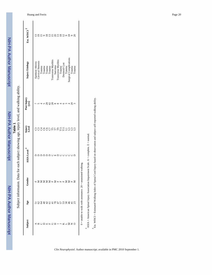
Fifteen individuals with incomplete spinal cord injury participated in this study after providingwritten informed consent. There were six subjects with a cervical injury, five with a thoracicinjury, and four with a lumbar injury (Table 1). All subjects were at least 12 months post-injuryand free of any conditions that would limit their ability to exercise safely. Subjects had to beable to perform the recumbent stepping task with just their upper limbs to participate in thestudy. All subjects were screened and approved for participation by a physician from thePhysical Medicine and Rehabilitation Department at the University of Michigan. TheUniversity of Michigan Medical School Institutional Review Board approved the protocol andconsent form in accord with the Declaration of Helsinki.
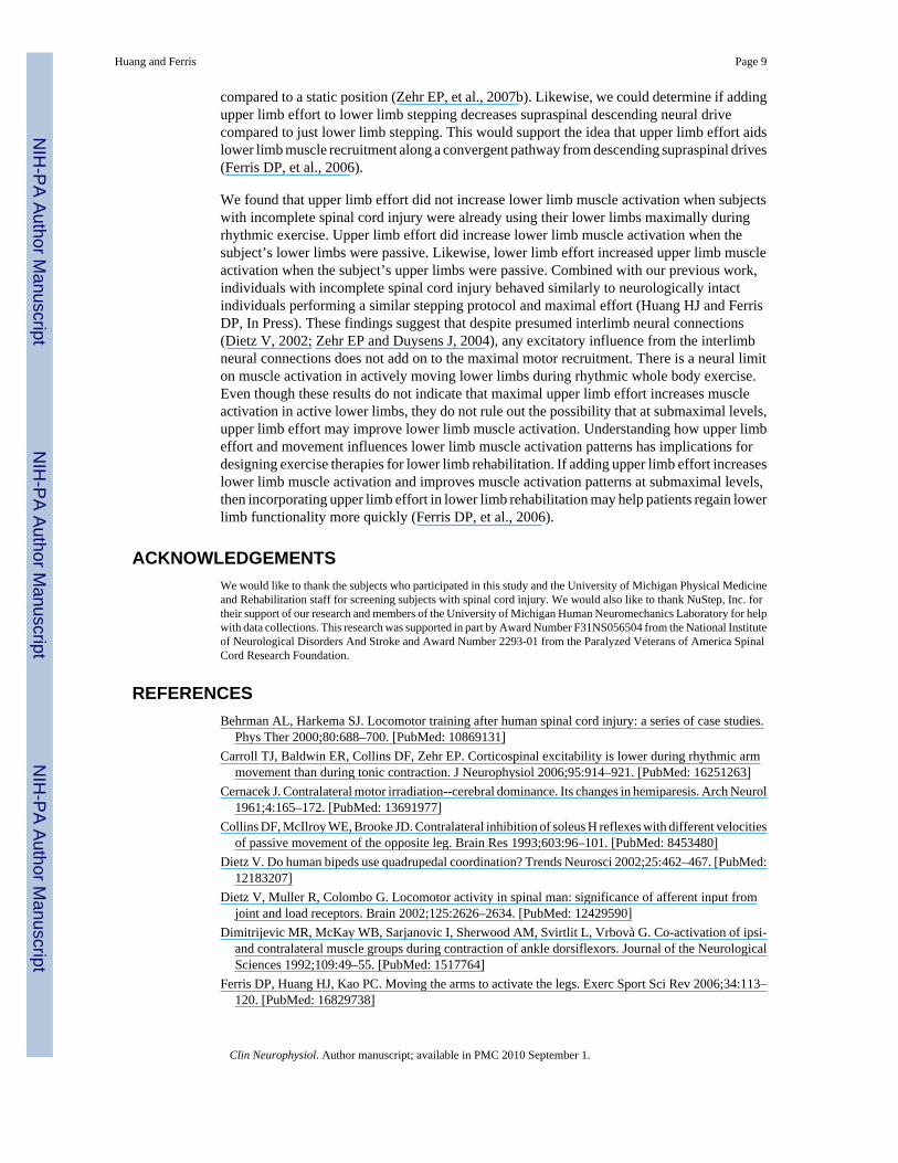
Computer-controlled Recumbent StepperWe have taken a commercially available recumbent stepper (TRS 4000, NuStep Inc., AnnArbor, MI) and modified it to have computer-controlled real-time resistance (Fig. 1) (HuangHJ and Ferris DP, In Press). We also instrumented the recumbent stepper with load cells to
Huang and Ferris Page 2
Clin Neurophysiol. Author manuscript; available in PMC 2010 September 1.
NIH
-PA Author Manuscript
NIH
-PA Author Manuscript
NIH
-PA Author Manuscript
measure handle and pedal forces. For this study, the stepper followed a prescribed sine-waveposition profile with a stepping frequency of 75 beats per minute (equivalent to the steppingfrequency of walking at ~1.25 m/s). If subjects were unable to step at the desired frequency,then the stepper drove the stepping motion. If subjects were strong enough to drive the steppingmotion faster than the desired stepping frequency, the motor generated a torque to oppose thesubject’s effort. This allowed the stepper to have a fixed position profile and to maintain thedesired stepping frequency.
Experimental Set UpWe adjusted the stepper to make the range of the stepping motion as comfortable as possiblefor each subject. The seat position was set so that the knees were near full extension but couldnot lock out. For some more hyper-reflexive subjects, we had to set the seat so that their legswere more flexed for safety reasons. If needed, we used leg stabilizers to prevent the subject’slegs from abducting and potentially colliding with the moving handles. We aligned each footto be centered within the pedal. As the pedal was only 5.5 inches wide, it prevented subjectsfrom rotating their feet medially or laterally. We used a torso strap to minimize torso movementduring stepping. We also used Velcro gloves to attach the hands to the handles and used footstraps to attach the feet to the pedals during passive conditions. This allowed subjects to be aspassive as possible because they did not have to actively hold the handles or keep their feet onthe pedals throughout the stepping motion.
ProtocolSubjects performed recumbent stepping using different combinations of upper (U) and lower(L) limb effort. For active effort, we instructed subjects to use maximal effort. For passiveeffort, we instructed subjects to relax as much as possible. We tested three active lower limbconditions: a) Resting Upper & Active Lower [RU-AL], b) Passive Upper & Active Lower[PU-AL], c) Active Upper & Active Lower [AU-AL]. For the resting upper limb condition,we had subjects cross their arms and rest them on his/her lap. These active lower limb conditionsexamined whether different upper limb states altered active lower limb muscleelectromyography amplitudes. We also tested two passive lower limb conditions, d) PassiveUpper & Passive Lower [PU-PL], and e) Active Upper & Passive Lower [AU-PL], to determinehow upper limb effort influences passive lower limb muscle activation in individuals withincomplete spinal cord injury.
We collected two sets of data, with each set consisting of five trials for each of the fiveconditions. Conditions were randomized for each subject. Before each trial, we verballydescribed the combination of arm and leg effort to the subject. Subjects were instructed to relaxand use the first fifteen seconds to get used to the stepping frequency as the stepper slowlyramped up to full range of motion. Then on a verbal cue, we instructed subjects to perform thestepping condition with maximal effort for approximately fifteen seconds. This yielded six toeight strides of data. Throughout the trial, we gave the subject verbal cues and encouragement.Subjects were also given an opportunity to practice the condition prior to testing at theirdiscretion. The average length of rest between trials was one minute.
Data AcquisitionWe collected data signals using two computer systems at a sampling rate of 1000 Hz. Onecomputer was used to collect electromyography, load cell, and joint angle data signals. Theother computer ran the real-time software program and sampled data signals related to therecumbent stepper hardware. We used a common data signal sampled in both systems tosynchronize the data offline.
Huang and Ferris Page 3
Clin Neurophysiol. Author manuscript; available in PMC 2010 September 1.
NIH
-PA Author Manuscript
NIH
-PA Author Manuscript
NIH
-PA Author Manuscript
Electromyography (EMG)—We measured surface electromyography (Delsys, Boston,MA) from sixteen muscles, four muscles on each limb. On each lower limb, we measuredmuscle activity from the vastus medialis (VM), medial hamstrings (MH), tibialis anterior (TA),and soleus (SO). On each upper limb, we measured muscle activity from the anterior deltoid(AD), posterior deltoid (PD), biceps brachii (BB), and lateral head of the triceps brachii (TB).We shaved each electrode site and cleaned them with rubbing alcohol. We then placed theelectrode sensor over the muscle belly along the long axis, secured the electrode with tape, andwrapped excess loose electrode wires to the limbs with elastic foam wrap. We processed theEMG data with a second order high-pass Butterworth filter with zero lag (cutoff frequency of20 Hz) to attenuate low frequency components such as mechanical artifact. We then full waverectified the EMG data signals.
Joint Angles—We measured bilateral joint angles of the ankles, knees, hips, and elbowsusing twin-axis electrogoniometers placed along the sagittal plane (Biometrics Ltd, Ladysmith,VA). Electrogoniometers were zeroed with the limbs in the anatomically neutral position. Jointangle data were processed with a second order low-pass Butterworth filter with zero lag (cutofffrequency of 6 Hz). Because of equipment malfunctions, we were not able to obtain data fromall eight goniometers during all conditions for every subject.
Kinetics—We calculated the forces each hand and foot contributed to the stepping motionvia single axis load cells (Fig. 1). Because the handle and contralateral pedal were part of asingle rigid body, the torques generated by a force from the hand and a force from thecontralateral foot summed and yielded a net torque for the handle-pedal unit (Fig. 1B). Wemeasured directly the force exerted by each hand through a load cell mounted in the handle.We also measured the force associated with the net torque for each handle-pedal unit througha load cell mounted in a connecting link between the handle-pedal unit and a cam. Using themeasured forces and moment arm relationships, we calculated the torques associated with eachhandle and handle-pedal unit. We subtracted the handle torque from the handle-pedal unittorque to determine the pedal torque of that contralateral hand-foot pair. We then divided thepedal torques by the pedal moment arm to find the pedal forces. We filtered measured forcedata using a second order low-pass Butterworth filter with zero lag (cutoff frequency of 6 Hz).
Data AnalysisFor all subjects, we analyzed the data from the second set. The subjects with incomplete spinalcord injury were more consistent during the second set.
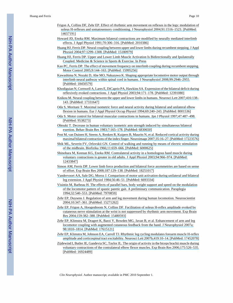
Calculation of Mean Profiles—To compare EMG patterns between conditions, wecalculated group normalized EMG mean profiles over a stride cycle for each condition. Thebeginning and end of each stride corresponded with the left lower limb and right upper limbat full extension as indicated from the position data (Fig. 2A). We first calculated an intra-subject EMG mean profile for a stride cycle per condition. We then normalized the intra-subjectEMG mean profiles to the maximum value among all conditions. We then calculated a groupnormalized EMG mean profile for each condition by averaging all of the intra-subjectnormalized EMG mean profiles for that condition. We used the same general procedure, butwithout normalization, for the joint angle and force mean profiles.
Calculation of EMG Amplitudes—To compare EMG amplitudes across conditions, wecalculated a group averaged normalized root-mean-square (RMS) EMG for each muscle andcondition. For each muscle, we only calculated RMS EMG during the half of the stride whenthe muscle was concentrically contracting. For example, for the right vastus medialis, wecalculated the RMS EMG during the first half of the stride cycle when the knee was extending(Fig. 2 grey blocks). We calculated each muscle’s RMS EMG for the concentric half of the
Huang and Ferris Page 4
Clin Neurophysiol. Author manuscript; available in PMC 2010 September 1.
NIH
-PA Author Manuscript
NIH
-PA Author Manuscript
NIH
-PA Author Manuscript
cycle for each subject-condition data set. We calculated an intra-subject average RMS EMGfor each muscle per condition. We then normalized the intra-subject RMS EMG amplitudesfor each muscle (left and right vastus medialis, medial hamstrings, soleus, tibialis anterior,anterior deltoid, posterior deltoid, biceps brachii, and triceps brachii ) to the maximum intra-subject average RMS EMG amplitude across all conditions. We excluded any subject’s datathat did not have at least a 10% difference between Passive Upper & Passive Lower and PassiveUpper & Active Lower conditions for lower limb RMS EMG and between Passive Upper &Passive Lower and Active Upper & Passive Lower conditions for upper limb RMS EMG. Wethen averaged across subjects to calculate the group averaged normalized RMS EMG amplitudefor each muscle per condition.
Statistical Analysis—We used a repeated measures analysis of variance (rmANOVA) todetermine if there were significant differences in lower limb muscle activation among activelower limb conditions. We also ran another rmANOVA to determine if there were significantdifferences in lower (or upper) limb muscle activation among passive lower (or upper) limbconditions. If the rmANOVA showed a significant difference among conditions, we used aTukey’s honestly significant difference (THSD) post hoc test to determine which conditionswere significantly different (P < 0.05).
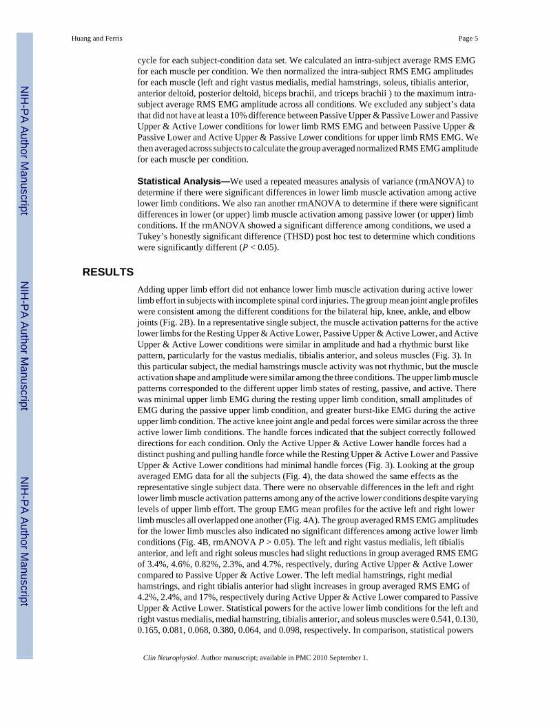
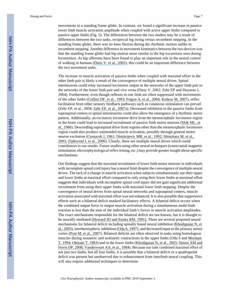
RESULTSAdding upper limb effort did not enhance lower limb muscle activation during active lowerlimb effort in subjects with incomplete spinal cord injuries. The group mean joint angle profileswere consistent among the different conditions for the bilateral hip, knee, ankle, and elbowjoints (Fig. 2B). In a representative single subject, the muscle activation patterns for the activelower limbs for the Resting Upper & Active Lower, Passive Upper & Active Lower, and ActiveUpper & Active Lower conditions were similar in amplitude and had a rhythmic burst likepattern, particularly for the vastus medialis, tibialis anterior, and soleus muscles (Fig. 3). Inthis particular subject, the medial hamstrings muscle activity was not rhythmic, but the muscleactivation shape and amplitude were similar among the three conditions. The upper limb musclepatterns corresponded to the different upper limb states of resting, passive, and active. Therewas minimal upper limb EMG during the resting upper limb condition, small amplitudes ofEMG during the passive upper limb condition, and greater burst-like EMG during the activeupper limb condition. The active knee joint angle and pedal forces were similar across the threeactive lower limb conditions. The handle forces indicated that the subject correctly followeddirections for each condition. Only the Active Upper & Active Lower handle forces had adistinct pushing and pulling handle force while the Resting Upper & Active Lower and PassiveUpper & Active Lower conditions had minimal handle forces (Fig. 3). Looking at the groupaveraged EMG data for all the subjects (Fig. 4), the data showed the same effects as therepresentative single subject data. There were no observable differences in the left and rightlower limb muscle activation patterns among any of the active lower conditions despite varyinglevels of upper limb effort. The group EMG mean profiles for the active left and right lowerlimb muscles all overlapped one another (Fig. 4A). The group averaged RMS EMG amplitudesfor the lower limb muscles also indicated no significant differences among active lower limbconditions (Fig. 4B, rmANOVA P > 0.05). The left and right vastus medialis, left tibialisanterior, and left and right soleus muscles had slight reductions in group averaged RMS EMGof 3.4%, 4.6%, 0.82%, 2.3%, and 4.7%, respectively, during Active Upper & Active Lowercompared to Passive Upper & Active Lower. The left medial hamstrings, right medialhamstrings, and right tibialis anterior had slight increases in group averaged RMS EMG of4.2%, 2.4%, and 17%, respectively during Active Upper & Active Lower compared to PassiveUpper & Active Lower. Statistical powers for the active lower limb conditions for the left andright vastus medialis, medial hamstring, tibialis anterior, and soleus muscles were 0.541, 0.130,0.165, 0.081, 0.068, 0.380, 0.064, and 0.098, respectively. In comparison, statistical powers
Huang and Ferris Page 5
Clin Neurophysiol. Author manuscript; available in PMC 2010 September 1.
NIH
-PA Author Manuscript
NIH
-PA Author Manuscript
NIH
-PA Author Manuscript
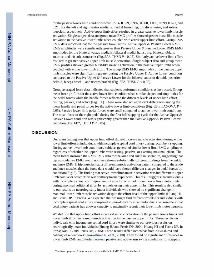
for the passive lower limb conditions were 0.514, 0.829, 0.997, 0.980, 1.000, 0.999, 0.623, and0.218 for the left and right vastus medialis, medial hamstring, tibialis anterior, and soleusmuscles, respectively. Active upper limb effort resulted in greater passive lower limb muscleactivation. Single subject data and group mean EMG profiles showed greater burst-like muscleactivation in the passive lower limbs when coupled with active upper limb effort. Group RMSEMG data indicated that for the passive lower limbs, Active Upper & Passive Lower RMSEMG amplitudes were significantly greater than Passive Upper & Passive Lower RMS EMGamplitudes for the bilateral vastus medialis, bilateral medial hamstring, bilateral tibialisanterior, and left soleus muscles (Fig. 5A*, THSD P < 0.05). Similarly, active lower limb effortresulted in greater passive upper limb muscle activation. Single subject data and group meanEMG profiles showed greater burst-like muscle activation in the passive upper limbs whencoupled with active lower limb effort. The group RMS EMG amplitudes of the passive upperlimb muscles were significantly greater during the Passive Upper & Active Lower conditioncompared to the Passive Upper & Passive Lower for the bilateral anterior deltoid, posteriordeltoid, biceps brachii, and triceps brachii (Fig. 5B*, THSD P < 0.05).
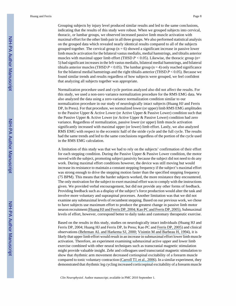
Group averaged force data indicated that subjects performed conditions as instructed. Groupmean force profiles for the active lower limb conditions had similar shapes and amplitudes forthe pedal forces while the handle forces reflected the different levels of upper limb effort,resting, passive, and active (Fig. 6A). There were also no significant differences among themean handle and pedal forces for the active lower limb conditions (Fig. 6B, rmANOVA P >0.05). Passive lower limb pedal forces were small compared to active lower limb conditions.The mean force of the right pedal during the first half stepping cycle for the Active Upper &Passive Lower condition was significantly greater than the Passive Upper & Passive Lowercondition (Fig. 6B*, THSD P < 0.05).
DISCUSSIONOur main finding was that upper limb effort did not increase muscle activation during activelower limb effort in individuals with incomplete spinal cord injury during recumbent stepping.During active lower limb conditions, subjects generated similar lower limb EMG amplitudesregardless of whether the upper limbs were resting, passive, or exerting maximal effort. Themean forces mirrored the RMS EMG data for the knee and ankle musculature, suggesting thathip musculature EMG would not have shown substantially different findings from the ankleand knee EMG. If hip muscles had a different muscle activation pattern compared to the ankleand knee muscles then the force data would have shown different changes in pedal forces bycondition (Fig. 6). The finding that active lower limb muscle activation was indifferent to upperlimb passive or active effort was contrary to our hypothesis. This result suggests that individualswith incomplete spinal cord injury are not able to recruit additional lower limb motor unitsduring maximal volitional effort by actively using their upper limbs. This result is also similarto our results on neurologically intact individuals who showed no significant change inmaximal lower limb muscle activation despite the effort level of the upper limbs (Huang HJand Ferris DP, In Press). We expected that we might find different results for individuals withincomplete spinal cord injury compared to neurologically intact individuals because the spinalcord injury patients had a lower capacity to maximally recruit their lower limb motor neurons.
We did find that upper limb effort increased muscle activation in the passive lower limbs andlower limb effort increased muscle activation in the passive upper limbs. These results onindividuals with incomplete spinal cord injury were similar to our previous results onneurologically intact individuals (Huang HJ and Ferris DP, 2004; Huang HJ and Ferris DP, InPress; Kao PC and Ferris DP, 2005). These results differ somewhat from Kawashima andcolleagues recent work (Kawashima N, et al., 2008). They found no significant differences inlower limb EMG amplitudes between passive and active arm swing conditions for stepping
Huang and Ferris Page 6
Clin Neurophysiol. Author manuscript; available in PMC 2010 September 1.
NIH
-PA Author Manuscript
NIH
-PA Author Manuscript
NIH
-PA Author Manuscript
movements in a standing frame glider. In contrast, we found a significant increase in passivelower limb muscle activation amplitude when coupled with active upper limbs compared topassive upper limbs (Fig. 5). The differences between the two studies may be a result ofdifferences between the two tasks, reciprocal leg swing versus recumbent stepping. In thestanding frame glider, there was no knee flexion during the rhythmic motion unlike inrecumbent stepping. Another difference in movement kinematics between the two devices wasthat the standing frame glider had hip motion more similar to the hip excursions seen duringlocomotion. As hip afferents have been found to play an important role in the neural controlof walking in humans (Dietz V, et al., 2002), this could be an important difference betweenthe two movement tasks.
The increase in muscle activation of passive limbs when coupled with maximal effort in theother limb pair is likely a result of the convergence of multiple neural drives. Spinalinterneurons could relay increased locomotor output in the networks of the upper limb pair tothe networks of the lower limb pair and vice versa (Dietz V, 2002; Zehr EP and Duysens J,2004). Furthermore, even though reflexes in one limb are often suppressed with movementsof the other limbs (Collins DF, et al., 1993; Frigon A, et al., 2004; Knikou M, 2007), reflexfacilitation from other sensory feedback pathways such as cutaneous stimulation can prevail(Zehr EP, et al., 2004; Zehr EP, et al., 2007a). Decreased inhibition to the passive limbs fromsupraspinal centers or spinal interneurons could also allow the emergence of a rhythmic motorpattern. Additionally, an excitatory locomotor drive from the mesencephalic locomotor regionin the brain could lead to increased recruitment of passive limb motor neurons (Shik ML, etal., 1966). Descending supraspinal drive from regions other than the mesencephalic locomotorregion could also produce unintended muscle activation, possibly through general motorneuron excitation (Cernacek J, 1961; Dimitrijevic MR, et al., 1992; Shinohara M, et al.,2003; Zijdewind I, et al., 2006). Clearly, there are multiple neural drives which couldcontributors to our results. Future studies using other neural techniques (transcranial magneticstimulation, electrophysiological reflex testing, etc.) may provide greater insight about specificmechanisms.
Our findings suggest that the maximal recruitment of lower limb motor neurons in individualswith incomplete spinal cord injury has a neural limit despite the convergence of multiple neuraldrives. The lack of a change in muscle activation when subjects simultaneously use their upperand lower limbs at maximal effort compared to only using their lower limbs at maximal effortsuggests that individuals with incomplete spinal cord injury did not gain significant additionalrecruitment from using their upper limbs with maximal lower limb stepping. Despite theconvergence of neural drives from spinal neural networks and supraspinal centers, muscleactivation associated with maximal effort was not enhanced. It is also possible that suppressiveeffects such as a bilateral deficit masked facilitatory effects. A bilateral deficit occurs whenthe combined output force or output muscle activation during a simultaneous multi-limbexertion is less than the sum of the individual limb’s forces or muscle activation amplitudes.The exact mechanisms responsible for the bilateral deficit are not known, but it is thought tobe neurally mediated (Howard JD and Enoka RM, 1991). There are several proposed neuralmechanisms for bilateral deficit including spinally based neural inhibition (Khodiguian N, etal., 2003), interhemispheric inhibition (Oda S, 1997), and decreased input to the primary motorcortex (Post M, et al., 2007). Bilateral deficits are often observed in tasks using homologousmuscles during isometric and isokinetic contractions in the upper limbs (Oda S and MoritaniT, 1994; Ohtsuki T, 1983) and in the lower limbs (Khodiguian N, et al., 2003; Simon AM andFerris DP, 2008; Vandervoort AA, et al., 1984). Because our task combined maximal effort ofnot just two limbs, but all four limbs, it is possible that a bilateral deficit or a quadrupedaldeficit was present but unobserved due to enhancement from interlimb neural coupling. Thiswill also require additional techniques to determine.
Huang and Ferris Page 7
Clin Neurophysiol. Author manuscript; available in PMC 2010 September 1.
NIH
-PA Author Manuscript
NIH
-PA Author Manuscript
NIH
-PA Author Manuscript
Grouping subjects by injury level produced similar results and led to the same conclusions,indicating that the results of this study were robust. When we grouped subjects into cervical,thoracic, or lumbar groups, we observed increased passive limb muscle activation withmaximal effort for the other limb pair in all three groups. We also performed statistical analysison the grouped data which revealed nearly identical results compared to all of the subjectsgrouped together. The cervical group (n = 6) showed a significant increase in passive lowerlimb muscle activation for the bilateral vastus medialis, medial hamstrings, and tibialis anteriormuscles with maximal upper limb effort (THSD P < 0.05). Likewise, the thoracic group (n=5) had significant increases in the left vastus medialis, bilateral medial hamstrings, and bilateraltibialis anterior muscles (THSD P < 0.05). The lumbar group (n = 4) only reached significancefor the bilateral medial hamstrings and the right tibialis anterior (THSD P < 0.05). Because wefound similar trends and results regardless of how subjects were grouped, we feel confidentthat analyzing all subjects together was appropriate.
Normalization procedure used and cycle portion analyzed also did not affect the results. Forthis study, we used a non-zero variance normalization procedure for the RMS EMG data. Wealso analyzed the data using a zero-variance normalization condition similar to ournormalization procedure in our study of neurologically intact subjects (Huang HJ and FerrisDP, In Press). For that procedure, we normalized lower (or upper) limb RMS EMG amplitudesto the Passive Upper & Active Lower (or Active Upper & Passive Lower) condition such thatthe Passive Upper & Active Lower (or Active Upper & Passive Lower) condition had zerovariance. Regardless of normalization, passive lower (or upper) limb muscle activationsignificantly increased with maximal upper (or lower) limb effort. Lastly, we also analyzedRMS EMG with respect to the eccentric half of the stride cycle and the full cycle. The resultshad the same trends and led to the same conclusions regardless of the portion of the cycle usedin the RMS EMG calculation.
A limitation of this study was that we had to rely on the subjects’ confirmation of their effortfor each stepping condition. During the Passive Upper & Passive Lower condition, the motormoved with the subject, promoting subject passivity because the subject did not need to do anywork. During maximal effort conditions however, the device was still moving but wouldincrease its resistance to maintain a constant stepping frequency if the subject’s maximal effortwas strong enough to drive the stepping motion faster than the specified stepping frequency(75 BPM). This means that the harder subjects worked, the more resistance they encountered.The only motivation for the subject to exert maximal effort was to comply with the instructionsgiven. We provided verbal encouragement, but did not provide any other forms of feedback.Providing feedback such as a display of the subject’s force production would alter the task andinvolve more voluntary and supraspinal processes. Another limitation was that we did notexamine any submaximal levels of recumbent stepping. Based on our previous work, we choseto have subjects use maximum effort to produce the greatest change in passive limb motorneuron recruitment (Huang HJ and Ferris DP, 2004; Kao PC and Ferris DP, 2005). Submaximallevels of effort, however, correspond better to daily tasks and customary therapeutic exercise.
Based on the results in this study, studies on neurologically intact individuals (Huang HJ andFerris DP, 2004; Huang HJ and Ferris DP, In Press; Kao PC and Ferris DP, 2005) and clinicalobservations (Behrman AL and Harkema SJ, 2000; Visintin M and Barbeau H, 1994), it islikely that upper limb effort would result in an increase in submaximal effort lower limb muscleactivation. Therefore, an experiment examining submaximal active upper and lower limbexercise combined with other neural techniques such as transcranial magnetic stimulationmight provide valuable insight. Zehr and colleagues used transcranial magnetic stimulation toshow that rhythmic arm movement decreased cortiospinal excitability of a forearm musclecompared to tonic voluntary contraction (Carroll TJ, et al., 2006). In a similar experiment, theydemonstrated that rhythmic leg cycling increased corticospinal excitability of a forearm muscle
Huang and Ferris Page 8
Clin Neurophysiol. Author manuscript; available in PMC 2010 September 1.
NIH
-PA Author Manuscript
NIH
-PA Author Manuscript
NIH
-PA Author Manuscript
compared to a static position (Zehr EP, et al., 2007b). Likewise, we could determine if addingupper limb effort to lower limb stepping decreases supraspinal descending neural drivecompared to just lower limb stepping. This would support the idea that upper limb effort aidslower limb muscle recruitment along a convergent pathway from descending supraspinal drives(Ferris DP, et al., 2006).
We found that upper limb effort did not increase lower limb muscle activation when subjectswith incomplete spinal cord injury were already using their lower limbs maximally duringrhythmic exercise. Upper limb effort did increase lower limb muscle activation when thesubject’s lower limbs were passive. Likewise, lower limb effort increased upper limb muscleactivation when the subject’s upper limbs were passive. Combined with our previous work,individuals with incomplete spinal cord injury behaved similarly to neurologically intactindividuals performing a similar stepping protocol and maximal effort (Huang HJ and FerrisDP, In Press). These findings suggest that despite presumed interlimb neural connections(Dietz V, 2002; Zehr EP and Duysens J, 2004), any excitatory influence from the interlimbneural connections does not add on to the maximal motor recruitment. There is a neural limiton muscle activation in actively moving lower limbs during rhythmic whole body exercise.Even though these results do not indicate that maximal upper limb effort increases muscleactivation in active lower limbs, they do not rule out the possibility that at submaximal levels,upper limb effort may improve lower limb muscle activation. Understanding how upper limbeffort and movement influences lower limb muscle activation patterns has implications fordesigning exercise therapies for lower limb rehabilitation. If adding upper limb effort increaseslower limb muscle activation and improves muscle activation patterns at submaximal levels,then incorporating upper limb effort in lower limb rehabilitation may help patients regain lowerlimb functionality more quickly (Ferris DP, et al., 2006).
ACKNOWLEDGEMENTSWe would like to thank the subjects who participated in this study and the University of Michigan Physical Medicineand Rehabilitation staff for screening subjects with spinal cord injury. We would also like to thank NuStep, Inc. fortheir support of our research and members of the University of Michigan Human Neuromechanics Laboratory for helpwith data collections. This research was supported in part by Award Number F31NS056504 from the National Instituteof Neurological Disorders And Stroke and Award Number 2293-01 from the Paralyzed Veterans of America SpinalCord Research Foundation.
REFERENCESBehrman AL, Harkema SJ. Locomotor training after human spinal cord injury: a series of case studies.
Phys Ther 2000;80:688–700. [PubMed: 10869131]Carroll TJ, Baldwin ER, Collins DF, Zehr EP. Corticospinal excitability is lower during rhythmic arm
movement than during tonic contraction. J Neurophysiol 2006;95:914–921. [PubMed: 16251263]Cernacek J. Contralateral motor irradiation--cerebral dominance. Its changes in hemiparesis. Arch Neurol
1961;4:165–172. [PubMed: 13691977]Collins DF, McIlroy WE, Brooke JD. Contralateral inhibition of soleus H reflexes with different velocities
of passive movement of the opposite leg. Brain Res 1993;603:96–101. [PubMed: 8453480]Dietz V. Do human bipeds use quadrupedal coordination? Trends Neurosci 2002;25:462–467. [PubMed:
12183207]Dietz V, Muller R, Colombo G. Locomotor activity in spinal man: significance of afferent input from
joint and load receptors. Brain 2002;125:2626–2634. [PubMed: 12429590]Dimitrijevic MR, McKay WB, Sarjanovic I, Sherwood AM, Svirtlit L, Vrbovà G. Co-activation of ipsi-
and contralateral muscle groups during contraction of ankle dorsiflexors. Journal of the NeurologicalSciences 1992;109:49–55. [PubMed: 1517764]
Ferris DP, Huang HJ, Kao PC. Moving the arms to activate the legs. Exerc Sport Sci Rev 2006;34:113–120. [PubMed: 16829738]
Huang and Ferris Page 9
Clin Neurophysiol. Author manuscript; available in PMC 2010 September 1.
NIH
-PA Author Manuscript
NIH
-PA Author Manuscript
NIH
-PA Author Manuscript
Frigon A, Collins DF, Zehr EP. Effect of rhythmic arm movement on reflexes in the legs: modulation ofsoleus H-reflexes and somatosensory conditioning. J Neurophysiol 2004;91:1516–1523. [PubMed:14657191]
Howard JD, Enoka RM. Maximum bilateral contractions are modified by neurally mediated interlimbeffects. J Appl Physiol 1991;70:306–316. [PubMed: 2010386]
Huang HJ, Ferris DP. Neural coupling between upper and lower limbs during recumbent stepping. J ApplPhysiol 2004;97:1299–1308. [PubMed: 15180979]
Huang HJ, Ferris DP. Upper and Lower Limb Muscle Activation Is Bidirectionally and IpsilaterallyCoupled. Medicine & Science in Sports & Exercise. In Press
Kao PC, Ferris DP. The effect of movement frequency on interlimb coupling during recumbent stepping.Motor Control 2005;9:144–163. [PubMed: 15995256]
Kawashima N, Nozaki D, Abe MO, Nakazawa K. Shaping appropriate locomotive motor output throughinterlimb neural pathway within spinal cord in humans. J Neurophysiol 2008;99:2946–2955.[PubMed: 18450579]
Khodiguian N, Cornwell A, Lares E, DiCaprio PA, Hawkins SA. Expression of the bilateral deficit duringreflexively evoked contractions. J Appl Physiol 2003;94:171–178. [PubMed: 12391080]
Knikou M. Neural coupling between the upper and lower limbs in humans. Neurosci Lett 2007;416:138–143. [PubMed: 17331647]
Oda S, Moritani T. Maximal isometric force and neural activity during bilateral and unilateral elbowflexion in humans. Eur J Appl Physiol Occup Physiol 1994;69:240–243. [PubMed: 8001536]
Oda S. Motor control for bilateral muscular contractions in humans. Jpn J Physiol 1997;47:487–498.[PubMed: 9538273]
Ohtsuki T. Decrease in human voluntary isometric arm strength induced by simultaneous bilateralexertion. Behav Brain Res 1983;7:165–178. [PubMed: 6830650]
Post M, van Duinen H, Steens A, Renken R, Kuipers B, Maurits N, et al. Reduced cortical activity duringmaximal bilateral contractions of the index finger. Neuroimage 2007;35:16–27. [PubMed: 17223576]
Shik ML, Severin FV, Orlovskii GN. Control of walking and running by means of electric stimulationof the midbrain. Biofizika 1966;11:659–666. [PubMed: 6000625]
Shinohara M, Keenan KG, Enoka RM. Contralateral activity in a homologous hand muscle duringvoluntary contractions is greater in old adults. J Appl Physiol 2003;94:966–974. [PubMed:12433847]
Simon AM, Ferris DP. Lower limb force production and bilateral force asymmetries are based on senseof effort. Exp Brain Res 2008;187:129–138. [PubMed: 18251017]
Vandervoort AA, Sale DG, Moroz J. Comparison of motor unit activation during unilateral and bilateralleg extension. J Appl Physiol 1984;56:46–51. [PubMed: 6693334]
Visintin M, Barbeau H. The effects of parallel bars, body weight support and speed on the modulationof the locomotor pattern of spastic paretic gait. A preliminary communication. Paraplegia1994;32:540–553. [PubMed: 7970859]
Zehr EP, Duysens J. Regulation of arm and leg movement during human locomotion. Neuroscientist2004;10:347–361. [PubMed: 15271262]
Zehr EP, Frigon A, Hoogenboom N, Collins DF. Facilitation of soleus H-reflex amplitude evoked bycutaneous nerve stimulation at the wrist is not suppressed by rhythmic arm movement. Exp BrainRes 2004;159:382–388. [PubMed: 15480593]
Zehr EP, Klimstra M, Dragert K, Barzi Y, Bowden MG, Javan B, et al. Enhancement of arm and leglocomotor coupling with augmented cutaneous feedback from the hand. J Neurophysiol 2007a;98:1810–1814. [PubMed: 17615121]
Zehr EP, Klimstra M, Johnson EA, Carroll TJ. Rhythmic leg cycling modulates forearm muscle H-reflexamplitude and corticospinal tract excitability. Neurosci Lett 2007b;419:10–14. [PubMed: 17452078]
Zijdewind I, Butler JE, Gandevia SC, Taylor JL. The origin of activity in the biceps brachii muscle duringvoluntary contractions of the contralateral elbow flexor muscles. Exp Brain Res 2006;175:526–535.[PubMed: 16924489]
Huang and Ferris Page 10
Clin Neurophysiol. Author manuscript; available in PMC 2010 September 1.
NIH
-PA Author Manuscript
NIH
-PA Author Manuscript
NIH
-PA Author Manuscript
Figure 1.A) Recumbent stepping machine with real-time computer-controlled resistance and force andposition sensors (modified TRS 4000, NuStep Inc, Ann Arbor, MI). The handles and seat areadjustable. Velcro gloves, foot straps, and a torso belt help minimize unwanted movement.Leg stabilizers also help prevent excessive medial-lateral movement. B) Schematic of theforces and torques for one handle-pedal unit on the recumbent stepper.
Huang and Ferris Page 11
Clin Neurophysiol. Author manuscript; available in PMC 2010 September 1.
NIH
-PA Author Manuscript
NIH
-PA Author Manuscript
NIH
-PA Author Manuscript
Figure 2.A) Schematic of recumbent stepping motion. At 0% of the stride cycle, the left lower limb andright upper limb are at full extension. From 0% to 50% of the stride cycle, the left lower limband right upper limb are flexing while the right lower limb and left upper limb are extending.At 50% of the stride cycle, the right lower limb and left upper limb are at full extension. From50% to 100% of the stride cycle, the right lower limb and left upper limb are flexing while theleft lower limb and right upper limb are extending. B) Group mean joint angle profiles for thebilateral hip, knee, ankle, and elbow during one stepping cycle. Black solid line: average forall conditions. Dotted grey lines: active lower limb conditions, Resting Upper & Active Lower,
Huang and Ferris Page 12
Clin Neurophysiol. Author manuscript; available in PMC 2010 September 1.
NIH
-PA Author Manuscript
NIH
-PA Author Manuscript
NIH
-PA Author Manuscript
Passive Upper & Active Lower, and Active Upper & Active Lower. Dashed grey lines: passivelower limb conditions, Passive Upper & Passive Lower and Active Upper & Passive Lower.
Huang and Ferris Page 13
Clin Neurophysiol. Author manuscript; available in PMC 2010 September 1.
NIH
-PA Author Manuscript
NIH
-PA Author Manuscript
NIH
-PA Author Manuscript
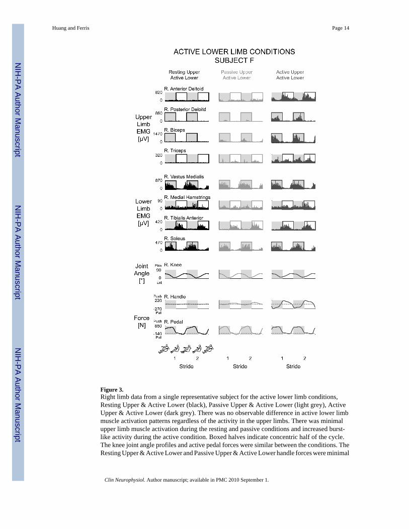
Figure 3.Right limb data from a single representative subject for the active lower limb conditions,Resting Upper & Active Lower (black), Passive Upper & Active Lower (light grey), ActiveUpper & Active Lower (dark grey). There was no observable difference in active lower limbmuscle activation patterns regardless of the activity in the upper limbs. There was minimalupper limb muscle activation during the resting and passive conditions and increased burst-like activity during the active condition. Boxed halves indicate concentric half of the cycle.The knee joint angle profiles and active pedal forces were similar between the conditions. TheResting Upper & Active Lower and Passive Upper & Active Lower handle forces were minimal
Huang and Ferris Page 14
Clin Neurophysiol. Author manuscript; available in PMC 2010 September 1.
NIH
-PA Author Manuscript
NIH
-PA Author Manuscript
NIH
-PA Author Manuscript
while the Active Upper & Active Lower handle forces had a large pushing and pulling force.Dashed lines in the force data is zero force.
Huang and Ferris Page 15
Clin Neurophysiol. Author manuscript; available in PMC 2010 September 1.
NIH
-PA Author Manuscript
NIH
-PA Author Manuscript
NIH
-PA Author Manuscript
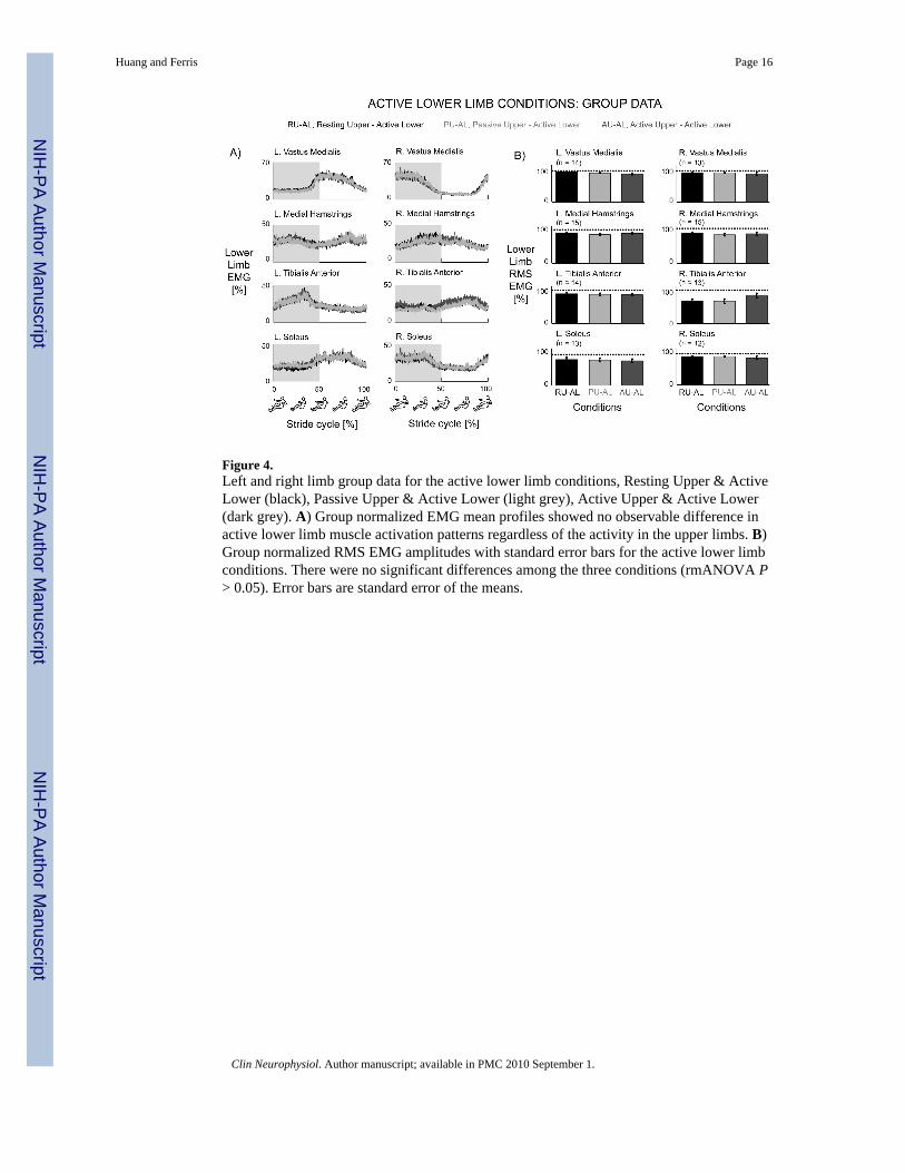
Figure 4.Left and right limb group data for the active lower limb conditions, Resting Upper & ActiveLower (black), Passive Upper & Active Lower (light grey), Active Upper & Active Lower(dark grey). A) Group normalized EMG mean profiles showed no observable difference inactive lower limb muscle activation patterns regardless of the activity in the upper limbs. B)Group normalized RMS EMG amplitudes with standard error bars for the active lower limbconditions. There were no significant differences among the three conditions (rmANOVA P> 0.05). Error bars are standard error of the means.
Huang and Ferris Page 16
Clin Neurophysiol. Author manuscript; available in PMC 2010 September 1.
NIH
-PA Author Manuscript
NIH
-PA Author Manuscript
NIH
-PA Author Manuscript
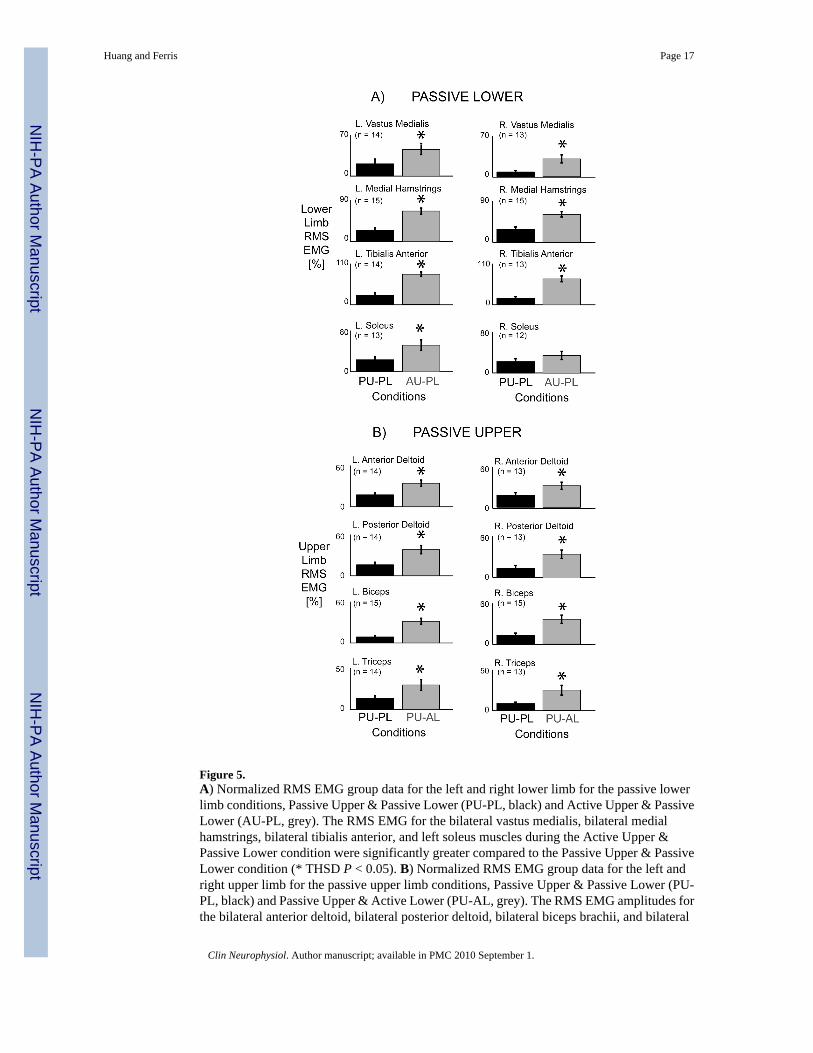
Figure 5.A) Normalized RMS EMG group data for the left and right lower limb for the passive lowerlimb conditions, Passive Upper & Passive Lower (PU-PL, black) and Active Upper & PassiveLower (AU-PL, grey). The RMS EMG for the bilateral vastus medialis, bilateral medialhamstrings, bilateral tibialis anterior, and left soleus muscles during the Active Upper &Passive Lower condition were significantly greater compared to the Passive Upper & PassiveLower condition (* THSD P < 0.05). B) Normalized RMS EMG group data for the left andright upper limb for the passive upper limb conditions, Passive Upper & Passive Lower (PU-PL, black) and Passive Upper & Active Lower (PU-AL, grey). The RMS EMG amplitudes forthe bilateral anterior deltoid, bilateral posterior deltoid, bilateral biceps brachii, and bilateral
Huang and Ferris Page 17
Clin Neurophysiol. Author manuscript; available in PMC 2010 September 1.
NIH
-PA Author Manuscript
NIH
-PA Author Manuscript
NIH
-PA Author Manuscript
triceps brachii muscles during the Passive Upper & Active Lower condition were significantlygreater compared to the Passive Upper & Passive Lower condition (* THSD P < 0.05). Errorbars are standard error of the means.
Huang and Ferris Page 18
Clin Neurophysiol. Author manuscript; available in PMC 2010 September 1.
NIH
-PA Author Manuscript
NIH
-PA Author Manuscript
NIH
-PA Author Manuscript
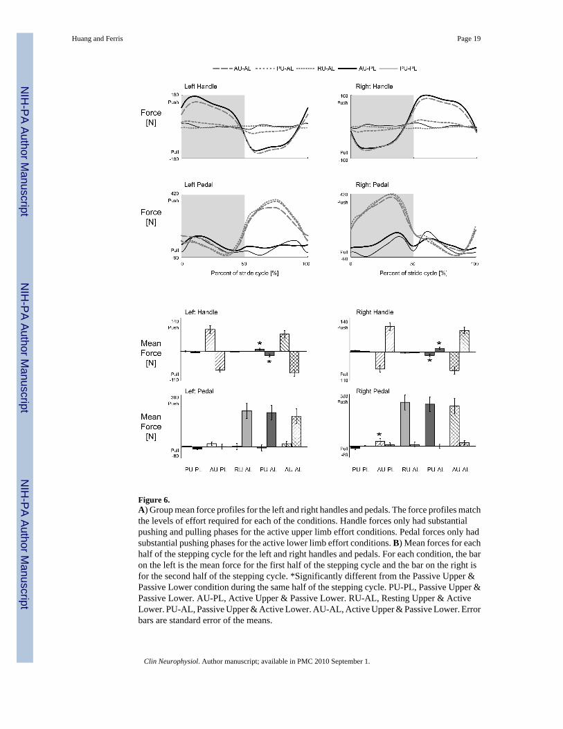
Figure 6.A) Group mean force profiles for the left and right handles and pedals. The force profiles matchthe levels of effort required for each of the conditions. Handle forces only had substantialpushing and pulling phases for the active upper limb effort conditions. Pedal forces only hadsubstantial pushing phases for the active lower limb effort conditions. B) Mean forces for eachhalf of the stepping cycle for the left and right handles and pedals. For each condition, the baron the left is the mean force for the first half of the stepping cycle and the bar on the right isfor the second half of the stepping cycle. *Significantly different from the Passive Upper &Passive Lower condition during the same half of the stepping cycle. PU-PL, Passive Upper &Passive Lower. AU-PL, Active Upper & Passive Lower. RU-AL, Resting Upper & ActiveLower. PU-AL, Passive Upper & Active Lower. AU-AL, Active Upper & Passive Lower. Errorbars are standard error of the means.
Huang and Ferris Page 19
Clin Neurophysiol. Author manuscript; available in PMC 2010 September 1.
NIH
-PA Author Manuscript
NIH
-PA Author Manuscript
NIH
-PA Author Manuscript
NIH
-PA Author Manuscript
NIH
-PA Author Manuscript
NIH
-PA Author Manuscript
Huang and Ferris Page 20Ta
ble
1Su
bjec
t inf
orm
aion
. Dat
a fo
r eac
h su
bjec
t sho
win
g ag
e, in
jury
leve
l, an
d w
alki
ng a
bilit
y.
Subj
ect
Age
Gen
der
ASI
A L
evel
*In
jury
Lev
elPo
st In
jury
(yrs
)In
jury
Etio
logy
Est
. WIS
CI †
A51
FD
C3
7Ep
idur
al A
bces
s13
B62
MD
C3
5C
ervi
cal S
teno
sis
19C
64M
DC
44
Trau
ma
15D
40M
DC
65
Trau
ma
9E
42M
DC
628
Trau
ma
18F
61M
DC
742
Trau
ma
13G
41F
CT5
28Tr
ansv
erse
Mye
litis
15H
78M
ET7
8Sa
rcom
a20
I47
FD
T98
Tran
sver
se M
yelit
is13
J57
FC
T11
4Tr
aum
a18
K53
FC
T11
6D
erm
oid
Cys
t12
L30
MC
L13
Trau
ma
9M
46M
CL3
4Su
rgic
al C
ompl
icat
ions
18N
41F
CL3
20Tr
aum
a9
O23
MD
L33
Trau
ma
20
0 =
unab
le to
wal
k w
ith a
ssis
tanc
e. 2
0 =
unas
sist
ed w
alki
ng.
* ASI
A =
Am
eric
an S
pina
l Inj
ury
Ass
ocia
tion
Impa
irmen
t Sca
le. A
= c
ompl
ete.
E =
nor
mal
.
† Est.
WIS
CI =
Est
imat
ed W
alki
ng In
dex
of S
pina
l Cor
d In
jury
bas
ed o
n ob
serv
atio
n an
d su
bjec
t sel
f-re
porte
d w
alki
ng a
bilit
y.
Clin Neurophysiol. Author manuscript; available in PMC 2010 September 1.


