Embed Size (px)
Citation preview
molecules
Article
Ursane-Type Triterpenes, Phenolics and Phenolic Derivativesfrom Globimetula braunii Leaf
Ayodeji Oluwabunmi Oriola 1,* , Adetunji Joseph Aladesanmi 2, Thomas Oyebode Idowu 3,Florence O. Akinwumi 4, Efere Martins Obuotor 5, Temilolu Idowu 6 and Adebola Omowunmi Oyedeji 1
�����������������
Citation: Oriola, A.O.; Aladesanmi,
A.J.; Idowu, T.O.; Akinwumi, F.O.;
Obuotor, E.M.; Idowu, T.; Oyedeji,
A.O. Ursane-Type Triterpenes,
Phenolics and Phenolic Derivatives
from Globimetula braunii Leaf.
Molecules 2021, 26, 6528. https://
doi.org/10.3390/molecules26216528
Academic Editor: Guy P.P. Kamatou
Received: 7 October 2021
Accepted: 25 October 2021
Published: 28 October 2021
Publisher’s Note: MDPI stays neutral
with regard to jurisdictional claims in
published maps and institutional affil-
iations.
Copyright: © 2021 by the authors.
Licensee MDPI, Basel, Switzerland.
This article is an open access article
distributed under the terms and
conditions of the Creative Commons
Attribution (CC BY) license (https://
creativecommons.org/licenses/by/
4.0/).
1 Department of Chemical and Physical Sciences, Faculty of Natural Sciences, Walter Sisulu University,Mthatha 5099, South Africa; [email protected]
2 Department of Pharmacognosy, Faculty of Pharmacy, Obafemi Awolowo University, Ile-Ife 220005, Nigeria;[email protected]
3 Department of Pharmaceutical Chemistry, Faculty of Pharmacy, Obafemi Awolowo University, Ile-Ife 220005,Nigeria; [email protected]
4 Department of Pharmaceutics, Faculty of Pharmacy, Obafemi Awolowo University, Ile-Ife 220005, Nigeria;[email protected]
5 Department of Biochemistry and Molecular Biology, Obafemi Awolowo University, Ile-Ife 220005, Nigeria;[email protected]
6 Department of Chemistry, Parkers Building, University of Manitoba, Winnipeg, MB R3T 2N2, Canada;[email protected]
* Correspondence: [email protected]; Tel.: +27-655934742
Abstract: Globimetula braunii is a hemi-parasitic plant used in African ethnomedicine for the manage-ment of microbial infections, rheumatic pain and tumors amongst others. We report the isolationand characterization of eight compounds with their antioxidant and antimicrobial activities. Theair-dried powdered leaf was macerated in EtOH/H20 (4:1). The extract was solvent-partitionedinto n-hexane, EtOAc, n-BuOH and aqueous fractions. The fractions were screened for their antioxi-dant properties, using DPPH, FRAP, TAC and FIC assays. Antimicrobial analysis was performedusing the micro-broth dilution method. The active EtOAc fraction was purified for its putativecompounds on a repeated silica gel column chromatography monitored with TLC-bioautography.The isolated compounds were characterized using spectroscopic methods of UV, FT-IR, NMR andMS. Eight compounds (1–8) were isolated and characterized as 13,27-cycloursane (1), phyllanthone(2), globraunone (3), three phenolics: methyl 3,5-dihydroxy-4-methoxybenzoate (4), methyl 3-methyl-4-hydroxybenzoate (5) and guaiacol (6), as well as two phenol derivatives: 4-formaldehyde phenone(7) and 6-methoxy-2H-inden-5-ol (8). The study identified 4 and 6 as natural antioxidant compoundswith potential as antimicrobial agents.
Keywords: Globimetula braunii; Loranthaceae; ursane-type triterpenes; phenolics; antioxidant;antimicrobial
1. Introduction
Globimetula braunii (Engl.) Van Tiegh. (Family Loranthaceae) is a hemi-parasitic andepiphytic plant that derives water and mineral nutrients from its hosts by means of aspecialized root system called “haustorium” [1]. It is commonly called “African Mistletoe”and locally called “Afomo Onisano” in Southwest Nigeria [2]. The plant is mostly found ondicot trees, such as Piliostigma thonningii, Leucena leucocephala and Theobroma cacao, where itbecomes bushy and woody, growing up to 5 ft in diameter until the host tree withers [3].At maturity, it produces reddish to reddish brown inflorescence with yellow patches in theform of a match sticks. It is widely distributed across tropical West African countries, suchas Nigeria, Ghana, Benin Republic and Cameroun [4].
G. braunii is implicated in the African ethnomedicine for the management of microbialinfections, wounds, cholera, hypertension, diabetes, rheumatism, ulcers and tumors [3,5,6].
Molecules 2021, 26, 6528. https://doi.org/10.3390/molecules26216528 https://www.mdpi.com/journal/molecules
Molecules 2021, 26, 6528 2 of 13
Biological studies have shown the antioxidant, antimicrobial and cytotoxic potentials ofthe leaf extract and its ethyl acetate fraction [7–9]. The hepatic, hematologic, anti-lipidemic,oxytocic, anti-hyperglycemic and anti-cancer activities of the plant’s alcoholic, aqueousand ethyl acetate extracts have also been reported [5,6,10–12] with a paucity of informationon its chemistry. Preliminary phytochemical reports showed that the plant containedphenolics, terpenes, flavonoids and sterols, while compounds, such as globrauneine A-F, lupeol, lupeol palmitate, β-sitosterol, friedelin, octanoic acid, lactones and flavonoids(quercetin, quercitrin, catechin, rutin and avicularin) have been reported in the plant [13,14].
We report, for the first time, the bioactivity-guided isolation and characterization ofthree ursane-type triterpenes, three phenolics and two phenol derivatives from the leaf ofG. braunii along with their antioxidant and antimicrobial activities.
2. Results and Discussion2.1. Spectra Data
Compound 1 (21 mg): 13,27-cycloursane, isolated as white amorphous powder, m.p.151–152 ◦C; ESI-MS: [M]+ at m/z 410.0, consistent with the molecular formula C30H50[M − C23H29]+ at m/z 105.3; 1H-NMR (300 MHz, CDCl3) δ ppm: 0.75 (3H, s, H-28), 0.89(3H, d, J = 6.0 Hz, H-30), 0.98 (3H, s, H-26), 1.03 (3H, d, J = 3.0 Hz, H-29), 1.07 (3H, s, H-25),1.20 (3H, s, H-24), 1.28 (3H, s, H-23); 13C-NMR (75 MHz, CDCl3) δ ppm: 39.27 (C-1), 22.30(C-2), 29.71 (C-3), 30.02 (C-4), 59.51 (C-5), 18.26 (C-6), 41.32 (C-7), 39.72 (C-8), 58.25 (C-9),37.47 (C-10), 30.52 (C-11), 41.55 (C-12), 38.32 (C-13), 42.16 (C-14), 36.03 (C-15), 32.46 (C-16),53.12 (C-17), 28.19 (C-18), 42.82 (C-19), 35.04 (C-20), 35.65 (C-21), 32.80 (C-22), 32.11 (C-23),31.80 (C-24), 20.27 (C-25), 17.96 (C-26), 35.36 (C-27), 14.68 (C-28), 18.68 (C-29), 6.84 (C-30).
Compound 2 (20 mg): 13,27-cycloursan-3-one (Phyllanthone) isolated as white amor-phous powder, m.p. 154–155 ◦C; ESI-MS: [M]+ at m/z 424.3 consistent with the molecularformula C30H48O, [M − CH2]+ at m/z 410.0, [M − C23H29]+ at m/z 105.3; UV (CHCl3)λmax: 270.50 nm; IR (KBr) υmax cm−1: 2948.3 (SP3 C-H), 2836.5 (SP3 C-H), 1681.0 (C=Oof ketone); 1H-NMR (300 MHz, CDCl3) δ ppm: 0.75 (3H, s, H-28), 0.89 (3H, d, J = 6.0 Hz,H-30), 0.98 (3H, s, H-26), 1.03 (3H, d, J = 3.0 Hz, H-29), 1.07 (3H, s, H-25), 1.20 (3H, s, H-24),1.28 (3H, s, H-23); 13C-NMR (75 MHz, CDCl3) δ ppm: 39.27 (C-1), 22.30 (C-2), 213.18 (C-3),30.02 (C-4), 59.51 (C-5), 18.26 (C-6), 41.32 (C-7), 39.72 (C-8), 58.25 (C-9), 37.47 (C-10), 30.52(C-11), 41.55 (C-12), 38.32 (C-13), 42.16 (C-14), 36.03 (C-15), 32.46 (C-16), 53.12 (C-17), 28.19(C-18), 42.82 (C-19), 35.04 (C-20), 35.65 (C-21), 32.80 (C-22), 32.11 (C-23), 31.80 (C-24), 20.27(C-25), 17.96 (C-26), 35.36 (C-27), 14.68 (C-28), 18.68 (C-29), 6.84 (C-30).
Compound 3 (28 mg): Globraunone, isolated as white amorphous powder, m.p.220–222 ◦C; ESI-MS: [M]+ at m/z 554.2, consistent with the molecular formula C37H62O3,base peak M+ at m/z 554.2, [M + H]+ at m/z 555.0, [M− CH3]+ at m/z 539.1, [M− C7H13O3]+
at m/z 409.9, [M − C14H25O3] + at m/z 313.6; UV (CHCl3) λmax: 229.00 nm, 282.00 nm; IR(KBr) υmax cm−1: 3332.2 (OH, broad), 2974.4 (SP3 C-H), 1656.8 (C=O, weak), 1381.0–1274.7(C–O). 1H-NMR (300 MHz, CDCl3) δ ppm: 0.75 (3H, s, H-28), 0.89 (3H, d, J = 6.0 Hz, H-30),0.97 (3H, s, H-26), 1.02 (3H, d, J = 3.0 Hz, H-29), 1.07 (3H, s, H-25), 1.20 (3H, d, J = 3.0 Hz,H-7′, of C-24), 1.28 (3H, s, H-23); 13C-NMR (75 MHz, CDCl3) δ ppm: 39.27 (C-1), 22.30 (C-2),213.19 (C-3), 30.02 (C-4), 59.51 (C-5), 18.26 (C-6), 41.32 (C-7), 39.72 (C-8), 58.25 (C-9), 37.47(C-10), 30.52 (C-11), 41.55 (C-12), 38.32 (C-13), 42.16 (C-14), 36.03 (C-15), 32.46 (C-16), 53.12(C-17), 28.19 (C-18), 42.82 (C-19), 35.04 (C-20), 35.65 (C-21), 32.80 (C-22), 32.11 (C-23), 20.27(C-25), 17.96 (C-26), 35.36 (C-27), 14.68 (C-28), 18.68 (C-29), 6.84 (C-30). C-24 Side Chain:41.74 (C-1′), 72.77 (C-2′), 30.66 (C-3′), 15.80 (C-4′), 35.57 (C-5′), 35.21 (C-6′), 31.80 (C-7′).
Compound 4 (1.22 g): methyl 3,5-dihydroxy-4-methoxybenzoate, isolated as ashamorphous powder, m.p. 160–161 ◦C; ESI-MS: m/z 198.0 [M]+ consistent with the molecularformula C9H10O5, loss of methoxy at m/z 167.3 [M − 31]+, m/z 154.3 [M − 44]+, loss ofmethyl ethanoate at m/z 135.2 [M − 61]+, loss of both methoxy and methyl ethanoateat m/z 107.1 [M − 91]+; UV-Vis (MeOH) λmax: 210 nm, 232 nm, 253.0 nm; 1H-NMR:(300 MHz, MeOD) δ ppm: 3.85 (3H, s, H-1a), 3.91 (3H, d, J = 3.0 Hz, H-4a), 4.89 (1H, s,H-3, H-5), 7.36 (1H, s, H-2, H-6); 13C-NMR: (75 MHz, MeOD) δ ppm: 55.26 (C-4-methoxy),
Molecules 2021, 26, 6528 3 of 13
59.72 (C-1-methoxy), 106.80 (C-3, C-5), 125.72 (C-1), 142.41 (C-4), 152.90 (C-2, C-6), 168.05(C-1-ester).
Compound 5 (26 mg): methyl 3-methyl-4-hydroxybenzoate, isolated as yellow semi-solid; ESI-MS: [M + H]+ at m/z 167.7, consistent with the molecular formula C9H10O3, basepeak M+ at m/z 149.2, [M − 61]+ at m/z 105.2; UV-Vis (MeOH) λmax: 212.50 nm, 242.00nm, 266.50 nm; IR (KBr) υmax cm−1: 3385.0 (Phenolic OH, broad), 2939.0 (C-H stretch),1715.0 (C=O, strong), 1586.0 (C=C aromatic), 1336.3–1123.8 (C–O stretch, strong); 1H-NMR:(300 MHz, MeOD) δ ppm: 2.05 (3H, s, Me), 3.90 (3H, s, -OCH3), 4.88 (1H, s, -OH), 6.83(1H, d, 3J = 9.0 Hz, H-5), 7.21 (1H, s, H-2), 7.46 (1H, d, J = 6.0 Hz, H-6); 13C-NMR: (75 MHz,MeOD) δ ppm: 20.35 (C-3, Me), 55.38 (C-1, -OCH3), 110.74 (C-2), 114.30 (C-5), 116.27 (C-6),122.43 (C-1), 144.62 (C-3), 150.09 (C-4), 168.74 (C-1-carbonyl ester).
Compound 6 (29 mg): Guaiacol, isolated as a reddish-brown semisolid, ESI-MS: m/z125.1 [M + H]+ consistent with the molecular formula C7H8O2; UV-Vis (MeOH) λmax:214.5 nm, 241.5 nm, 271.0 nm; IR (KBr) υmax cm−1: 3339.7 (Phenolic OH, strong), 1638.2(C=C aromatic, medium), 1094.0 (C–O, weak); 1H-NMR: (300 MHz, MeOD) δ ppm: 3.91(3H, s, H-2a), 4.87 (1H, s, H-1a), 6.73 (1H, dd, 3J = 9.0 Hz, H-3), 6.98 (1H, t, J = 3.0 Hz, H-4,H-5), 7.10 (1H, t, J = 3.0 Hz, H-6); 13C-NMR: (75 MHz, MeOD) δ ppm: 55.34 (C-6a), 102.52(C-4), 108.17 (C-5), 114.83 (C-3), 129.39 (C-6), 144.55 (C-2), 147.65 (C-1).
Compound 7 (20 mg): 4-methyl-4-formaldehyde phenone, isolated as brown semi-solid; ESI-MS: m/z 136.0 [M] + consistent with the molecular formula C8H8O2; m/z 119.2[M − 17]+, m/z 106.8 [M − 29]+, m/z 104.3 [M − 32]+; UV-Vis (MeOH) λmax: 234.5 nm,254.5 nm, 278.50 nm, 291.5 nm; IR (KBr) υmax cm−1: 2926.0 (C-H stretch), 1716.4 (C=O),1459.3 (C=C aromatic); 1H-NMR: (300 MHz, MeOD) δ ppm: 1.81 (3H, s, H-1a), 7.21 (1H,d, J = 3.0 Hz, H-2, H-6), 7.74 (1H, d, J = 3.0 Hz, H-3, H-5), 8.48 (1H, s, H-1b); 13C-NMR:(75 MHz, MeOD) δ ppm: 22.29 (C-4b), 54.45 (C-4), 125.56 (C-2, C6), 127.19 (C-3, C-5), 176.60(C-4a), 199.17 (C-1).
Compound 8 (26 mg): 6-methoxy-2H-inden-5-ol, isolated as yellow semi-solid; ESI-MS: m/z 162.0 [M]+ consistent with the molecular formula C10H10O2; loss of -OH at m/z145.0 [M − 17]+, loss of -OCH3 at m/z 131.0 [M − 31]+, m/z 114.3 [M − 48]+; UV-Vis(MeOH) λmax: 212 nm, 242 nm, 266 nm; IR (KBr) υmax cm−1: 3367.6 (Phenolic OH), 2931.6(SP3 − CH), 1586.0 (C=C aromatic, stretch), 1468.0 (C=C, stretch, 5-member ring), 1336.3(C–O, of an alcohol), 1213.2 (C–O of an alkoxy); 1H-NMR: (300 MHz, MeOD) δ ppm: 1.31(2H, brs, H-1), 3.90 (3H, s, H-6), 4.88 (1H, s, H-5), 7.08 (1H, d, H-4b), 7.21 (1H, t, H-2a, H-2b),7.46 (1H, dd, H-4a); 13C-NMR: (75 MHz, MeOD) δ ppm: 29.35 (C-1), 55.24 (C-6′), 104.89(C-2), 110.77 (C-9), 120.56 (C-3), 116.81 (C-8), 116.23 (C-4), 108.91 (C-7), 144.63 (C-6), 147.63(C-5).
2.2. Structure Elucidation
The 13C NMR spectra of 1 and 2 revealed C-30 compounds, while that of 3 showed37 signals. The DEPT-135 experiment of 1 showed twelve methylene carbons, twelvemethyl and methine carbons and six quaternary carbons. That of 2 differs from 1 by havingeleven methylene carbons (one carbon less) attributed to the ketone substituent at δC213.18 ppm. 1H NMR spectra of the three compounds showed seven methyl protons atδH 0.75–1.28 ppm, methylene at δH 1.50–2.32 ppm and methine at δH 2.50–3.50 ppm. Themethyl signals resonated as five singlets and two doublets, which is typical of an α-amyrin(ursane-type) of triterpene.
The signal at δC 213.18 ppm on both the spectra of 2 and 3 confirmed the pres-ence of a ketone. While the C=O attachment at the C-3 position was based on theHMBC experiment. Upon consideration of the spectra of 1–3 and in comparison withspectra data on similar compounds reported in the literature, they were identified as13,27-cycloursane (1), 13,27-cycloursan-3-one previously identified as phyllanthone (2)and hexadecahydro-8-hydroxy-9-(2-hydroxy-6-methylheptyl)-1,2,6a,6b,9,12a-hexamethyl-6bHcyclopropa[q]picen-10(11H,12bH,15H)-one (3), named globraunone [15–17].
Molecules 2021, 26, 6528 4 of 13
The 1H NMR of 4 showed four signals, which were two aromatic methoxy at δH3.85 and 3.91 ppm, hydroxy at δH 4.89 ppm and a singlet at δH 7.36 ppm indicating anaromatic proton. The 13C NMR showed nine signals, indicating a C9 compound. Thesignal at δC 168.05 ppm indicated a carbonyl ester, two pairs of signals, each at 106.80 forolefinic carbons (C-2/C-6) and 152.90 ppm for phenolic carbons (C-3/C-5), typical of anAABB para-substitution pattern [18]. The IR spectrum further corroborated 4 as a hydroxybenzoate with strong bands at 3367.6 cm−1 (OH) and 1586.0 cm−1 (R-O-C=O). Based onthe available spectra data and in comparison, with literature data, 4 was identified asmethyl-3,5-dihydroxy-4-methoxybenzoate, previously isolated from Sacoglottis gabonensisstem bark [19].
The 1H NMR of 5 showed three aromatic protons at δH 6.83, 7.21 and 7.46 ppm: aphenolic OH at δH 4.88 ppm and aromatic methoxy at δH 3.90 ppm. A de-shielded methylgroup at δH 2.01 ppm confirmed that it is directly attached to an aromatic ring. There werenine carbon signals on the 13C NMR spectrum. The most de-shielded signal resonatedat δC 168.74. IR spectrum showed having a broad phenolic OH band at 3385.0 cm−1, astrong carbonyl C=O band at 1715.0 cm−1, C=C aromatic band at 1586.0 cm−1 and C–Ostretching band at 1336.3–1123.8 cm−1. UV absorption at 267 nm showed the excitation ofa benzoate skeleton (tabulated as 268 nm). The spectra data of 5 was compared with that ofmethyl-4-hydroxybenzoate, a bacterial inhibitor previously reported in the bark of Tsugadumosa [20,21], and it was characterized as methyl-3-methyl-4-hydroxybenzoate.
The 13C NMR spectra of 6 showed seven signals. Based on the HSQC experiment, thekey functional groups identified include aromatic methoxy at δC 55.34 and δH 3.91 ppm,four olefinic protons at δH 6.73, 6.98, 7.10 and 7.20 ppm, with carbon signals at δC 114.83,108.17, 129.39 and 110.97 ppm, respectively. The NMR data of 6 agreed with that of guaiacolreported by Kitanovski et al. [22].
1H NMR of 7 showed a singlet signal at δH 8.48 representing an aldehyde carbonyl,two olefinic protons at 7.21 and 7.74 ppm, typical of an A2B2 para-substitution patternand a singlet at 1.82 ppm. The aldehydic (1716.4 cm−1) and olefinic (1459.3 cm−1) bandswere prominent on the IR spectrum, while the UV-Vis spectrum showed λmax of 254.5 nm,indicative of a benzene nucleus. Based on the comparison of its spectra data with literaturedata, 7 was identified as 4-methyl-4-formaldehyde phenone [18].
Compound 8 was showed ten signals on the 13C NMR as a C-10, characterized asδC 55.24 (methoxy), one methylene carbon at δC 29.35, four methine carbons at δC 104.89,108.91, 110.77 and 116.31 and four quaternary carbons at δC 116.81, 120.56, 144.63 and147.63 ppm, based on the DEPT135 experiment. HSQC experiment showed that the carbonsignals at δC 104.89 and 110.77 ppm were directly attached to the proton signal at δH 7.21,while the carbon signals at δC 116.81 and 108.91 ppm were directly attached to the protonsat δH 7.46 and 7.08 ppm, respectively, thus, confirming an AABC ring system [23].
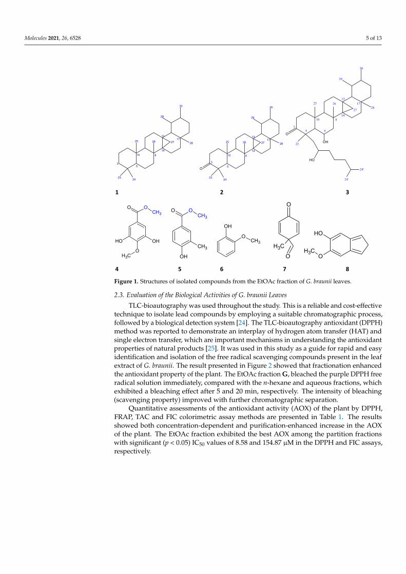
Three ursane-type triterpenes (1–3), three phenolics (4–6) and two phenolic derivatives(7 and 8) were isolated and identified in our study of G. braunii living on Leucena leucocephala(Fabaceae), its host, which marked the first report of these compounds in the plant andthe family Loranthaceae. Previous phytochemical studies on the plant have shown thepresence of tannins, phenolics, flavonoids, terpenoids and sterols [8,10], while compoundsreportedly isolated include lupeol-type triterpenes (globrauneine A-F, lupeol and lupeolacetate), lactones and flavonoids (quercetin, quercitrin, rutin and avicularin), identified inthe G. braunii living on Piliostigma thonningi (Fabaceae). Perhaps, this new additions to therepository of compounds in G. braunii might have occurred because of plant-host specificity,which was reported to play a critical role in the quality and quantity of constituents elicitedby Mistletoes as well as its influence on their biological properties [4].
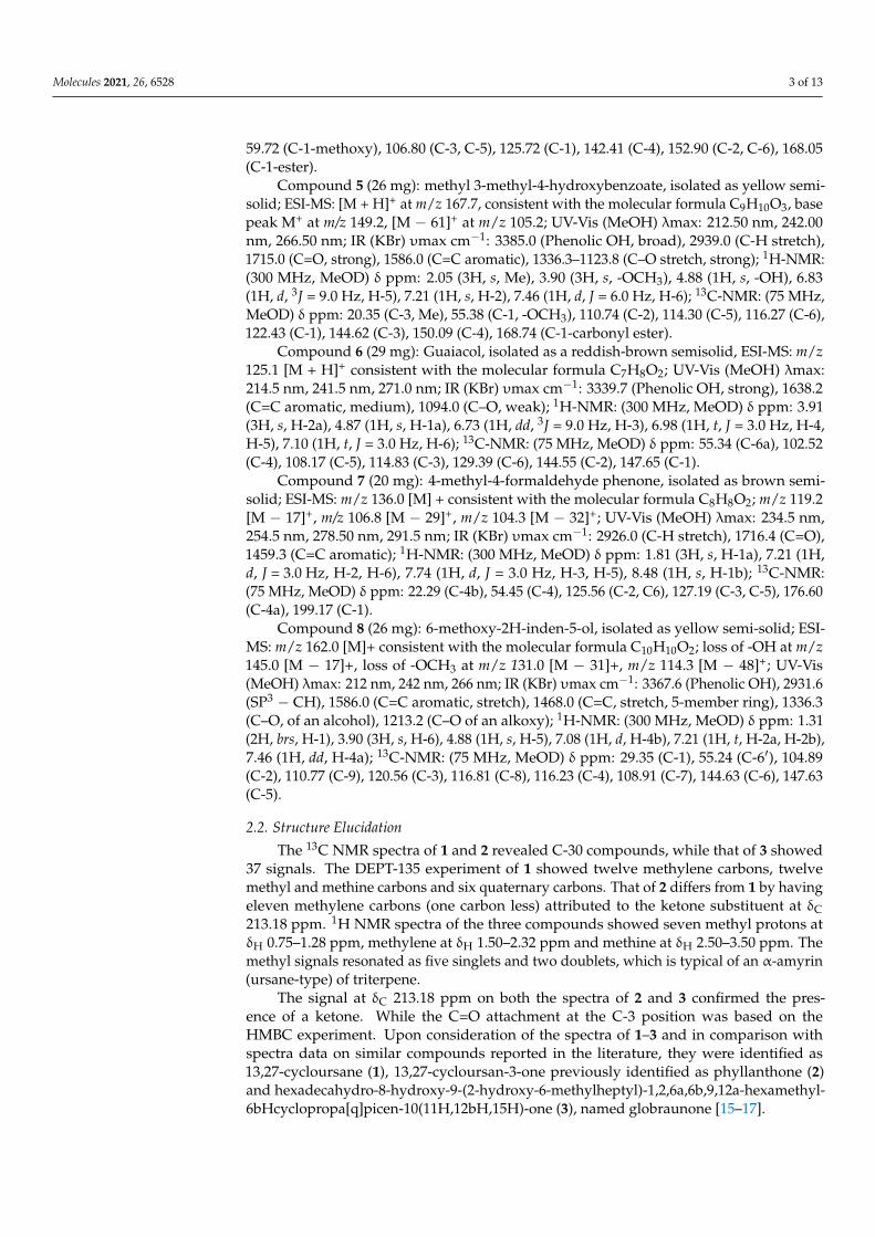
Structures of the isolated compounds 1–8 are presented in Figure 1.
Molecules 2021, 26, 6528 5 of 13Molecules 2021, 26, x FOR PEER REVIEW 5 of 13
Figure 1. Structures of isolated compounds from the EtOAc fraction of G. braunii leaves.
2.3. Evaluation of the Biological Activities of G. braunii Leaves TLC-bioautography was used throughout the study. This is a reliable and cost-effec-
tive technique to isolate lead compounds by employing a suitable chromatographic pro-cess, followed by a biological detection system [24]. The TLC-bioautography antioxidant (DPPH) method was reported to demonstrate an interplay of hydrogen atom transfer (HAT) and single electron transfer, which are important mechanisms in understanding the antioxidant properties of natural products [25]. It was used in this study as a guide for rapid and easy identification and isolation of the free radical scavenging compounds pre-sent in the leaf extract of G. braunii. The result presented in Figure 2 showed that fraction-ation enhanced the antioxidant property of the plant. The EtOAc fraction G, bleached the purple DPPH free radical solution immediately, compared with the n-hexane and aque-ous fractions, which exhibited a bleaching effect after 5 and 20 min, respectively. The in-tensity of bleaching (scavenging property) improved with further chromatographic sepa-ration.
1 2 3
4 5 6 7 8
27 28
29
30
13
14
2324
25 26
35
810
17
O
27 28
29
30
13
14
2324
25 26
35
810
17
O
OH
OH
34
10
6
8
13
14
17
23
24'
25 26
2728
29
30
24'
O OCH3
OCH3
OHOH
O OCH3
OH
CH3
OCH3
OH
CH3
O
O
OH
OCH3
Figure 1. Structures of isolated compounds from the EtOAc fraction of G. braunii leaves.
2.3. Evaluation of the Biological Activities of G. braunii Leaves
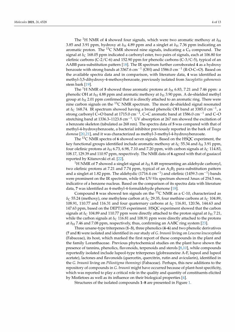
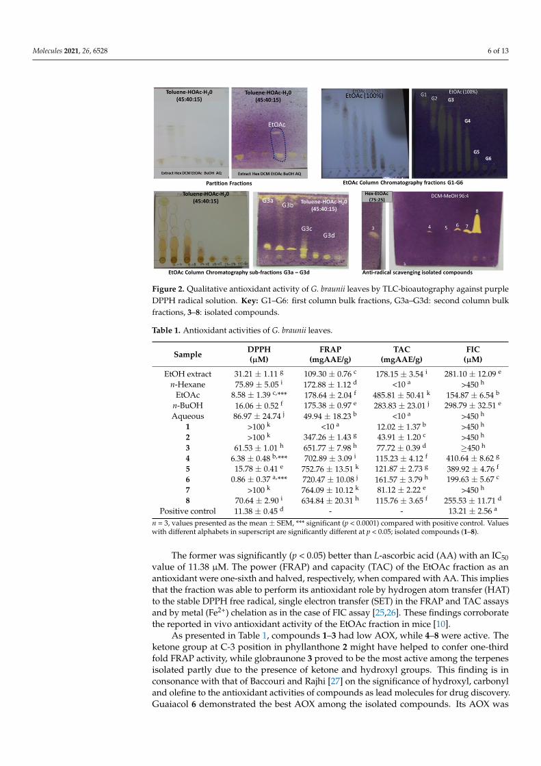
TLC-bioautography was used throughout the study. This is a reliable and cost-effectivetechnique to isolate lead compounds by employing a suitable chromatographic process,followed by a biological detection system [24]. The TLC-bioautography antioxidant (DPPH)method was reported to demonstrate an interplay of hydrogen atom transfer (HAT) andsingle electron transfer, which are important mechanisms in understanding the antioxidantproperties of natural products [25]. It was used in this study as a guide for rapid and easyidentification and isolation of the free radical scavenging compounds present in the leafextract of G. braunii. The result presented in Figure 2 showed that fractionation enhancedthe antioxidant property of the plant. The EtOAc fraction G, bleached the purple DPPH freeradical solution immediately, compared with the n-hexane and aqueous fractions, whichexhibited a bleaching effect after 5 and 20 min, respectively. The intensity of bleaching(scavenging property) improved with further chromatographic separation.
Quantitative assessments of the antioxidant activity (AOX) of the plant by DPPH,FRAP, TAC and FIC colorimetric assay methods are presented in Table 1. The resultsshowed both concentration-dependent and purification-enhanced increase in the AOXof the plant. The EtOAc fraction exhibited the best AOX among the partition fractionswith significant (p < 0.05) IC50 values of 8.58 and 154.87 µM in the DPPH and FIC assays,respectively.
Molecules 2021, 26, 6528 6 of 13Molecules 2021, 26, x FOR PEER REVIEW 6 of 13
Figure 2. Qualitative antioxidant activity of G. braunii leaves by TLC-bioautography against purple DPPH radical solution. Key: G1–G6: first column bulk fractions, G3a–G3d: second column bulk fractions, 3–8: isolated compounds.
Quantitative assessments of the antioxidant activity (AOX) of the plant by DPPH, FRAP, TAC and FIC colorimetric assay methods are presented in Table 1. The results showed both concentration-dependent and purification-enhanced increase in the AOX of the plant. The EtOAc fraction exhibited the best AOX among the partition fractions with significant (p < 0.05) IC50 values of 8.58 and 154.87 µM in the DPPH and FIC assays, re-spectively.
The former was significantly (p < 0.05) better than L-ascorbic acid (AA) with an IC50 value of 11.38 µM. The power (FRAP) and capacity (TAC) of the EtOAc fraction as an antioxidant were one-sixth and halved, respectively, when compared with AA. This im-plies that the fraction was able to perform its antioxidant role by hydrogen atom transfer (HAT) to the stable DPPH free radical, single electron transfer (SET) in the FRAP and TAC assays and by metal (Fe2+) chelation as in the case of FIC assay [25,26]. These findings corroborate the reported in vivo antioxidant activity of the EtOAc fraction in mice [10].
As presented in Table 1, compounds 1–3 had low AOX, while 4–8 were active. The ketone group at C-3 position in phyllanthone 2 might have helped to confer one-third fold FRAP activity, while globraunone 3 proved to be the most active among the terpenes iso-lated partly due to the presence of ketone and hydroxyl groups. This finding is in conso-nance with that of Baccouri and Rajhi [27] on the significance of hydroxyl, carbonyl and olefine to the antioxidant activities of compounds as lead molecules for drug discovery. Guaiacol 6 demonstrated the best AOX among the isolated compounds. Its AOX was 12 times better as a hydrogen-atom-donor (HAT) than AA in the DPPH assay, while, in the FRAP assay, it was 0.76-times as good as a single-electron-donor (SET) when compared with AA.
This could be due to its pi electron-rich benzene ring and hydroxyl group, which can exhibit both HAT and SET mechanisms of antioxidant action. HAT is a one-step reaction by phenolic O-H to effect bond dissociation enthalpy (BDE). SET is a sequential two-step reaction, which entails proton-loss followed by electron-transfer (SPLET), thus, leading to proton affinity (PA) and electron transfer enthalpy (ETE). These mechanisms have been reported to be responsible for the high antioxidant activities of many simple phenolics including phenolic acids [28].
The micro-broth dilution method of microbial susceptibility testing was adopted in the study. It is an objective, high-throughput, cost-effective and quantitative method. It offers high reproducibility, fast generation of MICs, convenience of having pre-prepared
Figure 2. Qualitative antioxidant activity of G. braunii leaves by TLC-bioautography against purpleDPPH radical solution. Key: G1–G6: first column bulk fractions, G3a–G3d: second column bulkfractions, 3–8: isolated compounds.
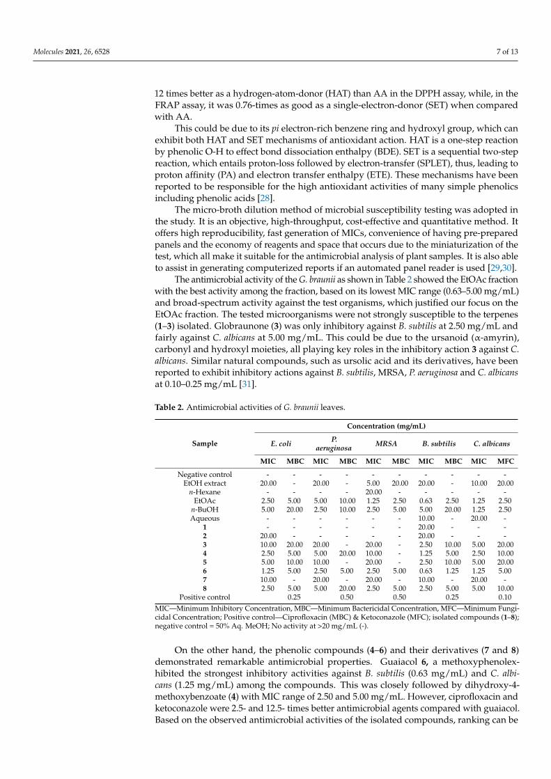
Table 1. Antioxidant activities of G. braunii leaves.
Sample DPPH(µM)
FRAP(mgAAE/g)
TAC(mgAAE/g)
FIC(µM)
EtOH extract 31.21 ± 1.11 g 109.30 ± 0.76 c 178.15 ± 3.54 i 281.10 ± 12.09 e
n-Hexane 75.89 ± 5.05 i 172.88 ± 1.12 d <10 a >450 h
EtOAc 8.58 ± 1.39 c,*** 178.64 ± 2.04 f 485.81 ± 50.41 k 154.87 ± 6.54 b
n-BuOH 16.06 ± 0.52 f 175.38 ± 0.97 e 283.83 ± 23.01 j 298.79 ± 32.51 e
Aqueous 86.97 ± 24.74 j 49.94 ± 18.23 b <10 a >450 h
1 >100 k <10 a 12.02 ± 1.37 b >450 h
2 >100 k 347.26 ± 1.43 g 43.91 ± 1.20 c >450 h
3 61.53 ± 1.01 h 651.77 ± 7.98 h 77.72 ± 0.39 d ≥450 h
4 6.38 ± 0.48 b,*** 702.89 ± 3.09 i 115.23 ± 4.12 f 410.64 ± 8.62 g
5 15.78 ± 0.41 e 752.76 ± 13.51 k 121.87 ± 2.73 g 389.92 ± 4.76 f
6 0.86 ± 0.37 a,*** 720.47 ± 10.08 j 161.57 ± 3.79 h 199.63 ± 5.67 c
7 >100 k 764.09 ± 10.12 k 81.12 ± 2.22 e >450 h
8 70.64 ± 2.90 i 634.84 ± 20.31 h 115.76 ± 3.65 f 255.53 ± 11.71 d
Positive control 11.38 ± 0.45 d - - 13.21 ± 2.56 a
n = 3, values presented as the mean ± SEM, *** significant (p < 0.0001) compared with positive control. Valueswith different alphabets in superscript are significantly different at p < 0.05; isolated compounds (1–8).
The former was significantly (p < 0.05) better than L-ascorbic acid (AA) with an IC50value of 11.38 µM. The power (FRAP) and capacity (TAC) of the EtOAc fraction as anantioxidant were one-sixth and halved, respectively, when compared with AA. This impliesthat the fraction was able to perform its antioxidant role by hydrogen atom transfer (HAT)to the stable DPPH free radical, single electron transfer (SET) in the FRAP and TAC assaysand by metal (Fe2+) chelation as in the case of FIC assay [25,26]. These findings corroboratethe reported in vivo antioxidant activity of the EtOAc fraction in mice [10].
As presented in Table 1, compounds 1–3 had low AOX, while 4–8 were active. Theketone group at C-3 position in phyllanthone 2 might have helped to confer one-thirdfold FRAP activity, while globraunone 3 proved to be the most active among the terpenesisolated partly due to the presence of ketone and hydroxyl groups. This finding is inconsonance with that of Baccouri and Rajhi [27] on the significance of hydroxyl, carbonyland olefine to the antioxidant activities of compounds as lead molecules for drug discovery.Guaiacol 6 demonstrated the best AOX among the isolated compounds. Its AOX was
Molecules 2021, 26, 6528 7 of 13
12 times better as a hydrogen-atom-donor (HAT) than AA in the DPPH assay, while, in theFRAP assay, it was 0.76-times as good as a single-electron-donor (SET) when comparedwith AA.
This could be due to its pi electron-rich benzene ring and hydroxyl group, which canexhibit both HAT and SET mechanisms of antioxidant action. HAT is a one-step reactionby phenolic O-H to effect bond dissociation enthalpy (BDE). SET is a sequential two-stepreaction, which entails proton-loss followed by electron-transfer (SPLET), thus, leading toproton affinity (PA) and electron transfer enthalpy (ETE). These mechanisms have beenreported to be responsible for the high antioxidant activities of many simple phenolicsincluding phenolic acids [28].
The micro-broth dilution method of microbial susceptibility testing was adopted inthe study. It is an objective, high-throughput, cost-effective and quantitative method. Itoffers high reproducibility, fast generation of MICs, convenience of having pre-preparedpanels and the economy of reagents and space that occurs due to the miniaturization of thetest, which all make it suitable for the antimicrobial analysis of plant samples. It is also ableto assist in generating computerized reports if an automated panel reader is used [29,30].
The antimicrobial activity of the G. braunii as shown in Table 2 showed the EtOAc fractionwith the best activity among the fraction, based on its lowest MIC range (0.63–5.00 mg/mL)and broad-spectrum activity against the test organisms, which justified our focus on theEtOAc fraction. The tested microorganisms were not strongly susceptible to the terpenes(1–3) isolated. Globraunone (3) was only inhibitory against B. subtilis at 2.50 mg/mL andfairly against C. albicans at 5.00 mg/mL. This could be due to the ursanoid (α-amyrin),carbonyl and hydroxyl moieties, all playing key roles in the inhibitory action 3 against C.albicans. Similar natural compounds, such as ursolic acid and its derivatives, have beenreported to exhibit inhibitory actions against B. subtilis, MRSA, P. aeruginosa and C. albicansat 0.10–0.25 mg/mL [31].
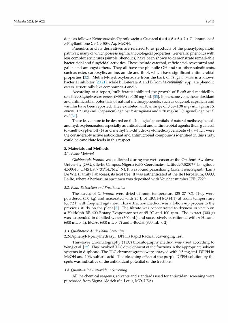
Table 2. Antimicrobial activities of G. braunii leaves.
Sample
Concentration (mg/mL)
E. coli P.aeruginosa MRSA B. subtilis C. albicans
MIC MBC MIC MBC MIC MBC MIC MBC MIC MFC
Negative control - - - - - - - - - -EtOH extract 20.00 - 20.00 - 5.00 20.00 20.00 - 10.00 20.00
n-Hexane - - - - 20.00 - - - - -EtOAc 2.50 5.00 5.00 10.00 1.25 2.50 0.63 2.50 1.25 2.50
n-BuOH 5.00 20.00 2.50 10.00 2.50 5.00 5.00 20.00 1.25 2.50Aqueous - - - - - - 10.00 - 20.00 -
1 - - - - - - 20.00 - - -2 20.00 - - - - - 20.00 - - -3 10.00 20.00 20.00 - 20.00 - 2.50 10.00 5.00 20.004 2.50 5.00 5.00 20.00 10.00 - 1.25 5.00 2.50 10.005 5.00 10.00 10.00 - 20.00 - 2.50 10.00 5.00 20.006 1.25 5.00 2.50 5.00 2.50 5.00 0.63 1.25 1.25 5.007 10.00 - 20.00 - 20.00 - 10.00 - 20.00 -8 2.50 5.00 5.00 20.00 2.50 5.00 2.50 5.00 5.00 10.00
Positive control 0.25 0.50 0.50 0.25 0.10MIC—Minimum Inhibitory Concentration, MBC—Minimum Bactericidal Concentration, MFC—Minimum Fungi-cidal Concentration; Positive control—Ciprofloxacin (MBC) & Ketoconazole (MFC); isolated compounds (1–8);negative control = 50% Aq. MeOH; No activity at >20 mg/mL (-).
On the other hand, the phenolic compounds (4–6) and their derivatives (7 and 8)demonstrated remarkable antimicrobial properties. Guaiacol 6, a methoxyphenolex-hibited the strongest inhibitory activities against B. subtilis (0.63 mg/mL) and C. albi-cans (1.25 mg/mL) among the compounds. This was closely followed by dihydroxy-4-methoxybenzoate (4) with MIC range of 2.50 and 5.00 mg/mL. However, ciprofloxacin andketoconazole were 2.5- and 12.5- times better antimicrobial agents compared with guaiacol.Based on the observed antimicrobial activities of the isolated compounds, ranking can be
Molecules 2021, 26, 6528 8 of 13
done as follows: Ketoconazole, Ciprofloxacin > Guaiacol 6 > 4 > 8 > 5 > 7 > Globraunone 3> Phyllanthone 2 > 1 > 50% Aq. MeOH.
Phenolics and its derivatives are referred to as products of the phenylpropanoidpathway, many of which possess significant biological properties. Generally, phenolics withless complex structures (simple phenolics) have been shown to demonstrate remarkablebactericidal and fungicidal activities. These include catechol, caffeic acid, resveratrol andgallic acid amongst others. They all have the phenolic OH and/or other substituents,such as ester, carboxylic, amine, amide and thiol, which have significant antimicrobialproperties [32]. Methyl-4-hydroxybenzoate from the bark of Tsuga dumosa is a knownbacterial inhibitor [20,21], while bulbiferate A and B from Microbulbifer spp. are phenolicesters, structurally like compounds 4 and 5.
According to a report, bulbiferates inhibited the growth of E coli and methicillin-sensitive Staphylococcus aureus (MSSA) at 0.20 mg/mL [33]. In the same vein, the antioxidantand antimicrobial potentials of natural methoxyphenols, such as eugenol, capsaicin andvanillin have been reported. They exhibited an IC50 range of 0.68–1.38 mg/mL against S.aureus, 1.21 mg/mL (capsaicin) against P. aeruginosa and 2.70 mg/mL (eugenol) against E.coli [34].
These leave more to be desired on the biological potentials of natural methoxyphenolsand hydroxybenzoates, especially as antioxidant and antimicrobial agents; thus, guaiacol(O-methoxyphenol) (6) and methyl 3,5-dihydroxy-4-methoxybenzoate (4), which werethe considerably active antioxidant and antimicrobial compounds identified in this study,could be candidate leads in this respect.
3. Materials and Methods3.1. Plant Material
Globimetula braunii was collected during the wet season at the Obafemi AwolowoUniversity (OAU), Ile-Ife Campus, Nigeria (GPS Coordinates: Latitude 7.520767, Longitude4.530315; DMS Lat 7◦31′14.7612′′ N). It was found parasitizing Leucena leucocephala (Lam)De Wit. (Family Fabaceae), its host tree. It was authenticated at the Ife Herbarium, OAU,Ile-Ife, where a herbarium specimen was deposited with Voucher number IFE 17229.
3.2. Plant Extraction and Fractionation
The leaves of G. braunii were dried at room temperature (25–27 ◦C). They werepowdered (5.0 kg) and macerated with 25 L of EtOH-H2O (4:1) at room temperaturefor 72 h with frequent agitation. This extraction method was a follow-up process to theprevious study on the plant [8]. The filtrate was concentrated to dryness in vacuo ona Heidolph RE 400 Rotary Evaporator set at 45 ◦C and 100 rpm. The extract (300 g)was suspended in distilled water (300 mL) and successively partitioned with n-Hexane(600 mL × 4), EtOAc (600 mL × 7) and n-BuOH (300 mL × 2).
3.3. Qualitative Antioxidant Screening2,2-Diphenyl-1-picrylhydrazyl (DPPH) Rapid Radical Scavenging Test
Thin-layer chromatography (TLC) bioautography method was used according toWang et al. [35]. This involved TLC development of the fractions in the appropriate solventsystems in duplicate. The TLC chromatograms were sprayed with 0.5 mg/mL DPPH inMeOH and 10% sulfuric acid. The bleaching effect of the purple DPPH solution by thespots was indicative of the antioxidant potential of the fractions.
3.4. Quantitative Antioxidant Screening
All the chemical reagents, solvents and standards used for antioxidant screening werepurchased from Sigma Aldrich (St. Louis, MO, USA).
Molecules 2021, 26, 6528 9 of 13
3.4.1. DPPH Spectrophotometric Assay
The DPPH spectrophotometric assay was carried out according to Xiao et al. [36]. A1 mL DPPH solution in methanol (0.05 mg/mL) was added to 1 mL samples ((positivecontrols: L-ascorbic acid) and (plant fractions/isolated compounds)) at varying concentra-tions: 50.00, 25.00, 12.50, 6.25 and 3.13 µg/mL. The experiment was carried out in triplicate.The samples were incubated in the dark room for 30 min after which the absorbance wasmeasured at 517 nm on a CamSpec M 107 Spectrophotometer (Spectronics Camspec Ltd.,Leeds, UK), where methanol (negative control) was used as the blank. The percentageinhibition of DPPH by each test sample was calculated thus:
% Inhibition of sample =Abscontrol − Abssample
Abscontrol× 100 (1)
where Abscontrol = Absorbance of negative control, Abssample = Absorbance of test sample.The result was expressed as % inhibition and/or IC50.
3.4.2. Ferric Reducing Antioxidant Power (FRAP) Assay
This is based on the reduction of the greenish ferric ion (Fe3+) 2,4,6-tri-(2-pyridyl)-1,3,5-triazine (TPTZ) to the bluish ferrous ion (Fe2+) by natural antioxidants at 593 nmabsorbance measurement. The ferric reducing power of plant extracts were determinedas ascorbic acid equivalent (AAE) from the calibration curve of the positive control (L-ascorbic acid) at concentrations 1000.00, 500.00, 250.00, 125.00, 62.50 and 31.25 µg/mL inmethanol [37].
3.4.3. Total Antioxidant Capacity (TAC) Assay
The TAC assay is based on the reduction of Mo6+ to Mo5+ by the plant samples andsubsequent formation of green phosphate/Mo (V) complex at acidic pH according to Prietoet al. [38]. A 0.3 mL extract was combined with 3 mL of reagent solution (0.6 M sulfuricacid, 28 mM sodium phosphate and 4 mM ammonium molybdate). The absorbance ofthe reaction mixture was measured at 695 nm. The calibration curve was prepared bymixing ascorbic (1000.00, 500.00, 250.00, 125.00, 62.50 and 31.25 µg/mL) with methanol.Data were expressed as mean ± standard error of mean (SEM). The TAC of each samplewas expressed as the number of gram equivalent of ascorbic acid (AAE/g).
3.4.4. Ferrous Ion Chelating (FIC) Ability
FIC assay was carried out according to the method of Singh and Rajini [39]. Solutionsof 2 mM FeCl2·4H2O and 5 mM ferrozine were diluted 20 times. An aliquot (1 mL) ofdifferent concentrations of extract was mixed with 1mL FeCl2·4H2O. After 5 min incubation,the reaction was initiated by the addition of ferrozine (1 mL). The mixture was shakenvigorously, and, after a further 10 min incubation period, the absorbance of the solution wasmeasured spectrophotometrically at 562 nm. The percentage inhibition of ferrozine–Fe2+
complex formation was calculated by using the formula:
% Chelating ability =Abscontrol − Abssample
Abscontrol× 100 (2)
3.5. Statistical Analysis
All quantitative antioxidant data were analyzed using a One-way Analysis of Variance(ANOVA), followed by the Bonferroni post-hoc test on a GraphPad Prism 9 (GraphPadSoftware Inc., San Diego, CA, USA).
3.6. Antimicrobial TestMicro-Broth Dilution Assay
The assay was carried according to the Clinical and Laboratory Standard Institute [40,41].Bacteria and fungi used for the antimicrobial screening were obtained from the culture
Molecules 2021, 26, 6528 10 of 13
collections of the Microbiology Laboratory of the Department of Pharmaceutics, ObafemiAwolowo University where the experiment was conducted. The bacteria and fungi strainswere isolated on a Nutrient broth and Sabouraud Dextrose broth (Merck KGaA, Darmstadt,Germany), respectively. The organisms were identified using their morphological character-istics and standard biochemical tests. The reference strains used were Escherichia coli ATCC25923, Pseudomonas aeruginosa ATCC 10145, Bacillus subtilis NCTC 8236, methicillin-resistantStaphylococcus aureus ATCC 29213 and Candida albicans ATCC 24433.
Bacteria were maintained on nutrient broth and fungi on Sabouraud Dextrose brothat 4 ◦C and sub-cultured regularly. Bacteria were grown for 18 h in Nutrient broth andculture suspensions of 108 cfu/mL (equivalent of 0.50 Mc Farland standard) were appliedto the dilutions of the fraction/isolates, positive controls (Ciprofloxacin, Ketoconazole) andnegative control 50% aqueous methanol employing a multipoint inoculator. Plates wereincubated at 37 ◦C for 24 h. for bacteria strains and 25 ◦C for 72 h for fungal strains, afterwhich all plates were observed for growth of the microorganisms. The minimum dilutionof fractions completely inhibiting the growth and killing each organism was taken as theMIC and MBC/MFC. The sample with the lowest range of MIC and the widest spectrumof activity against bacteria and fungi was taken as the most active.
3.7. Isolation of CompoundsColumn Chromatography of the EtOAc Fraction
The EtOAc fraction (G, 30.0 g) was adsorbed unto 30 g silica gel (70–230 ASTM mesh,Merck KGaA, Darmstadt, Germany), dry-packed on a 600 g silica gel stationary phasewithin a 300 cm× 5 cm glass column (L x i.d., Fisher Scientific, Waltham, MA, USA). Mobilephase comprising solvent systems of increasing polarity was introduced as thus: n-Hex(100%, 700 mL, de-gas), EtOAc (9:1, 8:2, . . . , 1:9; 500 mL each), EtOAc (100%; 700 mL),EtOAc-MeOH (95:5, 9:1, 8:2, 1:1; 500 mL) and MeOH (100%; 250 mL). Eluates were collectedin 20 mL test tubes (1–303). They were bulked into six sub-fractions G1–G6 based on theirTLC profiles (SiO2, Hex-EtOAc 75:25, 1:1, EtOAc-MeOH 1:1, UV 254 and 365 nm, 10%H2SO4 spray).
After a 24 h period, sub-fraction G1 (1.2 g) afforded solid deposits a, b and c, whilesub-fraction G2 (3.1 g) gave a solid deposit d. Each deposit was washed with 100 mLof MeOH (100%), affording compounds 1–4, respectively. The most active sub-fractionG3 (3.5 g), based on TLC-bioautography was further purified on a Silica gel column withmobile phase from DCM (100%, 100 mL) to DCM-MeOH (98:2, 96:4, . . . , 80:20; 100 mLeach). The eluates (1–187) were collected in 10 mL test tubes and were subsequently bulkedinto four sub-fractions, G3a–G3d, based on their TLC profiles (SiO2, DCM-MeOH 97:3,85:15, 1:1, UV 254 and 365 nm, 10% H2SO4 spray). A preparative TLC separation of G3c,using DCM-MeOH (96:4) afforded bands i–iv, labelled compounds 5–8.
3.8. Characterization of Isolated Compounds
Thin-layer chromatography (TLC) of compounds was performed on aluminum-backed silica gel 60 F254 GF plates (0.25 mm, Merck KGaA, Darmstadt, Germany). Chem-ical detection of the class of compound isolated was done by spraying the developedTLC plates with chromogenic reagents, such as 5% FeCl3 for phenolics and 10% H2SO4for terpenes. Melting point ranges of the solid compounds were determined on Gal-lenkamp MPD350-BM 3.5 electrothermal instrument (Gallenkamp, Kent, UK). The UV-Visabsorption was determined within 200–800 nm on Shimadzu UV-1800 UV/Visible Scan-ning Spectrophotometer: 115 VAC (Shimadzu Corporation, Nkagyo-Ku, Kyoto, Japan).Infrared spectroscopy was done within the 650–4000 cm−1 transmittance on Cary 630FTIR Spectrometer (Agilent Technologies Inc., Santa Clara, CA, USA). 1H, 13C and 2D(DEPT135, COSY, HSQC and HMBC) NMR spectra of compounds were recorded as solu-tions on Bruker AMX-300 Spectrometer (Bruker Corporation, Bremen, Germany), wheretetramethylsilane was used as the internal standard.
Molecules 2021, 26, 6528 11 of 13
Signals were recorded in the order of chemical shifts (δ) in part per million (ppm) relativeto the indicated deuterated solvents (CDCl3, MeOD), integral values (number of protons),multiplicity (s, singlet; d, doublet; t, triplet; and m, multiplet) and coupling constant (J) inhertz (Hz). Electrospray Ionization Mass Spectrometry (ESI-MS) was performed on a Varian500-MS ion trap Mass Spectrometer (Varian, Inc., Palo Alto, CA, USA) for molecular weightdetermination, expressed in mass-to-charge ratio (m/z). ESI-MS analysis was performedat 10 µL/min sample infusion flow rate; 2.56 kV capillary voltage; 3.0 V extraction cone;475 L/h desolvation-gas flow rate; 80 and 100 ◦C for the source- and desolvation-gastemperatures, respectively; and 5.82 mm Vernier-probe-adjuster position. The spectrometerscan range was 99.5–1500.5 m/z in the positive mode.
4. Conclusions
Our activity-guided study on the leaves of G. braunii led to isolation of eight com-pounds (1–8) from the most active EtOAc fraction. The compounds were identified basedon their spectroscopic data and in comparison with literature reports. They were ursane-type triterpenes (1–3), phenolics (4–6) and phenolic derivatives (7 and 8), all reportedfor the first time in the plant and in the family Loranthaceae. Guaiacol (6) and methyl3,5-dihydroxy-4-methoxybenzoate (4) were remarkably antioxidant with considerableantimicrobial potentials.
Author Contributions: Conceptualization, A.O.O. (Ayodeji Oluwabunmi Oriola) and A.J.A.; method-ology, A.O.O. (Ayodeji Oluwabunmi Oriola) and A.J.A.; software, A.O.O. (Ayodeji OluwabunmiOriola) and T.I.; validation, A.J.A., T.O.I., E.M.O. and A.O.O. (Adebola Omowunmi Oyedeji); formalanalysis, A.O.O. (Ayodeji Oluwabunmi Oriola), E.M.O., F.O.A. and T.I.; investigation, A.O.O. (AyodejiOluwabunmi Oriola); resources, A.O.O. (Ayodeji Oluwabunmi Oriola), E.M.O. and A.O.O. (AdebolaOmowunmi Oyedeji); data curation, A.O.O. (Ayodeji Oluwabunmi Oriola), E.M.O., F.O.A. and T.O.I.;writing—original draft preparation, A.O.O. (Ayodeji Oluwabunmi Oriola); writing—review and edit-ing, A.J.A. and A.O.O. (Adebola Omowunmi Oyedeji); visualization, A.J.A., T.O.I., E.M.O. and F.O.A.;supervision, A.J.A. and T.O.I.; project administration, A.O.O. (Ayodeji Oluwabunmi Oriola), A.J.A.,T.O.I.; funding acquisition, A.O.O. (Ayodeji Oluwabunmi Oriola) and A.O.O. (Adebola OmowunmiOyedeji). All authors have read and agreed to the published version of the manuscript.
Funding: The study received no funding support.
Institutional Review Board Statement: Not applicable.
Informed Consent Statement: Not applicable.
Data Availability Statement: Not available.
Acknowledgments: The corresponding author acknowledges the Drug Research and ProductionUnit, O.A.U., Ile-Ife, Nigeria, for study leave permission to Walter Sisulu University, South Africa.
Conflicts of Interest: The authors declare no conflict of interest.
Samples Availability: Samples of the compounds 1–8 are available from the authors.
References1. Siegfried Didier, D.; Obiang Nestor Laurier, E.; Din, N.; Richard Jules, P.; Victor, T.; Henri, F.; Georges, S.; Alain Didier, M.; Issaka
Joseph, B.; Akoa, A. An Assessment on the Uses of Loranthaceae in Ethno Pharmacology in Cameroon: A Case Study Made inLogbessou, North of Douala. J. Med. Plants Res. 2009, 3, 592–595.
2. Ayoola, M.D.; Oriola, A.O.; Faloye, K.O.; Aladesanmi, A.J. Two Antihyperglycaemic Compounds from Globimetula braunii (Engl.)Van Tiegh (Loranthaceae). GSC Biol. Pharm. Sci. 2020, 2020, 46–054. [CrossRef]
3. Burkill, H.M. The Useful Plants of West Tropical Africa. Available online: https://agris.fao.org/agris-search/search.do?recordID=GB9618106 (accessed on 8 August 2021).
4. Adesina, S.K.; Illoh, H.C.; Johnny, I.I.; Jacobs, I.E. African Mistletoes (Loranthaceae); Ethnopharmacology, Chemistry andMedicinal Values: An Update. Afr. J. Tradit. Complement. Altern. Med. AJTCAM 2013, 10, 161–170. [CrossRef]
5. Okpuzor, J.; Kareem, G.; Ejikeme, C. Lipid Lowering Activity of Globimetula braunii. Res. J. Med. Plant 2009, 3, 45–51. [CrossRef]6. Okpuzor, J.; Ogbunugafor, H.A.; Kareem, G.K. Hepatic and Hematologic Effects of Fractions of Globimetula braunii in Normal
Albino Rats. EXCLI J. 2009, 8, 182–189.
Molecules 2021, 26, 6528 12 of 13
7. Oriola, A.O.; Aladesanmi, A.J.; Arthur, G. Anticancer Activity of Three African Mistletoes. Niger. J. Nat. Prod. Med. 2018, 22,129–134. [CrossRef]
8. Oriola, A.O.; Aladesanmi, A.J.; Akinkunmi, E.O.; Olawuni, I.J. Antioxidant and Antimicrobial Studies of Some Hemi-ParasiticWest African Plants. Eur. J. Med. Plants 2020, 31, 17–26. [CrossRef]
9. Enehezeyi Aliyu, R.; Alonge, S.; Aliyu, R. Antibacterial Activity of Globimetula braunii Sourced from Five Different Host Trees inScreening Cowpea Magic Rils for Variation in Agro-Physiological Traits for Tolerance to Biotic and Abiotic Stresses View ProjectAntibacterial Activity of Globimetula braunii Sourced from Five Different Host Trees in Samaru, Zaria, Nigeria. Int. J. Curr. Sci.2015, 18, 117–123.
10. Okpuzor, J.; Ogbunugafor, H.; Karecm, G.K. Antioxidative Properties of Ethyl Acetate Fraction of Globimetula braunii in NormalAlbino Rats. J. Biol. Sci. 2009, 9, 470–475. [CrossRef]
11. Erukainure, O.L.; Abovwe, J.A.; Adefegha, A.S.; Egwuche, R.U.; Fafunso, M.A. Antilipemic and Hypocholesteremic Activities ofGlobimetula braunii in Rats. Exp. Toxicol. Pathol. 2010, 63, 657–661. [CrossRef]
12. Ie, O.; Zam, N. Oxytocic Properties of the Aqueous Extract of Globimetula braunii (Loranthaceae). Pak. J. Pharm. Sci. 2008, 21,356–360.
13. Muhammad, K.J.; Jamil, S.; Basar, N.; Bakri Bakar, M.; Sarker, S.D.; Flanagan, K.J.; Senge, M.O. Lactones and Flavonoids Isolatedfrom the Leaves of Globimetula braunii. NPC Nat. Prod. Commun. 2017, 12, 1455–1458.
14. Muhammad, K.J.; Jamil, S.; Basar, N.; Sarker, S.D.; Mohammed, M.G. Globrauneine A–F: Six New Triterpenoid Esters from theLeaves of Globimetula braunii. Nat. Prod. Res. 2019, 34, 2746–2753. [CrossRef] [PubMed]
15. Ren, F.C.; Li, G.Y.; Nama, N.; Liu, Z.H.; Yang, L.; Zhou, J.; Hu, J.M. 13,27-Cycloursane, Ursane and Oleanane Triterpenoids fromthe Leaves of Lucuma nervosa. Fitoterapia 2019, 136, 104178. [CrossRef] [PubMed]
16. Ndlebe, V.J.; Crouch, N.R.; Mulholland, D.A. Triterpenoids from the African Tree Phyllanthus polyanthus. Phytochem. Lett. 2008, 1,11–17. [CrossRef]
17. Lee, T.-H.; Juang, S.-H.; Hsu, F.-L.; Wu, C.-Y. Triterpene Acids from the Leaves of Planchonella duclitan (Blanco) Bakhuizan. J. Chin.Chem. Soc. 2005, 52, 1275–1280. [CrossRef]
18. Pavia, D.L.; Lampman, G.M.; Kriz, G.S.; Vyvyan, J.R. Nuclear Magnetic Resonance Spectroscopy Part Five: Advanced NMRTechniques. In Introduction to Spectroscopy; Thomson Learning Inc.: Boston, MA, USA, 2013; pp. 511–576.
19. Alade, G.O.; Moody, J.O.; Awotona, O.R.; Adesanya, S.A.; Lai, D.; Proksch, P. Spermicidal Constituents of Ethanolic Extract ofSacoglottis gabonensis Stem Bark. Folia Med. 2017, 59, 437–442. [CrossRef] [PubMed]
20. MedChemExpress. Methylparaben—Advanced Dermatology. Available online: https://www.advanced-dermatology.com.au/methylparaben (accessed on 25 September 2021).
21. MedChemExpress. Methyl Paraben (Methyl 4-Hydroxybenzoate)|Bacterial Inhibitor|MedChemExpress. Available online:https://www.medchemexpress.com/Methyl_Paraben.html (accessed on 6 October 2021).
22. Kitanovski, Z.; Cusak, A.; Grgic, I.; Claeys, M. Chemical Characterization of the Main Products Formed through Aqueous-PhasePhotonitration of Guaiacol. Atmos. Meas. Tech. 2014, 7, 2457–2470. [CrossRef]
23. Guthrie, R.D. Introduction to Spectroscopy (Pavia, Donald; Lampman, Gary, M.; Kriz, George, S., Jr.). J. Chem. Educ. 1979, 56,A323. [CrossRef]
24. Dewanjee, S.; Gangopadhyay, M.; Bhattacharya, N.; Khanra, R.; Dua, T.K. Bioautography and Its Scope in the Field of NaturalProduct Chemistry. J. Pharm. Anal. 2015, 5, 75–84. [CrossRef] [PubMed]
25. Santos-Sánchez, N.F.; Salas-Coronado, R.; Villanueva-Cañongo, C.; Hernández-Carlos, B. Antioxidant Compounds and TheirAntioxidant Mechanism; IntechOpen: London, UK, 2019. [CrossRef]
26. Siddeeg, A.; AlKehayez, N.M.; Abu-Hiamed, H.A.; Al-Sanea, E.A.; AL-Farga, A.M. Mode of Action and Determination ofAntioxidant Activity in the Dietary Sources: An Overview. Saudi J. Biol. Sci. 2021, 28, 1633–1644. [CrossRef]
27. Baccouri, B.; Rajhi, I. Potential Antioxidant Activity of Terpenes. In Terpenes and Terpenoids Recent Advances; Intech Open BookSeries; IntechOpen: London, UK, 2021. [CrossRef]
28. Chen, J.; Yang, J.; Ma, L.; Li, J.; Shahzad, N.; Kyung Kim, C.K. Structure-Antioxidant Activity Relationship of Methoxy, PhenolicHydroxyl, and Carboxylic Acid Groups of Phenolic Acids. Sci. Rep. 2020, 10, 2611. [CrossRef] [PubMed]
29. Chen, S.C.; Liu, J.W.; Wu, X.Z.; Cao, W.L.; Wang, F.; Huang, J.M.; Han, Y.; Zhu, X.Y.; Zhu, B.Y.; Gan, Q.; et al. Comparison ofMicrodilution Method with Agar Dilution Method for Antibiotic Susceptibility Test of Neisseria gonorrhoeae. Infect. Drug Resist.2020, 13, 1775–1780. [CrossRef]
30. Reller, L.B.; Weinstein, M.; Jorgensen, J.H.; Ferraro, M.J. Antimicrobial Susceptibility Testing: A Review of General Principles andContemporary Practices. Clin. Infect. Dis. 2009, 49, 1749–1755. [CrossRef]
31. Jesus, J.A.; Lago, J.H.G.; Laurenti, M.D.; Yamamoto, E.S.; Passero, L.F.D. Antimicrobial Activity of Oleanolic and Ursolic Acids:An Update. Evid.-Based Complement. Altern. Med. 2015, 2015. [CrossRef] [PubMed]
32. Maddox, C.E.; Laur, L.M.; Tian, L. Antibacterial Activity of Phenolic Compounds Against the Phytopathogen Xyllela fastidiosa.Curr. Microbiol. 2010, 60, 53–58. [CrossRef] [PubMed]
33. Jayanetti, D.R.; Braun, D.R.; Barns, K.J.; Rajski, S.R.; Bugni, T.S. Bulbiferates A and B: Antibacterial Acetamidohydroxybenzoatesfrom a Marine Proteobacterium, Microbulbifer sp. J. Nat. Prod. 2019, 82, 1930–1934. [CrossRef]
34. Orlo, E.; Russo, C.; Nugnes, R.; Lavorgna, M.; Isidori, M. Natural Methoxyphenol Compounds: Antimicrobial Activity AgainstFoodborne Pathogens and Food Spoilage Bacteria, and Roles in Antioxidant Processes. Foods 2021, 10, 1807. [CrossRef]
Molecules 2021, 26, 6528 13 of 13
35. Wang, J.; Yue, Y.D.; Tang, F.; Sun, J. TLC Screening for Antioxidant Activity of Extracts from Fifteen Bamboo Species andIdentification of Antioxidant Flavone Glycosides from Leaves of Bambusa textilis Mcclure. Molecules 2012, 17, 12297–12311.[CrossRef] [PubMed]
36. Xiao, F.; Xu, T.; Lu, B.; Liu, R. Guidelines for Antioxidant Assays for Food Components. Food Front. 2020, 1, 60–69. [CrossRef]37. Benzie, I.F.F.; Strain, J.J. [2] Ferric Reducing/Antioxidant Power Assay: Direct Measure of Total Antioxidant Activity of Biological
Fluids and Modified Version for Simultaneous Measurement of Total Antioxidant Power and Ascorbic Acid Concentration.Methods Enzymol. 1999, 299, 15–27. [CrossRef] [PubMed]
38. Prieto, P.; Pineda, M.; Aguilar, M. Spectrophotometric Quantitation of Antioxidant Capacity through the Formation of aPhosphomolybdenum Complex: Specific Application to the Determination of Vitamin E. Anal. Biochem. 1999, 269, 337–341.[CrossRef] [PubMed]
39. Singh, N.; Rajini, P. Free Radical Scavenging Activity of an Aqueous Extract of Potato. Food Chem. 2004, 85, 611–616. [CrossRef]40. Clinical and Laboratory Standard Institute, CLSI. M07-A10 Methods for Dilution Antimicrobial Susceptibility Tests for Bacteria That
Grow Aerobically; Approved Standard-Tenth Edition; Clinical and Laboratory Standard Institute: Annapolis Junction, MD, USA,2015.
41. Clinical and Laboratory Standard Institute, CLSI. Antimicrobial and Antifungal Susceptibility Testing Resources. Availableonline: https://clsi.org/about/about-clsi/about-clsi-antimicrobial-and-antifungal-susceptibility-testing-resources/# (accessedon 22 September 2021).


