Embed Size (px)
Citation preview

Neuro-immunity in
In vivo studies of postoperative ileus and colitisintestinal diseaseSusanne Anna Snoek
UITNODIGING
Tot het bijwonen van de openbare verdediging van het proefschrift:
Neuro-immunity in intestinal disease In vivo studies of postoperative ileus and colitisdoor Susanne Snoek
Op Woensdag 8 juni om 12.00 uur in de Agnietenkapel van de Universiteit van Amsterdam.
Adres: Oudezijds Voorburgwal 231 1012 EZ Amsterdam
Receptie na afloop van de promotie.
Paranimfen:
José van [email protected]
Esmerij van der [email protected]
Susanne SnoekMolukkenstraat 5871095 BJ [email protected]
Neu
ro-im
munity in
intestin
al disease
In vivo studies of postoperative ileus and colitisSusan
ne
Anna
Snoek
3
Neuro-immunity in intestinal disease In vivo studies of postoperative ileus and colitis
Susanne Anna Snoek

4
The printing of this thesis was financially supported by:Maag Lever Darm StichtingNederlandse Vereniging voor Gastro-enterologie
© Susanne A. Snoek, Amsterdam, The NetherlandsCover and Layout: Marijn van Oosten, www.visualgin.comPrinted by: Print Service Ede
5
Neuro-immunity in intestinal diseaseIn vivo studies of postoperative ileus and colitis
ACADEMISCH PROEFSCHRIFT
Ter verkrijging van de graad van doctor aan de Universiteit van Amsterdamop gezag van de Rector Magnificus
prof. dr. D.C. van den Boomten overstaan van een door het college voor promoties
ingestelde commissie, in het openbaar te verdedigen in de Agnietenkapelop woensdag 8 juni 2011 te 12.00 uur
door:
Susanne Anna Snoek
geboren te Wester-Koggenland
6
Promotiecommissie:
Promotor: Prof. dr. G.E. Boeckxstaens
Co-promotor: Dr. W.J. de Jonge
Overige leden: Prof. dr. W. A. Buurman Prof. dr. E. Cario Dr. J.N. Samsom Prof. dr. W.A. Bemelman Prof. dr. M.W. Hollmann Prof. dr. R.P.J. Oude Elferink Prof. dr. T. van der Poll
Faculteit der Geneeskunde

7
CHAPTER 1
Contents
General introduction and thesis outline
Neuro-immune regulation of epithelial barrier function: focus on postoperative ileus (review).Expert Rev Gastroenterol Hepatol. 2010 Oct;4(5):637-51 and Curr Pharm Des. 2010;16(9):1091-105
CHAPTER 2Intestinal manipulation causes mast cell-mediated epithelial barrier dysfunction in a mouse model of postoperative ileus Submitted
CHAPTER 3 A pathogenic role for IL-1β and MyD88 signalling in postoperative ileus Submitted
CHAPTER 4Vagus nerve activity augments intestinal macrophage phagocytosis via nicotinic acetylcholine receptor alpha4beta2.Gastroenterology. 2009 Sep;137(3):1029-39
CHAPTER 5Selective alpha7 nicotinic acetylcholine receptor agonists worsen disease in experimental colitis Br J Pharmacol. 2010 May;160(2):322-33
CHAPTER 6Summary and conclusionsImplications and future perspectivesNederlandse samenvattingDankwoordList of publications
9
17
35
55
75
99
119

9

10
GENERAL INTRODUCTION
Inflammatory mechanisms in postoperative ileus Postoperative ileus (POI) is a common clinical condition that arises after almost every abdomi-nal surgical procedure and refers to the disruption of the normal coordinated propulsive motor activity of the gastrointestinal tract (1). Hence, this increases patient morbidity and as a conse-quence, the length of hospital stay and involved costs. It has been shown that an inflammatory influx of neutrophils and monocytes into the muscularis externa of the small bowel underlies the impairment of gut motility after intestinal manipulation (IM) (2-4). This inflammatory reac-tion is initiated by activation of mast cells (5) and other innate immune cells that reside in the muscularis externa including macrophages (6) and dendritic cells (7).
Barrier dysfunction during abdominal surgery Mast cell activity has been linked to disruption of intestinal barrier integrity and diseaseprogression in several animal models of gut disease (8-11). Also, after abdominal surgery barrier dysfunction occurs, and has been associated with increased postoperative septicmorbidity (12). In an animal model for POI it has shown that orally administered beads ap-pear in local mesenteric vessels and the muscularis externa following IM (13). The existence of this pathway was confirmed in another study where fluorescent labelled beads and LPS were instilled into the colon and emerge in the small intestine muscularis layer after colonic manipu-lation (14). Given the potential of mast cells to regulate epithelial barrier, they may mediate the occurrence of the barrier dysfunction during abdominal surgery. This is investigated in Chapter 2 of this thesis.
Activation of muscularis externa phagocytes
Luminal microbiotaHow IM induced barrier dysfunction contributes to IM induced POI has not been shown so far. Luminal bacteria may activate the muscularis externa network of macrophages and dendritic cells, thereby playing a role in the pathogenesis of POI. It has been demonstrated that small fluorescent beads that given orally before IM, were ingested by monocytes that extravasated into the muscularis externa (13). However, monocyte loaded beads only started to appear in the muscularis externa 6h after intestinal manipulation (13) while the inflammatory cascade starts earlier after intestinal manipulation (3). For example ICAM-1 mRNA is expressed within 15 minutes of manipulation (15) and subsequent influx of immune cells within 3h after IM (3). Therefore, translocated bacteria may be involved at later stages after IM, but are probably not the initial trigger for the IM induced inflammatory cascade.
DAMPSAn early trigger may be the release of damage associated molecular patterns (DAMPs)(16). DAMPs are endogenous intracellular molecules such as reactive oxygen species(ROS) or ATP that are upregulated upon tissue damage (16) that is likely to occur during bowel handling. It has been shown that ATP is a potent activator of muscular phagocytes (17). Activation of innate immune cells by DAMPs results in the assembly of an intracellular protein complex called the ‘inflammasome’ leading to activation of caspase-1. This enzyme cleaves cyto-solic pro-IL-1β into mature IL-1β that is subsequently secreted (16). In this way, IL-1β may be released quickly after intestinal manipulation and initiate the inflammatory cascade via IL-1R-

11
Myd88 signalling in resident phagocytes. In this thesis, both the role for bacterial recognition through TLRs and IL-1R signalling in IM induced POI is explored.
The maintenance of intestinal barrier function To maintain barrier function and homeostasis in the gut, the regulated phagocytosis and processing of bacteria is of great importance. Mucosal immune cells are well adapted to deal with the great load of antigens that is present in the gut lumen. For instance, intestinal mucosal macrophages display strong phagocytic and bactericidal activities but do not produce cytokines upon ingestion of bacteria (18). Also gut dendritic cells are well programmed to maintain tole-rance to self-antigens and immunity to pathogens (19). In addition to the regulated uptake and processing of incoming antigens by the gut immune system, the integrity of the epithelial bar-rier is crucial in maintaining intestinal homeostasis. During inflammation, mediators released by activated immune cells reduce intestinal barrier function by affecting the epithelial tight junc-tions (TJs) (20). Tight junctions are intraepithelial protein complexes that function as a selective barrier to paracellular transport. For instance, the cytokines TNF and IL-1β cause TJ rearrange-ments thereby increasing paracellular permeability of the epithelium (20).
Neuro-immune regulation of barrier function
ENS Modulation of the epithelial integrity The extensive intestinal network of neurons that comprises the enteric nervous system (ENS) modulates immune mediated intestinal barrier function but also acts on the epithelial cells directly. For instance, the neurotransmitter VIP affects epithelial barrier function by regulating the organization of the tight junction protein complex (21). Enteric glial cells (EGC) are also part of the ENS and are located the mucosa and the plexuses of the ENS. EGCs are required to maintain epithelial barrier function, partly through their secretion of S-nitrosoglutathione (GSNO) and also through regulation of gut immune responses (22).
The vagus nerve in regulation of intestinal immunity In the gut, cholinergic fibres are located in close apposition to immune cells (23), and would thus be the ideal site for neuro-immune modulation. It has been shown that activation of the vagus nerve negatively regulates macrophage immune responses via the peripheral release of acetylcholine (ACh) (24;25). In particular, signalling through the nicotinic acetylcholine recep-tor α7 (nAChRα7) has been implicated in mediating the effects of ACh (24). Activation of this so-called ‘cholinergic anti-inflammatory pathway’ has been shown to ameliorate disease in various models of inflammation, including sepsis (24), ischemia reperfusion (26) haemorrhage (27) and POI (23). In mouse models of colitis, enhanced parasympathetic output is involved in the negative regulation of intestinal inflammation via efferent activity of the vagus nerve (28;29). Vagal activity may lead to peripheral release of its principal neurotransmitter ACh, but the vagal nerve particularly projects to other post-ganglionic enteric neurons. In this way the vagus nerve regulates gut immune function and barrier through neuropeptides and neurotransmitters released by the ENS.
12
Reference List
(1) Boeckxstaens GE, de Jonge WJ. Neuroimmune mechanisms in postoperative ileus. Gut 2009;58(9):1300-11.
(2) Kalff JC, Schraut WH, Simmons RL et al. Surgical manipulation of the gut elicits an intestinal muscularis
inflammatory response resulting in postsurgical ileus. Ann Surg 1998;228(5):652-63.
(3) Kalff JC, Buchholz BM, Eskandari MK et al. Biphasic response to gut manipulation and temporal correlation
of cellular infiltrates and muscle dysfunction in rat. Surgery 1999;126(3):498-509.
(4) Kalff JC, Carlos TM, Schraut WH et al. Surgically induced leukocytic infiltrates within the rat intestinal
muscularis mediate postoperative ileus. Gastroenterology 1999;117(2):378-87.
(5) de Jonge WJ, The FO, van der CD et al. Mast cell degranulation during abdominal surgery initiates postop-
erative ileus in mice. Gastroenterology 2004;127(2):535-45.
(6) Wehner S, Behrendt FF, Lyutenski BN et al. Inhibition of macrophage function prevents intestinal inflamma-
tion and postoperative ileus in rodents. Gut 2007;56(2):176-85.
(7) Engel DR, Koscielny A, Wehner S et al. T helper type 1 memory cells disseminate postoperative ileus over
the entire intestinal tract. Nat Med 2010;16(12):1407-13.
(8) McDermott JR, Bartram RE, Knight PA et al. Mast cells disrupt epithelial barrier function during enteric
nematode infection. Proc Natl Acad Sci U S A 2003;100(13):7761-6.
(9) Santos J, Yates D, Guilarte M et al. Stress neuropeptides evoke epithelial responses via mast cell activation in
the rat colon. Psychoneuroendocrinology 2008;33(9):1248-56.
(10) Soderholm JD, Perdue MH. II. Stress and intestinal barrier function. Am J Physiol Gastrointest Liver Physiol
2001;280(1):G7-G13.
(11) Yang PC, Berin MC, Yu L et al. Mucosal pathophysiology and inflammatory changes in the late phase of the
intestinal allergic reaction in the rat. Am J Pathol 2001;158(2):681-90.
(12) MacFie J, Reddy BS, Gatt M et al. Bacterial translocation studied in 927 patients over 13 years. Br J Surg
2006;93(1):87-93.
(13) Schwarz NT, Beer-Stolz D, Simmons RL et al. Pathogenesis of paralytic ileus: intestinal manipulation opens
a transient pathway between the intestinal lumen and the leukocytic infiltrate of the jejunal muscularis. Ann
Surg 2002;235(1):31-40.
(14) Turler A, Schnurr C, Nakao A et al. Endogenous endotoxin participates in causing a panenteric inflammatory
ileus after colonic surgery. Ann Surg 2007;245(5):734-44.
(15) Kalff JC, Carlos TM, Schraut WH et al. Surgically induced leukocytic infiltrates within the rat intestinal
muscularis mediate postoperative ileus. Gastroenterology 1999;117(2):378-87.
(16) Martinon F, Mayor A, Tschopp J. The inflammasomes: guardians of the body. Annu Rev Immunol
2009;27:229-65.
(17) Ozaki H, Kawai T, Shuttleworth CW et al. Isolation and characterization of resident macrophages from the
smooth muscle layers of murine small intestine. Neurogastroenterol Motil 2004;16(1):39-51.
(18) Smith PD, Ochsenbauer-Jambor C, Smythies LE. Intestinal macrophages: unique effector cells of
the innate immune system. Immunol Rev 2005;206:149-59.
(19) Tezuka H, Ohteki T. Regulation of intestinal homeostasis by dendritic cells. Immunol Rev 2010;234(1):247-
58.
(20) Al-Sadi R, Boivin M, Ma T. Mechanism of cytokine modulation of epithelial tight junction barrier.
Front Biosci 2009;14:2765-78.
(21) Neunlist M, Toumi F, Oreschkova T et al. Human ENS regulates the intestinal epithelial barrier permeabil-
ity and a tight junction-associated protein ZO-1 via VIPergic pathways. Am J Physiol Gastrointest Liver Physiol
2003;285(5):G1028-G1036.
(22) Ruhl A. Glial cells in the gut. Neurogastroenterol Motil 2005;17(6):777-90.
(23) de Jonge WJ, van der Zanden EP, The FO et al. Stimulation of the vagus nerve attenuates macrophage activa

13
tion by activating the Jak2-STAT3 signalling pathway. Nat Immunol 2005;6(8):844-51.
(24) Borovikova LV, Ivanova S, Zhang M et al. Vagus nerve stimulation attenuates the systemic inflammatory
response to endotoxin. Nature 2000;405(6785):458-62.
(25) Tracey KJ. The inflammatory reflex. Nature 2002;420(6917):853-9.
(26) Sadis C, Teske G, Stokman G et al. Nicotine protects kidney from renal ischemia/reperfusion injury
through the cholinergic anti-inflammatory pathway. PLoS ONE 2007;2(5):e469.
(27) Luyer MD, Greve JW, Hadfoune M et al. Nutritional stimulation of cholecystokinin receptors inhibits
inflammation via the vagus nerve. J Exp Med 2005;202(8):1023-9.
(28) Ghia JE, Blennerhassett P, El-Sharkawy RT et al. The protective effect of the vagus nerve in a murine model
of chronic relapsing colitis. Am J Physiol Gastrointest Liver Physiol 2007;293(4):G711-G718.
(29) Ghia JE, Blennerhassett P, Collins SM. Vagus nerve integrity and experimental colitis. Am J Physiol
Gastrointest Liver Physiol 2007;293(3):G560-G567.

14

15

16
OUTLINE OF THIS THESIS
In this thesis the mechanism of muscular inflammation that underlies the paralysis of the gas-trointestinal tract after abdominal surgery was investigated. More specifically, it was studied how innate immune cells that reside in the muscularis externa may be activated and through what pathways they affect immune responses and gut epithelial barrier function. Also ENS regula-tion of the intestinal barrier and intestinal immune responses, in particular by the vagus nerve is investigated. In Chapter 1, background is given on the clinical aspects and pathogenesis of POI. In particular, the role of mast cells and macrophages is reviewed. Also in this chapter, the composi-tion of the epithelial barrier is described and how this barrier is affected by immune mediators released from mast cells and other immune cells residing in the gut. In addition, the ENS func-tions in modulating immune responses and barrier integrity with focus on the cholinergic anti inflammatory pathway is described in this chapter. It has not been clarified yet how mast cell activation contributes to the pathogenesis of POI. The local release of mediators may directly activate the inflammatory cascade. Alternatively, it has been shown that barrier disturbances occur in patients during abdominal surgery and also in a mouse model of POI there are indications of a transient increase in epithelial perme-ability shortly after intestinal manipulation. Thus, mast cells may be involved in the pathogenesis of POI by modulation of the epithelial barrier. Therefore, in Chapter 2 the role of mast cells in IM induced barrier disruption is investigated by using two mast cell deficient mouse strains. Also in this chapter the possibility that a decrease in blood pressure during IM may lead to bar-rier disturbances is explored. The phagocytes that reside in the muscularis externa may be activated by incoming bacterial antigens. In Chapter 3 we studied the role of bacterial recognition through TLR signalling. Alternatively, the release of damage associated patterns (DAMPs) due to local tissue damage may initiate the inflammatory cascade via induction of IL-1β production. Therefore, also in this chapter, the role of IL-1β in the inflammatory response following intestinal manipu-lation was assessed.In the intestinal tract, immune cells are localized in close proximity of cholinergic fibers and are therefore ideal targets for immune modulation by ENS. Cholinergic inhibition of pro-inflam-matory cytokine production by macrophages has been firmly established. However, besides an effect on cytokine secretion, the cholinergic nervous system may also affect macrophage phago-cytotic properties. Therefore, in Chapter 4, we studied the modulation of intestinal macrophage functions, mainly phagocytic properties, by ACh mediated signalling. In addition we aimed to identify the nAChR subtype that mediates the effects of ACh on macrophages. In mouse models of inflammation, it has been shown that the anti-inflammatory ef-fects of ACh released from the vagus nerve depend on signalling through the nAChRα7 and administration of selective nAChRα7 agonists proved to be beneficial in disease including POI. Whether this pathway could also be used to treat inflammatory bowel disease has not been shown yet. Therefore our aim in Chapter 5 was to study the effects of nicotine and selective nAChRα7 agonists in colonic inflammation. To this end we performed an in vivo study in two mouse models of acute colitis.
17

18
CHAPTER 1
Neuro-immune regulation of epithelial barrier function:
focus on postoperative ileus
Published in part in:
Susanne A. Snoek¹, Marleen I. Verstege¹, Guy E. Boeckxstaens¹¸², René M van den Wijngaard¹, Wouter J. de Jonge¹ Expert Rev Gastroenterol Hepatol 2010: 4(5):637-51 (review)
and
Susanne A. Snoek¹, Keren S. Borensztajn³, René M. van den Wijngaard¹, Wouter J. de Jonge¹ Curr Pharm Des. 2010:16(9):1091-105. (review)
¹Tytgat Institute for Liver and Intestinal Research, Academic Medical Center, Amsterdam, The Netherlands,
²Department of Gastroenterology, Catholic University of Leuven, University Hospitals Leuven, Leuven, Belgium,
³Center for Experimental and Molecular Medicine (CEMM), Academic Medical Center, Amsterdam, The Netherlands
19
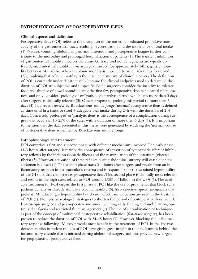
PATHOPHYSIOLOGY OF POSTOPERATIVE ILEUS
Clinical aspects and definition Postoperative ileus (POI) refers to the disruption of the normal coordinated propulsive motor activity of the gastrointestinal tract, resulting in constipation and the intolerance of oral intake (1). Nausea, vomiting, abdominal pain and distension, and postoperative fatigue further con-tribute to the morbidity and prolonged hospitalization of patients (1). The transient inhibition of gastrointestinal motility involves the entire GI tract and not all segments are equally af-fected; small intestinal motility is on average disturbed for approximately 24hrs, gastric moti-lity between 24 – 48 hrs whereas colonic motility is impaired between 48-72 hrs (reviewed in (2)), implying that colonic motility is the main determinant of clinical recovery. The definition of POI is currently under debate mainly because the clinical endpoints used to determine the duration of POI are subjective and unspecific. Some surgeons consider the inability to tolerate food and absence of bowel sounds during the first few postoperative days as a normal phenome-non, and only consider “prolonged” or “pathologic paralytic ileus”, which lasts more than 3 days after surgery, as clinically relevant (3). Others propose to prolong this period to more than 6 days (4). In a recent review by Boeckxstaens and de Jonge, ‘normal’ postoperative ileus is defined as ‘time until first flatus or stool + adequate oral intake during 24h with the duration of 2-4 days. Conversely, ‘prolonged’ or ‘paralytic ileus’ is the consequence of a complication during sur-gery that occurs in 10-25% of the cases with a duration of more than 6 days (1). It is important to mention that the data presented in this thesis were generated by studying the ‘normal’ course of postoperative ileus as defined by Boeckxstaens and De Jonge.
Pathophysiology and treatmentPOI comprises a first and a second phase with different mechanisms involved. The early phase (1-3 hours after surgery) is mainly the consequence of activation of sympathetic afferent inhibi-tory reflexes by the incision (somatic fibers) and the manipulation of the intestines (visceral fibers) (5). However, activation of these reflexes during abdominal surgery will cease once the abdomen is closed (1). The second phase starts 3-4 hours after surgery and results from an in-flammatory reaction in the muscularis externa and is responsible for the sustained hypomotility of the GI tract that characterises postoperative ileus. This second phase is clinically most relevant and results in the high costs related to POI, around US$1.47 billion in the USA (1). The avail-able treatment for POI targets the first phase of POI like the use of prokinetics that block sym-pathetic activity or directly stimulate colonic motility (6). Also, selective opioid antagonists that prevent IM induced gut hypomotility but do not affect pain reduction are used in the treatment of POI (1). Non pharmacological strategies to shorten the period of postoperative ileus include laparoscopic surgery and peri-operative measures including early feeding and mobilisation, op-timised analgesia and restricted fluid management (1). The use of a combination of techniques as part of the concept of multimodal postoperative rehabilitation (fast-track surgery), has been proven to reduce the duration of POI with 24-48 hours (7). However, blocking the inflamma-tory response following IM may provide most benefit in the treatment of POI. In the last two decades, studies in rodent models of POI have given great insight in the mechanisms behind the inflammatory cascade that is initiated during abdominal surgery and thus provide new targets for prophylaxis of postoperative ileus

20
Inflammatory mechanisms: animal modelsIn the late nineties, studies performed in rodent models demonstrated that gentle manipula-tion of the small bowel induces an inflammatory reaction in the muscularis externa of the small bowel (8). This inflammation is initiated by activation of resident leukocytes including mast cells (9) and macrophages, upregulation of the adhesion molecule ICAM-1 and subsequent influx of polymorphonuclear neutrophils (PMNs), monocytes, and mast cells (8;10;11) into the muscularis externa of the small intestine (see Fig. 1). As a consequence, muscle function is affected during intestinal manipulation as spontaneous and in vitro stimulated contractions of muscle strips are suppressed during this inflammation (8;10;11). This is mainly due to the increased expression of iNOS and COX-2 by resident macrophages in inflamed areas (12;13). The subsequent release of the mediators NO (14) and prostanoids (15) inhibit the contractility of intestinal smooth muscle locally. Pre-treatment with a mix of antibodies against the adhe-sion molecule ICAM-1, and the integrin molecules CD11a and CD18 (that together form Lymphocyte function-associated antigen 1 (LFA-1)) prevented the influx of leucocytes and also preserved normal neuromuscular function of muscle strips (11). It was later demonstrated that inflammatory mechanisms not only directly affect muscular function, but also trigger an inhibi-tory adrenergic pathway resulting in a delay in gastric emptying (16;17). In these experiments, the gastric muscularis was not inflamed indicating that neural pathways are responsible for gas-tric dysfunction and prolonged ileus after the small intestine muscle contractility has recovered (16) . Importantly, treatment with anti-LFA-1 and anti-ICAM (18) antibodies or an antisense ICAM-1 oligonucleotide (16), reduced the influx of leukocytes but also prevented activation of the inhibitory neural pathway (16) and subsequent gastroparesis (18). Pro-inflammatory cascades are also activated in areas distant from the manipulated site where muscle contractility is inhibited locally; this is referred to as the “field-effect” (19). This clinically important aspect of POI was recently shown to involve CCR9+T-cells that are activated at the site of manipulation and migrate to unmanipulated areas (20). There, IFN-γ release from the CCR9+T-cells activate local resident muscularis macrophages and suppress motility distant from manipulated areas (20). In the last two decades, many inflammatory mediators that contribute to the pathogenesis of postoperative ileus have been identified. The mediators that are upregulated in the muscularis layer of the small intestine include the adhesion molecule ICAM-I and integrin LFA-1, several cytokines including IL-6, MCP-1 (21;22) iNOS and NO, COX2 and PGE2 , and activation pro-inflammatory MAP-kinase pathways. Also the anti-inflammatory mediators HO-1 and IL-10 (22;23) are upregulated during intestinal manipulation. In Fig. 1 a schematic overview is shown of the inflammatory events that occur after abdominal surgery.
Figure 1: Schematic representation of the timing of the
inflammatory events triggered by abdominal surgery.
COX-2, cyclooxygenase 2

21
Inflammatory mechanisms: human dataIn conjunction to the observed inflammatory response to intestinal manipulation in rodent models, inflammation induced by handling of the intestine is also demonstrated in human tissue. Production of cytokines TNF, IL-6 and IL-10 was shown in the peritoneal fluid as well as sys-temically in patients that underwent gastrointestinal surgery (24). Also in muscle strips prepared from human small intestine specimens taken during abdominal surgery, mRNA of IL-6, Il-1β, TNF, iNOS and COX-2 was increased in a time dependent manner (25) and ex vivo muscle contractility was enhanced by application of iNOS and COX-2 inhibitors (25). Resident mus-cular macrophages in these muscle strips expressed LFA-1, IL-6 and IL-6-induced transcription factor Stat-3 during bowel surgery (25). These results were confirmed by a subsequent study showing that mRNA of ICAM-1 and iNOS as well as leucocyte influx was increased by intesti-nal handling in jejunal muscle specimens collected during biliary reconstructive surgery (26). This study also included groups undergoing abdominal hysterectomy that involves manipulation of the bowel, and transvaginal or laparoscopic hysterectomy, where the bowel is left untouched. The cytokines IL-6 and IL-8 and mast cell mediator tryptase, measured in peritoneal fluid, were increased during abdominal hysterectomy but not during transvaginal hysterectomy. In addition, leukocyte spect-imaging techniques showed an increase of leucocytes only when the bowel was touched (26). Importantly, these results indicate that also in patients, intestinal handling is the main cause for induction of inflammation in the muscularis externa of the small bowel. KEY ROLE FOR MACROPHAGES AND MAST CELLS
The main cell types that initiate the inflammatory cascade following intestinal manipulation are mast cells and macrophages that reside in the muscularis externa of the small bowel. How these cells are triggered and how they further activate the inflammatory cascade after intestinal ma-nipulation is one of topics investigated in this thesis. In the following section some background is given on these cell types.
Muscularis macrophages The muscularis macrophage (-like) cells are organized into a layer or ‘‘network’’ at the level of the myenteric plexus and at the serosal side of the intestinal tract (27). This phagocyte population in the muscularis externa has an interesting nature and most likely consists of diffe-rent subsets of phagocytes including macrophage-like cells expressing F4/80 (27) and dendritic cell-like cells expressing most common DC markers such as CD11c and DEC205 (28). How-ever in the mouse bowel wall, MHCII+ cells outnumber F4/80+ cells, indicating that a large number of these resident muscularis macrophages function as antigen presenting cells (APCs). Though double stainings for these markers have not been performed, so the MHC-II popula-tion may consist of an F4/80+ and F4/80- group (29). Also, these cells express the LPS-binding receptor CD14 (28) and stain positive for macrophage scavenger receptor CD163 (30), that has been shown to possess bacteria binding and sensing capacities (31). In a functional study, F4/80 positive phagocytes that were isolated from the murine muscularis externa and cultured for 3–4 days showed Ca2+ influxes and superoxide production upon stimulation with inflammatory mediators adenosine triphosphate (ATP), platelet-activating factor (paf) and bacterial lipopoly-saccharide (LPS) (32). Both Ca2+ release and subsequent superoxide production play a role in immune responses thereby further indicating that muscularis macrophages may play a role in host defence. As mentioned before, macrophages (8) that reside in the muscularis externa of the GI tract are

22
activated upon intestinal handling and (8). However, only few studies have been conducted to assess a causal role for muscularis macrophages in postoperative ileus. These studies were performed in two rodent models of macrophage depletion. In the rat model, macrophages were pharmacologically depleted and inactivated by administration of chlodronate liposomes; only mature macrophages are able to phagocyte the chlodronate liposomes in an effective (toxic) quantity. In the second model, mice carrying a mutation in the colony stimulating factor-1 gene, completely lack intestinal muscularis muscularis macrophages and have a diminished number of macrophages in the mucosa (33). Pharmacological or genetic depletion of resident macrophages resulted in a decrease of inflammatory mediators and diminished the recruitment of leucocytes into the muscularis after intestinal manipulation. Moreover, macrophage depletion led to a recovered in vitro jejunal circular muscle function and gastrointestinal transit after surgi-cal manipulation (34;35). In IM induced ileus, it is clear that the phagocyte network consisting of dendritic- and macrophage-like cells is crucial in the IM induced inflammatory response that leads to POI.
Mast cellsIn the body, mast cells reside in tissues that form a barrier with the external environment including skin, respiratory tract, intestinal tract and also near blood vessels (36). In rodents, mast cells can be classified into two subtypes, the mucosal mast cells (MMC), and connective tissue mast cells (CTMC). These types of mast cells are characterized by differences in phenotype, biochemistry, size, function and their responsiveness to drugs (36). In the gut, mucosal mast cells are mainly located in the small intestinal lamina propria, whereas CTMC are located within the serosa and mesentery. Like in rodents, also human mast cells are classified based upon their protease content. The MCT (Mast cells containing tryptase) is similar to the MMC, and the MCTC (Mast cells containing tryptase and chymase) is similar to the CTMC (36). However in human, mast cells are more closely related to monocytes and macrophages, whereas murine mast cells seem to be more similar to human basophils (37). The importance of mast cells in the inflammatory cascade triggered by intestinal manipula-tion, was demonstrated in experiments using mast cell stabilizers ketotifen and doxantrazole. Both compounds reduced the inflammatory response and delayed gastric emptying 24 h after abdominal surgery. Conversely, incubation of intestinal loops in solution containing the mast cell activator 48/80 induces an inflammatory response and POI (9). KitW/WV mutant mice that lack mast cells fail to develop an intestinal infiltrate following intestinal manipulation and recon-stitution with wildtype mast cells restores the capacity of mutant animals to recruit leucocytes to the intestine after surgery (9).When mast cell activation was determined in human POI, even very gentle inspection of the intestines at the beginning of the abdominal procedure increased the level of peritoneal mast cell derived tryptase (26). In contrast, in patients undergoing a laparoscopic or a vaginal hys-terectomy the increase in tryptase was minimal. Clinically, the time until discharge and first bowel movement was significantly lower after vaginal hysterectomy as compared to abdominal hysterectomy (26). This important finding was followed by a pilot study in patients to investigate the effects of mast cell stabilizers in the course of POI. Administration of the mast cell stabilizer ketotifen was effective in reduction of gastric emptying time and abdominal cramps (38). Alto-gether, the results from animal and human studies show that mast cells are activated by intestinal handling and play a role in the in the pathogenesis of POI. Importantly, mast cells are important mediators of intestinal barrier function and their activation results in loss of epithelial barrier integrity and subsequent disease progression in animal models of chronic stress (39;40), allergic
23
inflammation (41), parasitic infection (42) and endoxemia (43). In addition, during abdominal surgery the intestinal barrier is disturbed and may involve mast cell activation.
Barrier dysfunction during abdominal surgeryThe occurrence of barrier dysfunction following abdominal surgery may also contribute to the pathogenesis of gastro intestinal stasis and post-operative- or septic ileus. Although gastric and colonic dysmotlity are regarded as important contributors to ileus, small bowel motility is an important factor in regulation of the enteric bacterial population. A delayed transit time results in bacterial overgrowth, especially in the small bowel, and predisposes to bacterial translocation (44). Bacteria from the intestinal lumen might activate intestinal resident macrophages, given the observation the pretreatment with antibiotics prior to the intestinal manipulation reduced inflammatory responses. The ileus-associated bacterial translocation reflects disturbances in intes-tinal barrier function and does not only refer to the transepithelial passage of viable bacteria, but also (endo)toxins or antigens from the intestinal lumen. This is illustrated by the observation that in abdominal surgery, barrier dysfunction has been associated with increased postoperative septic morbidity in surgical patients undergoing laparotomy. Moreover, in experimental model for post-operative ileus the occurrence of ileus and the reduction of small intestinal smooth muscle contractility was not observed after surgery in TLR4 deficient mice (45). The occurrence of septic morbidity and even multiple organ failure in serious conditions such as surgery, trauma, ischemia reperfusion injury might be the result of a breakdown of the intestinal barrier and subsequent bacterial translocation. In a recent study including 927 patients over 13 years showed that surgery induced bacterial translocation and was associated with increased postoperative septic morbidity (46). However, the evidence for the so-called ‘gut origin of sepsis’ is at least in humans, controversial (47). The development of septic morbidity is multifactorial and in certain patients measures taken to prevent septic morbidity such as selective gut decontamination, and the use of pre- or probiotics have not been successful.
REGULATION OF EPITHELIAL BARRIER BY IMMUNE MEDIATORS
Mast cell activation and more in general, inflammation, is closely associated with intestinal bar-rier dysfunction and contributes to disease perpetuation and progression. In the following sec-tion background is given on the composition of the epithelial barrier and how inflammation af-fects barrier function. The causes of intestinal barrier dysfunction can be quite diverse and range from intestinal infection to allergic food components, malnutrition, toxic chemicals, NSAIDs and mechanical trauma. In addition, during recent years we have gained a lot of knowledge on the possible role of psychological-stress related impairment in barrier function in irritable bowel syndrome (IBS) and inflammatory bowel disease (IBD).
Epithelial cell function and barrier regulationThe intestinal epithelial barrier (IEB) stays in close contact with the environment and is composed of a monolayer of specialised intestinal epithelial cells (IEC). The IEB has devel-oped specific mechanisms to prevent access of luminal contents into the lamina propria, which includes the restriction of paracellular transport and the maintenance of the architecture of the epithelial barrier. This function is brought about by the apical junctional complex, which is composed of the TJ, or zonula occludens (ZO) and the subjacent adherens junction, or zonula adherens. Members of the ZO family are proteins that form a bridge between these membrane proteins and actin filaments, which are connected to the peri-junctional ring, a component of

24
the cellular cytoskeleton (48;49). The desmosomes, or macula adherens, are located along the lateral membranes beneath the adherens junction. Intracellular junctions including adherens junctions, desmosomes, gap junctions and TJs tightly regulate paracellular transport in conjunc-tion. Whereas TJs seal the paracellular pathway, the adherens junctions and desmosomes provide the strong bonds necessary to maintain cellular proximity and allow TJ assembly. Adherens junc-tions are also critical for epithelial polarization and differentiation, mucosal morphogenesis, and tumor suppression, processes that occur through a variety of interactions with other proteins, in-cluding actin and β-catenin. TJs are the most apical components of these intercellular junctions. The main functions of TJs are to prevent diffusion of membrane proteins and lipids between basolateral and apical membranes so that cell polarity is preserved (fence function) and impor-tantly, function as a selective barrier to paracellular transport (barrier function). TJs complexes are composed of a network of proteins that are coupled to actin filaments of the cytoskeleton (49). ZO-1, Occludin (62-82kDa), several members of the claudin family (20-27kDa) and junc-tional adhesion molecule (JAM) 1 (36-41kDa) are proteins that make up the membrane part of TJs (50-53)(Fig. 2).
Inflammatory mediators affecting epithelial TJ integrity Increased concentrations of pro-inflammatory cytokines are present in the intestine in ac-tive phase of inflammatory conditions such as IBD but also in conditions such as hemorrhagic shock or sepsis. In vitro studies in intestinal cell lines have demonstrated that pro-inflammatory cytokines decrease the barrier function of intestinal epithelial monolayers and induce reorgani-sation of several TJ-associated proteins, including ZO-1, JAM-1, occludin and claudin-1, and -4 (54;55). Examples of such cytokines that cause TJ rearrangements are TNF, IFN-γ, IL-8 and IL-1β (56-58). These cytokines influence the IEB primarily by acting on the epithelial expression and activation of myosin light chain kinase (MLCK) through PKC-activation. Upon activation, MLCK phosphorylates myosin light chain (MLC) which in turn causes contraction of the peri-junctional ring, a component of the cellular cytoskeleton, so that permeability of TJs increased (59-61). Another example of cytokine-mediated barrier changes is TNF-induced barrier defects that are associated with MLCK activation, and IL-13 dependent increase in claudin-2 expres-sion (62). As pointed out by the latter (62), cytokine releases and immune responses resulting from enhanced paracellular transport further augment MLCK-related TJ rearrangement.IL-1β increases the intestinal permeability by the induction of MLCK gene transcription result-ing in an increased MLCK protein activity, probably mediated by a rapid activation of transcrip-tion factor NF-ĸB (63). IL-1β-mediates increased intestinal permeability via increased paracel-lular transport of luminal antigens (57). Also TNF-mediated increased intestinal permeability leads to an NF-ĸB dependent down-regulation of ZO-1 proteins and alteration in junctional localisation (64). In turn, the anti-inflammatory cytokines IL-10, TGF-β and IL-17 protect from loss of TJ proteins (57). The role of IL-6 in modulation of the epithelial barrier is controversial, and may depend on the specific cell type or model system used (57) IL-6, as well as IL-13 (65) and TNF affect epithelial permeability and cell turnover through activation of pro-apoptotic pathways (66) and possibly the activation of PI-3kinase dependent signalling pathways (57). Al-together, cytokine mediated barrier dysfunction is brought about via modulation of TJs through distinct mechanisms and intracellular signalling pathways. Data indicate that MLCK and ZO-1 might be effector molecules in this process.

25
Mast cell mediators Initially studied in the context of allergic diseases, we now know that mast cells are highly ver-satile cells that not only have a sentinel role in host defence but also play a central role in intesti-nal disorders like IBD and IBS. Mast cells can be activated by a variety of stimuli and the type of stimulus determines their mediator release profile and subsequent consequences for neighbour-ing cells. In reference to the long list of cytokines that are be involved in modulation of barrier function (i.e. Il-10, IL-6, Il-13, TGF-β and TNF) it is important to notice that all of these can be expressed by mast cells (67). Most of them are de novo synthesized upon mast cell activation but an important exception to this is TNF. Results obtained by Bischoff et al. not only showed that this pro-inflammatory cytokine is constitutively expressed by these cells but also indicated that approximately 60% of TNF positive cells in the gut are in fact mast cells (68). As mentioned before, TNF induced barrier dysfunction depends on MLCK-mediated modulation of TJs (66) In addition, however, it was also shown that TNF potentiates histamine-induced ion secretion in enterocyte cell lines and isolated distal colon (69). Thus, although histamine was originally one of the classic mast cell mediators involved in itch, vasodilation and vascular permeability,
Figure 2: Neural networks
and epithelial integrity in
the intestine.

26
here it was shown that its synergistic action with TNF induces enhanced chloride secretion across the intestinal epithelium. This is highly relevant because it may lead to excessive water secretion and subsequent diarrhoea as observed in e.g. IBD and IBS. Next to histamine, another preformed mediator relevant to barrier dysfunction is tryptase that controls paracellular perme-ability through PAR-2. Tryptase-mediated cleavage of the N-terminal extracellular domain of this G-protein coupled receptor not only induces the redistribution of TJ proteins via extracel-lular signal-related kinases (ERK1/2) (70) but also via Ca2+/calmodulin mediated activation of MLCK (71). Being far from complete, this small overview clearly shows that mast cells and their mediators are major players in direct modulation of intestinal barrier function.
NEUROIMMUNE REGULATION OF EPITHELIAL BARRIER FUNCTION
The enteric nervous system (ENS)Intestinal phagocytes and mast cells are part of the gut extensive immune system with a number of immune cells equal to those in the remainder of the body. Equally impressive is that the GI tract neuronal network, the enteric nervous system (ENS) contains as many neurons as the spinal cord. The ENS comprises parasympathetic and sympathetic systems, as well as non-adren-ergic non-cholinergic systems that and can operate without the participation of the CNS. Im-portantly, in order to regulate gut function, the CNS interacts with the GI tract largely through the ENS in a bidirectional fashion via the so-called ‘brain-gut’ axis (see Fig. 2). The ENS is organized in several plexuses throughout the intestinal wall: the myenteric and submucosal ple-xuses, and the mucosal plexus, that contains nerve endings that are in close contact with mu-cosal immune cells and enterocytes. The ENS contains sensory neurons, interneurons and motor neurons, which primarily control peristalsis, local changes in blood flow, and secretion of water and electrolytes (72). Also, the ENS is involved in regulation of intestinal barrier function (73) An important component of the ENS is the population of enteric glial cells (EGC), that form a large and widespread network at all levels of the GI tract (74) and serve as intermediaries in the enteric neurotransmission and information processing. They show morphologic and functional similarity to brain astrocytes and control several aspects of gut function, including motility, microvascular circulation, epithelial secretion of fluid, ions, bioactive peptides and recently have been identified as important regulators of the intestinal barrier (75).
NeuropeptidesMore than 30 different neurotransmitters exist in the ENS, with most neurons expressing mul-tiple transmitters. Neuropeptides are considered key mediators in the communication between neurons (in particular sensory neurons) and effector cells (smooth muscle, glands and immune cells) (76) and exhibit a variety of functions in the gastro-intestinal tract. Neuropeptides are involved in secretion of salivary, gastric fluids and intestinal fluids and electrolytes. Besides the action on motor function of the gut, neuropeptides also function as co-transmitters of enteric cholinergic neurons, increase enteric neuron excitability, and consequently induce the release of enteric neurotransmitters, including acetylcholine (77). Neuropeptides are increasingly recog-nized as potent modulators of the immune response, which is underscored by the fact that, in addition to (afferent) neurons, several immune cells produce neuropeptides. In table 1 a selection of G protein coupled neuropeptides and their role in intestinal disease is depicted.

27
Neuropeptide Peptide Family
Peptide source ReceptorType
Effects of receptor activation in intestinal disease
Opioids Opioids Recruited immune cellsNerves throughout the
mu-, kappa- and delta-opioid
Induction of bowel dysfunction in patients(78).•mu-opioid receptor agonists:
Chapter 1 Table 1
GI tractEnteric nerve plexus CNS
receptors Decrease intestinal inflammation in TNBS induced and CD45RBhi transfer models of colitis (79)Inhibit GI transit in mice (80)•kappa-opioid receptor agonists:Reduce ileus by reduction of inhibitory reflexes and visceral nociception in rats (81)
Corticotropin-releasing hormone Urocortin 1Urocortin 2Urocortin 3
CRH Recruited immune cellsEpithelial and enterochromaffin cellsEnteric nerve plexus CNS
CRHR1CRHR2
Proinflammatory effects in development of TNBS colitis in mice (82)Proinflammatory effects in toxin A induced
intestinal inflammation in mice(83;84)Induction of gastric stasis via peripheral and central
pathways in rats (85;86)pathways in rats (85;86)
Ghrelin, Cortistatin
Motilin Recruited immune cellsEnteric nerve plexus Gastric EECsCNSRecruited immune cellsCNS
GHS-RGHS-RSST1-5MrgX2
Reduction of inflammation in TNBS induced colitis in mice (87)Acceleration of gastric emptying in POI in rats (88)and septic gastric ileus in mice (89).Amelioration of TNBS induced colitis inmice (90)
Vasoactive intestinal peptide
VIPPACAPGlucagonSecretin
Recruited immune cellsEnteric nerve plexus Lamina propria nerves CNS
VPAC1VPAC2
Amelioration of TNBS induced colitis by shifting T-cell responses from Th1 to Th2 (91;92).Inhibition of gastrointestinal transit in POI in rats
(93)
Substance P Tachykinins Recruited immune cells, lamina propria macrophages, colonic gliaMotorneurons of intestinal muscularisLamina propria nerves CNS
NK-1RNK-2RNK-3R
Antagonists ameliorate disease in a rat model of TNBS induced colitis (94;95)NK-1R-/- mice protected from inflammatory diarrhoea in C. difficile toxin A (96)
Neuropeptide YPeptide YYPancreatic polypeptide
Pancreatic polypeptide
CNSMononuclear blood leukocytesT cells APCs
Y1-6R enhanced T cell cytokine release(97-99)Reduced APC cytokine release and function(100;101)
T-cells, APCs
The vagus nerve in barrier regulationThe ENS receives input directly from the brain by the vagus nerve, the largest nerve in the body. Classically, the vagus nerve controls heart rate, hormone secretion, gastrointestinal (GI) peristalsis and digestion, and in the last decade has also been put forward as a regulator of the immune system (102) . Activation of vagal activity to modulate barrier function has been achieved via pharmacological and nutritional (103-105) techniques. For instance, in a model of hemorrhagic shock, cholecys-tokinin (CCK)-dependent stimulation of vagal activity by high fat nutrition has been shown to maintain intestinal barrier integrity. Translocation of bacteria, permeability to horse radish per-oxidase (HRP), disturbed expression of ZO-1 (104) and enterocyte damage (103) in high-lipid-treated animals was significantly reduced when compared with those in low-lipid-treated and fasted controls. The protective role of vagal nerve stimulation has been described in several other studies. Vagal efferent activity has previously been shown to decrease histological gut injury and intestinal inflammation in a model of colitis (106;107). Alternatively, in addition to fat feeding-mediated vagal improvement in barrier function in a shock model, peripheral as well as intracerebroventricular injection with ghrelin ameliorated the disrupted barrier function in rat models of intestinal ischemia-reperfusion (108) and sepsis (109). In both studies, vagotomy prevented the effect of ghrelin, demonstrating that these effects
Table 1: Distribution of neuropeptides and effects in intestinal disease.

28
depended on an intact vagus nerve. It is interesting to speculate on the working mechanism behind the vagal potential to affect bar-rier function. Vagal activity may lead to peripheral release of it’s principal neurotransmitter ACh, but the vagal nerve particularly projects to other post-ganglionic enteric neurons, in particular VIP (110)(studied in the duodenum) and 5-HT (111). In fact, some of the functional effects of vagus nerve stimulation are counteracted by VIP antisera (112). The release of VIP by enteric neurones is shown to inhibit intestinal epithelial cell proliferation and to maintain epithelial barrier integrity, and the effect of VIP on epithelial permeability is concomitant with a neu-ral induced increase in ZO-1 mRNA and protein expression in IEC (113). Besides VIP, ACh increases paracellular permeability in the healthy gut (114) setting the basis for a fine ‘tuning’ of the barrier permeability by the ENS, either with or without the intermediate action of enteric glia (Fig. 2). Interestingly, preliminary data from our own laboratory indicate that ACh reduces cytokine-induced epithelial activation and loss of tight junction integrity in epithelial cell lines (S. Dhawan, unpublished observations 2010)
The vagus nerve in intestinal immune regulationIn the gut, cholinergic fibres are located in close apposition to antigen presenting cells APCs (115) and would thus be the ideal site for neuro immune modulation by the vagus nerve (Fig 2). It has been shown that activation of the vagus nerve negatively regulates macrophage immune responses via the peripheral release of acetylcholine (ACh) (116;117). Activation of the so-called ‘cholinergic anti-inflammatory pathway’ has been shown to ameliorate disease in various mo-dels of inflammation, including sepsis (116), ischemia reperfusion (116) haemorrhage (105). In particular, the nicotinic acetylcholine receptor α7 (nAChRα7) is proposed as the receptor sub-type that potentiates the anti-inflammatory actions of ACh on immune cells and is expressed by macrophages (115;116;118;119), but also by monocytes, dendritic cells and mast cells (120;121). In the gut, vagus nerve activity can have a beneficial effect on the course of intestinal inflam-mation as shown in a mouse model for POI (122). In addition, administration of selective nAChRα7 agonists ameliorated inflammation and disease parameters in postoperative ileus (122). Also in mouse models of colitis, enhanced parasympathetic output is involved in the negative regulation of intestinal inflammation via efferent activity of the vagus nerve (106;107). Innate immune cells express a broad range of nAChR receptors (nAChRs), mus-carinic ACh receptors (123), and peptidergic receptors (124), and are therefore target cells for regulation by neuronal derived mediators. In this way, the ENS may mediate regulation of gut immune cell activity including cytokine production and phagocytic properties, thereby also indirectly affecting intestinal barrier function (Fig. 2). Taken together, novel therapeutic approaches to be designed the next years to specifi-cally target and intervene in disruption of neuroimmune communication in the gastrointestinal tract would be likely to benefit in treatment of disease. The advantages of the use of neuron derived chemical messengers are that they are short lived and act local. Importantly, in addition to the potential to ameliorate IEB, neuronal messengers also affect inflammatory processes, mo-tility, mucus production and water/electrolyte secretion, thereby further contributing to disease amelioration.

29
Reference List
(1) Boeckxstaens GE, de Jonge WJ. Neuroimmune mechanisms in postoperative ileus. Gut 2009;58(9):1300-11.
(2) Benson MJ and Wingate, D. L. Ileus and mechanical obstruction. [An illustrated guide to gastrointestinal
motility. London: Churchill Livingston, 1993:547-66t]. 2011. Ref Type: Generic
(3) Livingston EH, Passaro EP, Jr. Postoperative ileus. Dig Dis Sci 1990;35(1):121-32.
(4) Artinyan A, Nunoo-Mensah JW, Balasubramaniam S et al. Prolonged postoperative ileus-definition, risk fac-
tors, and predictors after surgery. World J Surg 2008;32(7):1495-500.
(5) Holte K, Kehlet H. Postoperative ileus: a preventable event. Br J Surg 2000;87(11):1480-93.
(6) Zeinali F, Stulberg JJ, Delaney CP. Pharmacological management of postoperative ileus. Can J Surg
2009;52(2):153-7.
(7) Kehlet H. Postoperative ileus--an update on preventive techniques. Nat Clin Pract Gastroenterol Hepatol
2008;5(10):552-8.
(8) Kalff JC, Schraut WH, Simmons RL et al. Surgical manipulation of the gut elicits an intestinal muscularis
inflammatory response resulting in postsurgical ileus. Ann Surg 1998;228(5):652-63.
(9) de Jonge WJ, The FO, van der CD et al. Mast cell degranulation during abdominal surgery initiates postop-
erative ileus in mice. Gastroenterology 2004;127(2):535-45.
(10) Kalff JC, Buchholz BM, Eskandari MK et al. Biphasic response to gut manipulation and temporal correlation
of cellular infiltrates and muscle dysfunction in rat. Surgery 1999;126(3):498-509.
(11) Kalff JC, Carlos TM, Schraut WH et al. Surgically induced leukocytic infiltrates within the rat intestinal
muscularis mediate postoperative ileus. Gastroenterology 1999;117(2):378-87.
(12) Kreiss C, Birder LA, Kiss S et al. COX-2 dependent inflammation increases spinal Fos expression during
rodent postoperative ileus. Gut 2003;52(4):527-34.
(13) Kreiss C, Toegel S, Bauer AJ. Alpha2-adrenergic regulation of NO production alters postoperative intesti-
nal smooth muscle dysfunction in rodents. Am J Physiol Gastrointest Liver Physiol 2004;287(3):G658-G666.
(14) Eskandari MK, Kalff JC, Billiar TR et al. LPS-induced muscularis macrophage nitric oxide suppresses rat
jejunal circular muscle activity. Am J Physiol 1999;277(2 Pt 1):G478-G486.
(15) Schwarz NT, Kalff JC, Turler A et al. Prostanoid production via COX-2 as a causative mechanism of rodent
postoperative ileus. Gastroenterology 2001;121(6):1354-71.
(16) de Jonge WJ, van den Wijngaard RM, The FO et al. Postoperative ileus is maintained by intestinal im-
mune infiltrates that activate inhibitory neural pathways in mice. Gastroenterology 2003;125(4):1137-47.
(17) Kreiss C, Birder LA, Kiss S et al. COX-2 dependent inflammation increases spinal Fos expression during
rodent postoperative ileus. Gut 2003;52(4):527-34.
(18) The FO, de Jonge WJ, Bennink RJ et al. The ICAM-1 antisense oligonucleotide ISIS-3082 prevents the
development of postoperative ileus in mice. Br J Pharmacol 2005;146(2):252-8.
(19) Schwarz NT, Kalff JC, Turler A et al. Selective jejunal manipulation causes postoperative pan-enteric inflam-
mation and dysmotility. Gastroenterology 2004;126(1):159-69.
(20) Engel DR, Koscielny A, Wehner S et al. T helper type 1 memory cells disseminate postoperative ileus over
the entire intestinal tract. Nat Med 2010;16(12):1407-13.
(21) De BO, Elinck E, Priem E et al. Peroxisome proliferator-activated receptor gamma activation alleviates post-
operative ileus in mice by inhibition of Egr-1 expression and its downstream target genes. J Pharmacol Exp
Ther 2009;331(2):496-503.
(22) De BO, Elinck E, Blanckaert B et al. Water-soluble CO-releasing molecules reduce the development
of postoperative ileus via modulation of MAPK/HO-1 signalling and reduction of oxidative stress. Gut
2009;58(3):347-56.
(23) Moore BA, Overhaus M, Whitcomb J et al. Brief inhalation of low-dose carbon monoxide protects rodents
and swine from postoperative ileus. Crit Care Med 2005;33(6):1317-26.

30
(24) van Berge Henegouwen MI, van der PT, van Deventer SJ et al. Peritoneal cytokine release after elective gas-
trointestinal surgery and postoperative complications. Am J Surg 1998;175(4):311-6.
(25) Kalff JC, Turler A, Schwarz NT et al. Intra-abdominal activation of a local inflammatory response within the
human muscularis externa during laparotomy. Ann Surg 2003;237(3):301-15.
(26) The FO, Bennink RJ, Ankum WM et al. Intestinal handling-induced mast cell activation and inflammation
in human postoperative ileus. Gut 2008;57(1):33-40.
(27) Mikkelsen HB. Macrophages in the external muscle layers of mammalian intestines. Histol Histopathol
1995;10(3):719-36.
(28) Flores-Langarica A, Meza-Perez S, Calderon-Amador J et al. Network of dendritic cells within the muscular
layer of the mouse intestine. Proc Natl Acad Sci U S A 2005;102(52):19039-44.
(29) Mikkelsen HB, Larsen JO, Hadberg H. The macrophage system in the intestinal muscularis externa during
inflammation: an immunohistochemical and quantitative study of osteopetrotic mice. Histochem Cell Biol
2008;130(2):363-73.
(30) Wehner S, Buchholz BM, Schuchtrup S et al. Mechanical strain and TLR4 synergistically induce cell-specific
inflammatory gene expression in intestinal smooth muscle cells and peritoneal macrophages. Am J Physiol
Gastrointest Liver Physiol 2010;299(5):G1187-G1197.
(31) Fabriek BO, Van Haastert ES, Galea I et al. CD163-positive perivascular macrophages in the human CNS
express molecules for antigen recognition and presentation. Glia 2005;51(4):297-305.
(32) Ozaki H, Kawai T, Shuttleworth CW et al. Isolation and characterization of resident macrophages from the
smooth muscle layers of murine small intestine. Neurogastroenterol Motil 2004;16(1):39-51.
(33) Mikkelsen HB, Thuneberg L. Op/op mice defective in production of functional colony-stimulating factor-1
lack macrophages in muscularis externa of the small intestine. Cell Tissue Res 1999;295(3):485-93.
(34) Wehner S, Behrendt FF, Lyutenski BN et al. Inhibition of macrophage function prevents intestinal inflamma-
tion and postoperative ileus in rodents. Gut 2007;56(2):176-85.
(35) Wehner S, Straesser S, Vilz TO et al. Inhibition of p38 mitogen-activated protein kinase pathway as prophy
laxis of postoperative ileus in mice. Gastroenterology 2009;136(2):619-29.
(36) Metcalfe DD, Baram D, Mekori YA. Mast cells. Physiol Rev 1997;77(4):1033-79.
(37) Bischoff SC. Physiological and pathophysiological functions of intestinal mast cells. Semin Immuno pathol
2009;31(2):185-205.
(38) The FO, Buist MR, Lei A et al. The role of mast cell stabilization in treatment of postoperative ileus: a pilot
study. Am J Gastroenterol 2009;104(9):2257-66.
(39) Santos J, Yates D, Guilarte M et al. Stress neuropeptides evoke epithelial responses via mast cell activation in
the rat colon. Psychoneuroendocrinology 2008;33(9):1248-56.
(40) Soderholm JD, Yang PC, Ceponis P et al. Chronic stress induces mast cell-dependent bacterial adherence and
initiates mucosal inflammation in rat intestine. Gastroenterology 2002;123(4):1099-108.
(41) Yang PC, Berin MC, Yu L et al. Mucosal pathophysiology and inflammatory changes in the late phase of the
intestinal allergic reaction in the rat. Am J Pathol 2001;158(2):681-90.
(42) McDermott JR, Bartram RE, Knight PA et al. Mast cells disrupt epithelial barrier function during enteric
nematode infection. Proc Natl Acad Sci U S A 2003;100(13):7761-6.
(43) Moriez R, Leveque M, Salvador-Cartier C et al. Mucosal mast cell proteases are involved in colonic perme
ability alterations and subsequent bacterial translocation in endotoxemic rats. Shock 2007;28(1):118-24.
(44) Balzan S, de Almeida QC, de CR et al. Bacterial translocation: overview of mechanisms and clinical
impact. J Gastroenterol Hepatol 2007;22(4):464-71.
(45) Turler A, Schnurr C, Nakao A et al. Endogenous endotoxin participates in causing a panenteric inflammatory
ileus after colonic surgery. Ann Surg 2007;245(5):734-44.
(46) MacFie J, Reddy BS, Gatt M et al. Bacterial translocation studied in 927 patients over 13 years. Br J
Surg 2006;93(1):87-93.

31
(47) MacFie J. Current status of bacterial translocation as a cause of surgical sepsis. Br Med Bull 2004;71:1-11.
(48) Fanning AS, Jameson BJ, Jesaitis LA et al. The tight junction protein ZO-1 establishes a link between the
transmembrane protein occludin and the actin cytoskeleton. J Biol Chem 1998;273(45):29745-53.
(49) Itoh M, Furuse M, Morita K et al. Direct binding of three tight junction-associated MAGUKs, ZO-1, ZO-2,
and ZO-3, with the COOH termini of claudins. J Cell Biol 1999;147(6):1351-63.
(50) Furuse M, Hirase T, Itoh M et al. Occludin: a novel integral membrane protein localizing at tight junctions. J
Cell Biol 1993;123(6 Pt 2):1777-88.
(51) Furuse M, Fujita K, Hiiragi T et al. Claudin-1 and -2: novel integral membrane proteins localizing at tight
junctions with no sequence similarity to occludin. J Cell Biol 1998;141(7):1539-50.
(52) Heiskala M, Peterson PA, Yang Y. The roles of claudin superfamily proteins in paracellular transport. Traffic
2001;2(2):93-8.
(53) Martin-Padura I, Lostaglio S, Schneemann M et al. Junctional adhesion molecule, a novel member of the
immunoglobulin superfamily that distributes at intercellular junctions and modulates mono cyte transmigra-
tion. J Cell Biol 1998;142(1):117-27.
(54) Bruewer M, Luegering A, Kucharzik T et al. Proinflammatory cytokines disrupt epithelial barrier function by
apoptosis-independent mechanisms. J Immunol 2003;171(11):6164-72.
(55) Zolotarevsky Y, Hecht G, Koutsouris A et al. A membrane-permeant peptide that inhibits MLC kinase
restores barrier function in in vitro models of intestinal disease. Gastroenterology 2002;123(1):163-72.
(56) Al-Sadi R, Ye D, Dokladny K et al. Mechanism of IL-1beta-induced increase in intestinal epithelial tight
junction permeability. J Immunol 2008;180(8):5653-61.
(57) Al-Sadi R, Boivin M, Ma T. Mechanism of cytokine modulation of epithelial tight junction barrier. Front
Biosci 2009;14:2765-78.
(58) Boivin MA, Roy PK, Bradley A et al. Mechanism of interferon-gamma-induced increase in T84 intestinal
epithelial tight junction. J Interferon Cytokine Res 2009;29(1):45-54.
(59) Anderson JM, Van Itallie CM. Tight junctions and the molecular basis for regulation of paracellular perme-
ability. Am J Physiol 1995;269(4 Pt 1):G467-G475.
(60) Hecht G, Pestic L, Nikcevic G et al. Expression of the catalytic domain of myosin light chain kinase increases
paracellular permeability. Am J Physiol 1996;271(5 Pt 1):C1678-C1684.
(61) Turner JR. Intestinal mucosal barrier function in health and disease. Nat Rev Immunol 2009;9(11):799-809.
(62) Webster EL, Torpy DJ, Elenkov IJ et al. Corticotropin-releasing hormone and inflammation. Ann N Y Acad
Sci 1998;840:21-32.
(63) Al-Sadi RM, Ma TY. IL-1beta causes an increase in intestinal epithelial tight junction permeability. J Immunol
2007;178(7):4641-9.
(64) Ma TY, Iwamoto GK, Hoa NT et al. TNF-alpha-induced increase in intestinal epithelial tight junction per-
meability requires NF-kappa B activation. Am J Physiol Gastrointest Liver Physiol 2004;286(3):G367-G376.
(65) Schulzke JD, Bojarski C, Zeissig S et al. Disrupted barrier function through epithelial cell apoptosis. Ann N Y
Acad Sci 2006;1072:288-99.
(66) Jin X, Zimmers TA, Zhang Z et al. Interleukin-6 Is an Important In Vivo Inhibitor of Intestinal Epithelial
Cell Death in Mice. Gut 2009.
(67) Marshall JS, Bienenstock J. The role of mast cells in inflammatory reactions of the airways, skin and intestine.
Curr Opin Immunol 1994;6(6):853-9.
(68) Bischoff SC, Lorentz A, Schwengberg S et al. Mast cells are an important cellular source of tumour necrosis
factor alpha in human intestinal tissue. Gut 1999;44(5):643-52.
(69) Oprins JC, van der Burg C, Meijer HP et al. Tumour necrosis factor alpha potentiates ion secretion induced
by histamine in a human intestinal epithelial cell line and in mouse colon: in volvement of the phospholipase
D pathway. Gut 2002;50(3):314-21.
(70) Jacob C, Yang PC, Darmoul D et al. Mast cell tryptase controls paracellular permeability of the intestine.

32
Role of protease-activated receptor 2 and beta-arrestins. J Biol Chem 2005;280(36):31936-48.
(71) Cenac N, Chin AC, Garcia-Villar R et al. PAR2 activation alters colonic paracellular permeability in mice
via IFN-gamma-dependent and -independent pathways. J Physiol 2004;558(Pt 3):913-25.
(72) Furness JB. Types of neurons in the enteric nervous system. J Auton Nerv Syst 2000;81(1-3):87-96.
(73) Genton L, Kudsk KA. Interactions between the enteric nervous system and the immune system: role of
neuropeptides and nutrition. Am J Surg 2003;186(3):253-8.
(74) Ruhl A. Glial cells in the gut. Neurogastroenterol Motil 2005;17(6):777-90.
(75) Savidge TC, Newman P, Pothoulakis C et al. Enteric glia regulate intestinal barrier function and inflamma-
tion via release of S-nitrosoglutathione. Gastroenterology 2007;132(4):1344-58.
(76) Felten DL, Felten SY, Carlson SL et al. Noradrenergic and peptidergic innervation of lymphoid tissue. J Im-
munol 1985;135(2 Suppl):755s-65s.
(77) Shimizu Y, Matsuyama H, Shiina T et al. Tachykinins and their functions in the gastrointestinal tract. Cell Mol
Life Sci 2008;65(2):295-311.
(78) Mehendale SR, Yuan CS. Opioid-induced gastrointestinal dysfunction. Dig Dis 2006;24(1-2):105-12.
(79) Philippe D, Dubuquoy L, Groux H et al. Anti-inflammatory properties of the mu opioid receptor support its
use in the treatment of colon inflammation. J Clin Invest 2003;111(9):1329-38.
(80) Roy S, Liu HC, Loh HH. mu-Opioid receptor-knockout mice: the role of mu-opioid receptor in gastroin
testinal transit. Brain Res Mol Brain Res 1998;56(1-2):281-3.
(81) De Winter BY, Boeckxstaens GE, De Man JG et al. Effects of mu- and kappa-opioid receptors on postopera-
tive ileus in rats. Eur J Pharmacol 1997;339(1):63-7.
(82) Gay J, Kokkotou E, O’Brien M et al. Corticotropin-releasing hormone deficiency is associated with re-
duced local inflammation in a mouse model of experimental colitis. Endocrinology 2008;149(7):3403-9.
(83) Wlk M, Wang CC, Venihaki M et al. Corticotropin-releasing hormone antagonists possess anti-inflammatory
effects in the mouse ileum. Gastroenterology 2002;123(2):505-15.
(84) Anton PM, Gay J, Mykoniatis A et al. Corticotropin-releasing hormone (CRH) requirement in Clostrid-
ium difficile toxin A-mediated intestinal inflammation. Proc Natl Acad Sci U S A 2004;101(22):8503-8.
(85) Martinez V, Rivier J, Tache Y. Peripheral injection of a new corticotropin-releasing factor (CRF) antagonist,
astressin, blocks peripheral CRF- and abdominal surgery-induced delayed gastric emptying in rats. J Pharma-
col Exp Ther 1999;290(2):629-34.
(86) Barquist E, Bonaz B, Martinez V et al. Neuronal pathways involved in abdominal surgery-induced gastric
ileus in rats. Am J Physiol 1996;270(4 Pt 2):888-94.
(87) Gonzalez-Rey E, Chorny A, Delgado M. Therapeutic action of ghrelin in a mouse model of colitis. Gastroen-
terology 2006;130(6):1707-20.
(88) Venkova K, Fraser G, Hoveyda HR et al. Prokinetic effects of a new ghrelin receptor agonist TZP-101 in a
rat model of postoperative ileus. Dig Dis Sci 2007;52(9):2241-8.
(89) De Winter BY, De Man JG, Seerden TC et al. Effect of ghrelin and growth hormone-releasing peptide 6 on
septic ileus in mice. Neurogastroenterol Motil 2004;16(4):439-46.
(90) Gonzalez-Rey E, Varela N, Sheibanie AF et al. Cortistatin, an antiinflammatory peptide with therapeutic ac-
tion in inflammatory bowel disease. Proc Natl Acad Sci U S A 2006;103(11):4228-33.
(91) Surrenti C, Renzi D, Garcea MR et al. Colonic vasoactive intestinal polypeptide in ulcerative colitis. J Physiol
Paris 1993;87(5):307-11.
(92) Gonzalez-Rey E, Chorny A, Fernandez-Martin A et al. Vasoactive intestinal peptide generates human tolero-
genic dendritic cells that induce CD4 and CD8 regulatory T cells. Blood 2006;107(9):3632-8.
(93) De Winter BY, Robberecht P, Boeckxstaens GE et al. Role of VIP1/PACAP receptors in postoperative ileus
in rats. Br J Pharmacol 1998;124(6):1181-6.
(94) Mazelin L, Theodorou V, More J et al. Comparative effects of nonpeptide tachykinin receptor an tagonists on
experimental gut inflammation in rats and guinea-pigs. Life Sci 1998;63(4):293-304.

33
(95) Di Sebastiano P, Grossi L, Di Mola FF et al. SR140333, a substance P receptor antagonist, influences mor
phological and motor changes in rat experimental colitis. Dig Dis Sci 1999;44(2):439-44.
(96) Alvaro G, Di Fabio R. Neurokinin 1 receptor antagonists--current prospects. Curr Opin Drug Discov Devel
2007;10(5):613-21.
(97) Naveilhan P, Hassani H, Lucas G et al. Reduced antinociception and plasma extravasation in mice lacking a
neuropeptide Y receptor. Nature 2001;409(6819):513-7.
(98) Wheway J, Herzog H, Mackay F. The Y1 receptor for NPY: a key modulator of the adaptive immune system.
Peptides 2007;28(2):453-8.
(99) Wheway J, Mackay CR, Newton RA et al. A fundamental bimodal role for neuropeptide Y1 receptor in the
immune system. J Exp Med 2005;202(11):1527-38.
(100) Naveilhan P, Hassani H, Lucas G et al. Reduced antinociception and plasma extravasation in mice lacking a
neuropeptide Y receptor. Nature 2001;409(6819):513-7.
(101) Wheway J, Herzog H, Mackay F. The Y1 receptor for NPY: a key modulator of the adaptive immune system.
Peptides 2007;28(2):453-8.
(102) van der Zanden EP, Boeckxstaens GE, de Jonge WJ. The vagus nerve as a modulator of intestinal inflamma-
tion. Neurogastroenterol Motil 2009;21(1):6-17.
(103) de Haan JJ, Lubbers T, Hadfoune M et al. Postshock intervention with high-lipid enteral nutrition reduces
inflammation and tissue damage. Ann Surg 2008;248(5):842-8.
(104) Luyer MD, Buurman WA, Hadfoune M et al. Pretreatment with high-fat enteral nutrition reduces endotoxin
and tumor necrosis factor-alpha and preserves gut barrier function early after hemorrhagic shock. Shock
2004;21(1):65-71.
(105) Luyer MD, Greve JW, Hadfoune M et al. Nutritional stimulation of cholecystokinin receptors inhibits in-
flammation via the vagus nerve. J Exp Med 2005;202(8):1023-9.
(106) Ghia JE, Blennerhassett P, El-Sharkawy RT et al. The protective effect of the vagus nerve in a murine model
of chronic relapsing colitis. Am J Physiol Gastrointest Liver Physiol 2007;293(4):G711-G718.
(107) Ghia JE, Blennerhassett P, Collins SM. Vagus nerve integrity and experimental colitis. Am J Physiol Gastroin-
test Liver Physiol 2007;293(3):G560-G567.
(108) Wu R, Dong W, Ji Y et al. Orexigenic hormone ghrelin attenuates local and remote organ injury after intesti-
nal ischemia-reperfusion. PLoS ONE 2008;3(4):e2026.
(109) Wu R, Dong W, Qiang X et al. Orexigenic hormone ghrelin ameliorates gut barrier dysfunction in sepsis in
rats. Crit Care Med 2009;37(8):2421-6.
(110) Yuan PQ, Kimura H, Million M et al. Central vagal stimulation activates enteric cholinergic neurons in the
stomach and VIP neurons in the duodenum in conscious rats. Peptides 2005;26(4):653-64.
(111) Kirchgessner AL, Gershon MD. Identification of vagal efferent fibers and putative target neurons in the
enteric nervous system of the rat. J Comp Neurol 1989;285(1):38-53.
(112) Dahlstrand C, Theodorsson E, Dahlstrom A et al. VIP antisera inhibit the relaxatory motor responses of the
feline sphincter of Oddi and gall-bladder induced by VIP or vagal nerve stimulation. Acta Physiol Scand
1989;137(3):375-8.
(113) Neunlist M, Toumi F, Oreschkova T et al. Human ENS regulates the intestinal epithelial barrier permeability
and a tight junction-associated protein ZO-1 via VIPergic pathways. Am J Physiol Gastrointest Liver Physiol
2003;285(5):G1028-G1036.
(114) van der Zanden EP, Snoek SA, Heinsbroek SE et al. Vagus nerve activity augments intestinal macrophage
phagocytosis via nicotinic acetylcholine receptor alpha4beta2. Gastroenterology 2009;137(3):1029-39, 1039.
(115) de Jonge WJ, van der Zanden EP, The FO et al. Stimulation of the vagus nerve attenuates macrophage activa-
tion by activating the Jak2-STAT3 signalling pathway. Nat Immunol 2005;6(8):844-51.
(116) Borovikova LV, Ivanova S, Zhang M et al. Vagus nerve stimulation attenuates the systemic inflammatory
response to endotoxin. Nature 2000;405(6785):458-62.

34
(117) Tracey KJ. The inflammatory reflex. Nature 2002;420(6917):853-9.
(118) Matsunaga K, Klein TW, Friedman H et al. Involvement of nicotinic acetylcholine receptors in suppression
of antimicrobial activity and cytokine responses of alveolar macrophages to Legionella pneumophila infec-
tion by nicotine. J Immunol 2001;167(11):6518-24.
(119) Wang H, Yu M, Ochani M et al. Nicotinic acetylcholine receptor alpha7 subunit is an essential regulator of
inflammation. Nature 2003;421(6921):384-8.
(120) Kawashima K, Yoshikawa K, Fujii YX et al. Expression and function of genes encoding cholinergic compo-
nents in murine immune cells. Life Sci 2007;80(24-25):2314-9.
(121) Nouri-Shirazi M, Tinajero R, Guinet E. Nicotine alters the biological activities of developing mouse bone
marrow-derived dendritic cells (DCs). Immunol Lett 2007;109(2):155-64.
(122) The FO, Boeckxstaens GE, Snoek SA et al. Activation of the cholinergic anti-inflammatory pathway amelio-
rates postoperative ileus in mice. Gastroenterology 2007;133(4):1219-28.
(123) Wessler I, Kirkpatrick CJ. Acetylcholine beyond neurons: the non-neuronal cholinergic system in humans. Br J
Pharmacol 2008;154(8):1558-71.
(124) Snoek SA, Borensztajn KS, van den Wijngaard RM et al. Neuropeptide Receptors in Intestinal Disease:
Physiology and Therapeutic Potential. Curr Pharm Des 2009. Chapter 2

S.A. Snoek
Neuro-immunity in intestinal disease: in vivo studies of postoperative ileus and colitis
Onder Embargo tot 8 juni 2012:
Chapter 2: Intestinal manipulation causes mast cell-mediated epithelial barrier dysfunction in a mouse model of postoperative ileus
Chapter 3: A pathogenic role for IL-1β and MyD88 signalling in postoperative ileus

75

76
CHAPTER 4
Vagus nerve activity augments intestinal macrophage
phagocytosis via nicotinic acetylcholine
receptor alpha4beta2
Esmerij P. van der Zanden1, Susanne A. Snoek1, Sigrid E. Heinsbroek1, Oana I. Stanisor1, Caroline Verseijden1, Guy E. Boeckxstaens1,2, Maikel P. Peppelenbosch3, David R. Greaves4, Siamon Gordon4, and Wouter J. de Jonge1,4.
1Department of Gastroenterology & Hepatology, Academic Medical Center, Amsterdam, The Netherlands, 2Dept. of Gastroenterology, University Hospital of Leuven, Catholic University of Leuven, Leuven, Belgium, 3Dep. of Cell Biology, University of Groningen, Groningen, The Netherlands,4Sr William Dunn School of Pathology, University of Oxford, Oxford, UK
Gastroenterology. 2009 Sep;137(3):1029-39

77
Abstract
Background and Aims: The vagus nerve negatively regulates macrophage cytokine produc-tion via the release of acetylcholine (ACh) and activation of nicotinic acetylcholine receptors (nAChR). In various models of intestinal inflammation, vagus nerve efferent stimulation amelio-rates disease. Given the actively constrained cytokine responses of intestinal macrophages, we explored the effect of nAChR activation on endo- and phagocytosis by macrophages residing in the peritoneal and mucosal compartment. Methods: The phagocytic uptake by intestinal and peritoneal macrophages was measured by FACS analysis, and the nAChR involved was determined by pharmacological blockade, shRNA-assisted gene knockdown and the use of specific nAChR KO mice. The effect of electrical vagus nerve stimulation on epithelial translocation and macrophage uptake of luminal particles was studied in mice.Results: In isolated intestinal and peritoneal macrophages, nAChR activation enhanced endocytosis and phagocytosis. This effect was mediated via stimulated recruitment of GTPase Dynamin-2 to the forming phagocytic cup. These effects involve nAChR α4/β2, rather than nAChR α7. Despite enhanced bacterial uptake, acetylcholine reduced NF-κB activation inflam-matory IL10 production. Vagus nerve stimulation in mice altered mucosal immune responses by augmenting epithelial transport and uptake of luminal bacteria by lamina propria macrophages.Conclusions: Acetylcholine enhances phagocytic potential while inhibiting immune reactivity via nAChR α4/β2 in mouse macrophages. Hence, vagus nerve efferent activity may stimulate surveillance in the intestinal mucosa and peritoneal compartment. and pro-inflammatory cy-tokine production, while stimulating anti-inflammatory IL10 production. Vagus nerve stimula-tion in mice altered mucosal immune responses by augmenting epithelial transport and uptake of luminal bacteria by lamina propria macrophages.Conclusions: Acetylcholine enhances phagocytic potential while inhibiting immune reactivity via nAChR α4/β2 in mouse macrophages. Hence, vagus nerve efferent activity may stimulate surveillance in the intestinal mucosa and peritoneal compartment.

78
IntroductionIn the gastro-intestinal tract, the vagus nerve regulates motility and digestive functions mostly via the peripheral release of the parasympathetic neurotransmitter acetylcholine (ACh) that activates nicotinic acetylcholine receptors (nAChRs). However, vagal activity has also immune-regulatory properties. While the afferent vagus system is known to regulate the inflammatory response via the hypothalamic pituitary adrenal axis, efferent vagus nerve activity possesses immuno-modulatory potential as well. Borovikova et al (1) have revealed the potency of the vagus nerve to inhibit TNF production by macrophages after systemic endotoxin (2). Peritoneal and PBMC-derived macrophages express nAChRs, and nAChR activation has been shown to inhibit NF-κB transcriptional activity (3;4) and pro-inflammatory cytokine production (4;5). In conjunction, electrical vagus nerve stimulation has been shown to ameliorate disease in animal models of inflammatory conditions such as postoperative ileus (4;6), colitis (7), peritonitis (8;9), and hemorrage (10). An immune regulating role for the cholinergic nervous system may be par-ticularly evident in intestinal tissue, given the dense cholinergic innervation, and the abundant number of resident macrophages that populate the intestinal mucosa and muscularisexterna, of which some closely associate with cholinergic fibers (6). Cholinergic inhibition of pro-inflammatory cytokine production by macrophages has been firmly established (1;3;6). However, besides an effect on cytokine secretion, the choliner-gic nervous system may also affect more professional macrophage functions such as endo- and phagocytosis of bacteria and particles. Especially in the intestinal compartment macrophages may rather function as phagocytes that, along with dendritic cells, form critical effectors in the surveillance of luminal antigens (11;12). Hence, the question arises whether the anti-inflamma-tory effect of vagus nerve activity in intestinal inflammation solely rests on reduced macrophage cytokine production, or whether the vagus nerve also regulates other macrophage functions important in host defense. This is supported by the observation that vagus nerve activity affects bacterial clearance and mortality in various mouse models of infectious disease (8;13;14). There-fore, we studied the effect of nAChR activation on endo- and phagocytosis by macrophages residing in the peritoneal and mucosal compartment. We show here that nAChR α4/β2, rather than α7, activation enhances the phagocytic potential in mouse macrophages. Despite enhanced phagocytosis, the activation of NF-κB activ-ity and pro-inflammatory cytokine production is inhibited. In conjunction, we also demonstrate that in mice, electrical stimulation of the vagus nerve increases the epithelial permeability for luminal bacteria and stimulates phagocytosis by F4/80+CD11b+ macrophages residing in the intestinal mucosa. Taken together, our data suggest that vagus nerve activity can enhance mac-rophage bacterial uptake via activation of the nAChR α4/β2, while reducing inflammatory cell activation.
Methods
Reagents and antibodies.(-)-Nicotine, (+)-Nicotine, Acetylcholine, α-Bugarotoxin, Methylcaconitine, 2,2,6,6- Tetrameth-ylpiperidin-4-yl methyl-2-pyrrolidinyl]isoxazole, Zymosan A from S. cerevisiae were from Sig-ma-Aldrich (Zwijndrecht, the Netherlands). Polyclonal antibodies against phosphorylated and unphosporylated Akt and PI3K, p38 and anti-NFkB-p65 were from Cell Signalling. Anti-α4nAChR was purchased from Santa Cruz Biotechnology; anti-β2 nAChR subunit (MAb clone270) and anti-Dynamin-2 (clone 3457) were from Abcam (Cambridge, UK). Dil-AcLDL was obtained from (Invitrogen). Rat anti mouse monoclonal CD11b, F4/80 and CD11c were

79
purchased from BDBiosciences (Franklin Lakes, NJ). FITC (Sigma-Aldrich), labelling of 1x10⁹ heat-killed E. faecium was performed in 0.1 M NaHCO3 pH9.0 for 1h at 37°C (in shaking waterbath). heptanoate, Dihydro-β-erythroidine,
Mammalian cell culture.Macrophages from an immortalized spleen macrophage cell line (Mf4/4(15)) were cultured in RPMI 1640/10% FCS. Primary peritoneal cells (PMF) from BALB/C, C57/Bl6 WT mice, nAChR alpha7-/- or nAChRbeta2-/- mice (kindly provided by Dr. G.LaRosa and Dr. U. Maskos respectively) were collected by lavage 4 days after ip. injection of 1ml of Biogel solution and cultured in OptiMEM (Invitrogen).
Phagocytosis assays.For phagocytosis assays, cells were plated to 80 % confluence in 24 well suspension plates and pretreated with cholinergic agonists for 20min. Subsequently, cells were challenged with FITC-labeled Zymosan particles (20 particles/cell) for 10min or other time-points where indicated. Cells were washed by PBS, final washing was in PBS/EDTA/lidocaine and cells were harvested by scraping. After fixation in 2% PFA, bead uptake was determined by flow cytometry (FACS Calibur, Becton Dickinson). Fc Receptor-mediated phagocytosis of RBC. Sheep RBC (300 μl of a 10% suspension; Cappel/ICN) were opsonized with 1:1000 diluted rabbit-anti-sheep RBC IgG (Cappel/ICN). Opsonized RBCs were allowed to bind to Mf4/4 or peritoneal macro-phages at a 1/80 cell to RBC ratio at 37°C. After 15min, slides were exposed to ice-cold water for 15 sec to lyse RBCs that were not internalized, washed in PBS and fixed.
Cell stimulationsPeritoneal macrophages were incubated with nicotinic agonists at the concentration indicated for 45 minutes and stimulated with Zymosan (10 particles/cell) for 6hrs. After treatment, the medium was removed; the cells washed three times with ice-cold PBS, and medium and cell lysates were measured by ELISA.
RT-PCR of total RNATo determine nAChR expression on MF4/4 cells and PMF, total RNA was isolated using theRNeasy Mini kit (Qiagen), treated with DNase, and reverse transcribed. The resulting cDNAwas subjected to Light Cycler polymerase chain reaction (Roche) for 35 cycles. Primer se-quences are provided in supplementary data.
Immunostaining and imagingImmune-histochemistry on intestinal sections was performed as described earlier(6). For con-focal microscopy, MF4/4 were grown on glass slides (Nuncbrand), pretreated with nicotine, challenged with FITC-labeled Zymosan and phagocytosis was allowed for 5min. For a detailed description of the protocols and antibodies used, see supplementary methods.
shRNA transfection2x106 MF4/4 cells were transfected by electroporation with 2 μg shRNA expression plasmid (psilencerTM-CMV4.1neo; Ambion, Austin, TX) according to the manufacturer’s instructions (570 V, 50 μs, Amaxa; Gaithersburg, MD). shRNA sequences are given in the supplementary method section.

80
ImmunoblottingImmunoblotting was performed as previously described(6). Plasma membrane was extracted using the cell compartment kit (Qiagen), according to the manufacturer’s instructions.
Isolation of lamina propria macrophagesLamina propria macrophages were isolated after neutral protease digestion (a modified protocol as described previously(16)) and Magnetic bead-assisted Cell Sorting (MACS; Miltenyi Biotec Inc., Auburn, CA). A detailed description of the procedure is provided in the supplementary methods. After isolation, cells were taken up in RPMI/1%FCS at 107 cells/mL, left for 2 h, al-lowed to phagocytose FITC labeled heat-killed E.faecium at 37°C during 1h, finally washed and analyzed by FACS.
In vivo uptake assay and electrical vagus nerve stimulationAnimals. Male C57/Bl6 mice (Harlan Nederland, Horst, The Netherlands), 12 to 15 weeks old, were kept under environmentally controlled conditions (light on from 8:00 AM till 8:00 PM; water and rodent non-purified diet ad libitum; temperature 20 C-22 C; 55% humidity). Experi-ments were performed according to the guidelines of the Ethical Animal Research Committee of the University of Amsterdam. Mice were used after a 7 days adaptation period.Electrical vagus nerve stimulation Mice were anesthetized by i.p. injection of a mixtureof Fentanyl Citrate / Fluanisone (Hypnorm; Janssen, Beerse, Belgium) and Midazolam (Dor-micum; Roche, Mijdrecht, The Netherlands). VNS was performed as described previously (6). In short: the right cervical vagal branch was prepared free from the carotid artery and ligated with 6-0 silk suture. The part distal from the ligation was attached to an electrode and 5V stimuli with a frequency of 5Hz, duration of 2ms (10) were applied for 5 min. In sham mice the cervical skin was opened and left for 30 min. covered by moist gauze. After a 30 min. recovery period, the in vivo uptake assay was initiated as described below.In vivo uptake. Surgical procedures were performed under sterile conditions. Mice underwent a laparotomy and an ileum segment 3-10 cm proximal from the caecum was opened and its lumen rinsed with pre-warmed (37°C) oxygenated Krebs buffer. The ileum segment was filled with 1-2 mL of buffer containing FITC-labeled E.faecium bacteria and Alexa546-labeled Dex-tran particles (10,000Mw, Mol. Probes), and clamped at both sides.After 30 min. the clamped intestinal segment was removed, washed in PBS and processed for immune-histochemistry. Bacterial translocation. Mice were killed 18 h after surgery and mesenteric lymph nodes (MLN) were harvested under aseptic conditions. Lymph nodes were weighed, placed in a tube contain-ing 300 μl of ice-cold Luria-Bertani (LB) broth, homogenized with a sterile grinder and plated onto blood agar plates in aerobic and anaerobic conditions. After 48 h of incubation at 37ºC, the number of colony forming units (CFU) per g lymph node was assessed.
Ussing chamber experimentsSegments of tissue of the distal small intestine were opened, cut and immersed Kreb’s buffer (115 mM NaCl, 1.25 mM CaCl2, 1.2 mM MgCl2, 2.0 mM KH2PO4, and 25 mM NaHCO3 at pH 7.35). Within 15 minutes the tissue was mounted in Ussing chambers (World Precision Instruments, Berlin, Germany) with serosal and mucosal areas exposed to 2 ml of circulating oxygenated Krebs buffer (containing 10 mM glucose) maintained at 37°C. Isc (in μA/cm2), a measure of net active ion transport, was recorded by a computer connected a voltage-clamp sys-tem. After 15 minutes, HRP was added to the luminal buffer at a final concentration of 10μM.

81
Samples (300μl) were taken at the serosal side and replaced with fresh buffer. After 30 minutes, nicotine (1μM), Carbachol (1 μM) or buffer was added to the serosal side. The enzymatic acti-vity of HRP was measured using tetramethyl-benzidine (TMB) (Biosource Europe, Nivelles, Belgium) as a substrate.
StatisticsStatistical analysis was performed using SPSS 12.02 software (SPSS Inc. Chicago, Ill, USA). The Friedman’s two-way analysis of variance was used to explore multiple dependent value assays. If the Friedman’s analysis was significant, individual values compared to the 0 nM concentration were tested with a Mann-Whitney U test. P-values <0.05 were considered statistically signifcant and results were expressed as mean ± SEM.
Results Cholinergic agonists enhance phagocytosis in macrophages. We first tested whether ACh, the main neurotransmitter of the vagus nerve, or nicotine, that ac-tivates nAChRs, affected macrophage endo- and phagocytosis. In PMF (Fig. 1A-D), and spleen macrophage Mf4/4 cells (not shown), ACh as well as nicotine significantly (p<0.05) enhanced phagocytosis in a time-dependent (Fig. 1B) and dose-dependent (Fig. 1C) manner as analyzed by FACS and fluorescence microscopy. The enhanced phagocytosis induced by nicotine was not based on enhanced particle binding to the cells, as no increase in phagocytosis was observed when cells were incubated on ice instead of 37°C (Fig. 1A, B). To assess whether the cholinergic stimulation of phagocytosis was receptor pathway specific, we compared the effect of nicotine on the uptake of heat-killed E.faecium, opsonised sheep red blood cells (RBC), Zymosan, and on the endocytosis of acetylated low-density lipoprotein (AcLDL) by PMF (Fig. 1D) and Mf4/4 macrophages (not shown). Zymosan uptake depends on Dectin-1, IgG-opsonized RBC serve as cargo for Fc receptor-mediated phagocytosis, while AcLDL endocytosis is dependent on the scavenger receptor binding. However, nicotine stimu-lated uptake via all these pathways to a similar extent, indicating that nicotinic enhancement of phagocytosis involves a general pathway in phagocytosis rather than a specific effect on receptor expression. Moreover, further analysis confirmed that expression of Dectin-1, CD11b/CD18, or scavenger receptor A was not affected in peritoneal macrophages after a 6h treatment with 1μM of nicotine (results not shown). >

82
Cholinergic agonists stimulate phagocytosis via the α4/β2 nAChR rather than α7 nAChR. We next sought to establish which nAChR mediated the cholinergic effects on macrophage phagocytosis. The nAChR α7 has been implicated in the inhibition of cytokine production and NF-κB activity by nicotinic receptor activation in macrophages (a.o. in (4;5) ). In accor-dance with these results, nicotine inhibited the activation of NF-κB induced by Zymosan ((4)and results not shown), inhibited Zymosan-induced TNFα and MIP-2 production and induced IL-10 production (Fig. 2A). This effect was not only blocked by nAChRα7 blockers Bunga-rotoxin (Bgt) and Methyllycaconitine (MLA), but also by nAChR α4/β2 antagonist dihydro-β-erythroidine (DHβE) (Fig. 2B). In PMF of nAChRα7 knock-out mice, nicotine failed to reduce TNF production, confirming earlier reports ((5)and results not shown).
Figure 1. Cholinergic agonists en-
hance macrophage phagocytosis.
Uptake of FITC-Zymosan (green
particles) in peritoneal macropha-
ges grown on glass slides. Cells
were pretreated with nicotine (1
μM) or ACh (1 μM) and incubated
at 37 ºC or on ice where indicated.
Cells were stained using phalloidin
(red), and DaPi (blue). Scale bar: 10
μm (A). Time course of uptake by
PMF pretreated with vehicle, ace-
tylcholine or nicotine (B). Nicotine
(triangles) as well as acetylcholine
(circles) increases uptake of FITC-
Zymosan in PMF. Data are given
as the percentage increase in the
number of FITC-Zymosan-positive
cells, compared to medium
treatment (100%) (C). The effect
of nicotine on the phagocytosis
of E.faecium bacteria, Zymosan,
opsonised RBC and AcLDL by PMF.
Data shown are averages +/- SEM
of 3-5 independent experiments in
triplicate. Asterisks indicate signifi-
cant difference (P<0.05) compared
to vehicle treatment (medium) (D).

83
Surprisingly, specific blockers of α7 nAChRs failed to block the nicotinic increase in phago-cytosis in peritoneal macrophages, in fact, MLA pretreatment further augmented nicotinic stimulation of phagocytosis. On the other hand, 2,2,6,6-tetramethylpiperidin-4-yl heptanoate (TMPH), a blocker of nAChR with low affinity for the α7 nAChR (17) blocked the nicotinic stimulation of phagocytosis (Fig. 3A, right panel). Subsequent analysis indicated that specific α4/β2 nAChR blocker DHβE almost completely counteracted the effect of nicotine on phagocy-tosis (Fig. 3A). In agreement with these results, an agonist for α7 nAChRs (AR-R17779) failed to induce phagocytosis, while a specific agonist of α4/β2 nAChR, 3-Methyl-5-[(2S)-1-methyl-2-pyrrolidinyl]isoxazole (ABT418) stimulated phagocytosis, albeit with less potency compared to nicotine (Fig. 3B). These results demonstrate that cholinergic stimulation of phagocytosis is mediated via nAChR α4β2 rather than α7 nAChRs. Prompted by these data, we analyzed the expression of α7 and α4/β2 nAChR subunits in intestinal and peritoneal macrophages. Quantitative RT-PCR analyses revealed expression of nAChR α4/β2 transcripts in lamina propria macrophages (LPMF) and PMF, as well as Mf4/4 cells, while we failed to detect α7 transcripts in either of these macrophage types (Fig. 3C). Pro-tein expression of the nAChR β2 and α4 subunits was confirmed in PMF (Fig. 3D) and Mf4/4 macrophages (not shown). To demonstrate further that the cholinergic increase in phagocytosis depended on β2 nAChR expression we transfected β2 shRNA to knock-down β2 nAChR transcripts in Mf4/4 cells (Fig. 4A-C). We confirmed the potency of the shRNA expression vector to reduce β2 nAChR mRNA expression to 10-20% of normal in these cells, while expression of regular housekeeping genes was not affected (Fig. 4C). Nicotine pretreatment enhanced phagocytosis in macrophages transfected with random scrambled shRNA expression vectors, but knockdown of β2 nAChR expression abolished this effect (Fig. 4A-B). In addition, in cells transfected with shRNAs directed to α7 nAChR the nicotinic increase in phagocytosis was unaffected. Finally, nicotine enhanced phagocytosis in peritoneal macrophages from both WT and nAChRα7-/- mice, while it failed to do so in PMF from AChRβ2-/- mice (Fig.4D). These data demonstrate that cholinergic agonists stimulate macrophage phagocytosis via activa-tion of α4/β2 nAChRs, rather than α7 nAChRs.
Figure 2. Nicotine reduces Zymosan-induced pro-inflammatory mediator expression in peritoneal macrophages.
Cytokine production induced by Zymosan phagocytosis, with pretreatment of nicotine in the indicated concentra-
tions. IL10intra is intracellular IL10 (A). TNF release induced by Zymosan after pre-incubation with nicotine (gray
bars) and nAChR blockers α-Bungarotoxin (Bgt), Methyllycaconitine (MLA) or dihydro-β-erythroidine (DHβE) where
indicated (B). Data shown are average +/- SEM of 3 independent experiments performed in triplicate. Asterisks
indicate significant differences (* P <0.05; ** P <0.01).

84
Figure 3. Cholinergic agonists stimulate phagocytosis via nAChR α4β2. Left panel, nicotine increases phagocytosis
(percentage of cells positive for Zymosan) dose-dependently. Right panel, nicotinic (10-6 M) increase in phagocy-
tosis is counteracted by cholinergic blocker DHβE and 2,2,6,6-Tetramethylpiperidin-4-yl heptanoate hydrochloride
(TMPH), but not by NAChR α7 blockers αBgt and MLA. Data shown are average +/- SEM of 3-5 independent experi-
ments in triplicate, asterisks indicate significant differences (P<0.05) compared to vehicle treated group (A). Nico-
tine, as well as nAChR α4β2 agonist 3-Methyl-5-[(2S)-1-methyl-2-pyrrolidinyl]isoxazole (ABT418) increase phago-
cytosis of Zymosan, while nAChR α7 agonists AR-R17779 is not effective. Data shown are averages +/- SEM of 3
assays, asterisks indicate significant differences (P<0.05) compared to vehicle treated group (B). RT-PCR showing
transcript expression of nAChR α4 and β2 subunits in RNA derived from mouse brain, spleen Mf4/4 macrophages,
lamina propria macrophages (LMPF) and PMF (C). Immune-histochemical staining showing expression of α4 or β2
subunit protein (red) in PMF. Dapi counterstain, scale bar: 1 μm (D).

85
Figure 4. NAChR α4β2 activation enhances macrophage phagocytosis of Zymosan.
(A)-(D): Phagocytosis of Zymosan in shRNA transfected Mf4/4 macrophages and in PMF from WT mice,
nAChRα7-/- mice or nAChRβ2-/- mice, after pretreatment with nicotine at the concentrations indicated. Cells were
transfected with scrambled control shRNA, or shRNA specific for nAChR β2, or α7 as indicated (A). Numbers are
given as percentage of cells positive for Zymosan (A), or in (B) as phagocytic index as percentage of positive cells
times the number of particles per cell. Asterisks indicate significant difference (P<0.05) compared to vehicle treat-
ment (medium). Transcript levels of nAChR β2, β-actin, and GAPDH in cells transfected with random scrambled
shRNA or nAChR β2-directed shRNA (C). Nicotine dose-dependently enhances Zymosan phagocytosis in PMF
from both WT mice and nAChRα7-/- mice, but not in PMF from nAChRβ2-/- mice. Data shown are average +/- SEM
of 3 independent experiments performed in triplicate. Asterisks indicate significant differences (*P < 0.05) (D).

86
Nicotine enhances Dynamin-2 recruitment to the phagocytic cup. To elucidate the underlying cellular mechanism behind the nicotinic stimulation of phagocyto-sis we analyzed whether nicotine augmented phosphatidylinositol 3-kinase (PI-3K)/Akt activa-tion, known to be required for phagocytosis. Macrophage phagocytosis of Zymosan stimulated PI-3K and Akt phosphorylation, but nicotine did not significantly enhance this process, or phosphorylaton of p38 MAP kinase, (Supplementary Fig.1a). In addition, we analyzed whether the enhanced phagocytosis by nAChR activation resulted from induced intracellular calcium fluxes. To this end, we analyzed whether nicotine induced calcium fluxes in freshly isolated peritoneal macrophages using FURA-3 probe assisted imaging. However, although macrophages respond potently to ATP, no calcium flux was observed after exposure of macrophages to nicotine at concentrations ranging from 0.1-25 μM (Supplementary Fig. 1b). Similarly, nicotine enhanced phagocytosis was not associated with enhanced Rac1 GTPase activity (Supplementary Fig. 1c). The polymerization of actin, and the phagocytic process are crucially dependent on the activity and cellular distribution of the large GTPase Dynamin-2, which mediates membrane extension, and formation of the phagocytic cup(18). In neuronal tissue, the nAChR β2 sub-unit has recently been shown to complex to Dynamin-1(19). We therefore investigated whether nAChR β2 stimulates phagocytosis by affecting the expression and cellular distribution of mac-rophage Dynamin-2, which is highly homologous to Dynamin-1. To this end we analyzed the distribution of Dynamin-2 protein, and cup formation in PMF after challenge with Zymosan particles. As shown in Fig. 5, Zymosan induced formation of the phagocytic cup in control cells within 5 min. (Fig.5A). Pretreatment with nicotine (1μM) augmented the recruitment of Dynamin-2 protein towards the phagocytic cup and stimulated cup-formation (Fig. 5B-C). This was corroborated by Western analysis of preparations of cell membranes; nicotine pretreatment led to a transient increase in membrane-associated Dynamin-2 protein (Fig. 5D). The enhanced membrane recruitment by nicotine peaked at 5 min, and was no longer observed at 20 min. after Zymosan exposure. >

87
Vagus nerve stimulation enhances luminal uptake by intestinal phagocytes. Next we investigated the effect of vagus nerve stimulation on macrophages residing in the intestinal lamina propria (LPMF). LPMF in human and mouse lack CD14 expression, produce little cytokines, and have a primarily phagocytic function (11) . Therefore, we tested the effect of nAChR activation on phagocytosis of E.faecium bacteria by isolated F4/80+CD11c- LPMF (Fig. 6A) that, as shown earlier in Fig. 3C, express the α4β2 nAChR. Furthermore, in analogy to PMF, LPMF pre-treated with nicotine showed increased uptake of E.faecium (Fig. 6B-C) or Zymosan (not shown).
Figure 5. Nicotine treatment enhances the recruitment of Dynamin-2 and actin to the phagocytic cup. Immune-
histochemical analyses of PMF pretreated with vehicle (A) or 1 μM nicotine (B). Left panels, Dynamin-2 (green);
middle panels: actin; right panels: merged. Zymosan particles are labeled magenta. Arrows indicate phagocytic
cup formation. Quantification of cells that form a phagocytic cup, data given as percentage of phagocytizing cells
(C). Asterisks indicate significant differences from vehicle treated cells. Western analyses of Dynamin-2, β-actin,
and GAPDH in membrane fractions, or total cellular lysates of Mf4/4 cells pretreated with nicotine (1μM), or me-
dium were indicated. Cells were allowed to phagocytize Zymosan for indicated time points (D).

88
Next, we tested whether VNS affected the mucosal uptake of to luminal particles in vivo. To this end, a segment of small intestine was ligated, allowing normal innervation and blood supply, and the effect of VNS on the mucosal uptake of heat-killed Enterococcus faecium and Dextran particles administered to the intestinal lumen was studied (Fig. 7A, B). In sham stimu-lated mice, FITC-labeled E.faecium adhered to enterocyte layer but was only scarcely found in the mucosal compartment. Similarly, Dextran particles were endocytosed by enterocytes, but no Dextran uptake was seen in lamina propria phagocytes after sham stimulation. However, VNS at 1 or 5 V stimuli led to enhanced (p=0.04) translocation and uptake of E.faecium and Dex-tran particles by lamina propria phagocytes (Fig. 7A,B). In the mucosa, staining for phagosome marker LAMP-2 partly co-localized with E.faecium, indicating that these bacteria were indeed in the phagosome (not shown). To identify the lamina propria cells that had taken up luminal antigen after VNS, we subsequently performed immune-histochemical staining using macrophage markers F4/80 and CD11b and CD11c in sections of VNS-intestinal tissue. These analyses indicated that lamina propria cells that had taken up E.faecium were F4/80+ and CD11b+(Fig. 7C-D). Moreover, most lamina propria phagocytes positive for E.faecium, stained negative for dendritic cells marker CD11c (Fig. 7E), indicating that bacteria were taken up by LPMF rather than dendritic cells. >
Figure 6. Nicotine stimulates phagocytosis in lamina propria macrophages. Nicotine enhances E.faecium uptake
in F4/80+CD11clow LPMF. FACS analysis of intestinal macrophages. Cells enriched using F4/80 MACS microbeads
are CD11c negative (A). Histogram plot showing E.faecium FITC positive macrophages that have phagocytized
FITC-E.faecium after 10 min. incubation at 37°C (B). Shaded plot indicates uptake after vehicle, open plot (red line)
shows uptake after 1 μM nicotine treatment. Quantification of LPMF positive for FITC-E.faecium treated with vehi-
cle (Veh) or nicotine (Nico), incubated on ice or at 37°C where indicated (C). Data are averages +/- SEM; n=3.

89
The increased uptake of luminal particles after VNS may also be the result of modulation of the epithelial barrier. Enhanced cholinergic activity has been implicated in changes in mucosal barrier function (20;21). To evaluate whether the enhanced mucosal uptake of luminal bacteria after VNS resulted from enhanced epithelial transport, we mounted intestinal tissue in Ussing chambers to measure the flux of HRP passing the bowel wall via para- or transcellular routes. As shown in Fig. 8A, basal flux of HRP was not altered in intestinal tissue derived from mice that had undergone VNS5V 2h earlier. However, when control tissue was mounted and nico-tine, or carbachol (an agonist for cholinergic receptors) was added to the mucosal compartment, a transient (30min.) increase in HRP flux towards the serosal layer was observed (Fig. 8A). No enhanced flux was observed after subsequent washout of nicotine (not shown).
Figure 7. Vagus nerve stimulation enhances luminal uptake by intestinal phagocytes.
Sections of small intestine of mice that underwent sham stimulation or electrical VNS at 1 or 5V stimuli (A, where
indicated) showing uptake of FITC-labeled E.faecium (green) and Dextran particles (red). Dapi nuclear coun-
terstain, scale bars: 50 μm. Number of E.faecium found in mucosa compartment counted in sections of entire
small intestine derived from 4 different mice. Average +/- SEM, n= 4 (B). (C)-(E) Immunohistochemical staining of
intestinal mucosa of mice that underwent 5V VNS and were exposed to luminal FITC-labeled E.faecium (green).
Sections were stained for anti-F4/80 (C), anti-CD11b (D), anti-CD11c (E). Arrows indicate CD11c negative phago-
cytes that have taken up E.faecium, arrowheads indicate E.faecium passing through enterocytes. Dapi nuclear
counterstain, scale bars: 50 μm.

90
These observations indicate that activation of cholinergic receptors in the intestinal tissue in-duces a transiently enhanced mucosal passage of luminal bacteria, in agreement with the role of ACh in stress-induced epithelial permeability (22;23). We next evaluated whether VNS also led to an enhanced drainage of phagocytosed bacteria to mesenteric lymph nodes. To this end, we determined whether VNS increased the number of Colony Forming Units (CFUs) cultured from MLN. As shown in Fig. 8B, stimulation of the vagus nerve led to a significant increase (aerobic p=0.04; anaerobic p=0.018) in the number of CFUs cultured as compared to sham stimulated mice, confirming that stimulation of vagal activ-ity enhances mucosal uptake and drainage of luminal bacteria.
Figure 8. Cholinergic activity reduces the epithelial barrier function.
Translocation of HRP over the intestinal mucosa measured in Ussing chambers (A). Nicotine (1μM) or Carbachol
(1μM) added to the chambers increases HRP flux to the serosal compartment. Data are average +/- SEM of 8
preparations from 4 mice. The number of bacteria cultured from mesenteric lymph node (MLN) after cervical
stimulation of the vagus nerve (VNS), compared to sham stimulation (B). Data shown are average +/- SEM, n = 5.
Asterisks indicate significant differences (*P <0.05; ** P <0.01).

91
Discussion The cholinergic anti-inflammatory pathway represents a physiological system to control macro-phage activation and inflammation (2). Its working mechanism ultimately involves modulation of STAT3 pathways (6) and prevention of NF-κB p65 transcriptional activity (3;4) following nAChR activation. Efferent vagus nerve signaling has been shown to reduce pro-inflammatory cytokine responses in macrophages stimulated with endotoxin (1). Much less attention has been paid to professional functions of macrophages, i.e. endo- and phagocytosis of bacteria and particles. We show here that the vagus nerve signaling has a dual effect in macrophages; nAChR activation stimulates phagocytosis, while reducing NF-κB activation and inflammatory cytokine production (Supplementary Fig. 2). Our data demonstrate that vagus activity conveys its anti-inflammatory effect via distinct nAChRs expressed in macrophages. The anti-inflammatory effects of nAChR activa-tion on macrophages have previously been solely attributed to activation of the nAChR α7(5;6). Although previous data imply a role for α7 nAChR in modulation of NF-κB activation(4), we clearly demonstrate that the nAChR α4β2, rather than α7, mediates stimulation of phagocyto-sis. Matsunaga et al. (24) and others (25) have shown expression of α4β2, and no α7 nAChR in alveolar macrophages. Likewise, we failed to demonstrate α7 nAChR transcripts in intestinal, Mf4/4 spleen or peritoneal macrophages. Further analysis of potential α7 nAChR protein in these macrophages was hampered by our observation that commercially available α7 nAChR antisera are not specific and stain an approximately 57kD protein in brain homogenates from wildtype as well as α7 nAChR -/- mice, in agreement with earlier reports (26). Surprisingly, α7 nAChR blockers were effective in counteracting nicotinic effects on NF-κB activity as well as cytokine production. These data indicate that the selectivity of commonly used blockers αBgt and MLA for α7 nAChR may be questioned, and both blockers have shown to bear affinity for other nAChR subunits, including α1, α6, α9, α10 and β2, as well (27). Alternatively, the β2 nAChR blocker DHβE used in the current study also bears affinity for other nAChRs. Together, our results indicate that ACh induces phagocytosis via α4β2 nAChR, but seemingly activates different nAChR systems on macrophages to achieve inflammatory anergy. Our data reveal the potency of vagus nerve activity to stimulate macrophage phagocy-tosis via the large GTPase Dynamin-2. In macrophages, Dynamin-2 plays a crucial role in exo-cytosis of endomembranes at the site of phagocytosis, and phagosome formation (28). Our data indicate that the vagal modulation of phagocytosis involves nAChRβ2 mediated modulation of Dynamin-2 activity and cellular distribution, rather than PI3K, Akt, Rac1 activation or calcium signaling pathways. Formation of the phagosome and subsequent phagocytosis is the result of cellular remodeling of actin filaments, a process that requires Dynamin GTPase activity (18). Interestingly, in mouse cholinergic neurons, the nAChR β2 subunit has recently been shown to complex to the GTPase Dynamin-1, which is highly homologous to the macrophage Dynamin-2. Although we do not know how the nAChR β2-mediated cellular redistribution of the Dynamin is brought about, it is tempting to speculate that in macrophages, in analogy to cholinergic neurons, nAChR β2 activation modulates Dynamin-2 protein distribution di-rectly. The recruitment of Dynamin to membrane receptors was initially thought to be mainly required for receptor internalization (29). However, the internalization of nAChRs has been shown not to involve Dynamin (30) and our results demonstrate that ACh mediated Dynamin-2 activation supports the phagocytic potential. The exact cellular mechanism by which nAChR β2 mediates Dynamin-2 cellular distribution is currently under investigation. Intestinal macrophages co-operate with dendritic cells to form the first line of defense in the intestinal mucosa. We show here that vagus nerve activity assists in surveillance of luminal

92
antigen uptake by inducing a transient increase in epithelial permeability and augmenting the uptake of luminal bacteria by mucosal macrophages. Our data confirm the intimate involvement of the autonomous nervous system in the maintenance of mucosal barrier function (23;31;32). Our current finding of the vagus nerve regulating the mucosal barrier function is in agreement with earlier reports of cholinergic regulation of enterocyte endocytosis and intestinal epithelial permeability (20), although epithelial signaling is thought to be mediated via non-neuronal ACh production (21). The cholinergic modulation of the epithelial barrier function was not explained by altered tight junction protein expression, as levels of occludin and claudin-2 were not affected in intestinal tissue from mice that underwent vagus nerve stimulation (results not shown). A possible alternative explanation for the transient reduction of barrier function after VNS is that stimulation of vagal activity, under normal physiological conditions, leads to a reduction in mean arterial blood pressure, a parameter that is shown to mediate tight junction integrity (33). Hence, although vagal activation preserves barrier function under pathological conditions such as ischemia, via its anti-inflammatory actions, our data indicate that enhanced vagus nerve activity under physiological conditions can allow transient passage of antigens, possibly to assist in routine surveillance. The physiological relevance of this system is currently under investigation.

93
Reference List (1) Borovikova LV, Ivanova S, Zhang M et al. Vagus nerve stimulation attenuates the systemic inflammatory
response to endotoxin. Nature 2000;405(6785):458-62.
(2) Tracey KJ. The inflammatory reflex. Nature 2002;420(6917):853-9.
(3) Wang H, Liao H, Ochani M et al. Cholinergic agonists inhibit HMGB1 release and improve survival in
experimental sepsis. Nat Med 2004;10(11):1216-21.
(4) The FO, Boeckxstaens GE, Snoek SA et al. Activation of the cholinergic anti-inflammatory pathway
ameliorates postoperative ileus in mice. Gastroenterology 2007;133(4):1219-28.
(5) Wang H, Yu M, Ochani M et al. Nicotinic acetylcholine receptor alpha7 subunit is an essential regulator of
inflammation. Nature 2003;421(6921):384-8.
(6) De Jonge WJ, van der Zanden EP, The FO et al. Stimulation of the vagus nerve attenuates macrophage
activation by activating the Jak2-STAT3 signaling pathway. Nat Immunol 2005;6(8):844-51.
(7) Ghia JE, Blennerhassett P, Kumar-Ondiveeran H et al. The vagus nerve: a tonic inhibitory influence
associated with inflammatory bowel disease in a murine model. Gastroenterology 2006;131(4):1122-30.
(8) Van Westerloo DJ, Giebelen IA, Florquin S et al. The cholinergic anti-inflammatory pathway regulates the
host response during septic peritonitis. J Infect Dis 2005;191(12):2138-48.
(9) Giebelen IA, van Westerloo DJ, Larosa GJ et al. Stimulation of alpha 7 cholinergic receptors inhibits
lipopolysaccharide-induced neutrophil recruitment by a tumor necrosis factor alpha-independent
mechanism. Shock 2007;27(4):443-7.
(10) Luyer MD, Greve JW, Hadfoune M et al. Nutritional stimulation of cholecystokinin receptors inhibits
inflammation via the vagus nerve. J Exp Med 2005;202(8):1023-9.
(11) Smythies LE, Sellers M, Clements RH et al. Human intestinal macrophages display profound inflammatory
anergy despite avid phagocytic and bacteriocidal activity. J Clin Invest 2005;115(1):66-75.
(12) Niess JH, Brand S, Gu X et al. CX3CR1-mediated dendritic cell access to the intestinal lumen and
bacterial clearance. Science 2005;307(5707):254-8.
(13) Kessler W, Traeger T, Westerholt A et al. The vagal nerve as a link between the nervous and immune
system in the instance of polymicrobial sepsis. Langenbecks Arch Surg 2006;391(2):83-7.
(14) Giebelen IA, Le Moine A, van den Pangaart PS et al. Deficiency of alpha7 cholinergic receptors
facilitates bacterial clearance in Escherichia coli peritonitis. J Infect Dis 2008;198(5):750-7.
(15) Desmedt M, Rottiers P, Dooms H et al. Macrophages induce cellular immunity by activating Th1 cell
responses and suppressing Th2 cell responses. J Immunol 1998;160(11):5300-8.
(16) Smith PD, Smythies LE, Mosteller-Barnum M et al. Intestinal macrophages lack CD14 an CD89 and
consequently are down-regulated for LPS- and IgA-mediated activities. JImmunol 2001;167(5):2651-6.
(17) Papke RL, Buhr JD, Francis MM et al. The effects of subunit composition on the inhibition of
nicotinic receptors by the amphipathic blocker 2,2,6,6-tetramethylpiperidin-4-yl heptanoate. Mol
Pharmacol 2005;67(6):1977-90.
(18) Gold ES, Underhill DM, Morrissette NS et al. Dynamin 2 is required for phagocytosis in macrophages.
J Exp Med 1999;190(12):1849-56.
(19) Kabbani N, Woll MP, Levenson R et al. Intracellular complexes of the beta2 subunit of the nicotinic
acetylcholine receptor in brain identified by proteomics. Proc Natl Acad Sci U S A 2007;104(51):20570-5.
(20) Greenwood B, Mantle M. Mucin and protein release in the rabbit jejunum: effects of bethanechol and vagal
nerve stimulation. Gastroenterology 1992;103(2):496-505.
(21) Groot J, Bijlsma P, Van Kalkeren A et al. Stress-induced decrease of the intestinal barrier function.
The role of muscarinic receptor activation. Ann N Y Acad Sci 2000;915:237-46.
(22) Gareau MG, Jury J, Perdue MH. Neonatal maternal separation of rat pups results in abnormal cholinergic
regulation of epithelial permeability. Am J Physiol Gastrointest Liver Physiol 2007;293(1):G198-G203.

94
(23) Soderholm JD, Perdue MH. Stress and gastrointestinal tract. II. Stress and intestinal barrier function.
Am J Physiol Gastrointest Liver Physiol 2001;280(1):G7-G13.
(24) Matsunaga K, Klein TW, Friedman H et al. Involvement of nicotinic acetylcholine receptors in suppression
of antimicrobial activity and cytokine responses of alveolar macrophages to Legionella pneumophila
infection by nicotine. J Immunol 2001;167(11):6518-24.
(25) Galvis G, Lips KS, Kummer W. Expression of nicotinic acetylcholine receptors on murine alveolar
macrophages. J Mol Neurosci 2006;30(1-2):107-8.
(26) Moser N, Mechawar N, Jones I et al. Evaluating the suitability of nicotinic acetylcholine receptor
antibodies for standard immunodetection procedures. J Neurochem 2007;102(2):479-92.
(27) Whiteaker P, Marks MJ, Christensen S et al. Synthesis and characterization of 125I-alpha-conotoxin
ArIB[V11L;V16A], a selective alpha7 nicotinic acetylcholine receptor antagonist. J Pharmacol Exp Ther
2008;325(3):910-9.
(28) Huynh KK, Grinstein S. Phagocytosis: dynamin’s dual role in phagosome biogenesis. Curr Biol
2008;18(13):R563-R565.
(29) van Koppen CJ, Kaiser B. Regulation of muscarinic acetylcholine receptor signaling. Pharmacol Ther
2003;98(2):197-220.
(30) Kumari S, Borroni V, Chaudhry A et al. Nicotinic acetylcholine receptor is internalized via a Racdependent,
dynamin-independent endocytic pathway. J Cell Biol 2008;181(7):1179-93.
(31) Savidge TC, Newman P, Pothoulakis C et al. Enteric glia regulate intestinal barrier function and
inflammation via release of S-nitrosoglutathione. Gastroenterology 2007;132(4):1344-58.
(32) Soderholm JD, Yates DA, Gareau MG et al. Neonatal maternal separation predisposes adult rats to colonic
barrier dysfunction in response to mild stress. Am J Physiol Gastrointest Liver Physiol
2002;283(6):G1257-G1263.
(33) Derikx JP, van Waardenburg DA, Thuijls G et al. New Insight in Loss of Gut Barrier during Major
Non-Abdominal Surgery. PLoS ONE 2008;3(12):e3954.
(34) DeSmedt M, Rottiers P, Dooms H et al. Macrophages induce cellular immunity by activating Th1 cell
responses and suppressing Th2 cell responses. J Immunol 1998;160(11):5300-8.

95
Supplementary Methods:
Suppl. figure 1. Cholinergic increase in phagocytosis does not involve PI3K, Akt or p38 pathways, calcium fluxes,
Rac1 GTPase activity. Peritoneal macrophages pretreated with indicated doses of nicotine, allowed to phagocytose
Zymosan particles for 5, or 10 min., lysed and analyzed by Western blot. Right graphs: quantification of optical
density of phospho-PI3K as compared to PI-3K and phospho-akt, as compared to akt. Grey bars: 5 min. incubation,
black bars 10 min. incubation. Gels shown are representative of 3-4 independent analyses (A). Nicotine (1μM) does
not elicit intracellular calcium release in peritoneal macrophages. Representative tracing from 5 analyses.
ATP added as a positive control, cells were permeabilized using 10% Triton X-100 as indicated (B). Rac1 activity in
Mf4/4 cells pretreated with medium (black bars) or nicotine (1mM; white bars) for the time indicated, in the
presence or absence of Zymosan, were indicated (C). Data shown are average +/- SEM of 3 independent analyses.

96
Suppl. Figure 2. Vagal signaling enhances phagocytosis but inhibits inflammatory response.
Vagal nerve activation and the release of acetylcholine drives macrophages towards an inflammatory anergic, but
highly phagocytic phenotype; nAChR activation enhances Dynamin-2 GTPase activation and endo-and phagocy-
tosis, whereas the subsequent activation of NF-κB and pro-inflammatory cytokine and chemokines release is in-
hibited. Both phenomena may involve distinct nAChRs. Nicotinic activation of phagocytosis involves nAChR α4β2,
while nicotinic effects on NF-κB activation involve nAChRα7 or alternative nAChR systems. The activation of the
STAT3 pathway is a critical intermediate in the cholinergic tolerization of macrophages, as described earlier (6).

97
Supplementary Methods: Primer sequences nAChR β2 sense, 5’- GGGCAGGCACACTATTCTTC-3’, and antisense, 5’-TCCAATC-CTCCCTCACACTC-3’; nAChR α7 sense, 5’-AAGTTGGGTCCTGGTCCTA-3’, and antisense, 5’- GATCATGGTGCTGGCGAAGTA-3’; nAChR α4 sense:5’ -GCCTAAGTG-GACCAGGATCA-3’ and antisense 5’ –CTCGGAAGCCAATGTAGAGC-3’.GAPDH sense, 5’- ATGTGTCCGTCGTGGATCTGA-3’, and antisense, 5’-ATGCCTGCTTCACCAC-CTTCT. Immunostaining and imaging For confocal microscopy, macrophages (105 cells per well) were left to adhere for 16-20 hrs on glass slides (Nunc, Rochester, NY, USA) in RPMI/10% FCS. After washing, cells were fixed in ice-cold 4% phosphate-buffered (pH 7.4) PFA for 1 h, permeabilized in 0.1 % saponin/2 % BSA in PBS, and incubated with primary antibodies (anti-nAChR α7 antiserum, 1:500; anti-nAChR α4, 1:500; anti-AChR β2, 1/500) at 4ºC for 16-20 hrs. Following washing (3 x 0.1 % saponin/2 % BSA in PBS), phalloidin-TRITC (Molecular Probes) and secondary antibod-ies (goat-anti-rabbit AlexaFluor647, goat anti-rat AlexaFluor546; Molecular Probes) were applied. After washing with ice-cold PBS pH 7.4, antibodies were visualized using Alexa546-streptavidin (Molecular Probes). Sections were mounted in glycerol mounting medium to which DAPI (10 μg/ml; Molecular Probes) nuclear counter stain was added. Cells were analyzed using a confocal laser scanning microscope LSM510 Meta (Zeiss) and the corresponding software. Dynamin-2 immunehistochemistry. MF4/4 cells were grown o/n on 8 well glass slides (nuncbrand) to a 80% confluence, washed and pretreated with nicotine (1μM) for 20 min. Slides were incu-bated on ice before FITC-labeled-Zymosan (5P/cell) was added and glasses were centrifuged for 2 min. at 400 g at 4°C to synchronize phagosomes. Phagocytosis was allowed for 5 min. at 37°C. Subsequently, cells were washed with ice-cold PBS (3x) and fixed in 2% paraformal-dehyde for 30 min on ice. Following washing (PBS 3x), cells were blocked (10% normal goat serum (NGS) in PBS) and incubated with primary antibody (anti-dynamin 2 (abcam), 1:600 in PBS with 1% NGS and 0,01%Triton X-100) o/n at 4oC. Dynamin-2 was visualized using biotinylated secondary antibody and alexa647 conjugated strepavidin (Invitrogen). F-actin was visualized by pre-incubation with phalloidin-TRITC (Sigma). Slides were analyzed using con-focal laser scanning microscope LSM510 Meta (Zeiss) and the corresponding software. Dynamin recruitment to the phagocytic cup was scored by analysis of 80-100 cells in a blinded fashion.
siRNA sequences nAChR α7, top: 5’-GATCCTGCCTAAGTGGACCAGGATTTCAAGAGA ATCCTGGTCC ACTTAGGCATT-3’, bottom: AGCTTAATGCCTAAGTGGACCAGGATTCTCTTGAA ATCCTGGTCCACTTAGGCAG; nAChR β2, top: 5’-GATCCGCCTGAGGATTTC-GACAAT TTCAAGAGAATTGTCGAAATCCTCAGGCTT A-3’, bottom 5’-AGCTTAA-GCCTGAGGA TTTCGACAATTCTCTTGAAATTGTCGAAATCCTCAGGCG-3’. Measurement of cellular calcium flux Peritoneal cells were harvested and resuspended in HBSS with CaCl2 and MgCl supplemented with 0.5% BSA and 10 mM Hepes (HBSS/BSA) at room temperature to a cell density of 107 cells/ml. The cell suspension was incubated with 2 μM Fura-PE3/AM (EMD) for 45 min at room temperature. The cells were then washed twice and resuspended in HBSS/BSA (3 x 10⁷

98
cells/ml). 100 μl of cell suspension (3 x 10⁶ cells) was added to a cuvette containing 400 μl of prewarmed (37°C) HBSS/BSA and placed in a luminescence spectrometer (LS55; PerkinElmer). After a period of at least 60 s, fluorescence measurements were recorded at excitation wave-lengths of 340 and 380 nm and an emission wavelength of 510 nm. 10 μl of agonist was added under constant stirring at appropriate intervals. At the end of each experiment, 10 μl 10% Triton X-100 was added to determine maximum calcium flux. Rac activity assays Cells were treated with ACh or nicotine for 30 min. and lysed in ice-cold lysis buffer containing 150mM NaCl, 0.5% Triton X-100, 5mM EDTA, 0.1% SDS, 0.5% deoxycholate, 10% glycerol, 1 mM Na3VO4, 50mM NaF, 1 μg/ml aproptinine, 1 μg/ml leupeptine, and 1mM PMSF. For measuring p21Rac GTP-loading we employed a 96-well plate coated with RBD domain of Rho-family effector proteins (POSH), based on the method described in United States Pa-tent 20070020687. The active GTP-bound form of Rac, but not the inactive GDP-bound form, from a biological sample will bind to the plate. Per well 1 μg of protein cell extract of the relevant experimental condition is loaded and after 1 hr of incubation plates are washed and incubated with an anti-Rac antibody followed by a secondary HRP-lapelled antibody and coloring reaction according to routine ELISA procedures. GTP-γ-S-loaded p21Rac serves as a positive control. Isolation Lamina Propria MF Jejunum and ileum tissue from Balb/C male mice was isolated, opened along the mesenteric border, and rinsed in ice-cold Ca2+- and Mg2+-free PBS. Mesentery and Peyer’s patches were removed, remaining intestine was washed in HBSS containing DTT to remove residual mucus and than in HBSS/0.125 M EDTA plus 10 mM β-mercaptoethanol to remove the epithelium. Tissue was minced and treated with the neutral protease collagenase (0.5 mg/ml; type IV; with <0.01 ng/ml endotoxin, Sigma-Aldrich) in RPMI/2,5% FCS for 45 min. The cell suspension was filtered, centrifuged, and resuspended in 40% Percoll solution in PBS pH 4.6. Lamia propria macrophages were enriched by a 40%/60% Percoll centrifugation at 400 g for 30 min. Cells were taken up in MACS buffer (0.5% BSA, 1 mM EDTA) at 2x10⁸ cells/mL and incubated with anti-F4/80 rat monoclonal antibody (1μg/107 cells; BMA Biomedicals, August, Switzer-land) for 15 min,washed and incubated with goat anti-rat microbead-conjungated antiserum (1 μg/107 cells; Miltenyi Biotec Inc.) on ice for 15 min. After washing, labeled cells were taken up in 1 mL MACS buffer, loaded onto a pre-cooled MACS MS column according to the manufac-turers’ instructions. The resulting macrophage fractions taken up in RPMI/1%FCS at 107 cells/mL, left for 2 h, and then allowed to phagocytose FITC labeled heat-killed E.faecium at 37°C during 1h, washed and analyzed by FACS. NF-kB activity assays Mf4/4(34) cells were co-transfected with NF-κB luciferase and CMV renilla reporter constructs (Clontech, Mountain View, CA) using Jet PEI (PolyTransfection), according to the manufac-turer’s instructions. For transfection of 2x10⁵ cells, 0,5μg of NF-kB-luc and 5ng CMV Renilla luciferase construct were suspended in 75μL of 150 mM sterile NaCl solution. After 16hrs, cells were washed and pre-treated with nicotinic agonists for 45 minutes, followed by Zymosan (5p/cell) stimulation for 6hrs. After treatment, cells were washed three times with ice-cold PBS and lysed with Passive Lysis Buffer supplied in the Dual LuciferaseTM Reporter Assay Kit (Prome-ga) and the lysate was assayed for luciferase activity according to the manufacturer’s instructions.

99

100
CHAPTER 5
Selective alpha7 nicotinic acetylcholine receptor
agonists worsen disease in experimental colitis
Susanne A. Snoek1, Marleen I. Verstege1, Esmerij P. van der Zanden1, Nigel Deeks2, David C Bulmer2, Michael Skynner2, Kevin Lee2, Anje A. te Velde1, Guy E. Boeckxstaens1,3, Wouter J. de Jonge1
1Tytgat Institute for Liver and Intestinal Research, Academic Medical Center, Amsterdam, the Netherlands, 2Immuno-Inflammation CEDD, GlaxoSmithKline, Stevenage, UK, 3Catholic University of Leuven, University Hospitals Leuven, Leuven, Belgium.
Br J Pharmacol 2010 May;160(2):322-33.

101
Abstract Background and aims: IBD patients suffer from chronic and relapsing intestinal inflammation. In various models vagus nerve activation has been shown to ameliorate intestinal inflammation, via nicotinic acetylcholine receptors (nAChRs) that are expressed on immune cells. As the α7 nAChR has been put forward to mediate this effect, we studied the effect of nicotine, and two selective agonists for the α7nAChR (AR-R17779 and GSK1345038A) on disease in two mouse models of experimental colitis. Methods: Colitis was induced by 1.5% dextran sodium sulfate (DSS) in drinking water or 2 mg 2,4,6-trinitrobenzene sulfonic acid (TNBS) intrarectally. Nicotine (0.25 and 2.5 μmol/kg), AR-R17779 (0.6-30 μmol/kg) and GSK1345038A (6-120 μmol/kg) was administered daily by intraperitoneal injection. After seven (DSS) or five (TNBS) days clinical parameters and colonic inflammation were scored. Results: Nicotine, as well as both selective α7 nAChR agonists reduced the activation of NF-κB and pro-inflammatory cytokines in whole blood and macrophage cultures. In DSS-induced colitis, nicotine treatment reduced colonic cytokine production, but failed to reduce disease parameters. Reciprocally, treatment with either α7 nAChR agonist AR-R17779 or GSK1345038A worsened disease parameters and led to increased colonic pro-inflammatory cytokine levels in DSS colitis. Only the highest doses of GSK1345038A (120 μmol/kg) and AR-R17779 (30 μmol/kg) ameliorated clinical parameters, albeit without affecting colonic inflammation. Neither agonist ameliorated TNBS induced colitis. Conclusions: Although nicotine reduces cytokine responses in vitro, both selective α7 nAChR agonists worsen DSS colitis or are ineffective in TNBS colitis. Our data warrant caution in evaluating α7 nAChR as a drug target in colitis.

102
Introduction Genetic association studies (1) and functional evidence (2;3) has increased the recogni-tion that intestinal macrophages play an important role in initiation and progression of inflam-matory bowel disease (IBD). In several studies it was demonstrated that resident macrophages in mucosal samples of active ulcerative colitis (UC) and Crohn’s disease (CD) patients differ phenotypically and functionally from healthy controls (4-6). Similarly, data obtained from mouse models of colitis imply an important role for macrophages: in IL-10 deficient mice that develop colitis spontaneously, intestinal inflammation is prevented by the use of antagonists of chemokine receptors (7) that are generally expressed by macrophages, or by elimination of tissue macrophages (3). Furthermore, colitis can still be induced in the absence of T and B cells (2). Recently, it has been shown that macrophage derived IL-10 is crucial for the induction of regulatory T-cells, thereby controlling intestinal inflammation in colitis (8). Recently, the parasympathetic system, in particular the vagus nerve, has been shown to negatively regulate macrophage immune responses via the peripheral release of acetylcho-line (ACh) (9;10). Activation of the so-called ‘cholinergic anti-inflammatory pathway’ has been shown to ameliorate disease in various models of inflammatory disease including, sepsis (9), ischemia reperfusion (11), hemorrhage (12) and postoperative ileus (13). In mouse models of colitis and postoperative ileus, enhanced parasympathetic output is involved in the negative regulation of intestinal inflammation via efferent activity of the vagus nerve (13-15). Ghia et al. have recently demonstrated that the vagus nerve controls gut inflammation in two experi-mental models of colitis (14;15). In these studies it was shown that chemical as well as surgical blockade of vagus nerve signaling significantly worsens colitis and enhances colonic inflamma-tory mediators. The vagus nerve anti-inflammatory effect most likely involves activation of the nicotinic acetylcholine receptors (nAChRs) on immune cells such as macrophages (9;13;16;17) or dendritic cells (18;19). This notion is supported by clinical observations that smoking, and the administration of nicotine (i.e. via patches) may have a protective effect on colonic inflam- mation in UC, even though results are generally disappointing due to the significant toxic adverse-events (20). The cellular pathways of nicotinic inhibition of macrophage activation involve the activation of anti-inflammatory Stat3/Socs3 signaling pathways (13) and inhibition of NF-κB signaling (21). Earlier studies indicate that the anti-inflammatory effect of acetylcholine is medi-ated through the α7 nicotinic acetylcholine receptor (α7 nAChR) (9;17) expressed by human (17;21) and mouse macrophages (17;18;21). Given the purported role of α7 nAChRs in medi-ating the cholinergic anti-inflammatory pathway (13;17;22), selective α7nAChR agonists may bear more therapeutic potential in ameliorating disease compared to nicotine. Therefore, we explored the potential of pharmacological activation of the cholinergic anti-inflammatory path-way by treatment with nicotine and two α7 nAChR selective agonists in two mouse models of colitis. In dextran sodium sulfate (DSS) induced colitis, our results show that nicotine does not affect disease severity. Both selective α7 nAChR agonists AR-R17779 and GSK1345038A affect disease severity in a bell-shaped response curve; low doses aggravate disease, while high doses ameliorate disease. In 2,4,6-trinitrobenzene sulfonic acid (TNBS) colitis, treatment with GSK1345038A was ineffective. These data have important repercussions on the therapeutic potential of selective α7 nAChR agonists in colitis.

103
Material and Methods Animals Female C57BL/6 mice (8-10 wk old and weighing 20-25 g; Charles River) were housed and maintained under standard conditions at our animal facility. Food and water were given ad libitum. All animal experiments were performed according to the guidelines of the Animal Research Ethics Committee of the University of Amsterdam. Induction of colitis. To induce DSS colitis, 1.5% (w/v) DSS (TdB Consultancy, Uppsala, Sweden) was administered in the drinking water of the mice during 7 days. Body weight was recorded daily and weight loss as on day 7 as compared to day 0 was calculated. Animals were killed on day 7 of DSS ad- ministration. Hapten-induced colitis was induced by rectal administration of one dose of 2 mg TNBS (Sigma Chemical Co, St Louis, MO) in 40% ethanol (Merck, Darmstadt, Germany),u-sing a vinyl catheter that was positioned 3 cm from the anus. During the instillation, the mice were anesthetized using isoflurane (Abbott Laboratories Ltd., Queenborough, Kent, UK), and after the instillation mice were kept vertically for 30 sec. Five days after TNBS instillation, mice were killed. Mice received a daily intraperitoneal (i.p.) injection with nicotine (0.25 or 2.5 μmol/kg) (Sigma-Aldrich, Zwijndrecht, the Netherlands); AR-R17779 ((-)-Spiro[1-azabicyc-lo[2.2.2] octane-3,5’-oxazolidin-2’-one)(0.6-30 μmol/kg) (kindly provided by Critical Thera-peutics, Lexington, MA) or GSK1345038 (6-120 μmol/kg) (kindly provided by Glaxo Smith-Kline, Stevenage, UK) in 1% methylcellulose. The treatment with the agonists was started at the first day of DSS administration. GSK1345038A pharmaco-kinetics. 60 or 120 μmol/kg of GSK1345038A was administered i.p. to C57Bl/6 mice (n = 4) and blood samples were taken at time points 0.5, 1, 1.5, 2, 3, 4, 5, 6, 8 and 12 h. Blood samples were analyzed for the free base of GSK1345038A using a method based on protein precipitation and HPLC-MS/MS analysis. To samples of blood (50 μL diluted with 50 μL with water), acetonitrile: ammonium acetate (10mM) (8:2, 250 μL) containing an appropriate internal standard was added. Samples were mixed thoroughly (mechanical shaking for 20 min), and then centrifuged (2465 x g for 15 min at room temperature). An aliquot of the resulting superna-tant was analyzed for GSK1345038A by reverse phase HPLC-MS/MS using a heat assisted electrospray interface in positive ion mode (Sciex API 4000) and a ACE-3 C18 column (50 x 4.6mm ID, 3um; Hichrom). The mobile phase was delivered as a linear gradient of 20% to 95% acetonitrile:ammonium acetate (1mM containing 0.1%v/v formic acid) over 0.8 min. The final composition was held for 0.8 minutes before return to initial composition. Nominal MRM transition for GSK1345038A was 454 to 123. Concentration range for the assay was; 0.011 to 22.0 μM with a lower limit of quantification (LLQ) of 0.011 μM. Assessment of colitis Faecal blood, diarrhoea and disease activity index (DAI) were scored as described in table 1. The wet weight of each colon was recorded and used as an index of disease-related intestinal wall thickening. The total length of the colon was measured and colon shortening as a consequence of DSS- induced colitis was used as a disease parameter. Subsequently, the colons were separated from mesentery and fat and longitudinally divided into two parts for histological examination and measurement of cytokines.

104
Histological examination The longitudinally divided colons were fixed in 4% formalin and embedded in paraffin for routine histology. An experienced pathologist microscopically evaluated formalin-fixed haema-toxylin tissue sections in a blinded fashion. Rolled colon was evaluated, and graded from 0 to 26 points as indicator of incidence and severity of inflammatory lesions based on the extent of the involved area, the number of follicle aggregates, oedema, fibrosis, hyperplasia, erosion/ulceration, crypt loss, and infiltration of granulocytes and mononuclear cells (table 2).
Colonic cytokine production For cytokine measurements, colons were diluted 1:9 in lysis buffer containing 300 mM NaCl, 30 mM Tris, 2 mM MgCl2, 2 mM CaCl2, 1% Triton X-100, pepstatin A, leupeptin, and apro-tinin (all 20 ng/ml; pH 7.4), and incubated at 4°C for 30 min. Homogenates were centrifuged at 1500 x g at 4°C for 15 min, and supernatants were stored at -20°C until analyses. TNF, IL-6 and IL-17 in supernatants were analyzed by mouse ELISA Duoset kits (R&D Systems, Minne-apolis, MN). Assays were performed according to the manufacturer’s instructions.
Whole blood stimulation assays Whole blood was taken via heart punction following anaesthesia. Aliquots of 50 μl were divided onto round bottom 96 wells plates and treated with appropriate concentrations of nicotinic
Table 1: Scoring of the Disease Activity
Index (DAI). To determine the DAI, scores
of a., b. and c. were combined and divided
by three. Bodyweight loss was calculated
as the percentage difference between the
body weight on day 0 and the body weight
on day 7 of the experiment.
The appearance of diarrhoea is defined as
mucus/faecal material adherent to anal fur.
Table 2: Histopathology scoring.

105
agonists diluted in 50uL of RPMI 1640 supplemented with 10% heat-inactivated foetal calf serum (FCS; Gibco-BRL, Breda, The Netherlands), 2 mM L-glutamine, 1000 U/ml penicillin, 1000 μg/ml streptomycin, 250 ng/ml amphotericin B (Gibco) for 15 min 37oC. Subsequently, heat killed E. coli (1x10⁴/well) or LPS (Sigma) at a final concentration of100 ng/ml was added to the wells. After 3 h of stimulation, plates were centrifuged, supernatants were collected and levels of TNF, IL-6 and IL-17 were analyzed by ELISA (R&D Systems). NF-κB activity assay Immortalized peritoneal macrophages RAW264.7 were stably transfected with a NF-κB luciferase reporter construct (Clontech, Mountain View, CA) in which a PDNA3.1(+) derived neomycin resistance TK cassette was inserted (referred to as pNF- κBneo-luc). Transfection was performed using Nucleofactor V (Lonza, Cologne, Germany). Briefly, 0.5 μg per 106 cells of constructs pNF- κBneo-luc was suspended in 75 μl of 150mM sterile NaCl solution. The trans-fection was allowed to proceed for 16 h, and the medium refreshed. Twenty-four h after trans-fection, neomycin resistant clones were selected and subcloned. For assay, cells were pre-treated with nicotinic agonists at the concentration indicated for 20 minutes, washed and subsequently stimulated with LPS (100ng/mL; Sigma) for 6 h. After treatment, the medium was removed; the cells washed three times with ice-cold PBS and cells lysed with Passive Lysis Buffer supplied in the LuciferaseTM Reporter Assay Kit (Promega Corporation, Madison, WI) and the lysate was assayed for luciferase activity according to the manufacturer’s instructions. Statistics The values of the clinical parameters of the groups without DSS and TNBS groups treated with nicotine and α7 nAChR agonists are relative values (%) as compared to the DSS and TNBS groups treated with vehicle. Differences between groups were analyzed using the nonparametric Mann-Whitney U test. P<0.05 was considered significant. All analyses were performed using SPSS (SPSS Inc. Chicago, Ill, USA) Nomenclature All drug/molecular target are termed in accordance with BJPs Guide to Receptors and Chan-nels (23).
Results Macrophage activation is modulated by nicotine, and α7 nAChR agonists GSK1345028A and AR-R17779. First, we reproduced that nicotine, AR-R17779 (13;24), and GSK1345028A reduced TNF and IL-6 release in-vitro in Biogel elicited peritoneal macrophages stimulated with heat-killed E. coli or LPS in a 0 -10μM concentration range (24)(and data not shown). In line with these previous observations, AR-R17779 and GSK1345028A, as selective α7 nAChR agonists, were less potent in reducing peritoneal macrophage cytokine release as compared to nicotine (24) (and data not shown). In whole blood stimulations (Fig. 1), nicotine or α7 nAChR agonists AR-R17779 and GSK1345028A significantly reduced IL-6 and TNF production in LPS-or heat-killed E. coli ac-tivated whole blood cell preparations, albeit the potency to reduce cytokine production was less pronounced. None of the three agonists significantly reduced IL-6 production after stimulation with LPS (Fig. 1). Values for non-stimulated cells were below detection (data not shown).

106
The potential of nicotinic agonists to reduce cytokine production has previously been associated with inhibition of NF-κB activity (22;24). We explored the potency of GSK1345028A and AR-R17779 to reduce NF-κB transcriptional activity in activated macrophages. To this end, we in-vestigated the effect of nicotine, AR-R17779 and GSK1345028A on NF-κB activation induced by LPS in a reporter assay using macrophage cell line RAW264.7 which was stably transfected with a NF- κB reporter construct. A shown in Fig. 2, LPS induced NF-κB transcriptional activ-ity that was significantly reduced by nicotine, AR-R17779, as well as GSK1345038A. >
Figure 1. LPS and E. coli induced cytokine production in mouse whole blood.
Whole blood from healthy female C57BL/6 mice was stimulated ex vivo with nicotine (0-1 µM), AR-R17779
(010µM), GSK1345038A (0-10 µM ) and subsequently incubated with LPS (100 ng/ml) (left panels) or E. coli. IL-6
(upper panels) and TNF (lower panels) values are indicated as compared to vehicle. Data represent three inde-
pendent experiments. Asterisks indicate significant differences
(* P < 0.05), bars indicate mean ± SEM.

107
Treatment with nicotine does not affect clinical parameters in DSS-induced colitis. Given the reported potential of the vagus nerve to reduce disease in various mice models, including colitis (14;15), and the positive association of smoking with the course of UC (25) we next tested whether treatment with nicotine affected the disease course of DSS–induced colitis. Daily treatment with nicotine did not alter weight loss (Fig. 3A) or DAI (Fig. 3B) as compared to vehicle treated group. Only colon weight, which represents thickening of the colon by oede-ma, was significantly reduced by treatment with both 0.25 and 2.5 μmol/kg nicotine (Fig. 3C) but colon shortening was not affected by nicotine administration (Fig. 3D). To test the effect of nicotine on intestinal inflammation we measured the production of TNF, IL-6 and IL-17 in colon homogenates. Although TNF levels were not altered, colonic IL-6 and IL-17 levels were significantly reduced by nicotine treatment (Fig. 3). However, this reduced cytokine production was not reflected in a decreased histopathology score (Table 3). >
Figure 2. Nicotine, AR-R17779 and GSK1345038A reduce NF-κB activation in RAW264.7 cells. RAW264.7 cells sta-
bly transfected with NF-ĸB luciferase reporter constructs were pre-treated with different concentrations of nicotine
(0-1 µM), AR-R17779 (0-10 µM) or GSK1345038A (0-10 µM) and stimulated with LPS (100 ng/ml). Values are relative
as compared to vehicle. EV= empty vector, NS = not stimulated. Data represent three independent experiments.
Asterisks indicate significant differences (* P < 0.05, ** P < 0.01) as compared to vehicle. Bars represent mean ±
SEM.

108
Figure 3. Effects of nicotine on DSS-induced colitis.
(A) % Body weight on day 7 as compared to body weight on day 0 of the experiment. (B) Disease activity index
(DAI) as described in material and methods. (C) Colon weight per cm colon. (D) Colon length. (E) TNF, IL-6 and IL-
17 levels in colon homogenates. Data are expressed as % of mice receiving DSS and treated with vehicle. Asterisks
indicate significant differences (* P<0.05, ** P<0.01) as compared to DSS group treated with vehicle. n = 10 per
group. Bars indicate mean ± SEM.

109
Treatment with α7 nAChR agonists GSK1345038A and AR-R17779 worsens clinical parameters of colitis. We next questioned whether nicotine treatment failed to ameliorate disease in DSS-induced colitis because nicotine does not selectively target the α7 nAChR. In separate experiments, we therefore tested the efficacy of AR-R17779 and GSK1345038A in DSS-induced colitis. A dose of 6 μmol/kg of AR-R17779 results in a Cmax of 4.6 μM and a half life of approximately 150 min (G.R., personal communication), and should thus reach the effective concentration range to reduce cytokine release in macrophages (24) and whole blood (Figs. 1 and 2) . Hence we dosed AR-R17779 at a 1.8-30 μmol/kg dose daily. Daily i.p. injection with 1.8, 6 and 18 μmol/kg with the α7 nAChR agonist AR-R17779 aggravated weight loss (AR-R17779: 1.8 μmol /kg: 88.7 ± 1.1%; 6 μmol /kg: 95.0 ± 1.1%; 18 μmol /kg: 95.0 ± 1.1% relative to vehicle group). In contrast, in the group treated with a highest dose of AR-R17779 (30 μmol/kg) weight loss was prevented (107.4 ± 1.9 % relative to vehicle treated group) (Fig. 4A). To confirm these data, we next tested another selective α7 nAChR agonist, GSK-1345038A in the same model of DSS-induced colitis. Similar to AR-R17779, we first assessed the optimal dosage range for GSK1345038A by measurement of the blood concen-tration of GSK1345038A. The pharmacokinetics indicated that GSK1345038A has a half live of 2-3hrs, and reaches blood concentrations of 5-25 μM in a dosage range of 60-120 μmol/kg mouse (Fig. 5), i.e. the effective dose range to reduce cytokine release in our in vitro assays (Figs. 1 and 2). Hence, to reach optimal circulation levels in vivo, we administered doses of 6, 20, 60, and 120 μmol/kg in daily i.p. injection protocol. In line with the results obtained using AR-R17779, weight loss was significantly enhanced by daily injection with 6, 20 or 60 μmol/kg GSK1345038A (GSK1345038A: 6 μmol/kg: 87.9 ± 1.2%; 20 μmol/kg: 92.1 ± 1.5%; 60 μmol/kg: 90.4 ± 1.9 % relative to vehicle group) (Fig. 4A). In correspondence to the effect of the highest dose of AR-R17779 on colitis course, weight loss was prevented by daily treatment with the highest dose of GSK1345038A (120 μmol/kg) tested (103.1 ± 1.9 % relative to vehicle group) (Fig. 4A). The effects of both α7 nAChR agonists on disease activity index (DAI) paralleled those of the effects on weight loss as treatment by AR-R17779 significantly enhanced DAI (AR-R17779: 1.8 μmol/kg: 150.5 ± 15.9%; 6 μmol /kg: 143.9 ± 9.2% and 18 μmol /kg:128.0 ± 8.0%) (Fig. 4B). In contrast, DAI was significantly reduced after treatment with the highest dose of 30 μmol/kg AR-R17779 (73.5 ± 6.9%) (Fig. 4B). Similar results were obtained by treatment
Table 3. The effect of nicotine on co-
lonic inflammation in DSS- induced colitis.
C57BL/6 mice were administered 1,5% DSS
in drinking water and killed at day 7. H&E
stainings were performed on whole colons
including rectum from groups treated with
vehicle, 0.25 and 2.5 µmol/kg nicotine and
and scored by an experienced pathologist.
H&E stainings of colon and rectum showed
inflammatory features including crypt dam-
age, follicles, edema, ulceration and influx
of inflammatory cells. Mice per group: n =
10. Data represent mean ± SEM

110
with GSK1345038A that significantly worsened disease as reflected in DAI (GSK1345038A: 6 μmol/kg: 131.3 ± 8.2%; 20 μmol/kg: 135.4 ± 12.5%; 60 μmol/kg: 154.2 ± 9.9%), except for the highest dose of 120 μmol/kg GSK1345038A, that ameliorated DAI (68.7 ± 6.5%) compared to vehicle (Fig.4B). In contrast to nicotine treatment, the increase of colon weight was unaf-fected by either of the α7 nAChR agonists (Fig. 4C), while the DSS- induced decrease in colon length was further reduced by AR-R17779 and treatment with GSK1345038A was ineffective (Fig. 4D).
Figure 4. Effects of the α7 nAChR agonists AR-R17779 and GSK1345038A on DSS- induced colitis. (A) % Body
weight on day 7 as compared to body weight on day 0 of the experiment.(B) Disease activity index (DAI) as de-
scribed in material and methods. (C) Colon weight per cm colon. (D) Colon length. Data are expressed as % of DSS
group treated with vehicle. Asteriks indicate significant difference (* P < 0.05, ** P < 0.01, *** P < 0.001) as com-
pared to DSS group treated with vehicle. Mice per group: 0.6, 1.8, 6, 18, µmol/kg AR-R17779 and 6, 20, 60 µmol/kg
GSK1345038A: n = 10; vehicle groups and 120 µmol/kg GSK1345038A: n = 18. Bars indicate mean ± SEM.

111
The effect of the α7 nAChR agonists GSK1345038A and AR-R17779 on colonic inflam-mation in DSS-induced colitis We next measured the effect of α7 nAChR agonists on colonic cytokine production after 7 days of DSS administration. In line with the augmented disease outcome, nicotine treatment (Fig. 3E), but neither of the α7 nAChR agonists reduced colonic TNF and IL-6 (Fig. 6). In contrast, colonic TNF, IL-6, and IL-17 were significantly elevated after treatment with AR-R17779, but not GSK1345038A (Fig. 6).
Figure 5. Blood time course con-
centrations of GSK1345038A. 60
(closed circles) or 120 µmol/kg of
GSK1345038A (open circles) was
administered i.p. to C57Bl/6 mice.
Concentrations of GSK1345038A
was measured in mouse blood was
assessed at indicated time points.
Data shown are the mean ± SEM of
triplicate measurements of 4 mice.
Figure 6. TNF, IL-6 and IL-17
production in the colon.
Effects of treatment with
AR-R17779 and GSK1345038A
at the indicated dose on
colonic cytokine production.
Asterisks indicate significant
differences (* P < 0.05) as
compared to vehicle. Agonist
treated groups: n = 10; vehi-
cle groups and120 µmol/kg
GSK1345038A: n = 18. Data
are expressed as the mean
± SEM.

112
In addition, histopathology scores were assessed for the doses of the α7 nAChR agonists with most pronounced effects on disease severity. As indicated in table 4, total histopathology scores after treatment with 30 μmol/kg AR-R17779 and 120 μmol/kg GSK1345038A did not parallel clinical scores as there was no significant difference between groups. Similar effects were ob-served by administration of lower doses of AR-R17779 (1.8 μmol/kg) and GSK1345038A (60 μmol /kg); clinical outcome was poorer, but total histopathology scores were not significantly different from vehicle controls, except for crypt loss that was significantly worsened by treat-ment with 1.8 μmol/kg AR-R17779 and 60 μmol/kg GSK1345038A dosage (Table 4).
The effect of the α7 nAChR agonist GSK1345038A in a mouse model of acute TNBS colitis To investigate whether the effects of α7 nAChR agonists on colitis were selectively observed in the acute DSS colitis model, we tested GSK1345038A in another acute model of colitis, TNBS induced colitis. The main read out for this model is colonic inflammation and not clinical parameters as the mice are allowed to recover after one dose of TNBS. As indicated in Fig. 7A, weight loss 5 days after instillation of TNBS was not significantly different between groups treated with GSK1345038A and vehicle. In addition, histopathology scores were not significantly altered by treatment with 60 μmol/kg and 120 μmol/kg GSK 1345038A (Table 5). GSK1345038A treatment did not alter colonic production of TNF and IL-17, while IL-6 production was below detection (Fig. 7B).
Table 4. The effect of α7 agonists AR-R17779 and GSK1345038A on colonic inflammation in DSS- induced colitis.
C57BL/6 mice were administered 1,5% DSS in drinking water and killed at day 7. H&E stainings were performed
on whole colons including rectum from groups treated with vehicle, 1.8 and 30 µmol/kg AR-R17779 or 60 and 90
µmol/kg GSK1345038A and and scored by an experienced pathologist. H&E stainings of colon and rectum showed
inflammatory features including crypt damage, follicles, edema, ulceration and influx of inflammatory cells. Aster-
isks indicate significant differences (* P < 0.05) as compared to vehicle. Mice per group: Vehicle: n = 18; 1.8 µmol/
kg AR-R17779 and 60 µmol/kg GSK1345038A n = 10; 120 µmol/kg GSK1345038A: n = 18. Data represent mean ±
SEM.

113
Figure 7. Effect of the α7 nAChR agonist GSK1345038A on TNBS-induced colitis. (A) Body weight is indicated as
percentage of body weight on day 0 of the experiment (B) Cytokine levels in colon homogenates. Asterisks indi-
cate significant differences (* P < 0.05) as compared to vehicle. Bars indicate mean ± SEM, n = 7.
Table 5. The effect of α7 agonists AR-R17779 and GSK1345038A and on colonic inflammation in TNBS induced
colitis. Mice (n = 7 per group) received one dose of 2 mg TNBS in 30 % ethanol intrarectally and were killed after
5 days. Vehicle, 60 or 120 µmol/kg GSK1345038A or was injected daily. H&E stainings were performed on whole
colons including rectum and scored by an experienced pathologist. H&E stainings of colon and rectum showed
inflammatory features including crypt damage, follicles, edema, ulceration, hyperplasia and influx of inflammatory
cells. Asterisks indicate significant differences (* P < 0.05) as compared to vehicle. Data represent mean ± SEM.

114
Discussion IBD patients suffer from chronic and relapsing intestinal inflammation, initiated by aberrant responses of the innate immune system (26;27). Recently, a number of animal studies de-monstrate that innate immune responses are attenuated by stimulation of the efferent arm of the vagus nerve through its neurotransmitter ACh, that acts on nAChRs, in particular the α7 nAChR, on resident macrophages (9;17). In various mouse models of inflammatory disease, we (13;24;28) and others (17;21), observed anti-inflammatory effects of vagus nerve stimulation, as well as pharmacological stimulation of the cholinergic system by administration of nicotine and α7 nAChR agonists. In the current study, we aimed to extend these studies by treating experimental colitis through targeting α7 nAChRs with nicotine, and two selective α7 nAChR agonists AR-R17779 and GSK1345038A. The agonists were tested in two mouse models of acute colitis: DSS and TNBS induced colitis. In vitro, nicotine reduces macrophage NF-κB activity and cytokine release significantly. In addition, treatment of DSS-induced colitis with nicotine led to a significant reduction in colonic oedema and colonic IL-6 and IL-17 produc-tion. However, this reduction was not enough pronounced to be reflected in clinical parameters and histopathology scores. The histopathology scores are the endpoint of the inflammatory reac-tion and contribute greatly to the functionality of the colon and thus have a large influence on clinical outcome. In addition, reduced IL-17 levels do not strictly imply reduced disease activity and it has been recently described that IL-17 might act as an anti-inflammatory cytokine in the gut (29). UC Patients with a history of smoking usually acquire their disease after they have stopped smoking (30-32). Patients who smoke intermittently often experience improvement in their colitis symptoms during the periods when smoking (25;31;33). Following this reasoning and given the previous reports on the positive effect of cholinergic activation in experimen-tal models of DSS colitis (14;15), nicotine treatment may well be beneficial in UC. Indeed, in patient studies treatment with transdermal nicotine was effective for the induction of disease remission in UC patients (20), but the number of patients that suffered from adverse effects was significantly higher in the nicotine treated patient groups (20) as compared to patients treated with standard therapy. Of note is that smoking in CD patients worsens disease. In the current study we tested, besides nicotine, two selective α7 nAChR agonists, AR-R17779 (34) and GSK1345038A, in the mouse model of DSS and induced colitis. Treatment with the α7 nAChR agonists both displayed a bell-shaped dose response curve; the highest doses of AR-R17779 and GSK1345038A significantly ameliorated clinical parameters, as lower doses of both compounds worsened or did not affect clinical parameters. The highest doses used ameliorated clinical outcome but did not affect inflammatory parameters. Although our data confirm the capacity of AR-R17779 and GSK1345038A to reduce pro-inflammatory mediator release in vitro in macrophage cultures (24) and whole blood, the reduction in cytokines as well as activation of NF-κB induced genes was around 20-40%, which proved not to affect disease outcome in the colitis models used in this study. However, a possibility exists that at the highest dose, the α7 nAChR agonists might show off target activity and lose their selectivity for the α7 nAChR, thereby affecting disease in an α7 nAChR independent fashion. We should keep in mind that nAChRs are expressed peripherally as well as centrally and that activation of nAChR on neurons can have analgesic effects, or modify mucus production, gut motility and blood flow to the gut (25;35). In the DSS colitis model, these effects might control food intake and formation of stools thereby influenc-ing disease activity parameters independently of the severity of colonic inflammation. Another effect of nAChR activation can be the change in muscle tone thereby reducing colon length.

115
This might play a role in the significant reduction of colon length we observed by treatment with AR-R17779. In addition, activation of nAChRs plays a role in regulating epithelial permeability (35;36) and bacterial clearance (37;38), important factors in the development of colitis which were not assessed in our experiments. Thus, nAChR activation can have a variety of effects on disease, independent of immune mediation, because of its widespread expression on different cell types as well as on different tissue types. Although we report here that treatment with nicotine, or selective α7 nAChR agonists, is not effective in experimental colitis, enhanced vagus nerve output has been shown to reduce inflammation in various mouse models (9;24;37;39-41). This cholinergic anti-inflammatory effect seems to rely on the expression of the α7 nAChR on innate immune cells (9;17). Reci-procally, in colitis mouse models, it has been shown that vagotomy worsens colitis, an effect that was shown to be counteracted by nicotine administration (14;15;42). Of note in the interpreta-tion of these studies is that the vagus nerve only marginally innervates the distal colon, making direct effects of ACh on colonic immune cells unlikely. Probably, the vagus nerve relays its im-mune modulatory effects to the colon in an indirect fashion, i.e. via postganglionic activation or by targeting alternative cell types. Of interest in this respect is a more recent study in which vagotomy was shown to worsen DSS colitis due to an impaired potential of antigen presenting cells (APCs) to induce regulatory T-cells (42). Notably, the physiological effects of vagus nerve stimulation or vagotomy as compared to pharmacological activation of ACh receptors differ greatly, especially when taking into account the changes in sympathetic output. In addition, va-gus nerve stimulation or vagotomy will not only target nAChRs, but also influence the release of a number of neurotransmitters in the gut that regulate immune functions, and gut functions such as permeability and blood flow that possibly influence disease outcome. Irrespectively however, nicotine administration ameliorated disease in previous studies of experimental colitis (14;43). We cannot explain why the effectiveness of nicotine to reduce disease parameters was less pronounced in our study. The nicotine dose used in this study was effective to reduce inflammation in other models of inflammation (24;28), but possibly, in our colitis experiments a higher dose is required. However, in mice, higher dosage would result in adverse effects because of activation of a broad range of receptors both peripherally and cen-trally (44). In addition, a large array of nAChRs subtypes are expressed (45), and previous studies point towards a role in modulation of intestinal inflammation for nAChRs containing α5 (46) or α4β2 (18). Thus, the nAChR subtype involved in the immunomodulatory properties of the vagus nerve remains to be established. Alternatively, the outcome of animal experiments with nAChR agonists could be dependent on the model of inflammation studied, as expression of the nAChR might vary depending on tissues and cell types involved in disease development. Notably, in other studies, nicotine treatment worsened the course of jenunitis in rodent models (47), and TNBS colitis (48). There are notable differences amongst colitis models (49), which might be important in the effectiveness of the administered agents. Thus, the effects of nicotine and α7 nAChR agonists may depend on many experimental factors such as the dose used, administration method, dis-ease severity and disease model used. We conclude that in developing a strategy for treating colitis using cholinergic agonists we should keep in mind that expression of nAChR is extremely widespread centrally and peripherally. In addition, expression of nAChRs subtypes on target cell types has to be carefully investigated before evaluating nAChRs α7 as drug target in colitis patients.

116
Reference List
(1) Barrett JC, Hansoul S, Nicolae DL et al. Genome-wide association defines more than 30 distinct susceptibil-
ity loci for Crohn’s disease. Nat Genet 2008;40(8):955-62.
(2) Dieleman LA, Ridwan BU, Tennyson GS et al. Dextran sulfate sodium-induced colitis occurs in severe
combined immunodeficient mice. Gastroenterology 1994;107(6):1643-52.
(3) Watanabe N, Ikuta K, Okazaki K et al. Elimination of local macrophages in intestine prevents chronic colitis
in interleukin-10-deficient mice. Dig Dis Sci 2003;48(2):408-14.
(4) MacDermott RP. Alterations of the mucosal immune system in inflammatory bowel disease. J Gastroenterol
1996;31(6):907-16.
(5) Rogler G, Hausmann M, Spottl T et al. T-cell co-stimulatory molecules are upregulated on intestinal mac-
rophages from inflammatory bowel disease mucosa. Eur J Gastroenterol Hepatol 1999;11(10):1105-11.
(6) Kamada N, Hisamatsu T, Okamoto S et al. Abnormally differentiated subsets of intestinal macrophage play
a key role in Th1-dominant chronic colitis through excess production of IL-12 and IL-23 in response to
bacteria. J Immunol 2005;175(10):6900-8.
(7) Tokuyama H, Ueha S, Kurachi M et al. The simultaneous blockade of chemokine receptors CCR2, CCR5
and CXCR3 by a non-peptide chemokine receptor antagonist protects mice from dextran sodium sulfate-
mediated colitis. Int Immunol 2005;17(8):1023-34.
(8) Murai M, Turovskaya O, Kim G et al. Interleukin 10 acts on regulatory T cells to maintain expression of the
transcription factor Foxp3 and suppressive function in mice with colitis. Nat Immunol 2009;10(11):1178-84.
(9) Borovikova LV, Ivanova S, Zhang M et al. Vagus nerve stimulation attenuates the systemic inflammatory
response to endotoxin. Nature 2000;405(6785):458-62.
(10) Tracey KJ, Czura CJ, Ivanova S. Mind over immunity. FASEB J 2001;15(9):1575-6.
(11) Sadis C, Teske G, Stokman G et al. Nicotine protects kidney from renal ischemia/reperfusion injury
through the cholinergic anti-inflammatory pathway. PLoS ONE 2007;2(5):e469.
(12) Luyer MD, Greve JW, Hadfoune M et al. Nutritional stimulation of cholecystokinin receptors inhibits
inflammation via the vagus nerve. J Exp Med 2005;202(8):1023-9.
(13) de Jonge WJ, van der Zanden EP, The FO et al. Stimulation of the vagus nerve attenuates macrophage acti-
vation by activating the Jak2-STAT3 signaling pathway. Nat Immunol 2005;6(8):844-51.
(14) Ghia JE, Blennerhassett P, Kumar-Ondiveeran H et al. The vagus nerve: a tonic inhibitory influence associ-
ated with inflammatory bowel disease in a murine model. Gastroenterology 2006;131(4):1122-30.
(15) Ghia JE, Blennerhassett P, El-Sharkawy RT et al. The protective effect of the vagus nerve in a murine model
of chronic relapsing colitis. Am J Physiol Gastrointest Liver Physiol 2007;293(4):G711-G718.
(16) Matsunaga K, Klein TW, Friedman H et al. Involvement of nicotinic acetylcholine receptors in suppres-
sion of antimicrobial activity and cytokine responses of alveolar macrophages to Legionella pneumophila
infection by nicotine. J Immunol 2001;167(11):6518-24.
(17) Wang H, Yu M, Ochani M et al. Nicotinic acetylcholine receptor alpha7 subunit is an essential regulator of
inflammation. Nature 2003;421(6921):384-8.
(18) Kawashima K, Yoshikawa K, Fujii YX et al. Expression and function of genes encoding cholinergic compo-
nents in murine immune cells. Life Sci 2007;80(24-25):2314-9.
(19) Nouri-Shirazi M, Tinajero R, Guinet E. Nicotine alters the biological activities of developing mouse bone
marrow-derived dendritic cells (DCs). Immunol Lett 2007;109(2):155-64.
(20) McGrath J, McDonald JW, Macdonald JK. Transdermal nicotine for induction of remission in ulcerative
colitis. Cochrane Database Syst Rev 2004;(4):CD004722.
(21) Wang H, Liao H, Ochani M et al. Cholinergic agonists inhibit HMGB1 release and improve survival in
experimental sepsis. Nat Med 2004;10(11):1216-21.

117
(22) Ulloa L. The vagus nerve and the nicotinic anti-inflammatory pathway. Nat Rev Drug Discov 2005;4(8):673-
84.
(23) Alexander SP, Mathie A, Peters JA. Guide to Receptors and Channels (GRAC), 3rd edition. Br J Pharmacol
2008;153 Suppl 2:S1-209.
(24) The FO, Boeckxstaens GE, Snoek SA et al. Activation of the cholinergic anti-inflammatory pathway amelio-
rates postoperative ileus in mice. Gastroenterology 2007;133(4):1219-28.
(25) Birrenbach T, Bocker U. Inflammatory bowel disease and smoking: a review of epidemiology, pathophysiol-
ogy, and therapeutic implications. Inflamm Bowel Dis 2004;10(6):848-59.
(26) Bouma G, Strober W. The immunological and genetic basis of inflammatory bowel disease. Nat Rev Im-
munol 2003;3(7):521-33.
(27) Xavier RJ, Podolsky DK. Unravelling the pathogenesis of inflammatory bowel disease. Nature
2007;448(7152):427-34.
(28) van Maanen MA, Lebre MC, van der PT et al. Stimulation of nicotinic acetylcholine receptors attenuates
collagen-induced arthritis in mice. Arthritis Rheum 2009;60(1):114-22.
(29) Ogawa A, Andoh A, Araki Y et al. Neutralization of interleukin-17 aggravates dextran sulfate sodium-
induced colitis in mice. Clin Immunol 2004;110(1):55-62.
(30) Ingram JR, Routledge P, Rhodes J et al. Nicotine enemas for treatment of ulcerative colitis: a study of the
pharmacokinetics and adverse events associated with three doses of nicotine. Aliment Pharmacol Ther
2004;20(8):859-65.
(31) Pullan RD, Rhodes J, Ganesh S et al. Transdermal nicotine for active ulcerative colitis. N Engl J Med
1994;330(12):811-5.
(32) Thomas GA, Rhodes J, Mani V et al. Transdermal nicotine as maintenance therapy for ulcerative colitis. N
Engl J Med 1995;332(15):988-92.
(33) Sandborn WJ. Nicotine therapy for ulcerative colitis: a review of rationale, mechanisms, pharmacology, and
clinical results. Am J Gastroenterol 1999;94(5):1161-71.
(34) Mullen G, Napier J, Balestra M et al. (-)-Spiro[1-azabicyclo[2.2.2]octane-3,5’-oxazolidin-2’-one], a confor-
mationally restricted analogue of acetylcholine, is a highly selective full agonist at the alpha 7 nicotinic
acetylcholine receptor. J Med Chem 2000;43(22):4045-50.
(35) Thomas GA, Rhodes J, Ingram JR. Mechanisms of disease: nicotine--a review of its actions in the context of
gastrointestinal disease. Nat Clin Pract Gastroenterol Hepatol 2005;2(11):536-44.
(36) McGilligan VE, Wallace JM, Heavey PM et al. Hypothesis about mechanisms through which nicotine might
exert its effect on the interdependence of inflammation and gut barrier function in ulcerative colitis. In
flamm Bowel Dis 2007;13(1):108-15.
(37) van Westerloo DJ, Giebelen IA, Florquin S et al. The cholinergic anti-inflammatory pathway regulates the
host response during septic peritonitis. J Infect Dis 2005;191(12):2138-48.
(38) van der Zanden EP, Snoek SA, Heinsbroek SE et al. Vagus nerve activity augments intestinal macrophage
phagocytosis via nicotinic acetylcholine receptor alpha4beta2. Gastroenterology 2009;137(3):1029-39, 1039.
(39) de Jonge WJ, Ulloa L. The alpha7 nicotinic acetylcholine receptor as a pharmacological target for inflamma-
tion. Br J Pharmacol 2007;151(7):915-29.
(40) van der Zanden EP, Boeckxstaens GE, de Jonge WJ. The vagus nerve as a modulator of intestinal inflamma-
tion. Neurogastroenterol Motil 2009;21(1):6-17.
(41) Luyer MD, Greve JW, Hadfoune M et al. Nutritional stimulation of cholecystokinin receptors inhibits
inflammation via the vagus nerve. J Exp Med 2005;202(8):1023-9.
(42) O’Mahony C, van der KH, Bienenstock J et al. Loss of vagal anti-inflammatory effect: in vivo visualization

118
and adoptive transfer. Am J Physiol Regul Integr Comp Physiol 2009;297(4):R1118-R1126.
(43) Sykes AP, Brampton C, Klee S et al. An investigation into the effect and mechanisms of action of nicotine in
inflammatory bowel disease. Inflamm Res 2000;49(7):311-9.
(44) Matta SG, Balfour DJ, Benowitz NL et al. Guidelines on nicotine dose selection for in vivo research. Psy-
chopharmacology (Berl) 2007;190(3):269-319.
(45) Wessler I, Kirkpatrick CJ. Acetylcholine beyond neurons: the non-neuronal cholinergic system in humans.
Br J Pharmacol 2008;154(8):1558-71.
(46) Orr-Urtreger A, Kedmi M, Rosner S et al. Increased severity of experimental colitis in alpha 5 nicotinic
acetylcholine receptor subunit-deficient mice. Neuroreport 2005;16(10):1123-7.
(47) Eliakim R, Fan QX, Babyatsky MW. Chronic nicotine administration differentially alters jejunal and colonic
inflammation in interleukin-10 deficient mice. Eur J Gastroenterol Hepatol 2002;14(6):607-14.
(48) Eliakim R, Karmeli F, Rachmilewitz D et al. Effect of chronic nicotine administration on trinitrobenzene
sulphonic acid-induced colitis. Eur J Gastroenterol Hepatol 1998;10(12):1013-9.
(49) Te Velde AA, de KF, Sterrenburg E et al. Comparative analysis of colonic gene expression of three exper
mental colitis models mimicking inflammatory bowel disease. Inflamm Bowel Dis 2007;13(3):325-30.

121
SUMMARY AND CONCLUSIONS In this thesis I have addressed the role of the extrinsic and enteric nervous system in the com-plex interplay of immune homeostasis of the gastrointestinal tract, the mediators, cell types and transduction pathways involved, based on experimental data obtained from models of intestinal surgery and colitis. The interplay within the gastrointestinal wall between neurons and mast cells, residential macrophages and dendritic cells, involves epithelial barrier function, Toll-like re-ceptors and a myriad of neuroactive substances. Multidirectional signaling between the different components occurs in the gastrointestinal wall, and spinal and central neuronal pathways impact inflammation and its consequences. Particularly in one clinically important phenomenon, postoperative ileus (POI), these neuro-immune pathways play a prominent role. It is important to realise that POI is a com-mon clinical condition that arises after almost every abdominal surgical procedure. In POI, the normal coordinated propulsive motor activity of the gastrointestinal tract is disrupted and this increases patient morbidity and as a consequence, the length of hospital stay and involved costs. It has been shown that an inflammatory response in the muscularis externa of the small bowel underlies the impairment of gut motility after abdominal surgery. In this thesis the mechanism behind this muscular inflammation was investigated in a mouse model of POI. More specifically, it was described how innate immune cells that reside in the muscularis externa may be activated and through what pathways these cells affect immune responses and gut epithelial barrier func-tion. Within the interaction of the nervous and immune “super-systems” the vagus nerve has been recently put forward to play a prominent role. This has let me to study the role of the vagus nerve in modulation of immune functions of resident innate immune cells, but also of important functional characteristics of the gut such as the maintenance of the intestinal epithe-lial barrier. Within this theme I investigated the potential of activation of nicotinic acetylcholine receptors (nAChRs) that are expressed by gut immune cells, to modulate intestinal macrophage function and dampen inflammation in experimental colitis. The pathogenesis of POI and neuro-immune regulation of the intestinal barrier is further reviewed in Chapter 1. During abdominal surgery in animal models and patients undergoing abdominal surgery, it has been shown that epithelial barrier is disturbed and this may affect the course of POI. In turn, mast cells are implicated in the regulation of the epithelial barrier in gut disease. In Chapter 2 we described how mast cell activation contributes to the pathogenesis of POI by eliciting a disturbance of intestinal barrier function. To this end we performed our experiments in a mouse model for POI in two mast cell deficient mouse strains. We show that intestinal manipulation during abdominal surgery in mice resulted in a mast cell dependent inflammation and barrier dysfunction. These data underscore the importance of mast cells in the pathogenesis of POI and the potential of mast cell stabilizers in the clinical setting to shorten the period of POI. In addition to activation of mast cells, the pathogenesis of POI involves the activation of macrophages and dendritic cells that reside in the muscularis externa. How these cells are activated is topic of investigation of Chapter 3. The influx of bacteria and their antigens across the epithelial barrier following intestinal manipulation may activate these cells. Alternatively, IL-1β that is quickly released as a response to tissue damage that may occur during abdominal surgery, may be of importance in IM induced inflammation. Therefore we investigated whether TLR- and/or IL-1R- signaling is involved in the inflammatory cascade after abdominal surgery. In this chapter we concluded that recognition of bacteria by TLRs is not a major factor in

122
IM induced POI while signaling through IL-1R and Myd88 is crucial in POI. Because these experiments indicate that the inflammasome formation is not required, our data point towards a mechanism in which IL-1β activation is mediated by mast cell derived proteases. Based on these results we speculate that intestinal manipulation elicits a local immune response that involves an early production of mast cell mediators and IL-1β that initiates the inflammatory cascade following intestinal manipulation. Barrier disturbances caused by mast cell mediators and immune responses lead to incoming bacteria that further contribute to IM induced inflam-mation. This cascade of events results in a major inflammatory response leading to impaired gastrointestinal motility and POI. In order to maintain barrier function and homeostasis in the gut, the regulated phagocytosis and processing of bacteria is of great importance. Stimulation of the vagus nerve has been shown to reduce immune responses and innate immune signaling through nAChRs that are expressed by macrophages and dendritic cells. In the gastrointestinal tract, immune cells are localized in close proximity of neuronal fibers and are therefore ideal targets for immune modulation by the so-called ‘cholinergic anti-inflammatory’ pathway. In Chapter 4, the modu-lation of intestinal macrophage functions, mainly phagocytic properties, by nAChR signaling and the receptor subtype that mediates these effects is assessed. We show here that nAChR α4/β2- rather than α7-activation enhances the phagocytic potential in mouse macrophages, while NF-κB activity and pro-inflammatory cytokine production is inhibited. Also, in mice, electri-cal stimulation of the vagus nerve increases the epithelial permeability for luminal bacteria. Therefore, a tempting and controversial hypothetical interpretation of these results could be that vagus nerve activity may stimulate surveillance in the intestinal mucosa and peritoneal compart-ment. Whether or not such a mechanism is in place in the healthy gastrointestinal tract deserves further attention in future studies. Although it has been shown in Chapter 4 that vagal or cholinergic modulation of macrophage functions is likely modulated by nAChR α4/β2, rather than nAChRα7, treatment with nAChRα7 agonists has been widely advocated to be beneficial in animal models of inflammatory disease including POI. In Chapter 5 we therefore further explored the potential of nAChRα7 activation the treatment of intestinal inflammation. In this chapter we tested two selective nAChRα7 agonists in mouse models of acute colitis. The results of this study are actually controversial because: although this study showed that nicotine moderately improved intestinal inflammation; both selective nAChRα7 agonists worsen DSS colitis or were ineffective in TNBS colitis. These effects were seemingly independent of inflammatory events and possibly caused by side effects due to the wide spread expression of nAChRα7 peripherally and centrally. The results of this study are important and warrant caution because nAChRα7 agonists are widely evaluated for clinical use to treat excess inflammation. Clearly the efficacy and outcome of nAChRα7 agonists is highly dependent on the inflammatory context and cell type to be targeted. Concluding, the results from the research performed in this thesis provide further evidence for targeting mast cells in POI and show that blocking IL-1R activation may prevent early inflammatory events and subsequent POI after abdominal surgery. Also, the data obtained here indicate that the expression of nAChRs and cellular mechanisms that lead to dampening of immune responses through nAChR activation need to be further investigated before evaluating nAChR agonists in a clinical setting.

123
IMPLICATIONS AND FUTURE PERSPECTIVES In this thesis, new mechanisms in the course of postoperative ileus (POI) and other models for intestinal inflammation were revealed, which has led to the identification of cellular targets for treatment. Intestinal manipulation (IM) during surgery activates mast cell- induced barrier dys-function and is likely to involve TLR activation and MyD88 signalling pathways. These data provide more evidence for the potential of mast cell stabilizers in a clinical set-ting. Recently, in a pilot study in humans, perioperative treatment with the mast cell stabilizer ketotifen showed a dose dependent restoration of gastric emptying in patients that underwent abdominal surgery. However, other parameters including recovery of colonic transit and clinical end points such as nausea, pain and abdominal cramping were not improved. Possibly, non his-tamine receptor (HR)-related properties of ketotifen such as inhibitory effects on motility may have affected the outcome of these parameters. Nonetheless, this pilot study shows potential of the use of mast cell stabilization in patients undergoing abdominal surgery, albeit the use of more selective mast cell stabilizers may improve efficacy in POI. We show that mast cell activation following IM induces barrier dysfunction. We investigated the role of barrier dysfunction in POI by using a variety of toll like receptor (TLR) KO mice. In TLRKO’s, MPO influx and IL-1β and IL-6 production was affected in TLR2 and TLR9 KO mice. These data, together with reported data showing that an influx of bacteria into the mus-cularis externa starts 6 hours after manipulation, indicate that bacterial recognition contributes to POI but may not be crucial in its initiation. Probably, luminal bacteria will play a role in the perpetuation of IM induced inflammation. However, to draw definitive conclusions, signalling of all TLRs should be inhibited or gut bacteria should be eliminated. The latter has been done in one experimental POI study, where treatment with antibiotics prevented loss of muscle con-tractility after colonic manipulation. However, inflammatory parameters were not determined, therefore this study showed no link between gut bacteria and the severity of inflammation. An-tibiotics are being used in patients undergoing (abdominal) surgery, although POI has not been assessed as the main endpoint in most of these studies. The clinical significance of bacterial translocation during surgery is under debate. The “gut origin of sepsis” hypothesis refers to the translocation of bacteria across the intestinal epithelial barrier and could induce sepsis. This may occur during abdominal as well as non-abdominal surgery. Recent studies point towards a correlation between bacterial translocation and increased septic morbidity in surgical patients and therefore prevention of bacterial translocation during surgery may reduce the incidence of septic complications. Since Myd88 is crucial in the induction of inflammation in after IM while TLR signalling is a minor factor, we looked further into activation of another receptor that is dependent on Myd88 signalling, the IL-1 receptor (IL-1R). Here we show that blocking of IL-1 receptor (IL-1R) signalling has potential in the treatment of POI. Importantly, the recombinant IL-1R antago-nist (IL-1RA, anakinra) we used in our studies has proven to be safe and effective in treatment of IL-1β mediated diseases such as gout. IL-1RA may be very effective in treatment of IM induced inflammation since IL-1β is possibly one of the first cytokines produced after IM as it is stored as a pro-form that does not need to be transcribed after activation. Simultaneously, IL-1β production is increased through NF-kappaB dependent transcription. Furthermore, IL-1β is implicated in initiation of factors that are upregulated early after IM, including synthesis of cyclooxygenase type 2 (COX-2), inducible nitric oxide synthase (iNOS), production of IL-6, and expression of adhesion molecules ICAM-1 and VCAM-1. The link to IL-1beta is particu-larly interesting with respect to intestinal barrier function.

126
Nederlandse Samenvatting In dit proefschrift heb ik de rol van het extrinsieke en het enterische zenuwstelsel in de im-muun homeostase van het maagdarm stelsel onderzocht: de mediatoren, cel types en tranductie routes, gebaseerd op experimentele data verkregen uit modellen voor abdominale chirurgie en colitis. De epitheliale barrière, Toll like receptors (TLRs) en een verscheidenheid aan neuroac-tieve mediatoren spelen een rol in de interactie tussen neuronen, mestcellen, macrofagen en dendritische cellen die zich bevinden in de muscularis externa. Multidirectionele signalering tussen de verschillende componenten vindt plaats in de darmwand; daar beïnvloeden neuronale banen vanuit het ruggenmerg en het centrale zenuwstelsel de ontstekingsreacties en de conse-quenties daarvan. Vooral in een belangrijke klinische aandoening, nl. postoperatieve ileus (POI) spelen deze pathways een belangrijke rol. Belangrijk te vermelden is dat POI optreedt na nagenoeg iedere abdominale chirurgische ingreep. In POI is de normale gecoördineerde motoractiviteit van het maag-darmkanaal verstoord. Dit leidt tot een toename in morbiditeit en daardoor de lengte van het ziekenhuisverblijf en de daarmee gepaarde kosten. Dieronderzoek heeft uitgewezen dat een ontstekingsreactie in de muscularis externa van de dunne darm de oorzaak is van de motiliteits-verstoring die optreedt na abdominale chirurgie. In dit proefschrift is beschreven hoe muscularis externa innate immuuncellen geactiveerd worden en via welke pathways deze cellen de im-muunresponsen en epitheliale barrière functie beïnvloeden. Binnen de interactie van deze ‘supersystemen’: het zenuwstelsel en het immuunstelsel, is recen-telijk beschreven dat de nervus vagus een prominente rol speelt. Daarom heb ik de rol van de nervus vagus in de modulatie van immune functies in residente innate immuuncellen bestu-deerd, maar ook de regulatie van een andere belangrijke functie van het gastrointestinale stelsel: de epitheliale barrière functie. Binnen dit thema heb ik het effect onderzocht van activatie van nicotinerge acetylcholinereceptoren (nAChR), die tot expressie gebracht worden door im-muuncellen in de darm, op de intestinale macrofaag functies en het onderdrukken van ontstek-ingsreacties in experimentele colitis. De pathogenese van POI en de neuro- immuun regulatie van de epitheliale barrière wordt verder samengevat in Hoofdstuk 1. Tijdens abdominale chirurgie in proefdieren en patiënten, is gevonden dat de intestinale bar-rière functie verstoord is en dit zou het verloop van POI kunnen beïnvloeden. Ook is gevonden dat mestcellen de epitheliale barrière kunnen verstoren tijdens ziekteprocessen. In Hoofdstuk 2 hebben we daarom onderzocht of mestcelactivatie bijdraagt aan de pathogenese van POI door het veroorzaken van barrière dysfunctie. Hiertoe hebben we onze experimenten uitgevoerd in twee mestceldeficiënte muizenstammen. We hebben gevonden dat intestinale manipulatie (IM) tijdens abdominale chirurgie in muizen een mestcel afhankelijke ontsteking en barrière dys-functie veroorzaakt. Deze resultaten benadrukken het belang van mestcellen in de pathogenese van POI en de potentie van het gebuik van mestcelstabilizatoren in de kliniek om de periode van POI te verkorten. Naast de activatie van mestcellen, spelen ook macrofagen en dendritische cellen, die zich in de spierlaag van de darm bevinden, een rol in de pathogenese van POI. Hoe deze cellen geac-tiveerd worden, wordt onderzocht in Hoofdstuk 3. De influx van bacteriën en hun antigenen over de epitheliale barrière na IM zouden deze immuuncellen kunnen activeren. Ook zou Il-1β, dat snel zou kunnen worden uitgescheiden als response op weefselschade, dat kan optreden tijdens abdominale chirurgie, belangrijk kunnen zijn in intestinale manipulatie (IM) geïndu-ceerde ontsteking. Daartoe hebben we onderzocht of TLR en IL-1R signalering betrokken is bij de ontstekingscascade die optreedt na abdominale chirurgie. In dit hoofdstuk hebben we

127
geconcludeerd dat herkenning van bacteriën door TLRs niet de belangrijkste factor is in IM geïnduceerde POI, terwijl IL-1R en Myd88 gemedieerde signalering cruciaal is in de ontstek-ingsreactie die optreedt tijdens POI. Omdat deze experimenten uitwijzen dat activatie van inflammasomen geen rol speelt, zou Il-1β activatie gemedieerd kunnen worden proteases die worden uitgescheiden door mestcellen. Gebaseerd op deze resultaten speculeren we dat IM een locale immuunrespons veroorzaakt waarbij de productie van mestcelmediatoren en Il-1β een de ontstekingsreactie initiëren. De barrière dysfunctie die wordt veroorzaakt door mestcel media-toren en immuunresponsen leiden tot translocatie van bacteriën die verder bijdragen aan de IM geїnduceerde ontsteking. Deze cascade van events resulteert uiteindelijk tot de ontstekingsreac-tie die leidt tot de verstoring van de maagdarm motiliteit en dus POI. Om de barrièrefunctie en homeostase in het maagdarmstelsel te handhaven, is de gereguleerde fagocytose en verwerking van bacteriën van groot belang. Stimulatie van de nervus vagus onderdrukt immuunresponsen en innate immuun signalering via nAChRs die tot expressie gebracht worden door macrofagen en dendritische cellen. In het maagdarmstelsel bevinden immuuncellen zich in de nabijheid van neuronale vezels en zijn daardoor de ideale targetcellen voor immuun modulatie door de ‘cholinergic anti inflammatory pathway’. In Hoofdstuk 4 is de modulatie van intestinale macrofaag functies, met name fagocytose ca-paciteiten, via nAChR signalering, en de receptor subtype dat deze effecten medieert onder-zocht. We hebben gevonden dat activatie van nAChR-α4β2 en niet -α7 de fagocytose activiteit van muis intestinale macrofagen verhoogt, terwijl activatie van NF-κB gereguleerde genen, en de productie van proinflammatoire cytokinen onderdrukt wordt. Ook leidt elektrische stimu-latie van de nervus vagus in muizen tot een toename in de epitheliale permeabiliteit voor bacteriën. Een controversiële hypothetische interpretatie van deze resultaten is dat nervus vagus activiteit de monitoring voor bacteriёn door de cellen van de intestinale mucosa en het perito-neale compartiment stimuleert. Of dit mechanisme daadwerkelijk van belang is in het gezonde maagdarmkanaal is een interessant onderwerp voor toekomstig onderzoek. Hoewel het in Hoofdstuk 4 gevonden is dat nervus vagus of cholinerge modulatie van macofaag functies waar-schijnlijk gemoduleerd wordt door de nAChRα4β2, en niet door nAChRα7, is behandeling met nAChRα7 agonisten effectief gebleken in diermodellen voor inflammatoire ziekten inclu-sief POI. In Hoofdstuk 5 hebben we daarom de rol van nAChRα7 activatie in de behandeling van intestinale inflammatie onderzocht. In dit hoofdstuk hebben we twee selectieve nAChRα7 agonisten getest in muismodellen voor acute colitis. De resultaten van deze studie zijn contro-versieel omdat deze studie liet zien dat nicotine de intestinale inflammatie enigszins verbeterde, terwijl beide selectieve nAChRα7 agonisten DSS colitis verergerden of ineffectief waren in TNBS colitis. Deze effecten waren klaarblijkelijk onafhankelijk van ontsteking en waarschijnlijk veroorzaakt door bijwerkingen vanwege de wijdverspreide expressie van nAChRα7, perifeer en centraal. De resultaten van deze studie zijn belangrijk en daarom is voorzichtigheid geboden wat betreft het gebruik nAChRα7 agonisten, die worden geëvalueerd voor toepassing in de kliniek voor behandeling van ontstekingsgemedieerde ziektes. Deze studie maakt duidelijk dat de effectiviteit en resultaten van het gebruik van nAChRα7 agonisten afhangen van de inflam-matoire context en het cell type dat wordt getarget. Concluderend, de resultaten beschreven in dit proefschrift verschaffen verder bewijs voor het targetten van mestcellen in POI en laten zien dat het blokkeren van IL-1R activatie de ontstek-ing na abdominale chirurgie zou kunnen voorkomen. Verder wijzen de data erop dat de expres-sie van nAChRs en cellulaire mechanismen die leiden tot het onderdrukken van immuunre-sponsen via nAChR activatie, nader onderzoek vereisen voordat nAChR agonisten geëvalueerd worden in de kliniek.

124
We show here that IL-1β increases epithelial permeability. So blocking IL-1R signalling during abdominal surgery may be effective in POI by preventing early events after surgery and reducing bacterial translocation. In the gut, cholinergic fibres are located in close apposition to immune cells and is therefore the ideal site for neuro-immune modulation. In the last decade, vagus nerve stimulation has been shown to dampen immune responses in a number of disease models; this is referred to as the ‘cholinergic anti inflammatory pathway’. Also in POI, electrical vagus nerve stimulation reduces inflammation and improves gastric emptying. However, electrical stimulation of the vagus nerve may not be an appropriate treatment option in patients. Nutritional stimulation (through high-lipid enteral nutrition) of the vagus nerve has been shown to attenuate inflammation and promote gastrointestinal motility in experimental models of POI and is a potentially well feasible strategy to prevent POI. Some studies show a beneficial effect of early feeding and even chewing gum on the course of POI. Whether these effects are mediated by the vagus nerve remains to be established. As an alternative route to stimulate vagus nerve output pharmacologically, nAChRα7 agonists are also effective in treatment of experimental POI. In order to therapeutically use nAChR agonists in patients, we have to consider the potential side effects because of widespread expres-sion of nAChRs, including in the brain. This is probably the cause of disease aggravation by treatment with nAChRα7 agonists in experimental colitis, presented in this thesis. Importantly, treatment with nicotine in much lower doses than nAChRα7 agonists moderately reduced inflammatory parameters. This indicates that other receptor subtypes besides the nAChRα7 may mediate the effects of nicotine. Higher doses would be more effective but this is not possible, because of unwanted side effects in the brain and the periphery. This has been shown in several studies where treatment with nicotine induced disease remission in UC patients, but adverse effects such as light-headedness and nausea make the use of nicotine inferior to the standard medical therapy patients receive. Therefore, selective agonists designed to be unable to pass the blood brain barrier and act on certain nAChR subtypes and cell types have potential in the treatment of intestinal inflammation. In POI, anti-inflammatory strategies can interfere with wound healing and defence against bacteria and therefore recovery of patients. The effects of anti inflammatory therapies could be investigated on wound healing in animal models before use in the clinic. Also it is important that treatment with anti-inflammatory mediators is short and directed at prevention of acute muscular inflammation.

125
Future perspectives In the last decade, in animal models of POI, experimental interventions including administra-tion of carbon monoxide, pretreatment with blocking antibodies against adhesion molecules, and mast cell stabilization have been shown to be effective preventing inflammation and motil-ity disturbances in animal models of POI. In addition, activation of the cholinergic anti inflam-matory pathway by electrical or nutrition stimulation of the vagus nerve and administration of cholinergic agonists have been successful in the prevention of experimental POI. Here we add a potential new target to the list: the IL-1R. However, despite the successful prevention of POI in animal models, there are still few studies conducted to evaluate the potential of anti-inflammatory strategies in patients. In addition, although it has been shown that inflammatory mechanisms take place in human POI, the correlation between muscular inflammation and the occurrence of POI still needs to be established. Therefore it is now important that new studies will be performed to indicate that the pathophysiological mechanism found in animal studies is similar in patients. In addition to developing new anti-inflammatory therapies for preven-tion of POI, the results from these studies will also show how relevant the POI animal model is for testing new treatment strategies for POI. Nevertheless, as in animals, in patients it has been shown that intestinal handling is the main trigger for inflammation and POI and should thus be prevented as much as possible. Hence, in addition to the existing measures taken to reduce the duration of POI, laparoscopy and perioperative, short treatment with anti-inflammatory media-tors will be likely to be successful in shortening the duration of POI.

131
List of publications Snoek SA, Verstege MI, Boeckxstaens GE, van den Wijngaard RM, de Jonge WJ. The enteric nervous system as a regulator of intestinal epithelial barrier function in health and disease. Expert Rev Gastroenterol Hepatol 2010;4(5):637-51. Review. van Dop WA, Heijmans J, Büller NV, Snoek SA, Rosekrans SL, Wassenberg EA, van den Bergh Weerman MA, Lanske B, Clarke AR, Winton DJ, Wijgerde M, Offerhaus GJ, Hommes DW, Hardwick JC, de Jonge WJ, Biemond I, van den Brink GR. Loss of Indian Hedgehog activates multiple aspects of a wound healing response in the mouse intestine. Gastroenterology 2010;139(5):1665-76, Snoek SA, Verstege MI, van der Zanden EP, Deeks N, Bulmer DC, Skynner M, Lee K, Te Velde AA, Boeckxstaens GE, de Jonge WJ. Selective alpha7 nicotinic acetylcholine receptor agonists worsen disease in experimental colitis. Br J Pharmacol 2010;160(2):322-33. Snoek SA, Borensztajn KS, van den Wijngaard RM, de Jonge WJ. Neuropeptide receptors in intestinal disease: physiology and therapeutic potential. Curr Pharm Des 2010;16(9):1091-105. Review. van der Zanden EP, Snoek SA, Heinsbroek SE, Stanisor OI, Verseijden C, Boeckxstaens GE, Peppelenbosch MP, Greaves DR, Gordon S, De Jonge WJ. Vagus nerve activity augments intesti-nal macrophage phagocytosis via nicotinic acetylcholine receptor alpha4beta2. Gastroenterology 2009;137(3):1029-39, The FO, Boeckxstaens GE, Snoek SA, Cash JL, Bennink R, Larosa GJ, van den Wijngaard RM, Greaves DR, de Jonge WJ. Activation of the cholinergicanti-inflammatory pathway ameliorates postoperative ileus in mice. Gastroenterology 2007;133(4):1219-28. van Leuven SI, Hezemans R, Levels JH, Snoek SA, Stokkers PC, Hovingh GK, Kastelein JJ, Stroes ES, de Groot E, Hommes DW. Enhanced atherogenesis and altered high density lipopro-tein in patients with Crohn’s disease. J Lipid Res. 2007;48(12):2640-6.

128
Dankwoord Wouter, Allereerst wil ik natuurlijk jou bedanken. Wouter, toen je me vroeg om aio te worden twijfelde ik, maar ik wist zeker dat mijn promotie in goede handen zou zijn bij jou. En dat bleek meer dan waar te zijn. Mede dankzij jouw toewijding en wetenschappelijk inzicht ligt hier nu dit proefschrift. Ook was het erg gezellig met je aparte grapjes die ik zeker wel kon waarderen. Ik heb het erg naar mijn zin gehad als aio, bedankt voor een leerzame en mooie tijd! Ik wens je heel veel succes in de wetenschap! Guy, in het begin hadden we elke week werkbespreking, wat erg prettig was vanwege je goede visie over de richting van het onderzoek. En met jou als hoofd van de motiliteitgroep veel dingen gedaan, onder andere een weekendje Antwerpen. Dat was erg leuk! Bedankt voor de nuttige commentaren tijdens mijn aioperiode. Succes in Leuven! Beste Prof. dr. Buurman, Prof. dr. Bemelman, Dr. Samsom, Prof. dr. Hollmann, Prof. dr. van der Poll, Prof. dr. Oude Elferink, hartelijk dank voor het plaatsnemen in mijn commis-sie, and I’d especially like to thank Prof. Dr. Cario for coming from Germany to take part in my committee. Ik had het geluk om deel te zijn van meerdere afdelingen, dus de collega’s waren talrijk. Ten eerste de CEMM, ik heb het altijd erg naar mijn zin gehad mede dankzij de goede sfeer die er heerste. Ik wil alle medewerkers van de CEMM bedanken voor hun hulp en advies bij mijn experimenten. Ik begon op de AIO kamer beneden met jou Tatjana, jouw verhalen waren altijd hilarisch maar voor jou zelf natuurlijk soms wat minder leuk. Ik heb ervan genoten jouw kamergenoot te zijn geweest, kon altijd mijn ei kwijt. Hopelijk tot ziens! Francesca, altijd erg leuk je te zien en ik ben erg blij dat het zo goed met je gaat! Succes met je onderzoek, en je gezinnetje, we zien elkaar zeker nog! Marcel, Ik zat jaren tegenover je, ik heb genoten van je fantastische opmerkingen en commentaren. Succes in de kliniek en vooral met afronden van je proefschrift! Joppe en Rianne, bedankt voor de gezelligheid in ons gedeelte van het CEMM lab. Liesbeth voor je hulp bij mijn allerlaatste experiment. Joost en Marieke voor jullie hulp bij mijn colitis experimenten. Masja voor je hulp bij de E.faecium experimenten. Regina voor het snijden van mijn coupes en hulp bij de ELISAs. En Cees en Alex voor jullie nuttige adviezen. En dan de motiliteit, nu deels op het Tytgat instituut. Vooral ons uitje dat uitmondde in een gevecht in de moddersloot waar echt IEDEREEN echt aan meedeed vergeet ik nooit meer. Ook ons Friesland reisje en DDW tripjes waren erg leuk. Bedankt, Hanneke, Wout, Marloes, Michiel, Rosa, Claire, Olle, Maartje, Babette, Roel, Simone, Sander, Esther, Jan, Sigrid, Anje, Charlotte Noor, we zijn twee keer op een tripje geweest in Amerika, dat was echt super! Heel leuk dat we collega’s waren en ik hoop je nog vaker te zien! Tamira, Breg, Olivia, Aaltje samen wat mooie feestjes en borrels en DDWs beleefd, bedankt voor een leuke tijd! Marleen, eerst kamergenoot en later allebei AIO onder Wouter. Ik waardeer erg dat je altijd je eigen weg volgt. Ook was je altijd erg behulpzaam, bedankt voor de prettige samenwerking! Laurens, gezellig dat je bij ons in de groep kwam werken, bedankt voor een leuke tijd! Sjoerd, altijd druk, maar leuk als je weer een keer langs kwam zoeven. Erg bedankt voor jouw bijdrage aan mijn proefschrift! Olaf, je zei nooit nee als ik je vroeg om me te helpen, erg bedankt! Sophie, altijd wel een leuk verhaal te vertellen en het was gezellig in Tubingen en op de Tipharma cursus! Oana, bedankt

129
voor je hulp met de Ussings, leuk je te hebben leren kennen en ik hoop nog een keertje langs te komen in Amstelveen! René, bedankt voor de bruikbare commentaren op werkbesprekingen af en toe een relaxt praatje. Cathy, we waren nog een tijdje kamergenoten in de kelder, bedankt voor je advies over dat ingewikkelde zenuwstelsel. Caroline en Francisca, bedankt voor het werk wat jullie gedaan hebben, dankzij jullie hulp heb ik mijn proefschrift kunnen afmaken. José, thanks for your con-tribution to my thesis. Ronald en Shobhit, niet erg lang samengewerkt helaas, ik vond het echt gezellig dat jullie bij ons op de kamer kwamen zitten! Marjolein, erg gezellig onze (werk)koffiedates en je had altijd wel een apart verhaal te vertellen! Veel geluk en hopelijk tot ziens! Willemijn, we hebben elkaar veel gezien buiten het AMC, van een klassiek concert tot techno in in Milan tot spinnen, fietsen en biertjes drinken, erg gezellig! Veel geluk in de toekomst en we zien elkaar nog zeker! Ook nog half jaartje op het levercentrum gewerkt, nu Tytgat instituut voor lever en intesti-nale ziekten. Bedankt vooral Mona voor al het geregel rond de promotie. Helaas niet al te veel sociale activiteiten meegemaakt, maar bedankt iedereen voor de hulp en goede sfeer! And I finished my thesis in my spare time in London, without the good atmosphere there it would have been a lot harder! Thank you David for giving me this opportunity and Tom for having a spot for me in your group and learn all about gut immunology. Especially Paolo, thank you for all the social events, good food, gossip and support in the weekends and evenings. For sure we keep in touch! Thanks Aijay, and Taneem, for listening to all my stories, you have a great sense of humour, loved chat-ting with you, keep in touch! Also I would like to say thanks to Joana, Nicolyn, Anna, Nadia, Iona, Jim, Cian, George, Madusha for making me feel at home in the Blizard Institute. Ik wil ook iedereen van US bedanken, echt superleuk om daar te handballen en een zeer goede afleiding tussen al het werk door. Helaas had ik op het einde van mijn promotie niet veel tijd meer, maar ik kom zeker weer terug! En dan mijn allerliefste vrienden, Es, we waren natuurlijk 4 jaar lang collega’s, maar in de eerste plaats vriendinnen! Echt fantas-tisch dat ik elke dag mijn verhaal kwijt kon, wat een luxe dat we samenwerkten! Dat mis ik wel nu. Leuk dat je mijn paranimf bent en we gaan een mooi feest hebben!! In dan in willekeurige volgorde: José, we kennen elkaar al zo lang en hebben veel samen beleefd. Echt zeer fijn dat we vrien-dinnen zijn, en bedankt voor al het nuttige advies dat je me hebt gegeven! Hopelijk snel weer biertjes drinken en super dat je mijn paranimf bent! Fleur, Ik zie je niet veel maar het lijkt niet zo, op de mail altijd beschikbaar voor een onzinver-haal en daardoor een zeer goede afleiding van al het harde werken! We zijn al heel lang vriend-innen en ik weet zeker dat het nog lang zo blijft, ik reis je gewoon achterna! Eva, Eef, eigenlijk familie, maar bovenal vriendin! Ik kan jou altijd bellen om een filmpje te zien, biertje te drinken of te fietsen of om op vakantie te gaan. En je bent een erg inspirerend persoon .We gaan zeker ons familiefiets project nog voortzetten! Lieselotte, het maakt niet uit of we elkaar veel of weinig zien, en we kunnen het over van alles

130
en nog wat hebben. Hopelijk zien we elkaar in de toekomst weer wat vaker! Sezgin, Bedankt voor alle goede adviezen wat betreft mijn proefschrift en over allerlei andere belangrijke zaken. Je bent speciaal voor mij, blij dat ik je al die jaren geleden weer ben tegenge-komen! Mieke, we hebben echt al een heel lang verleden samen en ik heb veel hele goede herinnerin-gen aan onze tijd in Westfriesland, Barcelona en natuurlijk Amsterdam. Echt cool dat je me zo goed kent en daardoor advies kan geven waar ik wat aan heb! Marijn, dat was een leuke zomer toen je bij me kwam wonen. Heel leuk dat we contact ge-houden hebben, elke keer als ik je zie leer ik weer iets nieuws! En natuurlijk heel erg bedankt voor het doen van de layout, heeft heel wat stress weggenomen en ik had niet anders verwacht, het is super geworden! Fe, Ik moet altijd erg om je lachen, maar je komt ook altijd met wijze adviezen die alles weer even overzichtelijk maken. En natuurlijk zijn onze tripjes naar de grote steden van Europa en niet te vergeten Mexico echt onvergetelijk geweest. Chantal, wat een leuke tijd hebben we altijd samen als er weer eens iets ondernomen wordt. Wat ik echt kan waarderen is dat je hard werkt en tegelijkertijd uit het leven haalt wat erin zit! Dankzij jou heb ik een fantastisch reisje naar Mexico beleefd! Martine, Ook met jouw echt leuke en aparte dingen beleefd. Leuk dat ik je de laatste tijd wat beter heb leren kennen! En bedankt voor alle gesprekken over de moeilijke en makkelijke din-gen in het leven. Apo, ik ken je nog niet heel lang, maar dat lijkt helemaal niet zo. Bedankt dat ik je altijd kan bellen als ik me heb buitengesloten of als mijn planten water nodig hebben, en natuurlijk ook voor gezellige activiteiten! En last but not least, Lia en Theo, heel erg fijn dat ik altijd bij jullie kan aankloppen voor een heerlijk maaltje, gezel-ligheid en een wijs advies. Ik ben erg dankbaar met zo´n familie zo dichtbij! Joris, Marieke, Kirsten, we gaan allemaal onze eigen weg maar als ik jullie zie merk ik dat we erg op elkaar lijken. Ik kan veel van jullie leren en ik ben erg trots op jullie! Hopelijk trekken we snel een weekendje met elkaar op. Mama en papa, jullie onbevooroordeeldheid over alles wat ik doe heeft me de persoon gemaakt die ik ben. Nooit jullie mening opdringen maar wel er zijn als het nodig is voor nuchter (relaxt) commentaar. Ik kan altijd op jullie hulp rekenen bij van alles en nog wat. Ik weet zeker dat ik jullie opvoeding eraan heeft bijgedragen dat ik dit bereikt heb!


