Embed Size (px)
Citation preview

Validation of a New Reference Standard for the Diagnosis ofVasospasm
Melissa Reichman, MD1, Rachel Gold2, Edward Greenberg, MD1, Jana Ivanidze3, ElliotElias4, Joseph Comunale, MD1, Apostolos J. Tsiouris, MD1, Carl Johnson, MD1, and PinaC. Sanelli, MD, MPH1,51Department of Radiology, Weill Cornell Medical College/New York Presbyterian Hospital 525East 68th Street New York, NY 100652New York College of Osteopathic Medicine 1 Northern Boulevard, P.O. Box 8000 Old Westbury,NY 115683Ludwig Maximilians University of Munich Geschwister-Scholl-Platz 1 Munich, Germany 805394Boston University School of Medicine 715 Albany Street Boston, Massachusetts, 021185Department of Public Health, Weill Cornell Medical College/New York –Presbyterian Hospital525 East 68th Street New York, NY 10065
AbstractPURPOSE: The purpose of our study is to perform an internal validation of a new referencestandard for vasospasm diagnosis in aneurysmal subarachnoid hemorrhage (A-SAH) patients.
METHODS: A retrospective study was performed on A-SAH patients from 1/2002-5/2009. Allpatients were applied to this new reference standard using a multi-stage hierarchical approachincorporating clinical and imaging criteria. An internal validation method was performed in twophases to compare the new reference standard with digital subtraction angiography (DSA) and toassess accuracy. In Phase I, the diagnostic outcomes from DSA at the primary level werecompared with the secondary/tertiary levels in the reference standard. In Phase II, the newreference standard was compared with chart diagnosis. Accuracy test characteristics, agreementrates, kappa values and bias indices were calculated.
RESULTS: In Phase I (n=85), there was 87% agreement rate, 0.674 kappa and 0.12 bias index.However, there was 100% agreement in patients diagnosed with vasospasm by DSA. Sensitivity,specificity, PPV and NPV are 100%, 61%, 83% and 100% respectively. In phase II (n=137), therewas 91% agreement rate, 0.824 kappa, and 0.04 bias index. Sensitivity, specificity, PPV and NPVare 88%, 95%, 96%, and 87% respectively.
CONCLUSIONS: Performing validation methods for a new reference standard is an evolvingand on-going process because limitations and bias in the reference standard are identified. Basedon the results of this internal validation, a modification in the new reference standard is made atthe primary level, resulting in improvement in its accuracy and classification of A-SAH patients.
© 2010 The Association of University Radiologists. Published by Elsevier Inc. All rights reserved.Corresponding Author: Melissa Reichman, MD Department of Radiology New York Presbyterian Hospital 525 East 68th New York,NY 10065 Telephone: (212)-774-1891 Street [email protected]'s Disclaimer: This is a PDF file of an unedited manuscript that has been accepted for publication. As a service to ourcustomers we are providing this early version of the manuscript. The manuscript will undergo copyediting, typesetting, and review ofthe resulting proof before it is published in its final citable form. Please note that during the production process errors may bediscovered which could affect the content, and all legal disclaimers that apply to the journal pertain.
NIH Public AccessAuthor ManuscriptAcad Radiol. Author manuscript; available in PMC 2011 September 1.
Published in final edited form as:Acad Radiol. 2010 September ; 17(9): 1083–1089. doi:10.1016/j.acra.2010.04.025.
NIH
-PA Author Manuscript
NIH
-PA Author Manuscript
NIH
-PA Author Manuscript

IntroductionAcute subarachnoid hemorrhage as a result of a ruptured aneurysm is a devastatingcondition, with an incidence of approximately 10 per 100,000 persons annually (1),associated with as great as 67% patient fatality (2). Delayed cerebral vasospasm is a seriouscomplication of aneurysmal subarachnoid hemorrhage (A-SAH), typically developing 4 to 9days after the hemorrhagic event. Symptomatic vasospasm has been reported in 22% to 40%of patients and is a significant cause of morbidity and mortality in this patient population(3). Poor clinical outcome is associated with vasospasm leading to permanent neurologicdeficits, stroke and death.
Today, developing new technology is being studied to improve early and more accuratediagnosis of vasospasm. Identification of these patients with vasospasm is important forinitiation of prompt treatment to prevent stroke and death. In addition, accurate classificationof patients without vasospasm is also needed to limit the neurologic and systemic adverseeffects associated with treatment of vasospasm. Serious complications can occur in patientsincorrectly classified as either false-positive or false-negative in this patient population.Thereby, critical assessment of the classification scheme and reference standard forvasospasm in A-SAH patients has become a primary focus of our research.
Currently, digital subtraction angiography (DSA) is considered the gold standard forangiographic vasospasm, which is diagnosed by imaging findings demonstrating narrowingof large or medium sized intracranial vessels (4). This is often associated with diminishedperfusion to the territory distal to the involved vessels. However, other gold standards havebeen used in the literature to determine vasospasm, such as clinical symptoms, transcranialDoppler ultrasound (TCD) and single photon emission computed tomography (SPECT)(5,6,7). Depending on the gold standard used, a definitional shift in the diagnosis ofvasospasm occurs and multiple terms are created; such as “symptomatic vasospasm”,“angiographic vasospasm”, “TCD vasospasm” and “hemodynamic vasospasm”. Thereby,the classification of patients with and without vasospasm will vary according to the goldstandard used. This variability may affect patient outcomes by altering treatment decisionsand medical management.
Even though several methods are used for the diagnosis of vasospasm, a perfect goldstandard does not exist. DSA, clinical exam, TCD and SPECT have their limitations inpractice. Alternative methods for a reference standard, such as dispute resolution orcomposite criterion, may be used when an acceptable gold standard does not exist (8). In ourprior work, we have developed a new reference standard using a composite criterionincorporating both clinical and imaging techniques for the diagnosis of vasospasm in A-SAH patients (6). Our new reference standard broadens the definition of vasospasm, byincluding imaging and clinical terms, in an attempt to improve the classification of patientswith and without vasospasm. Treatment decisions and clinical consequences may beaffected by a new classification scheme. Thereby, it is important for this new referencestandard to undergo a vigorous validation process prior to its implementation into practice.
The purpose of this study is to perform an internal validation of our new multi-stagehierarchical reference standard for vasospasm by evaluating its accuracy in correctlyclassifying patients with and without vasospasm.
Reichman et al. Page 2
Acad Radiol. Author manuscript; available in PMC 2011 September 1.
NIH
-PA Author Manuscript
NIH
-PA Author Manuscript
NIH
-PA Author Manuscript

Materials and MethodsStudy Population
We performed a retrospective study on consecutive patients admitted to our institution, withthe diagnosis of A-SAH from January 2002 – May 2009. Inclusion criterion for the studywas an admission diagnosis of A-SAH as determined by chart review. Institutional reviewboard approval was obtained.
Study DesignAll A-SAH patients were applied to this new multi-stage hierarchical reference standard in astepwise manner. An advantage of using this reference standard is that no patients wereexcluded and all patients underwent the same approach. This reference standard has aweighted design with the strongest evidence for vasospasm at the primary level and theweakest evidence at the tertiary (last) level. Figure 1 is a flow diagram illustrating thismultistage hierarchical design. The following is a brief description of the application of thereference standard. A detailed discussion of the advantages and limitations of this referencestandard is provided by Reichman et al. (6)
Step 1—At the primary level, DSA is used to determine the diagnostic outcome ofvasospasm. The angiographic criterion for vasospasm is based on the degree of vesselnarrowing as compared to the normal parent vessel diameter. No vasospasm is defined as noevidence of luminal narrowing; mild vasospasm is defined as less than 50% degree ofluminal narrowing; moderate vasospasm as 50%-75% degree of luminal narrowing; andsevere vasospasm as greater than 75% degree of luminal narrowing.
Step 2—Patients who did not have a DSA performed during hospitalization, proceeded tothe secondary level, using both clinical and imaging sequelae to determine a diagnosis ofvasospasm. The clinical criterion for a vasospasm diagnosis at this level is presence of apermanent neurologic deficit on clinical examination, distinct from the deficit at baselineproduced by the initial A-SAH. The imaging criterion for a vasospasm diagnosis is delayedinfarction present on follow up computed tomography (CT) or magnetic resonance imaging(MRI) of the brain. Delayed infarction is defined as a new infarct on CT or MRI, thatoccurred within the vasospasm period after day 4, which was not present on CT within 3days after onset. This criteria effectively excludes patients with brain damage from theinitial hemorrhagic event and postoperative complications (5). A patient who fit either orboth of these criteria was assigned a vasospasm diagnosis at the secondary level. Forpatients who did not have a DSA and did not fit either of the above criteria, and importantlydid not receive treatment for vasospasm, then a no vasospasm diagnosis was assigned.
Step 3—Patients without DSA, who did not manifest clinical or imaging sequelae ofvasospasm, and had received treatment for vasospasm, proceeded to the tertiary level in thereference standard. This level was based on evaluating response-to-treatment in patients whoreceived medical therapy, such as medically induced hypertension, hypervolemia andhemodilution (HHH), to augment cerebral perfusion pressure. Patients who demonstrated animprovement in symptoms and/or clinical examination after medical HHH therapy, asdetermined by medical record review, were considered responders to appropriate treatmentand were assigned a vasospasm diagnosis at the tertiary level. Patients who did not improveafter treatment and were found to have another etiology for symptoms, such ashydrocephalus, re-hemorrhage from aneurysm, postoperative infarction, infection, etc., wereassigned a no vasospasm diagnosis.
Reichman et al. Page 3
Acad Radiol. Author manuscript; available in PMC 2011 September 1.
NIH
-PA Author Manuscript
NIH
-PA Author Manuscript
NIH
-PA Author Manuscript

Validation ProcessFor the initial validation of our reference standard, we performed an internal validationprocess in two phases. Phase I was designed to evaluate the accuracy of the secondary/tertiary levels in the classification of patients by comparing it with the current gold standard,DSA. Only patients who were diagnosed at the primary level with DSA were then applied tothe secondary and/or tertiary levels to determine a vasospasm diagnosis, as discussed above.Phase II was performed to evaluate the accuracy of the new multi-stage hierarchicalreference standard as it is intended to be used in practice with all A-SAH patients. The goldstandard in Phase II of the study was chart diagnosis of vasospasm. Two research assistantsperformed a retrospective, blinded chart review of the hospital course, including thedischarge summary and progress notes. Additional evaluation of the primary, secondary andtertiary levels was performed individually to determine accuracy at each level.
Statistical AnalysisTest characteristics, including sensitivity, specificity, positive and negative predictive valueswere calculated in Phase I and II of the study. Agreement rates, kappa values, and biasindices were also performed. Kappa, a chance-adjusted measure of agreement betweenobservers, is affected by the presence of bias between observers and by the prevalence.Therefore, the bias index was used as a measure of variance, which accounts for thedifferences in assessment of the frequency of occurrence of a condition (9).
ResultsPatients
A total of 137 patients were identified for inclusion in this study. Importantly, no patientswere excluded from the study. There were 85 patients who had DSA performed during theirhospital course and were included in Phase I of the study. However, all 137 patients wereincluded in Phase II. Clinical and demographic data are presented in Table 1.
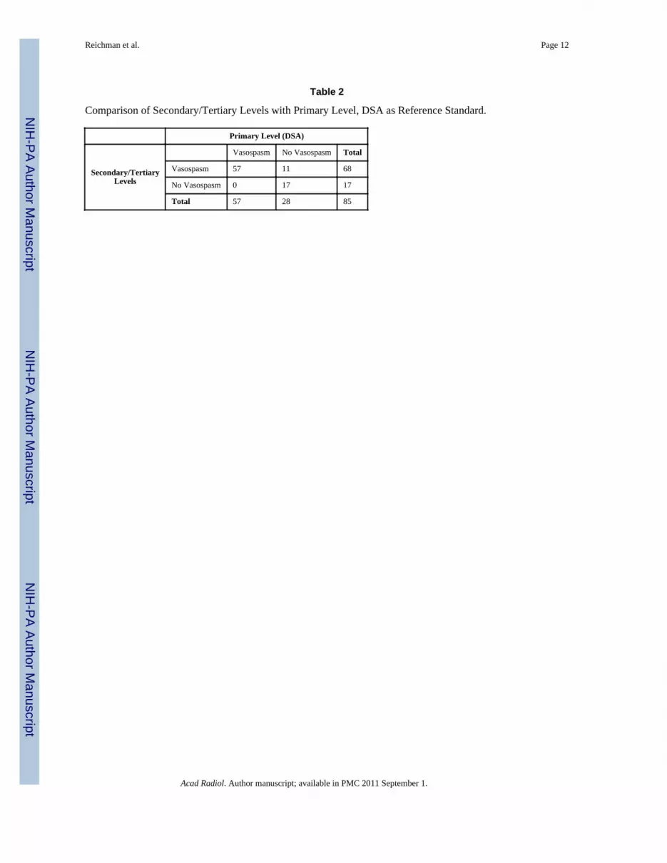
In Phase I (n=85), using DSA at the primary level as the gold standard, vasospasm wasdiagnosed in 57 (67%) patients and no vasospasm in 28 (33%) patients (Table 2). Thesecondary/tertiary levels in the reference standard determined a diagnostic outcome in 65(76%) patients at the secondary level and 20 (24%) at the tertiary level. At the secondarylevel, 48 (74%) patients were classified with vasospasm and 17 (26%) patients were novasospasm. At the tertiary level, 20 (100%) patients were classified with vasospasm. Theagreement rate between DSA and the secondary/tertiary levels for the diagnostic outcomewas 87% (74/85). In the subgroup of patients with vasospasm on DSA (n=57), there was100% agreement compared with the secondary/tertiary levels, resulting in 100% sensitivity.However, there was only 39% (11/28) agreement for the subgroup without vasospasm onDSA (n=28), resulting in low specificity of 61%. Positive predictive value was 83% andnegative predictive value was 100%. The kappa value was 0.674, considered as substantialstrength of agreement according to the Landis and Koch rating scale (10). The bias index,used as a measure of variance, was 0.12.
In Phase II (n=137), the multi-stage hierarchical reference standard as designed forimplementation in practice was compared to chart diagnosis. Overall, a diagnostic outcomewas determined at the primary level for 85 (62%) patients, secondary level for 45 (33%)patients, and tertiary level for 7 (5%) patients (Table 3). At the primary level, 57 (67%)patients were classified as vasospasm and 28 (33%) patients as no vasospasm. At thesecondary level, 6 (13%) patients were classified as vasospasm and 39 (87%) as novasospasm. At the tertiary level, 7 (100%) patients were classified as vasospasm. Accordingto chart review, vasospasm was determined for 76 (55%) patients and no vasospasm for 61
Reichman et al. Page 4
Acad Radiol. Author manuscript; available in PMC 2011 September 1.
NIH
-PA Author Manuscript
NIH
-PA Author Manuscript
NIH
-PA Author Manuscript

(45%) (Table 3). The agreement rate between chart review and the new reference standardfor a diagnostic outcome was 91% (125/137). Sensitivity, specificity, PPV and NPV are88%, 95%, 96%, and 87% respectively. The kappa value was 0.824, considered in the“almost perfect” agreement category, according to Landis and Koch (10), with a low biasindex of 0.04. In this phase, data analysis was also performed at each individual level in thereference standard and compared with chart diagnosis. At the primary level using DSA, theagreement rate was 88% (75/85) with chart diagnosis. Nine out of the 10 patients indisagreement, were considered as vasospasm by chart diagnosis and were assigned a novasospasm diagnosis using DSA. At the secondary level using clinical and imaging criteria,the agreement rate was 96% (43/45). The two patients in disagreement were considered asno vasospasm by chart diagnosis, however, both had infarcts seen on follow-up CT that metcriteria for a delayed infarction for a vasospasm diagnosis. The tertiary level using response-to-treatment analysis had 100% (7/7) agreement with chart review for a vasospasmdiagnosis.
DiscussionOften times, a perfect gold standard does not exist in clinical or research practice. Theaccuracy of a new diagnostic test determined by using an imperfect gold standard introducesbiases and inconsistencies in the results. In clinical practice, gold standards are rarer thanone might think (11). For example, colposcopy guided biopsy of the cervix has beenconsidered the gold standard for disease detection of cervical neoplasia for decades,however the sensitivity is only 60%, leading to misclassfication of patients with cervicalneoplasia as negative for the disease. Using such an imperfect gold standard will biasestimates of accuracy of a new and potentially better diagnostic test by categorizing theadditional cases of disease detected as “false positive” test results (8). In this situation, theaccuracy of the new diagnostic test is limited by the current gold standard used. Thereby, itis often very difficult to validate a new reference standard and few are actually validated thatwe commonly use today.
In this study, we have performed an internal validation of a new multistage hierarchicalreference standard for the diagnosis of vasospasm, to improve accuracy and classification ofA-SAH patients. Internal and external validation methods are important for the completeassessment of a reference standard. Internal validation refers to procedures restricted to asingle data set and assesses accuracy in the classification of patients with and withoutdisease. Whereas, external validation methods consider the generalizability of the referencestandard to other target populations by evaluating its reproducibility and re-test reliability. Arobust reference standard has achieved both validation components with high accuracy inthe classification of patients and applicability to other target populations (12).
The results from our study are an initial validation for a novel multistage hierarchicalreference standard for the diagnosis of vasospasm in A-SAH patients, incorporating bothimaging and clinical criteria. The reference standard includes a weighted design with thestrongest evidence of diagnosis at the primary level and the weakest evidence at the tertiarylevel. Our results support the hierarchical design of the reference standard, where themajority of patients (n=85) are determined for vasospasm diagnosis at the primary level,followed by the secondary level (n=45), and lastly the tertiary level (n=7). In Phase I,primary level patients with DSA were applied to the secondary/tertiary level, and again thehierarchy remains with most patients determined at the secondary level, followed by fewerat the tertiary level.
In Phase I, there was a moderately high agreement rate of 87% for the diagnostic outcomesbetween DSA and the secondary/tertiary levels in the reference standard. The kappa value of
Reichman et al. Page 5
Acad Radiol. Author manuscript; available in PMC 2011 September 1.
NIH
-PA Author Manuscript
NIH
-PA Author Manuscript
NIH
-PA Author Manuscript

0.674 is considered as substantial strength of agreement. On further analysis, there was100% agreement in the subgroup of patients with DSA positive vasospasm. There were 11discrepant cases with DSA negative for vasospasm, but were assigned a vasospasmdiagnosis at the secondary/tertiary levels according to clinical and imaging criteria. In thissubgroup, 82% (9/11) patients had a vasospasm diagnosis confirmed on chart review. Thesediscordant results may partly be due to the limited ability of DSA in detecting distalvasospasm. Vasospasm of small, intraparenchymal arteries has been postulated as the causeof delayed neurological deterioration in the absence of large artery vasospasm (5). Reviewof the literature also reveals that using DSA alone has a low sensitivity of 80% (3) for thedetection of vasospasm. Even the combined use of TCD and DSA together predictsoccurrence of delayed cerebral infarction with suboptimal sensitivity of 72% and specificityof 68% (13). Lastly, symptomatic vasospasm, which is defined by a neurologic deficit onclinical exam or delayed cerebral ischemia, and not angiographic vasospasm, may partlyattribute to these discrepancies observed between DSA and the chart diagnosis (5).
In Phase II, there was a high agreement rate of 91% between our multistage hierarchicalreference standard and chart diagnosis. The kappa value of 0.824 is considered in the“almost perfect” agreement category. Importantly, the agreement rate was improved whenthe secondary and tertiary levels are included in the reference standard. Evaluation of eachlevel individually revealed that the primary level did not actually have the highest agreement(88%) with chart diagnosis because of the subgroup of patients with DSA negative exams,as described above. The secondary and tertiary levels had excellent agreement with chartdiagnosis (96% and 100% respectively) supporting the importance of including clinical andimaging criteria in the reference standard design. The tertiary level shows promising results,however, the sample size is too small to make conclusive statements.
The strengths of this new reference standard are that it includes all patients in the A-SAHpopulation, including those with and without symptoms. To our knowledge, it is the firstreference standard for vasospasm that incorporates both clinical and imaging criteria. Sincevasospasm is a complex disease and its pathophysiology is poorly understood, it has beendefined according to both clinical and imaging criteria. Another unique feature of thisreference standard is at the tertiary level which incorporates treatment analysis. Consideringthe effects of treatment is rarely included in a reference standard because most patients whohave been treated for the disease would be excluded from the analysis. However, in thispatient population prophylactic treatment measures may be used and we developed areference standard that can be applied to all patients in the A-SAH population.
Performing a validation process for a new reference standard is necessary to determine itsaccuracy in the classification of patients with and without disease. However, we have alsorealized its additional value in identifying the limitations and bias that may exist in areference standard. Thereby, modification of the reference standard can be made to adjustfor these limitations and allow for improvement. The results of this study support a revisionof the reference standard at the primary level for the subgroup of patients that have DSAnegative exams. If the DSA is negative for vasospasm, then the patient is not assigned a novasospasm diagnosis at the primary level, and instead proceeds to the secondary level(Figure 2). The rationale is based on our data indicating that the some of these patients doindeed have vasospasm based on clinical criteria at the secondary level. A secondaryanalysis was performed using the revised reference standard to assess its improved accuracyin the classification of A-SAH patients. The 28 patients with DSA negative exams at theprimary level were evaluated at the secondary/tertiary levels. The overall agreement ratebetween the revised reference standard with the chart diagnosis improved to 98.5%.
Reichman et al. Page 6
Acad Radiol. Author manuscript; available in PMC 2011 September 1.
NIH
-PA Author Manuscript
NIH
-PA Author Manuscript
NIH
-PA Author Manuscript

The major limitations of this study are its retrospective study design relying on chart reviewfor data collection. There was variability in the chart documentation of vasospasm betweenthe discharge summary and the progress notes. Another limitation of the study is the smallsample size at the tertiary level. In Phase I, 20 patients were classified at the tertiary leveland in Phase II, only 7 patients. A larger prospective study is needed to further support ourattempts in validating this level separately. Lastly, this study tests diagnostic accuracy of thereference standard for vasospasm diagnosis, and does not address precision, the secondcomponent of validation. An external validation process assessing precision andreproducibility of the reference standard will be evaluated in our future work.
Validation of a new reference standard is an evolving and on-going process. Determiningthe accuracy of our new reference standard and its ability to correctly classify patients withand without vasospasm in the A-SAH population is the initial step in the validation process.Through internal validation, the limitations and bias in the reference standard are identified.Thus, a modification in the new reference standard was made at the primary level addressingthe limitation of using DSA alone in the diagnosis of vasospasm. Secondary analysisrevealed improved accuracy and classification of patients using the revised referencestandard. Once adequate accuracy has been achieved with the new reference standard, thenan external validation process is the next step to determine its generalizability to other targetpopulations by assessing its precision and reproducibility. Importantly, evaluation of theclinical effectiveness of the new reference standard in practice and its impact on treatmentdecisions and patient outcomes is recommended in the validation process.
AcknowledgmentsThis publication was made possible by Grant Number 5K23NS058387-02 from the National Institute ofNeurological Disorders and Stroke (NINDS), a component of the National Institutes of Health (NIH). Its contentsare solely the responsibility of the authors and do not necessarily represent the official view of NINDS or NIH.
References1. Ingal, ITJ.; Whisnant, JP. Epidemiology of subarachnoid hemorrhage. In: Yanagihara, T.; Pepegras,
DC.; Atkinson, JLD., editors. Subarachnoid hemorrhage: medical and surgical management. MarcelDekker; New York, NY: 1998. p. 194-206.
2. Hop JW, Rinkel GJ, Algra A, et al. Case fatality rates and functional outcome after subarachnoidhemorrhage: a systematic review. Stroke 1997;28:660–664. [PubMed: 9056628]
3. Suarez J, Qureshi A, Abutaher Y, et al. Symptomatic vasospasm diagnosis after subarachnoidhemorrhage: Evaluation of transcranial Doppler ultrasound and cerebral angiography as related tocompromised vascular distribution. Neurologic Critical Care 2002:1348–1355.
4. Janardhan, Vallabh; Biondi, A.; Riina, H., et al. Vasospasm in Aneurysmal SubarachnoidHemorrhage: Diagnosis, Prevention, and Management. Neuroimaging Clinics of North America2006;16:483–496. [PubMed: 16935712]
5. Macdonald R. Management of cerebral edema. Neurosurg Review 1998;29:179–193.6. Reichman M, Greenberg E, Gold R, et al. Developing Patient-centered Outcome Measures for
Evaluating Vasospasm in Aneurysmal Subarachnoid Hemorrhage. Academic Radiology2009;16:541–545. [PubMed: 19345894]
7. Zubkov A, Rabinstein A. Medical management of cerebral vasospasm: present and future.Neurological Research 2009;31:626–631. [PubMed: 19055879]
8. Alonzo T, Pepe M. Assessing the Accuracy of a New Diagnostic Test When a Gold Standard DoesNot Exist. UW Biostatistics Working Paper Series 1998:3–32.
9. Byrt T, Bishop J, Carlin JB. Bias, prevalence and kappa. Journal of Clinical Epidemiology1993;46:423–429. [PubMed: 8501467]
10. Landis R, Koch G. The measurement of observer agreement for categorical data. Biometrics1977;33:159–174. [PubMed: 843571]
Reichman et al. Page 7
Acad Radiol. Author manuscript; available in PMC 2011 September 1.
NIH
-PA Author Manuscript
NIH
-PA Author Manuscript
NIH
-PA Author Manuscript

11. Pfeiffer R, Castle P. With or Without a Gold Standard. Epidemiology 2005:595–597. [PubMed:16135933]
12. Altman DG, Royston P. What do we mean by validating a prognostic model? Statistics inMedicine 2000;19:453–473. [PubMed: 10694730]
13. Rabinstein A, Friedman J, Weigand S, et al. predictors of cerebral infarction in aneurysmal SAH.Stroke 2004;35(8):1862–1866. [PubMed: 15218156]
Reichman et al. Page 8
Acad Radiol. Author manuscript; available in PMC 2011 September 1.
NIH
-PA Author Manuscript
NIH
-PA Author Manuscript
NIH
-PA Author Manuscript

Figure 1.Flow Chart for Reference Standard To Determine the Study Outcome of VasospasmDiagnosisReprinted from Academic Radiology, 16/5, Reichman, et al., “Developing Patient-centeredOutcome Measures for Evaluating Vasospasm in Aneurysmal Subarachnoid Hemorrhage,”pg. 541-545, 2009, with permission from Elsevier
Reichman et al. Page 9
Acad Radiol. Author manuscript; available in PMC 2011 September 1.
NIH
-PA Author Manuscript
NIH
-PA Author Manuscript
NIH
-PA Author Manuscript

Figure 2.Revised Flow Chart for Reference Standard To Determine the Study Outcome ofVasospasm Diagnosis
Reichman et al. Page 10
Acad Radiol. Author manuscript; available in PMC 2011 September 1.
NIH
-PA Author Manuscript
NIH
-PA Author Manuscript
NIH
-PA Author Manuscript

NIH
-PA Author Manuscript
NIH
-PA Author Manuscript
NIH
-PA Author Manuscript
Reichman et al. Page 11
Table 1
Demographic Data
All (n=137) Vasospasm (n=70) No Vasospasm (n=67)
Age (years)
Median 51.5 49 52
Range 24-88 28-88 24-83
Gender
Male 38 (28%) 21 (30%) 17 (25%)
Female 99 (72%) 49 (70%) 50 (75%)
Aneurysm Location
Anterior 94 (69%) 52 (74%) 42 (63 %)
Posterior 43 (31%) 18 (26%) 25 ( 37%)
Treatment Type
Surgical Clipping 75 (55%) 37 (53%) 37 (55%)
Coil Embolization 59 (43%) 30 (43 %) 29 (43%)
Untreated 3 (2%) 3 (4%) 1 (1%)
Hunt Hess Grade
Low Grade (1-2) 69(50%) 28 (40%) 42 (59%)
High Grade (3,4,5) 68 (50 %) 42 (60%) 25 (37%)
Acad Radiol. Author manuscript; available in PMC 2011 September 1.
NIH
-PA Author Manuscript
NIH
-PA Author Manuscript
NIH
-PA Author Manuscript
Reichman et al. Page 12
Table 2
Comparison of Secondary/Tertiary Levels with Primary Level, DSA as Reference Standard.
Primary Level (DSA)
Secondary/TertiaryLevels
Vasospasm No Vasospasm Total
Vasospasm 57 11 68
No Vasospasm 0 17 17
Total 57 28 85
Acad Radiol. Author manuscript; available in PMC 2011 September 1.

NIH
-PA Author Manuscript
NIH
-PA Author Manuscript
NIH
-PA Author Manuscript
Reichman et al. Page 13
Table 3
Comparison of Primary/Secondary/Tertiary Levels with Chart Review and Discharge Summary, as referencestandard.
Primary Level (DSA)
Multistage Reference Standard
Vasospasm No Vasospasm Total
Vasospasm 67 3 70
No Vasospasm 9 58 67
Total 76 61 137
Acad Radiol. Author manuscript; available in PMC 2011 September 1.