Embed Size (px)
Citation preview

N
A
gbVThlca©
K
1
bbmaben3
DL
0d
ARTICLE IN PRESSBA-6876; No. of Pages 8
Neurobiology of Aging xxx (2007) xxx–xxx
VDR gene variants associate with cognitive function anddepressive symptoms in old age
Maris Kuningas a,b,∗, Simon P. Mooijaart a, Jelle Jolles c, P. Eline Slagboom d,Rudi G.J. Westendorp a, Diana van Heemst a
a Department of Gerontology and Geriatrics, Leiden University Medical Center, Leiden, The Netherlandsb Department of Biotechnology, Institute of Molecular and Cell Biology, Tartu University, Tartu, Estonia
c Department of Psychiatry and Neuropsychology, University Hospital Maastricht, Maastricht, The Netherlandsd Section of Molecular Epidemiology, Department of Medical Statistics, Leiden University Medical Center, Leiden, The Netherlands
Received 4 May 2007; accepted 7 July 2007
bstract
Vitamin D has been recently implicated in brain function. Our objective was to test whether genetic variance in the vitamin D receptor (VDR)ene is associated with cognitive functioning and depressive symptoms in old age. The study was carried out in the prospective population-ased Leiden 85-plus Study. All 563 participants of the study were genotyped for Cdx-2, FokI, BsmI, ApaI and TaqI polymorphisms in theDR gene. Our data revealed an overall worse performance on tests measuring cognitive functioning for carriers of BsmI (p = 0.013) andaqI (p = 0.004) polymorphisms, and of haplotype 2 (BAt) (p = 0.004). In contrast, carriers of ApaI variant-allele and of haplotype 1 (baT)ad better cognitive functioning together with less depressive symptoms. These associations could not be explained by differences in calcium
evels, and by selective survival, since no associations between the VDR gene variants and calcium levels and mortality were observed. Inonclusion, our results show that genetic variance in the VDR gene influences the susceptibility to age-related changes in cognitive functioningnd in depressive symptoms.2007 Elsevier Inc. All rights reserved.
s; Poly
egt
rtiD
eywords: Vitamin D receptor; Cognitive functioning; Depressive symptom
. Introduction
Ample data provide evidence that vitamin D is involved inrain function. The reported biological processes influencedy vitamin D in the brain include neuroprotection, immuno-odulation and detoxification (Brown et al., 2003; Garcion et
l., 2002). The neuroprotective effects of vitamin D appear toe exerted via the regulation of calcium homeostasis (Brewer
Please cite this article in press as: Kuningas, M. et al., VDR gene varianold age, Neurobiol Aging (2007), doi:10.1016/j.neurobiolaging.2007.07
t al., 2001, 2006; de Viragh et al., 1989), and synthesis ofeurotrophins, such as nerve growth factor and neurotrophin(Naveilhan et al., 1996; Neveu et al., 1994a, 1994b; Saporito
∗ Corresponding author at: Leiden University Medical Center (LUMC),epartment of Gerontology and Geriatrics, C2-R, PO Box 9600, 2300 RCeiden, The Netherlands. Tel.: +31 71 5266640; fax: +31 71 5248159.
E-mail address: [email protected] (M. Kuningas).
cm1MtpKa
197-4580/$ – see front matter © 2007 Elsevier Inc. All rights reserved.oi:10.1016/j.neurobiolaging.2007.07.001
morphisms; Haplotypes; Fractures; Mortality
t al., 1994; Wang et al., 2000). These biological effects sug-est that vitamin D could influence cognitive functioning andhe prevalence of depressive symptoms.
The functions of vitamin D are mediated by the vitamin Deceptor (VDR), which belongs to the nuclear hormone recep-or (NHR) super-family, and which is ubiquitously expressedn the organism (Kamei et al., 1995; Langub et al., 2001).efects in the vitamin D signaling system have been asso-
iated with multiple sclerosis, and various behavioral- andood-disorders in animals and humans (Cantorna et al.,
996; Garcion et al., 2002; Lansdowne and Provost, 1998;unger et al., 2004). It has been shown that animals exposed
ts associate with cognitive function and depressive symptoms in.001
o prenatal vitamin D deficiency have alterations in brain mor-hology (Eyles et al., 2003), locomotion (Burne et al., 2004;esby et al., 2006), and learning and memory (Becker et
l., 2005). In addition, mice lacking a functional VDR gene

INNBA-6876; No. of Pages 8
2 iology o
a2cwWaeomnaapehB
filiatcp
2
2
bLwcsc1ppmoeDLic
2
i7cfc
2
mFtp(trddabtwtptwcsWasn
2
tomatctsF
2
ocise
2
ARTICLEM. Kuningas et al. / Neurob
ppear to suffer from anxiety-like behavior (Kalueff et al.,004, 2006). In humans, vitamin D deficiency has been asso-iated with the presence of an active mood disorder and withorse cognitive functioning (Przybelski and Binkley, 2007;ilkins et al., 2006). In contrast, little is known about whether
nd how disturbed function of the VDR gene influences thesendpoints. The VDR gene contains several polymorphismsf which five; Cdx-2, FokI, BsmI, ApaI and TaqI, have beenost often investigated, and associated with a number of phe-
otypes, such as bone mineral density, and risks for fracturesnd cancer (Uitterlinden et al., 2004b). In addition, haplotypelleles have been identified that influence the risk of osteo-orotic fractures and the expression of the VDR gene (Fangt al., 2005; Grundberg et al., 2007). The risk haplotypes thatave recently emerged, baT and BAt, are composed of thesmI, ApaI and TaqI polymorphisms, located in the 3′-UTR.
The aim of this study was to assess the influence of theseve polymorphisms in the VDR gene and of the risk hap-
otypes on cognitive functioning and depressive symptomsn old age. Furthermore, the association with calcium levels,nd the incidence of fractures and mortality were assessed forhe VDR polymorphisms and the haplotypes. The study wasarried out in the Leiden 85-plus Study, a population-basedrospective study of the oldest old.
. Subjects and methods
.1. Subjects
The Leiden 85-plus Study is a prospective populationased study in which all 85-year-old inhabitants of the cityeiden, in The Netherlands, were invited to take part. Thereere no selection criteria related to health or demographic
haracteristics. The population under study consists of 599ubjects, all Caucasians and members of the 1912–1914 birthohort, enrolled in the month of their 85th birthday between997 and 1999 (Bootsma-van der Wiel et al., 2002). For theresent study, DNA was available for 563 participants. Allarticipants of the Leiden 85-plus Study were followed forortality until 1 August 2005. Primary causes of death were
btained from the Dutch Central Bureau of Statistics and cat-gorized according to the 10th International Classification ofiseases (ICD-10). The Medical Ethical Committee of theeiden University Medical Centre approved the study and
nformed consent was obtained from all participants or inase of severe cognitive impairment, from their guardian.
.2. Calcium levels at baseline
Calcium and albumin concentrations were determinedn serum using fully automated analyzers (Hitachi
Please cite this article in press as: Kuningas, M. et al., VDR gene varianold age, Neurobiol Aging (2007), doi:10.1016/j.neurobiolaging.2007.07
47 and 911; Hitachi Ltd., Tokyo, Japan). Total cal-ium levels were adjusted for albumin using theollowing formula: corrected calcium = uncorrected cal-ium − [(40 − albumin) × 0.02] (Palmer et al., 1988).
suCp
PRESSf Aging xxx (2007) xxx–xxx
.3. Cognitive function and depressive symptoms
Overall cognitive function was measured with the mini-ental state examination (MMSE) (Folstein et al., 1975).rom the specific domains of cognitive functioning, atten-
ion was assessed with the Stroop Test (Klein et al., 1997),rocessing speed with the letter digit coding test (LDT)Houx et al., 2002) and memory with the 12-word learningest, which assesses immediate recall (WLTI) and delayedecall (WLTD) (Brand and Jolles, 1985). The prevalence ofepressive symptoms was assessed with the 15-item geriatricepression scale (GDS-15) (De Craen et al., 2003). The testsssessing specific domains of cognitive functioning could note administered to 92 participants because of severe cogni-ive impairment (MMSE score ≤ 18 points). All participantsere visited annually for re-measurement of cognitive func-
ioning and depressive symptoms during a mean follow-uperiod of 4.2 years. During the study, parallel versions ofhe tests were used and details of testing are described else-here (Houx et al., 2002). In addition to the specific tests, a
omposite cognitive score was calculated by converting thecores of the individual tests (Stroop Test, LDT, WLTI and
LTD) into a z-score ((individual level − mean level)/S.D.),nd computing the average. A higher composite cognitivecore reflects better performance on the tests measuring cog-itive functioning.
.4. The incidence of fractures
The incidence of fractures was assessed yearly duringhe 5-year follow-up period. The number of fractures wasbtained by self-reporting using a standardized yes/no for-at. When the participant was severely cognitively impaired,guardian was asked for the information. In addition,
he general practitioner, or the nursing home physician inase of institutionalization, was interviewed concerning frac-ure related contacts with the participant. The composite ofelf-reported and physician reported fractures were used.ractures included hip, wrist and other fractures.
.5. Possible confounders
Socio-demographic characteristics, such as sex and levelf education were considered as possible confounders. Edu-ation was divided into two levels: a lower education level,ncluding individuals without schooling or with only primarychool education (less than 6 years of schooling), and a higherducation level (6 years or more of schooling).
.6. Genotyping
The Cdx-2 G/A (rs11568820) and BsmI C/T (rs1544410)
ts associate with cognitive function and depressive symptoms in.001
ingle nucleotide polymorphisms (SNPs) were genotypedsing an Assay-by-Design (Applied Biosystems, Foster City,A, USA), consisting of PCR primers and TaqMan MGBrobes, on an ABI 7900 HT real-time PCR machine (Applied

IN PRESSNBA-6876; No. of Pages 8
ology of Aging xxx (2007) xxx–xxx 3
BwiLFAMf
2
erbwgatSaadaealwbVaidhtuTh1
3
cpTBgmr2sdca
Table 1Characteristics of study participants
Number 563Agea 85Female (%) 375 (67%)Low level of education (%) 362 (65%)Calcium (mmol/l)a 2.23 (2.16–2.29)MMSE (points)a 26 (22–28)MMSE ≥ 19 points (%) 471 (84%)
Specific domains of cognitive functioninga
Stroop test (s) 74 (60–97)LDT (digits) 16 (12–21)WLTI (pictures) 25 (20–28)WLTD (pictures) 9 (7–11)
GDS-15 (points)a 2 (1–3)
Polymorphismsb
Cdx-2 (G/A) 0.19FokI (G/A) 0.34BsmI (C/T) 0.43ApaI (A/C) 0.46TaqI (A/G) 0.42
MMSE: mini-mental state examination; LDT: letter digit coding test; WLTI:word learning test immediate recall; WLTD: word learning test delayedr
were similar to those reported in other Caucasian populations(Fang et al., 2005; Grundberg et al., 2007).
Global cognitive functioning, attention, processing speed,memory and the prevalence of depressive symptoms were
Fig. 1. The VDR gene structure and haplotypes. The VDR gene spans agenomic region of 100 kb and contains 14 exons (indicated with boxes).
ARTICLEM. Kuningas et al. / Neurobi
iosystems, Foster City, CA, USA). Amplification reactionsere made at standard conditions except for the follow-
ng modifications. A qPCR core kit was used (Eurogentec,iege, Belgium) and one-third of the amounts of assay mix.okI G/A (rs10735810), ApaI A/C (rs7975232) and TaqI/G (rs731236) polymorphisms were genotyped using theassArray platform according to the protocols of the manu-
acturer (Sequenom Inc., San Diego, CA, USA).
.7. Statistical analysis
The program Haploview (Barrett et al., 2005) was used tostimate allele frequencies, test for Hardy–Weinberg equilib-ium and to estimate pair-wise linkage disequilibrium (LD)etween the SNPs. Haplotypes and haplotype frequenciesere calculated using the program SNPHAP (http://www-ene.cimr.cam.ac.uk/clayton/software). In order to take intoccount the uncertainty in haplotype probabilities, the mul-iple imputation approach was used (Rubin, 1987), and withNPHAP ten datasets were generated by randomly assigninghaplotype to each subject according to its haplotype prob-
bilities. All statistical analyses were performed with the tenatasets. The haplotype specific estimates were calculated byveraging the ten dataset-specific estimates, and the standardrrors were estimated using the estimated variance withinnd across the datasets. The associations between base-ine calcium levels and VDR polymorphisms and haplotypesere tested using sex adjusted linear regression. Associationsetween cognitive functioning, depressive symptoms andDR polymorphisms and haplotypes were analyzed usingsex and education adjusted linear mixed model, estimat-
ng the overall mean difference in cognitive functioning orepressive symptoms during follow-up. Cox proportionalazard model, measuring time-to-event was used to estimatehe risk of incident fractures, and mortality during the follow-p period, in relation to the polymorphisms or haplotypes.he reference group contained 0-copies of a risk allele oraplotype. All analyses were performed with SPSS, version2.0 (SPSS Inc., Chicago, IL, USA) statistical software.
. Results
Demographic characteristics and baseline measures ofognitive functioning and depressive symptoms of the 563articipants of the Leiden 85-plus Study are presented inable 1. All study subjects were genotyped for Cdx-2, FokI,smI, ApaI and TaqI polymorphisms in the VDR gene. Theenotype frequencies of the SNPs (Table 1) were in agree-ent with Hardy–Weinberg equilibrium and similar to those
eported in other Caucasian populations (Uitterlinden et al.,004a). The BsmI, ApaI and TaqI polymorphisms were in
Please cite this article in press as: Kuningas, M. et al., VDR gene varianold age, Neurobiol Aging (2007), doi:10.1016/j.neurobiolaging.2007.07
trong linkage disequilibrium (LD) (D′ > 0.99) (Fig. 1), andefined five haplotypes, of which the first three had frequen-ies >5%. These haplotypes have previously been describeds baT, BAt and bAT, respectively. The haplotype frequencies
Tashd
ecall; GDS-15: 15-item geriatric depression scale.a Data are presented as medians with interquartile ranges.b Data are presented as minor allele frequencies.
ts associate with cognitive function and depressive symptoms in.001
he approximate positions of the five polymorphisms analyzed in this studyre indicated with arrows. The BsmI, ApaI and TaqI polymorphisms are introng linkage disequilibrium (LD) and define five haplotypes. The first threeaplotypes, haplotype 1, haplotype 2 and haplotype 3 have previously beenescribed as baT, BAt and bAT, respectively.

ARTICLE IN PRESSNBA-6876; No. of Pages 8
4 M. Kuningas et al. / Neurobiology of Aging xxx (2007) xxx–xxx
Table 2Cognitive functioning and depressive symptoms during follow-up dependent on the VDR polymorphisms
VDR genotypes
wt/wt wt/var var/var ptrend
Mean (S.E.) Difference (S.E.) Difference (S.E.)
BsmI (C/T)Composite cognitive score −0.08 (0.06) −0.12 (0.08) −0.25 (0.10)* 0.013*MMSE (points) 22.6 (0.48) 0.47 (0.61) −0.15 (0.78) 0.999Stroop Test (s) 84.3 (2.31) 2.00 (2.95) 10.4 (3.78)* 0.010*LDT (digits) 16.2 (0.51) −1.04 (0.65) −0.34 (0.83) 0.471WLTI (pictures) 21.3 (0.48) −1.07 (0.61) −2.18 (0.77)* 0.004*WLTD (pictures) 7.33 (0.23) −0.30 (0.29) −0.78 (0.39)* 0.037*GDS-15 (points) 2.93 (0.21) −0.05 (0.26) 0.55 (0.34) 0.158
ApaI (A/C)Composite cognitive score −0.22 (0.07) 0.00 (0.08) 0.16 (0.10) 0.135MMSE (points) 23.1 (0.51) −0.32 (0.63) −0.56 (0.76) 0.456Stroop Test (s) 89.9 (2.45) −2.65 (3.05) −6.80 (3.68) 0.068LDT (digits) 16.4 (0.53) −1.40 (0.66)* −0.06 (0.80) 0.737WLTI (pictures) 19.8 (0.51) 0.49 (0.63) 1.61 (0.76)* 0.041*WLTD (pictures) 6.83 (0.24) 0.23 (0.30) 0.44 (0.36) 0.222GDS-15 (points) 3.42 (0.21) −0.56 (0.26)* −0.72 (0.32)* 0.019*
TaqI (A/G)Composite cognitive score −0.07 (0.06) −0.13 (0.08) −2.94 (0.10)* 0.004*MMSE (points) 22.7 (0.48) 0.55 (0.60) −0.30 (0.78) 0.899Stroop Test (s) 84.0 (2.29) 2.12 (2.92) 11.0 (3.80)* 0.008*LDT (digits) 16.3 (0.50) −0.96 (0.64) −0.63 (0.84) 0.310WLTI (pictures) 21.4 (0.47) −1.15 (0.60) −2.51 (0.78)* 0.001*WLTD (pictures) 7.40 (0.23) −0.37 (0.29) −0.99 (0.37)* 0.009*GDS-15 (points) 2.93 (0.20) −0.06 (0.26) 0.60 (0.33) 0.135
* : letterl
aafaacpfcopilpsptastd(f−−a
sa
wcmacdowmdnio(
eotc
p < 0.05; S.E.: standard error; MMSE: mini-mental state examination; LDTearning test delayed recall; GDS-15: 15-item geriatric depression scale.
ssessed at baseline, age 85 years, and re-examined annu-lly during a mean follow-up period of 4.2 years. Duringollow-up, a significant decline in cognitive functioning,nd an increase in depressive symptoms were observed inll participants (all p < 0.001) (Vinkers et al., 2005). Thesehanges were not attributable to the Cdx-2 or FokI polymor-hisms, since during follow-up no differences in cognitiveunctioning and depressive symptoms were observed forarriers of these polymorphisms (data not shown). On thether hand, carriers of the BsmI and TaqI polymorphismserformed worse on all tests measuring cognitive function-ng (Table 2). This worse performance was reflected by aower composite cognitive score (BsmI ptrend = 0.013; TaqItrend = 0.004), but not by a lower MMSE, which mea-ures global cognitive functioning (BsmI ptrend = 0.999; TaqItrend = 0.899). From specific domains of cognitive func-ioning, attention, immediate- and delayed-memory wereffected most, whereas for the prevalence of depressiveymptoms no differences were observed (Table 2). In con-rast, carriers of the ApaI variant-allele tended to have lessepressive symptoms than non-carriers during follow-upptrend = 0.019) (Table 2). These differences were observed
Please cite this article in press as: Kuningas, M. et al., VDR gene varianold age, Neurobiol Aging (2007), doi:10.1016/j.neurobiolaging.2007.07
or both heterozygous (−0.56 points, 95% CI: −1.07 to0.04, p = 0.036) and homozygous (−0.72 points, 95% CI:1.35 to −0.09, p = 0.026) ApaI variant-allele carriers. In
ddition, these participants performed better, although not
wset
digit coding test; WLTI: word learning test immediate recall; WLTD: word
tatistically significant, on tests measuring processing speed,ttention and memory (Table 2).
The haplotype analyses revealed similar results as thoseith the individual polymorphisms. Carriers of at least one
opy of haplotype 1 (baT), which contains the ApaI poly-orphism, had less depressive symptoms (ptrend = 0.026),
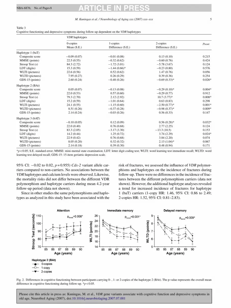
nd performed better, although statistically not signifi-antly, on tests measuring attention, immediate memory andelayed memory compared to non-carriers (Table 3). Thepposite was observed for carriers of haplotype 2 (BAt),hich combines the variant alleles of BsmI and TaqI poly-orphisms (Table 3). For all these associations, an allele
osage dependent effect was observed, which was more pro-ounced for haplotype 2 (BAt) carriers, who had mainlympairments in attention (ptrend = 0.008), immediate mem-ry (ptrend = 0.001) and delayed memory (ptrend = 0.009)Fig. 2).
In order to explore whether differences in calcium lev-ls or selective survival have influenced the associationsbserved with cognitive functioning, we analyzed the rela-ion between these phenotypes and VDR gene variants. Inross-sectional analyses at age 85 years, serum calcium levels
ts associate with cognitive function and depressive symptoms in.001
ere not associated with the VDR polymorphisms (data nothown), except the Cdx-2 polymorphism. Serum calcium lev-ls were lower in homozygous (−0.05 mmol/l, 95% CI: −10o −0.00, p = 0.032) but not in heterozygous (0.001 mmol/l,
ARTICLE IN PRESSNBA-6876; No. of Pages 8
M. Kuningas et al. / Neurobiology of Aging xxx (2007) xxx–xxx 5
Table 3Cognitive functioning and depressive symptoms during follow-up dependent on the VDR haplotypes
VDR haplotypes
0-copies 1-copies 2-copies ptrend
Mean (S.E.) Difference (S.E.) Difference (S.E.)
Haplotype 1 (baT)Composite score −0.09 (0.07) −0.01 (0.08) 0.13 (0.10) 0.215MMSE (points) 22.5 (0.55) −0.32 (0.62) −0.60 (0.76) 0.424Stroop Test (s) 84.3 (2.72) −1.72 (3.01) −5.78 (3.67) 0.124LDT (digits) 15.3 (0.59) −1.44 (0.66)* −0.23 (0.80) 0.576WLTI (pictures) 22.6 (0.56) 0.52 (0.62) 1.47 (0.76) 0.056WLTD (pictures) 7.95 (0.27) 0.26 (0.29) 0.39 (0.36) 0.254GDS-15 (points) 2.60 (0.24) −0.48 (0.26) −0.69 (0.33)* 0.026*
Haplotype 2 (BAt)Composite score 0.05 (0.07) −0.13 (0.08) −0.29 (0.10)* 0.004*MMSE (points) 22.0 (0.53) 0.57 (0.60) −0.29 (0.77) 0.912Stroop Test (s) 79.3 (2.70) 2.12 (2.92) 10.7 (3.77)* 0.008*LDT (digits) 15.2 (0.59) −1.01 (0.64) 0.63 (0.83) 0.298WLTI (pictures) 24.1 (0.55) −1.15 (0.60) −2.50 (0.77)* 0.001*WLTD (pictures) 8.51 (0.26) −0.37 (0.28) −0.98 (0.37)* 0.009*GDS-15 (points) 2.14 (0.24) −0.03 (0.26) 0.56 (0.33) 0.147
Haplotype 3 (bAT)Composite score −0.10 (0.05) 0.12 (0.09) 0.56 (0.28)* 0.032*MMSE (points) 22.0 (0.40) 0.76 (0.68) 2.77 (2.25) 0.124Stroop Test (s) 83.3 (2.05) −3.17 (3.30) −13.3 (10.5) 0.153LDT (digits) 14.2 (0.44) 1.25 (0.72) 3.74 (2.29) 0.024*WLTI (pictures) 22.9 (0.42) 0.76 (0.68) 3.84 (2.20) 0.074WLTD (pictures) 8.05 (0.20) 0.32 (0.32) 2.13 (1.04)* 0.067
0.39 (0
* : letterl
9rVtpf
t
rpft
Fd
GDS-15 (points) 2.14 (0.18)
p < 0.05; S.E. standard error; MMSE: mini-mental state examination; LDTearning test delayed recall; GDS-15: 15-item geriatric depression scale.
5% CI: −0.02 to 0.02, p = 0.955) Cdx-2 variant allele car-iers compared to non-carriers. No associations between theDR haplotypes and calcium levels were observed. Likewise,
he mortality risks did not differ between the different VDR
Please cite this article in press as: Kuningas, M. et al., VDR gene varianold age, Neurobiol Aging (2007), doi:10.1016/j.neurobiolaging.2007.07
olymorphism and haplotype carriers during mean 4.2-yearollow-up period (data not shown).
Since in other studies the same polymorphisms and haplo-ypes as analyzed in this study have been associated with the
sa12
ig. 2. Differences in cognitive functioning between participants carrying 0- , 1- orifference in cognitive functioning during follow-up. *p < 0.05.
.30) 0.48 (0.94) 0.171
digit coding test; WLTI: word learning test immediate recall; WLTD: word
isk of fractures, we assessed the influence of VDR polymor-hisms and haplotypes on the incidence of fractures duringollow-up. There were no differences in the incidence of frac-ures between the different polymorphism carriers (data not
ts associate with cognitive function and depressive symptoms in.001
hown). However, the additional haplotype analyses revealedtrend for increased incidence of fractures for haplotype(baT) carriers (1-copy HR: 1.46, 95% CI: 0.86 to 2.49;
-copies HR: 1.52, 95% CI: 0.81–2.83).
2-copies of the haplotype 2 (BAt). The p-value represents the overall mean

INNBA-6876; No. of Pages 8
6 iology o
4
ttbiovApSw
hoasl1smi2stdtTtce(ofwe
pfTlmiswitMd
ceVtn
sf2dmiplcsaecatresSpn
tdefiwoathoafcmdVtobriwtoi(tbbb
ARTICLEM. Kuningas et al. / Neurob
. Discussion
The main finding of this study is that genetic variance inhe VDR gene contributes to differences in cognitive func-ioning and depressive symptoms in old age. An overalletter performance on tests measuring attention, process-ng speed and memory, together with a lower prevalencef depressive symptoms were observed for carriers of ApaIariant-allele and of haplotype 1 (baT), which contains thepaI variant-allele. In contrast, carriers of the BsmI and TaqIolymorphisms had impairments in attention and memory.imilar associations were observed with haplotype 2 (BAt),hich combines the variant alleles of BsmI and TaqI.The research on the role of vitamin D in the human brain
as so far focused mainly on the influence of vitamin Dn mood disorders. Vitamin D deficiency is considered aspossible contributor to seasonal affective disorder (SAD),
ince SAD has been associated with winter months and sun-ight deprivation (Rosenthal et al., 1984; Schlager et al.,993; Spoont et al., 1991). In addition, there are studieshowing associations between low vitamin D levels andood disorders, accompanied with worse cognitive function-
ng (Lansdowne and Provost, 1998; Przybelski and Binkley,007; Wilkins et al., 2006). In accordance with the lattertudies, we observed in this study that genetic variance inhe VDR gene influences both cognitive functioning andepressive symptoms. From the specific domains of cogni-ive functioning, attention and memory were affected most.hese two cognitive domains are most vulnerable and tend
o decline constantly across adult lifespan, in contrast toognitive abilities such as autobiographical memory andmotional processing, which stay unchanged throughout lifeHedden and Gabrieli, 2004). Our data suggest that carriersf the BsmI and TaqI polymorphisms are more susceptibleor the age-related deterioration of cognitive functioning,hereas the ApaI polymorphism contributes to a protective
ffect.In the assessment of overall influence of the VDR gene
olymorphisms on cognitive functioning, we observed dif-erences with composite cognitive score but not with MMSE.he composite cognitive score used in this study was calcu-
ated from four individual tests that have been designed toeasure changes in specific domains of cognitive function-
ng. Therefore, the composite cognitive score might be moreensitive in detecting cognitive impairments than MMSE,hich has been designed to assess global cognitive function-
ng, and contains only few items from the specific cognitiveests. It also might be that due to the ‘ceiling’ effect of the
MSE, mild impairments in cognitive functioning are notetectable (Houx et al., 2002).
There are several mechanisms through which vitamin Dan affect mental performance. The downregulation of the
Please cite this article in press as: Kuningas, M. et al., VDR gene varianold age, Neurobiol Aging (2007), doi:10.1016/j.neurobiolaging.2007.07
xpression of L-type voltage-sensitive calcium-channels (L-SCC) by vitamin D in hippocampal neurons has been shown
o reduce the influx and excitotoxic effects of calcium toeurons (Brewer et al., 2001). The detrimental role of exces-
iVda
PRESSf Aging xxx (2007) xxx–xxx
ive calcium for memory formation and overall cognitiveunctioning is widely acknowledged (Sattler and Tymianski,000; Thibault et al., 2001; Veng et al., 2003). However, theifferences in cognitive functioning between the VDR poly-orphism and haplotype carriers were unlikely caused by
ncreased or decreased calcium levels, since none of theseolymorphisms and haplotypes were associated with calciumevels. However, it is unknown how well the peripheral cal-ium levels reflect those in the brain. The vitamin D endocrineystem plays an essential role in overall calcium homeostasisnd therefore we expected to see differences also in periph-ral calcium levels within the polymorphism and haplotypearriers. Possibly, other brain specific functions of vitamin Dre responsible for the observed effects. It has been shownhat in the brain, vitamin D increases the production of neu-otrophins, which support the survival of existing neurons andncourage the growth and differentiation of new neurons andynapses (Naveilhan et al., 1996; Neveu et al., 1994a, 1994b;aporito et al., 1994; Wang et al., 2000). These effects providerotection to, and diminish cognitive impairment underlyingeurodegenerative disorders.
In previous studies, several polymorphisms and haplo-ypes in the VDR gene have been associated with bone mineralensity and risk of fractures (Fang et al., 2005; Uitterlindent al., 2004b). The risk haplotypes that have been identi-ed include haplotype 1 (baT) and haplotype 2 (BAt), whichere reported to contribute to increased and decreased riskf fractures, respectively (Fang et al., 2005; Uitterlinden etl., 2004b). In this study, we analyzed the same risk haplo-ypes, and observed a trend for increased risk of fractures foraplotype 1 (baT) carriers, which is in accordance with thether studies. However, for the same haplotype carriers welso observed better performance on test measuring cognitiveunctioning. An explanation for the apparent contradictionould be that people with better cognitive functioning areore active and therefore may face a higher risk for an inci-
ent fall and fracture. The lack of associations between theDR polymorphisms and haplotypes and mortality suggest
hat selective survival has not influenced the associationsbserved with cognitive functioning. We speculate that theetter cognitive functioning in the haplotype 1 (baT) car-iers is due to a higher expression of the haplotype, sincencreased vitamin D levels have previously been associatedith better cognitive functioning. The evidence for the func-
ionality of haplotype 1 (baT), however, is contradictory. Inne study it was reported that the haplotype 1 (baT) resultsn lower VDR gene expression and increased mRNA decayFang et al., 2005), whereas in another study it was shownhat the haplotype 1 (baT) is overexpressed in human tra-ecular bone samples (Grundberg et al., 2007). It might alsoe that the associations observed in this study are causedy other polymorphisms that are in LD with those analyzed
ts associate with cognitive function and depressive symptoms in.001
n this study. Recently, several new polymorphisms in theDR gene have been identified together with a completeescription of the LD and haplo-block structure (Fang etl., 2005).

INNBA-6876; No. of Pages 8
ology o
bdaoiemAtteoVto
tl
D
ti
A
RAI(ca
R
B
B
B
B
B
B
B
B
C
D
d
E
F
F
G
G
H
H
K
K
K
K
K
L
ARTICLEM. Kuningas et al. / Neurobi
The strengths of the present study include the population-ased sample of the oldest old with a high incidence ofepression and cognitive decline, and the annual repeatedssessment of depressive symptoms and the functioningf various cognitive domains. The limitations of the studynclude the lack of vitamin D levels, and information onnvironmental factors, such as (dietary) calcium and vita-in D intake, which could have influenced the associations.nother limitation is the ascertainment of incident fractures
hrough self-reporting by a questionnaire. This might have ledo ascertainment errors, but random errors would only under-stimate associations. To our knowledge, this is the first andnly report of a relationship between genetic variance in theDR gene and cognitive functioning and depressive symp-
oms, and therefore until further replication, the possibilityf a chance finding cannot be excluded.
In conclusion, our results show that genetic variance inhe VDR gene influences cognitive functioning and the preva-ence of depressive symptoms in old age.
isclosure statement
All co-authors have seen and agreed with the contents ofhe manuscript, and none of the co-authors has any financialnterests to disclose.
cknowledgements
This work was supported by an Innovative Orientatedesearch (IOP) grant from the Dutch Ministry of Economicffairs (grant no. IGE010114), by the Netherlands Genomics
nitiative/Netherlands Organization for Scientific ResearchNWO) (grant no. 911-03-016) and by the Centre for Medi-al Systems Biology (CMSB), which is a centre of excellencepproved by the NWO.
eferences
arrett, J.C., Fry, B., Maller, J., Daly, M.J., 2005. Haploview: analysis andvisualization of LD and haplotype maps. Bioinformatics 21, 263–265.
ecker, A., Eyles, D.W., McGrath, J.J., Grecksch, G., 2005. Transient prena-tal vitamin D deficiency is associated with subtle alterations in learningand memory functions in adult rats. Behav. Brain Res. 161, 306–312.
ootsma-van der Wiel, A., van Exel, E., De Craen, A.J., Gussekloo, J.,Lagaay, A.M., Knook, D.L., Westendorp, R.G., 2002. A high responseis not essential to prevent selection bias: results from the Leiden 85-plusstudy. J. Clin. Epidemiol. 55, 1119–1125.
rand, N., Jolles, J., 1985. Learning and retrieval rate of words presentedauditorily and visually. J. Gen. Psychol. 112, 201–210.
rewer, L.D., Thibault, V., Chen, K.C., Langub, M.C., Landfield, P.W.,Porter, N.M., 2001. Vitamin D hormone confers neuroprotection in
Please cite this article in press as: Kuningas, M. et al., VDR gene varianold age, Neurobiol Aging (2007), doi:10.1016/j.neurobiolaging.2007.07
parallel with downregulation of L-type calcium channel expression inhippocampal neurons. J. Neurosci. 21, 98–108.
rewer, L.D., Porter, N.M., Kerr, D.S., Landfield, P.W., Thibault, O., 2006.Chronic 1alpha, 25-(OH)2 vitamin D3 treatment reduces Ca2+-mediatedhippocampal biomarkers of aging. Cell Calcium 40, 277–286.
L
PRESSf Aging xxx (2007) xxx–xxx 7
rown, J., Bianco, J.I., McGrath, J.J., Eyles, D.W., 2003. 1,25-Dihydroxyvitamin D3 induces nerve growth factor, promotes neuriteoutgrowth and inhibits mitosis in embryonic rat hippocampal neurons.Neurosci. Lett. 343, 139–143.
urne, T.H., Becker, A., Brown, J., Eyles, D.W., Mackay-Sim, A., McGrath,J.J., 2004. Transient prenatal Vitamin D deficiency is associated withhyperlocomotion in adult rats. Behav. Brain Res. 154, 549–555.
antorna, M.T., Hayes, C.E., DeLuca, H.F., 1996. 1,25-DihydroxyvitaminD3 reversibly blocks the progression of relapsing encephalomyelitis, amodel of multiple sclerosis. Proc. Natl. Acad. Sci. U.S.A. 93, 7861–7864.
e Craen, A.J., Heeren, T.J., Gussekloo, J., 2003. Accuracy of the 15-itemgeriatric depression scale (GDS-15) in a community sample of the oldestold. Int. J. Geriatr. Psychiatry 18, 63–66.
e Viragh, P.A., Haglid, K.G., Celio, M.R., 1989. Parvalbumin increases inthe caudate putamen of rats with vitamin D hypervitaminosis. Proc. Natl.Acad. Sci. U.S.A 86, 3887–3890.
yles, D., Brown, J., Mackay-Sim, A., McGrath, J., Feron, F., 2003. VitaminD3 and brain development. Neuroscience 118, 641–653.
ang, Y., van Meurs, J.B., d’Alesio, A., Jhamai, M., Zhao, H., Rivadeneira, F.,Hofman, A., van Leeuwen, J.P., Jehan, F., Pols, H.A., Uitterlinden, A.G.,2005. Promoter and 3′-untranslated-region haplotypes in the vitamin dreceptor gene predispose to osteoporotic fracture: the rotterdam study.Am. J. Hum. Genet. 77, 807–823.
olstein, M.F., Folstein, S.E., McHugh, P.R., 1975. “Mini-mental state.”A practical method for grading the cognitive state of patients for theclinician. J. Psychiatr. Res 12, 189–198.
arcion, E., Wion-Barbot, N., Montero-Menei, C.N., Berger, F., Wion, D.,2002. New clues about vitamin D functions in the nervous system. TrendsEndocrinol. Metab. 13, 100–105.
rundberg, E., Lau, E.M., Pastinen, T., Kindmark, A., Nilsson, O., Ljung-gren, O., Mellstrom, D., Orwoll, E., Redlund-Johnell, I., Holmberg, A.,Gurd, S., Leung, P.C., Kwok, T., Ohlsson, C., Mallmin, H., Brandstrom,H., 2007. Vitamin D receptor 3′-haplotypes are unequally expressed inprimary human bone cells and associated with increased fracture risk:the MrOS study in Sweden and Hong Kong. J. Bone Miner. Res. 22,832–840.
edden, T., Gabrieli, J.D., 2004. Insights into the ageing mind: a view fromcognitive neuroscience. Nat. Rev. Neurosci. 5, 87–96.
oux, P.J., Shepherd, J., Blauw, G.J., Murphy, M.B., Ford, I., Bollen, E.L.,Buckley, B., Stott, D.J., Jukema, W., Hyland, M., Gaw, A., Norrie, J.,Kamper, A.M., Perry, I.J., Macfarlane, P.W., Meinders, A.E., Sweeney,B.J., Packard, C.J., Twomey, C., Cobbe, S.M., Westendorp, R.G., 2002.Testing cognitive function in elderly populations: the PROSPER study.PROspective study of Pravastatin in the elderly at risk. J. Neurol. Neu-rosurg. Psychiatry 73, 385–389.
alueff, A.V., Lou, Y.R., Laaksi, I., Tuohimaa, P., 2004. Increased anxi-ety in mice lacking vitamin D receptor gene. Neuroreport 15, 1271–1274.
alueff, A.V., Keisala, T., Minasyan, A., Kuuslahti, M., Miettinen, S., Tuo-himaa, P., 2006. Behavioural anomalies in mice evoked by “Tokyo”disruption of the Vitamin D receptor gene. Neurosci. Res. 54, 254–260.
amei, Y., Kawada, T., Fukuwatari, T., Ono, T., Kato, S., Sugimoto, E.,1995. Cloning and sequencing of the gene encoding the mouse vitaminD receptor. Gene 152, 281–282.
esby, J.P., Burne, T.H., McGrath, J.J., Eyles, D.W., 2006. Developmen-tal vitamin D deficiency alters MK 801-induced hyperlocomotion inthe adult rat: an animal model of schizophrenia. Biol. Psychiatry 60,591–596.
lein, M., Ponds, R.W., Houx, P.J., Jolles, J., 1997. Effect of test duration onage-related differences in Stroop interference. J. Clin. Exp. Neuropsy-chol. 19, 77–82.
angub, M.C., Herman, J.P., Malluche, H.H., Koszewski, N.J., 2001.
ts associate with cognitive function and depressive symptoms in.001
Evidence of functional vitamin D receptors in rat hippocampus. Neu-roscience 104, 49–56.
ansdowne, A.T., Provost, S.C., 1998. Vitamin D3 enhances mood inhealthy subjects during winter. Psychopharmacology (Berlin) 135, 319–323.

INNBA-6876; No. of Pages 8
8 iology o
M
N
N
N
P
P
R
R
S
S
S
S
T
U
U
V
V
W
ARTICLEM. Kuningas et al. / Neurob
unger, K.L., Zhang, S.M., O’Reilly, E., Hernan, M.A., Olek, M.J., Willett,W.C., Ascherio, A., 2004. Vitamin D intake and incidence of multiplesclerosis. Neurology 62, 60–65.
aveilhan, P., Neveu, I., Wion, D., Brachet, P., 1996. 1,25-DihydroxyvitaminD3, an inducer of glial cell line-derived neurotrophic factor. Neuroreport7, 2171–2175.
eveu, I., Naveilhan, P., Baudet, C., Brachet, P., Metsis, M., 1994a. 1,25-Dihydroxyvitamin D3 regulates NT-3, NT-4 but not BDNF mRNA inastrocytes. Neuroreport 6, 124–126.
eveu, I., Naveilhan, P., Jehan, F., Baudet, C., Wion, D., De Luca, H.F.,Brachet, P., 1994b. 1,25-Dihydroxyvitamin D3 regulates the synthesisof nerve growth factor in primary cultures of glial cells. Brain Res. Mol.Brain Res. 24, 70–76.
almer, M., Jakobsson, S., Akerstrom, G., Ljunghall, S., 1988. Prevalence ofhypercalcaemia in a health survey: a 14-year follow-up study of serumcalcium values. Eur. J. Clin. Invest 18, 39–46.
rzybelski, R.J., Binkley, N.C., 2007. Is vitamin D important for preserv-ing cognition? A positive correlation of serum 25-hydroxyvitamin Dconcentration with cognitive function. Arch. Biochem. Biophys. 460,202–205.
osenthal, N.E., Sack, D.A., Gillin, J.C., Lewy, A.J., Goodwin, F.K., Dav-enport, Y., Mueller, P.S., Newsome, D.A., Wehr, T.A., 1984. Seasonalaffective disorder. A description of the syndrome and preliminary find-ings with light therapy. Arch. Gen. Psychiatry 41, 72–80.
ubin, D.B., 1987. Multiple Imputation for Nonresponse in Surveys. JohnWiley & Sons Inc., New York.
aporito, M.S., Brown, E.R., Hartpence, K.C., Wilcox, H.M., Vaught, J.L.,
Please cite this article in press as: Kuningas, M. et al., VDR gene varianold age, Neurobiol Aging (2007), doi:10.1016/j.neurobiolaging.2007.07
Carswell, S., 1994. Chronic 1,25-dihydroxyvitamin D3-mediated induc-tion of nerve growth factor mRNA and protein in L929 fibroblasts andin adult rat brain. Brain Res. 633, 189–196.
attler, R., Tymianski, M., 2000. Molecular mechanisms of calcium-dependent excitotoxicity. J. Mol. Med. 78, 3–13.
W
PRESSf Aging xxx (2007) xxx–xxx
chlager, D., Schwartz, J.E., Bromet, E.J., 1993. Seasonal variations ofcurrent symptoms in a healthy population. Br. J. Psychiatry 163, 322–326.
poont, M.R., Depue, R.A., Krauss, S.S., 1991. Dimensional measurementof seasonal variation in mood and behavior. Psychiatry Res. 39, 269–284.
hibault, O., Hadley, R., Landfield, P.W., 2001. Elevated postsynaptic[Ca2
+]i and L-type calcium channel activity in aged hippocampalneurons: relationship to impaired synaptic plasticity. J. Neurosci. 21,9744–9756.
itterlinden, A.G., Fang, Y., van Meurs, J.B., Pols, H.A., van Leeuwen,J.P., 2004a. Genetics and biology of vitamin D receptor polymorphisms.Gene 338, 143–156.
itterlinden, A.G., Fang, Y., van Meurs, J.B., van Leeuwen, H., Pols, H.A.,2004b. Vitamin D receptor gene polymorphisms in relation to VitaminD related disease states. J. Steroid Biochem. Mol. Biol 89-90, 187–193.
eng, L.M., Mesches, M.H., Browning, M.D., 2003. Age-related workingmemory impairment is correlated with increases in the L-type calciumchannel protein alpha1D (Cav1.3) in area CA1 of the hippocampus andboth are ameliorated by chronic nimodipine treatment. Brain Res. Mol.Brain Res. 110, 193–202.
inkers, D.J., Stek, M.L., van der Mast, R.C., De Craen, A.J., Le Cessie,S., Jolles, J., Westendorp, R.G., Gussekloo, J., 2005. Generalizedatherosclerosis, cognitive decline, and depressive symptoms in old age.Neurology 65, 107–112.
ang, Y., Chiang, Y.H., Su, T.P., Hayashi, T., Morales, M., Hoffer, B.J., Lin,S.Z., 2000. Vitamin D(3) attenuates cortical infarction induced by middle
ts associate with cognitive function and depressive symptoms in.001
cerebral arterial ligation in rats. Neuropharmacology 39, 873–880.ilkins, C.H., Sheline, Y.I., Roe, C.M., Birge, S.J., Morris, J.C., 2006.
Vitamin D deficiency is associated with low mood and worse cogni-tive performance in older adults. Am. J. Geriatr. Psychiatry 14, 1032–1040.


