Embed Size (px)
Citation preview
veterinarysciences
Article
Epidemiological and Histopathological Investigationof Sarcocystis spp. in Slaughtered Dromedary Camels(Camelus dromedarius) in Egypt
Ahmed Gareh 1, Mahmoud Soliman 2, Amira A. Saleh 3 , Fatma A. El-Gohary 4 ,Heba M. M. El-Sherbiny 5, Ragab H. Mohamed 6 and Ehab Kotb Elmahallawy 7,8,*
1 Department of Parasitology, Faculty of Veterinary Medicine, Aswan University, Aswan 24101, Egypt;[email protected]
2 Department of Pathology and Clinical Pathology, Faculty of Veterinary Medicine, Assiut University,Assiut 71515, Egypt; [email protected]
3 Department of Medical Parasitology, Faculty of Medicine, Zagazig University, Zagazig 44519, Egypt;[email protected]
4 Department of Hygiene and Zoonoses, Faculty of Veterinary Medicine, Mansoura University,Mansoura 35516, Egypt; [email protected]
5 Educational Veterinary Hospital, Faculty of Veterinary Medicine, Mansoura University,Mansoura 35516, Egypt; [email protected]
6 Department of Theriogenology, Faculty of Veterinary Medicine, Aswan University, Aswan 24101, Egypt;[email protected]
7 Department of Biomedical Sciences, University of León (ULE), 24071 León, Spain8 Department of Zoonoses, Faculty of Veterinary Medicine, Sohag University, Sohag 82524, Egypt* Correspondence: [email protected]
Received: 9 October 2020; Accepted: 23 October 2020; Published: 27 October 2020�����������������
Abstract: Sarcocystosis is considered one of the major parasitic diseases with a worldwide distribution.It is caused by the obligatory intracellular protozoan parasites of the genus Sarcocystis. Besides its publichealth issues, sarcocystosis results in significant economic losses due to its impact on productivity andmilk yield. A wide range of final and intermediate hosts have been identified, including mammals,birds, and reptiles; however, few studies have investigated the contribution of camels to maintainingthe epidemiological foci of the disease in countries such as Egypt. The present study was conductedto grossly and histopathologically identify the prevalence rate of Sarcocystis spp. in camels (N = 100)from the Aswan Governorate, Egypt. Furthermore, the major risk factors related to the developmentof sarcocystosis in camels were investigated. Samples from the diaphragm, cardiac muscle, esophagus,and testes of the slaughtered camels were collected. Interestingly, Sarcocystis was detected in 75%of the examined camels. Following the studied variable factors, camels aged 5 years or more werefound to be at higher risk, with an infection rate of 87.7% (57 of 65) than those younger than 5 years.The infection rate was 81.4% (57 of 70) in males and 60% (18 of 30) in females. The esophaguswas the most affected organ (49%), followed by the diaphragm (26%) and cardiac muscle (17%),whereas none of the testes samples were affected. Taken together, the present study demonstratesthe high prevalence of Sarcocystis in the examined camels and suggests the importance of theseanimals in preserving the epidemiological foci of sarcocystosis in Egypt. Future research should mapthe circulating strains in Egypt and aim to raise public health awareness about the importance ofsarcocystosis and other related zoonotic diseases.
Keywords: camel; Egypt; epidemiology; histopathology; Sarcocystis spp.
Vet. Sci. 2020, 7, 162; doi:10.3390/vetsci7040162 www.mdpi.com/journal/vetsci
Vet. Sci. 2020, 7, 162 2 of 10
1. Introduction
Sarcocystis spp. are intracellular protozoan parasites of the phylum Apicomplexa. They areconsidered one of the most common parasites with global distribution in humans and various animalspecies [1–3]. The parasite can infect a variety of intermediate hosts, including mammals, birds,and reptiles, whereas carnivores act as the final hosts [2–7]. The final hosts contract the infection byingestion of muscle cysts containing bradyzoites, whereas schizonts and merozoites are not infective forthe final hosts [8]. Camels may serve as intermediate hosts and can develop an infection following theingestion of sporulated oocysts excreted in feces from infected carnivores. In the camel gut, sporozoitesexcyst, divide rapidly in the gut wall, and then migrate to the skeletal and cardiac muscles to producethe distinctive sarcocyst. The life cycle then terminates when infective sarcocysts are ingested by thefinal host, which is typically a member of the Canidae family in the case of Sarcocystis cameli [6,9,10].Sporulated oocysts undergo asexual and sexual reproduction and are passed in the feces of finalhosts [6]. Sarcocysts are located mainly in the skeletal and cardiac muscles, occasionally in the brain.The size of these cysts varies by species and ranges from a few millimeters to centimeters in length [11].
Note that Sarcocystis spp. include macroscopic and microscopic species; however, macroscopic speciesare not important pathogenic agents when compared with microscopic ones, which comprise somezoonotic species. The microscopic species cannot be identified through routine meat inspection andtherefore do not lead to carcass condemnation. However, the high prevalence of these microscopicforms has a high economic impact on animal production [6,12,13]. The first study of Sarcocystisinfection in camels (Camelus dromedarius) from Egypt was reported in 1910; the observed sarcocystswere less than 12 mm in length, 1 mm in width, and appeared to be white lines with thin or thick cystwalls [14]. Clearly, at least two morphologically different Sarcocystis were affecting the camels (thin-and thick-walled) [15–17].
Humans acquire the infection through the ingestion of undercooked meat of animals contaminatedwith certain species of Sarcocystis [6]. Humans are the final hosts of Sarcocystis hominis and Sarcocystissuihominis, with cattle and pigs as intermediate hosts, respectively [18]. Given the predator–preyrelationship of the parasite, sarcocystosis does not represent a serious health threat to humans who mightserve as the dead-end hosts [1,19]. Hence, the disease is often asymptomatic in definitive hosts [1,19,20].In humans, the infection may lead to two different scenarios. In the first one, intestinal infection is usuallycaused by two species of coccidian parasites, namely, S. hominis and S. suihominis, developed throughthe consumption of raw infected beef and pork, respectively. The resulting symptoms may includenausea, vomiting, stomachache, diarrhea, and dyspnea. The second scenario is muscular involvement,which happens when humans serve as intermediate hosts. This presentation may be associated withmuscle pain, transitory edema, and fever [6,21,22]. However, no reports of the transmission of S. camelito humans through the consumption of camel meat are available. Clinical manifestations of theacute form of sarcocystosis in animal intermediate hosts may include encephalitis, bleeding diathesis,and inflammation of the brain and spinal cord [20,23]. The disease may also result in death, prematuredelivery, or abortion in pregnant animals [19,24]. Meanwhile, mild and chronic sarcocystosis leads to adrop in body weight and fur count, as well as significant changes in animal behavior [5]. Collectively,the parasite can affect animal growth and weight gain; reduce meat quality and milk yield; and causeanorexia, fever, anemia, abortion, muscle weakness, and even death of the intermediate hosts [4].
Importantly, mapping the epidemiology of these parasites is one of the key strategies for controllingthis disease, and epidemiological data can inform efforts to intercept the life cycle. It is often verydifficult to diagnose the acute stage of the disease in intermediate hosts. Various diagnostic methodsare available for the detection of sarcocystosis, which include muscle squashing, pepsin digestion,and histological techniques [25]. Histopathological examination of samples offers many advantagesin the detection of Sarcocystis in the major host groups [4,7,26]. Furthermore, the exploration ofvarious epidemiological variables and risk factors through field surveys appears to be critical to theimplementation of effective intervention strategies besides raising public health awareness of thedisease. Given the scarcity of information on sarcocystosis in camels in countries such as Egypt,
Vet. Sci. 2020, 7, 162 3 of 10
the present study was undertaken to identify the prevalence of Sarcocystis infection in camels in theAswan Governorate (Egypt) by examining histopathological changes and estimating the major variablerisk factors potentially associated with the infection.
2. Materials and Methods
2.1. Ethics Statement
The study protocol was carefully reviewed and approved by the local guidance of Research,Publication, and Ethics of the Faculty of Veterinary Medicine, Mansoura University, Egypt,which complies with all the relevant Egyptian laws.
2.2. Sampling and Study Area
The present study included 100 camels. Animals were slaughtered in the abattoir of Daraw,Aswan Governorate (Egypt), from March to November 2019. The Aswan Governorate (Egypt) is thesouthernmost part of Upper Egypt (latitude, 24◦5′20.1768” N and longitude 32◦53′59.3880” E) and islocated near the borders of Sudan. This province has a very strategic location for the importation ofanimals from other African countries.
2.3. Macroscopic Examination
Camels were admitted to Daraw abattoir for slaughter. The skeletal muscles of the fore and hindlimbs, diaphragm, intercostal muscles, heart, tongue, esophagus, and masseter muscles were carefullyinspected by the naked eye for the presence of Sarcocystis, as described elsewhere [4].
2.4. Microscopic Examination
Microscopic examination was conducted as follows:
(A) Direct examination: Small tissue samples (2 mm × 8 mm) from the cardiac muscle, esophagus,diaphragm, and testes were squashed between two glass slides and examined by light microscopy(×100). Later on, two preparations were made from each muscle sample and examined for thepositivity of Sarcocystis, as described elsewhere [27].
(B) Digestion method: Tissue digestion was used for enabling the observation of bradyzoites in thetested organ samples. Seventy grams of each tissue sample was ground and digested in 1.5% HCLacid and 0.5% pepsin at 29 ◦C overnight. The digested samples were then filtered through a meshand centrifuged at 1500 rpm for 10 min. Next, the supernatant fluid was discarded. The sedimentwas stained with Giemsa and examined microscopically for the detection of bradyzoites [28].
(C) Histopathological examination: Tissue specimens from positive cases were fixed in 10% neutralbuffered formalin, dehydrated in graded alcohol series, cleared in xylene, embedded in paraffin,sliced into 5 µm thick sections, and mounted on slides. The slides were then stained withhematoxylin and eosin and were examined microscopically [29].
2.5. Statistical Analysis
Statistical analysis was performed using the statistical software SPSS (Version 22, SPSS Inc.,Chicago, MI, USA) for Windows, and chi-square (χ2) tests were used to evaluate the correlationbetween the occurrence of Sarcocystis spp. and major variable risk factors for infection (age, sex,and organ involved). A probability (p) value of <0.05 was considered to indicate statistical significance.
Vet. Sci. 2020, 7, 162 4 of 10
3. Results
3.1. Prevalence of Sarcocystis spp. by Macroscopic and Direct Examination Methods
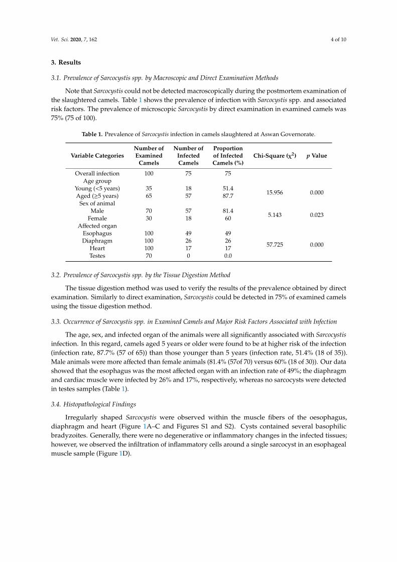
Note that Sarcocystis could not be detected macroscopically during the postmortem examination ofthe slaughtered camels. Table 1 shows the prevalence of infection with Sarcocystis spp. and associatedrisk factors. The prevalence of microscopic Sarcocystis by direct examination in examined camels was75% (75 of 100).
Table 1. Prevalence of Sarcocystis infection in camels slaughtered at Aswan Governorate.
Variable CategoriesNumber ofExamined
Camels
Number ofInfectedCamels
Proportionof InfectedCamels (%)
Chi-Square (χ2) p Value
Overall infection 100 75 75Age group
Young (<5 years) 35 18 51.415.956 0.000Aged (≥5 years) 65 57 87.7
Sex of animalMale 70 57 81.4
5.143 0.023Female 30 18 60Affected organ
Esophagus 100 49 49
57.725 0.000Diaphragm 100 26 26
Heart 100 17 17Testes 70 0 0.0
3.2. Prevalence of Sarcocystis spp. by the Tissue Digestion Method
The tissue digestion method was used to verify the results of the prevalence obtained by directexamination. Similarly to direct examination, Sarcocystis could be detected in 75% of examined camelsusing the tissue digestion method.
3.3. Occurrence of Sarcocystis spp. in Examined Camels and Major Risk Factors Associated with Infection
The age, sex, and infected organ of the animals were all significantly associated with Sarcocystisinfection. In this regard, camels aged 5 years or older were found to be at higher risk of the infection(infection rate, 87.7% (57 of 65)) than those younger than 5 years (infection rate, 51.4% (18 of 35)).Male animals were more affected than female animals (81.4% (57of 70) versus 60% (18 of 30)). Our datashowed that the esophagus was the most affected organ with an infection rate of 49%; the diaphragmand cardiac muscle were infected by 26% and 17%, respectively, whereas no sarcocysts were detectedin testes samples (Table 1).
3.4. Histopathological Findings
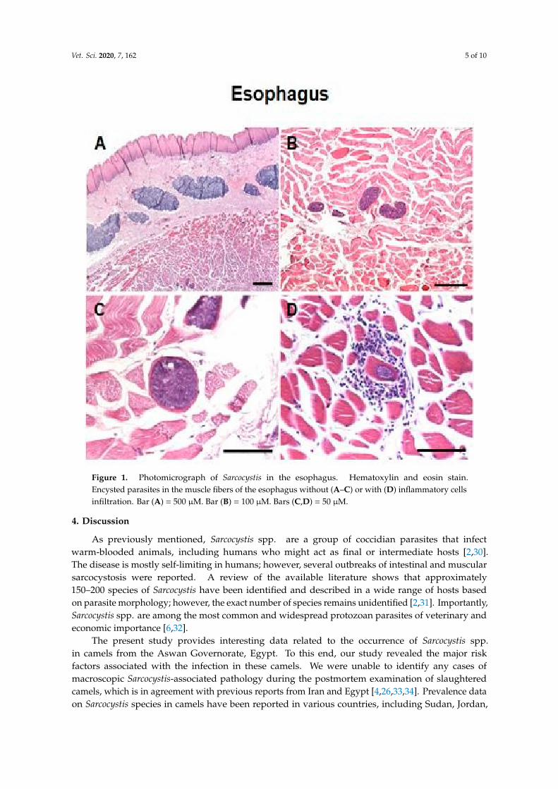
Irregularly shaped Sarcocystis were observed within the muscle fibers of the oesophagus,diaphragm and heart (Figure 1A–C and Figures S1 and S2). Cysts contained several basophilicbradyzoites. Generally, there were no degenerative or inflammatory changes in the infected tissues;however, we observed the infiltration of inflammatory cells around a single sarcocyst in an esophagealmuscle sample (Figure 1D).
Vet. Sci. 2020, 7, 162 5 of 10
Figure 1. Photomicrograph of Sarcocystis in the esophagus. Hematoxylin and eosin stain.Encysted parasites in the muscle fibers of the esophagus without (A–C) or with (D) inflammatory cellsinfiltration. Bar (A) = 500 µM. Bar (B) = 100 µM. Bars (C,D) = 50 µM.
4. Discussion
As previously mentioned, Sarcocystis spp. are a group of coccidian parasites that infectwarm-blooded animals, including humans who might act as final or intermediate hosts [2,30].The disease is mostly self-limiting in humans; however, several outbreaks of intestinal and muscularsarcocystosis were reported. A review of the available literature shows that approximately150–200 species of Sarcocystis have been identified and described in a wide range of hosts basedon parasite morphology; however, the exact number of species remains unidentified [2,31]. Importantly,Sarcocystis spp. are among the most common and widespread protozoan parasites of veterinary andeconomic importance [6,32].
The present study provides interesting data related to the occurrence of Sarcocystis spp.in camels from the Aswan Governorate, Egypt. To this end, our study revealed the major riskfactors associated with the infection in these camels. We were unable to identify any cases ofmacroscopic Sarcocystis-associated pathology during the postmortem examination of slaughteredcamels, which is in agreement with previous reports from Iran and Egypt [4,26,33,34]. Prevalence dataon Sarcocystis species in camels have been reported in various countries, including Sudan, Jordan,
Vet. Sci. 2020, 7, 162 6 of 10
Kazakhstan, Afghanistan, Morocco, the former Union of Soviet Socialist Republics [6,17], Egypt [35],Somalia [36], Saudi Arabia [15], Iraq [37], Southern Ethiopia [38], and Mongolia [27].
As shown in the present study, the prevalence of Sarcocystis infection in slaughtered camels inthe Aswan Governorate using microscopic examination was 75%. The observed prevalence is higherthan that reported in Egypt, Southern Ethiopia, and the Yazd Province (Iran), where the reportedprevalence rates were 42.3–60.0%, 45.45%, and 51.5%, respectively [33,34,38,39]. Moreover, a coupleof previous reports on Sarcocystis spp. in camels from Saudi Arabia, Afghanistan, and Moroccoreported prevalence rates of 56.7%, 47.3–66.3%, and 60%, respectively [40–42]. In a previous studyin Jordan, the reported prevalence was 6.6%, which is markedly lower than that identified in thepresent study [43]. In stark contrast, higher infection rates of 100% and 91.6% were reported inMongolia and Iraq, respectively [27,37]. Furthermore, in a previous study in Egypt, a prevalence of81% was reported [44], whereas a prevalence of 83.6% was reported in the eastern Provinces of Iran [4].The variation between our present results and those previously mentioned could be attributed tovarious factors including the degree of contact between camels and dogs since some camel pastoralistsare not using dogs in camel rearing (camels are reared on a free-range basis in the desert); therefore,differences in the systems used for camel keeping could influence the infection rate [4,7]. Meanwhile,a high prevalence (78.9%) of Sarcocystis infection was reported among slaughtered buffaloes in Beni-Suef,Egypt, and the authors attributed the high prevalence in this region to the close rearing of buffaloeswith dogs, cats, and even wild animals that act as final hosts for these protozoa [45].
In the present study, age was found to be a significant variable associated with infection.Older camels appeared to be at greater risk of acquiring infection than younger ones; hence, animals aged5 years or older were infected to a significantly larger extent (87.7%) than younger animals (51.4%)χ2 = 15.956, p = 0.000). Similar findings were reported in previous studies in the Menofia Governorate(Egypt) [46], Yazd Province (Iran) [33], and in Riyadh city (Saudi Arabia) [47]. The higher prevalenceof Sarcocystis infection in aged camels may likely reflect the higher rate of slaughtering of aged camelscompared with younger animals; moreover, slow development of detectable cysts may explain thelower prevalence in young camels [4,33,47]. Additionally, some owners kept the young camels indoorfor breeding, and therefore, the young camels might be less exposed to infection than older ones [4,33].
In the present investigation, the sex of the animal was another significant risk factor, with malesbeing at higher risk of infection than females (χ2 = 5.143, P = 0.023). As shown in Table 1, prevalence ratesof 81.4% and 60% were reported in male and female camels, respectively. Our finding is consistentwith several previous studies in Egypt [34], southern Ethiopia [38], and Iran [4]. This difference mightbe attributed to the fact that most female animals are kept indoor for reproduction under good andclean management, whereas most of the males are left for grazing outdoor and used by owners forhard work; they may therefore be more exposed to the infection [48].
Sarcocystis spp. infect muscular tissue of the heart, tongue, esophagus, and diaphragm.However, Sarcocystis spp. cysts have been reported in several other types of muscle tissue [49,50].Furthermore, some studies reported sarcocystosis in the cremaster muscle of an animal with orchitis,which observation encouraged us to investigate sarcocysts in testicular tissue samples [49,51]. In thepresent study, dissemination of Sarcocystis in different organs was observed especially in the esophagus,with a prevalence rate of 49% which is according to several previous reports either in the same speciesor different species [4,26,33,38,52]. Meanwhile, some studies found the diaphragm of camels to be themost commonly affected site [27,43], whereas another study identified the heart as the most commonlyinfected organ [53]. However, in other species, such as the water buffalo, the predilection sites forSarcocystis spp. appear to be the esophagus, tongue, and heart [54]. This difference could be explainedby different S. cameli strains or differences in definite host species [33].
Over the last few decades, extraordinary progress was made in developing the criteria for theidentification and diagnosis of sarcocystosis in both intermediate and definitive hosts. Several methodswere used to evaluate the sarcocyst and sporocyst morphology in their major intermediate hostsusing light microscopy to identify the asexual stages in the infected tissues of intermediate hosts
Vet. Sci. 2020, 7, 162 7 of 10
or the sexual stages in the gut of the definitive host. In this regard, the digestion method andhistopathological analysis have been implemented for the analysis of microscopic sarcocysts incamels. Moreover, ultrastructural analysis, using transmission electron microscopy of the cystwall appears very useful in the identification of sarcocysts [55]. Furthermore, several serologicaltechniques, including enzyme-linked immunosorbent assay and an indirect fluorescent antibodytest, were used for diagnosing infection. However, these methods are limited by low sensitivityand specificity due to cross-reactivity between various Sarcocystis spp. [5,56]. Additionally, the useof molecular methods, such as conventional polymerase chain reaction and restriction fragmentlength polymorphism, represents an essential alternative accurate technique for the identification ofendogenous and exogenous stages of Sarcocystis spp. [2,20,57]. However, these methods are typicallymore expensive compared with microscopic examination, and the lack of financial support remainsamong the major limitations of using such techniques in the field. Detection of the sporocysts ofSarcocystis spp. in the feces of definitive hosts could play an essential diagnostic role; however, based onmorphological identification, the various species are very difficult to distinguish [58]. Sarcocysts maytake years to grow in size to become macroscopically visible. This fact justifies our inability to detectmacroscopic sarcocysts in our present study. On the one hand, microscopic evaluation of muscle tissuesamples is important in the diagnosis of Sarcocystis infections in camels. On the other hand, detectionof the parasite stages (sarcocysts) in tissues using histopathological methods provides a confirmatorytool for the diagnosis [2]. In the present study, histological examination revealed the presence of twomorphologically distinct Sarcocystis embedded within the muscle fibers of the esophagus, diaphragm,and heart. Thin-walled Sarcocystis were found to be most common, which is consistent with someprevious reports [2]. The morphology of Sarcocystis is unique as they are intramyofiber protozoal cystswith two types of cyst walls, a palisade-like thick wall or a smooth thin wall. As shown in Figure 1and Figures S1 and S2, sarcocysts appear dark blue-colored due to the presence of many crescent-likebradyzoites inside the cysts.
No inflammatory reaction was observed in the tissue surrounding the cysts. The apparent lack ofinflammatory response might be attributed to the fact that protozoa are located in cysts within themuscle fibers, which offers protection from host immunity—a hypothesis that has been confirmed forvarious parasites [59–62]. Our results are in line with the fact that inflammatory cells are not oftenreported in Sarcocystis-infected tissue [63–65].
5. Conclusions
Current epidemiological and histopathological findings suggest a high occurrence of Sarcocystis incamels in this region of Egypt and indicate that camels may be critical to preserving the epidemiologicalfoci of the disease. The present study also disclosed various major risk factors associated withinfection, including animal age, sex, and anatomical predilection site. Further future molecular andepidemiological studies should focus on identifying the major circulating strains in Egyptian ecologicalniches. Strict hygienic measures may be critical to controlling the disease.
Supplementary Materials: The following are available online at http://www.mdpi.com/2306-7381/7/4/162/s1,Figure S1: Photomicrograph of Sarcocystis in the diaphragm. Encysted parasites in the muscle fibers of thediaphragm without inflammatory cells infiltration (A–D). Hematoxylin and eosin stain. Bar A = 100 µM.Bars B–D = 50 µM; Figure S2: Photomicrograph of Sarcocystis in the heart. Encysted parasite in the muscle fibersof the heart without inflammatory cells infiltration (A and B). Hematoxylin and eosin stain. Bar A = 100 µM.Bar B = 50 µM.
Author Contributions: A.G., M.S., A.A.S., F.A.E.-G., H.M.M.E.-S., R.H.M. and E.K.E. involved in the conceptionof the idea, methodology design, laboratory work, performed data analysis and interpretation. A.G., M.S. andE.K.E. contributed their scientific advice, prepared the manuscript for publication and revision. All authors haveread and agreed to the published version of the manuscript.
Funding: This research received no external funding.
Acknowledgments: The authors also thank the veterinarians for their support and help in providing data andsamples collection throughout the study.
Vet. Sci. 2020, 7, 162 8 of 10
Conflicts of Interest: The authors declare no conflict of interest.
References
1. Fayer, R. Sarcocystis spp. in human infections. Clin. Microbiol. Rev. 2004, 17, 894–902. [CrossRef] [PubMed]2. Fayer, R.; Esposito, D.H.; Dubey, J.P. Human infections with Sarcocystis species. Clin. Microbiol. Rev. 2015,
28, 295–311. [CrossRef] [PubMed]3. Dubey, J.; Calero-Bernal, R.; Rosenthal, B.M.; Speer, C.A.; Fayer, R. Sarcocystosis of Animals and Humans;
CRC Press: Boca Raton, FL, USA, 2015.4. Valinezhad, A.; Oryan, A.; Ahmadi, N. Sarcocystis and its complications in camels (Camelus dromedarius) of
eastern provinces of Iran. Korean J. Parasitol. 2008, 46, 229–234. [CrossRef] [PubMed]5. Saeed, M.A.; Rashid, M.H.; Vaughan, J.; Jabbar, A. Sarcocystosis in South American camelids: The state of
play revisited. Parasit. Vectors 2018, 11, 146. [CrossRef] [PubMed]6. Dubey, J.P.; Speer, C.; Fayer, R. Sarcocystosis of Animals and Man; CRC Press, Inc.: Boca Raton, FL, USA, 1989.7. Dubey, J.; Hilali, M.; Van Wilpe, E.; Calero-Bernal, R.; Verma, S.K.; Abbas, I. A review of sarcocystosis in
camels and redescription of Sarcocystis cameli and Sarcocystis ippeni sarcocysts from the one-humped camel(Camelus dromedarius). Parasitology 2015, 142. [CrossRef] [PubMed]
8. Ruiz, A.; Frenkel, J.K. Recognition of Cyclic Transmission of Sarcocystis muris by Cats. J. Infect. Dis. 1976,133, 409–418. [CrossRef] [PubMed]
9. Hilali, M.; Mohamed, A. The dog (Canis familiaris) as the final host of Sarcocystis cameli (Mason, 1910).Tropenmedizin Parasit. 1980, 31, 213–214.
10. Kuraev, G.T. Sarcocystosis of camel, Kzakhstan Sr. Vet. Moscow 1981, 7, 41.11. Niilo, L. Helminths, Arthropods and Protozoa of Domesticated Animals (Sixth Edition of Mönnig’s Veterinary
Helminthology and Entomology). Can. Vet. J. 1969, 10, 223.12. Oryan, A.; Moghaddar, N.; Gaur, S.N. The distribution pattern of Sarcocystis species, their transmission and
pathogenesis in sheep in Fars Province of Iran. Vet. Res. Commun. 1996, 20, 243–253. [CrossRef]13. Savini, G.; Dunsmore, J.D.; Robertson, I.D.; Seneviratna, P. Sarcocystis spp in Western Australian sheep.
Aust. Vet. J. 1993, 70, 152–154. [CrossRef]14. Mason, F.E. Sarcocystis in the camel in Egypt. J. Comp. Pathol. Ther. Edinb. 1910, 23, 168–176. [CrossRef]15. Fatani, A.; Hilali, M.; al-Atiya, S.; al-Shami, S. Prevalence of Sarcocystis in camels (Camelus dromedarius)
from Al-Ahsa, Saudi Arabia. Vet. Parasitol. 1996, 62, 241–245. [CrossRef]16. Odening, K. The present state of species-systematics in Sarcocystis Lankester, 1882 (Protista, Sporozoa,
Coccidia). Syst. Parasitol. 1998, 41, 209–233. [CrossRef]17. Boid, R.; Jones, T.; Luckins, A. 3. Protozoal diseases of camels. Br. Vet. J. 1985, 141, 87–105. [CrossRef]18. Rommel, M. Sarcocystosis of domestic animals and humans. In Pract. 1985, 7, 158–160. [CrossRef] [PubMed]19. Shaapan, R.M. The common zoonotic protozoal diseases causing abortion. J. Parasit. Dis. 2016, 40, 1116–1129.
[CrossRef] [PubMed]20. Stojecki, K.; Karamon, J.; Sroka, J.; Cencek, T. Molecular diagnostics of Sarcocystis spp. infections. Pol. J.
Vet. Sci. 2012, 15, 589–596. [CrossRef]21. Beaver, P.; Jung, R.C.; Cupp, E.W. Clinical Parasitology. 9th Edition. J. Med Entomol. 1984, 21, 136. [CrossRef]22. Frenkel, J.K. Sarcocystosis. In Pathology of Infectious Diseases; Conner, D.H., Chandler, F.W., Schwartz, D.A.,
Manz, H.J., Lack, E.E., Eds.; Appleton and Lange: Stamford, CT, USA, 1997; Volume 1997, pp. 1253–1259.23. Jehle, C.; Dinkel, A.; Sander, A.; Morent, M.; Romig, T.; Luc, P.V.; De, T.V.; Thai, V.V.; Mackenstedt, U.
Diagnosis of Sarcocystis spp. in cattle (Bos taurus) and water buffalo (Bubalus bubalis) in Northern Vietnam.Vet. Parasitol. 2009, 166, 314–320. [CrossRef]
24. Odening, K.; Stolte, M.; Bockhardt, I. On the diagnostics of Sarcocystis in cattle: Sarcocysts of a species unusualfor Bos taurus in a dwarf zebu. Vet. Parasitol. 1996, 66, 19–24. [CrossRef]
25. Beyazıt, A.; Yazıcıoglu, Ö.; Karaer, Z. The prevalence of ovine Sarcocystis species in Izmir province.Vet. Fak. Derg. 2007, 54, 111–116.
26. Wahba, A.; Ayoub, M.; Soliman, K. Light and ultrastructure of Sarcocystis spp. of camels and associatedpathological changes. Anim. Health Res. J. 2014, 2, 143–158.
27. Fukuyo, M.; Battsetseg, G.; Byambaa, B. Prevalence of Sarcocystis infection in meat-producing animals inMongolia. Southeast Asian J. Trop. Med. Public Health 2002, 33, 490–495. [PubMed]
Vet. Sci. 2020, 7, 162 9 of 10
28. Whaeeb, S.T.; Faraj, A.A. Molecular identification and phylogeny of microscopic Sarcocystis Sheep in Baghdadprovince. Int. J. Adv. Res. Biol. Sci. 2016, 3, 50–56.
29. Arafa, M.; Monib, M.; Dyab, A.; Abdel-Ghaffar, S. Studies on ocular Sarcocystis in buffaloes in assiutgovernorate. Ass. Univ. Bull. Environ. Res. 2003, 6, 27–35.
30. Pozio, E. Epidemiology and control prospects of foodborne parasitic zoonoses in the European Union.Parassitologia 2008, 50, 17–24.
31. Spickler, A.R. Sarcocystosis. Available online: http://www.cfsph.iastate.edu/Factsheets/pdfs/sarcocystosis.pdf.(accessed on 21 August 2020).
32. Tenter, A.M. Current research on Sarcocystis species of domestic animals. Int. J. Parasitol. 1995, 25, 1311–1330.[CrossRef]
33. Hamidinejat, H.; Hekmatimoghaddam, S.; Jafari, H.; Sazmand, A.; Haddad Molayan, P.; Derakhshan, L.;Mirabdollahi, S. Prevalence and distribution patterns of Sarcocystis in camels (Camelus dromedarius) in Yazdprovince, Iran. J. Parasit. Dis. 2013, 37, 163–165. [CrossRef]
34. Mandour, A.M.; Rabie, S.A.; Mohammed, N.I.; Hussein, N.M. On the presence of Sarcocystis miescherisp. nov. in camels of Qena Governorate. Egypt. Acad. J. Biol. Sci. E. Med. Entomol. Parasitol. 2011, 3, 1–7.[CrossRef]
35. Abdel Ghaffar, F.; Entzeroth, R.; Chobotar, B.; Scholtyseck, E. Ultastructural studies of Sarcocystis sp. fromthe camel (Camelus dromedarius) in Egypt. Tropenmed. Parasitol. 1979, 30, 434–438. [PubMed]
36. Borrow Hagi, A.; Mohammed Hassan, A.; di Sacco, B. Sarcocystis in Somali camel. Parassitologia 1989,31, 133–136.
37. Latif, B.M.; Al-Delemi, J.K.; Mohammed, B.S.; Al-Bayati, S.M.; Al-Amiry, A.M. Prevalence of Sarcocystis spp.in meat-producing animals in Iraq. Vet. Parasitol. 1999, 84, 85–90. [CrossRef]
38. Woldemeskel, M.; Gumi, B. Prevalence of Sarcocysts in one-humped camel (Camelus dromedarius) fromsouthern Ethiopia. J. Vet. Med. B Infect. Dis Vet. Public Health 2001, 48, 223–226. [CrossRef] [PubMed]
39. Abdel-Ghaffar, F.; Mehlhorn, H.; Bashtar, A.-R.; Al-Rasheid, K.; Sakran, T.; El-Fayoumi, H. Life cycle ofSarcocystis camelicanis infecting the camel (Camelus dromedarius) and the dog (Canis familiaris), light andelectron microscopic study. Parasitol. Res. 2009, 106, 189–195. [CrossRef] [PubMed]
40. Hussein, S.H. The prevalence of Sarcocystis infection in Saudi Arabian Najdi sheep and camels. Biol. Sci.1991, 1, 43–56.
41. Kirmse, P.; Mohanbabu, B. Sarcocystis sp. in the one-humped camel (Camelus dromedarius) from Afghanistan.Br. Vet. J. 1986, 142, 73–74. [CrossRef]
42. Kirmse, P. Sarcosporidiosis in equines of Morocco. Br. Vet. J. 1986, 142, 70–72. [CrossRef]43. Al-Ani, F.K.; Amr, Z. Sarcocystis spp Prevalence in Camel Meat in Jordan. Dairy Vet. Sci. J. 2017, 4, 1–3.44. El-Etreby, M. Myocardial sarcosporidiosis in the camel. Pathol. Vet. 1970, 7, 7–11. [CrossRef]45. El-Dakhly, K.M.; El-Nesr, K.A.; El-Nahass el, S.; Hirata, A.; Sakai, H.; Yanai, T. Prevalence and distribution
patterns of Sarcocystis spp. in buffaloes in Beni-Suef, Egypt. Trop. Anim. Health Prod. 2011, 43, 1549–1554.[CrossRef] [PubMed]
46. El-Bahy, N.; El-Bagory, A.-E.-R.; AbouLaila, M.; Elkhatam, A.; Mady, H.M. Prevalence of Sarcocystis fusiformisand Hydatid cyst among Different Ruminants at Menofia Governorate, Egypt. J. Curr. Vet. Res. 2019, 1, 1–10.[CrossRef]
47. Omer, S.A.; Alzuraiq, A.A.; Mohammed, O.B. Prevalence and molecular detection of Sarcocystis spp. infectionin the dromedary camel (Camelus dromedarius) in Riyadh city, Saudi Arabia. Biomed. Res. 2017, 28, 4962–4965.
48. Romero, S.; Carletti, T.; Decker Franco, C.; More, G.; Schnittger, L.; Florin-Christensen, M. Seropositivityto Sarcocystis infection of llamas correlates with breeding practices. Vet. Parasitol. Reg. Stud. Rep. 2017,10, 65–70. [CrossRef]
49. Bucca, M.; Brianti, E.; Giuffrida, A.; Ziino, G.; Cicciari, S.; Panebianco, A. Prevalence and distribution ofSarcocystis spp. cysts in several muscles of cattle slaughtered in Sicily, Southern Italy. Food Control 2011,22, 105–108. [CrossRef]
50. Ono, M.; Ohsumi, T. Prevalence of Sarcocystis spp. cysts in Japanese and imported beef (Loin:Musculus longissimus). Parasitol. Int. 1999, 48, 91–94. [CrossRef]
51. Saglam, K.; Keles, H. Sarcocystosis in the Cremaster Muscle of an Infertile Bull, Spermiostasis and Orchitis.Kocatepe Vet. J. 2016, 9, 252–254. [CrossRef]
Vet. Sci. 2020, 7, 162 10 of 10
52. Ahmed, A.M.; Elshraway, N.T.; Youssef, A.I. Survey on Sarcocystis in bovine carcasses slaughtered at themunicipal abattoir of El-Kharga, Egypt. Vet. World 2016, 9, 1461–1465. [CrossRef]
53. Shekarforoush, S.S.; Shakerian, A.; Hasanpoor, M.M. Prevalence of Sarcocystis in slaughtered one-humpedcamels (Camelus dromedarius) in Iran. Trop. Anim. Health Prod. 2006, 38, 301–303. [CrossRef]
54. Oryan, A.; Ahmadi, N.; Mousavi, S.M. Prevalence, biology, and distribution pattern of Sarcocystis infectionin water buffalo (Bubalus bubalis) in Iran. Trop. Anim. Health Prod. 2010, 42, 1513–1518. [CrossRef]
55. Metwally, D.M.; Al-Otaibi, T.T.; Al-Turaiki, I.M.; El-Khadragy, M.F.; Alajmi, R.A. Identification of SarcocystisSpp. in One-humped Camels (Camelus dromedarius) from Riyadh and Dammam, Saudi Arabia, via Histologicaland Phylogenetic Approaches. Animals 2020, 10, 1108. [CrossRef] [PubMed]
56. More, G.; Pardini, L.; Basso, W.; Marin, R.; Bacigalupe, D.; Auad, G.; Venturini, L.; Venturini, M.C.Seroprevalence of Neospora caninum, Toxoplasma gondii and Sarcocystis sp. in llamas (Lama glama) from Jujuy,Argentina. Vet. Parasitol. 2008, 155, 158–160. [CrossRef] [PubMed]
57. Castro-Forero, S.; Bulla-Castañeda, D.; Buitrago, H.; Díaz Anaya, A.; Madeira de Carvalho, L.;Pulido-Medellin, M. Sarcocystis spp., a parasite with zoonotic potential. Bulg. J. Vet. Med. 2020, 2020, 1–12.[CrossRef]
58. Beaver, P.C.; Gadgil, K.; Morera, P. Sarcocystis in man: A review and report of five cases. Am. J. Trop.Med. Hyg. 1979, 28, 819–844. [CrossRef]
59. Nance, J.P.; Vannella, K.M.; Worth, D.; David, C.; Carter, D.; Noor, S.; Hubeau, C.; Fitz, L.; Lane, T.E.;Wynn, T.A.; et al. Chitinase dependent control of protozoan cyst burden in the brain. PLoS Pathog. 2012,8, e1002990. [CrossRef]
60. Guerrant, R.L.; Walker, D.H.; Weller, P.F. Tropical Infectious Diseases: Principles, Pathogens and Practice, 3rd ed.;Elsevier: Amsterdam, The Netherlands, 2011; p. 24.
61. Vangeel, L. Bovine Sarcocystis Species and Their Role in Bovine Eosinophilic Myositis. Ph.D. Thesis,Ghent University, Ghend, Belgium, 2012.
62. Hidron, A.; Vogenthaler, N.; Santos-Preciado, J.I.; Rodriguez-Morales, A.J.; Franco-Paredes, C.; Rassi, A., Jr.Cardiac involvement with parasitic infections. Clin. Microbiol. Rev. 2010, 23, 324–349. [CrossRef]
63. Arness, M.K.; Brown, J.D.; Dubey, J.P.; Neafie, R.C.; Granstrom, D.E. An outbreak of acute eosinophilicmyositis attributed to human Sarcocystis parasitism. Am. J. Trop. Med. Hyg. 1999, 61, 548–553. [CrossRef]
64. McLeod, R.; Hirabayashi, R.N.; Rothman, W.; Remington, J.S. Necrotizing vasculitis and Sarcocystis:A cause-and-effect relationship? South. Med. J. 1980, 73, 1380–1383. [CrossRef]
65. Italiano, C.M.; Wong, K.T.; AbuBakar, S.; Lau, Y.L.; Ramli, N.; Syed Omar, S.F.; Kahar Bador, M.; Tan, C.T.Sarcocystis nesbitti causes acute, relapsing febrile myositis with a high attack rate: Description of a largeoutbreak of muscular sarcocystosis in Pangkor Island, Malaysia, 2012. PLoS Negl. Trop. Dis. 2014, 8, e2876.[CrossRef]
Publisher’s Note: MDPI stays neutral with regard to jurisdictional claims in published maps and institutionalaffiliations.
© 2020 by the authors. Licensee MDPI, Basel, Switzerland. This article is an open accessarticle distributed under the terms and conditions of the Creative Commons Attribution(CC BY) license (http://creativecommons.org/licenses/by/4.0/).