Embed Size (px)
Citation preview
Vitiligo: A Possible Model of Degenerative DiseasesBarbara Bellei1*, Angela Pitisci1, Monica Ottaviani1, Matteo Ludovici1, Carlo Cota2, Fabiola Luzi3,
Maria Lucia Dell’Anna , Mauro Picardo1 1
1 Laboratory of Cutaneous Physiopathology, San Gallicano Dermatologic Institute, Istituto Di Ricovero e Cura a Carattere Scientifico, Rome, Italy, 2 Dermatopathology
Unit, San Gallicano Dermatological Institute, Istituto Di Ricovero e Cura a Carattere Scientifico, Rome, Italy, 3 Struttura Complessa di Medicina Preventiva delle Migrazioni,
del Turismo e di Dermatologia Tropicale, San Gallicano Dermatological Institute, Istituto Di Ricovero e Cura a Carattere Scientifico, Rome, Italy
Abstract
Vitiligo is characterized by the progressive disappearance of pigment cells from skin and hair follicle. Several in vitro and invivo studies show evidence of an altered redox status, suggesting that loss of cellular redox equilibrium might be thepathogenic mechanism in vitiligo. However, despite the numerous data supporting a pathogenic role of oxidative stress,there is still no consensus explanation underlying the oxidative stress-driven disappear of melanocytes from the epidermis.In this study, in vitro characterization of melanocytes cultures from non-lesional vitiligo skin revealed at the cellular levelaberrant function of signal transduction pathways common with neurodegenerative diseases including modification of lipidmetabolism, hyperactivation of mitogen-activated protein kinase (MAPK) and cAMP response element-binding protein(CREB), constitutive p53-dependent stress signal transduction cascades, and enhanced sensibility to pro-apoptotic stimuli.Notably, these long-term effects of subcytotoxic oxidative stress are also biomarkers of pre-senescent cellular phenotype.Consistent with this, vitiligo cells showed a significant increase in p16 that did not correlate with the chronological age ofthe donor. Moreover, vitiligo melanocytes produced many biologically active proteins among the senescence-associatedsecretory phenotype (SAPS), such as interleukin-6 (IL-6), matrix metallo proteinase-3 (MMP3), cyclooxygenase-2 (Cox-2),insulin-like growth factor-binding protein-3 and 7 (IGFBP3, IGFBP7). Together, these data argue for a complicatedpathophysiologic puzzle underlying melanocytes degeneration resembling, from the biological point of view,neurodegenerative diseases. Our results suggest new possible targets for intervention that in combination with currenttherapies could correct melanocytes intrinsic defects.
Citation: Bellei B, Pitisci A, Ottaviani M, Ludovici M, Cota C, et al. (2013) Vitiligo: A Possible Model of Degenerative Diseases. PLoS ONE 8(3): e59782. doi:10.1371/journal.pone.0059782
Editor: Thomas G. Hofmann, German Cancer Research Center, Germany
Received November 5, 2012; Accepted February 18, 2013; Published March 26, 2013
Copyright: � 2013 Bellei et al. This is an open-access article distributed under the terms of the Creative Commons Attribution License, which permitsunrestricted use, distribution, and reproduction in any medium, provided the original author and source are credited.
Funding: The study was partially supported by Ministry of Health, Italy. The funders had no role in study design, data collection and analysis, decision to publish,or preparation of the manuscript. No additional funding received for this study.
Competing Interests: The authors have declared that no competing interests exist.
* E-mail: [email protected]
Introduction
Vitiligo is an acquired cutaneous skin and less frequently hair
disease characterized by declining melanocyte function and
depigmentation with an estimated prevalence of 0.5–1% in most
populations [1]. Depigmentation results in irregular white patches,
initially small, that often gradually enlarge and change shape and
this process is a chronic with a highly variable course. In contrast
to other prevalent skin diseases vitiligo has a near orphan status for
drug development. Several mechanisms of melanocyte degenera-
tion have been presented, including autoimmunity [2], auto-
cytotoxic/metabolic mechanism [3–4], and impaired melanocyte
migration and/or proliferation [5]. Studies have also pointed to a
significant role of genetic susceptibility to vitiligo [6]. Since none of
these processes alone are sufficient to fully explain the mechanisms
of the disease, and all of the proposed mechanisms are not
mutually exclusive, the convergence theories have been formulat-
ed combining biochemical, environmental and immunological
events, in a permissive genetic milieu [7]. Despite considerable
efforts, there are few data in the literature on in vitro studies of
vitiligo epidermal melanocytes, mainly due to the fact that cells
from normally pigmented skin of vitiligo patients demonstrate
reduced initial seeding and proliferation capacity compared to
healthy adult human skin [8–9].
Several investigators have proved the presence of oxidative
stress in cultured melanocytes coupled with an increased
susceptibility to pro-oxidant agents [3,10–11]. In vivo oxidative
stress has been attributed to a massive accumulation of H2O2 in
vitiligo skin, which is associated with impaired catalase and
glutathione peroxidase activities [12]. Moreover, there are several
lines of evidence for systemic oxidative stress, including alteration
of peripheral blood antioxidant patterns and oxidative DNA
damage [13–14]. Impairment of mitochondrial complex I
respiratory chain and increased expression of malate dehydroge-
nase suggest that in vitiligo mitochondria could be the site of
uncontrolled ROS production [13].
Recently, nuclear factor E2-related factor 2 (Nrf2), one of the
most critical antioxidant enzymatic systems, and its downstream
target genes were found increased in vitiligo non-lesional skin
biopsies, suggesting that a consistently higher Nrf2-dependent
transcriptional activity is required for the maintenance of redox
homeostasis in disease-free epidermis [15]. Consistent with the
idea that persistent intracellular oxidative stress burdens vitiligo
skin, overexpression of p53 protein has been reported in both
lesional and non-lesional epidermis of patients with vitiligo [16–
17]. However, despite numerous data supporting a pathogenic
role of oxidative stress, there is still no consensus explanation
PLOS ONE | www.plosone.org 1 March 2013 | Volume 8 | Issue 3 | e59782

underlying the oxidative stress-driven disappearance of melano-
cytes from the epidermis [18]. Even though increased susceptibility
to the toxic effects of chemical oxidants or UVB has been reported
in several in vitro studies [3,9,19] there are no convincing data
demonstrating the occurrence of melanocytes death by cytotox-
icity or apoptosis in vitiligo skin in vivo. Biopsy material from
established lesions contains no or few melanocytes, and it is
difficult to capture the essence of the melanocytes lost. However, it
is also possible that persistent stress-induced cell damage might
interfere with vitiligo melanocytes vitality and functionality by
initiating a senescence-driven melanocytes detachment.
Since many oxidative stress-related progressive or non-progres-
sive chronic diseases are referred to as degenerative diseases we
investigated the possibility that vitiligo melanocytes express
common features with cells of neurodegenerative disorders. For
most of these pathologies it has been demonstrated that sublethal
oxidative stress and subsequent cellular alterations, including
modification of lipid metabolism [20], impairment of the
mitochondrial respiratory chain [21], dysfunction of intracellular
signaling, and enhanced sensibility to pro-apoptotic stimuli [22],
culminate in aging and cell degeneration [23]. Moreover, a
premature senescent-like cell phenotype in vivo and in vitro has been
linked to the degenerative diseases [24]. Some cells in aging
organisms lose functionality: neurons, for example, lose the ability
to form synapses despite cell bodies remaining viable [25]. A
similar scenario is conceivable for vitiligo cells, because melano-
cytes are not necessarily completely absent in the depigmented
lesions [26].
Here we report convincing evidence that vitiligo melanocytes
have alterations of signal transduction pathways at the cellular and
molecular level that altogether argue for a condition of stress-
induced premature senescence-like phenotype supporting the
classification of vitiligo among the degenerative diseases. More-
over, vitiligo non-lesional skin biopsies showed that results
collected with cell culture experiments were not artificially-
induced by culture conditions and confirmed the acquisition of
p53-dependent pro-senescent phenotype.
Materials And Methods
Cell cultureVHM were isolated from skin biopsies obtained from a
normally pigmented area in the gluteal or armpit regions. Patients
(females = 6, male = 8) ranged from 7 to 56 years old (average 38.6
years). NHM were isolated from healthy individuals (females = 8,
male = 8) who underwent plastic surgery and ranged from 15 to 75
years old (average 50.1 years). Cells were cultured in M254
medium supplemented with Human Melanocyte Growth Supple-
ments (HMGS) (Cascade Biologics, Mansfield, UK) and used
between passage 2 and 8. Institutional Research Ethics Committee
(Istituti Fisioterapici Ospitalieri), approval was obtained to collect
samples of human material for research. The Declaration of
Helsinki Principles was followed and patients gave written
informed consent. The study included one children participant
and in this case his parents approved the written consent.
Cell viability and proliferationFor cells proliferation assay cells were incubated with M254
containing HMGS or M254 alone for 24 h and then treated with
N-acetyl-L-cystein (5 mM) (Sigma Aldrich, Milan, Italy), or
pifithrin-a (5 mM) (Santa Cruz Biotechnology, Santa Cruz, CA,
USA). Medium was replaced every 48 h and cells were left to
growth according with experimental design before Trypan blue
exclusion assay. For cell viability assay cells were exposed to t-BHP
for 24 h and then incubated with 3-(4,5 dimethylthiazol)-2,5-
diphenyl tetrazolium bromide (MTT) (Sigma Aldrich) for 2 h.
After this time, the medium was removed and the resulting crystals
were solubilized in DMSO. The absorbance was measured at
570 nm with a reference wavelength of 650 nm.
ImmunofluorescenceFor indirect immunofluorescence experiments, cells were grown
on coverslips and after treatment were fixed and permeabilized
with methanol for 10 min at 220uC (catalase and SOD2
antibodies) or fixed in 3% paraformaldehyde in PBS for 15 min
at room temperature and then permeabilized with 0.05% Trition
X-100 in PBS for 5 min (p16, p53 and cyclinD1). Cells were rinsed
in PBS and incubated for 2 h with mouse anti-catalase (Sigma,
Milan, Italy), anti-p16 (Santa Cruz Biotechnology, USA) anti-p53
or anti-cyclinD1 (Dako, Milan, Italy) primary antibodies or rabbit
anti-SOD2 (Stressgen Biothechnology (1:300 in PBS). Cells were
rinsed three times with PBS and incubated for 60 min with an
Alexa-Fluor-488-conjugated goat anti-mouse IgG or goat anti-
rabbit IgG (1:800 in PBS) (Molecular Probe). Nuclei were stained
with DAPI (Sigma, Milan Italy). For tissue immunofluorescence
serial sections (3 mm) derived from formalin-fixed and paraffin-
embedded blocks were dewaxed in xylene and rehydrated through
graded ethanol to PBS. Tissue sections were incubated with the
following primary antibodies: anti-p53 mouse monoclonal anti-
bodies (1:300) and anti-PML rabbit polyclonal antibodies (1:400)
plus anti-Tyrosinase antibody (1:200) (Santa Cruz Bioechnology).
Sections were then treated with conjugated with secondary
antibodies AlexaFluor-488 chicken-anti-goat plus AlexaFluor-546
goat anti-rabbit or plus AlexaFluor-546 goat anti-mouse (1:800)
and stained with DAPI.
Western blot analysisCell extracts were prepared with RIPA buffer containing
proteases and phosphatases inhibitors. Proteins were separated
on SDS-polyacrylamide gels, transferred to nitrocellulose mem-
branes and then treated with p53, SOD2, catalase, PML and
GADD45 (Santa Cruz Biotechnology, USA) antibodies. Anti-b-
tubulin (Sigma Aldrich) was used to normalize protein content.
Horseradish peroxide-conjugated goat anti-mouse or goat anti-
rabbit secondary antibody complexes were detected by chemilu-
minescence (Santa Cruz Biotechnology).
Determination of MMP3, IGFBP3 and IL-6 proteinsQuantitative measurement of the hIGFBP3, IL-6 (DRG
Diagnostic, Marburg, Germany) and hMMP3 (Boster Biological
Technology, Fremont, CA, USA) were used according with
manufacture instructions. Due to a significant interference with
HMGS components the level of IGFBP3 was determined using
protein cell extracts whereas the level of MMP3 protein was
measured using undiluted conditioned medium (96 h). Results
were normalized for proteins concentration.
Semi-quantitative RT-PCRTotal RNA was extracted using Aurum Total mini kit (BioRad,
Milan Italy). cDNA was synthesized from 1 mg of total RNA using
the FirstAid kit (Fermentas) and amplified in a reaction mixture
containing iQSYBR Green Supermix (BioRad, Milan Italy) and
25 pmol of forward and reverse primers using an iQ5 Light Cycler
(BioRad). All samples were run in triplicate, and relative
expression was determined by normalizing results to b-actin
mRNA. Due to the importance of the internal control gene chosen
for sample normalization, comparative analyses were randomly
Stress-Induced Cell Degeneration in Vitiligo
PLOS ONE | www.plosone.org 2 March 2013 | Volume 8 | Issue 3 | e59782

carried out using two commonly used housekeeping genes:
GAPDH and 18 S rRNA (data not shown). This confirmed that
b-actin mRNA was a satisfactory reference gene. Sequences of
primers can be found in Table S1.
Flow cytometry analysis for MAPK phosphorylationMelanocytes were fixed and permeabilized with Cytofix/
CitopermTMand then stained with anti-p38-Alexa-Fluor-488-
conjugated (pT180/pY182), anti-ERK-PE-conjugated (pT202/
pY204) or anti-CREB-PE-conjugated (pSer133) antibodies (BD
Bioscience, Erembodegem, Belgium) and then analyzed by flow
cytometry using a FACSCalibur. Data from 16104 cells were
acquired from each sample. Median Fluorescence Intensity (MFI)
was evaluated on a linear scale.
Assessment of intracellular reactive oxygen speciesProduction of ROS was assessed incubating cells with the
fluorescent dye 2979-dichlorodihydrofluorescein diacetate (Sigma
Aldrich, Milan) for 30 min at 37uC and 5% CO2 in the dark. Cells
were washed with PBS and trypsinized, centrifuged at 1000 rpm,
and then resuspended in PBS. Signals were measured by flow
cytometry (BD Bioscience).
Cholesterol membrane content measurementLipids were extracted twice in chloroform:methanol 2:1 in the
presence of butylated hydroxytoluene as an antioxidant and 5a-
cholestane as an internal standard and centrifuged at 1800 rpm
for 10 min. The lower phase was evaporated under a nitrogen
stream. Oxysterol esters were hydrolyzed by means of 1M KOH
in methanol. Combined organic extracts were evaporated under
N2 flow and then directly silylated with N,O-bis-(trimethylsilyl)-
trifluoroacetamide containing 1% trimethylchlorosilane as cata-
lyst. An oven temperature gradient 180uC–250uC at 20 uC/min
was used. Mass spectra were recorded in Electronic Impact and in
SIM modality. The separation was performed by capillary column
HP-5MS (30 m60,250 nm60,25 mm, Agilent Technologies INC)
using helium as the carrier gas. Results are expressed as
percentage 6 SD considering NHMs content as 100.
ImmunohistochemistrySerial sections (3 mm) derived from formalin-fixed and paraffin-
embedded blocks were dewaxed in xylene and rehydrated through
graded ethanol to PBS. Endogenous peroxidase activities were
blocked by 0.03% hydrogen peroxide. Tissue sections were
incubated with the following primary antibodies: anti-p53 mouse
monoclonal antibodies (1:300), anti-PML rabbit polyclonal anti-
bodies (1:400) and anti-GADD45 mouse monoclonal antibody
(1:300). Sections were then treated with peroxidase-labelled
polymer conjugated with secondary antibodies (Dako), incubated
with 3-amino-9-ethylcarbazole substrate chromogen (Dako) and
counterstained with haematoxylin. Negative controls were ob-
tained by omitting the primary antibodies.
Statistical analysisStudent’s t-test was used to assess statistical significance with
thresholds of * p#0.05 and ** p#0.01.
Results
Intrinsic oxidative stress in vitiligo melanocytes culturesPrevious clinical and experimental observations indicated
oxidative stress as a possible biological basis of melanocytes
disappearance [3,12]. In melanocytes isolated from vitiligo
patients we observed chronic activation of Nrf2, manganese
superoxide dismutase 2 (SOD2), hemeoxigenase-1 (HO-1),
NAD(P)H deydrogenase quinone-1 (NQO1) and catalase genes
expression that indicated a compromised redox homeostasis
(Fig. 1A). The expression of SOD2 and catalase were also
investigated at the protein level by immunofluorescence analysis
and densitometric evaluation of western blots. At the protein level
vitiligo and healthy melanocytes showed similar staining intensity
for catalase, whereas SOD2 labeling was slightly lower in vitiligo
cells (Fig 1 B and C). A significant increase in ROS concentration
(Fig. 1D) could explain the persistent stimulation of antioxidant
and detoxification genes in vitiligo melanocytes, whereas attenu-
ated protein expression could be directly caused by altered
intracellular redox balance affecting enzymes stability. In fact, it
has been demonstrated that acceleration of catalase degradation
caused by H2O2 determines the increase of the corresponding
gene transcription, producing a constant stady-state level of
protein expression [27]. Activation of antioxidant gene expression
indicated a compromised redox homeostasis in melanocytes
isolated from vitiligo patients. We decided to further investigate
this by studying the effect of induced oxidative stress by tert-butyl-
hydroperoxide (t-BHP), a membrane-permeant oxidant. As a
consequence of peroxide-mediated cell damage a dose-dependent
decrease in the number of viable melanocytes was evident (Fig. 1E).
At highest t-BHP concentrations (100 and 200 mM) the reduction
of cell viability was significantly greater in vitiligo human
melanocytes (VHM) than in normal human melanocytes
(NHM). The dose of 100 mM was selected for further experiments
in order to maximize the differences between normal and vitiligo
cultures. Consistent with the results obtained from MTT assays,
we observed a significant difference in the induction of catalase
and detoxification enzyme expression upon exposure of cells to
oxidative stress, with the exception of NQO1, which was similar in
all cell cultures (Fig. 1F). Consequently, vitiligo cells were not fully
competent to counteract the increase of oxygen species (Fig. 1G).
Alteration of intracellular signaling pathways in vitiligomelanocytes
Oxidants can trigger the activation of multiple signaling
pathways, including MAPK and CREB. MAPK kinase phosphor-
ylation was found to be dysregulated in vitiligo cells compared to
normal melanocytes (Fig. 2A), and CREB, a direct regulator of
antioxidant gene expression [28–29], was also hyperphosphory-
lated. The role of oxidative stress in the increased kinases
phosphorylation was supported by the demonstration of the same
hyperactivated profile in NHM in response to t-BHP (Fig. 2B). In
addition, vitiligo melanocytes conserved their intrinsic hyperpho-
sphorylation status independently to the presence of mitogens
confirming an endogenous source of MAPK activation, such as
excessive intracellular ROS levels (Fig 2C). To further confirm the
notion that MAPK activation in vitiligo cells is through generation
of ROS, N-acetyl-L-cystein (NAC), a ROS scavanger, was
included in the basal medium. NAC decreased p38 and ERK
phosphorylation, whereas, at least at the concentration used, there
were not significant modifications of CREB phosphorylation
(Fig. 2D). Collectively, these results indicate that in VHM chronic
redox imbalance is responsible of a permanent adaptive MAPK-
dependent signaling deregulation. Notably, accumulating evidenc-
es implicated both CREB and p38 MAPK constitutive phosphor-
ylation in neurodegenration [30–31].
Activated kinases can regulate a number of cellular substrates by
phosphorylation, leading to diverse cellular responses. Stress-
induced p38 acts as a protein kinase to phosphorylate p53, leading
to cell cycle arrest, apoptosis and cell survival. Hence, in
Stress-Induced Cell Degeneration in Vitiligo
PLOS ONE | www.plosone.org 3 March 2013 | Volume 8 | Issue 3 | e59782

agreement with earlier studies [16–17], analysis of p53 in vitiligo
melanocytes by western blot evidenced an increase of protein
expression (Fig. 2E) and more importantly, immunofluorescence
analysis demonstrated enhanced nuclear distribution of p53
indicating functional activation (Fig. 2F).
Altered expression of cell cycle regulatorsProlonged p53 activation can cause reduced proliferation and/
or cell cycle arrest and expression of senescence-related proteins.
In vitiligo melanocytes mRNA quantification demonstrated a
significant upregulation of several p53 downstream genes involved
in cell cycle regulation such as p21, GADD45a (growth arrest and
DNA damage-inducible), PML (promyelocytic leukemia) and a
slight increase of the expression of genes involved in p53-mediated
apoptosis, such as Bax and Pig-3 (Fig. 3A). The expression of the
two stress sensors PML and GADD45 was confirmed by western
blot analysis (Fig. 3B). Alternative splicing produces a large
number of PML isoforms ranging from 97 KDa of PML-I isoform
to 48 KDa of PML-VII isoform but only two specific PML splice
variants (PML-I and PML-IV) are targeted to the nucleolus by
stress conditions [32–33]. In adults melanocyte cultures two
isoforms were easily detectable: one PML high molecular weight
isoform compatible with PML-I and a low molecular weight
isoform compatible with PML-VI and PML-VII isoforms.
Interestingly, VHM express abundant PML-I isoform that is
required for the targeting of PML proteins to the nucleolus and for
the formation of very large PML nuclear bodies associated with
senescence [33]. Accordingly, immunofluorescence analysis con-
firmed higher level of PML expression and demonstrated a general
increase in PML nuclear bodies size (Fig. 3C). The disequilibrium
in cell cycle regulation was also demonstrated by increased
expression of p16 in VHM compared to age-matched control
Figure 1. Evidence for constitutive activation of cytoprotective enzymes network in vitiligo melanocyte cultures. (A) Total RNA wasextracted from eleven vitiligo and fifteen normal melanocyte cell cultures. RT-PCR analysis was performed for each sample in triplicate. The medianDCt value, calculated as the differences between the Ct value for the gene of interest and that for the endogenous control b-actin was used tocalculate 22DDCt, where DDCt represents DCt control -DCt vitiligo. Values represent the means 6 SD of DDCt. (B) Representative immunoflrurescencereactivity of catalase (upper panel) and SOD2 (lower panel) antibodies in vitiligo (right panel) and normal melanocytes (left panel). Nuclei werelabelled with bisbenzidine (DAPI). Original magnification 20x. There are no significant differences between VHM and NHM. (C) One representativewestern blot and denitometric analysis (NHM n = 9; VHM n = 9) of SOD2 and catalase expression. (D) Measurement of intracellular ROS production inlives cells performed by H2DCFDA method. Fluorescence intensity was measured by FACS analysis in duplicate. Data are expressed as –fold increase(VHM n = 7) over the control (NHM n = 8) and represent means 6 SD. ** p#0.01 versus control. (E) Comparative analysis of VHM (n = 8) and NHM(n = 8) sensitivity to different doses of t-BHP following 24 h treatments. After incubation period cytotoxicity was measured by MTT colorimetric assay.Values reported as O.D. decrease over untreated control represent the means 6 SD of experiments performed in triplicate. (F) RT-PCR analysis ofdetoxifing and antioxidant genes expression. Cells were treated with 100 mM t-BHP (or not) for 24 h before RNA extraction. The DDCt value wascalculated as reported in (A), VHM (n = 7) and NHM (n = 7). Control values (untreated VHM and NHM) taken as 1, is omitted. The stimulation of Cat,Nrf2, HO-1 and SOD2 mRNA was significantly lower in vitiligo cells whereas NQO1 induction was similar. * p#0.05; ** p#0.01 versus control.doi:10.1371/journal.pone.0059782.g001
Stress-Induced Cell Degeneration in Vitiligo
PLOS ONE | www.plosone.org 4 March 2013 | Volume 8 | Issue 3 | e59782

(Fig. 4A). Surprisingly, at the same time, we observed abundant
expression of the proliferation marker cyclinD1 in vitiligo cells
(Fig. 4B). Increased expression of p16 and cyclinD1 was also
confirmed by immunofluorescence analysis (Fig. 4C and D).
Overall, these data support a possible impairment of cell cycle
regulation in vitiligo. In complete culture medium (M254 +HMGS), used for routine growth, no differences were observed
between VHM and NHM (Fig. 5A). However, in basal medium
(M254+0.5%FBS) vitiligo cells exhibited reduced cell proliferation
compared with normal melanocytes (Fig. 5B), suggesting that
mitogens starvation impacted the vitiligo cells more than the
normal melanocytes. To test whether ROS accumulation plays a
role in the proliferation defect observed in VHM during
incubation with minimal medium, we measured ROS production
under starvation condition. Indeed, upon growth factors starva-
tion, intracellular ROS increased, indicating that cells accumulate
peroxides under these conditions (Fig. 5C). Notably, higher levels
of intracellular ROS were detected in vitiligo cells compared to
controls, also in minimal medium and intracellular, ROS levels
reached a more critic concentration in VHM that probably
compromised cell proliferation. Moreover, higher difference in the
level of p16 mRNA was observed as a consequence to growth
factor deprivation (up to 6-fold difference, VHM versus NHM)
(Fig. 5D). Results suggest that starvation induces ROS formation
intensifying the activation of stress-induced signaling and critical
mediator of cellular senescence in vitiligo melanocytes. But, are
ROS concentration and stress-dependent signaling important for
proliferation control in vitiligo cells? To address this question, we
tested the effect of NAC, a general antioxidant and pifithrin-a(PFT-a), a small molecule inhibitor of p53, on growth rate under
starvation condition. Addition of either NAC or PFT-a signifi-
cantly relieved the effect of growth factors deprivation on both
normal and vitiligo cells (Fig. 5E). At the end-point NAC caused in
VHM and NHM a similar inhibition of starvation-induced growth
arrest. Interestingly, the reversibility of growth inhibition obtained
by p53 inhibition was significantly higher in vitiligo cells suggesting
Figure 2. Analysis of MAPK and CREB activating phosphorylation. (A) Comparative analysis of CREB and MAPK level of phosphorylation inVHM (n = 10) and NHM (n = 10) was performed in duplicates by FACS analysis. Histogram represents means 6 SD of median fluorescence intensityMFI. (B) NHM (n = 8) demonstrated the same kinase activation profile as VHM (also compare to panel ‘‘A’’) in response to treatment with t-BHP(100 mM). (C) The same immunoreactivities as in (A) were also tested following 24 h of mitogenic factors starvation (M254 medium containing 0.5%FBS) (VHM n = 8; NHM n = 8); NHM and VHM significantly decreased the level of CREB and MAPK phosphorylation. In VHM a significant much higherlevel of MAPK phosphorylation (compared to NHM) was retained independently to the presence of mitogenic stimulation. p#0.05 (D) MAPK andCREB level of phosphorylation before and after treatment with NAC (5 mM) for 24 h, VHM = 6 (E) One representative western blot analysis of p53expression. (F) Immunofluorescence staining demonstrating increased p53 nuclear localization. Representative images of VHM and NHM stained withmouse anti-p53 antibody and DAPI. Original magnification 40x. ¡ and *p#0.05.doi:10.1371/journal.pone.0059782.g002
Stress-Induced Cell Degeneration in Vitiligo
PLOS ONE | www.plosone.org 5 March 2013 | Volume 8 | Issue 3 | e59782

that p53 overexpression exerts a central role in vitiligo cell cycle
regulation.
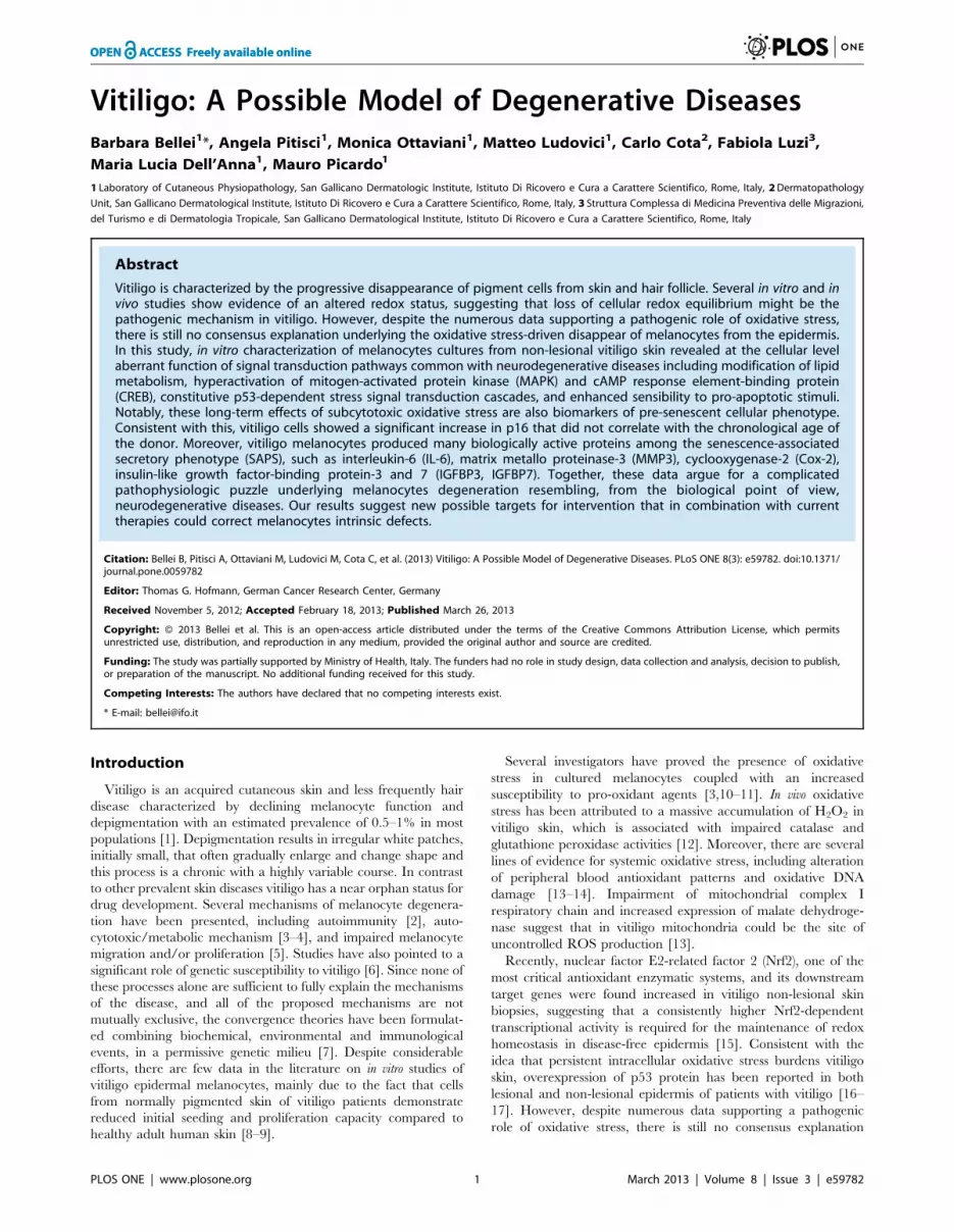
Cellular cholesterol, a marker of oxidative stress-inducedpremature senescence, is increased in vitiligomelanocytes
Increased cellular cholesterol is associated with replicative
senescence in vitro as well as with biological aging in vivo [34]
and may contribute to the onset of degenerative diseases [35]. In
VHM cultures the cholelesterol content was increased (Fig. 6A).
Furthermore, vitiligo melanocytes showed higher amounts of
oxysterols, in particular 7-beta-hydroxycholesterol and 7-ketocho-
lesterol (Fig. 6B). These cholesterol oxidation products have
important physiological/pathophysiological roles, including cho-
lesterol homeostasis and oxysterols-induced cell death that can
take part in degenerative pathologies [36]. Moreover, the
expression of 3-Hydroxy-3-methylglutaryl-coenzyme-A reductase
(HMG-CoAR), the rate-limiting enzyme in cholesterol synthesis,
was found increased in VHM and in NHM following t-BHP
treatment (data not shown). Treatment of NHM with t-BHP
significantly perturbs plasma membrane composition enriching
cholesterol and oxysterols content (Fig. 6C and D).
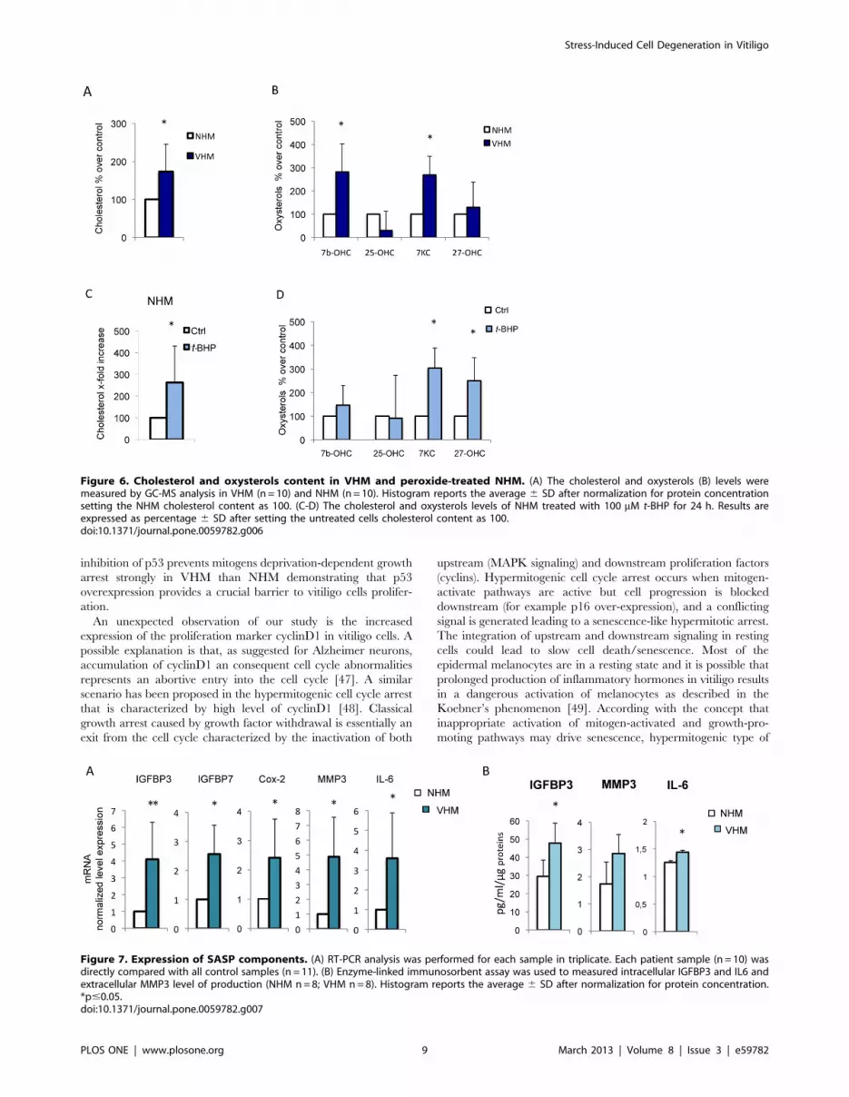
Authocrine and paracrine activity of vitiligo cellsIn vitiligo melanocytes, chronic oxidative stress appears to
activate adaptive program mainly via overexpression of p53 and
the activation of the MAPK signaling cascade. Although these
processes have been studied in several different cell types,
demonstrating a wide association of these markers with decreased
proliferation capacity and senescence, it is also known that
mechanisms of cellular degeneration and senescence greatly differ
between cell types. Permanent damaged human cells secreted
many biologically active proteins, a phenotype termed the
senescence-associated secretory phenotype (SASP) [37], including
cytokines, growth factors and regulators and molecules implicated
in cell adhesion and tissue remodeling. In human melanocytes,
senescence is associated with an increased production of insulin-
like growth factor binding proteins (IGFBP), and treatment with
recombinant exogenous IGFBP7 is sufficient to induce senescence
in melanocytes and apoptosis in certain melanoma cell lines [38].
Interestingly, the expression of IGFBP3 and IGFBP7 were
markedly higher in VHM (Fig. 7A). Notably, IGFBP3 gene
transcription is under the control of p53 [39]. Metalloproteinase-3
(MMP3), a classical marker of senescence in vitro, and IL-6 a
senescence-associated inflammatory mediator, were highly ex-
pressed by VHM at both mRNA (Fig. 7A) and protein level
Figure 3. Analysis of p53 target genes expression. (A) Quantitative RT-PCR analysis was performed for each sample in triplicate. The medianDDCt value, calculated as reported in Fig.1. Each patient sample (n = 11) was directly compared with all control samples (n = 15) and values representthe means 6 SD of fold increase of p53 target genes. (B) The increased expression of GADD45a and PML were also confirmed at protein level bywestern blot and densitometric analysis (VHM = 9; NHM = 9). For PML two different isoforms were detected and quantified separately. (C)Representative analysis of intracellular distribution of PML by immunofluorescence analysis (VHM = 6; NHM = 6). Nuclei were labelled withbisbenzidine (DAPI). Original magnification 63x. *p#0.05.doi:10.1371/journal.pone.0059782.g003
Stress-Induced Cell Degeneration in Vitiligo
PLOS ONE | www.plosone.org 6 March 2013 | Volume 8 | Issue 3 | e59782

(Fig. 7B). Cyclooxygenase-2 (Cox-2), an inducible enzyme of the
prostaglandin biosynthesis pathway, involved in stress-induced
senescence [40] was weakly but significantly upregulated.
Stress and senescent-associated markers validation onvitiligo tissue biopsies.
Vitiligo melanocyte cultures displayed the alteration of several
stress-induced markers including p53-dependent senescent pro-
teins. To confirm in vitro results we analyzed in skin biopsies the
expression of p53, and of two p53 target genes involved in cell
senescence: PML and GADD45 (Fig. 8). P53 localized exclusively
in the nuclear compartment in both healthy and vitiligo samples
with a stronger signal in vitiligo tissue. In healthy sample PML was
weakly detected only in the cytoplasm of upper epidermal layers
whereas in vitiligo biopsies a strong citoplasmic signal was
associated with an intense nuclear positivity of all epidermal
layers. Normal skins were negative for the expression of GADD45
whereas vitiligo samples showed a diffuse citoplasmic staining with
some sporadic cell in the basal layer showing an intense positivity.
Interestingly, our results demonstrated that the activation of p53-
dependent pathway was not restricted to melanocytes but was
generally diffuse in all the epidermis. To confirm that not only
keratinocytes but also melanocytes overexpress senescent-associ-
ated marker we performed double immunofluorescence analysis
confirming the co-expression of both the melanocyte specific
marker tyrosinase and p53 or PML in the same cell (Fig.9).
Discussion
Vitiligo is an acquired disorder of skin pigmentation character-
ized by localized destruction of cutaneous melanocytes that lacks a
satisfactory explanation. Mitochondrial dysfuction, oxidative
stress, and immune mechanisms [1] are among the factors
implicated in melanocytes disappearance in vitiligo. A critical
question is why specific melanocytes are selectively vulnerable in
vitiligo disease. The skin is one of the most impacted organs by
environmental-induced stress and the slow melanocytes turnover
[41], compared to keratinocytes, could contribute to the accumu-
lation of damage in growth-arrested cells in vitiligo skin.
Moreover, the melanin biosynthetic pathway is a potential
additional source of ROS inside the pigment-forming cells. Thus,
it is possible that, in some circumstances, melanogenesis primes an
acute stress response in vitiligo cells, which are more prone to lose
their redox balance equilibrium. Interestingly, increased metabolic
stress and failure to detoxify potential toxin due to the presence of
neuromelanin has been proposed as a pathogenic mechanism in
Parkinson’s disease [42].
Several lines of evidence showed relevant biological similitude
between vitiligo and neurodegenerative diseases. Interestingly due
to common embryonic origin of melanocytes and nervous system
neurons, skin-derived melanocytes were already proposed as a
possible model system to investigate pathological behaviors of less
accessible nervous system neurons [43]. Impaired activity of the
mitochondrial electron transport chain complex I in vitiligo cells
Figure 4. Analysis of cell cycle regulators expression. (A) p16 and (B) CyclinD1 mRNAs (VHM = 11; NHM = 15). (C-D) Immunoflurescenceanalysis of p16 and cyclinD1 expression and localization. Representative images of VHM (right panel) and NHM (left panel) stained with mouse anti-p16 and anti-cyclinD primary antibodies. Nuclei were labelled with bisbenzidine (DAPI). Original magnification 40x. *p#0.05.doi:10.1371/journal.pone.0059782.g004
Stress-Induced Cell Degeneration in Vitiligo
PLOS ONE | www.plosone.org 7 March 2013 | Volume 8 | Issue 3 | e59782

and increase in the expression of mitochondrial malate dehydro-
genase activity argue for an important role of mitochondrial
defective functionality in the pathogenesis of vitiligo [3,13].
Similarly, there are several degenerative disorders with evidence
of mitochondrial involvement. These include Parkinson disease
(where a complex I defect is described and free radicals are
generated from dopamine metabolism [44], amyotrophic later
sclerosis, and Alzheimer disease, where there is evidence to suggest
mitochondrial involvement perhaps secondary to other abnormal-
ities [38].
Here, we have demonstrated that continuous intracellular ROS
generation in the vitiligo melanocytes leads to a constitutive
stimulation of antioxidant enzymes expression at the mRNA level.
Although the mRNA expression of antioxidant enzymes is
increased, the protein levels of catalase and SOD2 remain low,
demonstrating an intrinsically higher detoxifying-enzyme turnover
that is independent of environmental stresses. An increased level of
expression and decreased level of activity of antioxidant enzymes
are often observed during a pathologically prolonged state of
oxidative stress, such as in Alzheimer’s disease [44], and are likely
to reflect the demand to increase expression due to attenuated
enzyme activity that could be directly caused by the oxidative
damage. Our results are consistent with previous studies demon-
strating that higher Nrf2-dependent transcriptional activity is
required for the maintenance of redox balance in vitiligo skin [15]
and that catalase protein expression and activity are low in vitiligo
skin [4]. Intriguingly, lowered catalase protein activity, elevated
production of H2O2 and a refractory stress response have been
reported to correlate with the aged phenotype of hair follicle
melanocytes [45]. ROS escape results in the activation of cytosolic
stress pathways including the upregulation of p53 that represents a
central integration point for various signaling pathways. P53
functions in a stimulus-dependent and cell-type-dependent man-
ner leading to cell cycle arrest, senescence or apoptosis as extreme
outcome. In VHM we observed permanent adaptive changes but
cells still function normally, or slightly suboptimally in a pro-
oxidant environment. In this situation VHM showed a higher level
of expression of senescent-associated p53-target genes such as
PML, GADD45, and IGFBP3 suggesting a pre-senescent pheno-
type. Interestingly, IGFP3 has been also implicated in neuronal
degeneration and clinical symptoms in Alzheimer disease [46].
Moreover, VHM demonstrated a significant increase in p16
expression that did not correlate with donor chronologic age. Over
a certain threshold (in terms of severity of oxidative stress), the loss
of oxidative equilibrium affects cell survival (t-BHP) and cell
proliferation (mitogens starvation). Importantly, pharmacological
Figure 5. Analysis of cell proliferation. Cells (VHM n = 8; NHM n = 8) were incubated with M254 medium plus HMGS (A) or M254 medium plus0.5% FBS (basal medium) (B). Growth medium was replaced with fresh medium every 48 h and cells were left to grow for 72, 96 h or a week beforeTrypan blue exclusion assay. The data show the mean6SD of experiments performed in triplicate. (C) Evaluation of ROS production following 24 h ofgrowth in basal medium. (D) Analysis of mitogen deprivation (24 h) on p16 mRNA in NHM (n = 8) and VHM (n = 8). (E) To investigate the role ofintracellular ROS and the specific role of stress-activated p53 on cell proliferation, starved cells were treated with 5 mM N-acetyl-L-cystein (NAC) or5 mM pifithrin-a (PFT-a). A control sample incubated with M254 plus HMGS was also included. After 5 days the number of viable cells were evacuatedby Trypan blue exclusion assay. Histograms represent –fold difference 6SD versus control (untreated cells at time 0). Experiments were performed intriplicate (NHM n = 6; VHM n = 6). *p#0.05; **p#0.01.doi:10.1371/journal.pone.0059782.g005
Stress-Induced Cell Degeneration in Vitiligo
PLOS ONE | www.plosone.org 8 March 2013 | Volume 8 | Issue 3 | e59782
inhibition of p53 prevents mitogens deprivation-dependent growth
arrest strongly in VHM than NHM demonstrating that p53
overexpression provides a crucial barrier to vitiligo cells prolifer-
ation.
An unexpected observation of our study is the increased
expression of the proliferation marker cyclinD1 in vitiligo cells. A
possible explanation is that, as suggested for Alzheimer neurons,
accumulation of cyclinD1 an consequent cell cycle abnormalities
represents an abortive entry into the cell cycle [47]. A similar
scenario has been proposed in the hypermitogenic cell cycle arrest
that is characterized by high level of cyclinD1 [48]. Classical
growth arrest caused by growth factor withdrawal is essentially an
exit from the cell cycle characterized by the inactivation of both
upstream (MAPK signaling) and downstream proliferation factors
(cyclins). Hypermitogenic cell cycle arrest occurs when mitogen-
activate pathways are active but cell progression is blocked
downstream (for example p16 over-expression), and a conflicting
signal is generated leading to a senescence-like hypermitotic arrest.
The integration of upstream and downstream signaling in resting
cells could lead to slow cell death/senescence. Most of the
epidermal melanocytes are in a resting state and it is possible that
prolonged production of inflammatory hormones in vitiligo results
in a dangerous activation of melanocytes as described in the
Koebner’s phenomenon [49]. According with the concept that
inappropriate activation of mitogen-activated and growth-pro-
moting pathways may drive senescence, hypermitogenic type of
Figure 6. Cholesterol and oxysterols content in VHM and peroxide-treated NHM. (A) The cholesterol and oxysterols (B) levels weremeasured by GC-MS analysis in VHM (n = 10) and NHM (n = 10). Histogram reports the average 6 SD after normalization for protein concentrationsetting the NHM cholesterol content as 100. (C-D) The cholesterol and oxysterols levels of NHM treated with 100 mM t-BHP for 24 h. Results areexpressed as percentage 6 SD after setting the untreated cells cholesterol content as 100.doi:10.1371/journal.pone.0059782.g006
Figure 7. Expression of SASP components. (A) RT-PCR analysis was performed for each sample in triplicate. Each patient sample (n = 10) wasdirectly compared with all control samples (n = 11). (B) Enzyme-linked immunosorbent assay was used to measured intracellular IGFBP3 and IL6 andextracellular MMP3 level of production (NHM n = 8; VHM n = 8). Histogram reports the average 6 SD after normalization for protein concentration.*p#0.05.doi:10.1371/journal.pone.0059782.g007
Stress-Induced Cell Degeneration in Vitiligo
PLOS ONE | www.plosone.org 9 March 2013 | Volume 8 | Issue 3 | e59782

Figure 8. Analysis of stress and senescent-associated markers on vitiligo tissue biopsies. P53 and its senescent-associated target genesPML and GADD45a were analyzed on tissue biopsies (VHM = 4; NHM = 4) by immunohistochemistry.doi:10.1371/journal.pone.0059782.g008
Figure 9. p53 and PML expression co-localize with melanocyte specific markers. Double immunofluoresce staining was used to co-localizethe expression of the melanocyte-specific marker tyrosinase and the expression of p53 and PML. Nuclei were labelled with bisbenzidine (DAPI).Original magnification 406.doi:10.1371/journal.pone.0059782.g009
Stress-Induced Cell Degeneration in Vitiligo
PLOS ONE | www.plosone.org 10 March 2013 | Volume 8 | Issue 3 | e59782
senescence is a form of an altered growth and not necessarily an
absence of cell cycle proliferation [50]. Increased cellular functions
(hypertrophy, pro-inflammatory and hyper-secretory phenotypes),
resembling senescence caused by DNA damaging agents, is the
most important marker of hypermitogenic arrest [51]. From the
medical point of view, these hyper-functions contribute to age-
related diseases, such as atherosclerosis, Alzheimer’s and other
neurodegenerative diseases and macular degeneration [52]. In the
case of vitiligo cells, long-term exposure to subcytotoxic oxidative
stress could cause chronic over-stimulation of MAPK, activation of
p53, and induction of both p16 and cyclinD1 expression at the
same time, resembling pro-senescent hypermitotic phenotype. A
progressive rise of oxidative stress and impaired ability to cope
with stressful stimuli may not be exclusive to epidermal
melanocytes in vitiligo patients. Like melanocytes, peripheral
blood mononuclear cells [13–14], cultured skin keratinocytes [53],
and fibroblasts from vitiligo patients show markers of oxidative
stress and increases in cholesterol content (unpublished data).
Interestingly, similar systemic alterations of cholesterol homeosta-
sis have also been reported in Alzheimer’s disease [19,54].
According with the non-exclusive involvement of melanocytes, a
previous study reported a shorter in vitro life span and senescence
markers in keratinocytes cultured from involved skin in vitiligo
patients [55]. In this study immunohistochemical analysis of skin
biopsies also showed that the activation of p53-dependent
signaling is not restricted to melanocytes but also involve
keratinocytes. The acquisition of senescence-associated secretory
phenotypes, combing the effect of intracellular signaling pathway
deregulation with extracellular environments perturbation, could
affect the behavior of neighboring cells and also systemic
homeostasis since melanocytes could release into circulation
melanocyte signaling molecules and neurotransmitter-like factors
[56]. The senescent-prone phenotype could also provide a
molecular explanation to the observation that vitiligo is associated
with fewer risks of skin cancer [57–58] since premalignant human
nevi contain cells that express senescence markers [59].
Histological data have provided good evidence of immune-
inflammatory components in the upper dermis and the dermal-
epidermal interface of vitiligo lesional and peri-lesional skin [7].
Intriguingly, repeated stimulations of fibroblasts with tumor
necrosis factor-alpha (TNF-a) or IL-a, two pro-inflammatory
cytokines that are higher expressed in lesional skin of vitiligo
patients [38,60], stimulate the appearance of biomarkers of in vitro
aging in a stress-dependent manner [61]. This example of long-
term effects of cytokines on human fibroblasts, and potentially on
other cell types, suggests that paracrine-acting messengers derived
from skin immune system and keratinocytes may be a focus
responsible for the start of melanocytes premature senescence and
vitiligo pathological process.
The senescent-prone melanocytes not only explain the disap-
pearance of functional melanocytes in lesional epidermis but also
the loss of normal regeneration that could be caused by depletion
of stem or progenitor cells. Interestingly, hyperactivation of p53
have been implicated in age-associated decline in tissue stem cell
regenerative function [62] and degenerative disorders [63]. This
depletion will compromise tissue repair, regeneration and normal
turnover, leading to functional decrements. Moreover, the in vitro
melanocytes cell culture model could represent a readily accessible
human cell model for the development of new therapy strategies
capable of inhibiting degeneration in the vitiligo cells and other
degenerative diseases.
Supporting Information
Table S1 List of primers used for quantitative real timePCR. Sequences of primers indicated with an F correspond to
sense strands and with an R correspond to anti-sense.
(TIF)
Author Contributions
Conceived and designed the experiments: BB MP. Performed the
experiments: BB AP MO ML MLD FL CC. Analyzed the data: BB MP
MO. Wrote the paper: BB MP.
References
1. Taieb A, Picardo M (2009) Clinical practice: Vitiligo. N Engl J Med 360: 160-
169.
2. Le Poole IC, Luiten RM (2008). Autoimmune etiology of generalized vitiligo.
Curr Dir Autoimmun 2008; 10: 227-243.
3. Dell’Anna ML, Ottaviani M, Albanesi V, Vidolin AP, Leone G, et al. (2007)
Membrane lipid alterations as a possible basis for melanocyte dagenaration in
vitiligo. J Invest Dermatol 127: 1226-1233.
4. Schallreuter KU, Rubsam K, Gibbons NC, Maitland DJ, Chavan B, et al.
(2008) Methionine sulfoxide reductases A and B deactivated by hydrogen
peroxide (H2O2) in the epidermis of patients with vitiligo. J Invest Dermatol
128: 808-815.
5. Gauthier Y, Cario Andre M, Taeb A (2003) A critical appraisal of vitiligo
etiologic theories. Is melanocyte loss a melanocytorrhagy? Pigment Cell Res 16:322-332.
6. Spritz RA (2008) The genetics of generalized vitiligo. Curr Dir Autoimmun 10:
244-257.
7. Taieb A (2011) Vitiligo as an inflammatory skin disorder: a therapeutic
perspective. Pigment Cell Mel Res 25: 9-13.
8. Medrano EE, Nordlund JJ (1990) Successful culture of adult human melanocytes
obtained from normal and vitiligo donors. J Invest Dermatol 95: 441-445.
9. Dell’Anna ML, Cario-Andre M, Bellei B, Taieb A, Picardo M (2012) In vitro
research on vitiligo: strategies, principles, methodological options, and common
pitfalls. Exp Dermatol 21: 490-496.
10. Maresca V, Roccella M, Rocella F, Camera E, Del Porto G, et al. (1997)
Increased sensivity to peroxidative agents as a possible pathogenic factor of
melanocytes damage in vitiligo. J Invest Dermatol 109: 310-313.
11. Boissy RE, Manga P (2004) On the etiology of contact/occupational vitiligo.
Pigment Cell Res 17: 208-214.
12. Schallreuter KU, Moore J, Wood JM, Beazley WD, Gaze DC, et al. (1999) In
vivo and in vitro evidence for hydrogen peroxide (H2O2) accumulation in the
epidermis of patients with vitiligo and its successful removal by a UVB-activated
pseudocatalase. J Invetsig Dermatol Symp Proc 4: 91-96.
13. Dell’Anna ML, Urbanelli S, Mastrofrancesco A, Camera E, Iacovelli P, et al.
(2003) Alterations of mitochondria in peripheral blood monoculear cells of
vitiligo patients. Pigment Cell Res 16: 553-559.
14. Giovannelli L, Bellandi S, Pitozzi V, Fabbri P, Dolara P, et al. (2004) Increased
oxidative DNA damage in mononuclear leukocytes in vitiligo. Mutat Res 556:
101-106.
15. Natarajan VT, Singh A, Kumar AA, Sharma P, Kar HK, et al. (2010)
Transcriptional upregulation of Nrf2-dependent phase II detoxification genes in
the involved epidermis of vitiligo vulgaris. J Invest Dermatol 130: 2781-2789.
16. Schallreuter KU, Behrens-Williams S, Khalig TP, Picksley SM, Peters EM, et al.
(2003) Increased epidermal functioning wild-type p53 expression in vitiligo. Exp
Dermitol 12: 268-277.
17. Salem MM, Shalbaf M, Gibbons NC, Chavan B, Thornton JM, et al. (2009)
Enhanced DNA binding capacity on up-regulated epidermal wild-type p53 in
vitiligo by H2O2-mediated oxidation: a possible repair mechanism for DNA
damage. FASEB J 23: 3790-380.
18. Schallreuter KU, Bahadoran P, Picardo M (2008) Vitiligo pathogenesis:
autoimmune disease, genetic defect, excessive reactive oxygen species, calcium
imbalance, or what else? Exp Dermatol 17: 139-140.
19. Jimbow K, Roth SI, Fitzpatrick TB, Szabo G (1975) Mitotic activity in non-
neoplastic melanocytes in vivo as determined by histochemical, autoradiograph-
ic, and electron microscope studies. J Cell Biol 66,663-670.
20. Pani A, Dessi S, Diaz G, La Colla P, Abete C, et al. (2009) Altered cholesterol
ester cycle in skin fibroblasts from patients with Alzheimer’s disease. J Alzheimers
Dis18, 829- 841 .
21. Schapira AH, Cooper JM, Dexter D, Clark JB, Jenner B, et al. (1990)
Mitochondrial complex I deficiency in Parkinson’s disease. J Neurochem. 54,
823- 827.
22. Mendonsa G, Dobrowolska J, Lin A (2009) Molecular profiling reveals diversity
of stress signal transduction cascades in highly penetrant Alzheimer’s disease
human skin fibroblasts. PLoS One 4:e4655.
Stress-Induced Cell Degeneration in Vitiligo
PLOS ONE | www.plosone.org 11 March 2013 | Volume 8 | Issue 3 | e59782

23. Lee HC, Wei YH (2001) Mitochondrial alterations, cellular response to oxidative
stress and defective degradation of proteins in aging. Biogerentology 2: 231-244.24. Chen J, Goligorsky MS (2006) Premature senescence of endothelial cells:
Methusaleh’s dilemma. Am J Physiol Heart Circ Physiol 290: 1729-1739.
25. Esiri MM (2007) Ageing and the brain. J Pathol 211: 181-187.26. Tobin DJ, Swanson NN, Pittelkow MR, Peters EM, Schallreuter KU (2000)
Melanocytes are not absent in lesional skin of long duration vitiligo. J Pathol191: 407-416.
27. Eising R, Suselbeck B (2003) Turnover of catalase heme and apoprotein moieties
in cotyledons of sunflowers seedings. Plant Physiol 97: 1422-1429.28. Bedogni B, Pani G, Colavitti R, Riccio A, Borrello S, et al. (2003) Redox
regulation of cAMP-responsive element–binding protein and induction ofmanganous superoxide dismutase in nerve growth factor-dependent cell survial.
J Biol Chem 278: 16510-16519.29. Kronke G, Bochkov VN, Huber J, Gruber F, Bluml, et al. (2003) Oxidized
phospholipids induce expression of human heme oxygenase-1 involving
activation of cAMP-responsive element-binding protein. J Biol Chem 278:51006-51014.
30. Munoz L, Ammit AJ (2010) Targeting p38 MAPK pathway for the treatment ofAlzheimer’s disease. Neuropharmacology 58: 561-568.
31. Muller M, Cardenas C, Mei L, Cheung KH, Foskett JK (2011) Costitutive camp
response element binding protein (CREB) activation by Alzheimer’s diseasepresenilin-driven inositol trisphosphate receptor (InsP3R) Ca2+ signaling. Proc
Natl Acad Sci USA 108:13293-13298.32. Jensen K, Shiels C, Freemont PS (2001) PML protein isoforms and RBCC/
TRIM motif. Oncogene 20: 7223-7233.33. Condemine W, Takahashi Y, Le Bras M, de The H (2007) A nucleolar targeting
signal in PML-I addresses PML to nucleolar caps in stressed or senescent cells.
J Cell Sci 120: 3219-3927.34. Nakamura M, Kondo H, Shimada Y, Waheed AA, Ohno-Iwashita Y (2003)
Cellular aging-dependent decrease in cholesterol in membrane microdomains ofhuman diploid fibroblasts. Exp Cell Res 290: 381-390.
35. Ohno-Iwashita Y, Shimada Y, Hayashi M, Inomata M (2010) Plasma
membrane microdomains in aging and disease. Getriatr Gerontol Int 10: 41-52.36. Gamba P, Leonarduzzi G, Tamagno E, Guglielmotto M, Testa G, et al. (2011)
Interaction between 24-hydroxycholesterol, oxidative stress, and amyloid-b inamplifying neuronal damage in Alzheimer’s disease: three partners in crime.
Aging Cell 10: 403-417.37. Coppe JP, Patil F, Rodier F, Sun Y, Munoz DP, et al. (2008) Senecence-
associated secretory phenotypes reveal cell-nonautonomous functions of
oncogenic RAS and the p53 tumor suppressor. PLoS Biol 6: 2853-2868.38. Wajapeyee N, Serra RW, Zhu X, Mahalingam M, Green MR (2008) Oncogenic
BRAF induces senescence and apoptosis through pathways mediated by thesecreted protein IGFBP7. Cell 132: 363-374.
39. Kim KS, Kim MS, Seu YB, Chung HY, Kim JH, et al. (2007) Regulation of
replicative senescence by insulin-like growth factor-binding protein 3 in humanumbilical vein endothelial cells. Aging Cell 6: 535-545.
40. Zdanov S, Bernard D, Debacq-Chainiaux F, Martien S, Gosselin K, et al. (2007)Normal or stress-induced fibroblast senescence involves COX-2 activity. Exp
Cell Res 313: 3046-3056.41. Yaar M, Gilchrest 1BA (2001) Ageing and photoageing of keratinocytes and
melanocytes. Clin Exp Dermatol 26: 583-591.
42. Lang AE, Lozano AM (1998) Parkinson’s disease. First of two parts. N Engl J339: 1130-1143.
43. Yaar M, Park HY (2011) Melanocytes a window into the nervous system. J InvestDermatol 132: 835-845.
44. Akterin S, Cowburn RF, Miranda-Vizuete A, Jimenez A, Bogdanovic N, et al.(2006) Involvement of glutaredoxin-1 and thioredoxin-1 in beta-amyloid toxicity
and Alzheimer’s disease. Cell Death Differ 13: 1454-1465.
45. Kauser S, Westgate GE, Green MR, Tobin DJ (2011) Human hair follicle andepidermal melanocytes exhibit striking differences in their aging profile which
involves catalase. J Invest Dermatol 131: 979-982.
46. Ikonen M, Liu B, Hashimoto Y (2003) Interaction between the Alzheimer’ssurvival peptide humanin and insulin-like growth factor-binding protein 3
regulates cell survival and apoptosis. Proc Natl Acad Sci USA 100: 13042-
13047.
47. Malik B, Currais A, Andres A (2008) Loss of neuronal cell cycle control as a
mechanism of neurodegeneration in the presenilin-1 Alzheimer’s disease brain.
Cell Cycle 7: 637-646.
48. Blagoskolonny MV (2003) Cell senescence and hypermitogenic arrest. EMBO
Rep 4: 358-362.
49. Gauthier Y, Cario-Andre M, Lepreux S (2003) Melanocyte detachment afterskin friction in non lesional skin of patients with generalized vitiligo.
Br J Dermatol 148: 95-101.
50. Blagosklonny MV (2009) Aging-suppressants: cellular senescence (hyperactiva-tion) and its pharmacologic deceleration. Cell Cycle 8: 1883-1887.
51. Blagosklonny MV (2011) Cell cycle arrest is not senescence. Aging 3: 94-101.
52. Blagosklonny MV (2006) Cell senescence: hypertrophic arrest beyond therestriction point. J Cell Physiol 209: 592-597.
53. Kostyuk VA, Potapovich AI, Cesareo E, Brescia S, Guerra L, et al. (2010).
Dysfunction of glutathione S- transferase leads to excess 4-hydroxy-2-nonenaland H(2)O(2) and impaired cytokine pattern in cultured keratinocytes and blood
of vitiligo patients. Antioxid Redox Signal 13: 607-620.
54. Pani A, Mandas A, Diaz G, Abete C, Cocco PL, et al. (2009) Accumulation ofneutral lipids in peripheral blood mononuclear cells as a distintive trait of
Alzheimer patients and asymptomatic subjects at risk of disease. BMC Med7,66.
55. Bondanza S, Maurelli R, Paterna P, Migliore E, Giacomo FD, et al. (2007)Keratinocyte cultures from involved skin in vitiligo patients show an impaired in
vitro behaviour. Pigment Cell Res 20: 288-300.
56. Slomonski A (2009) Neuroendocrine activity of the melanocyte. Exp Dermatol18: 760-763.
57. Schallreuter KU, Tobin DJ, Panske A (2002) Decreased photodamage and low
incidence of non-melanoma skin cancer in 136 sun exposed Caucasian patientswith vitiligo. Dermatol 204: 194-201.
58. Feily A, Pazyar N (2011) Why vitiligo is associated with fewer risk of skincancer?: providing a molecular mechanism. Arch Dermatol Res 303: 623-624.
59. Michaloglou C, Vredeveld LC, Soengas MS, Denoyelle C, Kuilman T, et al.
(2005) BRAF600-associated senescence-like cell cycle arrest of human naevi.Nature 4: 720-724.
60. Birol A, Kisa U, Kurtipek GS, Kara F, Kocak M, et al. (2006) Increased tumor
necrosis factor alpha (TNF-alpha) and interleukin 1 alpha (IL1-alpha) levels inthe lesional skin of patients with nonsegmental vitiligo. Int J Dermatol 45: 992-
993.
61. Dumont P, Balbeur L, Remacle J, Dierick JF, Pascal T, et al. (2000) Appearanceof biomarkers of in vitro ageing after successive stimulation of WI-38 fibroblasts
with IL-1alpha and TNF-alpha: senescence associated beta-galactosidase activity
and morphotype transition. J Anat 197: 529-537.
62. Gatza C, Moore L, Dumble M, Donehower LA (2007) Tumor suppressor
dosage regulates stem cell dynamics during aging. Cell Cycle 6: 52-55.
63. Ruzankina Y, Asare A, Brown EJ (2008) Replicative stress, stem cells and aging.Mech Ageing Dev 129: 1313-1319.
Stress-Induced Cell Degeneration in Vitiligo
PLOS ONE | www.plosone.org 12 March 2013 | Volume 8 | Issue 3 | e59782


