Embed Size (px)
Citation preview

This article appeared in a journal published by Elsevier. The attachedcopy is furnished to the author for internal non-commercial researchand education use, including for instruction at the authors institution
and sharing with colleagues.
Other uses, including reproduction and distribution, or selling orlicensing copies, or posting to personal, institutional or third party
websites are prohibited.
In most cases authors are permitted to post their version of thearticle (e.g. in Word or Tex form) to their personal website orinstitutional repository. Authors requiring further information
regarding Elsevier’s archiving and manuscript policies areencouraged to visit:
http://www.elsevier.com/copyright

Author's personal copy
DegenerativeLumbosacral Stenosisin Dogs
Bjorn P. Meij, DVM, PhDa,*, Niklas Bergknut, DVM, MSa,b
Degenerative lumbosacral stenosis (DLSS) is a common disorder seen mainly in largebreed dogs.1–4 DLSS has been attributed many names over the past 40 years: caudaequina syndrome, cauda equina compression, lumbosacral stenosis or disease,lumbosacral instability, and DLSS.2,4–8 All these terms refer to a degenerative disorderthat is of multifactorial origin in which intervertebral disc (IVD) degeneration playsa major role. IVD degeneration, and bony and soft-tissue proliferations contribute tospinal stenosis and cauda equina compression leading to pain, lameness, and neuro-logic signs. This article describes current knowledge on the pathogenesis of IVD degen-eration and recent advances in the management of DLSS. The biomechanics,pathogenesis, clinical manifestation, and treatments of lumbar disc herniation arevery similar between dogs and humans. Hence many studies in humans with lumbardisc herniation can contribute to a better understanding of DLSS in dogs, and dogsmay in turn be used as a spontaneous animal model for lumbar disc herniation inhumans.
CLINICAL ANATOMYDevelopment
During embryonic development, the spinal cord (derived from the ectoderm) and thevertebral column (derived from the mesoderm) grow at a different rate, leading in thenewborn animal to the spinal column being longer than the spinal cord. In most dogsthe conus medullaris (end point of the spinal cord) is located in the caudal half of L6
There are no conflicts of interest to report and no external funding has been received.a Department of Clinical Sciences of Companion Animals, Faculty of Veterinary Medicine,Utrecht University, PO Box 80.154, Yalelaan 108, NL-3508 TD, Utrecht, The Netherlandsb Division of Small Animals, Department of Clinical Sciences, Faculty of Veterinary Medicineand Animal Sciences, Swedish University of Agricultural Sciences, Box 7054, Ulls vag 12, 75007 Uppsala, Sweden* Corresponding author.E-mail address: [email protected]
KEYWORDS
� Degenerative lumbosacral stenosis � Cauda equina syndrome� Lumbosacral disease � Lumbosacral instability� Intervertebral disc degeneration � Dogs
Vet Clin Small Anim 40 (2010) 983–1009doi:10.1016/j.cvsm.2010.05.006 vetsmall.theclinics.com0195-5616/10/$ – see front matter ª 2010 Elsevier Inc. All rights reserved.

Author's personal copy
and the cranial part of L7 (Fig. 1).9 In smaller breed dogs this is further caudally.9 Thedural sac extends into the sacrum in more than 80% of the dogs10 but varies consid-erably in its caudal extension (Fig. 2).
The cauda equina originates from the conus medullaris and is composed of thespinal nerves L6, L7, S1-S3, and Cd1-Cd5 (see Fig. 1; Table 1) and stretches fromvertebra L6 to Cd5. The structures surrounding the cauda equina are (1) ventral: thedorsal longitudinal ligament, the L7-S1 IVD, and neighboring vertebral bodies; (2)lateral: the pedicles of L7 and S1 and intervertebral foramina; and (3) dorsal: the inter-arcuate ligament and the lamina of L7 and S1.
The IVD and 2 synovial facet joints connect L7 and S1, which is further stabilized bythe ventral and dorsal longitudinal ligaments, the interarcuate ligament, the interspi-nous ligament, and the surrounding fascia and muscular tissues.
The Intervertebral Disc
The lumbosacral IVD has a triangular appearance on a lateral view, being thicker onthe ventral side than on the dorsal side, and is entirely dependent on osmosis and
Fig. 1. Ventrodorsal radiograph of the lumbosacral area of a dog. The overlay outlines theapproximate location of the spinal segments, the caudal extent of the spinal cord, and theorigin of the spinal nerves comprising the cauda equina. Each individual spinal segment(lumbar 4–7 and sacral 1–3) and its corresponding spinal nerve have been marked withthe same number and color. Co, coccygeal spinal segment and nerve; Fe, femoral nerve;Gl, gluteal nerve; LST, lumbosacral trunc; Ob, obturator nerve; Pe/Sa, pelvic and sacralnerves; Pu, pudendal nerve; Sc, sciatic nerve. (Modified from Wheeler SJ. Lumbosacraldisease. Vet Clin North Am Small Anim Pract 1992;22:937–50; with permission.)
Meij & Bergknut984

Author's personal copy
diffusion for nutrients to reach its cells.11,12 The IVD is made up of 3 different parts(Figs. 3 and 4):
1. The annulus fibrosus (AF) provides tensile strength and connects the 2 adjoiningvertebrae rigidly together. The AF is comprised of well-organized lamellar layersof type I collagen.
2. The nucleus pulposus (NP) evenly distributes the load over the vertebral end platesand acts as a shock absorber when the spine is subjected to compressive forces.The gelatinous NP contains 80% to 88% water.13–15 The remaining 18% to 20% ismade up of disorganized type II collagen and negatively charged proteoglycans at-tracting water and nutrients into the NP through osmosis.16,17 Therefore, positively
Fig. 2. Lateral radiograph of the lumbosacral spine of a dog after myelography, illustratingthe extension of the dural sac (arrow).
Table 1Peripheral nerves with clinical significance originating from the cauda equina: normalfunction and dysfunction in dogs with degenerative lumbosacral stenosis (DLSS)
Nerve Segment Reflex FunctionNeurologic Findingsin DLSS
N. femoralis(Femoral nerve)
L4–L6 Patellar Flexion hipExtension stifle
Normal or pseudo-hyperreflexia
N. ischiadicus(Sciatic nerve)
L6–S1 Cranial tibialGastrocnemiusWithdrawal
Extension hipFlexion stifleFlexion and
extension oftarsus
Proprioception
Muscle atrophyNormal or decreased
reflexesNormal or decreased
consciousproprioception
N. pelvicus andsacrales (Pelvicand sacral nerves)
S1–S3 Urinary bladder None or urinaryincontinence
N. pudendus(Pudendal nerve)
S1–S3 PerinealAnal
Anal and urinarybladder sphincters
None or decreasedperineal reflex
None or urinary orfecal incontinence
N. caudales (Tailnerves)
Cd1–Cd5 Tail tone Normal orhypotonia
Degenerative Lumbosacral Stenosis in Dogs 985

Author's personal copy
charged antibiotics such as aminoglycosides (eg, gentamicin) penetrate moreeasily into the NP than negatively charged ones such as cefuroxime or penicillin.18
3. The cartilaginous end plate is a thin structure of hyaline cartilage covering the verte-bral end plates. It functions as a semipermeable membrane so that nutrients canenter the disc by diffusion or osmosis.12,19
PATHOPHYSIOLOGYGeneral
DLSS is a multifactorial degenerative disorder resulting in stenosis of the spinal canaland compression of the cauda equina or its blood supply. Several pathologiescontribute to DLSS such as:
� Hansen type II (or less commonly type I) IVD herniation.2,3 IVD herniation ispreceded by degeneration of the disc20,21
� Ventral subluxation of S1 (lumbosacral instability),20,22–25 and misalignment ofthe facet joints26–28
� Congenital vertebral anomalies such as symmetric or asymmetric transitional orextra vertebrae29–34
� Proliferation of the soft tissues surrounding the cauda equina such as hyper-trophy of the interarcuate ligament, the joint capsule, and epidural fibrosis35
� Sacral osteochondrosis36–38
� Vascular compromise of the blood supply to the spinal nerves.8,35,39
Pathogenesis
The following pathogenesis is proposed for DLSS in dogs:
1. An abnormal motion pattern of the lumbosacral junction (commonly caused byrepetitive stress, genetic or congenital abnormalities) predisposes for degenerationof the L7-S1 disc.20,22–25
2. IVD degeneration is initiated by degradation of the proteoglycans; subsequentlyless nutrients and water are being drawn into the disc leading to dehydration,further degeneration, and loss of disc width.40,41
3. The load bearing of the unstable spinal segment is then shifted from the central axisof the IVD to the peripheral parts of the spine (facet joints and ventral aspect of thevertebral bodies). Due to the angulation of the facet joints, there can also be
Fig. 3. Transverse view of a healthy canine intervertebral disc showing a clear demarcationbetween the lamellar structure of the annulus fibrosus and the gelatinous nucleus pulposus.
Meij & Bergknut986
Author's personal copy
a ventral subluxation of the sacrum at this point causing dynamic impingement ofthe cauda equina.
4. To compensate for the increasing instability, there is a proliferation of thesurrounding soft-tissue structures causing hypertrophy of the interarcuate liga-ment, epidural fibrosis, and thickening of the capsules of the articular processes.
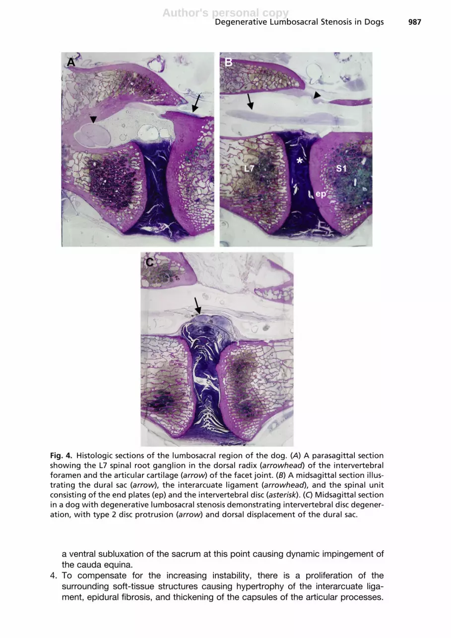
Fig. 4. Histologic sections of the lumbosacral region of the dog. (A) A parasagittal sectionshowing the L7 spinal root ganglion in the dorsal radix (arrowhead) of the intervertebralforamen and the articular cartilage (arrow) of the facet joint. (B) A midsagittal section illus-trating the dural sac (arrow), the interarcuate ligament (arrowhead), and the spinal unitconsisting of the end plates (ep) and the intervertebral disc (asterisk). (C) Midsagittal sectionin a dog with degenerative lumbosacral stenosis demonstrating intervertebral disc degener-ation, with type 2 disc protrusion (arrow) and dorsal displacement of the dural sac.
Degenerative Lumbosacral Stenosis in Dogs 987

Author's personal copy
To compensate for the loss of load-bearing properties, the cartilaginous end platesthicken and bony proliferations develop, such as osteophytes and ventral spondy-losis. This process further impairs the nutritional supply to the disc, triggeringa negative spiral leading to structural failure of the disc.16,42
5. Narrowing of the IVD width and loss of AF compliance to compressive forces leadto bulging of the annulus fibrosus and type II disc herniation.20,43
6. Cell-mediated inflammatory responses lead to ingrowth of blood vessels andnerves into the damaged disc, which contributes to the lumbosacral pain.20,44
Biomechanics of the Lumbosacral Area
Axial compressive forces in humans are similar to those in dogs.45,46 The IVD is themost important structure for maintaining stability between adjoining vertebrae.20,23,25
The healthy L7-S1 segment has a greater mobility in flexion-extension (FE) than theother lumbar segments in dogs.47–49 The most prominent motion direction is FE,although lateral bending and torsion are also possible.49,50 Torsional stiffness issmaller in the LS segment than in the other caudal lumbar segments.51 The angulationof the facet joints is reported to have a significant effect on the mobility of the lumbo-sacral junction.28 Dogs with DLSS have been shown to have a reduced range ofmotion in FE of the lumbosacral segment compared with healthy dogs.52 A kinematicstudy of the spinal movement of healthy malinois dogs found significant differences inthe motion patterns between dogs with normal lumbosacral junctions compared withdogs with radiographic changes of degeneration or transitional vertebrae in thelumbosacral region.53 Dorsal laminectomy with concomitant facetectomy has beendemonstrated to increase instability in healthy cadaveric lumbosacral spines54
whereas dorsal laminectomy and nucleotomy alone did not increase instability inflexion and extension.55 However, a later, extended, and more detailed study by thesame investigators showed that stability was negatively affected in all 3 motiondirections in the healthy lumbosacral spine after dorsal laminectomy andnucleotomy.56
EPIDEMIOLOGY
Several studies have reported on the common occurrence of DLSS in large breeddogs, with a predisposition for the German Shepherd dog and working dogs.1–3,57
The average age at presentation is 7 years, and DLSS is more common in malesthan females.1–3 It is not directly possible to relate case-based measures (proportionalmeasures) from larger referral hospitals to the occurrence in the base population.However, basing the estimations on a canine population insured for veterinary carein Sweden (in general dogs %12 years of age), the incidence in the Swedish dog pop-ulation was further explored. Data of 200,000 dogs from the insurance company Agria(Agria pet insurance, Stockholm, Sweden) were used spanning a 12-year period(1995–2006), previously reported as representative for the whole Swedish dog popu-lation of more than 800,000 dogs.58–60 Incidence rates were calculated based on dogyears at risk (Table 2). The main differences from previous studies is that the occur-rence of DLSS increases with age and is not most common around the age of 7, aspreviously stated. The most likely reason for this discrepancy is that older dogs diag-nosed with DLSS are more seldom referred for surgery. It must be emphasized that theepidemiologic data of the Swedish population may not apply to canine populationsfrom other countries.
Meij & Bergknut988

Author's personal copy
CLINICAL SIGNSHistory, Clinical Signs, and Clinical Examination
The majority of dogs with DLSS are presented with a history of caudal lumbar orlumbosacral pain. Owners may report pelvic limb lameness, hyperesthesia or self-mutilation of the lumbosacral area or pelvic limbs, difficulty with rising, sitting, or lyingdown, reluctance to jump or climb, dragging of toes, a low carriage of the tail, andurinary or fecal incontinence.1–3,61 Working dogs may refuse to perform certain exer-cises like jumping. In companion animals the clinical signs may only be evident afterheavy exercise or play, after which the dog may show muscle tremors or pacing beforelying down. In these situations it may help to ask the owner to record a home video.
The clinical signs of DLSS in dogs have been well documented.61–66 In general, find-ings during clinical examination are related to the compression of the cauda equina,for example, lumbosacral pain, hyperesthesia of the lumbosacral region, difficultyrising, jumping, and/or getting into the car, unilateral or bilateral pelvic limb lameness,and posterior paresis (Fig. 5). The most consistent finding during clinical examinationin dogs with DLSS is pain evoked by pressure applied over the lumbosacral region.61
Specific tests in the standing or recumbent animal can be performed to evoke this painresponse. Hyperextension of the caudal lumbar spine with lumbosacral pressure(lordosis test), tail hyperextension, and the lumbosacral pressure test may evoke resis-tance or a pain response. An experienced clinician is able to differentiate between painevoked by hyperextension of the hip joints and pain on hyperextension of the lumbo-sacral region. Lumbosacral pain can further be examined by individual hyperextensionof each pelvic limb and simultaneous lumbosacral pressure to confirm lateralization(left or right) of the lumbosacral pain.
In some dogs with DLSS, nonweight-bearing pelvic limb lameness is the mostevident clinical sign (see Fig. 5). This unilateral lameness may be the result of unilateral
Table 2Breed-specific incidence rates for the 15 most common dog breeds presented with DLSS per10,000 dog years at risk (DAR) in the Swedish canine insurance population (data from 1995 to2006)
Dog BreedIncidence Rate (No. of Cases/10,000 DAR)
German Shepherd dog 33.7
Boxer 21.7
Rottweiler 18.0
Doberman Pinscher 17.5
Briard 17.4
Rhodesian Ridgeback 15.7
Bernese Mountain dog 14.9
Dalmatian dog 13.6
Hovawart 10.5
Greyhound 10.0
Riesenschnauzer 9.1
Great Dane 8.9
Labrador Retriever 8.8
Nova Scotia Duck Tolling Retriever 8.2
Standard Poodle 7.8
Degenerative Lumbosacral Stenosis in Dogs 989
Author's personal copy
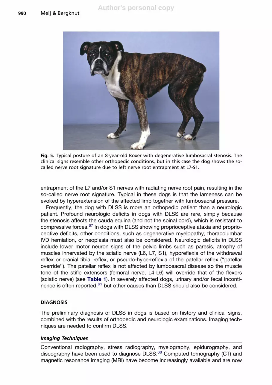
entrapment of the L7 and/or S1 nerves with radiating nerve root pain, resulting in theso-called nerve root signature. Typical in these dogs is that the lameness can beevoked by hyperextension of the affected limb together with lumbosacral pressure.
Frequently, the dog with DLSS is more an orthopedic patient than a neurologicpatient. Profound neurologic deficits in dogs with DLSS are rare, simply becausethe stenosis affects the cauda equina (and not the spinal cord), which is resistant tocompressive forces.67 In dogs with DLSS showing proprioceptive ataxia and proprio-ceptive deficits, other conditions, such as degenerative myelopathy, thoracolumbarIVD herniation, or neoplasia must also be considered. Neurologic deficits in DLSSinclude lower motor neuron signs of the pelvic limbs such as paresis, atrophy ofmuscles innervated by the sciatic nerve (L6, L7, S1), hyporeflexia of the withdrawalreflex or cranial tibial reflex, or pseudo-hyperreflexia of the patellar reflex (‘‘patellaroverride’’). The patellar reflex is not affected by lumbosacral disease so the muscletone of the stifle extensors (femoral nerve, L4-L6) will override that of the flexors(sciatic nerve) (see Table 1). In severely affected dogs, urinary and/or fecal inconti-nence is often reported,61 but other causes than DLSS should also be considered.
DIAGNOSIS
The preliminary diagnosis of DLSS in dogs is based on history and clinical signs,combined with the results of orthopedic and neurologic examinations. Imaging tech-niques are needed to confirm DLSS.
Imaging Techniques
Conventional radiography, stress radiography, myelography, epidurography, anddiscography have been used to diagnose DLSS.68 Computed tomography (CT) andmagnetic resonance imaging (MRI) have become increasingly available and are now
Fig. 5. Typical posture of an 8-year-old Boxer with degenerative lumbosacral stenosis. Theclinical signs resemble other orthopedic conditions, but in this case the dog shows the so-called nerve root signature due to left nerve root entrapment at L7-S1.
Meij & Bergknut990

Author's personal copy
standard diagnostic tools for DLSS,68–70 eliminating the need for standard radio-graphic studies. Moreover, normal radiographs do not exclude DLSS.71,72 However,because many veterinarians rely on routine radiographic techniques for the diagnosisof DLSS, the most important aspects are reported here.
Conventional radiographyThe lateral radiographic view is the most informative for DLSS.69,73–75 Common findingsin dogs with DLSS include collapse of the IVD space, sclerosis of the vertebral endplates, elongation of the sacral lamina (‘‘telescoping’’) in the caudal aperture of L7,lumbosacral step formation with ventral subluxation of S1, the vacuum phenomenon,and ventral spondylosis (Figs. 6 and 7).2 Sacral osteochondrosis, symmetric or asym-metric transitional vertebrae, an additional eighth lumbar vertebra, or congenital sacralanomalies may also play a role in DLSS.29–33,36–38 Survey radiographs help to excludeneoplasia with bone involvement (eg, metastatic disease from prostate carcinoma),traumatic luxation, and discospondylitis. Stress radiography of the lumbosacral region,such as dynamic flexion/extension studies, may enhance the lumbosacral stepformation.76
MyelographyThe usefulness of myelography in DLSS is debated because it depends on the exten-sion of the dural sac over the lumbosacral junction. In large dogs the spinal cord endsat L6 and the dural sac extends further caudally (see Fig. 2). In myelography, nonioniccontrast medium is injected into the subarachnoid space at the cerebellomedullary
Fig. 6. Lateral radiograph of the lumbosacral region. (A) Normal dog. (B) Dog with degen-erative lumbosacral stenosis and a transitional vertebra (asterisk), telescoping of the laminaof S1 into the caudal aperture of L7 (arrow), and vacuum phenomenon between L7 and S1(arrowhead).
Degenerative Lumbosacral Stenosis in Dogs 991

Author's personal copy
cistern or at a caudal lumbar site (L5-L6),69,77 so only the dural sac is visualized at thelumbosacral level. Myelography has been reported to be successful in the diagnosis ofDLSS.10 However, lumbosacral stenosis may be present although no abnormalitiesare seen on myelography.78 Myelography is more sensitive than routine radiography,and its sensitivity can be increased by dynamic flexion/extension studies.79,80
EpidurographyIn epidurography, the contrast medium is injected into the epidural space at thelumbosacral or sacrococcygeal junction. Epidurography is technically easier than
Fig. 7. Lateral radiograph (A) and sagittal CT reconstruction (B) of the lumbosacral region ina dog with degenerative lumbosacral stenosis. Typical findings are: collapse of the interver-tebral disc space, end plate sclerosis, vacuum phenomenon (arrowhead), ventral spondylo-sis, ventral subluxation of S1 (dotted line), and elongation of the sacral lamina in the caudalaperture of L7 (arrow).
Meij & Bergknut992

Author's personal copy
myelography and has a low morbidity.68 An epidurogram in dogs with DLSS may shownarrowing, elevation, deviation, or obstruction of the epidural contrast-medium lines,especially when combined with dynamic flexion/extension studies81 (Fig. 8). Superim-position of structures, adipose tissue, incomplete filling, and leakage through the inter-vertebral foramina make interpretation of the epidurogram difficult.68 Usually thelateral view is the most informative view. Epidurography gives minimal informationon lateralization of compressive lesions.68
DiscographyIn discography, contrast medium is injected through the dorsal AF into the NP andleaks into the degenerated disc (more than 0.3 mL is abnormal).77,82,83 The techniqueis controversial because disc puncture itself can initiate disc degeneration.84,85 Bothdiscography and epidurography are rarely used nowadays because of the availabilityof CT and MRI.
Computed tomographyCT provides better soft-tissue contrast resolution than conventional radiog-raphy.68,86–88 Transverse CT images can be reconstructed to view structures in thesagittal, dorsal, or oblique planes and computer processing techniques also allowfor 3-dimensional reconstructions. The CT findings in DLSS are the same as for radi-ography, but in addition CT shows Hansen type II disc herniation, hypertrophy of theinterarcuate ligament, and joint capsules (see Fig. 7; Fig. 9). Transverse views arehelpful to identify entrapment of thickened nerve roots and pre- and poststenotic dila-tation of the dural sac. Parasagittal, dorsoplanar, and transverse views give detailedinformation on the L7-S1 intervertebral foramina.89 Disc protrusion can be central oreccentric, and may vary between moderate (<50% of the spinal canal diameter) orsevere (>50%) protrusion. In the case of disc protrusion and hypertrophy of the inter-arcuate ligament, the dural sac and nerves may no longer be protected by epidural fat.
Fig. 8. (A) Normal epidurogram. (B) Epidurogram in a dog with degenerative lumbosacralstenosis showing dorsal elevation of the ventral contrast line indicating cauda equinacompression.
Degenerative Lumbosacral Stenosis in Dogs 993

Author's personal copy
The vacuum phenomenon is formed when nitrogen gas accumulates in a ruptureddegenerated disc. CT is often less sensitive than MRI for discriminating soft tissueswithin the spinal canal69 but is more sensitive for soft-tissue calcifications, corticalbone spurs, and degenerative changes in the facet joints.
Magnetic resonance imagingMR findings in dogs with DLSS are the same as for CT,90 but MRI provides moredetailed information on IVD degeneration, dural sac, and/or nerve root displacementas well as loss of epidural fat (Fig. 10). On T1-weighted images, the IVD is of uniformmedium signal intensity, slightly greater than that of the spinal cord, nerve roots, andbone marrow. Epidural fat has a very high signal intensity and appears bright white(see Fig. 10). On sagittal T2-weighted images, normal IVDs have a high NP signal sur-rounded by a medium AF signal. The signal intensity is related to the concentrations ofmatrix hyaluronic acid and glycosaminoglycans (GAG), which in turn attract and holdwater.91,92 The NP normally possesses the highest GAG concentration, and thereforehas a prominent T2 signal. IVD degeneration is characterized by a decreased T2 signalintensity within the NP (see Fig. 10).93–95 Parasagittal and transverse MR imagesprovide valuable information on stenosis of the L7-S1 intervertebral foramina.96 Thereis a high degree of agreement between CT and MRI findings in dogs with DLSS butless so between imaging findings and surgical findings.90,97
Electrodiagnostic techniquesElectromyography (EMG) is a diagnostic tool that can be used to support the diag-nosis of DLSS,98,99 but it does not provide information regarding the source and direc-tion of compression. Somatosensory evoked potentials (SEPs) provide informationabout lesion location and sensory nerve root involvement. SEP abnormalities (delay
Fig. 9. Transverse CT images of the lumbosacral intervertebral disc space in a dog withdegenerative lumbosacral stenosis: severe disc protrusion (top arrow), dorsal displacementof the dural sac and nerves, and accumulation of nitrogen gas, the so-called vacuumphenomenon (bottom arrow).
Meij & Bergknut994

Author's personal copy
in latency and reduction in amplitude) occurred before deficits in an experimentalcanine model of lumbosacral stenosis.67 In a study comparing tibial nerve SEPs indogs with DLSS with those in healthy dogs, SEPs recorded over the lumbosacral spinewere delayed by 1 to 2 milliseconds.100 Measurement of SEPs in dogs is technicallydemanding and time consuming, and therefore remains largely a research tool.
Force plate analysisMeasurement of ground reaction forces (GRFs) using force plate analysis (FPA)enables noninvasive, objective measurement of the locomotion in dogs. The propul-sive forces of the pelvic limbs in dogs with DLSS were significantly lower than thoseof healthy dogs,2,101 reflecting impaired use of the pelvic limbs due to cauda equinacompression. FPA has been used to evaluate short- and long-term outcome afterdorsal laminectomy in dogs with DLSS.2,101
DIFFERENTIAL DIAGNOSIS
Other orthopedic conditions resemble DLSS in clinical signs, age, and breed predis-position. For the German Shepherd dog the differential diagnosis of DLSS may includecranial cruciate ligament rupture, hip dysplasia, psoas muscle injury, and gracilis andsemitendinosus contracture. When neurologic deficits are evident, the differentialdiagnosis of DLSS may be extended with degenerative myelopathy (also often seen
Fig. 10. Sagittal MR images of a dog with degenerative lumbosacral stenosis. The T1-weighted (top) and T2-weighted (bottom) midsagittal images demonstrate severe discbulging at L7-S1 (arrow), attenuation of epidural fat on T1, and loss of the nucleus pulposuswater signal on T2 indicating disc degeneration.
Degenerative Lumbosacral Stenosis in Dogs 995
Author's personal copy
in the German Shepherd dog), thoracolumbar IVD disease, neoplasia (eg, peripheralnerve sheath tumor), and severe discospondylitis.35,66
TREATMENTConservative Treatment
The conservative treatment of DLSS consists of the use of nonsteroidal anti-inflammatory drugs (NSAIDs), a change in exercise pattern, and body weight reduc-tion, the same as for degenerative osteoarthritis. The use of systemic corticosteroidtreatment is controversial because the analgesic effect provided through their anti-inflammatory actions can be achieved using NSAIDs, which has significantly fewerside effects.
Lumbosacral epidural injections of corticosteroids have recently been reported asa treatment method in dogs, showing improvement in 79% of the patients.102 Fortreatment of epidural injections of corticosteroids to be successful, the patientsmust not have any proprioceptive deficits in the hind limbs nor display urinary or fecalincontinence.102 The treatment regime proposed constituted of 3 injections of 1 mg/kgof methylprednisolone acetate, injected at day 1, day 14 and finally at day 42. Localepidural injections of steroids may cause adverse effects and lower the immuneresponse, and result in a flare-up of an unrecognized discospondylitis.
The exercise pattern in dogs with DLSS should include regular short leash walks tomaintain muscle mass. Also, regular walking on an underwater treadmill may helprecovery. Working dogs with recurrent episodes of lumbosacral pain may improvewhen work demands are decreased. Conservative treatment does not cure the under-lying problem (ie, IVD disease) but may result in sufficient pain management. Nostudies evaluating conservative treatment of DLSS in dogs can be found in theliterature.
Surgical Treatment
Surgical treatment of DLSS is indicated in dogs with moderate to severe lumbosacralpain unresponsive to conservative treatment and in dogs with neurologic deficits. Theaim of the surgery is to decompress the cauda equina and free the entrapped nerveroots. The primary surgical procedure comprises dorsal laminectomy, which isextended with additional procedures when further decompression is required: (1)partial discectomy consisting of dorsal fenestration (or dorsal annulectomy) andnuclear pulpectomy (or nucleotomy); (2) foraminotomy1,103–105; and rarely, (3) facetec-tomy. Stabilization by fixation and fusion is indicated when ventral subluxation of S1 ispresent, or to prevent further development of lumbosacral instability. In some patientsforaminotomy on its own, without concurrent dorsal laminectomy, has beenreported.96
Dorsal laminectomyDorsal laminectomy is performed with a motorized burr. The caudal two-thirds of theL7 laminar bone is removed, leaving a cranial laminar bridge; however, the slot may beextended up to L6 when necessary (Fig. 11). Bone is removed as far lateral aspossible, including sublaminar extensions of the interarcuate ligament extendingunder the caudal facet of L7, thereby freeing entrapped L7 and S1 nerve roots inthe lateral recesses (Fig. 12). The cauda equina nerve roots and dural sac are identifiedand inspected for swelling and adhesions. In the case of adhesions, the nerve tissue isgently freed from the disc protrusion, taking care not to damage the venous sinuses(Fig. 13). Partial discectomy is performed to further relieve compression. This proce-dure is started with a dorsal fenestration (or annulectomy) (Fig. 14) and is continued
Meij & Bergknut996

Author's personal copy
with a nuclear pulpectomy (or nucleotomy) (Fig. 15). A small bone spoon or curette isused to remove degenerated disc material. The IVD space is routinely swabbed forbacterial culture.
Further decompression may be achieved by facetectomy and/or foraminotomy.Facetectomy should be avoided whenever possible because this will increase lumbo-sacral instability.54
Following decompression a free subcutaneous fat graft is harvested, a small piece(1 cm� 0.5 cm) is used as a ventral sling under the cauda equina (Fig. 16), and a largepiece is transplanted dorsally to the laminectomy site to prevent dural adhesions andnew bone formation.106,107 Inadequate closure techniques and poor hemostasis mayresult in seroma formation and a high risk of postoperative infection.4,63,104,108
Over the last decade, several studies have reported on short- and long-term resultsafter decompressive surgery (Table 3). Outcome assessed by veterinary surgeons orowners is good to excellent for treatment of caudal lumbar pain, but results may be
Fig. 12. Dorsal laminectomy includes the caudal two-thirds of the L7 lamina and thecomplete S1 lamina. The S1 lamina should be removed as far lateral (arrowheads) aspossible, extending under the caudal L7 facet and giving the laminectomy a keyholeappearance.
Fig. 11. Bony specimen of the canine lumbosacral spine showing the extension of dorsallaminectomy. The facet joints (asterisk) are left intact. The laminectomy usually includesthe caudal two-thirds of L7 (dashed line) but may be extended cranially (uninterruptedline). The lamina of S1 should be removed as far lateral as possible (arrowheads) to freethe L7 and S1 nerves in the lateral recesses.
Degenerative Lumbosacral Stenosis in Dogs 997

Author's personal copy
biased. In studies that involved working dogs, results were less favorable because ofhigher demands on performance. Objective assessment of gait using FPA showedthat propulsive forces were not restored after decompressive surgery in dogs withDLSS, although owners were very satisfied with the outcome of surgical treatment.101
Resolution of urinary and/or fecal incontinence after surgery is poor.2,3,109 Recurrenceof clinical signs has been reported in 18% of dogs after dorsal laminectomy.1
ForaminotomyIn some patients, if clinical signs and diagnostic imaging combined suggests thatnerve root compression is the core problem and no spinal canal stenosis is seen onCT or MRI, surgical treatment consisting of only lateral foraminotomy via a lateralapproach has proved sufficient to achieve a good to excellent outcome in 8 of 8dogs.96 Lateral foraminotomy can also be combined with a standard dorsal laminec-tomy as described, or via a mini-dorsal laminectomy assisted by endoscope.89 Acadaveric study has demonstrated the accessibility to the lumbosacral foramina viaa transiliac approach,110 and suggests that this could be a less invasive approach
Fig. 14. The dorsal annulus fibrosus is excised in a 2-step procedure with a Beaver knife,always pointing the cutting edge away from the nerves: first, incision of the ipsilateralside while retracting the cauda equina (like a curtain) to the contralateral side, followedby incision of the contralateral side while retracting the cauda equina to the ipsilateral side.
Fig. 13. The cauda equina and dural sac (arrowhead) are gently retracted, exposing the discprotrusion (asterisk). Care should be taken not to damage the venous sinuses (arrow).
Meij & Bergknut998

Author's personal copy
to surgically treat DLSS. The clinical usefulness of this method, however, is yet to beproven in vivo.
FixationDorsal distraction fixation-fusion The goal is to restore disc width and the foraminae,to relieve the pressure on neural tissues, and to stabilize the lumbosacral joint withpins or screws. Fusion is promoted by placing a cancellous bone graft over the dorsallamina. After distraction, fixation is achieved by embedding pin ends or screw heads inpolymethylmethacrylate, which functions as an internal fixator along the dorsal aspectof the lumbosacral spine.111,112 This procedure may be combined with dorsal laminec-tomy.112,113 In the dorsal cross-pinning technique pins are driven in a cross-directivefashion through the base of the L7 spinous process, across the L7-S1 facet joints intothe ilial wings.114 This technique is dependent on the integrity of the spinous processof L7, so dorsal laminectomy can only be focused on S1. A complications of fixationtechniques is implant failure.111,113
Pedicle screw and rod fixation Lumbosacral fixation with pedicle screw and rod fixa-tion aims at alignment and fusion of the vertebral bodies. In dogs with DLSS, ventralsubluxation of S1 is common (see Figs. 6 and 7), whereas in humans with
Fig. 16. To protect the cauda equina on the ventral side, a free fat graft sling (asterisk) ispositioned under the cauda equina.
Fig. 15. Following dorsal annulectomy and nucleotomy with a grasping forceps, an emptyintervertebral disc remains.
Degenerative Lumbosacral Stenosis in Dogs 999

Author's personal copy
Tab
le3
Stu
die
sfr
om
1999
to2009
incl
ud
ing
resu
lts
of
deco
mp
ress
ive
surg
ery
ind
og
sw
ith
deg
en
era
tive
lum
bo
sacr
al
sten
osi
s
Dan
iels
son
an
d
Sjo
stro
m,
1999
1
Jan
ssen
s
et
al,
2000
118
Jon
es
et
al,
2000
97
De
Ris
io
et
al,
2001
3
Lin
net
al,
2003
109
Kin
zel
et
al,
2004
119
Van
Kla
vere
net
al,
2005
120
Su
wan
ko
ng
et
al,
2007
101
Go
dd
ean
d
Ste
ffen
,2007
96
Su
wan
ko
ng
et
al,
2008
2
Retr
osp
ect
ive/
Pro
spect
ive
stu
dy
Retr
osp
ect
ive
Retr
osp
ect
ive
Pro
spect
ive
Retr
osp
ect
ive
Retr
osp
ect
ive
Retr
osp
ect
ive
Pro
spect
ive
Pro
spect
ive
Retr
osp
ect
ive
Retr
osp
ect
ive
Nu
mb
er
of
do
gs
131
35
12
mil
itary
69
29
mil
itary
86
12
31
20
156
Germ
an
Shep
herd
do
gs
56.5
%23%
25%
27.5
%31%
83.7
%25%
29%
40%
25.6
%
Male
:fem
ale
rati
o
2:1
2.5
:1M
ale
s2.6
:14.8
:12.9
:11.4
:12.4
:10.7
:11.7
:1
Ag
e(y
ears
);
Mean�
SDo
r
med
ian
(ran
ges)
5.5�
2.0
7.2
(2–1
2)
6.7
(4–9
)6.8�
2.8
(2–1
3)
7.4
5.2
(0.6
–11)
4.7�
2.5
5.4�
2.3
5.7
(2–1
1)
5.8�
2.5
Lum
bo
sacr
al
pain
84.7
%90%
100%
76.8
%72.4
%100%
100%
100%
100%
68.6
%
Pro
pri
oce
pti
ve
defi
cits
9.2
%0%
0%
39.1
%55.2
%39.5
%0%
0%
45%
NA
Lam
en
ess
54.2
%55%
100%
37.7
%72.4
%29.1
%58.3
%64.5
%100%
41%
Uri
nary
inco
nti
nen
ce
9.2
%0%
0%
14.5
%6.9
%0%
0%
0%
0%
5.8
%
Dis
cp
rotr
usi
on
85.5
%100%
91.7
%75%
or
98%
50%
?75%
93.6
%35%
95.2
%
Meij & Bergknut1000

Author's personal copy
Foll
ow
up
meth
od
a
MR
&T
QSP
T&
MR
MR
&T
&Q
MR
?FP
AFP
A&
MR
&
Q
MR
&T
MR
&Q
Foll
ow
-up
peri
od
(years
)
Mean�
SD/
med
ian
(ran
ges)
2.2�
1.5
2.5
0.5
3.1�
1.9
2.7�
3.5
(0.6
–5.2
)
20.5
2.2�
0.5
1.3
(0.5
–3.5
)M
R:
1.6
(0.2
–3.5
)
Q:
2.1
(0.3
–5)
Po
sto
pera
tive
imp
rove
men
t
93.2
%85%
Sho
rt
term
69%
Lon
g
term
66.7
%78%
79.3
%96.5
%G
RF
imp
rove
dG
RF
no
t
imp
rove
d
Q:
91%
95%
MR
:79%
Q:
76%
Uri
nary
inco
nti
nen
ce
imp
rove
d
11
of
12
do
gs
——
5o
f10
do
gs
1o
f2
do
gs
——
——
MR
:4
of
8
do
gs
Q:3
of
8d
og
s
aA
bb
revi
ati
on
s:FP
A,f
orc
ep
late
an
aly
sis;
GR
F,g
rou
nd
react
ion
forc
es;
MR
,med
icalr
eco
rd;N
A,n
ot
ava
ilab
le;Q
,wri
tten
qu
est
ion
nair
es;
SPT,
stan
dard
ized
perf
or-
man
cete
st;
T,te
lep
ho
ne
inte
rvie
w.
Degenerative Lumbosacral Stenosis in Dogs 1001

Author's personal copy
spondylolisthesis L5 is displaced anteriorly. Pedicle screw and rod fixation treatspreexistent lumbosacral instability. The technique can be combined with dorsal lam-inectomy (Fig. 17). Distraction of vertebral bodies widens the intervertebral foramina.Spinal fusion is promoted by packing a cancellous bone graft (from the spinousprocesses and the laminar bone) in the IVD space after nucleotomy and carefulremoval of the cartilaginous end plates by curettage. The titanium screws are insertedunder fluoroscopic control and are connected with contoured titanium rods.115 Theimplants may be visualized postoperatively with radiographs (Fig. 18), CT, and/orMRI. The surgical technique and biomechanical characteristics of pedicle screwand rod fixation for the canine lumbosacral spine have been described.55 In humans,adjacent segment disease is a major problem after spinal fusion.116,117
POSTOPERATIVE MANAGEMENT AND REHABILITATION
Postoperative treatment consists of analgesic medication and limited, controlled exer-cise. Antibiotic treatment is only indicated when needed. One study showed that indogs operated for DLSS, 23% of bacterial cultures of disc material were positive.2
Because the clinical signs and deficits caused by DLSS can vary considerablybetween patients, it is important to tailor rehabilitation programs to the needs of theindividual patient. Close cooperation between veterinary surgeons and qualified
Fig. 18. Lateral radiograph of the lumbosacral region after pedicle screw-rod fixation of L7and S1.
Fig. 17. After dorsal laminectomy (asterisk indicates dural sac) the lumbosacral junction canbe stabilized using pedicle screw-rod fixation. The pedicle screws are inserted into the L7and S1 pedicles and connected with 2 titanium rods.
Meij & Bergknut1002

Author's personal copy
animal physiotherapists during the rehabilitation program, including the use of under-water treadmills, can improve long-term functional outcome.
SUMMARY
� DLSS is the most common cause of lumbosacral pain in dogs.� IVD degeneration plays an important role in DLSS.� Advanced diagnostic imaging techniques such as CT and MRI have greatly
contributed to our knowledge on DLSS and enable treatments tailored to theindividual patient.� The most common surgical treatment is dorsal decompressive laminectomy.� Well-designed clinically controlled studies are needed to assess the value of
lumbosacral spinal fusion in the management of dogs with DLSS.
ACKNOWLEDGMENTS
The support of Joop Fama (photography), Agneta Egenvall (epidemiology), andNiyada Suwankong and Luc Smolders (manuscript) is highly appreciated.
REFERENCES
1. Danielsson F, Sjostrom L. Surgical treatment of degenerative lumbosacralstenosis in dogs. Vet Surg 1999;28:91–8.
2. Suwankong N, Meij BP, Voorhout G, et al. Review and retrospective analysis ofdegenerative lumbosacral stenosis in 156 dogs treated by dorsal laminectomy.Vet Comp Orthop Traumatol 2008;21:285–93.
3. De Risio L, Sharp NJ, Olby NJ, et al. Predictors of outcome after dorsal decom-pressive laminectomy for degenerative lumbosacral stenosis in dogs: 69 cases(1987–1997). J Am Vet Med Assoc 2001;219:624–8.
4. Oliver JE Jr, Selcer RR, Simpson S. Cauda equina compression from lumbosacralmalarticulationandmalformation in thedog.JAmVetMedAssoc1978;173:207–14.
5. Wheeler SJ. Lumbosacral disease. Vet Clin North Am Small Anim Pract 1992;22:937–50.
6. Koppel E, Rein D. [Lumbosacral instability. The cauda equina compressionsyndrome in dogs]. Tierarztl Prax 1992;20:637–45 [in German].
7. Orendacova J, Cizkova D, Kafka J, et al. Cauda equina syndrome. Prog Neuro-biol 2001;64:613–37.
8. Tarvin G, Prata RG. Lumbosacral stenosis in dogs. J Am Vet Med Assoc 1980;177:154–9.
9. Fletcher TF, Kitchell RL. Anatomical studies on the spinal cord segments of thedog. Am J Vet Res 1966;27:1759–67.
10. Lang J. Flexion-extension myelography of the canine cauda equina. Vet Radiol1988;29:242–57.
11. Maroudas A, Stockwell RA, Nachemson A, et al. Factors involved in the nutritionof the human lumbar intervertebral disc: cellularity and diffusion of glucose invitro. J Anat 1975;120:113–30.
12. Urban JP, Smith S, Fairbank JC. Nutrition of the intervertebral disc. Spine 2004;29:2700–9.
13. Hendry NG. The hydration of the nucleus pulposus and its relation to interverte-bral disc derangement. J Bone Joint Surg Br 1958;40:132–44.
Degenerative Lumbosacral Stenosis in Dogs 1003
Author's personal copy
14. Hansen HJ. A pathologic–anatomical study on disc degeneration in dog, withspecial reference to the so-called enchondrosis intervertebralis. Acta OrthopScand Suppl 1952;11:1–117.
15. Bray JP, Burbidge HM. The canine intervertebral disk: part one: structure andfunction. J Am Anim Hosp Assoc 1998;34:55–63.
16. Raj PP. Intervertebral disc: anatomy–physiology–pathophysiology-treatment.Pain Pract 2008;8:18–44.
17. Roughley PJ. Biology of intervertebral disc aging and degeneration: involve-ment of the extracellular matrix. Spine 2004;29:2691–9.
18. Tai CC, Want S, Quraishi NA, et al. Antibiotic prophylaxis in surgery of the inter-vertebral disc. A comparison between gentamicin and cefuroxime. J Bone JointSurg Br 2002;84:1036–9.
19. Humzah MD, Soames RW. Human intervertebral disc: structure and function.Anat Rec 1988;220:337–56.
20. Adams MA, Roughley PJ. What is intervertebral disc degeneration, and whatcauses it? Spine 2006;31:2151–61.
21. Gordon SJ, Yang KH, Mayer PJ, et al. Mechanism of disc rupture. A preliminaryreport. Spine (Phila Pa 1976) 1991;16:450–6.
22. Tanaka N, An HS, Lim TH, et al. The relationship between disc degeneration andflexibility of the lumbar spine. Spine J 2001;1:47–56.
23. Zhao F, Pollintine P, Hole BD, et al. Discogenic origins of spinal instability. Spine(Phila Pa 1976) 2005;30:2621–30.
24. Kaigle AM, Holm SH, Hansson TH. Experimental instability in the lumbar spine.Spine (Phila Pa 1976) 1995;20:421–30.
25. Krismer M, Haid C, Ogon M, et al. [Biomechanics of lumbar instability]. Ortho-pade 1997;26:516–20 [in German].
26. Seiler GS, Hani H, Busato AR, et al. Facet joint geometry and intervertebral diskdegeneration in the L5-S1 region of the vertebral column in German Shepherddogs. Am J Vet Res 2002;63:86–90.
27. Rossi F, Seiler G, Busato A, et al. Magnetic resonance imaging of articularprocess joint geometry and intervertebral disk degeneration in the caudallumbar spine (L5-S1) of dogs with clinical signs of cauda equina compression.Vet Radiol Ultrasound 2004;45:381–7.
28. Benninger MI, Seiler GS, Robinson LE, et al. Effects of anatomic conformationon three-dimensional motion of the caudal lumbar and lumbosacral portionsof the vertebral column of dogs. Am J Vet Res 2006;67:43–50.
29. Aihara T, Takahashi K, Ogasawara A, et al. Intervertebral disc degenerationassociated with lumbosacral transitional vertebrae: a clinical and anatomicalstudy. J Bone Joint Surg Br 2005;87:687–91.
30. Damur-Djuric N, Steffen F, Hassig M, et al. Lumbosacral transitional vertebrae indogs: classification, prevalence, and association with sacroiliac morphology.Vet Radiol Ultrasound 2006;47:32–8.
31. Fluckiger MA, Damur-Djuric N, Hassig M, et al. A lumbosacral transitionalvertebra in the dog predisposes to cauda equina syndrome. Vet Radiol Ultra-sound 2006;47:39–44.
32. Morgan JP. Transitional lumbosacral vertebral anomaly in the dog: a radio-graphic study. J Small Anim Pract 1999;40:167–72.
33. Meij BP, Voorhout G, Wolvekamp WT. Epidural lipomatosis in a six-year-olddachshund. Vet Rec 1996;138:492–5.
Meij & Bergknut1004

Author's personal copy
34. Steffen F, Berger M, Morgan JP. Asymmetrical, transitional, lumbosacral verte-bral segments in six dogs: a characteristic spinal syndrome. J Am Anim HospAssoc 2004;40:338–44.
35. Sharp N, Wheeler S. Small animal spinal disorders. 2nd edition. Philadelphia:Elsevier; 2005. p. 181–3.
36. Mathis KR, Havlicek M, Beck JB, et al. Sacral osteochondrosis in two GermanShepherd dogs. Aust Vet J 2009;87:249–52.
37. Lang J, Hani H, Schawalder P. A sacral lesion resembling osteochondrosis inthe German Shepherd dog. Vet Radiol Ultrasound 1992;33:69–76.
38. Hanna FY. Lumbosacral osteochondrosis: radiological features and surgicalmanagement in 34 dogs. J Small Anim Pract 2001;42:272–8.
39. Sugawara O, Atsuta Y, Iwahara T, et al. The effects of mechanical compressionand hypoxia on nerve root and dorsal root ganglia. An analysis of ectopic firingusing an in vitro model. Spine 1996;21:2089–94.
40. Roughley PJ, Alini M, Antoniou J. The role of proteoglycans in aging, degener-ation and repair of the intervertebral disc. Biochem Soc Trans 2002;30:869–74.
41. Bray JP, Burbidge HM. The canine intervertebral disk. Part two: degenerativechanges—nonchondrodystrophoid versus chondrodystrophoid disks. J AmAnim Hosp Assoc 1998;34:135–44.
42. Colombini A, Lombardi G, Corsi MM, et al. Pathophysiology of the human inter-vertebral disc. Int J Biochem Cell Biol 2008;40:837–42.
43. Brinckmann P, Grootenboer H. Change of disc height, radial disc bulge, and in-tradiscal pressure from discectomy. An in vitro investigation on human lumbardiscs. Spine (Phila Pa 1976) 1991;16:641–6.
44. Freemont AJ, Peacock TE, Goupille P, et al. Nerve ingrowth into diseased inter-vertebral disc in chronic back pain. Lancet 1997;350:178–81.
45. Zimmerman MC, Vuono-Hawkins M, Parsons JR, et al. The mechanical proper-ties of the canine lumbar disc and motion segment. Spine 1992;17:213–20.
46. Smit TH. The use of a quadruped as an in vivo model for the study of the spine—biomechanical considerations. Eur Spine J 2002;11:137–44.
47. Braund KG, Taylor TK, Ghosh P, et al. Spinal mobility in the dog. A study in chondro-dystrophoid and non-chondrodystrophoid animals. Res Vet Sci 1977;22:78–82.
48. Benninger MI, Seiler GS, Robinson LE, et al. Three-dimensional motion patternof the caudal lumbar and lumbosacral portions of the vertebral column of dogs.Am J Vet Res 2004;65:544–51.
49. Burger R, Lang J. [Kinetic studies of the lumbar vertebrae and the lumbosacraltransition in the German shepherd dog. 2. Our personal investigations]. SchweizArch Tierheilkd 1993;135:35–43 [in German].
50. Burger R, Lang J. [Kinetic study of the lumbar vertebrae and the lumbosacralpassage in German shepherd dogs. 1. Functional anatomy and kinetic founda-tion]. Schweiz Arch Tierheilkd 1992;134:411–6 [in German].
51. Hediger KU, Ferguson SJ, Gedet P, et al. Biomechanical analysis of torsion andshear forces in lumbar and lumbosacral spine segments of nonchondrody-strophic dogs. Vet Surg 2009;38:874–80.
52. Schmid V, Lang J. Measurements on the lumbosacral junction in normal dogsand those with cauda-equina compression. J Small Anim Pract 1993;34:437–42.
53. Gradner G, Bockstahler B, Peham C, et al. Kinematic study of back movementin clinically sound malinois dogs with consideration of the effect of radiographicchanges in the lumbosacral junction. Vet Surg 2007;36:472–81.
Degenerative Lumbosacral Stenosis in Dogs 1005

Author's personal copy
54. Smith M, Bebchuk T, Shmon C. An in vitro biomechanical study of the effects ofsurgical modification upon the canine lumbosacral spine. Vet Comp OrthopTraumatol 2004;17:17–24.
55. Meij BP, Suwankong N, Van der Veen AJ, et al. Biomechanical flexion–extensionforces in normal canine lumbosacral cadaver specimens before and after dorsallaminectomy–discectomy and pedicle screw-rod fixation. Vet Surg 2007;36:742–51.
56. Smolders LA, Bergknut N, van der Veen AJ, et al. Biomechanical testing ofa lumbosacral nucleus pulposus prosthesis: a canine cadaver study. In: Euro-pean Veterinary Conference, 2009;24.
57. Moore GE, Burkman KD, Carter MN, et al. Causes of death or reasons for eutha-nasia in military working dogs: 927 cases (1993–1996). J Am Vet Med Assoc2001;219:209–14.
58. Egenvall A, Bonnett BN, Olson P, et al. Gender, age, breed and distribution ofmorbidity and mortality in insured dogs in Sweden during 1995 and 1996. VetRec 2000;146:519–25.
59. Egenvall A, Bonnett BN, Olson P, et al. Validation of computerized Swedish dogand cat insurance data against veterinary practice records. Prev Vet Med 1998;36:51–65.
60. Egenvall A, Hedhammar A, Bonnett BN, et al. Survey of the Swedish dog pop-ulation: age, gender, breed, location and enrollment in animal insurance. ActaVet Scand 1999;40:231–40.
61. Indrieri RJ. Lumbosacral stenosis and injury of the cauda equina. Vet Clin NorthAm Small Anim Pract 1988;18:697–710.
62. Mayhew PD, Kapatkin AS, Wortman JA, et al. Association of cauda equinacompression on magnetic resonance images and clinical signs in dogswith degenerative lumbosacral stenosis. J Am Anim Hosp Assoc 2002;38:555–62.
63. Ness M. Degenerative lumbosacral stenosis in the dog: a review of 30 cases.J Small Anim Pract 1994;35:185–90.
64. Morgan JP, Wind A, Davidson AP. Lumbosacral disease. Hannover: Schluter-sche GmbH & Co. KG; 2000.
65. Palmer RH, Chambers JN. Canine lumbosacral diseases. Part I. Anatomy, path-ophysiology, and clinical presentation. Compend Contin Educ Pract Vet 1991;13:61–8.
66. De Risio L, Thomas WB, Sharp NJ. Degenerative lumbosacral stenosis. Vet ClinNorth Am Small Anim Pract 2000;30:111–32.
67. Delamarter RB, Bohlman HH, Dodge LD, et al. Experimental lumbar spinalstenosis. Analysis of the cortical evoked potentials, microvasculature, and histo-pathology. J Bone Joint Surg Am 1990;72:110–20.
68. Ramirez O 3rd, Thrall DE. A review of imaging techniques for canine caudaequina syndrome. Vet Radiol Ultrasound 1998;39:283–96.
69. Sande RD. Radiography, myelography, computed tomography, and magneticresonance imaging of the spine. Vet Clin North Am Small Anim Pract 1992;22:811–31.
70. Brawner W. Neuroradiology. In: Slatter D, editor. Textbook of small animalsurgery. Philadelphia: W.B. Saunders; 1993. p. 1008–22.
71. Steffen F, Hunold K, Scharf G, et al. A follow-up study of neurologic and radio-graphic findings in working German Shepherd dogs with and without degener-ative lumbosacral stenosis. J Am Vet Med Assoc 2007;231:1529–33.
Meij & Bergknut1006

Author's personal copy
72. Scharf G, Steffen F, Grunenfelder F, et al. The lumbosacral junction in workingGerman shepherd dogs—neurological and radiological evaluation. J Vet MedA Physiol Pathol Clin Med 2004;51:27–32.
73. Morgan J. Techniques of veterinary radiography. 5th edition. Ames (IA): IowaState University Press; 1993.
74. Middleton DL. Radiographic positioning for the spine and skull. Vet Clin NorthAm Small Anim Pract 1993;23:253–68.
75. Dennis R. Radiographic examination of the canine spine. Vet Rec 1987;121:31–5.
76. Lang J, Jaggy A. [X-ray studies of the cauda equina of dogs]. Schweiz ArchTierheilkd 1989;131:299–309 [in German].
77. Morgan J, Bailey C. Cauda equina syndrome in the dog: radiographic evalua-tion. J Small Anim Pract 1990;31:69–76.
78. Hachcock JT, Pechmanm RD, Dillon AR, et al. Comparison of three radiographiccontrast procedures in the evaluation of the canine lumbosacral spinal canal.Vet Radiol 1988;29:4–15.
79. Kirberger R, Roos C, Lubbe A. The radiological diagnosis of thoracolumbar discdisease in the Dachsund. Vet Radiol 1992;33:255–61.
80. Olby N, Dyce J, Houlton J. Correlation of plain radiographic and lumbar myelo-graphic findings in thoracolumbar disc disease. J Small Anim Pract 1994;35:345.
81. Robert RE, Selcer BA. Diagnostic imaging: myelography and epidurography.Vet Clin North Am Small Anim Pract 1993;23:307–29.
82. Barthez PY, Morgan JP, Lipsitz D. Discography and epidurography for evalua-tion of the lumbosacral junction in dogs with cauda equina syndrome. Vet RadiolUltrasound 1994;35:152–7.
83. Park RD. Diagnostic imaging of the spine. Prog Vet Neurol 1990;1:371–86.84. Masuda K, Aota Y, Muehleman C, et al. A novel rabbit model of mild, reproduc-
ible disc degeneration by an annulus needle puncture: correlation between thedegree of disc injury and radiological and histological appearances of discdegeneration. Spine (Phila Pa 1976) 2005;30:5–14.
85. Sobajima S, Kompel JF, Kim JS, et al. A slowly progressive and reproducibleanimal model of intervertebral disc degeneration characterized by MRI, X-ray,and histology. Spine (Phila Pa 1976) 2005;30:15–24.
86. Jones JC, Wilson ME, Bartels JE. A review of high resolution computed tomog-raphy and a proposed technique for regional examination of the canine lumbo-sacral spine. Vet Radiol Ultrasound 1994;35:339–46.
87. Jones JC, Sorjonen DC, Simpson ST, et al. Comparison between computedtomographic and surgical findings in nine large-breed dogs with lumbosacralstenosis. Vet Radiol Ultrasound 1996;37:247–56.
88. Jones JC, Inzana KD. Subclinical CT abnormalities in the lumbosacral spine ofolder large-breed dogs. Vet Radiol Ultrasound 2000;41:19–26.
89. Wood BC, Lanz OI, Jones JC, et al. Endoscopic-assisted lumbosacral foramin-otomy in the dog. Vet Surg 2004;33:221–31.
90. Suwankong N, Voorhout G, Hazewinkel HA, et al. Agreement between computedtomography, magnetic resonance imaging, and surgical findings in dogs withdegenerative lumbosacral stenosis. J Am Vet Med Assoc 2006;229:1924–9.
91. Pearce RH, Thompson JP, Bebault GM, et al. Magnetic resonance imagingreflects the chemical changes of aging degeneration in the human intervertebraldisk. J Rheumatol Suppl 1991;27:42–3.
Degenerative Lumbosacral Stenosis in Dogs 1007

Author's personal copy
92. Benneker LM, Heini PF, Anderson SE, et al. Correlation of radiographic and MRIparameters to morphological and biochemical assessment of intervertebral discdegeneration. Eur Spine J 2005;14:27–35.
93. Adams WH, Daniel GB, Pardo AD, et al. Magnetic resonance imaging of thecaudal lumbar and lumbosacral spine in 13 dogs (1990–1993). Vet Radiol Ultra-sound 1995;36:3–13.
94. de Haan JJ, Shelton SB, Ackerman N. Magnetic resonance imaging in the diag-nosis of degenerative lumbosacral stenosis in four dogs. Vet Surg 1993;22:1–4.
95. Karkkainen M, Punto LU, Tulamo RM. Magnetic resonance imaging of caninedegenerative lumbar spine diseases. Vet Radiol Ultrasound 1993;34:399–404.
96. Godde T, Steffen F. Surgical treatment of lumbosacral foraminal stenosis usinga lateral approach in twenty dogs with degenerative lumbosacral stenosis. VetSurg 2007;36:705–13.
97. Jones JC, Banfield CM, Ward DL. Association between postoperative outcomeand results of magnetic resonance imaging and computed tomography inworking dogs with degenerative lumbosacral stenosis. J Am Vet Med Assoc2000;216:1769–74.
98. Sisson AF, LeCouteur RA, Ingram JT, et al. Diagnosis of cauda equina abnormal-ities by using electromyography, discography, and epidurography in dogs. J VetIntern Med 1992;6:253–63.
99. Kornberg M, Bichsel P, Lang J. [Electromyography and spinal evoked potentialsin cauda equina syndrome of dogs]. Schweiz Arch Tierheilkd 1989;131:287–98[in German].
100. Meij BP, Suwankong N, van den Brom WE, et al. Tibial nerve somatosensoryevoked potentials in dogs with degenerative lumbosacral stenosis. Vet Surg2006;35:168–75.
101. Suwankong N, Meij BP, Van Klaveren NJ, et al. Assessment of decompressivesurgery in dogs with degenerative lumbosacral stenosis using force plate anal-ysis and questionnaires. Vet Surg 2007;36:423–31.
102. Janssens L, Beosier Y, Daems R. Lumbosacral degenerative stenosis in thedog. The results of epidural infiltration with methylprednisolone acetate: a retro-spective study. Vet Comp Orthop Traumatol 2009;22:486–91.
103. Denny HR, Gibbs C, Holt PE. The diagnosis and treatment of cauda equinalesions in the dog. J Small Anim Pract 1982;23:425–43.
104. Chambers J, Selcer B, Oliver J. Results of treatment of degenerative lumbosa-cral stenosis in dogs by exploration and excision. Vet Comp Orthop Traumatol1988;3:130–3.
105. Lenehan T, Tarvin G. Surgical treatment of cauda equina compression syndromeby laminectomy. In: Bojrab M, editor. Current techniques in small animal surgery.5th edition. Philadelphia: Lea & Febiger; 1998. p. 859–61.
106. Quist JJ, Dhert WJA, Meij BP, et al. The prevention of peridural adhesions,a comparative long-term histomorphometric study using a biodegradablebarrier and a fat graft. J Bone Joint Surg Br 1998;80:520–6.
107. Trevor PB, Martin RA, Saunders GK, et al. Healing characteristics of free andpedicle fat grafts after dorsal laminectomy and durotomy in dogs. Vet Surg1991;20:282–90.
108. Watt PR. Degenerative lumbosacral stenosis in 18 dogs. J Small Anim Pract1991;32:125–34.
109. Linn LL, Bartels KE, Rochat MC, et al. Lumbosacral stenosis in 29 militaryworking dogs: epidemiologic findings and outcome after surgical intervention(1990–1999). Vet Surg 2003;32:21–9.
Meij & Bergknut1008

Author's personal copy
110. Carozzo C, Cachon T, Genevois JP, et al. Transiliac approach for exposure oflumbosacral intervertebral disk and foramen: technique description. Vet Surg2008;37:27–31.
111. Slocum B, Devine T. L7-S1 fixation-fusion for treatment of cauda equinacompression in the dog. J Am Vet Med Assoc 1986;188:31–5.
112. Slocum B. L7-S1 fixation-fusion technique for cauda equina syndrome. In:Bojrab M, editor. Current techniques in small animal surgery. 5th edition. Phila-delphia: Lea&Febiger; 1998. p. 861–4.
113. Bagley R. Surgical stabilization of the lumbosacral joint. In: Slatter D, editor.Textbook of small animal surgery. 3rd edition. Philadelphia: Elsevier Science;2003. p. 1238–43.
114. Jeffery N. Treatment of cauda equina syndrome. ESVOT – Eur Soc Vet OrthopTraumatol. 1998. p. 74.
115. van der Veen AJ, Bergknut N, Voorhout G, et al. Assessment of safe corridors forpedicle screw insertion in canine lumbosacral vertebras. In: European Veteri-nary Conference, Voorjaarsdagen. 2009. Chapter 5, p. 27.
116. Hoogendoorn RJ, Helder MN, Wuisman PI, et al. Adjacent segment degenera-tion: observations in a goat spinal fusion study. Spine 2008;33:1337–43.
117. Yang JY, Lee JK, Song HS. The impact of adjacent segment degeneration on theclinical outcome after lumbar spinal fusion. Spine 2008;33:503–7.
118. Janssens L, Moens Y, Coppens P, et al. Lumbosacral degenerative stenosis inthe dog. Vet Comp Orthop Traumatol 2000;13:97–103.
119. Kinzel S, Koch J, Stopinski T, et al. [Cauda equina compression syndrome(CECS): retrospective study of surgical treatment with partial dorsal laminec-tomy in 86 dogs with lumbosacral stenosis]. Berl Munch Tierarztl Wochenschr2004;117:334–40 [in German].
120. van Klaveren NJ, Suwankong N, De Boer S, et al. Force plate analysis beforeand after dorsal decompression for treatment of degenerative lumbosacralstenosis in dogs. Vet Surg 2005;34:450–6.
Degenerative Lumbosacral Stenosis in Dogs 1009


