Embed Size (px)
Citation preview
Korkusuz et al. Journal of Therapeutic Ultrasound (2015) 3:4 DOI 10.1186/s40349-015-0024-9
RESEARCH Open Access
Volume reduction of benign thyroid nodules3 months after a single treatment withhigh-intensity focused ultrasound (HIFU)Huedayi Korkusuz1,2†, Niklas Fehre1*†, Michael Sennert1†, Christian Happel1,2 and Frank Grünwald1,2
Abstract
Background: High-intensity focused ultrasound (HIFU) is a promising, non-invasive technique in treating benignthyroid nodules (TNs). The aim of this study was to evaluate the efficacy of HIFU to induce clinically meaningfulshrinkage in benign predominantly solid TNs and to identify variables that influence or predict the magnitude ofTN volume reduction.
Methods: For each of ten subjects, HIFU treatment was conducted on a single nodule. Nodular volume was measuredsonographically at baseline and at 3 months post-procedure. Nodular function and early treatment assessment wasdone scintigraphically.
Results: Median nodular volume reduction was 0.7 ml absolute and 48.8% relative to pre-interventional size (p < 0.05).Absolute shrinkage was negatively correlated with the average treatment depth (τ = −0.61, p < 0.05). Absolute nodularvolume was positively correlated with the scintigraphic nodular uptake reduction (τ = 0.66, p < 0.05).
Conclusions: HIFU treatment of benign predominantly solid TNs appears to be safe and effective for inducing nodularshrinkage. Despite potential for improvement, a single treatment session with HIFU is already a viable alternative tomore standard methods. The feasibility of multiple HIFU treatments requires further investigation. Due to the smallsample size, the findings of this analysis need conformation by larger studies.
Keywords: High-intensity focused ultrasound ablation, Thyroid nodule, Ablation techniques, Volume reduction
BackgroundThyroid nodules (TNs) are common in western popula-tions, most being of a benign nature [1]. Apart from au-tonomous nodular overproduction, all issues related tothese nodules, such as swallowing disorders, throattightness, hoarseness, neck pain or discomfort, are directlycaused by TNs’ volume which puts pressure on neighbour-ing anatomical structures. Additionally, cosmetic problemsassociated with TNs are directly related to the nodules’volume.For many years, surgery and radioiodine therapy (RIT)
were the only options for treatment of benign TNs. Bothprocedures are costly and the former is not without risk
* Correspondence: [email protected]†Equal contributors1Department of Nuclear Medicine, University Hospital Frankfurt,Theodor-Stern-Kai 7, 60590 Frankfurt am Main, GermanyFull list of author information is available at the end of the article
© 2015 Korkusuz et al.; licensee BioMed CentrCommons Attribution License (http://creativecreproduction in any medium, provided the orDedication waiver (http://creativecommons.orunless otherwise stated.
[2]. Moreover, some patients are uncomfortable with thesemethods due to their invasive or radioactive nature [3].Additional techniques that have been widely discussed
in recent years include ethanol injection into the TN(ethanol ablation (EA)) [4,5], thermal destruction of thenodular tissue via the application of microwave ablation(MWA) [6,7], interstitial laser photocoagulation (ILP)[8-10] and radio-frequency ablation (RFA) [11,12]. Severalstudies report that these methods are effective and havelow complication rates [13]. Such techniques are minim-ally or not at all invasive and, in many cases, are less costlythan surgery or RIT. However, relative to thyroidectomyor RIT, assessments are lacking of these methods’ risks,costs, long-term clinical outcomes and patient satisfac-tion [9].To date, use of non-invasive tissue ablation by high-
intensity focused ultrasound (HIFU) has been reported ina number of indications including uterine fibroids [14],
al. This is an Open Access article distributed under the terms of the Creativeommons.org/licenses/by/4.0), which permits unrestricted use, distribution, andiginal work is properly credited. The Creative Commons Public Domaing/publicdomain/zero/1.0/) applies to the data made available in this article,
Korkusuz et al. Journal of Therapeutic Ultrasound (2015) 3:4 Page 2 of 10
prostate cancer [15] and benign breast lesions [16], and isunder development for liver and kidney tumours [17].To our knowledge, the application of HIFU to TN has
only been reported in two preclinical studies [18,19] andone human case study [20]. The method’s ability to in-duce locally limited ablation, as opposed to efficacy, wasevaluated histologically in a study where patients weretreated with HIFU prior to scheduled thyroidectomy [21].In living human tissue, temperatures between 60°C and
100°C induce coagulation necrosis without vaporizationand carbonation [22]. Thermal ablation uses this methodwithout inflicting damage to surrounding structures, pro-viding a method to inflict precise localized destruction ofthe target structure. In the case of HIFU, this can be doneeven without surgical penetration of the skin. As ablatedtissue is disintegrated over time by immunology cells, theintervention can cause a significant shrinkage of the targetstructure.The objectives of this study were to quantify 3-month
shrinkage of benign TNs in humans resulting from asingle HIFU treatment session and to characterize TNfeatures having potential to promote and/or predict clin-ically meaningful reduction in nodule volume.
MethodsRecruitmentThis study was limited to ten patients only. Accordingto inclusion and exclusion criteria, the first ten recruitscould all be included in the study. Recruitment was closedafterwards. Recruitment took place in late 2013, and alltreatment was done in January 2014. As one patient re-ceived therapy additional to HIFU treatment, this analysisis based on data of the nine remaining patients.
Inclusion criteriaPatients over 18 years of age with at least one benignthyroid nodule and associated issues such as neck pain,hoarseness, swallowing disorders, discomfort, cosmeticconcerns and/or thyrotoxicosis, who either refused sur-gery or RIT or who were contraindicated for them, wereincluded in the study.
Exclusion criteriaPatients with malignant nodules were excluded. Malig-nancy was diagnosed by atypical findings in preliminary99mTc-MIBI scintigraphy and confirmed by histologicalindication of follicular proliferation based on fine-needleaspiration biopsy (FNAB). Also excluded were patientswith target nodules close to sensible structures like thetrachea, oesophagus, recurrent nerve and carotid artery.Patients who showed any contraindication to HIFU wereexcluded. Contraindications are recurrent nerve anom-alies and target volumes not clearly circumscribable inB-mode ultrasound (US).
If a patient presented with multiple TNs, the one chosenfor treatment was the one primarily responsible for patientsymptoms.HIFU treatment was presented as an alternative to
MWA, and patients were informed of known procedurerisks (pain, recurrence of the treated nodule, vocal cordpalsy, skin burn, tracheal and oesophageal injuries). Allparticipating patients gave written informed consent. Thestudy was approved by the ethics commission of theUniversity Hospital Frankfurt.
EquipmentHIFU treatment was conducted with the US-guidedEchoPulse® (Theraclion SA, Malakoff, France). The guid-ing US operates at 7.5 to 12 MHz, and the therapeuticHIFU emits pulses at 3 MHz with a maximum soundpower of 125 W. Both guiding and therapeutic US areemitted by a probe held by a robotic arm. The probe emit-ting the imaging US is placed in the middle of the HIFUemitter, so that the focal point of the therapeutic beam isalways displayed in the centre of the guiding US image.The physician plans treatment by defining the target
area and sensible structures at about 10 to 20 sagittaland transversal section plane images of the guiding USat the EchoPulse® user interface. Security margins of 2 mmto the carotid, 5 mm to the skin and at least 3 mm to thetrachea are automatically created by the EchoPulse®, andtreatment in those zones is blocked. After treatmentand non-treatment areas are defined, the HIFU deviceproposes a layer of treatment voxels. These so-called treat-ment sites have the approximate form of a rice corn; theirlong sides adjoin each other. Treatment sites are displayedwithin the guiding US image and can be deselected manu-ally and thus excluded from treatment at any time.Each HIFU pulse of 4-s duration is directed at one site
within the nodular target volume, a focusing that is cap-able of inducing temperatures between 60°C and 80°C atthe target ellipsoid site (diameter 2 mm, length 9 mm).As treatment sites directly border one another, the dis-tance between two focal point centres is 2 mm. Duringthe inter-pulse cooling period, the robotic arm redirectsthe probe at the next site. Cooling is provided by a circu-lating liquid of 10°C within a plastic balloon positioned atthe forefront of the probe. This cooling of the skin abovethe treatment area prevents pre-focal tissue damage.To avoid incongruency between planning and actual treat-
ment, a laser-based distance measurement is implemented.This system automatically stops treatment if changes above1 mm in the span between the robotic arm holding theprobe and laser and the patient’s skin are registered.
ProcedureThe treatment was planned and carried out with theEchoPulse® user interface by a trained physician. To ensure
Korkusuz et al. Journal of Therapeutic Ultrasound (2015) 3:4 Page 3 of 10
sufficient heating in the target area without applying morepotentially harmful energy than necessary, initial test pulseswere emitted to adjust US intensity to just below the pointwhere the so-called US-detectable “heat bubbles” indicatesufficient heating of TN tissue to induce necrosis.If tissue is sufficiently heated, decentralized development
of steam leads to the formation of the so-called heat- ormicrobubbles. Wood et al. [23] report that US-detectablemicrobubble generation in different human types of tissuestarts somewhere between 80°C and 90°C.The detection of heat bubbles thus indicates tempera-
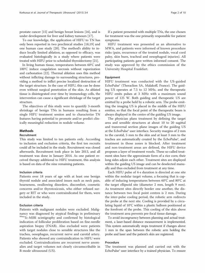
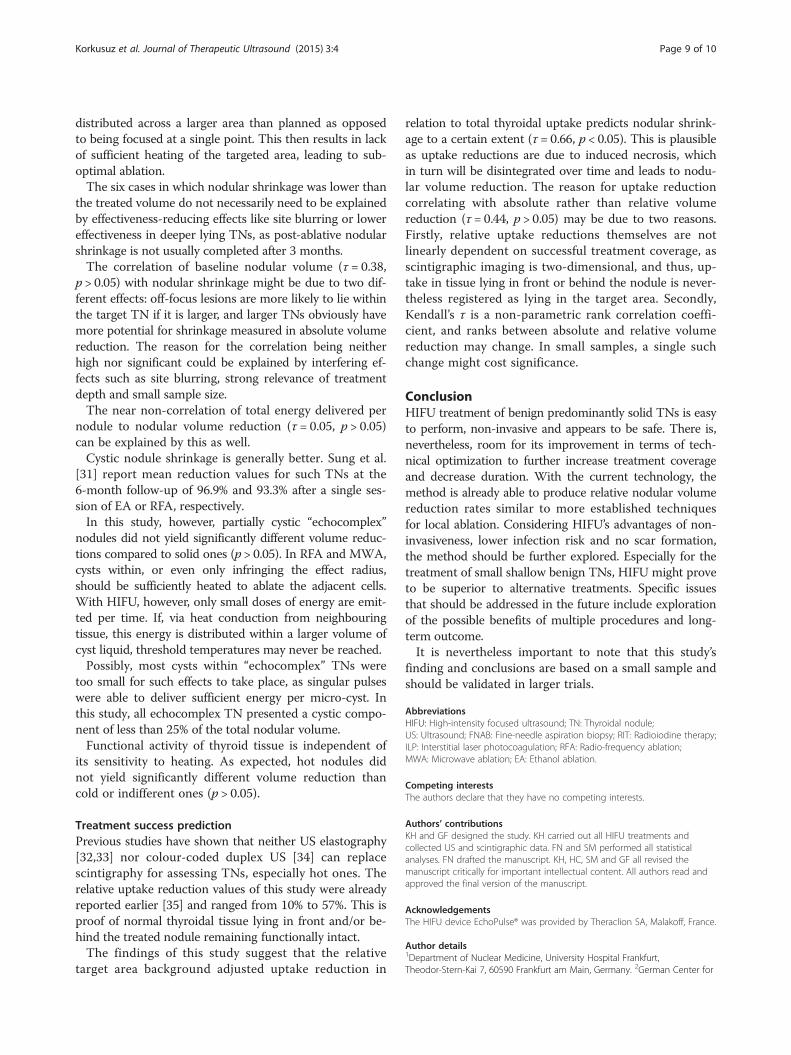
tures of around 80°C–90°C, while only temperatures ofabove 60°C are necessary to induce the desired necrosis.To limit the applied energy (which always needs to passthrough overlying tissue that we wanted to stay unharmed),we operated the HIFU just below the point of heat bubbleformation. Figure 1 shows an exemplary nodule before andimmediately after an initial test pulse. The clearly visible so-called hyperechoic mark (HEM) consists of microbubblesand indicates temperatures above 80°C.During treatment, the planned outlines of the target
area and sensible structures are displayed at all times, asare the actual anatomical localizations due to the under-lying B-mode guiding US imaging. A potential deviation
Figure 1 Visual feedback during initial test pulse. During initialtest pulses, beam intensity was set to be just below the point wherethe generation of a so-called hyperechoic mark (HEM) indicatestemperatures above 80°C–90°C. This mark is visible in B-mode USdue to the hyperechogenic character of microbubbles. These consist ofsteam. Shown is an exemplary nodule before (A) and directly after (B)such a test pulse. Clearly visible is the HEM, which vanishes withinseconds afterwards.
of the planned treatment grid and reality, if not detectedby the laser measurement, can be easily spotted by theperforming physician. In this case, the treatment can bemanually interrupted until congruency is restored byprobe repositioning. Such probe readjustments may bedone manually or via steering the robotic arm using theinterface. Inter-pulse probe repositioning to target thenext treatment site is done automatically.
Baseline evaluation and endpointsBefore HIFU treatment, nodules were assessed for size,structure and exact location using B-mode US imaging.Functional activity was evaluated using 99mTc-pertechnetatescintigraphy (75 MBq, imaging 20 min post-administrationwith a thyroid-specific scintillation camera (MedisoNucline® TH/22, acquisition time: 300 s, matrix: 128 ×128 × 16, low-energy collimator)). Nodules presentingas hypofunctional in this imaging were further examinedusing 99mTc-MIBI (441 MBq, imaging 10 and 60 minpost-administration with the same camera mentionedabove, with 500-s acquisition time and 128 × 128 × 16matrix) as well as FNAB cytology. Malignancy could beruled out in all cases.Due to reactive edema formation, ablative effects of
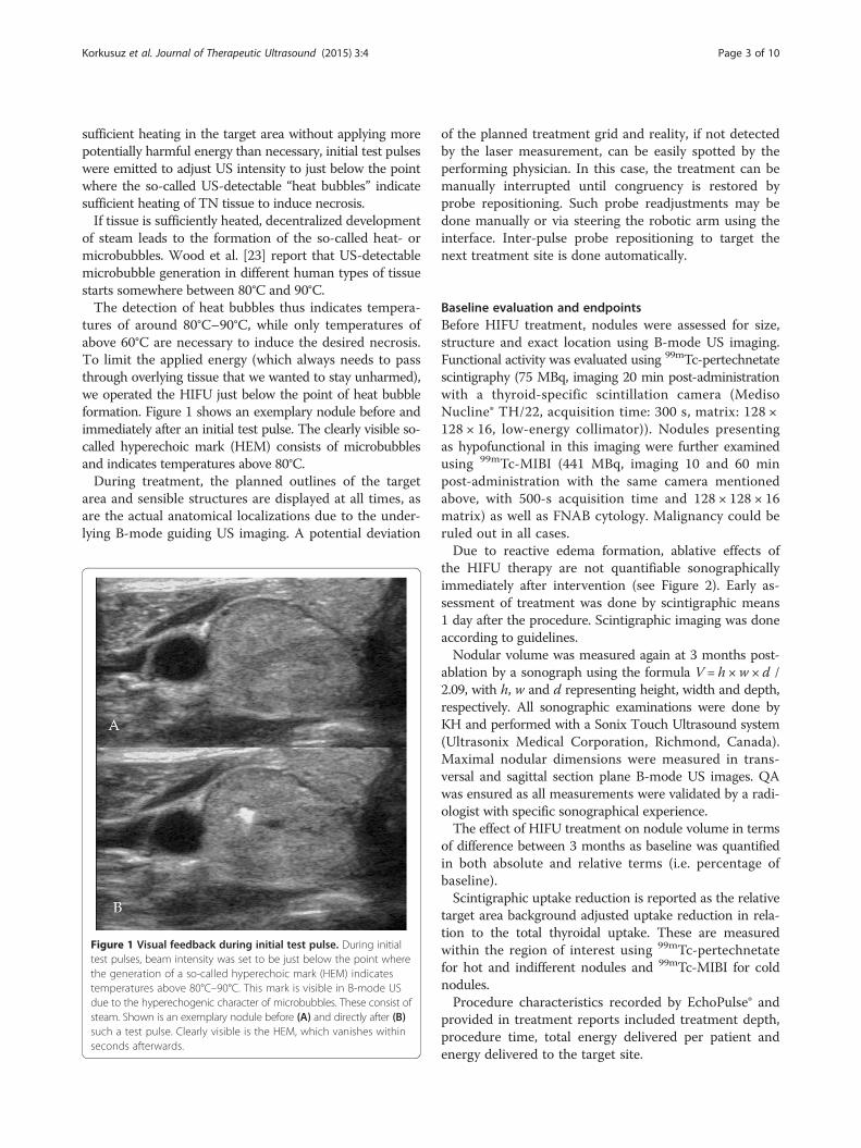
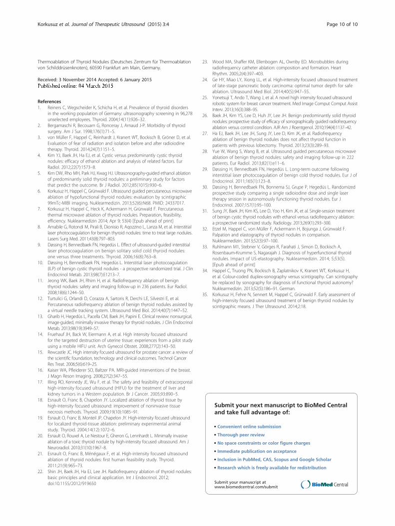
the HIFU therapy are not quantifiable sonographicallyimmediately after intervention (see Figure 2). Early as-sessment of treatment was done by scintigraphic means1 day after the procedure. Scintigraphic imaging was doneaccording to guidelines.Nodular volume was measured again at 3 months post-
ablation by a sonograph using the formula V = h ×w × d /2.09, with h, w and d representing height, width and depth,respectively. All sonographic examinations were done byKH and performed with a Sonix Touch Ultrasound system(Ultrasonix Medical Corporation, Richmond, Canada).Maximal nodular dimensions were measured in trans-versal and sagittal section plane B-mode US images. QAwas ensured as all measurements were validated by a radi-ologist with specific sonographical experience.The effect of HIFU treatment on nodule volume in terms
of difference between 3 months as baseline was quantifiedin both absolute and relative terms (i.e. percentage ofbaseline).Scintigraphic uptake reduction is reported as the relative
target area background adjusted uptake reduction in rela-tion to the total thyroidal uptake. These are measuredwithin the region of interest using 99mTc-pertechnetatefor hot and indifferent nodules and 99mTc-MIBI for coldnodules.Procedure characteristics recorded by EchoPulse® and
provided in treatment reports included treatment depth,procedure time, total energy delivered per patient andenergy delivered to the target site.
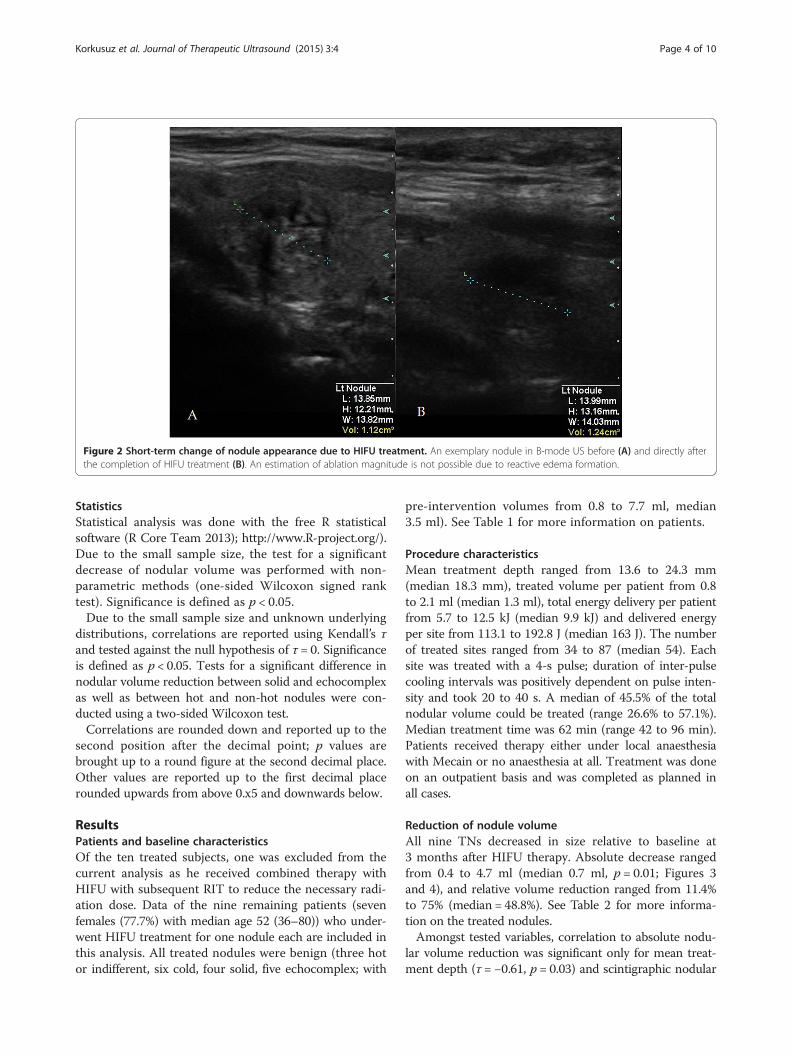
Figure 2 Short-term change of nodule appearance due to HIFU treatment. An exemplary nodule in B-mode US before (A) and directly afterthe completion of HIFU treatment (B). An estimation of ablation magnitude is not possible due to reactive edema formation.
Korkusuz et al. Journal of Therapeutic Ultrasound (2015) 3:4 Page 4 of 10
StatisticsStatistical analysis was done with the free R statisticalsoftware (R Core Team 2013); http://www.R-project.org/).Due to the small sample size, the test for a significantdecrease of nodular volume was performed with non-parametric methods (one-sided Wilcoxon signed ranktest). Significance is defined as p < 0.05.Due to the small sample size and unknown underlying
distributions, correlations are reported using Kendall’s τand tested against the null hypothesis of τ = 0. Significanceis defined as p < 0.05. Tests for a significant difference innodular volume reduction between solid and echocomplexas well as between hot and non-hot nodules were con-ducted using a two-sided Wilcoxon test.Correlations are rounded down and reported up to the
second position after the decimal point; p values arebrought up to a round figure at the second decimal place.Other values are reported up to the first decimal placerounded upwards from above 0.x5 and downwards below.
ResultsPatients and baseline characteristicsOf the ten treated subjects, one was excluded from thecurrent analysis as he received combined therapy withHIFU with subsequent RIT to reduce the necessary radi-ation dose. Data of the nine remaining patients (sevenfemales (77.7%) with median age 52 (36–80)) who under-went HIFU treatment for one nodule each are included inthis analysis. All treated nodules were benign (three hotor indifferent, six cold, four solid, five echocomplex; with
pre-intervention volumes from 0.8 to 7.7 ml, median3.5 ml). See Table 1 for more information on patients.
Procedure characteristicsMean treatment depth ranged from 13.6 to 24.3 mm(median 18.3 mm), treated volume per patient from 0.8to 2.1 ml (median 1.3 ml), total energy delivery per patientfrom 5.7 to 12.5 kJ (median 9.9 kJ) and delivered energyper site from 113.1 to 192.8 J (median 163 J). The numberof treated sites ranged from 34 to 87 (median 54). Eachsite was treated with a 4-s pulse; duration of inter-pulsecooling intervals was positively dependent on pulse inten-sity and took 20 to 40 s. A median of 45.5% of the totalnodular volume could be treated (range 26.6% to 57.1%).Median treatment time was 62 min (range 42 to 96 min).Patients received therapy either under local anaesthesiawith Mecain or no anaesthesia at all. Treatment was doneon an outpatient basis and was completed as planned inall cases.
Reduction of nodule volumeAll nine TNs decreased in size relative to baseline at3 months after HIFU therapy. Absolute decrease rangedfrom 0.4 to 4.7 ml (median 0.7 ml, p = 0.01; Figures 3and 4), and relative volume reduction ranged from 11.4%to 75% (median = 48.8%). See Table 2 for more informa-tion on the treated nodules.Amongst tested variables, correlation to absolute nodu-
lar volume reduction was significant only for mean treat-ment depth (τ = −0.61, p = 0.03) and scintigraphic nodular
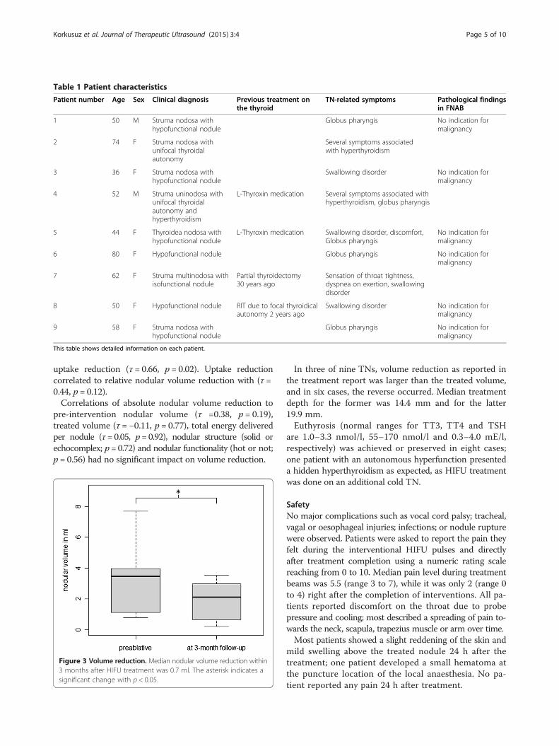
Table 1 Patient characteristics
Patient number Age Sex Clinical diagnosis Previous treatment onthe thyroid
TN-related symptoms Pathological findingsin FNAB
1 50 M Struma nodosa withhypofunctional nodule
Globus pharyngis No indication formalignancy
2 74 F Struma nodosa withunifocal thyroidalautonomy
Several symptoms associatedwith hyperthyroidism
3 36 F Struma nodosa withhypofunctional nodule
Swallowing disorder No indication formalignancy
4 52 M Struma uninodosa withunifocal thyroidalautonomy andhyperthyroidism
L-Thyroxin medication Several symptoms associated withhyperthyroidism, globus pharyngis
5 44 F Thyroidea nodosa withhypofunctional nodule
L-Thyroxin medication Swallowing disorder, discomfort,Globus pharyngis
No indication formalignancy
6 80 F Hypofunctional nodule Globus pharyngis No indication formalignancy
7 62 F Struma multinodosa withisofunctional nodule
Partial thyroidectomy30 years ago
Sensation of throat tightness,dyspnea on exertion, swallowingdisorder
8 50 F Hypofunctional nodule RIT due to focal thyroidicalautonomy 2 years ago
Swallowing disorder No indication formalignancy
9 58 F Struma nodosa withhypofunctional nodule
Globus pharyngis No indication formalignancy
This table shows detailed information on each patient.
Korkusuz et al. Journal of Therapeutic Ultrasound (2015) 3:4 Page 5 of 10
uptake reduction (τ = 0.66, p = 0.02). Uptake reductioncorrelated to relative nodular volume reduction with (τ =0.44, p = 0.12).Correlations of absolute nodular volume reduction to
pre-intervention nodular volume (τ =0.38, p = 0.19),treated volume (τ = −0.11, p = 0.77), total energy deliveredper nodule (τ= 0.05, p= 0.92), nodular structure (solid orechocomplex; p= 0.72) and nodular functionality (hot or not;p = 0.56) had no significant impact on volume reduction.
Figure 3 Volume reduction. Median nodular volume reduction within3 months after HIFU treatment was 0.7 ml. The asterisk indicates asignificant change with p < 0.05.
In three of nine TNs, volume reduction as reported inthe treatment report was larger than the treated volume,and in six cases, the reverse occurred. Median treatmentdepth for the former was 14.4 mm and for the latter19.9 mm.Euthyrosis (normal ranges for TT3, TT4 and TSH
are 1.0–3.3 nmol/l, 55–170 nmol/l and 0.3–4.0 mE/l,respectively) was achieved or preserved in eight cases;one patient with an autonomous hyperfunction presenteda hidden hyperthyroidism as expected, as HIFU treatmentwas done on an additional cold TN.
SafetyNo major complications such as vocal cord palsy; tracheal,vagal or oesophageal injuries; infections; or nodule rupturewere observed. Patients were asked to report the pain theyfelt during the interventional HIFU pulses and directlyafter treatment completion using a numeric rating scalereaching from 0 to 10. Median pain level during treatmentbeams was 5.5 (range 3 to 7), while it was only 2 (range 0to 4) right after the completion of interventions. All pa-tients reported discomfort on the throat due to probepressure and cooling; most described a spreading of pain to-wards the neck, scapula, trapezius muscle or arm over time.Most patients showed a slight reddening of the skin and
mild swelling above the treated nodule 24 h after thetreatment; one patient developed a small hematoma atthe puncture location of the local anaesthesia. No pa-tient reported any pain 24 h after treatment.
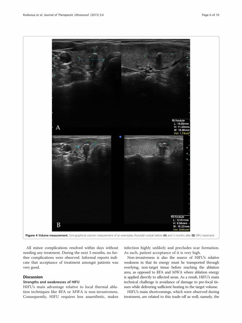
Figure 4 Volume measurement. Sonographical volume measurement of an exemplary thyroidal nodule before (A) and 3 months after (B) HIFU treatment.
Korkusuz et al. Journal of Therapeutic Ultrasound (2015) 3:4 Page 6 of 10
All minor complications resolved within days withoutneeding any treatment. During the next 3 months, no fur-ther complications were observed. Informal reports indi-cate that acceptance of treatment amongst patients wasvery good.
DiscussionStrengths and weaknesses of HIFUHIFU’s main advantage relative to local thermal abla-tion techniques like RFA or MWA is non-invasiveness.Consequently, HIFU requires less anaesthetic, makes
infection highly unlikely and precludes scar formation.As such, patient acceptance of it is very high.Non-invasiveness is also the source of HIFU’s relative
weakness in that its energy must be transported throughoverlying, non-target tissue before reaching the ablationarea, as opposed to RFA and MWA where ablation energyis applied directly to affected areas. As a result, HIFU’s maintechnical challenge is avoidance of damage to pre-focal tis-sues while delivering sufficient heating to the target volume.HIFU’s main shortcomings, which were observed during
treatment, are related to this trade-off as well, namely, the
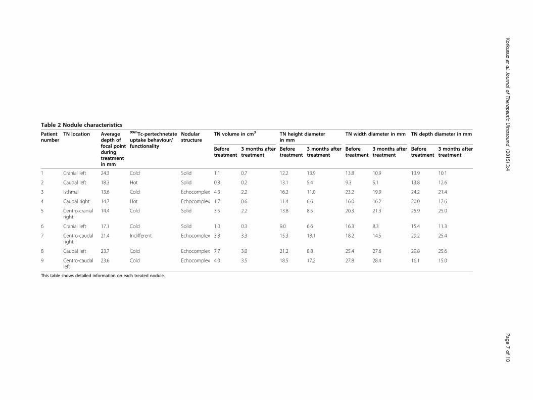
Table 2 Nodule characteristics
Patientnumber
TN location Averagedepth offocal pointduringtreatmentin mm
99mTc-pertechnetateuptake behaviour/functionality
Nodularstructure
TN volume in cm3 TN height diameterin mm
TN width diameter in mm TN depth diameter in mm
Beforetreatment
3 months aftertreatment
Beforetreatment
3 months aftertreatment
Beforetreatment
3 months aftertreatment
Beforetreatment
3 months aftertreatment
1 Cranial left 24.3 Cold Solid 1.1 0.7 12.2 13.9 13.8 10.9 13.9 10.1
2 Caudal left 18.3 Hot Solid 0.8 0.2 13.1 5.4 9.3 5.1 13.8 12.6
3 Isthmal 13.6 Cold Echocomplex 4.3 2.2 16.2 11.0 23.2 19.9 24.2 21.4
4 Caudal right 14.7 Hot Echocomplex 1.7 0.6 11.4 6.6 16.0 16.2 20.0 12.6
5 Centro-cranialright
14.4 Cold Solid 3.5 2.2 13.8 8.5 20.3 21.3 25.9 25.0
6 Cranial left 17.1 Cold Solid 1.0 0.3 9.0 6.6 16.3 8.3 15.4 11.3
7 Centro-caudalright
21.4 Indifferent Echocomplex 3.8 3.3 15.3 18.1 18.2 14.5 29.2 25.4
8 Caudal left 23.7 Cold Echocomplex 7.7 3.0 21.2 8.8 25.4 27.6 29.8 25.6
9 Centro-caudalleft
23.6 Cold Echocomplex 4.0 3.5 18.5 17.2 27.8 28.4 16.1 15.0
This table shows detailed information on each treated nodule.
Korkusuzet
al.JournalofTherapeutic
Ultrasound
(2015) 3:4 Page
7of
10
Korkusuz et al. Journal of Therapeutic Ultrasound (2015) 3:4 Page 8 of 10
procedure’s long duration and its incomplete ablation. Theamount of volume that could actually be treated in rela-tion to initial nodular volume was limited by four built-inconstrains of the EchoPulse®: (1) application to only onelayer of treatment sites, (2) maximum treatment depth ofapproximately 2.8 cm, (3) security margins around sens-ible structures and (4) only one direction of site alignmentof 90° to the skin.Current experience with the device and TN treatment
in general argues against changing (1) since it wouldlead to prolonging an already lengthy treatment whosemedian duration was 62 min. At the same time, removingthis limitation might increase treatment coverage and,with it, considerably increase therapy effectiveness. Thismight be especially useful for larger TNs, since these aremore likely to have relevant masses above and/or belowthe treatment layer.Treatment depth limitation of HIFU has already been
reported in other indications [24]. This is due to the factthat a sufficient heating of deeper layers of tissue requireslarger amounts of energy to be emitted, as more of it islost in pre-focal areas, which in turn can only handle lim-ited heating per time unit without being damaged. TheEchoPulse® addresses this problem by increasing heat fluxfrom the non-targeted tissue the US pulse passes throughby cooling the overlying skin. Yet, median treatment timeis still 62 min and median ablated volume only 1.3 ml. More-over, 3-month volume reduction of deeper lying nodules islower relative to that of shallower nodules, with correlationof treatment depth and volume reduction being τ = −0.61(p < 0.05). A potential solution for this could be utilizationof either a larger probe or multiple probes targeting thesame focal point from different angles, thus allocating en-ergy lost in pre-focal tissue to more volume. Additionally,more flexible probe positioning may make it possible tosend each pulse through a different part of overlaying tis-sue and hence minimize inter-pulse treatment pauses.Yonetsuji et al. [25] suggest that an increase in degrees
of probe rotation helps reduce skin burns, long treatmenttime and incomplete ablation, which are the main prob-lems of HIFU therapy in general. This indicates that (4)indeed has been limiting treatment success.Sufficient security margins (3) are an essential require-
ment considering our data showing effects of HIFU be-yond the targeted area, as implied by nodular shrinkageslarger than the treated volumes. Specifically, this was ob-served in three of our nine cases with reductions of 2.1,1.4 and 4.7 ml induced when treating volumes of 1.3, 1.0and 1.5 ml, respectively.These off-focus lesions should be majorly situated
above the focal point, as there the pulse is more power-ful than below it. Parafocal lesions should occur due toheat conduction as well as convection by blood flow butshould only be marginal.
The main reason for long treatment durations is thatafter each pulse emission, the non-targeted area aroundand especially above the nodule needs to cool down, whichtakes time increasing with the energy absorbed in pre-focaltissue. As noted, this could be addressed by larger, mul-tiple, or more flexible probes.
Volume reductionTurtulici et al. [12] found that a single RFA treatment oflarger benign non-functioning TNs (mean pre-ablationvolume 13.5 ml) could induce mean relative shrinkage of72.6% after 6 months. Baek et al. [26] observed averagedecreases of 82.6% for predominantly solid benign TNsafter the same period.Ha et al. [27] who used HIFU for nodules with pre-
ablative volume of 9.7 ml were able to induce a meanreduction of 87.2% after an average of 43.7 months. Ameta-analysis by Shin et al. [22] demonstrated that asingle RFA session induces reduction of 33% to 49% inpredominantly solid TNs after 1 month and 51% to 80%after 6 months.Korkusuz et al. [7] report volume reductions of 54.2%
3 months after MWA in benign cold TNs. Yue et al. [28]induced shrinkages of 65% on average using MWA, mea-sured 6 months after ablation.Døssing et al. [10] report a median reduction of the
solid component volume of a cystic TN of 54% 6 monthsafter a combined therapy of cyst aspiration and subsequentILP. They also produced a median volume reduction of be-nign cold solid TNs of 51% a median of 67 months after asingle ILP session [29]. They also compare ILP and RIT onsolitary autonomous nodules and found similar volumereductions after 6 months on average of 44% and 47%,respectively [30].Compared to these values, the findings of this study
regarding a relative median shrinkage of 48.8% of relativelysmall TNs after only 3 months are decent, especiallyconsidering the technical constraints explained abovethat were limiting treatment coverage.
Factors influencing successAs deeper lying TNs presented significantly smaller vol-ume reductions (τ = −0.61, p < 0.05), pre-focal energy lossesappear to have been higher than anticipated. Continuousintra-procedure power adjustment by “heat bubble” obser-vation, as opposed to baseline only, might enable sufficientfocal heating by real-time treatment effect visualization.The unexpected lack of relationship between planned
treatment volume and volume reduction may have beendue to patient movement causing site blurring (e.g.swallowing can create tissue movement in a cranio-caudaldirection that does not necessarily induce laser-detectableskin movement above 1 mm in a lateral or dorso-frontaldirection). Site blurring may cause energy pulses to be
Korkusuz et al. Journal of Therapeutic Ultrasound (2015) 3:4 Page 9 of 10
distributed across a larger area than planned as opposedto being focused at a single point. This then results in lackof sufficient heating of the targeted area, leading to sub-optimal ablation.The six cases in which nodular shrinkage was lower than
the treated volume do not necessarily need to be explainedby effectiveness-reducing effects like site blurring or lowereffectiveness in deeper lying TNs, as post-ablative nodularshrinkage is not usually completed after 3 months.The correlation of baseline nodular volume (τ = 0.38,
p > 0.05) with nodular shrinkage might be due to two dif-ferent effects: off-focus lesions are more likely to lie withinthe target TN if it is larger, and larger TNs obviously havemore potential for shrinkage measured in absolute volumereduction. The reason for the correlation being neitherhigh nor significant could be explained by interfering ef-fects such as site blurring, strong relevance of treatmentdepth and small sample size.The near non-correlation of total energy delivered per
nodule to nodular volume reduction (τ = 0.05, p > 0.05)can be explained by this as well.Cystic nodule shrinkage is generally better. Sung et al.
[31] report mean reduction values for such TNs at the6-month follow-up of 96.9% and 93.3% after a single ses-sion of EA or RFA, respectively.In this study, however, partially cystic “echocomplex”
nodules did not yield significantly different volume reduc-tions compared to solid ones (p > 0.05). In RFA and MWA,cysts within, or even only infringing the effect radius,should be sufficiently heated to ablate the adjacent cells.With HIFU, however, only small doses of energy are emit-ted per time. If, via heat conduction from neighbouringtissue, this energy is distributed within a larger volume ofcyst liquid, threshold temperatures may never be reached.Possibly, most cysts within “echocomplex” TNs were
too small for such effects to take place, as singular pulseswere able to deliver sufficient energy per micro-cyst. Inthis study, all echocomplex TN presented a cystic compo-nent of less than 25% of the total nodular volume.Functional activity of thyroid tissue is independent of
its sensitivity to heating. As expected, hot nodules didnot yield significantly different volume reduction thancold or indifferent ones (p > 0.05).
Treatment success predictionPrevious studies have shown that neither US elastography[32,33] nor colour-coded duplex US [34] can replacescintigraphy for assessing TNs, especially hot ones. Therelative uptake reduction values of this study were alreadyreported earlier [35] and ranged from 10% to 57%. This isproof of normal thyroidal tissue lying in front and/or be-hind the treated nodule remaining functionally intact.The findings of this study suggest that the relative
target area background adjusted uptake reduction in
relation to total thyroidal uptake predicts nodular shrink-age to a certain extent (τ = 0.66, p < 0.05). This is plausibleas uptake reductions are due to induced necrosis, whichin turn will be disintegrated over time and leads to nodu-lar volume reduction. The reason for uptake reductioncorrelating with absolute rather than relative volumereduction (τ = 0.44, p > 0.05) may be due to two reasons.Firstly, relative uptake reductions themselves are notlinearly dependent on successful treatment coverage, asscintigraphic imaging is two-dimensional, and thus, up-take in tissue lying in front or behind the nodule is never-theless registered as lying in the target area. Secondly,Kendall’s τ is a non-parametric rank correlation coeffi-cient, and ranks between absolute and relative volumereduction may change. In small samples, a single suchchange might cost significance.
ConclusionHIFU treatment of benign predominantly solid TNs is easyto perform, non-invasive and appears to be safe. There is,nevertheless, room for its improvement in terms of tech-nical optimization to further increase treatment coverageand decrease duration. With the current technology, themethod is already able to produce relative nodular volumereduction rates similar to more established techniquesfor local ablation. Considering HIFU’s advantages of non-invasiveness, lower infection risk and no scar formation,the method should be further explored. Especially for thetreatment of small shallow benign TNs, HIFU might proveto be superior to alternative treatments. Specific issuesthat should be addressed in the future include explorationof the possible benefits of multiple procedures and long-term outcome.It is nevertheless important to note that this study’s
finding and conclusions are based on a small sample andshould be validated in larger trials.
AbbreviationsHIFU: High-intensity focused ultrasound; TN: Thyroidal nodule;US: Ultrasound; FNAB: Fine-needle aspiration biopsy; RIT: Radioiodine therapy;ILP: Interstitial laser photocoagulation; RFA: Radio-frequency ablation;MWA: Microwave ablation; EA: Ethanol ablation.
Competing interestsThe authors declare that they have no competing interests.
Authors’ contributionsKH and GF designed the study. KH carried out all HIFU treatments andcollected US and scintigraphic data. FN and SM performed all statisticalanalyses. FN drafted the manuscript. KH, HC, SM and GF all revised themanuscript critically for important intellectual content. All authors read andapproved the final version of the manuscript.
AcknowledgementsThe HIFU device EchoPulse® was provided by Theraclion SA, Malakoff, France.
Author details1Department of Nuclear Medicine, University Hospital Frankfurt,Theodor-Stern-Kai 7, 60590 Frankfurt am Main, Germany. 2German Center for
Korkusuz et al. Journal of Therapeutic Ultrasound (2015) 3:4 Page 10 of 10
Thermoablation of Thyroid Nodules (Deutsches Zentrum für Thermoablationvon Schilddrüsenknoten), 60590 Frankfurt am Main, Germany.
Received: 3 November 2014 Accepted: 6 January 2015
References1. Reiners C, Wegscheider K, Schicha H, et al. Prevalence of thyroid disorders
in the working population of Germany: ultrasonography screening in 96,278unselected employees. Thyroid. 2004;14(11):926–32.
2. Bergamaschi R, Becouarn G, Ronceray J, Arnaud J-P. Morbidity of thyroidsurgery. Am J Sur. 1998;176(1):71–5.
3. von Müller F, Happel C, Reinhardt J, Kranert WT, Bockisch B, Gröner D, et al.Evaluation of fear of radiation and isolation before and after radioiodinetherapy. Thyroid. 2014;24(7):1151–5.
4. Kim YJ, Baek JH, Ha EJ, et al. Cystic versus predominantly cystic thyroidnodules: efficacy of ethanol ablation and analysis of related factors. EurRadiol. 2012;22(7):1573–8.
5. Kim DW, Rho MH, Park HJ, Kwag HJ. Ultrasonography-guided ethanol ablationof predominantly solid thyroid nodules: a preliminary study for factorsthat predict the outcome. Br J Radiol. 2012;85(1015):930–6.
6. Korkusuz H, Happel C, Grünwald F. Ultrasound guided percutaneous microwaveablation of hypofunctional thyroid nodules: evaluation by scintigraphic99mTc-MIBI imaging. Nuklearmedizin. 2013;52(6):N68. PMID: 24337017.
7. Korkusuz H, Happel C, Heck K, Ackermann H, Grünwald F. Percutaneousthermal microwave ablation of thyroid nodules. Preparation, feasibility,efficiency. Nuklearmedizin 2014; Apr 9; 53(4) [Epub ahead of print]
8. Amabile G, Rotondi M, Pirali B, Dionisio R, Aqozzino L, Lanza M, et al. Interstitiallaser photocoagulation for benign thyroid nodules: time to treat large nodules.Lasers Surg Med. 2011;43(8):797–803.
9. Døssing H, Bennedbaek FN, Hegedüs L. Effect of ultrasound-guided interstitiallaser photocoagulation on benign solitary solid cold thyroid nodules:one versus three treatments. Thyroid. 2006;16(8):763–8.
10. Døssing H, Bennedbaek FN, Hegedüs L. Interstitial laser photocoagulation(ILP) of benign cystic thyroid nodules - a prospective randomized trial. J ClinEndocrinol Metab. 2013;98(7):E1213–7.
11. Jeong WK, Baek JH, Rhim H, et al. Radiofrequency ablation of benignthyroid nodules: safety and imaging follow-up in 236 patients. Eur Radiol.2008;18(6):1244–50.
12. Turtulici G, Orlandi D, Corazza A, Sartoris R, Derchi LE, Silvestri E, et al.Percutaneous radiofrequency ablation of benign thyroid nodules assisted bya virtual needle tracking system. Ultrasound Med Biol. 2014;40(7):1447–52.
13. Gharib H, Hegedüs L, Pacella CM, Baek JH, Papini E. Clinical review: nonsurgical,image-guided, minimally invasive therapy for thyroid nodules. J Clin EndocrinolMetab. 2013;98(19):3949–57.
14. Fruehauf JH, Back W, Eiermann A, et al. High intensity focused ultrasoundfor the targeted destruction of uterine tissue: experiences from a pilot studyusing a mobile HIFU unit. Arch Gynecol Obstet. 2008;277(2):143–50.
15. Rewcastle JC. High intensity focused ultrasound for prostate cancer: a review ofthe scientific foundation, technology and clinical outcomes. Technol CancerRes Treat. 2006;5(6):619–25.
16. Kaiser WA, Pfleiderer SO, Baltzer PA. MRI-guided interventions of the breast.J Magn Reson Imaging. 2008;27(2):347–55.
17. Illing RO, Kennedy JE, Wu F, et al. The safety and feasibility of extracorporealhigh-intensity focused ultrasound (HIFU) for the treatment of liver andkidney tumors in a Western population. Br J Cancer. 2005;93:890–5.
18. Esnault O, Franc B, Chapelon JY. Localized ablation of thyroid tissue byhigh-intensity focused ultrasound: improvement of noninvasive tissuenecrosis methods. Thyroid. 2009;19(10):1085–91.
19. Esnault O, Franc B, Monteil JP, Chapelon JY. High-intensity focused ultrasoundfor localized thyroid-tissue ablation: preliminary experimental animalstudy. Thyroid. 2004;14(12):1072–6.
20. Esnault O, Rouxel A, Le Nestour E, Gheron G, Lennhardt L. Minimally invasiveablation of a toxic thyroid nodule by high-intensity focused ultrasound. Am JNeuroradiol. 2010;31(10):1967–8.
21. Esnault O, Franc B, Ménégaux F, et al. High-intensity focused ultrasoundablation of thyroid nodules: first human feasibility study. Thyroid.2011;21(9):965–73.
22. Shin JH, Baek JH, Ha EJ, Lee JH. Radiofrequency ablation of thyroid nodules:basic principles and clinical application. Int J Endocrinol. 2012;doi:10.1155/2012/919650
23. Wood MA, Shaffer KM, Ellenbogen AL, Ownby ED. Microbubbles duringradiofrequency catheter ablation: composition and formation. HeartRhythm. 2005;2(4):397–403.
24. Ge HY, Miao LY, Xiong LL, et al. High-intensity focused ultrasound treatmentof late-stage pancreatic body carcinoma: optimal tumor depth for safeablation. Ultrasound Med Biol. 2014;40(5):947–55.
25. Yonetsuji T, Ando T, Wang J, et al. A novel high intensity focused ultrasoundrobotic system for breast cancer treatment. Med Image Comput Comput AssistInterv. 2013;16(3):388–95.
26. Baek JH, Kim YS, Lee D, Huh JY, Lee JH. Benign predominantly solid thyroidnodules: prospective study of efficacy of sonographically guided radiofrequencyablation versus control condition. AJR Am J Roentgenol. 2010;194(4):1137–42.
27. Ha EJ, Baek JH, Lee JH, Sung JY, Lee D, Kim JK, et al. Radiofrequencyablation of benign thyroid nodules does not affect thyroid function inpatients with previous lobectomy. Thyroid. 2013;23(3):289–93.
28. Yue W, Wang S, Wang B, et al. Ultrasound guided percutaneous microwaveablation of benign thyroid nodules: safety and imaging follow-up in 222patients. Eur Radiol. 2013;82(1):e11–6.
29. Døssing H, Bennedbæk FN, Hegedüs L. Long-term outcome followinginterstitial laser photocoagulation of benign cold thyroid nodules. Eur J ofEndocrinol. 2011;165(1):123–8.
30. Døssing H, Bennedbaek FN, Bonnema SJ, Grupe P, Hegedüs L. Randomizedprospective study comparing a single radioiodine dose and single lasertherapy session in autonomously functioning thyroid nodules. Eur JEndocrinol. 2007;157(1):95–100.
31. Sung JY, Baek JH, Kim KS, Lee D, Yoo H, Kim JK, et al. Single-session treatmentof benign cystic thyroid nodules with ethanol versus radiofrequency ablation:a prospective randomized study. Radiology. 2013;269(1):293–300.
32. Etzel M, Happel C, von Müller F, Ackermann H, Bojunga J, Grünwald F.Palpation and elastography of thyroid nodules in comparison.Nuklearmedizin. 2013;52(3):97–100.
33. Ruhlmann M1, Stebner V, Görges R, Farahati J, Simon D, Bockisch A,Rosenbaum-Krumme S, Nagarajah J. Diagnosis of hyperfunctional thyroidnodules. Impact of US-elastography. Nuklearmedizin. 2014; 5;53(5).[Epub ahead of print]
34. Happel C, Truong PN, Bockisch B, Zaplatnikov K, Kranert WT, Korkusuz H,et al. Colour-coded duplex-sonography versus scintigraphy. Can scintigraphybe replaced by sonography for diagnosis of functional thyroid autonomy?Nuklearmedizin. 2013;52(5):186–91. German.
35. Korkusuz H, Fehre N, Sennert M, Happel C, Grünwald F. Early assessment ofhigh-intensity focused ultrasound treatment of benign thyroid nodules byscintigraphic means. J Ther Ultrasound. 2014;2:18.
Submit your next manuscript to BioMed Centraland take full advantage of:
• Convenient online submission
• Thorough peer review
• No space constraints or color figure charges
• Immediate publication on acceptance
• Inclusion in PubMed, CAS, Scopus and Google Scholar
• Research which is freely available for redistribution
Submit your manuscript at www.biomedcentral.com/submit


