Embed Size (px)
Citation preview

1
Water soluble ruthenium complexes bearing activity against protozoan parasites
Cynthia Sarnigueta, Jeannette Tolozab, Micaella Cipriania, Michel Lapierc, Marisol
Vieitesa, Yanis Toledano-Magañad, Juan Carlos García-Ramose, Lena Ruiz-Azuarae,
Virtudes Morenof, Juan Diego Mayac, Claudio Olea Azarb, Dinorah Gambino*a, Lucía
Otero*a
aCátedra de Química Inorgánica, DEC, Facultad de Química, Universidad de la
República, Gral. Flores 2124, C. C. 1157, 11800 Montevideo, Uruguay
bDepartamento de Química Inorgánica y Analítica, Facultad de Ciencias Químicas y
Farmacéuticas, Universidad de Chile, Sergio Livingstone 1007, Casilla 233, Santiago,
Chile
cDepartamento de Farmacología Clínica y Molecular, ICBM, Facultad de Medicina,
Universidad de Chile, Independencia 1027, Santiago, Chile
dDepartamento de Inmunología, Instituto de Investigaciones Biomédicas, Universidad
Nacional Autónoma de México, Avenida Universidad 3000, 04510, Mexico City,
Mexico
eDepartamento de Química Inorgánica y Nuclear, Facultad de Química, Universidad
Nacional Autónoma de México, Avenida Universidad 3000, 04510, Mexico City,
Mexico
fDepartamento de Química Inorgánica, Universitat Barcelona, Martí i Franquès 1-11,
08028, Barcelona, Spain
(*) Corresponding authors´ fax number: +59829241906, telephone: +59829249739, e-mail addresses:
2
ABSTRACT
Parasitic illnesses are major causes of human disease and misery worldwide. Among
them, both amoebiasis and Chagas disease, caused by the protozoan parasites
Entamoeba histolytica and Trypanosoma cruzi, are responsible for thousands of annual
deaths. The lack of safe and effective chemotherapy and/or the appearance of current
drug resistance make it relevant the development of novel pharmacological tools for
their treatment. In these sense, within the framework of the Medicinal Inorganic
Chemistry, metal-based drugs appear to be a good alternative to find a pharmacological
answer to parasitic diseases. In this work, novel ruthenium complexes
[RuCl2(HL)(HPTA)2]Cl2 with HL = bioactive 5-nitrofuryl containing
thiosemicarbazones and PTA = 1,3,5-triaza-7-phosphaadamantane have been
synthesized and fully characterized. PTA was included as co-ligand in order to
modulate complexes aqueous solubility. In fact, obtained complexes were water soluble.
Their activity against T. cruzi and E. histolytica was evaluated in vitro.
[RuCl2(HL4)(HPTA)2]Cl2 complex, with HL4 = N-phenyl-5-nitrofuryl-
thiosemicarbazone was the most active compound against both parasites. In particular, it
showed an excellent activity against E. histolytica (IC50 = 5.2 M), even higher than
that of the reference drug Metronidazol. In addition, this complex turns out to be
selective for E. histolytica (Selectivity index SI > 38). The potential mechanism of
antiparasitic action of the obtained ruthenium complexes could involve oxidative stress
for both parasites. Additionally, complexes could interact with DNA as second potential
target by an intercalative-like mode. Obtained results could be considered a contribution
in the search for metal compounds that could be active against multiple parasites.

3
Keywords: Trypanosoma cruzi; Entamoeba histolytica; ruthenium; 5-nitrofuryl
containing thiosemicarbazones; PTA (1,3,5-triaza-7-phosphaadamantane).
Introduction
Amoebiasis and Chagas disease, caused by the protozoan parasites Entamoeba
histolytica (E. histolytica) and Trypanosoma cruzi (T. cruzi) respectively, are
responsible of human disease and they lead to thousands of annual deaths [1-3].
Chagas disease is a chronic, systemic, parasitic infection that affects about 8 million
people in Latin America but also, due to immigration, hundreds of thousands in USA
and Europe. Amoebiasis is a health risk in almost all countries principally due to poor
hygiene practices, resulting in 40000 annual deaths [3-7].
Despite these illnesses present high prevalence worldwide, the chemotherapy for
Chagas disease and amoebiasis is limited to a few drugs in the market (Nifurtimox and
Benznidazol for the former and Metronidazol for the latter), most of which are of low
efficacy, show toxic side effects, and frequently lead to the appearance of resistant
strains. The development of more efficient and safe drugs is still a challenge [8-12].
Metal-based drugs appear to be a good alternative to find a pharmacological answer to
parasitic diseases. In particular, the complexation of transition metals with a proved or
potential biologically active ligand could lead to enhance its pharmacological and
chemical properties such as potency, toxicity, solubility, chemical stability, and
lipophilicity. In addition, a metal-drug synergism could be achieved through a dual or
multiple mechanism of action [12-19]. Some bioactive ligands that have been
extensively explored in this sense are thiosemicarbazones. These small and versatile
molecules have been related to many pharmacological activities, like antiparasitic, due

4
to the highlighting results obtained against both E. histolytica and T. cruzi, among other
parasites [12, 20-22].
In this frame, some of us have been involved in the development of metal complexes
with antichagasic 5-nitrofuryl containing thiosemicarbazones (Figure 1). We have
rationally designed and developed more than fifty compounds including palladium,
platinum and ruthenium complexes as well as ruthenium organometallic compounds in
order to evaluate the effect of metal complexation and the presence of different co-
ligands on the pharmacological profile of the bioactive ligands. Most complexes
resulted more or at least equally active than the corresponding ligands and the activity
could be correlated to properties like solubility, stability, lipophilicity or protein
interaction. Compounds mechanisms of anti T. cruzi action were also extensively
studied [18,23-32]. In addition, palladium, copper, vanadium and ruthenium
thiosemicarbazone complexes have been explored in order to obtain safer and more
active drugs against E. histolytica. Most of the obtained compounds were more active
than the corresponding ligands, showing promising amoebicidal activity [12].
Based on these previous results, in this work we have developed a new series of
ruthenium complexes with the 5-nitrofuryl containing thiosemicarbazones showed in
Figure 1 as bioactive ligands. PTA (1, 3, 5-triaza-7-phosphaadamantane, Figure 1) was
included in the ruthenium coordination sphere in order to modulate water solubility of
the compounds. PTA is a hydrophilic phosphine that has been extensively used as
ligand in aqueous homogeneous catalysis [33,34]. Recently, the interest in this ligand
has been renewed due to its usefulness as ancillary ligand for medicinal chemistry
purposes as it is known to endow metal complexes with aqueous solubility and
solubility in polar organic solvents [33,34]. Ruthenium has been selected as metal
center due to its suitable chemical and biological properties i.e. biologically attainable

5
oxidation states (II, III), coordination versatility, octahedral geometry that allows
ancillary ligands, appropriate ligand substitution kinetics, low toxicity to humans related
to metabolic similarities with iron [18,35]. Finally, related to the potential mechanism
of antiparasitic action of the ruthenium-PTA-thiosemicarbazone complexes, a dual
mechanism of action could be expected, combining the pharmacological properties of
the ligands and the metal. On one side, the main mode of action of the 5-nitrofuryl
containing thiosemicarbazone compounds is related to the intracellular reduction of the
nitro moiety followed by redox cycling, yielding reduced oxygen species (ROS) that
cause cellular damage [36]. On the other, one of the reported targets of ruthenium
bioactive compounds is DNA [35]. Therefore, in addition to the synthesis,
characterization and evaluation of the activity against E. histolytica and T. cruzi of the
complexes, their ability of generating intraparasitic oxidative stress as well as of
interacting with DNA was studied.
Figure 1. (a) 5-nitrofuryl containing thiosemicarbazones, (b) PTA (1,3,5-triaza-7-
phosphaadamantane).
Materials and Methods
All common laboratory chemicals were purchased from commercial sources and used
without further purification. PTA (1, 3, 5-triaza-7-phosphaadamantane) was purchased
from Sigma-Aldrich. [RuCl2(DMSO)4], with DMSO = dimethylsulfoxide, was
OO2N
N
NH NHR
SN
NN
P
(a) (b)
R = H HL1
R = methyl HL2
R = ethyl HL3
R = phenyl HL4

6
prepared according to literature procedures [37]. All 5-nitrofuryl containing
thiosemicarbazones were synthesized using the previously reported methodology [36].
Syntheses of [RuIICl2(HL)(HPTA)2]Cl2·H2O·C2H5OH
[RuCl2(DMSO)4] (100 mg, 0.2 mmol) and PTA (65 mg, 0.4 mmol) were dissolved in 10
mL ethanol. The mixture was refluxed for 2 h after which the corresponding
thiosemicarbazone ligand HL (0.2 mmol) was added to the red solution. The solution
was heated under reflux for 24 more hours. The obtained brown microcrystalline solid
was filtered off and washed with ethanol (Scheme 1).
[RuIICl2(HL1)(HPTA)2]Cl2·H2O·C2H5OH. Yield: 92 mg, 54 %. Found: C, 28.8; H, 4.6;
N, 16.6; S, 4.0; H2O+C2H5OH 7.8 %. Calc. for C20H40N10O5SP2Cl4Ru: C, 28.7; H, 4.8;
N, 16.7; S, 3.8; H2O+C2H5OH 7,6 %. 31P1H NMR (121.4 MHz, D2O): = -11.40 (s,
1P, HPTA), -12.58 (s, 1P, HPTA).
[RuIICl2(HL2)(HPTA)2]Cl2·H2O·C2H5OH. Yield: 83 mg, 48 %. Found: C, 29.7; H, 4.9;
N, 16.6; S, 3.8; H2O+C2H5OH 7.2 %. Calc. for C21H42N10O5SP2Cl4Ru: C, 29.6; H, 4.9;
N, 16.4; S, 3.8; H2O+C2H5OH 7.5 %. 31P1H NMR (121.4 MHz, D2O): = -11.51 (s,
1P, HPTA), -12.70 (s, 1P, HPTA).
[RuIICl2(HL3)(HPTA)2]Cl2·H2O·C2H5OH. Yield: 78 mg, 44 %. Found: C, 29.9; H, 4.9;
N, 16.1; S, 3.7; H2O+C2H5OH 7.8 %. Calc. for C22H44N10O5SP2Cl4Ru: C, 30.5; H, 5.1;
N, 16.2; S, 3.7; H2O+C2H5OH 7.4 %. 31P1H NMR (121.4 MHz, D2O): = -11.27 (s,
1P, HPTA), -12.34 (s, 1P, HPTA).

7
[RuIICl2(HL4)(HPTA)2]Cl2·H2O·C2H5OH. Yield: 73 mg, 41%. Found: C, 34.5; H, 4.7;
N, 15.0; S, 3.5: H2O+C2H5OH 6.6 %. Calc. for C26H44N10O5SP2Cl4Ru: C, 34.2; H, 4.8;
N, 15.3; S, 3.5; H2O+C2H5OH 7.0 %. 31P1H NMR (121.4 MHz, D2O): = -11.46 (s,
1P, HPTA), -12.63 (s, 1P,HPTA).
Scheme 1. Synthesis of [RuIICl2(HL)(HPTA)2]Cl2 complexes.
Physicochemical characterization
C, H, N and S analyses were performed with a Thermo Scientific Flash 2000 elemental
analyzer. Thermogravimetric measurements (TGA) were done on a Shimadzu TGA 50
thermobalance with a platinum cell, working under flowing nitrogen (50 mL min1) and
a heating rate of 0.5 °C min1 (RT - 80 °C range) and 1.0 °C min1 (80 °C – 350 °C
range). Conductimetric measurements were performed at 25 ºC in 1mM water solutions
using a Conductivity Meter 4310 Jenway. FTIR spectra (4000–400 cm-1) of the
complexes and the free ligands were measured as KBr pellets with a Bomen FTIR
model M102 instrument. Electronic spectra were recorded on a Spectronic 3000
spectrophotometer. 1H-NMR and 31P-NMR spectra of the complexes were recorded at
300.13 MHz and 121.4 MHz respectively with a Bruker Avance DRX-300 instrument.
Experiments were performed at 30 ◦C in D2O. Electrochemical behaviour was studied
by cyclic voltammetry. Cyclic voltammograms were obtained with a Epsilon
Cl
OO2N
N
NH
S
NHR
Ru Cl
P
NH+N
N P
N
NH+
NCl
-
Cl-
[RuCl2(DMSO)4] PTA HL

8
Electrochemical Analyzer. A standard electrochemical three electrode cell of 10 mL
volume completed the system. Hanging drop mercury electrode (HDME) was employed
as working electrode. A platinum wire was used as counter electrode, while a Ag/Ag+
electrode was used as a reference electrode. Measurements were performed at room
temperature in 1mM DMSO solutions of the complexes using tetrabutyl amonium
hexafluorophosphate (c.a. 0.1 M) as supporting electrolyte. Solutions were
deoxygenated via purging with nitrogen for 15 min prior to the measurements. A
continuous gas stream was passed over the solution during the measurements.
Lipophilicity studies
Lipophilicity tests were performed by the “shake flask” methodology determining the
partition coefficient (POW) of the complexes in n-octanol/physiological solution
(phosphate buffer pH=7.4, 0.15 M NaCl) [38]. Concentration of Ru complexes in both
phases was determined spectrophotometrically by measuring the absorbance at 436 nm.
Additionally, lipophilicity was determined by reversed-phase TLC experiments
performed on precoated TLC plates SIL RP-18W/UV254 and eluted with
DMSO/physiological serum (60:40 v/v). Stock solutions were prepared in pure acetone
prior to use. The plates were developed in a closed chromatographic tank, dried and the
spots were located under UV light. The Rf values were averaged from three
determinations, and converted into RM values via the relationship: RM=log [(1/Rf)-1]
[27].
Biological studies
Activity on Dm28c strain T. cruzi epimastigotes. T. cruzi epimastigotes Dm28c strain,
from our own collection (Programa de Farmacología Molecular y Clínica, Facultad de
Medicina, Universidad de Chile) were grown at 28 ºC in Diamond’s monophasic

9
medium, as reported earlier but replacing blood by 4 μM hemin [39]. Heat-inactivated
newborn calf serum was added to a final concentration of 4 %. Compounds dissolved in
DMSO (1 % final concentration) were added to a suspension of 3 106
epimastigotes/mL. Parasite growth was followed by nephelometry. No toxic effect of
DMSO alone was observed. From the epimastigote exponential growth curve, the
culture growth constant (k) for each compound concentration and for controls were
calculated (regression coefficient > 0.9, p < 0.05). This constant corresponds to the
slope resulting from plotting the natural logarithm (Ln) of nephelometry lecture versus
time [40]. IC50 is the drug concentration needed to reduce k in 50 % and it was
calculated by linear regression analysis from the k values and the concentrations used
for the experiments. Reported values are the mean of at least three independent
experiments.
Viability on T. cruzi (Dm28c clone) trypomastigotes. Vero cells were infected with T.
cruzi metacyclic trypomastigotes from 15 days old Dm28c clone epimastigote cultures.
Subsequently, the trypomastigotes harvested from this culture were used to infect
further Vero cell cultures at a density of 1 106 parasites per 25 cm3. These
trypomastigote-infected Vero cell cultures were incubated at 37 °C in humidified air
and 5 % CO2 for 5 to 7 days. After this time, culture media were collected and
centrifuged at 3,000 g for 5 min, The trypomastigote-containing pellets were
resuspended in Roswell Park Memorial Institute Media (RPMI) supplemented with 5 %
fetal bovine serum and penicillin-streptomycin at a final density of 1 107 parasites/ml.
210 106 trypomastigotes are equivalent to 1 mg of protein or 12 mg of wet weight.
Viability assays were performed by using the MTT (3-(4,5-dimethylthiazol-2-yl)-2,5-
diphenyl tetrazolium bromide) reduction method as previously described [41,42].
Briefly, 1 107 trypomastigotes were incubated in fetal bovine serum-RPMI culture

10
medium at 37 °C for 24 h with and without the Ru complexes under study at 5 to 100
µM final concentrations. An aliquot of the parasite suspension was extracted and it was
incubated in a flat-bottom 96-well plate and MTT was added at a final concentration of
0.5 mg/ml, incubated at 28 °C for 4 h, and then solubilized with 10 % sodium dodecyl
sulfate 0.1 mM HCl and incubated overnight. Formazan formation was measured at 570
nm with the reference wavelength at 690 nm in a multiwell reader (Labsystems
Multiskan MS). Untreated parasites were used as negative controls (100% of viability).
Finally, a non-linear regression analysis, using Log concentration vs normalized
response fit, by Graph Pad prism software was performed. Results are reported as IC50
values.
In vitro amoebicide activity
Parasite culture. E. histolytica HM1-IMSS trophozoites were axenically grown in TYI-
S33 medium [43]. Amoebas (1 x 10⁵ of live trophozoites) were placed in vials with
3mL of TYI-S33 culture media, each coordination compound previously dissolved in
DMSO was added to get final concentrations of 100, 50, 10 and 1μM. Positive controls
with Metronidazol and negative controls with phosphate buffer solution (PBS) were
used.
Viability. Amoebic trophozoite viability was assessed employing two different methods,
1) vital marker trypan blue and 2) Carboxyfluoresceine diacetate, succinimidyl ester
(CFDA-SE) and propidium iodide fluorescent markers. In both methods, the viability
counts were done at 24, 48 and 72 h. A hematocytometer was used to realize the
parasite counts with trypan blue, meanwhile, counting with fluorescent dyes were done
in a fluorescent microscope Olympus BX51 following the instructions provided by the
supplier.

11
Cytotoxicity assay against murine macrophage RAW 264.7
Macrophage proliferation assay. The macrophage proliferation index was investigated
using the MTT assay. RAW 264.7 cells were seeded in a 96-well plate at a
concentration of 3 × 10⁴ cells/well and incubated for 24 h with different concentrations
of the compounds, at 37°C in serum-free medium. Then, cells were submitted to the
MTT assay. The absorbance was measured at 570 nm in a multiwell reader (Asys
Expert Plus©, Austria). The relative cell viability was determined by the amount of
MTT converted to the insoluble formazan salt. Data are expressed as the mean
percentage of viable cells as compared to the respective control cultures treated with
solvent.
Mechanism of action
Free radicals production in T. cruzi epimastigotes (Dm28c strain). The free radical
production capacity of the new complexes was assessed in the parasite by ESR using
5,5-dimethyl-1-pyrroline-N-oxide (DMPO) for spin trapping. Each tested compound
was dissolved in DMF (spectroscopy grade) (ca. 1 mM) and the solution was added to a
mixture containing the homogenized epimastigote form of T. cruzi (Dm28c strain, final
protein concentration 4-8 mg/mL) and DMPO (final concentration 250 mM). The
mixture was transferred to a 50 L capillary. ESR spectra were recorded in the X band
(9.85 GHz) using a Bruker ECS 106 spectrometer with a rectangular cavity and 50 KHz
field modulation. All the spectra were registered in the same scale, after 15 scans [32].
DNA interaction studies
Calf thymus DNA interaction studies. The complexes were tested for their DNA
interaction ability using native Calf thymus DNA, CT DNA, (Type I) from Sigma

12
Chemical Co. by a modification of a previously reported procedure [32]. CT DNA was
dissolved in water (overnight, c.a. 1x10⁻⁴ mol/mL). Solutions of the complexes in
DMSO (1 mL, 10-3 M) were incubated at 37ºC with 1 mL of CT DNA solution during
96 h (complex:base pair molar ratio = 1). In order to eliminate the unbound complex,
DNA/complex mixture was exhaustively washed (treated with ethanol to precipitate
DNA and with water to redissolve it). Quantification of bound ruthenium was done by
atomic absorption spectroscopy on a Perkin Elmer 380 spectrometer. DNA
concentration per nucleotide was determined by UV absorption spectroscopy using the
molar absorption coefficient of 6000 mol-1dm3cm-1 at 260nm. Interaction levels were
determined as nmol of Ru bound per mg of DNA base or as mol of Ru bound per mol of
DNA base.
Atomic force microscopy (AFM) studies. To optimize the observation of the
conformational changes in the tertiary structure of pBR322 plasmid DNA, the plasmid
was heated at 60 ºC for 30 min to obtain a majority of open circular form. 15 ng of
pBR322 DNA were incubated in an appropriate volume with the required compound
concentration corresponding to the molar ratio base pairs (bp):compound 5:1. Each
ruthenium complex was dissolved in a minimal amount of DMSO, and (4-(2-
hydroxyethyl)-1-piperazineethanesulfonic acid buffer (HEPES) pH 7.4 was then added
up to the required concentration. The different solutions as well as Milli-Q water were
filtered with 0.2 m FP030/3 filters (Schleicher & Schuell GmbH, Germany).
Incubations were carried out at 37 ºC for 5 h. Samples were prepared by placing a drop
of DNA solution or DNA-compound solution onto mica (TED PELLA, Inc. California,
USA). After adsorption for 5 min at room temperature, the samples were rinsed for 10 s
in a jet of deionised water (18 Mcm1 from a Milli-Q water purification system)
directed onto the surface. The samples were blow dried with compressed argon and then

13
imaged by AFM. The samples were imaged by a Nanoscope III Multimode AFM
(Digital Instrumentals Inc., Santa Barbara, CA) operating in tapping mode in air at a
scan rate of 1-3 Hz. The AFM probe was a 125 mm-long monocrystalline silicon
cantilever with integrated conical shaped Si tips (Nanosensors GmbH Germany) with an
average resonance frequency fo = 330 KHz and spring constant K = 50 N/m. The
cantilever was rectangular and the tip radius given by the supplier was 10 nm, a cone
angle of 35º and high aspect ratio. The images were obtained at room temperature (T =
23 2 ºC) and the relative humidity was usually lower than 40 % [44].
Viscosity measurements. Viscosity experiments were conducted at 25 ºC on an
automated AND viscometer model SV-10. Stock solutions of each complex were
prepared in DMSO/water (4:1) and used immediately after preparation. A 1 mM CT
DNA solution was diluted 1:4 with TE buffer. For each complex increasing amounts of
complex stock solution were added to this DNA solution to reach complex/DNA molar
ratios in the range 0 – 2.0. The DMSO amount in the samples never exceeded 2 %.
After thermal equilibrium was achieved (15 min), the viscosity of each sample was
repeatedly measured. Mean values of five measurements performed at intervals of one
minute were used to evaluate the viscosity of each sample.
Fluorescence studies. To a 50 µg/mL CT DNA solution in Milli-Q® water, 30 µL of a
5mM ethidium bromide solution was added to get a 1:1 molar ratio. The mixture was
incubated for 30 min at 37 °C. Increasing amounts of a 1.5 mM DMSO/Milli-Q® water
stock solution of the complex under study were added to reach the following final
concentrations of the complex: 0, 10, 20, 30, 40 and 50 µM. Fluorescence spectra (λex =
514 nm) were recorded at room temperature with a HORIBA Nanolog iHR 320
spectrophotometer in the wavelength range 530–670 nm after a short incubation time
[45].

14
Results and Discussion
Characterization
Four new ruthenium complexes with 5-nitrofuryl containing thiosemicarbazones and
PTA as ligands have been synthesized and fully characterized. All obtained complexes
were very soluble in water (S > 10 mM). Complexes of the formula
[RuIICl2(HL)(HPTA)2]Cl2·H2O·C2H5OH (Scheme 1) were obtained. Analytical data,
including thermogravimetric analysis results, confirmed the proposed formula and the
presence of water and ethanol as crystallisation solvents. The presence of crystallization
ethanol was also confirmed by the presence of the typical signals at c.a. 1.1 ppm (triplet)
and 3.6 ppm (quatriplet) in the 1H-NMR spectra of all the complexes.
In the 1H NMR spectra of all obtained compounds in D2O, a complicated pattern of
wide signals that would correspond to protons of the thiosemicarbazone ligands were
observed between 7.2-8.5 ppm. For the PTA ligand, the signals at 3.9 and 4.4 ppm that
are observed as a doublet (PCH2N) and multiplet (NCH2N) respectively in the free
PTA, changed to complex broad multiplets at 3.2-3.4 and 4.1-4.6 ppm as a consequence
of complexes´ formation.
One unique feature of PTA ligand is its ability to be regioselectively protonated at
nitrogen centre forming the ammonium-phosphine [(H)PTA]+. Some HPTA complexes
of various transition metals have been characterised [33,34]. The presence of protonated
PTA ligands in the obtained complexes was confirmed by different characterization
techniques. On one hand, conductivity measurements in 1mM water solutions showed
molar conductivity values in the range 245-259 Scm2mol-1. These values are in
accordance with a 1:2 electrolyte [46]. On the other, the N-protonation of PTA
decreases molecular symmetry of the ligand resulting in a highly complex 1H-NMR

15
spectra with multiple spin systems [33]. This fact was observed in the 1H-NMR spectra
in D2O of all the obtained complexes. In addition, it has been proposed that the presence
of crystallization water molecules would favour the stabilization of the protonated
ligand by hydrogen bonding formation [33,34].
31P-NMR spectra of free PTA in D2O showed a singlet at -95.9 ppm. For the obtained
complexes, two signals in the range -11.3 to -12.9 were observed. These signals would
be in agreement with a cis geometry of the HPTA ligands in the complexes [47,48].
Previously reported trans PTARuPTA moiety showed a singlet at around -50 ppm in
D2O or a less negative one, -40 ppm, if HPTA is present [48]. No signals in this spectral
zone were observed for the obtained complexes.
The infrared vibrational spectroscopic behaviour of all ruthenium complexes was
studied in the solid state and compared with that previously reported for the free
thiosemicarbazone ligands and their metal complexes [27,32,49]. Bands corresponding
to both PTA and the thiosemicarbazone ligands confirm their presence in the obtained
complexes. Significant infrared vibration bands, useful for determining the ligands
mode of coordination, were tentatively assigned for the complexes and are shown in
Table 1. After coordination clear changes in the wavenumber of the υ(C=N) bands of
the free thiosemicarbazone ligands, at approximately 1500–1600 cm-1, are observed.
This modification is consistent with coordination of the thiosemicarbazone ligands
through the iminic nitrogen. On the other hand, the shift to higher wave numbers of the
υ(N–N) band, has also been related to the electronic delocalization produced as a
consequence of coordination through the iminic nitrogen atom [27,29,49]. In addition,
υ(C=S) bands at around 820-850 cm-1, should shift to lower wave numbers when
thiosemicarbazones act as N, S bidentate ligands but as it had been previously stated,
they could not be unambiguously assigned for the complexes [49]. In agreement with

16
the reported formulae of the complexes, the υ(NH) band, at approximately 3120–3150
cm-1, is present in all complexes indicating that the thiosemicarbazone ligand is non
deprotonated. In addition, an almost identical band shift pattern of the
thiosemicarbazone ligands was observed for the obtained complexes in comparison to
all previously reported ones that show the ligand in the neutral form [27,29,49]. As
shown in Table 1, some bands of the PTA ligand were tentatively assigned. As
expected, some of them were shifted in the complexes as a consequence of coordination
[33,34,50].
To our knowledge, only one Ru-thiosemicarbazone-PTA complex
[RuLCl(HPTA)2]Cl2·C2H5OH·H2O, very similar to those reported here, had been
previously obtained with the tridentate 2-acetylpyridine N4,N4-
dimethylthiosemicarbazone (L) [47].
Table 1. Tentative assignment of the main characteristic IR bands of the ligands and their
ruthenium complexes (cm-1).
Compound (C=N) s(NO2) (N-N) δ(NO2)+
furana PTA
HL1 1602 1356 1108 811 -
[RuCl2(HL1)(HPTA)2]Cl2 1618 1353 1160 811 1311, 1244, 1114-
1097, 975, 950, 573
HL2 1599 1360 1120 810 -
[RuCl2(HL2)(HPTA)2]Cl2 1564 1352 1163 811 1308, 1244, 1113-
1098, 975, 951, 573
HL3 1602 1352 1104 805 -
[RuCl2(HL3)(HPTA)2]Cl2 1560 1351 1152 811 1313, 1243, 1114-
1097, 975, 951, 572
HL4 1595 1344 1104 811 -

17
Cyclic voltammetry
All ruthenium compounds displayed comparable voltammetric behavior in DMSO
solution. Electrochemical processes for both free PTA and thiosemicarbazone ligands
could be observed in the complexes (Figure 2). The metal complexes showed two well-
defined quasi-reversible waves at around -0.4 (I) and -1.1 (II) V vs. Ag/Ag+. The first
one corresponds to a PTA centered reduction process that appears at lower potentials
because of ruthenium complexation. The second one, related to the thiosemicarbazone
ligand, could be assigned to a process involving a one electron transfer leading to the
generation of the anion radical RNO2.- by reduction of the nitro moiety [27,29,32].
0 -1 -2
-2,0x10⁻⁶
0,0
2,0x10⁻⁶
Curr
ent/
A
Potential/V
Figure 2. Cyclic voltammetric results for [RuCl2(HL1)(HPTA)2]Cl2. Experimental
conditions: hanging drop Hg working electrode, Pt wire auxiliary electrode, Ag/Ag+
[RuCl2(HL4)(HPTA)2]Cl2 1598 1352 1209 812 1311, 1244, 1114-
1096, 975, 951, 573
: stretching; δ: bending; ρ: rocking; a δ (NO2)+ furan modes or furan hydrogen wagging symmetric
modes; Free PTA ligand bands: (CNC) + δ(CH2) 1297, 1242, δ(CNC) + δ(PCN) 1105, δ(CNC)
+ ρ(PH2) 971, 952, δ(PCN) 582 cm-1.
PTA
HL1
[RuCl2(HL1)(HPTA)2]Cl2
Ic
Ia
IIc
IIa

18
reference electrode, supporting electrolyte 0.1 M TBAP, 1 mM DMSO, sweep rate 100
mV/s.
It had been previously demonstrated that the mechanism of antitrypanosomal action of
these nitrofuryl containing thiosemicarbazone ligands is the same as that reported for
the reference drug nifurtimox [36]. The first step of this mechanism involves the
reduction of the nitro moiety leading to a nitro anion radical that through redox cycling
could generate other toxic radical species [31,36]. In this sense, the redox potential of
the nitro group could be a way of predicting how easy this reduction process in vivo
could be. Therefore, the effect of ruthenium complexation and the presence of PTA co-
ligand on this potential were studied. Results are shown in Table 2. The reduction
potentials of the nitro moiety slightly changed as a consequence of complexes formation
being the obtained compounds more difficult to reduce than the corresponding ligands.
Although complexation seems to have a negative effect on the nitro moiety potential
that could lead to a negative influence on biological activity, the complexes are still
more easily bioreducible than the reference drug Nifurtimox.
Table 2. Reduction potentials of the nitro moiety and lipophilicity values of the
obtained ruthenium complexes and the corresponding ligands.
Compound
Couple II
Powd RM
e
Epc (a,c) (V) Epa
(b,c) (V)
HL1 -1.10 -1.00 - -1.2
[RuCl2(HL1)(HPTA)2]Cl2 -1.14 -1.10 1.0 -0.5

19
HL2 -1.09 -1.00 - -0.6
[RuCl2(HL2)(HPTA)2]Cl2 -1.11 -1.00 1.4 0.7
HL3 -1.11 -1.02 - -0.5
[RuCl2(HL3)(HPTA)2]Cl2 -1.11 -1.01 2.3 0.8
HL4 -1.07 -0.99 - -0.1
[RuCl2(HL4)(HPTA)2]Cl2 -1.11 -1.00 4.8 1.1
Nifurtimox -1.17 -1.11 - -
aCathodic peak potential. bAnodic peak potential. cRedox potentials measured in DMSO
at 100 mV/s, vs. Ag/Ag+ electrode. dPartition coefficient n-octanol/physiological
solution. eRM=log [(1/Rf)-1], elution mixture DMSO/physiological serum 60:40, v/v.
Lipophilicity studies
Lipophilicity of the complexes was experimentally determined through the partition
coefficient physiological solution/n-octanol, POW. Due to the very low water solubility
of the thiosemicarbazone ligands, POW could not be determined for these compounds.
Therefore, in order to study the effect of complex formation and the presence of the
hydrophilic PTA co-ligand on the thiosemicarbazone ligands’ physicochemical
properties, RM values were obtained for all the compounds. In such experiments,
lipophilicity was experimentally determined using reversed-phase TLC where the
stationary phase (precoated TLC-C18) may be considered to simulate lipids of
biological membranes or receptors, and the mobile phase, (DMSO/physiological serum
60:40 v/v) resembles the aqueous biological milieu. The composition of the mobile
phase was adjusted in order to allow differentiating complexes according to their
lipophilicity [27]. Results are shown in Table 2.
20
All obtained complexes were more lipophilic than the corresponding thiosemicarbazone
ligands in spite of the presence of the hydrophilic PTA ligand in the complexes.
Obtained RM values were similar to those previously reported for [RuCl2(HL)2]
complexes [27]. As expected, the lipophilicity of the obtained complexes increases as
the N-substituent in the thiosemicarbazone ligand changes from H to phenyl.
[RuCl2(HL4)(HPTA)2]Cl2 complex, having the most lipophilic ligand, was also the most
lipophilic complex.
Biological results
The stability of solutions of the complexes in DMSO and in water was determined by
UV-visible spectrophotometry and conductivity mesurements for ten days. Complexes
were stable during the studied period, no changes in the visible spectra or in the molar
conductivity values were detected.
In vitro anti Trypanosoma cruzi activity
The complexes in vitro activities against epimastigotes of T. cruzi (Dm28 c strain) were
evaluated as a preliminary screening assay (Table 3). Obtained complexes resulted less
toxic to the epimastigote form of the parasite than Nifurtimox, being all of them less
active than the corresponding free thiosemicarbazone ligands.
[RuCl2(HL4)(HPTA)2]Cl2 complex was the most active one (IC50 = 84.21 µM). It is
known that the activity against one form of the parasite life cycle does not mean a
similar activity against the other forms due to the metabolic changes that occur during
transformation between forms along the biological cycle. Therefore, all complexes were
tested for their activity against the infective trypomastigote form of the parasite. Results
are depicted in Table 3. In this case, similar results were obtained for both parasite

21
forms being [RuCl2(HL4)(HPTA)2]Cl2 complex the most active compound with similar
IC50 values (84.21 µM for epimastigotes and 85,23 for the trypomastigote form).
A decrease in the anti T. cruzi activity as a consequence of ruthenium complexation had
just been observed for other ruthenium 5-nitrofuryl thiosemicarbazone complexes
[23,27]. For some of them, the poor activity had been correlated with solubility and/or
lipophilicity problems [27]. This correlation was not observed for
[RuCl2(HL)(HPTA)2]Cl2 complexes that are water soluble and present similar
lipophilicity to that of other more active ruthenium compounds. However, as previously
observed, the most lipophilic complex [RuCl2(HL4)(HPTA)2]Cl2 turned out to be the
most active one.
Table 3. In vitro activity against T. cruzi epimastigotes and trypomastigotes (Dm28c
clone), E. histolytica HM1:IMSS trophozoites and cytotoxicity on RAW 264.7 murine
macrophages of the ligands and the obtained ruthenium complexes.
Compound IC50 / M
macrophages
T. cruzi IC50 / M
E. histolytica IC50 / M
epimastigotes
IC50 / M
trypomastigotes
HL1 10.2±0.7 11.8±2.9a 9.8±1.5a 100
[RuCl2(HL1)(HPTA)2]Cl2 200 100 100 100
HL2 20.5±0.2 11.9±1.7a 17.4±1.9a 100
[RuCl2(HL2)(HPTA)2]Cl2 200 100 100 100
HL3 22.8±0.8 15.9±2.8a 18.5±1.7a 100
[RuCl2(HL3)(HPTA)2]Cl2 200 100 100 100
HL4 57.8±1.2 9.5±1.6a 22.7±1.6a 100
[RuCl2(HL4)(HPTA)2]Cl2 200 84,2±1.3 85,2±1.9 5.2±0.4
Nifurtimox 266.0±1.1 22.8±0.7 20.1±0.8 -
Metronidazol - - - 6.8±0.2
Results are the means of three different experiments. a Data from reference [23]

22
In vitro amoebicide activity
In vitro amoebicide activity of ruthenium compounds was studied in a cell culture of
trophozoites HM1: IMSS. Positive controls with Metronidazol and negative controls
with phosphate buffer solution (PBS) were used. Results are depicted in Table 3.
Results clearly showed the inefficacy of free thiosemicarbazone ligands to inhibit the
proliferation of E. histolytica trophozoites in vitro. The same behavior was observed for
[RuCl2(HL1)(HPTA)2]Cl2 and [RuCl2(HL2)(HPTA)2]Cl2 coordination compounds with
maximum growth inhibition of 10% at final concentration of 100 µM.
[RuCl2(HL3)(HPTA)2]Cl2 presented an increase in antiproliferative activity reaching
25% of culture inhibition after 24 h incubation. The effect is conserved at 48 and 72 h
with no evidence of culture recovery. The antiproliferative activity exhibited by the
compound [RuCl2(HL4)(HPTA)2]Cl2 is really important since 24 h of incubation and
even the lower doses (1µM) produced 30 % of culture inhibition. The graph shown in
figure 3 suggests a dose-dependent behavior, with the maximum effect reached after 24
h of incubation and conserved till the end of the experiment. The IC50 calculated for this
compound is 5.2 ± 0.4 µM, value slightly lower than the observed for the first choice
drug Metronidazol (6.8 ± 0.2 µM).

23
Figure 3. Growth inhibition activity of compound [RuCl2(HL4)(HPTA)2]Cl2 against E.
histolytica trophozoites HM1: IMSS.
Cytotoxicity on RAW 264.7 murine macrophages
The specificity of the antiparasitic activity of the obtained ruthenium compounds and
the thiosemicarbazone ligands was evaluated by analyzing their cytotoxicity against a
murine macrophage like cell line (RAW 264.7). Results are shown in Table 3. The 5-
nitrofuryl containing thiosemicarbazones were very toxic against these mammalian
cells. On the other hand, obtained ruthenium complexes were not cytotoxic. These
results account for the fact that [RuCl2(HL4)(HPTA)2]Cl2 turned out to be selective for
both T. cruzi and E. histolytica parasites with an excellent selectivity index (SI = IC50
macrophages/IC50 parasite) for E. histolytica (SI > 38).
Mechanism of action
Free radicals production in T. cruzi
The mechanism of anti T. cruzi action of the 5-nitrofuryl containing thiosemicarbazones
involves bioreduction and the production of toxic free radical species. This mechanism

24
of action seems to remain for other previously reported metal complexes of these
bioactive ligands [23,25,28-32]. Therefore, the free radical production capacity of the
obtained ruthenium compounds was assessed by EPR incubating the compounds with T.
cruzi (Dm28c strain) epimastigotes. In order to detect possible intra-celullar free radical
species having short half-lives, DMPO was added as spin trapping agent. All complexes
showed a similar line pattern on the EPR spectra. As an example, the EPR spectrum for
[RuCl2(HL4)(HPTA)2]Cl2 complex is shown in Figure 4.
Figure 4. (a) Control EPR spectrum (without metal complex) (b) EPR spectrum
obtained after 5 min incubation of [RuCl2(HL4)(HPTA)2]Cl2 (1 mM) with T. cruzi
epimastigotes (Dm28c strain, final protein concentration 4-8 mg/mL), NADPH (1mM),
and DMPO (100 mM). (“) characteristic signals of DMPO-nitrocompound spin adduct.
(*) characteristic signals of DMPO-OH spin adduct. (#) DMPO or DMPO-OH
oxidation signals.
25
A thirteen line spectral pattern was observed for all ruthenium complexes corresponding
to three different DMPO spin adducts. One of the detected spin adducts (“) corresponds
to the trapping of a carbon-centered radical by DMPO (aN=15.0 G, aH=22.5 G) (Makino
et al. 1991). This spin adduct could be related to the bioreduction of the complexes and
the formation of a DMPO-nitroheterocyclic radical species. In addition, intra-cellular
hydroxyl radical species that could arise due to redox cycling processes was also
observed in low concentration. The corresponding DMPO-OH spin adduct (*) consists
of four lines of 1:2:2:1 intensities (aN = aH= 15 G) (Makino et al. 1991). The third line
pattern (#) could be related to the oxidation of the spin trap and/or the rapid
decomposition of DMPO-OH adduct [51,52].
These results confirm that all compounds were capable of producing free radicals and
oxidative stress in the parasite even though the redox potential of the nitro moiety was
less favorable than for the free ligands. Difficulties of the complexes in trespassing the
parasitic membrane by irreversible interaction of the compounds with the proteins in the
culture media could explain their poor observed anti T. cruzi activity. A similar
behavior had been previously observed for other ruthenium complexes [18].
Morphological changes in E. histolytica trophozoites
Once the cells were exposed during 24 h to [RuCl2(HL4)(HPTA)2]Cl2, morphological
changes in trophozoites like adoption of cellular rounding and shrinkage could be
observed. These morphological changes were also observed in E. histolytica cultures
exposed to oxidative stress inductors like aminoglucoside G418 [53], hydrogen
peroxide [54,55], other nitric oxide species [56] and first row transition metals
coordination compounds [57]. None of these changes were observed when the parasites

26
were exposed to commonly used antiparasitic compounds such as metronidazol,
benzimidazole, mebendazole, albendazole, niclosamide [58,59] and non-redox active Zn
coordination compounds [57,60]. Authors of the mentioned studies corroborate the
apoptosis-like processes and agree with Huppertz in that reduction in cell size and
shrinkage of the cytoplasm are two of the most reliable morphological criteria for
defining programmed cell death (PCD) processes [61]. In addition, cell volume
decreases and DNA damage can be observed in trophozoites cultures stained with the
fluorescent markers carboxyfluorescein diacetate succinimidyl ester (CFDA-SE) and
propidium iodide (PI) (Figure 5). CFDA-SE is colorless and non fluorescent until its
acetate groups are cleaved by intracellular esterases to yield highly green fluorescence.
On the other hand, PI has a very intense red fluorescence when it interacts with DNA,
which only happens when nuclear membrane is significantly damaged and the genetic
material is exposed. For control culture, trophozoites have an intense green fluorescence
with cellular size between 25 and 40 µm (Figure 5a). Meanwhile, trophozoites exposed
to 10 µM of [RuCl2(HL4)(HPTA)2]Cl2 for 48 h show only red color indicating
interaction of the fluorescent marker with the DNA due to severe cell damage (Figure
5b).

27
Figure 5. Fluorescence image of E. histolytica trophozoites: a) control culture; b)
culture incubated 24 h with 10 µM of [RuCl2(HL4)(HPTA)2]Cl2.
Cellular size is also confirmed with the data obtained from TC10 Automated Cell
Counter of Bio-Rad (Figure S1). Trophozoites size range without treatment is 22 to 40
µm. Once the culture is incubated for 24 h with 10 µM of [RuCl2(HL4)(HPTA)2]Cl2 an
important size decrease is observed, reaching only 5 µm. This size is not only observed
in dead trophozoites but also in alive ones, suggesting an irreparable damage in cells
that are still alive.
All these results suggest that [RuCl2(HL4)(HPTA)2]Cl2 exert an important cellular
damage, probably due to the production of reactive oxygen species. ROS production
could explain the morphological changes in trophozoites cultures and could be the
trigger of apoptosis-like processes.
DNA interaction studies
Quantification of calf thymus DNA binding
In order to preliminary address if the interaction with DNA could be part of the mode of
action of the obtained ruthenium complexes, experiments with calf thymus DNA (CT
DNA) were carried out. Binding of all ruthenium complexes to DNA was studied by
combining atomic absorption determinations (for the metal) and electronic absorption
measurements for DNA quantification. All complexes are very good binding agents for
CT DNA (Table 4). The observed ruthenium to DNA binding levels are similar or
higher than those reported for antitumor metal complexes having DNA as molecular
target and than other previously reported anti T. cruzi agents [23-25,28,29,32,62].
28
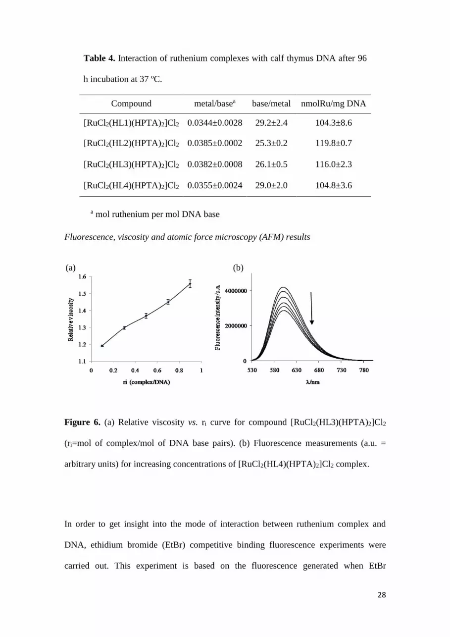
Table 4. Interaction of ruthenium complexes with calf thymus DNA after 96
h incubation at 37 ºC.
Compound metal/basea base/metal nmolRu/mg DNA
[RuCl2(HL1)(HPTA)2]Cl2 0.0344±0.0028 29.2±2.4 104.3±8.6
[RuCl2(HL2)(HPTA)2]Cl2 0.0385±0.0002 25.3±0.2 119.8±0.7
[RuCl2(HL3)(HPTA)2]Cl2 0.0382±0.0008 26.1±0.5 116.0±2.3
[RuCl2(HL4)(HPTA)2]Cl2 0.0355±0.0024 29.0±2.0 104.8±3.6
a mol ruthenium per mol DNA base
Fluorescence, viscosity and atomic force microscopy (AFM) results
Figure 6. (a) Relative viscosity vs. ri curve for compound [RuCl2(HL3)(HPTA)2]Cl2
(ri=mol of complex/mol of DNA base pairs). (b) Fluorescence measurements (a.u. =
arbitrary units) for increasing concentrations of [RuCl2(HL4)(HPTA)2]Cl2 complex.
In order to get insight into the mode of interaction between ruthenium complex and
DNA, ethidium bromide (EtBr) competitive binding fluorescence experiments were
carried out. This experiment is based on the fluorescence generated when EtBr
(a) (b)

29
intercalates between the DNA base pairs. If the assayed compound is able to modify
EtBr binding, increasing concentrations of it would result in a reduction of fluorescence
intensity. This effect was observed for all the obtained ruthenium complexes as it is
shown in Figure 6b for [RuCl2(HL4)(HPTA)2]Cl2. The quenching of the emission
maximum with increasing concentrations of the ruthenium complex indicates that a
deactivation of the adduct´s excited state occurs. This deactivation may occur due to a
competition between the ruthenium complex and EtBr for the intercalating binding sites
in DNA. However, changes in DNA configuration could also alter EtBr binding [63].
The effect of a compound on DNA viscosity is also indicative of its binding mode to
DNA in solution. Viscosity measurements are considered the most important tools when
studying the mode of DNA interaction. Viscosity is sensitive to length change of DNA.
In this sense, an intercalation mode of interaction lengthens the DNA helix, as base
pairs are separated to accommodate the binding ligand, leading to the increase of DNA
viscosity. In contrast, other modes of interaction, including covalent binding, could
bend (or kink) the DNA helix reducing its effective length and, hence, its viscosity [64-
66]. In order to further elucidate the DNA binding mode of the obtained ruthenium
complexes, viscosity measurements were carried out on CT DNA by varying the ri
(ri=mol of complex/mol of DNA base pairs) values. All the studied complexes increased
the viscosity of CT DNA solutions in a concentration dependent manner. Results for
compound [RuCl2(HL3)(HPTA)2]Cl2 are shown in Figure 6a, as an example. These
results are in accordance with those obtained in the fluorescence experiments.
AFM has proved to be a useful tool for imaging DNA and also DNA interactions with
metal complexes [25,67]. Ruthenium complexes were incubated with pBR322 plasmid
DNA. Obtained AFM images are depicted in Figure 7. Different extent of changes in
the shape of plasmid DNA were observed as a consequence of the supercoiling induced

30
by the ruthenium compounds. The observed effect is clearly more pronounced for
[RuCl2(HL4)(HPTA)2]Cl2. The very intense modifications of DNA tertiary structure for
this complex can be visualized as DNA balls formation (see Figure 7d).
Figure 7. AFM images showing the modifications suffered by pBR322 DNA due to
interaction with the Ru compounds: a) [RuCl2(HL1)(HPTA)2]Cl2, b)
[RuCl2(HL2)(HPTA)2]Cl2, c) [RuCl2(HL3)(HPTA)2]Cl2, d) [RuCl2(HL4)(HPTA)2]Cl2
for molar ratio compound: DNA base pairs 1:5 and 5 h incubation at 37 ºC, e) no metal
compound added.
Conclusions
In the search for potential antiparasitic metal complexes, four new ruthenium complexes
of the formula [RuIICl2(HL)(HPTA)2]Cl2 with 5-nitrofuryl containing
31
thiosemicarbazones as bioactive ligand and PTA (1,3,5-triaza-7-phosphaadamantane) as
co-ligand were synthesized and fully characterized. The effect of the presence of the
hydrophilic phosphine PTA resulted evident as obtained compounds were very soluble
in water. Their activity against T. cruzi and E. histolytica was evaluated in vitro.
[RuCl2(HL4)(HPTA)2]Cl2 was the most active complex against both parasites showing
an excellent activity against E. histolytica, even higher than that of the reference drug
metronidazol. As it had been previously observed, the most lipophilic compound turned
out to be the most active one. In addition, this complex turns out to be selective for the
parasites as it was not cytotoxic against murine macrophages (SI > 38).
Related to the potential mechanism of antiparasitic action of the obtained compounds,
their ability to generate oxidative stress and to interact with DNA was investigated.
Ruthenium compounds were able to generate free radicals (nitro anion and hydroxyl
radicals) in T. cruzi epimastigotes evidencing their ability to generate oxidative stress.
Similarly, [RuCl2(HL4)(HPTA)2]Cl2 produced cellular rounding and shrinkage on E.
histolytica trophozoites as well as damage to the nuclear membrane. These
morphological changes have been associated with ROS production and these could be
the trigger of the apoptosis-like processes. In addition, complexes were able to interact
with DNA. The increase in CT DNA viscosity and the decrease of CT DNA-EtBr
fluorescence produced by the complexes would be in accordance to an intercalative-like
mode of interaction. In addition, modification of plasmid DNA tertiary structure was
also shown in the AFM studies as a consequence of exposure of plasmid DNA to the
ruthenium complexes. Obtained results could be considered a contribution in the search
for compounds that could be active against multiple parasites.
Acknowledgments

32
Authors wish to thank CYTED through network 209RT0380, ANII-Uruguay (project
FCE_2007_188) and Fondecyt 11080166 and Fondecyt and International Cooperation
Project 1110029 Chile for financial support. CS thanks ANII-Uruguay for research
grant Be_INI_2010_1864. Authors also thank Prof. Jorge Castiglioni for the thermal
analysis experiments.
References
[1] Beaumier CM, Gillespie PM, Hotez PJ, Bottazzi ME (2013) New vaccines for
neglected parasitic diseases and dengue. Translational Res 162(3):144-155.
[2] Liese B, Rosenberg M, Schratz A (2010) Programs, partnerships, and governance
for elimination and control of neglected tropical diseases. Lancet 375:67-76.
[3] Astelbauer F, Walochnik J (2011) Antiprotozoal compounds: state of the art and
new developments. Int J Antimicrob Agents 38:118-124.
[4] Stanley Jr SL (2003) Amoebiasis. Lancet 361:1025-1034.
[5] Ximénez C, Morán P, Rojas L, Valadez A, Gómez A, Ramiro M, Cerritos R,
González E, Hernández E, Oswaldo P (2011) Novelties on amoebiasis: A neglected
tropical disease. J Glob Infect Dis 3:166-174.
[6] Rassi Jr A, Rassi A, Marin-Neto JA (2010) Chagas disease. Lancet 375:1388-1402.
[7] Rodrigues Coura J, Borges-Pereira J (2010) Chagas disease: 100 years after its
discovery. A systemic review. Acta Tropica 115:5-13.
[8] Chaudhary K, Roos DS (2005) Protozoan genomics for drug discovery. Nat Biotech
23:1089-1091.
[9] Urbina JA (2010) Specific chemotherapy of Chagas disease: relevance, current
limitations and new approaches. Acta. Tropica 115:55-68.

33
[10] Merlino A, Gonzalez M, Cerecetto H (2010) Targets for anti-T. cruzi drugs in the
post-genomic era. Curr Enzime Inhib 6:195-210.
[11] Le Loup G, Pialoux G, Lescure FX (2011) Update in treatment of Chagas disease.
Curr Opin Infect Dis 24:428-434.
[12] Singh S, Bharti N, Mohapatra PP (2009) Chemistry and Biology of Synthetic and
Naturally Occurring Antiamoebic Agents. Chem Rev 109:1900-1947.
[13] Navarro M, Gabbiani G, Messori L, Gambino D (2010) Metal based Drugs for
Malaria, Trypanosomiasis and Leishmaniasis. Recent achievements and perspectives
Drug Discovery Today 15:1070-1077.
[14] Gambino D (2011) Potentiality of vanadium compounds as anti-parasitic agents.
Coord Chem Rev 255:2193-2203.
[15] Sánchez-Delgado RA, Anzellotti A (2004) Metal complexes as chemotherapeutic
agents against tropical diseases: trypanosomiasis, malaria and leishmaniasis. Mini-Rev
Med Chem 4:22-30.
[16] Navarro M (2009) Gold complexes as potential anti-parasitic agents. Coord Chem
Rev 253:1619-1626.
[17] Fricker SP, Mosi RM, Cameron BR, Baird I, Zhu Y, Anastassov V, Cox J, Doyle
PS, Hansell E, Lau G, Langille J, Olsen M, Qin L, Skerlj R, Wong RSY, Santucci Z,
McKerrow JH (2008) Metal compounds for the treatment of parasitic diseases. J Inorg
Biochem 102:1839-1845.
[18] Gambino D, Otero L (2012) Perspectives on what ruthenium-based compounds
could offer in the development of potential antiparasitic drugs. Inorg Chim Acta
393:103-114.
[19] Bahl D, Athar F, Pereira Soares MB, Santos de Sá M, Magalhães Moreira DR,
Srivastava RM, Lima Leite AC, Azam A (2010) Structure–activity relationships of

34
mononuclear metal–thiosemicarbazone complexes endowed with potent antiplasmodial
and antiamoebic activities. Bioorg Med Chem 18:6857-6864.
[20] Beraldo H, Gambino D (2004) The wide pharmacological versatility of
semicarbazones, thiosemicarba-zones and their metal complexes. Mini Rev Med Chem
4:31-39.
[21] Lobana TS, Sharma R, Bawa G, Khanna S (2009) Bonding and structure trends of
thiosemicarbazone derivatives of metals-An overview. Coord Chem Rev 253:977-1055.
[22] Greenbaum DC, Mackey Z, Hansell E, Doyle P, Gut J, Caffrey CR, Lehrman J,
Rosenthal PJ, McKerrow JH, Chibale K (2004) Synthesis and Structure−Activity
Relationships of Parasiticidal Thiosemicarbazone Cysteine Protease Inhibitors against
Plasmodium falciparum, Trypanosoma brucei, and Trypanosoma cruzi. J Med Chem
47:3212-3219.
[23] Demoro B, Rossi M, Caruso F, Liebowitz D, Olea-Azar C, Kemmerling U, Maya
JD, Guiset H, Moreno V, Pizzo Ch, Mahler G, Otero L, Gambino D (2013) Potential
Mechanism of the Anti-trypanosomal Activity of Organoruthenium Complexes with
Bioactive Thiosemicarbazones. Biol Trace Elem Res 153(1-3):371-381.
[24] Demoro B, Sarniguet C, Sánchez-Delgado R, Rossi M, Liebowitz D, Caruso F,
Olea-Azar C, Moreno V, Medeiros A, Comini MA, Otero L, Gambino D (2012) New
organoruthenium complexes with bioactive thiosemicarbazones as co-ligands: potential
anti-trypanosomal agents. Dalton Trans 41:1534-1543.
[25] Vieites M, Smircich P, Pagano M, Otero L, Luane F, Terenzi H, Prieto MJ,
Moreno V, Garat B, Gambino D (2011) DNA as molecular target of analogous
palladium and platinum anti-Trypanosoma cruzi compounds: A comparative study. J
Inorg Biochem 105:1704-1711.

35
[26] Merlino A, Otero L, Gambino D, Coitiño EL (2011) In search of patterns over
physicochemical properties and pharmacological activities for a set of
[MCl2(thiosemicarbazone)] complexes (M = Pt/Pd): Support for multiple mechanisms
of antichagasic action excluding DNA-bonding in vivo? Eur J Med Chem 46(7):2639-
2651.
[27] Pagano M, Demoro B, Toloza J, Boiani L, González M, Cerecetto H, Olea-Azar C,
Norambuena E, Gambino D, Otero L (2009) Effect of ruthenium complexation on
trypanocidal activity of 5-nitrofuryl containing thiosemicarbazones. Eur J Med Chem
44:4937-4943.
[28] Vieites M, Otero L, Santos D, Olea-Azar C, Norambuena E, Aguirre G, Cerecetto
H, González M, Kemmerling U, Morello A, Maya JD, Gambino D (2009) Platinum-
based complexes of bioactive 3-(5-nitrofuryl)acroleine thiosemicarbazones showing
anti-Trypanosoma cruzi activity. J Inorg Biochem 103(3):411-418.
[29] Vieites M, Otero L, Santos D, Gajardo D, Toloza J, Figueroa R, Norambuena E,
Olea-Azar C, Aguirre G, Cerecetto H, González M, Morello A, Maya JD, Garat B,
Gambino D (2008) Platinum(II) metal complexes as potential anti-Trypanosoma cruzi
agents. J Inorg Biochem 102:1033-1043.
[30] Otero L, Maya JD, Morello A, Rigol C, Barriga G, Rodriguez J, Folch C,
Norambuena E, Gonzalez M, Olea-Azar C, Cerecetto H, Gambino D (2008) Insight into
the bioreductive mode of action of antitrypanosomal 5-nitrofuryl containing
thiosemicarbazones. Med Chem 4 (1):11-17.
[31] Otero L, Folch C, Barriga G, Rigol C, Opazo L, Vieites M, Gambino D, Cerecetto
H, Norambuena E, Olea-Azar C (2008) ESR, electrochemical and reactivity studies of
antitrypanosomal palladium thiosemicarbazone complexes. Spectrochim. Acta Part A
70:519-523.

36
[32] Otero L, Vieites M, Boiani L, Denicola A, Rigol C, Opazo L, Olea-Azar C, Maya
JD, Morello A, Krauth-Siegel RL, Piro OE, Castellano E, González M, Gambino D,
Cerecetto H (2006) Novel Antitrypanosomal Agents Based on Palladium
Nitrofurylthiosemicarbazone Complexes: DNA and Redox Metabolism as Potential
Therapeutic Targets. J Med Chem 49:3322-3331.
[33] Phillips AD, Gonsalvi L, Romerosa A, Vizza F, Peruzzini M (2004) Coordination
chemistry of 1,3,5-triaza-7-phosphaadamantane (PTA) - Transition metal complexes
and related catalytic, medicinal and photoluminescent applications. Coord Chem Rev
248:955-993.
[34] Bravo J, Bolaño S, Gonsalvi L, Peruzzini M (2010) Coordination chemistry of
1,3,5-Triaza-7-phosphaadamantane (PTA) and derivatives. Part II. The quest for
tailored ligands, complexes and related applications. Coord Chem Rev 254:555-607.
[35] Bergamo A, Gaiddon C, Schellens JHM, Beijnen JH, Sava G (2012) Approaching
tumour therapy beyond platinum drugs: Status of the art and perspectives of ruthenium
drug candidates. J Inorg Biochem 106:90–99.
[36] Aguirre G, Boiani L, Cerecetto H, González M, Denicola A, Otero L, Gambino D,
Rigol C, Olea-Azar C, Faundez M (2004) In vitro activity and mechanism of action
against the protozoan parasite Trypanosoma cruzi of 5-nitrofuryl containing
thiosemicarbazones. Bioorg Med Chem 12:4885-4893.
[37] Evans I, Spencer A, Wilkinson GJ (1973) Dichlorotetrakis(dimethyl
sulphoxide)ruthenium(II) and its use as a source material for some new ruthenium(II)
complexes. Chem Soc Dalton Trans 2:204-209.
[38] Hansch C, Leo A (1995) The hydrophobic parameter: measurement and
calculation, Exploring QSAR. Fundamentals and Applications in Chemistry and
Biology, American Chemical Society, Washington, 97–124.

37
[39] Maya JD, Morello A, Repetto Y, Tellez R, Rodriguez A, Zelada U, Puebla P,
Bontá M, Bollo S, San Feliciano A (2000) Effects of 3-chloro-phenyl-1,4-
dihydropyridine derivatives on Trypanosome cruzi epimastigotes. Comp Biochem
Physiol C 125:103-109.
[40] Cuellar MA, Salas C, Cortés MJ, Morello A, Maya JD, Preite MD (2003) Synthesis
and in vitro trypanocide activity of several polycyclic drimane-quinone derivatives.
Bioorg Med Chem 11:2489-2497.
[41] Muelas-Serrano S, Nogal-Ruiz JJ, Gómez-Barrio A (2000) Setting of a
colorimetric method to determine the viability of Trypanosoma cruzi epimastigotes.
Parasitol Res 86:999-1002.
[42] Faundez M, Pino L, Letelier P, Ortiz C, López R, Seguel C, Ferreira J, Pavani M,
Morello A, Maya JD (2005) Buthionine sulfoximine increases the toxicity of nifurtimox
and benznidazole to Trypanosoma cruzi. Antimicrob Agents Chemother 49:126-130.
[43] Diamond L (1961) Axenic cultivation of Entamoeba histolytica. Science 134:336-
337.
[44] Benítez J, Guggeri L, Tomaz I, Costa Pessoa J, Moreno V, Lorenzo J, Avilés FX,
Garat B, Gambino D (2009) A novel vanadyl complex with a polypyridyl DNA
intercalator as ligand: A potential anti-protozoa and anti-tumor agent. J Inorg Biochem
103:1386-1394.
[45] Zhang G, Guo J, Pan J, Chen X, Wang J (2009) Spectroscopic studies on the
interaction of morin–Eu(III) complex with calf thymus DNA. J Mol Structure 923:114-
119.
[46] Geary WJ (1971) The use of conductivity measurements in organic solvents for the
characterisation of coordination compounds. Coord Chem Rev 7:81-122.

38
[47] Grguric-Sipka S, Kowol CR, Valiahdi S, Eichinger R, Jakupec MA, Roller A,
Shova S, Arion VB, Keppler BK (2007) Ruthenium(II) Complexes of
Thiosemicarbazones: The First Water-Soluble Complex with pH-Dependent
Antiproliferative Activity. Eur J Inorg Chem 18:2870–2878.
[48] Mebi CA, Frost BJ (2007) Isomerization of trans-[Ru(PTA)4Cl2] to cis-
[Ru(PTA)4Cl2] in Water and Organic Solvent: Revisiting the Chemistry of
[Ru(PTA)4Cl2]. Inorg Chem 46:7115-7120.
[49] Gambino D, Otero L, Vieites M, Boiani M, Gonzalez M, Baran EJ, Cerecetto H
(2007) Vibrational spectra of palladium 5-nitrofuryl thiosemicarbazone complexes:
Experimental and theoretical study. Spectrochim Acta Part A 68:341–348.
[50] Jogun K, Stezowski JJ, Fluck E, Weidlein J (1978) Molecular structure of p-
substituted 1,3,5-triaza-7-phosphaadamantanes: vibration spectra and crystal structure
analyses. Phosphorous Sulfur 4:99-204.
[51] Makino K, Hagiwara T, Murakami A (1991) Fundamental-Aspects of Spin
Trapping with Dmpo. Radiat Phys Chem 37:657-665.
[52] Aguilera-Venegas B, Olea-Azar C, Arán V, Maya JD, Kemmerling U, Speisky H,
Mendizábal F (2012) Electrochemical, ESR and Theoretical Insights into the Free
Radical Generation by 1,1'-Hydrocarbylenebisindazoles and Its Evaluation as Potential
Bio-Active Compounds. Int J Electrochem Sci 7:5837-5863.
[53] Villalba JD, Gómez C, Medel O, Sánchez V, Carrero JC, Shibayama M, Pérez DG
Ishiwara (2007) Programmed Cell Death in Entamoeba histolytica induced by the
Aminoglycoside G418. Microbiology 153:3852-3863.
[54] Aguilar-Díaz H, Díaz-Gallardo M, Laclette JP, Carrero JC (2010) In vitro
induction of Entamoeba histolytica cyst-like structures from trophozoites. PLoS Negl
Trop Dis 4:e607.

39
[55] Nandi N, Sen A, Banerjee R, Kumar S, Kumar V, Ghosh AN, Das P (2010)
Hydrogen peroxide induces apoptosis-like death in Entamoeba histolytic trophozoites.
Microbiology 156:1926-1941.
[56] Ramos E, Olivos-García A, Nequiz M, Saavedra E, Tello E, Saralegui A, Montfort
I, Pérez Tamayo R (2007) Entamoeba histolytica: Apoptosis induced in vitro by nitric
oxide species. Exp Parasitol 116:257-265.
[57] García-Ramos JC, Toledano-Magaña Y, Talavera-Contreras LG, Flores-Álamo M,
Ramírez-Delgado V, Morales-León E, Ortiz-Frade L, Grizett Gutiérrez A, Vázquez-
Aguirre A, Mejía C, Carrero JC, Laclette JP, Moreno-Esparza R, Ruiz-Azuara L
(2012) Potential cytotoxic and amoebicide activity of first row transition metal
compounds with 2,9-bis-(2′,5′-diazahexanyl)-1,1-phenanthroline (L1). Dalton Trans
41:10164-10174.
[58] Freeman CD, Klutman NE, Lamp KC (1997) Metronidazole. A therapeutic review
and update. Drugs, 54:679-708.
[59] Bansal D, Malla N, Mahajan MC (2006) Drug resistance in amoebiasis. Indian J
Med Res 123:115-118.
[60] Zambrano-Villa S, Rosales-Borjas D, Carrero JC, Ortiz-Ortiz L (2002) How
protozoan parasites evade the immune response. Trends Parasitol 18:272-278.
[61] Huppertz B, Frank HG, Kaufmann P (1999) The apoptosis cascade--morphological
and immunohistochemical methods for its visualization. Anat Embryol 200:1-18.
[62] Gómez-Quiroga A, Navarro-Ranninger C (2004) Contribution to the SAR field of
metallated and coordination complexes: Studies of the palladium and platinum
derivatives with selected thiosemicarbazones as antitumoral drugs. Coord Chem Rev
248:119-133.

40
[63] Mahadevan S, Palaniandavar M (1998) Spectral and Electrochemical Behavior of
Copper(II)−Phenanthrolines Bound to Calf Thymus DNA. [(5,6-dimethyl-OP)2Cu]2+
(5,6-dimethyl-OP = 5,6-Dimethyl-1,10-phenanthroline) Induces a Conformational
Transition from B to Z DNA. Inorg Chem 37:3927-3934.
[64] Satyanarayana S, Dabroniak JC, Chaires JB (1992) Neither .DELTA.- nor
.LAMBDA.-tris(phenanthroline)ruthenium(II) binds to DNA by classical intercalation.
Biochemistry 31:9319–9324.
[65] Satyanaryana S, Daborusak JC, Chaires JB (1993)
Tris(phenanthroline)ruthenium(II) enantiomer interactions with DNA: Mode and
specificity of binding. Biochemistry 32:2573–2584.
[66] Liu Y, Chao H, Tan L, Yuan Y, Wei W, Ji L (2004) A comparative study of the
interaction of two structurally analogue ruthenium(II) complexes with DNA. J Inorg
Biochem 98:2011–2015.
[67] Onoa GB, Cervantes G, Moreno V, Prieto MJ (1998) Study of the interaction of
DNA with cisplatin and other Pd(II) and Pt(II) complexes by atomic force microscopy.
Nucleic Acids Res 26:1473–1480.

![Fulvene–Ruthenium and Cp–Ruthenium Complexes via [2 + 2 + 1] Cyclotrimerization of Phenylacetylene with [RuCl(Tp)(1,5-cod)]](https://img.pdfslide.net/doc/110x75/63571545debc1859f603a50f/fulveneruthenium-and-cpruthenium-complexes-via-2-2-1-cyclotrimerization.jpg)


