Embed Size (px)
Citation preview
(12) INTERNATIONAL APPLICATION PUBLISHED UNDER THE PATENT COOPERATION TREATY (PCT)
(19) World Intellectual PropertyOrganization
International Bureau (10) International Publication Number
(43) International Publication Date WO 2018/209324 A215 November 2018 (15.11.2018) W ! P O PCT
(51) International Patent Classification: (72) Inventors; andC12Q 1/6886 (2018.01) (71) Applicants: SADE-FELDMAN, Moshe [US/US]; 55
Fruit Street, Boston, MA 021 14 (US). YIZHAK, Keren(21) International Application Number:
[US/US]; 415 Main Street, Cambridge, MA 02142 (US).PCT/US20 18/032466
GETZ, Gad [US/US]; 55 Fruit Street, Boston, MA 021 14
(22) International Filing Date: (US). HACOHEN, Nir [US/US]; 55 Fruit Street, Boston,11 May 2018 ( 11.05.2018) MA 021 14 (US).
(25) Filing Language: English (74) Agent: SCHER, Michael, B. et al; Johnson, Marcou &Isaacs, LLC, P.O. Box 6 1, Hoschton, GA 30548 (US).
(26) Publication Language: English(81) Designated States (unless otherwise indicated, for every
(30) Priority Data: kind of national protection available): AE, AG, AL, AM,62/505,101 11 May 2017 ( 11.05.2017) AO, AT, AU, AZ, BA, BB, BG, BH, BN, BR, BW, BY, BZ,62/574,878 20 October 20 17 (20. 10.201 7) CA, CH, CL, CN, CO, CR, CU, CZ, DE, DJ, DK, DM, DO,
(71) Applicants: THE BROAD INSTITUTE, INC. [US/US]; DZ, EC, EE, EG, ES, FI, GB, GD, GE, GH, GM, GT, HN,
415 Main Street, Cambridge, MA 02142 (US). THE GEN¬ HR, HU, ID, IL, IN, IR, IS, JO, JP, KE, KG, KH, KN, KP,
ERAL HOSPITAL CORPORATION [US/US]; 55 Fruit KR, KW, KZ, LA, LC, LK, LR, LS, LU, LY, MA, MD, ME,
Street, Boston, MA 021 14 (US). MG, MK, MN, MW, MX, MY, MZ, NA, NG, NI, NO, NZ,OM, PA, PE, PG, PH, PL, PT, QA, RO, RS, RU, RW, SA,
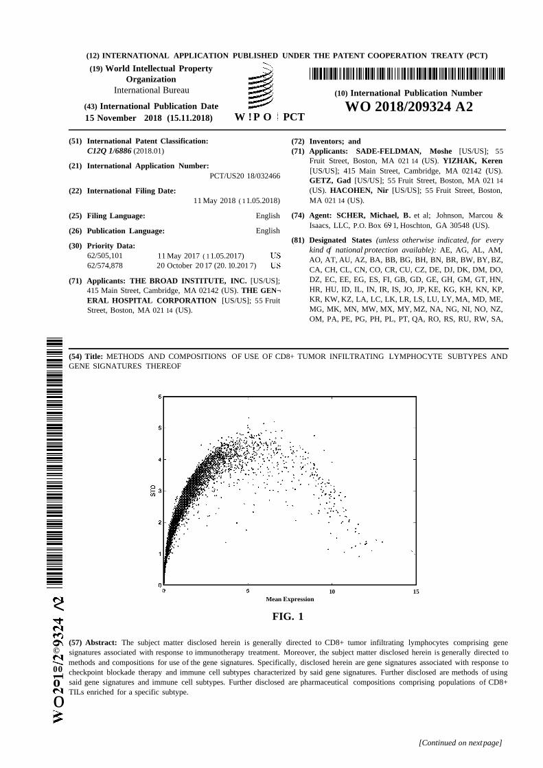
(54) Title: METHODS AND COMPOSITIONS OF USE OF CD8+ TUMOR INFILTRATING LYMPHOCYTE SUBTYPES ANDGENE SIGNATURES THEREOF
10 15Mean Expression<
FIG. 1
(57) Abstract: The subject matter disclosed herein is generally directed to CD8+ tumor infiltrating lymphocytes comprising gene© signatures associated with response to immunotherapy treatment. Moreover, the subject matter disclosed herein is generally directed to
methods and compositions for use of the gene signatures. Specifically, disclosed herein are gene signatures associated with response to00 checkpoint blockade therapy and immune cell subtypes characterized by said gene signatures. Further disclosed are methods of using
o said gene signatures and immune cell subtypes. Further disclosed are pharmaceutical compositions comprising populations of CD8+TILs enriched for a specific subtype.o
[Continued on nextpage]
WO 2018/209324 A2 llll I I I I 11III II I 11II IIII 11I I III I I I II
SC, SD, SE, SG, SK, SL, SM, ST, SV, SY, TH, TJ, TM, TN,
TR, TT, TZ, UA, UG, US, UZ, VC, VN, ZA, ZM, ZW.
(84) Designated States (unless otherwise indicated, for everykind of regional protection available): ARIPO (BW, GH,
GM, KE, LR, LS, MW, MZ, NA, RW, SD, SL, ST, SZ, TZ,
UG, ZM, ZW), Eurasian (AM, AZ, BY, KG, KZ, RU, TJ,
TM), European (AL, AT, BE, BG, CH, CY, CZ, DE, DK,
EE, ES, FI, FR, GB, GR, HR, HU, IE, IS, IT, LT, LU, LV,
MC, MK, MT, NL, NO, PL, PT, RO, RS, SE, SI, SK, SM,
TR), OAPI (BF, BJ, CF, CG, CI, CM, GA, GN, GQ, GW,
KM, ML, MR, NE, SN, TD, TG).
Published:— without international search report and to be republished
upon receipt of that report (Rule 48.2(g))— with sequence listing part of description (Rule 5.2(a))
METHODS AND COMPOSITION OF USE OF CD8+ TUMOR INFILTRATINGLYMPHOCYTE SUBTYPES AND GENE SIGNATURES THEREOF
CROSS-REFERENCE TO RELATED APPLICATIONS
[0001] This application claims the benefit of U.S. Provisional Application Nos.
62/505,101, filed May 11, 2017 and 62/574,878, filed October 20, 2017. The entire contents
of the above-identified applications are hereby fully incorporated herein by reference.
STATEMENT REGARDING FEDERALLY SPONSORED RESEARCH
[0002] This invention was made with government support under Grant No. CA208756
awarded by the National Institutes of Health. The government has certain rights in the
invention.
TECHNICAL FIELD
[0003] The subject matter disclosed herein is generally directed to CD8+ tumor
infiltrating lymphocytes comprising gene signatures associated with response to
immunotherapy treatment and overall survival. Moreover, the subject matter disclosed herein
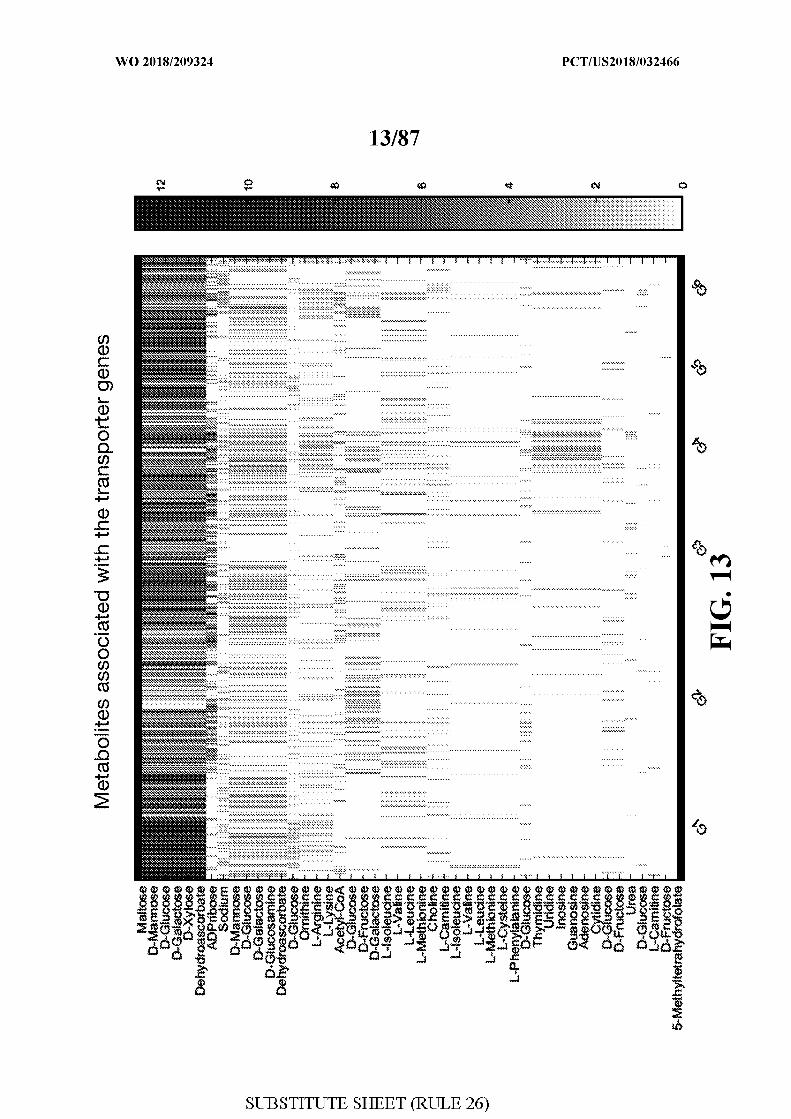
is generally directed to methods and compositions for use of the signature genes.
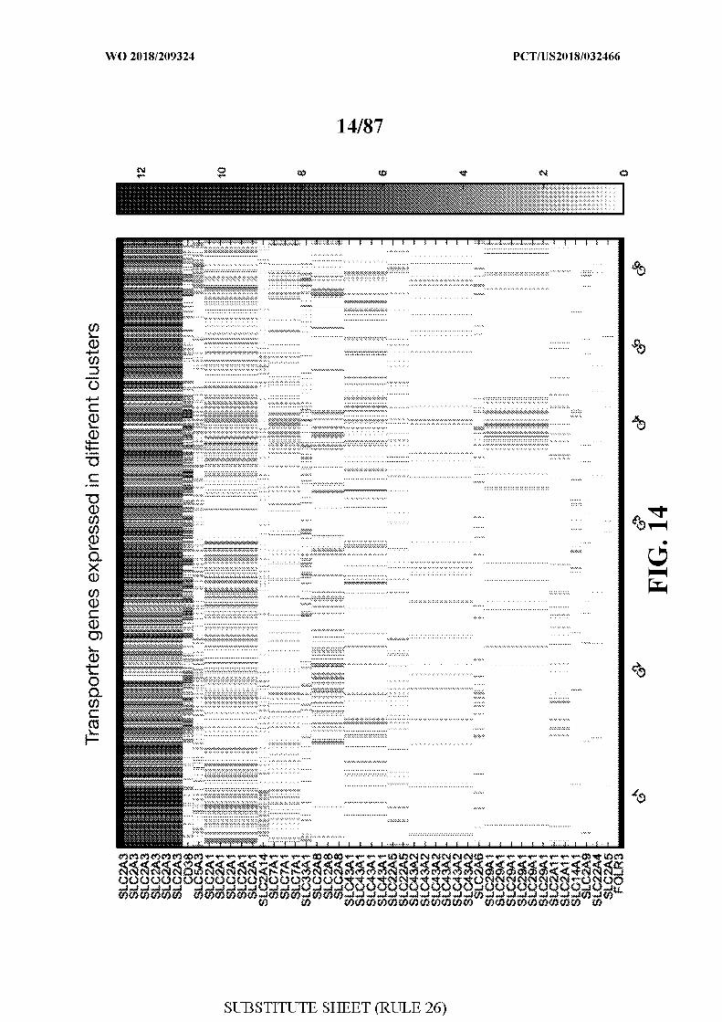
BACKGROUND
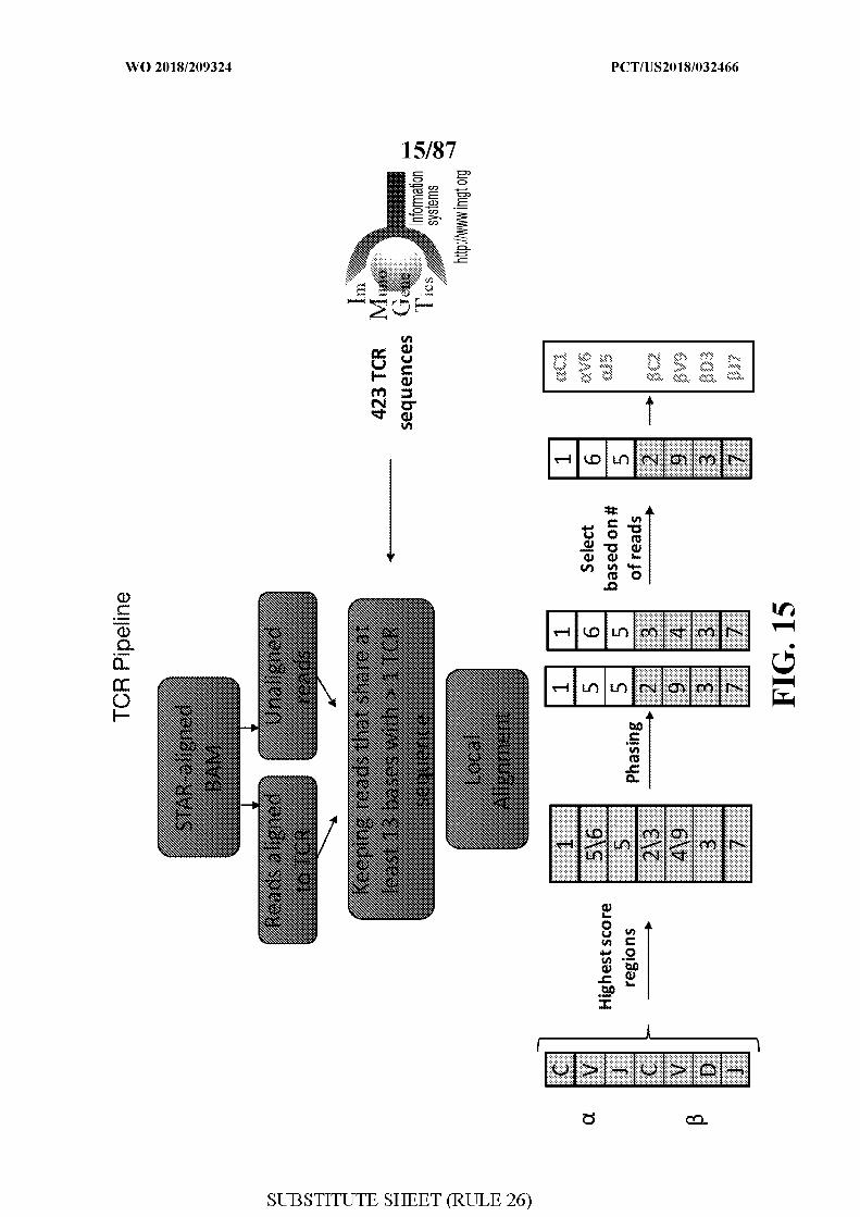
[0004] The development of antibodies that effectively block the activities of immune
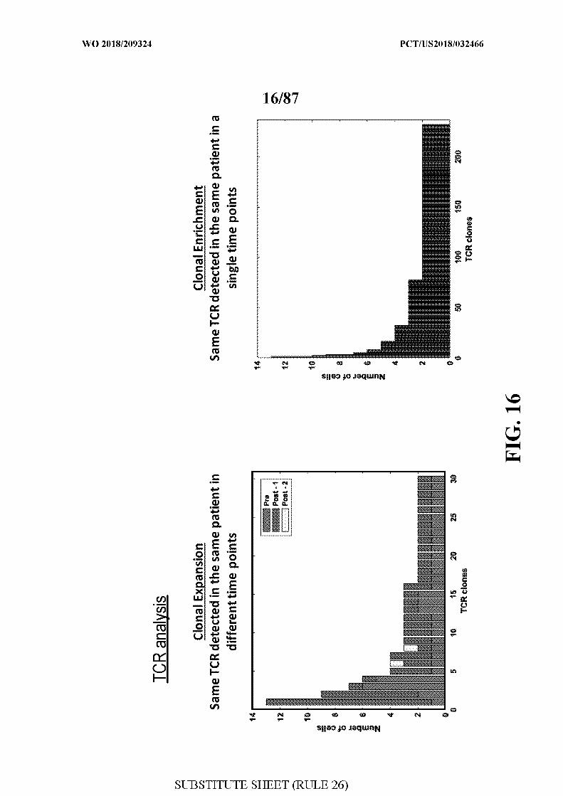
checkpoint proteins, including CTLA4, PD-1 or its ligand, PD-L1 , has led to their approval
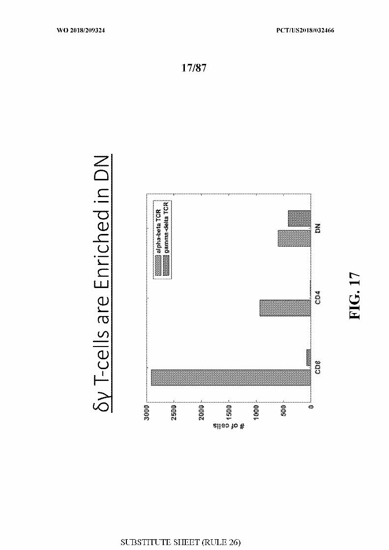
by the FDA for treating a wide variety of cancers, including melanoma, non-small-cell lung
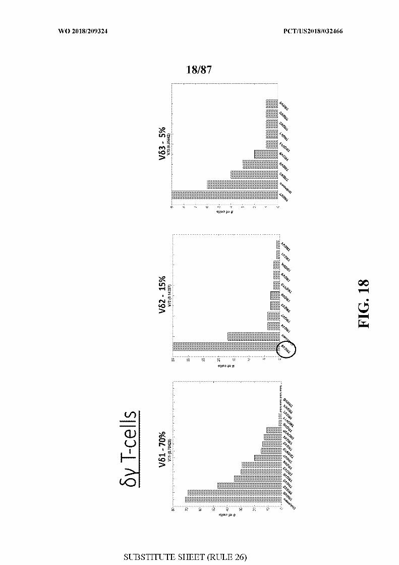
carcinoma (NSCLC), renal cell carcinoma (RCC), urothelial bladder cancer (UBC), head and
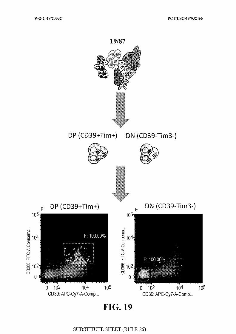
neck squamous cell carcinoma (HNSCC), refractory Hodgkin's lymphoma (HL), and most
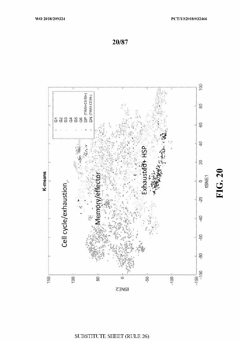
recently hepatocellular carcinoma (HCC) and gastric cancer 2. In melanoma, despite the high
response rate (-20% for anti-CTLA4, -45% for anti-PD-1, -60% for anti-PD-l+anti-
CTLA4) '4, most patients are refractory to therapy or acquire resistance, and eventually
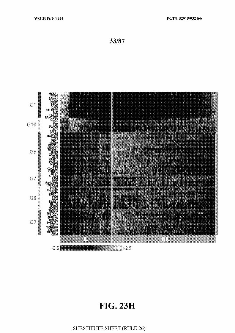
succumb to disease. Thus, identification of the key components that drive or prevent effective
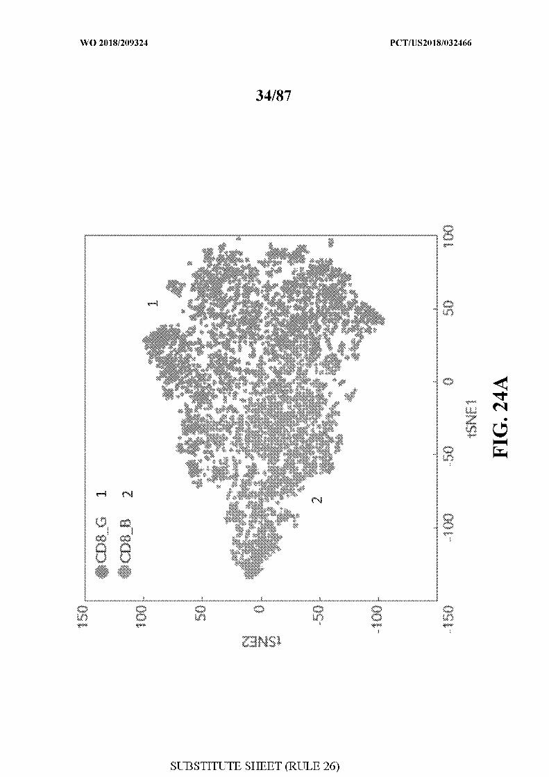
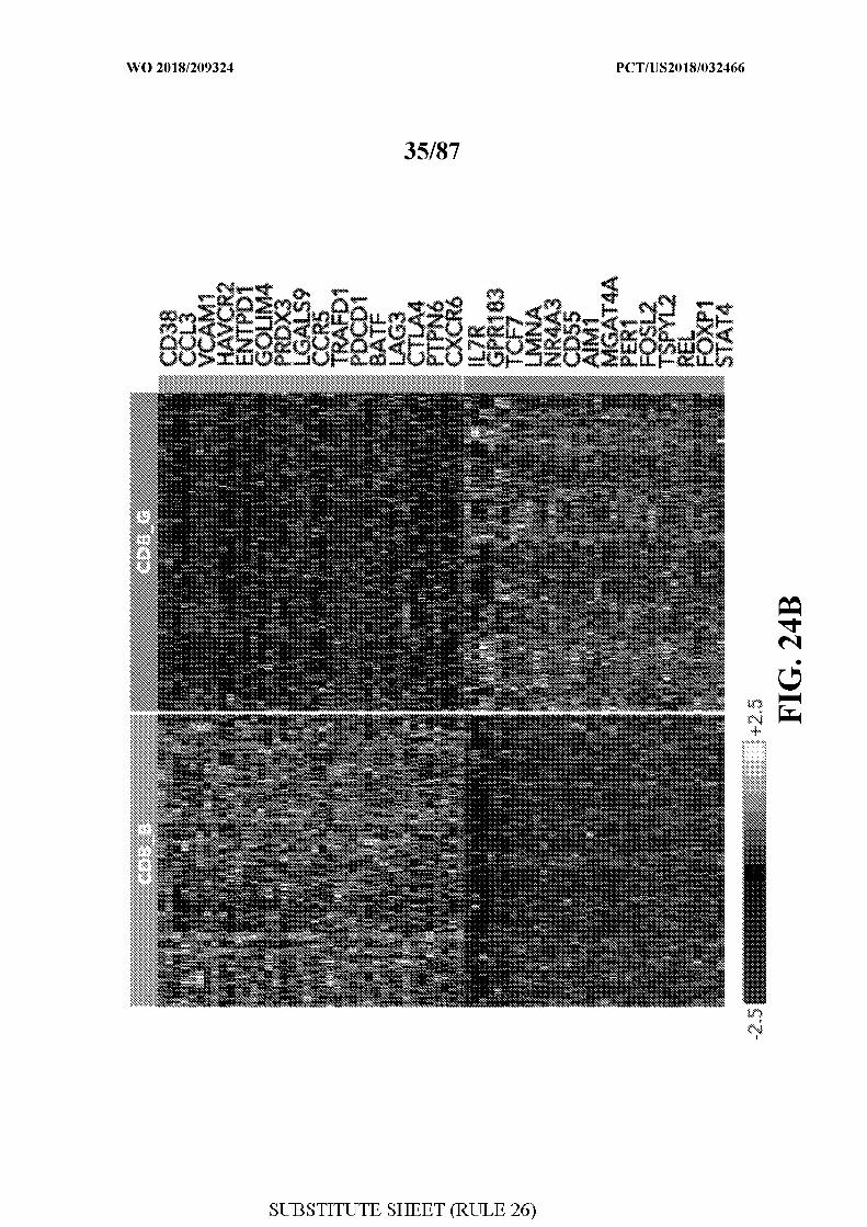
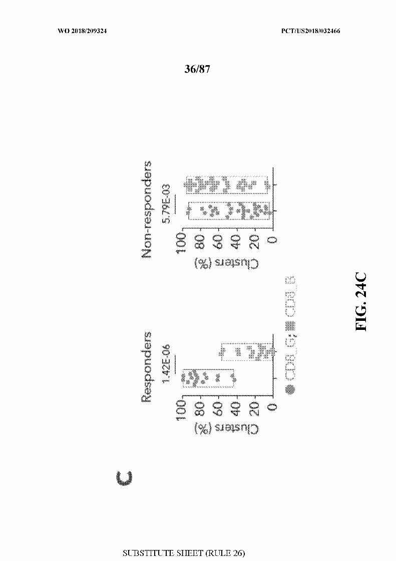
responses to checkpoint therapy remains an urgent need for accelerating progress in the fields
of cancer immunotherapy, and perhaps, medical oncology.
[0005] Checkpoint therapies are designed to overcome the inhibition of antigen-specific,
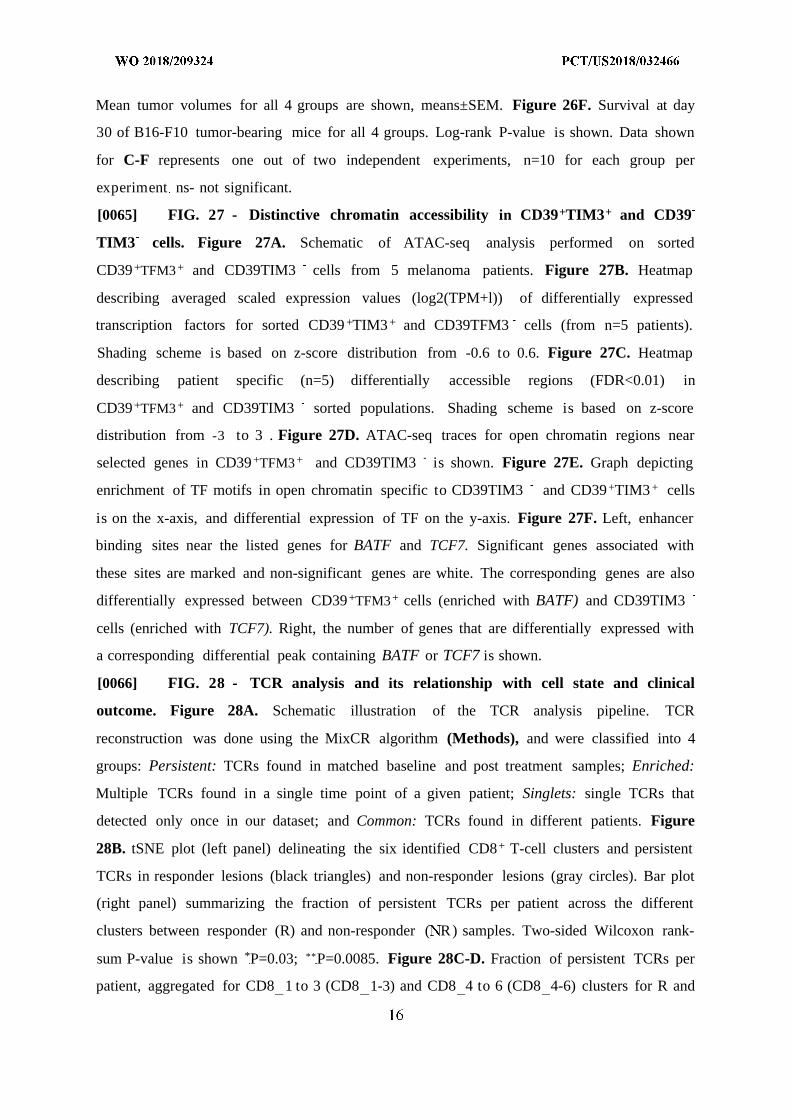
effector T lymphocytes (T-cells) by the tumor or the immune microenvironment. Thus, the
state and number of these cells, especially CD8+ cytotoxic T-cells are likely to determine the
clinical outcome. Indeed, the number of infiltrating CD8+ T-cells detected before or during
early treatment 6 have been shown to be associated with clinical outcome. The ability of these
CD8+ T-cells to target the malignant cells is dampened by persistent exposure to stimulation
and co-inhibition by checkpoint proteins, resulting in a state of exhaustion ' , characterized
by the expression of multiple co-inhibitory receptors on the T-cell surface (e.g. PD1, CTLA4,
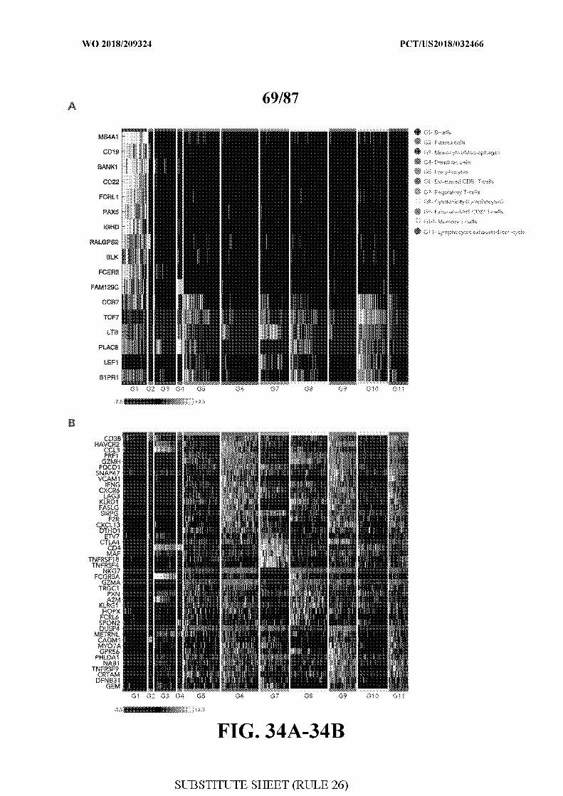
TIM3, TIGIT), unique regulators of gene expression (BATF, PRDM1), and most importantly,
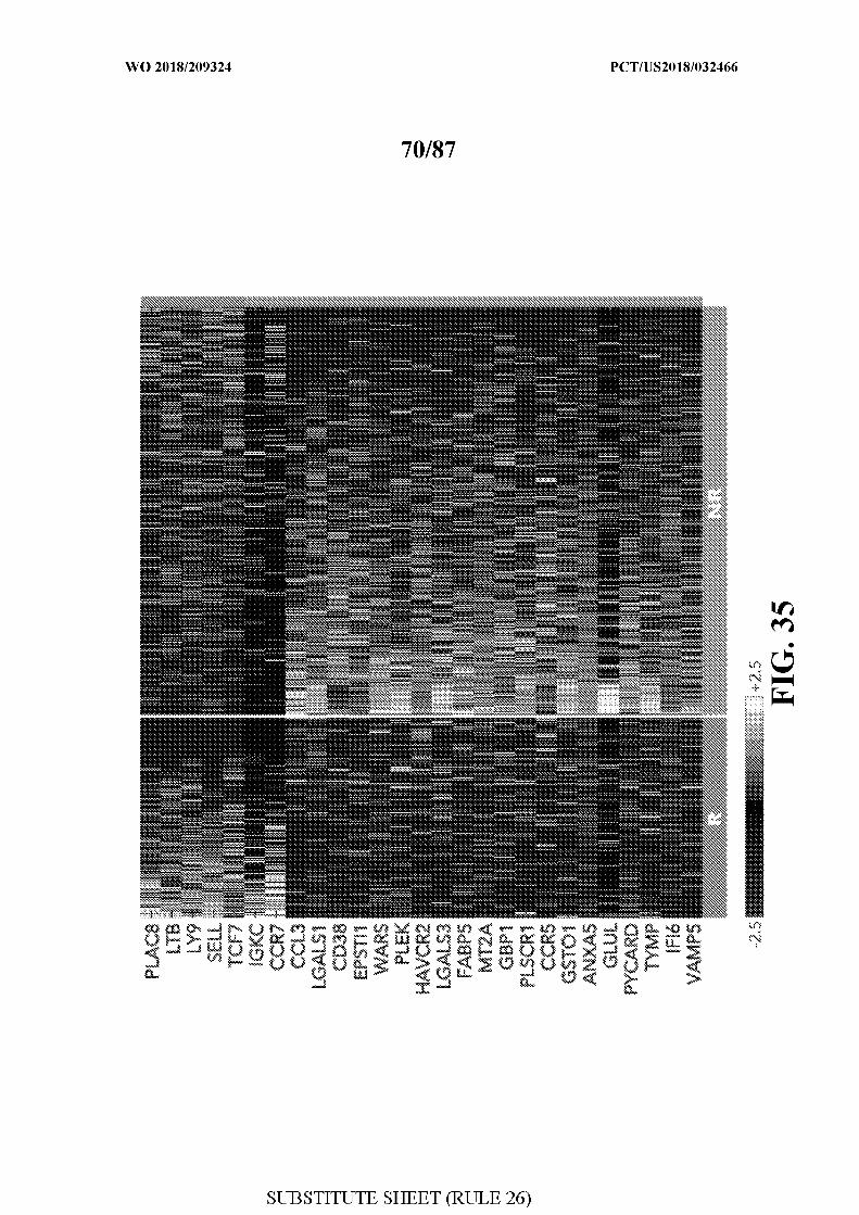
dysfunctional effector activity. Additionally, the efficiency of checkpoint therapy depends on
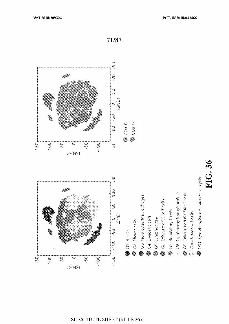
CD8+ T-cell recognition of neoantigens presented on human leukocyte antigen (HLA) class I
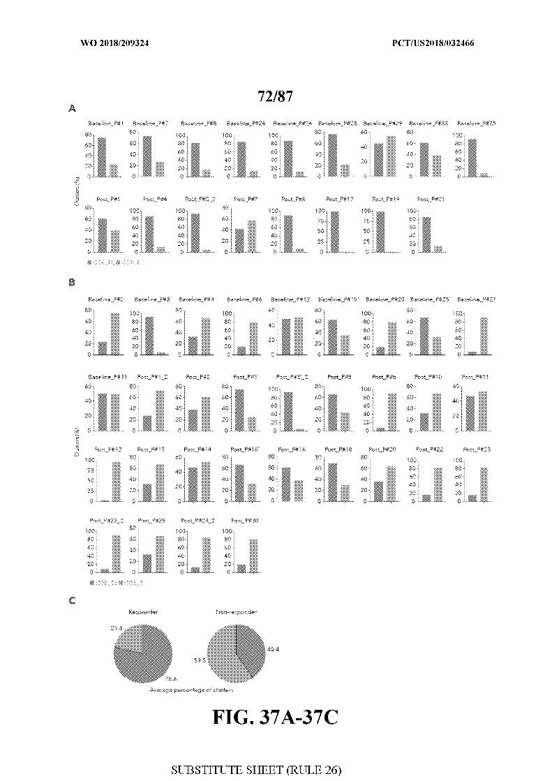
by tumor cells 1° . Hence, a deeper understanding of the cellular and molecular determinants
of response are needed.
[0006] To date, several factors have been analyzed for their association with tumor
growth and clinical outcome in patients. These include levels of PD-L1 protein 4 , load of
tumor-derived neoantigens 12 , defects in antigen presentation and IFNg pathways 13 16,
abundance of partially exhausted CD8+ T-cells in the tumor 1 , proportion of suppressive
myeloid cells in the blood 1 , and the magnitude of T-cell reinvigoration in relation to
pretreatment tumor burden 19 . While these studies have collectively contributed to the model
explaining the efficacy of checkpoint therapy, their major limitations include low predictive
power and the use of pre-defined immune markers, limiting their ability to identify optimal
and novel components that explain or predict clinical outcomes. Thus, there is a need to more
systematically identify markers and mechanisms associated with response to therapy.
[0007] Citation or identification of any document in this application is not an admission
that such document is available as prior art to the present invention.
SUMMARY
[0008] In one aspect, the present invention provides for a method of detecting a
checkpoint blockade (CPB) therapy responder gene signature comprising, detecting in
CD45+ cells obtained from a biological sample the expression of a gene signature comprising
one or more genes or polypeptides selected from the group consisting of: TCF7; or TCF7,
PLAC8, LTB, and CCR7; or TCF7, LEF1, S1PR1, PLAC8, LTB, CCR7, IGHD, PAX5,
FCRLl, FCER2, CD19, CD22, BANKl, MS4A1, BLK, RALGPS2 and FAM129C; or
TCF7, PLAC8, LTB, LY9, SELL, IGKC and CCR7.
[0009] In another aspect, the present invention provides for a method of detecting a
checkpoint blockade (CPB) therapy responder gene signature comprising, detecting in CD8+
T cells obtained from a biological sample the expression of a gene signature comprising one
or more genes or polypeptides selected from the group consisting of: TCF7; or TCF7 and
IL7R; or TCF7, IL7R, FOSL2, REL, FOXPl, and STAT4; or TCF7, PLAC8, LTB, and
CCR7; or TCF7, LEF1, S1PR1, PLAC8, LTB, and CCR7; or TCF7, IL7R, GPR183, and
MGAT4A; or TCF7, IL7R, GPR183, LMNA, R4A3, CD55, AFM1, MGAT4A, PERI,
FOSL2, TSPYL2, REL, FAM177A1, YPEL5, TC2N and CSR P 1; or TCF7, IL7R,
GPR183, LMNA, NR4A3, CD55, AFM1, MGAT4A, PERI, FOSL2, TSPYL2, REL,
FAM177A1, YPEL5, TC2N, CSRNP1, FAM65B, PIK3R1, RGPD6, SKIL, TSC22D2,
USP36, FOXPl, EGRl, MYADM, ZFP36L2, FAM102A, RGCC, PDE4B, PFKFB3, FOSB,
DCTN6 and BTG2; or CD8 G genes listed in Table 6 .
[0010] In certain embodiments, the CD8 T cells having a responder signature does not
express ENTPDl (CD39) and HAVCR2.
[0011] In another aspect, the present invention provides for a method of detecting a
checkpoint blockade (CPB) therapy non-responder gene signature comprising, detecting in
CD45+ cells obtained from a biological sample the expression of a gene signature comprising
one or more genes or polypeptides selected from the group consisting of: ENTPDl and
HAVCR2; or CCL3, CD38 and HAVCR2; or CD38, PDCD1, CCL3, SNAP47, VCAM1,
HAVCR2, FASLG, ENTPDl, SIRPG, MY07A, FABP5, NDUFB3, UBE2F, CLTA and
SNRPD1; or FASLG, VCAM1, CCL3, LAG3, CXCR6, IFNG, PDCD1, KLRD1, HAVCR2,
SIRPG, SNAP47, DTHD1, PRF1, GZMH, F2R, CD38, CXCL13, TNFRSF4, TNFRSF18,
MAF, ETV7, CD4, CTLA4, FCRL6, SPON2, KLRG1, TRGC1, A2M, FCGR3A, GZMA,
HOPX, NKG7, PXN, TNFRSF9, GEM, NABl, DFNB31, CADMl, CRTAM, GPR56,
MY07A, DUSP4, METRNL and PHLDA1; or LAYN, GEM, VCAM1, RDH10,
TNFRSF18, FAM3C, AFAP1L2, KIR2DL4, MTSS1, ETV1, CTLA4, MY07A, ENTPDl,
TNFRSF9, CADMl, DFNB31, CXCL13, HAVCR2, GPR56, GOLFM4, NABl, PHLDA1,
TGIF1, SEC14L1, IGFLR1, NAMPTL, PAM, HSPB1, TNIP3, BPGM, TP53INP1, TRPS1,
UBE2F, NDFIP2, PON2, PELIl, METRNL, SNAP47 and APLP2; or CCL3, LGALSl,
CD38, EPSTI1, WARS, PLEK, HAVCR2, LGALS3, FABP5, MT2A, GBP1, PLSCR1,
CCR5, GSTOl, ANXA5, GLUL, PYCARD, TYMP, IFI6, VAMP5, OASL, GZMB, TXN,
SQRDL, RHOC, AP2S1, GZMH, CCL4L2, SNAP47, LAP3, ATP6V1B2, CCL4L1,
LAMP2, PSMA4, SERPINBl, HIGD1A, UBE2F, TALDOl, CD63, CLTA, S100A1 1,
PHPT1, GBP4, PRDX3, PSMB2, BST2, GBP 5, CTSC, NDUFB3, NPC2, GALM, GLIPR2,
CCL4, PRF1, IFNG, IFI30, CHST12, ISG15, MYD88, IDH2, MTHFD2, CHMP2A,
UFA9, CHMP5, CALM3, ANXA2, PPTl, GTF3C6, NDUFABl, CXCR6, RNF181,
LGALS9, COX5A, OAS2, PDCD1, SNRPC, BHLHE40, TWF2, SLAMF7, TXN2,
CARD 16, ANAPCl l , MRPL51, LIMS1, NDUFA12, RANBP1, GBP2, PSMC1, ACTR1A,
CD2BP2, VDAC1, EMC7, MX1, GPS1, ATP5J2, USMG5, SHFM1, ATP51, FAM96A,
CASP1, PARP9, NOP10, GNG5, CYC1, RABl lA, PGAM1, ENTPD1, PDIA6, PSMC3,
TMBIM1, UBE2L6, PSMA6, EIF6, DCTN3, SEC1 1A, CSTB, ETFB, DBI, GRN, ELOVLl,
UBE2L3, PSMB3, NDUFB7, DOK2, SEC61G, IGFLR1, ATP5H, COPZ1, ATP6V1F,
BNIP3L, NUTF2, AKRIAI, MDH2, VAMP8, ROMOl, CXCR3, SAMHDl, NUCBl,
ACTN4, ZYX, FLOT1, BLOC1S1, STAT1, VFMP, PAM, NUDT21, MYOIG, C17orf49,
GTF2A2, HIST2H2AA4, C19orfl0, ABI3, TRAPPC5, PSMC4, NDUFC2, HN1, SNRPD3,
CMC1, RAB27A, NDUFA6, POMP, PFKP, ATP5G3, TMEM179B, PSMD9, IRF7, CNIH1,
DYNLRBl, APOL2, TKT, DCTN2, GSDMD, STOM, CTSD, KDELR2, ATP5J, RPS27L,
PSME2, DRAPl, NDUFBIO, DECR1, GSTP1, TMED9, MGAT1, HSPB1, COX8A, ZEB2,
ILK, PSMB6, HK1, CD58, TMX1, GZMA, SRI, PSMG2, ARL8B, NKG7, GPX1, ACP5,
CHP1, GPR171, ATP6V0B, KLRD1, H2AFY, PPM1G, PRDX5, PSMA5, FBXW5,
ATP6AP1, CD4, SNRPD1, XAFl, LY6E, DYNLTl, AK2, PSMA2, YIPF3, S100A10,
SCP2, MRPS34, PSMD4, CDC123, BTG3, TMEM258, TSPO, SDHB, TCEB1, WDR830S,
HCST, NAA10, CTSB, YARS, GLRX, RBCK1, RBX1, LAMTOR1, UQCRFS1, NDUFB4,
CAPZA2, BRK1, ADRM1, NDUFB2, ETFA, VDAC3, NUDT5, IFITM3, BANFl, ZNHIT1,
CAPG, NHP2, LASP1, TOMM5, MVP, CTSW, AURKAIP1, RARRES3, PSMB10,
TMEM173, SLX1A, APOBEC3G, GFMAP4, EIF4E, CTLA4, NDUFS8, CYB5B, PIK3R5,
HEXB, STXBP2, PSMD8, SEC61B, RGS10, PHB, ATP5C1, ARF5, SUM03, PRDX6,
RNH1, ATP5F1, UQCRC1, SARNP, PLIN2, PIN1, SDHC, SF3B14, CAPRINl, POLR2G,
COX7B, UQCR10, FBX07, NDUFB6, S100A4, PRELID1, TRPV2, SF3B5, MYOIF,
SCAMP2, RNF7, CXCL13, RABIB, SHKBP1, PET100, HM13, VTI1B, S100A6, ARPC5,
FDPS, MINOS 1, RABIO, NEDD8, BATF, PHB2, ERH, NCOA4, PDIA4, PSMB9,
Cl lorf48, TMEM50A, TIGIT, NDUFAl l , NELFE, COX6C, SLA2, PSMB8, NDUFS7,
RERl, RAB8A, CAPNl, MRPL20, COX5B, SEC13, FKBPIA, PRDMl, RABIA, RHOG,
CYB5R3, AIP, ABRACL, PSMB7, COX6B1, PSMD7, PPA1, PCMT1, SURF4, ENY2,
TCEB2, MAP2K3, AL353354.2, AKIRIN2, MAPREl, GRSF1, DUSP4, ATG3, SRGAP2,
ATP6V0D1, NELFCD, LRPAPl, C14orfl66, SNRPB2, CHMP4A, SFT2D1, CASP4,
NME1-NME2, FAM96B, FDFTl, SLC25A39, LMAN2, MDHl, RHBDD2, ARPC5L,
TBCA, EBP, SEC14L1, EIF2S2, CST7, STARD7, SOD2, SPN, FAM32A, SEC1 1C,
TNFRSF1B, POLR2E, NDUFA13, OSTC, UFCl, C18orf32, SRP19, C14or£2, UQCR1 1,
PDCD6, AP2M1, PPP1CA, ATP6AP2, SSR3, UNCI 3D, FERMT3, ARHGAPl, EIF3I,
CECR1, MRPS6, DNPHl, DCXR, PSMF1, SNRPG, CNDP2, ANXA1 1, SLM02, C16orfl3,
CAPN2, BSG, LAMTOR5, SIVA1, TRAPPCl, TMCOl, PSMD13, PSMB1, RSU1,
NDUFAl, TUBB, DCTN1, SH3GLB1, BCAP31, RTFDC1, UFD1L, GPI, DNAJB11,
SNX17, SH2D2A, Clorf43, BUD31, PSTPIP1, CTSA, TPST2, MPV17, APMAP, CMC2,
UQCRQ, TBCB, C9orfl6, PARK7, ATP5EP2, SHISA5, SMC4, TAPl, SCANDl, SIRPG,
HDLBP, EMC4, FIS1, TPI1, GOLGA7, POLR2J, EIF2S1, UBA3, P4HB, UQCRH,
CSNK2B, SZRD1, NDUFA3, ATP50, DERL2, COPS6, COPE, SNX6, FLU and ERGIC3.
[0012] In another aspect, the present invention provides for a method of detecting a
checkpoint blockade (CPB) therapy non-responder gene signature comprising, detecting in
CD8+ T cells obtained from a biological sample the expression of a gene signature
comprising one or more genes or polypeptides selected from the group consisting of:
ENTPDl and HAVCR2; or CCL3, CD38 and HAVCR2; or CD38, CCL3, VCAMl,
GOLEVI4, HAVCR2, PRDX3, ENTPDl, PTTGl, CCR5, TRAFDl, PDCD1, CXCR6,
BATF, PTPN6, LAG3 and CTLA4; or LAYN, GEM, VCAMl, RDH10, TNFRSF18,
FAM3C, AFAP1L2, KIR2DL4, MTSSl, ETVl, CTLA4, MY07A, ENTPDl, TNFRSF9,
CADMl, DFNB31, CXCL13, HAVCR2, GPR56, GOLIM4, NABl, PHLDA1, TGIF1,
SEC14L1, IGFLR1, NAMPTL, PAM, HSPB1, TNIP3, BPGM, TP53INP1, TRPS1, UBE2F,
NDFIP2, PON2, PELIl, METRNL, SNAP47 and APLP2; or CD38, EPSTIl, GOLFM4,
WARS, PDCD1, CCL3, SNAP47, VCAMl, SKA2, HAVCR2, LGALS9, PRDX3, FASLG,
ENTPDl, FABP5, SIRPG, LSM2, NDUFB3, TRAFDl, UBE2F, NMI, IFI35, CLTA,
MTHFDl, MY07A, IFI27L2, MCM5, STMNl, ID3, RGS3, SNRPDl, PTTGl and FIBP; or
CD8 B genes listed in Table 6 .
[0013] In certain embodiments, the biological sample is a tumor sample obtained from a
subject in need thereof. In certain embodiments, the gene signature is detected in tumor
infiltrating lymphocytes (TILs). In certain embodiments, the biological sample comprises ex
vivo or in vitro immune cells, preferably CD8+ T cells. In certain embodiments, the gene
signature is detected by deconvolution of bulk expression data such that gene expression in
immune cells is detected.
[0014] In certain embodiments, detecting a higher proportion immune cells expressing a
responder signature as compared to a non-responder signature indicates sensitivity to
checkpoint blockade (CPB) therapy and an increased overall survival, and wherein detecting
a higher proportion immune cells expressing a non-responder signature indicates resistance to
checkpoint blockade (CPB) therapy and a decreased overall survival. In certain embodiments,
detecting a higher proportion of TCF7+CD8+ as compared to TCF7-CD8+ T cells indicates
sensitivity to checkpoint blockade (CPB) therapy and an increased overall survival, and
wherein detecting a higher proportion TCF7-CD8+ as compared to TCF7+CD8+ T cells
indicates resistance to checkpoint blockade (CPB) therapy and a decreased overall survival.
In certain embodiments, TCF7+CD8+ and TCF7-CD8+ T cells are detected by
immunofluorescence. In certain embodiments, the checkpoint blockade (CPB) therapy
comprises anti-CTLA4, anti-PD-Ll, anti-PDl therapy or combinations thereof.
[0015] In another aspect, the present invention provides for a method of predicting cancer
clinical outcome in a subject in need thereof comprising detecting in a sample obtained from
the subject the ratio of immune cells enriched for expression of a gene signature according to
any of claims 1 to 3 as compared to immune cells enriched for expression of a gene signature
according to claims 4 or 5, wherein a ratio greater than one indicates sensitivity to an
immunotherapy and an increased overall survival, and wherein a ratio less than one indicates
resistance to an immunotherapy and a decreased overall survival.
[0016] In another aspect, the present invention provides for a method of predicting cancer
clinical outcome in a subject in need thereof comprising detecting in a sample obtained from
the subject the ratio of TCF7+CD8+ to TCF7-CD8+ T cells, wherein a ratio greater than one
indicates sensitivity to an immunotherapy and an increased overall survival and wherein a
ratio less than one indicates resistance to an immunotherapy and a decreased overall survival.
In certain embodiments, TCF7+CD8+ and TCF7-CD8+ T cells are detected by
immunofluorescence.
[0017] In certain embodiments, the method further comprises detecting mutations
associated with loss of antigen presentation in tumor cells obtained from the subject, wherein
detecting a mutation associated with loss of antigen presentation indicates resistance to an
immunotherapy and a decreased overall survival. In certain embodiments, the mutations
result in the loss of one or more genes or polypeptides selected from the group consisting of
B2M, ULA-A, ULA-B, and ULA-C. In certain embodiments, predicting cancer clinical
outcome is performed before, after or during treatment with a checkpoint blockade (CPB)
therapy.
[0018] In another aspect, the present invention provides for a method of enriching for
memory/effector CD8+ T cells comprising sorting for CD8+ T cells lacking expression of
ENTPDl and HAVCR2 and/or lacking expression of CD38.
[0019] In another aspect, the present invention provides for a method of enriching for
exhausted CD8+ T cells comprising sorting for CD8+ T cells that express ENTPDl and
HAVCR2 and/or express CD38.
[0020] In certain embodiments, the cells are sorted using antibodies specific to ENTPDl
and HAVCR2 and/or CD38.
[0021] In another aspect, the present invention provides for a population of CD8+ T cells,
wherein the population of cells comprises CD8+ T cells that lack expression of ENTPDl and
HAVCR2 and/or CD38. The population of cells may be depleted for CD8+ T cells that
express ENTPDl and HAVCR2 and/or CD38. The population of cells may be enriched for
CD8+ T cells that lack expression of ENTPDl and HAVCR2 and/or CD38.
[0022] In another aspect, the present invention provides for a population of CD8+ T cells,
wherein the population of cells comprises cells having a responder gene signature according
to any of claims 1 to 3 . The population of cells may be depleted for cells having a non-
responder gene signature according to claims 4 or 5 . The population of cells may be enriched
for cells having a responder gene signature according to any of claims 1 to 3 . The population
of cells may express a chimeric antigen receptor (CAR) or an endogenous T cell receptor
(TCR). The population of cells may comprise CD8+ T cells obtained from a subject suffering
from cancer.
[0023] In certain embodiments, the population of CD8+ T cells are modulated to decrease
activity or expression of one or more genes or polypeptides selected from the group
consisting of: ENTPDl and HAVCR2; or CCL3, CD38 and HAVCR2; or CD38, CCL3,
VCAM1, GOLIM4, HAVCR2, PRDX3, ENTPDl, PTTG1, CCR5, TRAFD1, PDCD1,
CXCR6, BATF, PTPN6, LAG3 and CTLA4; or CD38, EPSTI1, GOLIM4, WARS, PDCD1,
CCL3, SNAP47, VCAM1, SKA2, HAVCR2, LGALS9, PRDX3, FASLG, ENTPDl, FABP5,
SIRPG, LSM2, NDUFB3, TRAFD1, UBE2F, NMI, IFI35, CLTA, MTHFD1, MY07A,
IFI27L2, MCM5, STMN1, ID3, RGS3, SNRPD1, PTTG1 and FIBP; or CD8 B genes listed
in Table 6 .
[0024] In certain embodiments, the population of CD8+ T cells are modulated to increase
activity or expression one or more genes or polypeptides selected from the group consisting
of: TCF7; or TCF7 and IL7R; or TCF7, IL7R, FOSL2, REL, FOXP1, and STAT4; or TCF7,
PLAC8, LTB, and CCR7; or TCF7, LEF1, S1PR1, PLAC8, LTB, and CCR7; or TCF7,
IL7R, GPR183, and MGAT4A; or TCF7, IL7R, GPR183, LMNA, R4A3, CD55, AIM1,
MGAT4A, PERI, FOSL2, TSPYL2, REL, FAM177A1, YPEL5, TC2N and CSRNPl; or
TCF7, IL7R, GPR183, LMNA, NR4A3, CD55, AIM1, MGAT4A, PERI, FOSL2, TSPYL2,
REL, FAM177A1, YPEL5, TC2N, CSRNPl, FAM65B, PIK3R1, RGPD6, SKIL, TSC22D2,
USP36, FOXP1, EGR1, MYADM, ZFP36L2, FAM102A, RGCC, PDE4B, PFKFB3, FOSB,
DCTN6 and BTG2; or CD8 G genes listed in Table 6 .
[0025] In certain embodiments, the one or more genes are modulated with a genetic
modifying agent. In certain embodiments, the population of cells comprises activated T cells.
In certain embodiments, the population of cells comprises T cells activated with tumor
specific antigens. In certain embodiments, the tumor specific antigens are subject specific
antigens.
[0026] In another aspect, the present invention provides for a pharmaceutical composition
comprising the population of cells according to any embodiment herein.
[0027] In another aspect, the present invention provides for a method of treating cancer in
a subject in need thereof comprising administering an inhibitor of CD39 and an inhibitor of
TFM3 or an inhibitor of CD39 and an inhibitor of PD1. The inhibitor of TIM3 may comprise
anti-TIM3 antibodies or the inhibitor of PD1 may comprise anti-PDl antibodies. The
inhibitor of CD39 may comprise POM-1.
[0028] In another aspect, the present invention provides for a method of treating cancer in
a subject in need thereof comprising: predicting cancer clinical outcome in the subject
according to any of claims 14 to 19; and treating the subject, wherein responders are treated
with an immunotherapy comprising checkpoint blockade (CPB) therapy, wherein non-
responders are treated with: adoptive cell transfer and optionally checkpoint blockade (CPB)
therapy; or an inhibitor of CD39 and an inhibitor of TIM3; or an inhibitor of CD39 and an
inhibitor of PD1; or an agent capable of targeting, inhibiting or depleting CD8+ TILs having
said non-responder signature and optionally checkpoint blockade (CPB) therapy; or an agent
capable of activating, maintaining or increasing CD8+ TILs having said responder signature
and optionally checkpoint blockade (CPB) therapy, or wherein non-responders comprising
tumors not capable of presenting antigens are treated with a therapy other than checkpoint
blockade (CPB) therapy.
[0029] In certain embodiments, the adoptive cell transfer comprises: autologous T cells
having the responder signature; or autologous T cells specific against tumor antigens, having
the responder signature; or autologous T cells transduced with T cell receptors targeting
tumor antigens, having the responder signature; or autologous CAR T cells having the
responder gene signature; or allogenic T cells having the responder signature; or allogenic T
cells specific against tumor antigens, having the responder signature; or allogenic T cells
transduced with T cell receptors targeting tumor antigens, having the responder signature; or
allogenic CAR T cells having the responder gene signature. In certain embodiments, the
autologous T cells are obtained from the subject and cells having the non-responder signature
are depleted and/or cells having the responder signature are expanded. In certain
embodiments, CAR T cells are enriched for cells having a responder signature or depleted for
cells having a non-responder signature. In certain embodiments, the agent capable of
targeting, inhibiting or depleting CD8+ TILs having a non-responder signature comprises: an
agent capable of binding to a cell surface or secreted CD8+ T cell non-responder signature
gene; or an agent capable of reducing the expression or activity of the non-responder
signature. In certain embodiments, the agent capable of activating, maintaining or increasing
CD8+ TILs having a responder signature comprises an agent capable of increasing or
activating the expression of the responder signature. In certain embodiments, checkpoint
blockade (CPB) therapy comprises anti-CTLA4, anti-PD-Ll, anti-PDl therapy or
combinations thereof.
[0030] In another aspect, the present invention provides for a method of treating cancer in
a subject in need thereof comprising administering an agent capable of increasing the
expression or activity of one or more genes or polypeptides selected from the group
consisting of TCF7, IL7R, GPR183, LMNA, R4A3, CD55, AIM1, MGAT4A, PERI,
FOSL2, TSPYL2, REL, FAM177A1, YPEL5, TC2N, CSR P 1, FAM65B, PIK3R1, RGPD6,
SKIL, TSC22D2, USP36, FOXP1, STAT4, PLAC8, LTB LEF1, S1PR1, EGR1, MYADM,
ZFP36L2, FAM102A, RGCC, PDE4B, PFKFB3, FOSB, DCTN6 and BTG2 in combination
with checkpoint blockade therapy.
[0031] In another aspect, the present invention provides for a method of treating cancer in
a subject in need thereof comprising administering an agent capable of reducing the
expression or activity of one or more genes or polypeptides selected from the group
consisting of CD38, CCL3, VCAM1, GOLFM4, HAVCR2, PRDX3, ENTPD1, PTTG1,
CCR5, TRAFDl, PDCD1, CXCR6, BATF, PTPN6, LAG3 and CTLA4 in combination with
checkpoint blockade therapy.
[0032] In another aspect, the present invention provides for a method of treating cancer in
a subject in need thereof comprising administering CD8+ T cells expressing a gene signature
comprising of one or more genes selected from the group consisting of TCF7, IL7R,
GPR183, LMNA, R4A3, CD55, AEVI1, MGAT4A, PERI, FOSL2, TSPYL2, REL,
FAM177A1, YPEL5, TC2N, CSRNPl, FAM65B, PIK3R1, RGPD6, SKIL, TSC22D2,
USP36, FOXPl, STAT4, PLAC8, LTB LEFl, SlPRl, EGRl, MYADM, ZFP36L2,
FAM102A, RGCC, PDE4B, PFKFB3, FOSB, DCTN6 and BTG2 in combination with
checkpoint blockade therapy.
[0033] In certain embodiments, agent comprises a therapeutic antibody, antibody
fragment, antibody-like protein scaffold, aptamer, protein, genetic modifying agent or small
molecule.
[0034] In another aspect, the present invention provides for a method of monitoring a
subject in need thereof undergoing treatment with checkpoint blockade (CPB) therapy, said
method comprising detecting in a tumor sample obtained from the subject the expression or
activity of a gene signature comprising one or more genes or polypeptides selected from the
group consisting of: ENTPDl and HAVCR2; or CCL3, CD38 and HAVCR2; or CD38,
CCL3, VCAM1, GOLIM4, HAVCR2, PRDX3, ENTPDl, PTTGl, CCR5, TRAFDl,
PDCD1, CXCR6, BATF, PTPN6, LAG3 and CTLA4; or CD38, EPSTI1, GOLIM4, WARS,
PDCD1, CCL3, SNAP47, VCAM1, SKA2, HAVCR2, LGALS9, PRDX3, FASLG,
ENTPDl, FABP5, SIRPG, LSM2, NDUFB3, TRAFDl, UBE2F, NMI, IFI35, CLTA,
MTHFDl, MY07A, IFI27L2, MCM5, STMNl, ID3, RGS3, SNRPDl, PTTGl and FIBP; or
CD8 B genes listed in Table 6, wherein the treatment is adjusted if the signature is increased
in CD8+ TILs after treatment.
[0035] In another aspect, the present invention provides for a method of monitoring a
subject in need thereof undergoing treatment with checkpoint blockade (CPB) therapy, said
method comprising detecting in a tumor sample obtained from the subject the expression or
activity of a gene signature comprising one or more genes or polypeptides selected from the
group consisting of: TCF7; or TCF7 and IL7R; or TCF7, IL7R, FOSL2, REL, FOXPl, and
STAT4; or TCF7, PLAC8, LTB, and CCR7; or TCF7, LEFl, SlPRl, PLAC8, LTB, and
CCR7; or TCF7, IL7R, GPR183, and MGAT4A; or TCF7, IL7R, GPR183, LMNA, NR4A3,
CD55, AIM1, MGAT4A, PERI, FOSL2, TSPYL2, REL, FAM177A1, YPEL5, TC2N and
CSRNPl; or TCF7, IL7R, GPR183, LMNA, NR4A3, CD55, AFM1, MGAT4A, PERI,
FOSL2, TSPYL2, REL, FAM177A1, YPEL5, TC2N, CSRNPl, FAM65B, PIK3R1, RGPD6,
SKIL, TSC22D2, USP36, FOXPl, EGRl, MYADM, ZFP36L2, FAM102A, RGCC, PDE4B,
PFKFB3, FOSB, DCTN6 and BTG2; or CD8 G genes listed in Table 6, wherein the
treatment is adjusted if the signature is decreased in CD8+ TILs after treatment.
[0036] In another aspect, the present invention provides for a method of manufacturing
cells for use in adoptive cell transfer comprising: obtaining CD8+ T cells; and depleting cells
having a non-responder signature as defined in claims 4 or 5 or selecting for cells having a
responder signature as defined in any of claims 1 to 3 . The method may further comprise
expanding cells having a responder signature. The method may further comprise activating
the cells. The method may further comprise expressing a chimeric antigen receptor (CAR) or
an endogenous T cell receptor (TCR) in the cells.
[0037] In another aspect, the present invention provides for a kit comprising reagents to
detect at least one gene or polypeptide according to a gene signature as defined in claims 1 or
5 . The kit may comprise at least one antibody, antibody fragment, or aptamer. The kit may
comprise primers and/or probes or fluorescently bar-coded oligonucleotide probes for
hybridization to RNA.
[0038] These and other aspects, objects, features, and advantages of the example
embodiments will become apparent to those having ordinary skill in the art upon
consideration of the following detailed description of illustrated example embodiments.
BRIEF DESCRIPTION OF THE DRAWINGS
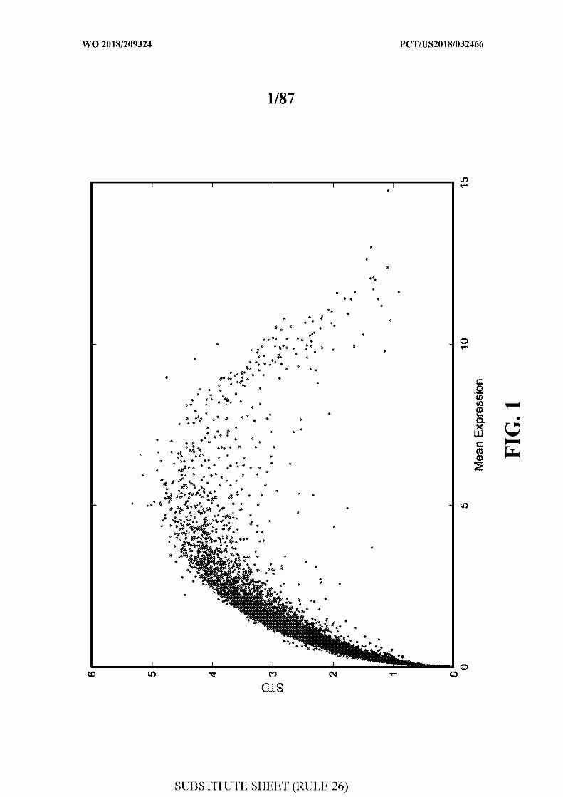
[0039] FIG. 1 - illustrates the mean expression of genes in CD8+ T cells and the
variability in expression. The most variable genes are selected for tS E analysis based on
genes with a var>6 and that are expressed in at least 5% of the cells (-4000 genes).
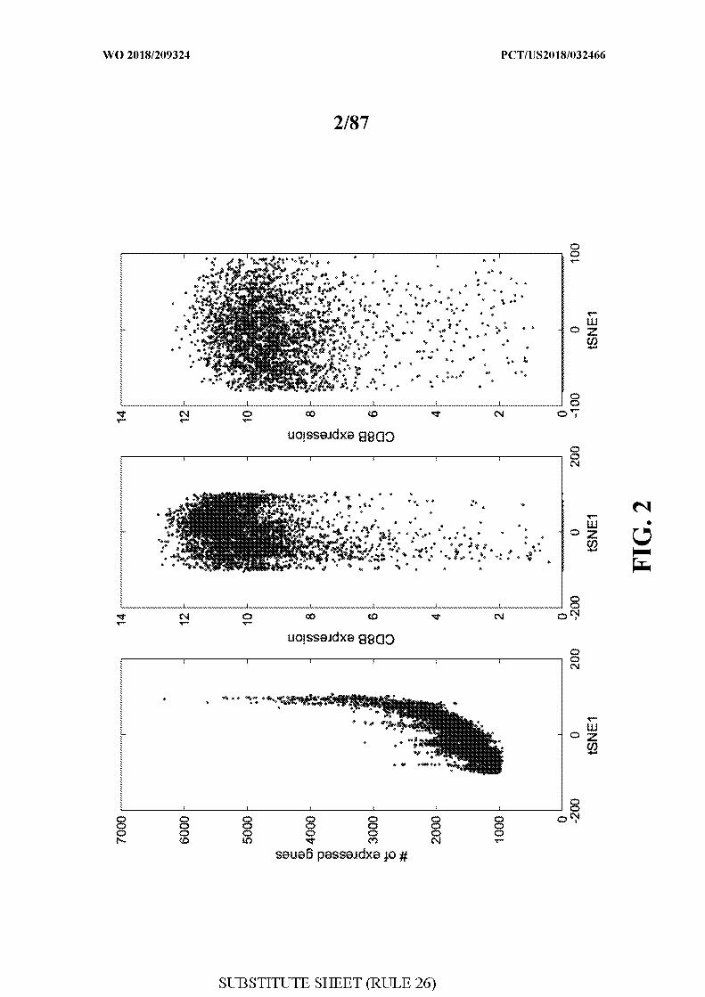
[0040] FIG. 2 - illustrates tSNE analysis based on most variable genes in CD8 cells.
tS E l is correlated with the number of expressed genes. As a control, expression of
CD8A\CD8B is not correlated with tSNE score
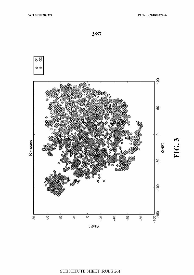
[0041] FIG. 3 - illustrates clustering by tSNE analysis of single cells and association
with response to check point blockade therapy.
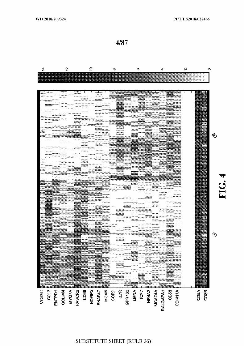
[0042] FIG. 4 - illustrates a heatmap of genes in the clusters from figure 3.
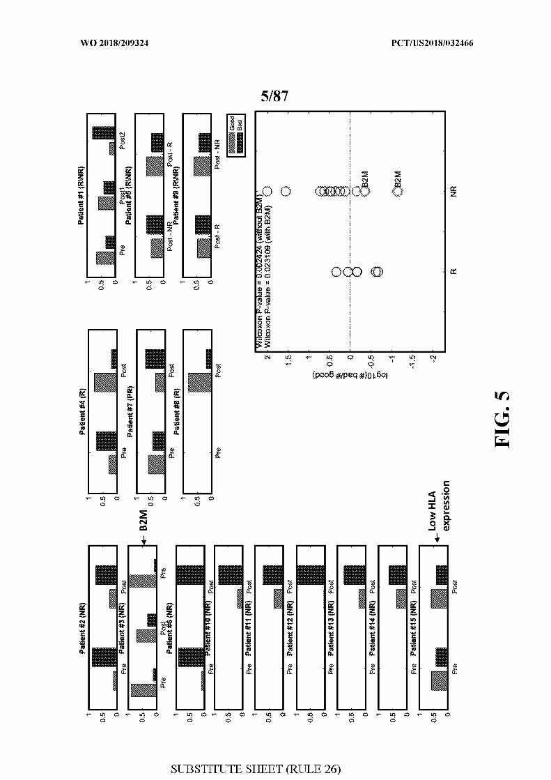
[0043] FIG. 5 - illustrates the percentage of cells having a non-responder (Bad) and
responder (Good) signature in patients that responded (R) or did not respond (NR) to
checkpoint blockade therapy.
[0044] FIG. 6 - illustrates further cluster analysis showing the non-responder cluster can
be split into two clusters.
[0045] FIG. 7 - illustrates a heatmap of genes in the clusters from figure 6 .
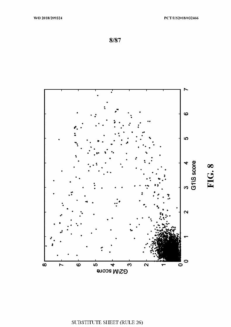
[0046] FIG. 8 - illustrates cell cycle analysis of the single cells.
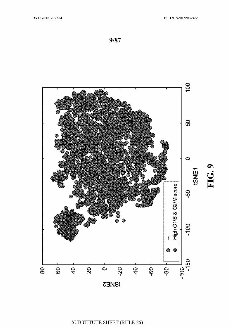
[0047] FIG. 9 - illustrates a cell cycle cluster based on figure 8 .
[0048] FIG. 10 - illustrates further cluster analysis showing the T cells can be separated
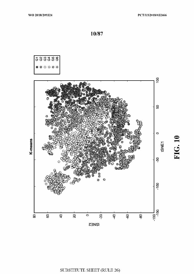
into 6 clusters.
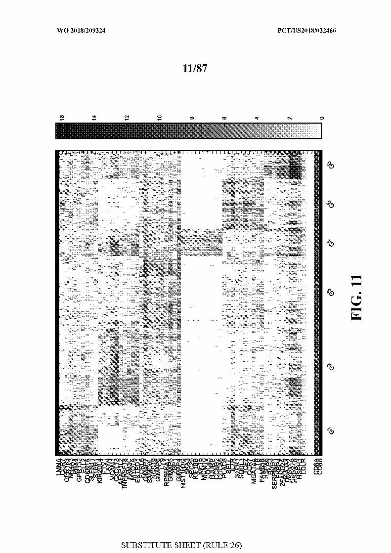
[0049] FIG. 11 - illustrates a heatmap of genes in the clusters from figure 10.
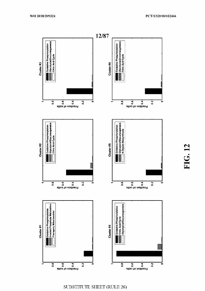
[0050] FIG. 12 - illustrates the enrichment for metabolic functions in the 6 clusters from
figure 10.
[0051] FIG. 13 - illustrates transport reaction activity across the 6 clusters from figure
10. Heatmap shows metabolites associated with the transporter genes.
[0052] FIG. 14 - illustrates transport reaction activity across the 6 clusters from figure
10. Heatmap shows transporter genes expressed in the different clusters.
[0053] FIG. 15 - illustrates a pipeline for determining T cell receptors (TCR).
[0054] FIG. 16 - illustrates TCR analysis in the single T cells. Left panel shows clonal
expansion as determined by the same TCR being detected in the same patient in different
time points. Right panel shows clonal enrichment as determined by the same TCR being
detected in the same patient in single time points.
[0055] FIG. 17 - illustrates that δγ T-cells are enriched in CD4/CD8 double negative T
cells (DN).
[0056] FIG. 18 - illustrates analysis of V51, V 2 and V 3 T-cells.
[0057] FIG. 19 - illustrates FACS analysis of CD39+Tim3+ (DP) cells and CD39-Tim3-
(DN) cells sorted from patient samples using cluster specific markers.
[0058] FIG. 20 - illustrates tSNE analysis of cells sorted in figure 19 using cluster
specific markers.
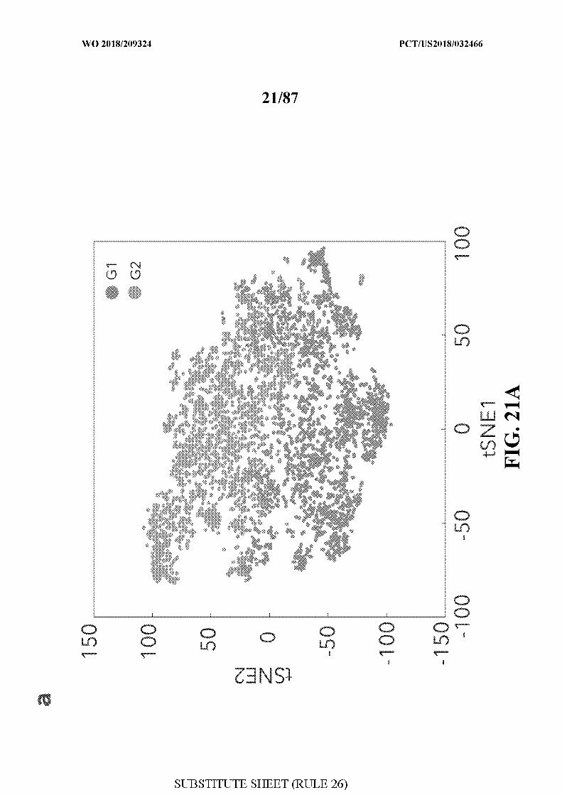
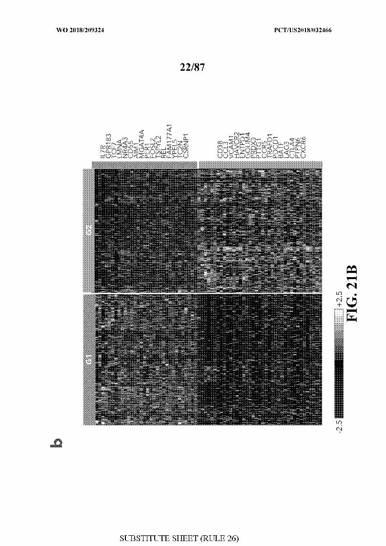
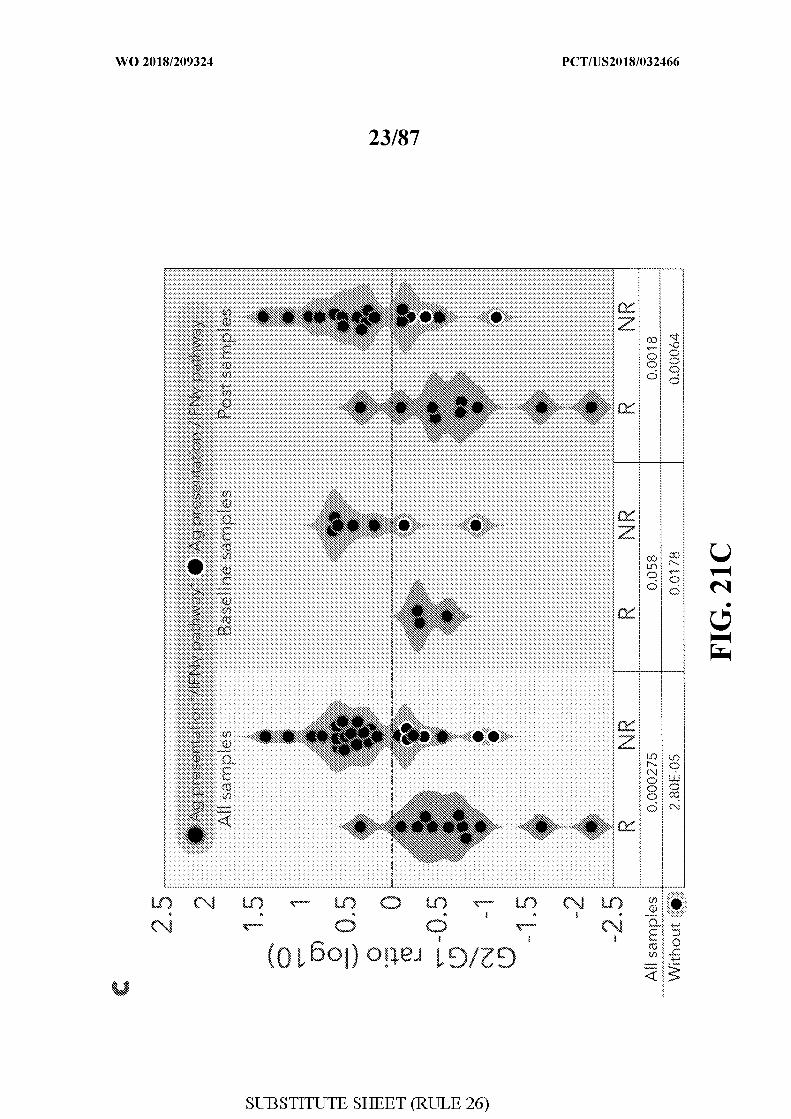
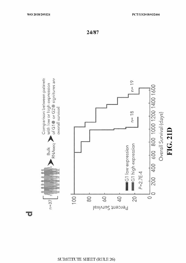
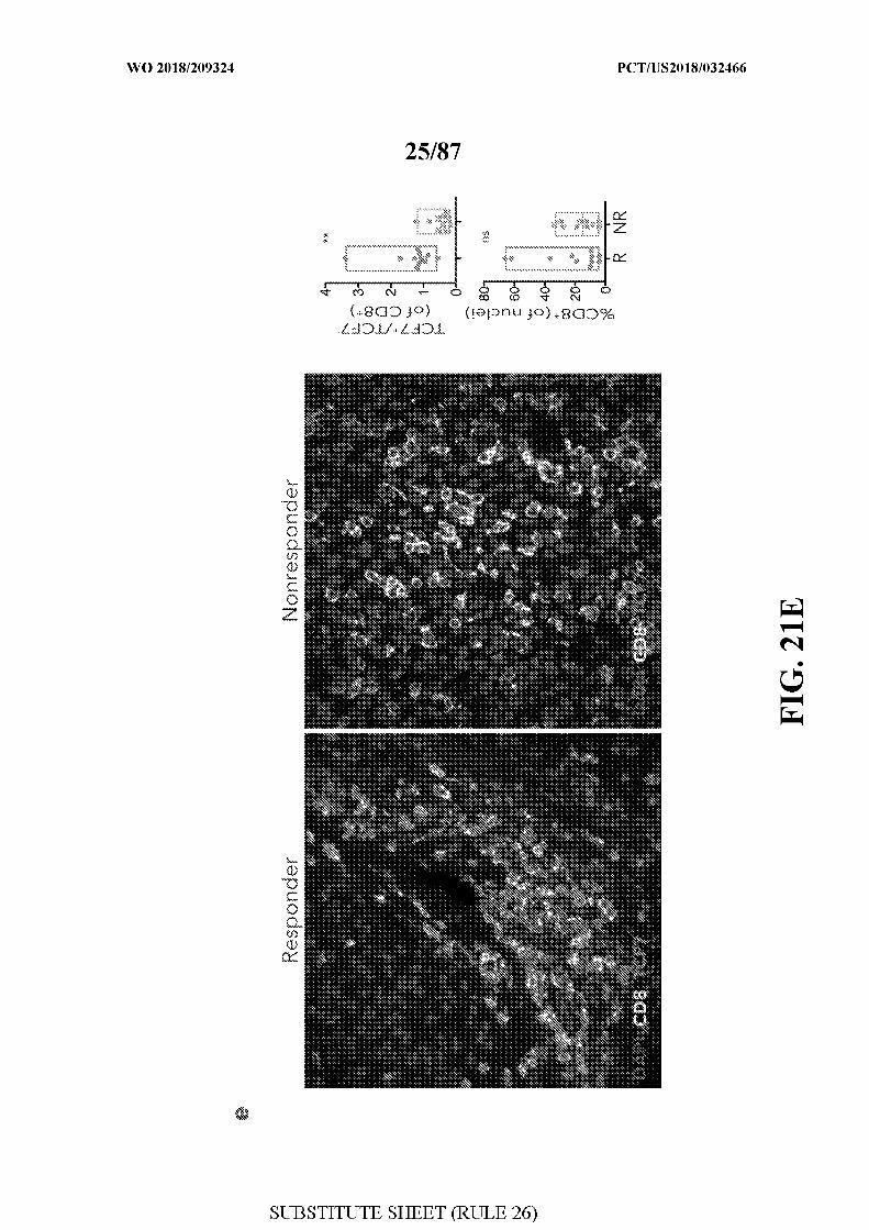
[0059] FIG. 21A-21E - illustrates characterization of tumor infiltrating CD8 T cells. Gl
refers to group 1, responder signature and G2 refers to group 2, non-responder signature in
this figure. Figure 21A illustrates a tSNE analysis showing that tumor infiltrating CD8 T
cells cluster into Gl or G2. Figure 21B illustrates a heat map showing expression of Gl and
G2 genes in responders and non-responders. Figure 21C illustrates the ratio of G2/G1
expression in responders and non-responders. Patients positive for antigen presentation and
the IFN gamma pathway and patients defective for antigen presentation and the IFN gamma
pathway are distinguished. Figure 21D illustrates a graph showing overall survival of
patients with low or high expression of Gl. Figure 21E illustrates immunofluorescence
images stained for CD8 and TCF7 in a responder and non-responder patient. The percentage
of CD8+ cells and the ratio of TCF+/TCF- CD8+ cells are calculated for the responder and
non-responder patient.
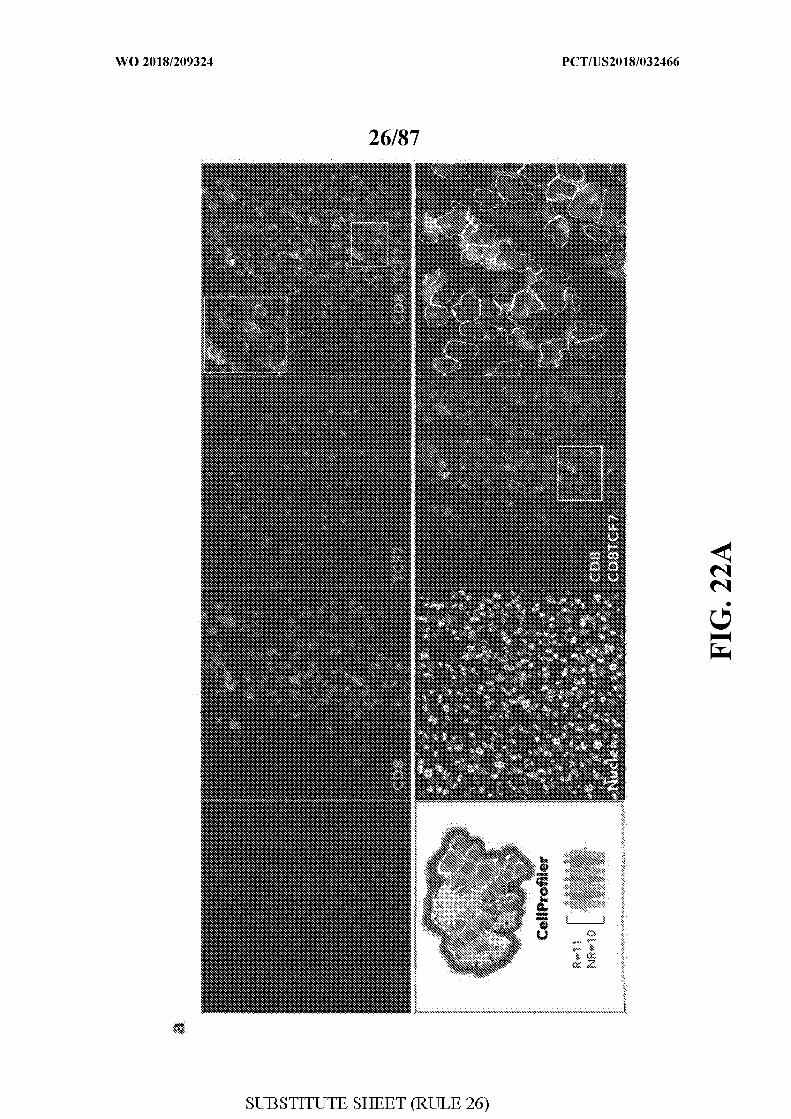
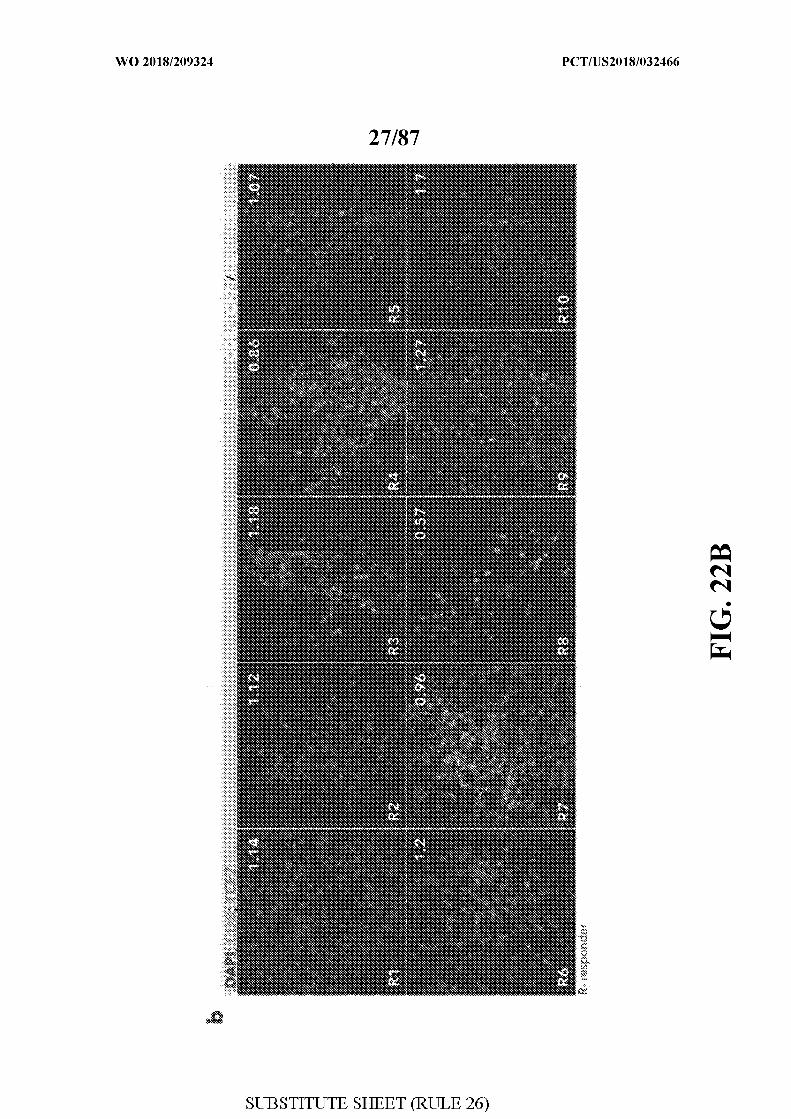
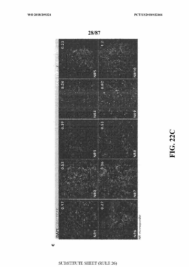
[0060] FIG 22A-22C - illustrate immunofluorescence imaging and calculation of TCF7
positive CD8 cells using CellProfiler (cellprofiler.org) and a novel pipeline.
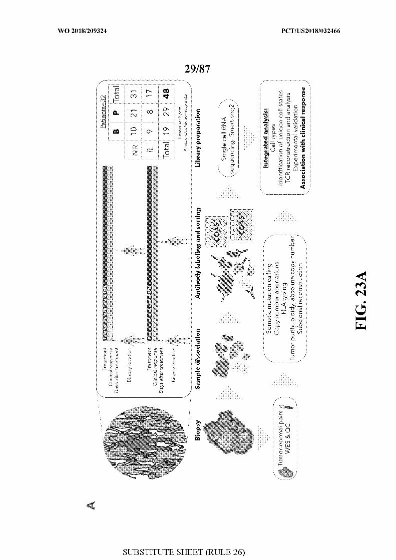
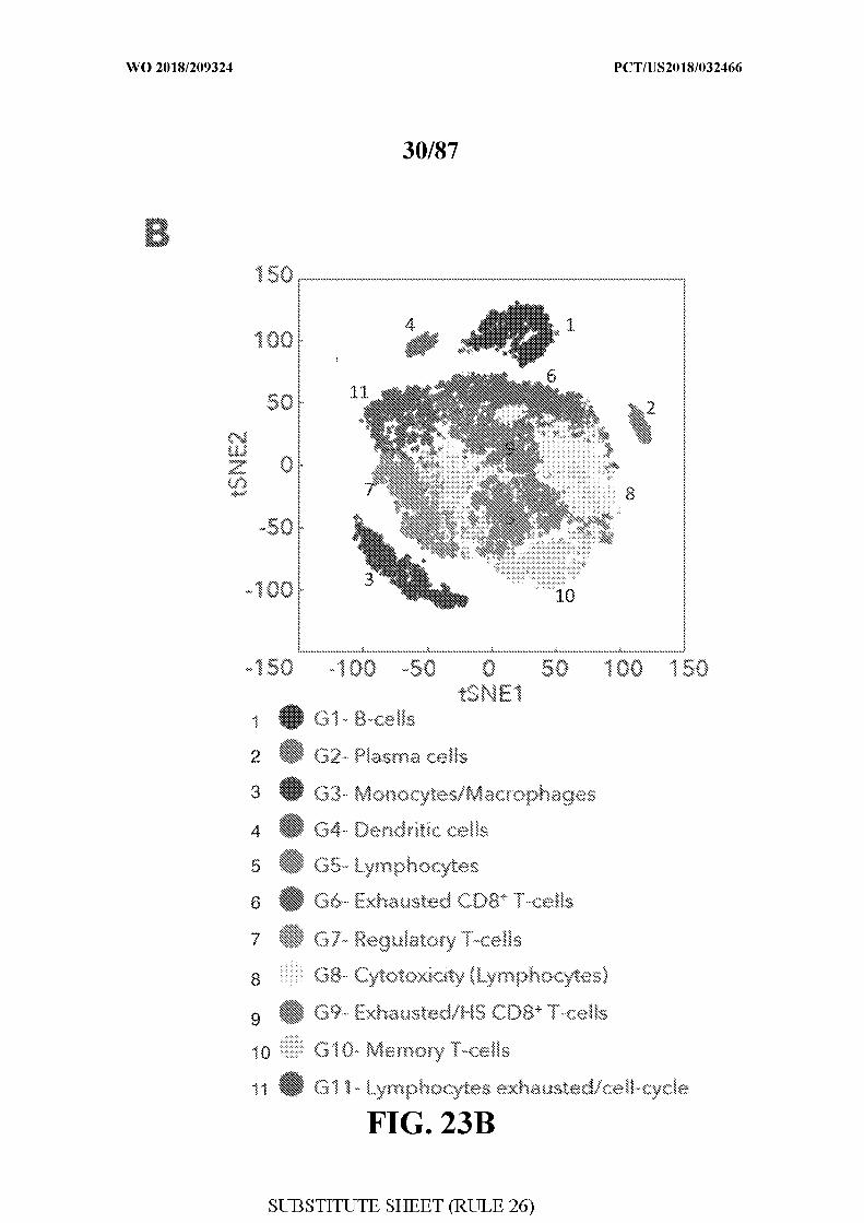
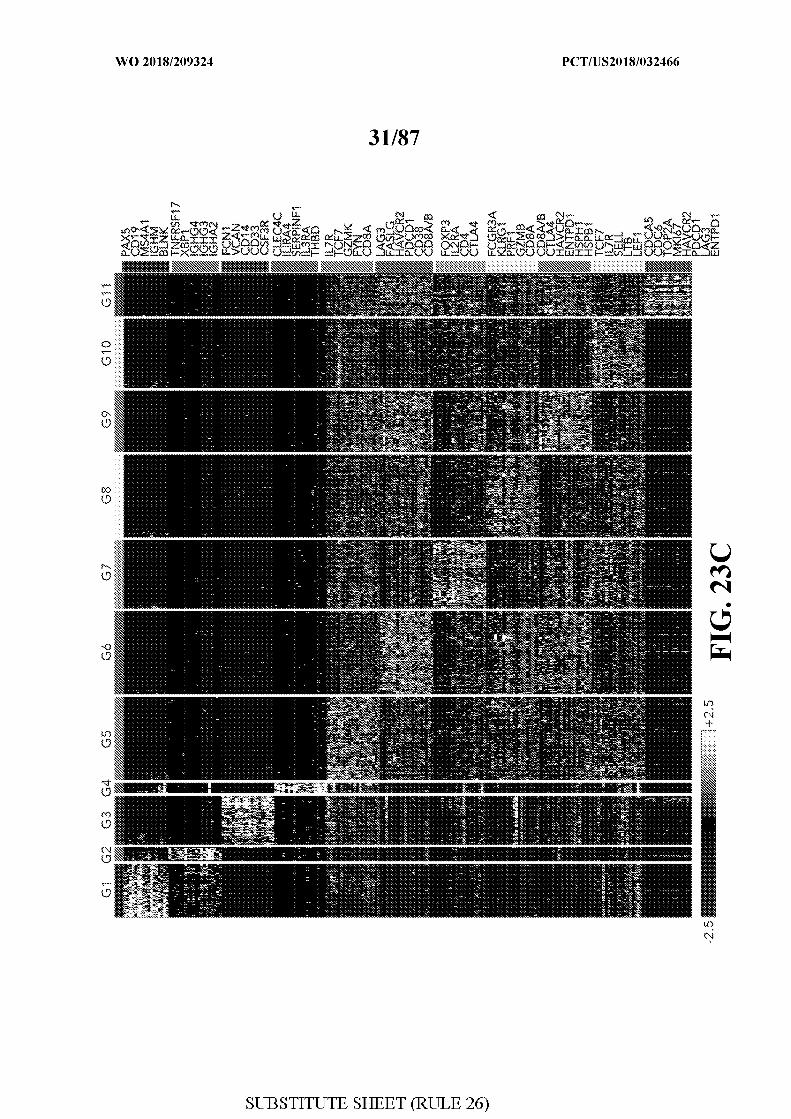
[0061] FIG. 23 - The immune landscape of melanoma patients treated with
checkpoint therapy. Figure23A. Schematic of the studied cohort. Top panel describes the
32 patient cohort, number of samples taken relevant to treatment initiation (baseline-B,
on/post treatment-P) along with the clinical status (responder-R, nonresponder-NR). Lower
panel delineates the workflow used. WES- whole exome sequencing, QC- quality control,
TCR- T cell receptor. Figure23B. tS E plot of all CD45+ cells collected in this study. Cells
are shaded based on 11 clusters identified by &-means clustering analysis (Methods).
Figure23C. Heatmap describing scaled expression values (log2(TPM+l)) of discriminative
gene sets for each cluster defined in (B). A list of representative genes is shown for each
cluster next to the right margin bars. Shading scheme is based on z-score distribution from -
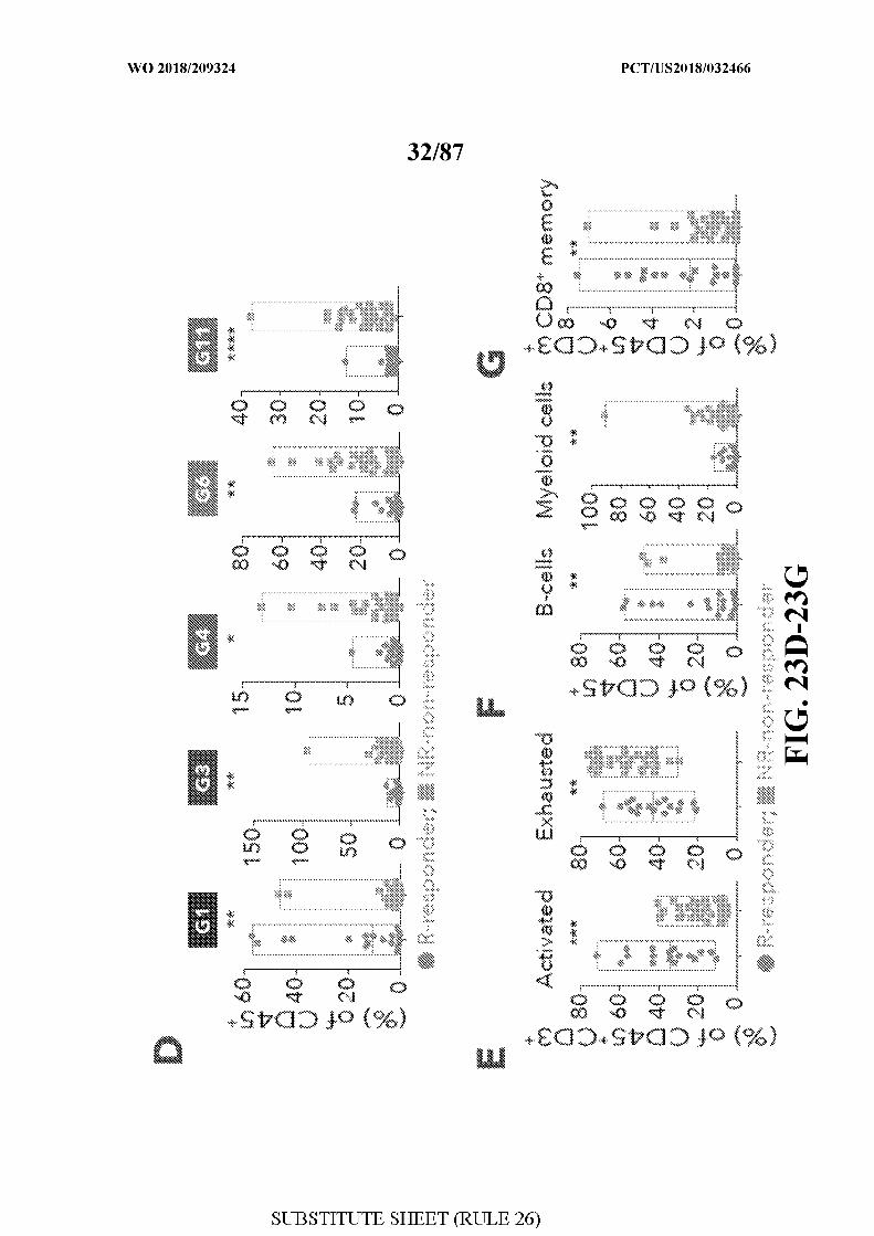
2.5 to 2.5. Figure23D. Box plots comparing the percentage of cells (out of CD45+) in Gl,
G3, G4, G6 and Gi l clusters as defined in (B), between responder and non-responder
lesions. Each symbol represents an individual sample. Two-sided Wilcoxon rank-sum P-
value is shown **P=0.003; **P=0.003; *P=0.01; **P=0.005; ***>=1.3xl0 5 . Figure23E. Box
plots comparing the percentage of exhausted and activated CD45+CD3+ cells on the basis of
a pre-defined list of known marker genes (table 3), between responder and non-responder
lesions. Each symbol represents an individual sample. Two-sided Wilcoxon rank-sum P-
value is shown ***P=2xlO 4 **P=0.002. Figure23F. Box plots comparing the percentage of
B-cells and Myeloid cells on the basis of a pre-defined list of known marker genes (table 3)
between responder and non-responder lesions. Each symbol represents an individual sample.
Two-sided Wilcoxon rank-sum -value is shown **P=0.004; **P=0.002. Figure23G. Box
plots comparing the percentage of Memory CD8+ T-cells on the basis of a pre-defined list of
known marker genes (table 3) between responder and non-responder lesions. Each symbol
represents an individual sample. Two-sided Wilcoxon rank-sum -value is shown **P=0.001.
Figure23H. Heatmap describing scaled expression values (log2(TPM+l)) of discriminative
gene sets between responder and non-responder lesions. Marker genes are shown per cluster.
Shading scheme is based on z-score distribution from -2.5 to 2.5.
[0062] FIG. 24 - Identification of CD8+ T-cell states associated with clinical
outcome. Figure 24A. tS E plot of all CD8+ T cells collected in this study. Cells are shaded
based on 2 clusters identified by &-means clustering (Methods). Figure 24B. Heatmap
showing scaled expression values (log2(TPM+l)) of discriminating genes for the clusters
defined in (A). A list of representative genes are shown for each cluster next to the right
margin bars. Shading scheme is based on z-score distribution from -2.5 to 2.5 . Figure 24C.
Box plots comparing the percentage of CD8 G and CD8 B (out of CD8+ cells) clusters in
responder and non-responder lesions . Each symbol represents an individual sample. Two-
sided Wilcoxon rank-sum P-value is shown i^l^xlO^responders; Ρ=0.005 non-responders.
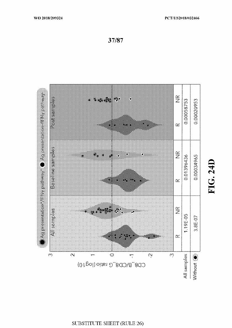
Figure 24D. The logio ratio between the number of cells in CD8 B/CD8 G per sample is
computed. A comparison of this measurement between responder and non-responder lesions
is shown for all samples, baseline and post-treatment samples separately. Circles marked in
white represents samples with defects in antigen presentation and the IFN pathway (Ag
presentationVIFNy pathway ) as inferred by WES, IHC and flow-cytometry analysis. Marked
Circles represent samples without defects in those pathways. The significance score (one
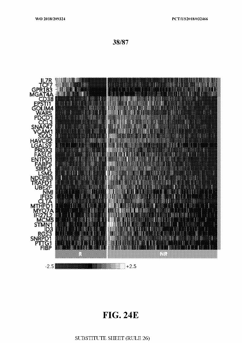
sided Wilcoxon P-value) for each comparison is shown below. Figure 24E. Heatmap
describing scaled expression values (log2(TPM+l)) of discriminative gene sets from CD8 G
and CD8 B clusters between responder and non-responder lesions. Marker genes are shown
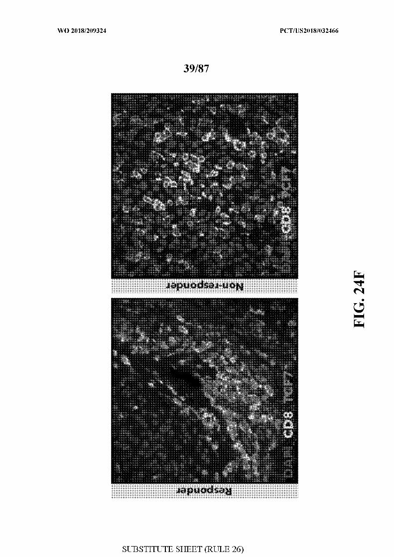
per cluster. Shading scheme is based on z-score distribution from -2.5 to 2.5 . Figure 24F.
Representative images from the multiplex immunofluorescence staining of tissue nuclei
stained with DAPI, CD8 and TCF7 from a responder and non-responder patient prior to
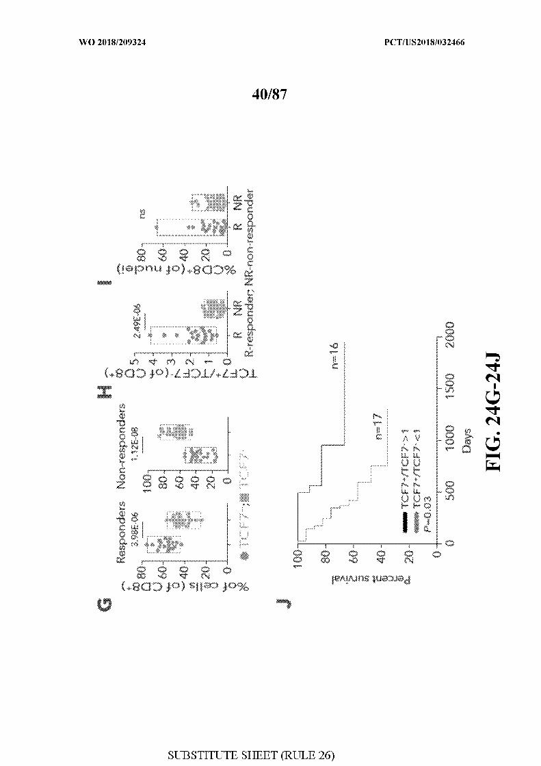
therapy with anti-PDl. Original Magnification X400. G. Box plots comparing the percentage
of CD8+TCF7+ and CD8+TCF7 cells as inferred by immunofluorescence staining, between
responder and non-responder patients. Each symbol represents an individual sample. Two-
sided Wilcoxon rank-sum P-value is shown, responders; R =l.lxl0 8 non-
responders. Figure 24H-I. Box plots showing a quantitative analysis (Methods) of
TCF7+CD8+/TCF7 CD8+ ratio out of CD8+ cells (H) and of CD8+ cells out of all nuclei (I)
between responder (n=20) and non-responder patients (n=23). n.s- not significant. One-sided
Wilcoxon P-value is shown J. Kaplan-Meier survival curve, composed of data
from 33 patients treated with anti-PDl therapy. Patients were divided into two groups based
on TCF7+CD8+/TCF7 CD8+ ratio (n=16 >1; n=17 <1) as inferred by immunofluorescence
staining. A ratio of TCF7+CD87TCF7 CD8+ > 1 was associated with better overall survival
(log-rank P=0.03) when compared to patients with TCF7+CD8+/TCF7 CD8+ <1.
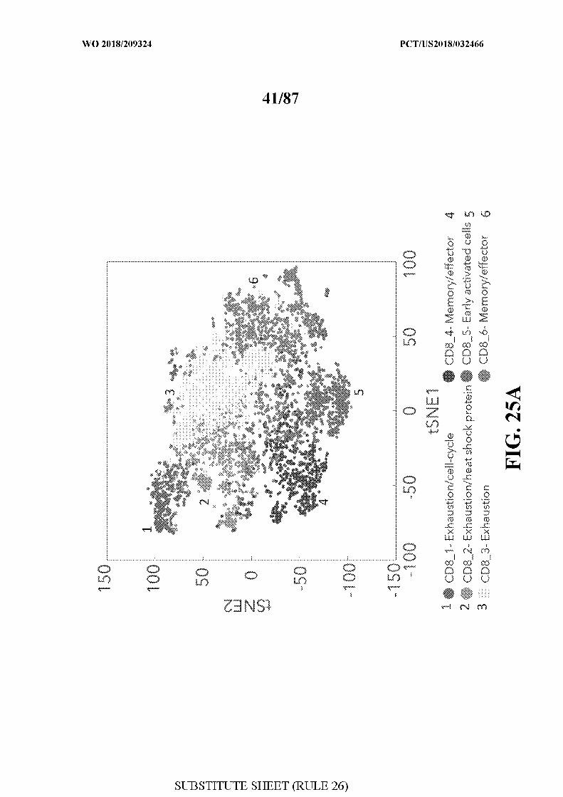
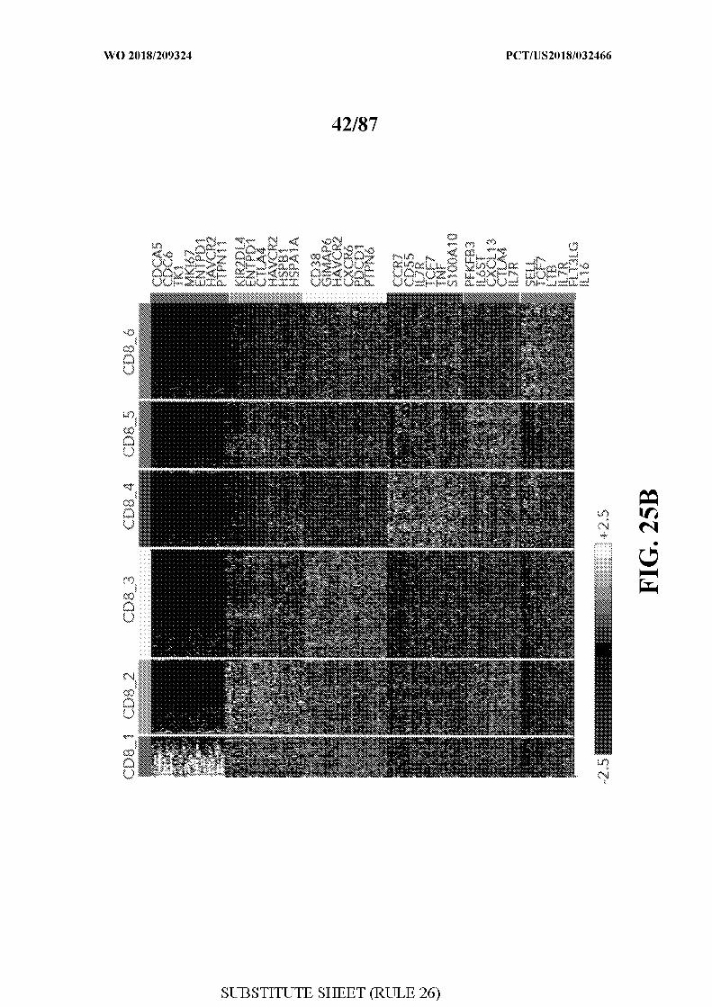
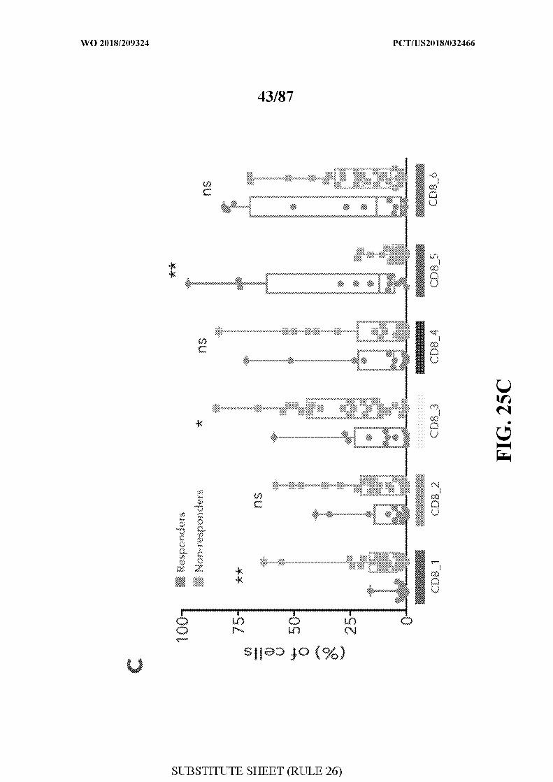
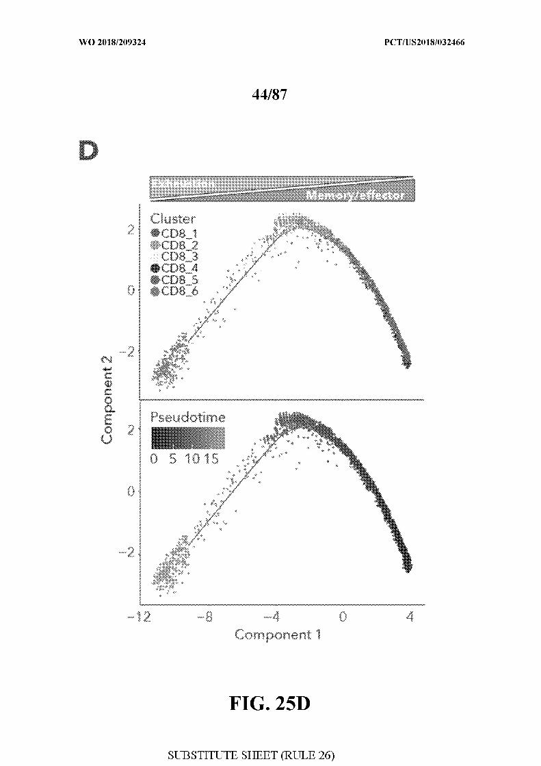
[0063] FIG. 25 - CD8+ T-cell state heterogeneity and its association with clinical
response. Figure 25A. tS E plot of all CD8+ T-cells collected in this study. Cells are shaded
based on 6 clusters identified by &-means clustering (Methods). Figure 25B. Heatmap
showing scaled expression values (log2( TPM+1)) of discriminative gene sets for each cluster
defined in (A). A list of representative genes is shown for each cluster next to the right
margin bars. Shading scheme is based on z-score distribution from -2.5 to 2.5 . Figure 25C.
Box plots comparing the percentage of CD8 1 to 6 (out of CD8+ cells) clusters between
responders and non-responders . Each symbol represents an individual sample. One-sided
Wilcoxon P-values are shown, **P-value=0.00l for CD8 1, *P-value=0.013 for CD8 3, **P-
v / =0.003, and ns-not significant. D. Trajectory analysis for the 6 CD8+ T-cells clusters
identified in (A). Cell expression profiles in a two dimensional independent space. Solid
black line indicates the main diameter path of the minimum spanning tree (MST) and
provides the backbone of Monocle's pseudotime ordering of the cells. Each dot represents an
individual cell shaded by cluster (upper plot) or by pseudotime (lower plot).
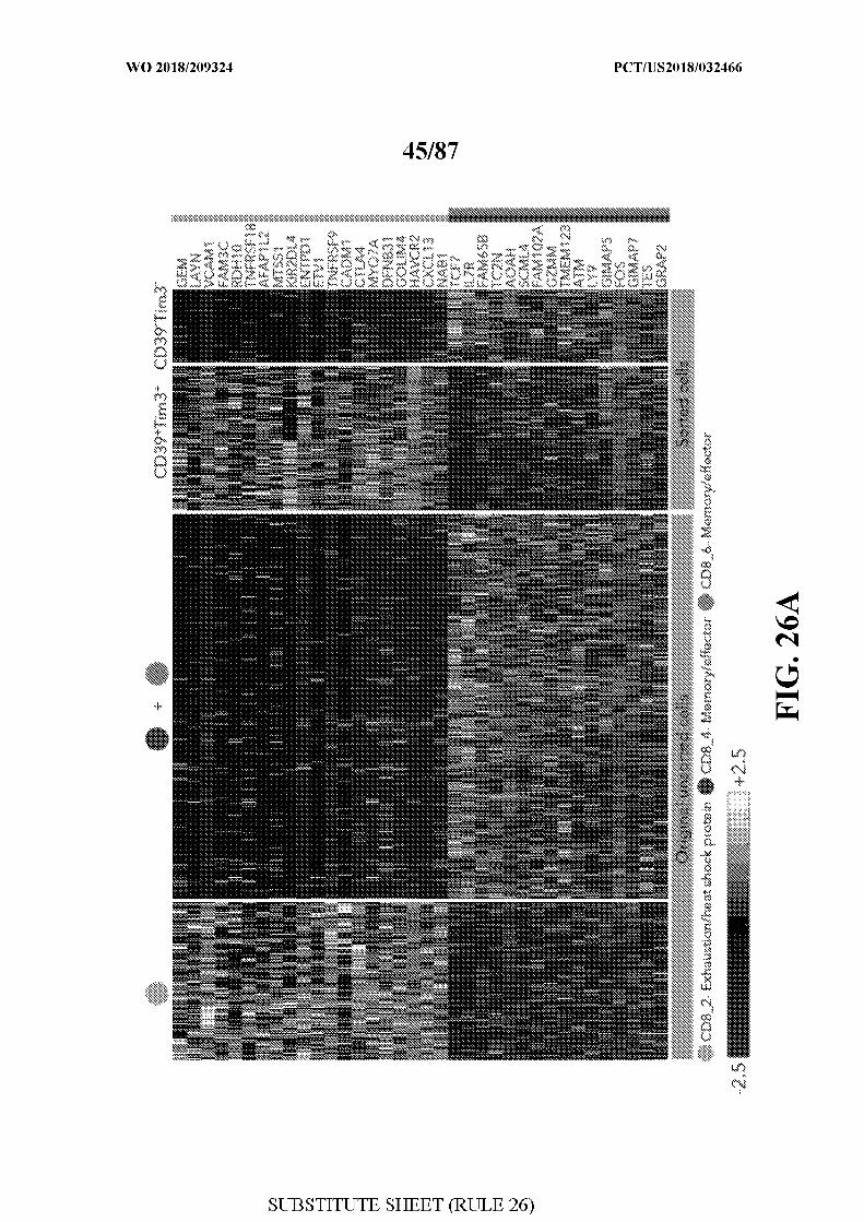
[0064] FIG. 26 - Discriminating exhausted from memory cells using TIM3 and
ENTPD1/CD39. Figure 26A. Heatmap showing scaled expression values (log2(TPM+l)) of
discriminative gene sets between CD8 2 (exhaustion) and CD8 4+6 (memory/effector) with
original unsorted, and sorted (CD39+TIM3+ and CD39TIM3 ) cells. A list of representative
genes are shown for each cluster next to the right margin bars. Shading scheme is based on z-
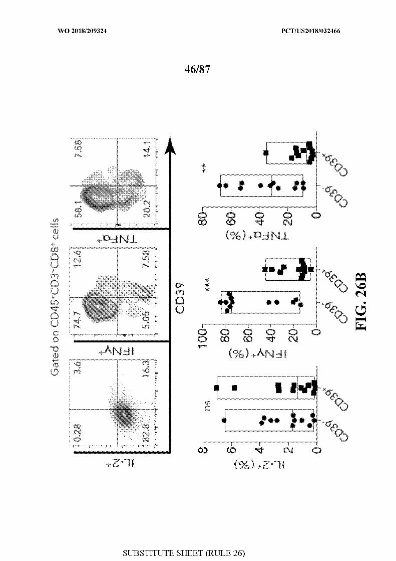
score distribution from -2.5 to 2.5 . Figure 26B. Representative flow cytometric plots (upper
part) of intracellular staining for IL-2, IFNy and T F in CD39+ and CD39+ cells (out of
gated CD45+CD3+CD8+ cells). Flow cytometry quantification of cytokine-producing cells
obtained from 12 metastatic melanoma patients (lower part). Bars indicate the mean values.
Data were combined from 2 replicate experiments. Unpaired-student's t-test with **P-
value=0.0016 and *** P-value=5xlO 4 is shown. Figure 26C. A schematic summary of the
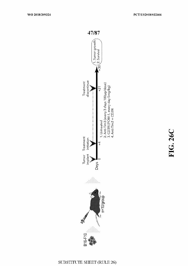
therapy regimen used in the transplantable B16-F10 mouse model (described in Methods).
Mice were divided into four groups (n=10 per group): untreated (vehicle control), anti-TIM3,
CD39 inhibitor (CD39i, using POM-1 small molecule) and anti-TIM3 in combination with
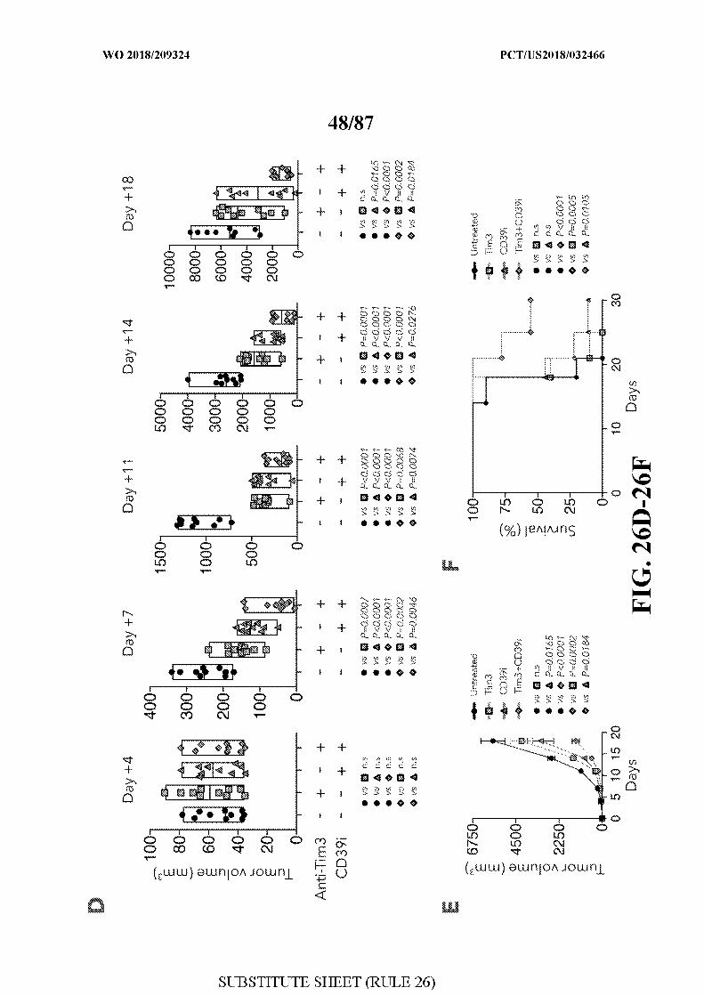
CD39L Figure 26D. Box plots showing the kinetics of tumor growth between the different
groups of mice on days +4, +7, + 11, +14 and +18 post tumor transplantation. Data in box
plots are means+SEM. P-value was determined by unpaired-student's t-test. Figure 26E.
Mean tumor volumes for all 4 groups are shown, means±SEM. Figure 26F. Survival at day
30 of B16-F10 tumor-bearing mice for all 4 groups. Log-rank P-value is shown. Data shown
for C-F represents one out of two independent experiments, n=10 for each group per
experiment ns- not significant.
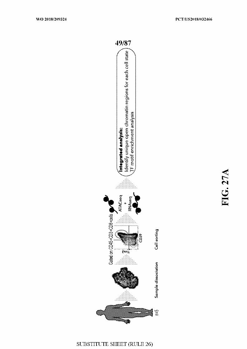
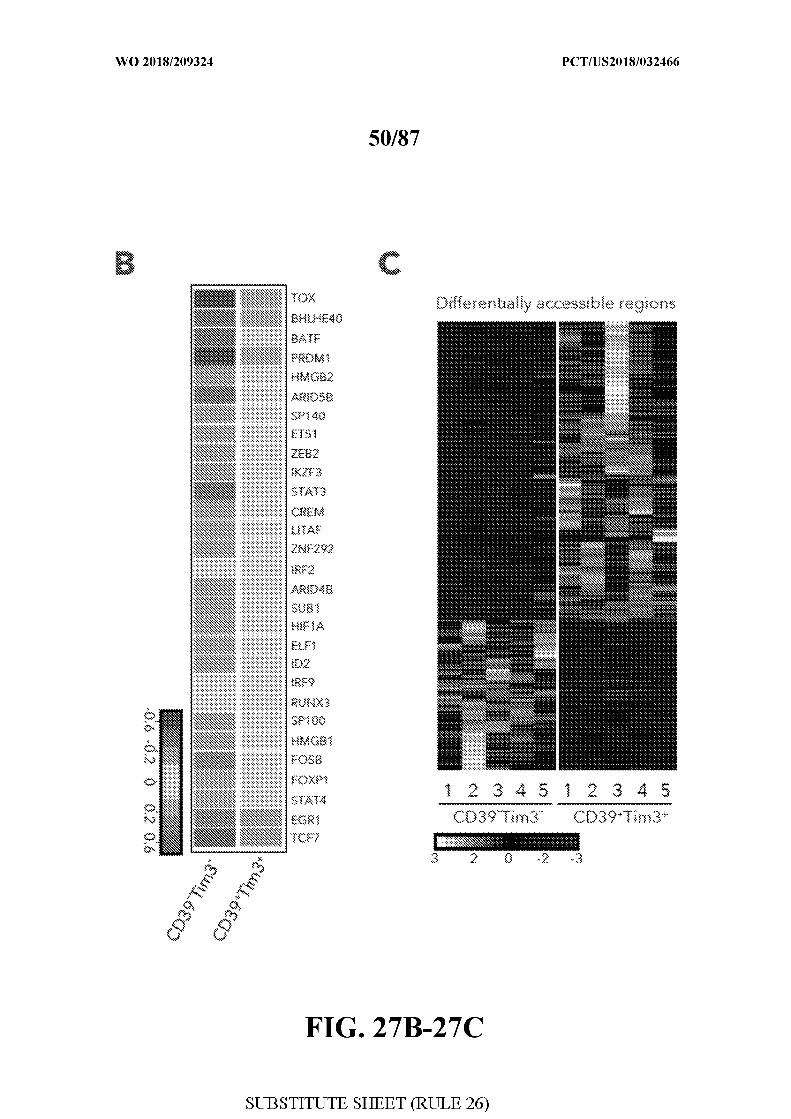
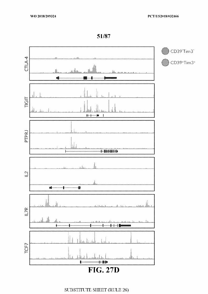
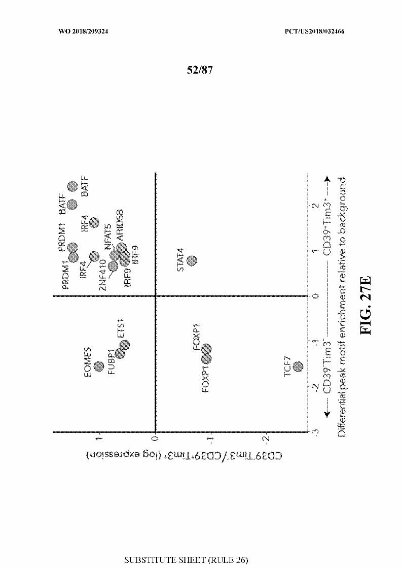
[0065] FIG. 27 - Distinctive chromatin accessibility in CD39+TIM3+ and CD39
TIM3 cells. Figure 27A. Schematic of ATAC-seq analysis performed on sorted
CD39+TFM3 + and CD39TIM3 cells from 5 melanoma patients. Figure 27B. Heatmap
describing averaged scaled expression values (log2(TPM+l)) of differentially expressed
transcription factors for sorted CD39 +TIM3 + and CD39TFM3 cells (from n=5 patients).
Shading scheme is based on z-score distribution from -0.6 to 0.6. Figure 27C. Heatmap
describing patient specific (n=5) differentially accessible regions (FDR<0.01) in
CD39+TFM3 + and CD39TIM3 sorted populations. Shading scheme is based on z-score
distribution from -3 to 3 . Figure 27D. ATAC-seq traces for open chromatin regions near
selected genes in CD39+TFM3 + and CD39TIM3 is shown. Figure 27E. Graph depicting
enrichment of TF motifs in open chromatin specific to CD39TIM3 and CD39+TIM3 + cells
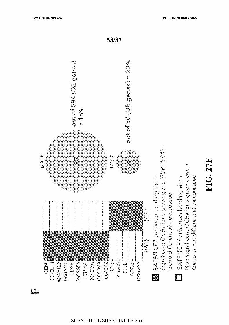
is on the x-axis, and differential expression of TF on the y-axis. Figure 27F. Left, enhancer
binding sites near the listed genes for BATF and TCF7. Significant genes associated with
these sites are marked and non-significant genes are white. The corresponding genes are also
differentially expressed between CD39+TFM3 + cells (enriched with BATF) and CD39TIM3
cells (enriched with TCF7). Right, the number of genes that are differentially expressed with
a corresponding differential peak containing BATF or TCF7 is shown.
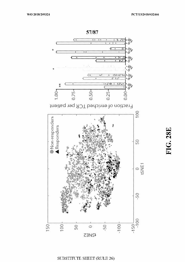
[0066] FIG. 28 - TCR analysis and its relationship with cell state and clinical
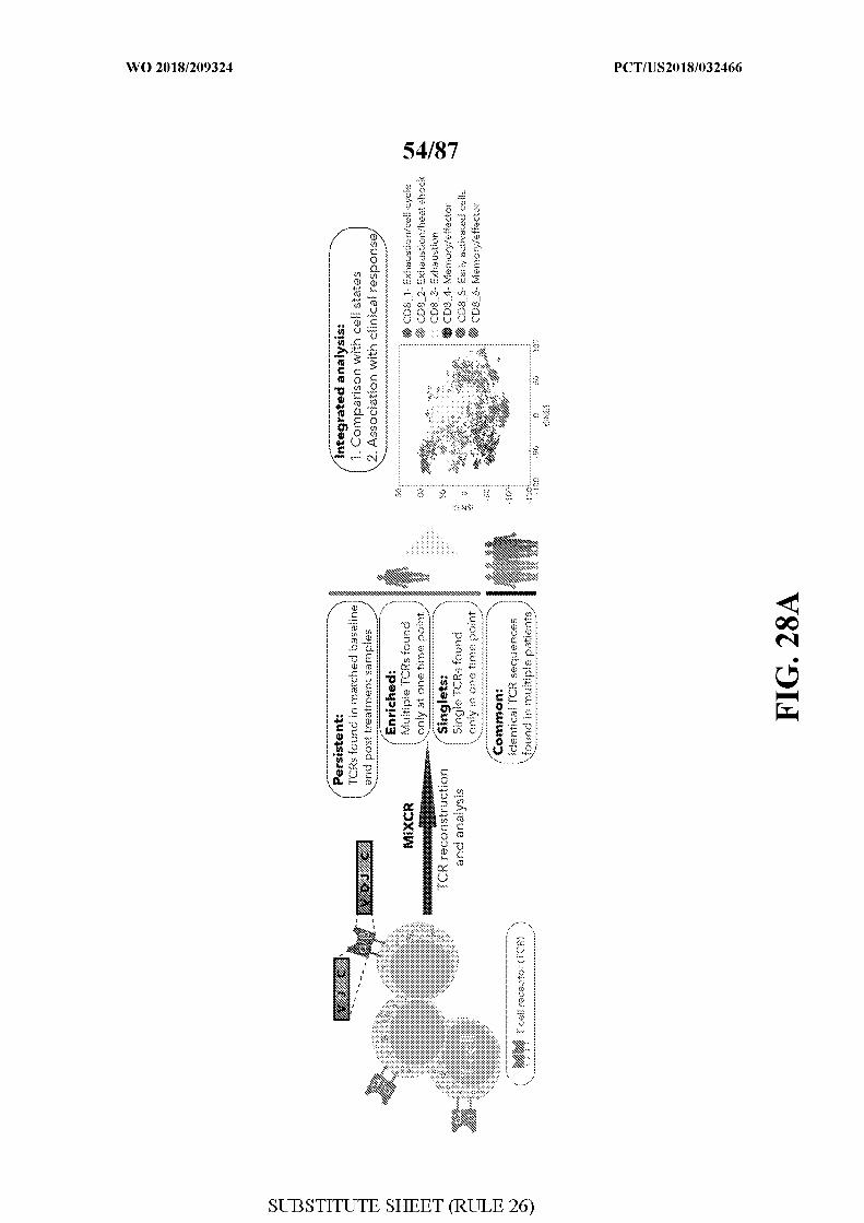
outcome. Figure 28A. Schematic illustration of the TCR analysis pipeline. TCR
reconstruction was done using the MixCR algorithm (Methods), and were classified into 4
groups: Persistent: TCRs found in matched baseline and post treatment samples; Enriched:
Multiple TCRs found in a single time point of a given patient; Singlets: single TCRs that
detected only once in our dataset; and Common: TCRs found in different patients. Figure
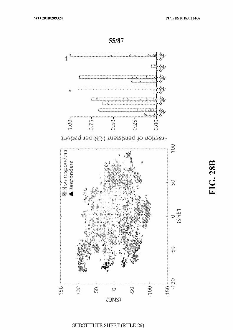
28B. tSNE plot (left panel) delineating the six identified CD8+ T-cell clusters and persistent
TCRs in responder lesions (black triangles) and non-responder lesions (gray circles). Bar plot
(right panel) summarizing the fraction of persistent TCRs per patient across the different
clusters between responder (R) and non-responder ( R) samples. Two-sided Wilcoxon rank-
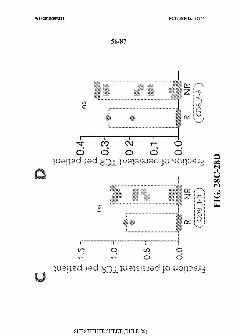
sum P-value is shown *P=0.03; **P=0.0085. Figure 28C-D. Fraction of persistent TCRs per
patient, aggregated for CD8 1 to 3 (CD8 1-3) and CD8 4 to 6 (CD8 4-6) clusters for R and
R samples; ns- not significant. Figure 28E. tS E plot (left panel) delineating the six
identified clusters and enriched TCRs in responders (black triangles) and non-responders
(gray circles). Bar plot (right panel) summarizing the fraction of enriched TCRs per patient
across the different clusters and split into R and NR samples. **P=0.003; *P=0.03 for CD8 3;
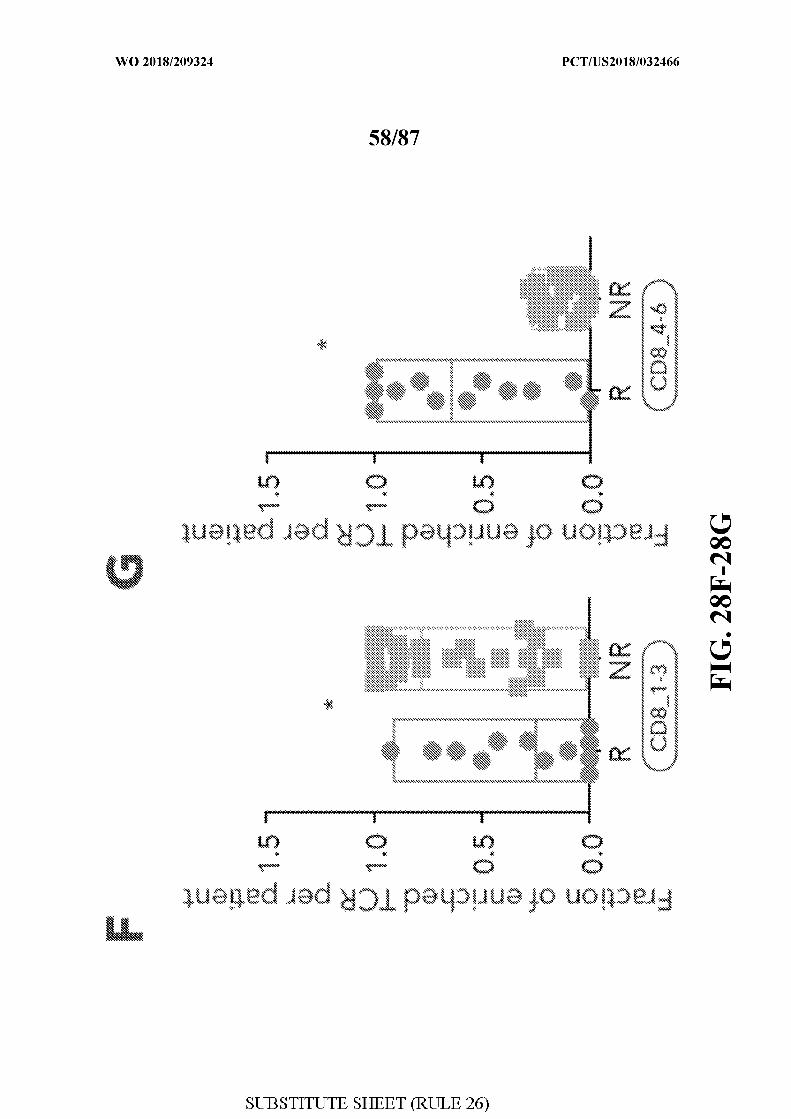
*P=0.02 for CD8 5 . Figure 28F-G. Fraction of enriched TCR per patient, aggregated for
CD8 1 to 3 (CD8 1-3; P=0.014) and CD8 4 to 6 (CD8 4-6; P=0.019) clusters for R and NR
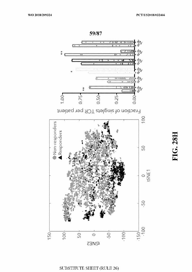
samples. Figure 28H. tSNE plot (left panel) delineating the six identified clusters and
singlets TCRs in responders (black triangles) and non-responders (gray circles). Bar plot
(right panel) summarizing the fraction of singlets TCRs per patient across the different
clusters and split into R and NR samples. *V=0.009 for CD8_1; V=0.02; *V=0.004 for
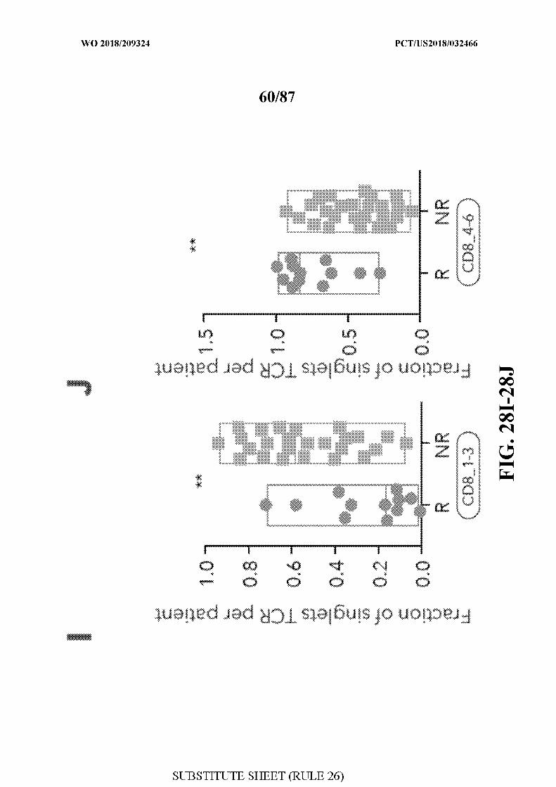
CD8 5 . Figure 281-J. Fraction of singlets TCR per patient, aggregated for CD8 1 to 3
(CD8 1-3; P=0.002) and CD8 4 to 6 (CD8 4-6; P=0.002) clusters for R and NR samples.
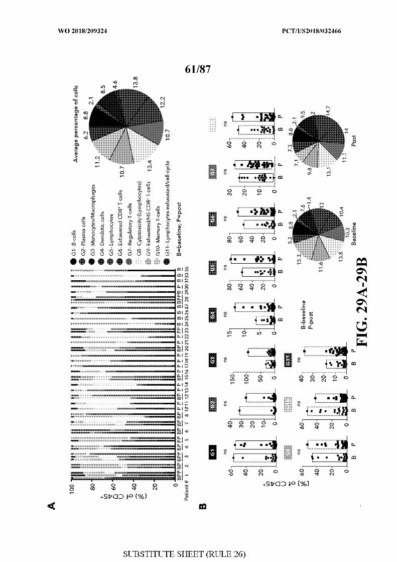
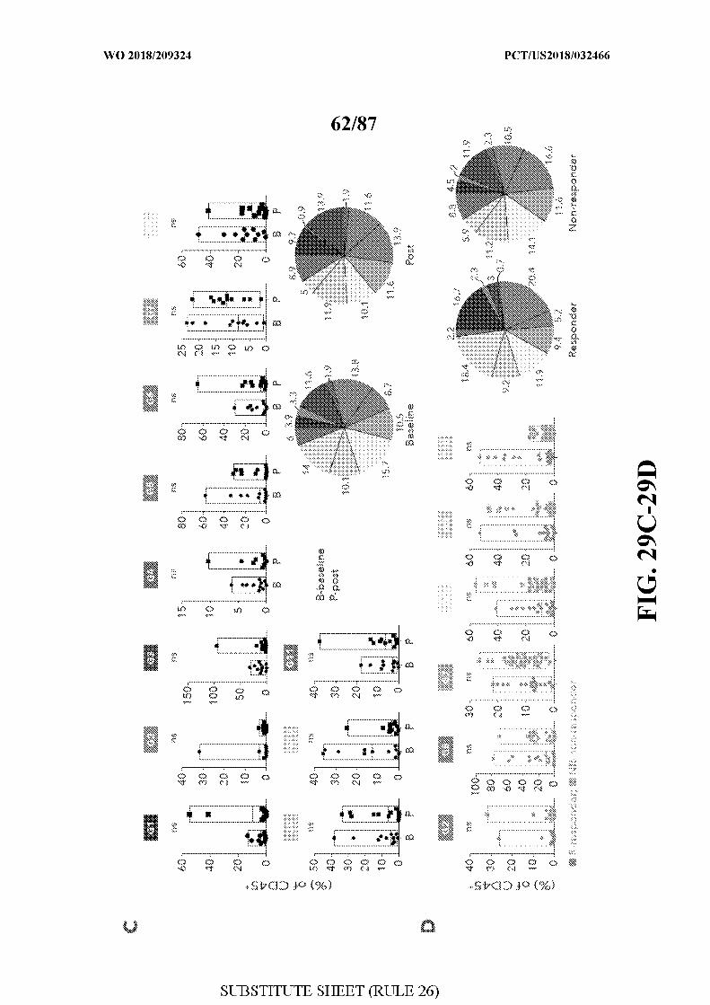
[0067] FIG. 29 - Association of the 11 CD45+clusters with clinical outcome during
the course of checkpoint therapy. Figure 29A. For each patient, the percentage of cells (out
of CD45 cells) classified to one of the 11 clusters identified by &-means clustering is shown.
Pie chart on the right, summarizes the corresponding percentages across all cells collected in
this study. Figure 29B-C. Box plots comparing the abundance of cells in the corresponding
clusters between all baseline and post-treatment samples (B) and only in patients with
matched longitudinal samples (C). Pie charts on the right, summarizes the corresponding
percentages for each analysis. Figure 29D. Box plots comparing the abundance of cells in the
corresponding clusters between responder and non-responder lesions. Pie chart on the right,
summarizes the corresponding percentages for each cluster. B- baseline, P- post, R -
responder, NR-non-responder, n.s- not significant.
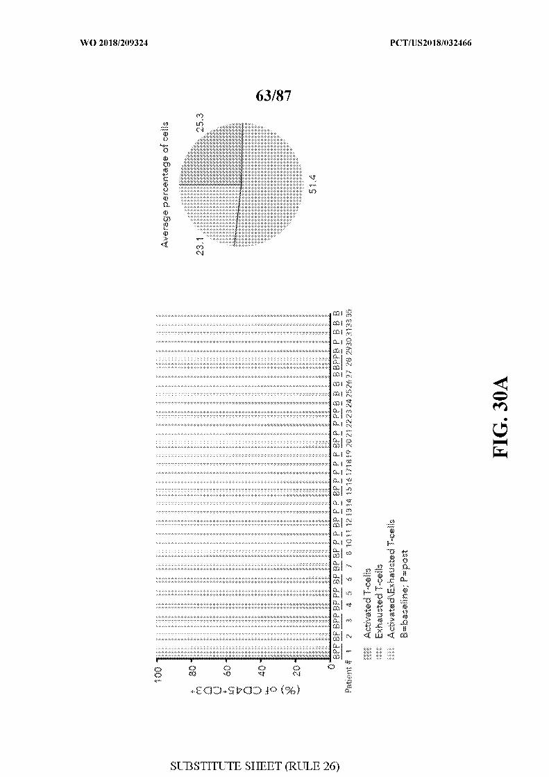
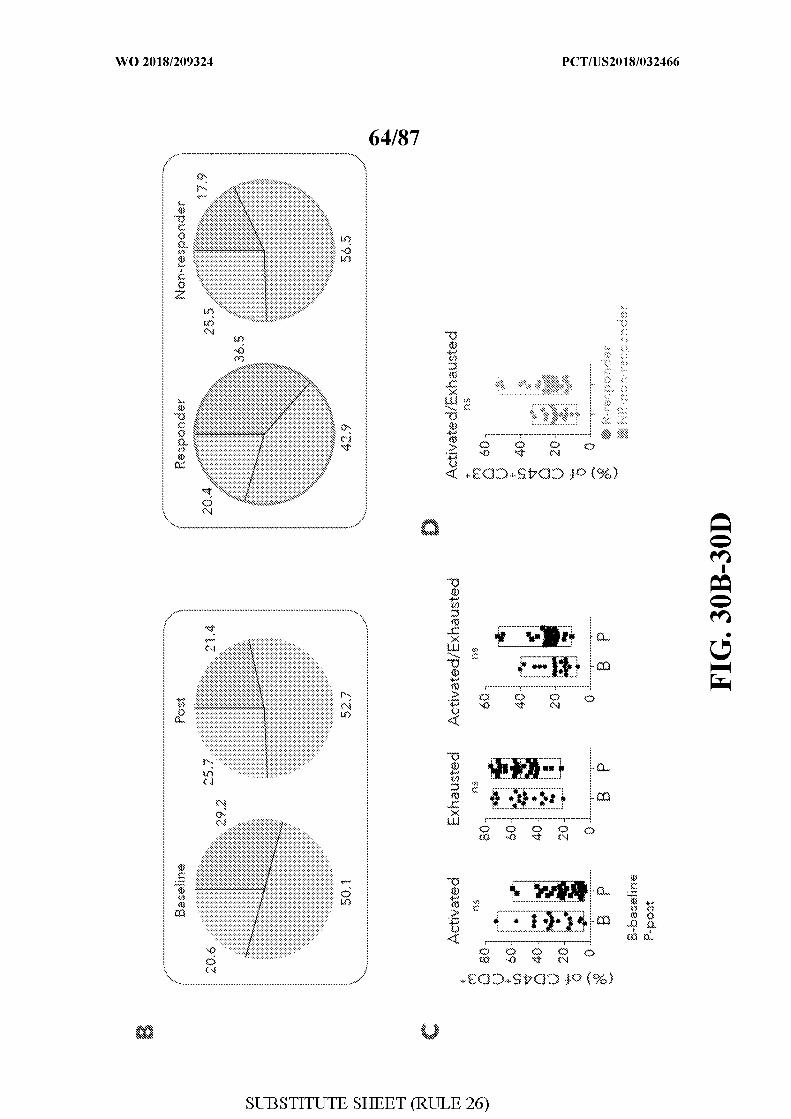
[0068] FIG. 30 - Supervised analysis of T cell states. Figure 30A. The percentage of
exhausted, activated or activated/exhausted CD45 +CD3 + cells in each patient, on the basis of
a pre-defined list of known marker genes is shown. Pie chart on the right summarizes the
corresponding percentage across all CD45 +CD3 + T-cells collected in this study Figure 30B.
Comparison of the abundance of all three cell states as in (A) between baseline and post-
treatment samples (left) and responder and non-responder lesions (right), on the basis of the
pre-defined list of known genes as in (A). Figure 30C-D. Box plots comparing the
abundance of cells in all three cell states between baseline and post-treatment samples (C)
and for the activated/exhausted state between responder and non-responder lesions (D). B -
baseline, P- post, R- responder, NR-non-responder, ns- not significant.
[0069] FIG. 31 - Comparing the composition of known cell types with clinical
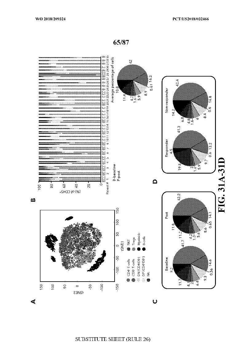
outcome and checkpoint therapy. Figure 31A. tSNE plot of all CD45 +cells collected in this
study. Cells are shaded by cell type on the basis of pre-defined markers (table 3). Figure
31B. The percentage of known immune cell types in each patient, on the basis of a pre
defined list of known marker genes. Pie chart (below bar graph) summarizes the
corresponding percentage of known cell types across all CD45+cells collected in this cohort.
Figure 31C-D. A comparison of the abundance of known cell types as in (B) between
baseline and post-treatment samples (C) and responder and non-responder lesions (D).
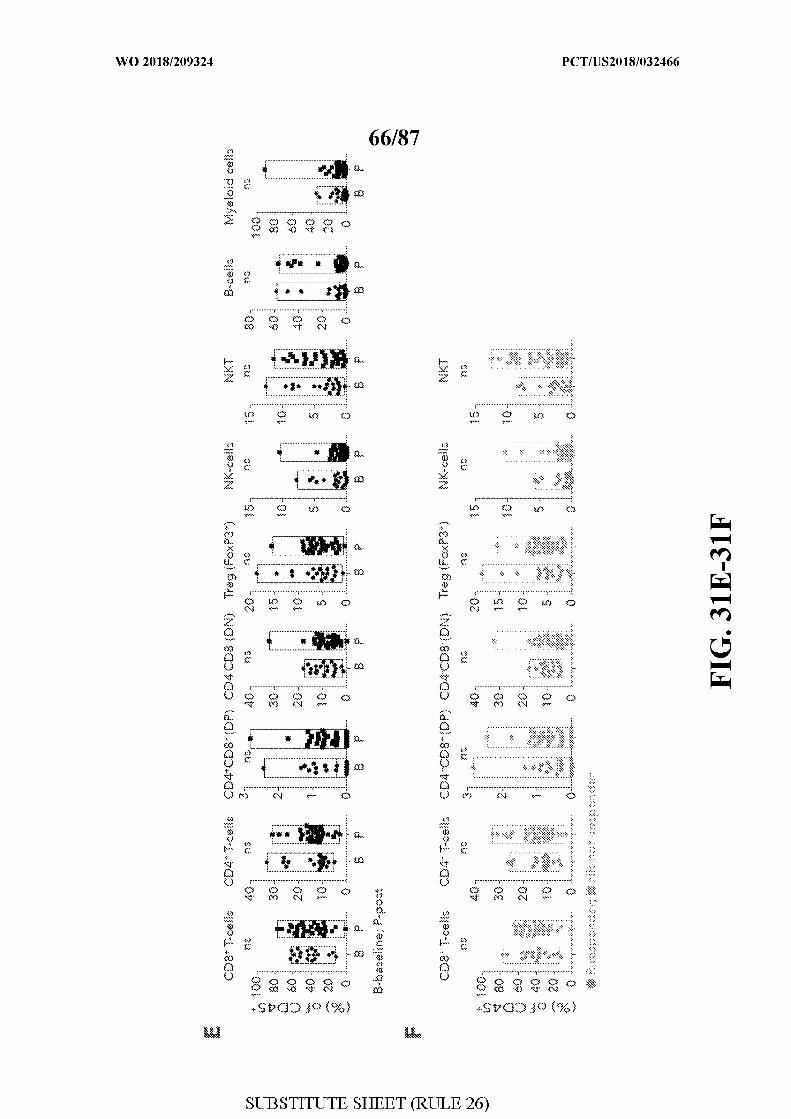
Figure 31E-F. Box plots comparing the abundance of known cell types between baseline and
post-treatment samples (E) and between responder and non-responder lesions (F). B-
baseline, P- post, R- responder, NR-non-responder, ns- not significant.
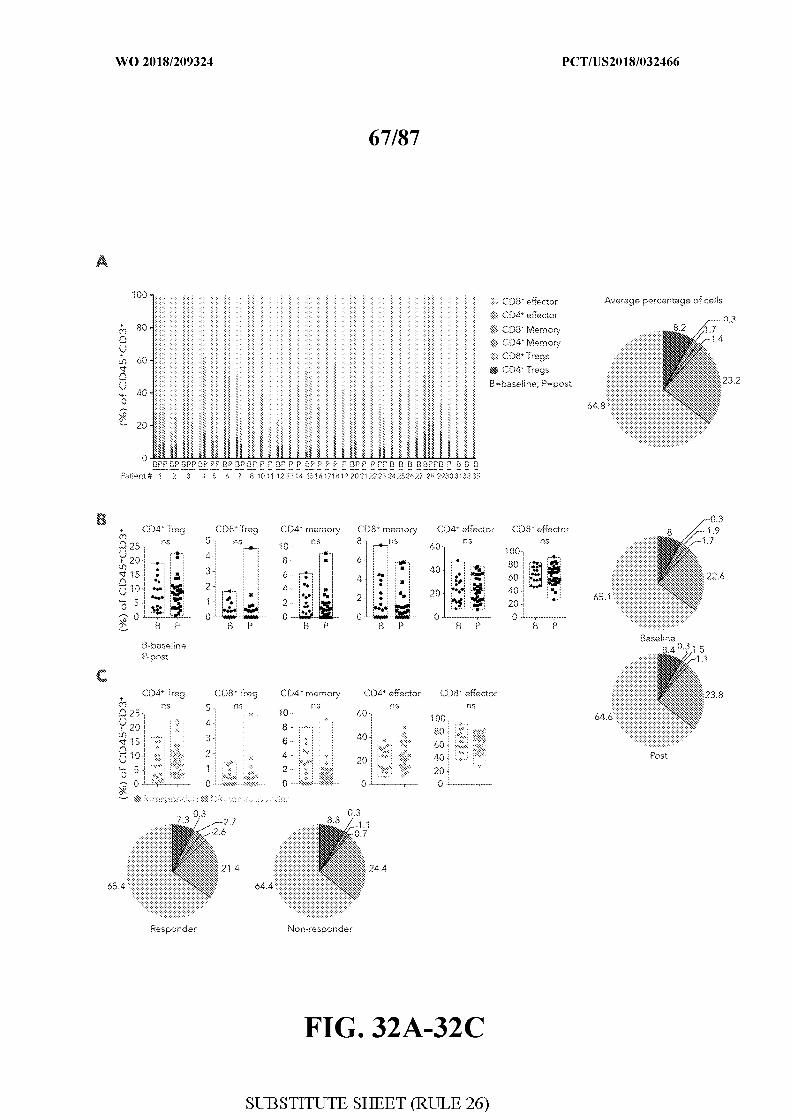
[0070] FIG. 32 - Supervised analysis of CD4+ and CD8+ T-cells expressing effector,
memory and regulatory genes. Figure 32A. The percentage of effector, memory and
regulatory CD45+CD3+cells in each patient, on the basis of pre-defined list of known marker
genes is shown. Pie chart on the right summarizes the corresponding percentage across all
CD45+CD3+ T-cells collected in this study. Figure 32B-C. Box plots and pie charts
comparing the abundance of different T-cell types between baseline and post-treatment
samples (B) and between responder and non-responder lesions (C). B- baseline, P- post, R-
responder, NR-non-responder, ns- not significant.
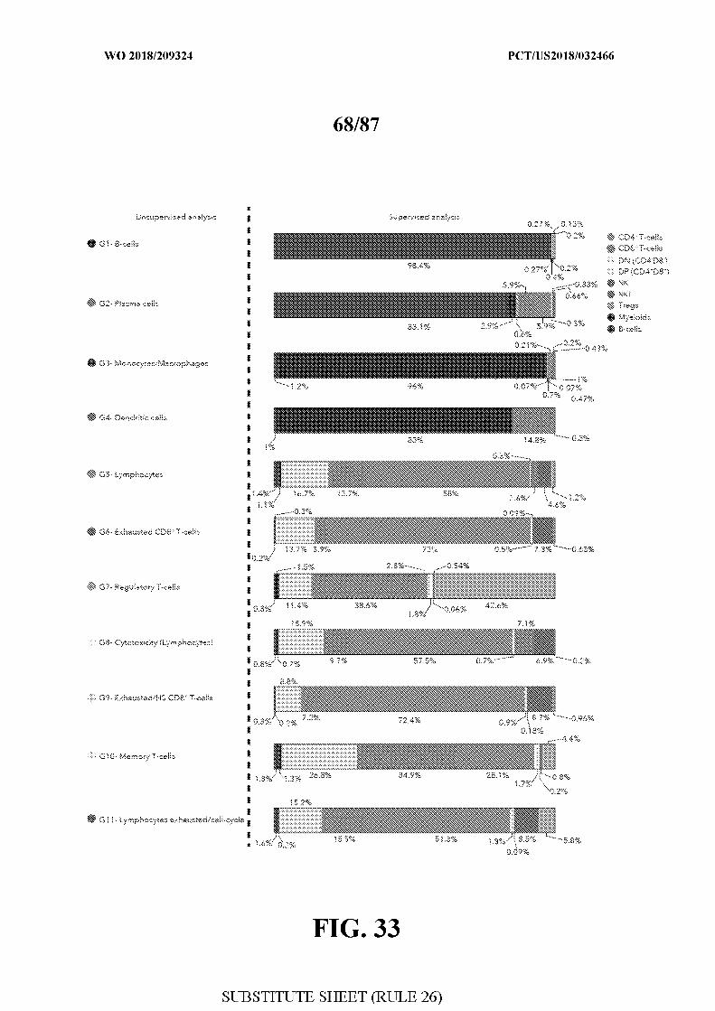
[0071] FIG. 33 - Comparing the supervised cell type classification to the
unsupervised clustering of immune cells. A comparison between the supervised
classification of single cells to cell types (right) to the unsupervised clustering of immune
cells identified by &-means clustering (left). For each one of the 11 unsupervised clusters
identified, the percentage of cell types as defined by the supervised analysis is shown.
[0072] FIG. 34 - Detection of cluster-specific genes differentially expressed between
responder and non-responder samples. Figure 34A-B. Heatmap showing scaled
expression values (log2( TPM+1)) of genes that are significantly more expressed in responder
(A) and non-responder (B) samples. The analysis was done on a specific set of genes (top 20
cluster-specific marker genes, Table 2). A list of all significant genes is shown for each
cluster next to the left margin. Shading scheme is based on z-score distribution from -2.5 to
2.5 .
[0073] FIG. 35 - Detection of genes differentially expressed between responder and
non-responder samples. Heatmap showing scaled expression values (log2( TPM+1)) of
genes that are differentially expressed between responder and non-responder samples. A list
of representative genes is shown for each cluster next to the left margin. Shading scheme is
based on z-score distribution from -2.5 to 2.5.
[0074] FIG. 36 - Annotating CD8 G and CD8 B clusters to the whole immune cell
population clusters. tSNE plot of all CD45+ clustres (n=l l ) collected in this study (left) is
shown. Cells are shaded based on 11 clusters identified by &-means clustering analysis
(Methods). Right tSNE plot shows the distribution of CD8 G and CD8 B in relation to all
immune cells analyzed in this study.
[0075] FIG. 37 - Quantification of two CD8+ T cell states associated with clinical
response. Figure 37A-B. For each sample, the percentage of cells found in CD8 G and
CD8 B (out of all CD8+T cells) in responder lesions (A) and non-responder lesions (B) is
shown. * symbol marks samples with defects in antigen presentation and the IFNypathway as
inferred from WES, IHC and flow-cytometry analysis. P indicates patient number as
described in table 1 . Figure 37C. Pie charts summarize the average percentage of the 2
clusters in the responders and non-responders groups.
[0076] FIG. 38 - Detection of defects in antigen presentation increases response
prediction. Figure 38A. Representative immunohistochemistry staining ( 1 out of 3) of
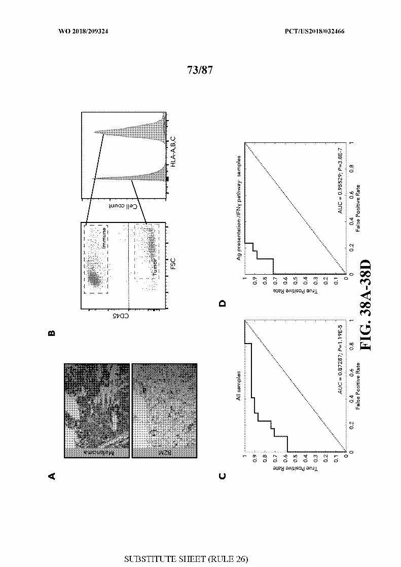
sections from patient #3 with homozygote mutations in B2M(as inferred from WES).
Sections were stained with an antibody cocktail for melanoma cells (mel. cocktail) using anti-
melanosome (HMB45), anti-MART-l/melan A and anti-Tyrosinase, to discern melanoma
cells from normal cells; or with an antibody specific for B2M. Original Magnification X I00.
Figure 38B. Flow-cytometry plot (left) and histogram (right), showing the expression of
HLA-A,B,C in immune and tumor cells in patient #15. Figure 38C-D. Receiver operating
characteristic (ROC) analysis was constructed to evaluate the prognostic power of the ratio
between CD8 B/CD8 G as shown in Fig 24D between responder and non-responder lesions.
The area under the ROC curve (AUC) was used to quantify response prediction, and one
sided Wilcoxon test was used to assess significance of the AUC results. The AUC value for
all samples (C) was 0.87 when excluding the 6
samples with defects in antigen presentation and the IFNypathway as inferred by WES, IHC
and flow-cytometry analysis.
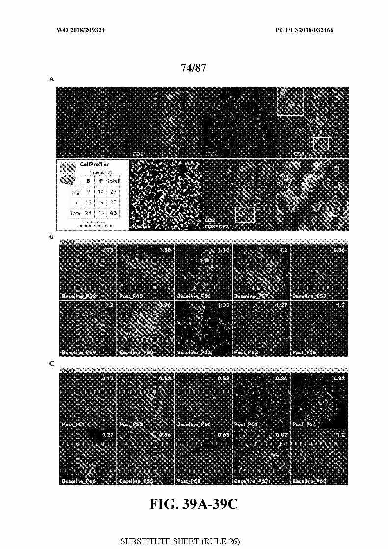
[0077] FIG. 39 - Detection and quantitation of TCF7+CD8+and TCF7 CD8+ cells in a
cohort of patients treated with anti-PDl. Figure 39A. Schematic illustration for the
immunofluorescence pipeline. Sections from an independent cohort of 33 patients (n=43
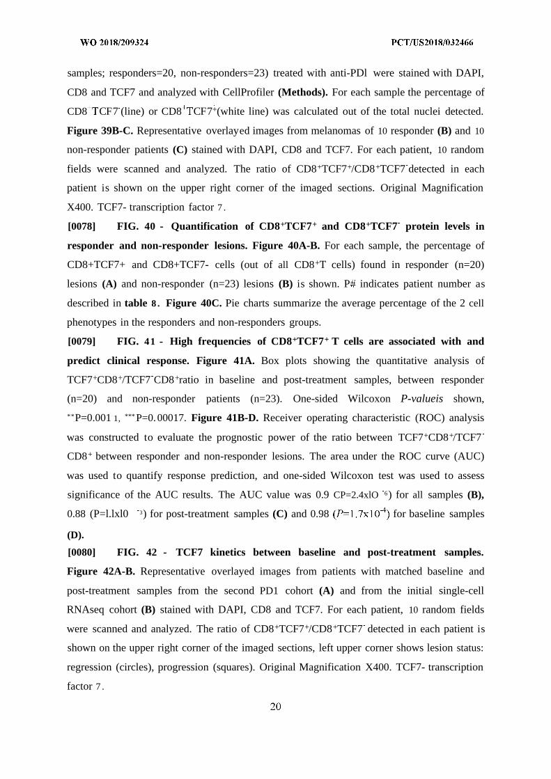
samples; responders=20, non-responders=23) treated with anti-PDl were stained with DAPI,
CD8 and TCF7 and analyzed with CellProfiler (Methods). For each sample the percentage of
CD8 CF7 (line) or CD8 CF7+(white line) was calculated out of the total nuclei detected.
Figure 39B-C. Representative overlayed images from melanomas of 10 responder (B) and 10
non-responder patients (C) stained with DAPI, CD8 and TCF7. For each patient, 10 random
fields were scanned and analyzed. The ratio of CD8+TCF7+/CD8+TCF7 detected in each
patient is shown on the upper right corner of the imaged sections. Original Magnification
X400. TCF7- transcription factor 7 .
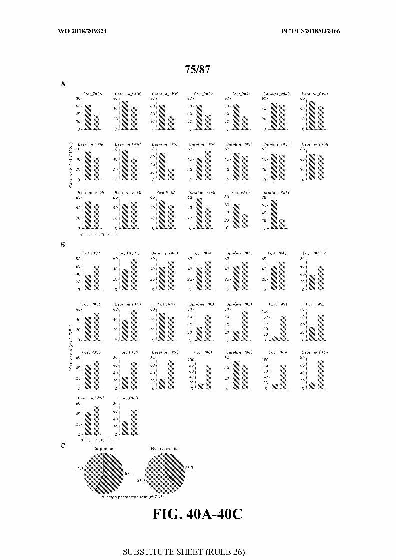
[0078] FIG. 40 - Quantification of CD8+TCF7+ and CD8+TCF7 protein levels in
responder and non-responder lesions. Figure 40A-B. For each sample, the percentage of
CD8+TCF7+ and CD8+TCF7- cells (out of all CD8+T cells) found in responder (n=20)
lesions (A) and non-responder (n=23) lesions (B) is shown. P indicates patient number as
described in table 8 . Figure 40C. Pie charts summarize the average percentage of the 2 cell
phenotypes in the responders and non-responders groups.
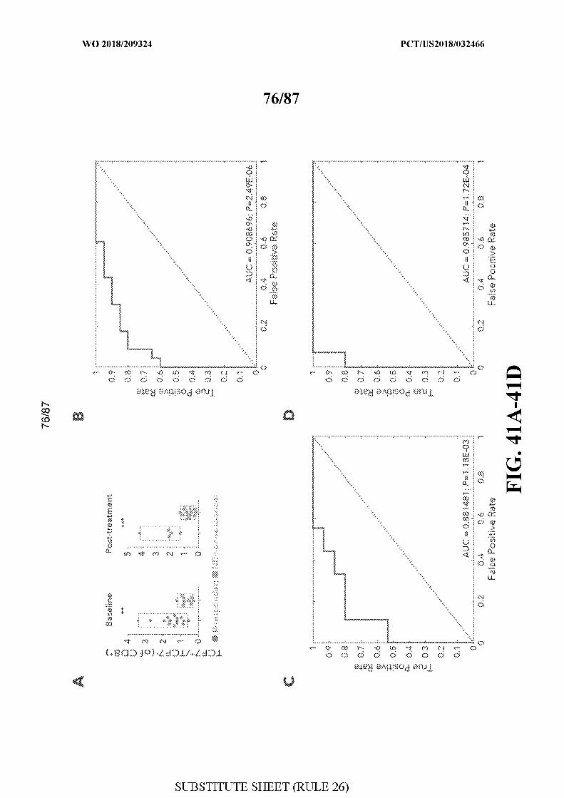
[0079] FIG. 41 - High frequencies of CD8+TCF7+ T cells are associated with and
predict clinical response. Figure 41A. Box plots showing the quantitative analysis of
TCF7+CD8+/TCF7 CD8+ratio in baseline and post-treatment samples, between responder
(n=20) and non-responder patients (n=23). One-sided Wilcoxon P-valueis shown,
**P=0.001 1, *** P=0. 00017. Figure 41B-D. Receiver operating characteristic (ROC) analysis
was constructed to evaluate the prognostic power of the ratio between TCF7+CD8+/TCF7
CD8+ between responder and non-responder lesions. The area under the ROC curve (AUC)
was used to quantify response prediction, and one-sided Wilcoxon test was used to assess
significance of the AUC results. The AUC value was 0.9 CP=2.4xlO 6) for all samples (B),
0.88 (P=l.lxl0 3) for post-treatment samples (C) and 0.98 for baseline samples
(D).
[0080] FIG. 42 - TCF7 kinetics between baseline and post-treatment samples.
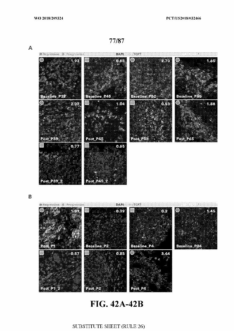
Figure 42A-B. Representative overlayed images from patients with matched baseline and
post-treatment samples from the second PD1 cohort (A) and from the initial single-cell
RNAseq cohort (B) stained with DAPI, CD8 and TCF7. For each patient, 10 random fields
were scanned and analyzed. The ratio of CD8+TCF7+/CD8+TCF7 detected in each patient is
shown on the upper right corner of the imaged sections, left upper corner shows lesion status:
regression (circles), progression (squares). Original Magnification X400. TCF7- transcription
factor 7 .
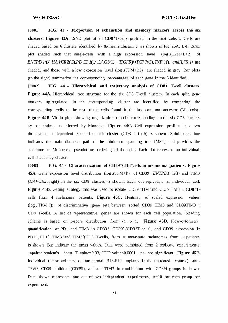
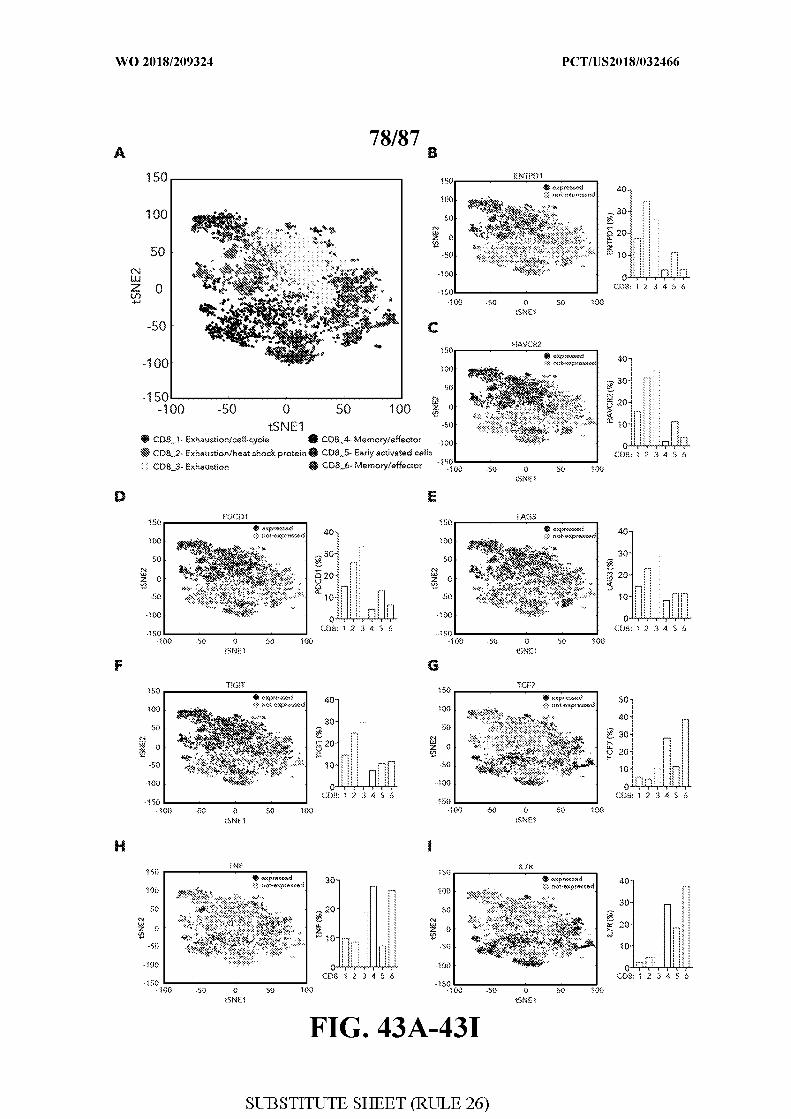
[0081] FIG. 43 - Proportion of exhaustion and memory markers across the six
clusters. Figure 43A. tSNE plot of all CD8 +T-cells profiled in the first cohort. Cells are
shaded based on 6 clusters identified by &-means clustering as shown in Fig 25A. B-I. tSNE
plot shaded such that single-cells with a high expression level (log 2(TPM+l)>2) of
ENTPD1( HA VCR2(C PDCD1 LAG3( TIGIT )TCF 7(G TNF andIL7R(l) are
shaded, and those with a low expression level (log 2(TPM+l)2) are shaded in gray. Bar plots
(to the right) summarize the corresponding percentages of each gene in the 6 identified.
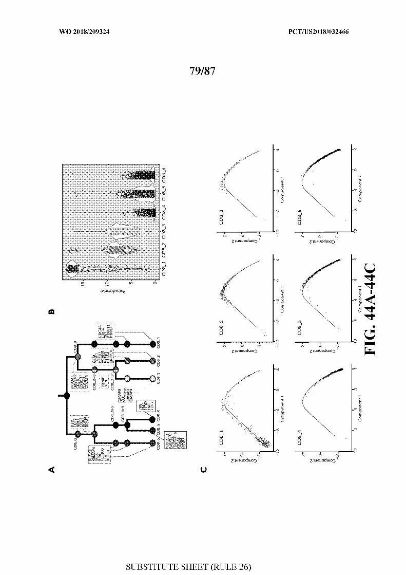
[0082] FIG. 44 - Hierarchical and trajectory analysis of CD8+ T-cell clusters.
Figure 44A. Hierarchical tree structure for the six CD8 +T-cell clusters. In each split, gene
markers up-regulated in the corresponding cluster are identified by comparing the
corresponding cells to the rest of the cells found in the last common ancestor (Methods).
Figure 44B. Violin plots showing organization of cells corresponding to the six CD8 clusters
by pseudotime as inferred by Monocle. Figure 44C. Cell expression profiles in a two
dimensional independent space for each cluster (CD8 1 to 6) is shown. Solid black line
indicates the main diameter path of the minimum spanning tree (MST) and provides the
backbone of Monocle's pseudotime ordering of the cells. Each dot represent an individual
cell shaded by cluster.
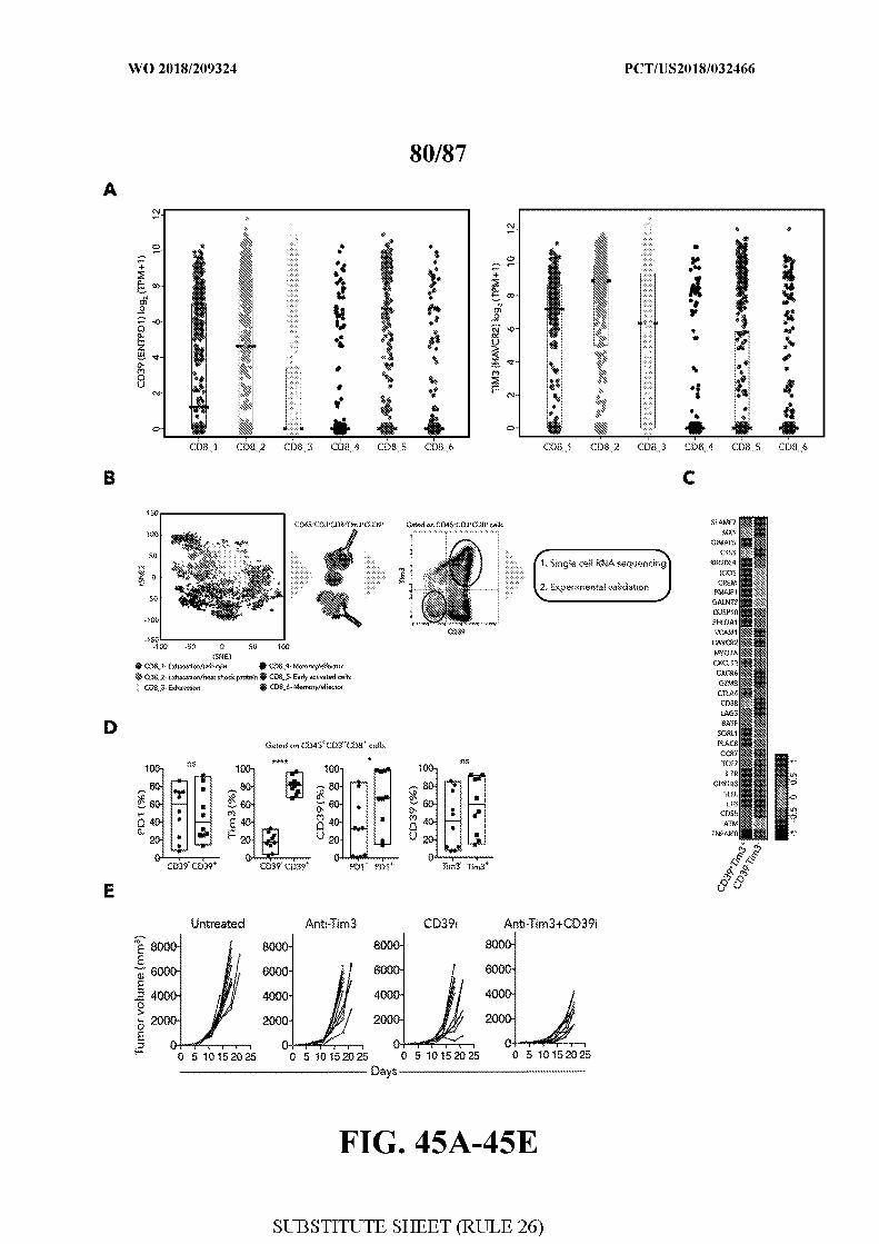
[0083] FIG. 45 - Characterization of CD39+CD8+cells in melanoma patients. Figure
45A. Gene expression level distribution (log 2(TPM+l)) of CD39 (ENTPD1, left) and TIM3
(HAVCR2, right) in the six CD8 clusters is shown. Each dot represents an individual cell.
Figure 45B. Gating strategy that was used to isolate CD39 +TIM +and CD39TIM3 , CD8 +T-
cells from 4 melanoma patients. Figure 45C. Heatmap of scaled expression values
(log 2(TPM+l)) of discriminative gene sets between sorted CD39 +TIM3 +and CD39TIM3 ,
CD8 +T-cells. A list of representative genes are shown for each cell population. Shading
scheme is based on z-score distribution from - 1 to 1 . Figure 45D. Flow-cytometry
quantification of PD1 and TIM3 in CD39 +, CD39 (CD8 +T-cells), and CD39 expression in
PD1 +, PD1 , TIM3 +and TIM3 (CD8 +T-cells) from 10 metastatic melanomas from 10 patients
is shown. Bar indicate the mean values. Data were combined from 2 replicate experiments
unpaired-student's t-test *P-value=0.03, **** P-value<0.0001, ns- not significant. Figure 45E.
Individual tumor volumes of intradermal B16-F10 implants in the untreated (control), anti-
TEVI3, CD39 inhibitor (CD39i), and anti-TIM3 in combination with CD39i groups is shown.
Data shown represents one out of two independent experiments, n=10 for each group per
experiment.
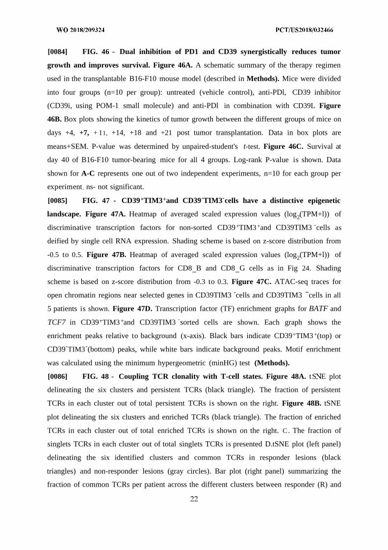
[0084] FIG. 46 - Dual inhibition of PD1 and CD39 synergistically reduces tumor
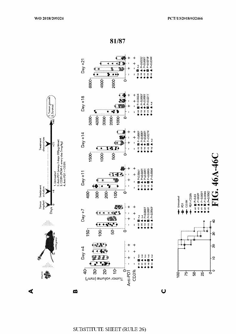
growth and improves survival. Figure 46A. A schematic summary of the therapy regimen
used in the transplantable B16-F10 mouse model (described in Methods). Mice were divided
into four groups (n=10 per group): untreated (vehicle control), anti-PDl, CD39 inhibitor
(CD39i, using POM-1 small molecule) and anti-PDl in combination with CD39L Figure
46B. Box plots showing the kinetics of tumor growth between the different groups of mice on
days +4, +7, + 11, +14, +18 and +21 post tumor transplantation. Data in box plots are
means+SEM. P-value was determined by unpaired-student's t-test. Figure 46C. Survival at
day 40 of B16-F10 tumor-bearing mice for all 4 groups. Log-rank P-value is shown. Data
shown for A-C represents one out of two independent experiments, n=10 for each group per
experiment ns- not significant.
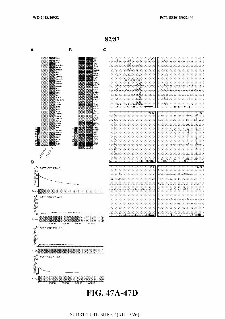
[0085] FIG. 47 - CD39+TIM3+and CD39 TIM3 cells have a distinctive epigenetic
landscape. Figure 47A. Heatmap of averaged scaled expression values (log2(TPM+l)) of
discriminative transcription factors for non-sorted CD39+TIM3+and CD39TIM3 cells as
deified by single cell RNA expression. Shading scheme is based on z-score distribution from
-0.5 to 0.5. Figure 47B. Heatmap of averaged scaled expression values (log2(TPM+l)) of
discriminative transcription factors for CD8 B and CD8 G cells as in Fig 24. Shading
scheme is based on z-score distribution from -0.3 to 0.3. Figure 47C. ATAC-seq traces for
open chromatin regions near selected genes in CD39TIM3 cells and CD39TIM3 cells in all
5 patients is shown. Figure 47D. Transcription factor (TF) enrichment graphs for BATF and
TCF7 in CD39+TIM3+and CD39TIM3 sorted cells are shown. Each graph shows the
enrichment peaks relative to background (x-axis). Black bars indicate CD39+TIM3+(top) or
CD39 TIM3 (bottom) peaks, while white bars indicate background peaks. Motif enrichment
was calculated using the minimum hypergeometric (minHG) test (Methods).
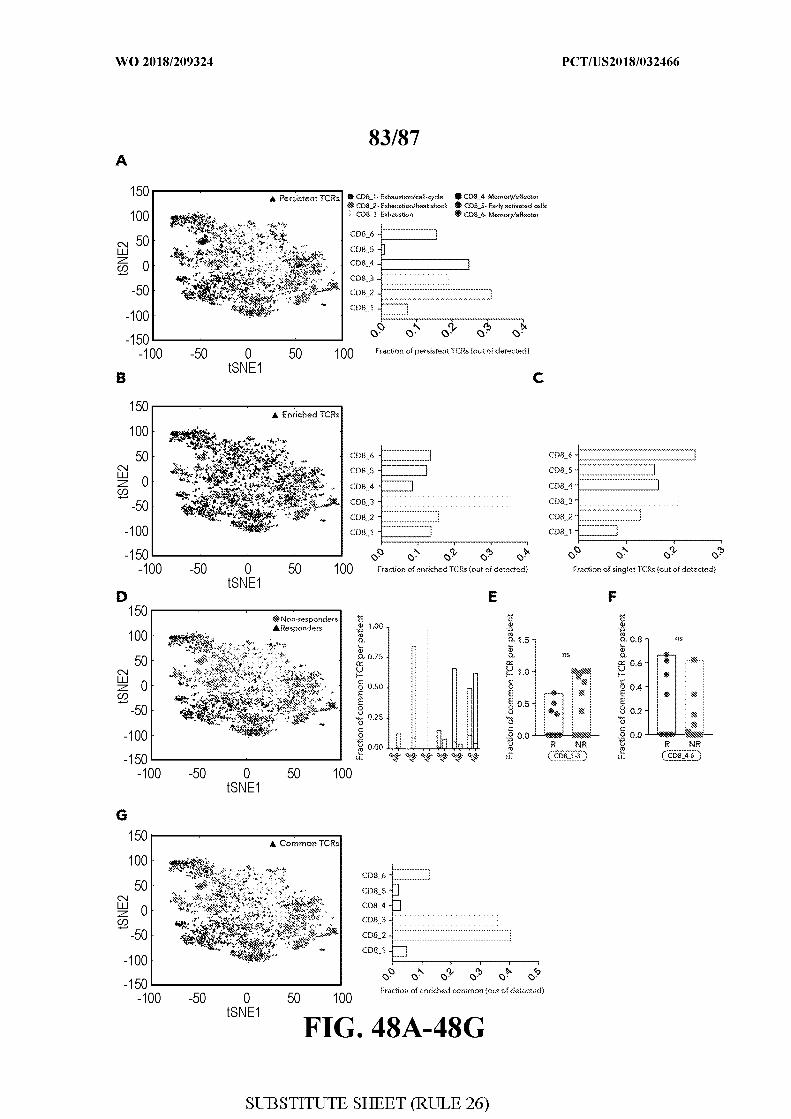
[0086] FIG. 48 - Coupling TCR clonality with T-cell states. Figure 48A. tS E plot
delineating the six clusters and persistent TCRs (black triangle). The fraction of persistent
TCRs in each cluster out of total persistent TCRs is shown on the right. Figure 48B. tSNE
plot delineating the six clusters and enriched TCRs (black triangle). The fraction of enriched
TCRs in each cluster out of total enriched TCRs is shown on the right. C . The fraction of
singlets TCRs in each cluster out of total singlets TCRs is presented D.tSNE plot (left panel)
delineating the six identified clusters and common TCRs in responder lesions (black
triangles) and non-responder lesions (gray circles). Bar plot (right panel) summarizing the
fraction of common TCRs per patient across the different clusters between responder (R) and
non-responder (NR) samples ns- not significant. E-F.Fraction of common TCRs per patient,
aggregated for CD8 1 to 3 (CD8 1-3) and CD8 4 to 6 (CD8 4-6) clusters for R and NR
samples. G . tSNE plot delineating the six clusters and common TCRs (black triangle). The
fraction of common TCRs in each cluster out of total common TCRs is shown on the right.
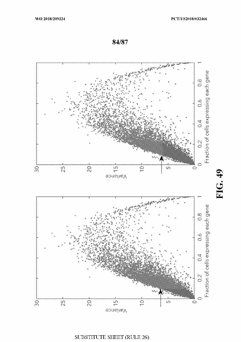
[0087] FIG. 49 - Gene Variance used for the unsupervised clustering. Variance of
each gene vs. the fraction of cells expressing each gene (log2(TPM+l)>0). Left panel: genes
expressed in more than 10% of the cells and less than 90% are shaded. Right panel: genes
with variance 6 are shaded. As the set of genes expressed in less than 10% of the cells are of
less interest for clustering analysis, we set as a minimal threshold the maximal variance
observed in this group of genes, as indicated by the black arrow.
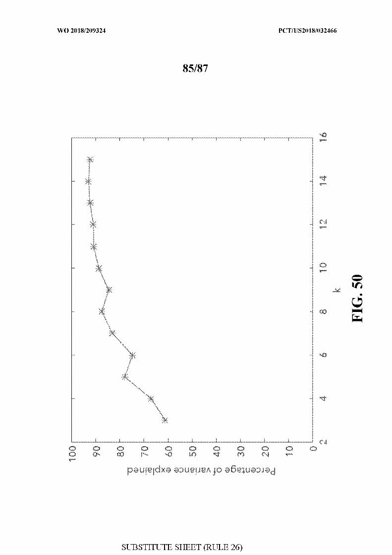
[0088] FIG. 50 - Determining an optimal number of clusters for all immune cells.
Variance explained by each &-means solution ranging from k=3,...,15, when applied to all
analyzed single-cells. Percentage of variance explained is computed as described in the
Methods section.
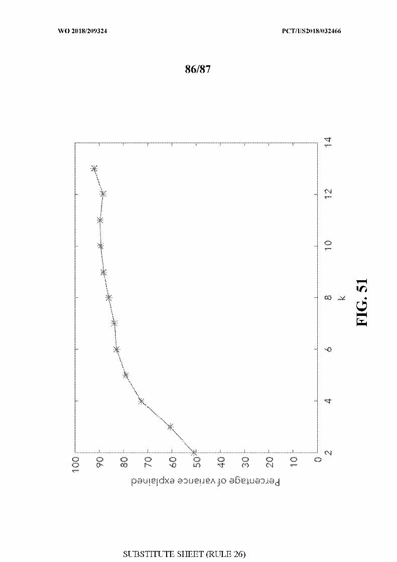
[0089] FIG. 51 - Determining an optimal number of clusters for all CD8+ T-cells.
Variance explained by each &-means solution ranging from k=2,...,13, when applied to all
analyzed CD8 T-cells. Percentage of variance explained is computes as described in the
Methods section.
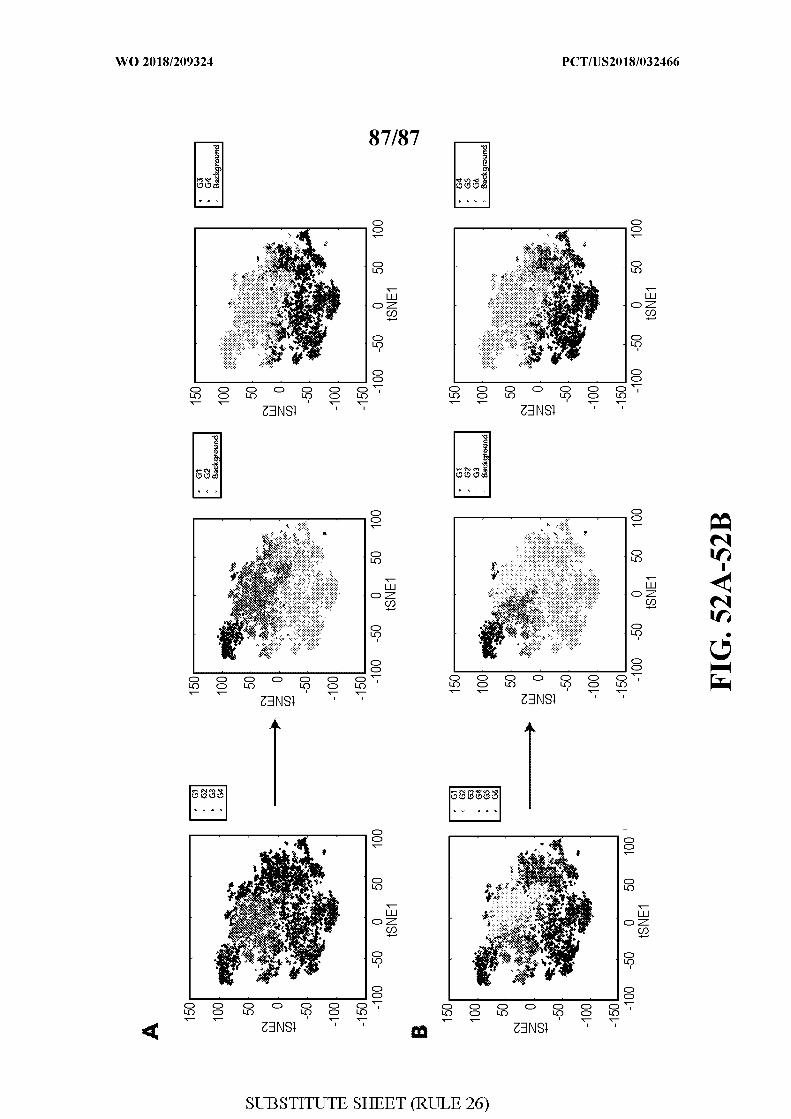
[0090] FIG. 52 - Hierarchical structure of splitting clusters . Clustering of CD8 B
and CD8 G, separately, into two (upper panel) or three (lower panel) clusters. Figure 52A.
Hierarchical structure is shown where CD8 B is split into 2 or 3 clusters, which correspond
to the k = 4 and k = 6 solutions, respectively. Figure 52B. Hierarchical structure is shown
where CD8 G is split into 2 and 3 clusters, corresponding to the k = 4 and k = 6 solutions,
respectively.
DETAILED DESCRIPTION
[0091] Unless defined otherwise, technical and scientific terms used herein have the same
meaning as commonly understood by one of ordinary skill in the art to which this disclosure
pertains. Definitions of common terms and techniques in molecular biology may be found in
Molecular Cloning: A Laboratory Manual, 2nd edition (1989) (Sambrook, Fritsch, and
Maniatis); Molecular Cloning: A Laboratory Manual, 4th edition (2012) (Green and
Sambrook); Current Protocols in Molecular Biology (1987) (F.M. Ausubel et al. eds.); the
series Methods in Enzymology (Academic Press, Inc.): PCR 2 : A Practical Approach (1995)
(M.J. MacPherson, B.D. Hames, and G.R. Taylor eds.): Antibodies, A Laboraotry Manual
(1988) (Harlow and Lane, eds.): Antibodies A Laboraotry Manual, 2nd edition 2013 (E.A.
Greenfield ed.); Animal Cell Culture (1987) (R.I. Freshney, ed.); Benjamin Lewin, Genes IX,
published by Jones and Bartlet, 2008 (ISBN 0763752223); Kendrew et al. (eds.), The
Encyclopedia of Molecular Biology, published by Blackwell Science Ltd., 1994 (ISBN
0632021829); Robert A . Meyers (ed.), Molecular Biology and Biotechnology: a
Comprehensive Desk Reference, published by VCH Publishers, Inc., 1995 (ISBN
9780471 185710); Singleton et al., Dictionary of Microbiology and Molecular Biology 2nd
ed., J . Wiley & Sons (New York, N.Y. 1994), March, Advanced Organic Chemistry
Reactions, Mechanisms and Structure 4th ed., John Wiley & Sons (New York, N.Y. 1992);
and Marten H . Hofker and Jan van Deursen, Transgenic Mouse Methods and Protocols, 2nd
edition (201 1 .
[0092] As used herein, the singular forms "a", "an", and "the" include both singular and
plural referents unless the context clearly dictates otherwise.
[0093] The term "optional" or "optionally" means that the subsequent described event,
circumstance or substituent may or may not occur, and that the description includes instances
where the event or circumstance occurs and instances where it does not.
[0094] The recitation of numerical ranges by endpoints includes all numbers and
fractions subsumed within the respective ranges, as well as the recited endpoints.
[0095] The terms "about" or "approximately" as used herein when referring to a
measurable value such as a parameter, an amount, a temporal duration, and the like, are
meant to encompass variations of and from the specified value, such as variations of +/-10%
or less, +1-5% or less, +/-1% or less, and +/-0.1% or less of and from the specified value,
insofar such variations are appropriate to perform in the disclosed invention. It is to be
understood that the value to which the modifier "about" or "approximately" refers is itself
also specifically, and preferably, disclosed.
[0096] Reference throughout this specification to "one embodiment", "an embodiment,"
"an example embodiment," means that a particular feature, structure or characteristic
described in connection with the embodiment is included in at least one embodiment of the
present invention. Thus, appearances of the phrases "in one embodiment," "in an
embodiment," or "an example embodiment" in various places throughout this specification
are not necessarily all referring to the same embodiment, but may. Furthermore, the particular
features, structures or characteristics may be combined in any suitable manner, as would be
apparent to a person skilled in the art from this disclosure, in one or more embodiments.
Furthermore, while some embodiments described herein include some but not other features
included in other embodiments, combinations of features of different embodiments are meant
to be within the scope of the invention. For example, in the appended claims, any of the
claimed embodiments can be used in any combination.
[0097] The terms "subject", "individual" or "patient" are used interchangeably
throughout this specification, and typically and preferably denote humans, but may also
encompass reference to non-human animals, preferably warm-blooded animals, even more
preferably mammals, such as, e.g., non-human primates, rodents, canines, felines, equines,
ovines, porcines, and the like. The term "non-human animals" includes all vertebrates, e.g.,
mammals, such as non-human primates, (particularly higher primates), sheep, dog, rodent
(e.g. mouse or rat), guinea pig, goat, pig, cat, rabbits, cows, and non-mammals such as
chickens, amphibians, reptiles etc. In one embodiment, the subject is a non-human mammal.
In another embodiment, the subject is human. In another embodiment, the subject is an
experimental animal or animal substitute as a disease model. The term does not denote a
particular age or sex. Thus, adult and newborn subjects, as well as fetuses, whether male or
female, are intended to be covered. Examples of subjects include humans, dogs, cats, cows,
goats, and mice. The term subject is further intended to include transgenic species.
[0098] All publications, published patent documents, and patent applications cited in this
application are indicative of the level of skill in the art(s) to which the application pertains.
All publications, published patent documents, and patent applications cited herein are hereby
incorporated by reference to the same extent as though each individual publication, published
patent document, or patent application was specifically and individually indicated as being
incorporated by reference.
Overview
[0099] Embodiments disclosed herein relate to cell products, substances, compositions,
markers, marker signatures, molecular targets, kits of parts and methods useful in
characterizing, evaluating and modulating the immune system and immune responses.
Applicants used single-cell RNA sequencing (scRNA-seq) to gain a deeper understanding of
the cellular and molecular components orchestrating immunity in melanoma patients treated
with checkpoint therapy. Through this unbiased approach, Applicants defined the immune
cell composition of melanoma tumors, identified unique cell states coupled with response,
and developed a simple assay that can accurately predict clinical outcome in an independent
cohort. Moreover, Applicants assessed the identity and function of some of the newly
identified cell states, delineated their epigenetic landscape, and tested a new therapeutic
combination that enhanced immunity in a mouse melanoma model. The analysis
demonstrates the utility of applying unbiased single-cell methods to uncover the principles
that underlie the success or failure of immunotherapy.
[0100] Thus, it is an objective of the present invention to determine whether a patient
should be treated with a checkpoint blockade (CPB) therapy. Applicants have identified that
the ratio of CD8+ TILs expressing a non-responder signature and CD8+ TILs expressing a
responder signature can predict sensitivity or resistance to CPB therapy as well as predicting
overall survival. Applicants have further identified that detecting the quantity of CD8+ T
cells expressing a single transcription factor that is part of the responder signature can be
used to distinguish between CPB therapy responders and non-responders. It is another
objective of the present invention to modulate the ratio of CD8+ TILs expressing a non-
responder signature to CD8+ TILs expressing a responder signature. It is another objective of
the present invention to provide for adoptive cell transfer methods for treatment of a cancer
patient, wherein the cells are enriched for CD8+ TILs expressing a responder signature. It is
another objective of the present invention to select patients for treatment with an
immunotherapy. It is another object of the invention to target non-responder CD8+ T cells for
cancer therapy.
[0101] The biomarkers of the present invention were discovered by analysis of
expression profiles of single immune cells within populations of cells from freshly isolated
tumors, thus allowing the discovery of novel gene signatures and immune cell subtypes that
were previously unrecognized. Treatment of solid tumors has been revolutionized by immune
checkpoint blockade therapies; yet even in melanoma, for which high response rates are
observed, the majority of patients do not respond. Specifically, to identify key immunological
components associated with success or failure of immunotherapy, Applicants profiled 16,291
immune cells from 48 tumor samples of melanoma patients treated with checkpoint
inhibitors, using single-cell transcriptomics. Applicants obtained samples from melanoma
patients receiving checkpoint blockade therapy both before they received treatment and after
they received treatment with a checkpoint inhibitor. Applicants have identified a non-
responder signature and a responder signature in the CD8+ TILs. Applicants have identified
that the ratio of CD8+ TILs expressing a non-responder signature and CD8+ TILs expressing
a responder signature can predict sensitivity or resistance to CPB therapy, as well as
predicting overall survival. Applicants identified unique exhaustion and memory/effector
states of CD8+ T-cells associated with tumor regression, and found that the expression of a
single transcription factor, TCF7, in CD8+ T-cells was sufficient to predict clinical outcome
in an independent cohort. Specifically, Applicants show using immunofluorescence that
responders have more CD8+ TCF7+ T cells than CD8+ TCF7- T cells and vice versa. Thus,
detection of CD8+ TCF7+ T cells may be used to predict overall survival in cancer patients.
Applicants delineated the epigenetic landscape and clonality of these T-cell states, and
demonstrated enhanced anti-tumor immunity by targeting a novel combination of factors
identified in exhausted cells. Applicants, show using a melanoma cancer model that targeting
CD39 and TIM3 on non-responder cells results in a significant increase in survival. This
study provides extensive unbiased data in human tumors for discovery of predictors,
therapeutic targets and combination therapies for enhancing checkpoint immunotherapy.
[0102] The presence of CD8+ T cell subtypes may be determined by subtype specific
signature biomarkers. It is generally recognized within the art, that tumors are a
conglomeration of many cells that make up a tumor microenvironment, whereby the cells
communicate and affect each other in specific ways. As such, specific immune cell types
within this microenvironment may express certain gene products for this microenvironment.
[0103] In further aspects, the invention relates to a signature or set of biomarkers (e.g.,
responder and/or non-responder signature) that may be detected in combination with other
signatures or set of biomarkers (e.g., malignant cell signatures). The signatures may be a gene
signature, protein signature, and/or other genetic or epigenetic signature of particular tumor
cell subpopulations, as defined herein (e.g., tumor cells with mutations in genes associated
with antigen presentation or the IFN gamma pathway).
[0104] The invention hereto also further relates to particular immune cell subpopulations,
which may be identified based on the methods according to the invention as discussed herein;
as well as methods to obtain such cell subpopulations; use of such subpopulations in
therapeutics; controlling therapeutic responses by targeting biomarkers relevant to the cell
subpopulation; and screening methods to identify agents capable of inducing or suppressing
particular immune cell (sub)populations.
[0105] In certain example embodiments, the immune cells comprise two sub-populations.
A first subpopulation characterized by the expression of a number of inhibitory receptors
(non-responder), and a second subpopulation characterized by the expression of a number of
memory and/or differentiation genes (responder). In certain example embodiments, these
subpopulations may be used to determine responsiveness to various therapeutics. Particular
advantageous uses include methods for identifying agents capable of inducing or suppressing
particular immune cell (sub)populations based on the gene signatures, protein signature,
and/or other genetic or epigenetic signature as defined herein.
[0106] The invention further relates to agents capable of inducing or suppressing
particular immune cell (sub)populations based on the gene signatures, protein signature,
and/or other genetic or epigenetic signature as defined herein, as well as their use for
modulating, such as inducing or repressing, a particular gene signature, protein signature,
and/or other genetic or epigenetic signature. In one embodiment, genes in one population of
cells may be activated or suppressed in order to affect the cells of another population. In
related aspects, modulating, such as inducing or repressing, a particular a particular gene
signature, protein signature, and/or other genetic or epigenetic signature may modify overall
immune cell composition, such as immune cell composition, such as immune cell
subpopulation composition or distribution, or functionality.
Biomarkers and Signatures
[0107] The invention further relates to various biomarkers for detecting immune cell
(e.g., CD8+ T cell) subpopulations. In certain example embodiments, these CD8+ T cell
populations are tumor infiltrating lymphocytes (TIL). The methods may comprise detecting a
first population of CD8+ T cells as described further below, a second population of CD8+ T
cells as described further below, or both. The first and second CD8+ T cell populations may
be detected by detecting one or more biomarkers in a sample.
[0108] The term "biomarker" is widespread in the art and commonly broadly denotes a
biological molecule, more particularly an endogenous biological molecule, and/or a
detectable portion thereof, whose qualitative and/or quantitative evaluation in a tested object
(e.g., in or on a cell, cell population, tissue, organ, or organism, e.g., in a biological sample of
a subject) is predictive or informative with respect to one or more aspects of the tested
object's phenotype and/or genotype. The terms "marker" and "biomarker" may be used
interchangeably throughout this specification. Biomarkers as intended herein may be nucleic
acid-based or peptide-, polypeptide- and/or protein-based. For example, a marker may be
comprised of peptide(s), polypeptide(s) and/or protein(s) encoded by a given gene, or of
detectable portions thereof. Further, whereas the term "nucleic acid" generally encompasses
DNA, RNA and DNA/RNA hybrid molecules, in the context of markers the term may
typically refer to heterogeneous nuclear RNA (hnRNA), pre-mRNA, messenger RNA
(mRNA), or complementary DNA (cDNA), or detectable portions thereof. Such nucleic acid
species are particularly useful as markers, since they contain qualitative and/or quantitative
information about the expression of the gene. Particularly preferably, a nucleic acid-based
marker may encompass mRNA of a given gene, or cDNA made of the mRNA, or detectable
portions thereof. Any such nucleic acid(s), peptide(s), polypeptide(s) and/or protein(s)
encoded by or produced from a given gene are encompassed by the term "gene product(s)".
[0109] Preferably, markers as intended herein may be extracellular or cell surface
markers, as methods to measure extracellular or cell surface marker(s) need not disturb the
integrity of the cell membrane and may not require fixation / permeabilization of the cells.
[0110] Unless otherwise apparent from the context, reference herein to any marker, such
as a peptide, polypeptide, protein, or nucleic acid, may generally also encompass modified
forms of said marker, such as bearing post-expression modifications including, for example,
phosphorylation, glycosylation, lipidation, methylation, cysteinylation, sulphonation,
glutathionylation, acetylation, oxidation of methionine to methionine sulphoxide or
methionine sulphone, and the like.
[0111] The term "peptide" as used throughout this specification preferably refers to a
polypeptide as used herein consisting essentially of 50 amino acids or less, e.g., 45 amino
acids or less, preferably 40 amino acids or less, e.g., 35 amino acids or less, more preferably
30 amino acids or less, e.g., 25 or less, 20 or less, 15 or less, 10 or less or 5 or less amino
acids.
[0112] The term "polypeptide" as used throughout this specification generally
encompasses polymeric chains of amino acid residues linked by peptide bonds. Hence,
insofar a protein is only composed of a single polypeptide chain, the terms "protein" and
"polypeptide" may be used interchangeably herein to denote such a protein. The term is not
limited to any minimum length of the polypeptide chain. The term may encompass naturally,
recombinantly, semi-synthetically or synthetically produced polypeptides. The term also
encompasses polypeptides that carry one or more co- or post-expression-type modifications
of the polypeptide chain, such as, without limitation, glycosylation, acetylation,
phosphorylation, sulfonation, methylation, ubiquitination, signal peptide removal, N-terminal
Met removal, conversion of pro-enzymes or pre-hormones into active forms, etc. The term
further also includes polypeptide variants or mutants which carry amino acid sequence
variations vis-a-vis a corresponding native polypeptide, such as, e.g., amino acid deletions,
additions and/or substitutions. The term contemplates both full-length polypeptides and
polypeptide parts or fragments, e.g., naturally-occurring polypeptide parts that ensue from
processing of such full-length polypeptides.
[0113] The term "protein" as used throughout this specification generally encompasses
macromolecules comprising one or more polypeptide chains, i.e., polymeric chains of amino
acid residues linked by peptide bonds. The term may encompass naturally, recombinantly,
semi-synthetically or synthetically produced proteins. The term also encompasses proteins
that carry one or more co- or post-expression-type modifications of the polypeptide chain(s),
such as, without limitation, glycosylation, acetylation, phosphorylation, sulfonation,
methylation, ubiquitination, signal peptide removal, N-terminal Met removal, conversion of
pro-enzymes or pre-hormones into active forms, etc. The term further also includes protein
variants or mutants which carry amino acid sequence variations vis-a-vis a corresponding
native proteins, such as, e.g., amino acid deletions, additions and/or substitutions. The term
contemplates both full-length proteins and protein parts or fragments, e.g., naturally-
occurring protein parts that ensue from processing of such full-length proteins.
[0114] The reference to any marker, including any peptide, polypeptide, protein, or
nucleic acid, corresponds to the marker commonly known under the respective designations
in the art. The terms encompass such markers of any organism where found, and particularly
of animals, preferably warm-blooded animals, more preferably vertebrates, yet more
preferably mammals, including humans and non-human mammals, still more preferably of
humans. All gene name symbols refer to the gene as commonly known in the art. The
examples described herein that refer to the human gene names are to be understood to also
encompasses genes in any other organism (e.g., homologous, orthologous genes). The term,
homolog, may apply to the relationship between genes separated by the event of speciation
(e.g., ortholog). Orthologs are genes in different species that evolved from a common
ancestral gene by speciation. Normally, orthologs retain the same function in the course of
evolution. Gene symbols may be those referred to by the HUGO Gene Nomenclature
Committee (HGNC) or National Center for Biotechnology Information (NCBI). Any
reference to the gene symbol is a reference made to the entire gene or variants of the gene.
The signature as described herein may encompass any of the genes described herein.
[0115] The terms particularly encompass such markers, including any peptides,
polypeptides, proteins, or nucleic acids, with a native sequence, i.e., ones of which the
primary sequence is the same as that of the markers found in or derived from nature. A
skilled person understands that native sequences may differ between different species due to
genetic divergence between such species. Moreover, native sequences may differ between or
within different individuals of the same species due to normal genetic diversity (variation)
within a given species. Also, native sequences may differ between or even within different
individuals of the same species due to somatic mutations, or post-transcriptional or post-
translational modifications. Any such variants or isoforms of markers are intended herein.
Accordingly, all sequences of markers found in or derived from nature are considered
"native". The terms encompass the markers when forming a part of a living organism, organ,
tissue or cell, when forming a part of a biological sample, as well as when at least partly
isolated from such sources. The terms also encompass markers when produced by
recombinant or synthetic means.
[0116] In certain embodiments, markers, including any peptides, polypeptides, proteins,
or nucleic acids, may be human, i.e., their primary sequence may be the same as a
corresponding primary sequence of or present in a naturally occurring human markers.
Hence, the qualifier "human" in this connection relates to the primary sequence of the
respective markers, rather than to their origin or source. For example, such markers may be
present in or isolated from samples of human subjects or may be obtained by other means
(e.g., by recombinant expression, cell-free transcription or translation, or non-biological
nucleic acid or peptide synthesis).
[0117] The reference herein to any marker, including any peptide, polypeptide, protein,
or nucleic acid, also encompasses fragments thereof. Hence, the reference herein to
measuring (or measuring the quantity of) any one marker may encompass measuring the
marker and/or measuring one or more fragments thereof.
[0118] For example, any marker and/or one or more fragments thereof may be measured
collectively, such that the measured quantity corresponds to the sum amounts of the
collectively measured species. In another example, any marker and/or one or more fragments
thereof may be measured each individually. The terms encompass fragments arising by any
mechanism, in vivo and/or in vitro, such as, without limitation, by alternative transcription or
translation, exo- and/or endo-proteolysis, exo- and/or endo-nucleolysis, or degradation of the
peptide, polypeptide, protein, or nucleic acid, such as, for example, by physical, chemical
and/or enzymatic proteolysis or nucleolysis.
[0119] The term "fragment" as used throughout this specification with reference to a
peptide, polypeptide, or protein generally denotes a portion of the peptide, polypeptide, or
protein, such as typically an N- and/or C-terminally truncated form of the peptide,
polypeptide, or protein. Preferably, a fragment may comprise at least about 30%, e.g., at least
about 50% or at least about 70%, preferably at least about 80%>, e.g., at least about 85%>, more
preferably at least about 90%, and yet more preferably at least about 95% or even about 99%
of the amino acid sequence length of said peptide, polypeptide, or protein. For example,
insofar not exceeding the length of the full-length peptide, polypeptide, or protein, a fragment
may include a sequence of > 5 consecutive amino acids, or > 10 consecutive amino acids, or
> 20 consecutive amino acids, or > 30 consecutive amino acids, e.g., >40 consecutive amino
acids, such as for example > 50 consecutive amino acids, e.g., > 60, > 70, > 80, > 90, > 100,
> 200, > 300, > 400, > 500 or > 600 consecutive amino acids of the corresponding full-length
peptide, polypeptide, or protein.
[0120] The term "fragment" as used throughout this specification with reference to a
nucleic acid (polynucleotide) generally denotes a 5'- and/or 3'-truncated form of a nucleic
acid. Preferably, a fragment may comprise at least about 30%, e.g., at least about 50% or at
least about 70%, preferably at least about 80%, e.g., at least about 85%, more preferably at
least about 90%, and yet more preferably at least about 95% or even about 99% of the nucleic
acid sequence length of said nucleic acid. For example, insofar not exceeding the length of
the full-length nucleic acid, a fragment may include a sequence of > 5 consecutive
nucleotides, or > 10 consecutive nucleotides, or > 20 consecutive nucleotides, or > 30
consecutive nucleotides, e.g., >40 consecutive nucleotides, such as for example > 50
consecutive nucleotides, e.g., > 60, > 70, > 80, > 90, > 100, > 200, > 300, > 400, > 500 or >
600 consecutive nucleotides of the corresponding full-length nucleic acid.
[0121] Cells such as immune cells as disclosed herein may in the context of the present
specification be said to "comprise the expression" or conversely to "not express" one or more
markers, such as one or more genes or gene products; or be described as "positive" or
conversely as "negative" for one or more markers, such as one or more genes or gene
products; or be said to "comprise" a defined "gene or gene product signature".
[0122] Such terms are commonplace and well-understood by the skilled person when
characterizing cell phenotypes. By means of additional guidance, when a cell is said to be
positive for or to express or comprise expression of a given marker, such as a given gene or
gene product, a skilled person would conclude the presence or evidence of a distinct signal
for the marker when carrying out a measurement capable of detecting or quantifying the
marker in or on the cell. Suitably, the presence or evidence of the distinct signal for the
marker would be concluded based on a comparison of the measurement result obtained for
the cell to a result of the same measurement carried out for a negative control (for example, a
cell known to not express the marker) and/or a positive control (for example, a cell known to
express the marker). Where the measurement method allows for a quantitative assessment of
the marker, a positive cell may generate a signal for the marker that is at least 1.5-fold higher
than a signal generated for the marker by a negative control cell or than an average signal
generated for the marker by a population of negative control cells, e.g., at least 2-fold, at least
4-fold, at least 10-fold, at least 20-fold, at least 30-fold, at least 40-fold, at least 50-fold
higher or even higher. Further, a positive cell may generate a signal for the marker that is 3.0
or more standard deviations, e.g., 3.5 or more, 4.0 or more, 4.5 or more, or 5.0 or more
standard deviations, higher than an average signal generated for the marker by a population
of negative control cells.
Use of Signature Genes
[0123] The present invention is also directed to signatures and uses thereof. As used
herein a "signature" may encompass any gene or genes, protein or proteins, or epigenetic
element(s) whose expression profile or whose occurrence is associated with a specific cell
type, subtype, or cell state of a specific cell type or subtype within a population of cells (e.g.,
tumor infiltrating lymphocytes). In certain embodiments, the expression of the CD8+ TIL
signatures are dependent on epigenetic modification of the genes or regulatory elements
associated with the genes. Thus, in certain embodiments, use of signature genes includes
epigenetic modifications that may be detected or modulated. For ease of discussion, when
discussing gene expression, any of gene or genes, protein or proteins, or epigenetic
element(s) may be substituted. As used herein, the terms "signature", "expression profile", or
"expression program" may be used interchangeably. It is to be understood that also when
referring to proteins (e.g. differentially expressed proteins), such may fall within the
definition of "gene" signature. Levels of expression or activity or prevalence may be
compared between different cells in order to characterize or identify for instance signatures
specific for cell (sub)populations. Increased or decreased expression or activity of signature
genes may be compared between different cells in order to characterize or identify for
instance specific cell (sub)populations. The detection of a signature in single cells may be
used to identify and quantitate for instance specific cell (sub)populations. A signature may
include a gene or genes, protein or proteins, or epigenetic element(s) whose expression or
occurrence is specific to a cell (sub)population, such that expression or occurrence is
exclusive to the cell (sub)population. A gene signature as used herein, may thus refer to any
set of up- and down-regulated genes that are representative of a cell type or subtype. A gene
signature as used herein, may also refer to any set of up- and down-regulated genes between
different cells or cell (sub)populations derived from a gene-expression profile. For example, a
gene signature may comprise a list of genes differentially expressed in a distinction of
interest.
[0124] The signature as defined herein (being it a gene signature, protein signature or
other genetic or epigenetic signature) can be used to indicate the presence of a cell type, a
subtype of the cell type, the state of the microenvironment of a population of cells, a
particular cell type population or subpopulation, and/or the overall status of the entire cell
(sub)population. Furthermore, the signature may be indicative of cells within a population of
cells in vivo. The signature may also be used to suggest for instance particular therapies, or to
follow up treatment, or to suggest ways to modulate immune systems. The signatures of the
present invention may be discovered by analysis of expression profiles of single-cells within
a population of cells from isolated samples (e.g. tumor samples), thus allowing the discovery
of novel cell subtypes or cell states that were previously invisible or unrecognized. The
presence of subtypes or cell states may be determined by subtype specific or cell state
specific signatures. The presence of these specific cell (sub)types or cell states may be
determined by applying the signature genes to bulk sequencing data in a sample. Not being
bound by a theory the signatures of the present invention may be microenvironment specific,
such as their expression in a particular spatio-temporal context. Not being bound by a theory,
signatures as discussed herein are specific to a particular pathological context. Not being
bound by a theory, a combination of cell subtypes having a particular signature may indicate
an outcome. Not being bound by a theory, the signatures can be used to deconvolute the
network of cells present in a particular pathological condition. Not being bound by a theory
the presence of specific cells and cell subtypes are indicative of a particular response to
treatment, such as including increased or decreased susceptibility to treatment. The signature
may indicate the presence of one particular cell type.
[0125] The signature according to certain embodiments of the present invention may
comprise or consist of one or more genes, proteins and/or epigenetic elements, such as for
instance 1, 2, 3, 4, 5, 6, 7, 8, 9, 10 or more. In certain embodiments, the signature may
comprise or consist of two or more genes, proteins and/or epigenetic elements, such as for
instance 2, 3, 4, 5, 6, 7, 8, 9, 10 or more. In certain embodiments, the signature may comprise
or consist of three or more genes, proteins and/or epigenetic elements, such as for instance 3,
4, 5, 6, 7, 8, 9, 10 or more. In certain embodiments, the signature may comprise or consist of
four or more genes, proteins and/or epigenetic elements, such as for instance 4, 5, 6, 7, 8, 9,
10 or more. In certain embodiments, the signature may comprise or consist of five or more
genes, proteins and/or epigenetic elements, such as for instance 5, 6, 7, 8, 9, 10 or more. In
certain embodiments, the signature may comprise or consist of six or more genes, proteins
and/or epigenetic elements, such as for instance 6, 7, 8, 9, 10 or more. In certain
embodiments, the signature may comprise or consist of seven or more genes, proteins and/or
epigenetic elements, such as for instance 7, 8, 9, 10 or more. In certain embodiments, the
signature may comprise or consist of eight or more genes, proteins and/or epigenetic
elements, such as for instance 8, 9, 10 or more. In certain embodiments, the signature may
comprise or consist of nine or more genes, proteins and/or epigenetic elements, such as for
instance 9, 10 or more. In certain embodiments, the signature may comprise or consist of ten
or more genes, proteins and/or epigenetic elements, such as for instance 10, 11, 12, 13, 14,
15, or more. It is to be understood that a signature according to the invention may for instance
also include genes or proteins as well as epigenetic elements combined.
[0126] In certain embodiments, a signature is characterized as being specific for a
particular immune cell or immune cell (sub)population if it is upregulated or only present,
detected or detectable in that particular immune cell or immune cell (sub)population, or
alternatively is downregulated or only absent, or undetectable in that particular immune cell
or immune cell (sub)population. In this context, a signature consists of one or more
differentially expressed genes/proteins or differential epigenetic elements when comparing
different cells or cell (sub)populations, including comparing different immune cell or
immune cell (sub)populations, as well as comparing immune cell or immune cell
(sub)populations with non- immune cell or non-immune cell (sub)populations. It is to be
understood that "differentially expressed" genes/proteins include genes/proteins which are
up- or down-regulated as well as genes/proteins which are turned on or off. When referring to
up-or down-regulation, in certain embodiments, such up- or down-regulation is preferably at
least two-fold, such as two-fold, three-fold, four-fold, five-fold, or more, such as for instance
at least ten-fold, at least 20-fold, at least 30-fold, at least 40-fold, at least 50-fold, or more.
Alternatively, or in addition, differential expression may be determined based on common
statistical tests, as is known in the art.
[0127] As discussed herein, differentially expressed genes/proteins, or differential
epigenetic elements may be differentially expressed on a single cell level, or may be
differentially expressed on a cell population level. Preferably, the differentially expressed
genes/ proteins or epigenetic elements as discussed herein, such as constituting the gene
signatures as discussed herein, when as to the cell population or subpopulation level, refer to
genes that are differentially expressed in all or substantially all cells of the population or
subpopulation (such as at least 80%, preferably at least 90%, such as at least 95% of the
individual cells). This allows one to define a particular subpopulation of immune cells. As
referred to herein, a "subpopulation" of cells preferably refers to a particular subset of cells of
a particular cell type which can be distinguished or are uniquely identifiable and set apart
from other cells of this cell type. The cell subpopulation may be phenotypically
characterized, and is preferably characterized by the signature as discussed herein. A cell
(sub)population as referred to herein may constitute of a (sub)population of cells of a
particular cell type characterized by a specific cell state.
[0128] When referring to induction, or alternatively suppression of a particular signature,
preferable is meant induction or alternatively suppression (or upregulation or
downregulation) of at least one gene/protein and/or epigenetic element of the signature, such
as for instance at least to, at least three, at least four, at least five, at least six, or all
genes/proteins and/or epigenetic elements of the signature.
[0129] Signatures may be functionally validated as being uniquely associated with a
particular immune responder phenotype. Induction or suppression of a particular signature
may consequentially be associated with or causally drive a particular immune responder
phenotype.
[0130] Various aspects and embodiments of the invention may involve analyzing gene
signatures, protein signature, and/or other genetic or epigenetic signature based on single cell
analyses (e.g. single cell RNA sequencing) or alternatively based on cell population analyses,
as is defined herein elsewhere.
[0131] In certain example embodiments, the signature genes may be used to deconvolute
the network of cells present in a tumor based on comparing them to data from bulk analysis
of a tumor sample. In certain example embodiments, the presence of specific immune cells
and immune cell subtypes may be indicative of tumor growth, invasiveness and/or resistance
to treatment. In one example embodiment, detection or one or more signature genes may
indicate the presence of a particular cell type or cell types. In certain example embodiments,
the presence of immune cell types within a tumor may indicate that the tumor will be
resistant to a treatment. In one embodiment, the signature genes of the present invention are
applied to bulk sequencing data from a tumor sample obtained from a subject, such that
information relating to disease outcome and personalized treatments is determined.
[0132] In certain embodiments, cell type markers for immune cells present in a tumor
(i.e., tumor microenvironment) may be used to deconvolute bulk expression data (see, e.g,
Venteicher, A.S., Tirosh, L, Hebert, C , Yizhak, K., C , N., Filbin, M.G., Hoverstadt, V.,
Escalante, L.E., Saw, M.L., Rodman, C , et al. (2017). Decoupling genetics, lineages and
tumor micro-environment in gliomas by single-cell RNA-seq. Science 355; Tirosh, L, Izar,
B ., Prakadan, S.M., Wadsworth, M.H., 2nd, Treacy, D., Trombetta, J .J., Rotem, A., Rodman,
C , Lian, C , Murphy, G., et al. (2016a). Dissecting the multicellular ecosystem of metastatic
melanoma by single-cell RNA-seq. Science 352, 189-196; and Tirosh, L, Venteicher, A.S.,
Hebert, C , Escalante, L.E., Patel, A.P., Yizhak, K., Fisher, J.M., Rodman, C , Mount, C ,
Filbin, M.G., et al. (2016b). Single-cell RNA-seq supports a developmental hierarchy in
human oligodendroglioma. Nature 539, 309-313).
[0133] In certain embodiments, mutations occur in individual cancers that may be used to
detect cancer progression. These mutations may be used in conjunction with the responder
and non-responder phenotypes described herein. Mutations related to T cell cytolytic activity
against tumors have been characterized and may be detected as part of the present invention
(see e.g., Rooney et al., Molecular and genetic properties of tumors associated with local
immune cytolytic activity, Cell. 2015 January 15; 160(1-2): 48-61). In certain example
embodiments, a patient may be selected for a check point blockade therapy based on
detection of a gene signature as described herein in combination with mutations related to T
cell cytolytic activity (see e.g., WO2016100975A1). In certain embodiments, cancer specific
mutations associated with cytolytic activity may be a mutation in a gene selected from the
group consisting of CASP8, B2M, PIK3CA, SMC1A, ARID5B, TET2, ALPK2, COL5A1,
TP53, DNER, NCOR1, MORC4, CIC, IRF6, MYOCD, ANKLE 1, CNKSR1, NFl, SOS1,
ARID2, CUL4B, DDX3X, FUBP1, TCP1 1L2, HLA-A, B or C, CSNK2A1, MET, ASXL1,
PD-L1, PD-L2, IDOl, ID02, ALOX12B and ALOX15B, or copy number gain, excluding
whole-chromosome events, impacting any of the following chromosomal bands: 6ql6.1-q21,
6q22.31-q24.1, 6q25.1-q26, 7pl 1.2-ql 1.1, 8p23.1, 8pl 1.23-pl 1.21 (containing IDOl,
ID02), 9p24.2-p23 (containing PDL1, PDL2), 10pl5.3, 10pl5.1-pl3, l lpl4.1,
12pl3.32-pl3.2, 17pl3.1 (containing ALOX12B, ALOX15B), and 22ql 1.1-ql 1.21. In
certain embodiments, the mutation is further associated with antigen presentation. Mutations
associated with antigen presentation may comprises mutations in B2M and HLA-A, B or C .
Detection of "Non-Responder" Sub-Populations
[0134] In one embodiment, the method comprises detecting a first population of immune
cells from a biological sample of a subject. Immune cells can be detected by sorting for
CD45+ cells. CD45 is a pan-leukocyte protein. In one embodiment, the method comprises
detecting a first population of CD8+ TIL from a biological sample of a subject. In certain
example embodiments, detection of the first population in the biological sample indicates a
likelihood that a subject will be non-responsive to a particular therapy. In certain example
embodiments, detection of the first population indicates a likelihood the subject will be non-
responsive to a checkpoint blockade therapy. The first population may generally be
characterized by increased expression of inhibitory receptors.
[0135] A marker, for example a gene or gene product, for example a peptide,
polypeptide, protein, or nucleic acid, or a group of two or more markers, is "detected" or
"measured" in a tested object (e.g., in or on a cell, cell population, tissue, organ, or organism,
e.g., in a biological sample of a subject) when the presence or absence and/or quantity of said
marker or said group of markers is detected or determined in the tested object, preferably
substantially to the exclusion of other molecules and analytes, e.g., other genes or gene
products.
[0136] The terms "sample" or "biological sample" as used throughout this specification
include any biological specimen obtained from a subject. Particularly useful samples are
those known to comprise, or expected or predicted to comprise immune cells as taught
herein. Preferably, a sample may be readily obtainable by minimally invasive methods, such
as blood collection or tissue biopsy, allowing the removal / isolation / provision of the sample
from the subject. Examples of particularly useful samples include without limitation whole
blood or a cell-containing fraction of whole blood, such as serum, white blood cells, or
peripheral blood mononuclear cells (PBMC), lymph, lymphatic tissue, inflammation fluid,
tissue specimens, or tissue biopsies. The term "tissue" as used throughout this specification
refers to any animal tissue types including, but not limited to, bone, bone marrow, neural
tissue, fibrous connective tissue, cartilage, muscle, vasculature, skin, adipose tissue, blood
and glandular tissue or other non-bone tissue. The tissue may be healthy or affected by
pathological alterations, e.g., tumor tissue or tissue affected by a disease comprising an
immune component. The tissue may be from a living subject or may be cadaveric tissue. The
tissue may be autologous tissue or syngeneic tissue or may be allograft or xenograft tissue. A
biological sample may also include cells grown in tissue culture, such as cells used for
screening drugs or primary cells grown in culture for expansion.
[0137] The term "immune cell" as used throughout this specification generally
encompasses any cell derived from a hematopoietic stem cell that plays a role in the immune
response. The term is intended to encompass immune cells both of the innate or adaptive
immune system. The immune cell as referred to herein may be a leukocyte, at any stage of
differentiation (e.g., a stem cell, a progenitor cell, a mature cell) or any activation stage.
Immune cells include lymphocytes (such as natural killer cells, T-cells (including, e.g.,
thymocytes, Th or Tc; Thl, Th2, Thl7, Τΐ αβ, CD4+, CD8+, effector Th, memory Th,
regulatory Th, CD4+/CD8+ thymocytes, CD4-/CD8- thymocytes, γδ T cells, etc.) or B-cells
(including, e.g., pro-B cells, early pro-B cells, late pro-B cells, pre-B cells, large pre-B cells,
small pre-B cells, immature or mature B-cells, producing antibodies of any isotype, Tl B-
cells, T2, B-cells, naive B-cells, GC B-cells, plasmablasts, memory B-cells, plasma cells,
follicular B-cells, marginal zone B-cells, B-1 cells, B-2 cells, regulatory B cells, etc.), such as
for instance, monocytes (including, e.g., classical, non-classical, or intermediate monocytes),
(segmented or banded) neutrophils, eosinophils, basophils, mast cells, histiocytes, microglia,
including various subtypes, maturation, differentiation, or activation stages, such as for
instance hematopoietic stem cells, myeloid progenitors, lymphoid progenitors, myeloblasts,
promyelocytes, myelocytes, metamyelocytes, monoblasts, promonocytes, lymphoblasts,
prolymphocytes, small lymphocytes, macrophages (including, e.g., Kupffer cells, stellate
macrophages, M l or M2 macrophages), (myeloid or lymphoid) dendritic cells (including,
e.g., Langerhans cells, conventional or myeloid dendritic cells, plasmacytoid dendritic cells,
mDC-1, mDC-2, Mo-DC, HP-DC, veiled cells), granulocytes, polymorphonuclear cells,
antigen-presenting cells (APC), etc.
[0138] The terms "quantity", "amount" and "level" are synonymous and generally well-
understood in the art. The terms as used throughout this specification may particularly refer
to an absolute quantification of a marker in a tested object (e.g., in or on a cell, cell
population, tissue, organ, or organism, e.g., in a biological sample of a subject), or to a
relative quantification of a marker in a tested object, i.e., relative to another value such as
relative to a reference value, or to a range of values indicating a base-line of the marker. Such
values or ranges may be obtained as conventionally known.
[0139] An absolute quantity of a marker may be advantageously expressed as weight or
as molar amount, or more commonly as a concentration, e.g., weight per volume or mol per
volume. A relative quantity of a marker may be advantageously expressed as an increase or
decrease or as a fold-increase or fold-decrease relative to said another value, such as relative
to a reference value. Performing a relative comparison between first and second variables
(e.g., first and second quantities) may but need not require determining first the absolute
values of said first and second variables. For example, a measurement method may produce
quantifiable readouts (such as, e.g., signal intensities) for said first and second variables,
wherein said readouts are a function of the value of said variables, and wherein said readouts
may be directly compared to produce a relative value for the first variable vs. the second
variable, without the actual need to first convert the readouts to absolute values of the
respective variables.
[0140] The terms "diagnosis" and "monitoring" are commonplace and well-understood in
medical practice. By means of further explanation and without limitation the term
"diagnosis" generally refers to the process or act of recognising, deciding on or concluding
on a disease or condition in a subject on the basis of symptoms and signs and/or from results
of various diagnostic procedures (such as, for example, from knowing the presence, absence
and/or quantity of one or more biomarkers characteristic of the diagnosed disease or
condition).
[0141] The term "monitoring" generally refers to the follow-up of a disease or a
condition in a subject for any changes which may occur over time.
[0142] The terms "prognosing" or "prognosis" generally refer to an anticipation on the
progression of a disease or condition and the prospect (e.g., the probability, duration, and/or
extent) of recovery. A good prognosis of the diseases or conditions taught herein may
generally encompass anticipation of a satisfactory partial or complete recovery from the
diseases or conditions, preferably within an acceptable time period. A good prognosis of such
may more commonly encompass anticipation of not further worsening or aggravating of
such, preferably within a given time period. A poor prognosis of the diseases or conditions as
taught herein may generally encompass anticipation of a substandard recovery and/or
unsatisfactorily slow recovery, or to substantially no recovery or even further worsening of
such.
[0143] The terms also encompass prediction of a disease. The terms "predicting" or
"prediction" generally refer to an advance declaration, indication or foretelling of a disease or
condition in a subject not (yet) having said disease or condition. For example, a prediction of
a disease or condition in a subject may indicate a probability, chance or risk that the subject
will develop said disease or condition, for example within a certain time period or by a
certain age. Said probability, chance or risk may be indicated inter alia as an absolute value,
range or statistics, or may be indicated relative to a suitable control subject or subject
population (such as, e.g., relative to a general, normal or healthy subject or subject
population). Hence, the probability, chance or risk that a subject will develop a disease or
condition may be advantageously indicated as increased or decreased, or as fold-increased or
fold-decreased relative to a suitable control subject or subject population. As used herein, the
term "prediction" of the conditions or diseases as taught herein in a subject may also
particularly mean that the subject has a 'positive' prediction of such, i.e., that the subject is at
risk of having such (e.g., the risk is significantly increased vis-a-vis a control subject or
subject population). The term "prediction of no" diseases or conditions as taught herein as
described herein in a subject may particularly mean that the subject has a 'negative' prediction
of such, i.e., that the subject's risk of having such is not significantly increased vis-a-vis a
control subject or subject population.
[0144] Reference values may be established according to known procedures previously
employed for other cell populations, biomarkers and gene or gene product signatures. For
example, a reference value may be established in an individual or a population of individuals
characterized by a particular diagnosis, prediction and/or prognosis of said disease or
condition (i.e., for whom said diagnosis, prediction and/or prognosis of the disease or
condition holds true). Such population may comprise without limitation 2 or more, 10 or
more, 100 or more, or even several hundred or more individuals.
[0145] A "deviation" of a first value from a second value may generally encompass any
direction (e.g., increase: first value > second value; or decrease: first value < second value)
and any extent of alteration.
[0146] For example, a deviation may encompass a decrease in a first value by, without
limitation, at least about 10% (about 0.9-fold or less), or by at least about 20% (about 0.8-
fold or less), or by at least about 30% (about 0.7-fold or less), or by at least about 40% (about
0.6-fold or less), or by at least about 50% (about 0.5-fold or less), or by at least about 60%
(about 0.4-fold or less), or by at least about 70% (about 0.3 -fold or less), or by at least about
80% (about 0.2-fold or less), or by at least about 90% (about 0.1-fold or less), relative to a
second value with which a comparison is being made.
[0147] For example, a deviation may encompass an increase of a first value by, without
limitation, at least about 10% (about 1.1 -fold or more), or by at least about 20% (about 1.2-
fold or more), or by at least about 30% (about 1.3-fold or more), or by at least about 40%
(about 1.4-fold or more), or by at least about 50% (about 1.5-fold or more), or by at least
about 60% (about 1.6-fold or more), or by at least about 70% (about 1.7-fold or more), or by
at least about 80% (about 1.8-fold or more), or by at least about 90% (about 1.9-fold or
more), or by at least about 100% (about 2-fold or more), or by at least about 150% (about
2.5-fold or more), or by at least about 200% (about 3-fold or more), or by at least about 500%
(about 6-fold or more), or by at least about 700% (about 8-fold or more), or like, relative to a
second value with which a comparison is being made.
[0148] Preferably, a deviation may refer to a statistically significant observed alteration.
For example, a deviation may refer to an observed alteration which falls outside of error
margins of reference values in a given population (as expressed, for example, by standard
deviation or standard error, or by a predetermined multiple thereof, e.g., ±lxSD or ±2xSD or
±3xSD, or ±lxSE or ±2xSE or ±3xSE). Deviation may also refer to a value falling outside of
a reference range defined by values in a given population (for example, outside of a range
which comprises >40%, > 50%, >60%, >70%, >75% or >80% or >85% or >90% or >95% or
even >100% of values in said population).
[0149] In a further embodiment, a deviation may be concluded if an observed alteration is
beyond a given threshold or cut-off. Such threshold or cut-off may be selected as generally
known in the art to provide for a chosen sensitivity and/or specificity of the prediction
methods, e.g., sensitivity and/or specificity of at least 50%, or at least 60%, or at least 70%,
or at least 80%, or at least 85%, or at least 90%, or at least 95%.
[0150] For example, receiver-operating characteristic (ROC) curve analysis can be used
to select an optimal cut-off value of the quantity of a given immune cell population,
biomarker or gene or gene product signatures, for clinical use of the present diagnostic tests,
based on acceptable sensitivity and specificity, or related performance measures which are
well-known per se, such as positive predictive value (PPV), negative predictive value (NPV),
positive likelihood ratio (LR+), negative likelihood ratio (LR-), Youden index, or similar.
[0151] In certain embodiments, subjects comprising CD8+ TILs having a non-responder
gene signature as described herein are treated with a non-immunotherapy treatment. A non-
immunotherapy treatment may involve a non-immunotherapy standard of care. Aspects of the
invention involve modifying the therapy within a standard of care based on the detection of a
gene signature as described herein. The term "standard of care" as used herein refers to the
current treatment that is accepted by medical experts as a proper treatment for a certain type
of disease and that is widely used by healthcare professionals. Standard of care is also called
best practice, standard medical care, and standard therapy. Standards of care for cancer
generally include surgery, lymph node removal, radiation, chemotherapy, targeted therapies,
antibodies targeting the tumor, and immunotherapy. The standards of care for the most
common cancers can be found on the website of National Cancer Institute
(www.cancer.gov/cancertopics). A treatment clinical trial is a research study meant to help
improve current treatments or obtain information on new treatments for patients with cancer.
When clinical trials show that a new treatment is better than the standard treatment, the new
treatment may be considered the new standard treatment.
[0152] In certain example embodiments, a method for detecting the first CD8+ tumor
infiltrating lymphocyte (TIL) sub-population comprises detecting increased expression of one
or more biomarkers in a sample, wherein the one or more biomarkers are selected from a first
group consisting of CD38, CCL3, VCAM1, GOLIM4, HAVCR2, PRDX3, ENTPD1,
PTTG1, CCR5, TRAFD1, PDCD1, CXCR6, BATF, PTPN6, LAG3 and CTLA4 or any
CD8 B gene listed in Table 6 .
[0153] The terms "increased" or "increase" or "upregulated" or "upregulate" as used
herein generally mean an increase by a statically significant amount. For avoidance of doubt,
"increased" means a statistically significant increase of at least 10% as compared to a
reference level, including an increase of at least 20%, at least 30%, at least 40%, at least 50%,
at least 60%, at least 70%, at least 80%, at least 90%, at least 100% or more, including, for
example at least 2-fold, at least 3-fold, at least 4-fold, at least 5-fold, at least 10-fold increase
or greater as compared to a reference level, as that term is defined herein.
[0154] In one example embodiment, the method comprises detecting CD83 and at least N
additional biomarkers selected from the first group, wherein N equals 2, 3, 4, 5, 6, 7, 8, 9, 10,
11, 12, 13, 14, or 15.
[0155] In another example embodiment, the method comprises detecting CCL3 and at
least N additional biomarkers selected from the first group, wherein N equals 2, 3, 4, 5, 6, 7,
8, 9, 10, 11, 12, 13, 14, or 15.
[0156] In another example embodiment, the method comprises detecting VCAMl and at
least N additional biomarkers selected from the first group, wherein N equals 2, 3, 4, 5, 6, 7,
8, 9, 10, 11, 12, 13, 14, or 15.
[0157] In another example embodiment, the method comprises detecting GOLIM4 and at
least N additional biomarkers selected from the first group, wherein N equals 2, 3, 4, 5, 6, 7,
8, 9, 10, 11, 12, 13, 14, or 15.
[0158] In another example embodiment, the method comprises detecting HAVCR2 and at
least N additional biomarkers selected from the first group, wherein N equals 2, 3, 4, 5, 6, 7,
8, 9, 10, 11, 12, 13, 14, or 15.
[0159] In another example embodiment, the method comprises detecting PRDX3 and at
least N additional biomarkers selected from the first group, wherein N equals 2, 3, 4, 5, 6, 7,
8, 9, 10, 11, 12, 13, 14, or 15.
[0160] In another example embodiment, the method comprises detecting ENTPD1 and at
least N additional biomarkers selected from the first group, wherein N equals 2, 3, 4, 5, 6, 7,
8, 9, 10, 11, 12, 13, 14, or 15.
[0161] In another example embodiment, the method comprises detecting PTTG1 and at
least N additional biomarkers selected from the first group, wherein N equals 2, 3, 4, 5, 6, 7,
8, 9, 10, 11, 12, 13, 14, or 15.
[0162] In another example embodiments, the method may comprise detecting CCR5 and
at least N additional biomarkers selected from the first group, wherein N equals 2, 3, 4, 5, 6,
7, 8, 9, 10, 11, 12, 13, 14, or 15.
[0163] In another example embodiment, the method may comprise detecting TRAFD1
and at least N additional biomarkers selected from the first group, wherein N equals 2, 3, 4, 5,
6, 7, 8, 9, 10, 11, 12, 13, 14, or 15.
[0164] In another example embodiment, the method may comprise detecting PDCD1 and
at least N additional biomarkers selected from the first group, wherein N equals 2, 3, 4, 5, 6,
7, 8, 9, 10, 11, 12, 13, 14, or 15.
[0165] In another example embodiments, the method may comprise detecting CXCR6,
and at least N additional biomarkers selected from the first group, wherein N equals 2, 3, 4, 5,
6, 7, 8, 9, 10, 11, 12, 13, 14, or 15.
[0166] In another example embodiment, the method may comprise detecting BATF and
at least N additional biomarkers selected from the first group, wherein N equals 2, 3, 4, 5, 6,
7, 8, 9, 10, 11, 12, 13, 14, or 15.
[0167] In another example embodiment, the method may comprise detecting PTPN6 and
at least N additional biomarkers selected from the first group, wherein N equals 2, 3, 4, 5, 6,
7, 8, 9, 10, 11, 12, 13, 14, or 15.
[0168] In another example embodiment, the method may comprise detecting LAG3 and
at least N additional biomarkers selected from the first group, wherein N equals 2, 3, 4, 5, 6,
7, 8, 9, 10, 11, 12, 13, 14, or 15.
[0169] In another example embodiment, the method may comprise detecting CTLA4 and
at least N additional biomarkers selected from the first group, wherein N equals 2, 3, 4, 5, 6,
7, 8, 9, 10, 11, 12, 13, 14, or 15.
[0170] In certain example embodiments, the method comprises detecting a first
population of CD45+ cells obtained from a biological sample. In certain example
embodiments, detection of the population in the biological sample indicates a likelihood that
a subject will be responsive to a particular therapy. In certain example embodiments,
detection of the population indicates a likelihood the subject will be responsive to a
checkpoint blockade therapy. The population may generally be characterized by increased
expression of memory and differentiation genes.
[0171] In certain embodiments, the population is characterized by expression of a gene
signature comprising one or more genes or polypeptides selected from the group consisting
of: TCF7; TCF7, PLAC8, LTB, and CCR7; or TCF7, LEF1, S1PR1, PLAC8, LTB, CCR7,
IGHD, PAX5, FCRL1, FCER2, CD19, CD22, BA 1, MS4A1, BLK, RALGPS2 and
FAM129C; or TCF7, PLAC8, LTB, LY9, SELL, IGKC and CCR7 (Tables 4 and 5).
Detection of "Responder" Sub-Populations
[0172] In another embodiment, the method comprises detecting a population of CD8+
TIL from a biological sample of a subject. In certain example embodiments, detection of the
population in the biological sample indicates a likelihood that a subject will be responsive to
a particular therapy. In certain example embodiments, detection of the population indicates a
likelihood the subject will be responsive to a checkpoint blockade therapy. The second
population may generally be characterized by increased expression of memory and
differentiation genes.
[0173] In certain example embodiments, a method for detecting the second CD8+ tumor
infiltrating lymphocyte (TIL) sub-population comprises detecting increased expression of one
or more biomarkers selected from a second group consisting of IL7R, GPR183, TCF7,
LMNA, R4A3, CD55, AIM1, MGAT4A, PERI, FOSL2, TSPYL2, REL, FAM177A1,
YPEL5, TC2N, CSR P 1, FAM65B, PIK3R1, RGPD6, SKIL, TSC22D2, USP36, FOXP1,
EGRl, MYADM, ZFP36L2, FAM102A, RGCC, PDE4B, PFKFB3, FOSB, DCTN6 and
BTG2. In certain embodiments, the method comprises detecting increased expression of one
or more biomarkers selected from a second group consisting of IL7R, GPR183, TCF7,
LMNA, NR4A3, CD55, AIMl, MGAT4A, PERI, FOSL2, TSPYL2, REL, FAM177A1,
YPEL5, TC2N and CSRNP1. In certain embodiments, the method comprises detecting
increased expression of TCF7. In certain embodiments, the method comprises detecting
increased expression or any CD8 G gene listed in Table 6 .
[0174] In one example embodiment, the method of detecting the second population may
comprise detecting IL7R and at least N additional biomarkers selected from the first group,
wherein N equals 2, 3, 4, 5, 6, 7, 8, 9, 10, 11, 12, 13, 14, 15, 16, 17, 18, 19, 20, 21, 22, 23, 24,
25, 26, 27, 28, 29, 30, 31, or 32
[0175] In another example embodiment, the method of detecting the second population
may comprise GPR183 and at least N additional biomarkers selected from the first group,
wherein N equals 2, 3, 4, 5, 6, 7, 8, 9, 10, 11, 12, 13, 14, 15, 16, 17, 18, 19, 20, 21, 22, 23, 24,
25, 26, 27, 28, 29, 30, 31, or 32
[0176] In another example embodiment, the method of detecting the second population
may comprise TCF7 and at least N additional biomarkers selected from the first group,
wherein N equals 2, 3, 4, 5, 6, 7, 8, 9, 10, 11, 12, 13, 14, 15, 16, 17, 18, 19, 20, 21, 22, 23, 24,
25, 26, 27, 28, 29, 30, 31, or 32.
[0177] In another example embodiment, the method of detecting the second population
may comprise LMNA and at least N additional biomarkers selected from the first group,
wherein N equals 2, 3, 4, 5, 6, 7, 8, 9, 10, 11, 12, 13, 14, 15, 16, 17, 18, 19, 20, 21, 22, 23, 24,
25, 26, 27, 28, 29, 30, 31, or 32.
[0178] In another example embodiment, the method of detecting the second population
may comprise NR4A3 and at least N additional biomarkers selected from the first group,
wherein N equals 2, 3, 4, 5, 6, 7, 8, 9, 10, 11, 12, 13, 14, 15, 16, 17, 18, 19, 20, 21, 22, 23, 24,
25, 26, 27, 28, 29, 30, 31, or 32.
[0179] In another example embodiment, the method of detecting the second population
may comprise CD55 and at least N additional biomarkers selected from the first group,
wherein N equals 2, 3, 4, 5, 6, 7, 8, 9, 10, 11, 12, 13, 14, 15, 16, 17, 18, 19, 20, 21, 22, 23, 24,
25, 26, 27, 28, 29, 30, 31, or 32.
[0180] In another example embodiment, the method of detecting the second population
may comprise AIMl and at least N additional biomarkers selected from the first group,
wherein N equals 2, 3, 4, 5, 6, 7, 8, 9, 10, 11, 12, 13, 14, 15, 16, 17, 18, 19, 20, 21, 22, 23, 24,
25, 26, 27, 28, 29, 30, 31, or 32.
[0181] In another example embodiment, the method of detecting the second population
may comprise MGAT4A and at least N additional biomarkers selected from the first group,
wherein N equals 2, 3, 4, 5, 6, 7, 8, 9, 10, 11, 12, 13, 14, 15, 16, 17, 18, 19, 20, 21, 22, 23, 24,
25, 26, 27, 28, 29, 30, 31, or 32.
[0182] In another example embodiment, the method of detecting the second population
may comprise PERI and at least N additional biomarkers selected from the first group,
wherein N equals 2, 3, 4, 5, 6, 7, 8, 9, 10, 11, 12, 13, 14, 15, 16, 17, 18, 19, 20, 21, 22, 23, 24,
25, 26, 27, 28, 29, 30, 31, or 32.
[0183] In another example embodiment, the method of detecting the second population
may comprise FOSL2 and at least N additional biomarkers selected from the first group,
wherein N equals 2, 3, 4, 5, 6, 7, 8, 9, 10, 11, 12, 13, 14, 15, 16, 17, 18, 19, 20, 21, 22, 23, 24,
25, 26, 27, 28, 29, 30, 31, or 32.
[0184] In another example embodiment, the method of detecting the second population
may comprise TSPYL2 and at least N additional biomarkers selected from the first group,
wherein N equals 2, 3, 4, 5, 6, 7, 8, 9, 10, 11, 12, 13, 14, 15, 16, 17, 18, 19, 20, 21, 22, 23, 24,
25, 26, 27, 28, 29, 30, 31, or 32.
[0185] In another example embodiment, the method of detecting the second population
may comprise REL and at least N additional biomarkers selected from the first group,
wherein N equals 2, 3, 4, 5, 6, 7, 8, 9, 10, 11, 12, 13, 14, 15, 16, 17, 18, 19, 20, 21, 22, 23, 24,
25, 26, 27, 28, 29, 30, 31, or 32.
[0186] In another example embodiment, the method of detecting the second population
may comprise FAM177A1 and at least N additional biomarkers selected from the first group,
wherein N equals 2, 3, 4, 5, 6, 7, 8, 9, 10, 11, 12, 13, 14, 15, 16, 17, 18, 19, 20, 21, 22, 23, 24,
25, 26, 27, 28, 29, 30, 31, or 32.
[0187] In another example embodiment, the method of detecting the second population
may comprise YPEL5 and at least N additional biomarkers selected from the first group,
wherein N equals 2, 3, 4, 5, 6, 7, 8, 9, 10, 11, 12, 13, 14, 15, 16, 17, 18, 19, 20, 21, 22, 23, 24,
25, 26, 27, 28, 29, 30, 31, or 32.
[0188] In another example embodiment, the method of detecting the second population
may comprise TC2N and at least N additional biomarkers selected from the first group,
wherein N equals 2, 3, 4, 5, 6, 7, 8, 9, 10, 11, 12, 13, 14, 15, 16, 17, 18, 19, 20, 21, 22, 23, 24,
25, 26, 27, 28, 29, 30, 31, or 32.

[0189] In another example embodiment, the method of detecting the second population
may comprise CSRNP1 and at least N additional biomarkers selected from the first group,
wherein N equals 2, 3, 4, 5, 6, 7, 8, 9, 10, 11, 12, 13, 14, 15, 16, 17, 18, 19, 20, 21, 22, 23, 24,
25, 26, 27, 28, 29, 30, 31, or 32.
[0190] In another example embodiment, the method of detecting the second population
may comprise FAM65B and at least N additional biomarkers selected from the first group,
wherein N equals 2, 3, 4, 5, 6, 7, 8, 9, 10, 11, 12, 13, 14, 15, 16, 17, 18, 19, 20, 21, 22, 23, 24,
25, 26, 27, 28, 29, 30, 31, or 32.
[0191] In another example embodiment, the method of detecting the second population
may comprise PIK3R1 and at least N additional biomarkers selected from the first group,
wherein N equals 2, 3, 4, 5, 6, 7, 8, 9, 10, 11, 12, 13, 14, 15, 16, 17, 18, 19, 20, 21, 22, 23, 24,
25, 26, 27, 28, 29, 30, 31, or 32.
[0192] In another example embodiment, the method of detecting the second population
may comprise RGPD6 and at least N additional biomarkers selected from the first group,
wherein N equals 2, 3, 4, 5, 6, 7, 8, 9, 10, 11, 12, 13, 14, 15, 16, 17, 18, 19, 20, 21, 22, 23, 24,
25, 26, 27, 28, 29, 30, 31, or 32.
[0193] In another example embodiment, the method of detecting the second population
may comprise SKIL and at least N additional biomarkers selected from the first group,
wherein N equals 2, 3, 4, 5, 6, 7, 8, 9, 10, 11, 12, 13, 14, 15, 16, 17, 18, 19, 20, 21, 22, 23, 24,
25, 26, 27, 28, 29, 30, 31, or 32.
[0194] In another example embodiment, the method of detecting the second population
may comprise TSC22D2 and at least N additional biomarkers selected from the first group,
wherein N equals 2, 3, 4, 5, 6, 7, 8, 9, 10, 11, 12, 13, 14, 15, 16, 17, 18, 19, 20, 21, 22, 23, 24,
25, 26, 27, 28, 29, 30, 31, or 32.
[0195] In another example embodiment, the method of detecting the second population
may comprise USP36 and at least N additional biomarkers selected from the first group,
wherein N equals 2, 3, 4, 5, 6, 7, 8, 9, 10, 11, 12, 13, 14, 15, 16, 17, 18, 19, 20, 21, 22, 23, 24,
25, 26, 27, 28, 29, 30, 31, or 32.
[0196] In another example embodiment, the method of detecting the second population
may comprise FOXP1 and at least N additional biomarkers selected from the first group,
wherein N equals 2, 3, 4, 5, 6, 7, 8, 9, 10, 11, 12, 13, 14, 15, 16, 17, 18, 19, 20, 21, 22, 23, 24,
25, 26, 27, 28, 29, 30, 31, or 32.
[0197] In another example embodiment, the method of detecting the second population
may comprise EGR1 and at least N additional biomarkers selected from the first group,
wherein N equals 2, 3, 4, 5, 6, 7, 8, 9, 10, 11, 12, 13, 14, 15, 16, 17, 18, 19, 20, 21, 22, 23, 24,
25, 26, 27, 28, 29, 30, 31, or 32.
[0198] In another example embodiment, the method of detecting the second population
may comprise MYADM and at least N additional biomarkers selected from the first group,
wherein N equals 2, 3, 4, 5, 6, 7, 8, 9, 10, 11, 12, 13, 14, 15, 16, 17, 18, 19, 20, 21, 22, 23, 24,
25, 26, 27, 28, 29, 30, 31, or 32.
[0199] In another example embodiment, the method of detecting the second population
may comprise ZP36L2 and at least N additional biomarkers selected from the first group,
wherein N equals 2, 3, 4, 5, 6, 7, 8, 9, 10, 11, 12, 13, 14, 15, 16, 17, 18, 19, 20, 21, 22, 23, 24,
25, 26, 27, 28, 29, 30, 31, or 32.
[0200] In another example embodiment, the method of detecting the second population
may comprise FAM102A and at least N additional biomarkers selected from the first group,
wherein N equals 2, 3, 4, 5, 6, 7, 8, 9, 10, 11, 12, 13, 14, 15, 16, 17, 18, 19, 20, 21, 22, 23, 24,
25, 26, 27, 28, 29, 30, 31, or 32.
[0201] In another example embodiment, the method of detecting the second population
may comprise RGCC and at least N additional biomarkers selected from the first group,
wherein N equals 2, 3, 4, 5, 6, 7, 8, 9, 10, 11, 12, 13, 14, 15, 16, 17, 18, 19, 20, 21, 22, 23, 24,
25, 26, 27, 28, 29, 30, 31, or 32.
[0202] In another example embodiment, the method of detecting the second population
may comprise PDE4B and at least N additional biomarkers selected from the first group,
wherein N equals 2, 3, 4, 5, 6, 7, 8, 9, 10, 11, 12, 13, 14, 15, 16, 17, 18, 19, 20, 21, 22, 23, 24,
25, 26, 27, 28, 29, 30, 31, or 32.
[0203] In another example embodiment, the method of detecting the second population
may comprise PFKFB3 and at least N additional biomarkers selected from the first group,
wherein N equals 2, 3, 4, 5, 6, 7, 8, 9, 10, 11, 12, 13, 14, 15, 16, 17, 18, 19, 20, 21, 22, 23, 24,
25, 26, 27, 28, 29, 30, 31, or 32.
[0204] In another example embodiment, the method of detecting the second population
may comprise FOSB and at least N additional biomarkers selected from the first group,
wherein N equals 2, 3, 4, 5, 6, 7, 8, 9, 10, 11, 12, 13, 14, 15, 16, 17, 18, 19, 20, 21, 22, 23, 24,
25, 26, 27, 28, 29, 30, 31, or 32.
[0205] In another example embodiment, the method of detecting the second population
may comprise DCTN6 and at least N additional biomarkers selected from the first group,
wherein N equals 2, 3, 4, 5, 6, 7, 8, 9, 10, 11, 12, 13, 14, 15, 16, 17, 18, 19, 20, 21, 22, 23, 24,
25, 26, 27, 28, 29, 30, 31, or 32.
[0206] In another example embodiment, the method of detecting the second population
may comprise BTG2 and at least N additional biomarkers selected from the first group,
wherein N equals 2, 3, 4, 5, 6, 7, 8, 9, 10, 11, 12, 13, 14, 15, 16, 17, 18, 19, 20, 21, 22, 23, 24,
25, 26, 27, 28, 29, 30, 31, or 32.
[0207] In certain example embodiments, the method comprises detecting a second
population of CD45+ cells obtained from a biological sample. In certain example
embodiments, detection of the population in the biological sample indicates a likelihood that
a subject will be nonresponsive to a particular therapy. In certain example embodiments,
detection of the population indicates a likelihood the subject will be nonresponsive to a
checkpoint blockade therapy.
[0208] In certain embodiments, the population is characterized by expression of a gene
signature comprising one or more genes or polypeptides selected from the group consisting
of: ENTPD1 and HAVCR2; or CCL3, CD38 and HAVCR2; or CD38, PDCDl, CCL3,
SNAP47, VCAMl, HAVCR2, FASLG, ENTPD1, SIRPG, MY07A, FABP5, DUFB3,
UBE2F, CLTA and SNRPDl; or FASLG, VCAMl, CCL3, LAG3, CXCR6, IFNG, PDCDl,
KLRD1, HAVCR2, SIRPG, SNAP47, DTHD1, PRF1, GZMH, F2R, CD38, CXCL13,
TNFRSF4, TNFRSF18, MAF, ETV7, CD4, CTLA4, FCRL6, SPON2, KLRG1, TRGC1,
A2M, FCGR3A, GZMA, HOPX, NKG7, PXN, TNFRSF9, GEM, NABl, DFNB31,
CADMl, CRTAM, GPR56, MY07A, DUSP4, METRNL and PHLDA1; or CCL3, LGALSl,
CD38, EPSTI1, WARS, PLEK, HAVCR2, LGALS3, FABP5, MT2A, GBP1, PLSCR1,
CCR5, GSTOl, ANXA5, GLUL, PYCARD, TYMP, IFI6, VAMP5, OASL, GZMB, TXN,
SQRDL, RHOC, AP2S1, GZMH, CCL4L2, SNAP47, LAP3, ATP6V1B2, CCL4L1,
LAMP2, PSMA4, SERPINBl, HIGD1A, UBE2F, TALDOl, CD63, CLTA, S100A1 1,
PHPT1, GBP4, PRDX3, PSMB2, BST2, GBP5, CTSC, NDUFB3, NPC2, GALM, GLIPR2,
CCL4, PRF1, IFNG, IFI30, CHST12, ISG15, MYD88, IDH2, MTHFD2, CHMP2A,
NDUFA9, CHMP5, CALM3, ANXA2, PPTl, GTF3C6, NDUFABl, CXCR6, RNF181,
LGALS9, COX5A, OAS2, PDCDl, SNRPC, BHLHE40, TWF2, SLAMF7, TXN2,
CARD 16, ANAPCl l , MRPL51, LIMS1, NDUFA12, RANBPl, GBP2, PSMC1, ACTR1A,
CD2BP2, VDAC1, EMC7, MX1, GPS1, ATP5J2, USMG5, SHFM1, ATP51, FAM96A,
CASP1, PARP9, NOP10, GNG5, CYC1, RABl lA, PGAM1, ENTPD1, PDIA6, PSMC3,
TMBIM1, UBE2L6, PSMA6, EIF6, DCTN3, SEC 11A, CSTB, ETFB, DBI, GRN, ELOVLl,
UBE2L3, PSMB3, U B7, DOK2, SEC61G, IGFLR1, ATP5H, COPZ1, ATP6V1F,
BNIP3L, NUTF2, AKRIAI, MDH2, VAMP8, ROMOl, CXCR3, SAMHDl, NUCBl,
ACTN4, ZYX, FLOT1, BLOC1S1, STAT1, VFMP, PAM, NUDT21, MYOIG, C17orf49,
GTF2A2, HIST2H2AA4, C19orfl0, ABI3, TRAPPC5, PSMC4, NDUFC2, HN1, SNRPD3,
CMC1, RAB27A, NDUFA6, POMP, PFKP, ATP5G3, TMEM179B, PSMD9, IRF7, CNIH1,
DYNLRBl, APOL2, TKT, DCTN2, GSDMD, STOM, CTSD, KDELR2, ATP5J, RPS27L,
PSME2, DRAPl, NDUFBIO, DECR1, GSTP1, TMED9, MGAT1, HSPB1, COX8A, ZEB2,
ILK, PSMB6, HK1, CD58, TMX1, GZMA, SRI, PSMG2, ARL8B, NKG7, GPX1, ACP5,
CHP1, GPR171, ATP6V0B, KLRD1, H2AFY, PPM1G, PRDX5, PSMA5, FBXW5,
ATP6AP1, CD4, SNRPD1, XAFl, LY6E, DYNLTl, AK2, PSMA2, YIPF3, S100A10,
SCP2, MRPS34, PSMD4, CDC123, BTG3, TMEM258, TSPO, SDHB, TCEB1, WDR830S,
HCST, NAA10, CTSB, YARS, GLRX, RBCKl, RBXl, LAMTORl, UQCRFSl, NDUFB4,
CAPZA2, BRK1, ADRM1, NDUFB2, ETFA, VDAC3, NUDT5, IFITM3, BANFl, ZNHITl,
CAPG, NHP2, LASP1, TOMM5, MVP, CTSW, AURKAIP1, RARRES3, PSMB10,
TMEM173, SLX1A, APOBEC3G, GFMAP4, EIF4E, CTLA4, NDUFS8, CYB5B, PIK3R5,
HEXB, STXBP2, PSMD8, SEC61B, RGS10, PHB, ATP5C1, ARF5, SUM03, PRDX6,
RNHl, ATP5F1, UQCRC1, SARNP, PLIN2, PIN1, SDHC, SF3B14, CAPRINl, POLR2G,
COX7B, UQCR10, FBX07, NDUFB6, S100A4, PRELID1, TRPV2, SF3B5, MYOIF,
SCAMP2, RNF7, CXCL13, RABIB, SHKBP1, PET100, HM13, VTI1B, S100A6, ARPC5,
FDPS, MINOS 1, RABIO, NEDD8, BATF, PHB2, ERH, NCOA4, PDIA4, PSMB9,
Cl lorf48, TMEM50A, TIGIT, NDUFAl l , NELFE, COX6C, SLA2, PSMB8, NDUFS7,
RER1, RAB8A, CAPNl, MRPL20, COX5B, SEC13, FKBP1A, PRDM1, RABIA, RHOG,
CYB5R3, AIP, ABRACL, PSMB7, COX6B1, PSMD7, PPA1, PCMT1, SURF4, ENY2,
TCEB2, MAP2K3, AL353354.2, AKIRIN2, MAPREl, GRSF1, DUSP4, ATG3, SRGAP2,
ATP6V0D1, NELFCD, LRPAPl, C14orfl66, SNRPB2, CHMP4A, SFT2D1, CASP4,
NME1-NME2, FAM96B, FDFTl, SLC25A39, LMAN2, MDHl, RHBDD2, ARPC5L,
TBCA, EBP, SEC14L1, EIF2S2, CST7, STARD7, SOD2, SPN, FAM32A, SEC1 1C,
TNFRSFIB, POLR2E, NDUFA13, OSTC, UFCl, C18orf32, SRP19, C14or£2, UQCR1 1,
PDCD6, AP2M1, PPP1CA, ATP6AP2, SSR3, UNCI 3D, FERMT3, ARHGAPl, EIF3I,
CECR1, MRPS6, DNPHl, DCXR, PSMF1, SNRPG, CNDP2, ANXA1 1, SLM02, C16orfl3,
CAPN2, BSG, LAMTOR5, SIVA1, TRAPPCl, TMCOl, PSMD13, PSMB1, RSU1,
U Al , TUBB, DCTN1, SH3GLB1, BCAP31, RTFDC1, UFDIL, GPI, DNAJB11,
SNX17, SH2D2A, Clorf43, BUD31, PSTPIP1, CTSA, TPST2, MPV17, APMAP, CMC2,
UQCRQ, TBCB, C9orfl6, PARK7, ATP5EP2, SHISA5, SMC4, TAPl, SCA 1, SIRPG,
HDLBP, EMC4, FIS1, TPI1, GOLGA7, POLR2J, EIF2S1, UBA3, P4HB, UQCRH,
CS 2B, SZRD1, U A3, ATP50, DERL2, COPS6, COPE, SNX6, FLU and ERGIC3.
(Tables 4 and 5).
Treatment Selection
[0209] In another aspect, the invention comprises determining a subject's responsiveness
to a particular therapeutic, including a checkpoint blockade therapeutic, by determining a
ratio of the first cell population to the second cell population, wherein if the ratio of the
second population in a sample is lower relative to the first population the subject is classified
as non-responsive to the therapeutic, and wherein if the ratio of the second cell population is
higher in a sample relative to the first population the patient is classified as responsive to the
therapeutic.
[0210] Thus, in certain example embodiments a method of treating a subject in need
thereof may comprise detecting a first CD8+ cell population from a sample from the subject
using any of the biomarkers or combination of biomarkers discussed above, detecting a
second CD8+ cell population from a same or different sample from the subject using any of
the biomarkers or combination of biomarkers discussed above, and determining a ratio of the
first CD8+ population to the second CD8+ population, wherein the subject is treated with a
first therapeutic or therapeutic combination if the ratio of the first population is higher
relative to the second population, and wherein the subject is treated with a second therapeutic
or therapeutic combination if the ratio of the second population is higher relative to the first
population. In certain example embodiment, the first therapeutic or therapeutic combination
is a non-checkpoint blockade therapeutic, and the second therapeutic or therapeutic
combination is a checkpoint blockade therapeutic.
[0211] Thus, in certain example embodiments a method of treating a subject in need
thereof may comprise detecting a first CD45+ cell population from a sample from the subject
using any of the biomarkers or combination of biomarkers discussed above, detecting a
second CD45+ cell population from a same or different sample from the subject using any of
the biomarkers or combination of biomarkers discussed above, and determining a ratio of the
first CD45+ population to the second CD45+ population, wherein the subject is treated with a
first therapeutic or therapeutic combination if the ratio of the first population is higher
relative to the second population, and wherein the subject is treated with a second therapeutic
or therapeutic combination if the ratio of the second population is higher relative to the first
population. In certain example embodiment, the first therapeutic or therapeutic combination
is a non-checkpoint blockade therapeutic, and the second therapeutic or therapeutic
combination is a checkpoint blockade therapeutic.
Detection of Biomarkers
[0212] Depending on factors that can be evaluated and decided on by a skilled person,
such as, inter alia, the type of a marker (e.g., peptide, polypeptide, protein, or nucleic acid),
the type of the tested object (e.g., a cell, cell population, tissue, organ, or organism, e.g., the
type of biological sample of a subject, e.g., whole blood, plasma, serum, tissue biopsy), the
expected abundance of the marker in the tested object, the type, robustness, sensitivity and/or
specificity of the detection method used to detect the marker, etc., the marker may be
measured directly in the tested object, or the tested object may be subjected to one or more
processing steps aimed at achieving an adequate measurement of the marker.
[0213] In one embodiment, the biomarkers are detected by immunofluorescence,
immunohistochemistry, fluorescence activated cell sorting (FACS), mass cytometry
(CyTOF), Drop-seq, RNA-seq, scRNA-seq, InDrop, single cell qPCR, MERFISH (multiplex
(in situ) RNA FISH) and/or by in situ hybridization. Other methods including absorbance
assays and colorimetric assays are known in the art and may be used herein. In certain
embodiments, primers and/or probes or fluorescently bar-coded oligonucleotide probes for
hybridization to RNA are used to detect biomarkers (see e.g., Geiss GK, et al., Direct
multiplexed measurement of gene expression with color-coded probe pairs. Nat Biotechnol.
2008 Mar;26(3):3 17-25).
[0214] In other example embodiments, detection of a mark may include immunological
assay methods, wherein the ability of an assay to separate, detect and/or quantify a marker
(such as, preferably, peptide, polypeptide, or protein) is conferred by specific binding
between a separable, detectable and/or quantifiable immunological binding agent (antibody)
and the marker. Immunological assay methods include without limitation
immunohistochemistry, immunocytochemistry, flow cytometry, mass cytometry,
fluorescence activated cell sorting (FACS), fluorescence microscopy, fluorescence based cell
sorting using microfluidic systems, immunoaffinity adsorption based techniques such as
affinity chromatography, magnetic particle separation, magnetic activated cell sorting or bead
based cell sorting using microfluidic systems, enzyme-linked immunosorbent assay (ELISA)
and ELISPOT based techniques, radioimmunoassay (RIA), Western blot, etc.
[0215] In certain example embodiments, detection of a marker or signature may include
biochemical assay methods, including inter alia assays of enzymatic activity, membrane
channel activity, substance-binding activity, gene regulatory activity, or cell signalling
activity of a marker, e.g., peptide, polypeptide, protein, or nucleic acid.
[0216] In other example embodiments, detection of a mark may include mass
spectrometry analysis methods. Generally, any mass spectrometric (MS) techniques that are
capable of obtaining precise information on the mass of peptides, and preferably also on
fragmentation and/or (partial) amino acid sequence of selected peptides (e.g., in tandem mass
spectrometry, MS/MS; or in post source decay, TOF MS), may be useful herein for
separation, detection and/or quantification of markers (such as, preferably, peptides,
polypeptides, or proteins). Suitable peptide M S and MS/MS techniques and systems are well-
known per se (see, e.g., Methods in Molecular Biology, vol. 146: "Mass Spectrometry of
Proteins and Peptides", by Chapman, ed., Humana Press 2000, ISBN 089603 609x; Biemann
1990. Methods Enzymol 193: 455-79; or Methods in Enzymology, vol. 402: "Biological
Mass Spectrometry", by Burlingame, ed., Academic Press 2005, ISBN 9780121828073) and
may be used herein. MS arrangements, instruments and systems suitable for biomarker
peptide analysis may include, without limitation, matrix-assisted laser desorption/ionisation
time-of-flight (MALDI-TOF) MS; MALDI-TOF post-source-decay (PSD); MALDI-
TOF/TOF; surface-enhanced laser desorption/ionization time-of-flight mass spectrometry
(SELDI-TOF) MS; electrospray ionization mass spectrometry (ESI-MS); ESI-MS/MS; ESI-
MS/(MS)n (n is an integer greater than zero); ESI 3D or linear (2D) ion trap MS; ESI triple
quadrupole MS; ESI quadrupole orthogonal TOF (Q-TOF); ESI Fourier transform MS
systems; desorption/ionization on silicon (DIOS); secondary ion mass spectrometry (SIMS);
atmospheric pressure chemical ionization mass spectrometry (APCI-MS); APCI-MS/MS;
APCI- (MS)n; atmospheric pressure photoionization mass spectrometry (APPI-MS); APPI-
MS/MS; and APPI- (MS)n. Peptide ion fragmentation in tandem MS (MS/MS) arrangements
may be achieved using manners established in the art, such as, e.g., collision induced
dissociation (CID). Detection and quantification of markers by mass spectrometry may
involve multiple reaction monitoring (MRM), such as described among others by Kuhn et al.
2004 (Proteomics 4 : 1175-86). MS peptide analysis methods may be advantageously
combined with upstream peptide or protein separation or fractionation methods, such as for
example with the chromatographic and other methods.
[0217] In other example embodiments, detection of a marker may include
chromatography methods. In a one example embodiment, chromatography refers to a process
in which a mixture of substances (analytes) carried by a moving stream of liquid or gas
("mobile phase") is separated into components as a result of differential distribution of the
analytes, as they flow around or over a stationary liquid or solid phase ("stationary phase"),
between said mobile phase and said stationary phase. The stationary phase may be usually a
finely divided solid, a sheet of filter material, or a thin film of a liquid on the surface of a
solid, or the like. Chromatography may be columnar. While particulars of chromatography
are well known in the art, for further guidance see, e.g., Meyer M., 1998, ISBN:
047198373X, and "Practical HPLC Methodology and Applications", Bidlingmeyer, B . A.,
John Wiley & Sons Inc., 1993. Exemplary types of chromatography include, without
limitation, high-performance liquid chromatography (HPLC), normal phase HPLC (NP-
HPLC), reversed phase HPLC (RP-HPLC), ion exchange chromatography (IEC), such as
cation or anion exchange chromatography, hydrophilic interaction chromatography (HILIC),
hydrophobic interaction chromatography (HIC), size exclusion chromatography (SEC)
including gel filtration chromatography or gel permeation chromatography,
chromatofocusing, affinity chromatography such as immunoaffinity, immobilised metal
affinity chromatography, and the like.
[0218] In certain embodiments, further techniques for separating, detecting and/or
quantifying markers may be used in conjunction with any of the above described detection
methods. Such methods include, without limitation, chemical extraction partitioning,
isoelectric focusing (IEF) including capillary isoelectric focusing (CIEF), capillary
isotachophoresis (CITP), capillary electrochromatography (CEC), and the like, one-
dimensional polyacrylamide gel electrophoresis (PAGE), two-dimensional polyacrylamide
gel electrophoresis (2D-PAGE), capillary gel electrophoresis (CGE), capillary zone
electrophoresis (CZE), micellar electrokinetic chromatography (MEKC), free flow
electrophoresis (FFE), etc.
[0219] In certain examples, such methods may include separating, detecting and/or
quantifying markers at the nucleic acid level, more particularly RNA level, e.g., at the level
of hnRNA, pre-mRNA, mRNA, or cDNA. Standard quantitative RNA or cDNA
measurement tools known in the art may be used. Non-limiting examples include
hybridization-based analysis, microarray expression analysis, digital gene expression
profiling (DGE), RNA-in-situ hybridization (RISH), Northern-blot analysis and the like;
PCR, RT-PCR, RT-qPCR, end-point PCR, digital PCR or the like; supported oligonucleotide
detection, pyrosequencing, polony cyclic sequencing by synthesis, simultaneous b i
directional sequencing, single-molecule sequencing, single molecule real time sequencing,
true single molecule sequencing, hybridization-assisted nanopore sequencing, sequencing by
synthesis, single-cell RNA sequencing (sc-RNA seq), or the like.
[0220] In certain embodiments, the invention involves plate based single cell RNA
sequencing (see, e.g., Picelli, S . et al., 2014, "Full-length RNA-seq from single cells using
Smart-seq2" Nature protocols 9, 171-181, doi:10.1038/nprot.2014.006).
[0221] In certain embodiments, the invention involves high-throughput single-cell RNA-
seq and/or targeted nucleic acid profiling (for example, sequencing, quantitative reverse
transcription polymerase chain reaction, and the like) where the RNAs from different cells
are tagged individually, allowing a single library to be created while retaining the cell identity
of each read. In this regard reference is made to Macosko et al., 2015, "Highly Parallel
Genome-wide Expression Profiling of Individual Cells Using Nanoliter Droplets" Cell 161,
1202-1214; International patent application number PCT/US20 15/049 178, published as
WO2016/040476 on March 17, 2016; Klein et al., 2015, "Droplet Barcoding for Single-Cell
Transcriptomics Applied to Embryonic Stem Cells" Cell 161, 1187-1201; International
patent application number PCT/US2016/027734, published as WO2016168584A1 on
October 20, 2016; Zheng, et al., 2016, "Haplotyping germline and cancer genomes with high-
throughput linked-read sequencing" Nature Biotechnology 34, 303-31 1; Zheng, et al., 2017,
"Massively parallel digital transcriptional profiling of single cells" Nat. Commun. 8, 14049
doi: 10.1038/ncommsl4049; International patent publication number WO2014210353A2;
Zilionis, et al., 2017, "Single-cell barcoding and sequencing using droplet microfluidics" Nat
Protoc. Jan;12(l):44-73; Cao et al., 2017, "Comprehensive single cell transcriptional
profiling of a multicellular organism by combinatorial indexing" bioRxiv preprint first posted
online Feb. 2, 2017, doi: dx.doi. org/10. 1101/104844; Rosenberg et al., 2017, "Scaling single
cell transcriptomics through split pool barcoding" bioRxiv preprint first posted online Feb. 2,
2017, doi: dx.doi.org/10. 1101/105 163; Vitak, et al., "Sequencing thousands of single-cell
genomes with combinatorial indexing" Nature Methods, 14(3):302-308, 2017; Cao, et al.,
Comprehensive single-cell transcriptional profiling of a multicellular organism. Science,
357(6352):661-667, 2017; and Gierahn et al., "Seq-Well: portable, low-cost RNA
sequencing of single cells at high throughput" Nature Methods 14, 395-398 (2017), all the
contents and disclosure of each of which are herein incorporated by reference in their
entirety.
[0222] In certain embodiments, the invention involves single nucleus RNA sequencing.
In this regard reference is made to Swiech et al., 2014, "In vivo interrogation of gene function
in the mammalian brain using CRISPR-Cas9" Nature Biotechnology Vol. 33, pp. 102-106;
Habib et al., 2016, "Div-Seq: Single-nucleus RNA-Seq reveals dynamics of rare adult
newborn neurons" Science, Vol. 353, Issue 6302, pp. 925-928; Habib et al., 2017, "Massively
parallel single-nucleus RNA-seq with DroNc-seq" Nat Methods. 2017 Oct;14(10):955-958;
and International patent application number PCT/US2016/059239, published as
WO2017164936 on September 28, 2017, which are herein incorporated by reference in their
entirety.
[0223] In one embodiment, immune cells are stained for immune cell subtype specific
signature genes. In one embodiment, the cells are fixed. In another embodiment, the cells are
formalin fixed and paraffin embedded. In another example embodiment, the immune cell
subtypes may be quantitated in a section of a tumor.
[0224] The method may allow to detect or conclude the presence or absence of the
specified immune cells in a tested object (e.g., in a cell population, tissue, organ, organism, or
in a biological sample of a subject). The method may also allow to quantify the specified
immune cells in a tested object (e.g., in a cell population, tissue, organ, organism, or in a
biological sample of a subject). The quantity of the specified immune cells in the tested
object such as the biological sample may be suitably expressed for example as the number
(count) of the specified immune cells per standard unit of volume (e.g., ml, µΐ or nl) or
weight (e.g., g or mg or ng) of the tested object such as the biological sample. The quantity of
the specified immune cells in the tested object such as the biological sample may also be
suitably expressed as a percentage or fraction (by number) of all cells comprised in the tested
object such as the biological sample, or as a percentage or fraction (by number) of a select
subset of the cells comprised in the tested object such as the biological sample, e.g., as a
percentage or fraction (by number) of white blood cells, peripheral blood mononuclear cells,
immune cells, antigen presenting cells, or dendritic cells comprised in the tested object such
as the biological sample. The quantity of the specified immune cells in the tested object such
as the biological sample may also be suitably represented by an absolute or relative quantity
of a suitable surrogate analyte, such as a peptide, polypeptide, protein, or nucleic acid
expressed or comprised by the specified immune cells.
[0225] Where a marker is detected in or on a cell, the cell may be conventionally denoted
as positive (+) or negative (-) for the marker. Semi-quantitative denotations of marker
expression in cells are also commonplace in the art, such as particularly in flow cytometry
quantifications, for example, "dim" vs. "bright", or "low" vs. "medium" / "intermediate" vs.
"high", or "-" vs. "+" vs. "++", commonly controlled in flow cytometry quantifications by
setting of the gates. Where a marker is quantified in or on a cell, absolute quantity of the
marker may also be expressed for example as the number of molecules of the marker
comprised by the cell.
[0226] Where a marker is detected and/or quantified on a single cell level in a cell
population, the quantity of the marker may also be expressed as a percentage or fraction (by
number) of cells comprised in said population that are positive for said marker, or as
percentages or fractions (by number) of cells comprised in said population that are "dim" or
"bright", or that are "low" or "medium" / "intermediate" or "high", or that are "-" or "+" or
"++". By means of an example, a sizeable proportion of the tested cells of the cell population
may be positive for the marker, e.g., at least about 20%, at least about 40%, at least about
50% , at least about 60%, at least about 70%, at least about 80%, at least about 90%, at least
about 95%, or up to 100%.
Isolated Cells
[0227] In one aspect, the invention is directed to isolated cell populations having the
phenotypes described herein and/or as identified by the signatures defined herein.
Accordingly, methods for detecting, quantifying or isolating the specified immune cells may
be marker-based or gene or gene product signature-based, i.e., may involve isolation of cells
expressing or not expressing marker(s) or combination(s) of markers the expression or lack of
expression of which is taught herein as typifying or characterizing the specified immune
cells, or may involve detection, quantification or isolation of cells comprising gene or gene
product signature(s) taught herein as typifying or characterizing the specified immune cells.
[0228] The terms "isolating" or "purifying" as used throughout this specification with
reference to a particular component of a composition or mixture (e.g., the tested object such
as the biological sample) encompass processes or techniques whereby such component is
separated from one or more or (substantially) all other components of the composition or
mixture (e.g., the tested object such as the biological sample). The terms do not require
absolute purity. Instead, isolating or purifying the component will produce a discrete
environment in which the abundance of the component relative to one or more or all other
components is greater than in the starting composition or mixture (e.g., the tested object such
as the biological sample). A discrete environment may denote a single medium, such as for
example a single solution, dispersion, gel, precipitate, etc. Isolating or purifying the specified
immune cells from the tested object such as the biological sample may increase the
abundance of the specified immune cells relative to all other cells comprised in the tested
object such as the biological sample, or relative to other cells of a select subset of the cells
comprised in the tested object such as the biological sample, e.g., relative to other white
blood cells, peripheral blood mononuclear cells, immune cells, antigen presenting cells, or
dendritic cells comprised in the tested object such as the biological sample. By means of
example, isolating or purifying the specified immune cells from the tested object such as the
biological sample may yield a cell population, in which the specified immune cells constitute
at least 40% (by number) of all cells of said cell population, for example, at least 45%,
preferably at least 50%, at least 55%, more preferably at least 60%>, at least 65%>, still more
preferably at least 70%, at least 75%, even more preferably at least 80%, at least 85%, and
yet more preferably at least 90%, at least 95%, at least 96%, at least 97%, at least 98%, at
least 9 9% , or even 100% of all cells of said cell population.
[0229] Any existing, available or conventional separation, detection and/or quantification
methods may be used to measure the presence or absence (e.g., readout being present vs.
absent; or detectable amount vs. undetectable amount) and/or quantity (e.g., readout being an
absolute or relative quantity) of the specified immune cells in, or to isolate the specified
immune cells from, a tested object (e.g., a cell population, tissue, organ, organism, or a
biological sample of a subject). Such methods allow to detect, quantify or isolate the
specified immune cells in or from the tested object (e.g., a cell population, tissue, organ,
organism, or a biological sample of a subject) substantially to the exclusion of other cells
comprised in the tested object. Such methods may allow to detect, quantify or isolate the
specified immune cells with sensitivity of at least 50%, at least 55%, at least 60%, at least
65%o, preferably at least 70%, at least 75%, more preferably at least 80%, at least 85%, even
more preferably at least 90%, at least 95%, at least 96%, at least 97%, at least 98%, at least
9 9% , or even 100%>, and/or with specificity of at least 50%, at least 55%, at least 60%, at
least 65%o, preferably at least 70%, at least 75%, more preferably at least 80%, at least 85%,
even more preferably at least 90%, at least 95%, at least 96%, at least 97%, at least 98%, at
least 99%, or even 100%. By means of example, at least 40% (by number), for example at
least 4 5% , preferably at least 50%, at least 55%, more preferably at least 60%, at least 65%,
still more preferably at least 70%, at least 75%, even more preferably at least 80%, at least
85% , and yet more preferably at least 90%, at least 95%, at least 96%, at least 97%, at least
9 8% , at least 99%, or even 100% of all cells detected, quantified or isolated by such methods
may correspond to the specified immune cells.
[0230] The isolated immune cells or immune cell populations as disclosed throughout
this specification may be suitably cultured or cultivated in vitro. The terms "culturing" or
"cell culture" are common in the art and broadly refer to maintenance of cells and potentially
expansion (proliferation, propagation) of cells in vitro. Typically, animal cells, such as
mammalian cells, such as human cells, are cultured by exposing them to (i.e., contacting
them with) a suitable cell culture medium in a vessel or container adequate for the purpose
(e.g., a 96-, 24-, or 6-well plate, a T-25, T-75, T-150 or T-225 flask, or a cell factory), at art-
known conditions conducive to in vitro cell culture, such as temperature of 37°C, 5% v/v
C02 and > 95% humidity.
[0231] The term "medium" as used herein broadly encompasses any cell culture medium
conducive to maintenance of cells, preferably conducive to proliferation of cells. Typically,
the medium will be a liquid culture medium, which facilitates easy manipulation (e.g.,
decantation, pipetting, centrifugation, filtration, and such) thereof.
[0232] Typically, the medium will comprise a basal medium formulation as known in the
art. Many basal media formulations (available, e.g., from the American Type Culture
Collection, ATCC; or from Invitrogen, Carlsbad, California) can be used, including but not
limited to Eagle's Minimum Essential Medium (MEM), Dulbecco's Modified Eagle's
Medium (DMEM), alpha modified Minimum Essential Medium (alpha-MEM), Basal
Medium Essential (BME), Iscove's Modified Dulbecco's Medium (IMDM), BGJb medium,
F-12 Nutrient Mixture (Ham), Liebovitz L-15, DMEM/F-12, Essential Modified Eagle's
Medium (EMEM), RPMI-1640, Medium 199, Waymouth's MB 752/1 or Williams Medium
E, and modifications and/or combinations thereof. Compositions of basal media are generally
known in the art and it is within the skill of one in the art to modify or modulate
concentrations of media and/or media supplements as necessary for the cells cultured.
[0233] Such basal media formulations contain ingredients necessary for mammalian cell
development, which are known per se. By means of illustration and not limitation, these
ingredients may include inorganic salts (in particular salts containing Na, K, Mg, Ca, CI, P
and possibly Cu, Fe, Se and Zn), physiological buffers (e.g., HEPES, bicarbonate),
nucleotides, nucleosides and/or nucleic acid bases, ribose, deoxyribose, amino acids,
vitamins, antioxidants (e.g., glutathione) and sources of carbon (e.g., glucose, sodium
pyruvate, sodium acetate), etc.
[0234] For use in culture, basal media can be supplied with one or more further
components. For example, additional supplements can be used to supply the cells with the
necessary trace elements and substances for optimal growth and expansion. Furthermore,
antioxidant supplements may be added, e.g., β-mercaptoethanol. While many basal media
already contain amino acids, some amino acids may be supplemented later, e.g., L-glutamine,
which is known to be less stable when in solution. A medium may be further supplied with
antibiotic and/or antimycotic compounds, such as, typically, mixtures of penicillin and
streptomycin, and/or other compounds, exemplified but not limited to, amphotericin,
ampicillin, gentamicin, bleomycin, hygromycin, kanamycin, mitomycin, mycophenolic acid,
nalidixic acid, neomycin, nystatin, paromomycin, polymyxin, puromycin, rifampicin,
spectinomycin, tetracycline, tylosin, and zeocin.
[0235] Lipids and lipid carriers can also be used to supplement cell culture media. Such
lipids and carriers can include, but are not limited to cyclodextrin, cholesterol, linoleic acid
conjugated to albumin, linoleic acid and oleic acid conjugated to albumin, unconjugated
linoleic acid, linoleic-oleic-arachidonic acid conjugated to albumin, oleic acid unconjugated
and conjugated to albumin, among others. Albumin can similarly be used in fatty-acid free
formulations.
[0236] Also contemplated is supplementation of cell culture media with mammalian
plasma or sera. Plasma or sera often contain cellular factors and components that facilitate
cell viability and expansion. Optionally, plasma or serum may be heat inactivated. Heat
inactivation is used in the art mainly to remove the complement. Heat inactivation typically
involves incubating the plasma or serum at 56°C for 30 to 60min, e.g., 30min, with steady
mixing, after which the plasma or serum is allowed to gradually cool to ambient temperature.
A skilled person will be aware of any common modifications and requirements of the above
procedure. Optionally, plasma or serum may be sterilised prior to storage or use. Usual means
of sterilisation may involve, e.g., filtration through one or more filters with pore size smaller
than Ιµπι , preferably smaller than 0.5 µπ , e.g., smaller than 0.45 µπ , 0.40µπ , 0.35 µπ ,
0.30µπι or 0.25 µπι, more preferably 0.2µπι or smaller, e.g., 0.15 µπι or smaller, Ο.Ι Οµ or
smaller. Suitable sera or plasmas for use in media as taught herein may include human serum
or plasma, or serum or plasma from non-human animals, preferably non-human mammals,
such as, e.g., non-human primates (e.g., lemurs, monkeys, apes), foetal or adult bovine, horse,
porcine, lamb, goat, dog, rabbit, mouse or rat serum or plasma, etc., or any combination of
such. In certain preferred embodiments, a medium as taught herein may comprise bovine
serum or plasma, preferably foetal bovine (calf) serum or plasma, more preferably foetal
bovine (calf) serum (FCS or FBS). When culturing human cells, media may preferably
comprise human serum or plasma, such as autologous or allogeneic human serum or plasma,
preferably human serum, such as autologous or allogeneic human serum, more preferably
autologous human serum or plasma, even more preferably autologous human serum.
[0237] In certain preferred embodiments, serum or plasma can be substituted in media by
serum replacements, such as to provide for serum-free media (i.e., chemically defined
media). The provision of serum-free media may be advantageous particularly with view to
administration of the media or fraction(s) thereof to subjects, especially to human subjects
(e.g., improved bio-safety). By the term "serum replacement" it is broadly meant any a
composition that may be used to replace the functions (e.g., cell maintenance and growth
supportive function) of animal serum in a cell culture medium. A conventional serum
replacement may typically comprise vitamins, albumin, lipids, amino acids, transferrin,
antioxidants, insulin and trace elements. Many commercialized serum replacement additives,
such as KnockOut Serum Replacement (KOSR), N2, B27, Insulin-Transferrin-Selenium
Supplement (ITS), and G5 are well known and are readily available to those skilled in the art.
[0238] Plasma or serum or serum replacement may be comprised in media as taught
herein at a proportion (volume of plasma or serum or serum replacement /volume of medium)
between about 0.5% v/v and about 40.0% v/v, preferably between about 5.0% v/v and about
20 .0% v/v, e.g., between about 5.0% v/v and about 15.0 % v/v, more preferably between
about 8.0% v/v and about 12.0% v/v, e.g., about 10.0% v/v.
[0239] In certain embodiments, methods for detecting, quantifying or isolating the
specified immune cells may be single-cell-based, i.e., may allow to discretely detect, quantify
or isolate the specified immune cells as individual cells. In other embodiments, methods for
detecting, quantifying or isolating the specified immune cells may be cell population-based,
i.e., may only allow to detect, quantify or isolate the specified immune cells as a group or
collection of cells, without providing information on or allowing to isolate individual cells.
[0240] Methods for detecting, quantifying or isolating the specified immune cells may
employ any of the above-described techniques for measuring markers, insofar the separation
or the qualitative and/or quantitative measurement of the marker(s) can be correlated with or
translated into detection, quantification or isolation of the specified immune cells. For
example, any of the above-described biochemical assay methods, immunological assay
methods, mass spectrometry analysis methods, chromatography methods, or nucleic acid
analysis method, or combinations thereof for measuring markers, may be employed for
detecting, quantifying or isolating the specified immune cells.
[0241] In certain embodiments, the cells are detected, quantified or isolated using a
technique selected from the group consisting of flow cytometry, fluorescence activated cell
sorting, mass cytometry, fluorescence microscopy, affinity separation, magnetic cell
separation, microfluidic separation, and combinations thereof.
[0242] Flow cytometry encompasses methods by which individual cells of a cell
population are analyzed by their optical properties (e.g., light absorbance, light scattering and
fluorescence properties, etc.) as they pass in a narrow stream in single file through a laser
beam. Flow cytometry methods include fluorescence activated cell sorting (FACS) methods
by which a population of cells having particular optical properties are separated from other
cells.
[0243] Elemental mass spectrometry-based flow cytometry, or mass cytometry, offers an
approach to analyze cells by replacing fluorochrome-labelled binding reagents with mass
tagged binding reagents, i.e., tagged with an element or isotope having a defined mass. In
these methods, labeled particles are introduced into a mass cytometer, where they are
individually atomized and ionized. The individual particles are then subjected to elemental
analysis, which identifies and measures the abundance of the mass tags used. The identities
and the amounts of the isotopic elements associated with each particle are then stored and
analyzed. Due to the resolution of elemental analysis and the number of elemental isotopes
that can be used, it is possible to simultaneously measure up to 100 or more parameters on a
single particle.
[0244] Fluorescence microscopy broadly encompasses methods by which individual cells
of a cell population are microscopically analyzed by their fluorescence properties.
Fluorescence microscopy approaches may be manual or preferably automated.
[0245] Affinity separation also referred to as affinity chromatography broadly
encompasses techniques involving specific interactions of cells present in a mobile phase,
such as a suitable liquid phase (e.g., cell population in an aqueous suspension) with, and
thereby adsorption of the cells to, a stationary phase, such as a suitable solid phase; followed
by separation of the stationary phase from the remainder of the mobile phase; and recovery
(e.g., elution) of the adsorbed cells from the stationary phase. Affinity separation may be
columnar, or alternatively, may entail batch treatment, wherein the stationary phase is
collected / separated from the liquid phases by suitable techniques, such as centrifugation or
application of magnetic field (e.g., where the stationary phase comprises magnetic substrate,
such as magnetic particles or beads). Accordingly, magnetic cell separation is also envisaged
herein.
[0246] Microfluidic systems allow for accurate and high throughput cell detection,
quantification and/or sorting, exploiting a variety of physical principles. Cell sorting on
microchips provides numerous advantages by reducing the size of necessary equipment,
eliminating potentially biohazardous aerosols, and simplifying the complex protocols
commonly associated with cell sorting. The term "microfluidic system" as used throughout
this specification broadly refers to systems having one or more fluid microchannels.
Microchannels denote fluid channels having cross-sectional dimensions the largest of which
are typically less than 1 mm, preferably less than 500 µπ , more preferably less than 400 µπ ,
more preferably less than 300 µπ , more preferably less than 200 µπ , e.g., 100 µ or smaller.
Such microfluidic systems can be used for manipulating fluid and/or objects such as droplets,
bubbles, capsules, particles, cells and the like. Microfluidic systems may allow for example
for fluorescent label-based (e.g., employing fluorophore-conjugated binding agent(s), such as
fluorophore-conjugated antibody(ies)), bead-based (e.g., bead-conjugated binding agent(s),
such as bead-conjugated antibody(ies)), or label-free cell sorting (reviewed in Shields et al.,
Lab Chip. 2015, vol. 15: 1230-1249).
Use of Specific Binding Agents
[0247] In certain embodiments, the aforementioned methods and techniques may employ
agent(s) capable of specifically binding to one or more gene products, e.g., peptides,
polypeptides, proteins, or nucleic acids, expressed or not expressed by the immune cells as
taught herein. In certain preferred embodiments, such one or more gene products, e.g.,
peptides, polypeptides, or proteins, may be expressed on the cell surface of the immune cells
(i.e., cell surface markers, e.g., transmembrane peptides, polypeptides or proteins, or secreted
peptides, polypeptides or proteins which remain associated with the cell surface). Hence,
further disclosed are binding agents capable of specifically binding to markers, such as genes
or gene products, e.g., peptides, polypeptides, proteins, or nucleic acids as taught herein.
Binding agents as intended throughout this specification may include inter alia antibodies,
aptamers, spiegelmers (L-aptamers), photoaptamers, protein, peptides, peptidomimetics,
nucleic acids such as oligonucleotides (e.g., hybridization probes or amplification or
sequencing primers and primer pairs), small molecules, or combinations thereof.
[0248] The term "aptamer" refers to single-stranded or double-stranded oligo-DNA,
oligo-RNA or oligo-DNA/RNA or any analogue thereof that specifically binds to a target
molecule such as a peptide. Advantageously, aptamers display fairly high specificity and
affinity (e.g., KA in the order 1x10⁹ M-l) for their targets. Aptamer production is described
inter alia in US 5,270,163; Ellington & Szostak 1990 (Nature 346: 818-822); Tuerk & Gold
1990 (Science 249: 505-510); or "The Aptamer Handbook: Functional Oligonucleotides and
Their Applications", by Klussmann, ed., Wiley-VCH 2006, ISBN 3527310592, incorporated
by reference herein. The term "photoaptamer" refers to an aptamer that contains one or more
photoreactive functional groups that can covalently bind to or crosslink with a target
molecule. The term "spiegelmer" refers to an aptamer which includes L-DNA, L-RNA, or
other left-handed nucleotide derivatives or nucleotide-like molecules. Aptamers containing
left-handed nucleotides are resistant to degradation by naturally occurring enzymes, which
normally act on substrates containing right-handed nucleotides. The term "peptidomimetic"
refers to a non-peptide agent that is a topological analogue of a corresponding peptide.
Methods of rationally designing peptidomimetics of peptides are known in the art. For
example, the rational design of three peptidomimetics based on the sulphated 8-mer peptide
CCK26-33, and of two peptidomimetics based on the 11-mer peptide Substance P, and
related peptidomimetic design principles, are described in Horwell 1995 (Trends Biotechnol
13: 132-134).
[0249] Binding agents may be in various forms, e.g., lyophilised, free in solution, or
immobilised on a solid phase. They may be, e.g., provided in a multi-well plate or as an array
or microarray, or they may be packaged separately, individually, or in combination.
[0250] The term "specifically bind" as used throughout this specification means that an
agent (denoted herein also as "specific-binding agent") binds to one or more desired
molecules or analytes (e.g., peptides, polypeptides, proteins, or nucleic acids) substantially to
the exclusion of other molecules which are random or unrelated, and optionally substantially
to the exclusion of other molecules that are structurally related. The term "specifically bind"
does not necessarily require that an agent binds exclusively to its intended target(s). For
example, an agent may be said to specifically bind to target(s) of interest if its affinity for
such intended target(s) under the conditions of binding is at least about 2-fold greater,
preferably at least about 5-fold greater, more preferably at least about 10-fold greater, yet
more preferably at least about 25-fold greater, still more preferably at least about 50-fold
greater, and even more preferably at least about 100-fold, or at least about 1000-fold, or at
least about 104-fold, or at least about 10 -fold, or at least about 106-fold or more greater, than
its affinity for a non-target molecule, such as for a suitable control molecule (e.g., bovine
serum albumin, casein).
[0251] Preferably, the specific binding agent may bind to its intended target(s) with
affinity constant (KA) of such binding KA > lxlO 6 M 1, more preferably KA > lxlO M 1,
yet more preferably KA > lxlO 8 M 1, even more preferably KA > lxlO 9 M 1, and still more
preferably KA > lxlO 10 M 1 or KA > lxlO 1 1 M 1 or KA > lxlO 12 M 1, wherein KA =
[SBA_T]/[SBA][T], SBA denotes the specific-binding agent, T denotes the intended target.
Determination of KA can be carried out by methods known in the art, such as for example,
using equilibrium dialysis and Scatchard plot analysis.
[0252] In certain embodiments, the one or more binding agents may be one or more
antibodies. As used herein, the term "antibody" is used in its broadest sense and generally
refers to any immunologic binding agent. The term specifically encompasses intact
monoclonal antibodies, polyclonal antibodies, multivalent (e.g., 2-, 3- or more-valent) and/or
multi-specific antibodies (e.g., bi- or more-specific antibodies) formed from at least two
intact antibodies, and antibody fragments insofar they exhibit the desired biological activity
(particularly, ability to specifically bind an antigen of interest, i.e., antigen-binding
fragments), as well as multivalent and/or multi-specific composites of such fragments. The
term "antibody" is not only inclusive of antibodies generated by methods comprising
immunization, but also includes any polypeptide, e.g., a recombinantly expressed
polypeptide, which is made to encompass at least one complementarity-determining region
(CDR) capable of specifically binding to an epitope on an antigen of interest. Hence, the term
applies to such molecules regardless whether they are produced in vitro or in vivo.
Antibodies also encompasses chimeric, humanized and fully humanized antibodies.
[0253] An antibody may be any of IgA, IgD, IgE, IgG and IgM classes, and preferably
IgG class antibody. An antibody may be a polyclonal antibody, e.g., an antiserum or
immunoglobulins purified there from (e.g., affinity-purified). An antibody may be a
monoclonal antibody or a mixture of monoclonal antibodies. Monoclonal antibodies can
target a particular antigen or a particular epitope within an antigen with greater selectivity
and reproducibility. By means of example and not limitation, monoclonal antibodies may be
made by the hybridoma method first described by Kohler et al. 1975 (Nature 256: 495), or
may be made by recombinant DNA methods (e.g., as in US 4,816,567). Monoclonal
antibodies may also be isolated from phage antibody libraries using techniques as described
by Clackson et al. 1991 (Nature 352: 624-628) and Marks et al. 1991 (J Mol Biol 222: 581-
597), for example.
[0254] Antibody binding agents may be antibody fragments. "Antibody fragments"
comprise a portion of an intact antibody, comprising the antigen-binding or variable region
thereof. Examples of antibody fragments include Fab, Fab', F(ab')2, Fv and scFv fragments,
single domain (sd) Fv, such as VH domains, VL domains and VHH domains; diabodies;
linear antibodies; single-chain antibody molecules, in particular heavy-chain antibodies; and
multivalent and/or multispecific antibodies formed from antibody fragment(s), e.g., dibodies,
tribodies, and multibodies. The above designations Fab, Fab', F(ab')2, Fv, scFv etc. are
intended to have their art-established meaning.
[0255] The term antibody includes antibodies originating from or comprising one or more
portions derived from any animal species, preferably vertebrate species, including, e.g., birds
and mammals. Without limitation, the antibodies may be chicken, turkey, goose, duck,
guinea fowl, quail or pheasant. Also without limitation, the antibodies may be human, murine
(e.g., mouse, rat, etc.), donkey, rabbit, goat, sheep, guinea pig, camel (e.g., Camelus
bactrianus and Camelus dromaderius), llama (e.g., Lama paccos, Lama glama or Lama
vicugna) or horse.
[0256] A skilled person will understand that an antibody can include one or more amino
acid deletions, additions and/or substitutions (e.g., conservative substitutions), insofar such
alterations preserve its binding of the respective antigen. An antibody may also include one
or more native or artificial modifications of its constituent amino acid residues (e.g.,
glycosylation, etc.).
[0257] Methods of producing polyclonal and monoclonal antibodies as well as fragments
thereof are well known in the art, as are methods to produce recombinant antibodies or
fragments thereof (see for example, Harlow and Lane, "Antibodies: A Laboratory Manual",
Cold Spring Harbour Laboratory, New York, 1988; Harlow and Lane, "Using Antibodies: A
Laboratory Manual", Cold Spring Harbour Laboratory, New York, 1999, ISBN 0879695447;
"Monoclonal Antibodies: A Manual of Techniques", by Zola, ed., CRC Press 1987, ISBN
0849364760; "Monoclonal Antibodies: A Practical Approach", by Dean & Shepherd, eds.,
Oxford University Press 2000, ISBN 0199637229; Methods in Molecular Biology, vol. 248:
"Antibody Engineering: Methods and Protocols", Lo, ed., Humana Press 2004, ISBN
1588290921).
[0258] As used herein, a "blocking" antibody or an antibody "antagonist" is one which
inhibits or reduces biological activity of the antigen(s) it binds. In certain embodiments, the
blocking antibodies or antagonist antibodies or portions thereof described herein completely
inhibit the biological activity of the antigen(s).
[0259] Antibodies may act as agonists or antagonists of the recognized polypeptides. For
example, the present invention includes antibodies which disrupt receptor/ligand interactions
either partially or fully. The invention features both receptor-specific antibodies and ligand-
specific antibodies. The invention also features receptor-specific antibodies which do not
prevent ligand binding but prevent receptor activation. Receptor activation (i.e., signaling)
may be determined by techniques described herein or otherwise known in the art. For
example, receptor activation can be determined by detecting the phosphorylation (e.g.,
tyrosine or serine/threonine) of the receptor or of one of its down-stream substrates by
immunoprecipitation followed by western blot analysis. In specific embodiments, antibodies
are provided that inhibit ligand activity or receptor activity by at least 95%, at least 90%, at
least 85%, at least 80%, at least 75%, at least 70%, at least 60%, or at least 50% of the
activity in absence of the antibody.
[0260] The invention also features receptor-specific antibodies which both prevent ligand
binding and receptor activation as well as antibodies that recognize the receptor-ligand
complex. Likewise, encompassed by the invention are neutralizing antibodies which bind the
ligand and prevent binding of the ligand to the receptor, as well as antibodies which bind the
ligand, thereby preventing receptor activation, but do not prevent the ligand from binding the
receptor. Further included in the invention are antibodies which activate the receptor. These
antibodies may act as receptor agonists, i.e., potentiate or activate either all or a subset of the
biological activities of the ligand-mediated receptor activation, for example, by inducing
dimerization of the receptor. The antibodies may be specified as agonists, antagonists or
inverse agonists for biological activities comprising the specific biological activities of the
peptides disclosed herein. The antibody agonists and antagonists can be made using methods
known in the art. See, e.g., PCT publication WO 96/40281; U.S. Pat. No. 5,81 1,097; Deng et
al., Blood 92(6): 1981-1988 (1998); Chen et al., Cancer Res. 58(16):3668-3678 (1998);
Harrop et al., J . Immunol. 161(4): 1786-1794 (1998); Zhu et al., Cancer Res. 58(15):3209-
3214 (1998); Yoon et al., J . Immunol. 160(7):3 170-3 179 (1998); Prat et al., J . Cell. Sci. Ill
(Pt2):237-247 (1998); Pitard et al., J . Immunol. Methods 205(2): 177-190 (1997); Liautard et
al., Cytokine 9(4):233-241 (1997); Carlson et al., J . Biol. Chem. 272(17): 11295-1 1301
(1997) ; Taryman et al., Neuron 14(4):755-762 (1995); Muller et al., Structure 6(9): 1153-1 167
(1998) ; Bartunek et al., Cytokine 8(1): 14-20 (1996).
[0261] The antibodies as defined for the present invention include derivatives that are
modified, i.e., by the covalent attachment of any type of molecule to the antibody such that
covalent attachment does not prevent the antibody from generating an anti-idiotypic
response. For example, but not by way of limitation, the antibody derivatives include
antibodies that have been modified, e.g., by glycosylation, acetylation, pegylation,
phosphylation, amidation, derivatization by known protecting/blocking groups, proteolytic
cleavage, linkage to a cellular ligand or other protein, etc. Any of numerous chemical
modifications may be carried out by known techniques, including, but not limited to specific
chemical cleavage, acetylation, formylation, metabolic synthesis of tunicamycin, etc.
Additionally, the derivative may contain one or more non-classical amino acids.
[0262] Simple binding assays can be used to screen for or detect agents that bind to a
target protein, or disrupt the interaction between proteins (e.g., a receptor and a ligand).
Because certain targets of the present invention are transmembrane proteins, assays that use
the soluble forms of these proteins rather than full-length protein can be used, in some
embodiments. Soluble forms include, for example, those lacking the transmembrane domain
and/or those comprising the IgV domain or fragments thereof which retain their ability to
bind their cognate binding partners. Further, agents that inhibit or enhance protein
interactions for use in the compositions and methods described herein, can include
recombinant peptido-mimetics.
[0263] Detection methods useful in screening assays include antibody-based methods,
detection of a reporter moiety, detection of cytokines as described herein, and detection of a
gene signature as described herein.
[0264] Another variation of assays to determine binding of a receptor protein to a ligand
protein is through the use of affinity biosensor methods. Such methods may be based on the
piezoelectric effect, electrochemistry, or optical methods, such as ellipsometry, optical wave
guidance, and surface plasmon resonance (SPR).
[0265] The term "antibody-like protein scaffolds" or "engineered protein scaffolds"
broadly encompasses proteinaceous non-immunoglobulin specific-binding agents, typically
obtained by combinatorial engineering (such as site-directed random mutagenesis in
combination with phage display or other molecular selection techniques). Usually, such
scaffolds are derived from robust and small soluble monomelic proteins (such as Kunitz
inhibitors or lipocalins) or from a stably folded extra-membrane domain of a cell surface
receptor (such as protein A, fibronectin or the ankyrin repeat).
[0266] Such scaffolds have been extensively reviewed in Binz et al. (Engineering novel
binding proteins from nonimmunoglobulin domains. Nat Biotechnol 2005, 23:1257-1268),
Gebauer and Skerra (Engineered protein scaffolds as next-generation antibody therapeutics.
Curr Opin Chem Biol. 2009, 13:245-55), Gill and Damle (Biopharmaceutical drug discovery
using novel protein scaffolds. Curr Opin Biotechnol 2006, 17:653-658), Skerra (Engineered
protein scaffolds for molecular recognition. J Mol Recognit 2000, 13:167-187), and Skerra
(Alternative non-antibody scaffolds for molecular recognition. Curr Opin Biotechnol 2007,
18:295-304), and include without limitation affibodies, based on the Z-domain of
staphylococcal protein A, a three-helix bundle of 58 residues providing an interface on two of
its alpha-helices (Nygren, Alternative binding proteins: Affibody binding proteins developed
from a small three-helix bundle scaffold. FEBS J 2008, 275:2668-2676); engineered Kunitz
domains based on a small (ca. 58 residues) and robust, disulphide-crosslinked serine protease
inhibitor, typically of human origin (e.g. LACI-D1), which can be engineered for different
protease specificities (Nixon and Wood, Engineered protein inhibitors of proteases. Curr
Opin Drug Discov Dev 2006, 9:261-268); monobodies or adnectins based on the 10th
extracellular domain of human fibronectin III (10Fn3), which adopts an Ig-like beta-
sandwich fold (94 residues) with 2-3 exposed loops, but lacks the central disulphide bridge
(Koide and Koide, Monobodies: antibody mimics based on the scaffold of the fibronectin
type III domain. Methods Mol Biol 2007, 352:95-109); anticalins derived from the lipocalins,
a diverse family of eight-stranded beta-barrel proteins (ca. 180 residues) that naturally form
binding sites for small ligands by means of four structurally variable loops at the open end,
which are abundant in humans, insects, and many other organisms (Skerra, Alternative
binding proteins: Anticalins—harnessing the structural plasticity of the lipocalin ligand
pocket to engineer novel binding activities. FEBS J 2008, 275:2677-2683); DARPins,
designed ankyrin repeat domains (166 residues), which provide a rigid interface arising from
typically three repeated beta-turns (Stumpp et al., DARPins: a new generation of protein
therapeutics. Drug Discov Today 2008, 13:695-701); avimers (multimerized LDLR-A
module) (Silverman et al., Multivalent avimer proteins evolved by exon shuffling of a family
of human receptor domains. Nat Biotechnol 2005, 23:1556-1561); and cysteine-rich knottin
peptides (Kolmar, Alternative binding proteins: biological activity and therapeutic potential
of cystine-knot miniproteins. FEBS J 2008, 275:2684-2690).
[0267] Nucleic acid binding agents, such as oligonucleotide binding agents, are typically
at least partly antisense to a target nucleic acid of interest. The term "antisense" generally
refers to an agent (e.g., an oligonucleotide) configured to specifically anneal with (hybridize
to) a given sequence in a target nucleic acid, such as for example in a target DNA, hnRNA,
pre-mRNA or mRNA, and typically comprises, consist essentially of or consist of a nucleic
acid sequence that is complementary or substantially complementary to said target nucleic
acid sequence. Antisense agents suitable for use herein, such as hybridization probes or
amplification or sequencing primers and primer pairs) may typically be capable of annealing
with (hybridizing to) the respective target nucleic acid sequences at high stringency
conditions, and capable of hybridizing specifically to the target under physiological
conditions. The terms "complementary" or "complementarity" as used throughout this
specification with reference to nucleic acids, refer to the normal binding of single-stranded
nucleic acids under permissive salt (ionic strength) and temperature conditions by base
pairing, preferably Watson-Crick base pairing. By means of example, complementary
Watson-Crick base pairing occurs between the bases A and T, A and U or G and C . For
example, the sequence 5'-A-G-U-3' is complementary to sequence 5'-A-C-U-3'.
[0268] The reference to oligonucleotides may in particular but without limitation include
hybridization probes and/or amplification primers and/or sequencing primers, etc., as
commonly used in nucleic acid detection technologies.
[0269] Binding agents as discussed herein may suitably comprise a detectable label. The
term "label" refers to any atom, molecule, moiety or biomolecule that may be used to provide
a detectable and preferably quantifiable read-out or property, and that may be attached to or
made part of an entity of interest, such as a binding agent. Labels may be suitably detectable
by for example mass spectrometric, spectroscopic, optical, colourimetric, magnetic,
photochemical, biochemical, immunochemical or chemical means. Labels include without32 33 35 125 131limitation dyes; radiolabels such as P, P, S, I, I ; electron-dense reagents; enzymes
(e.g., horse-radish peroxidase or alkaline phosphatase as commonly used in immunoassays);
binding moieties such as biotin-streptavidin; haptens such as digoxigenin; luminogenic,
phosphorescent or fluorogenic moieties; mass tags; and fluorescent dyes alone or in
combination with moieties that may suppress or shift emission spectra by fluorescence
resonance energy transfer (FRET).
[0270] In some embodiments, binding agents may be provided with a tag that permits
detection with another agent (e.g., with a probe binding partner). Such tags may be, for
example, biotin, streptavidin, his-tag, myc tag, maltose, maltose binding protein or any other
kind of tag known in the art that has a binding partner. Example of associations which may
be utilised in the probe:binding partner arrangement may be any, and includes, for example
biotin: streptavidin, his-tag:metal ion (e.g., Ni2+), maltose:maltose binding protein, etc.
[0271] The marker-binding agent conjugate may be associated with or attached to a
detection agent to facilitate detection. Examples of detection agents include, but are not
limited to, luminescent labels; colourimetric labels, such as dyes; fluorescent labels; or
chemical labels, such as electroactive agents (e.g., ferrocyanide); enzymes; radioactive labels;
or radiofrequency labels. The detection agent may be a particle. Examples of such particles
include, but are not limited to, colloidal gold particles; colloidal sulphur particles; colloidal
selenium particles; colloidal barium sulfate particles; colloidal iron sulfate particles; metal
iodate particles; silver halide particles; silica particles; colloidal metal (hydrous) oxide
particles; colloidal metal sulfide particles; colloidal lead selenide particles; colloidal
cadmium selenide particles; colloidal metal phosphate particles; colloidal metal ferrite
particles; any of the above-mentioned colloidal particles coated with organic or inorganic
layers; protein or peptide molecules; liposomes; or organic polymer latex particles, such as
polystyrene latex beads. Preferable particles may be colloidal gold particles.
[0272] In certain embodiments, the one or more binding agents are configured for use in
a technique selected from the group consisting of flow cytometry, fluorescence activated cell
sorting, mass cytometry, fluorescence microscopy, affinity separation, magnetic cell
separation, microfluidic separation, and combinations thereof.
Therapeutic Uses of Isolated Cells
[0273] In certain embodiments, the method may comprise: a) isolating from a biological
sample of the subject an immune cell or immune cell population as disclosed herein; b) in
vitro expanding the immune cell or immune cell population of a); and c) administering the in
vitro expanded immune cell or immune cell population of b) to the subject.
[0274] In certain embodiments, the method may further comprise formulating the in vitro
expanded immune cell or immune cell population of b) into a pharmaceutical composition.
[0275] A "pharmaceutical composition" refers to a composition that usually contains an
excipient, such as a pharmaceutically acceptable carrier that is conventional in the art and that
is suitable for administration to cells or to a subject.
[0276] The term "pharmaceutically acceptable" as used throughout this specification is
consistent with the art and means compatible with the other ingredients of a pharmaceutical
composition and not deleterious to the recipient thereof.
[0277] As used herein, "carrier" or "excipient" includes any and all solvents, diluents,
buffers (such as, e.g., neutral buffered saline or phosphate buffered saline), solubilisers,
colloids, dispersion media, vehicles, fillers, chelating agents (such as, e.g., EDTA or
glutathione), amino acids (such as, e.g., glycine), proteins, disintegrants, binders, lubricants,
wetting agents, emulsifiers, sweeteners, colorants, flavourings, aromatisers, thickeners,
agents for achieving a depot effect, coatings, antifungal agents, preservatives, stabilisers,
antioxidants, tonicity controlling agents, absorption delaying agents, and the like. The use of
such media and agents for pharmaceutical active components is well known in the art. Such
materials should be non-toxic and should not interfere with the activity of the cells or active
components.
[0278] The precise nature of the carrier or excipient or other material will depend on the
route of administration. For example, the composition may be in the form of a parenterally
acceptable aqueous solution, which is pyrogen-free and has suitable pH, isotonicity and
stability. For general principles in medicinal formulation, the reader is referred to Cell
Therapy: Stem Cell Transplantation, Gene Therapy, and Cellular Immunotherapy, by G .
Morstyn & W. Sheridan eds., Cambridge University Press, 1996; and Hematopoietic Stem
Cell Therapy, E . D . Ball, J . Lister & P. Law, Churchill Livingstone, 2000.
[0279] The pharmaceutical composition can be applied parenterally, rectally, orally or
topically. Preferably, the pharmaceutical composition may be used for intravenous,
intramuscular, subcutaneous, peritoneal, peridural, rectal, nasal, pulmonary, mucosal, or oral
application. In a preferred embodiment, the pharmaceutical composition according to the
invention is intended to be used as an infuse. The skilled person will understand that
compositions which are to be administered orally or topically will usually not comprise cells,
although it may be envisioned for oral compositions to also comprise cells, for example when
gastro-intestinal tract indications are treated. Each of the cells or active components (e.g.,
immunomodulants) as discussed herein may be administered by the same route or may be
administered by a different route. By means of example, and without limitation, cells may be
administered parenterally and other active components may be administered orally.
[0280] Liquid pharmaceutical compositions may generally include a liquid carrier such as
water or a pharmaceutically acceptable aqueous solution. For example, physiological saline
solution, tissue or cell culture media, dextrose or other saccharide solution or glycols such as
ethylene glycol, propylene glycol or polyethylene glycol may be included.
[0281] The composition may include one or more cell protective molecules, cell
regenerative molecules, growth factors, anti-apoptotic factors or factors that regulate gene
expression in the cells. Such substances may render the cells independent of their
environment.
[0282] Such pharmaceutical compositions may contain further components ensuring the
viability of the cells therein. For example, the compositions may comprise a suitable buffer
system (e.g., phosphate or carbonate buffer system) to achieve desirable pH, more usually
near neutral pH, and may comprise sufficient salt to ensure isoosmotic conditions for the cells
to prevent osmotic stress. For example, suitable solution for these purposes may be
phosphate-buffered saline (PBS), sodium chloride solution, Ringer's Injection or Lactated
Ringer's Injection, as known in the art. Further, the composition may comprise a carrier
protein, e.g., albumin (e.g., bovine or human albumin), which may increase the viability of
the cells.
[0283] Further suitably pharmaceutically acceptable carriers or additives are well known
to those skilled in the art and for instance may be selected from proteins such as collagen or
gelatine, carbohydrates such as starch, polysaccharides, sugars (dextrose, glucose and
sucrose), cellulose derivatives like sodium or calcium carboxymethylcellulose,
hydroxypropyl cellulose or hydroxypropylmethyl cellulose, pregeletanized starches, pectin
agar, carrageenan, clays, hydrophilic gums (acacia gum, guar gum, arabic gum and xanthan
gum), alginic acid, alginates, hyaluronic acid, polyglycolic and polylactic acid, dextran,
pectins, synthetic polymers such as water-soluble acrylic polymer or polyvinylpyrrolidone,
proteoglycans, calcium phosphate and the like.
[0284] In certain embodiments, a pharmaceutical cell preparation as taught herein may be
administered in a form of liquid composition. In embodiments, the cells or pharmaceutical
composition comprising such can be administered systemically, topically, within an organ or
at a site of organ dysfunction or lesion.
[0285] Preferably, the pharmaceutical compositions may comprise a therapeutically
effective amount of the specified immune cells and/or other active components (e.g.,
immunomodulants). The term "therapeutically effective amount" refers to an amount which
can elicit a biological or medicinal response in a tissue, system, animal or human that is being
sought by a researcher, veterinarian, medical doctor or other clinician, and in particular can
prevent or alleviate one or more of the local or systemic symptoms or features of a disease or
condition being treated.
[0286] In certain embodiments, a treatment or pharmaceutical composition that reduces a
non-responder signature, increases a responder signature, modulates the ratio of responder to
non-responder CD8+ TILs, depletes non-responder CD8+ TILs, or increases responder CD8+
TILs is co-administered with a check point blockade therapy or is administered before
administration of a check point blockade therapy. The check point blockade therapy may be
an inhibitor of any check point protein described herein. The checkpoint blockade therapy
may comprise anti-TIM3, anti-CTLA4, anti-PD-Ll, anti-PDl, anti-TIGIT, anti-LAG3, or
combinations thereof. Specific check point inhibitors include, but are not limited to anti-
CTLA4 antibodies (e.g., Ipilimumab), anti-PD-1 antibodies (e.g., Nivolumab,
Pembrolizumab), and anti-PD-Ll antibodies (e.g., Atezolizumab).
[0287] In certain embodiments, a marker of non-responder cells is targeted as described
herein. In certain embodiments, CD39 is targeted. In certain embodiments, CD39 is targeted
in combination with one or more checkpoint inhibitors. In certain embodiments, CD39 is
inhibited in combination with one or more checkpoint inhibitors. In certain embodiments,
CD39 is inhibited in combination with anti-TIM3. In certain embodiments, CD39 is inhibited
in combination with anti-PDl. In certain embodiments, CD39 is inhibited in combination
with anti-CTLA4. In certain embodiments, CD39 is inhibited in combination with anti-PD-
Ll. In certain embodiments, CD39 is inhibited in combination with anti-TIM3 and anti-
CTLA4. In certain embodiments, CD39 is inhibited in combination with anti-TIM3 and anti-
PD-1. In certain embodiments, CD39 is inhibited in combination of any one or more of anti-
TEVI3, anti-CTLA4, anti-PD-Ll, anti-PDl, anti-TIGIT, or anti-LAG3.
[0288] As used herein, the term "CD39" has its general meaning in the art and refers to
the CD39 protein also named as ectonucleoside triphosphate diphosphohydrolase-1
(ENTPD1). CD39 is an ectoenzyme that hydrolases ATP/UTP and ADP/UDP to the
respective nucleosides such as AMP. Accordingly, the term "CD39 inhibitor" refers to a
compound that inhibits the activity or expression of CD39. In some embodiments, the CD39
inhibitor is an antibody having specificity for CD39. In certain embodiments, the CD39
inhibitor is a small molecule. CD39 activity modulators are well known in the art. For
example, 6-N,N -Diethyl-d-y?-y-dibromomethylene adenosine triphosphate (ARL 67156)
(Levesque et al (2007) Br, J . Pharmacol, 152: 141-150; Crack et al. ( 1959) Br. J . Pharmacol.
114: 475-481; Kennedy et al. (1996) Semtn. Neurosci. 8 : 195-199) and 8-thiobutyladenosine
5' -triphosphate (8-Bu-S- ATP) are small molecule CD39 inhibitors (Gendron et al. (2000) J
Med Chem. 43:2239- 2247). Other small molecule CD39 inhibitors, such as polyoxymetate-1
(POM- 1) and α,β-methylene ADP (APCP), are also well known in the art (see,
U.S.20 10/204 182 and US2013/0123345; U.S. Pat. 6,617,439). In addition, nucieic acid and
antibody inhibitors of CD39 are also well known in the art (see, e.g., US20130273062A1).
[0289] As used throughout this specification, "immune response" refers to a response by
a cell of the immune system, such as a B cell, T cell (CD4+ or CD8+), regulatory T cell,
antigen-presenting cell, dendritic cell, monocyte, macrophage, T cell, N cell, basophil,
eosinophil, or neutrophil, to a stimulus. In some embodiments, the response is specific for a
particular antigen (an "antigen-specific response"), and refers to a response by a CD4 T cell,
CD8 T cell, or B cell via their antigen-specific receptor. In some embodiments, an immune
response is a T cell response, such as a CD4+ response or a CD8+ response. Such responses
by these cells can include, for example, cytotoxicity, proliferation, cytokine or chemokine
production, trafficking, or phagocytosis, and can be dependent on the nature of the immune
cell undergoing the response.
[0290] T cell response refers more specifically to an immune response in which T cells
directly or indirectly mediate or otherwise contribute to an immune response in a subject. T
cell-mediated response may be associated with cell mediated effects, cytokine mediated
effects, and even effects associated with B cells if the B cells are stimulated, for example, by
cytokines secreted by T cells. By means of an example but without limitation, effector
functions of MHC class I restricted Cytotoxic T lymphocytes (CTLs), may include cytokine
and/or cytolytic capabilities, such as lysis of target cells presenting an antigen peptide
recognised by the T cell receptor (naturally-occurring TCR or genetically engineered TCR,
e.g., chimeric antigen receptor, CAR), secretion of cytokines, preferably IFN gamma, T
alpha and/or or more immunostimulatory cytokines, such as IL-2, and/or antigen peptide-
induced secretion of cytotoxic effector molecules, such as granzymes, perforins or
granulysin. By means of example but without limitation, for MHC class II restricted T helper
(Th) cells, effector functions may be antigen peptide-induced secretion of cytokines,
preferably, IFN gamma, TNF alpha, IL-4, IL5, IL-10, and/or IL-2. By means of example but
without limitation, for T regulatory (Treg) cells, effector functions may be antigen peptide-
induced secretion of cytokines, preferably, IL-10, IL-35, and/or TGF-beta. B cell response
refers more specifically to an immune response in which B cells directly or indirectly mediate
or otherwise contribute to an immune response in a subject. Effector functions of B cells may
include in particular production and secretion of antigen-specific antibodies by B cells (e.g.,
polyclonal B cell response to a plurality of the epitopes of an antigen (antigen-specific
antibody response)), antigen presentation, and/or cytokine secretion.
[0291] The term "immune tolerance" as used throughout this specification refers to any
mechanism by which a potentially injurious immune response is prevented, suppressed,
delayed in the onset or progression, reduced in the risk of the onset or progression, or shifted
to a non-injurious immune response. Specific immune tolerance occurs when immune
tolerance is preferentially invoked against certain antigen(s) in comparison with others.
[0292] The term "antigen" as used throughout this specification refers to a molecule or a
portion of a molecule capable of being bound by an antibody, or by a T cell receptor (TCR)
when presented by MHC molecules. At the molecular level, an antigen is characterized by its
ability to be bound at the antigen-binding site of an antibody. The specific binding denotes
that the antigen will be bound in a highly selective manner by its cognate antibody and not by
the multitude of other antibodies which may be evoked by other antigens. An antigen is
additionally capable of being recognized by the immune system. In some instances, an
antigen is capable of eliciting a humoral immune response in a subject. In some instances, an
antigen is capable of eliciting a cellular immune response in a subject, leading to the
activation of B- and/or T-lymphocytes. In some instances, an antigen is capable of eliciting a
humoral and cellular immune response in a subject. Hence, an antigen may be preferably
antigenic and immunogenic. Alternatively, an antigen may be antigenic and not
immunogenic. Typically, an antigen may be a peptide, polypeptide, protein, nucleic acid, an
oligo- or polysaccharide, or a lipid, or any combination thereof, a glycoprotein, proteoglycan,
glycolipid, etc. In certain embodiments, an antigen may be a peptide, polypeptide, or protein.
An antigen may have one or more than one epitope. The terms "antigenic determinant" or
"epitope" generally refer to the region or part of an antigen that specifically reacts with or is
recognized by the immune system, specifically by antibodies, B cells, or T cells.
[0293] An antigen as contemplated throughout this specification may be obtained by any
means available to a skilled person, e.g., may be isolated from a naturally-occurring material
comprising the antigen, or may be produced recombinantly by a suitable host or host cell
expression system and optionally isolated therefrom (e.g., a suitable bacterial, yeast, fungal,
plant or animal host or host cell expression system), or may be produced recombinantly by
cell-free transcription or translation, or non-biological nucleic acid or peptide synthesis.
[0294] The term "tumor antigen" as used throughout this specification refers to an
antigen that is uniquely or differentially expressed by a tumor cell, whether intracellular or on
the tumor cell surface (preferably on the tumor cell surface), compared to a normal or non
neoplastic cell. By means of example, a tumor antigen may be present in or on a tumor cell
and not typically in or on normal cells or non-neoplastic cells (e.g., only expressed by a
restricted number of normal tissues, such as testis and/or placenta), or a tumor antigen may be
present in or on a tumor cell in greater amounts than in or on normal or non-neoplastic cells,
or a tumor antigen may be present in or on tumor cells in a different form than that found in
or on normal or non-neoplastic cells. The term thus includes tumor-specific antigens (TSA),
including tumor-specific membrane antigens, tumor-associated antigens (TAA), including
tumor-associated membrane antigens, embryonic antigens on tumors, growth factor
receptors, growth factor ligands, etc. The term further includes cancer/testis (CT) antigens.
Examples of tumor antigens include, without limitation, β-human chorionic gonadotropin
(PHCG), glycoprotein 100 (gplOO/Pmel 17), carcinoembryonic antigen (CEA), tyrosinase,
tyrosinase-related protein 1 (gp75/TRPl), tyrosinase-related protein 2 (TRP-2), NY-BR-1,
NY-CO-58, NY-ESO-1, MN/gp250, idiotypes, telomerase, synovial sarcoma X breakpoint 2
(SSX2), mucin 1 (MUC-1), antigens of the melanoma-associated antigen (MAGE) family,
high molecular weight-melanoma associated antigen (HMW-MAA), melanoma antigen
recognized by T cells 1 (MARTI), Wilms' tumor gene 1 (WT1), HER2/neu, mesothelin
(MSLN), alphafetoprotein (AFP), cancer antigen 125 (CA-125), and abnormal forms of ras or
p53 (see also, WO2016187508A2). Tumor antigens may also be subject specific (e.g., subject
specific neoantigens; see, e.g., U.S. patent 9,1 15,402; and international patent application
publication numbers WO2016100977A1, WO2014168874A2, WO2015085233A1, and
WO201509581 1A2).
[0295] The term "in vitro" generally denotes outside, or external to, a body, e.g., an
animal or human body. The term encompasses "ex vivo".
[0296] In certain embodiments, the immune cell or immune cell population is autologous
to said subject, i.e., the immune cell or immune cell population is isolated from the same
subject as the subject to which / whom the immune cell or immune cell population is to be
administered. In certain further embodiments, the immune cell or immune cell population is
syngeneic to said subject, i.e., the immune cell or immune cell population is isolated from an
identical twin of the subject to which / whom the immune cell or immune cell population is to
be administered. In certain further embodiments, the immune cell or immune cell population
is allogeneic to said subject, i.e., the immune cell or immune cell population is isolated from
a different subject of the same species as the subject to which / whom the immune cell or
immune cell population is to be administered. In certain embodiments, the immune cell or
immune cell population may even be xenogeneic to said subject, i.e., the immune cell or
immune cell population may be isolated from a subject of a different species than the subject
to which / whom the immune cell or immune cell population is to be administered.
[0297] Preferably, non-autologous, such as allogeneic cells may be selected such as to
maximize the tissue compatibility between the subject and the administered cells, thereby
reducing the chance of rejection of the administered cells by patient's immune system or
graft-vs.-host reaction. For example, advantageously the cells may be typically selected
which have either identical HLA haplotypes (including one or preferably more HLA-A,
HLA-B, HLA-C, HLA-D, HLA-DR, HLA-DP and HLA-DQ) to the subject, or which have
the most HLA antigen alleles common to the subject and none or the least of HLA antigens to
which the subject contains pre-existing anti-HLA antibodies.
Activated T Cell Compositions
[0298] A further aspect of the invention relates to a method for preparing a composition
comprising activated T cells, the method comprising isolating T cells from a biological
sample of a subject and contacting said T cells in vitro with an immune cell or immune cell
population, wherein the immune cell or immune cell population has been loaded with an
antigen.
[0299] "Activation" generally refers to the state of a cell, such as preferably T cell,
following sufficient cell surface moiety ligation (e.g., interaction between the T cell receptor
on the surface of a T cell (such as naturally-occurring TCR or genetically engineered TCR,
e.g., chimeric antigen receptor, CAR) and MHC-bound antigen peptide presented on the
surface of an antigen presenting cell (e.g., dendritic cell) to induce a noticeable biochemical
or morphological change of the cell, such as preferably T cell. In particular, "activation" may
refer to the state of a T cell that has been sufficiently stimulated to induce detectable cellular
proliferation of the T cell. Activation can also encompass induced cytokine production, and
detectable T cell effector functions, e.g., regulatory or cytolytic effector functions. The T
cells and antigen presenting cells may be suitably contacted by admixing the T cells and
antigen presenting cells in an aqueous composition, e.g., in a culture medium, in sufficient
numbers and for a sufficient duration of time to produce the desired T cell activation.
[0300] A further aspect of the invention relates to a method for adoptive immunotherapy
in a subject in need thereof comprising administering to said subject a composition
comprising activated T cells prepared with the method as taught above.
[0301] In certain embodiments, said T cells are CD8+ T cells, i.e., T cells expressing the
CD8+ cell surface marker. More preferably, said T cells may be CD8+ T cells and said
subject is suffering from proliferative disease.
[0302] In certain embodiments, the T cell, preferably a CD8+ T cell, may display
specificity to a desired antigen, such as specificity to a tumor antigen (tumor antigen
specificity). By means of an example, the T cell, preferably a CD8+ T cell, may have been
isolated from a tumor of a subject. More preferably, the immune cell may be a tumor
infiltrating lymphocyte (TIL). Generally, "tumor infiltrating lymphocytes" or "TILs" refer to
white blood cells that have left the bloodstream and migrated into a tumor. Such T cells
typically endogenously express a T cell receptor having specificity to an antigen expressed by
the tumor cells (tumor antigen specificity).
[0303] In alternative embodiments, a T cell, preferably a CD8+ T cell, may be engineered
to express a T cell receptor having specificity to a desired antigen, such as specificity to a
tumor antigen (tumor antigen specificity). For example, the T cell, preferably a CD8+ T cell,
may comprise a chimeric antigen receptor (CAR) having specificity to a desired antigen, such
as a tumor-specific chimeric antigen receptor (CAR).
Adoptive Cell Therapy
[0304] In certain embodiments, the immune cells or immune cell populations as taught
herein may be used for adoptive cell transfer (ACT). In certain embodiments, responder T
cells are used for adoptive transfer. The cells may be further modified as discussed herein.
The cells may express an endogenous T cell receptor (TCR) or a chimeric antigen receptor
(CAR). As used herein, "ACT", "adoptive cell therapy" and "adoptive cell transfer" may be
used interchangeably. In certain embodiments, Adoptive cell therapy (ACT) can refer to the
transfer of cells to a patient with the goal of transferring the functionality and characteristics
into the new host by engraftment of the cells (see, e.g., Mettananda et al., Editing an a-globin
enhancer in primary human hematopoietic stem cells as a treatment for β-thalassemia, Nat
Commun. 2017 Sep 4;8(1):424). As used herein, the term "engraft" or "engraftment" refers to
the process of cell incorporation into a tissue of interest in vivo through contact with existing
cells of the tissue. Adoptive cell therapy (ACT) can refer to the transfer of cells, most
commonly immune-derived cells, back into the same patient or into a new recipient host with
the goal of transferring the immunologic functionality and characteristics into the new host. If
possible, use of autologous cells helps the recipient by minimizing GVHD issues. The
adoptive transfer of autologous tumor infiltrating lymphocytes (TIL) (Besser et al., (2010)
Clin. Cancer Res 16 (9) 2646-55; Dudley et al., (2002) Science 298 (5594): 850-4; and
Dudley et al., (2005) Journal of Clinical Oncology 23 (10): 2346-57.) or genetically re
directed peripheral blood mononuclear cells (Johnson et al., (2009) Blood 114 (3): 535-46;
and Morgan et al., (2006) Science 314(5796) 126-9) has been used to successfully treat
patients with advanced solid tumors, including melanoma and colorectal carcinoma, as well
as patients with CD19-expressing hematologic malignancies (Kalos et al., (201 1) Science
Translational Medicine 3 (95): 95ra73). In certain embodiments, allogenic cells immune cells
are transferred (see, e.g., Ren et al., (2017) Clin Cancer Res 23 (9) 2255-2266). As described
further herein, allogenic cells can be edited to reduce alloreactivity and prevent graft-versus-
host disease. Thus, use of allogenic cells allows for cells to be obtained from healthy donors
and prepared for use in patients as opposed to preparing autologous cells from a patient after
diagnosis.
[0305] Aspects of the invention involve the adoptive transfer of immune system cells,
such as T cells, specific for selected antigens, such as tumor associated antigens or tumor
specific neoantigens (see, e.g., Maus et al., 2014, Adoptive Immunotherapy for Cancer or
Viruses, Annual Review of Immunology, Vol. 32: 189-225; Rosenberg and Restifo, 2015,
Adoptive cell transfer as personalized immunotherapy for human cancer, Science Vol. 348
no. 6230 pp. 62-68; Restifo et al., 2015, Adoptive immunotherapy for cancer: harnessing the
T cell response. Nat. Rev. Immunol. 12(4): 269-281; and Jenson and Riddell, 2014, Design
and implementation of adoptive therapy with chimeric antigen receptor-modified T cells.
Immunol Rev. 257(1): 127-144; and Rajasagi et al., 2014, Systematic identification of
personal tumor-specific neoantigens in chronic lymphocytic leukemia. Blood. 2014 Jul
17;124(3):453-62).
[0306] In certain embodiments, an antigen (such as a tumor antigen) to be targeted in
adoptive cell therapy (such as particularly CAR or TCR T-cell therapy) of a disease (such as
particularly of tumor or cancer) may be selected from a group consisting of: B cell maturation
antigen (BCMA) (see, e.g., Friedman et al., Effective Targeting of Multiple BCMA-
Expressing Hematological Malignancies by Anti-BCMA CAR T Cells, Hum Gene Ther.
2018 Mar 8; Berdeja JG, et al. Durable clinical responses in heavily pretreated patients with
relapsed/refractory multiple myeloma: updated results from a multicenter study of bb2121
anti-Bcma CAR T cell therapy. Blood. 2017; 130:740; and Mouhieddine and Ghobrial,
Immunotherapy in Multiple Myeloma: The Era of CAR T Cell Therapy, Hematologist, May-
June 2018, Volume 15, issue 3); PSA (prostate-specific antigen); prostate-specific membrane
antigen (PSMA); PSCA (Prostate stem cell antigen); Tyrosine-protein kinase transmembrane
receptor ROR1; fibroblast activation protein (FAP); Tumor-associated glycoprotein 72
(TAG72); Carcinoembryonic antigen (CEA); Epithelial cell adhesion molecule (EPCAM);
Mesothelin; Human Epidermal growth factor Receptor 2 (ERBB2 (Her2/neu)); Prostase;
Prostatic acid phosphatase (PAP); elongation factor 2 mutant (ELF2M); Insulin-like growth
factor 1 receptor (IGF-1R); gplOO; BCR-ABL (breakpoint cluster region-Abelson);
tyrosinase; New York esophageal squamous cell carcinoma 1 (NY-ESO-1); κ-light chain,
LAGE (L antigen); MAGE (melanoma antigen); Melanoma-associated antigen 1 (MAGE-
Al); MAGE A3; MAGE A6; legumain; Human papillomavirus (HPV) E6; HPV E7;
prostein; survivin; PCTA1 (Galectin 8); Melan-A/MART-1; Ras mutant; TRP-1 (tyrosinase
related protein 1, or gp75); Tyrosinase-related Protein 2 (TRP2); TRP-2/INT2 (TRP-2/intron
2); RAGE (renal antigen); receptor for advanced glycation end products 1 (RAGEl); Renal
ubiquitous 1, 2 (RU1, RU2); intestinal carboxyl esterase (iCE); Heat shock protein 70-2
(HSP70-2) mutant; thyroid stimulating hormone receptor (TSHR); CD 123; CD171; CD 19;
CD20; CD22; CD26; CD30; CD33; CD44v7/8 (cluster of differentiation 44, exons 7/8);
CD53; CD92; CDIOO; CD148; CD150; CD200; CD261; CD262; CD362; CS-1 (CD2 subset
1, CRACC, SLAMF7, CD319, and 19A24); C-type lectin-like molecule-1 (CLL-1);
ganglioside GD3 (aNeu5Ac(2-8)aNeu5Ac(2-3)bDGalp(l-4)bDGlcp(l-l)Cer); Tn antigen (Tn
Ag); Fms-Like Tyrosine Kinase 3 (FLT3); CD38; CD 13 8; CD44v6; B7H3 (CD276); KIT
(CD1 17); Interleukin-13 receptor subunit alpha-2 (IL-13Ra2); Interleukin 11 receptor alpha
(IL-l lRa); prostate stem cell antigen (PSCA); Protease Serine 2 1 (PRSS21); vascular
endothelial growth factor receptor 2 (VEGFR2); Lewis(Y) antigen; CD24; Platelet-derived
growth factor receptor beta (PDGFR-beta); stage-specific embryonic antigen-4 (SSEA-4);
Mucin 1, cell surface associated (MUC1); mucin 16 (MUC16); epidermal growth factor
receptor (EGFR); epidermal growth factor receptor variant III (EGFRvIII); neural cell
adhesion molecule (NCAM); carbonic anhydrase IX (CAIX); Proteasome (Prosome,
Macropain) Subunit, Beta Type, 9 (LMP2); ephrin type-A receptor 2 (EphA2); Ephrin B2;
Fucosyl GM1; sialyl Lewis adhesion molecule (sLe); ganglioside GM3 (aNeu5Ac(2-
3)bDGalp(l-4)bDGlcp(l-l)Cer); TGS5; high molecular weight-melanoma-associated antigen
(HMWMAA); o-acetyl-GD2 ganglioside (OAcGD2); Folate receptor alpha; Folate receptor
beta; tumor endothelial marker 1 (TEM1/CD248); tumor endothelial marker 7-related
(TEM7R); claudin 6 (CLDN6); G protein-coupled receptor class C group 5, member D
(GPRC5D); chromosome X open reading frame 6 1 (CXORF61); CD97; CD 179a; anaplastic
lymphoma kinase (ALK); Polysialic acid; placenta-specific 1 (PLAC1); hexasaccharide
portion of globoH glycoceramide (GloboH); mammary gland differentiation antigen (NY-
BR-1); uroplakin 2 (UPK2); Hepatitis A virus cellular receptor 1 (HAVCR1); adrenoceptor
beta 3 (ADRB3); pannexin 3 (PANX3); G protein-coupled receptor 20 (GPR20); lymphocyte
antigen 6 complex, locus K 9 (LY6K); Olfactory receptor 51E2 (OR51E2); TCR Gamma
Alternate Reading Frame Protein (TARP); Wilms tumor protein (WT1); ETS translocati on-
variant gene 6, located on chromosome 12p (ETV6-AML); sperm protein 17 (SPA17); X
Antigen Family, Member 1A (XAGE1); angiopoietin-binding cell surface receptor 2 (Tie 2);
CT (cancer/testis (antigen)); melanoma cancer testis antigen-1 (MAD-CT-1); melanoma
cancer testis antigen-2 (MAD-CT-2); Fos-related antigen 1; p53; p53 mutant; human
Telomerase reverse transcriptase (hTERT); sarcoma translocation breakpoints; melanoma
inhibitor of apoptosis (ML-IAP); ERG (transmembrane protease, serine 2 (TMPRSS2) ETS
fusion gene); N-Acetyl glucosaminyl-transferase V (NA17); paired box protein Pax-3
(PAX3); Androgen receptor; Cyclin Bl; Cyclin Dl; v-myc avian myelocytomatosis viral
oncogene neuroblastoma derived homolog (MYCN); Ras Homolog Family Member C
(RhoC); Cytochrome P450 1B1 (CYPIBI); CCCTC-Binding Factor (Zinc Finger Protein-
Like (BORIS); Squamous Cell Carcinoma Antigen Recognized By T Cells- 1 or 3 (SARTl,
SART3); Paired box protein Pax-5 (PAX5); proacrosin binding protein sp32 (OY-TES1);
lymphocyte-specific protein tyrosine kinase (LCK); A kinase anchor protein 4 (AKAP-4);
synovial sarcoma, X breakpoint-1, -2, -3 or -4 (SSX1, SSX2, SSX3, SSX4); CD79a; CD79b;
CD72; Leukocyte-associated immunoglobulin-like receptor 1 (LAIRl); Fc fragment of IgA
receptor (FCAR); Leukocyte immunoglobulin-like receptor subfamily A member 2
(LILRA2); CD300 molecule-like family member f (CD300LF); C-type lectin domain family
12 member A (CLEC12A); bone marrow stromal cell antigen 2 (BST2); EGF-like module-
containing mucin-like hormone receptor-like 2 (EMR2); lymphocyte antigen 75 (LY75);
Glypican-3 (GPC3); Fc receptor-like 5 (FCRL5); mouse double minute 2 homolog (MDM2);
livin; alphafetoprotein (AFP); transmembrane activator and CAML Interactor (TACI); B-cell
activating factor receptor (BAFF-R); V-Ki-ras2 Kirsten rat sarcoma viral oncogene homolog
(KRAS); immunoglobulin lambda-like polypeptide 1 (IGLL1); 707-AP (707 alanine proline);
ART-4 (adenocarcinoma antigen recognized by T4 cells); BAGE (B antigen; b-catenin/m, b-
catenin/mutated); CAMEL (CTL-recognized antigen on melanoma); CAP1
(carcinoembryonic antigen peptide 1); CASP-8 (caspase-8); CDC27m (cell-division cycle 27
mutated); CDK4/m (cycline-dependent kinase 4 mutated); Cyp-B (cyclophilin B); DAM
(differentiation antigen melanoma); EGP-2 (epithelial glycoprotein 2); EGP-40 (epithelial
glycoprotein 40); Erbb2, 3, 4 (erythroblastic leukemia viral oncogene homolog-2, -3, 4); FBP
(folate binding protein); , fAchR (Fetal acetylcholine receptor); G250 (glycoprotein 250);
GAGE (G antigen); GnT-V (N-acetylglucosaminyltransferase V); HAGE (helicose antigen);
ULA-A (human leukocyte antigen-A); HST2 (human signet ring tumor 2); KIAA0205; KDR
(kinase insert domain receptor); LDLR/FUT (low density lipid receptor/GDP L-fucose: b-D-
galactosidase 2-a-L fucosyltransferase); L1CAM (LI cell adhesion molecule); MC1R
(melanocortin 1 receptor); Myosin/m (myosin mutated); MUM-1, -2, -3 (melanoma
ubiquitous mutated 1, 2, 3); NA88-A (NA cDNA clone of patient M88); KG2D (Natural
killer group 2, member D) ligands; oncofetal antigen (h5T4); pi 90 minor bcr-abl (protein of
190KD bcr-abl); Pml/RARa (promyelocytic leukaemia/retinoic acid receptor a); PRAME
(preferentially expressed antigen of melanoma); SAGE (sarcoma antigen); TEL/AMLl
(translocation Ets-family leukemia/acute myeloid leukemia 1); TPI/m (triosephosphate
isomerase mutated); CD70; and any combination thereof.
[0307] In certain embodiments, an antigen to be targeted in adoptive cell therapy (such as
particularly CAR or TCR T-cell therapy) of a disease (such as particularly of tumor or
cancer) is a tumor-specific antigen (TSA).
[0308] In certain embodiments, an antigen to be targeted in adoptive cell therapy (such as
particularly CAR or TCR T-cell therapy) of a disease (such as particularly of tumor or
cancer) is a neoantigen.
[0309] In certain embodiments, an antigen to be targeted in adoptive cell therapy (such as
particularly CAR or TCR T-cell therapy) of a disease (such as particularly of tumor or
cancer) is a tumor-associated antigen (TAA).
[0310] In certain embodiments, an antigen to be targeted in adoptive cell therapy (such as
particularly CAR or TCR T-cell therapy) of a disease (such as particularly of tumor or
cancer) is a universal tumor antigen. In certain preferred embodiments, the universal tumor
antigen is selected from the group consisting of: a human telomerase reverse transcriptase
(hTERT), survivin, mouse double minute 2 homolog (MDM2), cytochrome P450 IB 1
(CYP1B), HER2/neu, Wilms' tumor gene 1 (WT1), livin, alphafetoprotein (AFP),
carcinoembryonic antigen (CEA), mucin 16 (MUC16), MUC1, prostate-specific membrane
antigen (PSMA), p53, cyclin (Dl), and any combinations thereof.
[0311] In certain embodiments, an antigen (such as a tumor antigen) to be targeted in
adoptive cell therapy (such as particularly CAR or TCR T-cell therapy) of a disease (such as
particularly of tumor or cancer) may be selected from a group consisting of: CD19, BCMA,
CD70, CLL-1, MAGE A3, MAGE A6, HPV E6, HPV E7, WT1, CD22, CD171, ROR1,
MUC16, and SSX2. In certain preferred embodiments, the antigen may be CD19. For
example, CD 19 may be targeted in hematologic malignancies, such as in lymphomas, more
particularly in B-cell lymphomas, such as without limitation in diffuse large B-cell
lymphoma, primary mediastinal b-cell lymphoma, transformed follicular lymphoma,
marginal zone lymphoma, mantle cell lymphoma, acute lymphoblastic leukemia including
adult and pediatric ALL, non-Hodgkin lymphoma, indolent non-Hodgkin lymphoma, or
chronic lymphocytic leukemia. For example, BCMA may be targeted in multiple myeloma or
plasma cell leukemia (see, e.g., 2018 American Association for Cancer Research (AACR)
Annual meeting Poster: Allogeneic Chimeric Antigen Receptor T Cells Targeting B Cell
Maturation Antigen). For example, CLL1 may be targeted in acute myeloid leukemia. For
example, MAGE A3, MAGE A6, SSX2, and/or KRAS may be targeted in solid tumors. For
example, HPV E6 and/or HPV E7 may be targeted in cervical cancer or head and neck
cancer. For example, WT1 may be targeted in acute myeloid leukemia (AML),
myelodysplastic syndromes (MDS), chronic myeloid leukemia (CML), non-small cell lung
cancer, breast, pancreatic, ovarian or colorectal cancers, or mesothelioma. For example,
CD22 may be targeted in B cell malignancies, including non-Hodgkin lymphoma, diffuse
large B-cell lymphoma, or acute lymphoblastic leukemia. For example, CD171 may be
targeted in neuroblastoma, glioblastoma, or lung, pancreatic, or ovarian cancers. For
example, ROR1 may be targeted in ROR1+ malignancies, including non-small cell lung
cancer, triple negative breast cancer, pancreatic cancer, prostate cancer, ALL, chronic
lymphocytic leukemia, or mantle cell lymphoma. For example, MUC16 may be targeted in
MUC16ecto+ epithelial ovarian, fallopian tube or primary peritoneal cancer. For example,
CD70 may be targeted in both hematologic malignancies as well as in solid cancers such as
renal cell carcinoma (RCC), gliomas (e.g., GBM), and head and neck cancers (HNSCC).
CD70 is expressed in both hematologic malignancies as well as in solid cancers, while its
expression in normal tissues is restricted to a subset of lymphoid cell types (see, e.g., 2018
American Association for Cancer Research (AACR) Annual meeting Poster: Allogeneic
CRISPR Engineered Anti-CD70 CAR-T Cells Demonstrate Potent Preclinical Activity
Against Both Solid and Hematological Cancer Cells).
[0312] Various strategies may for example be employed to genetically modify T cells by
altering the specificity of the T cell receptor (TCR) for example by introducing new TCR a
and β chains with selected peptide specificity (see U.S. Patent No. 8,697,854; PCT Patent
Publications: WO2003020763, WO2004033685, WO2004044004, WO20051 14215,
WO2006000830, WO2008038002, WO2008039818, WO2004074322, WO20051 13595,
WO2006125962, WO2013 166321, WO2013039889, WO2014018863, WO2014083173;
U.S. Patent No. 8,088,379).
[0313] As an alternative to, or addition to, TCR modifications, chimeric antigen receptors
(CARs) may be used in order to generate immunoresponsive cells, such as T cells, specific
for selected targets, such as malignant cells, with a wide variety of receptor chimera
constructs having been described (see U.S. Patent Nos. 5,843,728; 5,851,828; 5,912,170;
6,004,81 1; 6,284,240; 6,392,013; 6,410,014; 6,753,162; 8,21 1,422; and, PCT Publication
W09215322).
[0314] In general, CARs are comprised of an extracellular domain, a transmembrane
domain, and an intracellular domain, wherein the extracellular domain comprises an antigen-
binding domain that is specific for a predetermined target. While the antigen-binding domain
of a CAR is often an antibody or antibody fragment (e.g., a single chain variable fragment,
scFv), the binding domain is not particularly limited so long as it results in specific
recognition of a target. For example, in some embodiments, the antigen-binding domain may
comprise a receptor, such that the CAR is capable of binding to the ligand of the receptor.
Alternatively, the antigen-binding domain may comprise a ligand, such that the CAR is
capable of binding the endogenous receptor of that ligand.
[0315] The antigen-binding domain of a CAR is generally separated from the
transmembrane domain by a hinge or spacer. The spacer is also not particularly limited, and it
is designed to provide the CAR with flexibility. For example, a spacer domain may comprise
a portion of a human Fc domain, including a portion of the CH3 domain, or the hinge region
of any immunoglobulin, such as IgA, IgD, IgE, IgG, or IgM, or variants thereof.
Furthermore, the hinge region may be modified so as to prevent off-target binding by FcRs or
other potential interfering objects. For example, the hinge may comprise an IgG4 Fc domain
with or without a S228P, L235E, and/or N297Q mutation (according to Kabat numbering) in
order to decrease binding to FcRs. Additional spacers/hinges include, but are not limited to,
CD4, CD8, and CD28 hinge regions.
[0316] The transmembrane domain of a CAR may be derived either from a natural or
from a synthetic source. Where the source is natural, the domain may be derived from any
membrane bound or transmembrane protein. Transmembrane regions of particular use in this
disclosure may be derived from CD8, CD28, CD3, CD45, CD4, CD5, CDS, CD9, CD 16,
CD22, CD33, CD37, CD64, CD80, CD86, CD 134, CD137, CD 154, TCR. Alternatively, the
transmembrane domain may be synthetic, in which case it will comprise predominantly
hydrophobic residues such as leucine and valine. Preferably a triplet of phenylalanine,
tryptophan and valine will be found at each end of a synthetic transmembrane domain.
Optionally, a short oligo- or polypeptide linker, preferably between 2 and 10 amino acids in
length may form the linkage between the transmembrane domain and the cytoplasmic
signaling domain of the CAR. A glycine-serine doublet provides a particularly suitable
linker.
[0317] Alternative CAR constructs may be characterized as belonging to successive
generations. First-generation CARs typically consist of a single-chain variable fragment of an
antibody specific for an antigen, for example comprising a VL linked to a VH of a specific
antibody, linked by a flexible linker, for example by a CD8a hinge domain and a CD8a
transmembrane domain, to the transmembrane and intracellular signaling domains of either
CD3C or FcR (scFv-CD3C or scFv-FcRy; see U.S. Patent No. 7,741,465; U.S. Patent No.
5,912,172; U.S. Patent No. 5,906,936). Second-generation CARs incorporate the intracellular
domains of one or more costimulatory molecules, such as CD28, OX40 (CD134), or 4-1BB
(CD137) within the endodomain (for example scFv-CD28/OX40/4-lBB-CD3£ see U.S.
Patent Nos. 8,91 1,993; 8,916,381; 8,975,071; 9,101,584; 9,102,760; 9,102,761). Third-
generation CARs include a combination of costimulatory endodomains, such a
CD97, GDI la-CD18, CD2, ICOS, CD27, CD154, CDS, OX40, 4-1BB, CD2, CD7, LIGHT,
LFA-1, NKG2C, B7-H3, CD30, CD40, PD-1, or CD28 signaling domains (for example scFv-
CD28-4-lBB-CD3C or scFv-CD28-OX40-CD3Q see U.S. Patent No. 8,906,682; U.S. Patent
No. 8,399,645; U.S. Pat. No. 5,686,281; PCT Publication No. WO2014134165; PCT
Publication No. WO20 12079000). In certain embodiments, the primary signaling domain
comprises a functional signaling domain of a protein selected from the group consisting of
CD3 zeta, CD3 gamma, CD3 delta, CD3 epsilon, common FcR gamma (FCERIG), FcR beta
(Fc Epsilon Rib), CD79a, CD79b, Fc gamma Rlla, DAPIO, and DAP12. In certain preferred
embodiments, the primary signaling domain comprises a functional signaling domain of
CD3 ζ or FcRy. In certain embodiments, the one or more costimulatory signaling domains
comprise a functional signaling domain of a protein selected, each independently, from the
group consisting of: CD27, CD28, 4-1BB (CD137), OX40, CD30, CD40, PD-1, ICOS,
lymphocyte function-associated antigen- 1 (LFA-1), CD2, CD7, LIGHT, NKG2C, B7-H3, a
ligand that specifically binds with CD83, CDS, ICAM-1, GITR, BAFFR, HVEM (LIGHTR),
SLAMF7, NKp80 (KLRF1), CD 160, CD 19, CD4, CD8 alpha, CD8 beta, IL2R beta, IL2R
gamma, IL7R alpha, ITGA4, VLAl, CD49a, ITGA4, IA4, CD49D, ITGA6, VLA-6, CD49f,
ITGAD, CDl ld, ITGAE, CD103, ITGAL, CDl la, LFA-1, ITGAM, CDl lb, ITGAX,
CD 11c, ITGB1, CD29, ITGB2, CD 18, ITGB7, TNFR2, TRANCE/RANKL, DNAM1
(CD226), SLAMF4 (CD244, 2B4), CD84, CD96 (Tactile), CEACAM1, CRTAM, Ly9
(CD229), CD160 (BY55), PSGL1, CD100 (SEMA4D), CD69, SLAMF6 (NTB-A, Lyl08),
SLAM (SLAMF1, CD 150, IPO-3), BLAME (SLAMF8), SELPLG (CD 162), LTBR, LAT,
GADS, SLP-76, PAG/Cbp, NKp44, NKp30, NKp46, and NKG2D. In certain embodiments,
the one or more costimulatory signaling domains comprise a functional signaling domain of a
protein selected, each independently, from the group consisting of: 4-1BB, CD27, and CD28.
In certain embodiments, a chimeric antigen receptor may have the design as described in U.S.
Patent No. 7,446,190, comprising an intracellular domain of CD3 ζ chain (such as amino acid
residues 52-163 of the human CD3 zeta chain, as shown in SEQ ID NO: 14 of US
7,446,190), a signaling region from CD28 and an antigen-binding element (or portion or
domain; such as scFv). The CD28 portion, when between the zeta chain portion and the
antigen-binding element, may suitably include the transmembrane and signaling domains of
CD28 (such as amino acid residues 114-220 of SEQ ID NO: 10, full sequence shown in SEQ
ID NO: 6 of US 7,446,190; these can include the following portion of CD28 as set forth in
Genbank identifier NM_006139 (sequence version 1, 2 or 3):
IEVMYPPPYLDNEKSNGTIIHVKGKHLCPSPLFPGPSKPFWVLVVVGGVLACYSLLVT
VAFIIFWVRSKRSRLLHSDYMNMTPRRPGPTRKHYQPYAPPRDFAAYRS)(SEQ ID
No. 1). Alternatively, when the zeta sequence lies between the CD28 sequence and the
antigen-binding element, intracellular domain of CD28 can be used alone (such as amino
sequence set forth in SEQ ID NO: 9 of US 7,446,190). Hence, certain embodiments employ a
CAR comprising (a) a zeta chain portion comprising the intracellular domain of human 3ζ
chain, (b) a costimulatory signaling region, and (c) an antigen-binding element (or portion or
domain), wherein the costimulatory signaling region comprises the amino acid sequence
encoded by SEQ ID NO: 6 of US 7,446,190.
[0318] Alternatively, costimulation may be orchestrated by expressing CARs in antigen-
specific T cells, chosen so as to be activated and expanded following engagement of their
native aPTCR, for example by antigen on professional antigen-presenting cells, with
attendant costimulation. In addition, additional engineered receptors may be provided on the
immunoresponsive cells, for example to improve targeting of a T-cell attack and/or minimize
side effects
[0319] By means of an example and without limitation, Kochenderfer et al., (2009) J
Immunother. 32 (7): 689-702 described anti-CD19 chimeric antigen receptors (CAR).
FMC63-28Z CAR contained a single chain variable region moiety (scFv) recognizing CD 19
derived from the FMC63 mouse hybridoma (described in Nicholson et al., (1997) Molecular
Immunology 34: 1157-1 165), a portion of the human CD28 molecule, and the intracellular
component of the human TCR-ζ molecule. FMC63-CD828BBZ CAR contained the FMC63
scFv, the hinge and transmembrane regions of the CD8 molecule, the cytoplasmic portions of
CD28 and 4-IBB, and the cytoplasmic component of the TCR-ζ molecule. The exact
sequence of the CD28 molecule included in the FMC63-28Z CAR corresponded to Genbank
identifier NM 006139; the sequence included all amino acids starting with the amino acid
sequence IEVMYPPPY and continuing all the way to the carboxy-terminus of the protein. To
encode the anti-CD 19 scFv component of the vector, the authors designed a DNA sequence
which was based on a portion of a previously published CAR (Cooper et al., (2003) Blood
101: 1637-1644). This sequence encoded the following components in frame from the 5' end
to the 3' end: an Xhol site, the human granulocyte-macrophage colony-stimulating factor
(GM-CSF) receptor a-chain signal sequence, the FMC63 light chain variable region (as in
Nicholson et al., supra), a linker peptide (as in Cooper et al., supra), the FMC63 heavy chain
variable region (as in Nicholson et al., supra), and a Notl site. A plasmid encoding this
sequence was digested with Xhol and Not! To form the MSGV-FMC63-28Z retroviral
vector, the Xhol and Notl-digested fragment encoding the FMC63 scFv was ligated into a
second Xhol and Notl-digested fragment that encoded the MSGV retroviral backbone (as in
Hughes et al., (2005) Human Gene Therapy 16: 457-472) as well as part of the extracellular
portion of human CD28, the entire transmembrane and cytoplasmic portion of human CD28,
and the cytoplasmic portion of the human TCR-ζ molecule (as in Maher et al., 2002) Nature
Biotechnology 20: 70-75). The FMC63-28Z CAR is included in the KTE-C19 (axicabtagene
ciloleucel) anti-CD 19 CAR-T therapy product in development by Kite Pharma, Inc. for the
treatment of inter alia patients with relapsed/refractory aggressive B-cell non-Hodgkin
lymphoma (NHL). Accordingly, in certain embodiments, cells intended for adoptive cell
therapies, more particularly immunoresponsive cells such as T cells, may express the
FMC63-28Z CAR as described by Kochenderfer et al. {supra). Hence, in certain
embodiments, cells intended for adoptive cell therapies, more particularly immunoresponsive
cells such as T cells, may comprise a CAR comprising an extracellular antigen-binding
element (or portion or domain; such as scFv) that specifically binds to an antigen, an
intracellular signaling domain comprising an intracellular domain of a CD3 ζ chain, and a
costimulatory signaling region comprising a signaling domain of CD28. Preferably, the CD28
amino acid sequence is as set forth in Genbank identifier NM 006 139 (sequence version 1, 2
or 3) starting with the amino acid sequence IEVMYPPPY (SEQ. ID. No. 2) and continuing
all the way to the carboxy-terminus of the protein. The sequence is reproduced herein:
IEVMYPPPYLDNEKSNGTIIHVKGKHLCPSPLFPGPSKPFWVLVVVGGVLACYSLLVT
VAFIIFWVRSKRSRLLHSDYMNMTPRRPGPTRKHYQPYAPPRDFAAYRS (SEQ. ID.
No. 3). Preferably, the antigen is CD 19, more preferably the antigen-binding element is an
anti-CD 19 scFv, even more preferably the anti-CD 19 scFv as described by Kochenderfer et
al. {supra).
[0320] Additional anti-CD 19 CARs are further described in WO201 5187528. More
particularly Example 1 and Table 1 of WO201 5187528, incorporated by reference herein,
demonstrate the generation of anti-CD 19 CARs based on a fully human anti-CD 19
monoclonal antibody (47G4, as described in US20 100 104509) and murine anti-CD 19
monoclonal antibody (as described in Nicholson et al. and explained above). Various
combinations of a signal sequence (human CD8-alpha or GM-CSF receptor), extracellular
and transmembrane regions (human CD8-alpha) and intracellular T-cell signalling domains
(CD28-CD3Q 4-lBB-CD3Q CD27-CD3Q CD28-CD27-CD3C, 4-lBB-CD27-CD3Q CD27-
4-1ΒΒ 3ζ; CD28-CD27-FceRI gamma chain; or CD28-FceRI gamma chain) were
disclosed. Hence, in certain embodiments, cells intended for adoptive cell therapies, more
particularly immunoresponsive cells such as T cells, may comprise a CAR comprising an
extracellular antigen-binding element that specifically binds to an antigen, an extracellular
and transmembrane region as set forth in Table 1 of WO201 5187528 and an intracellular T-
cell signalling domain as set forth in Table 1 of WO2015187528. Preferably, the antigen is
CD 19, more preferably the antigen-binding element is an anti-CD 19 scFv, even more
preferably the mouse or human anti-CD 19 scFv as described in Example 1 of
WO2015187528. In certain embodiments, the CAR comprises, consists essentially of or
consists of an amino acid sequence of SEQ ID NO: 1, SEQ ID NO: 2, SEQ ID NO: 3, SEQ
ID NO: 4, SEQ ID NO: 5, SEQ ID NO: 6, SEQ ID NO: 7, SEQ ID NO: 8, SEQ ID NO: 9,
SEQ ID NO: 10, SEQ ID NO: 11, SEQ ID NO: 12, or SEQ ID NO: 13 as set forth in Table 1
ofWO2015187528.
[0321] By means of an example and without limitation, chimeric antigen receptor that
recognizes the CD70 antigen is described in WO20 1205 8460A2 (see also, Park et al., CD70
as a target for chimeric antigen receptor T cells in head and neck squamous cell carcinoma,
Oral Oncol. 2018 Mar;78: 145-150; and Jin et al., CD70, a novel target of CAR T-cell therapy
for gliomas, Neuro Oncol. 2018 Jan 10;20(l):55-65). CD70 is expressed by diffuse large B-
cell and follicular lymphoma and also by the malignant cells of Hodgkins lymphoma,
Waldenstrom's macroglobulinemia and multiple myeloma, and by HTLV-1- and EBV-
associated malignancies. (Agathanggelou et al. Am.J.Pathol. 1995;147: 1152-1 160; Hunter et
al., Blood 2004; 104:4881. 26; Lens et al., J Immunol. 2005;174:6212-6219; Baba et al., J
Virol. 2008;82:3843-3852.) In addition, CD70 is expressed by non-hematological
malignancies such as renal cell carcinoma and glioblastoma. (Junker et al., J Urol.
2005;173:2150-2153; Chahlavi et al., Cancer Res 2005;65:5428-5438) Physiologically,
CD70 expression is transient and restricted to a subset of highly activated T, B, and dendritic
cells.
[0322] By means of an example and without limitation, chimeric antigen receptor that
recognizes BCMA has been described (see, e.g., US20160046724A1; WO2016014789A2;
WO201721 1900A1; WO2015158671A1; US20180085444A1; WO2018028647A1;
US20 170283 504A1 ; and WO20 13154760A1 ) .
[0323] In certain embodiments, the immune cell may, in addition to a CAR or exogenous
TCR as described herein, further comprise a chimeric inhibitory receptor (inhibitory CAR)
that specifically binds to a second target antigen and is capable of inducing an inhibitory or
immunosuppressive or repressive signal to the cell upon recognition of the second target
antigen. In certain embodiments, the chimeric inhibitory receptor comprises an extracellular
antigen-binding element (or portion or domain) configured to specifically bind to a target
antigen, a transmembrane domain, and an intracellular immunosuppressive or repressive
signaling domain. In certain embodiments, the second target antigen is an antigen that is not
expressed on the surface of a cancer cell or infected cell or the expression of which is
downregulated on a cancer cell or an infected cell. In certain embodiments, the second target
antigen is an MHC-class I molecule. In certain embodiments, the intracellular signaling
domain comprises a functional signaling portion of an immune checkpoint molecule, such as
for example PD-1 or CTLA4. Advantageously, the inclusion of such inhibitory CAR reduces
the chance of the engineered immune cells attacking non-target (e.g., non-cancer) tissues.
[0324] Alternatively, T-cells expressing CARs may be further modified to reduce or
eliminate expression of endogenous TCRs in order to reduce off-target effects. Reduction or
elimination of endogenous TCRs can reduce off-target effects and increase the effectiveness
of the T cells (U.S. 9,181,527). T cells stably lacking expression of a functional TCR may be
produced using a variety of approaches. T cells internalize, sort, and degrade the entire T cell
receptor as a complex, with a half-life of about 10 hours in resting T cells and 3 hours in
stimulated T cells (von Essen, M . et al. 2004. J . Immunol. 173:384-393). Proper functioning
of the TCR complex requires the proper stoichiometric ratio of the proteins that compose the
TCR complex. TCR function also requires two functioning TCR zeta proteins with ITAM
motifs. The activation of the TCR upon engagement of its MHC-peptide ligand requires the
engagement of several TCRs on the same T cell, which all must signal properly. Thus, if a
TCR complex is destabilized with proteins that do not associate properly or cannot signal
optimally, the T cell will not become activated sufficiently to begin a cellular response.
[0325] Accordingly, in some embodiments, TCR expression may eliminated using RNA
interference (e.g., shRNA, siRNA, miRNA, etc.), CRISPR, or other methods that target the
nucleic acids encoding specific TCRs (e.g., TCR-a and TCR-β) and/or CD3 chains in
primary T cells. By blocking expression of one or more of these proteins, the T cell will no
longer produce one or more of the key components of the TCR complex, thereby
destabilizing the TCR complex and preventing cell surface expression of a functional TCR.
[0326] In some instances, CAR may also comprise a switch mechanism for controlling
expression and/or activation of the CAR. For example, a CAR may comprise an extracellular,
transmembrane, and intracellular domain, in which the extracellular domain comprises a
target-specific binding element that comprises a label, binding domain, or tag that is specific
for a molecule other than the target antigen that is expressed on or by a target cell. In such
embodiments, the specificity of the CAR is provided by a second construct that comprises a
target antigen binding domain (e.g., an scFv or a bispecific antibody that is specific for both
the target antigen and the label or tag on the CAR) and a domain that is recognized by or
binds to the label, binding domain, or tag on the CAR. See, e.g., WO 2013/044225, WO
2016/000304, WO 2015/057834, WO 2015/057852, WO 2016/070061, US 9,233,125, US
2016/0129109. In this way, a T-cell that expresses the CAR can be administered to a subject,
but the CAR cannot bind its target antigen until the second composition comprising an
antigen-specific binding domain is administered.
[0327] Alternative switch mechanisms include CARs that require multimerization in
order to activate their signaling function (see, e.g., US 2015/0368342, US 2016/0175359, US
2015/0368360) and/or an exogenous signal, such as a small molecule drug (US
2016/0166613, Yung et al., Science, 2015), in order to elicit a T-cell response. Some CARs
may also comprise a "suicide switch" to induce cell death of the CAR T-cells following
treatment (Buddee et al., PLoS One, 2013) or to downregulate expression of the CAR
following binding to the target antigen (WO 2016/01 1210).
[0328] Alternative techniques may be used to transform target immunoresponsive cells,
such as protoplast fusion, lipofection, transfection or electroporation. A wide variety of
vectors may be used, such as retroviral vectors, lentiviral vectors, adenoviral vectors, adeno-
associated viral vectors, plasmids or transposons, such as a Sleeping Beauty transposon (see
U.S. Patent Nos. 6,489,458; 7,148,203; 7,160,682; 7,985,739; 8,227,432), may be used to
introduce CARs, for example using 2nd generation antigen-specific CARs signaling through
3ζ and either CD28 or CD137. Viral vectors may for example include vectors based on
HIV, SV40, EBV, HSV or BPV.
[0329] Cells that are targeted for transformation may for example include T cells, Natural
Killer (NK) cells, cytotoxic T lymphocytes (CTL), regulatory T cells, human embryonic stem
cells, tumor-infiltrating lymphocytes (TIL) or a pluripotent stem cell from which lymphoid
cells may be differentiated. T cells expressing a desired CAR may for example be selected
through co-culture with γ -irradiated activating and propagating cells (AaPC), which co-
express the cancer antigen and co-stimulatory molecules. The engineered CAR T-cells may
be expanded, for example by co-culture on AaPC in presence of soluble factors, such as IL-2
and IL-21. This expansion may for example be carried out so as to provide memory CAR+ T
cells (which may for example be assayed by non-enzymatic digital array and/or multi-panel
flow cytometry). In this way, CAR T cells may be provided that have specific cytotoxic
activity against antigen-bearing tumors (optionally in conjunction with production of desired
chemokines such as interferon-γ ) . CAR T cells of this kind may for example be used in
animal models, for example to treat tumor xenografts.
[0330] In certain embodiments, ACT includes co-transferring CD4+ Thl cells and CD8+
CTLs to induce a synergistic antitumour response (see, e.g., Li et al., Adoptive cell therapy
with CD4+ T helper 1 cells and CD8+ cytotoxic T cells enhances complete rejection of an
established tumour, leading to generation of endogenous memory responses to non-targeted
tumour epitopes. Clin Transl Immunology. 2017 Oct; 6(10): el60).
[0331] In certain embodiments, Thl7 cells are transferred to a subject in need thereof.
Thl7 cells have been reported to directly eradicate melanoma tumors in mice to a greater
extent than Thl cells (Muranski P, et al., Tumor-specific Thl7-polarized cells eradicate large
established melanoma. Blood. 2008 Jul 15; 112(2): 362-73; and Martin-Orozco N, et al., T
helper 17 cells promote cytotoxic T cell activation in tumor immunity. Immunity. 2009 Nov
20; 31(5):787-98). Those studies involved an adoptive T cell transfer (ACT) therapy
approach, which takes advantage of CD4+ T cells that express a TCR recognizing tyrosinase
tumor antigen. Exploitation of the TCR leads to rapid expansion of Thl7 populations to large
numbers ex vivo for reinfusion into the autologous tumor-bearing hosts.
[0332] In certain embodiments, ACT may include autologous iPSC-based vaccines, such
as irradiated iPSCs in autologous anti-tumor vaccines (see e.g., Kooreman, Nigel G . et al.,
Autologous iPSC-Based Vaccines Elicit Anti-tumor Responses In Vivo, Cell Stem Cell 22,
1-13, 2018, doi.org/10.1016/j.stem.2018.01.016).
[0333] Unlike T-cell receptors (TCRs) that are MHC restricted, CARs can potentially
bind any cell surface-expressed antigen and can thus be more universally used to treat
patients (see Irving et al., Engineering Chimeric Antigen Receptor T-Cells for Racing in
Solid Tumors: Don't Forget the Fuel, Front. Immunol., 03 April 2017,
doi.org/10.3389/fimmu.2017.00267). In certain embodiments, in the absence of endogenous
T-cell infiltrate (e.g., due to aberrant antigen processing and presentation), which precludes
the use of TIL therapy and immune checkpoint blockade, the transfer of CAR T-cells may be
used to treat patients (see, e.g., Hinrichs CS, Rosenberg SA. Exploiting the curative potential
of adoptive T-cell therapy for cancer. Immunol Rev (2014) 257(1):56-71. doi: 10.1 111/
imr. 12132).
[0334] Approaches such as the foregoing may be adapted to provide methods of treating
and/or increasing survival of a subject having a disease, such as a neoplasia, for example by
administering an effective amount of an immunoresponsive cell comprising an antigen
recognizing receptor that binds a selected antigen, wherein the binding activates the
immunoresponsive cell, thereby treating or preventing the disease (such as a neoplasia, a
pathogen infection, an autoimmune disorder, or an allogeneic transplant reaction).
[0335] In certain embodiments, the treatment can be administered after lymphodepleting
pretreatment in the form of chemotherapy (typically a combination of cyclophosphamide and
fludarabine) or radiation therapy. Initial studies in ACT had short lived responses and the
transferred cells did not persist in vivo for very long (Houot et al., T-cell-based
immunotherapy: adoptive cell transfer and checkpoint inhibition. Cancer Immunol Res
(2015) 3(10): 1115-22; and Kamta et al., Advancing Cancer Therapy with Present and
Emerging Immuno-Oncology Approaches. Front. Oncol. (2017) 7:64). Immune suppressor
cells like Tregs and MDSCs may attenuate the activity of transferred cells by outcompeting
them for the necessary cytokines. Not being bound by a theory lymphodepleting pretreatment
may eliminate the suppressor cells allowing the TILs to persist.
[0336] In one embodiment, the treatment can be administrated into patients undergoing
an immunosuppressive treatment (e.g., glucocorticoid treatment). The cells or population of
cells, may be made resistant to at least one immunosuppressive agent due to the inactivation
of a gene encoding a receptor for such immunosuppressive agent. In certain embodiments,
the immunosuppressive treatment provides for the selection and expansion of the
immunoresponsive T cells within the patient.
[0337] In certain embodiments, the treatment can be administered before primary
treatment (e.g., surgery or radiation therapy) to shrink a tumor before the primary treatment.
In another embodiment, the treatment can be administered after primary treatment to remove
any remaining cancer cells.
[0338] In certain embodiments, immunometabolic barriers can be targeted therapeutically
prior to and/or during ACT to enhance responses to ACT or CAR T-cell therapy and to
support endogenous immunity (see, e.g., Irving et al., Engineering Chimeric Antigen
Receptor T-Cells for Racing in Solid Tumors: Don't Forget the Fuel, Front. Immunol., 03
April 2017, doi.org/10.3389/fimmu.2017.00267).
[0339] The administration of cells or population of cells, such as immune system cells or
cell populations, such as more particularly immunoresponsive cells or cell populations, as
disclosed herein may be carried out in any convenient manner, including by aerosol
inhalation, injection, ingestion, transfusion, implantation or transplantation. The cells or
population of cells may be administered to a patient subcutaneously, intradermally,
intratumorally, intranodally, intramedullary, intramuscularly, intrathecally, by intravenous or
intralymphatic injection, or intraperitoneally. In some embodiments, the disclosed CARs may
be delivered or administered into a cavity formed by the resection of tumor tissue (i.e.
intracavity delivery) or directly into a tumor prior to resection (i.e. intratumoral delivery). In
one embodiment, the cell compositions of the present invention are preferably administered
by intravenous injection.
[0340] The administration of the cells or population of cells can consist of the
administration of 104- 109 cells per kg body weight, preferably 105 to 106 cells/kg body
weight including all integer values of cell numbers within those ranges. Dosing in CAR T
cell therapies may for example involve administration of from 106 to 109 cells/kg, with or
without a course of lymphodepletion, for example with cyclophosphamide. The cells or
population of cells can be administrated in one or more doses. In another embodiment, the
effective amount of cells are administrated as a single dose. In another embodiment, the
effective amount of cells are administrated as more than one dose over a period time. Timing
of administration is within the judgment of managing physician and depends on the clinical
condition of the patient. The cells or population of cells may be obtained from any source,
such as a blood bank or a donor. While individual needs vary, determination of optimal
ranges of effective amounts of a given cell type for a particular disease or conditions are
within the skill of one in the art. An effective amount means an amount which provides a
therapeutic or prophylactic benefit. The dosage administrated will be dependent upon the age,
health and weight of the recipient, kind of concurrent treatment, if any, frequency of
treatment and the nature of the effect desired.
[0341] In another embodiment, the effective amount of cells or composition comprising
those cells are administrated parenterally. The administration can be an intravenous
administration. The administration can be directly done by injection within a tumor.
[0342] To guard against possible adverse reactions, engineered immunoresponsive cells
may be equipped with a transgenic safety switch, in the form of a transgene that renders the
cells vulnerable to exposure to a specific signal. For example, the herpes simplex viral
thymidine kinase (TK) gene may be used in this way, for example by introduction into
allogeneic T lymphocytes used as donor lymphocyte infusions following stem cell
transplantation (Greco, et al., Improving the safety of cell therapy with the TK-suicide gene.
Front. Pharmacol. 2015; 6 : 95). In such cells, administration of a nucleoside prodrug such as
ganciclovir or acyclovir causes cell death. Alternative safety switch constructs include
inducible caspase 9, for example triggered by administration of a small-molecule dimerizer
that brings together two nonfunctional icasp9 molecules to form the active enzyme. A wide
variety of alternative approaches to implementing cellular proliferation controls have been
described (see U.S. Patent Publication No. 20130071414; PCT Patent Publication
WO201 1146862; PCT Patent Publication WO201401 1987; PCT Patent Publication
WO20 13040371; Zhou et al. BLOOD, 2014, 123/25:3895 - 3905; Di Stasi et al., The New
England Journal of Medicine 201 1; 365:1673-1683; Sadelain M, The New England Journal
of Medicine 201 1; 365:1735-173; Ramos et al ., Stem Cells 28(6) :1107- 15 (20 10)).
[0343] In a further refinement of adoptive therapies, genome editing may be used to tailor
immunoresponsive cells to alternative implementations, for example providing edited CAR T
cells (see Poirot et al., 2015, Multiplex genome edited T-cell manufacturing platform for
"off-the-shelf adoptive T-cell immunotherapies, Cancer Res 75 (18): 3853; Ren et al., 2017,
Multiplex genome editing to generate universal CAR T cells resistant to PD1 inhibition, Clin
Cancer Res. 2017 May l;23(9):2255-2266. doi: 10.1 158/1078-0432.CCR-16-1300. Epub
2016 Nov 4; Qasim et al., 2017, Molecular remission of infant B-ALL after infusion of
universal TALEN gene-edited CAR T cells, Sci Transl Med. 2017 Jan 25;9(374); Legut, et
al., 2018, CRISPR-mediated TCR replacement generates superior anticancer transgenic T
cells. Blood, 131(3), 3 11-322; and Georgiadis et al., Long Terminal Repeat CRISPR-CAR-
Coupled "Universal" T Cells Mediate Potent Anti-leukemic Effects, Molecular Therapy, In
Press, Corrected Proof, Available online 6 March 2018). Cells may be edited using any
CRISPR system and method of use thereof as described herein. CRISPR systems may be
delivered to an immune cell by any method described herein. In preferred embodiments, cells
are edited ex vivo and transferred to a subject in need thereof. Immunoresponsive cells, CAR
T cells or any cells used for adoptive cell transfer may be edited. Editing may be performed
for example to insert or knock-in an exogenous gene, such as an exogenous gene encoding a
CAR or a TCR, at a preselected locus in a cell (e.g. TRAC locus); to eliminate potential
alloreactive T-cell receptors (TCR) or to prevent inappropriate pairing between endogenous
and exogenous TCR chains, such as to knock-out or knock-down expression of an
endogenous TCR in a cell; to disrupt the target of a chemotherapeutic agent in a cell; to block
an immune checkpoint, such as to knock-out or knock-down expression of an immune
checkpoint protein or receptor in a cell; to knock-out or knock-down expression of other gene
or genes in a cell, the reduced expression or lack of expression of which can enhance the
efficacy of adoptive therapies using the cell; to knock-out or knock-down expression of an
endogenous gene in a cell, said endogenous gene encoding an antigen targeted by an
exogenous CAR or TCR; to knock-out or knock-down expression of one or more MHC
constituent proteins in a cell; to activate a T cell; to modulate cells such that the cells are
resistant to exhaustion or dysfunction; and/or increase the differentiation and/or proliferation
of functionally exhausted or dysfunctional CD8+ T-cells (see PCT Patent Publications:
WO2013 176915, WO2014059173, WO2014172606, WO2014184744, and
WO2014191 128).
[0344] In certain embodiments, editing may result in inactivation of a gene. By
inactivating a gene, it is intended that the gene of interest is not expressed in a functional
protein form. In a particular embodiment, the CRISPR system specifically catalyzes cleavage
in one targeted gene thereby inactivating said targeted gene. The nucleic acid strand breaks
caused are commonly repaired through the distinct mechanisms of homologous
recombination or non-homologous end joining (NHEJ). However, HEJ is an imperfect
repair process that often results in changes to the DNA sequence at the site of the cleavage.
Repair via non-homologous end joining (NHEJ) often results in small insertions or deletions
(Indel) and can be used for the creation of specific gene knockouts. Cells in which a cleavage
induced mutagenesis event has occurred can be identified and/or selected by well-known
methods in the art. In certain embodiments, homology directed repair (HDR) is used to
concurrently inactivate a gene (e.g., TRAC) and insert an endogenous TCR or CAR into the
inactivated locus.
[0345] Hence, in certain embodiments, editing of cells (such as by CRISPR/Cas),
particularly cells intended for adoptive cell therapies, more particularly immunoresponsive
cells such as T cells, may be performed to insert or knock-in an exogenous gene, such as an
exogenous gene encoding a CAR or a TCR, at a preselected locus in a cell. Conventionally,
nucleic acid molecules encoding CARs or TCRs are transfected or transduced to cells using
randomly integrating vectors, which, depending on the site of integration, may lead to clonal
expansion, oncogenic transformation, variegated transgene expression and/or transcriptional
silencing of the transgene. Directing of transgene(s) to a specific locus in a cell can minimize
or avoid such risks and advantageously provide for uniform expression of the transgene(s) by
the cells. Without limitation, suitable 'safe harbor' loci for directed transgene integration
include CCR5 or AAVS1. Homology-directed repair (HDR) strategies are known and
described elsewhere in this specification allowing to insert transgenes into desired loci (e.g.,
TRAC locus).
[0346] Further suitable loci for insertion of transgenes, in particular CAR or exogenous
TCR transgenes, include without limitation loci comprising genes coding for constituents of
endogenous T-cell receptor, such as T-cell receptor alpha locus (TRA) or T-cell receptor beta
locus (TRB), for example T-cell receptor alpha constant (TRAC) locus, T-cell receptor beta
constant 1 (TRBCl) locus or T-cell receptor beta constant 2 (TRBCl) locus.
Advantageously, insertion of a transgene into such locus can simultaneously achieve
expression of the transgene, potentially controlled by the endogenous promoter, and knock
out expression of the endogenous TCR. This approach has been exemplified in Eyquem et al.,
(2017) Nature 543: 113-1 17, wherein the authors used CRISPR/Cas9 gene editing to knock-
in a DNA molecule encoding a CD19-specific CAR into the TRAC locus downstream of the
endogenous promoter; the CAR-T cells obtained by CRISPR were significantly superior in
terms of reduced tonic CAR signaling and exhaustion.
[0347] T cell receptors (TCR) are cell surface receptors that participate in the activation
of T cells in response to the presentation of antigen. The TCR is generally made from two
chains, a and β, which assemble to form a heterodimer and associates with the CD3-
transducing subunits to form the T cell receptor complex present on the cell surface. Each a
and β chain of the TCR consists of an immunoglobulin-like N-terminal variable (V) and
constant (C) region, a hydrophobic transmembrane domain, and a short cytoplasmic region.
As for immunoglobulin molecules, the variable region of the a and β chains are generated by
V(D)J recombination, creating a large diversity of antigen specificities within the population
of T cells. However, in contrast to immunoglobulins that recognize intact antigen, T cells are
activated by processed peptide fragments in association with an MHC molecule, introducing
an extra dimension to antigen recognition by T cells, known as MHC restriction. Recognition
of MHC disparities between the donor and recipient through the T cell receptor leads to T
cell proliferation and the potential development of graft versus host disease (GVHD). The
inactivation of TCRa or TCRP can result in the elimination of the TCR from the surface of T
cells preventing recognition of alloantigen and thus GVHD. However, TCR disruption
generally results in the elimination of the CD3 signaling component and alters the means of
further T cell expansion.
[0348] Hence, in certain embodiments, editing of cells (such as by CRISPR/Cas),
particularly cells intended for adoptive cell therapies, more particularly immunoresponsive
cells such as T cells, may be performed to knock-out or knock-down expression of an
endogenous TCR in a cell. For example, NHEJ-based or HDR-based gene editing approaches
can be employed to disrupt the endogenous TCR alpha and/or beta chain genes. For example,
gene editing system or systems, such as CRISPR/Cas system or systems, can be designed to
target a sequence found within the TCR beta chain conserved between the beta 1 and beta 2
constant region genes (TRBC1 and TRBC2) and/or to target the constant region of the TCR
alpha chain (TRAC) gene.
[0349] Allogeneic cells are rapidly rejected by the host immune system. It has been
demonstrated that, allogeneic leukocytes present in non-irradiated blood products will persist
for no more than 5 to 6 days (Boni, Muranski et al. 2008 Blood l;1 12(12):4746-54). Thus, to
prevent rejection of allogeneic cells, the host's immune system usually has to be suppressed
to some extent. However, in the case of adoptive cell transfer the use of immunosuppressive
drugs also have a detrimental effect on the introduced therapeutic T cells. Therefore, to
effectively use an adoptive immunotherapy approach in these conditions, the introduced cells
would need to be resistant to the immunosuppressive treatment. Thus, in a particular
embodiment, the present invention further comprises a step of modifying T cells to make
them resistant to an immunosuppressive agent, preferably by inactivating at least one gene
encoding a target for an immunosuppressive agent. An immunosuppressive agent is an agent
that suppresses immune function by one of several mechanisms of action. An
immunosuppressive agent can be, but is not limited to a calcineurin inhibitor, a target of
rapamycin, an interleukin-2 receptor a-chain blocker, an inhibitor of inosine monophosphate
dehydrogenase, an inhibitor of dihydrofolic acid reductase, a corticosteroid or an
immunosuppressive antimetabolite. The present invention allows conferring
immunosuppressive resistance to T cells for immunotherapy by inactivating the target of the
immunosuppressive agent in T cells. As non-limiting examples, targets for an
immunosuppressive agent can be a receptor for an immunosuppressive agent such as: CD52,
glucocorticoid receptor (GR), a FKBP family gene member and a cyclophilin family gene
member.
[0350] In certain embodiments, editing of cells (such as by CRISPR/Cas), particularly
cells intended for adoptive cell therapies, more particularly immunoresponsive cells such as T
cells, may be performed to block an immune checkpoint, such as to knock-out or knock
down expression of an immune checkpoint protein or receptor in a cell. Immune checkpoints
are inhibitory pathways that slow down or stop immune reactions and prevent excessive
tissue damage from uncontrolled activity of immune cells. In certain embodiments, the
immune checkpoint targeted is the programmed death- 1 (PD-1 or CD279) gene (PDCD1). In
other embodiments, the immune checkpoint targeted is cytotoxic T-lymphocyte-associated
antigen (CTLA-4). In additional embodiments, the immune checkpoint targeted is another
member of the CD28 and CTLA4 Ig superfamily such as BTLA, LAG3, ICOS, PDL1 or
KIR. In further additional embodiments, the immune checkpoint targeted is a member of the
T R superfamily such as CD40, OX40, CD 137, GITR, CD27 or TIM-3.
[0351] Additional immune checkpoints include Src homology 2 domain-containing
protein tyrosine phosphatase 1 (SHP-1) (Watson HA, et al., SHP-1: the next checkpoint
target for cancer immunotherapy? Biochem Soc Trans. 2016 Apr 15;44(2):356-62). SHP-1 is
a widely expressed inhibitory protein tyrosine phosphatase (PTP). In T-cells, it is a negative
regulator of antigen-dependent activation and proliferation. It is a cytosolic protein, and
therefore not amenable to antibody-mediated therapies, but its role in activation and
proliferation makes it an attractive target for genetic manipulation in adoptive transfer
strategies, such as chimeric antigen receptor (CAR) T cells. Immune checkpoints may also
include T cell immunoreceptor with Ig and ITEVI domains (TIGIT/Vstm3/WUCAM/VSIG9)
and VISTA (Le Mercier I, et al., (2015) Beyond CTLA-4 and PD-1, the generation Z of
negative checkpoint regulators. Front. Immunol. 6:418).
[0352] WO20 14 172606 relates to the use of MT1 and/or MT2 inhibitors to increase
proliferation and/or activity of exhausted CD8+ T-cells and to decrease CD8+ T-cell
exhaustion (e.g., decrease functionally exhausted or unresponsive CD8+ immune cells). In
certain embodiments, metallothioneins are targeted by gene editing in adoptively transferred
T cells.
[0353] In certain embodiments, targets of gene editing may be at least one targeted locus
involved in the expression of an immune checkpoint protein. Such targets may include, but
are not limited to CTLA4, PPP2CA, PPP2CB, PTPN6, PTPN22, PDCD1, ICOS (CD278),
PDL1, KIR, LAG3, HAVCR2, BTLA, CD160, TIGIT, CD96, CRTAM, LAIR1, SIGLEC7,
SIGLEC9, CD244 (2B4), TNFRSF10B, TNFRSF10A, CASP8, CASP 10, CASP3, CASP6,
CASP7, FADD, FAS, TGFBRII, TGFRBRI, SMAD2, SMAD3, SMAD4, SMAD10, SKI,
SKIL, TGIF1, IL10RA, IL10RB, HMOX2, IL6R, IL6ST, EIF2AK4, CSK, PAG1, SIT1,
FOXP3, PRDM1, BATF, VISTA, GUCY1A2, GUCY1A3, GUCY1B2, GUCY1B3, MT1,
MT2, CD40, OX40, CD 137, GITR, CD27, SHP-1, TIM-3, CEACAM-1, CEACAM-3, or
CEACAM-5. In preferred embodiments, the gene locus involved in the expression of PD-1 or
CTLA-4 genes is targeted. In other preferred embodiments, combinations of genes are
targeted, such as but not limited to PD-1 and TIGIT.
[0354] By means of an example and without limitation, WO2016196388 concerns an
engineered T cell comprising (a) a genetically engineered antigen receptor that specifically
binds to an antigen, which receptor may be a CAR; and (b) a disrupted gene encoding a PD-
Ll, an agent for disruption of a gene encoding a PD- LI, and/or disruption of a gene
encoding PD-Ll, wherein the disruption of the gene may be mediated by a gene editing
nuclease, a zinc finger nuclease (ZFN), CRISPR/Cas9 and/or TALEN. WO20 15 142675
relates to immune effector cells comprising a CAR in combination with an agent (such as
CRISPR, TALEN or ZFN) that increases the efficacy of the immune effector cells in the
treatment of cancer, wherein the agent may inhibit an immune inhibitory molecule, such as
PDl, PD-Ll, CTLA-4, TIM-3, LAG-3, VISTA, BTLA, TIGIT, LAIRl, CD 160, 2B4, TGFR
beta, CEACAM-1, CEACAM-3, or CEACAM-5. Ren et al., (2017) Clin Cancer Res 23 (9)
2255-2266 performed lentiviral delivery of CAR and electro-transfer of Cas9 mRNA and
gRNAs targeting endogenous TCR, β-2 microglobulin (B2M) and PDl simultaneously, to
generate gene-disrupted allogeneic CAR T cells deficient of TCR, ULA class I molecule and
PDl.
[0355] In certain embodiments, cells may be engineered to express a CAR, wherein
expression and/or function of methylcytosine dioxygenase genes (TET1, TET2 and/or TET3)
in the cells has been reduced or eliminated, such as by CRISPR, ZNF or TALEN (for
example, as described in WO20 17049 16).
[0356] In certain embodiments, editing of cells (such as by CRISPR/Cas), particularly
cells intended for adoptive cell therapies, more particularly immunoresponsive cells such as T
cells, may be performed to knock-out or knock-down expression of an endogenous gene in a
cell, said endogenous gene encoding an antigen targeted by an exogenous CAR or TCR,
thereby reducing the likelihood of targeting of the engineered cells. In certain embodiments,
the targeted antigen may be one or more antigen selected from the group consisting of CD38,
CD138, CS-1, CD33, CD26, CD30, CD53, CD92, CD100, CD148, CD150, CD200, CD261,
CD262, CD362, human telom erase reverse transcriptase (hTERT), survivin, mouse double
minute 2 homolog (MDM2), cytochrome P450 1B1 (CYP1B), HER2/neu, Wilms' tumor
gene 1 (WT1), livin, alphafetoprotein (AFP), carcinoembryonic antigen (CEA), mucin 16
(MUC16), MUC1, prostate-specific membrane antigen (PSMA), p53, cyclin (Dl), B cell
maturation antigen (BCMA), transmembrane activator and CAML Interactor (TACI), and B-
cell activating factor receptor (BAFF-R) (for example, as described in WO201601 1210 and
WO20 170 11804).

[0357] In certain embodiments, editing of cells (such as by CRISPR/Cas), particularly
cells intended for adoptive cell therapies, more particularly immunoresponsive cells such as T
cells, may be performed to knock-out or knock-down expression of one or more MHC
constituent proteins, such as one or more HLA proteins and/or beta-2 microglobulin (B2M),
in a cell, whereby rejection of non-autologous (e.g., allogeneic) cells by the recipient's
immune system can be reduced or avoided. In preferred embodiments, one or more HLA
class I proteins, such as HLA-A, B and/or C, and/or B2M may be knocked-out or knocked-
down. Preferably, B2M may be knocked-out or knocked-down. By means of an example,
Ren et al., (2017) Clin Cancer Res 23 (9) 2255-2266 performed lentiviral delivery of CAR
and electro-transfer of Cas9 mRNA and gRNAs targeting endogenous TCR, β-2
microglobulin (B2M) and PDl simultaneously, to generate gene-disrupted allogeneic CAR T
cells deficient of TCR, HLA class I molecule and PDl.
[0358] In other embodiments, at least two genes are edited. Pairs of genes may include,
but are not limited to PDl and TCRa, PDl and TCRp, CTLA-4 and TCRa, CTLA-4 and
TCRp, LAG3 and TCRa, LAG3 and TCRp, Tim3 and TCRa, Tim3 and TCRp, BTLA and
TCRa, BTLA and TCRp, BY55 and TCRa, BY55 and TCRp, TIGIT and TCRa, TIGIT and
TCRp, B7H5 and TCRa, B7H5 and TCRp, LAIR1 and TCRa, LAIR1 and TCRp, SIGLEC10
and TCRa, SIGLEC10 and TCRp, 2B4 and TCRa, 2B4 and TCRp, B2M and TCRa, B2M
and TCRp.
[0359] In certain embodiments, a cell may be multiply edited (multiplex genome editing)
as taught herein to (1) knock-out or knock-down expression of an endogenous TCR (for
example, TRBCl, TRBC2 and/or TRAC), (2) knock-out or knock-down expression of an
immune checkpoint protein or receptor (for example PDl, PD-L1 and/or CTLA4); and (3)
knock-out or knock-down expression of one or more MHC constituent proteins (for example,
HLA-A, B and/or C, and/or B2M, preferably B2M).
[0360] Whether prior to or after genetic modification of the T cells, the T cells can be
activated and expanded generally using methods as described, for example, in U.S. Patents
6,352,694; 6,534,055; 6,905,680; 5,858,358; 6,887,466; 6,905,681; 7,144,575; 7,232,566;
7,175,843; 5,883,223; 6,905,874; 6,797,514; 6,867,041; and 7,572,631. T cells can be
expanded in vitro or in vivo.
[0361] Immune cells may be obtained using any method known in the art. In one
embodiment, allogenic T cells may be obtained from healthy subjects. In one embodiment T
cells that have infiltrated a tumor are isolated. T cells may be removed during surgery. T cells
may be isolated after removal of tumor tissue by biopsy. T cells may be isolated by any
means known in the art. In one embodiment, T cells are obtained by apheresis. In one
embodiment, the method may comprise obtaining a bulk population of T cells from a tumor
sample by any suitable method known in the art. For example, a bulk population of T cells
can be obtained from a tumor sample by dissociating the tumor sample into a cell suspension
from which specific cell populations can be selected. Suitable methods of obtaining a bulk
population of T cells may include, but are not limited to, any one or more of mechanically
dissociating (e.g., mincing) the tumor, enzymatically dissociating (e.g., digesting) the tumor,
and aspiration (e.g., as with a needle).
[0362] The bulk population of T cells obtained from a tumor sample may comprise any
suitable type of T cell. Preferably, the bulk population of T cells obtained from a tumor
sample comprises tumor infiltrating lymphocytes (TILs).
[0363] The tumor sample may be obtained from any mammal. Unless stated otherwise, as
used herein, the term "mammal" refers to any mammal including, but not limited to,
mammals of the order Logomorpha, such as rabbits; the order Carnivora, including Felines
(cats) and Canines (dogs); the order Artiodactyla, including Bovines (cows) and Swines
(pigs); or of the order Perssodactyla, including Equines (horses). The mammals may be non-
human primates, e.g., of the order Primates, Ceboids, or Simoids (monkeys) or of the order
Anthropoids (humans and apes). In some embodiments, the mammal may be a mammal of
the order Rodentia, such as mice and hamsters. Preferably, the mammal is a non-human
primate or a human. An especially preferred mammal is the human.
[0364] T cells can be obtained from a number of sources, including peripheral blood
mononuclear cells, bone marrow, lymph node tissue, spleen tissue, and tumors. In certain
embodiments of the present invention, T cells can be obtained from a unit of blood collected
from a subject using any number of techniques known to the skilled artisan, such as Ficoll
separation. In one preferred embodiment, cells from the circulating blood of an individual are
obtained by apheresis or leukapheresis. The apheresis product typically contains
lymphocytes, including T cells, monocytes, granulocytes, B cells, other nucleated white
blood cells, red blood cells, and platelets. In one embodiment, the cells collected by apheresis
may be washed to remove the plasma fraction and to place the cells in an appropriate buffer
or media for subsequent processing steps. In one embodiment of the invention, the cells are
washed with phosphate buffered saline (PBS). In an alternative embodiment, the wash
solution lacks calcium and may lack magnesium or may lack many if not all divalent cations.
Initial activation steps in the absence of calcium lead to magnified activation. As those of
ordinary skill in the art would readily appreciate a washing step may be accomplished by
methods known to those in the art, such as by using a semi-automated "flow-through"
centrifuge (for example, the Cobe 2991 cell processor) according to the manufacturer's
instructions. After washing, the cells may be resuspended in a variety of biocompatible
buffers, such as, for example, Ca-free, Mg-free PBS. Alternatively, the undesirable
components of the apheresis sample may be removed and the cells directly resuspended in
culture media.
[0365] In another embodiment, T cells are isolated from peripheral blood lymphocytes by
lysing the red blood cells and depleting the monocytes, for example, by centrifugation
through a PERCOLL™ gradient. A specific subpopulation of T cells, such as CD28+, CD4+,
CDC, CD45RA+, and CD45RO+ T cells, can be further isolated by positive or negative
selection techniques. For example, in one preferred embodiment, T cells are isolated by
incubation with anti-CD3/anti-CD28 (i.e., 3x28)-conjugated beads, such as DYNABEADS®
M-450 CD3/CD28 T, or XCYTE DYNABEADS™ for a time period sufficient for positive
selection of the desired T cells. In one embodiment, the time period is about 30 minutes. In a
further embodiment, the time period ranges from 30 minutes to 36 hours or longer and all
integer values there between. In a further embodiment, the time period is at least 1, 2, 3, 4, 5,
or 6 hours. In yet another preferred embodiment, the time period is 10 to 24 hours. In one
preferred embodiment, the incubation time period is 24 hours. For isolation of T cells from
patients with leukemia, use of longer incubation times, such as 24 hours, can increase cell
yield. Longer incubation times may be used to isolate T cells in any situation where there are
few T cells as compared to other cell types, such in isolating tumor infiltrating lymphocytes
(TIL) from tumor tissue or from immunocompromised individuals. Further, use of longer
incubation times can increase the efficiency of capture of CD8+ T cells.
[0366] Enrichment of a T cell population by negative selection can be accomplished with
a combination of antibodies directed to surface markers unique to the negatively selected
cells. A preferred method is cell sorting and/or selection via negative magnetic
immunoadherence or flow cytometry that uses a cocktail of monoclonal antibodies directed
to cell surface markers present on the cells negatively selected. For example, to enrich for
CD4+ cells by negative selection, a monoclonal antibody cocktail typically includes
antibodies to CD14, CD20, CDl lb, CD16, HLA-DR, and CD8.
[0367] Further, monocyte populations (i.e., CD14+ cells) may be depleted from blood
preparations by a variety of methodologies, including anti-CD 14 coated beads or columns, or
utilization of the phagocytotic activity of these cells to facilitate removal. Accordingly, in one
embodiment, the invention uses paramagnetic particles of a size sufficient to be engulfed by
phagocytotic monocytes. In certain embodiments, the paramagnetic particles are
commercially available beads, for example, those produced by Life Technologies under the
trade name Dynabeads™. In one embodiment, other non-specific cells are removed by
coating the paramagnetic particles with "irrelevant" proteins (e.g., serum proteins or
antibodies). Irrelevant proteins and antibodies include those proteins and antibodies or
fragments thereof that do not specifically target the T cells to be isolated. In certain
embodiments, the irrelevant beads include beads coated with sheep anti-mouse antibodies,
goat anti-mouse antibodies, and human serum albumin.
[0368] In brief, such depletion of monocytes is performed by preincubating T cells
isolated from whole blood, apheresed peripheral blood, or tumors with one or more varieties
of irrelevant or non-antibody coupled paramagnetic particles at any amount that allows for
removal of monocytes (approximately a 20:1 beadxell ratio) for about 30 minutes to 2 hours
at 22 to 37 degrees C , followed by magnetic removal of cells which have attached to or
engulfed the paramagnetic particles. Such separation can be performed using standard
methods available in the art. For example, any magnetic separation methodology may be used
including a variety of which are commercially available, (e.g., DYNAL® Magnetic Particle
Concentrator (DYNAL MPC®)). Assurance of requisite depletion can be monitored by a
variety of methodologies known to those of ordinary skill in the art, including flow
cytometric analysis of CD14 positive cells, before and after depletion.
[0369] For isolation of a desired population of cells by positive or negative selection, the
concentration of cells and surface (e.g., particles such as beads) can be varied. In certain
embodiments, it may be desirable to significantly decrease the volume in which beads and
cells are mixed together (i.e., increase the concentration of cells), to ensure maximum contact
of cells and beads. For example, in one embodiment, a concentration of 2 billion cells/ml is
used. In one embodiment, a concentration of 1 billion cells/ml is used. In a further
embodiment, greater than 100 million cells/ml is used. In a further embodiment, a
concentration of cells of 10, 15, 20, 25, 30, 35, 40, 45, or 50 million cells/ml is used. In yet
another embodiment, a concentration of cells from 75, 80, 85, 90, 95, or 100 million cells/ml
is used. In further embodiments, concentrations of 125 or 150 million cells/ml can be used.
Using high concentrations can result in increased cell yield, cell activation, and cell
expansion. Further, use of high cell concentrations allows more efficient capture of cells that
may weakly express target antigens of interest, such as CD28-negative T cells, or from
samples where there are many tumor cells present (i.e., leukemic blood, tumor tissue, etc).
Such populations of cells may have therapeutic value and would be desirable to obtain. For
example, using high concentration of cells allows more efficient selection of CD8+ T cells
that normally have weaker CD28 expression.
[0370] In a related embodiment, it may be desirable to use lower concentrations of cells.
By significantly diluting the mixture of T cells and surface (e.g., particles such as beads),
interactions between the particles and cells is minimized. This selects for cells that express
high amounts of desired antigens to be bound to the particles. For example, CD4+ T cells
express higher levels of CD28 and are more efficiently captured than CD8+ T cells in dilute
concentrations. In one embodiment, the concentration of cells used is 5xl0 6/ml. In other
embodiments, the concentration used can be from about lxl0 /ml to lxl0 6/ml, and any
integer value in between.
[0371] T cells can also be frozen. Wishing not to be bound by theory, the freeze and
subsequent thaw step provides a more uniform product by removing granulocytes and to
some extent monocytes in the cell population. After a washing step to remove plasma and
platelets, the cells may be suspended in a freezing solution. While many freezing solutions
and parameters are known in the art and will be useful in this context, one method involves
using PBS containing 20% DMSO and 8% human serum albumin, or other suitable cell
freezing media, the cells then are frozen to -80° C at a rate of 1° per minute and stored in the
vapor phase of a liquid nitrogen storage tank. Other methods of controlled freezing may be
used as well as uncontrolled freezing immediately at -20° C . or in liquid nitrogen.
[0372] T cells for use in the present invention may also be antigen-specific T cells. For
example, tumor-specific T cells can be used. In certain embodiments, antigen-specific T cells
can be isolated from a patient of interest, such as a patient afflicted with a cancer or an
infectious disease. In one embodiment, neoepitopes are determined for a subject and T cells
specific to these antigens are isolated. Antigen-specific cells for use in expansion may also be
generated in vitro using any number of methods known in the art, for example, as described
in U.S. Patent Publication No. US 20040224402 entitled, Generation and Isolation of
Antigen-Specific T Cells, or in U.S. Pat. Nos. 6,040,177. Antigen-specific cells for use in the
present invention may also be generated using any number of methods known in the art, for
example, as described in Current Protocols in Immunology, or Current Protocols in Cell
Biology, both published by John Wiley & Sons, Inc., Boston, Mass.
[0373] In a related embodiment, it may be desirable to sort or otherwise positively select
(e.g. via magnetic selection) the antigen specific cells prior to or following one or two rounds
of expansion. Sorting or positively selecting antigen-specific cells can be carried out using
peptide-MHC tetramers (Altman, et al., Science. 1996 Oct. 4; 274(5284):94-6). In another
embodiment, the adaptable tetramer technology approach is used (Andersen et al., 2012 Nat
Protoc. 7:891-902). Tetramers are limited by the need to utilize predicted binding peptides
based on prior hypotheses, and the restriction to specific HLAs. Peptide-MHC tetramers can
be generated using techniques known in the art and can be made with any MHC molecule of
interest and any antigen of interest as described herein. Specific epitopes to be used in this
context can be identified using numerous assays known in the art. For example, the ability of
a polypeptide to bind to MHC class I may be evaluated indirectly by monitoring the ability to
promote incorporation of 125I labeled P2-microglobulin (β2π ) into MHC class I/p2m/peptide
heterotrimeric complexes (see Parker et al., J . Immunol. 152:163, 1994).
[0374] In one embodiment cells are directly labeled with an epitope-specific reagent for
isolation by flow cytometry followed by characterization of phenotype and TCRs. In one
embodiment, T cells are isolated by contacting with T cell specific antibodies. Sorting of
antigen-specific T cells, or generally any cells of the present invention, can be carried out
using any of a variety of commercially available cell sorters, including, but not limited to,
MoFlo sorter (DakoCytomation, Fort Collins, Colo.), FACSAria™, FACSArray™,
FACSVantage™, BD™ LSR II, and FACSCalibur™ (BD Biosciences, San Jose, Calif).
[0375] In a preferred embodiment, the method comprises selecting cells that also express
CD3. The method may comprise specifically selecting the cells in any suitable manner.
Preferably, the selecting is carried out using flow cytometry. The flow cytometry may be
carried out using any suitable method known in the art. The flow cytometry may employ any
suitable antibodies and stains. Preferably, the antibody is chosen such that it specifically
recognizes and binds to the particular biomarker being selected. For example, the specific
selection of CD3, CD8, TIM-3, LAG-3, 4-1BB, or PD-1 may be carried out using anti-CD3,
anti-CD8, anti-TIM-3, anti-LAG-3, anti-4-lBB, or anti-PD-1 antibodies, respectively. The
antibody or antibodies may be conjugated to a bead (e.g., a magnetic bead) or to a
fluorochrome. Preferably, the flow cytometry is fluorescence-activated cell sorting (FACS).
TCRs expressed on T cells can be selected based on reactivity to autologous tumors.
Additionally, T cells that are reactive to tumors can be selected for based on markers using
the methods described in patent publication Nos. WO2014133567 and WO2014133568,
herein incorporated by reference in their entirety. Additionally, activated T cells can be
selected for based on surface expression of CD 107a.
[0376] In one embodiment of the invention, the method further comprises expanding the
numbers of T cells in the enriched cell population. Such methods are described in U.S. Patent
No. 8,637,307 and is herein incorporated by reference in its entirety. The numbers of T cells
may be increased at least about 3-fold (or 4-, 5-, 6-, 7-, 8-, or 9-fold), more preferably at least
about 10-fold (or 20-, 30-, 40-, 50-, 60-, 70-, 80-, or 90-fold), more preferably at least about
100-fold, more preferably at least about 1,000 fold, or most preferably at least about 100,000-
fold. The numbers of T cells may be expanded using any suitable method known in the art.
Exemplary methods of expanding the numbers of cells are described in patent publication
No. WO 2003057171, U.S. Patent No. 8,034,334, and U.S. Patent Application Publication
No. 2012/0244133, each of which is incorporated herein by reference.
[0377] In one embodiment, ex vivo T cell expansion can be performed by isolation of T
cells and subsequent stimulation or activation followed by further expansion. In one
embodiment of the invention, the T cells may be stimulated or activated by a single agent. In
another embodiment, T cells are stimulated or activated with two agents, one that induces a
primary signal and a second that is a co-stimulatory signal. Ligands useful for stimulating a
single signal or stimulating a primary signal and an accessory molecule that stimulates a
second signal may be used in soluble form. Ligands may be attached to the surface of a cell,
to an Engineered Multivalent Signaling Platform (EMSP), or immobilized on a surface. In a
preferred embodiment both primary and secondary agents are co-immobilized on a surface,
for example a bead or a cell. In one embodiment, the molecule providing the primary
activation signal may be a CD3 ligand, and the co-stimulatory molecule may be a CD28
ligand or 4-1BB ligand.
[0378] In certain embodiments, T cells comprising a CAR or an exogenous TCR, may be
manufactured as described in WO2015120096, by a method comprising: enriching a
population of lymphocytes obtained from a donor subject; stimulating the population of
lymphocytes with one or more T-cell stimulating agents to produce a population of activated
T cells, wherein the stimulation is performed in a closed system using serum-free culture
medium; transducing the population of activated T cells with a viral vector comprising a
nucleic acid molecule which encodes the CAR or TCR, using a single cycle transduction to
produce a population of transduced T cells, wherein the transduction is performed in a closed
system using serum-free culture medium; and expanding the population of transduced T cells
for a predetermined time to produce a population of engineered T cells, wherein the
expansion is performed in a closed system using serum-free culture medium. In certain
embodiments, T cells comprising a CAR or an exogenous TCR, may be manufactured as
described in WO2015120096, by a method comprising: obtaining a population of
lymphocytes; stimulating the population of lymphocytes with one or more stimulating agents
to produce a population of activated T cells, wherein the stimulation is performed in a closed
system using serum-free culture medium; transducing the population of activated T cells with
a viral vector comprising a nucleic acid molecule which encodes the CAR or TCR, using at
least one cycle transduction to produce a population of transduced T cells, wherein the
transduction is performed in a closed system using serum-free culture medium; and
expanding the population of transduced T cells to produce a population of engineered T cells,
wherein the expansion is performed in a closed system using serum-free culture medium. The
predetermined time for expanding the population of transduced T cells may be 3 days. The
time from enriching the population of lymphocytes to producing the engineered T cells may
be 6 days. The closed system may be a closed bag system. Further provided is population of
T cells comprising a CAR or an exogenous TCR obtainable or obtained by said method, and
a pharmaceutical composition comprising such cells.
[0379] In certain embodiments, T cell maturation or differentiation in vitro may be
delayed or inhibited by the method as described in WO2017070395, comprising contacting
one or more T cells from a subject in need of a T cell therapy with an AKT inhibitor (such as,
e.g., one or a combination of two or more AKT inhibitors disclosed in claim 8 of
WO20 170703 95) and at least one of exogenous Interleukin-7 (IL-7) and exogenous
Interleukin-15 (IL-15), wherein the resulting T cells exhibit delayed maturation or
differentiation, and/or wherein the resulting T cells exhibit improved T cell function (such as,
e.g., increased T cell proliferation; increased cytokine production; and/or increased cytolytic
activity) relative to a T cell function of a T cell cultured in the absence of an AKT inhibitor.
[0380] In certain embodiments, a patient in need of a T cell therapy may be
conditioned by a method as described in WO2016191756 comprising administering to
the patient a dose of cyclophosphamide between 200 mg/m2/day and 2000 mg/m2/day
and a dose of fludarabine between 20 mg/m2/day and 900 mg/m /day.
[0381] In one embodiment, adoptive cell transfer may comprise: isolating from a
biological sample of the subject a CD8+ T cell population; depleting from the CD8+ T cell
population CD8+ T cells having a non-responder signature as described herein; in vitro
expanding the depleted CD8+ T cell population; and administering the in vitro expanded
CD8+ T cell population to the subject. In one embodiment, adoptive cell transfer may
comprise: isolating from a biological sample of the subject a CD8+ T cell or CD8+ T cell
population having a responder signature as described herein; in vitro expanding the CD8+ T
cell or CD8+ T cell population; and administering the in vitro expanded CD8+ T cell or CD8+
T cell population to the subject. The method may further comprise enriching the expanded
cells for CD8+ T cells having a responder signature as described herein. The method may
further comprise depleting the expanded cells for CD8+ T cells having a non-responder
signature as described herein. In certain embodiments, the method may further comprise
formulating the in vitro expanded immune cell or immune cell population into a
pharmaceutical composition.
Cancer
[0382] In certain example embodiments, the pharmaceutical compositions and adoptive
cell transfer strategies may be used to treat various forms of cancer. Examples of cancer
include but are not limited to carcinoma, lymphoma, blastoma, sarcoma, and leukemia or
lymphoid malignancies. More particular examples of such cancers include without limitation:
squamous cell cancer (e.g., epithelial squamous cell cancer), lung cancer including small-cell
lung cancer, non-small cell lung cancer, adenocarcinoma of the lung, squamous carcinoma of
the lung and large cell carcinoma of the lung, cancer of the peritoneum, hepatocellular
cancer, gastric or stomach cancer including gastrointestinal cancer, pancreatic cancer, glioma,
glioblastoma, cervical cancer, ovarian cancer, liver cancer, bladder cancer, hepatoma, breast
cancer, colon cancer, rectal cancer, colorectal cancer, endometrial cancer or uterine
carcinoma, salivary gland carcinoma, kidney or renal cancer, prostate cancer, vulval cancer,
thyroid cancer, hepatic carcinoma, anal carcinoma, penile carcinoma, as well as CNS cancer,
melanoma, head and neck cancer, bone cancer, bone marrow cancer, duodenum cancer,
oesophageal cancer, thyroid cancer, or hematological cancer.
[0383] Other non-limiting examples of cancers or malignancies include, but are not
limited to: Acute Childhood Lymphoblastic Leukemia, Acute Lymphoblastic Leukemia,
Acute Lymphocytic Leukemia, Acute Myeloid Leukemia, Adrenocortical Carcinoma, Adult
(Primary) Hepatocellular Cancer, Adult (Primary) Liver Cancer, Adult Acute Lymphocytic
Leukemia, Adult Acute Myeloid Leukemia, Adult Hodgkin's Disease, Adult Hodgkin's
Lymphoma, Adult Lymphocytic Leukemia, Adult Non-Hodgkin's Lymphoma, Adult Primary
Liver Cancer, Adult Soft Tissue Sarcoma, AIDS-Related Lymphoma, AIDS-Related
Malignancies, Anal Cancer, Astrocytoma, Bile Duct Cancer, Bladder Cancer, Bone Cancer,
Brain Stem Glioma, Brain Tumours, Breast Cancer, Cancer of the Renal Pelvis and Urethra,
Central Nervous System (Primary) Lymphoma, Central Nervous System Lymphoma,
Cerebellar Astrocytoma, Cerebral Astrocytoma, Cervical Cancer, Childhood (Primary)
Hepatocellular Cancer, Childhood (Primary) Liver Cancer, Childhood Acute Lymphoblastic
Leukemia, Childhood Acute Myeloid Leukemia, Childhood Brain Stem Glioma,
Glioblastoma, Childhood Cerebellar Astrocytoma, Childhood Cerebral Astrocytoma,
Childhood Extracranial Germ Cell Tumours, Childhood Hodgkin's Disease, Childhood
Hodgkin's Lymphoma, Childhood Hypothalamic and Visual Pathway Glioma, Childhood
Lymphoblastic Leukemia, Childhood Medulloblastoma, Childhood Non-Hodgkin's
Lymphoma, Childhood Pineal and Supratentorial Primitive Neuroectodermal Tumours,
Childhood Primary Liver Cancer, Childhood Rhabdomyosarcoma, Childhood Soft Tissue
Sarcoma, Childhood Visual Pathway and Hypothalamic Glioma, Chronic Lymphocytic
Leukemia, Chronic Myelogenous Leukemia, Colon Cancer, Cutaneous T-Cell Lymphoma,
Endocrine Pancreas Islet Cell Carcinoma, Endometrial Cancer, Ependymoma, Epithelial
Cancer, Esophageal Cancer, Ewing's Sarcoma and Related Tumours, Exocrine Pancreatic
Cancer, Extracranial Germ Cell Tumour, Extragonadal Germ Cell Tumour, Extrahepatic Bile
Duct Cancer, Eye Cancer, Female Breast Cancer, Gaucher's Disease, Gallbladder Cancer,
Gastric Cancer, Gastrointestinal Carcinoid Tumour, Gastrointestinal Tumours, Germ Cell
Tumours, Gestational Trophoblastic Tumour, Hairy Cell Leukemia, Head and Neck Cancer,
Hepatocellular Cancer, Hodgkin's Disease, Hodgkin's Lymphoma,
Hypergammaglobulinemia, Hypopharyngeal Cancer, Intestinal Cancers, Intraocular
Melanoma, Islet Cell Carcinoma, Islet Cell Pancreatic Cancer, Kaposi's Sarcoma, Kidney
Cancer, Laryngeal Cancer, Lip and Oral Cavity Cancer, Liver Cancer, Lung Cancer,
Lymphoproliferative Disorders, Macroglobulinemia, Male Breast Cancer, Malignant
Mesothelioma, Malignant Thymoma, Medulloblastoma, Melanoma, Mesothelioma,
Metastatic Occult Primary Squamous Neck Cancer, Metastatic Primary Squamous Neck
Cancer, Metastatic Squamous Neck Cancer, Multiple Myeloma, Multiple Myeloma/Plasma
Cell Neoplasm, Myelodysplastic Syndrome, Myelogenous Leukemia, Myeloid Leukemia,
Myeloproliferative Disorders, Nasal Cavity and Paranasal Sinus Cancer, Nasopharyngeal
Cancer, Neuroblastoma, Non-Hodgkin's Lymphoma During Pregnancy, Nonmelanoma Skin
Cancer, Non-Small Cell Lung Cancer, Occult Primary Metastatic Squamous Neck Cancer,
Oropharyngeal Cancer, Osteo-/Malignant Fibrous Sarcoma, Osteosarcoma/Malignant Fibrous
Histiocytoma, Osteosarcoma/Malignant Fibrous Histiocytoma of Bone, Ovarian Epithelial
Cancer, Ovarian Germ Cell Tumour, Ovarian Low Malignant Potential Tumour, Pancreatic
Cancer, Paraproteinemias, Purpura, Parathyroid Cancer,Penile Cancer, Pheochromocytoma,
Pituitary Tumour, Plasma Cell Neoplasm/Multiple Myeloma, Primary Central Nervous
System Lymphoma, Primary Liver Cancer, Prostate Cancer, Rectal Cancer, Renal Cell
Cancer, Renal Pelvis and Urethra Cancer, Retinoblastoma, Rhabdomyosarcoma, Salivary
Gland Cancer, Sarcoidosis Sarcomas, Sezary Syndrome, Skin Cancer, Small Cell Lung
Cancer, Small Intestine Cancer, Soft Tissue Sarcoma, Squamous Neck Cancer, Stomach
Cancer, Supratentorial Primitive Neuroectodermal and Pineal Tumours, T-Cell Lymphoma,
Testicular Cancer, Thymoma, Thyroid Cancer, Transitional Cell Cancer of the Renal Pelvis
and Urethra, Transitional Renal Pelvis and Urethra Cancer, Trophoblastic Tumours, Urethra
and Renal Pelvis Cell Cancer, Urethral Cancer, Uterine Cancer, Uterine Sarcoma, Vaginal
Cancer, Visual Pathway and Hypothalamic Glioma, Vulvar Cancer, Waldenstrom's
Macroglobulinemia, or Wilms' Tumour.
[0384] In further examples, any combinations of methods such as discussed herein may
be employed.
Identifying Immunomodulators
[0385] A further aspect of the invention relates to a method for identifying an
immunomodulant capable of modulating one or more phenotypic aspects of an immune cell
or immune cell population as disclosed herein, comprising: a) applying a candidate
immunomodulant to the immune cell or immune cell population; b) detecting modulation of
one or more phenotypic aspects of the immune cell or immune cell population by the
candidate immunomodulant, thereby identifying the immunomodulant.
[0386] The term "modulate" broadly denotes a qualitative and/or quantitative alteration,
change or variation in that which is being modulated. Where modulation can be assessed
quantitatively - for example, where modulation comprises or consists of a change in a
quantifiable variable such as a quantifiable property of a cell or where a quantifiable variable
provides a suitable surrogate for the modulation - modulation specifically encompasses both
increase (e.g., activation) or decrease (e.g., inhibition) in the measured variable. The term
encompasses any extent of such modulation, e.g., any extent of such increase or decrease, and
may more particularly refer to statistically significant increase or decrease in the measured
variable. By means of example, modulation may encompass an increase in the value of the
measured variable by at least about 10%, e.g., by at least about 20%, preferably by at least
about 30% , e.g., by at least about 40%, more preferably by at least about 50%, e.g., by at
least about 75%, even more preferably by at least about 100%, e.g., by at least about 150%,
200%, 250%, 300%, 400% or by at least about 500%, compared to a reference situation
without said modulation; or modulation may encompass a decrease or reduction in the value
of the measured variable by at least about 10%, e.g., by at least about 20%, by at least about
30% , e.g., by at least about 40%, by at least about 50%, e.g., by at least about 60%, by at
least about 70%, e.g., by at least about 80%, by at least about 90%, e.g., by at least about
95%, such as by at least about 96%, 97%, 98%, 99% or even by 100%, compared to a
reference situation without said modulation. Preferably, modulation may be specific or
selective, hence, one or more desired phenotypic aspects of an immune cell or immune cell
population may be modulated without substantially altering other (unintended, undesired)
phenotypic aspect(s).
[0387] The term "immunomodulant" broadly encompasses any condition, substance or
agent capable of modulating one or more phenotypic aspects of an immune cell or immune
cell population as disclosed herein. Such conditions, substances or agents may be of physical,
chemical, biochemical and/or biological nature. The term "candidate immunomodulant"
refers to any condition, substance or agent that is being examined for the ability to modulate
one or more phenotypic aspects of an immune cell or immune cell population as disclosed
herein in a method comprising applying the candidate immunomodulant to the immune cell
or immune cell population (e.g., exposing the immune cell or immune cell population to the
candidate immunomodulant or contacting the immune cell or immune cell population with
the candidate immunomodulant) and observing whether the desired modulation takes place.
[0388] Immunomodulants may include any potential class of biologically active
conditions, substances or agents, such as for instance antibodies, proteins, peptides, nucleic
acids, oligonucleotides, small molecules, or combinations thereof.
[0389] By means of example but without limitation, immunomodulants can include low
molecular weight compounds, but may also be larger compounds, or any organic or inorganic
molecule effective in the given situation, including modified and unmodified nucleic acids
such as antisense nucleic acids, RNAi, such as siRNA or shRNA, CRISPR/Cas systems,
peptides, peptidomimetics, receptors, ligands, and antibodies, aptamers, polypeptides, nucleic
acid analogues or variants thereof. Examples include an oligomer of nucleic acids, amino
acids, or carbohydrates including without limitation proteins, oligonucleotides, ribozymes,
DNAzymes, glycoproteins, siRNAs, lipoproteins, aptamers, and modifications and
combinations thereof. Agents can be selected from a group comprising: chemicals; small
molecules; nucleic acid sequences; nucleic acid analogues; proteins; peptides; aptamers;
antibodies; or fragments thereof. A nucleic acid sequence can be RNA or DNA, and can be
single or double stranded, and can be selected from a group comprising; nucleic acid
encoding a protein of interest, oligonucleotides, nucleic acid analogues, for example peptide -
nucleic acid (PNA), pseudo-complementary PNA (pc-PNA), locked nucleic acid (LNA),
modified RNA (mod-RNA), single guide RNA etc. Such nucleic acid sequences include, for
example, but are not limited to, nucleic acid sequence encoding proteins, for example that act
as transcriptional repressors, antisense molecules, ribozymes, small inhibitory nucleic acid
sequences, for example but are not limited to RNAi, shRNAi, siRNA, micro RNAi (mRNAi),
antisense oligonucleotides, CRISPR guide RNA, for example that target a CRISPR enzyme
to a specific DNA target sequence etc. A protein and/or peptide or fragment thereof can be
any protein of interest, for example, but are not limited to: mutated proteins; therapeutic
proteins and truncated proteins, wherein the protein is normally absent or expressed at lower
levels in the cell. Proteins can also be selected from a group comprising; mutated proteins,
genetically engineered proteins, peptides, synthetic peptides, recombinant proteins, chimeric
proteins, antibodies, midibodies, minibodies, triabodies, humanized proteins, humanized
antibodies, chimeric antibodies, modified proteins and fragments thereof. Alternatively, the
agent can be intracellular within the cell as a result of introduction of a nucleic acid sequence
into the cell and its transcription resulting in the production of the nucleic acid and/or protein
modulator of a gene within the cell. In some embodiments, the agent is any chemical, entity
or moiety, including without limitation synthetic and naturally-occurring non-proteinaceous
entities. In certain embodiments, the agent is a small molecule having a chemical moiety.
Agents can be known to have a desired activity and/or property, or can be selected from a
library of diverse compounds.
[0390] In certain embodiments, an immunomodulant may be a hormone, a cytokine, a
lymphokine, a growth factor, a chemokine, a cell surface receptor ligand such as a cell
surface receptor agonist or antagonist, or a mitogen.
[0391] Non-limiting examples of hormones include growth hormone (GH),
adrenocorticotropic hormone (ACTH), dehydroepiandrosterone (DHEA), Cortisol,
epinephrine, thyroid hormone, estrogen, progesterone, testosterone, or combinations thereof.
[0392] Non-limiting examples of cytokines include lymphokines (e.g., interferon- γ , IL-2,
IL-3, IL-4, IL-6, granulocyte-macrophage colony-stimulating factor (GM-CSF), interferon- γ ,
leukocyte migration inhibitory factors (T-LIF, B-LIF), lymphotoxin-alpha, macrophage-
activating factor (MAF), macrophage migration-inhibitory factor (MIF), neuroleukin,
immunologic suppressor factors, transfer factors, or combinations thereof), monokines (e.g.,
IL-1, TNF-alpha, interferon-a, interferon- β, colony stimulating factors, e.g., CSF2, CSF3,
macrophage CSF or GM-CSF, or combinations thereof), chemokines (e.g., beta-
thromboglobulin, C chemokines, CC chemokines, CXC chemokines, CX3C chemokines,
macrophage inflammatory protein (MIP), or combinations thereof), interleukins (e.g., IL-1,
IL-2, IL-3, IL-4, IL-5, IL-6, IL-7, IL-8, IL-9, IL-10, IL-1 1, IL-12, IL-13, IL-14, IL-15, IL-17,
IL-18, IL-19, IL-20, IL-21, IL-22, IL-23, IL-24, IL-25, IL-26, IL-27, IL-28, IL-29, IL-30, IL-
31, IL-32, IL-33, IL-34, IL-35, IL-36, or combinations thereof), and several related signalling
molecules, such as tumour necrosis factor (TNF) and interferons (e.g., interferon-a,
interferon- β, interferon- γ , interferon- λ, or combinations thereof).
[0393] Non-limiting examples of growth factors include those of fibroblast growth factor
(FGF) family, bone morphogenic protein (BMP) family, platelet derived growth factor
(PDGF) family, transforming growth factor beta (TGFbeta) family, nerve growth factor
(NGF) family, epidermal growth factor (EGF) family, insulin related growth factor (IGF)
family, hepatocyte growth factor (HGF) family, hematopoietic growth factors (HeGFs),
platelet-derived endothelial cell growth factor (PD-ECGF), angiopoietin, vascular endothelial
growth factor (VEGF) family, glucocorticoids, or combinations thereof.
[0394] Non-limiting examples of mitogens include phytohaemagglutinin (PHA),
concanavalin A (conA), lipopolysaccharide (LPS), pokeweed mitogen (PWM), phorbol ester
such as phorbol myristate acetate (PMA) with or without ionomycin, or combinations
thereof.
[0395] Non-limiting examples of cell surface receptors the ligands of which may act as
immunomodulants include Toll-like receptors (TLRs) (e.g., TLR1, TLR2, TLR3, TLR4,
TLR5, TLR6, TLR7, TLR8, TLR9, TLR10, TLR1 1, TLR12 or TLR13), CD80, CD86, CD40,
CCR7, or C-type lectin receptors.
Altering Expression Using Immunomodulants
[0396] In certain embodiments, an immunomodulant may comprise altering expression
and/or activity of one or more endogenous genes of the immune cell. The term "altered
expression" denotes that the modification of the immune cell alters, i.e., changes or
modulates, the expression of the recited gene(s) or polypeptides(s). The term "altered
expression" encompasses any direction and any extent of said alteration. Hence, "altered
expression" may reflect qualitative and/or quantitative change(s) of expression, and
specifically encompasses both increase (e.g., activation or stimulation) or decrease (e.g.,
inhibition) of expression.
[0397] In certain embodiments, the present invention provides for gene signature
screening. The concept of signature screening was introduced by Stegmaier et al. (Gene
expression-based high-throughput screening (GE-HTS) and application to leukemia
differentiation. Nature Genet. 36, 257-263 (2004)), who realized that if a gene-expression
signature was the proxy for a phenotype of interest, it could be used to find small molecules
that effect that phenotype without knowledge of a validated drug target. The signature of the
present may be used to screen for drugs that reduce the signature in immune cells as
described herein. The signature may be used for GE-HTS. In certain embodiments,
pharmacological screens may be used to identify drugs that are selectively toxic to immune
cells having a non-responder signature. In certain embodiments, drugs selectively toxic to
immune cells having a non-responder signature are used for treatment of a cancer patient.
[0398] The Connectivity Map (cmap) is a collection of genome-wide transcriptional
expression data from cultured human cells treated with bioactive small molecules and simple
pattern-matching algorithms that together enable the discovery of functional connections
between drugs, genes and diseases through the transitory feature of common gene-expression
changes (see, Lamb et al., The Connectivity Map: Using Gene-Expression Signatures to
Connect Small Molecules, Genes, and Disease. Science 29 Sep 2006: Vol. 313, Issue 5795,
pp. 1929-1935, DOI: 10. 1126/science. 1132939; and Lamb, J., The Connectivity Map: a new
tool for biomedical research. Nature Reviews Cancer January 2007: Vol. 7, pp. 54-60). In
certain embodiments, Cmap can be used to screen for small molecules capable of modulating
a signature of the present invention in silico.
[0399] Any one or more of the several successive molecular mechanisms involved in the
expression of a given gene or polypeptide may be targeted by the immune cell modification
as intended herein. Without limitation, these may include targeting the gene sequence (e.g.,
targeting the polypeptide-encoding, non-coding and/or regulatory portions of the gene
sequence), the transcription of the gene into RNA, the polyadenylation and where applicable
splicing and/or other post-transcriptional modifications of the RNA into mRNA, the
localization of the mRNA into cell cytoplasm, where applicable other post-transcriptional
modifications of the mRNA, the translation of the mRNA into a polypeptide chain, where
applicable post-translational modifications of the polypeptide, and/or folding of the
polypeptide chain into the mature conformation of the polypeptide. For compartmentalized
polypeptides, such as secreted polypeptides and transmembrane polypeptides, this may
further include targeting trafficking of the polypeptides, i.e., the cellular mechanism by which
polypeptides are transported to the appropriate sub-cellular compartment or organelle,
membrane, e.g. the plasma membrane, or outside the cell.
[0400] Hence, "altered expression" may particularly denote altered production of the
recited gene products by the modified immune cell. As used herein, the term "gene
product(s)" includes RNA transcribed from a gene (e.g., mRNA), or a polypeptide encoded
by a gene or translated from RNA.
[0401] Also, "altered expression" as intended herein may encompass modulating the
activity of one or more endogenous gene products. Accordingly, "altered expression",
"altering expression", "modulating expression", or "detecting expression" or similar may be
used interchangeably with respectively "altered expression or activity", "altering expression
or activity", "modulating expression or activity", or "detecting expression or activity" or
similar. As used herein, "modulating" or "to modulate" generally means either reducing or
inhibiting the activity of a target or antigen, or alternatively increasing the activity of the
target or antigen, as measured using a suitable in vitro, cellular or in vivo assay. In particular,
"modulating" or "to modulate" can mean either reducing or inhibiting the (relevant or
intended) activity of, or alternatively increasing the (relevant or intended) biological activity
of the target or antigen, as measured using a suitable in vitro, cellular or in vivo assay (which
will usually depend on the target or antigen involved), by at least 5%, at least 10%, at least
25%o, at least 50%, at least 60%>, at least 70%, at least 80%>, or 90% or more, compared to
activity of the target or antigen in the same assay under the same conditions but without the
presence of the inhibitor/antagonist agents or activator/agonist agents described herein.
[0402] As will be clear to the skilled person, "modulating" can also involve effecting a
change (which can either be an increase or a decrease) in affinity, avidity, specificity and/or
selectivity of a target or antigen, for one or more of its targets compared to the same
conditions but without the presence of a modulating agent. Again, this can be determined in
any suitable manner and/or using any suitable assay known per se, depending on the target. In
particular, an action as an inhibitor/antagonist or activator/agonist can be such that an
intended biological or physiological activity is increased or decreased, respectively, by at
least 5%, at least 10%, at least 25%, at least 50%, at least 60%, at least 70%, at least 80%, or
90% or more, compared to the biological or physiological activity in the same assay under
the same conditions but without the presence of the inhibitor/antagonist agent or
activator/agonist agent. Modulating can also involve activating the target or antigen or the
mechanism or pathway in which it is involved.
[0403] In certain embodiments, an immunomodulant may be or may result in a genetic
modification (e.g., mutation, editing, transgenesis, or combinations thereof) of an immune
cell, for example, a genetic perturbation, such as a knock-out (i.e., resulting in a complete
absence of expression and/or activity) of one or more endogenous genes / gene products, or a
knock-down (i.e., resulting in a partial absence of expression and/or activity) of one or more
endogenous genes / gene products, or another type of genetic modification modulating the
expression and/or activity of one or more endogenous genes / gene products, or for example,
introduction of one or more transgenes, such as one or more transgenes encoding one or more
gene products. Such transgene may be suitably operably linked to suitable regulatory
sequences, e.g., may be comprised in an expression cassette or an expression vector
comprising suitable regulatory sequences, or may be configured to become operably linked to
suitable regulatory sequences once inserted into the genetic material (e.g., genome) of the
immune cell.
[0404] Any types of mutations achieving the intended effects are contemplated herein.
For example, suitable mutations may include deletions, insertions, and/or substitutions. The
term "deletion" refers to a mutation wherein one or more nucleotides, typically consecutive
nucleotides, of a nucleic acid are removed, i.e., deleted, from the nucleic acid. The term
"insertion" refers to a mutation wherein one or more nucleotides, typically consecutive
nucleotides, are added, i.e., inserted, into a nucleic acid. The term "substitution" refers to a
mutation wherein one or more nucleotides of a nucleic acid are each independently replaced,
i.e., substituted, by another nucleotide.
[0405] In certain embodiments, a mutation may introduce a premature in-frame stop
codon into the open reading frame (ORF) encoding a gene product. Such premature stop
codon may lead to production of a C-terminally truncated form of said polypeptide (this may
preferably affect, such as diminish or abolish, some or all biological function(s) of the
polypeptide) or, especially when the stop codon is introduced close to (e.g., about 20 or less,
or about 10 or less amino acids downstream of) the translation initiation codon of the ORF,
the stop codon may effectively abolish the production of the polypeptide. Various ways of
introducing a premature in-frame stop codon are apparent to a skilled person. For example
but without limitation, a suitable insertion, deletion or substitution of one or more nucleotides
in the ORF may introduce the premature in-frame stop codon.
[0406] In other embodiments, a mutation may introduce a frame shift (e.g., + 1 or +2
frame shift) in the ORF encoding a gene product. Typically, such frame shift may lead to a
previously out-of-frame stop codon downstream of the mutation becoming an in-frame stop
codon. Hence, such frame shift may lead to production of a form of the polypeptide having
an alternative C-terminal portion and/or a C-terminally truncated form of said polypeptide
(this may preferably affect, such as diminish or abolish, some or all biological function(s) of
the polypeptide) or, especially when the mutation is introduced close to (e.g., about 20 or
less, or about 10 or less amino acids downstream of) the translation initiation codon of the
ORF, the frame shift may effectively abolish the production of the polypeptide. Various ways
of introducing a frame shift are apparent to a skilled person. For example but without
limitation, a suitable insertion or deletion of one or more (not multiple of 3) nucleotides in the
ORF may lead to a frame shift.
[0407] In further embodiments, a mutation may delete at least a portion of the ORF
encoding a gene product. Such deletion may lead to production of an N-terminally truncated
form, a C-terminally truncated form and/or an internally deleted form of said polypeptide
(this may preferably affect, such as diminish or abolish, some or all biological function(s) of
the polypeptide). Preferably, the deletion may remove about 20% or more, or about 50% or
more of the ORF's nucleotides. Especially when the deletion removes a sizeable portion of
the ORF (e.g., about 50% or more, preferably about 60% or more, more preferably about
70% or more, even more preferably about 80%> or more, still more preferably about 90% or
more of the ORF's nucleotides) or when the deletion removes the entire ORF, the deletion
may effectively abolish the production of the polypeptide. The skilled person can readily
introduce such deletions.
[0408] In further embodiments, a mutation may delete at least a portion of a gene
promoter, leading to impaired transcription of the gene product.
[0409] In certain other embodiments, a mutation may be a substitution of one or more
nucleotides in the ORF encoding a gene product resulting in substitution of one or more
amino acids of the polypeptide. Such mutation may typically preserve the production of the
polypeptide, and may preferably affect, such as diminish or abolish, some or all biological
function(s) of the polypeptide. The skilled person can readily introduce such substitutions.
[0410] In certain preferred embodiments, a mutation may abolish native splicing of a pre-
mRNA encoding a gene product. In the absence of native splicing, the pre-mRNA may be
degraded, or the pre-mRNA may be alternatively spliced, or the pre-mRNA may be spliced
improperly employing latent splice site(s) if available. Hence, such mutation may typically
effectively abolish the production of the polypeptide's mRNA and thus the production of the
polypeptide. Various ways of interfering with proper splicing are available to a skilled
person, such as for example but without limitation, mutations which alter the sequence of one
or more sequence elements required for splicing to render them inoperable, or mutations
which comprise or consist of a deletion of one or more sequence elements required for
splicing. The terms "splicing", "splicing of a gene", "splicing of a pre-mRNA" and similar as
used herein are synonymous and have their art-established meaning. By means of additional
explanation, splicing denotes the process and means of removing intervening sequences
(introns) from pre-mRNA in the process of producing mature mRNA. The reference to
splicing particularly aims at native splicing such as occurs under normal physiological
conditions. The terms "pre-mRNA" and "transcript" are used herein to denote RNA species
that precede mature mRNA, such as in particular a primary RNA transcript and any partially
processed forms thereof. Sequence elements required for splicing refer particularly to cis
elements in the sequence of pre-mRNA which direct the cellular splicing machinery
(spliceosome) towards correct and precise removal of introns from the pre-mRNA. Sequence
elements involved in splicing are generally known per se and can be further determined by
known techniques including inter alia mutation or deletion analysis. By means of further
explanation, "splice donor site" or "5' splice site" generally refer to a conserved sequence
immediately adjacent to an exon-intron boundary at the 5' end of an intron. Commonly, a
splice donor site may contain a dinucleotide GU, and may involve a consensus sequence of
about 8 bases at about positions +2 to -6. "Splice acceptor site" or "3' splice site" generally
refers to a conserved sequence immediately adjacent to an intron-exon boundary at the 3' end
of an intron. Commonly, a splice acceptor site may contain a dinucleotide AG, and may
involve a consensus sequence of about 16 bases at about positions -14 to +2.
Genetic Modifiying Agents
[0411] In certain embodiments, the one or more modulating agents may be a genetic
modifying agent. The genetic modifying agent may comprise a CRISPR system, a zinc finger
nuclease system, a TALEN, or a meganuclease.
[0412] In general, a CRISPR-Cas or CRISPR system as used in herein and in documents,
such as WO 2014/093622 (PCT/US20 13/074667), refers collectively to transcripts and other
elements involved in the expression of or directing the activity of CRISPR-associated ("Cas")
genes, including sequences encoding a Cas gene, a tracr (trans-activating CRISPR) sequence
(e.g. tracrRNA or an active partial tracrRNA), a tracr-mate sequence (encompassing a "direct
repeat" and a tracrRNA-processed partial direct repeat in the context of an endogenous
CRISPR system), a guide sequence (also referred to as a "spacer" in the context of an
endogenous CRISPR system), or "RNA(s)" as that term is herein used (e.g., RNA(s) to guide
Cas, such as Cas9, e.g. CRISPR RNA and transactivating (tracr) RNA or a single guide RNA
(sgRNA) (chimeric RNA)) or other sequences and transcripts from a CRISPR locus. In
general, a CRISPR system is characterized by elements that promote the formation of a
CRISPR complex at the site of a target sequence (also referred to as a protospacer in the
context of an endogenous CRISPR system). See, e.g, Shmakov et al. (2015) "Discovery and
Functional Characterization of Diverse Class 2 CRISPR-Cas Systems", Molecular Cell, DOI:
dx.doi.org/10.1016/j.molcel.2015. 10.008.
[0413] In certain embodiments, a protospacer adjacent motif (PAM) or PAM-like motif
directs binding of the effector protein complex as disclosed herein to the target locus of
interest. In some embodiments, the PAM may be a 5' PAM (i.e., located upstream of the 5'
end of the protospacer). In other embodiments, the PAM may be a 3' PAM (i.e., located
downstream of the 5' end of the protospacer). The term "PAM" may be used interchangeably
with the term "PFS" or "protospacer flanking site" or "protospacer flanking sequence".
[0414] In a preferred embodiment, the CRISPR effector protein may recognize a 3'
PAM. In certain embodiments, the CRISPR effector protein may recognize a 3' PAM which
is 5Ή , wherein H is A, C or U .
[0415] In the context of formation of a CRISPR complex, "target sequence" refers to a
sequence to which a guide sequence is designed to have complementarity, where
hybridization between a target sequence and a guide sequence promotes the formation of a
CRISPR complex. A target sequence may comprise RNA polynucleotides. The term "target
RNA" refers to a RNA polynucleotide being or comprising the target sequence. In other
words, the target RNA may be a RNA polynucleotide or a part of a RNA polynucleotide to
which a part of the gRNA, i.e. the guide sequence, is designed to have complementarity and
to which the effector function mediated by the complex comprising CRISPR effector protein
and a gRNA is to be directed. In some embodiments, a target sequence is located in the
nucleus or cytoplasm of a cell.
[0416] In certain example embodiments, the CRISPR effector protein may be delivered
using a nucleic acid molecule encoding the CRISPR effector protein. The nucleic acid
molecule encoding a CRISPR effector protein, may advantageously be a codon optimized
CRISPR effector protein. An example of a codon optimized sequence, is in this instance a
sequence optimized for expression in eukaryote, e.g., humans (i.e. being optimized for
expression in humans), or for another eukaryote, animal or mammal as herein discussed; see,
e.g., SaCas9 human codon optimized sequence in WO 2014/093622 (PCT/US2013/074667).
Whilst this is preferred, it will be appreciated that other examples are possible and codon
optimization for a host species other than human, or for codon optimization for specific
organs is known. In some embodiments, an enzyme coding sequence encoding a CRISPR
effector protein is a codon optimized for expression in particular cells, such as eukaryotic
cells. The eukaryotic cells may be those of or derived from a particular organism, such as a
plant or a mammal, including but not limited to human, or non-human eukaryote or animal or
mammal as herein discussed, e.g., mouse, rat, rabbit, dog, livestock, or non-human mammal
or primate. In some embodiments, processes for modifying the germ line genetic identity of
human beings and/or processes for modifying the genetic identity of animals which are likely
to cause them suffering without any substantial medical benefit to man or animal, and also
animals resulting from such processes, may be excluded. In general, codon optimization
refers to a process of modifying a nucleic acid sequence for enhanced expression in the host
cells of interest by replacing at least one codon (e.g. about or more than about 1, 2, 3, 4, 5,
10, 15, 20, 25, 50, or more codons) of the native sequence with codons that are more
frequently or most frequently used in the genes of that host cell while maintaining the native
amino acid sequence. Various species exhibit particular bias for certain codons of a particular
amino acid. Codon bias (differences in codon usage between organisms) often correlates with
the efficiency of translation of messenger RNA (mRNA), which is in turn believed to be
dependent on, among other things, the properties of the codons being translated and the
availability of particular transfer RNA (tRNA) molecules. The predominance of selected
tRNAs in a cell is generally a reflection of the codons used most frequently in peptide
synthesis. Accordingly, genes can be tailored for optimal gene expression in a given
organism based on codon optimization. Codon usage tables are readily available, for
example, at the "Codon Usage Database" available at kazusa.orjp/codon/ and these tables can
be adapted in a number of ways. See Nakamura, Y., et al. "Codon usage tabulated from the
international DNA sequence databases: status for the year 2000" Nucl. Acids Res. 28:292
(2000). Computer algorithms for codon optimizing a particular sequence for expression in a
particular host cell are also available, such as Gene Forge (Aptagen; Jacobus, PA), are also
available. In some embodiments, one or more codons (e.g. 1, 2, 3, 4, 5, 10, 15, 20, 25, 50, or
more, or all codons) in a sequence encoding a Cas correspond to the most frequently used
codon for a particular amino acid.
[0417] In certain embodiments, the methods as described herein may comprise providing
a Cas transgenic cell in which one or more nucleic acids encoding one or more guide RNAs
are provided or introduced operably connected in the cell with a regulatory element
comprising a promoter of one or more gene of interest. As used herein, the term "Cas
transgenic cell" refers to a cell, such as a eukaryotic cell, in which a Cas gene has been
genomically integrated. The nature, type, or origin of the cell are not particularly limiting
according to the present invention. Also the way the Cas transgene is introduced in the cell
may vary and can be any method as is known in the art. In certain embodiments, the Cas
transgenic cell is obtained by introducing the Cas transgene in an isolated cell. In certain
other embodiments, the Cas transgenic cell is obtained by isolating cells from a Cas
transgenic organism. By means of example, and without limitation, the Cas transgenic cell as
referred to herein may be derived from a Cas transgenic eukaryote, such as a Cas knock-in
eukaryote. Reference is made to WO 2014/093622 (PCT/US 13/74667), incorporated herein
by reference. Methods of US Patent Publication Nos. 20120017290 and 201 10265198
assigned to Sangamo Biosciences, Inc. directed to targeting the Rosa locus may be modified
to utilize the CRISPR Cas system of the present invention. Methods of US Patent Publication
No. 20130236946 assigned to Cellectis directed to targeting the Rosa locus may also be
modified to utilize the CRISPR Cas system of the present invention. By means of further
example reference is made to Piatt et. al. (Cell; 159(2):440-455 (2014)), describing a Cas9
knock-in mouse, which is incorporated herein by reference. The Cas transgene can further
comprise a Lox-Stop-polyA-Lox(LSL) cassette thereby rendering Cas expression inducible
by Cre recombinase. Alternatively, the Cas transgenic cell may be obtained by introducing
the Cas transgene in an isolated cell. Delivery systems for transgenes are well known in the
art. By means of example, the Cas transgene may be delivered in for instance eukaryotic cell
by means of vector (e.g., AAV, adenovirus, lentivirus) and/or particle and/or nanoparticle
delivery, as also described herein elsewhere.
[0418] It will be understood by the skilled person that the cell, such as the Cas transgenic
cell, as referred to herein may comprise further genomic alterations besides having an
integrated Cas gene or the mutations arising from the sequence specific action of Cas when
complexed with RNA capable of guiding Cas to a target locus.
[0419] In certain aspects the invention involves vectors, e.g. for delivering or introducing
in a cell Cas and/or RNA capable of guiding Cas to a target locus (i.e. guide RNA), but also
for propagating these components (e.g. in prokaryotic cells). A used herein, a "vector" is a
tool that allows or facilitates the transfer of an entity from one environment to another. It is a
replicon, such as a plasmid, phage, or cosmid, into which another DNA segment may be
inserted so as to bring about the replication of the inserted segment. Generally, a vector is
capable of replication when associated with the proper control elements. In general, the term
"vector" refers to a nucleic acid molecule capable of transporting another nucleic acid to
which it has been linked. Vectors include, but are not limited to, nucleic acid molecules that
are single-stranded, double-stranded, or partially double-stranded; nucleic acid molecules that
comprise one or more free ends, no free ends (e.g. circular); nucleic acid molecules that
comprise DNA, RNA, or both; and other varieties of polynucleotides known in the art. One
type of vector is a "plasmid," which refers to a circular double stranded DNA loop into which
additional DNA segments can be inserted, such as by standard molecular cloning techniques.
Another type of vector is a viral vector, wherein virally-derived DNA or RNA sequences are
present in the vector for packaging into a virus (e.g. retroviruses, replication defective
retroviruses, adenoviruses, replication defective adenoviruses, and adeno-associated viruses
(AAVs)). Viral vectors also include polynucleotides carried by a virus for transfection into a
host cell. Certain vectors are capable of autonomous replication in a host cell into which they
are introduced (e.g. bacterial vectors having a bacterial origin of replication and episomal
mammalian vectors). Other vectors (e.g., non-episomal mammalian vectors) are integrated
into the genome of a host cell upon introduction into the host cell, and thereby are replicated
along with the host genome. Moreover, certain vectors are capable of directing the expression
of genes to which they are operatively-linked. Such vectors are referred to herein as
"expression vectors." Common expression vectors of utility in recombinant DNA techniques
are often in the form of plasmids.
[0420] Recombinant expression vectors can comprise a nucleic acid of the invention in a
form suitable for expression of the nucleic acid in a host cell, which means that the
recombinant expression vectors include one or more regulatory elements, which may be
selected on the basis of the host cells to be used for expression, that is operatively-linked to
the nucleic acid sequence to be expressed. Within a recombinant expression vector, "operably
linked" is intended to mean that the nucleotide sequence of interest is linked to the regulatory
element(s) in a manner that allows for expression of the nucleotide sequence (e.g. in an in
vitro transcription/translation system or in a host cell when the vector is introduced into the
host cell). With regards to recombination and cloning methods, mention is made of U.S.
patent application 10/815,730, published September 2, 2004 as US 2004-0171 156 Al, the
contents of which are herein incorporated by reference in their entirety. Thus, the
embodiments disclosed herein may also comprise transgenic cells comprising the CRISPR
effector system. In certain example embodiments, the transgenic cell may function as an
individual discrete volume. In other words samples comprising a masking construct may be
delivered to a cell, for example in a suitable delivery vesicle and if the target is present in the
delivery vesicle the CRISPR effector is activated and a detectable signal generated.
[0421] The vector(s) can include the regulatory element(s), e.g., promoter(s). The
vector(s) can comprise Cas encoding sequences, and/or a single, but possibly also can
comprise at least 3 or 8 or 16 or 32 or 48 or 50 guide RNA(s) (e.g., sgRNAs) encoding
sequences, such as 1-2, 1-3, 1-4 1-5, 3-6, 3-7, 3-8, 3-9, 3-10, 3-8, 3-16, 3-30, 3-32, 3-48, 3-50
RNA(s) (e.g., sgRNAs). In a single vector there can be a promoter for each RNA (e.g.,
sgRNA), advantageously when there are up to about 16 RNA(s); and, when a single vector
provides for more than 16 RNA(s), one or more promoter(s) can drive expression of more
than one of the RNA(s), e.g., when there are 32 RNA(s), each promoter can drive expression
of two RNA(s), and when there are 48 RNA(s), each promoter can drive expression of three
RNA(s). By simple arithmetic and well established cloning protocols and the teachings in this
disclosure one skilled in the art can readily practice the invention as to the RNA(s) for a
suitable exemplary vector such as AAV, and a suitable promoter such as the U6 promoter.
For example, the packaging limit of AAV is -4.7 kb. The length of a single U6-gRNA (plus
restriction sites for cloning) is 361 bp. Therefore, the skilled person can readily fit about 12-
16, e.g., 13 U6-gRNA cassettes in a single vector. This can be assembled by any suitable
means, such as a golden gate strategy used for TALE assembly (genome-
engineering.org/taleffectors/). The skilled person can also use a tandem guide strategy to
increase the number of U6-gRNAs by approximately 1.5 times, e.g., to increase from 12-16,
e.g., 13 to approximately 18-24, e.g., about 19 U6-gRNAs. Therefore, one skilled in the art
can readily reach approximately 18-24, e.g., about 19 promoter-RNAs, e.g., U6-gRNAs in a
single vector, e.g., an AAV vector. A further means for increasing the number of promoters
and RNAs in a vector is to use a single promoter (e.g., U6) to express an array of RNAs
separated by cleavable sequences. And an even further means for increasing the number of
promoter-RNAs in a vector, is to express an array of promoter-RNAs separated by cleavable
sequences in the intron of a coding sequence or gene; and, in this instance it is advantageous
to use a polymerase II promoter, which can have increased expression and enable the
transcription of long RNA in a tissue specific manner. (see, e.g.,
nar.oxfordj ournals.org/content/34/7/e53.short and
nature.com/mt/journal/vl6/n9/abs/mt2008144a.html). In an advantageous embodiment, AAV
may package U6 tandem gRNA targeting up to about 50 genes. Accordingly, from the
knowledge in the art and the teachings in this disclosure the skilled person can readily make
and use vector(s), e.g., a single vector, expressing multiple RNAs or guides under the control
or operatively or functionally linked to one or more promoters—especially as to the numbers
of RNAs or guides discussed herein, without any undue experimentation.
[0422] The guide RNA(s) encoding sequences and/or Cas encoding sequences, can be
functionally or operatively linked to regulatory element(s) and hence the regulatory
element(s) drive expression. The promoter(s) can be constitutive promoter(s) and/or
conditional promoter(s) and/or inducible promoter(s) and/or tissue specific promoter(s). The
promoter can be selected from the group consisting of RNA polymerases, pol I, pol II, pol III,
T7, U6, HI, retroviral Rous sarcoma virus (RSV) LTR promoter, the cytomegalovirus
(CMV) promoter, the SV40 promoter, the dihydrofolate reductase promoter, the β-actin
promoter, the phosphoglycerol kinase (PGK) promoter, and the EFla promoter. An
advantageous promoter is the promoter is U6.
[0423] Additional effectors for use according to the invention can be identified by their
proximity to casl genes, for example, though not limited to, within the region 20 kb from the
start of the casl gene and 20 kb from the end of the casl gene. In certain embodiments, the
effector protein comprises at least one HEPN domain and at least 500 amino acids, and
wherein the C2c2 effector protein is naturally present in a prokaryotic genome within 20 kb
upstream or downstream of a Cas gene or a CRISPR array. Non-limiting examples of Cas
proteins include Casl, CaslB, Cas2, Cas3, Cas4, Cas5, Cas6, Cas7, Cas8, Cas9 (also known
as Csnl and Csxl2), CaslO, Csyl, Csy2, Csy3, Csel, Cse2, Cscl, Csc2, Csa5, Csn2, Csm2,
Csm3, Csm4, Csm5, Csm6, Cmrl, Cmr3, Cmr4, Cmr5, Cmr6, Csbl, Csb2, Csb3, Csxl7,
Csxl4, CsxlO, Csxl6, CsaX, Csx3, Csxl, Csxl5, Csfl, Csf2, Csf3, Csf4, homologues
thereof, or modified versions thereof. In certain example embodiments, the C2c2 effector
protein is naturally present in a prokaryotic genome within 20kb upstream or downstream of
a Cas 1 gene. The terms "orthologue" (also referred to as "ortholog" herein) and
"homologue" (also referred to as "homolog" herein) are well known in the art. By means of
further guidance, a "homologue" of a protein as used herein is a protein of the same species
which performs the same or a similar function as the protein it is a homologue of.
Homologous proteins may but need not be structurally related, or are only partially
structurally related. An "orthologue" of a protein as used herein is a protein of a different
species which performs the same or a similar function as the protein it is an orthologue of.
Orthologous proteins may but need not be structurally related, or are only partially
structurally related.
[0424] In some embodiments, the Cas sequence is fused to one or more nuclear
localization sequences (NLSs), such as about or more than about 1, 2, 3, 4, 5, 6, 7, 8, 9, 10, or
more NLSs. In some embodiments, the Cas comprises about or more than about 1, 2, 3, 4, 5,
6, 7, 8, 9, 10, or more NLSs at or near the amino-terminus, about or more than about 1, 2, 3,
4, 5, 6, 7, 8, 9, 10, or more NLSs at or near the carboxy-terminus, or a combination of these
(e.g. zero or at least one or more NLS at the amino-terminus and zero or at one or more NLS
at the carboxy terminus). When more than one NLS is present, each may be selected
independently of the others, such that a single NLS may be present in more than one copy
and/or in combination with one or more other NLSs present in one or more copies. In a
preferred embodiment of the invention, the Cas comprises at most 6 NLSs. In some
embodiments, an NLS is considered near the N- or C-terminus when the nearest amino acid
of the NLS is within about 1, 2, 3, 4, 5, 10, 15, 20, 25, 30, 40, 50, or more amino acids along
the polypeptide chain from the N- or C-terminus. Non-limiting examples of NLSs include an
NLS sequence derived from: the NLS of the SV40 virus large T-antigen, having the amino
acid sequence PKKKRKV(SEQ ID NO: 4); the NLS from nucleoplasm^ (e.g. the
nucleoplasm^ bipartite NLS with the sequence KRPAATKKAGQAKKKK) (SEQ ID NO:
5); the c-myc NLS having the amino acid sequence PAAKRVKLD (SEQ ID NO: 6) or
RQRRNELKRSP(SEQ ID NO: 7); the hRNPAl M9 NLS having the sequence
NQS SNFGPMKGGNFGGRS SGPYGGGGQYF AKPRNQGGY(SEQ ID NO: 8); the
sequence RMRIZFKNKGKDTAELRRRRVEVSVELRKAKKDEQILKRRNV (SEQ ID NO:
9) of the IBB domain from importin-alpha; the sequences VSRKRPRP (SEQ ID NO: 10) and
PPKKARED (SEQ ID NO: 11) of the myoma T protein; the sequence POPKKKPL (SEQ ID
NO: 12) of human p53; the sequence SALIKKKKKMAP (SEQ ID NO: 13) of mouse c-abl
IV; the sequences DRLRR (SEQ ID NO: 14) and PKQKKRK (SEQ ID NO: 15) of the
influenza virus NSl; the sequence RKLKKKIKKL (SEQ ID NO: 16) of the Hepatitis virus
delta antigen; the sequence REKKKFLKRR (SEQ ID NO: 17) of the mouse Mxl protein; the
sequence KRKGDEVDGVDEVAKKKSKK (SEQ ID NO: 18) of the human poly(ADP-
ribose) polymerase; and the sequence RKCLQAGMNLEARKTKK (SEQ ID NO: 19) of the
steroid hormone receptors (human) glucocorticoid. In general, the one or more NLSs are of
sufficient strength to drive accumulation of the Cas in a detectable amount in the nucleus of a
eukaryotic cell. In general, strength of nuclear localization activity may derive from the
number of NLSs in the Cas, the particular NLS(s) used, or a combination of these factors.
Detection of accumulation in the nucleus may be performed by any suitable technique. For
example, a detectable marker may be fused to the Cas, such that location within a cell may be
visualized, such as in combination with a means for detecting the location of the nucleus (e.g.
a stain specific for the nucleus such as DAPI). Cell nuclei may also be isolated from cells, the
contents of which may then be analyzed by any suitable process for detecting protein, such as
immunohistochemistry, Western blot, or enzyme activity assay. Accumulation in the nucleus
may also be determined indirectly, such as by an assay for the effect of CRISPR complex
formation (e.g. assay for DNA cleavage or mutation at the target sequence, or assay for
altered gene expression activity affected by CRISPR complex formation and/or Cas enzyme
activity), as compared to a control no exposed to the Cas or complex, or exposed to a Cas
lacking the one or more NLSs.
Guide Molecules
[0425] The methods described herein may be used to screen inhibition of CRISPR
systems employing different types of guide molecules. As used herein, the term "guide
sequence" and "guide molecule" in the context of a CRISPR-Cas system, comprises any
polynucleotide sequence having sufficient complementarity with a target nucleic acid
sequence to hybridize with the target nucleic acid sequence and direct sequence-specific
binding of a nucleic acid-targeting complex to the target nucleic acid sequence. The guide
sequences made using the methods disclosed herein may be a full-length guide sequence, a
truncated guide sequence, a full-length sgRNA sequence, a truncated sgRNA sequence, or an
E+F sgRNA sequence. In some embodiments, the degree of complementarity of the guide
sequence to a given target sequence, when optimally aligned using a suitable alignment
algorithm, is about or more than about 50%, 60%, 75%, 80%, 85%, 90%, 95%, 97.5%, 99%,
or more. In certain example embodiments, the guide molecule comprises a guide sequence
that may be designed to have at least one mismatch with the target sequence, such that a
RNA duplex formed between the guide sequence and the target sequence. Accordingly, the
degree of complementarity is preferably less than 99%. For instance, where the guide
sequence consists of 24 nucleotides, the degree of complementarity is more particularly about
96% or less. In particular embodiments, the guide sequence is designed to have a stretch of
two or more adjacent mismatching nucleotides, such that the degree of complementarity over
the entire guide sequence is further reduced. For instance, where the guide sequence consists
of 24 nucleotides, the degree of complementarity is more particularly about 96% or less,
more particularly, about 92% or less, more particularly about 88% or less, more particularly
about 84% or less, more particularly about 80% or less, more particularly about 76% or less,
more particularly about 72% or less, depending on whether the stretch of two or more
mismatching nucleotides encompasses 2, 3, 4, 5, 6 or 7 nucleotides, etc. In some
embodiments, aside from the stretch of one or more mismatching nucleotides, the degree of
complementarity, when optimally aligned using a suitable alignment algorithm, is about or
more than about 50%, 60%, 75%, 80%, 85%, 90%, 95%, 97.5%, 99%, or more. Optimal
alignment may be determined with the use of any suitable algorithm for aligning sequences,
non-limiting example of which include the Smith-Waterman algorithm, the Needleman-
Wunsch algorithm, algorithms based on the Burrows-Wheeler Transform (e.g., the Burrows
Wheeler Aligner), ClustalW, Clustal X, BLAT, Novoalign (Novocraft Technologies;
available at www.novocraft.com), ELAND (Illumina, San Diego, CA), SOAP (available at
soap.genomics.org.cn), and Maq (available at maq.sourceforge.net). The ability of a guide
sequence (within a nucleic acid-targeting guide RNA) to direct sequence-specific binding of
a nucleic acid -targeting complex to a target nucleic acid sequence may be assessed by any
suitable assay. For example, the components of a nucleic acid-targeting CRISPR system
sufficient to form a nucleic acid-targeting complex, including the guide sequence to be tested,
may be provided to a host cell having the corresponding target nucleic acid sequence, such as
by transfection with vectors encoding the components of the nucleic acid-targeting complex,
followed by an assessment of preferential targeting (e.g., cleavage) within the target nucleic
acid sequence, such as by Surveyor assay as described herein. Similarly, cleavage of a target
nucleic acid sequence (or a sequence in the vicinity thereof) may be evaluated in a test tube
by providing the target nucleic acid sequence, components of a nucleic acid-targeting
complex, including the guide sequence to be tested and a control guide sequence different
from the test guide sequence, and comparing binding or rate of cleavage at or in the vicinity
of the target sequence between the test and control guide sequence reactions. Other assays
are possible, and will occur to those skilled in the art. A guide sequence, and hence a nucleic
acid-targeting guide RNA may be selected to target any target nucleic acid sequence.
[0426] In certain embodiments, the guide sequence or spacer length of the guide
molecules is from 15 to 50 nt. In certain embodiments, the spacer length of the guide RNA is
at least 15 nucleotides. In certain embodiments, the spacer length is from 15 to 17 nt, e.g., 15,
16, or 17 nt, from 17 to 20 nt, e.g., 17, 18, 19, or 20 nt, from 20 to 24 nt, e.g., 20, 21, 22, 23,
or 24 nt, from 23 to 25 nt, e.g., 23, 24, or 25 nt, from 24 to 27 nt, e.g., 24, 25, 26, or 27 nt,
from 27-30 nt, e.g., 27, 28, 29, or 30 nt, from 30-35 nt, e.g., 30, 31, 32, 33, 34, or 35 nt, or 35
nt or longer. In certain example embodiment, the guide sequence is 15, 16, 17,18, 19, 20, 21,
22, 23, 24, 25, 26, 27, 28, 29, 30, 31, 32, 33, 34, 35, 36, 37, 38, 39 40, 41, 42, 43, 44, 45, 46,
47 48, 49, 50, 51, 52, 53, 54, 55, 56, 57, 58, 59, 60, 61, 62, 63, 64, 65, 66, 67, 68, 69, 70, 71,
72, 73, 74, 75, 76, 77, 78, 79, 80, 81, 82, 83, 84, 85, 86, 87, 88, 89, 90, 91, 92, 93, 94, 95, 96,
97, 98, 99, or 100 nt.
[0427] In some embodiments, the guide sequence is an RNA sequence of between 10 to
50 nt in length, but more particularly of about 20-30 nt advantageously about 20 nt, 23-25 nt
or 24 nt. The guide sequence is selected so as to ensure that it hybridizes to the target
sequence. This is described more in detail below. Selection can encompass further steps
which increase efficacy and specificity.
[0428] In some embodiments, the guide sequence has a canonical length (e.g., about 15-
30 nt) is used to hybridize with the target RNA or DNA. In some embodiments, a guide
molecule is longer than the canonical length (e.g., >30 nt) is used to hybridize with the target
RNA or DNA, such that a region of the guide sequence hybridizes with a region of the RNA
or DNA strand outside of the Cas-guide target complex. This can be of interest where
additional modifications, such deamination of nucleotides is of interest. In alternative
embodiments, it is of interest to maintain the limitation of the canonical guide sequence
length.
[0429] In some embodiments, the sequence of the guide molecule (direct repeat and/or
spacer) is selected to reduce the degree secondary structure within the guide molecule. In
some embodiments, about or less than about 75%, 50%, 40%, 30%, 25%, 20%, 15%, 10%,
5% , 1% , or fewer of the nucleotides of the nucleic acid-targeting guide RNA participate in
self-complementary base pairing when optimally folded. Optimal folding may be determined
by any suitable polynucleotide folding algorithm. Some programs are based on calculating
the minimal Gibbs free energy. An example of one such algorithm is mFold, as described by
Zuker and Stiegler (Nucleic Acids Res. 9 (1981), 133-148). Another example folding
algorithm is the online webserver RNAfold, developed at Institute for Theoretical Chemistry
at the University of Vienna, using the centroid structure prediction algorithm (see e.g., A.R.
Gruber et al., 2008, Cell 106(1): 23-24; and PA Carr and GM Church, 2009, Nature
Biotechnology 27(12): 1151-62).
[0430] In some embodiments, it is of interest to reduce the susceptibility of the guide
molecule to RNA cleavage, such as to cleavage by Casl3. Accordingly, in particular
embodiments, the guide molecule is adjusted to avoide cleavage by Casl3 or other RNA-
cleaving enzymes.
[0431] In certain embodiments, the guide molecule comprises non-naturally occurring
nucleic acids and/or non-naturally occurring nucleotides and/or nucleotide analogs, and/or
chemically modifications. Preferably, these non-naturally occurring nucleic acids and non-
naturally occurring nucleotides are located outside the guide sequence. Non-naturally
occurring nucleic acids can include, for example, mixtures of naturally and non-naturally
occurring nucleotides. Non-naturally occurring nucleotides and/or nucleotide analogs may be
modified at the ribose, phosphate, and/or base moiety. In an embodiment of the invention, a
guide nucleic acid comprises ribonucleotides and non-ribonucleotides. In one such
embodiment, a guide comprises one or more ribonucleotides and one or more
deoxyribonucleotides. In an embodiment of the invention, the guide comprises one or more
non-naturally occurring nucleotide or nucleotide analog such as a nucleotide with
phosphorothioate linkage, a locked nucleic acid (LNA) nucleotides comprising a methylene
bridge between the 2' and 4 carbons of the ribose ring, or bridged nucleic acids (BNA).
Other examples of modified nucleotides include 2'-0 -methyl analogs, 2'-deoxy analogs, or 2'-
fluoro analogs. Further examples of modified bases include, but are not limited to, 2-
aminopurine, 5-bromo-uridine, pseudouridine, inosine, 7-methylguanosine. Examples of
guide RNA chemical modifications include, without limitation, incorporation of 2' -O-
methyl (M), 2' -O-methyl 3' phosphorothioate (MS), S-constrained ethyl(cEt), or 2' -O-
methyl 3' thioPACE (MSP) at one or more terminal nucleotides. Such chemically modified
guides can comprise increased stability and increased activity as compared to unmodified
guides, though on-target vs. off-target specificity is not predictable. (See, Hendel, 2015, Nat
Biotechnol. 33(9):985-9, doi: 10.1038/nbt.3290, published online 29 June 2015 Ragdarm et
al., 0215, PNAS, E71 10-E71 11; Allerson et al., J. Med. Chem. 2005, 48:901-904; Bramsen et
al., Front. Genet., 2012, 3:154; Deng et al., PNAS, 2015, 112:1 1870-1 1875; Sharma et al.,
MedChemComm., 2014, 5:1454-1471; Hendel et al., Nat. Biotechnol. (2015) 33(9): 985-989;
Li et al., Nature Biomedical Engineering, 2017, 1, 0066 DOI:10.1038/s41551-017-0066). In
some embodiments, the 5' and/or 3' end of a guide RNA is modified by a variety of
functional moieties including fluorescent dyes, polyethylene glycol, cholesterol, proteins, or
detection tags. (See Kelly et al., 2016, J. Biotech. 233:74-83). In certain embodiments, a
guide comprises ribonucleotides in a region that binds to a target RNA and one or more
deoxyribonucletides and/or nucleotide analogs in a region that binds to Casl3. In an
embodiment of the invention, deoxyribonucleotides and/or nucleotide analogs are
incorporated in engineered guide structures, such as, without limitation, stem-loop regions,
and the seed region. For Casl3 guide, in certain embodiments, the modification is not in the
5'-handle of the stem-loop regions. Chemical modification in the 5'-handle of the stem-loop
region of a guide may abolish its function (see Li, et al., Nature Biomedical Engineering,
2017, 1:0066). In certain embodiments, at least 1, 2, 3, 4, 5, 6, 7, 8, 9, 10, 11, 12, 13, 14, 15,
16, 17, 18, 19, 20, 21, 22, 23, 24, 25, 26, 27, 28, 29, 30, 35, 40, 45, 50, or 75 nucleotides of a
guide is chemically modified. In some embodiments, 3-5 nucleotides at either the 3' or the 5'
end of a guide is chemically modified. In some embodiments, only minor modifications are
introduced in the seed region, such as 2'-F modifications. In some embodiments, 2'-F
modification is introduced at the 3' end of a guide. In certain embodiments, three to five
nucleotides at the 5' and/or the 3' end of the guide are chemicially modified with 2'-0-
methyl (M), 2'-0-methyl 3' phosphorothioate (MS), S-constrained ethyl(cEt), or 2'-0-methyl
3' thioPACE (MSP). Such modification can enhance genome editing efficiency (see Hendel
et al., Nat. Biotechnol. (2015) 33(9): 985-989). In certain embodiments, all of the
phosphodiester bonds of a guide are substituted with phosphorothioates (PS) for enhancing
levels of gene disruption. In certain embodiments, more than five nucleotides at the 5' and/or
the 3' end of the guide are chemicially modified with 2'-0-Me, 2'-F or ^-constrained
ethyl(cEt). Such chemically modified guide can mediate enhanced levels of gene disruption
(see Ragdarm et al., 0215, PNAS, E71 10-E71 11). In an embodiment of the invention, a guide
is modified to comprise a chemical moiety at its 3' and/or 5' end. Such moieties include, but
are not limited to amine, azide, alkyne, thio, dibenzocyclooctyne (DBCO), or Rhodamine. In
certain embodiment, the chemical moiety is conjugated to the guide by a linker, such as an
alkyl chain. In certain embodiments, the chemical moiety of the modified guide can be used
to attach the guide to another molecule, such as DNA, RNA, protein, or nanoparticles. Such
chemically modified guide can be used to identify or enrich cells genetically edited by a
CRISPR system (see Lee et al., eLife, 2017, 6:e25312, DOI: 10.7554).
[0432] In some embodiments, the modification to the guide is a chemical modification,
an insertion, a deletion or a split. In some embodiments, the chemical modification includes,
but is not limited to, incorporation of 2'-0 -methyl (M) analogs, 2'-deoxy analogs, 2-
thiouridine analogs, N6-methyladenosine analogs, 2'-fluoro analogs, 2-aminopurine, 5-
bromo-uridine, pseudouridine (Ψ), Nl-methylpseudouridine ( η ΙΨ), 5-
methoxyuridine(5moU), inosine, 7-methylguanosine, 2'-0 -methyl 3'phosphorothioate (MS),
S-constrained ethyl(cEt), phosphorothioate (PS), or 2'-0 -methyl 3'thioPACE (MSP). In some
embodiments, the guide comprises one or more of phosphorothioate modifications. In certain
embodiments, at least 1, 2, 3, 4, 5, 6, 7, 8, 9, 10, 11, 12, 13, 14, 15, 16, 17, 18, 19, 20, or 25
nucleotides of the guide are chemically modified. In certain embodiments, one or more
nucleotides in the seed region are chemically modified. In certain embodiments, one or more
nucleotides in the 3'-terminus are chemically modified. In certain embodiments, none of the
nucleotides in the 5'-handle is chemically modified. In some embodiments, the chemical
modification in the seed region is a minor modification, such as incorporation of a 2'-fluoro
analog. In a specific embodiment, one nucleotide of the seed region is replaced with a 2'-
fluoro analog. In some embodiments, 5 to 10 nucleotides in the 3'-terminus are chemically
modified. Such chemical modifications at the 3'-terminus of the Casl3 CrRNA may improve
Casl3 activity. In a specific embodiment, 1, 2, 3, 4, 5, 6, 7, 8, 9 or 10 nucleotides in the 3'-
terminus are replaced with 2'-fluoro analogues. In a specific embodiment, 1, 2, 3, 4, 5, 6, 7,
8, 9 or 10 nucleotides in the 3'-terminus are replaced with 2'- O-methyl (M) analogs.
[0433] In some embodiments, the loop of the 5'-handle of the guide is modified. In some
embodiments, the loop of the 5'-handle of the guide is modified to have a deletion, an
insertion, a split, or chemical modifications. In certain embodiments, the modified loop
comprises 3, 4, or 5 nucleotides. In certain embodiments, the loop comprises the sequence of
UCUU, UUUU, UAUU, or UGUU.
[0434] In some embodiments, the guide molecule forms a stemloop with a separate non-
covalently linked sequence, which can be DNA or RNA. In particular embodiments, the
sequences forming the guide are first synthesized using the standard phosphoramidite
synthetic protocol (Herdewijn, P., ed., Methods in Molecular Biology Col 288,
Oligonucleotide Synthesis: Methods and Applications, Humana Press, New Jersey (2012)). In
some embodiments, these sequences can be functionalized to contain an appropriate
functional group for ligation using the standard protocol known in the art (Hermanson, G . T.,
Bioconjugate Techniques, Academic Press (2013)). Examples of functional groups include,
but are not limited to, hydroxyl, amine, carboxylic acid, carboxylic acid halide, carboxylic
acid active ester, aldehyde, carbonyl, chlorocarbonyl, imidazolylcarbonyl, hydrozide,
semicarbazide, thio semicarbazide, thiol, maleimide, haloalkyl, sufonyl, ally, propargyl,
diene, alkyne, and azide. Once this sequence is functionalized, a covalent chemical bond or
linkage can be formed between this sequence and the direct repeat sequence. Examples of
chemical bonds include, but are not limited to, those based on carbamates, ethers, esters,
amides, imines, amidines, aminotrizines, hydrozone, disulfides, thioethers, thioesters,
phosphorothioates, phosphorodithioates, sulfonamides, sulfonates, fulfones, sulfoxides, ureas,
thioureas, hydrazide, oxime, triazole, photolabile linkages, C-C bond forming groups such as
Diels-Alder cyclo-addition pairs or ring-closing metathesis pairs, and Michael reaction pairs.
[0435] In some embodiments, these stem-loop forming sequences can be chemically
synthesized. In some embodiments, the chemical synthesis uses automated, solid-phase
oligonucleotide synthesis machines with 2'-acetoxyethyl orthoester (2'-ACE) (Scaringe et al.,
J . Am. Chem. Soc. (1998) 120: 11820-1 1821; Scaringe, Methods Enzymol. (2000) 317: 3-18)
or 2'-thionocarbamate (2'-TC) chemistry (Dellinger et al., J . Am. Chem. Soc. (201 1) 133:
11540-1 1546; Hendel et al., Nat. Biotechnol. (2015) 33:985-989).
[0436] In certain embodiments, the guide molecule comprises (1) a guide sequence
capable of hybridizing to a target locus and (2) a tracr mate or direct repeat sequence
whereby the direct repeat sequence is located upstream (i.e., 5') from the guide sequence. In a
particular embodiment the seed sequence (i.e. the sequence essential critical for recognition
and/or hybridization to the sequence at the target locus) of th guide sequence is
approximately within the first 10 nucleotides of the guide sequence.
[0437] In a particular embodiment the guide molecule comprises a guide sequence linked
to a direct repeat sequence, wherein the direct repeat sequence comprises one or more stem
loops or optimized secondary structures. In particular embodiments, the direct repeat has a
minimum length of 16 nts and a single stem loop. In further embodiments the direct repeat
has a length longer than 16 nts, preferably more than 17 nts, and has more than one stem
loops or optimized secondary structures. In particular embodiments the guide molecule
comprises or consists of the guide sequence linked to all or part of the natural direct repeat
sequence. A typical Type V or Type VI CRISPR-cas guide molecule comprises (in 3' to 5'
direction or in 5' to 3' direction): a guide sequence a first complimentary stretch (the
"repeat"), a loop (which is typically 4 or 5 nucleotides long), a second complimentary stretch
(the "anti-repeat" being complimentary to the repeat), and a poly A (often poly U in RNA)
tail (terminator). In certain embodiments, the direct repeat sequence retains its natural
architecture and forms a single stem loop. In particular embodiments, certain aspects of the
guide architecture can be modified, for example by addition, subtraction, or substitution of
features, whereas certain other aspects of guide architecture are maintained. Preferred
locations for engineered guide molecule modifications, including but not limited to
insertions, deletions, and substitutions include guide termini and regions of the guide
molecule that are exposed when complexed with the CRISPR-Cas protein and/or target, for
example the stemloop of the direct repeat sequence.
[0438] In particular embodiments, the stem comprises at least about 4bp comprising
complementary X and Y sequences, although stems of more, e.g., 5, 6, 7, 8, 9, 10, 11 or 12 or
fewer, e.g., 3, 2, base pairs are also contemplated. Thus, for example X2-10 and Y2-10
(wherein X and Y represent any complementary set of nucleotides) may be contemplated. In
one aspect, the stem made of the X and Y nucleotides, together with the loop will form a
complete hairpin in the overall secondary structure; and, this may be advantageous and the
amount of base pairs can be any amount that forms a complete hairpin. In one aspect, any
complementary X:Y basepairing sequence (e.g., as to length) is tolerated, so long as the
secondary structure of the entire guide molecule is preserved. In one aspect, the loop that
connects the stem made of X:Y basepairs can be any sequence of the same length (e.g., 4 or
5 nucleotides) or longer that does not interrupt the overall secondary structure of the guide
molecule. In one aspect, the stemloop can further comprise, e.g. an MS2 aptamer. In one
aspect, the stem comprises about 5-7bp comprising complementary X and Y sequences,
although stems of more or fewer basepairs are also contemplated. In one aspect, non-Watson
Crick basepairing is contemplated, where such pairing otherwise generally preserves the
architecture of the stemloop at that position.
[0439] In particular embodiments the natural hairpin or stemloop structure of the guide
molecule is extended or replaced by an extended stemloop. It has been demonstrated that
extension of the stem can enhance the assembly of the guide molecule with the CRISPR-Cas
proten (Chen et al. Cell. (2013); 155(7): 1479-1491). In particular embodiments the stem of
the stemloop is extended by at least 1, 2, 3, 4, 5 or more complementary basepairs (i.e.
corresponding to the addition of 2,4, 6, 8, 10 or more nucleotides in the guide molecule). In
particular embodiments these are located at the end of the stem, adjacent to the loop of the
stemloop.
[0440] In particular embodiments, the susceptibility of the guide molecule to RNAses or
to decreased expression can be reduced by slight modifications of the sequence of the guide
molecule which do not affect its function. For instance, in particular embodiments, premature
termination of transcription, such as premature transcription of U6 Pol-III, can be removed by
modifying a putative Pol-III terminator (4 consecutive U's) in the guide molecules sequence.
Where such sequence modification is required in the stemloop of the guide molecule, it is
preferably ensured by a basepair flip.
[0441] In a particular embodiment, the direct repeat may be modified to comprise one or
more protein-binding RNA aptamers. In a particular embodiment, one or more aptamers may
be included such as part of optimized secondary structure. Such aptamers may be capable of
binding a bacteriophage coat protein as detailed further herein.
[0442] In some embodiments, the guide molecule forms a duplex with a target RNA
comprising at least one target cytosine residue to be edited. Upon hybridization of the guide
RNA molecule to the target RNA, the cytidine deaminase binds to the single strand RNA in
the duplex made accessible by the mismatch in the guide sequence and catalyzes deamination
of one or more target cytosine residues comprised within the stretch of mismatching
nucleotides.
[0443] A guide sequence, and hence a nucleic acid-targeting guide RNA may be selected
to target any target nucleic acid sequence. The target sequence may be mRNA.
[0444] In certain embodiments, the target sequence should be associated with a PAM
(protospacer adjacent motif) or PFS (protospacer flanking sequence or site); that is, a short
sequence recognized by the CRISPR complex. Depending on the nature of the CRISPR-Cas
protein, the target sequence should be selected such that its complementary sequence in the
DNA duplex (also referred to herein as the non-target sequence) is upstream or downstream
of the PAM. In the embodiments of the present invention where the CRISPR-Cas protein is a
Casl3 protein, the compelementary sequence of the target sequence is downstream or 3' of
the PAM or upstream or 5' of the PAM. The precise sequence and length requirements for
the PAM differ depending on the Casl3 protein used, but PAMs are typically 2-5 base pair
sequences adjacent the protospacer (that is, the target sequence). Examples of the natural
PAM sequences for different Casl3 orthologues are provided herein below and the skilled
person will be able to identify further PAM sequences for use with a given Casl3 protein.
[0445] Further, engineering of the PAM Interacting (PI) domain may allow programing
of PAM specificity, improve target site recognition fidelity, and increase the versatility of the
CRISPR-Cas protein, for example as described for Cas9 in Kleinstiver BP et al. Engineered
CRISPR-Cas9 nucleases with altered PAM specificities. Nature. 2015 Jul 23;523(7561):481-
5 . doi: 10.1038/naturel4592. As further detailed herein, the skilled person will understand
that Casl3 proteins may be modified analogously.
[0446] In particular embodiment, the guide is an escorted guide. By "escorted" is meant
that the CRISPR-Cas system or complex or guide is delivered to a selected time or place
within a cell, so that activity of the CRISPR-Cas system or complex or guide is spatially or
temporally controlled. For example, the activity and destination of the 3 CRISPR-Cas system
or complex or guide may be controlled by an escort RNA aptamer sequence that has binding
affinity for an aptamer ligand, such as a cell surface protein or other localized cellular
component. Alternatively, the escort aptamer may for example be responsive to an aptamer
effector on or in the cell, such as a transient effector, such as an external energy source that is
applied to the cell at a particular time.
[0447] The escorted CRISPR-Cas systems or complexes have a guide molecule with a
functional structure designed to improve guide molecule structure, architecture, stability,
genetic expression, or any combination thereof. Such a structure can include an aptamer.
[0448] Aptamers are biomolecules that can be designed or selected to bind tightly to
other ligands, for example using a technique called systematic evolution of ligands by
exponential enrichment (SELEX; Tuerk C, Gold L : "Systematic evolution of ligands by
exponential enrichment: RNA ligands to bacteriophage T4 DNA polymerase." Science 1990,
249:505-510). Nucleic acid aptamers can for example be selected from pools of random-
sequence oligonucleotides, with high binding affinities and specificities for a wide range of
biomedically relevant targets, suggesting a wide range of therapeutic utilities for aptamers
(Keefe, Anthony D., Supriya Pai, and Andrew Ellington. "Aptamers as therapeutics." Nature
Reviews Drug Discovery 9.7 (2010): 537-550). These characteristics also suggest a wide
range of uses for aptamers as drug delivery vehicles (Levy-Nissenbaum, Etgar, et al.
"Nanotechnology and aptamers: applications in drug delivery." Trends in biotechnology 26.8
(2008): 442-449; and, Hicke BJ, Stephens AW. "Escort aptamers: a delivery service for
diagnosis and therapy." J Clin Invest 2000, 106:923-928.). Aptamers may also be constructed
that function as molecular switches, responding to a que by changing properties, such as
RNA aptamers that bind fluorophores to mimic the activity of green flourescent protein
(Paige, Jeremy S., Karen Y. Wu, and Sarnie R . Jaffrey. "RNA mimics of green fluorescent
protein." Science 333.6042 (201 1): 642-646). It has also been suggested that aptamers may
be used as components of targeted siRNA therapeutic delivery systems, for example targeting
cell surface proteins (Zhou, Jiehua, and John J . Rossi. "Aptamer-targeted cell-specific RNA
interference." Silence 1.1 (2010): 4).
[0449] Accordingly, in particular embodiments, the guide molecule is modified, e.g., by
one or more aptamer(s) designed to improve guide molecule delivery, including delivery
across the cellular membrane, to intracellular compartments, or into the nucleus. Such a
structure can include, either in addition to the one or more aptamer(s) or without such one or
more aptamer(s), moiety(ies) so as to render the guide molecule deliverable, inducible or
responsive to a selected effector. The invention accordingly comprehends an guide molecule
that responds to normal or pathological physiological conditions, including without limitation
pH, hypoxia, 0 2 concentration, temperature, protein concentration, enzymatic concentration,
lipid structure, light exposure, mechanical disruption (e.g. ultrasound waves), magnetic
fields, electric fields, or electromagnetic radiation.
[0450] Light responsiveness of an inducible system may be achieved via the activation
and binding of cryptochrome-2 and CIB1. Blue light stimulation induces an activating
conformational change in cryptochrome-2, resulting in recruitment of its binding partner
CIB1. This binding is fast and reversible, achieving saturation in <15 sec following pulsed
stimulation and returning to baseline <15 min after the end of stimulation. These rapid
binding kinetics result in a system temporally bound only by the speed of
transcription/translation and transcript/protein degradation, rather than uptake and clearance
of inducing agents. Crytochrome-2 activation is also highly sensitive, allowing for the use of
low light intensity stimulation and mitigating the risks of phototoxicity. Further, in a context
such as the intact mammalian brain, variable light intensity may be used to control the size of
a stimulated region, allowing for greater precision than vector delivery alone may offer.
[0451] The invention contemplates energy sources such as electromagnetic radiation,
sound energy or thermal energy to induce the guide. Advantageously, the electromagnetic
radiation is a component of visible light. In a preferred embodiment, the light is a blue light
with a wavelength of about 450 to about 495 nm. In an especially preferred embodiment, the
wavelength is about 488 nm. In another preferred embodiment, the light stimulation is via
pulses. The light power may range from about 0-9 mW/cm2. In a preferred embodiment, a
stimulation paradigm of as low as 0.25 sec every 15 sec should result in maximal activation.
[0452] The chemical or energy sensitive guide may undergo a conformational change
upon induction by the binding of a chemical source or by the energy allowing it act as a guide
and have the Casl3 CRISPR-Cas system or complex function. The invention can involve
applying the chemical source or energy so as to have the guide function and the Casl3
CRISPR-Cas system or complex function; and optionally further determining that the
expression of the genomic locus is altered.
[0453] There are several different designs of this chemical inducible system: 1 . ABI-PYL
based system inducible by Abscisic Acid (ABA) (see, e.g.,
stke.sciencemag.org/cgi/content/abstract/sigtrans;4/164/rs2), 2 . FKBP-FRB based system
inducible by rapamycin (or related chemicals based on rapamycin) (see, e.g.,
www.nature.com/nmeth/journal/v2/n6/full/nmeth763.html), 3 . GID1-GAI based system
inducible by Gibberellin (GA) (see, e.g.,
www.nature.com/nchembio/journal/v8/n5/full/nchembio.922.html).
[0454] A chemical inducible system can be an estrogen receptor (ER) based system
inducible by 4-hydroxytamoxifen (40HT) (see, e.g.,
www.pnas.org/content/104/3/1027. abstract). A mutated ligand-binding domain of the
estrogen receptor called ERT2 translocates into the nucleus of cells upon binding of 4-
hydroxytamoxifen. In further embodiments of the invention any naturally occurring or
engineered derivative of any nuclear receptor, thyroid hormone receptor, retinoic acid
receptor, estrogren receptor, estrogen-related receptor, glucocorticoid receptor, progesterone
receptor, androgen receptor may be used in inducible systems analogous to the ER based
inducible system.
[0455] Another inducible system is based on the design using Transient receptor potential
(TRP) ion channel based system inducible by energy, heat or radio-wave (see, e.g.,
www.sciencemag.org/content/336/6081/604). These TRP family proteins respond to different
stimuli, including light and heat. When this protein is activated by light or heat, the ion
channel will open and allow the entering of ions such as calcium into the plasma membrane.
This influx of ions will bind to intracellular ion interacting partners linked to a polypeptide
including the guide and the other components of the Casl3 CRISPR-Cas complex or system,
and the binding will induce the change of sub-cellular localization of the polypeptide, leading
to the entire polypeptide entering the nucleus of cells. Once inside the nucleus, the guide
protein and the other components of the Casl3 CRISPR-Cas complex will be active and
modulating target gene expression in cells.
[0456] While light activation may be an advantageous embodiment, sometimes it may be
disadvantageous especially for in vivo applications in which the light may not penetrate the
skin or other organs. In this instance, other methods of energy activation are contemplated, in
particular, electric field energy and/or ultrasound which have a similar effect.
[0457] Electric field energy is preferably administered substantially as described in the
art, using one or more electric pulses of from about 1 Volt/cm to about 10 kVolts/cm under in
vivo conditions. Instead of or in addition to the pulses, the electric field may be delivered in a
continuous manner. The electric pulse may be applied for between 1 and 500 milliseconds,
preferably between 1 and 100 milliseconds. The electric field may be applied continuously
or in a pulsed manner for 5 about minutes.
[0458] As used herein, 'electric field energy' is the electrical energy to which a cell is
exposed. Preferably the electric field has a strength of from about 1 Volt/cm to about 10
kVolts/cm or more under in vivo conditions (see WO97/49450).
[0459] As used herein, the term "electric field" includes one or more pulses at variable
capacitance and voltage and including exponential and/or square wave and/or modulated
wave and/or modulated square wave forms. References to electric fields and electricity
should be taken to include reference the presence of an electric potential difference in the
environment of a cell. Such an environment may be set up by way of static electricity,
alternating current (AC), direct current (DC), etc, as known in the art. The electric field may
be uniform, non-uniform or otherwise, and may vary in strength and/or direction in a time
dependent manner.
[0460] Single or multiple applications of electric field, as well as single or multiple
applications of ultrasound are also possible, in any order and in any combination. The
ultrasound and/or the electric field may be delivered as single or multiple continuous
applications, or as pulses (pulsatile delivery).
[0461] Electroporation has been used in both in vitro and in vivo procedures to introduce
foreign material into living cells. With in vitro applications, a sample of live cells is first
mixed with the agent of interest and placed between electrodes such as parallel plates. Then,
the electrodes apply an electrical field to the cell/implant mixture. Examples of systems that
perform in vitro electroporation include the Electro Cell Manipulator ECM600 product, and
the Electro Square Porator T820, both made by the BTX Division of Genetronics, Inc (see
U.S. Pat. No 5,869,326).
[0462] The known electroporation techniques (both in vitro and in vivo) function by
applying a brief high voltage pulse to electrodes positioned around the treatment region. The
electric field generated between the electrodes causes the cell membranes to temporarily
become porous, whereupon molecules of the agent of interest enter the cells. In known
electroporation applications, this electric field comprises a single square wave pulse on the
order of 1000 V/cm, of about 100 .mu.s duration. Such a pulse may be generated, for
example, in known applications of the Electro Square Porator T820.
[0463] Preferably, the electric field has a strength of from about 1 V/cm to about 10
kV/cm under in vitro conditions. Thus, the electric field may have a strength of 1 V/cm, 2
V/cm, 3 V/cm, 4 V/cm, 5 V/cm, 6 V/cm, 7 V/cm, 8 V/cm, 9 V/cm, 10 V/cm, 20 V/cm, 50
V/cm, 100 V/cm, 200 V/cm, 300 V/cm, 400 V/cm, 500 V/cm, 600 V/cm, 700 V/cm, 800
V/cm, 900 V/cm, 1 kV/cm, 2 kV/cm, 5 kV/cm, 10 kV/cm, 20 kV/cm, 50 kV/cm or more.
More preferably from about 0.5 kV/cm to about 4.0 kV/cm under in vitro conditions.
Preferably the electric field has a strength of from about 1 V/cm to about 10 kV/cm under in
vivo conditions. However, the electric field strengths may be lowered where the number of
pulses delivered to the target site are increased. Thus, pulsatile delivery of electric fields at
lower field strengths is envisaged.
[0464] Preferably the application of the electric field is in the form of multiple pulses
such as double pulses of the same strength and capacitance or sequential pulses of varying
strength and/or capacitance. As used herein, the term "pulse" includes one or more electric
pulses at variable capacitance and voltage and including exponential and/or square wave
and/or modulated wave/square wave forms.
[0465] Preferably the electric pulse is delivered as a waveform selected from an
exponential wave form, a square wave form, a modulated wave form and a modulated square
wave form.
[0466] A preferred embodiment employs direct current at low voltage. Thus, Applicants
disclose the use of an electric field which is applied to the cell, tissue or tissue mass at a field
strength of between lV/cm and 20V/cm, for a period of 100 milliseconds or more, preferably
15 minutes or more.
[0467] Ultrasound is advantageously administered at a power level of from about 0.05
W/cm2 to about 100 W/cm2. Diagnostic or therapeutic ultrasound may be used, or
combinations thereof.
[0468] As used herein, the term "ultrasound" refers to a form of energy which consists of
mechanical vibrations the frequencies of which are so high they are above the range of
human hearing. Lower frequency limit of the ultrasonic spectrum may generally be taken as
about 20 kHz. Most diagnostic applications of ultrasound employ frequencies in the range 1
and 15 MHz' (From Ultrasonics in Clinical Diagnosis, P . N . T. Wells, ed., 2nd. Edition, Publ.
Churchill Livingstone [Edinburgh, London & NY, 1977]).
[0469] Ultrasound has been used in both diagnostic and therapeutic applications. When
used as a diagnostic tool ("diagnostic ultrasound"), ultrasound is typically used in an energy
density range of up to about 100 mW/cm2 (FDA recommendation), although energy densities
of up to 750 mW/cm2 have been used. In physiotherapy, ultrasound is typically used as an
energy source in a range up to about 3 to 4 W/cm2 (WHO recommendation). In other
therapeutic applications, higher intensities of ultrasound may be employed, for example,
HIFU at 100 W/cm up to 1 kW/cm2 (or even higher) for short periods of time. The term
"ultrasound" as used in this specification is intended to encompass diagnostic, therapeutic and
focused ultrasound.
[0470] Focused ultrasound (FUS) allows thermal energy to be delivered without an
invasive probe (see Morocz et al 1998 Journal of Magnetic Resonance Imaging Vol.8, No. 1,
pp. 136-142. Another form of focused ultrasound is high intensity focused ultrasound (HIFU)
which is reviewed by Moussatov et al in Ultrasonics (1998) Vol.36, No. 8, pp. 893-900 and
TranHuuHue et al in Acustica (1997) Vol.83, No.6, pp. 1103-1 106.
[0471] Preferably, a combination of diagnostic ultrasound and a therapeutic ultrasound is
employed. This combination is not intended to be limiting, however, and the skilled reader
will appreciate that any variety of combinations of ultrasound may be used. Additionally, the
energy density, frequency of ultrasound, and period of exposure may be varied.
[0472] Preferably the exposure to an ultrasound energy source is at a power density of
from about 0.05 to about 100 Wcm-2. Even more preferably, the exposure to an ultrasound
energy source is at a power density of from about 1 to about 15 Wcm-2.
[0473] Preferably the exposure to an ultrasound energy source is at a frequency of from
about 0.015 to about 10.0 MHz. More preferably the exposure to an ultrasound energy source
is at a frequency of from about 0.02 to about 5.0 MHz or about 6.0 MHz. Most preferably,
the ultrasound is applied at a frequency of 3 MHz.
[0474] Preferably the exposure is for periods of from about 10 milliseconds to about 60
minutes. Preferably the exposure is for periods of from about 1 second to about 5 minutes.
More preferably, the ultrasound is applied for about 2 minutes. Depending on the particular
target cell to be disrupted, however, the exposure may be for a longer duration, for example,
for 15 minutes.
[0475] Advantageously, the target tissue is exposed to an ultrasound energy source at an
acoustic power density of from about 0.05 Wcm-2 to about 10 Wcm-2 with a frequency
ranging from about 0.015 to about 10 MHz (see WO 98/52609). However, alternatives are
also possible, for example, exposure to an ultrasound energy source at an acoustic power
density of above 100 Wcm-2, but for reduced periods of time, for example, 1000 Wcm-2 for
periods in the millisecond range or less.
[0476] Preferably the application of the ultrasound is in the form of multiple pulses; thus,
both continuous wave and pulsed wave (pulsatile delivery of ultrasound) may be employed in
any combination. For example, continuous wave ultrasound may be applied, followed by
pulsed wave ultrasound, or vice versa. This may be repeated any number of times, in any
order and combination. The pulsed wave ultrasound may be applied against a background of
continuous wave ultrasound, and any number of pulses may be used in any number of groups.
[0477] Preferably, the ultrasound may comprise pulsed wave ultrasound. In a highly
preferred embodiment, the ultrasound is applied at a power density of 0.7 Wcm-2 or 1.25
Wcm-2 as a continuous wave. Higher power densities may be employed if pulsed wave
ultrasound is used.
[0478] Use of ultrasound is advantageous as, like light, it may be focused accurately on a
target. Moreover, ultrasound is advantageous as it may be focused more deeply into tissues
unlike light. It is therefore better suited to whole-tissue penetration (such as but not limited to
a lobe of the liver) or whole organ (such as but not limited to the entire liver or an entire
muscle, such as the heart) therapy. Another important advantage is that ultrasound is a non-
invasive stimulus which is used in a wide variety of diagnostic and therapeutic applications.
By way of example, ultrasound is well known in medical imaging techniques and,
additionally, in orthopedic therapy. Furthermore, instruments suitable for the application of
ultrasound to a subject vertebrate are widely available and their use is well known in the art.
[0479] In particular embodiments, the guide molecule is modified by a secondary
structure to increase the specificity of the CRISPR-Cas system and the secondary structure
can protect against exonuclease activity and allow for 5' additions to the guide sequence also
referred to herein as a protected guide molecule.
[0480] In one aspect, the invention provides for hybridizing a "protector RNA" to a
sequence of the guide molecule, wherein the "protector RNA" is an RNA strand
complementary to the 3' end of the guide molecule to thereby generate a partially double-
stranded guide RNA. In an embodiment of the invention, protecting mismatched bases (i.e.
the bases of the guide molecule which do not form part of the guide sequence) with a
perfectly complementary protector sequence decreases the likelihood of target RNA binding
to the mismatched basepairs at the 3' end. In particular embodiments of the invention,
additional sequences comprising an extented length may also be present within the guide
molecule such that the guide comprises a protector sequence within the guide molecule. This
"protector sequence" ensures that the guide molecule comprises a "protected sequence" in
addition to an "exposed sequence" (comprising the part of the guide sequence hybridizing to
the target sequence). In particular embodiments, the guide molecule is modified by the
presence of the protector guide to comprise a secondary structure such as a hairpin.
Advantageously there are three or four to thirty or more, e.g., about 10 or more, contiguous
base pairs having complementarity to the protected sequence, the guide sequence or both. It
is advantageous that the protected portion does not impede thermodynamics of the CRISPR-
Cas system interacting with its target. By providing such an extension including a partially
double stranded guide moleucle, the guide molecule is considered protected and results in
improved specific binding of the CRISPR-Cas complex, while maintaining specific activity.
[0481] In particular embodiments, use is made of a truncated guide (tru-guide), i.e. a
guide molecule which comprises a guide sequence which is truncated in length with respect
to the canonical guide sequence length. As described by Nowak et al. (Nucleic Acids Res
(2016) 44 (20): 9555-9564), such guides may allow catalytically active CRISPR-Cas enzyme
to bind its target without cleaving the target RNA. In particular embodiments, a truncated
guide is used which allows the binding of the target but retains only nickase activity of the
CRISPR-Cas enzyme.
CRISPR RNA-Targeting Effector Proteins
[0482] In one example embodiment, the CRISPR system effector protein is an RNA-
targeting effector protein. In certain embodiments, the CRISPR system effector protein is a
Type VI CRISPR system targeting RNA (e.g., Casl3a, Casl3b, Casl3c or Casl3d). Example
RNA-targeting effector proteins include Casl3b and C2c2 (now known as Casl3a). It will be
understood that the term "C2c2" herein is used interchangeably with "Casl3a". "C2c2" is
now referred to as "Casl3a", and the terms are used interchangeably herein unless indicated
otherwise. As used herein, the term "Casl3" refers to any Type VI CRISPR system targeting
RNA (e.g., Casl3a, Casl3b, Casl3c or Casl3d). When the CRISPR protein is a C2c2
protein, a tracrRNA is not required. C2c2 has been described in Abudayyeh et al. (2016)
"C2c2 is a single-component programmable RNA-guided RNA-targeting CRISPR effector";
Science; DOI: 10.1 126/science.aaf5573; and Shmakov et al. (2015) "Discovery and
Functional Characterization of Diverse Class 2 CRISPR-Cas Systems", Molecular Cell, DOI:
dx.doi. org/10. 1016/j.molcel. 2015. 10. 008; which are incorporated herein in their entirety by
reference. Casl3b has been described in Smargon et al. (2017) "Casl3b Is a Type VI-B
CRISPR-Associated RNA-Guided RNases Differentially Regulated by Accessory Proteins
Csx27 and Csx28," Molecular Cell. 65, 1-13; dx.doi.org/10.1016/j.molcel.2016.12.023.,
which is incorporated herein in its entirety by reference.
[0483] In some embodiments, one or more elements of a nucleic acid-targeting system is
derived from a particular organism comprising an endogenous CRISPR RNA-targeting
system. In certain example embodiments, the effector protein CRISPR RNA-targeting system
comprises at least one HEPN domain, including but not limited to the HEPN domains
described herein, HEPN domains known in the art, and domains recognized to be HEPN
domains by comparison to consensus sequence motifs. Several such domains are provided
herein. In one non-limiting example, a consensus sequence can be derived from the
sequences of C2c2 or Casl3b orthologs provided herein. In certain example embodiments,
the effector protein comprises a single HEPN domain. In certain other example embodiments,
the effector protein comprises two HEPN domains.
[0484] In one example embodiment, the effector protein comprise one or more HEPN
domains comprising a RxxxxH motif sequence. The RxxxxH motif sequence can be, without
limitation, from a HEPN domain described herein or a HEPN domain known in the art.
RxxxxH motif sequences further include motif sequences created by combining portions of
two or more HEPN domains. As noted, consensus sequences can be derived from the
sequences of the orthologs disclosed in U.S. Provisional Patent Application 62/432,240
entitled "Novel CRISPR Enzymes and Systems," U.S. Provisional Patent Application
62/471,710 entitled "Novel Type VI CRISPR Orthologs and Systems" filed on March 15,
2017, and U.S. Provisional Patent Application entitled "Novel Type VI CRISPR Orthologs
and Systems," labeled as attorney docket number 47627-05-2133 and filed on April 12, 2017.
[0485] In certain other example embodiments, the CRISPR system effector protein is a
C2c2 nuclease. The activity of C2c2 may depend on the presence of two HEPN domains.
These have been shown to be RNase domains, i.e. nuclease (in particular an endonuclease)
cutting RNA. C2c2 HEPN may also target DNA, or potentially DNA and/or RNA. On the
basis that the HEPN domains of C2c2 are at least capable of binding to and, in their wild-type
form, cutting RNA, then it is preferred that the C2c2 effector protein has RNase function.
Regarding C2c2 CRISPR systems, reference is made to U.S. Provisional 62/351,662 filed on
June 17, 2016 and U.S. Provisional 62/376,377 filed on August 17, 2016. Reference is also
made to U.S. Provisional 62/351,803 filed on June 17, 2016. Reference is also made to U.S.
Provisional entitled "Novel Crispr Enzymes and Systems" filed December 8, 2016 bearing
Broad Institute No. 10035.PA4 and Attorney Docket No. 47627.03.2133. Reference is further
made to East-Seletsky et al. "Two distinct RNase activities of CRISPR-C2c2 enable guide-
RNA processing and RNA detection" Nature doi: 10/1 038/nature 19802 and Abudayyeh et al.
"C2c2 is a single-component programmable RNA-guided RNA targeting CRISPR effector"
bioRxiv doi: 10. 1101/054742.
[0486] In certain embodiments, the C2c2 effector protein is from an organism of a genus
selected from the group consisting of: Leptotrichia, Listeria, Corynebacter, Sutterella,
Legionella, Treponema, Filifactor, Eubacterium, Streptococcus, Lactobacillus, Mycoplasma,
Bacteroides, Flaviivola, Flavobacterium, Sphaerochaeta, Azospirillum, Gluconacetobacter,
Neisseria, Roseburia, Parvibaculum, Staphylococcus, Nitratifractor, Mycoplasma,
Campylobacter, and Lachnospira, or the C2c2 effector protein is an organism selected from
the group consisting of: Leptotrichia shahii, Leptotrichia. wadei, Listeria seeligeri,
Clostridium aminophilum, Carnobacterium gallinarum, Paludibacter propionicigenes, Listeria
weihenstephanensis, or the C2c2 effector protein is a L . wadei F0279 or L . wadei F0279
(Lw2) C2C2 effector protein. In another embodiment, the one or more guide RNAs are
designed to detect a single nucleotide polymorphism, splice variant of a transcript, or a
frameshift mutation in a target RNA or DNA.
[0487] In certain example embodiments, the RNA-targeting effector protein is a Type VI-
B effector protein, such as Casl3b and Group 29 or Group 30 proteins. In certain example
embodiments, the RNA-targeting effector protein comprises one or more HEPN domains. In
certain example embodiments, the RNA-targeting effector protein comprises a C-terminal
HEPN domain, a N-terminal HEPN domain, or both. Regarding example Type VI-B effector
proteins that may be used in the context of this invention, reference is made to US
Application No. 15/331,792 entitled "Novel CRISPR Enzymes and Systems" and filed
October 21, 2016, International Patent Application No. PCT/US2016/058302 entitled "Novel
CRISPR Enzymes and Systems", and filed October 21, 2016, and Smargon et al. "Casl3b is
a Type VI-B CRISPR-associated RNA-Guided RNase differentially regulated by accessory
proteins Csx27 and Csx28" Molecular Cell, 65, 1-13 (2017);
dx.doi.org/10.1016/j.molcel.2016. 12.023, and U.S. Provisional Application No. to be
assigned, entitled "Novel Casl3b Orthologues CRISPR Enzymes and System" filed March
15, 2017. In particular embodiments, the Casl3b enzyme is derived from Bergeyella
zoohelcum.
[0488] In certain example embodiments, the RNA-targeting effector protein is a Casl3c
effector protein as disclosed in U.S. Provisional Patent Application No. 62/525,165 filed June
26, 2017, and PCT Application No. U S 2017/047193 filed August 16, 2017.
[0489] In some embodiments, one or more elements of a nucleic acid-targeting system is
derived from a particular organism comprising an endogenous CRISPR RNA-targeting
system. In certain embodiments, the CRISPR RNA-targeting system is found in Eubacterium
and Ruminococcus . In certain embodiments, the effector protein comprises targeted and
collateral ssRNA cleavage activity. In certain embodiments, the effector protein comprises
dual HEPN domains. In certain embodiments, the effector protein lacks a counterpart to the
Helical-1 domain of Casl3a. In certain embodiments, the effector protein is smaller than
previously characterized class 2 CRISPR effectors, with a median size of 928 aa. This
median size is 190 aa (17%) less than that of Casl3c, more than 200 aa (18%) less than that
of Casl3b, and more than 300 aa (26%) less than that of Casl3a. In certain embodiments, the
effector protein has no requirement for a flanking sequence (e.g., PFS, PAM).
[0490] In certain embodiments, the effector protein locus structures include a WYL
domain containing accessory protein (so denoted after three amino acids that were conserved
in the originally identified group of these domains; see, e.g., WYL domain IPR026881). In
certain embodiments, the WYL domain accessory protein comprises at least one helix-turn-
helix (HTH) or ribbon-helix-helix (RHH) DNA-binding domain. In certain embodiments, the
WYL domain containing accessory protein increases both the targeted and the collateral
ssRNA cleavage activity of the RNA-targeting effector protein. In certain embodiments, the
WYL domain containing accessory protein comprises an N-terminal RHH domain, as well as
a pattern of primarily hydrophobic conserved residues, including an invariant tyrosine-
leucine doublet corresponding to the original WYL motif. In certain embodiments, the WYL
domain containing accessory protein is WYLl. WYL1 is a single WYL-domain protein
associated primarily with Ruminococcus.
[0491] In other example embodiments, the Type VI RNA-targeting Cas enzyme is
Casl3d. In certain embodiments, Casl3d is Eubacterium siraeum DSM 15702 (EsCasl3d) or
Ruminococcus sp. N15.MGS-57 (RspCasl3d) (see, e.g., Yan et al., Casl3d Is a Compact
RNA-Targeting Type VI CRISPR Effector Positively Modulated by a WYL-Domain-
Containing Accessory Protein, Molecular Cell (2018),
doi.org/10. 1016/j.molcel.2018. 02.028). RspCasl3d and EsCasl3d have no flanking sequence
requirements (e.g., PFS, PAM).
[00100] Casl3 RNA Editing
[0492] In one aspect, the invention provides a method of modifying or editing a target
transcript in a eukaryotic cell. In some embodiments, the method comprises allowing a
CRISPR-Cas effector module complex to bind to the target polynucleotide to effect RNA
base editing, wherein the CRISPR-Cas effector module complex comprises a Cas effector
module complexed with a guide sequence hybridized to a target sequence within said target
polynucleotide, wherein said guide sequence is linked to a direct repeat sequence. In some
embodiments, the Cas effector module comprises a catalytically inactive CRISPR-Cas
protein. In some embodiments, the guide sequence is designed to introduce one or more
mismatches to the RNA/RNA duplex formed between the target sequence and the guide
sequence. In particular embodiments, the mismatch is an A-C mismatch. In some
embodiments, the Cas effector may associate with one or more functional domains (e.g. via
fusion protein or suitable linkers). In some embodiments, the effector domain comprises one
or more cytindine or adenosine deaminases that mediate endogenous editing of via hydrolytic
deamination. In particular embodiments, the effector domain comprises the adenosine
deaminase acting on RNA (ADAR) family of enzymes. In particular embodiments, the
adenosine deaminase protein or catalytic domain thereof capable of deaminating adenosine or
cytidine in RNA or is an RNA specific adenosine deaminase and/or is a bacterial, human,
cephalopod, or Drosophila adenosine deaminase protein or catalytic domain thereof,
preferably TadA, more preferably ADAR, optionally huADAR, optionally (hu)ADARl or
(hu)ADAR2, preferably huADAR2 or catalytic domain thereof.
[0493] The present application relates to modifying a target RNA sequence of interest
(see, e.g, Cox et al., Science. 2017 Nov 24;358(6366): 1019-1027). Using RNA-targeting
rather than DNA targeting offers several advantages relevant for therapeutic development.
First, there are substantial safety benefits to targeting RNA: there will be fewer off-target
events because the available sequence space in the transcriptome is significantly smaller than
the genome, and if an off-target event does occur, it will be transient and less likely to induce
negative side effects. Second, RNA-targeting therapeutics will be more efficient because they
are cell-type independent and not have to enter the nucleus, making them easier to deliver.
[0494] A further aspect of the invention relates to the method and composition as
envisaged herein for use in prophylactic or therapeutic treatment, preferably wherein said
target locus of interest is within a human or animal and to methods of modifying an Adenine
or Cytidine in a target RNA sequence of interest, comprising delivering to said target RNA,
the composition as described herein. In particular embodiments, the CRISPR system and the
adenonsine deaminase, or catalytic domain thereof, are delivered as one or more
polynucleotide molecules, as a ribonucleoprotein complex, optionally via particles, vesicles,
or one or more viral vectors. In particular embodiments, the invention thus comprises
compositions for use in therapy. This implies that the methods can be performed in vivo, ex
vivo or in vitro. In particular embodiments, when the target is a human or animal target, the
method is carried out ex vivo or in vitro.
[0495] A further aspect of the invention relates to the method as envisaged herein for use
in prophylactic or therapeutic treatment, preferably wherein said target of interest is within a
human or animal and to methods of modifying an Adenine or Cytidine in a target RNA
sequence of interest, comprising delivering to said target RNA, the composition as described
herein. In particular embodiments, the CRISPR system and the adenonsine deaminase, or
catalytic domain thereof, are delivered as one or more polynucleotide molecules, as a
ribonucleoprotein complex, optionally via particles, vesicles, or one or more viral vectors.
[0496] In one aspect, the invention provides a method of generating a eukaryotic cell
comprising a modified or edited gene. In some embodiments, the method comprises (a)
introducing one or more vectors into a eukaryotic cell, wherein the one or more vectors drive
expression of one or more of: Cas effector module, and a guide sequence linked to a direct
repeat sequence, wherein the Cas effector module associate one or more effector domains
that mediate base editing, and (b) allowing a CRISPR-Cas effector module complex to bind
to a target polynucleotide to effect base editing of the target polynucleotide within said
disease gene, wherein the CRISPR-Cas effector module complex comprises a Cas effector
module complexed with the guide sequence that is hybridized to the target sequence within
the target polynucleotide, wherein the guide sequence may be designed to introduce one or
more mismatches between the RNA/RNA duplex formed between the guide sequence and the
target sequence. In particular embodiments, the mismatch is an A-C mismatch. In some
embodiments, the Cas effector may associate with one or more functional domains (e.g. via
fusion protein or suitable linkers). In some embodiments, the effector domain comprises one
or more cytidine or adenosine deaminases that mediate endogenous editing of via hydrolytic
deamination. In particular embodiments, the effector domain comprises the adenosine
deaminase acting on RNA (ADAR) family of enzymes. In particular embodiments, the
adenosine deaminase protein or catalytic domain thereof capable of deaminating adenosine or
cytidine in RNA or is an RNA specific adenosine deaminase and/or is a bacterial, human,
cephalopod, or Drosophila adenosine deaminase protein or catalytic domain thereof,
preferably TadA, more preferably ADAR, optionally huADAR, optionally (hu)ADARl or
(hu)ADAR2, preferably huADAR2 or catalytic domain thereof.
[0497] A further aspect relates to an isolated cell obtained or obtainable from the methods
described herein comprising the composition described herein or progeny of said modified
cell, preferably wherein said cell comprises a hypoxanthine or a guanine in replace of said
Adenine in said target RNA of interest compared to a corresponding cell not subjected to the
method. In particular embodiments, the cell is a eukaryotic cell, preferably a human or non-
human animal cell, optionally a therapeutic T cell or an antibody-producing B-cell.
[0498] In some embodiments, the modified cell is a therapeutic T cell, such as a T cell
suitable for adoptive cell transfer therapies (e.g., CAR-T therapies). The modification may
result in one or more desirable traits in the therapeutic T cell, as described further herein.
[0499] The invention further relates to a method for cell therapy, comprising
administering to a patient in need thereof the modified cell described herein, wherein the
presence of the modified cell remedies a disease in the patient.
[0500] The present invention may be further illustrated and extended based on aspects of
CRISPR-Cas development and use as set forth in the following articles and particularly as
relates to delivery of a CRISPR protein complex and uses of an RNA guided endonuclease in
cells and organisms:
Multiplex genome engineering using CRISPR-Cas systems. Cong, L., Ran, F.A., Cox,
D., Lin, S., Barretto, R., Habib, N., Hsu, P.D., Wu, X., Jiang, W., Marraffini, L.A., &
Zhang, F. Science Feb 15;339(6121):819-23 (2013);
RNA-guided editing of bacterial genomes using CRISPR-Cas systems. Jiang W.,
Bikard D., Cox D., Zhang F, Marraffini LA. Nat Biotechnol Mar;31(3):233-9 (2013);
One-Step Generation of Mice Carrying Mutations in Multiple Genes by CRISPR-Cas-
Mediated Genome Engineering. Wang H., Yang H., Shivalila CS., Dawlaty MM.,
Cheng AW., Zhang F., Jaenisch R . Cell May 9;153(4):910-8 (2013);
Optical control of mammalian endogenous transcription and epigenetic states.
Konermann S, Brigham MD, Trevino AE, Hsu PD, Heidenreich M, Cong L, Piatt RJ,
Scott DA, Church GM, Zhang F. Nature. Aug 22;500(7463):472-6. doi:
10.1038/Naturel2466. Epub 2013 Aug 23 (2013);
> Double Nicking by RNA-Guided CRISPR Cas9 for Enhanced Genome Editing
Specificity. Ran, FA., Hsu, PD., Lin, CY., Gootenberg, JS., Konermann, S., Trevino,
AE., Scott, DA., Inoue, A., Matoba, S., Zhang, Y., & Zhang, F. Cell Aug 28. pii:
S0092-8674(13)01015-5 (2013-A);
DNA targeting specificity of RNA-guided Cas9 nucleases. Hsu, P., Scott, D.,
Weinstein, J., Ran, FA., Konermann, S., Agarwala, V., Li, Y., Fine, E., Wu, X.,
Shalem, O., Cradick, TJ., Marraffini, LA., Bao, G., & Zhang, F. Nat Biotechnol
doi:10.1038/nbt.2647 (2013);
Genome engineering using the CRISPR-Cas9 system. Ran, FA., Hsu, PD., Wright, J.,
Agarwala, V., Scott, DA., Zhang, F. Nature Protocols Nov;8(l l):2281-308 (2013-B);
Genome-Scale CRISPR-Cas9 Knockout Screening in Human Cells. Shalem, O.,
Sanjana, NE., Hartenian, E., Shi, X., Scott, DA., Mikkelson, T., Heckl, D., Ebert, BL.,
Root, DE., Doench, JG, Zhang, F. Science Dec 12. (2013);
Crystal structure of cas9 in complex with guide RNA and target DNA. Nishimasu, H.,
Ran, FA., Hsu, PD., Konermann, S., Shehata, S , Dohmae, N., Ishitani, R., Zhang, F.,
Nureki, O . Cell Feb 27, 156(5):935-49 (2014);
Genome-wide binding of the CRISPR endonuclease Cas9 in mammalian cells. Wu
X., Scott DA., Kriz AJ., Chiu AC, Hsu PD., Dadon DB., Cheng AW., Trevino AE.,
Konermann S., Chen S., Jaenisch R., Zhang F., Sha PA. Nat Biotechnol. Apr 20.
doi: 10.1038/nbt.2889 (2014);
> CRISPR-Cas9 Knockin Mice for Genome Editing and Cancer Modeling. Piatt RJ,
Chen S, Zhou Y, Yim MJ, Swiech L, Kempton HR, Dahlman JE, Parnas O,
Eisenhaure TM, Jovanovic M, Graham DB, Jhunjhunwala S, Heidenreich M, Xavier
RJ, Langer R, Anderson DG, Hacohen N, Regev A, Feng G, Sharp PA, Zhang F. Cell
159(2): 440-455 DOI: 10.1016/j.cell.2014.09.014(2014);
Development and Applications of CRISPR-Cas9 for Genome Engineering, Hsu PD,
Lander ES, Zhang F., Cell. Jun 5;157(6):1262-78 (2014).
Genetic screens in human cells using the CRISPR-Cas9 system, Wang T, Wei JJ,
Sabatini DM, Lander ES., Science. January 3; 343(6166): 80-84.
doi :10 .1126/science. 124698 1 (20 14);
Rational design of highly active sgRNAs for CRISPR-Cas9-mediated gene
inactivation, Doench JG, Hartenian E, Graham DB, Tothova Z, Hegde M, Smith I,
Sullender M, Ebert BL, Xavier RJ, Root DE., (published online 3 September 2014)
Nat Biotechnol. Dec;32(12): 1262-7 (2014);
In vivo interrogation of gene function in the mammalian brain using CRISPR-Cas9,
Swiech L, Heidenreich M, Banerjee A, Habib N, Li Y, Trombetta J, Sur M, Zhang F.,
(published online 19 October 2014) Nat Biotechnol. Jan;33(l): 102-6 (2015);
Genome-scale transcriptional activation by an engineered CRISPR-Cas9 complex,
Konermann S, Brigham MD, Trevino AE, Joung J, Abudayyeh OO, Barcena C, Hsu
PD, Habib N, Gootenberg JS, Nishimasu H, Nureki O, Zhang F., Nature. Jan
29;517(7536):583-8 (2015).
A split-Cas9 architecture for inducible genome editing and transcription modulation,
Zetsche B, Volz SE, Zhang F., (published online 02 February 2015) Nat Biotechnol.
Feb;33(2): 139-42 (2015);
Genome-wide CRISPR Screen in a Mouse Model of Tumor Growth and Metastasis,
Chen S, Sanjana NE, Zheng K, Shalem O, Lee K, Shi X, Scott DA, Song J, Pan JQ,
Weissleder R, Lee H, Zhang F, Sharp PA. Cell 160, 1246-1260, March 12, 2015
(multiplex screen in mouse), and
In vivo genome editing using Staphylococcus aureus Cas9, Ran FA, Cong L, Yan
WX, Scott DA, Gootenberg JS, Kriz AJ, Zetsche B, Shalem O, Wu X, Makarova KS,
Koonin EV, Sharp PA, Zhang F., (published online 0 1 April 2015), Nature. Apr
9;520(7546): 186-91 (2015).
Shalem et al., "High-throughput functional genomics using CRISPR-Cas9," Nature
Reviews Genetics 16, 299-31 1 (May 2015).
Xu et al., "Sequence determinants of improved CRISPR sgRNA design," Genome
Research 25, 1147-1 157 (August 2015).
Parnas et al., "A Genome-wide CRISPR Screen in Primary Immune Cells to Dissect
Regulatory Networks," Cell 162, 675-686 (July 30, 2015).
Ramanan et al., CRISPR-Cas9 cleavage of viral DNA efficiently suppresses hepatitis
B virus," Scientific Reports 5:10833. doi: 10.1038/srepl0833 (June 2, 2015)
Nishimasu et al., Crystal Structure of Staphylococcus aureus Cas9," Cell 162, 1113-
1126 (Aug. 27, 2015)
BCL1 1A enhancer dissection by Cas9-mediated in situ saturating mutagenesis,
Canver et al., Nature 527(7577): 192-7 (Nov. 12, 2015) doi: 10.1038/naturel5521.
Epub 2015 Sep 16.
Cpfl Is a Single RNA-Guided Endonuclease of a Class 2 CRISPR-Cas System,
Zetsche et al., Cell 163, 759-71 (Sep 25, 2015).
Discovery and Functional Characterization of Diverse Class 2 CRISPR-Cas Systems,
Shmakov et al., Molecular Cell, 60(3), 385-397 doi: 10.1016/j.molcel.2015. 10.008
Epub October 22, 2015.
Rationally engineered Cas9 nucleases with improved specificity, Slaymaker et al.,
Science 2016 Jan 1 351(6268): 84-88 doi: 10.1 126/science.aad5227. Epub 2015 Dec
1 .
Gao et al, "Engineered Cpfl Enzymes with Altered PAM Specificities," bioRxiv
09161 1; doi: http://dx.doi.org/10.1 101/09161 1 (Dec. 4, 2016).
> Cox et al., "RNA editing with CRISPR-Cas 13," Science. 2017 Nov
24;358(6366):1019-1027. doi: 10.1 126/science.aaq0180. Epub 2017 Oct 25.
[0501] each of which is incorporated herein by reference, may be considered in the
practice of the instant invention, and discussed briefly below:
Cong etal. engineered type II CRISPR-Cas systems for use in eukaryotic cells based
on both Streptococcus thermophilus Cas9 and also Streptococcuspyogenes Cas9 and
demonstrated that Cas9 nucleases can be directed by short RNAs to induce precise
cleavage of DNA in human and mouse cells. Their study further showed that Cas9 as
converted into a nicking enzyme can be used to facilitate homology-directed repair in
eukaryotic cells with minimal mutagenic activity. Additionally, their study
demonstrated that multiple guide sequences can be encoded into a single CRISPR
array to enable simultaneous editing of several at endogenous genomic loci sites
within the mammalian genome, demonstrating easy programmability and wide
applicability of the RNA-guided nuclease technology. This ability to use RNA to
program sequence specific DNA cleavage in cells defined a new class of genome
engineering tools. These studies further showed that other CRISPR loci are likely to
be transplantable into mammalian cells and can also mediate mammalian genome
cleavage. Importantly, it can be envisaged that several aspects of the CRISPR-Cas
system can be further improved to increase its efficiency and versatility.
Jiang et al. used the clustered, regularly interspaced, short palindromic repeats
(CRISPR)-associated Cas9 endonuclease complexed with dual-RNAs to introduce
precise mutations in the genomes of Streptococcus pneumoniae and Escherichia coli.
The approach relied on dual-RNA:Cas9-directed cleavage at the targeted genomic site
to kill unmutated cells and circumvents the need for selectable markers or counter-
selection systems. The study reported reprogramming dual-RNA:Cas9 specificity by
changing the sequence of short CRISPR RNA (crRNA) to make single- and
multinucleotide changes carried on editing templates. The study showed that
simultaneous use of two crRNAs enabled multiplex mutagenesis. Furthermore, when
the approach was used in combination with recombineering, in S. pneumoniae, nearly
100% of cells that were recovered using the described approach contained the desired
mutation, and in E. coli, 65% that were recovered contained the mutation.
Wang etal. (2013) used the CRISPR-Cas system for the one-step generation of mice
carrying mutations in multiple genes which were traditionally generated in multiple
steps by sequential recombination in embryonic stem cells and/or time-consuming
intercrossing of mice with a single mutation. The CRISPR-Cas system will greatly
accelerate the in vivo study of functionally redundant genes and of epistatic gene
interactions.
Konermann et al. (2013) addressed the need in the art for versatile and robust
technologies that enable optical and chemical modulation of DNA-binding domains
based CRISPR Cas9 enzyme and also Transcriptional Activator Like Effectors
Ran etal. (2013-A) described an approach that combined a Cas9 nickase mutant with
paired guide RNAs to introduce targeted double-strand breaks. This addresses the
issue of the Cas9 nuclease from the microbial CRISPR-Cas system being targeted to
specific genomic loci by a guide sequence, which can tolerate certain mismatches to
the DNA target and thereby promote undesired off-target mutagenesis. Because
individual nicks in the genome are repaired with high fidelity, simultaneous nicking
via appropriately offset guide RNAs is required for double-stranded breaks and
extends the number of specifically recognized bases for target cleavage. The authors
demonstrated that using paired nicking can reduce off-target activity by 50- to 1,500-
fold in cell lines and to facilitate gene knockout in mouse zygotes without sacrificing
on-target cleavage efficiency. This versatile strategy enables a wide variety of
genome editing applications that require high specificity.
Hsu etal. (2013) characterized SpCas9 targeting specificity in human cells to inform
the selection of target sites and avoid off-target effects. The study evaluated >700
guide RNA variants and SpCas9-induced indel mutation levels at >100 predicted
genomic off-target loci in 293T and 293FT cells. The authors that SpCas9 tolerates
mismatches between guide RNA and target DNA at different positions in a sequence-
dependent manner, sensitive to the number, position and distribution of mismatches.
The authors further showed that SpCas9-mediated cleavage is unaffected by DNA
methylation and that the dosage of SpCas9 and guide RNA can be titrated to
minimize off-target modification. Additionally, to facilitate mammalian genome
engineering applications, the authors reported providing a web-based software tool to
guide the selection and validation of target sequences as well as off-target analyses.
Ran et al. (2013-B) described a set of tools for Cas9-mediated genome editing via
non-homologous end joining (NHEJ) or homology-directed repair (HDR) in
mammalian cells, as well as generation of modified cell lines for downstream
functional studies. To minimize off-target cleavage, the authors further described a
double-nicking strategy using the Cas9 nickase mutant with paired guide RNAs. The
protocol provided by the authors experimentally derived guidelines for the selection
of target sites, evaluation of cleavage efficiency and analysis of off-target activity.
The studies showed that beginning with target design, gene modifications can be
achieved within as little as 1-2 weeks, and modified clonal cell lines can be derived
within 2-3 weeks.
Shalem et al. described a new way to interrogate gene function on a genome-wide
scale. Their studies showed that delivery of a genome-scale CRISPR-Cas9 knockout
(GeCKO) library targeted 18,080 genes with 64,751 unique guide sequences enabled
both negative and positive selection screening in human cells. First, the authors
showed use of the GeCKO library to identify genes essential for cell viability in
cancer and pluripotent stem cells. Next, in a melanoma model, the authors screened
for genes whose loss is involved in resistance to vemurafenib, a therapeutic that
inhibits mutant protein kinase BRAF. Their studies showed that the highest-ranking
candidates included previously validated genes NF1 and MED 12 as well as novel hits
NF2, CUL3, TADA2B, and TADA1. The authors observed a high level of
consistency between independent guide RNAs targeting the same gene and a high rate
of hit confirmation, and thus demonstrated the promise of genome-scale screening
with Cas9.
Nishimasu et al. reported the crystal structure of Streptococcus pyogenes Cas9 in
complex with sgRNA and its target DNA at 2.5 A° resolution. The structure revealed
a bilobed architecture composed of target recognition and nuclease lobes,
accommodating the sgRNA:DNA heteroduplex in a positively charged groove at their
interface. Whereas the recognition lobe is essential for binding sgRNA and DNA, the
nuclease lobe contains the HNH and RuvC nuclease domains, which are properly
positioned for cleavage of the complementary and non-complementary strands of the
target DNA, respectively. The nuclease lobe also contains a carboxyl-terminal domain
responsible for the interaction with the protospacer adjacent motif (PAM). This high-
resolution structure and accompanying functional analyses have revealed the
molecular mechanism of RNA-guided DNA targeting by Cas9, thus paving the way
for the rational design of new, versatile genome-editing technologies.
Wu et al. mapped genome-wide binding sites of a catalytically inactive Cas9 (dCas9)
from Streptococcus pyogenes loaded with single guide RNAs (sgRNAs) in mouse
embryonic stem cells (mESCs). The authors showed that each of the four sgRNAs
tested targets dCas9 to between tens and thousands of genomic sites, frequently
characterized by a 5-nucleotide seed region in the sgRNA and an NGG protospacer
adjacent motif (PAM). Chromatin inaccessibility decreases dCas9 binding to other
sites with matching seed sequences; thus 70% of off-target sites are associated with
genes. The authors showed that targeted sequencing of 295 dCas9 binding sites in
mESCs transfected with catalytically active Cas9 identified only one site mutated
above background levels. The authors proposed a two-state model for Cas9 binding
and cleavage, in which a seed match triggers binding but extensive pairing with target
DNA is required for cleavage.
Piatt et al. established a Cre-dependent Cas9 knockin mouse. The authors
demonstrated in vivo as well as ex vivo genome editing using adeno-associated virus
(AAV)-, lentivirus-, or particle-mediated delivery of guide RNA in neurons, immune
cells, and endothelial cells.
Hsu et al. (2014) is a review article that discusses generally CRISPR-Cas9 history
from yogurt to genome editing, including genetic screening of cells.
Wang et al. (2014) relates to a pooled, loss-of-function genetic screening approach
suitable for both positive and negative selection that uses a genome-scale lentiviral
single guide RNA (sgRNA) library.
Doench et al. created a pool of sgRNAs, tiling across all possible target sites of a
panel of six endogenous mouse and three endogenous human genes and quantitatively
assessed their ability to produce null alleles of their target gene by antibody staining
and flow cytometry. The authors showed that optimization of the PAM improved
activity and also provided an on-line tool for designing sgRNAs.
Swiech et al. demonstrate that AAV-mediated SpCas9 genome editing can enable
reverse genetic studies of gene function in the brain.
Konermann et al. (2015) discusses the ability to attach multiple effector domains, e.g.,
transcriptional activator, functional and epigenomic regulators at appropriate positions
on the guide such as stem or tetraloop with and without linkers.
Zetsche et al. demonstrates that the Cas9 enzyme can be split into two and hence the
assembly of Cas9 for activation can be controlled.
Chen et al. relates to multiplex screening by demonstrating that a genome-wide in
vivo CRISPR-Cas9 screen in mice reveals genes regulating lung metastasis.
Ran et al. (2015) relates to SaCas9 and its ability to edit genomes and demonstrates
that one cannot extrapolate from biochemical assays.
Shalem et al. (2015) described ways in which catalytically inactive Cas9 (dCas9)
fusions are used to synthetically repress (CRISPRi) or activate (CRISPRa) expression,
showing advances using Cas9 for genome-scale screens, including arrayed and
pooled screens, knockout approaches that inactivate genomic loci and strategies that
modulate transcriptional activity.
Xu et al. (2015) assessed the DNA sequence features that contribute to single guide
RNA (sgRNA) efficiency in CRISPR-based screens. The authors explored efficiency
of CRISPR-Cas9 knockout and nucleotide preference at the cleavage site. The
authors also found that the sequence preference for CRISPRi/a is substantially
different from that for CRISPR-Cas9 knockout.
Parnas et al. (2015) introduced genome-wide pooled CRISPR-Cas9 libraries into
dendritic cells (DCs) to identify genes that control the induction of tumor necrosis
factor (Tnf) by bacterial lipopolysaccharide (LPS). Known regulators of Tlr4
signaling and previously unknown candidates were identified and classified into three
functional modules with distinct effects on the canonical responses to LPS.
Ramanan et al (2015) demonstrated cleavage of viral episomal DNA (cccDNA) in
infected cells. The HBV genome exists in the nuclei of infected hepatocytes as a
3.2kb double-stranded episomal DNA species called covalently closed circular DNA
(cccDNA), which is a key component in the HBV life cycle whose replication is not
inhibited by current therapies. The authors showed that sgRNAs specifically
targeting highly conserved regions of HBV robustly suppresses viral replication and
depleted cccDNA.
Nishimasu et al. (2015) reported the crystal structures of SaCas9 in complex with a
single guide RNA (sgRNA) and its double-stranded DNA targets, containing the 5'-
TTGAAT-3' PAM and the 5'-TTGGGT-3' PAM. A structural comparison of SaCas9
with SpCas9 highlighted both structural conservation and divergence, explaining their
distinct PAM specificities and orthologous sgRNA recognition.
Canver et al. (2015) demonstrated a CRISPR-Cas9-based functional investigation of
non-coding genomic elements. The authors we developed pooled CRISPR-Cas9 guide
RNA libraries to perform in situ saturating mutagenesis of the human and mouse
BCL1 1A enhancers which revealed critical features of the enhancers.
Zetsche et al. (2015) reported characterization of Cpfl, a class 2 CRISPR nuclease
from Francisella novicida U 112 having features distinct from Cas9. Cpfl is a single
RNA-guided endonuclease lacking tracrRNA, utilizes a T-rich protospacer-adjacent
motif, and cleaves DNA via a staggered DNA double-stranded break.
Shmakov et al. (2015) reported three distinct Class 2 CRISPR-Cas systems. Two
system CRISPR enzymes (C2cl and C2c3) contain RuvC-like endonuclease domains
distantly related to Cpfl. Unlike Cpfl, C2cl depends on both crRNA and tracrRNA
for DNA cleavage. The third enzyme (C2c2) contains two predicted HEPN RNase
domains and is tracrRNA independent.
Slaymaker et al (2016) reported the use of structure-guided protein engineering to
improve the specificity of Streptococcus pyogenes Cas9 (SpCas9). The authors
developed "enhanced specificity" SpCas9 (eSpCas9) variants which maintained
robust on-target cleavage with reduced off-target effects.
Cox et al., (2017) reported the use of catalytically inactive Casl3 (dCasl3) to direct
adenosine-to-inosine deaminase activity by ADAR2 (adenosine deaminase acting on
RNA type 2) to transcripts in mammalian cells. The system, referred to as RNA
Editing for Programmable A to I Replacement (REPAIR), has no strict sequence
constraints and can be used to edit full-length transcripts. The authors further
engineered the system to create a high-specificity variant and minimized the system to
facilitate viral delivery.
[0502] The methods and tools provided herein are may be designed for use with or
Casl3, a type II nuclease that does not make use of tracrRNA. Orthologs of Casl3 have been
identified in different bacterial species as described herein. Further type II nucleases with
similar properties can be identified using methods described in the art (Shmakov et al. 2015,
60:385-397; Abudayeh et al. 2016, Science, 5;353(6299)). In particular embodiments, such
methods for identifying novel CRISPR effector proteins may comprise the steps of selecting
sequences from the database encoding a seed which identifies the presence of a CRISPR Cas
locus, identifying loci located within 10 kb of the seed comprising Open Reading Frames
(ORFs) in the selected sequences, selecting therefrom loci comprising ORFs of which only a
single ORF encodes a novel CRISPR effector having greater than 700 amino acids and no
more than 90% homology to a known CRISPR effector. In particular embodiments, the seed
is a protein that is common to the CRISPR-Cas system, such as Casl. In further
embodiments, the CRISPR array is used as a seed to identify new effector proteins.
[0503] Also, "Dimeric CRISPR RNA-guided Fokl nucleases for highly specific genome
editing", Shengdar Q . Tsai, Nicolas Wyvekens, Cyd Khayter, Jennifer A . Foden, Vishal

Thapar, Deepak Reyon, Mathew J . Goodwin, Martin J . Aryee, J . Keith Joung Nature
Biotechnology 32(6): 569-77 (2014), relates to dimeric RNA-guided Fokl Nucleases that
recognize extended sequences and can edit endogenous genes with high efficiencies in
human cells.
[0504] With respect to general information on CRISPR/Cas Systems, components
thereof, and delivery of such components, including methods, materials, delivery vehicles,
vectors, particles, and making and using thereof, including as to amounts and formulations, as
well as CRISPR-Cas-expressing eukaryotic cells, CRISPR-Cas expressing eukaryotes, such
as a mouse, reference is made to: US Patents Nos. 8,999,641, 8,993,233, 8,697,359,
8,771,945, 8,795,965, 8,865,406, 8,871,445, 8,889,356, 8,889,418, 8,895,308, 8,906,616,
8,932,814, and 8,945,839; US Patent Publications US 2014-0310830 (US App. Ser. No.
14/105,031), US 2014-0287938 A l (U.S. App. Ser. No. 14/213,991), US 2014-0273234 A l
(U.S. App. Ser. No. 14/293,674), US2014-0273232 A l (U.S. App. Ser. No. 14/290,575), US
2014-0273231 (U.S. App. Ser. No. 14/259,420), US 2014-0256046 A l (U.S. App. Ser. No.
14/226,274), US 2014-0248702 A l (U.S. App. Ser. No. 14/258,458), US 2014-0242700 A l
(U.S. App. Ser. No. 14/222,930), US 2014-0242699 A l (U.S. App. Ser. No. 14/183,512), US
2014-0242664 A l (U.S. App. Ser. No. 14/104,990), US 2014-0234972 A l (U.S. App. Ser.
No. 14/183,471), US 2014-0227787 A l (U.S. App. Ser. No. 14/256,912), U S 2014-0189896
A l (U.S. App. Ser. No. 14/105,035), US 2014-0186958 (U.S. App. Ser. No. 14/105,017), US
2014-0186919 A l (U.S. App. Ser. No. 14/104,977), US 2014-0186843 A l (U.S. App. Ser.
No. 14/104,900), US 2014-0179770 A l (U.S. App. Ser. No. 14/104,837) and US 2014-
0179006 A l (U.S. App. Ser. No. 14/183,486), U S 2014-0170753 (US App Ser No
14/183,429); US 2015-0184139 (U.S. App. Ser. No. 14/324,960); 14/054,414 European
Patent Applications EP 2 771 468 (EP13818570.7), EP 2 764 103 (EP 13 824232 .6), and EP 2
784 162 (EP 14 1703 83.5); and PCT Patent Publications WO20 14/093 661
(PCT/US20 13/074743), WO2014/093694 (PCT/US20 13/074790), WO2014/093595
(PCT/US20 13/0746 11) WO20 14/0937 18 (PCT/US20 13/074825), WO20 14/093 709
(PCT/US20 13/0748 12) WO20 14/093 622 (PCT/US20 13/074667), WO2014/093635
(PCT/US20 13/074691) WO2014/093655 (PCT/US20 13/07473 6), WO20 14/0937 12
(PCT/US20 13/0748 19) WO20 14/093 701 (PCT/US20 13/074800), WO20 14/0 18423
(PCT/US2013/051418) WO20 14/204723 (PCT/US20 14/04 1790), WO20 14/204724
(PCT/US20 14/04 1800) WO20 14/204725 (PCT/US2014/041803), WO20 14/204726
(PCT/US20 14/04 1804) WO20 14/204727 (PCT/US20 14/04 1806), WO20 14/204728
(PCT/US20 14/04 1808) WO20 14/204729 (PCT/US20 14/04 1809) WO20 15/0893 5 1
(PCT/US20 14/069897) WO20 15/0893 54 (PCT/US20 14/069902) WO2015/089364
(PCT/US20 14/069925) WO20 15/089427 (PCT/US20 14/070068) WO20 15/089462
(PCT/US20 14/070 127) WO20 15/0894 19 (PCT/US2014/070057) WO20 15/089465
(PCT/US2014/070135) WO20 15/089486 (PCT/US20 14/070 175) WO2015/058052
(PCT/US20 14/06 1077) WO20 15/070083 (PCT/US2014/064663) WO20 15/0893 54
(PCT/US20 14/069902) WO20 15/0893 5 1 (PCT/US20 14/069897) WO2015/089364
(PCT/US20 14/069925) WO20 15/089427 (PCT/US20 14/070068) WO20 15/089473
(PCT/US20 14/070 152) WO20 15/089486 (PCT/US20 14/070 175) WO20 16/04925 8
(PCT/US20 15/05 1830) WO20 16/094867 (PCT/US20 15/0653 85) WO20 16/094872
(PCT/US2015/065393) WO20 16/094874 (PCT/US2015/065396) WO20 16/1 06244
(PCT/US20 15/067 177).
[0505] Mention is also made of US application 62/180,709, 17-Jun-15, PROTECTED
GUIDE RNAS (PGRNAS); US application 62/091,455, filed, 12-Dec-14, PROTECTED
GUIDE RNAS (PGRNAS); US application 62/096,708, 24-Dec-14, PROTECTED GUIDE
RNAS (PGRNAS); US applications 62/091,462, 12-Dec-14, 62/096,324, 23-Dec-14,
62/180,681, 17-Jun-2015, and 62/237,496, 5-Oct-2015, DEAD GUIDES FOR CRISPR
TRANSCRIPTION FACTORS; US application 62/091,456, 12-Dec-14 and 62/180,692, 17-
Jun-2015, ESCORTED AND FUNCTION ALIZED GUIDES FOR CRISPR-CAS
SYSTEMS; U S application 62/091,461, 12-Dec-14, DELIVERY, USE AND
THERAPEUTIC APPLICATIONS OF THE CRISPR-CAS SYSTEMS AND
COMPOSITIONS FOR GENOME EDITING AS TO HEMATOPOETIC STEM CELLS
(HSCs); US application 62/094,903, 19-Dec-14, UNBIASED IDENTIFICATION OF
DOUBLE-STRAND BREAKS AND GENOMIC REARRANGEMENT BY GENOME-
WISE INSERT CAPTURE SEQUENCING; US application 62/096,761, 24-Dec-14,
ENGINEERING OF SYSTEMS, METHODS AND OPTFMIZED ENZYME AND GUIDE
SCAFFOLDS FOR SEQUENCE MANIPULATION; US application 62/098,059, 30-Dec-14,
62/181,641, 18-Jun-2015, and 62/181,667, 18-Jun-2015, RNA-TARGETING SYSTEM; US
application 62/096,656, 24-Dec-14 and 62/181,151, 17-Jun-2015, CRISPR HAVING OR
ASSOCIATED WITH DESTABILIZATION DOMAINS; U S application 62/096,697, 24-
Dec-14, CRISPR HAVING OR ASSOCIATED WITH AAV; US application 62/098,158, 30-
Dec-14, ENGINEERED CRISPR COMPLEX INSERTIONAL TARGETING SYSTEMS;
US application 62/151,052, 22-Apr-15, CELLULAR TARGETING FOR
EXTRACELLULAR EXOSOMAL REPORTING; US application 62/054,490, 24-Sep-14,
DELIVERY, USE AND THERAPEUTIC APPLICATIONS OF THE CRISPR-CAS
SYSTEMS AND COMPOSITIONS FOR TARGETING DISORDERS AND DISEASES
USING PARTICLE DELIVERY COMPONENTS; US application 61/939,154, 12-F
EB-14, SYSTEMS, METHODS AND COMPOSITIONS FOR SEQUENCE
MANIPULATION WITH OPTFMIZED FUNCTIONAL CRISPR-CAS SYSTEMS; US
application 62/055,484, 25-Sep-14, SYSTEMS, METHODS AND COMPOSITIONS FOR
SEQUENCE MANIPULATION WITH OPTFMIZED FUNCTIONAL CRISPR-CAS
SYSTEMS; US application 62/087,537, 4-Dec-14, SYSTEMS, METHODS AND
COMPOSITIONS FOR SEQUENCE MANIPULATION WITH OPTIMIZED
FUNCTIONAL CRISPR-CAS SYSTEMS; US application 62/054,651, 24-Sep-14,
DELIVERY, USE AND THERAPEUTIC APPLICATIONS OF THE CRISPR-CAS
SYSTEMS AND COMPOSITIONS FOR MODELING COMPETITION OF MULTIPLE
CANCER MUTATIONS IN VIVO; US application 62/067,886, 23-Oct-14, DELIVERY,
USE AND THERAPEUTIC APPLICATIONS OF THE CRISPR-CAS SYSTEMS AND
COMPOSITIONS FOR MODELING COMPETITION OF MULTIPLE CANCER
MUTATIONS IN VIVO; US applications 62/054,675, 24-Sep-14 and 62/181,002, 17-Jun-
2015, DELIVERY, USE AND THERAPEUTIC APPLICATIONS OF THE CRISPR-CAS
SYSTEMS AND COMPOSITIONS IN NEURONAL CELLS/TISSUES; US application
62/054,528, 24-Sep-14, DELIVERY, USE AND THERAPEUTIC APPLICATIONS OF
THE CRISPR-CAS SYSTEMS AND COMPOSITIONS IN FMMUNE DISEASES OR
DISORDERS; U S application 62/055,454, 25-Sep-14, DELIVERY, USE AND
THERAPEUTIC APPLICATIONS OF THE CRISPR-CAS SYSTEMS AND
COMPOSITIONS FOR TARGETING DISORDERS AND DISEASES USING CELL
PENETRATION PEPTIDES (CPP); US application 62/055,460, 25-Sep-14,
MULTIFUNCTIONAL-CRISPR COMPLEXES AND/OR OPTIMIZED ENZYME
LINKED FUNCTIONAL-CRISPR COMPLEXES; US application 62/087,475, 4-Dec-14 and
62/181,690, 18-Jun-2015, FUNCTIONAL SCREENING WITH OPTIMIZED
FUNCTIONAL CRISPR-CAS SYSTEMS; US application 62/055,487, 25-Sep-14,
FUNCTIONAL SCREENING WITH OPTIMIZED FUNCTIONAL CRISPR-CAS
SYSTEMS; US application 62/087,546, 4-Dec-14 and 62/181,687, 18-Jun-2015,
MULTIFUNCTIONAL CRISPR COMPLEXES AND/OR OPTIMIZED ENZYME LINKED
FUNCTIONAL-CRISPR COMPLEXES; and US application 62/098,285, 30-Dec-14,
CRISPR MEDIATED IN VIVO MODELING AND GENETIC SCREENING OF TUMOR
GROWTH AND METASTASIS.
[0506] Mention is made of US applications 62/181,659, 18-Jun-2015 and 62/207,318, 19-
Aug-2015, ENGINEERING AND OPTIMIZATION OF SYSTEMS, METHODS, ENZYME
AND GUIDE SCAFFOLDS OF CAS9 ORTHOLOGS AND VARIANTS FOR SEQUENCE
MANIPULATION. Mention is made of US applications 62/181,663, 18-Jun-2015 and
62/245,264, 22-Oct-2015, NOVEL CRISPR ENZYMES AND SYSTEMS, US applications
62/181,675, 18-Jun-2015, 62/285,349, 22-Oct-2015, 62/296,522, 17-Feb-2016, and
62/320,231, 8-Apr-2016, NOVEL CRISPR ENZYMES AND SYSTEMS, US application
62/232,067, 24-Sep-2015, US Application 14/975,085, 18-Dec-2015, European application
No. 16150428.7, US application 62/205,733, 16-Aug-2015, US application 62/201,542, 5-
Aug-2015, US application 62/193,507, 16-M-2015, and US application 62/181,739, 18-Jun-
2015, each entitled NOVEL CRISPR ENZYMES AND SYSTEMS and of US application
62/245,270, 22-Oct-2015, NOVEL CRISPR ENZYMES AND SYSTEMS. Mention is also
made of US application 61/939,256, 12-Feb-2014, and WO 2015/089473
(PCT/US20 14/070 152), 12-Dec-2014, each entitled ENGINEERING OF SYSTEMS,
METHODS AND OPTIMIZED GUIDE COMPOSITIONS WITH NEW
ARCHITECTURES FOR SEQUENCE MANIPULATION. Mention is also made of
PCT/US2015/045504, 15-Aug-2015, US application 62/180,699, 17-Jun-2015, and US
application 62/038,358, 17-Aug-2014, each entitled GENOME EDITING USING CAS9
NICKASES.
[0507] Each of these patents, patent publications, and applications, and all documents
cited therein or during their prosecution ("appln cited documents") and all documents cited or
referenced in the appln cited documents, together with any instructions, descriptions, product
specifications, and product sheets for any products mentioned therein or in any document
therein and incorporated by reference herein, are hereby incorporated herein by reference,
and may be employed in the practice of the invention. All documents (e.g., these patents,
patent publications and applications and the appln cited documents) are incorporated herein
by reference to the same extent as if each individual document was specifically and
individually indicated to be incorporated by reference.
[0508] In particular embodiments, pre-complexed guide RNA and CRISPR effector
protein, (optionally, adenosine deaminase fused to a CRISPR protein or an adaptor) are
delivered as a ribonucleoprotein (RNP). RNPs have the advantage that they lead to rapid
editing effects even more so than the RNA method because this process avoids the need for
transcription. An important advantage is that both R P delivery is transient, reducing off-
target effects and toxicity issues. Efficient genome editing in different cell types has been
observed by Kim et al. (2014, Genome Res. 24(6): 1012-9), Paix et al. (2015, Genetics
204(l):47-54), Chu et al. (2016, BMC Biotechnol. 16:4), and Wang et al. (2013, Cell.
9;153(4):910-8).
[0509] In particular embodiments, the ribonucleoprotein is delivered by way of a
polypeptide-based shuttle agent as described in WO2016161516. WO2016161516 describes
efficient transduction of polypeptide cargos using synthetic peptides comprising an endosome
leakage domain (ELD) operably linked to a cell penetrating domain (CPD), to a histidine-rich
domain and a CPD. Similarly these polypeptides can be used for the delivery of CRISPR-
effector based RNPs in eukaryotic cells.
ZN-Finger Nucleases
[0510] Other preferred tools for genome editing for use in the context of this invention
include zinc finger systems and TALE systems. One type of programmable DNA-binding
domain is provided by artificial zinc-finger (ZF) technology, which involves arrays of ZF
modules to target new DNA-binding sites in the genome. Each finger module in a ZF array
targets three DNA bases. A customized array of individual zinc finger domains is assembled
into a ZF protein (ZFP).
[0511] ZFPs can comprise a functional domain. The first synthetic zinc finger nucleases
(ZFNs) were developed by fusing a ZF protein to the catalytic domain of the Type IIS
restriction enzyme Fokl. (Kim, Y. G . et al., 1994, Chimeric restriction endonuclease, Proc.
Natl. Acad. Sci. U.S.A. 91, 883-887; Kim, Y. G . et al., 1996, Hybrid restriction enzymes:
zinc finger fusions to Fok I cleavage domain. Proc. Natl. Acad. Sci. U.S.A. 93, 1156-1 160).
Increased cleavage specificity can be attained with decreased off target activity by use of
paired ZFN heterodimers, each targeting different nucleotide sequences separated by a short
spacer. (Doyon, Y. et al., 201 1, Enhancing zinc-finger-nuclease activity with improved
obligate heterodimeric architectures. Nat. Methods 8, 74-79). ZFPs can also be designed as
transcription activators and repressors and have been used to target many genes in a wide
variety of organisms. Exemplary methods of genome editing using ZFNs can be found for
example in U.S. Patent Nos. 6,534,261, 6,607,882, 6,746,838, 6,794,136, 6,824,978,
6,866,997, 6,933,1 13, 6,979,539, 7,013,219, 7,030,215, 7,220,719, 7,241,573, 7,241,574,
7,585,849, 7,595,376, 6,903,185, and 6,479,626, all of which are specifically incorporated by
reference.
Tale Systems
[0512] As disclosed herein editing can be made by way of the transcription activator-like
effector nucleases (TALENs) system. Transcription activator-like effectors (TALEs) can be
engineered to bind practically any desired DNA sequence. Exemplary methods of genome
editing using the TALEN system can be found for example in Cermak T. Doyle EL. Christian
M . Wang L . Zhang Y. Schmidt C, et al. Efficient design and assembly of custom TALEN and
other TAL effector-based constructs for DNA targeting. Nucleic Acids Res. 201 1;39:e82;
Zhang F. Cong L . Lodato S . Kosuri S . Church GM. Arlotta P Efficient construction of
sequence-specific TAL effectors for modulating mammalian transcription. Nat Biotechnol.
201 1;29:149-153 and US Patent Nos. 8,450,471, 8,440,431 and 8,440,432, all of which are
specifically incorporated by reference.
[0513] In advantageous embodiments of the invention, the methods provided herein use
isolated, non-naturally occurring, recombinant or engineered DNA binding proteins that
comprise TALE monomers as a part of their organizational structure that enable the targeting
of nucleic acid sequences with improved efficiency and expanded specificity.
[0514] Naturally occurring TALEs or "wild type TALEs" are nucleic acid binding
proteins secreted by numerous species of proteobacteria. TALE polypeptides contain a
nucleic acid binding domain composed of tandem repeats of highly conserved monomer
polypeptides that are predominantly 33, 34 or 35 amino acids in length and that differ from
each other mainly in amino acid positions 12 and 13. In advantageous embodiments the
nucleic acid is DNA. As used herein, the term "polypeptide monomers", or "TALE
monomers" will be used to refer to the highly conserved repetitive polypeptide sequences
within the TALE nucleic acid binding domain and the term "repeat variable di-residues" or
"RVD" will be used to refer to the highly variable amino acids at positions 12 and 13 of the
polypeptide monomers. As provided throughout the disclosure, the amino acid residues of the
RVD are depicted using the IUPAC single letter code for amino acids. A general
representation of a TALE monomer which is comprised within the DNA binding domain is
Xl-l l-(X12X13)-X14-33 or 34 or 35, where the subscript indicates the amino acid position
and X represents any amino acid. X12X13 indicate the RVDs. In some polypeptide
monomers, the variable amino acid at position 13 is missing or absent and in such
polypeptide monomers, the RVD consists of a single amino acid. In such cases the RVD may
be alternatively represented as X*, where X represents X12 and (*) indicates that X13 is
absent. The DNA binding domain comprises several repeats of TALE monomers and this
may be represented as (Xl-l l-(X12X13)-X14-33 or 34 or 35)z, where in an advantageous
embodiment, z is at least 5 to 40. In a further advantageous embodiment, z is at least 10 to 26.
[0515] The TALE monomers have a nucleotide binding affinity that is determined by the
identity of the amino acids in its RVD. For example, polypeptide monomers with an RVD of
NI preferentially bind to adenine (A), polypeptide monomers with an RVD of NG
preferentially bind to thymine (T), polypeptide monomers with an RVD of HD preferentially
bind to cytosine (C) and polypeptide monomers with an RVD of NN preferentially bind to
both adenine (A) and guanine (G). In yet another embodiment of the invention, polypeptide
monomers with an RVD of IG preferentially bind to T. Thus, the number and order of the
polypeptide monomer repeats in the nucleic acid binding domain of a TALE determines its
nucleic acid target specificity. In still further embodiments of the invention, polypeptide
monomers with an RVD of NS recognize all four base pairs and may bind to A, T, G or C .
The structure and function of TALEs is further described in, for example, Moscou et al.,
Science 326:1501 (2009); Boch et al., Science 326:1509-1512 (2009); and Zhang et al.,
Nature Biotechnology 29:149-153 (201 1), each of which is incorporated by reference in its
entirety.
[0516] The TALE polypeptides used in methods of the invention are isolated, non-
naturally occurring, recombinant or engineered nucleic acid-binding proteins that have
nucleic acid or DNA binding regions containing polypeptide monomer repeats that are
designed to target specific nucleic acid sequences.
[0517] As described herein, polypeptide monomers having an RVD of HN or NH
preferentially bind to guanine and thereby allow the generation of TALE polypeptides with
high binding specificity for guanine containing target nucleic acid sequences. In a preferred
embodiment of the invention, polypeptide monomers having RVDs RN, NN, NK, SN, NH,
KN, HN, NQ, HH, RG, KH, RH and SS preferentially bind to guanine. In a much more
advantageous embodiment of the invention, polypeptide monomers having RVDs RN, NK,
NQ, HH, KH, RH, SS and SN preferentially bind to guanine and thereby allow the generation
of TALE polypeptides with high binding specificity for guanine containing target nucleic
acid sequences. In an even more advantageous embodiment of the invention, polypeptide
monomers having RVDs HH, KH, NH, NK, NQ, RH, RN and SS preferentially bind to
guanine and thereby allow the generation of TALE polypeptides with high binding specificity
for guanine containing target nucleic acid sequences. In a further advantageous embodiment,
the RVDs that have high binding specificity for guanine are RN, NH RH and KH.
Furthermore, polypeptide monomers having an RVD of NV preferentially bind to adenine
and guanine. In more preferred embodiments of the invention, polypeptide monomers having
RVDs of H*, HA, KA, N*, NA, NC, NS, RA, and S* bind to adenine, guanine, cytosine and
thymine with comparable affinity.
[0518] The predetermined N-terminal to C-terminal order of the one or more polypeptide
monomers of the nucleic acid or DNA binding domain determines the corresponding
predetermined target nucleic acid sequence to which the TALE polypeptides will bind. As
used herein the polypeptide monomers and at least one or more half polypeptide monomers
are "specifically ordered to target" the genomic locus or gene of interest. In plant genomes,
the natural TALE-binding sites always begin with a thymine (T), which may be specified by
a cryptic signal within the non-repetitive N-terminus of the TALE polypeptide; in some cases
this region may be referred to as repeat 0 . In animal genomes, TALE binding sites do not
necessarily have to begin with a thymine (T) and TALE polypeptides may target DNA
sequences that begin with T, A, G or C . The tandem repeat of TALE monomers always ends
with a half-length repeat or a stretch of sequence that may share identity with only the first 20
amino acids of a repetitive full length TALE monomer and this half repeat may be referred to
as a half-monomer (FIG. 8), which is included in the term "TALE monomer". Therefore, it
follows that the length of the nucleic acid or DNA being targeted is equal to the number of
full polypeptide monomers plus two.
[0519] As described in Zhang et al., Nature Biotechnology 29:149-153 (201 1), TALE
polypeptide binding efficiency may be increased by including amino acid sequences from the
"capping regions" that are directly N-terminal or C-terminal of the DNA binding region of
naturally occurring TALEs into the engineered TALEs at positions N-terminal or C-terminal
of the engineered TALE DNA binding region. Thus, in certain embodiments, the TALE
polypeptides described herein further comprise an N-terminal capping region and/or a C-
terminal capping region.
[0520] An exemplary amino acid sequence of a N-terminal capping region is:
M D P I R S R T P S P A R E L L S G P Q P D G V Q P T A D R G V S P
P A G G P L D G L P A R R T M S R T R L P S P P A P S P A F S A D S
F S D L L R Q F D P S L F N T S L F D S L P P F G A H H T E A A T G
E W D E V Q S G L R A A D A P P P T M R V A V T A A R P P R A KP A
P R R R A A Q P S D A S P A A Q V D L R T L G Y S Q Q Q Q E K I KP
K V R S T V A Q H H E A L V G H G F T H A H I V A L S Q H P A A LG
T V A V K Y Q D M I A A L P E A T H E A I V G V G K Q W S G A RA L
E A L L T V A G E L R G P P L Q L D T G Q L L K I A K R G G V T AV
E A V H A W R N A L T G A P L N (SEQ ID NO :20)
[0521] An exemplary amino acid sequence of a C-terminal capping region is:
R P A L E S I V A Q L S R P D P A L A A L T N D H L V A L A C L G
G R P A L D A V K K G L P H A P A L I K R T N R R I P E R T S H R
V A D H A Q V V R V L G F F Q C H S H P A Q A F D D A M T Q F GM
S R H G L L Q L F R R V G V T E L E A R S G T L P P A S Q R W D R
I L Q A S G M K R A K P S P T S T Q T P D Q A S L H A F A D S L E
R D L D A P S P M H E G D Q T R A S (SEQ ID NO:21)
[0522] As used herein the predetermined "N-terminus" to "C terminus" orientation of the
N-terminal capping region, the DNA binding domain comprising the repeat TALE monomers
and the C-terminal capping region provide structural basis for the organization of different
domains in the d-TALEs or polypeptides of the invention.
[0523] The entire N-terminal and/or C-terminal capping regions are not necessary to
enhance the binding activity of the DNA binding region. Therefore, in certain embodiments,
fragments of the N-terminal and/or C-terminal capping regions are included in the TALE
polypeptides described herein.
[0524] In certain embodiments, the TALE polypeptides described herein contain a N-
terminal capping region fragment that included at least 10, 20, 30, 40, 50, 54, 60, 70, 80, 87,
90, 94, 100, 102, 110, 117, 120, 130, 140, 147, 150, 160, 170, 180, 190, 200, 210, 220, 230,
240, 250, 260 or 270 amino acids of an N-terminal capping region. In certain embodiments,
the N-terminal capping region fragment amino acids are of the C-terminus (the DNA-binding
region proximal end) of an N-terminal capping region. As described in Zhang et al., Nature
Biotechnology 29:149-153 (201 1), N-terminal capping region fragments that include the C-
terminal 240 amino acids enhance binding activity equal to the full length capping region,
while fragments that include the C-terminal 147 amino acids retain greater than 80% of the
efficacy of the full length capping region, and fragments that include the C-terminal 117
amino acids retain greater than 50% of the activity of the full-length capping region.
[0525] In some embodiments, the TALE polypeptides described herein contain a C-
terminal capping region fragment that included at least 6, 10, 20, 30, 37, 40, 50, 60, 68, 70,
80, 90, 100, 110, 120, 127, 130, 140, 150, 155, 160, 170, 180 amino acids of a C-terminal
capping region. In certain embodiments, the C-terminal capping region fragment amino acids
are of the N-terminus (the DNA-binding region proximal end) of a C-terminal capping
region. As described in Zhang et al., Nature Biotechnology 29:149-153 (201 1), C-terminal
capping region fragments that include the C-terminal 68 amino acids enhance binding
activity equal to the full length capping region, while fragments that include the C-terminal
20 amino acids retain greater than 50% of the efficacy of the full length capping region.
[0526] In certain embodiments, the capping regions of the TALE polypeptides described
herein do not need to have identical sequences to the capping region sequences provided
herein. Thus, in some embodiments, the capping region of the TALE polypeptides described
herein have sequences that are at least 50%, 60%, 70%, 80%, 85%, 90%, 91%, 92%, 93%,
94% , 9 5% , 96% , 97% , 98%> or 99% identical or share identity to the capping region amino
acid sequences provided herein. Sequence identity is related to sequence homology.
Homology comparisons may be conducted by eye, or more usually, with the aid of readily
available sequence comparison programs. These commercially available computer programs
may calculate percent (%) homology between two or more sequences and may also calculate
the sequence identity shared by two or more amino acid or nucleic acid sequences. In some
preferred embodiments, the capping region of the TALE polypeptides described herein have
sequences that are at least 95% identical or share identity to the capping region amino acid
sequences provided herein.
[0527] Sequence homologies may be generated by any of a number of computer
programs known in the art, which include but are not limited to BLAST or FASTA. Suitable
computer program for carrying out alignments like the GCG Wisconsin Bestfit package may
also be used. Once the software has produced an optimal alignment, it is possible to calculate
% homology, preferably % sequence identity. The software typically does this as part of the
sequence comparison and generates a numerical result.
[0528] In advantageous embodiments described herein, the TALE polypeptides of the
invention include a nucleic acid binding domain linked to the one or more effector domains.
The terms "effector domain" or "regulatory and functional domain" refer to a polypeptide
sequence that has an activity other than binding to the nucleic acid sequence recognized by
the nucleic acid binding domain. By combining a nucleic acid binding domain with one or
more effector domains, the polypeptides of the invention may be used to target the one or
more functions or activities mediated by the effector domain to a particular target DNA
sequence to which the nucleic acid binding domain specifically binds.
[0529] In some embodiments of the TALE polypeptides described herein, the activity
mediated by the effector domain is a biological activity. For example, in some embodiments
the effector domain is a transcriptional inhibitor (i.e., a repressor domain), such as an mSin
interaction domain (SID). SID4X domain or a Kriippel-associated box (KRAB) or fragments
of the KRAB domain. In some embodiments the effector domain is an enhancer of
transcription (i.e. an activation domain), such as the VP 16, VP64 or p65 activation domain.
In some embodiments, the nucleic acid binding is linked, for example, with an effector
domain that includes but is not limited to a transposase, integrase, recombinase, resolvase,
invertase, protease, DNA methyltransferase, DNA demethylase, histone acetylase, histone
deacetylase, nuclease, transcriptional repressor, transcriptional activator, transcription factor
recruiting, protein nuclear-localization signal or cellular uptake signal.
[0530] In some embodiments, the effector domain is a protein domain which exhibits
activities which include but are not limited to transposase activity, integrase activity,
recombinase activity, resolvase activity, invertase activity, protease activity, DNA
methyltransferase activity, DNA demethylase activity, histone acetylase activity, histone
deacetylase activity, nuclease activity, nuclear-localization signaling activity, transcriptional
repressor activity, transcriptional activator activity, transcription factor recruiting activity, or
cellular uptake signaling activity. Other preferred embodiments of the invention may include
any combination the activities described herein.
Meganucleases
[0531] As disclosed herein editing can be made by way of meganucleases, which are
endodeoxyribonucleases characterized by a large recognition site (double-stranded DNA
sequences of 12 to 40 base pairs). Exemplary method for using meganucleases can be found
in US Patent Nos: 8,163,514; 8,133,697; 8,021,867; 8,1 19,361; 8,1 19,381; 8,124,369; and
8,129,134, which are specifically incorporated by reference.
Transcriptional Activation/Repression
[0532] In certain embodiments, an immunomodulant may comprise (i) a DNA-binding
portion configured to specifically bind to the endogenous gene and (ii) an effector domain
mediating a biological activity.
[0533] In certain embodiments, the DNA-binding portion may comprises a zinc finger
protein or DNA-binding domain thereof, a transcription activator-like effector (TALE)
protein or DNA-binding domain thereof, or an RNA-guided protein or DNA-binding domain
thereof.
[0534] In certain embodiments, the DNA-binding portion may comprise (i) Cas9 or Cpfl
or any Cas protein described herein modified to eliminate its nuclease activity, or (ii) DNA-
binding domain of Cas9 or Cpfl or any Cas protein described herein.
[0535] In some embodiments, the effector domain may be a transcriptional inhibitor (i.e.,
a repressor domain), such as an mSin interaction domain (SID). SID4X domain or a Kriippel-
associated box (KRAB) or fragments of the KRAB domain. In some embodiments the
effector domain may be an enhancer of transcription (i.e. an activation domain), such as the
VP 16, VP64 or p65 activation domain. In some embodiments, the nucleic acid binding
portion may be linked, for example, with an effector domain that includes but is not limited
to a transposase, integrase, recombinase, resolvase, invertase, protease, DNA
methyltransferase, DNA demethylase, histone acetylase, histone deacetylase, nuclease,
transcriptional repressor, transcriptional activator, transcription factor recruiting, protein
nuclear-localization signal or cellular uptake signal. In some embodiments, the effector
domain may be a protein domain which exhibits activities which include but are not limited
to transposase activity, integrase activity, recombinase activity, resolvase activity, invertase
activity, protease activity, DNA methyltransferase activity, DNA demethylase activity,
histone acetylase activity, histone deacetylase activity, nuclease activity, nuclear-localization
signaling activity, transcriptional repressor activity, transcriptional activator activity,
transcription factor recruiting activity, or cellular uptake signaling activity. Other preferred
embodiments of the invention may include any combination the activities described herein.
[0536] In certain embodiments, the agent capable of specifically binding to a gene
product expressed on the cell surface of the immune cell is an antibody.
[0537] By means of an example, an agent, such as an antibody, capable of specifically
binding to a gene product expressed on the cell surface of the immune cells may be
conjugated with a therapeutic or effector agent for targeted delivery of the therapeutic or
effector agent to the immune cells.
[0538] Examples of such therapeutic or effector agents include immunomodulatory
classes as discussed herein, such as without limitation a toxin, drug, radionuclide, cytokine,
lymphokine, chemokine, growth factor, tumor necrosis factor, hormone, hormone antagonist,
enzyme, oligonucleotide, siRNA, RNAi, photoactive therapeutic agent, anti-angiogenic agent
and pro-apoptotic agent.
[0539] Example toxins include ricin, abrin, alpha toxin, saporin, ribonuclease (RNase),
DNase I, Staphylococcal enterotoxin-A, pokeweed antiviral protein, gelonin, diphtheria
toxin, Pseudomonas exotoxin, or Pseudomonas endotoxin.
[0540] Example radionuclides include 1 mRh, 1 Ru, 1 Rh, 1 Ru, 1 Hg, 1 Pd, 1 Pt,
Ag, In, 113mIn 119Sb, C, 121mTe, 122mTe, 125I, 125mTe, 126I, 131I, 133I, 13N, 142Pr, 143Pr,
149Pm, 152Dy, 153 Sm, 1 0 , 161Ho, 161Tb, 165Tm, 166Dy, 166Ho, 167Tm, 168Tm, 16 Er, 16 Yb, 177Lu,
186Re, 1 Re, 1 mOs, 1 Re, 1 2Ir, 194Ir, 1 7Pt, 1 Au, 1 Au, 201 T1, 203Hg, 211At, 211Bi, 211Pb,
212Bi, 212Pb, 213Bi, 215Po, 217At, 21 Rn, 221Fr, 223Ra, 224Ac, 225Ac, 225Fm, 32P, 33P, 47Sc, 1Cr,
7Co, Co, Fe, 62Cu, 67Cu, 67Ga, 7 Br, 7 Se, 76Br, 77As, 77Br, 0 Br, Sr, 0Y, Ru, 7Ru,
"Mo or mTc. Preferably, the radionuclide may be an alpha-particle-emitting radionuclide.
[0541] Example enzymes include malate dehydrogenase, staphylococcal nuclease, delta-
V-steroid isomerase, yeast alcohol dehydrogenase, alpha-glycerophosphate dehydrogenase,
triose phosphate isomerase, horseradish peroxidase, alkaline phosphatase, asparaginase,
glucose oxidase, beta-galactosidase, ribonuclease, urease, catalase, glucose-6-phosphate
dehydrogenase, glucoamylase or acetylcholinesterase. Such enzymes may be used, for
example, in combination with prodrugs that are administered in relatively non-toxic form and
converted at the target site by the enzyme into a cytotoxic agent. In other alternatives, a drug
may be converted into less toxic form by endogenous enzymes in the subject but may be
reconverted into a cytotoxic form by the therapeutic enzyme.
Kits
[0542] In another aspect, the invention is directed to kit and kit of parts. The terms "kit of
parts" and "kit" as used throughout this specification refer to a product containing
components necessary for carrying out the specified methods (e.g., methods for detecting,
quantifying or isolating immune cells as taught herein), packed so as to allow their transport
and storage. Materials suitable for packing the components comprised in a kit include crystal,
plastic (e.g., polyethylene, polypropylene, polycarbonate), bottles, flasks, vials, ampules,
paper, envelopes, or other types of containers, carriers or supports. Where a kit comprises a
plurality of components, at least a subset of the components (e.g., two or more of the plurality
of components) or all of the components may be physically separated, e.g., comprised in or
on separate containers, carriers or supports. The components comprised in a kit may be
sufficient or may not be sufficient for carrying out the specified methods, such that external
reagents or substances may not be necessary or may be necessary for performing the
methods, respectively. Typically, kits are employed in conjunction with standard laboratory
equipment, such as liquid handling equipment, environment (e.g., temperature) controlling
equipment, analytical instruments, etc. In addition to the recited binding agents(s) as taught
herein, such as for example, antibodies, hybridization probes, amplification and/or
sequencing primers, optionally provided on arrays or microarrays, the present kits may also
include some or all of solvents, buffers (such as for example but without limitation histidine-
buffers, citrate-buffers, succinate-buffers, acetate-buffers, phosphate-buffers, formate buffers,
benzoate buffers, TRIS (Tris(hydroxymethyl)-aminomethan) buffers or maleate buffers, or
mixtures thereof), enzymes (such as for example but without limitation thermostable DNA
polymerase), detectable labels, detection reagents, and control formulations (positive and/or
negative), useful in the specified methods. Typically, the kits may also include instructions
for use thereof, such as on a printed insert or on a computer readable medium. The terms may
be used interchangeably with the term "article of manufacture", which broadly encompasses
any man-made tangible structural product, when used in the present context.
[0543] The invention is further described in the following examples, which do not limit
the scope of the invention described in the claims.
EXAMPLES
Example 1 - Identification of CD8+ TIL sub-types in melanoma patients treated with
checkpoint blockade therapy
[0544] Applicants obtained samples from melanoma patients receiving checkpoint
blockade therapy both before they received treatment and after they received treatment with a
checkpoint inhibitor (3 patients were treated with anti-CTLA4, 2 were treated with a
combination of anti-CTLA4 + anti-PDl and the rest were treated with anti-PDl).
[0545] Single immune cells from these patient samples were each sequenced by RNA-seq
and computational analysis was performed on each immune cell type to determine whether
there was a correlation between immune cells and how well or not well the patient responded
to the treatment. Figure 1 shows the mean expression of genes in the single cells and shows
the variability of gene expression between single cells. A threshold was established, such that
genes with (var\mean)>6 and genes that were expressed in at least 5% of the cells were
selected. About 4000 genes were selected and the results were robust to this threshold.
Dimension reduction is performed such that the genes with the most variance are used to
further cluster the cells (e.g., tS E analysis). Applicants performed tS E analysis using
tS E l based on the most variable genes in CD8+ cells (Figure 2). tS E l is correlated with
the number of expressed genes (Figure 2, left). As a control, expression of CD8A\CD8B is
not correlated with tSNE score (Figure 2, middle and right). In other words, the level of
expression of the genes in tSNEl did not correlate to expression of CD8A\CD8B.
[0546] CD8+ TILs were clustered into two groups (dark - Gl; light - G2) according to
tSNEl and tS E2 (Figure 3). Starting with two clusters, applicants clearly see one cluster
(Gl) enriched with inhibitory receptors and the other one (G2) enriched with memory and
differentiation genes (Figure 4). Group 2 CD8+ TILs are not functional.
Example 2 - The ratio of CD8+ TIL subtypes correlates to response to checkpoint
blockade therapy
[0547] Applicants discovered a correlation in the CD8+ T cell context in that group 1 was
enriched in non-responders to checkpoint blockade therapy and group 2 was enriched in
responders to checkpoint blockade therapy (Figure 5). There are some intermediate cells
between the two clusters, but in this analysis Applicants divided the cells only into two
clusters. For each patient Applicants measured the ratio between responder and non-
responder cells. Overall there are more responder cells in responders and vice versa. Outliers
in the non-responder group were deficient in expression of genes associated with antigen
presentation (e.g., B2M and ULA).
[0548] Applicants determined combinations of genes corresponding to the non-responder
gene signature, such that the combinations of genes detected in the "non-responder"
subpopulation can be used to distinguish "non-responder" from "responder" CD8+ T Cells.
One combination comprises LAYN, GEM, VCAM1, RDH10, TNFRSF18, FAM3C,
AFAP1L2, KTR2DL4, MTSS1, ETV1, CTLA4, MY07A, ENTPD1, TNFRSF9, CADM1,
DF B31, CXCL13, HAVCR2, GPR56, GOLEVI4, NABl, PHLDA1, TGIF1, SEC14L1,
IGFLR1, NAMPTL, PAM, HSPB1, TNIP3, BPGM, TP53INP1, TRPS1, UBE2F, DFIP2,
PON2, PELIl, METRNL, SNAP47, APLP2 and/or PDCDl. Another combination comprises
the genes in ranked order CD38, CCL3, VCAM1, MY07A, GOLFM4, HAVCR2, MCM5,
NDFIP2, WARS, STMN1, LSM2, PRDX3, MTHFD1, SKA2, ENTPD1, SNAP47, FASLG,
IFI35, PTTGl, DNPHl, EPSTIl, UBE2F, NMI, ACP5, CCR5, TRAFDl and PDCDl. In
preferred embodiments, the combination of non-responder signature comprises the genes in
ranked order CD38, CCL3, VCAM1, GOLIM4, HAVCR2, PRDX3, ENTPD1, PTTGl,
CCR5, TRAFDl, PDCDl, CXCR6, BATF, PTPN6, LAG3 and CTLA4. In certain
embodiments, the non-responder signature comprises one or more genes, starting at the first
gene and ending at the last gene, according to the ranked order.
[0549] Applicants determined that the ratio of CD8+ TILs having a non-responder
signature and responder signature could be used to predict the response to checkpoint
blockade therapy. Applicants measured CD8+ TIL populations in pre-treatment and post
treatment samples (Figure 5). The responder subpopulation was enriched in samples from
patients who responded to checkpoint blockade therapy and the non-responder subpopulation
was enriched in samples from patients who did not respond. More than one or two known
inhibitory receptors are expressed on the non-responder cells. In the non-responder signature,
there are receptors that could be used as potential targets (e.g., KIR2DL4 and ENTPD1).
Targeting more than one receptor can be more effective than a single checkpoint blockade
therapy.
Example 3 - Further cluster analysis of CD8+ TIL sub-types
[0550] Applicants performed further clustering analysis and determined that the "non-
responder" cluster can be split into two clusters (G2 & G3) (Figure 6). Both clusters express
co-inhibitory receptors, but a subset of the cells (G2) also have a high expression of cell cycle
genes (Figure 7)
[0551] Applicants performed cell cycle analysis of CD8+ TILs (Figure 8, 9). G1\S and
G2\M scores are based on the average expression of genes from Tirosh, L, et al. (2016,
Dissecting the multicellular ecosystem of metastatic melanoma by single-cell RNA-seq.
Science 352, 189-196). Cells with a high G1\S and G2\M score correspond to the cell cycle
cluster.
[0552] Further clustering analysis indicated that the CD8+ TILs can be divided into 6
clusters (Figure 10). Expression of genes in each cluster is shown in a heatmap (Figure 11).
Expression of genes in Gl related to activation (response to stimulation), G2 related to
exhaustion genes, G4 related to cell cycle genes, G5 related to memory\differentiation genes,
and G6 related to heatshock\stress response genes.
[0553] Exemplary gene lists include "J " related to exhaustion in CD8 cells melanoma
patients (Baitsch, et al., J Clin Invest. 201 1;121(6):2350-2360. doi:10.1 172/JCI46102),
"Smith" related to autoimmunity (McKinney et al., Nat Med. 2010 May; 16(5): 586-91, l p
following 591. doi: 10.1038/nm.2130) and "Held" related to TCF7 and memory
(Utzschneider et al., Immunity. 2016 Aug 16;45(2):415-27. doi:
10.1016/j.immuni.2016.07.021). Focusing on the gene lists: the JCI list is enriched with
cluster #2 (P=4.3e-1 1) and the Smith list is enriched with cluster #4 (P=1.7e-9). The MSigDB
includes 4872 gene sets associated with immunity. Dozens to hundreds of them correspond
with the clusters described herein.
[0554] Applicants analyzed the enrichment for metabolic functions. Applicants analyzed
the fraction of cells in each cluster that were significantly enriched with a metabolic pathway
(Figure 12). The exhaustion (#2) and cell cycle (#4) clusters had the highest degree of
metabolic activity with many oxphos expressed genes. Applicants analyzed transport reaction
activity across the clusters. Figure 13 shows levels of metabolites associated with the
transporter genes. Figure 14 shows expression of transporter genes expressed in the different
clusters.
Example 4 - Isolation, depletion and enrichment of CD8+ TIL sub-types from
melanoma samples.
[0555] Modulation of the ratio of responder to non-responder CD8+TILs may be used in
the treatment of cancer patients (e.g., adoptive cell therapy, CAR T cells). Applicants isolated
the non-responder and responder CD8+ TIL populations from melanoma tumors. The
isolation may be used to enrich responder populations or deplete non-responder populations,
preferably in adoptive T cell transfer and CAR-T cells therapies. Isolation may be performed
using population specific surface markers.
[0556] Specifically, Applicants isolated the different populations of cells from patient
samples, performed scRNAseq and mapped them back to the clusters originally found using
tSNE analysis (Figure 19 and 20). For isolation of cells from cluster G2 Applicants used
CD45+CD3+CD8+CD39+Tim3+ antibodies and for cluster G5 Applicants used
CD45+CD3+CD8+PD1+Tim3+ antibodies. In this tSNE plot G2 and G5 represent cells from
the two exhaustion clusters. Applicants sorted for cells using CD45+CD3+CD8+CD39-
Tim3- markers and CD45+CD3+CD8+PD1-Tim3- markers and obtained cells from cluster
Gl in this tS E plot. Gl in this tS E analysis represents effector/memory cells (e.g.,
responder). Thus, cells from the exhaustion cluster (non-responder gene signature) and
effector/memory cluster (responder gene signature) can be isolated and enriched. These
results were validated in 3 different patients. In other words, Applicants sorted cells that
should be in clusters G2, G5 and Gl based on discriminative markers identified in the
original scRNAseq. Applicants then used scRNA-seq to show that the sorted cells map back
to the original G2, G5 and Gl clusters, demonstrating that the markers work and that the
clusters are reproducible.
Example 5 - Identifying marker genes
[0557] In an exemplary embodiment, Applicants perform the following steps for
identifying marker genes:
1 . Given a cluster i and a gene j , Applicants apply a fisher test using the number of cells
expressing gene j in cluster i vs. the number of cells expressing gene j in cells that do
not belong to cluster i .
2 . Removing genes that have a low expression in both groups.
3 . Considering only genes that pass Bonferroni correction and log(FC)>0.5.
4 . Sorting by log Fold-change (genes that are highly expressed in cluster i are ranked the
highest).
Example 6 - T cell receptor (TCR) analysis
[0558] Figure 15 shows a TCR pipeline for identifying TCRs. Figure 16 shows clonal
expansion of TCRs pre- and post-treatment with checkpoint blockade therapy (left) and
clonal enrichment of TCRs detected in the same patient at single time points (right).
Example 7 - δγ T-cell analysis
[0559] Applicants determined that δγ T-cells are Enriched in CD4/CD8 double negative
(DN) T cells (Figure 17). Applicants observed no difference between responders and non-
responders in terms of VI and V2 (Figure 18). V 1 T-cells have a high expression of
inhibitory receptors and V 2 T-cells have a higher expression of KLRB1 and other genes.
Example 8 - TCF7 expression distinguishes between Responder and Non-Responder
Patients
[0560] Tcf7 is also known as TCF-1 (encoded by Tc , and as used herein Tcf7 refers to
the human gene, mouse gene and all other orthologues. Tcf7 may refer to the genes identified
by the accession numbers NM_009331.4, NM_001313981.1, NM_003202.4, NM_213648.4,
NM_201634.4, M 001 134851.3, NM_201632.4, M_001346425.1, and
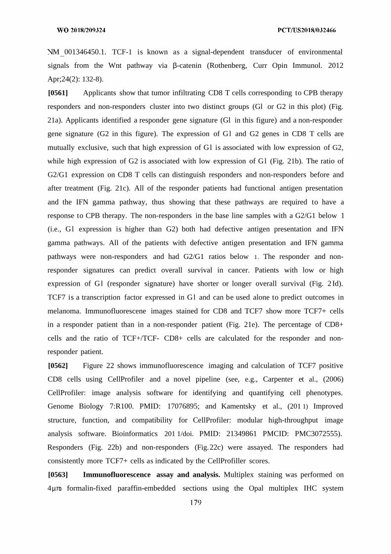
M 001346450.1. TCF-1 is known as a signal-dependent transducer of environmental
signals from the Wnt pathway via β-catenin (Rothenberg, Curr Opin Immunol. 2012
Apr;24(2): 132-8).
[0561] Applicants show that tumor infiltrating CD8 T cells corresponding to CPB therapy
responders and non-responders cluster into two distinct groups (Gl or G2 in this plot) (Fig.
21a). Applicants identified a responder gene signature (Gl in this figure) and a non-responder
gene signature (G2 in this figure). The expression of Gl and G2 genes in CD8 T cells are
mutually exclusive, such that high expression of Gl is associated with low expression of G2,
while high expression of G2 is associated with low expression of Gl (Fig. 21b). The ratio of
G2/G1 expression on CD8 T cells can distinguish responders and non-responders before and
after treatment (Fig. 21c). All of the responder patients had functional antigen presentation
and the IFN gamma pathway, thus showing that these pathways are required to have a
response to CPB therapy. The non-responders in the base line samples with a G2/G1 below 1
(i.e., Gl expression is higher than G2) both had defective antigen presentation and IFN
gamma pathways. All of the patients with defective antigen presentation and IFN gamma
pathways were non-responders and had G2/G1 ratios below 1 . The responder and non-
responder signatures can predict overall survival in cancer. Patients with low or high
expression of Gl (responder signature) have shorter or longer overall survival (Fig. 2Id).
TCF7 is a transcription factor expressed in Gl and can be used alone to predict outcomes in
melanoma. Immunofluorescene images stained for CD8 and TCF7 show more TCF7+ cells
in a responder patient than in a non-responder patient (Fig. 21e). The percentage of CD8+
cells and the ratio of TCF+/TCF- CD8+ cells are calculated for the responder and non-
responder patient.
[0562] Figure 22 shows immunofluorescence imaging and calculation of TCF7 positive
CD8 cells using CellProfiler and a novel pipeline (see, e.g., Carpenter et al., (2006)
CellProfiler: image analysis software for identifying and quantifying cell phenotypes.
Genome Biology 7:R100. PMID: 17076895; and Kamentsky et al., (201 1) Improved
structure, function, and compatibility for CellProfiler: modular high-throughput image
analysis software. Bioinformatics 201 1/doi. PMID: 21349861 PMCID: PMC3072555).
Responders (Fig. 22b) and non-responders (Fig.22c) were assayed. The responders had
consistently more TCF7+ cells as indicated by the CellProfiller scores.
[0563] Immunofluorescence assay and analysis. Multiplex staining was performed on
4µπ formalin-fixed paraffin-embedded sections using the Opal multiplex IHC system
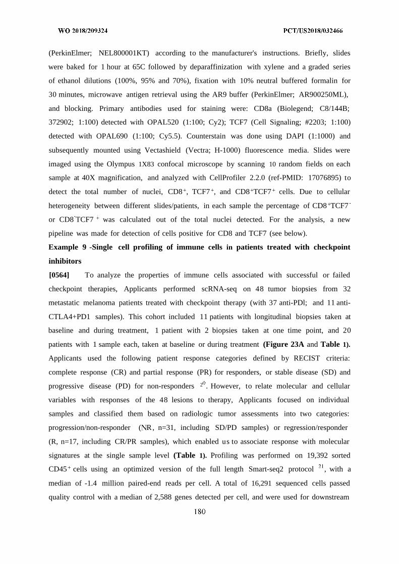
(PerkinElmer; NEL800001KT) according to the manufacturer's instructions. Briefly, slides
were baked for 1 hour at 65C followed by deparaffinization with xylene and a graded series
of ethanol dilutions (100%, 95% and 70%), fixation with 10% neutral buffered formalin for
30 minutes, microwave antigen retrieval using the AR9 buffer (PerkinElmer; AR900250ML),
and blocking. Primary antibodies used for staining were: CD8a (Biolegend; C8/144B;
372902; 1:100) detected with OPAL520 (1:100; Cy2); TCF7 (Cell Signaling; #2203; 1:100)
detected with OPAL690 (1:100; Cy5.5). Counterstain was done using DAPI (1:1000) and
subsequently mounted using Vectashield (Vectra; H-1000) fluorescence media. Slides were
imaged using the Olympus 1X83 confocal microscope by scanning 10 random fields on each
sample at 40X magnification, and analyzed with CellProfiler 2.2.0 (ref-PMID: 17076895) to
detect the total number of nuclei, CD8+, TCF7 +, and CD8+TCF7 + cells. Due to cellular
heterogeneity between different slides/patients, in each sample the percentage of CD8+TCF7
or CD8 TCF7 + was calculated out of the total nuclei detected. For the analysis, a new
pipeline was made for detection of cells positive for CD8 and TCF7 (see below).
Example 9 -Single cell profiling of immune cells in patients treated with checkpoint
inhibitors
[0564] To analyze the properties of immune cells associated with successful or failed
checkpoint therapies, Applicants performed scRNA-seq on 48 tumor biopsies from 32
metastatic melanoma patients treated with checkpoint therapy (with 37 anti-PDl; and 11 anti-
CTLA4+PD1 samples). This cohort included 11 patients with longitudinal biopsies taken at
baseline and during treatment, 1 patient with 2 biopsies taken at one time point, and 20
patients with 1 sample each, taken at baseline or during treatment (Figure 23A and Table 1).
Applicants used the following patient response categories defined by RECIST criteria:
complete response (CR) and partial response (PR) for responders, or stable disease (SD) and
progressive disease (PD) for non-responders 2 . However, to relate molecular and cellular
variables with responses of the 48 lesions to therapy, Applicants focused on individual
samples and classified them based on radiologic tumor assessments into two categories:
progression/non-responder ( R, n=31, including SD/PD samples) or regression/responder
(R, n=17, including CR/PR samples), which enabled us to associate response with molecular
signatures at the single sample level (Table 1). Profiling was performed on 19,392 sorted
CD45+ cells using an optimized version of the full length Smart-seq2 protocol , with a
median of -1.4 million paired-end reads per cell. A total of 16,291 sequenced cells passed
quality control with a median of 2,588 genes detected per cell, and were used for downstream
analysis (Methods). Whole exome sequencing (WES) was available for 20 patients, with 4
that had mutations in B2M, JAK1, STAT1 and IFNGR1 (Table 1), recently reported as
mechanisms for primary or acquired resistance to checkpoint therapy in melanoma 13 15.
Example 10 - The immune cell composition of melanoma tumors and their association
with clinical outcome
[0565] To define the immune landscape in an unbiased manner, Applicants initially
performed unsupervised clustering of cells (on 16,291 cells that passed quality control, based
on the -4,000 most variable genes across all cells) using k-means clustering with a
correlation distance metric (Methods). After testing for the robustness of this clustering
solution and relationships to previously known cell types, Applicants identified 11 clusters
that included, 2 B-cell clusters (Gl- B-cells; G2 - plasma cells), 2 myeloid clusters (G3 -
monocytes/macrophages; G4 - dendritic cells) and 7 clusters enriched for T/NK/NKT cells
(G5-1 1), accounting for most of the immune infiltrate detected within the cohort (Figure
23B-C and Table 2). Applicants then tested whether these clusters change between baseline
and post-therapy samples, or between responder and non-responder tumors. While
differences in cluster frequencies were detected when looking at the single patient level
between baseline and post-treatment samples (Figure 29A), no statistically significant
changes in cluster frequencies were seen when Applicants compared all aggregated baseline
to all aggregated on-treatment samples, or when only looking at patients with matched
longitudinal samples (Figure 29B-C). However, when analyzing by clinical outcome,
Applicants found that one cluster is significantly enriched in responder lesions while 4
clusters are enriched in non-responder lesions. Specifically, those include Gl (B-cells; Two-
sided Wilcoxon P-value Ρ=0.003), G3 (monocytes/macrophages, P-value=0.003), G4
(dendritic cells, P-value=0.015) G6 (Exhausted CD8+ T-cells, P-value=0.005) and Gl l (
Lymphocytes exhausted/cell cycle, P-value=1.33xl0 'Figure 23D and Figure 29D). While
both G6 and Gi l clusters were enriched for genes linked to T-cell exhaustion, with
differentially higher expression of co-inhibitory receptors {LAG3, PDCD1, CD38, HAVCR2,
TIGIT and ENTPD1); Gi l was also enriched for cell cycle genes (negative regulators:
CASP3, CDK2, BRCA2, RBI and TP 53 positive regulators: CDK1, CCNB1, MKI67, CDK4,
CDCA5 and TOP2A) (Table 2). Consistent with these results, when using previously defined
signatures for T-cell exhaustion 22 (Table 3), Applicants observed a significant enrichment
of T-cells with an exhausted signature in non-responder lesions (two-sided Wilcoxon P-
value=0.002) and with an activated signature in responder lesions (R-value=2xl0 4), but not
when these signatures were compared between baseline and post-therapy samples (Figure
23E and Figure 30)
[0566] Since the clusters (G5-G1 1) from the unsupervised analysis did not separate
specific cell types, but mostly cell states, likely due to the shared transcriptional programs
between T, NK and T cells 23'24, Applicants also determined the composition of known
cell types using pre-defined markers (Table 3 and Figure 31 and 32). In agreement with the
unsupervised analysis, Applicants found a significant enrichment of B-cells in responder
lesions (i -value=0.004) and of myeloid cells in non-responder lesions (i -value=0.002;
Figure 23F). Moreover, Applicants observed a significant enrichment of CD8+ memory T-
cells in responder lesions (P-value=0.001, Figure 23G). No significant differences in the
composition of known cell types were detected between baseline and post-treatment samples
when samples were aggregated together. However, changes were observed when looking at
the single patient level (Figure 31 and 32). Next, when comparing all 11 clusters identified in
the unsupervised analysis to the pre-defined markers, Applicants found very high
correspondence between clusters G1-G4 and the pre-defined B/myeloid cell markers. In
contrast, clusters G5-G1 1 did not show strong correspondence to a specific cell type,
suggesting that different lymphocytes (e.g. T, NK and NKT cells) share similar cell states of
exhaustion, activation, cytotoxicity and memory (Figure 33). Finally, Applicants wanted to
leverage the unbiased approach to identify not only clusters but also specific markers
associated with response. To that end, Applicants used two different strategies : (1). focusing
on cluster discriminating genes, and examining whether they are differentially expressed
between responder and non-responder samples (Table 4 ; Figure 23H and Figure 34); and
(2). examining all genes in an unbiased manner and searching for differentially expressed
genes (from all cells) between responder and non-responder samples (Table 5 and Figure
35). sing these two different approaches (cluster specific and non-specific) Applicants
identified an overlap for markers significantly enriched in responders (PLAC8, LTB, TCF7
and CCR7) and non-responder (CCL3, CD38 and HAVCR2) samples. Applicants conclude
that the methods used here , reveal novel cell states and markers that associate with the
clinical outcome of individual tumors to therapy, demonstrating the power of unbiased
approaches to find molecular correlates of response.
Example 11 - Unbiased definition of CD8+ T-cell states and their association with
response to therapy
[0567] Based on the significant association of T-cell states and markers with clinical
response using either unsupervised or supervised analyses, their highest abundance within the
cohort, and due to the dependency of checkpoint therapies on CD8+ T-cell recognition of
tumor antigens presented by human leukocyte antigen (HLA) class-I complexes '25,
Applicants next focused the analysis on CD8+ T-cells. Clustering of all CD8+ T-cells
(n=6,350) by k-means clustering revealed 2 cell states:CD8_G with increased expression of
genes linked to memory, activation and cell survival (IL7R, TCF7, REL, FOXP1, FOSL2 and
STAT4) 26 and reduced expression of co-inhibitory molecules; and CD8 B enriched for genes
linked to cell exhaustion (CD38, HAVCR2, ENTPD1, PDCD1, BATF, LAG3, CTLA4 and
PTPN6) (Figure 24A-B and Table 6). When annotating these two clusters to the 11 clusters
identified by the unsupervised analysis (Figure 23B), CD8 G cells were distributed
primarily in G10 (memory T-cells), G5 (lymphocytes) and G8 (cytotoxicity) clusters, and
CD8 B cells were localized mainly in clusters Gl 1 (lymphocytes exhausted/cell-cycle), G6
(exhausted CD8+ T-cells) and G9 (exhausted/HS CD8+ T-cells; Figure 36). A central
question is whether these states are associated with clinical outcome and what is their
predictive power to distinguish responding from non-responding tumors. When comparing
between these two cell states, Applicants found a significant enrichment for CD8 G in
responding lesions (two-sided Wilcoxon P-value=\ .4xl0 6) and CD8 B in non-responding
lesions (P-val e= . 5 Figure 24C). While cells with both states coexist in each of the
responder and non-responder lesions, an overall higher proportion of CD8 G cells is found in
responders, and CD8 B cells in non-responders (Figure 37). Thus, Applicants decided to
calculate the ratio between the number of cells in these 2 clusters and observed a significant
separation between responders (CD8_B/CD8_G<1) and non-responders (CD8_B/CD8_G>1)
when looking at all samples, as well as baseline or post-treatment samples separately (Figure
24D). However, 9 non-responding lesions had unexpected ratios (CD8_B/CD8_G<1), and
were more enriched for CD8 G . Applicants hypothesized that although these patients might
have productive immunity, they had developed de novo resistance to checkpoint therapy. To
dissect the genetic alterations associated with resistance, Applicants performed WES
followed by immunohistochemistry and flow cytometry, and observed that 6 out of 9 samples
(no DNA or slides were available for the 3 remaining lesions) showed complete loss of B2M
or HLA-A,B,C (class-I), recently reported as a mechanism of resistance to checkpoint
inhibition therapy in melanoma 14 1 (Table 1 and Figure 38A-B). An analysis of predictive
performance for the identified signatures demonstrated excellent predictive power when
considering all samples (AUC of ROC= 0.87; one-sided Wilcoxon P-value=l.lxlO 5) .
However, when excluding the 6 samples deficient for B2M or HLA-A,B,C, the predictive
power was increased significantly (AUC of ROC=0.96; P-value = 3.8xl0 , Figure 38C-D).
[0568] Similarly to the analysis performed on all immune cells, Applicants sought to
identify specific CD8+ markers associated with clinical outcome. To that end Applicants
focused on CD8 G and CD8 B top discriminating genes and examined whether they are
differentially expressed between responder and non-responder samples. Applicants identified
TCF7 and IL7R as the top two CD8+ markers to be significantly associated with response, the
first being also significant in the initial marker analysis when looking at all CD45+ cells.
Moreover, Applicants found a -45% overlap between markers associated with non-responder
lesions in CD8+ T-cells and all immune cells (CD38, PDCD1, CCL3, SNAP47, VCAM1,
HAVCR2, FASLG, ENTPD1, SIRPG, M 0 7A, FABP5, NDUFB3, UBE2F, CLTA and
SNRPDl; Figure 24E and Table 7). . Overall, the results suggest that the abundance of
CD8 G and CD8 B cellular states could be critical to the success of checkpoint therapy.
Example 12 - Elevated frequencies of CD8+TCF7+ T-cells are associated with outcome
in a second independent anti-PDl-treated cohort
[0569] Since the ratio between CD8 G and CD8 B was significantly associated with
response, , Applicants considered whether any of the specific markers identified in these
clusters could be used to predict response to treatment in a second cohort, using a different
approach that could easily be applied in the clinic. Applicants selected the proteins CD8a and
transcription factor 7 (TCF7) for this analysis, because TCF7 was the only top marker linked
to response when analyzing all immune or only CD8+ T-cells (Figure 23H; Figure 35 and
Figure 24E), differentially expressed in CD8 G (4-fold higher expression than in CD8 B),
and since all lymphocytes related clusters that were associated with response were either
enriched for or completely composed of CD8+ T-cells (Figure 23 and 24 and Figure 33).
TCF7 is part of the Wnt/p-catenin signaling pathway 2 and has been shown to be crucial for
differentiation, self-renewal and persistence of memory CD8+ T-cells 2 , as well as
reinvigoration and effective immunity of CD8+ T-cells against chronic LCMV infection upon
anti-PDl treatment 29'3 . Thus, Applicants considered its association with response in a
second cohort of 33 patients (n=43 samples) treated with anti-PDl (Table 8). Using
immunofluorescence staining followed by automated image analysis with CellProfiler 1
(Figure 24F, Figure 39 and pipeline), Applicants calculated the ratio of TCF7+CD8+ to
TCF7 CD8+ cells and its association with response. When comparing between these two cell
phenotypes Applicants found a significant enrichment for TCF7+CD8+ in responding patients
(two-sided Wilcoxon P-value=3 .9xl0 6) and TCF7 CD8+ in non-responder patients (P-
v / =l .lx l 0 ; Figure 24G), and saw that cells with both states coexist in each of the
responder and non-responder lesions (Figure 40). Analogous to the CD8+ single-cell
RNAseq analysis Applicants performed (Fig 24D), Applicants found that a ratio>l of
TCF7+CD8+ to TCF7 CD8+ is typically associated with clinical response and a ratio < 1 with
lack of response when looking at all (n=43; one-sided Wilcoxon P-value=2.4xl0 ~6 Figure
24H), baseline (n=24; P-val e= . 1) or post-treatment (n=19; P-value=Uxl0 4 Figure
41A) samples. In contrast, no significant difference was observed when looking only at the
percentage of tumor-associated CD8+ T-cells between responding and non-responding
patients (Figure 241). Consistent with the ability to predict lesion-level responses using the
single-cell RNAseq-derived signatures in the first cohort, the power to predict response was
similar in this independent cohort for all (AUC=0.91; Figure 41B), baseline (AUC=0.88;
Figure 41C) or post-treatment samples (AUC=0.98; Figure 41D). Additionally, , when
performing a Kaplan-Meier survival analysis Applicants found that patients with a ratio > 1
have a significantly higher survival rate as compared to those with a ratio < 1 (logrank P-
value= . , Figure 24J).
[0570] Finally, Applicants asked if TCF7 protein levels changes overtime between
matched baseline and post-treatment samples in the same patient. 8 patients out of 33 in the
second cohort had matched baseline and post-treatment samples (Table 8). Although minor
differences were detected in TCF7+CD8+ levels between baseline and post-treatment
samples, the main change was observed when classifying samples by their response or lack of
response to therapy, regardless to the fact if the sample was taken at baseline or during/after
treatment (Figure 40 and Figure 42A). Moreover, immunofluorescence staining of
additional 7 samples (n=4 patients), initially analyzed in the single-cell RNAseq cohort
revealed similar pattern between the TCF7+CD8+/TCF7 CD8+ ratio detected when
performing the immunofluorescence staining pipeline to the CD8 B/CD8 G ratio Applicants
identified when analyzing the single-cell RNAseq data (Figure S9, Figure 42B and Table 8).
Collectively, the results suggest that the ratio between CD8+ T-cell subsets with distinctive
phenotypes is a predictor of clinical outcome and survival.
Example 13 - Higher resolution analysis of CD8+ T-cells discovers novel exhausted and
memory subsets
[0571] While the two CD8+ T-cell clusters were able to separate responders from non-
responders, Applicants wondered if a greater heterogeneity in cell states could be observed in
the single cell dataset. Using k-means clustering with a correlation distance metric on all
CD8+ T-cells that passed quality control, and after testing for the robustness of this clustering
solution (0.89; Methods) and relationships to previously known cell states, Applicants found
6 clusters, with CD8 G and CD8 B, each splitting into 3 clusters (Figure 25A-B). To
identify cluster-specific gene markers, Applicants compared the expression level of genes
associated with cells within a given cluster, to that of cells outside this cluster (Methods).
Applicants found that these 6 new clusters included a mixture of known and novel exhaustion
and memory/effector markers and were linked to treatment outcome (Figure 25B). CD8 1
expressed multiple markers of exhaustion (HAVCR2, ENTPD1, PDCDl and PTPN11) and
negative (CASP3, CDK2, and TP53) or positive {CDKI, CCNBl and MKI67) regulators of
cell cycle (Table 9), similar to the pattern observed in cluster G12 when analyzing all
immune cells (Table 2), and to the transcriptional phenotype of terminally exhausted CD39+
(ENTPDl) CD8+ T-cells, detected during chronic infection with hepatitis C virus (HCV) .
While CD8 2 expressed many exhaustion markers together with heat shock proteins
(HAVCR2, CTLA4, TIGIT, PDCDl, HSPB1, HSPA1A and HSPA4) along with additional
inhibitory receptors {ENTPDl and KIR2DL4), CD8 3 expressed several known exhaustion
markers {HAVCR2, CD38, PDCDl and PTPN6), but lacked the expression of heat shock
proteins and cell-cycle genes. In contrast, CD8_4 {CCR7, IL7R, TCF7, TNF and S100A10),
and CD8 6 (SELL, TCF7, LTB, IL7R, FLT3LG, IL16) had a memory/effector like phenotype,
while CD8 5 had the phenotype of early activated and more differentiated cells (IL6ST,
CXCL13, IL7R and CTLA4), but was more enriched for HAVCR2 and PDCDl when
compared to CD8 4 and 6, but not CD8 1 to 3 (Figure 43). Interestingly, GZMA, GZMB and
PRFl (coding for granzyme A, B and perforin 1), had much higher expression in the
exhausted clusters CD8 1 to 3 (Table 9) and resembled exhaustion programs previously
reported in melanoma and in a mouse model of chronic LCMV infection . Interestingly,
when examining the clustering results when using fewer clusters, Applicants found that the
clusters formed a hierarchy, that is, clusters split and did not mix as the number of clusters
increased. To further examine the robustness and unique biological function of each of these
clusters, Applicants took another approach, in which Applicants identified gene markers that
are differentially expressed between the cluster and an "ancestor" cluster found in a lower-
resolution cluster analysis, thus creating a functional annotation along the hierarchical
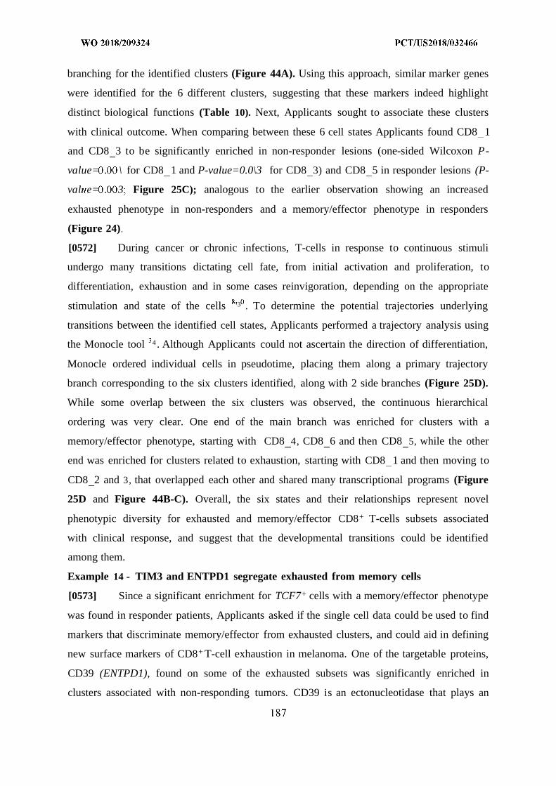
branching for the identified clusters (Figure 44A). Using this approach, similar marker genes
were identified for the 6 different clusters, suggesting that these markers indeed highlight
distinct biological functions (Table 10). Next, Applicants sought to associate these clusters
with clinical outcome. When comparing between these 6 cell states Applicants found CD8 1
and CD8 3 to be significantly enriched in non-responder lesions (one-sided Wilcoxon P-
value= . \ for CD8 1 and P-value=0.0\3 for CD8 3) and CD8 5 in responder lesions (P-
val e= . 3 Figure 25C); analogous to the earlier observation showing an increased
exhausted phenotype in non-responders and a memory/effector phenotype in responders
(Figure 24)
[0572] During cancer or chronic infections, T-cells in response to continuous stimuli
undergo many transitions dictating cell fate, from initial activation and proliferation, to
differentiation, exhaustion and in some cases reinvigoration, depending on the appropriate
stimulation and state of the cells '3 . To determine the potential trajectories underlying
transitions between the identified cell states, Applicants performed a trajectory analysis using
the Monocle tool 4 . Although Applicants could not ascertain the direction of differentiation,
Monocle ordered individual cells in pseudotime, placing them along a primary trajectory
branch corresponding to the six clusters identified, along with 2 side branches (Figure 25D).
While some overlap between the six clusters was observed, the continuous hierarchical
ordering was very clear. One end of the main branch was enriched for clusters with a
memory/effector phenotype, starting with CD8 4, CD8 6 and then CD8 5, while the other
end was enriched for clusters related to exhaustion, starting with CD8 1 and then moving to
CD8 2 and 3, that overlapped each other and shared many transcriptional programs (Figure
25D and Figure 44B-C). Overall, the six states and their relationships represent novel
phenotypic diversity for exhausted and memory/effector CD8+ T-cells subsets associated
with clinical response, and suggest that the developmental transitions could be identified
among them.
Example 14 - TIM3 and ENTPD1 segregate exhausted from memory cells
[0573] Since a significant enrichment for TCF7+ cells with a memory/effector phenotype
was found in responder patients, Applicants asked if the single cell data could be used to find
markers that discriminate memory/effector from exhausted clusters, and could aid in defining
new surface markers of CD8+ T-cell exhaustion in melanoma. One of the targetable proteins,
CD39 (ENTPD1), found on some of the exhausted subsets was significantly enriched in
clusters associated with non-responding tumors. CD39 is an ectonucleotidase that plays an
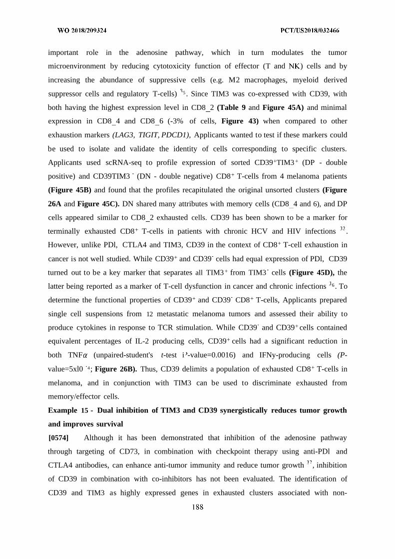
important role in the adenosine pathway, which in turn modulates the tumor
microenvironment by reducing cytotoxicity function of effector (T and ) cells and by
increasing the abundance of suppressive cells (e.g. M2 macrophages, myeloid derived
suppressor cells and regulatory T-cells) 5 . Since TIM3 was co-expressed with CD39, with
both having the highest expression level in CD8 2 (Table 9 and Figure 45A) and minimal
expression in CD8 4 and CD8 6 (-3% of cells, Figure 43) when compared to other
exhaustion markers (LAG3, TIGIT, PDCD1), Applicants wanted to test if these markers could
be used to isolate and validate the identity of cells corresponding to specific clusters.
Applicants used scRNA-seq to profile expression of sorted CD39+TIM3+ (DP - double
positive) and CD39TIM3 (DN - double negative) CD8+ T-cells from 4 melanoma patients
(Figure 45B) and found that the profiles recapitulated the original unsorted clusters (Figure
26A and Figure 45C). DN shared many attributes with memory cells (CD8 4 and 6), and DP
cells appeared similar to CD8 2 exhausted cells. CD39 has been shown to be a marker for
terminally exhausted CD8+ T-cells in patients with chronic HCV and HIV infections .
However, unlike PDl, CTLA4 and TIM3, CD39 in the context of CD8+ T-cell exhaustion in
cancer is not well studied. While CD39+ and CD39 cells had equal expression of PDl, CD39
turned out to be a key marker that separates all TIM3+ from TIM3 cells (Figure 45D), the
latter being reported as a marker of T-cell dysfunction in cancer and chronic infections 6. To
determine the functional properties of CD39+ and CD39 CD8+ T-cells, Applicants prepared
single cell suspensions from 12 metastatic melanoma tumors and assessed their ability to
produce cytokines in response to TCR stimulation. While CD39 and CD39+ cells contained
equivalent percentages of IL-2 producing cells, CD39+ cells had a significant reduction in
both TNF (unpaired-student's t-test i -value=0.0016) and IFNy-producing cells (P-
value=5xl0 4; Figure 26B). Thus, CD39 delimits a population of exhausted CD8+ T-cells in
melanoma, and in conjunction with TIM3 can be used to discriminate exhausted from
memory/effector cells.
Example 15 - Dual inhibition of TIM3 and CD39 synergistically reduces tumor growth
and improves survival
[0574] Although it has been demonstrated that inhibition of the adenosine pathway
through targeting of CD73, in combination with checkpoint therapy using anti-PDl and
CTLA4 antibodies, can enhance anti-tumor immunity and reduce tumor growth , inhibition
of CD39 in combination with co-inhibitors has not been evaluated. The identification of
CD39 and TIM3 as highly expressed genes in exhausted clusters associated with non-

responding lesions, led us to examine the combined effect of CD39 and TIM3 blockade. To
that end Applicants used the aggressive B16-F 10 melanoma mouse model. Mice were treated
with the small molecule POM-1 to block CD39 activity , alone or in combination with anti-
TIM3 blocking antibodies (Figure 26C). While monotherapy with either POM-1 or anti-
TIM3 transiently reduced tumor growth until day 14 (with CD39 inhibition having a stronger
effect), combination of both caused a dramatic reduction in tumor size, and more importantly,
significantly increased survival (50% survival vs. 10% for CD39 inhibition or 0% for anti-
TEVI3 and the untreated group) on day 30 (Figure 26D-F and Figure 45E). Since anti-PDl
treatment is currently the standard of care in metastatic melanoma patients, and since PD1
similarly to TIM3 was one of the top markers associated with lack of response, Applicants
asked whether dual inhibition of PD1 and CD39 will increase response and improve survival.
Similar to the observations when targeting both CD39 and TIM3 (although to a lesser extent),
dual inhibition of CD39 and PD1 significantly reduced tumor burden on day 2 1 post
transplantation, and improved survival when compared to untreated and mono therapy treated
mice (Figure 46). Hence, through the single cell analysis of human melanoma tumors,
Applicants identified new putative immunotherapeutic targets and combinations to enhance
immunity.
Example 16 - Chromatin accessibility of melanoma-associated exhausted and memory
cells
[0575] Since prospectively isolated CD39 +TIM3 + (DP) and CD39TIM3 (DN) cells
recapitulated phenotypes of cells within the previously unrecognized CD8 2 and CD8 4+6
clusters respectively, Applicants asked what are the epigenetic programs that govern these
distinctive cell states. To dissect differential regions of open chromatin that corresponded to
differences in gene expression, Applicants isolated DP and DN cells from 5 metastatic
melanoma patients treated with the checkpoint inhibitor PD-1 and performed assays for
transposase-accessible chromatin with next generation sequencing (ATAC-seq) and scRNA-
seq (Figure 27A). scRNA-seq analysis identified exclusive patterns of transcription factor
(TF) expression for each of the sorted populations. Applicants found that DN cells had higher
expression for several TFs, including TCF7, STAT4, FOXP1 and FOSB, previously shown to
be enriched in stem-cell memory CD8 + T-cells 26, while DP cells were enriched for BATF,
PRDM1, TOX, HMGB2 and IRF2, as previously described in exhausted CD8 + T-cells 4
(Figure 27B). Furthermore, similar patterns of TF expression were identified when compared
to the original unsorted single cells separated computationally by expression of CD39 and
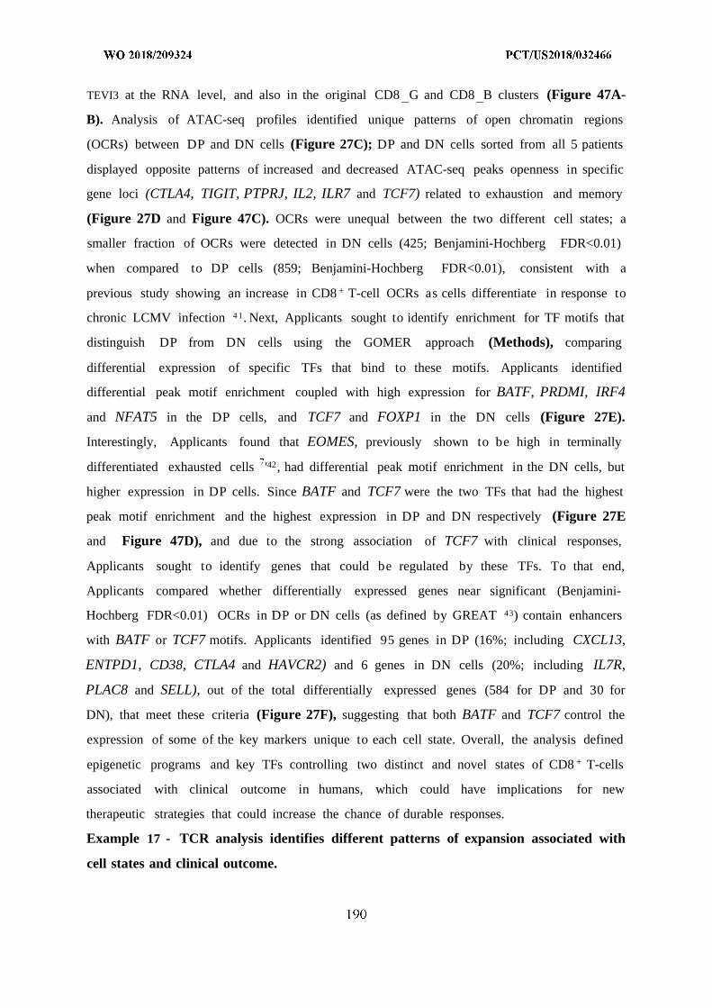
TEVI3 at the RNA level, and also in the original CD8 G and CD8 B clusters (Figure 47A-
B). Analysis of ATAC-seq profiles identified unique patterns of open chromatin regions
(OCRs) between DP and DN cells (Figure 27C); DP and DN cells sorted from all 5 patients
displayed opposite patterns of increased and decreased ATAC-seq peaks openness in specific
gene loci (CTLA4, TIGIT, PTPRJ, IL2, ILR7 and TCF7) related to exhaustion and memory
(Figure 27D and Figure 47C). OCRs were unequal between the two different cell states; a
smaller fraction of OCRs were detected in DN cells (425; Benjamini-Hochberg FDR<0.01)
when compared to DP cells (859; Benjamini-Hochberg FDR<0.01), consistent with a
previous study showing an increase in CD8 + T-cell OCRs as cells differentiate in response to
chronic LCMV infection 4 1. Next, Applicants sought to identify enrichment for TF motifs that
distinguish DP from DN cells using the GOMER approach (Methods), comparing
differential expression of specific TFs that bind to these motifs. Applicants identified
differential peak motif enrichment coupled with high expression for BATF, PRDMI, IRF4
and NFAT5 in the DP cells, and TCF7 and FOXP1 in the DN cells (Figure 27E).
Interestingly, Applicants found that EOMES, previously shown to be high in terminally
differentiated exhausted cells '42, had differential peak motif enrichment in the DN cells, but
higher expression in DP cells. Since BATF and TCF7 were the two TFs that had the highest
peak motif enrichment and the highest expression in DP and DN respectively (Figure 27E
and Figure 47D), and due to the strong association of TCF7 with clinical responses,
Applicants sought to identify genes that could be regulated by these TFs. To that end,
Applicants compared whether differentially expressed genes near significant (Benjamini-
Hochberg FDR<0.01) OCRs in DP or DN cells (as defined by GREAT 43) contain enhancers
with BATF or TCF7 motifs. Applicants identified 95 genes in DP (16%; including CXCL13,
ENTPD1, CD38, CTLA4 and HAVCR2) and 6 genes in DN cells (20%; including IL7R,
PLAC8 and SELL), out of the total differentially expressed genes (584 for DP and 30 for
DN), that meet these criteria (Figure 27F), suggesting that both BATF and TCF7 control the
expression of some of the key markers unique to each cell state. Overall, the analysis defined
epigenetic programs and key TFs controlling two distinct and novel states of CD8 + T-cells
associated with clinical outcome in humans, which could have implications for new
therapeutic strategies that could increase the chance of durable responses.
Example 17 - TCR analysis identifies different patterns of expansion associated with
cell states and clinical outcome.
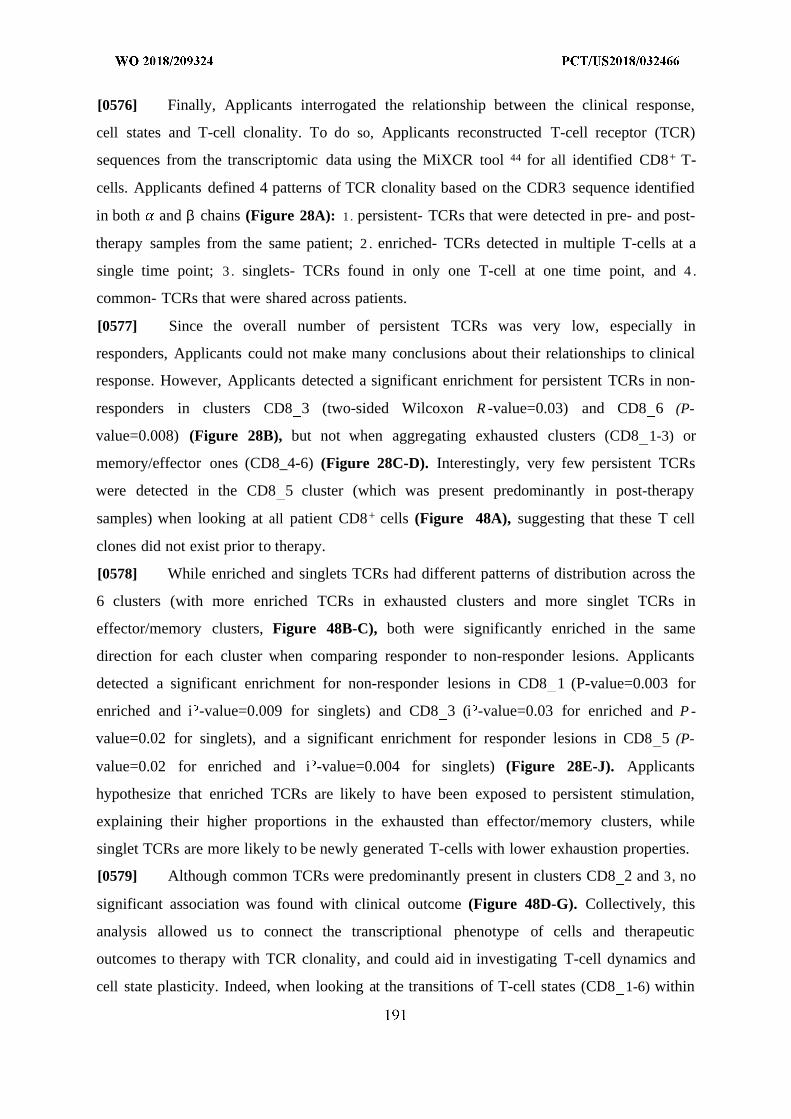
[0576] Finally, Applicants interrogated the relationship between the clinical response,
cell states and T-cell clonality. To do so, Applicants reconstructed T-cell receptor (TCR)
sequences from the transcriptomic data using the MiXCR tool 44 for all identified CD8+ T-
cells. Applicants defined 4 patterns of TCR clonality based on the CDR3 sequence identified
in both and β chains (Figure 28A): 1 . persistent- TCRs that were detected in pre- and post-
therapy samples from the same patient; 2 . enriched- TCRs detected in multiple T-cells at a
single time point; 3 . singlets- TCRs found in only one T-cell at one time point, and 4 .
common- TCRs that were shared across patients.
[0577] Since the overall number of persistent TCRs was very low, especially in
responders, Applicants could not make many conclusions about their relationships to clinical
response. However, Applicants detected a significant enrichment for persistent TCRs in non-
responders in clusters CD8 3 (two-sided Wilcoxon R -value=0.03) and CD8 6 (P-
value=0.008) (Figure 28B), but not when aggregating exhausted clusters (CD8 1-3) or
memory/effector ones (CD8_4-6) (Figure 28C-D). Interestingly, very few persistent TCRs
were detected in the CD8 5 cluster (which was present predominantly in post-therapy
samples) when looking at all patient CD8+ cells (Figure 48A), suggesting that these T cell
clones did not exist prior to therapy.
[0578] While enriched and singlets TCRs had different patterns of distribution across the
6 clusters (with more enriched TCRs in exhausted clusters and more singlet TCRs in
effector/memory clusters, Figure 48B-C), both were significantly enriched in the same
direction for each cluster when comparing responder to non-responder lesions. Applicants
detected a significant enrichment for non-responder lesions in CD8 1 (P-value=0.003 for
enriched and i -value=0.009 for singlets) and CD8 3 (i -value=0.03 for enriched and P -
value=0.02 for singlets), and a significant enrichment for responder lesions in CD8 5 (P-
value=0.02 for enriched and i -value=0.004 for singlets) (Figure 28E-J). Applicants
hypothesize that enriched TCRs are likely to have been exposed to persistent stimulation,
explaining their higher proportions in the exhausted than effector/memory clusters, while
singlet TCRs are more likely to be newly generated T-cells with lower exhaustion properties.
[0579] Although common TCRs were predominantly present in clusters CD8 2 and 3, no
significant association was found with clinical outcome (Figure 48D-G). Collectively, this
analysis allowed us to connect the transcriptional phenotype of cells and therapeutic
outcomes to therapy with TCR clonality, and could aid in investigating T-cell dynamics and
cell state plasticity. Indeed, when looking at the transitions of T-cell states (CD8 1-6) within
a specific clone (based on identical TCR sequence) across longitudinal samples in the same
patient, Applicants discovered bilateral transitions between exhausted and memory/effector
states.
Example 18 - Discussion
[0580] Although immune checkpoint blockade leads to durable responses in patients with
metastatic melanoma, refractory disease and progression after initial response remain major
causes of mortality '4. While many studies have identified different components associated
with clinical outcome, the principles of the immune system that underlie the success or
failure to checkpoint therapy in humans remain relatively unexplored. To address this issue
Applicants performed an unbiased analysis of immune cells using single-cell transcriptomics,
to dissect the cellular and molecular determinants of response. Within the tumor, Applicants
found specific CD8+ T-cell states associated with clinical outcome in melanoma patients
treated with checkpoint therapy. The association between the cellular states (as determined by
gene expression or antibody staining) and treatment outcome was observed in both baseline
and on-therapy samples. Applicants validated these associations in two independent cohorts:
one cohort of anti-PDl patients (n=30) using a signature of memory/effector T-cells, and a
second anti-PDl cohort (n=21) using the fraction of TCF7+CD8+ T-cells based on
immunofluorescence staining. The analyses also identified novel sub-states of exhausted and
memory/effector T-cells. Among the identified markers of the exhausted sub-states, CD39
(ENTPD1) emerged as a promising discriminator of exhausted CD8+ T-cells in melanoma
patients, and as a potential target when blocked together with TIM3 (HAVCR2) using a
mouse melanoma model.
[0581] Previous studies have shown that the number of infiltrating CD8+ T-cells detected
in patient biopsies is significantly enriched in responders. However, some inconsistencies
were found between these studies: the first showed that the number of infiltrating CD8+ T-
cells detected before treatment can predict clinical outcome , whereas the second showed
that only early on-treatment, but not baseline quantification is significantly associated with
patient response 6. Despite potential spatial and temporal heterogeneity in biopsy-based
studies of cancer, Applicants identified robust predictive markers in baseline and on-
treatment samples, reflecting quality metrics of T-cell states (TCF7+CD8+), rather than the
quantity of T-cells (CD8+) . Indeed, the number of CD8+ T-cells was comparable across
responders and non-responders within the cohort, consistent with the fact that the T-cell
number is not a reliable predictor in pretreatment biopsies 6. Several other studies found that
the spatial distribution of the T-cells was more important in predicting survival . Hence,
further studies are needed to explore the relationship between the spatial distribution of T-
cells and the cellular states Applicants identified using highly multiplexed tissue imaging
systems.
[0582] One of the key markers expressed in clusters associated with response, which was
found in an unbiased manner as a result of data analysis and discriminated responders from
non-responders, was the transcription factor TCF7. Although, these results are in line with
previous studies showing that TCF7 is required for reinvigorating CD8 + T-cells in response
to anti-PDl or anti-PDLl therapies to resolve chronic LCMV infection in mice 29'3 , its
association with clinical outcome in patients treated with checkpoint immunotherapy has not
been previously demonstrated. More specifically, it has been shown that TCF7 is essential for
the expansion of CXCR5 +TFM3 CD8 + T-cells (but not TFM3 + cells) that are important for the
control of the virus . Indeed, Applicants also found that TEVI3 cells are TCF7 + and are
associated with response; however Applicants did not find CXCR5 expression associated
with TCF7 + cells, suggesting that this chemokine receptor may not be a critical marker for
cells associated with response in the melanoma context. Additionally, a recent study
suggested that decreased open chromatin regions at TCF7 sites are associated with reduced
activation of non-programmable, dysfunctional PD1 T-cells 4 .
[0583] Furthermore, the data revealed significant heterogeneity of exhausted CD8 + T-cell
states, with 2 of the 3 clusters showing stronger association with non-responding lesions.
While most studies have focused on PDl, TIM3, LAG3 and CTLA-4 to mark exhausted
cells, Applicants identified CD39 as an additional marker for human CD8 + T-cell exhaustion
in melanoma, and found it to be enriched in non-responsive lesions. While not previously
observed in melanoma or tested functionally, this result is consistent with recent findings,
demonstrating that CD39 is a marker for terminally exhausted CD8 + T-cells in HIV and HCV
infected patients . The results in mice support the targeting of CD39 together with TIM-3
for boosting anti-tumor immunity. Additionally, Applicants found that in all samples,
regardless of whether they were collected at baseline or during treatment, there was a
heterogeneous mixture of all the identified CD8 + T-cell states, but that their proportions were
associated with clinical outcome. Further studies will be needed to test whether other
immunotherapeutic combinations or targets (e.g KTR2DL4 and CD38), which Applicants
found to be enriched in clusters associated with lack of response, could be used to enhance
immunity and overcome resistance.
[0584] A critical question that still remains unanswered is how the 6 CD8+ T-cell states
Applicants identified relate to each other. In line with previously published studies,
Applicants propose that cells can transition between memory and exhausted states, and
bolster this hypothesis through finding divergent memory and exhaustion phenotypes in
clonal T cell populations (determined based on identical TCR sequences). Surprisingly
Applicants found that CD8 5 T cells, which were predominantly found in post-therapy tumor
samples, hardly share TCRs with the baseline sample, suggesting that members of the CD8 5
cluster are generated outside of the tumor and subsequently migrate to the tumor. This is
consistent with observations from a recent study 4 demonstrating that lymphoid-organ
derived T-cells are required for anti-PDl potency.
[0585] Despite the identification of key components associated with response, the study
has technical limitations and open biological questions that necessitate future studies to
address them. First, although Applicants used an optimized version of the full length Smart-
seq2 protocol, which has the highest sensitivity and lowest dropout probability compared to
other methods , the resulting transcriptomes need to be interpreted with caution because
lack of detection of a transcript in a single cell does not guarantee its absence. Applicants
address this dropout challenge by relying on signatures composed of many (not single) genes
in order to increase confidence in cluster definition. Second, while the approach of scRNA-
seq generates relatively unbiased genome-scale transcriptome data, which is then used to
identify novel immune cell states and molecular determinants of response, future studies will
need to validate these signatures at the protein level (as Applicants did for TCF7) using
highly multiplexed tissue imaging or mass cytometry in melanoma as well as other types of
cancer. Third, although Applicants identified T cell states associated with responses,
Applicants do not know which cells kill the tumors. Fourth, what are the factors that control
the proportions of the identified CD8+ cell states in patient samples? Is the responder-
associated memory phenotype maintained post treatment? Can cells be reprogrammed from
an exhausted to a memory phenotype? While the approach in this study is useful in
identifying new components associated with response and useful in generating hypotheses,
future mechanistic studies will be required to demonstrate these developmental process.
Finally, the study focused on melanoma patients treated with checkpoint inhibitors, but
Applicants need to address whether these predictive markers of response are relevant to other
types of malignancies or therapies.
[0586] This study addresses the long-standing question of which immune cell states are
important for the activity of checkpoint therapy in humans, and provide a powerful tool both
to the medical and research communities in several ways: first, the data and analysis enables
the prediction of clinical outcome in patients treated with anti-PDl and may impact clinical
trial design and execution, as well as application of this therapy as standard of care in
thousands of patients across the world. Second, the identification of new targets, and the
subsequent validation of two specific ones in the study, may lead to new trials focused on
these targets and other combinations of targets associated with success or failure of therapy in
the study (e.g. KIR2DL4 and CD38). Third, the finding that specific memory-like signatures
are associated with response —together with recent studies showing that CAR T cell activity
is enhanced by generating more memory-like cells using the IL-7 or IL-15 pathways, leading
to better outcome in preclinical models 26 1 —suggests that manipulation of T cells prior to
adoptive cell therapies by increasing the ratio of CD8 G to CD8 B (especially in patients
with a low ratio) would strongly boost clinical responses when combined with checkpoint
therapies.
[0587] Since an increasing number of patients are being treated with checkpoint
inhibitors as a standard of care, it has become essential to fully understand the determinants
of response. Utilizing the data presented in this study, one can potentially help to optimally
select patients for therapy, identify new therapeutic strategies and thus increase the chance of
durable responses.
Example 19 - Methods
[0588] Patient samples. Metastatic melanoma patients treated with checkpoint blockade
therapy at Massachusetts General Hospital (Boston, MA) and University of Texas MD
Anderson Cancer Center (Houston, TX) provided written informed consent for the collection
of tissue and blood samples for research and genomic profiling, as approved by the Dana-
Farber/Harvard Cancer Center Institutional Review Board (DF/HCC Protocol 11-181) and
UT MD Anderson Cancer Center (IRB LABOO-063 and 2012-0846). Matched tumor and
normal blood samples were obtained from 23 patients at baseline and/or after checkpoint
treatment.
[0589] Sample dissociation. Fresh isolated tumor samples were collected immediately
after surgery and were dissociated within 1 hour using the human tumor dissociation kit
(Miltenyi Biotec; 130-095-929) with the following modifications. Tissue was minced into
small pieces using a scalpel and put into a 1.5ml eppendorf tube containing ΙΟΟµΙ of enzyme
H, 50µ1 of enzyme R, 12.5 µ1 of enzyme A (all provided in the kit), and 837. 5µ1 of RPMI,
followed by a 20 minute incubation in a therm omixer (Eppendorf; F1.5) at 37C, 600 rpm.
After incubation, debris were removed by filtering through a 70µπ cell strainer, followed by
mincing of the remaining tissue left on the strainer with a plunger in order to increase cell
yield. Dissociated cells were subsequently washed with cold IX PBS containing 1.5% heat
inactivated FCS, spun down at 1300 rpm, 4°C for 5 minutes, resuspended, and counted for
yield and viability with trypan blue using a Countess automated cell counter (Invitrogen).
[0590] Flow cytometry and cell sorting. For both flow cytometry and cell sorting,
Human TrueStain FcX (Biolegend, 422302) was used for blocking Fc receptors before
labeling cells. To discriminate live from dead cells Applicants used Zombie violet Dye
(Biolegend, 77477) for 15 min at 4°C, followed by surface labelling of cells for 30 min at
4°C, using standard protocols. The antibodies used for cell surface labelling were PE anti-
human CD45 (Biolegend, 304008), APC anti-human CD3 (Biolegend, 300412), FITC anti-
human HLA-A,B,C (Biolegend, 3 11426), APC/Cy7 anti-human CD235a (Biolegend,
3491 16), PE/Cy5 anti-human CD3 (Biolegend, 300309), BV421 anti-human PD1 (Biolegend,
329919), PE/Cy7 anti-human TEVI3 (Biolegend, 345013), APC/Cy7 anti-human CD39
(Biolegend, 328226), AF700 anti-human CD4 (Biolegend, 317425), BV650 anti-human CD 8
(biolegend, 301041). The antibodies used for intracellular staining were FITC anti-human
IFNy (Miltenyi Biotec, 130-097-936), PE anti-human IL2 (Miltenyi Biotec, 130-099-391),
APC anti-human TNF (Miltenyi Biotec, 130-099-197). Sorting of cells was performed on a
BD Fusion instrument using the following antibody panel: Zombie dye, CD45, CD235a and
HLA-A,B,C. CD45 + cells from dissociated samples were sorted into 96-well plates
(Eppendorf, 951020401) containing ΙΟµΙ of lysis buffer (TCL buffer, Qiagen 1031576,
supplemented with 1% β-mercaptoethanol), sealed, vortexed, spun down at 2500 rpm for 30
seconds, immediately placed on dry ice, and then stored at -80°C until processing with the
Smart-Seq2 protocol. For flow cytometry Applicants used the Beckman Coulter CytoFLEX
instrument and analyzed the data with FlowJo vl0.0.8rl software.
[0591] Single cell RNA sequencing procedure. Libraries from single cell lysates were
generated with the Smart-Seq2 protocol 2 with some modifications in the reverse
transcription step as recently described 2 1. 96-well plates containing cell lysates were thawed
on ice, spun down at 1500 rpm for 30 seconds, and mixed with Agencourt RNAClean XP
SPRI beads (Beckman Coulter) for RNA purification. Purified RNA was resuspended in 4µ1
of Mix- 1 , denatured at 72°C for 3 min and placed immediately on ice for 1 min before 7µ1of
Mix-2 was added. Reverse transcription was carried out at 50°C for 90 min, followed by 5
min incubation at 85°C. 14 µ1 of Mix-3 was added in each well and the whole-transcriptome
amplification step was performed at 98°C for 3 min, followed by 2 1 cycles at (98°C for 15
sec, 67°C for 20 sec and 72°C for 6 min), and final extension at 72°C for 5min. cDNA was
then purified with Agencourt AMPureXP SPRI beads (Beckman Coulter) as described , to
remove all primer dimers residues. Quality control steps were performed on samples before
library construction and included the following steps: (1) concentration measurements, using
the Qubit dsDNA high sensitivity assay kit on the Synergy H I Hybrid Microplate Reader
(BioTek); (2) cDNA size distribution using the High-Sensitivity Bioanalyzer Kit. Libraries
were generated using the Nextera XT Library Prep kit (Illumina) with custom indexing
adapters 2 1 in a 384-well PCR plate, followed by a cleanup step to remove residual primer
dimers. Combined libraries from 384 cells were then sequenced on a NextSeq 500 sequencer
(Illumina), using paired-end 38-base reads.
[0592] Immunofluorescence assay and analysis. Multiplex staining was performed on
4µπ formalin-fixed paraffin-embedded sections using the Opal multiplex IHC system
(PerkinElmer; NEL800001KT) according to the manufacturer's instructions. Briefly, slides
were baked for 1 hour at 65C followed by deparaffinization with xylene and a graded series
of ethanol dilutions (100%, 95% and 70%), fixation with 10% neutral buffered formalin for
30 minutes, microwave antigen retrieval using the AR9 buffer (PerkinElmer; AR900250ML),
and blocking. Primary antibodies used for staining were: CD8a (Biolegend; C8/144B;
372902; 1:100) detected with OPAL520 (1:100; Cy2); TCF7 (Cell Signaling; #2203; 1:100)
detected with OPAL690 (1:100; Cy5.5). Counterstain was done using DAPI (1:1000) and
subsequently mounted using Vectashield (Vectra; H-1000) fluorescence media. Slides were
imaged using the Olympus 1X83 confocal microscope by scanning 10 random fields on each
sample at 40X magnification, and analyzed with CellProfiler 2.2.0 1 to detect the total
number of nuclei, CD8+, TCF7 +, and CD8+TCF7+ cells. Due to cellular heterogeneity
between different slides/patients, in each sample the percentage of CD8+TCF7 or
CD8 CF7+ was calculated out of the total nuclei detected. For the analysis, a new pipeline
was made for detection of cells positive for CD8 and TCF7 (pipeline).
[0593] Immunohistochemistry. Procedures were done on the automated Ventana
Discovery Ultra staining system, using 4µπ formalin-fixed paraffin-embedded sections.
Sections were deparaffinized in xylene and graded alcohols, followed by antigen retrieval
(EDTA), blocking with Discovery inhibitor (Ventana; 760-4840), incubation with primary
antibodies for 16 minutes, washing and incubation with a secondary antibody conjugated
with horseradish peroxidase (HRP). Sections were developed with discovery purple
chromogen kit (Ventana; 760-229) and were then counterstained with hematoxylin. Primary
antibodies used were: B2M (Abeam; ab27588; 1:1000); anti melanoma triple cocktail
(Ventana; 790-4677; 1:100) containing antibodies against melanosome (HMB45), Mart-
1/melan A (A103), tyrosinase (T31 1). The melanoma triple cocktail was used to separate
tumor from normal cells enabling detection of B2M in the cancerous cell fraction.
[0594] Intracellular cytokine detection. For intracellular cytokine analysis of human
CD8+ T-cells, 5xl0 5 cells from dissociated samples (n=12) were cultured in the presence of
soluble LEAF purified anti-CD3 (Biolegend, 317303, 2 g/ml), anti-CD28 (Biolegend,
302913, ^g/ml) and GolgiPlug (BD, 555029) for 6 hours at 37°C. Intracellular cytokine
labelling was performed following surface staining, fixation and permeabilization using the
BD Cytofix/Cytoperm Plus kit (BD, 555028) according to the manufacturer's instructions.
[0595] Single cell RNA-seq data generation and processing. FASTQ files were aligned
to the NCBI Human Reference Genome Build GRCh37 (hgl9) using STAR . Expression
levels were quantified as Transcripts Per Million (TPM) and were computed by the RSEM
tool 4. For each cell Applicants used three quality control (QC) measures. Applicants
excluded: (1) cells with a zero expression of both CD45 and CD3E; (2) cells expressing less
than 1000 genes; (3) cells with an average expression of housekeeping genes, log2(TPM+l) <
2.5. For downstream analysis Applicants used the set of genes with expression levels
log2(TPM+l) 4.5 in at least 10 cells or genes with a particularly high expression level
(log2(TPM+l) > 12) in one or more cells.
[0596] A supervised classification of single cells to cell types. To classify each single
cell that passed QC to a pre-defined cell type, Applicants performed a supervised analysis
based on a list of known marker genes (table 3). This was done by defining a set of genes per
cell type which must or must not be expressed. On average, this approach led to the
unambiguous classification of 80% of the cells. The remaining cells were then annotated
using a manual review process. Following this step Applicants validated that no cell had an
ambiguous classification (e.g., a T-cell and a B-cell).
[0597] Unsupervised clustering of immune cells. To cluster all cells that passed QC
Applicants applied the k-means algorithm with a correlation distance metric, testing
k = 3, . . .,15. The algorithm was applied using all genes with variance >6, yielding -4000
genes. This value was selected based on the relation between the variance and the number of
cells expressing each gene (fig 49). To determine the optimal number of clusters, Applicants
first computed the Pearson correlation matrix R and the distance matrix ) as (1 — R .
Applicants then computed the sum of pair-wise distances between all cells in different
clusters Disb = ∑ l=1 ∑ ieci i Ci D _ j and the total distance Dis = ∑ i D i,j . The ratio
between these two measures V = Disb /Dis was used to estimate the variance explained by
a given solution (fig 50). Specifically, in the extreme case where all cells are clustered
together or the case where each cell is a single cluster, this ratio would be 0 and 1,
respectively. Applicants then selected the optimal number of clusters, k, as the number of
clusters for which there was no extreme increase in beyond it. As a few such similar
solutions exist, Applicants used prior biological knowledge on distinct cell types (B-cells,
myeloids and regulatory T-cells), and selected the solution in which these groups were
separated into different clusters (k = 11). In addition, Applicants excluded solutions in
which the number of gene markers significantly increased in a given cluster is smaller than
20 genes. To determine the robustness of this clustering solution, Applicants performed 100
iterations in which Applicants randomly removed 10% of the cells, and re-ran the k-means
algorithm and checked the stability of the clustering solution. Applicants quantified the
agreement of a given solution with the original one as the number of pairs of cells that were
either clustered together, or not clustered together, in both solutions, divided by the total
number pairs shared between the runs. This process yielded a robustness measure of 0.94 for
the selected .
[0598] To examine if there is a significant difference between responders and non-
responders for a given cluster i, Applicants computed the fraction of cells in each lesion
assigned to cluster i, and applied the Wilcoxon rank-sum test to the corresponding values of
responders and non-responder lesions. P-values were corrected using the Benjamini-
Hochberg False Discovery Rate (FDR) procedure and were considered significant if the FDR
q-value 0.1.
[0599] Unsupervised clustering of CD8 T-cells. To identify different CD8 T-cell
clusters Applicants first extracted all single-cells classified as CD8 in the supervised analysis.
Applicants performed a similar clustering analysis as described above (fig 51). Applicants
identified k = 6 as the optimal number of clusters since: (1) no significant increase in V was
observed for K > = 7 ; (2) All splits up to k=6 followed a hierarchical pattern in which
whenever Applicants increased k a single cluster split into two sub-clusters (fig 52). This
pattern was not seen for K > = 7 . Performing a robustness analysis as defined above,
Applicants found that this solution yields a robustness value of 0.9. Computation of
significant difference between responders and non-responders for a given cluster was done as
explained above.
[0600] Differential expression analysis. In all cases, differential expression analysis was
applied to all genes that had an average expression level log2(TPM+l) > 2 in either tested
groups, G and G2 . Then, for each gene i, Applicants count the number of cells in G and G2
that express it with an expression level log2(TPM+l) > 2 or 2 . Applicants then apply Fisher's
Exact test for the corresponding 2x2 table. To identify significant differences Applicants
considered genes with a Bonferroni-corrected q-value 0.05 and log2(fold-change) > 0.5.
[0601] Trajectory analysis of CD8 T-cells. To analyze the trajectory of CD8 T-cells
based on single-cell RNA-seq expression data, Applicants used Monocle v. 2.5.4 5. As input
to Monocle's Reversed Graph Embedding algorithm, Applicants selected a set of 426 genes
that was the union of the top 100 differentially expressed genes ordered by ascending q-value
(as described above) for each of the six CD8 T-cell clusters (or all such genes for two clusters
that had fewer than 100 significant genes).
[0602] T-cell Receptor (TCR) reconstruction. Applicants applied the MixCr tool for
reconstructing TCRs from all identified T-cells 44 . Applicants defined persistent TCRs as
TCRs having an identical CDR3 sequence in both chains and were detected in baseline (pre-
therapy) and post-therapy samples from the same patient. Enriched TCRs were defined as
TCRs having an identical CDR3 sequence in both chains and detected in the same patient at a
single time point, or in two parallel time points (e.g., multiple biopsies collected at the same
time point). Lastly, common TCRs were defined as those having an identical CDR3 sequence
in both chains and detected in different patients.
[0603] Mice and tumor transplant. Female C57BL/6 mice, age of 8-9 weeks were
purchased from Jackson Laboratory and were housed at Massachusetts General Hospital
under SPF conditions. All experiments followed protocols approved by the Massachusetts
General Hospital Institutional Animal Care and use Committee (IACUC). B16-F10 was
generously provided by Mikael Pittet. B16-F10 cells (0.5xl0 6) were intradermally injected
into the right flank using a 30g needle and tumors were measured every 4 days in two
dimensions using a digital caliper. Tumor volume (mm3) was calculated using the following
formula V= (L * W2)/2 (V=volume, L= tumor length, tumor width). All treatments started on
day 4 post transplantation after 100% of tumors were visible. Invivo plus rat IgG2a isotype
control (BioXCell; 2A3; BE0089) 10(^g/dose (for TIM3 experiments) and 20(^g/dose (for
PD1 experiments), was intraperitoneally (i.p.) injected to the control (untreated) group every
3 days. Invivo plus anti-mouse TEVI3 (BioXCell; RMT3-23; BE01 15) 10(^g/dose was i.p.
injected every 3 days. Invivo plus anti-mouse PD1 (BioXCell; 29f.lA12; BE0273) 200
g/dose was i.p. injected every 3 days. POM-1 (polyoxometalate-1) 5mg/kg/day (ChemCruz;
sc-203205), a CD39 inhibitor, was i.p. injected on a daily basis.
[0604] ATAC-seq tagmentation. Methods for tagmentation are as previously reported
6 . Briefly, 5,000-10,000 cells were cell sorted into RPMI containing 10% FBS, 1%
Pen/Strep, 1% L-Glutamine, and 1% HEPES. The cells were then centrifuged at 500xg at 4°C
for 10 minutes, the supernatant aspirated, and resuspended in tagmentation mixture (25 ul
tagmentation buffer (Illumina, FC-121-1031), 2.5 ul TBE (Illumina, FC-121-1031), 0.5 ul
1% digitonin (Promega, G9441) , and 22 ul H20). The cells were then incubated at 37°C in a
therm omixer, mixing at 300 RPM for 30 min. Following tagmentation, the sample was
immediately purified via minElute PCR cleanup column (QIAGEN, 28006), and eluted in 10
ul. The tagmented DNA was then PCR'ed using Nextera indexing primers with sequencing
adapters for 5 cycles in a 50 ul reaction. 5 ul of the reaction was then used for qPCR to
determine the remaining number of PCR cycles required (as determined by the cycle number
of each sample when it reaches 1/3 the fluorescence threshold), followed by PCR of each
individual sample according to this cycle number. The samples were purified using 1.5X
Agencourt AMPure XP beads (A63880), followed by two 70% EtOH washes, and elution of
DNA in 15 ul buffer EB (QIAGEN, 19086). Each sample was quantified by Qubit, and
measured for fragment lengths on a Tape Station. The samples were pooled and sequenced on
an Illumina Nextseq 500 using 75 bp PE reads to a sequencing depth of 30 million reads per
sample.
[0605] ATAC-seq analysis. Sequencing reads for each sample were aligned to hgl9
using Bowtie 2.2.1 with a max insert size of 2000 bp. SAM files were converted to BAM
files and sorted using Samtools 1.3 . Duplicate (as defined by
broadinstitute.github.io/picard) and mitochondrial reads were removed, and peaks were
called, initially by making tag directories according to chromosome and then by finding
peaks (areas with more sequencing reads than expected by chance) for each sample, using the
"DNase" peak finding style ('makeTagDirectory -format sam' and 'findPeaks -style dnase',
Homer version 4.9) 9 . Overlapping peaks were then merged. The number of Tn5
transposition events (5' ends of reads) lying within each peak were quantified for each
sample, yielding a matrix of peaks by samples containing ATAC read counts. EdgeR 3.14.0
was used to call CD39+TIM3+ (DP)/CD39TIM3 (DN)-specific peaks, first by grouping the
samples by cell type (DP and DN) and pairing the samples from each patient, and then using
EdgeR 6 to estimate the tagwise dispersion using generalized linear models
(estimateGLMTagwiseDisp function). Applicants then performed a likelihood ratio test to
identify differential accessibility between paired samples from each patient (glmFit,
glmLRT). Applicants obtained the top differential peaks (topTags), sorting peaks by their
FDR q-value. Differential peaks between DP and DN were called significant if their FDR q-
value was 0.01.
[0606] Motif Analysis. To identify TF motifs that distinguish DP- and DN-specific peaks
from non-specific (background) peaks, each peak was scanned with the human motifs from
the CIS-BP database , using the GOMER approach 62, yielding a binding score for each
peak for each TF motif. The minimum hypergeometric (minHG) test was then used to gauge
how well motif scores enrich DP- or DN-specific peaks (FDR q < 0.01) compared to
background peaks, considering the top N ( 1 up to 3000) highest scoring peaks. Here,
background peaks included those whose ATAC DP-vs-DN FDR was over 0.1 (i.e. not
significantly DP- or DN-specific) and had an average counts per million (CPM) greater than
the minimum CPM of DP/DN-specific peaks (i.e. enough reads that a difference could have
been detected). MinHG P-values were corrected by Benjamini-Hochberg FDR, counting
each minHG test as independent (resulting in more conservative FDR q-values).
[0607] Whole exome sequencing (WES). WES of DNA from tumor and matched
normal blood samples was done as previously described 15 . Briefly, 250-500ng of extracted
DNA, using Qiagen AllPrep DNA/RNA Mini Kit (cat# 80204), was used as input for library
preparation. Sample were barcoded using unique 8 base molecular barcodes followed by a
library enrichment process, and all libraries above 40ng^l were considered acceptable for
solution-phase hybrid selection and sequencing. Libraries preparation was carried out using
the SureSelect Target Enrichment System Sequencing Platform Library Prep v2 (Agilent
Technologies, G3360-90000), according to manufacturer's specifications, followed by
quantification and normalization using PicoGreen to ensure equal concentration. Libraries
were then quantified using qPCR (KAPA Biosystems, KK4832), denturated with 0.2M
NaOH and diluted to 20pM using hybridization buffer (Illumina). Cluster amplification was
performed according to the manufacturer's protocol (Illumina), HiSeq 2500 v4 cluster
chemistry and flowcells, as well as Illumina' s Multiplexing Sequencing Primer Kit. Libraries
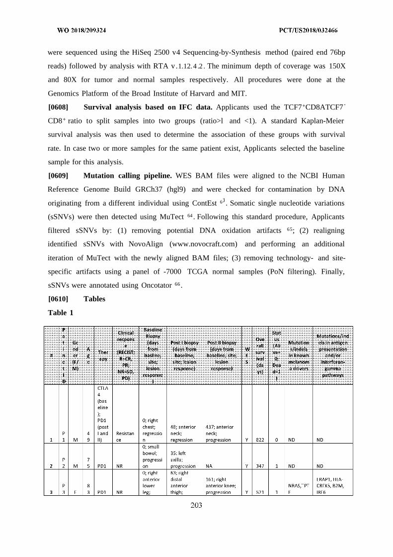
were sequenced using the HiSeq 2500 v4 Sequencing-by-Synthesis method (paired end 76bp
reads) followed by analysis with RTA v.1.12.4 .2 . The minimum depth of coverage was 150X
and 80X for tumor and normal samples respectively. All procedures were done at the
Genomics Platform of the Broad Institute of Harvard and MIT.
[0608] Survival analysis based on IFC data. Applicants used the TCF7+CD8ATCF7
CD8+ ratio to split samples into two groups (ratio>l and <1). A standard Kaplan-Meier
survival analysis was then used to determine the association of these groups with survival
rate. In case two or more samples for the same patient exist, Applicants selected the baseline
sample for this analysis.
[0609] Mutation calling pipeline. WES BAM files were aligned to the NCBI Human
Reference Genome Build GRCh37 (hgl9) and were checked for contamination by DNA
originating from a different individual using ContEst 6 . Somatic single nucleotide variations
(sSNVs) were then detected using MuTect 64 . Following this standard procedure, Applicants
filtered sSNVs by: (1) removing potential DNA oxidation artifacts 65; (2) realigning
identified sSNVs with NovoAlign (www.novocraft.com) and performing an additional
iteration of MuTect with the newly aligned BAM files; (3) removing technology- and site-
specific artifacts using a panel of -7000 TCGA normal samples (PoN filtering). Finally,
sSNVs were annotated using Oncotator 66 .
[0610] Tables
Table 1
progressi progression
on
(-2); left
shoulder;
progressi
CTLA on prior 35; leftP 2 4+PD to shoulder;
4 M 9 1 R therpay regression NA Y 539 0 TPTE ND
199;Post_P#5-right iliac
soft tissuemass;
NR progression.
(patient Post_P#5_2-had mix right
P 3 respons buttock;
5 M 3 PDl ) NA regression NA Y 369 1 TP53 ND
CTLA
4
(bas
eline (-70); left 270; right
) ; upper colectomy
PDl back; mass in
P 6 (post progress! cecum; NRAS,TPT
6 F 6 ) NR on progression NA Y 777 0 E ND
0; left
forehead
CTLA 22; leftP 7 4+PD regressio forehead; IFNGR1JPP2,
7 M 4 1 R n regression NA Y 339 0 TPTE ADAR
(-12); left
axillarylymph 62; left
CTLA node; axillary
P 4 4+PD regressio lymph node;8 M 9 1 R n regression NA N 388 0 NA NA
P 760; small
1 6 bowel mass; 119 IFNGR1,PSMB9
0 F 0 PDl NR NA progression NA Y 7 0 TP53 , TBX21
118; right
P inguinal
1 6 lymph node; NRAS,
1 F 8 PDl NR NA progression NA Y 300 1 TP53 ND
(-16);smallbawel
resection 77; left
P anterior1 6 progress! shoulder;
2 M 8 PDl NR on progression NA Y 101 1 NRAS TPP2
NR
(patientP CTLA had mix 297; porta MAP2K1,
1 4 4+PD respons hepatis; TP53,
3 M 8 1 ) NA progression NA Y 507 0 TPTE ND
P 462; left CTNNB1,
1 7 axilla lesion; NRAS,
4 M 0 PDl NR NA progression NA Y 588 1 TPTE PSMB9
0; rightback
subcutan JAK1, STATI,
P eous; 73; left CD45- cells1 7 progress! lower back; lack HLA-I
5 M 2 PDl NR on progression NA Y 163 1 TPTE expression
67; right
P abdomen1 6 soft tissue;
6 M 2 PDl NR NA progression NA N 476 1 NA NA
61; rightP axillary1 6 lymph node;7 F 8 PD1 NA regression NA N 361 0 NA NA
NR
(patient 237; rightP had mix inguinal1 7 respons lymph node;8 M 9 PD1 ) NA progression NA Y 746 0 ND ND
21; leftP axillary1 5 lymph node;9 M 3 PD1 R NA regression NA N 570 0 NA NA
0; rightinguinoiliac
NR lymphad(patient eneccto
P had mix my; 199; right2 6 respons progressi pelvic mass;
0 F 4 PD1 ) on progression NA Y 413 0 ND ND
P 867;2 7 jejunum; 1341 F 5 PD1 R NA regression NA Y 0 0 ND ND
31; rightP supraclavicul2 5 ar mass;
2 M 6 PD1 NR NA progression NA N 64 1 NA NA
403; leftP frontal 622; left neck2 6 craniotomy; mass;
3 M 2 PD1 NR NA progression progression Y 674 0 TP53 IL3RA, IRF6
0; leftlower
P back;2 7 regressio4 M 3 PD1 R n NA NA N 54 0 NA NA
0;adrenal
P gland; CD45- cells2 7 progressi lack HLA-I
5 M 4 PD1 NR on NA NA Y 676 0 ND expression
(-56);axillarylymph
P CTLA node,2 7 4+PD regressio6 M 2 1 R n NA NA N 517 0 NA NA
(-35);upperabdomen
P2 6 progress!7 F 2 PD1 NR on NA NA N 73 0 NA NA
0; rightP CTLA groin; 41; right2 6 4+PD Resistan regressio groin; 89; right groin;8 F 7 1 ce n progression progression Y 61 0 ND ND
(-67); leftaxillarylymph
P node;2 7 regressio9 M 9 PD1 R n NA NA N 417 0 NA NA
573; leftlaparoscopic
P adrenalecto3 6 my;0 M 4 PD1 NR NA progression NA N 580 0 NA NA
(-7); right
P axilla;
3 5 progressi
30 1 M 2 PD1 N o n NA NA N 126 0 NA NA
(-66); left
axillary
lymph
P node;
3 6 regressio
31 3 F 5 PD1 R n NA NA N 130 0 NA NA
(-31 ) ;right
iliaclymph
P node;3 7 regressi
32 5 M 0 PD1 R o n NA NA Y 511 0 ND ND
F- female; M- male; NR- nonresponder; R- responder; NA- not available; ND- notdetected; DOD- dead of disease; AWD- alive with disease
Table 2A
Gl- B cells G2- PI sma cells
Gene P- Moan Mean Mean MeanNam valu cxprcssio expression GencN cxprcssio expressione e n Gl non-Gl amc P-valuc n G2 non-G2
adjusted P- adjusted P-
<le- 5.3771086 0.02412688 value = 2.4c- 4.2338608 0.00862474 va u = 2.4c-IGHD 300 13 8 5 SDC1 <le-300 33 6 S
<le- 4.8307840 0.02321438 IGLV6- 2.4025581 0.02615370
PAX5 300 38 4 57 9.81E-130 75 9
FCRL <le- 5.7587154 0.03384063 IGHV3 2.2299108
1 300 72 3 OR16-9 3.35E-147 69 0.02923494
<le- 3.1779645 0.02606056 TNFRSF 3.8145218 0.06042765
CR2 300 45 7 17 4.73E-257 14 2
VPRE <le- 3.7796315 0.03121607 IGLV3- 4.1162931 0.06792561
B3 300 99 8 1 6.62E-229 7 9
FCER <le- 5.1756426 2.5301487 0.04191897
2 300 62 0.04801409 HID1 1.13E-207 75 1
<le- 6.8794951 0.06740436 7.4354631
CD19 300 85 3 IGHG4 3.21E-239 73 0.14692745
<le- 2.5783675 0.02583382 IGHV3- 2.6404904 0.06023521
EBF1 300 56 1 48 1.50E-135 19 7
<le- 6.8174367 0.06854110 4.0156623 0.09378567
CD22 300 7 6 IGHA2 2.18E-131 76 1
BANK <le- 6.7391301 0.07032866 IGHV3- 3.1489753
1 300 58 4 7 5.17E-127 71 0.07603257
IGKV3
CLEC <le- 3.0533710 0.03842337 OR2- 2.6602446 0.06967666
17A 300 72 3 268 5.87E-151 92 1
FCRL <le- 3.6213324 0.05640783 IGHV3- 2.1389978 0.05703562
A 300 6 7 11 6.43E-108 25 5
FCRL <le- 2.3503036 0.03666980 8.7049802 0.25394845
2 300 18 6 IGHG2 1.87E-225 23 8
MS4 <le- 9.4287148 0.15357791 7.0813680 0.21231582
Al 300 65 9 IGHA1 2.97E-249 71 3
<le- 3.4349996 0.09073335 8.9367770 0.28058643
BLK 300 29 6 IGHG3 6.04E-223 49 7
RALG <le- 3.7572152 0.10422786 7.4721484
PS2 300 67 2 DERL3 <le-300 0 1 0.24163785
TCL1 <le- 4.6537884 0.14776274 7.3906410 0.24235704
A 300 76 4 IGLCl <le-300 29 4
TLR1 <le- 2.0333744 IGKV4- 2.7669405 0.09157096
0 300 59 0.07030556 1 2.60E-105 07 6
FAM <le- 3.3258764 0.13062055 IGKV3 2.6442219
129C 300 84 5 D-15 4.23E-97 4 0.08987916
<le- 2.2307334 0.08769665 2.7149164 0.09271453
CN 2 300 39 5 IGLC7 8.72E-116 12 2
ARH
GAP2 <le- 3.9866383 0.16394866 5.7985527 0.205363124 300 22 6 IGLL5 3.08E-262 99 1
9.02
KIAA E- 2.1033251 0.08667368 IGHV3- 2.7039222 0.096513210125 271 1 8 21 2.09E-117 07 1
HLA- <le- 3.8311540 0.16554764 IGKV3- 2.7711741
DOB 300 47 9 11 1.40E-80 04 0.10253285
<le- 9.3272050 0.40745498 IGHV1- 2.2326666IGHM 300 93 7 69 1.36E-88 27 0.08526387
KIAA 6.11700000
0226 <le- 4.8298782 0.21132716 OOOOOOe- 11.105462 0.47109533
L 300 93 9 IGJ 321 5 8
CD79 <le- 8.3395337 0.37017964 11.944645 0.50683376
A 300 9 1 7 IGHG1 2.80E-181 57 5
BCL1 <le- 7.4105008 0.38361486 IGHV3- 3.5764142 0.159436931A 300 24 6 23 8.56E-108 72 9
1.92909797
STAP <le- 3.6246375 0.19870336 TXNDC 995300e- 5.7598913 0.26051721
1 300 62 5 5 312 8 1 3
8.54
E- 3.3730505 0.18979311 2.0684614 0.09843747
IGLC3 270 02 8 PYCR1 8.08E-123 38 9
5.82 4.59306305
E- 2.7683946 897400e- 5.0366142
PKIG 295 43 0.15679259 FCRL5 312 5 0.24157025
1.17FCRL E- 2.3237129 0.13594682 3.6150117 0.20271791
5 285 4 1 1 SPAG4 3.46E-165 52 3
<le- 3.1703638 0.20072994 IGKV1 2.2990930 0.13406608
AFF3 300 03 3 D-39 1.94E-62 53 3
4.92
COBL E- 2.1471729 0.13739352 IGKV3- 2.8044501 0.16530640
LI 254 35 1 20 2.66E-79 55 4
<le- 3.8307691 0.25308751 5.1124481 0.33468206
SPIB 300 52 4 IGLC2 1.01E-104 2 3
SWA <le- 4.0124140 0.28577176 2.1565034 0.15028415
P70 300 59 7 TRAM2 1.38E-156 84 7
9.41
E- 2.0018349 0.14416444 2.9167284 0.20362128
IGLL5 205 23 1 CPNE5 1.27E-199 94 7
ADA <le- 5.9514160 0.44283933 9.5794608 0.71062497M28 300 93 1 MZB1 1.21E-302 42 3
<le- 4.2832777 0.34011246 2.0434333 0.15669701
BLNK 300 5 9 PNOC 8.98E-103 7 3
2.61
E- 2.6218525 0.20859588 2.3821253 0.18761370
IGLC2 171 9 1 9 GAB1 2.78E-127 52 3
<le- 8.1930256 0.77094020 IGHV3- 2.1871708 0.17941979IGKC 300 68 6 30 1.77E-62 65 9
IGHG <le- 2.5036609 0.24051935 4.7205773 0.39308038
3 300 02 7 IGLC3 3.42E-89 62 8
WDF <le- 4.1436026 0.40502326 2.7366232 0.22969453Y4 300 65 3 P2RX1 1.03E-145 27 5
MEF2 <le- 4.6828284 0.46396047 COBLL 3.0150997 0.26541357
C 300 75 3 1 6.99E-133 92 8
IGHG <le- 2.2823146 0.22875917 13.299584 1.20744076
2 300 94 6 IGKC 1.10E-152 29 3
2.39
E- 2.4969530 0.25601685 HIST1H 2.0597600 0.20012590CCR6 258 86 8 2BG 1.52E-74 08 2
5.30
E- 2.0376411 0.21324463 2.4238597IGLCl 148 02 6 SPATS2 9.27E-117 73 0.25377366
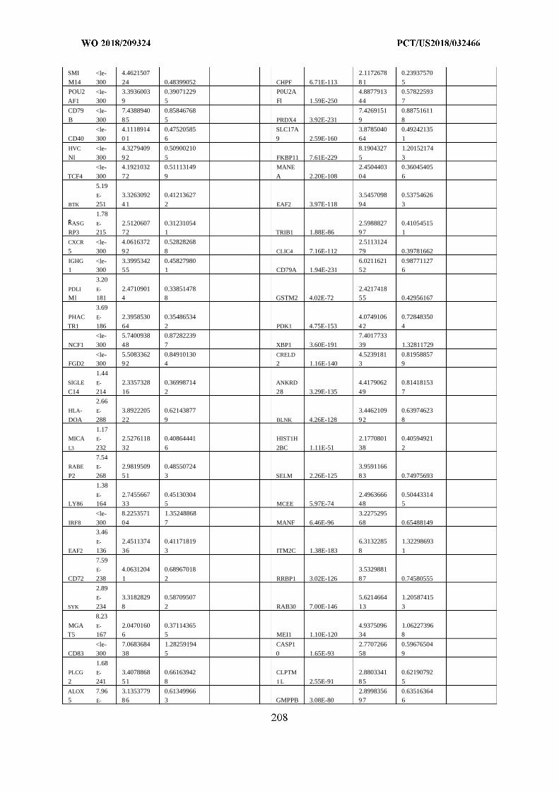
SMI <le- 4.4621507 2.1172678 0.23937570
M14 300 24 0.48399052 CHPF 6.71E-113 8 1 5
POU2 <le- 3.3936003 0.39071229 P0U2A 4.8877913 0.57822593
AF1 300 9 5 Fl 1.59E-250 44 7
CD79 <le- 7.4388940 0.85846768 7.4269151 0.88751611
B 300 85 5 PRDX4 3.92E-231 9 8
<le- 4.1118914 0.47520585 SLC17A 3.8785040 0.49242135
CD40 300 0 1 6 9 2.59E-160 64 1
HVC <le- 4.3279409 0.50900210 8.1904327 1.20152174
Nl 300 92 5 FKBP11 7.61E-229 5 3
<le- 4.1921032 0.51113149 MANE 2.4504403 0.36045405
TCF4 300 72 9 A 2.20E-108 04 6
5.19
E- 3.3263092 0.41213627 3.5457098 0.53754626
BTK 251 4 1 2 EAF2 3.97E-118 94 3
1.78
ASG E- 2.5120607 0.31231054 2.5988827 0.41054515RP3 215 72 1 TRIB1 1.88E-86 97 1
CXCR <le- 4.0616372 0.52828268 2.5113124
5 300 92 8 CLIC4 7.16E-112 79 0.39781662
IGHG <le- 3.3995342 0.45827980 6.0211621 0.987711271 300 55 1 CD79A 1.94E-231 52 6
3.20
PDLI E- 2.4710901 0.33851478 2.4217418Ml 181 4 8 GSTM2 4.02E-72 55 0.42956167
3.69
PHAC E- 2.3958530 0.35486534 4.0749106 0.72848350
TR1 186 64 2 PDK1 4.75E-153 42 4
<le- 5.7400938 0.87282239 7.4017733
NCF1 300 48 7 XBP1 3.60E-191 39 1.32811729
<le- 5.5083362 0.84910130 CRELD 4.5239181 0.81958857FGD2 300 92 4 2 1.16E-140 3 9
1.44
SIGLE E- 2.3357328 0.36998714 ANKRD 4.4179062 0.81418153
C14 214 16 2 28 3.29E-135 49 7
2.66
HLA- E- 3.8922205 0.62143877 3.4462109 0.63974623
DOA 288 22 9 BLNK 4.26E-128 92 8
1.17MICA E- 2.5276118 0.40864441 HIST1H 2.1770801 0.40594921
L3 232 32 6 2BC 1.11E-51 38 2
7.54RABE E- 2.9819509 0.48550724 3.9591166
P2 268 51 3 SELM 2.26E-125 83 0.74975693
1.38
E- 2.7455667 0.45130304 2.4963666 0.50443314LY86 164 33 5 MCEE 5.97E-74 48 5
<le- 8.2253571 1.35248868 3.2275295
IRF8 300 04 7 MANF 6.46E-96 68 0.65488149
3.46
E- 2.4511374 0.41171819 6.3132285 1.32298693
EAF2 136 36 3 ITM2C 1.38E-183 8 1
7.59
E- 4.0631204 0.68967018 3.5329881
CD72 238 1 2 RRBP1 3.02E-126 87 0.74580555
2.89
E- 3.3182829 0.58709507 5.6214664 1.20587415
SYK 234 8 2 RAB30 7.00E-146 13 3
8.23
MGA E- 2.0470160 0.37114365 4.9375096 1.06227396T5 167 6 5 MEI1 1.10E-120 34 8
<le- 7.0683684 1.28259194 CASP1 2.7707266 0.59676504
CD83 300 38 5 0 1.65E-93 58 9
1.68PLCG E- 3.4078868 0.66163942 CLPTM 2.8803341 0.62190792
2 241 51 8 1 L 2.55E-91 85 5
ALOX 7.96 3.1353779 0.61349966 2.8998356 0.635163645 E- 86 3 GMPPB 3.08E-80 97 6
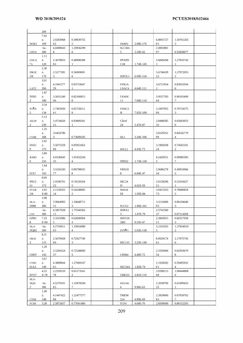
240
7.02
E- 2.0283968 0.39839755 6.8001727 1.50761265
DOK3 109 62 5 FKBP2 2.68E-176 8 1 5
<le- 6.0498043 1.20936299 SLC38A 2.4902881
CIITA 300 8 6 5 2.20E-65 97 0.55858077
1.11
CDCA E- 2.3678953 0.48998398 PPAPD 5.6849268 1.27835742
7 L 120 82 1 C1B 1.74E-145 1 1
2.30PIK3C E- 2.5277381 0.56069691 5.6746439 1.27872055
2 B 170 3 6 SDF2L1 6.09E-154 55 2
3.01E- 4.1941277 0.93733647 ST6GA 3.6721054 0.83025034
LAT2 206 29 1 LNAC4 4.64E-111 1 6
1.87
TPD5 E- 3.5031240 0.82100815 TXNDC 3.9557783 0.901054992 186 66 8 11 7.00E-110 44 7
3.54
P2 X E- 2.7402950 0.65728211 FNDC3 3.1007955 0.707343755 134 4 1 9 B 7.02E-109 96 3
5.13
AKAP E- 3.4734620 0.83809341 Cllorf 2.8488585 0.65603053
2 239 21 1 24 5.47E-87 32 9
1.25
E- 3.0424786 3.6529351 0.84542770
CYBB 268 3 0.73699245 SIL1 3.34E-106 99 4
2.02
SNX2 E- 3.5072329 0.85951663 3.1966938 0.74502181
9 175 08 4 BSCL2 4.43E-73 24 2
3.89RAB3 E- 4.0336645 1.01932256 4.2420551 0.99985585
0 210 35 4 TPD52 1.73E-130 5 7
1.64
E- 3.5256183 0.89798035 FBXOl 2.0686278 0.49933066
STX7 193 77 8 8 6.84E-47 34 5
8.69
P0U2 E- 2.9196701 0.74518104 SEC24 2.0158296 0.51016637F2 172 22 5 D 4.01E-59 51 1
FCGR 3.63 2.1139355 0.54248685 MAGE 3.0415323 0.78686818
2 B E-89 14 2 Dl 1.05E-84 73 5
3.98HLA- E- 5.9664905 1.54648711 3.3110496 0.86104640
DMB 286 31 3 PLCG2 1.90E-101 05 3
<le- 6.5857928 1.75540362 HSPA1 2.5741560LY9 300 11 5 3 1.67E-79 37 0.67114269
GPR1 7.19 2.3319386 0.62606994 HIST1H 2.2845815 0.60317058
8 E-94 5 3 2BD 9.55E-47 51 3
HLA- <le- 4.2743911 1.19916080 5.2101655 1.37834919DQB2 300 03 4 SYV 1 5.03E-138 5 2
8.21
ORAI E- 2.5670928 0.72927749 8.0929174 2.170757452 115 06 3 SEC11C 3.25E-149 61 6
1.20
E- 2.5204324 0.72548600 2.3359406 0.62910679
CHD7 143 37 5 CPEB4 4.40E-72 54 8
3.67
CYB5 E- 4.3889844 1.27069167 2.1928282 0.59495932
61A3 149 0 1 1 SEC24A 1.82E-74 0 1 4
4.23 2.2359310 0.65173541 3.9398115 1.08444808IFT57 E-78 78 2 UBE2J1 2.81E-110 69 6
HLA-
DQA <le- 4.5270101 1.33478294 EIF2AK 2.2038700 0.610994332 300 85 7 4 9.96E-63 35 1
1.00
E- 4.1447422 1.22477577 TMEM 2.3959600 0.67059762
CTSH 148 88 1 214 4.99E-69 83 7
FCHS 3.28 2.5872657 0.77041480 TCF4 4.60E-70 2.8509049 0.80152203
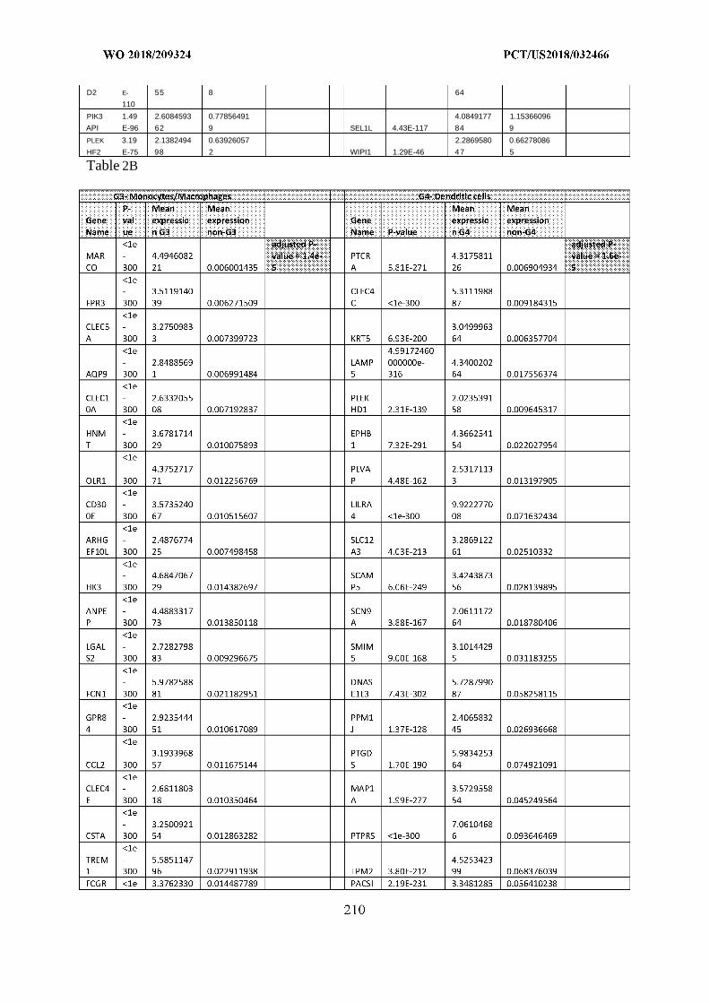
D2 E- 55 8 64
110
PIK3 1.49 2.6084593 0.77856491 4.0849177 1.15366096
API E-96 62 9 SEL1L 4.43E-117 84 9
PLEK 3.19 2.1382494 0.63926057 2.2869580 0.66278086
HF2 E-75 98 2 WIPI1 1.29E-46 47 5
Table 2B
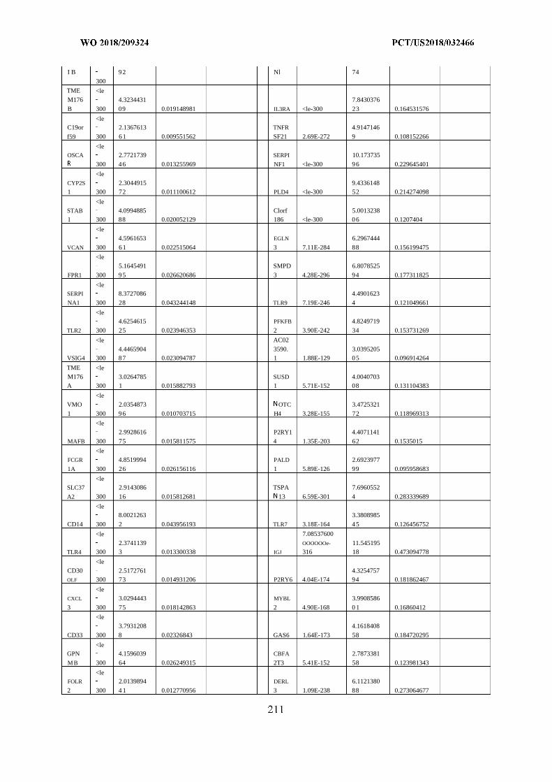
I B 92 Nl 74
300
TME <leM176 4.3234431 7.8430376
B 300 09 0.019148981 IL3RA <le-300 23 0.164531576
<leC19or 2.1367613 TNFR 4.9147146
f59 300 61 0.009551562 SF21 2.69E-272 9 0.108152266
<leOSCA 2.7721739 SERPI 10.173735
300 46 0.013255969 NF1 <le-300 96 0.229645401
<leCYP2S 2.3044915 9.43361481 300 72 0.011100612 PLD4 <le-300 52 0.214274098
<leSTAB 4.0994885 Clorf 5.00132381 300 88 0.020052129 186 <le-300 06 0.1207404
<le4.5961653 EGLN 6.2967444
VCAN 300 61 0.022515064 3 7.11E-284 88 0.156199475
<le5.1645491 SMPD 6.8078525
FPR1 300 95 0.026620686 3 4.28E-296 94 0.177311825
<leSERPI 8.3727086 4.4901623
NA1 300 28 0.043244148 TLR9 7.19E-246 4 0.121049661
<le4.6254615 PFKFB 4.8249719
TLR2 300 25 0.023946353 2 3.90E-242 34 0.153731269
<le AC02
4.4465904 3590. 3.0395205VSIG4 300 87 0.023094787 1 1.88E-129 05 0.096914264
TME <leM176 3.0264785 SUSD 4.0040703A 300 1 0.015882793 1 5.71E-152 08 0.131104383
<leVMO 2.0354873 OTC 3.4725321
1 300 96 0.010703715 H4 3.28E-155 72 0.118969313
<le2.9928616 P2RY1 4.4071141
MAFB 300 75 0.015811575 4 1.35E-203 62 0.1535015
<leFCGR 4.8519994 PALD 2.6923977
1A 300 26 0.026156116 1 5.89E-126 99 0.095958683
<leSLC37 2.9143086 TSPA 7.6960552
A2 300 16 0.015812681 13 6.59E-301 4 0.283339689
<le8.0021263 3.3808985
CD14 300 2 0.043956193 TLR7 3.18E-164 45 0.126456752
<le 7.08537600
2.3741139 OOOOOOe- 11.545195TLR4 300 3 0.013300338 IGJ 316 18 0.473094778
<leCD30 2.5172761 4.3254757OLF 300 73 0.014931206 P2RY6 4.04E-174 94 0.181862467
<leCXCL 3.0294443 MYBL 3.9908586
3 300 75 0.018142863 2 4.90E-168 0 1 0.16860412
<le3.7931208 4.1618408
CD33 300 8 0.02326843 GAS6 1.64E-173 58 0.184720295
<leGPN 4.1596039 CBFA 2.7873381
MB 300 64 0.026249315 2T3 5.41E-152 58 0.123981343
<leFOLR 2.0139894 DERL 6.1121380
2 300 4 1 0.012770956 3 1.09E-238 88 0.273064677
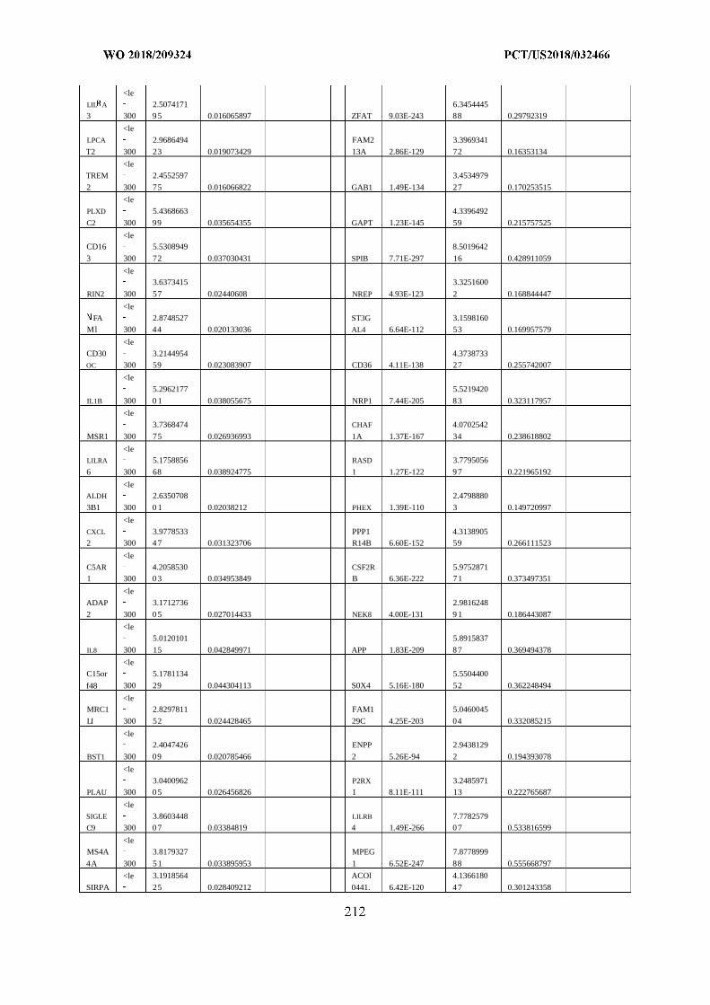
<leLIL A 2.5074171 6.34544453 300 95 0.016065897 ZFAT 9.03E-243 88 0.29792319
<leLPCA 2.9686494 FAM2 3.3969341
T2 300 23 0.019073429 13A 2.86E-129 72 0.16353134
<leTREM 2.4552597 3.4534979
2 300 75 0.016066822 GAB1 1.49E-134 27 0.170253515
<lePLXD 5.4368663 4.3396492
C2 300 99 0.035654355 GAPT 1.23E-145 59 0.215757525
<leCD16 5.5308949 8.5019642
3 300 72 0.037030431 SPIB 7.71E-297 16 0.428911059
<le3.6373415 3.3251600
RIN2 300 57 0.02440608 NREP 4.93E-123 2 0.168844447
<leFA 2.8748527 ST3G 3.1598160
Ml 300 44 0.020133036 AL4 6.64E-112 53 0.169957579
<leCD30 3.2144954 4.3738733OC 300 59 0.023083907 CD36 4.11E-138 27 0.255742007
<le5.2962177 5.5219420
IL1B 300 0 1 0.038055675 NRP1 7.44E-205 83 0.323117957
<le3.7368474 CHAF 4.0702542
MSR1 300 75 0.026936993 1A 1.37E-167 34 0.238618802
<leLILRA 5.1758856 RASD 3.7795056
6 300 68 0.038924775 1 1.27E-122 97 0.221965192
<leALDH 2.6350708 2.4798880
3B1 300 0 1 0.02038212 PHEX 1.39E-110 3 0.149720997
<leCXCL 3.9778533 PPP1 4.31389052 300 47 0.031323706 R14B 6.60E-152 59 0.266111523
<leC5AR 4.2058530 CSF2R 5.97528711 300 03 0.034953849 B 6.36E-222 71 0.373497351
<leADAP 3.1712736 2.9816248
2 300 05 0.027014433 NEK8 4.00E-131 9 1 0.186443087
<le5.0120101 5.8915837
IL8 300 15 0.042849971 APP 1.83E-209 87 0.369494378
<leC15or 5.1781134 5.5504400
f48 300 29 0.044304113 S0X4 5.16E-180 52 0.362248494
<leMRC1 2.8297811 FAM1 5.0460045
LI 300 52 0.024428465 29C 4.25E-203 04 0.332085215
<le2.4047426 ENPP 2.9438129
BST1 300 09 0.020785466 2 5.26E-94 2 0.194393078
<le3.0400962 P2RX 3.2485971
PLAU 300 05 0.026456826 1 8.11E-111 13 0.222765687
<leSIGLE 3.8603448 LILRB 7.7782579C9 300 07 0.03384819 4 1.49E-266 07 0.533816599
<leMS4A 3.8179327 MPEG 7.8778999
4A 300 51 0.033895953 1 6.52E-247 88 0.555668797
<le 3.1918564 ACOl 4.1366180
SIRPA 25 0.028409212 0441. 6.42E-120 47 0.301243358
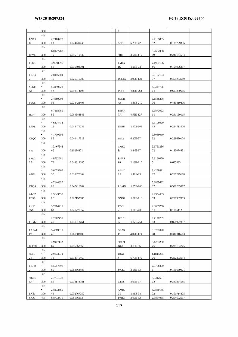
300 1
<leNAS 2.7463772 2.4105865
El 300 0 1 0.024449745 ADC 6.29E-72 52 0.175729336
<le6.0127783 3.2654938
CPVL 300 12 0.055310537 SRC 3.66E-110 69 0.240164554
<lePLBD 3.9598696 TMIG 2.19871561 300 83 0.036493191 D2 1.29E-74 46 0.164006857
<leLILRA 2.8416304 6.0102163
2 300 17 0.026715789 TCL1A 4.00E-130 57 0.451253519
<leSLC11 5.3149622 8.9319796
Al 300 94 0.050314006 TCF4 4.86E-264 74 0.693230615
<le2.4689004 SLC15 6.1538278
PYGL 300 05 0.023422496 A4 1.81E-219 06 0.483410876
<le6.7803785 SEMA 3.6873092
HCK 300 85 0.064583888 7A 4.55E-127 11 0.291190115
<le4.6304714 3.5108020
LRP1 300 18 0.044479138 THBD 1.47E-103 43 0.284711606
<le4.1790296 2.8059010
C1QC 300 93 0.040417513 TEX2 6.20E-97 92 0.228630174
<le10.467341 CMKL 2.1761236
LYZ 300 62 0.10234471 Rl 3.84E-67 02 0.183074451
<leLRRC 4.8712661 RNAS 7.8186070
25 300 78 0.048319185 E6 2.13E-210 5 0.665833
<le3.0033969 ABHD 2.4298811
ADM 300 35 0.030070209 15 1.49E-83 82 0.207279178
<le4.7144827 5.8889632
C1QA 300 08 0.047416804 LGMN 1.55E-166 37 0.508285977
<leAPOB 2.5643538 2.9334683
EC3A 300 86 0.027152185 GNG7 1.56E-116 53 0.259987053
<leZNF3 3.7984419 TTYH 2.003525685A 300 61 0.041277352 2 1.70E-70 61 0.1786112
<le2.7962499 BCL11 9.4106769
TGM2 300 49 0.031313461 A 1.32E-264 83 0.858977097
<leT FAI 5.4589619 GRAS 3.3791020
P2 300 46 0.061582086 P 4.07E-119 98 0.310016663
<le4.9947132 SERPI 3.1233230
CSF3R 300 67 0.05686716 NG1 3.19E-95 76 0.289184775
<leSLCO 2.9873971 TRAF 4.1845265
2B1 300 71 0.034015469 4 6.79E-170 26 0.392893634
<leLILRB 5.5957398 2.0758400
2 300 66 0.064663485 MGLL 2.58E-63 1 0.196630971
<leSIGLE 2.7731938 3.5312551C7 300 53 0.032173181 CTNS 2.97E-97 22 0.343834585
<le2.8172360 AMIG 3.0839135
TNS1 300 45 0.032767759 0 3 1.45E-98 02 0.301714405
SIOO <le 6.8772470 0.08156152 PMEP 2.00E-82 2.5864085 0.254602597
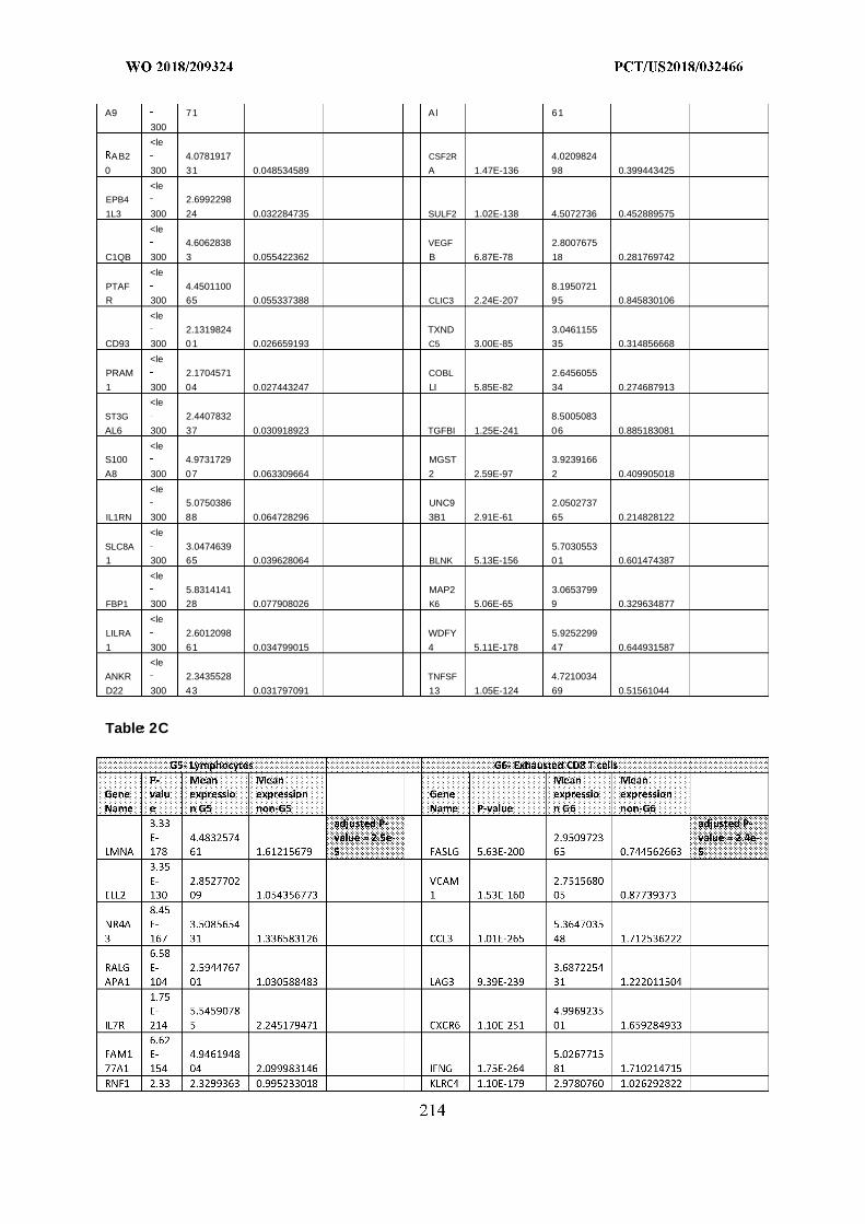
A9 71 Al 61
300
<le
AB2 4.0781917 CSF2R 4.0209824
0 300 31 0.048534589 A 1.47E-136 98 0.399443425
<leEPB4 2.6992298
1L3 300 24 0.032284735 SULF2 1.02E-138 4.5072736 0.452889575
<le4.6062838 VEGF 2.8007675
C1QB 300 3 0.055422362 B 6.87E-78 18 0.281769742
<le
PTAF 4.4501100 8.1950721
R 300 65 0.055337388 CLIC3 2.24E-207 95 0.845830106
<le
2.1319824 TXND 3.0461155
CD93 300 01 0.026659193 C5 3.00E-85 35 0.314856668
<le
PRAM 2.1704571 COBL 2.6456055
1 300 04 0.027443247 LI 5.85E-82 34 0.274687913
<le
ST3G 2.4407832 8.5005083
AL6 300 37 0.030918923 TGFBI 1.25E-241 06 0.885183081
<leS100 4.9731729 MGST 3.9239166
A8 300 07 0.063309664 2 2.59E-97 2 0.409905018
<le5.0750386 UNC9 2.0502737
IL1RN 300 88 0.064728296 3B1 2.91E-61 65 0.214828122
<le
SLC8A 3.0474639 5.7030553
1 300 65 0.039628064 BLNK 5.13E-156 01 0.601474387
<le
5.8314141 MAP2 3.0653799
FBP1 300 28 0.077908026 K6 5.06E-65 9 0.329634877
<le
LILRA 2.6012098 WDFY 5.9252299
1 300 61 0.034799015 4 5.11E-178 47 0.644931587
<le
ANKR 2.3435528 TNFSF 4.7210034
D22 300 43 0.031797091 13 1.05E-124 69 0.51561044
Table 2C
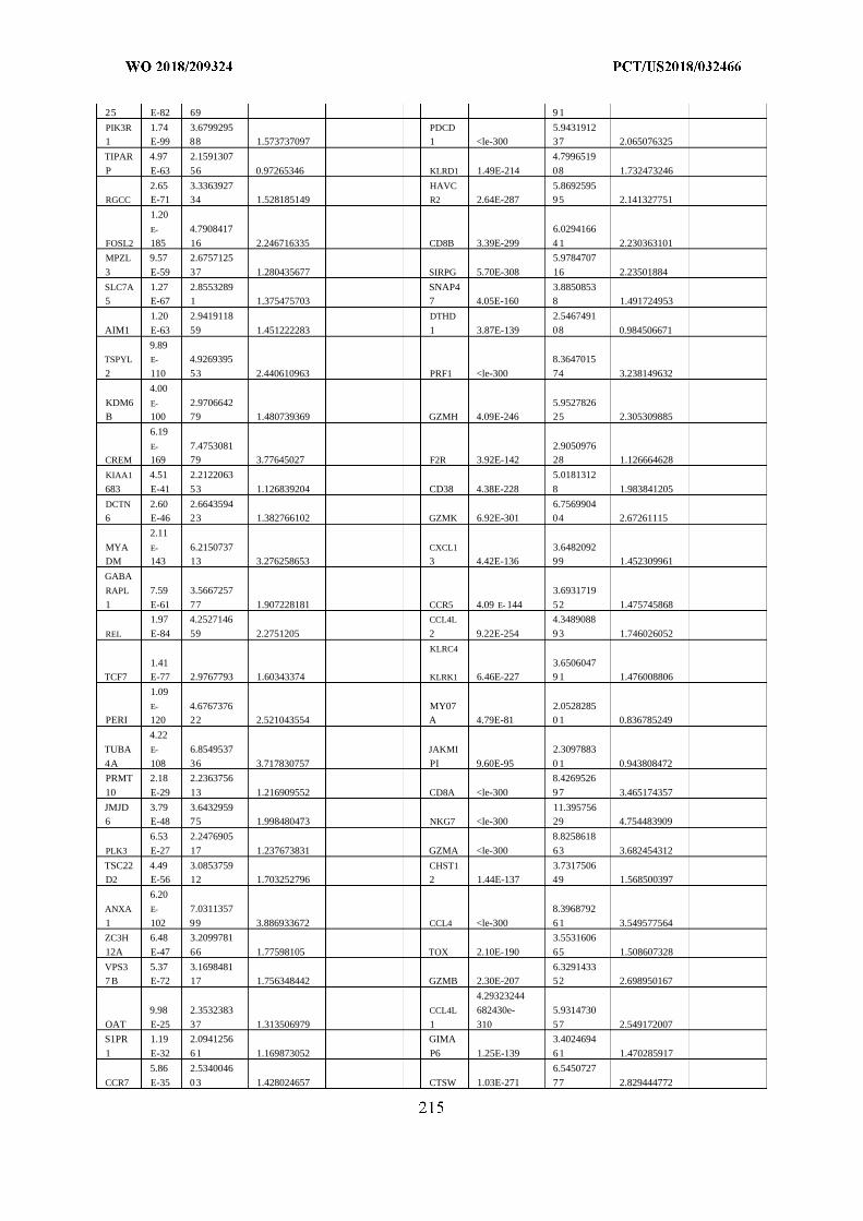
25 E-82 69 9 1
PIK3R 1.74 3.6799295 PDCD 5.94319121 E-99 88 1.573737097 1 <le-300 37 2.065076325
TIPAR 4.97 2.1591307 4.7996519
P E-63 56 0.97265346 KLRD1 1.49E-214 08 1.732473246
2.65 3.3363927 HAVC 5.8692595RGCC E-71 34 1.528185149 R2 2.64E-287 95 2.141327751
1.20
E- 4.7908417 6.0294166
FOSL2 185 16 2.246716335 CD8B 3.39E-299 4 1 2.230363101
MPZL 9.57 2.6757125 5.9784707
3 E-59 37 1.280435677 SIRPG 5.70E-308 16 2.23501884
SLC7A 1.27 2.8553289 SNAP4 3.88508535 E-67 1 1.375475703 7 4.05E-160 8 1.491724953
1.20 2.9419118 DTHD 2.5467491
AIM1 E-63 59 1.451222283 1 3.87E-139 08 0.984506671
9.89TSPYL E- 4.9269395 8.3647015
2 110 53 2.440610963 PRF1 <le-300 74 3.238149632
4.00
KDM6 E- 2.9706642 5.9527826B 100 79 1.480739369 GZMH 4.09E-246 25 2.305309885
6.19
E- 7.4753081 2.9050976CREM 169 79 3.77645027 F2R 3.92E-142 28 1.126664628
KIAA1 4.51 2.2122063 5.0181312
683 E-41 53 1.126839204 CD38 4.38E-228 8 1.983841205
DCTN 2.60 2.6643594 6.75699046 E-46 23 1.382766102 GZMK 6.92E-301 04 2.67261115
2.11
MYA E- 6.2150737 CXCL1 3.6482092DM 143 13 3.276258653 3 4.42E-136 99 1.452309961
GABA
RAPL 7.59 3.5667257 3.6931719
1 E-61 77 1.907228181 CCR5 4.09 E- 144 52 1.475745868
1.97 4.2527146 CCL4L 4.3489088
REL E-84 59 2.2751205 2 9.22E-254 93 1.746026052
KLRC4
1.41 3.6506047TCF7 E-77 2.9767793 1.60343374 KLRK1 6.46E-227 9 1 1.476008806
1.09
E- 4.6767376 MY07 2.0528285PERI 120 22 2.521043554 A 4.79E-81 0 1 0.836785249
4.22
TUBA E- 6.8549537 JAKMI 2.3097883
4A 108 36 3.717830757 PI 9.60E-95 0 1 0.943808472
PRMT 2.18 2.2363756 8.4269526
10 E-29 13 1.216909552 CD8A <le-300 97 3.465174357
JMJD 3.79 3.6432959 11.3957566 E-48 75 1.998480473 NKG7 <le-300 29 4.754483909
6.53 2.2476905 8.8258618
PLK3 E-27 17 1.237673831 GZMA <le-300 63 3.682454312
TSC22 4.49 3.0853759 CHST1 3.7317506D2 E-56 12 1.703252796 2 1.44E-137 49 1.568500397
6.20
ANXA E- 7.0311357 8.3968792
1 102 99 3.886933672 CCL4 <le-300 61 3.549577564
ZC3H 6.48 3.2099781 3.5531606
12A E-47 66 1.77598105 TOX 2.10E-190 65 1.508607328
VPS3 5.37 3.1698481 6.32914337 B E-72 17 1.756348442 GZMB 2.30E-207 52 2.698950167
4.29323244
9.98 2.3532383 CCL4L 682430e- 5.9314730
OAT E-25 37 1.313506979 1 310 57 2.549172007
S1PR 1.19 2.0941256 GIMA 3.4024694
1 E-32 61 1.169873052 P6 1.25E-139 61 1.470285917
5.86 2.5340046 6.5450727
CCR7 E-35 03 1.428024657 CTSW 1.03E-271 77 2.829444772
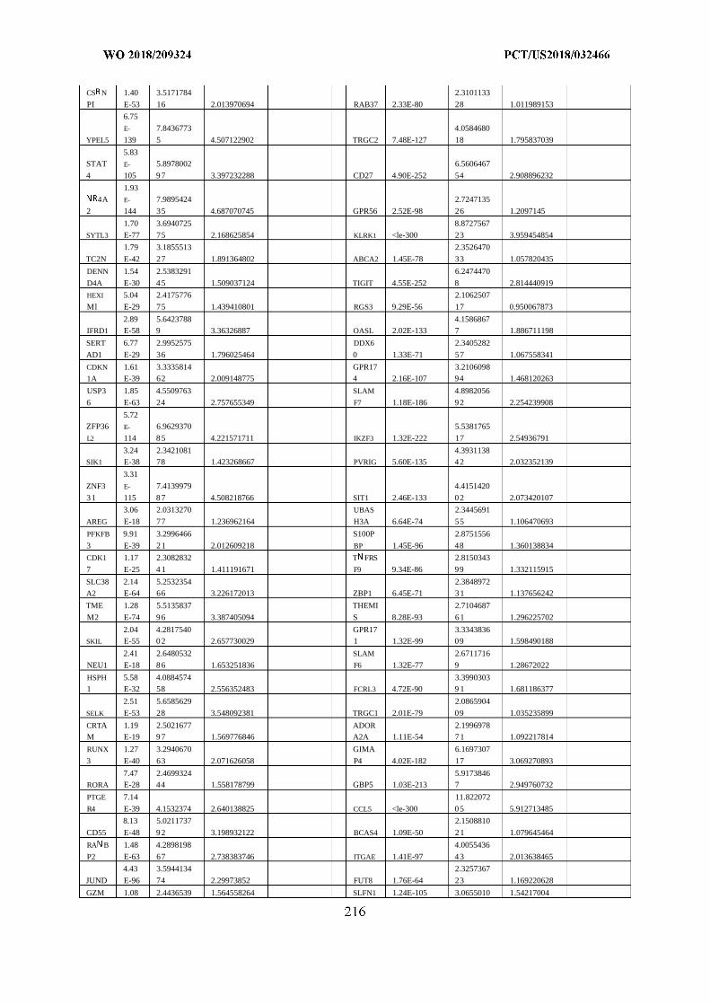
CS N 1.40 3.5171784 2.3101133
PI E-53 16 2.013970694 RAB37 2.33E-80 28 1.011989153
6.75
E- 7.8436773 4.0584680
YPEL5 139 5 4.507122902 TRGC2 7.48E-127 18 1.795837039
5.83STAT E- 5.8978002 6.5606467
4 105 97 3.397232288 CD27 4.90E-252 54 2.908896232
1.934A E- 7.9895424 2.7247135
2 144 35 4.687070745 GPR56 2.52E-98 26 1.2097145
1.70 3.6940725 8.8727567
SYTL3 E-77 75 2.168625854 KLRK1 <le-300 23 3.959454854
1.79 3.1855513 2.3526470
TC2N E-42 27 1.891364802 ABCA2 1.45E-78 33 1.057820435
DENN 1.54 2.5383291 6.2474470
D4A E-30 45 1.509037124 TIGIT 4.55E-252 8 2.814440919
HEXI 5.04 2.4175776 2.1062507
Ml E-29 75 1.439410801 RGS3 9.29E-56 17 0.950067873
2.89 5.6423788 4.1586867IFRD1 E-58 9 3.36326887 OASL 2.02E-133 7 1.886711198
SERT 6.77 2.9952575 DDX6 2.3405282
AD1 E-29 36 1.796025464 0 1.33E-71 57 1.067558341
CDKN 1.61 3.3335814 GPR17 3.21060981A E-39 62 2.009148775 4 2.16E-107 94 1.468120263
USP3 1.85 4.5509763 SLAM 4.8982056
6 E-63 24 2.757655349 F7 1.18E-186 92 2.254239908
5.72ZFP36 E- 6.9629370 5.5381765
L2 114 85 4.221571711 IKZF3 1.32E-222 17 2.54936791
3.24 2.3421081 4.3931138SIK1 E-38 78 1.423268667 PVRIG 5.60E-135 42 2.032352139
3.31
ZNF3 E- 7.4139979 4.4151420
31 115 87 4.508218766 SIT1 2.46E-133 02 2.073420107
3.06 2.0313270 UBAS 2.3445691
AREG E-18 77 1.236962164 H3A 6.64E-74 55 1.106470693
PFKFB 9.91 3.2996466 S100P 2.8751556
3 E-39 21 2.012609218 BP 1.45E-96 48 1.360138834
CDK1 1.17 2.3082832 T FRS 2.8150343
7 E-25 4 1 1.411191671 F9 9.34E-86 99 1.332115915
SLC38 2.14 5.2532354 2.3848972A2 E-64 66 3.226172013 ZBP1 6.45E-71 31 1.137656242
TME 1.28 5.5135837 THEMI 2.7104687
M2 E-74 96 3.387405094 S 8.28E-93 61 1.296225702
2.04 4.2817540 GPR17 3.3343836SKIL E-55 02 2.657730029 1 1.32E-99 09 1.598490188
2.41 2.6480532 SLAM 2.6711716
NEU1 E-18 86 1.653251836 F6 1.32E-77 9 1.28672022
HSPH 5.58 4.0884574 3.39903031 E-32 58 2.556352483 FCRL3 4.72E-90 9 1 1.681186377
2.51 5.6585629 2.0865904
SELK E-53 28 3.548092381 TRGC1 2.01E-79 09 1.035235899
CRTA 1.19 2.5021677 ADOR 2.1996978M E-19 97 1.569776846 A2A 1.11E-54 71 1.092217814
RUNX 1.27 3.2940670 GIMA 6.1697307
3 E-40 63 2.071626058 P4 4.02E-182 17 3.069270893
7.47 2.4699324 5.9173846RORA E-28 44 1.558178799 GBP5 1.03E-213 7 2.949760732
PTGE 7.14 11.822072
R4 E-39 4.1532374 2.640138825 CCL5 <le-300 05 5.912713485
8.13 5.0211737 2.1508810
CD55 E-48 92 3.198932122 BCAS4 1.09E-50 21 1.079645464
RA B 1.48 4.2898198 4.0055436
P2 E-63 67 2.738383746 ITGAE 1.41E-97 43 2.013638465
4.43 3.5944134 2.3257367
JUND E-96 74 2.29973852 FUT8 1.76E-64 23 1.169220628
GZM 1.08 2.4436539 1.564558264 SLFN1 1.24E-105 3.0655010 1.54217004
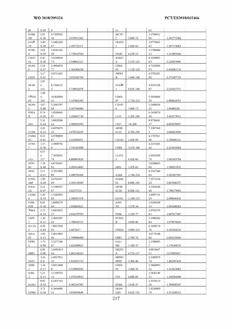
M E-20 4 2 L 62
TUBB 2.91 4.7290502 MCTP 2.37606114 B E-38 16 3.033912363 2 5.08E-72 96 1.202772368
PO L 3.40 3.5481420 TRAFD 2.9770641
3 E E-28 07 2.292755371 1 2.66E-63 02 1.507174063
SCML 3.02 2.6541162 2.57894844 E-29 39 1.720547954 ITGB7 4.22E-53 22 1.312803444
CNOT 6.42 3.9249018 RAB27 4.3269805
6 L E-50 73 2.548661221 A 5.11E-125 03 2.226815906
RGPD 5.54 2.4064476 GIMA 4.71029696 E-27 77 1.564306238 P5 1.13E-120 03 2.436961134
3.17 4.0711205 INPP4 4.5783291
CHD1 E-41 2 2.653545764 B 1.04E-146 96 2.375307719
2.87DUSP E- 8.7564122 PYHI 4.9201338
2 112 5 5.738855078 1 6.81E-148 87 2.554327371
1.90T FAI E- 10.826684 GIMA 5.5920004
P3 143 31 7.147865185 P7 1.71E-153 59 2.909624761
RGPD 3.67 5.2943787 C5orf5 3.2496926
5 E-48 89 3.507206484 6 1.80E-73 25 1.69486526
PDE4 9.74 5.0508445 6.5426074
B E-39 07 3.358452739 LYST 2.20E-240 46 3.416273912
5.31 3.8826586 8.9736041IDI1 E-24 42 2.586954705 CST7 <le-300 37 4.695878907
2.81 4.6078479 APOB 7.3387360
CCNH E-23 25 3.070532419 EC3G 6.76E-230 2 3.846833696
FAM4 8.53 4.9789809 4.17979216C E-61 87 3.332095051 CXCR3 2.43E-99 99 2.199446151
ATXN 1.17 2.5908792 4.2215505
7 E-30 6 1.741425098 CD84 3.21E-108 44 2.223035064
9.57
E- 7.2658541 CLSTN 2.0502569
FYN 117 76 4.888805828 3 4.45E-60 79 1.081603794
ATP1 1.18 4.8720507 3.0288603B3 E-40 8 1 3.285414683 ABI3 1.47E-63 86 1.598221831
NFE2 5.11 3.7012983 6.2547584
L2 E-21 89 2.498795494 IL2RB 1.74E-224 04 3.301967305
IVNS1 1.90 4.6762437 RARRE 7.2571218ABP E-28 09 3.183118599 S3 8.68E-193 26 3.867006375
POLR 4.12 5.3186457 APOB 3.3504240
2A E-47 97 3.62575721 EC3D 8.50E-121 48 1.798179903
CAMK 2.20 2.3282003 3.90871314 E-19 69 1.589207478 SLFN5 1.14E-125 59 2.098640434
CHM 8.02 2.8690279 HAPL 2.0269549
P1B E-18 66 1.958603621 N3 1.57E-34 15 1.091698363
R4A 2.73 3.8452825 2.97655761 E-19 37 2.656379795 PAM 1.25E-77 8 1 1.607617497
GSPT 1.30 2.4693307 PCED1 3.1088282
1 E-12 42 1.708454721 B 3.69E-86 84 1.679076643
SLC2A 4.58 7.8657934 6.16092743 E-63 95 5.4474217 ITM2A 3.98E-153 76 3.332344533
IQGA 1.05 2.4613802 3.6776148
P2 E-15 66 1.709898404 GBP1 2.78E-76 84 2.002535044
GPR6 1.75 2.3277246 GOLI 2.1588695
5 E-08 67 1.624386821 M4 1.18E-57 7 1.176344532
6.96 2.6681814 SH2D1 4.8816047
HBP1 E-08 06 1.865144355 A 4.72E-127 51 2.670995027
3.43 2.6057951 MPHO 2.5389079
PAF1 E-ll 32 1.822622722 SPH9 5.36E-46 74 1.402397418
AMD 1.34 3.6013408 GIMA 2.3660093
1 E-17 8 2.518994593 P2 1.96E-35 31 1.312623963
SORL 5.21 2.1109703 2.2642140
1 E-13 13 1.479529912 TTN 4.68E-69 31 1.256504449
8.05 6.2437143 2.3519123KLRK1 E-54 1 4.382216785 IFI44L 3.63E-37 34 1.306690547
3.71 4.3404489 DENN 5.9230809
GZMK E-34 31 3.059459649 D2D 6.62E-132 79 3.313248223
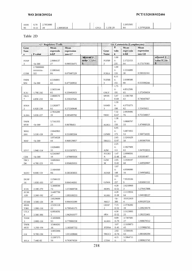
AKIRI 6.70 2.7053080 2.2053505
Nl E-12 28 1.90930518 GYG1 1.15E-29 84 1.237052039
Table 2D
G7- Regulatory Tcells G8- Cytotoxicity (Lymphocytes)
Gene Mean Mean P. Mcan MeanNam expresslo expression Gene valu exprcssio expressione P-va non-G7 Name non-G8
A t P- 1.00 adjusted P-
FOXP 2.9084137 f e = 2 4e FGFBP E- 2.1722153 value 2.6e-3 <le-300 27 0.090556475 2 235 04 0.173170381
2.70000000 1.00
OOOOOOe- 2.1881641 E- 2.2316089
CCR8 322 06 0.075687229 FCRL6 116 38 0.590102161
6.11
TNFR 4.5184001 TGFBR E- 2.0398588
SF4 <le-300 42 0.477269932 3 151 98 0.553300244
1.75
2.3655144 E- 4.8512506
ICA1 1.79E-242 85 0.259492653 GNLY 191 65 1.372456654
FBLN 2.0808570 SPON 5.07 2.1381768
7 4.85E-210 66 0.259247645 2 E-69 42 0.746447667
1.59
RTKN 2.1282877 SAMD E- 4.3753273
2 6.92E-238 92 0.272269648 3 198 62 1.55410411
2.7816289 7.32 2.0939496
IL2RA 3.03E-227 19 0.385409782 TRDC E-67 76 0.751546817
1.38
TNFR 5.7161355 E- 3.9808797
SF18 <le-300 23 0.96786451 KLRG1 150 33 1.474660126
6.95
MAG 2.6642863 E- 5.6871855
EH1 3.53E-159 29 0.531885504 GZMH 173 53 2.360734181
3.9734528 2.65 2.3241629
MAF <le-300 44 0.968129857 TRGC1 E-97 58 1.003067036
2.25
2.0284881 E- 2.3927909
ETV7 1.94E-114 35 0.511507871 A2M 111 02 1.047033015
6.5343907 FCGR3 1.47 2.0998591
CD4 <le-300 19 1.678805026 A E-48 68 0.93181407
TBC1 3.6002661 GZM 2.05 3.1939197
D4 4.78E-223 05 0.956829353 M E-82 68 1.449569907
1.07
2.1604360 E- 4.9506080
IKZF2 8.60E-133 96 0.585303833 AOAH 148 16 2.349056852
1.01
DUSP 2.2566122 E- 7.9535536
16 1.83E-103 97 0.664144261 GZMA 257 9 3.836901532
5.3008590 4.28 2.6218969
ICOS 2.18E-270 72 1.572669758 HOPX E-51 2 1.276417898
ZC3H 3.4497790 4.28 2.1119916
12D 2.26E-192 47 1.093100253 KLRB1 E-38 64 1.040188137
2.8529699 <le- 10.012419
STAM 3.50E-126 28 0.904163289 NKG7 300 4 4.993297224
HS3S 2.5191118 GRAP 7.15 2.3736282
T3B1 1.99E-116 22 0.799545175 2 E-52 19 1.209239579
CTLA 6.0954591 4.30 2.5123005
4 2.58E-306 1 1.962910377 P N E-52 25 1.282253401
TIAM 2.3450606 8.89 3.7307681
1 2.80E-116 93 0.779900258 KLRD1 E-79 27 1.908670512
TNFR 3.5464261 3.07 2.3109873
SF25 1.25E-156 16 1.183587732 PTPN4 E-45 45 1.219806765
3.0055006 2.82 3.5584181
G K 9.70E-136 3 1.011438666 TRGC2 E-74 63 1.881606394
2.2287275 4.15 5.5964711
BTLA 7.44E-82 56 0.763674526 CTSW E- 31 2.989823745
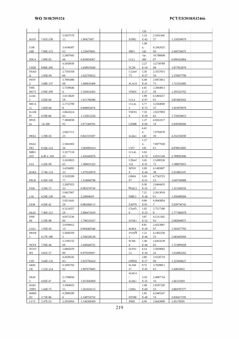
126
5.5037579 1.53 2.1631445BATF 7.65E-238 11 1.90427697 S1PR1 E-42 37 1.159294979
1.08
COR 3.4146587 E- 6.3362025
OIB 7.06E-123 0 2 1.250470601 PRF1 142 69 3.569730675
2.2607945 <le- 10.780699
SDC4 1.89E-82 44 0.829654367 CCL5 300 0 7 6.096162864
4.5050039 2.27 3.2739789
CD28 8.86E-200 5 1.658919166 TC2N E-54 48 1.877812076
THAD 2.7593358 C12orf 5.50 2.3557915
A 1.95E-94 8 6 1.022705012 75 E-27 56 1.376957789
PHTF 3.7095480 6.48 2.94728122 3.08E-137 69 1.400635409 PLAC8 E-41 72 1.723352085
TME 5.7339646 4.45 2.2064813
M173 2.94E-209 6 2.183616303 ITM2C E-27 28 1.295325762
KLRB 2.6114620 1.89 6.54042571 2.92E-84 59 1.011766586 CCL4 E-97 9 1 3.853663502
MICA 2.1712700 CCL4L 5.77 3.2564040
L2 1.83E-66 8 0.848952074 2 E-73 37 1.923970079
SLAM 2.8641614 TSEN5 7.10 2.9227893Fl 4.59E-86 4 2 1.132013256 4 E-29 62 1.735034013
SPOC 7.3804938 1.27 4.9263317
K2 <le-300 9 2 2.971304765 GZMB E-69 19 2.928598206
4.412.0627111 E- 7.0792678
PBX4 1.39E-65 25 0.841335307 KLRK1 140 39 4.254156938
1.27PHAC 2.5941003 E- 7.8977928
TR2 9.50E-114 34 1.063992414 CST7 142 0 1 4.878022605
MBO 3.3577118 CCL4L 5.63
ATI 6.49 E- 104 23 1.432443676 1 E-70 4.4551144 2.789091846
3.2410823 C20orf 3.45 2.5280536
FAS 5.43E-90 65 1.384655322 112 E-31 72 1.588078455
3.4256895 MY01 2.09 4.1495897RORA 3.74E-114 33 1.470520079 F E-44 38 2.633861547
3.5335599 GIMA 5.03 4.7743723
PELIl 6.50E-100 77 1.524060798 P7 E-51 23 3.045760988
2.2697023 9.30 2.0464433TNIK 3.34E-75 32 0.983370734 LRC3 E-25 27 1.315260556
5.0627007 7.21 2.2613018
LTB 4.24E-138 04 2.20604619 S0RL1 E-26 0 1 1.456489584
2.0211641 9.08 6.0943854GEM 4.56E-42 59 0.882068113 ZAP70 E-91 7 3.928736743
6.5124357 C5orf5 1.82 2.7517180
TIGIT 7.46E-212 29 2.896471619 6 E-23 8 1.777460478
DNP 4.0375236 3.87 4.1511302HI 2.19E-88 8 6 1.796331637 SYNE1 E-52 9 2 2.682600875
2.0199412 8.81 2.4223087
UXS1 1.95E-45 13 0.904382548 RORA E-29 9 7 1.565477765
PBXIP 5.5660359 PYHI 1.21 4.14022581 4.17E-189 5 2.594328128 1 E-46 25 2.683405094
2.2185155 SCML 1.40 2.6453228
NCF4 7.94E-46 09 1.040444721 4 E-44 65 1.721895658
HTAT 2.0845470 SLFN1 4.14 2.5059065
IP2 4.81E-37 89 0.979359507 2 L E-30 24 1.634082262
4.2639545 2.80 2.0120719
CD5 3.64E-116 8 3 2.025795414 GPR56 E-17 98 1.325049617
ARID 6.1005792 SLAM 9.72 3.7028811
5 B 1.55E-214 62 2.907675605 F7 E-42 0 3 2.44810822
KLRC4
TRAF 2.1708050 3.18 2.49877543 4.82E-47 54 1.051843069 KLRK1 E-35 16 1.66131693
RAB1 3.1904023 1.68 5.8197329
1FIP1 1.64E-75 5 1 1.561024115 CD8A E-69 55 3.884787277
RHBD 4.2990189 1.81 4.2445547
D2 2.73E-86 6 2.148754716 STOM E-40 74 2.836417195
LY75 2.47E-53 2.2916954 1.146266509 PIM1 6.06 3.6643899 2.45178595
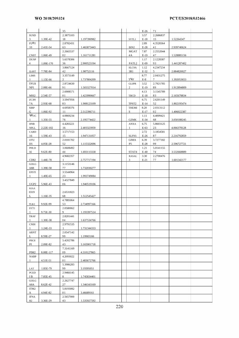
35 E-26 71
SUSD 2.3875103 3.57 2.26868373 1.39E-42 18 1.197390982 SYTL1 E-18 15 1.52264347
P2 Y 2.8782431 2.89 4.3528364
10 2.41E-54 63 1.465873443 BIN2 E-28 4 1 2.939740624
2.2665537 MGAT 7.87 2.2552044CNST 1.06E-49 02 1.161711281 4A E-19 47 1.528885156
DUSP 5.6578306 1.17 2.1228387
4 1.09E-170 36 2.900523194 PATL2 E-09 93 1.441287402
3.8667124 SLC9A 1.12 4.2347234IL6ST 7.78E-84 42 1.98752116 3R1 E-32 5 2.884820027
LIMS 3.3573149 8.77 2.0431275
1 1.15E-66 2 1.737804209 T F E-ll 7 1.392053653
TP53I 2.8724630 GLIPR 3.52 2.7921785NP1 2.08E-66 9 1 1.503327014 2 E-19 89 1.912894809
2.6908571 4.13 3.1545789
MSI2 2.54E-57 0 1 1.423990667 TBCD E-18 83 2.165678834
ZC3H 3.5947056 6.75 2.6201149
7A 2.93E-68 83 1.908123109 TPST2 E-14 52 1.802195474
SIRP 4.6958376 THEMI 8.20 2.0313112
G 1.06E-92 11 2.512396711 S E-17 02 1.406022387
3C 4.0869234 1.11 4.4009421
1 1.35E-55 76 2.195774422 GZMK E-34 68 3.050188245
HNR 4.4283221 ANXA 6.75 5.8603125
NPLL 2.22E-102 16 2.403323959 1 E-63 23 4.066378128
CARD 3.5717153 2.72 3.1854581
16 1.59E-43 15 1.947131057 SLFN5 E-26 87 2.216792859
OTU 2.0181702 GIMA 6.39 3.7277182D5 4.05E-28 52 1.115322696 P5 E-28 99 2.596727721
PHLD 3.0608492 1.21 5.0141155
Al 9.62E-80 44 1.693115558 STAT4 E-40 74 3.532668889
4.9682357 RASAL 3.30 2.2700476CD82 1.44E-78 1 2.757717194 3 E-21 77 1.601342177
GOLG 3.1153146
A8B 1.39E-58 77 1.732030277
EPSTI 3.55449641 1.40E-65 23 1.993749084
3.4327849
UGP2 5.96E-43 06 1.940519106
KIAA
0319 2.6531823
L 1.16E-35 68 1.512545427
4.7885064TLK1 9.92E-99 53 2.734997166
SYT1 2.0580062
1 9.75E-30 9 1.193397514
TRAF 2.82014411 1.30E-38 04 1.637534766
CNIH 2.9791535
1 1.24E-33 1 1.731344333
ARNT 2.0547142L 8.59E-27 99 1.19965166
PIK3I 5.4202786
PI 2.09E-82 43 3.165901718
7.3141169PIM2 8.08E-117 09 4.310127865
NABP 4.2093022
1 4.51E-51 0 1 2.483672766
5.3986283
LAT 1.85E-79 99 3.19395051
PCED 2.9466145
I B 7.85E-45 8 1.745834401
GOLG 2.2627747
A8A 8.62E-42 27 1.346343169
ITM2 5.8193082
A 4.94E-82 0 1 3.46689161
IFNA 2.5657000
R2 3.36E-29 43 1.535927592
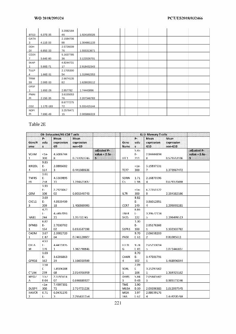
3.2082184
BTG3 6.37E-35 46 1.924165026
GATA 2.1584706
3 4.11E-33 98 1.304861133
DDH 2.5726039
Dl 6.85E-33 76 1.555313671
CD24 5.1637785
7 5.64E-80 36 3.122026701
SKAP 4.8244731
1 5.90E-71 17 2.918432343
TULP 2.1705300
4 1.66E-31 54 1.319962353
TRIM 2.6674135
59 2.00E-33 82 1.628028112
GRSF
1 1.65E-26 2.857782 1.74443896
PMAI 3.6155053
PI 2.15E-35 76 2.207346783
8.6777275
CD2 1.17E-183 72 5.331415144
NDFI 3.2576471
PI 7.69E-49 15 2.005866319
Table 2E
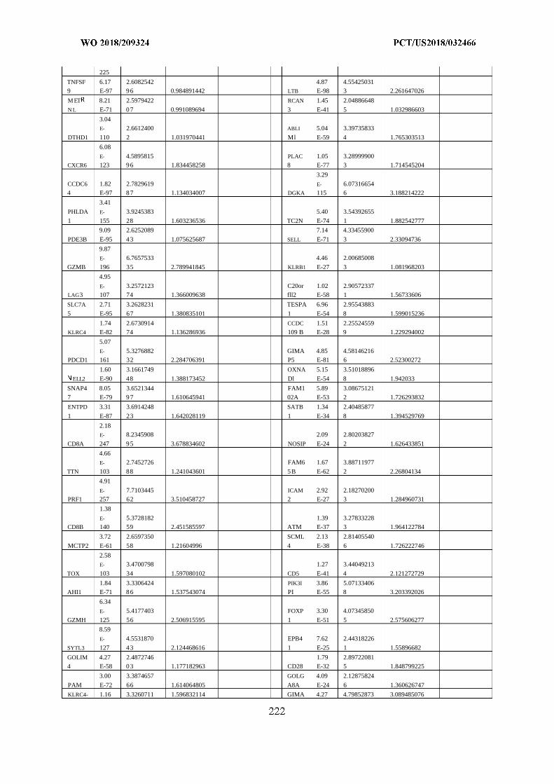
225
TNFSF 6.17 2.6082542 4.87 4.554250319 E-97 96 0.984891442 LTB E-98 3 2.261647026
M ET 8.21 2.5979422 RCAN 1.45 2.04886648
N L E-71 07 0.991089694 3 E-41 5 1.032986603
3.04E- 2.6612400 ABLI 5.04 3.39735833
DTHD1 110 2 1.031970441 Ml E-59 4 1.765303513
6.08
E- 4.5895815 PLAC 1.05 3.28999900CXCR6 123 96 1.834458258 8 E-77 3 1.714545204
3.29
CCDC6 1.82 2.7829619 E- 6.073166544 E-97 87 1.134034007 DGKA 115 6 3.188214222
3.41
PHLDA E- 3.9245383 5.40 3.54392655
1 155 28 1.603236536 TC2N E-74 1 1.882542777
9.09 2.6252089 7.14 4.33455900
PDE3B E-95 43 1.075625687 SELL E-71 3 2.33094736
9.87
E- 6.7657533 4.46 2.00685008
GZMB 196 35 2.789941845 KLRB1 E-27 3 1.081968203
4.95
E- 3.2572123 C20or 1.02 2.90572337LAG3 107 74 1.366009638 fll2 E-58 1 1.56733606
SLC7A 2.71 3.2628231 TESPA 6.96 2.95543883
5 E-95 67 1.380835101 1 E-54 8 1.599015236
1.74 2.6730914 CCDC 1.51 2.25524559KLRC4 E-82 74 1.136286936 109 B E-28 9 1.229294002
5.07
E- 5.3276882 GIMA 4.85 4.58146216PDCD1 161 32 2.284706391 P5 E-81 6 2.52300272
1.60 3.1661749 OXNA 5.15 3.51018896
ELL2 E-90 48 1.388173452 Dl E-54 8 1.942033
SNAP4 8.05 3.6521344 FAM1 5.89 3.086751217 E-79 97 1.610645941 02A E-53 2 1.726293832
ENTPD 3.31 3.6914248 SATB 1.34 2.40485877
1 E-87 23 1.642028119 1 E-34 8 1.394529769
2.18
E- 8.2345908 2.09 2.80203827
CD8A 247 95 3.678834602 NOSIP E-24 2 1.626433851
4.66
E- 2.7452726 FAM6 1.67 3.88711977
TTN 103 88 1.241043601 5 B E-62 2 2.26804134
4.91
E- 7.7103445 ICAM 2.92 2.18270200PRF1 257 62 3.510458727 2 E-27 3 1.284960731
1.38
E- 5.3728182 1.39 3.27833228CD8B 140 59 2.451585597 ATM E-37 3 1.964122784
3.72 2.6597350 SCML 2.13 2.81405540
MCTP2 E-61 58 1.21604996 4 E-38 6 1.726222746
2.58
E- 3.4700798 1.27 3.44049213
TOX 103 34 1.597080102 CD5 E-41 4 2.121272729
1.84 3.3306424 PIK3I 3.86 5.07133406AHI1 E-71 86 1.537543074 PI E-55 8 3.203392026
6.34
E- 5.4177403 FOXP 3.30 4.07345850
GZMH 125 56 2.506915595 1 E-51 5 2.575606277
8.59
E- 4.5531870 EPB4 7.62 2.44318226
SYTL3 127 43 2.124468616 1 E-25 1 1.55896682
GOLIM 4.27 2.4872746 1.79 2.897220814 E-58 03 1.177182963 CD28 E-32 5 1.848799225
3.00 3.3874657 GOLG 4.09 2.12875824
PAM E-72 66 1.614064805 A8A E-24 6 1.360626747
KLRC4- 1.16 3.3260711 1.596832114 GIMA 4.27 4.79852873 3.089485076
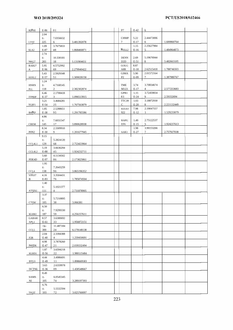
KL Kl E-86 0 1 P7 E-42 6
2.04E- 7.0356632 CHMP 5.22 2.44473006
LYST 225 6 3.481392078 7 E-17 6 1.600860764
1.09 3.7675833 1.15 2.25627984
SLA2 E-87 48 1.868466871 ELL2 E-16 5 1.484964873
2.74
E- 10.330181 DENN 2.69 5.19676944
NKG7 283 18 5.131904651 D2D E-51 8 3.482663105
RAB27 5.95 4.5752992 GOLG 8.87
A E-96 69 2.279940422 A8B E-18 2.62515418 1.788746503
5.43 2.5929348 GIMA 5.90 2.01572164
ASXL2 E-57 51 1.309028158 P2 E-09 7 1.387980767
1.24
HNRN E- 4.7168145 TME 3.74 3.70834674
PLL 118 2 2.382302874 M123 E-17 4 2.577253683
6.46 2.1708418 GPR1 1.15 3.72459834
ITPRIP E-37 3 1.096513915 83 E-24 9 2.59332694
3.21 3.4684281 TTC39 1.03 3.18872930
TGIF1 E-54 25 1.767563879 C E-20 8 2.221132449
1.85 2.5288651 KIAAO 7.98 2.19047557
BA P E-38 95 1.291785586 922 E-12 1 1.529233079
4.86
E- 7.6031547 RAPG 1.49 2.75122537
CREM 145 17 3.890628938 EF6 E-15 5 1.924237613
8.34 2.3309910 1.98 3.90153266
P0N2 E-30 9 1.201677945 AAK1 E-27 7 2.757927038
9.15E- 5.2824120
CCL4L1 129 68 2.753423964
5.19 3.6636294CCL4L2 E-88 45 1.924232711
5.60 4.1134502
PDE4D E-97 86 2.173825861
1.95E- 7.2643259
CCL4 138 96 3.865196352
VPS37 4.16 3.3504431
B E-83 76 1.785074504
1.40
E- 5.1021377
ATXN1 111 4 2.731878905
3.37
E- 5.7210895
CTSW 105 38 3.066381
6.50
E- 7.9299330
KLRKl 187 59 4.256157611
GABAR 8.57 3.6380692
APL1 E-61 33 1.956872151
<le- 11.487184
CCL5 300 24 6.179148138
2.04 2.3394388
F2R E-40 4 1.259450691
4.98 3.7878260
JMJD6 E-47 21 2.039332494
1.87 3.6594218KLRD1 E-56 22 1.980115484
4.44 3.4986691
BTG3 E-49 11 1.898669583
3.63 2.6359978DCTN6 E-36 09 1.430548667
8.49
SAMS E- 6.0545345
Nl 105 74 3.289197393
6.76
E- 5.5532594
TIGIT 103 72 3.025760097
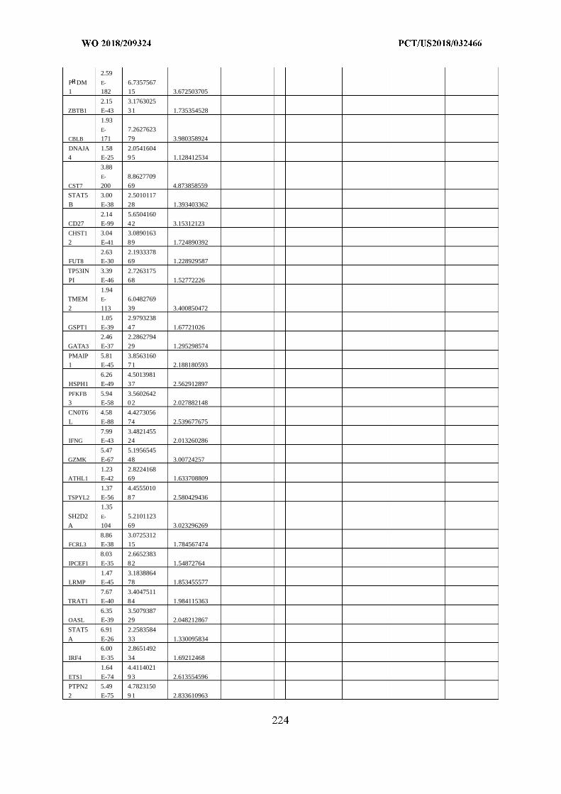
2.59
P DM E- 6.73575671 182 15 3.672503705
2.15 3.1763025
ZBTB1 E-43 31 1.735354528
1.93
E- 7.2627623
CBLB 171 79 3.980358924
DNAJA 1.58 2.05416044 E-25 95 1.128412534
3.88
E- 8.8627709
CST7 200 69 4.873858559
STAT5 3.00 2.5010117
B E-38 28 1.393403362
2.14 5.6504160
CD27 E-99 42 3.15312123
CHST1 3.04 3.0890163
2 E-41 89 1.724890392
2.63 2.1933378FUT8 E-30 69 1.228929587
TP53IN 3.39 2.7263175
PI E-46 68 1.52772226
1.94TMEM E- 6.0482769
2 113 39 3.400850472
1.05 2.9793238
GSPT1 E-39 47 1.67721026
2.46 2.2862794
GATA3 E-37 29 1.295298574
PMAIP 5.81 3.85631601 E-45 71 2.188180593
6.26 4.5013981
HSPH1 E-49 37 2.562912897
PFKFB 5.94 3.56026423 E-58 02 2.027882148
CN0T6 4.58 4.4273056
L E-88 74 2.539677675
7.99 3.4821455IFNG E-43 24 2.013260286
5.47 5.1956545
GZMK E-67 48 3.00724257
1.23 2.8224168ATHL1 E-42 69 1.633708809
1.37 4.4555010
TSPYL2 E-56 87 2.580429436
1.35
SH2D2 E- 5.2101123
A 104 69 3.023296269
8.86 3.0725312FCRL3 E-38 15 1.784567474
8.03 2.6652383
IPCEF1 E-35 82 1.54872764
1.47 3.1838864LRMP E-45 78 1.853455577
7.67 3.4047511
TRAT1 E-40 84 1.984115363
6.35 3.5079387OASL E-39 29 2.048212867
STAT5 6.91 2.2583584
A E-26 33 1.330095834
6.00 2.8651492
IRF4 E-35 34 1.69212468
1.64 4.4114021
ETS1 E-74 93 2.613554596
PTPN2 5.49 4.7823150
2 E-75 9 1 2.833610963
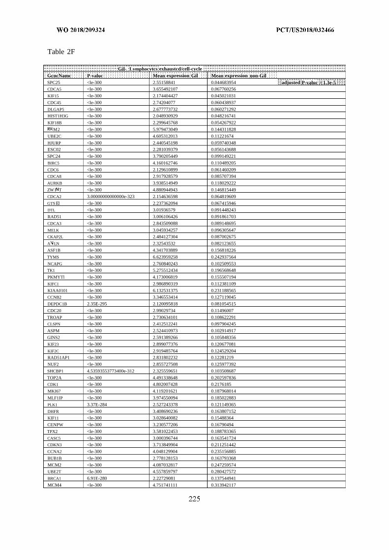
Table 2F
Gil- Lymphocytcs exhaustcd/cell-cycleGcncNamc P-valuc Mean expression Gil Mean expression non-GilSPC25 <le-300 2.55158841 0.044683954 adjusted P-valuc = 1.3e-5CDCA5 <le-300 3.655492107 0.067760256
KIF15 <le-300 2.174404427 0.045021031
CDC45 <le-300 2.74204077 0.060438937
DLGAP5 <le-300 2.677773732 0.060271292
HIST1H3G <le-300 2.048930929 0.048216741
KIF18B <le-300 2.299645768 0.054267922
M2 <le-300 5.979473049 0.144311828
UBE2C <le-300 4.605312013 0.11221674
HJURP <le-300 2.440545198 0.059740348
ESC02 <le-300 2.281039379 0.056143688
SPC24 <le-300 3.790205449 0.099149221
BIRC5 <le-300 4.160162746 0.110489205
CDC6 <le-300 2.129610899 0.061460209
CDCA8 <le-300 2.917928579 0.085707394
AURKB <le-300 3.938514949 0.118029222
ZW I T <le-300 4.880944943 0.146815449
CDCA2 3.00000000000000e-323 2.154636598 0.064819609
GTS El <le-300 2.237362094 0.067415946
DTL <le-300 3.01936579 0.091448243
RAD51 <le-300 3.006106426 0.091861703
CDCA3 <le-300 2.843509088 0.089148695
MELK <le-300 3.045934257 0.096305647
CKAP2L <le-300 2.484127304 0.087002675
A LN <le-300 2.32543532 0.082123655
ASF1B <le-300 4.341703889 0.156818226
TYMS <le-300 6.623959258 0.242937564
NCAPG <le-300 2.760840243 0.102509553
TK1 <le-300 5.275512434 0.196568648
PKMYTl <le-300 4.173006819 0.155507194
KIFC1 <le-300 2.986890319 0.112381109
KIAA0101 <le-300 6.132531375 0.231188565
CCNB2 <le-300 3.346553414 0.127119045
DEPDC1B 2.35E-295 2.120095818 0.081054515
CDC20 <le-300 2.99029734 0.11496007
TROAP <le-300 2.730634101 0.108622291
CLSPN <le-300 2.412512241 0.097904245
ASPM <le-300 2.524410973 0.102914917
GINS2 <le-300 2.591389266 0.105848356
KIF23 <le-300 2.899077376 0.120677081
KIF2C <le-300 2.919485764 0.124529204
RAD51AP1 <le-300 2.831802232 0.12281219
NUF2 <le-300 2.855727508 0.125977392
SHCBP1 4.53593553773400e-312 2.325559651 0.103508687
TOP2A <le-300 4.491338648 0.202597836
CDK1 <le-300 4.802007428 0.2176185
MKI67 <le-300 4.119201621 0.187968014
MLF1IP <le-300 3.974550094 0.185022883
PLK1 3.37E-284 2.527243378 0.121149365
DHFR <le-300 3.408690236 0.163807152
KIF11 <le-300 3.028640082 0.15488364
CENPW <le-300 3.230577206 0.16790494
TPX2 <le-300 3.581022453 0.188783365
CASC5 <le-300 3.000396744 0.163541724
CDKN3 <le-300 3.713849904 0.211251442
CCNA2 <le-300 4.048129904 0.235156885
BUB1B <le-300 2.778128153 0.163793368
MCM2 <le-300 4.087032817 0.247259574
UBE2T <le-300 4.557859797 0.280427572
BRCA1 6.91E-280 2.22729081 0.137544941
MCM4 <le-300 4.751741111 0.313942117
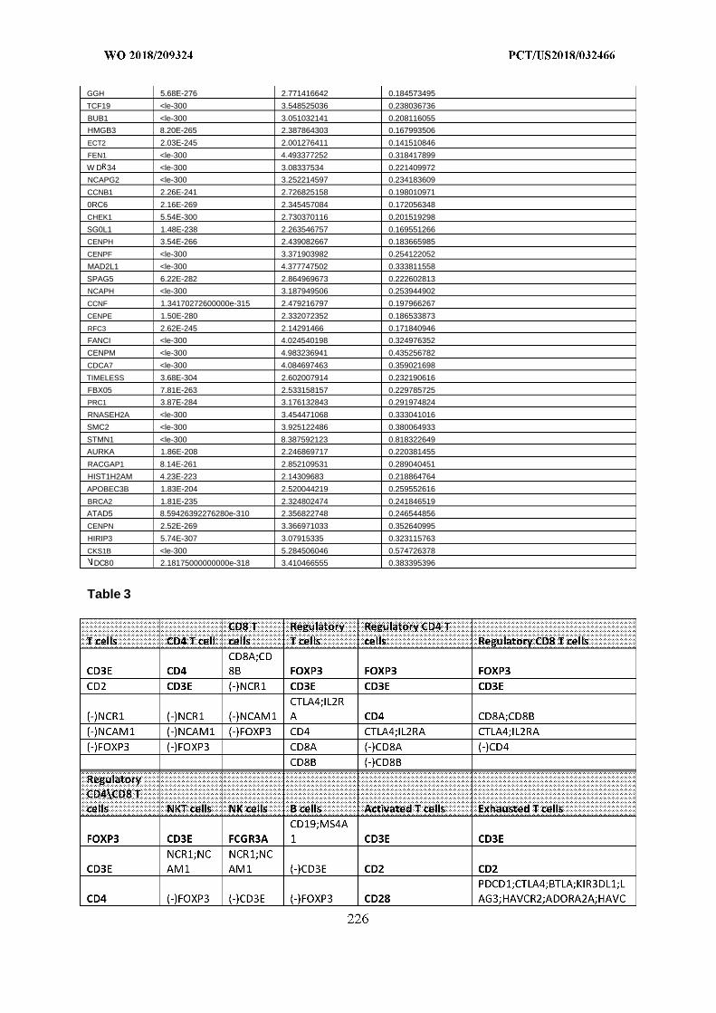
GGH 5.68E-276 2.771416642 0.184573495
TCF19 <le-300 3.548525036 0.238036736
BUB1 <le-300 3.051032141 0.208116055
HMGB3 8.20E-265 2.387864303 0.167993506
ECT2 2.03E-245 2.001276411 0.141510846
FEN1 <le-300 4.493377252 0.318417899
W D 34 <le-300 3.08337534 0.221409972
NCAPG2 <le-300 3.252214597 0.234183609
CCNB1 2.26E-241 2.726825158 0.198010971
0RC6 2.16E-269 2.345457084 0.172056348
CHEK1 5.54E-300 2.730370116 0.201519298
SG0L1 1.48E-238 2.263546757 0.169551266
CENPH 3.54E-266 2.439082667 0.183665985
CENPF <le-300 3.371903982 0.254122052
MAD2L1 <le-300 4.377747502 0.333811558
SPAG5 6.22E-282 2.864969673 0.222602813
NCAPH <le-300 3.187949506 0.253944902
CCNF 1.34170272600000e-315 2.479216797 0.197966267
CENPE 1.50E-280 2.332072352 0.186533873
RFC3 2.62E-245 2.14291466 0.171840946
FANCI <le-300 4.024540198 0.324976352
CENPM <le-300 4.983236941 0.435256782
CDCA7 <le-300 4.084697463 0.359021698
TIMELESS 3.68E-304 2.602007914 0.232190616
FBX05 7.81E-263 2.533158157 0.229785725
PRC1 3.87E-284 3.176132843 0.291974824
RNASEH2A <le-300 3.454471068 0.333041016
SMC2 <le-300 3.925122486 0.380064933
STMN1 <le-300 8.387592123 0.818322649
AURKA 1.86E-208 2.246869717 0.220381455
RACGAP1 8.14E-261 2.852109531 0.289040451
HIST1H2AM 4.23E-223 2.14309683 0.218864764
APOBEC3B 1.83E-204 2.520044219 0.259552616
BRCA2 1.81E-235 2.324802474 0.241846519
ATAD5 8.59426392276280e-310 2.356822748 0.246544856
CENPN 2.52E-269 3.366971033 0.352640995
HIRIP3 5.74E-307 3.07915335 0.323115763
CKS1B <le-300 5.284506046 0.574726378
DC80 2.18175000000000e-318 3.410466555 0.383395396
Table 3
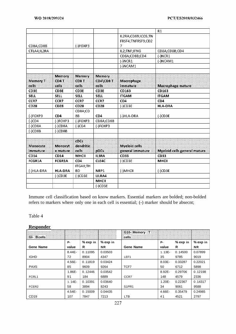
Immune cell classification based on know markers. Essential markers are bolded; non-boldedrefers to markers where only one in each cell is essential; (-) marker should be absecnt;
Table 4
ResponderG10- Memory T
Gl- B cells cells
P- % exp in % exp in P- % exp in % exp in
Gene Name value R NR Gene Name value R NR
8.44E- 0 .11095 0.03503 1.13E- 0 .14500 0.07899
IGHD 72 8904 4347 LEF1 35 9785 9019
4.56E- 0 .11819 0.03424 8.03E- 0.33287 0.22021
PAX5 85 9609 9264 TCF7 50 6712 5898
1.86E- 0 .12446 0.03542 8.92E- 0.29706 0 .12198
FCRL1 91 184 6889 CCR7 148 4579 2336
1.14E- 0 .10391 0.03640 1.20E- 0.22367 0 .14317
FCER2 58 3894 8243 S1PR1 34 9061 9588
4.54E- 0 .15009 0.04435 4.66E- 0.35479 0.24985
CD19 107 7847 7213 LTB 4 1 4521 2797
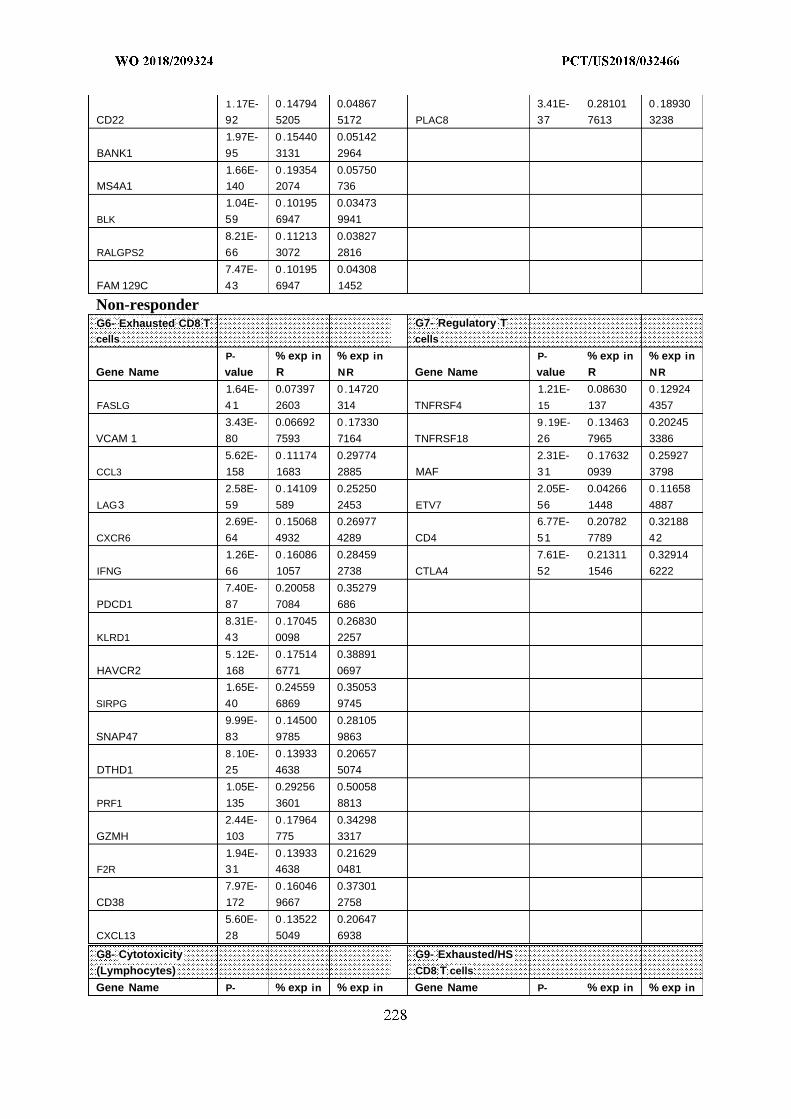
1.17E- 0 .14794 0.04867 3.41E- 0.28101 0 .18930
CD22 92 5205 5172 PLAC8 37 7613 3238
1.97E- 0 .15440 0.05142
BANK1 95 3131 2964
1.66E- 0 .19354 0.05750
MS4A1 140 2074 736
1.04E- 0 .10195 0.03473
BLK 59 6947 9941
8.21E- 0 .11213 0.03827
RALGPS2 66 3072 2816
7.47E- 0 .10195 0.04308
FAM 129C 43 6947 1452
Non-responderG6- Exhausted CD8 T G7- Regulatory Tcells cells
P- % exp in % exp in P- % exp in % exp in
Gene Name value R NR Gene Name value R NR
1.64E- 0.07397 0 .14720 1.21E- 0.08630 0 .12924
FASLG 4 1 2603 314 TNFRSF4 15 137 4357
3.43E- 0.06692 0 .17330 9.19E- 0 .13463 0.20245
VCAM 1 80 7593 7164 TNFRSF18 26 7965 3386
5.62E- 0 .11174 0.29774 2.31E- 0 .17632 0.25927
CCL3 158 1683 2885 MAF 31 0939 3798
2.58E- 0 .14109 0.25250 2.05E- 0.04266 0 .11658
LAG3 59 589 2453 ETV7 56 1448 4887
2.69E- 0 .15068 0.26977 6.77E- 0.20782 0.32188
CXCR6 64 4932 4289 CD4 51 7789 42
1.26E- 0 .16086 0.28459 7.61E- 0.21311 0.32914
IFNG 66 1057 2738 CTLA4 52 1546 6222
7.40E- 0.20058 0.35279
PDCD1 87 7084 686
8.31E- 0 .17045 0.26830
KLRD1 43 0098 2257
5.12E- 0 .17514 0.38891
HAVCR2 168 6771 0697
1.65E- 0.24559 0.35053
SIRPG 40 6869 9745
9.99E- 0 .14500 0.28105
SNAP47 83 9785 9863
8.10E- 0 .13933 0.20657
DTHD1 25 4638 5074
1.05E- 0.29256 0.50058
PRF1 135 3601 8813
2.44E- 0 .17964 0.34298
GZMH 103 775 3317
1.94E- 0 .13933 0.21629
F2R 31 4638 0481
7.97E- 0 .16046 0.37301
CD38 172 9667 2758
5.60E- 0 .13522 0.20647
CXCL13 28 5049 6938
G8- Cytotoxicity G9- Exhausted/HS(Lymphocytes) CD8 T cells
Gene Name P- % exp in % exp in Gene Name P- % exp in % exp in
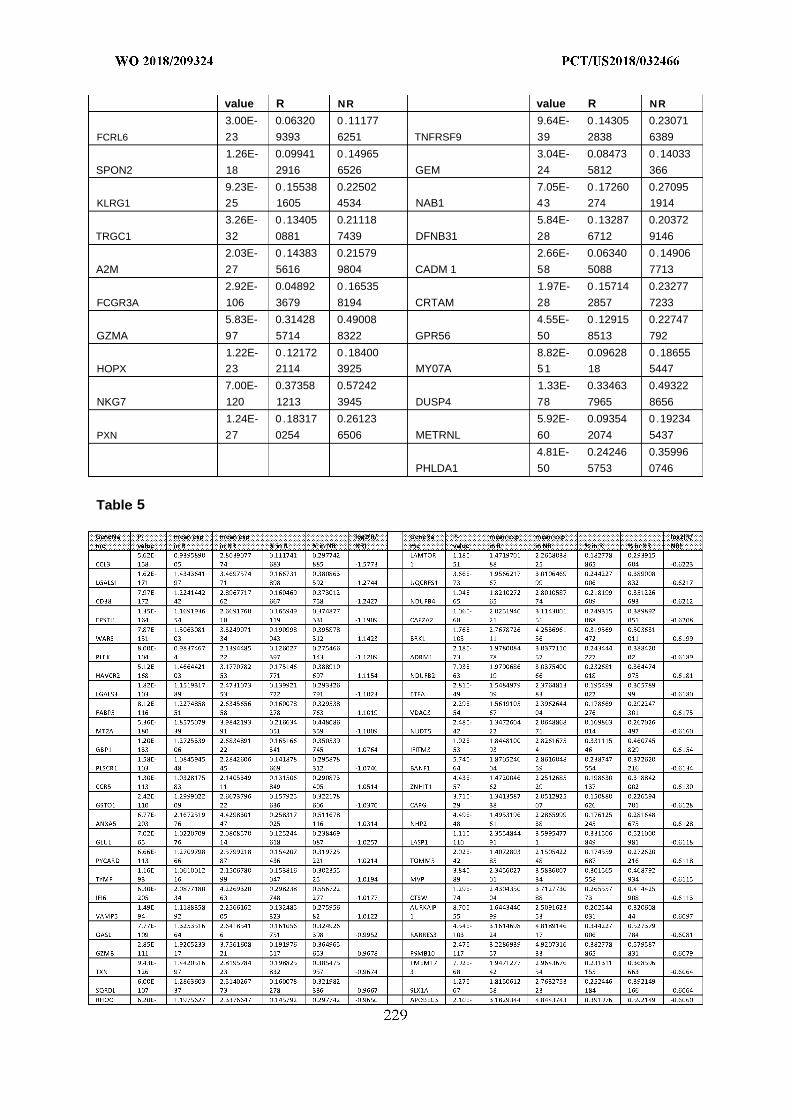
value R NR value R NR
3.00E- 0.06320 0 .11177 9.64E- 0 .14305 0.23071
FCRL6 23 9393 6251 TNFRSF9 39 2838 6389
1.26E- 0.09941 0 .14965 3.04E- 0.08473 0 .14033
SPON2 18 2916 6526 GEM 24 5812 366
9.23E- 0 .15538 0.22502 7.05E- 0 .17260 0.27095
KLRG1 25 1605 4534 NAB1 43 274 1914
3.26E- 0 .13405 0.21118 5.84E- 0 .13287 0.20372
TRGC1 32 0881 7439 DFNB31 28 6712 9146
2.03E- 0 .14383 0.21579 2.66E- 0.06340 0 .14906
A2M 27 5616 9804 CADM 1 58 5088 7713
2.92E- 0.04892 0 .16535 1.97E- 0 .15714 0.23277
FCGR3A 106 3679 8194 CRTAM 28 2857 7233
5.83E- 0.31428 0.49008 4.55E- 0 .12915 0.22747
GZMA 97 5714 8322 GPR56 50 8513 792
1.22E- 0 .12172 0 .18400 8.82E- 0.09628 0 .18655
HOPX 23 2114 3925 MY07A 51 18 5447
7.00E- 0.37358 0.57242 1.33E- 0.33463 0.49322
NKG7 120 1213 3945 DUSP4 78 7965 8656
1.24E- 0 .18317 0.26123 5.92E- 0.09354 0 .19234
PXN 27 0254 6506 METRNL 60 2074 5437
4.81E- 0.24246 0.35996
PHLDA1 50 5753 0746
Table 5
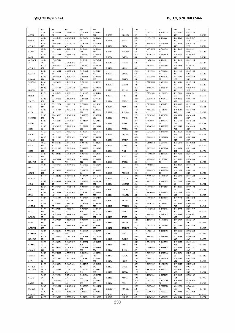
230
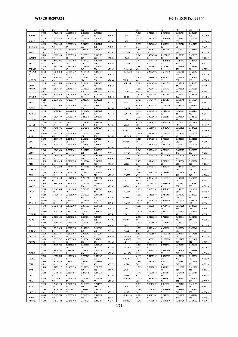
232
233
Table 6
HLA-DMA 2.9SE-144 4.115547348 1.607931363
STRA13 1.95E-63 2.095400445 0.820913992
HSD17B10 6.03E-69 2.108135203 0.828597124
VAMP5 1.36E-S7 2.727461347 1.081471049
NDUFAB1 1.62E-72 2.286416107 0.907988058
BATF 1.20E-106 3.284547348 1.305467896
NDUFS2 2.46E-6S 2.191532071 0.873496229
C17orf49 S.32E-134 4.085892692 1.629745209
GNG5 1.61E-151 4.397466903 1.757469193
PSMB2 3.17E-117 2.884465746 1.155182139
PDIA6 1.40E-148 3.943893622 1.579997884
C0MMD3 6.05E-60 2.035170674 0.8192485
CD63 4.0SE-144 4.568304134 1.847290214
PSMA4 3.50E-118 3.826465017 1.551917638
SAE1 2.67E-71 2.324560064 0.943978493
ATP5J 1.44E-104 3.193882543 1.29728302
MEA1 1.79E-73 2.330314867 0.949658623
EX0SC9 3.63E-69 2.020491198 0.824607562
ARPC5L 1.04E-111 3.068312322 1.253247292
BL0C1S1 5.50E-102 3.225259843 1.318190273
HELLS 4.5SE-76 2.130695664 0.871697169
CXCR6 5.36E-126 4.305209437 1.774354247
BCAS4 6.50E-65 2.098722766 0.865111332
ETFB 6.7SE-S1 2.418481829 0.997801189
TXN2 1.99E-66 2.306400284 0.953598266
PTPN6 3.30E-156 5.015528117 2.077075152
SIT1 1.00E-144 4.332124739 1.796590741
FKBP1A 1.29E-227 5.929887981 2.462211689
COPZ1 1.12E-107 3.528959222 1.466159688
HLA-DRA 1.57E-195 6.430869882 2.672266924
CDC123 S.06E-66 2.391351443 0.996238928
AP2S1 3.62E-S2 2.615042085 1.089662717
FUTS 7.70E-SS 2.38753102 0.995229203
BST2 3.S0E-13S 4.205209714 1.757935193
ATP6V1E1 7.69E-66 2.184741648 0.916464286
CD2BP2 2.25E-131 3.693163626 1.549681009
HLA-DQA1 6.24E-104 3.237470161 1.360566264
ZCRB1 1.20E-61 2.010211694 0.845742982
MX1 1.00E-75 2.837779179 1.194997598
TNFRSF9 9.6SE-106 3.467676923 1.462547382
SQRDL 6.22E-79 2.622852717 1.106783424
SERPINB1 2.57E-110 3.515777369 1.483727648
PHPT1 1.4SE-65 2.194049577 0.926881074
CALM3 3.56E-192 5.170473018 2.185344461
TOX 9.95E-15S 3.545285595 1.49868579
SNRPC 1.47E-70 2.474584608 1.046413984
MRPS34 4.95E-S4 2.768976948 1.175826428
NUTF2 2.74E-S2 2.608754415 1.108160937
NDUFS6 2.S5E-72 2.035602108 0.864947239
PSMB3 1.75E-15S 4.904107876 2.08491137
CHMP2A 4.S4E-S1 2.857076253 1.222009155
SLC25A11 6.27E-61 2.027829326 0.867613358
SHFM1 9.64E-130 3.777245159 1.616846165
TMEM179B 2.42E-76 2.767358685 1.18524941
EIF6 2.20E-72 2.502067278 1.074616725
ANXA5 5.S4E-162 5.223830407 2.244806053
JAKMIP1 1.03E-71 2.312789337 0.994466875
TALDOl 9.37E-S9 3.134857922 1.349123107
GLRX3 6.4SE-57 2.089535894 0.90076918
ANAPC11 1.04E-S0 2.8896458 1.252200712
DUT 4.99E-53 2.104622655 0.913261117
PDCD5 9.09E-55 2.041601583 0.885963761
ATP5G3 1.69E-143 4.749757651 2.062994434
CHMP5 1.42E-65 2.186115189 0.950427238
TWF2 1.86E-107 3.526082815 1.536184109
IDH2 4.64E-133 4.501434308 1.965274937
MPG 5.23E-65 2.276372945 0.994200645
SNRPF 7.S0E-SS 2.269196926 0.991701642
NDUFC1 2.30E-53 2.074849616 0.907011779
GBP1 2.07E-117 3.524455833 1.542528045
DCTN3 1.76E-6S 2.617143149 1.145880627
ERH 2.0SE-S0 2.64205175 1.161491201
NDUFA12 7.16E-61 2.312758316 1.017291863
LIMS1 1.13E-64 2.066400504 0.909789851
BANF1 5.56E-92 3.26276779 1.436691138
NDUFC2 S.04E-97 3.456390807 1.527036232
PSMC3 1.79E-73 2.746119429 1.213266782
PON2 2.47E-62 2.403166306 1.062325344
PRDX5 1.39E-125 4.231392267 1.870991009
TMX1 1.34E-101 2.956400489 1.312311214
STOML2 5.02E-61 2.169463241 0.963097654
RPS6KA1 5.43E-62 2.231976177 0.991943879
PAM 1.41E-102 3.194179878 1.420230794
ATP5J2 3.01E-77 2.948363375 1.311670832
GIMAP6 2.57E-9S 2.930366838 1.304224929
NDUFB7 7.S5E-70 2.601941528 1.158242834
DBI 2.97E-96 3.575932852 1.592327842
IFI6 3.3SE-154 5.201445339 2.318825526
TSTA3 5.7SE-63 2.319464423 1.036532055
SSNA1 7.93E-62 2.042647073 0.913112139
ADORA2A 2.25E-55 2.0S016922S 0.930560714
FDPS 2.S3E-62 2.425943S92 1.086244465
CYC1 1.72E-66 2.539964355 1.137807765
PSMD4 3.20E-104 3.66S547931 1.646002485
FAM96A 1.S9E-56 2.081417771 0.934343428
0AS2 5.39E-90 2.S7S625307 1.292503506
ERCC1 1.64E-49 2.0S5642591 0.938003773
PDHB 2.1SE-52 2.055225667 0.926591496
CD27 3.60E-194 6.136699677 2.773576115
SNRPA 6.25E-61 2.067275057 0.935086166
UBE2L3 9.31E-115 3.592S59999 1.625412091
MDH1 9.43E-99 3.721279971 1.683801365
SDHC 2.76E-63 2.347811248 1.064694838
PSMG2 2.76E-64 2.555665014 1.159473162
Cllorf48 2.63E-76 2.890040778 1.313040137
PSMA2 2.43E-15S 4.802864375 2.188108226
C7orf73 2.09E-62 2.160459497 0.984637314
MRPS16 9.40E-56 2.076926967 0.948382985
MCM7 2.41E-42 2.11796775 0.969278006
SN 20 2.36E-59 2.087926298 0.957095188
AK2 1.15E-75 2.611969942 1.197612803
RBBP7 1.21E-69 2.818309884 1.293003788
TIGIT 3.77E-190 5.930720988 2.725798008
TMPO 3.11E-79 2.779105016 1.27759614
CTSB 1.01E-83 3.306011196 1.520579095
PARP1 1.79E-S7 2.902379776 1.33623695
USB1 4.00E-56 2.178707226 1.003328766
MRPS7 1.90E-51 2.0636019 0.951959238
NHP2 2.37E-60 2.464790149 1.137484098
ATP5I 2.14E-S1 3.169807005 1.463809999
PSMC1 6.43E-77 2.849473341 1.317477763
VDAC1 6.12E-S6 3.404397887 1.574958304
CARD16 3.2SE-65 2.589653803 1.198332561
RNF1S1 2.3SE-55 2.235233402 1.035620008
PGAM1 1.06E-178 6.143413578 2.855010843
NT5C 6.96E-49 2.010909629 0.935299453
IRF2 9.60E-101 3.388234685 1.577120507
NUDT22 1.S2E-54 2.244019782 1.045018606
NDUFA9 5.79E-62 2.3334169 1.08808713
SRI 1.91E-100 4.000315311 1.870272516
GBP4 S.65E-92 2.833516253 1.325233526
NDUFSS 2.74E-75 3.092642125 1.446840419
PSMC2 7.23E-49 2.012346972 0.941902901
FPGS 3.10E-62 2.264011843 1.060107164
PLSCR1 4.91E-51 2.23546722 1.048506785
P0LR2G 2.S0E-73 3.048974111 1.430789702
COXSA 6.54E-123 4.967137562 2.331202931
SLX1B 5.75E-73 2.245393336 1.054066173
TRAPPC1 2.17E-124 3.492707062 1.643504305
ABI3 7.7SE-79 3.099603274 1.461146971
CBX5 7.46E-S9 2.590646216 1.221380459
PSMD14 2.33E-56 2.112233684 0.999656109
UBE2L6 1.55E-175 5.968070327 2.832430432
IFNG 1.S1E-123 4.707056597 2.234490186
DECR1 4.66E-62 2.629425264 1.248765457
ITGB1BP1 3.53E-56 2.406344071 1.143163002
AKR1B1 4.64E-62 2.540626941 1.209149866
PSMA5 S.29E-125 4.746837028 2.25955883
NUDT21 2.99E-S0 3.06028903 1.457121678
NDUFAF3 S.SSE-55 2.383919436 1.137322319
MITD1 3.07E-47 2.009722381 0.960963898
NUDT5 l.SSE-56 2.434893144 1.165636277
SNRPD3 6.21E-74 2.92881676 1.403249077
TMEM25S 3.10E-72 2.970486925 1.424616179
UQCRC1 7.64E-S2 3.645888901 1.749594365
TRIM59 S.92E-S2 2.643379535 1.268587986
DTX3L 1.69E-79 2.33543546 1.121027282
PHB 2.03E-54 2.272531231 1.092024482
VOPP1 6.19E-77 2.780981344 1.336514767
GTF2A2 6.39E-4S 2.25475435 1.085682726
PSMB10 1.16E-171 6.030269925 2.906188687
WDRS30S 3.57E-S6 3.557054354 1.715109338
NDUFB10 1.43E-59 2.688975493 1.296723088
YARS 4.21E-92 3.297927977 1.591534117
AIP 1.21E-102 4.122714607 1.990604407
CHST12 5.59E-99 3.595802492 1.736806178
DCTN2 1.59E-60 2.576256487 1.244453561
ATP5F1 5.24E-S4 3.962567401 1.921573174
SMC3 5.S4E-72 2.682357487 1.302035445
CISC 2.24E-115 4.560963578 2.216349374
SAMD9L 3.25E-66 2.495152105 1.213261926
PEF1 2.74E-44 2.106208751 1.024666417
H2AFY 4.36E-67 2.366480968 1.151785138
C9orfl6 2.77E-97 3.649773462 1.776991219
SEC11A 2.43E-74 3.183121169 1.550593035
SF3B14 2.53E-56 2.470585546 1.203633519
SNX17 S.57E-S1 3.340407S42 1.630944085
SLX1A 9.65E-94 3.397692612 1.659810557
CNP 3.3SE-51 2.010494426 0.98281337
PIN1 9.51E-75 3.030442S03 1.48319666
CNIH1 6.20E-4S 2.222542426 1.090375464
AL353354.2 9.9SE-63 2.61S432506 1.286402608
T0MM5 3.00E-55 2.536152718 1.246669896
RAB27A 2.13E-123 4.687421809 2.304364209
PSMDS 9.26E-117 4.575753315 2.256456493
NDUFS7 1.21E-S6 3.590142368 1.771365198
GSDMD 1.S9E-75 3.363352126 1.660918068
SEC61G 5.37E-55 2.522814855 1.246782876
ETFA 2.63E-56 2.651933137 1.311401693
AP0BEC3D 5.34E-130 3.380245529 1.671679024
GSTP1 5.11E-113 4.912488909 2.430082491
FDFT1 5.14E-62 2.704718169 1.338918193
LAMT0R1 3.10E-49 2.16882921 1.075600567
LGALS1 5.S5E-64 3.357009584 1.664933266
LAG3 7.60E-111 3.541064229 1.757280767
ACTR1A 3.40E-65 2.510683291 1.245955297
DYNLRB1 1.92E-65 2.957742724 1.468856794
PPP1CC 3.64E-52 2.203796011 1.094519379
GYG1 5.40E-45 2.230119979 1.108318616
BRK1 2.2SE-116 4.649300583 2.312956199
PHB2 1.97E-71 3.202588722 1.594157768
SFT2D1 3.01E-51 2.34543133 1.167512921
PLEKHJ1 9.S4E-52 2.354150689 1.174696649
PUF60 4.55E-S5 3.561505558 1.777203021
FCRL3 1.64E-73 3.174160935 1.586070711
SARNP 5.01E-61 2.731949001 1.365444301
HK1 1.S4E-62 2.457208967 1.228174703
CTLA4 2.91E-99 3.690690102 1.845201044
MDH2 1.S6E-SS 3.481165243 1.741079456
ATP5H 1.41E-62 2.92556102 1.463512768
BAK1 1.44E-57 2.406565266 1.20447788
PCMT1 1.S3E-66 2.945615435 1.474281001
TRIM69 2.32E-49 2.077449634 1.040347476
G0RASP2 1.20E-4S 2.164740148 1.085770361
PPM1M 1.0SE-59 2.236488966 1.122173655
RABSA 1.66E-S2 3.273781635 1.643043985
AURKAIP1 5.64E-65 2.88947544 1.450645103
ATP5C1 1.41E-106 4.552287027 2.285753391
T N 2.64E-69 2.769173937 1.392219502
SNRNP40 3.25E-47 2.257709257 1.135424061
M6PR 1.07E-103 4.274687856 2.150113046
C12orf75 3.3SE-60 2.676137792 1.346789383
RAB11A 4.43E-51 2.302870226 1.160980005
TMCOl 5.19E-70 3.2016094 1.615421544
NME1-NME2 1.16E-S4 3.973214185 2.005844343
OSTC 3.S6E-51 2.420274514 1.222245854
PARP9 1.54E-91 3.484009902 1.761121297
ELOVL1 4.25E-50 2.330492905 1.179464783
NEDDS 1.07E-S5 3.862897554 1.956859328
MT2A 6.11E-105 4.731740516 2.397041493
TKT 2.12E-52 2.34031621 1.185718352
CDK2AP2 2.99E-106 4.37439044 2.217013994
HLA-DQB1 6.95E-S1 3.631009793 1.842553206
TXLNA 4.S4E-76 2.517656577 1.277762337
PPP1R7 2.S0E-41 2.08959601 1.060776221
PPM1G 3.17E-74 3.137917906 1.593139835
GBP5 2.61E-152 5.386152272 2.735263811
ARPC5 7.91E-140 5.353269289 2.718813537
SDHB 1.15E-45 2.398175813 1.219644736
EIF2S1 7.S2E-45 2.351574855 1.196180759
KDELR2 6.33E-62 2.459379931 1.251223349
NDUFA2 2.96E-47 2.074992277 1.056426235
FIS1 2.S7E-55 2.583194526 1.315724451
HIGD1A 1.00E-40 2.019928924 1.030208517
TAF9 1.62E-4S 2.322383485 1.184774779
CHMP4A 4.27E-77 3.474750511 1.77335484
PF P 3.56E-66 2.753096572 1.405205921
YIPF3 6.39E-47 2.248718147 1.148418285
CS 2.52E-S1 3.250034273 1.660326862
COPS6 1.99E-6S 3.276262524 1.674127159
NELFCD 1.10E-71 3.08944576 1.579246323
TMED9 1.32E-44 2.196556187 1.12350714
PDAP1 3.32E-7S 2.481359623 1.269843284
CXCR3 5.7SE-90 3.985402999 2.039885776
SIVA1 1.54E-4S 2.687595565 1.375766449
TMEM140 4.32E-50 2.069594409 1.062932531
THYN1 1.05E-42 2.105211154 1.081406696
RBX1 3.36E-76 3.036093563 1.562362913
C14orf2 1.12E-61 2.912725644 1.499001063
TEX264 1.45E-41 2.101028148 1.081949894
C14orfl66 4.93E-7S 3.620612979 1.864921756
EZH2 1.13E-57 2.403595203 1.239474049
CLNS1A 1.57E-5S 3.025513649 1.560188965
UQCR10 3.47E-71 3.075630459 1.587072528
PSMD9 3.70E-47 2.3512S4S54 1.213670213
EIF4E S.17E-66 2.775116394 1.43287626
TUBB 4.49E-76 4.440002507 2.294311683
RPA2 7.46E-47 2.296714526 1.187201726
ATP50 5.10E-71 3.559S77S7 1.841606686
PSMA6 2.01E-91 4.545432082 2.35375124
HLA-DRB5 3.29E-172 5.3202965S1 2.756910928
EID1 2.S2E-133 5.106215408 2.647522052
HMGN3 1.15E-53 2.782395503 1.442863787
LBR 5.99E-73 3.0415S6205 1.577781934
GZMB 1.7SE-115 6.601718045 3.42740232
R0M01 5.21E-42 2.177555494 1.132261022
MPV17 2.40E-40 2.173491046 1.132459063
HSPB11 1.01E-44 2.009504626 1.047387698
PSMD13 4.20E-67 3.529735619 1.842882774
Cl l o r l 2.S1E-63 3.060043S36 1.598063589
BOLA2B 1.17E-45 2.106214295 1.099994061
LASP1 6.79E-133 4.465323537 2.332109448
PPP2R1A 1.46E-S5 3.7S5S12027 1.97977335
DCAF7 4.35E-60 2.33S42S225 1.223321312
MDM2 4.0SE-75 3.1S45S2499 1.667095595
DGUOK 7.9SE-56 2.808834118 1.471314642
SF3B5 1.37E-52 2.630323364 1.377975865
S100PBP 3.52E-60 2.455692833 1.286831295
COX6B1 1.S2E-90 4.154064004 2.177319719
GALM 5.53E-SS 3.780473588 1.981554833
POMP 1.31E-106 3.792006612 1.988048302
LAMTOR5 7.17E-54 2.805109982 1.470782799
CYB5B 3.67E-49 2.222962355 1.166862154
USMG5 1.27E-49 2.818179477 1.481020226
PMF1 2.53E-54 2.979208475 1.567302781
UBE2N 2.77E-71 3.458312581 1.819652579
TSG101 6.93E-35 2.008441993 1.056868043
COX6C 2.47E-90 4.297502135 2.261527732
MMADHC 5.25E-54 2.546849333 1.340626269
PDCD6 1.39E-56 2.867232793 1.509515224
PRMT1 7.16E-51 2.292687821 1.207942454
LAMP2 1.32E-43 2.060477022 1.085928068
PPA1 1.00E-55 2.837052137 1.495534417
RPS27L 2.25E-45 2.229881524 1.175627681
CASP3 4.24E-4S 2.186665748 1.153377383
ABRACL 4.19E-61 2.984389399 1.574402165
MRPL20 6.59E-46 2.360837079 1.245620918
SCP2 1.21E-66 3.275205948 1.728686685
PSMB9 5.99E-195 7.561703783 3.992218943
IKZF3 1.93E-141 5.1333572 2.717179773
GPAA1 4.2SE-44 2.145220765 1.13554062
PSMB7 2.11E-60 3.085829834 1.635268266
NDUFB9 3.00E-53 2.86716967 1.519616379
DCAF11 4.32E-44 2.09743389 1.111780514
VAMPS S.17E-71 3.617767006 1.918122959
SRSF4 5.57E-63 2.855488067 1.515999785
SDHD 5.47E-55 2.573395818 1.368524673
CAPRIN1 1.42E-57 2.578084513 1.372114847
PSMC4 1.52E-43 2.18285663 1.16256485
TRAF5 2.43E-93 3.831282066 2.040560405
DRAP1 2.69E-9S 3.548162957 1.891345203
SMARCE1 7.62E-61 2.922024113 1.560821455
ATP5A1 4.80E-118 5.270852224 2.816853507
SRP9 5.99E-93 4.658812631 2.490284429
TSTD1 4.01E-49 2.745580493 1.468068825
GPS1 9.2SE-44 2.323324562 1.242993963
LAP3 7.20E-51 2.803015323 1.500994577
PSME2 1.96E-193 7.712640895 4.131300065
FAM195B 1.40E-49 2.331982837 1.249248514
GBP2 1.42E-129 5.627221555 3.015042873
CCTS 2.17E-74 3.71843876 1.992518788
PSMBS 5.15E-170 7.132616946 3.82284933
RAB1B 3.30E-110 4.215409902 2.259559985
MPC2 2.79E-43 2.161625503 1.159002392
NDUFS3 S.34E-37 2.142760678 1.149303577
DAXX 2.90E-46 2.244036601 1.204997514
ITGAE 1.43E-77 3.917097291 2.104574161
NAA3S 1.39E-50 2.377261287 1.279778449
ARF5 1.S5E-S7 4.060602326 2.186438939
OSBPL3 5.S1E-63 2.73529629 1.474000296
TIMM17B 5.43E-43 2.079702541 1.12088566
MUSS1 S.91E-52 2.134593232 1.150493376
TBCA 3.4SE-41 2.124444985 1.145265082
ANP32A 5.13E-71 2.136382557 1.151790944
CAPZA2 1.S7E-61 3.152541287 1.700959916
CLPP 3.3SE-39 2.245357913 1.212130221
PPP1CA 2.73E-146 6.212183494 3.35851502
NDUFB6 9.53E-42 2.125216074 1.15197839
DENR 1.29E-4S 2.143S50554 1.162452736
NDUFA11 6.66E-76 3.815650312 2.070588162
PSMB6 2.46E-79 4.124762054 2.239163153
P0LR2J 4.27E-44 2.406944153 1.306660817
CSNK2B S.94E-91 4.43712145 2.409240456
PDCD10 4.11E-3S 2.169152245 1.178244394
COPS3 6.67E-35 2.044398957 1.110864996
CASP1 9.07E-41 2.17195558 1.180502204
RER1 3.75E-76 3.593428378 1.95333784
ATXN10 3.21E-42 2.148757117 1.169066474
HNRNPF 3.SSE-123 5.428744397 2.953899961
SASH3 2.69E-112 4.801901906 2.613223269
HNRNPD 6.81E-106 3.831133687 2.085635183
RBCK1 4.42E-S0 3.890691947 2.118157521
ADRM1 1.70E-66 3.369979655 1.834832786
GPR174 7.12E-62 2.909857314 1.584737538
SZRD1 5.51E-60 2.387023453 1.300070432
UQCRFS1 2.63E-71 3.398175193 1.85184326
NDUFV2 6.02E-76 4.018898441 2.19071813
PREUD1 9.33E-94 4.54782629 2.479670957
SRGAP2 2.3SE-64 2.393103187 1.305643597
PRF1 S.71E-221 8.148002222 4.447049496
ZNHIT1 3.S7E-4S 2.358081672 1.287827441
DNAJCS 1.0SE-6S 3.507921737 1.916118334
ECH1 5.91E-95 4.513875901 2.465864659
AP1M1 3.92E-51 2.606686949 1.424541654
STSSIA4 5.44E-52 2.13008172 1.164971994
ATP6V1F 3.74E-46 2.778310201 1.520163309
DBNL 1.26E-51 3.119955701 1.707484627
SDHAF2 2.55E-42 2.231651492 1.221549855
SNX5 3.71E-63 3.272448498 1.791443438
RHOC 1.52E-43 2.351991862 1.287709659
TCEB1 S.92E-44 2.176761792 1.191861997
PCED1B 1.30E-67 2.944884906 1.613053079
DCP2 4.12E-54 2.574367761 1.411556472
P0LR2E 2.34E-66 3.389657353 1.859132157
PPT1 2.1SE-40 2.226713439 1.221724453
CDS2 1.S7E-5S 3.550286691 1.949856093
SUM03 2.22E-4S 2.192537316 1.204675279
PDIA4 1.37E-45 2.060217384 1.13206285
OASL 6.62E-77 4.080162676 2.242370091
SF3B3 5.14E-53 2.718364208 1.494574908
GARS 5.03E-43 2.095421762 1.152341989
HLA-DPA1 9.63E-171 7.690356053 4.231368311
UQCRH 5.60E-65 3.748982903 2.063606297
SMC4 4.74E-49 2.877003225 1.584007396
CCT5 3.05E-50 3.07573076 1.693789585
TSPO 9.94E-S7 4.503418544 2.480531605
GDI2 5.63E-91 4.137894871 2.279348624
AAMP 7.66E-41 2.476207502 1.364373891
UBASH3A 3.96E-41 2.172861948 1.198199292
HDAC1 1.77E-73 3.679902245 2.030395951
GPR56 4.7SE-55 2.845762606 1.570335058
COX6A1 9.74E-S5 4.597021905 2.538102143
CKLF S.31E-70 3.533151041 1.950775788
SNX1 1.S0E-59 2.947913118 1.628494353
TINF2 3.49E-3S 2.47000819 1.365517115
MPHOSPH9 1.62E-47 2.462069474 1.361302101
HNRNPR S.30E-S1 3.944593996 2.181355015
ATPIF1 9.94E-64 3.827276198 2.117266119
DPF2 1.50E-45 2.283166496 1.264710228
CAPN1 1.S1E-49 2.676883079 1.483514192
ANP32B 1.77E-73 3.75028518 2.080260257
NELFE 2.43E-40 2.304716716 1.279213279
RTFDC1 2.7SE-56 3.009168631 1.672110798
SEC11C 3.55E-40 2.49896405 1.389247286
S100A11 2.27E-92 4.795138112 2.666628822
C16orfl3 2.92E-43 2.413956514 1.342681762
SUPT16H 2.51E-52 2.486538637 1.383065963
RFC1 S.03E-47 2.081093915 1.15756462
AATF S.09E-43 2.24825377 1.250722686
STT3A 9.31E-59 2.831860143 1.575388056
SYNGR2 1.92E-77 4.083210069 2.272278003
MFF 6.65E-41 2.265772763 1.261193134
UBE2V2 S.14E-3S 2.058332586 1.146110328
SF3A3 5.66E-37 2.130428547 1.186285181
MCTP2 3.35E-49 2.348089564 1.3081431
PARK7 4.34E-131 6.514285459 3.629195583
CYB5R3 5.56E-40 2.149492284 1.197739754
SCAMP2 5.22E-73 3.533358638 1.969288978
ZNF706 S.40E-6S 3.758731285 2.094961179
LYPLA2 4.77E-3S 2.111783157 1.17720598
NCOA4 1.11E-46 2.42508573 1.352964533
PA2G4 1.7SE-65 3.542495188 1.977145652
NDUFB4 1.05E-52 2.869072844 1.60149626
COX5B 2.60E-S2 4.197187824 2.347363182
PTPN7 7.32E-100 4.962296328 2.775731868
SEC13 3.27E-40 2.3S747S6S1 1.335942068
EMC7 7.94E-47 2.6S300402S 1.501941678
ILK 6.62E-44 2.434167629 1.362800687
DAD1 S.74E-69 3.736519S99 2.091949725
TMBIM4 7.46E-S0 3.63S3 19372 2.037367883
SRP19 9.00E-40 2.27542S772 1.274995319
ITM2A 3.24E-117 6.150S54246 3.448522261
C19orf53 4.47E-62 3.439266377 1.9285571
XRCC5 3.62E-SS 4.3207SS579 2.42472928
EIF3I 9.57E-54 3.119987352 1.750940226
VDAC3 1.94E-44 2.625250731 1.474648028
UBE2K 7.02E-3S 2.257519674 1.268418919
MRPS21 3.15E-46 2.32412113 1.306502385
GIMAP4 1.94E-104 5.335535476 3.003143476
MEAF6 7.14E-52 2.6674S427 1.502330936
PET100 1.02E-42 2.511283089 1.414738891
TCEB2 l.OSE-95 4.602014608 2.593134304
NAA10 6.03E-37 2.273235998 1.281421157
NDUFB11 3.32E-57 2.853365581 1.608439899
SNRPB 3.41E-96 5.128555101 2.891284185
C21orf33 3.50E-50 2.512882177 1.416695681
SF3B4 1.60E-4S 2.225339306 1.254652104
LSM7 2.19E-43 2.597846536 1.464910635
CCT3 7.14E-62 3.702446152 2.088709284
C12orf57 3.S3E-60 3.323240918 1.874858495
DYNLL1 5.S1E-60 3.684041564 2.07877425
ESYT1 4.54E-60 3.285822163 1.856171524
F2R 1.65E-52 2.625227856 1.483165607
FIP1L1 4.06E-43 2.372173615 1.340613928
CASP2 2.95E-69 3.030701507 1.713160684
STAT2 1.61E-59 2.8142147 1.591185386
PVRIG 1.23E-65 3.861993979 2.183660575
SNW1 1.90E-40 2.240520696 1.266900748
PSMB1 1.73E-S6 5.074311692 2.870398857
LY6E 7.30E-165 6.009189042 3.400342445
KIF22 5.72E-32 2.041998279 1.155711967
ISG15 1.S1E-90 5.278851896 2.98841662
FERMT3 2.77E-70 3.763681847 2.131397907
CDK6 S.5SE-51 2.443856705 1.384757455
ZC3H7A 2.93E-52 2.456371941 1.393177961
C19orf24 7.11E-3S 2.038582649 1.156930025
PSMD11 5.S1E-62 3.193231368 1.812524189
ACTR3 2.62E-133 6.151550845 3.491770598
ARHGAP1 3.02E-5S 2.283923702 1.296543845
NDUFB2 9.SSE-47 2.955264647 1.683076084
SMIM7 2.36E-32 2.045495605 1.166469824
VTI1B 5.3SE-32 2.271012962 1.296326986
CCT7 3.73E-65 3.781517822 2.159130382
COX7A2 3.52E-75 4.547667793 2.599484015
TUFM 1.06E-59 3.423716784 1.960362498
EIF2S2 S.96E-54 2.413538809 1.383047818
UBE2A 2.16E-43 2.493342543 1.429235196
LSM4 4.05E-40 2.447377251 1.40315026
PKM 1.33E-153 6.179521227 3.54300505
PFKL 3.57E-47 2.406279403 1.38002417
HADHA 2.29E-49 2.810865774 1.612627924
MY01G 1.6SE-59 3.240016542 1.859070572
SPCS2 7.25E-6S 3.811066908 2.189851049
HLA-DRB1 3.77E-165 6.967266382 4.00442777
CNOTS 1.50E-40 2.432739323 1.398761456
EIF3CL 1.23E-55 2.748844976 1.580784317
GRSF1 S.36E-37 2.143522011 1.233334784
CHFR 1.69E-37 2.114229595 1.216750772
DDOST 9.62E-90 4.673164159 2.69003846
SCAMP3 1.09E-33 2.173346259 1.251186304
ACTR2 4.15E-6S 3.191222066 1.837780259
SLBP 4.37E-33 2.034977143 1.172823601
RAB10 1.59E-50 2.696983817 1.554789953
PRDX6 1.03E-64 3.885077785 2.241772484
NDUFA13 3.66E-90 4.998517507 2.885087091
SNRPG 2.61E-45 2.976214081 1.718308873
ACLY 1.15E-44 2.379660011 1.374353644
NDUFA1 1.6SE-55 3.37702517 1.950682295
ACPI 2.45E-47 2.81906422 1.628461461
ZBP1 1.12E-33 2.006786173 1.159547377
ATP6V0E2 1.44E-34 2.168896362 1.253228804
MED4 4.61E-41 2.454344233 1.418229547
SEC61B 1.54E-60 3.6693737 2.122504368
CNDP2 4.03E-42 2.433574726 1.408888708
MTHFD2 2.25E-52 3.216472819 1.864006829
DERL2 7.6SE-35 2.317280583 1.342915
CLTC 5.54E-4S 2.286040953 1.325431599
APOBEC3G 4.39E-169 7.387496204 4.285227047
HNRNPH2 2.12E-35 2.105987544 1.221991416
ATF6B 2.04E-51 2.730617224 1.58502401
SHKBP1 1.40E-55 3.246521987 1.88515449
CBX3 1.41E-S6 4.370196565 2.5392877
STATl 3.35E-119 5.871721173 3.414712047
AP2M1 7.2SE-62 3.3642249S5 1.958107331
PSMD7 2.90E-39 2.5S4577S46 1.505225089
CNPY2 1.42E-34 2.22220602 1.294765954
PSMF1 4.73E-62 3.S56697471 2.247402371
RPA1 7.21E-40 2.028592351 1.182269083
DEK 4.13E-6S 3.S35775009 2.237733473
GMFG 3.75E-77 4.617705133 2.694975168
ARPP19 4.12E-54 3.13275547 1.829847012
CMTM6 3.S3E-57 2.719332241 1.588656201
HN1 9.22E-51 3.1642303SS 1.849404684
P0LR1D 4.S0E-46 2.837670349 1.659097961
CCDC12 1.6SE-39 2.7033337S1 1.580761946
NUCB1 5.75E-6S 3.349321427 1.959011308
POLD4 2.03E-64 3.4130S5521 1.998190932
ARPC1B 1.78E-160 7.440338777 4.356042726
CNPY3 2.19E-4S 2.906730404 1.702201148
WDR1 5.44E-123 5.301253851 3.104574596
DLD S.96E-3S 2.173121961 1.273371189
NCKAP1L 1.S3E-41 2.177225621 1.275859341
SPN 2.05E-59 3.26SSSSS22 1.917193626
UBA3 2.40E-42 2.346926931 1.37756974
OTUB1 5.06E-72 4.001172429 2.348997366
RALY 1.34E-51 3.23S547541 1.902042773
C19orfl0 5.77E-39 2.194410859 1.28894004
CTSS 1.07E-4S 3.23S021065 1.902534072
PIH1D1 7.1SE-33 2.156620901 1.268662345
MYDSS 1.11E-35 2.296118284 1.350893587
ARHGAP30 S.37E-73 3.643988384 2.144212036
SP140 1.95E-61 3.581496238 2.107700389
TOMM6 9.35E-40 2.636942561 1.551895264
ATP5D 2.S0E-77 3.97069663 2.337834529
MINOS1 1.34E-36 2.619683698 1.542628797
FAM96B 3.52E-56 3.495232888 2.060010521
HOPX S.26E-33 2.671478308 1.575465985
SNRNP27 1.60E-3S 2.27848107 1.344322105
CASP4 3.S1E-70 4.500271672 2.655700938
MAPRE1 2.4SE-46 2.855612801 1.685446275
NDUFBS 2.12E-S1 4.562793927 2.69319591
VCP 2.63E-S3 4.176202737 2.465209746
ENSA S.65E-S1 3.347095606 1.975961694
UQCRQ 3.13E-65 3.971405319 2.34596961
PPCS 4.30E-36 2.263033129 1.336883791
DNMT1 3.30E-51 3.355560977 1.982540994
TMEM109 1.25E-45 2.922630843 1.727400943
RBBP4 5.39E-61 3.884808608 2.296584282
UFC1 4.09E-47 2.929065197 1.731861768
CTNNBL1 9.03E-34 2.011805059 1.190096386
PSMA1 1.57E-S0 4.470406933 2.646200971
RAD21 2.47E-40 2.188661996 1.295760943
ARS 1.5SE-37 2.482437262 1.472273377
GTF3C1 1.1SE-27 2.053035546 1.217688343
SPCS1 9.S5E-70 4.342427969 2.576198999
LMAN2 1.90E-74 4.233762231 2.512699961
CHMP1A 1.07E-36 2.167820355 1.287451502
SNX14 2.76E-39 2.307111802 1.371304965
NUBP2 1.40E-31 2.060139802 1.225689618
ACAA1 4.SSE-30 2.092522101 1.245199394
NDUFA4 5.69E-7S 3.639458342 2.166682567
CIAOl S.35E-3S 2.405011278 1.432469594
PSMD2 1.42E-33 2.334094941 1.392275458
DDB1 4.95E-42 2.532118824 1.510505688
ATP6V0B 6.96E-39 2.80574037 1.674421148
ILF2 3.70E-51 3.376083165 2.015288469
ITGB7 6.39E-36 2.472216783 1.476023317
UBE2V1 4.45E-SS 4.775893402 2.852785225
XAF1 2.49E-63 3.621032676 2.163568099
NUCB2 5.31E-41 3.045367444 1.82027719
ATP6V0D1 1.21E-33 2.433517425 1.454761148
BABAM1 1.11E-35 2.489980121 1.4885406
SLAMF7 1.S2E-S4 4.403054002 2.633365188
RGS10 1.64E-44 2.760205778 1.65087194
SH3BGRL 2.43E-63 3.673615864 2.198524241
TMEM230 5.66E-44 2.948120907 1.764685206
SFXN1 1.13E-61 3.425027863 2.050331362
GOLGA7 1.14E-41 2.4748934 1.481793613
TMEM14B 3.30E-39 2.669337892 1.598221489
OCIAD1 1.6SE-3S 2.550827587 1.527459075
IAH1 2.56E-47 3.310667233 1.982882161
Clorf43 4.04E-44 2.913590992 1.746072105
USP39 6.46E-34 2.253191519 1.351529262
CHTOP 1.1SE-39 2.455200876 1.472895285
SNRPD2 1.29E-61 4.556391844 2.733660493
CAP1 6.20E-159 6.587729015 3.953535771
CKAP2 1.34E-32 2.019820487 1.212316639
HCFC1 3.40E-52 2.405949753 1.445431568
COX7A2L 5.09E-51 3.165310074 1.903124683
SLC25A39 4.30E-31 2.1S69746S3 1.316076859
IRF9 4.S4E-S2 4.S5290S536 2.920824885
UCP2 2.15E-93 6.566217146 3.952308259
MGAT1 6.60E-46 2.76656S464 1.665299426
CTSD 7.99E-115 6.002393763 3.613129473
SSBP1 1.59E-37 2.S742402 1.730879067
UFD1L 2.12E-33 2.45043032S 1.475774991
CNPPD1 7.27E-30 2.003207449 1.207019064
NUP50 4.57E-4S 2.427163276 1.462662319
TMEM219 1.01E-33 2.3623323 1.424213697
UNC13D 4.S6E-41 2.400S66965 1.448024258
RNASEK-C17orf49 4.1SE-74 2.6266132S1 1.584930988
FLU 5.16E-62 3.95S454S33 2.388711356
KLRD1 2.50E-62 4.7S52324S7 2.88846001
NMT1 S.6SE-44 2.55701375 1.544151148
SUM02 1.32E-135 6.S34S40357 4.127950433
CMTM3 4.20E-44 2.03741SS71 1.231124403
GHITM l.SSE-65 3.S64410453 2.335491023
AN A6 2.S1E-12S 6.137607S03 3.714349198
P N1 9.57E-33 2.091753964 1.266131927
MIS1SBP1 1.14E-39 2.266019504 1.372095957
RPS19BP1 2.21E-30 2.045430756 1.238694653
T0R1AIP1 4.60E-31 2.118965183 1.285089538
UQCR11 6.03E-70 3.81499423 2.313899577
RNPS1 1.04E-51 3.621261758 2.196520824
PPP1R1S 4.59E-41 2.407582254 1.46043692
ATRAID 3.97E-33 2.582840298 1.566956202
SNRPB2 1.37E-44 2.76625871 1.678255108
HADHB 1.41E-45 2.864129737 1.738105589
EIF3C 5.S7E-70 4.306295836 2.613902117
TBCB 2.00E-54 3.688196621 2.240515824
SH3GLB1 1.14E-37 2.53906017 1.543070541
HIGD2A 4.75E-5S 3.980579233 2.419574317
IRF7 7.49E-32 2.380521094 1.447232527
PITPNB 1.34E-37 2.325670154 1.415011998
TPI1 1.79E-121 7.395881465 4.500070553
CEP57 1.50E-34 2.212695351 1.346684502
PHF11 9.09E-49 3.158822197 1.922889936
TRPV2 S.SOE-32 2.367709838 1.441911108
SRSF10 4.47E-44 2.980511358 1.815932343
OSTF1 2.00E-42 2.999144375 1.827395893
MTG1 1.22E-31 2.218643439 1.3521046
CPSF3L 1.63E-31 2.211174717 1.347725009
PIGT 2.S0E-34 2.513227558 1.532672741
ARPC4 1.30E-157 6.851827127 4.181712088
HMOX2 4.77E-41 3.022930956 1.844946179
C19orf66 1.40E-47 2.993548301 1.827579941
MVP 1.73E-60 3.722136708 2.272412176
SSR3 1.45E-44 3.40424105 2.079401191
FBX07 3.01E-47 3.198461449 1.956190438
EMC4 1.17E-35 2.835750302 1.735406425
C17orf62 1.71E-S3 5.33172607 3.263614179
PSMD6 4.03E-3S 2.830631129 1.733510045
RAN 1.37E-9S 6.544539291 4.009440789
NT5C3A 1.35E-27 2.140812368 1.311837323
CLIC1 7.42E-17S 8.433449003 5.169662289
ERGIC3 1.60E-41 2.995469656 1.837187244
PYURF 5.47E-34 2.348458197 1.440953676
VPS26A 2.11E-30 2.078535177 1.275365333
GIMAP2 4.S2E-24 2.035579516 1.249501889
VPS29 1.0SE-44 3.142644748 1.929865286
NDUFA3 3.10E-44 3.176811748 1.951182522
ARL6IP4 5.99E-55 3.819418005 2.346764209
COPE 4.S2E-99 5.259363554 3.231909649
PRKAR1A 1.22E-76 4.416608671 2.714563501
ANP32E 4.47E-6S 4.261755181 2.622919456
FAS 1.63E-30 2.02784923 1.248420175
TIA1 1.04E-44 3.042713453 1.873605809
POLR2K 1.01E-36 2.458093031 1.51421495
HSPB1 6.13E-4S 2.906386041 1.790537005
IFI16 5.40E-119 6.172864049 3.804689139
CALCOC02 5.2SE-46 3.250633353 2.004822383
C5orf56 S.27E-42 2.961541242 1.826940818
BUB3 2.SSE-77 5.1357333 3.169195142
STXBP2 5.59E-43 2.690955946 1.661080838
AP3S1 5.75E-35 2.455741841 1.516783649
TPM4 3.79E-70 4.197012241 2.593640032
GPI 1.02E-90 5.719836631 3.534864332
ANXA11 3.41E-52 3.361449258 2.078475622
SNX6 9.10E-41 2.969658784 1.83647297
RSU1 4.77E-29 2.181984201 1.349562929
COX7B 5.S4E-32 2.110043765 1.305363144
NDUFA10 5.3SE-36 2.822888697 1.746415241
HM13 3.35E-42 3.185304677 1.970775858
ACSL5 3.69E-31 2.131074781 1.318955199
MAP2K3 2.09E-36 2.818078082 1.744193579
GABARAP 1.06E-107 6.463268119 4.002597503
XRCC6 2.24E-66 5.332S6631 3.521552017
HINT1 2.00E-69 5.444269191 3.595154415
COMMD6 1.S0E-47 3.9429S9244 2.603894656
GTF2I 1.03E-39 2.605337448 1.720800727
ARFGAP2 1.11E-26 2.162S7S566 1.428586221
RPN2 1.63E-50 4.076933247 2.693455312
CACYBP 2.40E-40 3.568917103 2.360132739
FAM49B 2.S0E-54 4.50407231 2.978784542
LYST 2.S5E-111 6.0352067S3 3.993901492
CCT2 4.17E-27 2.457412094 1.627710862
GIMAP5 5.74E-43 3.971161226 2.630381797
ABCA2 1.06E-23 2.03954305S 1.351010325
ARL6IP5 2.79E-S0 5.5516S42S 3.677661258
PJA2 5.00E-31 2.135106401 1.416392377
PAG1 9.42E-44 2.771094813 1.838611606
ICAM3 2.50E-55 4.1S4529093 2.77808883
RABGGTB 2.94E-24 2.190477192 1.455249049
CBX6 1.35E-6S 3.207576836 2.131129713
ANAPC16 1.60E-60 4.4412S641 2.951423548
UBE2D2 1.54E-49 3.332037896 2.214660952
CMC2 1.34E-32 3.045057414 2.024752334
CYTH4 6.93E-29 2.419694933 1.609750572
DHPS 1.64E-21 2.141145494 1.424754178
ARLSB 3.S2E-27 2.315513892 1.541026135
SRP54 S.06E-25 2.172518355 1.446446899
HNRNPA3 2.02E-51 4.177375906 2.782387515
WDR33 1.44E-29 2.355311056 1.569538437
PSD4 2.22E-37 2.687355534 1.791678741
ARMCS 1.33E-2S 2.176526092 1.451279875
PRRC2A 2.37E-36 2.172182912 1.450127423
DERL1 1.07E-30 2.65210845 1.770523144
U2SURP S.S7E-3S 3.084151355 2.059096389
CERS5 9.S1E-2S 2.570971162 1.716920578
NUDC 2.SSE-44 3.484075924 2.327423089
PRDX2 1.75E-35 3.43953608 2.298228498
AHI1 6.76E-31 2.398367212 1.603880689
SSR1 4.06E-43 3.517697239 2.353620607
P0LR2L 1.06E-32 2.491882379 1.667384234
INPP4B 2.00E-71 4.37672435 2.9288058
ASXL2 3.17E-32 2.108317351 1.411605009
ATG4B 5.16E-25 2.225661294 1.492125773
LSM12 2.66E-42 3.000791735 2.011861684
PPP6C 1.94E-25 2.328711003 1.562259411
RNF4 9.6SE-49 3.772541129 2.532500365
EFCAB14 6.51E-36 2.717792101 1.826863823
EWSR1 1.02E-61 5.27069016 3.543415803
ATP6AP2 3.67E-33 2.87956881 1.938545201
EIF3H 7.42E-66 5.23594245 3.525377653
TCIRG1 2.95E-47 4.148180031 2.793223022
CM CI 3.1SE-25 2.600570046 1.752536789
SEPW1 7.17E-3S 3.616324667 2.437144725
BTG3 5.S7E-33 2.661145175 1.793626458
HSPA4 S.41E-27 2.044235635 1.378377771
BUD31 1.S9E-30 3.232431906 2.180395611
MLF2 6.07E-37 3.252881601 2.194581786
BRDS 1.S9E-23 2.094298243 1.413056636
ARHGEF3 3.03E-30 2.707102202 1.82704047
ZFR 5.21E-29 2.405365423 1.623894417
RNH1 2.16E-24 2.394140938 1.616911589
RQCD1 2.16E-6S 4.384180221 2.960957559
RHOA 6.46E-133 7.902922231 5.338622161
IRF3 1.57E-25 2.452275937 1.656606717
ERAP1 1.11E-29 2.384571644 1.611020213
EDF1 3.39E-76 5.942447836 4.016529304
CCNDBP1 S.39E-51 4.382605668 2.962971368
ATG3 1.21E-20 2.021986514 1.367075446
C7orf55-LUC7L2 9.40E-37 2.3808909 1.609954217
ST6GALNAC6 4.05E-2S 2.719470888 1.839496906
OST4 4.22E-67 5.471192314 3.700928585
TPR 1.13E-3S 3.246980918 2.197245647
CCL4L1 3.04E-S2 5.49689949 3.723660885
RALGDS 6.97E-33 3.030525765 2.053538761
LARP4B 6.35E-3S 2.326104909 1.577917765
CNTRL 6.04E-29 2.35429884 1.597295692
TESPA1 1.97E-25 2.168661239 1.472198248
RBM17 2.30E-33 3.037559551 2.062735027
PARP14 1.15E-4S 3.051280657 2.073373049
IRF4 1.26E-26 2.303676426 1.565454138
OGDH 1.69E-2S 2.241572448 1.523431079
DO 2 5.3SE-3S 3.74488755 2.545605284
JTB 2.32E-21 2.104395367 1.430642404
CNOT7 1.06E-31 2.801609526 1.904721688
SRP14 2.15E-103 7.49178249 5.093426717
ELMOl 1.01E-37 3.216027022 2.186654238
SDHA 2.92E-33 3.468793636 2.359065802
CAPZB 1.29E-97 6.388918429 4.345279637
EIF4H 5.60E-55 4.6389405 3.157794688
METTL23 1.50E-19 2.124483219 1.446921131
PRDX1 1.44E-44 4.396664763 2.995606468
ISCU 2.06E-44 3.938433077 2.683637768
ERP29 9.S7E-40 3.419556374 2.331961517
TMED10 9.34E-41 3.511693538 2.395543973
MRFAP1L1 4.11E-23 2.229358073 1.520923709
T0X4 5.77E-30 2.797710276 1.909218279
PCBP1 6.62E-5S 4.102823361 2.800690533
DENND2D 5.62E-57 5.158701613 3.522687205
SARS 4.60E-31 2.986622641 2.039576497
RPN1 3.79E-55 4.36650348 2.983135575
PSMA3 S.73E-37 4.340402057 2.965569046
NECAP2 3.29E-2S 2.409938585 1.646810367
GLIPR2 1.15E-20 2.331104148 1.592954483
ETNK1 2.37E-S4 4.798208956 3.278870917
YWHAQ 9.21E-42 4.104026719 2.804935287
SPCS3 1.5SE-34 3.127057817 2.13767614
BROX 2.77E-43 2.996718284 2.051504294
MRPL10 1.51E-20 2.354203605 1.613160182
GIT2 3.73E-30 2.698452322 1.849510866
COX7C 1.32E-55 5.381874146 3.690008942
PSMA7 1.17E-96 7.078722592 4.855421638
SOD1 1.35E-72 6.411638789 4.399025983
USP4 1.6SE-24 2.307293925 1.58314858
GPS2 1.21E-26 2.906658924 1.994953297
NHP2L1 7.S2E-43 3.977571773 2.733500307
HDLBP 1.43E-26 2.457331413 1.689720212
CCL4 7.70E-S7 7.808356716 5.372671392
RAPGEF1 3.42E-27 2.063916233 1.420260587
LCP2 1.00E-S6 5.663713418 3.901307532
MYL12B 7.6SE-99 7.213916034 4.970445418
PRR13 1.24E-62 5.322610825 3.667346792
SS1SL2 4.25E-19 2.106912803 1.452105718
TNFRSF1B 2.25E-53 4.644268949 3.201693272
PTBP1 1.45E-45 3.794388964 2.616270243
CCND2 1.44E-51 4.061882522 2.800977741
RNF114 3.71E-26 2.173974866 1.501754604
PPIB 1.91E-S2 6.548210729 4.523737308
PRPF40A 1.S4E-26 2.648378951 1.831100989
Cllorf58 3.73E-62 5.222474652 3.611557859
APOL6 6.90E-35 2.198346714 1.520484108
PNRC2 2.01E-47 4.010840361 2.774926298
PSMB4 5.20E-4S 4.66978486 3.232451809
ANKRD10 4.3SE-99 6.007181504 4.160622794
ATP5B 4.73E-65 6.115050004 4.235792502
DDX39A 4.S4E-30 3.712509471 2.572770358
SLFN12L 2.12E-39 2.927231643 2.028795903
SSR4 6.53E-49 4.995265015 3.462286143
PCIF1 4.10E-22 2.301368176 1.595419291
NFAT5 1.31E-25 2.204989861 1.528663709
GPRIN3 2.50E-40 2.567966118 1.780423811
LRBA 1.57E-31 2.339541687 1.622592678
RAP1GDS1 1.4SE-29 2.557433928 1.774580173
TCERG1 3.09E-32 2.785889889 1.933391533
DEGS1 4.72E-20 2.016088116 1.399270935
HMGN2 2.02E-71 7.681448766 5.331525844
RAC2 7.60E-141 8.860802821 6.150823213
BIN1 2.66E-29 2.83383341 1.97085633
CASC4 9.33E-24 2.159554092 1.501983373
TARDBP 1.41E-32 2.739400892 1.90547687
TANK 2.S6E-44 4.063031988 2.827364459
RAB7L1 3.07E-24 2.459221023 1.71154895
EXOC7 4.S9E-22 2.105264834 1.465731362
SSU72 2.09E-1S 2.027775189 1.412453934
SEC24C 5.05E-2S 2.414842903 1.682107238
ANXA2 3.71E-40 4.612027355 3.215640818
FYTTDl 3.22E-22 2.049003575 1.428906094
NDUFA5 1.S4E-25 2.468249327 1.722233131
TPST2 2.41E-23 2.472095316 1.725695558
H2AFV 1.13E-16 2.238773457 1.56333869
RABAC1 2.SSE-40 4.222925453 2.949687442
CRKL 1.06E-24 2.120279667 1.481085685
PIM1 1.57E-29 3.223902327 2.252243029
SH3KBP1 2.17E-59 4.755111806 3.32353741
PTBP3 1.51E-31 2.530875076 1.768986375
UBXN1 2.34E-47 4.586560378 3.207664016
GRB2 5.S4E-4S 3.973754698 2.779366956
U2AF1 5.42E-4S 4.509899275 3.15466215
PPIE 2.77E-26 2.616046102 1.830196288
ZNF410 2.91E-1S 2.218616014 1.552580564
NONO 1.36E-73 5.933940446 4.153338329
DNAJA2 2.06E-26 2.432867867 1.703382509
KIF2A 3.43E-24 2.134265978 1.494728594
NKG7 1.05E-169 11.35967924 7.957885544
NPEPPS S.93E-47 3.173894128 2.22416716
VAPA 1.70E-33 3.681594647 2.58107836
ADIPOR1 1.65E-22 2.367477939 1.662704735
Table 7
Significant in Non-respcmder
Gene Name P-value % exp in R % exp in NR
CD38 1.97E-112 0.1522694 0.427109974
EPSTI 1 4.28E-56 0.141532455 0.321553127
GOLIM4 1.38E-49 0.162030259 0.335038363
WARS 2.44E-49 0.155197657 0.3257382
PDCD1 8.07E-47 0.292337726 0.48058591
CCL3 1.45E-45 0.187408492 0.357823762
SNAP47 2.24E-43 0.19228892 1 0.358986282
VCA 1 8.75E-43 0.145436798 0.299697745
SKA2 3.86E-38 0.133235725 0.273889793
HAVCR2 2.66E-37 0.273792094 0.438502674
LGALS9 8.18E-32 0.096632504 0.211578703
PRDX3 1.15E-31 0.169350903 0.303882818
FASLG 2.46E-29 0.133235725 0.253429435
ENTPD1 3.76E-29 0.167886774 0.29597768
FABP5 2.54E-28 0.221571498 0.356893746
SIRPG 3.49E-28 0.310395315 0.454545455
LS 2 2.20E-27 0.133235725 0.24854685
NDUFB3 5.28E-27 0.167886774 0.290165078
TRAFD1 2.07E-25 0.153733529 0.268774704
UBE2F 4.24E-25 0.152757443 0.266914671
NM I 4.08E-24 0.123474866 0.227853987
IFI35 1.58E-22 0.128355295 0.229946524
CLTA 3.50E-22 0.170326989 0.279702395
MTH FD1 5.40E-21 0.12249878 0.21832132 1
MY07A 1.36E-20 0.132259639 0.229249012
IFI27L2 1.46E-16 0.138604197 0.224831435
MCM5 6.19E-16 0.142020498 0.227156475
ST N1 2.12E-15 0.110297706 0.186933271
ID3 1.31E-14 0.108833577 0.1825 15694
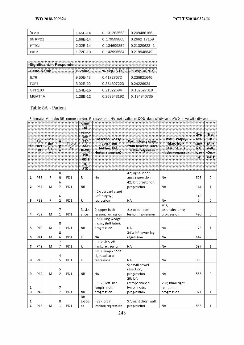
GS3 1.65E-14 0.131283553 0.209486166
SN RPD1 1.66E-14 0.179599805 0.2662 17159
PTTG l 2.02E-14 0.134699854 0.21320623 1
FIBP 1.72E-13 0.142996584 0.219948849
Significant in Responder
Gene Name P-value % exp in R % exp in NR
IL7R 9.60E-48 0.41727672 0.236921646
TCF7 3.02E-20 0.354807223 0.24226924
GPR183 1.54E-16 0.21522694 0.132527319
MGAT4A 1.28E-12 0.263543192 0.184840735
Table 8A - Patient
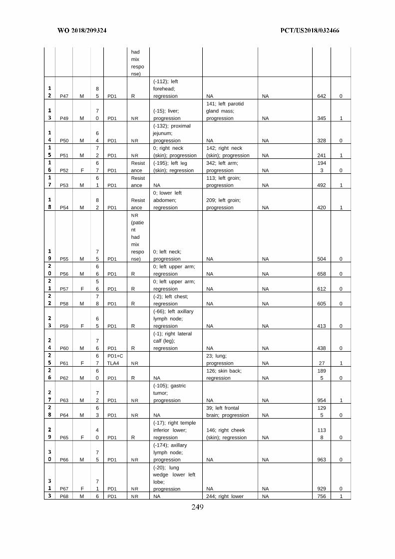
had
mixresponse)
(-112); left8 forehead;
P47 M 5 PD1 R regression NA NA 642 0
141; left parotid7 (-15); liver; gland mass;
P49 M 0 PD1 NR progression progression NA 345 1
(-132); proximal6 jejunum;
P50 M 4 PD1 NR progression NA NA 328 0
7 0; right neck 142; right neckP51 M 2 PD1 NR (skin); progression (skin); progression NA 241 1
6 Resist (-195); left leg 342; left arm; 194P52 F 7 PD1 ance (skin); regression progression NA 3 0
6 Resist 113; left groin;P53 M 1 PD1 ance NA progression NA 492 1
0; lower left8 Resist abdomen; 209; left groin;
P54 M 2 PD1 ance regression progression NA 420 1
NR
(patienthad
mix7 respo 0; left neck;
P55 M 5 PD1 nse) progression NA NA 504 0
6 0; left upper arm;P56 M 6 PD1 R regression NA NA 658 0
5 0; left upper arm;P57 F 6 PD1 R regression NA NA 612 0
7 (-2); left chest;P58 M 8 PD1 R regression NA NA 605 0
(-66); left axillary6 lymph node;
P59 F 5 PD1 R regression NA NA 413 0
(-1); right lateral7 calf (leg);
P60 M 6 PD1 R regression NA NA 438 0
6 PD1+C 23; lung;P61 F 7 TLA4 NR progression NA 27 1
6 126; skin back; 189P62 M 0 PD1 R NA regression NA 5 0
(-105); gastric7 tumor;
P63 M 2 PD1 NR progression NA NA 954 1
6 39; left frontal 129P64 M 3 PD1 NR NA brain; progression NA 5 0
(-17); right temple4 inferior lower; 146; right cheek 113
P65 F 0 PD1 R regression (skin); regression NA 8 0
(-174); axillary7 lymph node;
P66 M 5 PD1 NR progression NA NA 963 0
(-20); lungwedge lower left
7 lobe;P67 F 1 PD1 NR progression NA NA 929 0
P68 M 6 PD1 NR NA 244; right lower NA 756 1
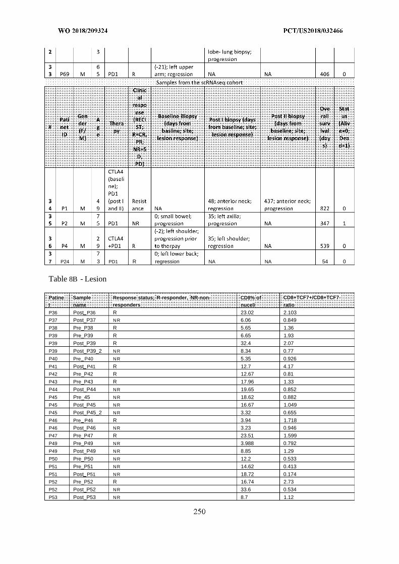
7 P24 M 3 PD1 R regression NA NA 54 0
Table 8B - Lesion
Patine Sample Response status; R-responder, NR-non- CD8% of CD8+TCF7+/CD8+TCF7-t name responders nuceli ratio
P36 Post P36 R 23.02 2.103
P37 Post_P37 NR 6.06 0.849
P38 Pre_P38 R 5.65 1.36
P39 Pre_P39 R 6.65 1.93
P39 Post_P39 R 32.4 2.07
P39 Post_P39_2 NR 8.34 0.77
P40 Pre P40 NR 5.35 0.926
P41 Post P41 R 12.7 4.17
P42 Pre_P42 R 12.67 0.81
P43 Pre_P43 R 17.96 1.33
P44 Post_P44 NR 19.65 0.852
P45 Pre_45 NR 18.62 0.882
P45 Post_P45 NR 16.67 1.049
P45 Post_P45_2 NR 3.32 0.655
P46 Pre P46 R 3.94 1.718
P46 Post_P46 NR 3.23 0.946
P47 Pre_P47 R 23.51 1.599
P49 Pre_P49 NR 3.988 0.792
P49 Post_P49 NR 8.85 1.29
P50 Pre_P50 NR 12.2 0.533
P51 Pre_P51 NR 14.62 0.413
P51 Post P51 NR 18.72 0.174
P52 Pre_P52 R 16.74 2.73
P52 Post_P52 NR 33.6 0.534
P53 Post_P53 NR 8.7 1.12
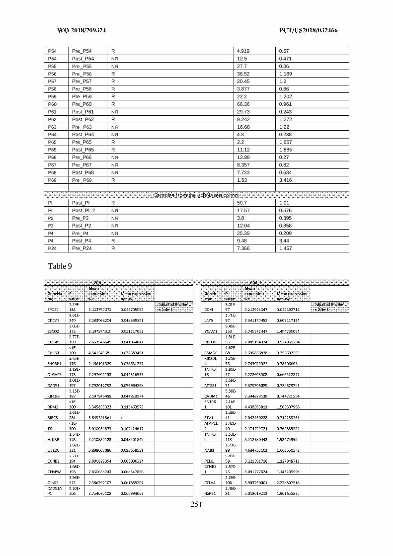
P54 Pre_P54 R 4.919 0.57
P54 Post_P54 NR 12.5 0.471
P55 Pre P55 NR 27.7 0.36
P56 Pre P56 R 36.52 1.189
P57 Pre_P57 R 20.45 1.2
P58 Pre_P58 R 3.877 0.86
P59 Pre_P59 R 22.2 1.202
P60 Pre_P60 R 66.36 0.961
P61 Post_P61 NR 29.73 0.243
P62 Post_P62 R 9.242 1.272
P63 Pre P63 NR 16.68 1.22
P64 Post_P64 NR 4.3 0.238
P65 Pre_P65 R 2.2 1.657
P65 Post_P65 R 11.12 1.885
P66 Pre_P66 NR 12.88 0.27
P67 Pre_P67 NR 8.357 0.82
P68 Post_P68 NR 7.723 0.634
P69 Pre P69 R 1.53 3.416
S mpf f o the sc As coh t
PI Post_Pl R 50.7 1.01
PI Post_Pl_2 NR 17.57 0.576
P2 Pre_P2 NR 3.8 0.395
P2 Post_P2 NR 12.04 0.858
P4 Pre P4 NR 25.39 0.209
P4 Post_P4 R 9.48 3.44
P24 Pre_P24 R 7.366 1.457
Table 9
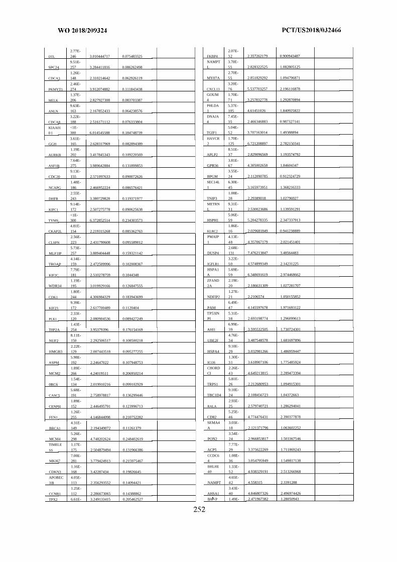
2.77E- 2.07E-
DTL 246 3.010444717 0.075483325 FKBP4 3 2 2.357262179 0.900943487
9.51E- NAMPT 3.70E-
SPC24 257 3.284411816 0.086262498 L 55 2.828322525 1.082805125
1.26E- 2.70E-
CDCA3 148 2.310214642 0.062926119 MY07A 55 2.851829292 1.094796871
2.46E- 3.20E-
PKMYT1 274 3.912074882 0.111843438 CXCL13 76 5.537703257 2.196116878
1.37E- GOUM 1.70E-
MELK 206 2.827927308 0.083703387 4 7 1 3.257832778 1.292870894
9.63E- PHLDA 5.37E-
ANLN 163 2.167852433 0.064238576 1 105 4.61451026 1.840925822
3.22E- DNAJA 7.45E-
CDCA8 188 2.516171112 0.076333804 4 35 2.466346883 0.987327141
KIAA01 <1E- 5.04E-
0 1 300 6.014545588 0.184748739 TGIF1 5 2 3.707163014 1.49388894
3.61E- HAVCR 1.70E-
GGH 165 2.628317969 0.082894389 2 125 6.721208897 2.782150341
1.19E- 8.51E-
AURKB 202 3.417845343 0.109220569 APLP2 37 2.829096569 1.193574792
7.64E- 3.81E-
ASF1B 275 3.989642884 0.131899853 GPR56 67 4.305002658 1.84604347
9.13E- 3.55E-
CDC20 155 2.571097633 0.090072626 BPGM 24 2.112090785 0.912324729
1.48E- SEC14L 6.30E-
NCAPG 186 2.466952224 0.086576421 1 45 3.165973951 1.368216333
2.55E- 1.08E-
DHFR 243 3.380729828 0.119371977 TNIP3 28 2.29389018 1.02796927
9.14E- METRN 9.31E-
KIFC1 172 2.507275778 0.090625638 L 3 1 2.530023686 1.139591291
<1E- 5.06E-
TYMS 300 6.372852514 0.234303573 HSPH1 59 5.204278335 2.347337913
4.81E- 1.86E-
CKAP2L 154 2.219315268 0.085362763 KLRC2 16 2.029681849 0.941238889
2.56E- PMAIP 4.13E-
CLSPN 223 2.431790608 0.095589012 1 48 4.357067179 2.021451401
5.73E- 2.68E-
MLF1IP 257 3.889404448 0.159321142 DUSP4 131 7.476213847 3.48564483
4.14E- 3.22E-
TROAP 159 2.472509996 0.102008367 IGFLR1 50 4.574899349 2.14231225
7.79E- HSPA1 5.69E-
KIF2C 181 2.510278759 0.1044348 A 59 6.348691619 2.974468662
1.19E- ZFAND 2.19E-
WDR34 195 3.019929166 0.126847555 2A 20 2.186631309 1.027281707
1.80E- 1.27E-
CDK1 244 4.306984329 0.183943699 NDFIP2 21 2.2106574 1.050155852
9.39E- 6.49E-
KIF23 172 2.617700489 0.1120404 PAM 47 4.145597678 1.971693122
2.33E- TP53IN 5.31E-
PLK1 120 2.080904536 0.089427249 PI 38 2.693198774 1.296099613
1.43E- 6.99E-
T0P2A 254 3.95379396 0.176154169 AHI1 39 3.595532505 1.730724301
8.11E- 4.76E-
NUF2 150 2.292506517 0.108500218 UBE2F 34 3.487548578 1.681697896
2.22E- 9.10E-
HMGB3 129 2.007443518 0.095277255 HSPA4 29 3.032981266 1.486959447
5.98E- 1.30E-
ASPM 192 2.24647022 0.107948753 ICOS 3 1 3.618907106 1.775485924
1.89E- CHORD 2.26E-
MCM2 266 4.24019511 0.206950214 CI 43 4.649213815 2.289473394
1.54E- 5.81E-
0RC6 134 2.019910216 0.099102929 TRPS1 26 2.212680953 1.094915301
5.68E- 9.10E-
CASC5 191 2.758978817 0.136299446 TBC1D4 24 2.108456723 1.04372663
1.89E- 2.93E-
CENPH 152 2.446495791 0.123996713 RALA 25 2.579740721 1.286294041
1.26E- 5.25E-
FEN1 255 4.146844098 0.210752282 CD82 46 4.774476431 2.390377878
4.31E- SEMA4 3.03E-
BRCA1 149 2.194349072 0.11261379 A 18 2.121371796 1.063602252
5.26E- 3.54E-
MCM4 298 4.748202624 0.248402619 PON2 24 2.966853817 1.503367546
TIMELE 1.17E- 7.77E-
SS 175 2.504879494 0.131966386 ACP5 29 3.375622269 1.711869243
7.00E- CCDC6 1.08E-
MKI67 281 3.779424913 0.215975467 4 36 3.054795949 1.549817138
1.16E- BHLHE 1.33E-CDKN3 168 3.42287434 0.19826645 40 5 2 4.938329191 2.513266968
APOBEC 4.05E- 4.65E-
3 B 113 2.356293552 0.14094421 NAMPT 4 2 4.558315 2.3391288
1.25E- 3.43E-
CCNB1 112 2.286673065 0.14388862 AHSA1 40 4.846807326 2.496974426
TPX2 6.61E- 3.249133415 0.205462527 BA P 1.49E- 2.471967382 1.28050943
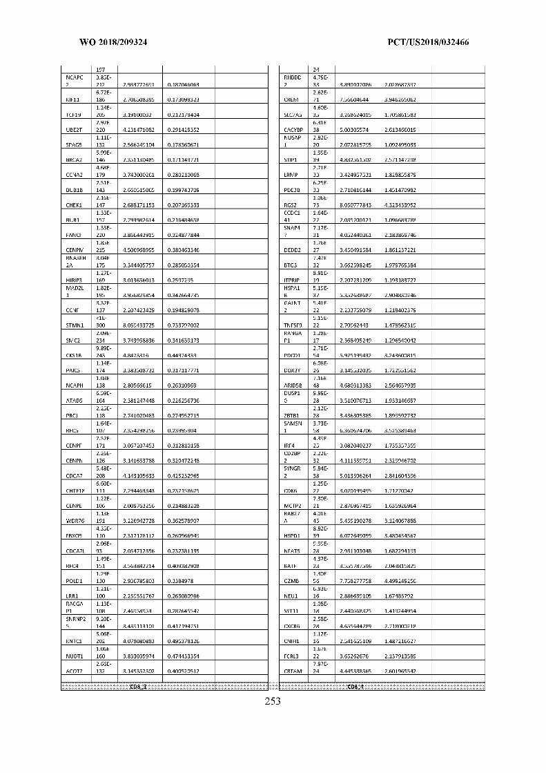
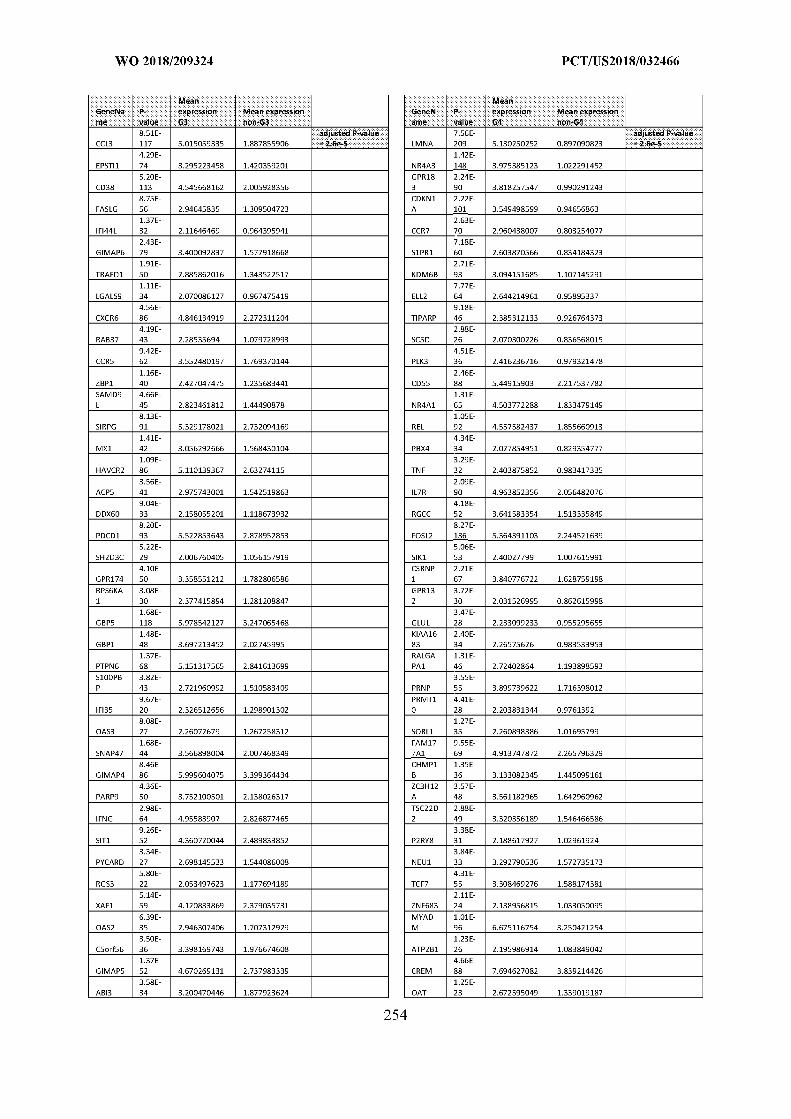
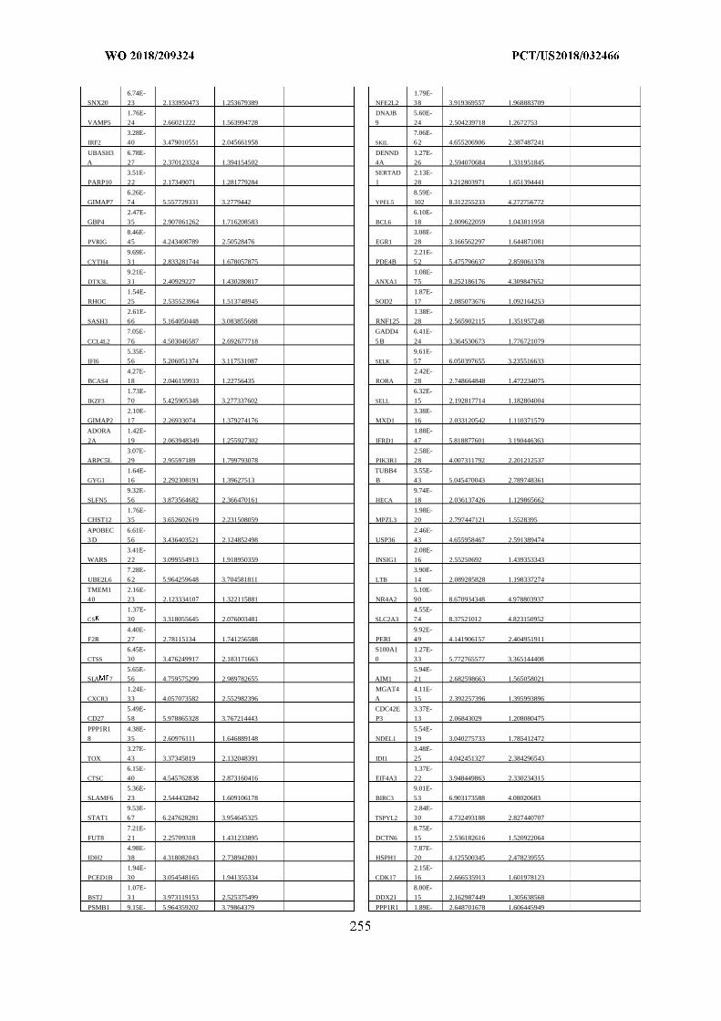
6.74E- 1.79E-
SNX20 23 2.133950473 1.253679389 NFE2L2 38 3.919369557 1.968883709
1.76E- DNAJB 5.60E-
VAMP5 24 2.66021222 1.563994728 9 24 2.504239718 1.2672753
3.28E- 7.06E-
IRF2 40 3.479010551 2.045661958 SKIL 6 2 4.655206906 2.387487241
UBASH3 6.78E- DENND 1.27E-
A 27 2.370123324 1.394154502 4A 26 2.594070684 1.331951845
3.51E- SERTAD 2.13E-
PARP10 22 2.17349071 1.281779284 1 28 3.212803971 1.651394441
6.26E- 8.59E-
GIMAP7 74 5.557729331 3.2779442 YPEL5 102 8.312255233 4.272756772
2.47E- 6.10E-
GBP4 35 2.907061262 1.716208583 BCL6 18 2.009622059 1.043811958
8.46E- 3.08E-
PVRIG 45 4.243408789 2.50528476 EGR1 28 3.166562297 1.644871081
9.69E- 2.21E-
CYTH4 3 1 2.833281744 1.678057875 PDE4B 5 2 5.475796637 2.859061378
9.21E- 1.08E-
DTX3L 3 1 2.40929227 1.430280817 ANXA1 75 8.252186176 4.309847652
1.54E- 1.87E-
RHOC 25 2.535523964 1.513748945 SOD2 17 2.085073676 1.092164253
2.61E- 1.38E-
SASH3 66 5.164050448 3.083855688 RNF125 28 2.565902115 1.351957248
7.05E- GADD4 6.41E-
CCL4L2 76 4.503046587 2.692677718 5 B 24 3.364530673 1.776721079
5.35E- 9.61E-
IFI6 56 5.206051374 3.117531087 SELK 57 6.050397655 3.235516633
4.27E- 2.42E-
BCAS4 18 2.046159933 1.22756435 RORA 28 2.748664848 1.472234075
1.73E- 6.32E-
IKZF3 70 5.425905348 3.277337602 SELL 15 2.192817714 1.182804004
2.10E- 3.38E-
GIMAP2 17 2.26933074 1.379274176 MXD1 16 2.033120542 1.110371579
ADORA 1.42E- 1.88E-
2A 19 2.063948349 1.255927302 IFRD1 47 5.818877601 3.190446363
3.07E- 2.58E-
ARPC5L 29 2.95597189 1.799793078 PIK3R1 28 4.007311792 2.201212537
1.64E- TUBB4 3.55E-
GYG1 16 2.292308191 1.39627513 B 43 5.045470043 2.789748361
9.32E- 9.74E-
SLFN5 56 3.873564682 2.366470161 HECA 18 2.036137426 1.129865662
1.76E- 1.98E-
CHST12 35 3.652602619 2.231508059 MPZL3 20 2.797447121 1.5528395
APOBEC 6.61E- 2.46E-
3 D 56 3.436403521 2.124852498 USP36 43 4.655958467 2.591389474
3.41E- 2.08E-
WARS 22 3.099554913 1.918950359 INSIG1 16 2.55250692 1.439353343
7.28E- 3.90E-
UBE2L6 6 2 5.964259648 3.704581811 LTB 14 2.089285828 1.198337274
TMEM1 2.16E- 5.10E-
40 23 2.123334107 1.322115881 NR4A2 90 8.670934348 4.978803937
1.37E- 4.55E-
CS 30 3.318055645 2.076003481 SLC2A3 74 8.37521012 4.823150952
4.40E- 9.92E-
F2R 27 2.78115134 1.741256588 PERI 49 4.141906157 2.404951911
6.45E- S100A1 1.27E-
CTSS 30 3.476249917 2.183171663 0 33 5.772765577 3.365144408
5.65E- 5.94E-
SLA 7 56 4.759575299 2.989782655 AIM1 21 2.682598663 1.565058021
1.24E- MGAT4 4.11E-
CXCR3 33 4.057073582 2.552982396 A 15 2.392257396 1.395993896
5.49E- CDC42E 3.37E-
CD27 58 5.978865328 3.767214443 P3 13 2.06843029 1.208080475
PPP1R1 4.38E- 5.54E-
8 35 2.60976111 1.646889148 NDEL1 19 3.040275733 1.785412472
3.27E- 3.48E-
TOX 43 3.37345819 2.132048391 IDI1 25 4.042451327 2.384296543
6.15E- 1.37E-
CTSC 40 4.545762838 2.873160416 EIF4A3 22 3.948449863 2.330234315
5.36E- 9.01E-
SLAMF6 23 2.544432842 1.609106178 BIRC3 53 6.903173588 4.08020683
9.53E- 2.84E-
STAT1 67 6.247628281 3.954645325 TSPYL2 30 4.732493188 2.827440707
7.21E- 8.75E-
FUT8 21 2.25709318 1.431233895 DCTN6 15 2.536182616 1.520922064
4.98E- 7.87E-
IDH2 38 4.318082043 2.738942801 HSPH1 20 4.125500345 2.478239555
1.94E- 2.15E-
PCED1B 30 3.054548165 1.941355334 CDK17 16 2.666535913 1.601978123
1.07E- 8.00E-
BST2 3 1 3.973119153 2.525375499 DDX21 15 2.162987449 1.305638568
PSMB1 9.15E- 5.964359202 3.79864379 PPP1R1 1.89E- 2.648701678 1.606445949
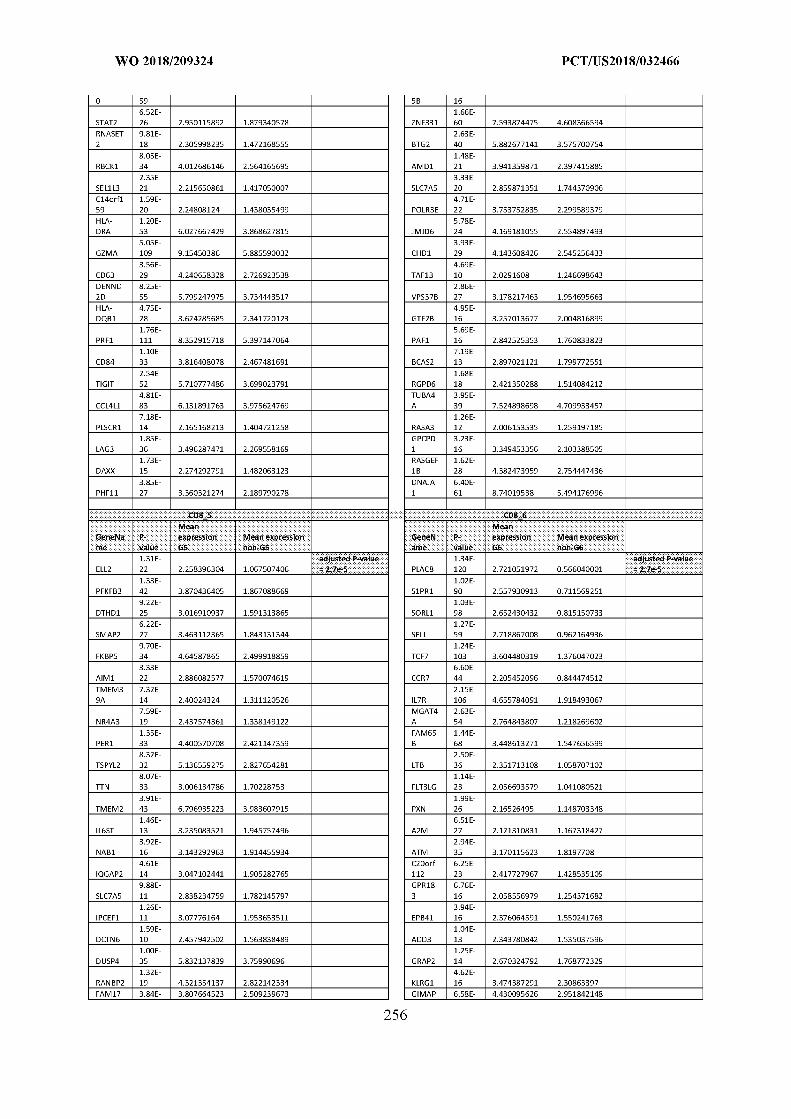
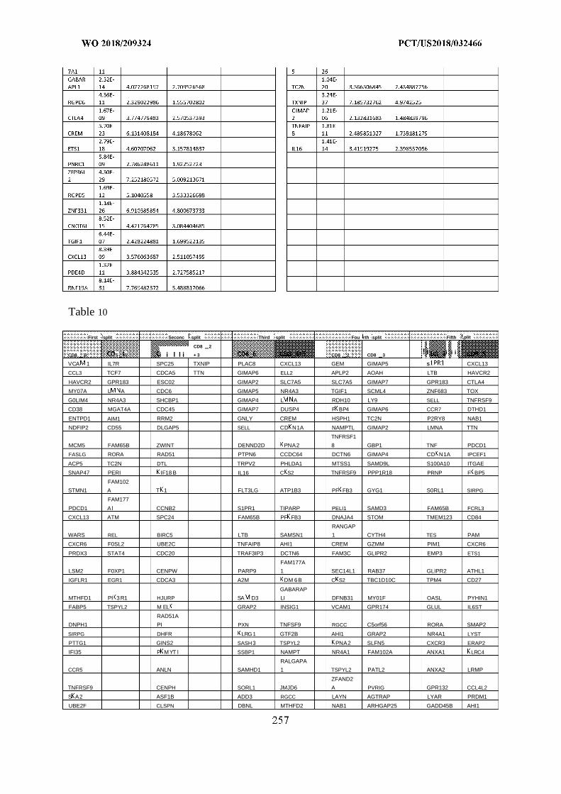
Table 10
First split Seconc split Third split Fou rth split Fifth ,plit
CD8 2
CD8 B S S i i l l i + 3 CD8 2 CD8 3
VCA 1 IL7R SPC25 TXNIP PLAC8 CXCL13 GEM GIMAP5
! iS
i i i
CXCL13
CCL3 TCF7 CDCA5 TTN GIMAP6 ELL2 APLP2 AOAH LTB HAVCR2
HAVCR2 GPR183 ESC02 GIMAP2 SLC7A5 SLC7A5 GIMAP7 GPR183 CTLA4
MY07A L A CDC6 GIMAP5 NR4A3 TGIF1 SCML4 ZNF683 TOX
G0LIM4 NR4A3 SHCBP1 GIMAP4 L A RDH10 LY9 SELL TNFRSF9
CD38 MGAT4A CDC45 GIMAP7 DUSP4 F BP4 GIMAP6 CCR7 DTHD1
ENTPD1 AIM1 RRM2 GNLY CREM HSPH1 TC2N P2RY8 NAB1
NDFIP2 CD55 DLGAP5 SELL CD N1A NAMPTL GIMAP2 LMNA TTN
TNFRSF1
MCM5 FAM65B ZWINT DENND2D PNA2 8 GBP1 TNF PDCD1
FASLG RORA RAD51 PTPN6 CCDC64 DCTN6 GIMAP4 CD N1A IPCEF1
ACP5 TC2N DTL TRPV2 PHLDA1 MTSS1 SAMD9L S100A10 ITGAE
SNAP47 PERI IF18 B IL16 C S2 TNFRSF9 PPP1R18 PRNP F BP5
FAM102
STMN1 A T 1 FLT3LG ATP1B3 PF FB3 GYG1 S0RL1 SIRPG
FAM177
PDCD1 A l CCNB2 S1PR1 TIPARP PELI1 SAMD3 FAM65B FCRL3
CXCL13 ATM SPC24 FAM65B PF FB3 DNAJA4 STOM TMEM123 CD84
RANGAP
WARS REL BIRC5 LTB SAMSN1 1 CYTH4 TES PAM
CXCR6 F0SL2 UBE2C TNFAIP8 AHI1 CREM GZMM PIM1 CXCR6
PRDX3 STAT4 CDC20 TRAF3IP3 DCTN6 FAM3C GLIPR2 EMP3 ETS1
FAM177A
LSM2 F0XP1 CENPW PARP9 1 SEC14L1 RAB37 GLIPR2 ATHL1
IGFLR1 EGR1 CDCA3 A2M DM 6B C S2 TBC1D10C TPM4 CD27
GABARAP
MTHFD1 PI 3R1 HJURP SA D3 LI DFNB31 MY01F OASL PYHIN1
FABP5 TSPYL2 M EL GRAP2 INSIG1 VCAM1 GPR174 GLUL IL6ST
RAD51A
DNPH1 PI PXN TNFSF9 RGCC C5orf56 RORA SMAP2
SIRPG DHFR LRG 1 GTF2B AHI1 GRAP2 NR4A1 LYST
PTTG1 GINS2 SASH3 TSPYL2 PNA2 SLFN5 CXCR3 ERAP2
IFI35 P M YT l SSBP1 NAMPT NR4A1 FAM102A ANXA1 LRC4
RALGAPA
CCR5 ANLN SAMHD1 1 TSPYL2 PATL2 ANXA2 LRMP
ZFAND2
TNFRSF9 CENPH SORL1 JMJD6 A PVRIG GPR132 CCL4L2
S A2 ASF1B ADD3 RGCC LAYN AGTRAP LYAR PRDM1
UBE2F CLSPN DBNL MTHFD2 NAB1 ARHGAP25 GADD45B AHI1
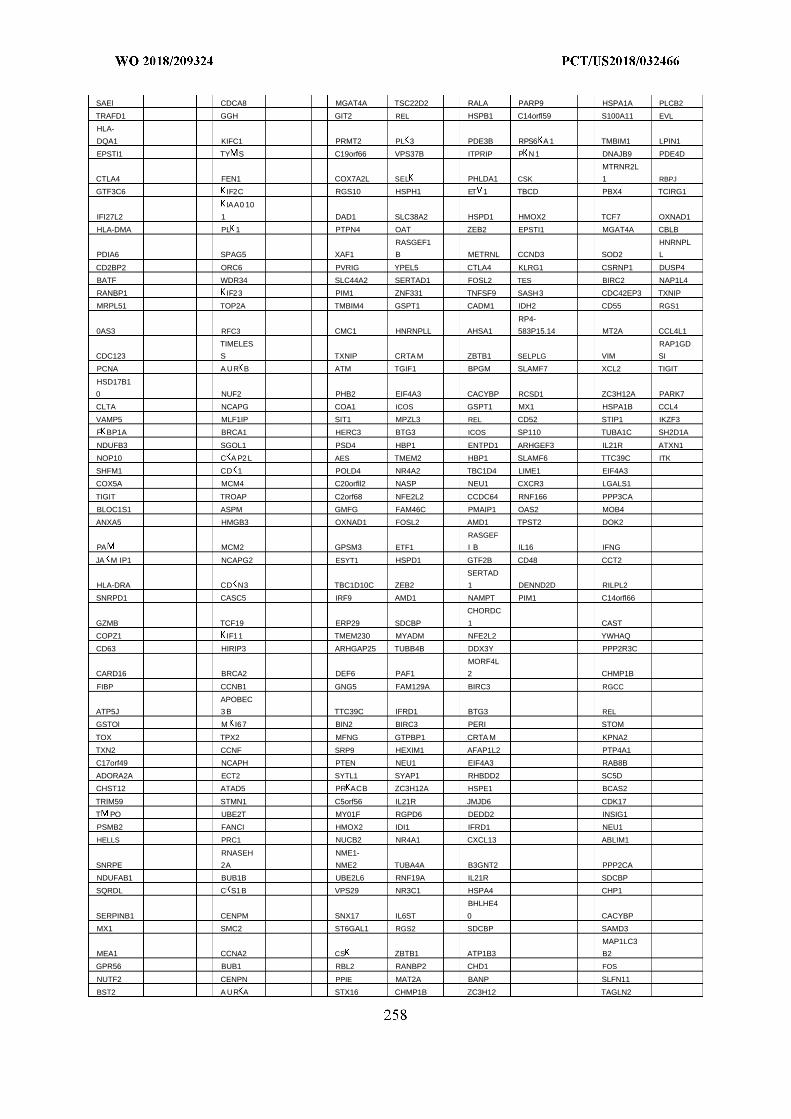
SAEl CDCA8 MGAT4A TSC22D2 RALA PARP9 HSPA1A PLCB2
TRAFD1 GGH GIT2 REL HSPB1 C14orfl59 S100A11 EVL
HLA-
DQA1 KIFC1 PRMT2 PL 3 PDE3B RPS6 A 1 TMBIM1 LPIN1
EPSTI1 TY S C19orf66 VPS37B ITPRIP P N 1 DNAJB9 PDE4D
MTRNR2L
CTLA4 FEN1 COX7A2L SEL PHLDA1 CSK 1 RBPJ
GTF3C6 IF2C RGS10 HSPH1 ET 1 TBCD PBX4 TCIRG1
IAA0 10
IFI27L2 1 DAD1 SLC38A2 HSPD1 HMOX2 TCF7 OXNAD1
HLA-DMA PL 1 PTPN4 OAT ZEB2 EPSTI1 MGAT4A CBLB
RASGEF1 HNRNPL
PDIA6 SPAG5 XAF1 B METRNL CCND3 SOD2 L
CD2BP2 ORC6 PVRIG YPEL5 CTLA4 KLRG1 CSRNP1 DUSP4
BATF WDR34 SLC44A2 SERTAD1 FOSL2 TES BIRC2 NAP1L4
RANBP1 IF23 PIM1 ZNF331 TNFSF9 SASH3 CDC42EP3 TXNIP
MRPL51 TOP2A TMBIM4 GSPT1 CADM1 IDH2 CD55 RGS1
RP4-
0AS3 RFC3 CMC1 HNRNPLL AHSA1 583P15.14 MT2A CCL4L1
TIMELES RAP1GD
CDC123 S TXNIP CRTA M ZBTB1 SELPLG VIM SI
PCNA A UR B ATM TGIF1 BPGM SLAMF7 XCL2 TIGIT
HSD17B1
0 NUF2 PHB2 EIF4A3 CACYBP RCSD1 ZC3H12A PARK7
CLTA NCAPG COA1 ICOS GSPT1 MX1 HSPA1B CCL4
VAMP5 MLF1IP SIT1 MPZL3 REL CD52 STIP1 IKZF3
F BP1A BRCA1 HERC3 BTG3 ICOS SP110 TUBA1C SH2D1A
NDUFB3 SGOL1 PSD4 HBP1 ENTPD1 ARHGEF3 IL21R ATXN1
NOP10 C A P2 L AES TMEM2 HBP1 SLAMF6 TTC39C ITK
SHFM1 CD 1 POLD4 NR4A2 TBC1D4 LIME1 EIF4A3
COX5A MCM4 C20orfll2 NASP NEU1 CXCR3 LGALS1
TIGIT TROAP C2orf68 NFE2L2 CCDC64 RNF166 PPP3CA
BLOC1S1 ASPM GMFG FAM46C PMAIP1 OAS2 MOB4
ANXA5 HMGB3 OXNAD1 FOSL2 AMD1 TPST2 DOK2
RASGEF
PA MCM2 GPSM3 ETF1 I B IL16 IFNG
JA M IP1 NCAPG2 ESYT1 HSPD1 GTF2B CD48 CCT2
SERTAD
HLA-DRA CD N3 TBC1D10C ZEB2 1 DENND2D RILPL2
SNRPD1 CASC5 IRF9 AMD1 NAMPT PIM1 C14orfl66
CHORDC
GZMB TCF19 ERP29 SDCBP 1 CAST
COPZ1 IF11 TMEM230 MYADM NFE2L2 YWHAQ
CD63 HIRIP3 ARHGAP25 TUBB4B DDX3Y PPP2R3C
MORF4L
CARD16 BRCA2 DEF6 PAF1 2 CHMP1B
FIBP CCNB1 GNG5 FAM129A BIRC3 RGCC
APOBEC
ATP5J 3 B TTC39C IFRD1 BTG3 REL
GSTOl M I67 BIN2 BIRC3 PERI STOM
TOX TPX2 MFNG GTPBP1 CRTA M KPNA2
TXN2 CCNF SRP9 HEXIM1 AFAP1L2 PTP4A1
C17orf49 NCAPH PTEN NEU1 EIF4A3 RAB8B
ADORA2A ECT2 SYTL1 SYAP1 RHBDD2 SC5D
CHST12 ATAD5 PR ACB ZC3H12A HSPE1 BCAS2
TRIM59 STMN1 C5orf56 IL21R JMJD6 CDK17
T PO UBE2T MY01F RGPD6 DEDD2 INSIG1
PSMB2 FANCI HMOX2 IDI1 IFRD1 NEU1
HELLS PRC1 NUCB2 NR4A1 CXCL13 ABLIM1
RNASEH NME1-
SNRPE 2A NME2 TUBA4A B3GNT2 PPP2CA
NDUFAB1 BUB1B UBE2L6 RNF19A IL21R SDCBP
SQRDL C S1B VPS29 NR3C1 HSPA4 CHP1
BHLHE4
SERPINB1 CENPM SNX17 IL6ST 0 CACYBP
MX1 SMC2 ST6GAL1 RGS2 SDCBP SAMD3
MAP1LC3
MEA1 CCNA2 CS ZBTB1 ATP1B3 B2
GPR56 BUB1 RBL2 RANBP2 CHD1 FOS
NUTF2 CENPN PPIE MAT2A BANP SLFN11
BST2 A UR A STX16 CHMP1B ZC3H12 TAGLN2
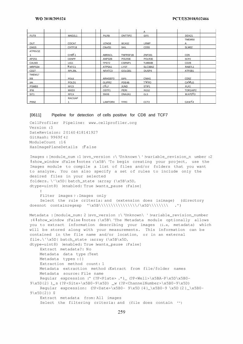
A
FUT8 MAD2L1 PILRB DNTTIP2 EIF5 DDX21
TMEM50
DUT CDCA7 LENG8 BCAS2 LRMP A
GNG5 CHTF18 Cllorf31 SKIL CD55 SLM02
ATP6V1E
1 CHE 1 ABRACL TNFRSF1B ZNF331 CD5
AP2S1 CENPF ANP32B POLR3E POLR3E SCP2
CALM3 LIG1 TPST2 CSRNP1 TUBB4B CD28
MRPS34 NTC1 ATP5A1 LYST SLC38A2 RAB7L1
CD27 RPL39L NFATC3 GOLGB1 DUSP4 ATP2B1
TMEM17
9 B POLE ARHGEF3 EIF5 CNIH1 CD52
SRI POLD1 GLIPR2 PDE4B T PO CA LG
PSMB3 RFC5 C LF JUND STIP1 PLP2
IFI6 MXD3 OSTF1 PERI RGS2 TOR1AIP2
SIT1 RFC4 SNX6 DNAJA1 GLS M AP2 3
RACGAP
P0N2 1 LAMTOR4 TFRC CCT2 CAM 4
[0611] Pipeline for detection of cells positive for CD8 and TCF7
CellProfiler Pipeline: www.cellprofiler.orgVersion :3DateRevision: 20160 418141927GitHash: 9969f 42ModuleCount :16HasImagePlaneDetails : alse
Images :[module_num :1 |svn_version :\ 'Unknown \'|variable_revision_n umber :2Ishow_window :False Inotes :\x5B\ 'To begin creating your project, use theImages module to compile a list of files and/or folders that you wantto analyze. You can also specify a set of rules to include only thedesired files in your selectedfolders. \'\x5D| batch_state :array (\x5B\x5D,dtype=uint8) |enabled: True |wants_pause :False]
Filter images ? :Images onlySelect the rule criteria: and (extension does isimage) (directory
doesnot containregexp "\x5B\\\\\\\\\\\\\\\\/\x5D\\\\\\\\ .")
Metadata :[module_num: 2 |svn_version :\'Unknown\ '|variable_revision_number:4 Ishow_window :False Inotes :\x5B\ 'The Metadata module optionally allowsyou to extract information describing your images (i.e, metadata) whichwill be stored along with your measurements. This information can becontained in the file name and/or location, or in an externalfile.\'\x5D| batch_state :array (\x5B\x5D,dtype=uint8) |enabled: True |wants_pause :False]
Extract metadata?: NoMetadata data type :TextMetadata types :{}Extraction method count: 1Metadata extraction method :Extract from file/folder namesMetadata source: File nameRegular expression : (?P<Plate> .*)_ (?P<Well>\x5BA-P\x5D\x5B0-
9\x5D{2} )_s (?P<Site>\x5B0-9\x5D) _w (?P<ChannelNumber>\x5B0-9\x5D)Regular expression: (?P<Date>\x5B0- 9\x5D {4}_\x5B0-9 \x5D {2}_\x5B0-
9\x5D{2}) $Extract metadata from: All imagesSelect the filtering criteria: and (file does contain "")
Metadata file location:Match file and image metadata :\x5B\x5DUse case insensitive matching? :No
NamesAndTypes :[module_num: 3 |svn_version: \'Unknown\ '|variable_revis ion_number :6 show_window : alse notes :\x5B\ 'The NamesAndTypes module allowsyou to assign a meaningful name to each image by which other moduleswill refer to it .\ '\x5D |batch_state :array (\x5B\x5D,dtype=uint8) |enabled: True |wants_pause :False]
Assign a name to:All imagesSelect the image type: Color imageName to assign these images :ColorlmageMatch metadata :\x5B\x5DImage set matching method: OrderSet intensity range from: Image metadataAssignments count: 1Single images count:Maximum intensity :255 .0Select the rule criteria: and (file does contain "")
Name to assign these images :DNAName to assign these objects: CellSelect the image type :Grayscale imageSet intensity range from: Image metadataRetain outlines of loaded objects? :NoName the outline image :LoadedOutlinesMaximum intensity :255 .0
Groups :[module_num : |svn_version :\ 'Unknown \'|variable_revision_n umber :2show_window :False notes :\x5B\ 'The Groups module optionally allows you
to split your list of images into image subsets (groups) which will beprocessed independently of each other. Examples of groupings includescreening batches, microtiter plates, time-lapse movies,etc.\'\x5D| batch_state :array (\x5B\x5D,dtype=uint8) |enabled: True |wants_pause :False]
Do you want to group your images?: Nogrouping metadata count: 1Metadata category :None
ColorToGray: [module_num: 5 |svn_vers ion :\'Unknown \'|variable_revision_number :3 show_window :False |notes: \x5B\x5D |batch_state :array (\x5B\x5D,dtype=uint8) |enabled: True |wants_pause :False]
Select the input image :Color ImageConversion method: SplitImage type :ChannelsName the output image :OrigGrayRelative weight of the red channel: 1.0Relative weight of the green channel: 1.0Relative weight of the blue channel: 1.0Convert red to gray?: YesName the output image :OrigRedConvert green to gray?: YesName the output image :OrigGreenConvert blue to gray?: YesName the output image :OrigBlueConvert hue to gray?: YesName the output image :OrigHueConvert saturation to gray?: Yes
Name the output image :OrigSaturationConvert value to gray?: YesName the output image :OrigValueChannel count: 3Channel number :Red\x3A 1Relative weight of the channel: 1.0Image name :TCF7Channel number :Green\x3A 2Relative weight of the channel: 1.0Image name :CD8Channel number :Blue\x3A 3Relative weight of the channel: 1.0Image name: DAP I
EnhanceOr Suppress Features :[module_num: 6|svn_version :\'Unknown \'|variable_revision_n umber :5 |show_window :False|notes:\x5B\x5D| batch_state :array (\x5B\x5D, dtype=uint8) |enabled :True |wants_pause :False ]
Select the input image :DAPIName the output image :FilteredDAPISelect the operation :EnhanceFeature size:20Feature type :SpecklesRange of hole sizes: 1,10Smoothing scale: 2.0Shear angle :0.0Decay: 0.95Enhancement method :Tubenes sSpeed and accuracy :Fast / hexagonal
Ident ifyPrimaryOb ects :[module_num :7 |svn_version :\'Unknown \'|variable_revision_number :10 |show_window :False |notes: \x5B\x5D |batch_s tate :array (\x5B\x5D, dtype=uint8) |enabled: True |wants_pause :False]
Select the input image :FilteredDAPIName the primary objects to be identified: NucleiTypical diameter of objects, in pixel units (Min,Max) :15,50Discard objects outside the diameter range?:YesTry to merge too small objects with nearby larger objects? :NoDiscard objects touching the border of the image?: YesMethod to distinguish clumped obj ects :ShapeMethod to draw dividing lines between clumped obj ects :PropagateSize of smoothing filter: 10
Suppress local maxima that are closer than this minimum alloweddistance :7.0
Speed up by using lower-resolution image to find local maxima?: YesName the outline image :PrimaryOutlinesFill holes in identified obj ects? :After both thresholding and
declumpingAutomatically calculate size of smoothing filter for
declumping? :YesAutomatically calculate minimum allowed distance between local
maxima? :YesRetain outlines of the identified objects? :NoAutomatically calculate the threshold using the Otsu method?: YesEnter Laplacian of Gaussian threshold :0.5Automatically calculate the size of objects for the Laplacian of
Gaussian filter?:YesEnter LoG filter diameter: 5.0
Handling of objects if excessive number of objectsidentified: Continue
Maximum number of objects: 500Threshold setting version: 2Threshold strategy :AdaptiveThresholding method: OtsuSelect the smoothing method for thresholding :AutomaticThreshold smoothing scale: 1.0Threshold correction factor: 1.0Lower and upper bounds on threshold: 0.0 ,1.0Approximate fraction of image covered by objects ?:0.01Manual threshold :0.0Select the measurement to threshold with:NoneSelect binary image:NoneMasking objects :NoneTwo-class or three-class thresholding? :Two classesMinimize the weighted variance or the entropy? :Weighted varianceAssign pixels in the middle intensity class to the foreground or
the background? :ForegroundMethod to calculate adaptive window size: Image sizeSize of adaptive window: 10
Use default parameters ? :DefaultLower outlier fraction :0.05Upper outlier fraction :0.05Averaging method:MeanVariance method :Standard deviation# of deviations :2.0
Rescalelntensity: [module_num: 8 |svn_version :\'Unknown\'|variable_revision_number :2 |show_window :False |notes :\x5B\x5D |batch_state :array (\x5B\x5D,dtype=uint8) |enabled: True |wants_pause :False]
Select the input image :CD8Name the output image :RescaledCD8Rescaling method :Stretch each image to use the full intensity rangeMethod to calculate the minimum intens ity :CustomMethod to calculate the maximum intens ity :CustomLower intensity limit for the input image :0.0Upper intensity limit for the input image :1.0Intensity range for the input image :0.0,1.0Intensity range for the output image :0.0,1.0Method to rescale pixels below the lower limit:Mask pixelsCustom value for pixels below lower limit: 0.0Method to rescale pixels above the upper limit :Mask pixelsCustom value for pixels above upper limit: 0.0Select image to match in maximum intensity :NoneDivisor value: 1.0Divisor measurement :None
MeasureOb jectlntensity: [module_num :9 |svn_version :\'Unknown \'|variable_revision_number :3 |show_window :False|notes:\x5B\x5D| batch_state :array (\x5
B\x5D, dtype=uint8) |enabled :True |wants_pause :False ]Hidden :1Select an image to measure :RescaledCD8Select objects to measure :Nuclei
FilterObjects: [module_num: 10 |svn_version :\'Unknown \'|variable_revision_number :7 |show_window :False|notes:\x5B\x5D| batch_state :array (\x5B\x5D,dtype=uint8) |enabled: True |wants_pause :False]
Name the output ob ects :CD8PosNucleiSelect the object to filter :NucleiSelect the filtering mode :MeasurementsSelect the filtering method :LimitsSelect the objects that contain the filtered objects:NoneRetain outlines of the identified objects? :NoName the outline image :FilteredObj ectsRules file location :Elsewhere ... \x7CRules file name :rules .txtClass number :1Measurement count: 1Additional object count:Assign overlapping child to: Both parentsSelect the measurement to filter
by :Intensity_MeanIntensityEdge_RescaledCD8Filter using a minimum measurement value?: YesMinimum value: 0.1Filter using a maximum measurement value?: NoMaximum value: 1.0
Ident ifyPrimaryOb jects :[module_num :11 |svn_vers ion :\'Unknown\'|variable_revision_number :10 |show_window :False |notes :\x5B\x5D |batch_state :array (\x5B\x5D, dtype=uint8) |enabled :True |wants_pause :False ]
Select the input image :TCF7Name the primary objects to be identified: TCF7NucleiTypical diameter of objects, in pixel units (Min,Max) :10,40Discard objects outside the diameter range?:YesTry to merge too small objects with nearby larger objects? :NoDiscard objects touching the border of the image?: YesMethod to distinguish clumped obj ects :IntensityMethod to draw dividing lines between clumped obj ects :IntensitySize of smoothing filter: 10
Suppress local maxima that are closer than this minimum alloweddistance :7.0
Speed up by using lower-resolution image to find local maxima?: YesName the outline image :PrimaryOutlinesFill holes in identified obj ects? :After both thresholding and
declumpingAutomatically calculate size of smoothing filter for
declumping? :YesAutomatically calculate minimum allowed distance between local
maxima? :YesRetain outlines of the identified objects? :NoAutomatically calculate the threshold using the Otsu method?: YesEnter Laplacian of Gaussian threshold :0.5Automatically calculate the size of objects for the Laplacian of
Gaussian filter?:YesEnter LoG filter diameter: 5.0Handling of objects if excessive number of objects
identified: ContinueMaximum number of objects: 500Threshold setting version: 2Threshold strategy :AutomaticThresholding method: Otsu
Select the smoothing method for thresholding :AutomaticThreshold smoothing scale: 1.0Threshold correction factor: 1.0Lower and upper bounds on threshold: 0.0 ,1.0Approximate fraction of image covered by ob ects ?:0.01Manual threshold :0.0Select the measurement to threshold with:NoneSelect binary image:NoneMasking objects :NoneTwo-class or three-class thresholding? :Two classesMinimize the weighted variance or the entropy? :Weighted varianceAssign pixels in the middle intensity class to the foreground or
the background? :ForegroundMethod to calculate adaptive window size: Image sizeSize of adaptive window: 10
Use default parameters ? :DefaultLower outlier fraction :0.05Upper outlier fraction :0.05Averaging method:MeanVariance method :Standard deviation# of deviations :2.0
MaskObj ects :[module_num: 12 |svn_version :\'Unknown\ '|variable_revision_number :2 |show_window :False|notes:\x5B\x5D| batch_state :array (\x5B\x5D,dtype=uint8) |enabled: True |wants_pause :False]
Select objects to be masked: CD8PosNucleiName the masked obj ects :TCF7PosCD8PosNucleiMask using a region defined by other objects or by binary
image? :Obj ectsSelect the masking object :TCF7NucleiSelect the masking image:NoneHandling of objects that are partially mas ked :Remove depending on
overlapFraction of object that must overlap: 0.3Numbering of resulting obj ects :RenumberRetain outlines of the resulting objects?:NoName the outline image :Mas kedOutlinesInvert the mask?:No
CalculateMath :[module_num: 13 |svn_version :\'Unknown \'|variable_revision_number :2 |show_window :False|notes:\x5B\x5D| batch_state :array (\x5B\x5D,dtype=uint8) |enabled: True |wants_pause :False]
Name the output measurement :Count_CD8Pos_TCFNegOperation: SubtractSelect the minuend measurement type: ImageSelect the minuend obj ects: NoneSelect the minuend measurement :Count_CD8PosNucleiMultiply the above operand by: 1.0Raise the power of above operand by: 1.0Select the subtrahend measurement type :ImageSelect the subtrahend obj ects: NoneSelect the subtrahend measurement :Count_TCF7PosCD8PosNucleiMultiply the above operand by: 1.0Raise the power of above operand by: 1.0Take loglO of result?:NoMultiply the result by: 1.0Raise the power of result by: 1.0
Add to the result: 0.0Constrain the result to a lower bound? :NoEnter the lower bound: 0.0Constrain the result to an upper bound? :NoEnter the upper bound: 1.0
OverlayOutlines :[module_num: 1 |svn_version :\'Unknown\ '|variable_revision_number :3 |show_window :False |notes :\x5B\x5D |batch_state :array (\x5B\x5D,dtype=uint8) |enabled: True |wants_pause :False]
Display outlines on a blank image? :NoSelect image on which to display outlines :Color ImageName the output image :OrigOver layOutline display mode: ColorSelect method to determine brightness of outlines :Max of imageWidth of outlines :1
Select outlines to display:NoneSelect outline color :#F8FF2DLoad outlines from an image or ob ects ?:Ob ectsSelect objects to display :CD8PosNucleiSelect outlines to display:NoneSelect outline color :#F1F7F7Load outlines from an image or obj ects ? :Ob ectsSelect objects to display :TCF7PosCD8PosNuclei
Save Images :[module_num :15 |svn_vers ion :\'Unknown \'|variable_revision_number :11 |show_window :False|notes:\x5B\x5D| batch_state :array (\x5B\x5D,dtype=uint 8) |enabled :True |wants_pause :True ]
Select the type of image to save: ImageSelect the image to save :OrigOverlaySelect the objects to save:NoneSelect the module display window to save: NoneSelect method for constructing file names: From image filenameSelect image name for file pre fix :Color ImageEnter single file name :OrigBlueNumber of digits:Append a suffix to the image file name?: YesText to append to the image name :_Over laySaved file format: tifOutput file location :Default Output Folder\x7CImage bit depth: 8-bit integerOverwrite existing files without warning?: NoWhen to save: Every cycleRescale the images? :YesSave as grayscale or color image? :GrayscaleSelect colormap :grayRecord the file and path information to the saved image?: NoCreate subfolders in the output folder? :NoBase image folder :Elsewhere ... \x7CSaved movie format :avi
Export ToSpreadsheet :[module_num: 16 |svn_version :\'Unknown \'|variable_revision_number :11 |show_window :False|notes:\x5B\x5D| batch_state :array(\x5B\x5D, dtype=uint8) |enabled :True |wants_pause :False ]
Select the column delimiter :Comma (",")
Add image metadata columns to your object data file?: NoLimit output to a size that is allowed in Excel? :NoSelect the measurements to export: Yes
Calculate the per-image mean values for object measurements ? :NoCalculate the per-image median values for object measurements ? :NoCalculate the per-image standard deviation values for object
measurements? :NoOutput file location :Default Output Folder\x7CCreate a GenePattern GCT file?:NoSelect source of sample row name :MetadataSelect the image to use as the identifier :NoneSelect the metadata to use as the identifier :NoneExport all measurement types?: No
:Image\x7CCount_TCF7Nuclei, Image\x7CCount_Nuclei, Image\x7CCount_CD8PosNuclei, Image \x7CCount_TCF7PosCD8PosNuclei, Image \x7CMath_Count_CD8Pos_TCFNeg
Representation of Nan/Inf:NaNAdd a prefix to file names? :NoFilename prefix :MyExpt_Overwrite existing files without warning?: NoData to export: ImageCombine these object measurements with those of the previous
object? :NoFile name :DATA. csvUse the object name for the file name?: Yes
REFERENCES
1 Callahan, M . K., Postow, M . A . & Wolchok, J . D . Targeting T Cell Co-
receptors for Cancer Therapy. Immunity 44, 1069-1078, doi:10.1016/j.immuni.2016.04.023
(2016) .
2 Iwai, Y., Hamanishi, J., Chamoto, K . & Honjo, T. Cancer immunotherapies
targeting the PD-1 signaling pathway. J Biomed Sci 24, 26, doi:10.1 186/sl2929-017-0329-9
(2017) .
3 Robert, C . et al. Pembrolizumab versus Ipilimumab in Advanced Melanoma.
NEnglJMed312, 2521-2532, doi:10.1056/NEJMoal503093 (2015).
4 Larkin, J . et al. Combined Nivolumab and Ipilimumab or Monotherapy in
Untreated Melanoma. N Engl J Med 373, 23-34, doi:10.1056/NEJMoal504030 (2015).
5 Tumeh, P . C . et al. PD-1 blockade induces responses by inhibiting adaptive
immune resistance. Nature 515, 568-571, doi:10.1038/naturel3954 (2014).
6 Chen, P . L . et al. Analysis of Immune Signatures in Longitudinal Tumor
Samples Yields Insight into Biomarkers of Response and Mechanisms of Resistance to
Immune Checkpoint Blockade. Cancer Discov 6, 827-837, doi:10.1 158/2159-8290.CD-15-
1545 (2016).
7 Wherry, E . J . et al. Molecular signature of CD8+ T cell exhaustion during
chronic viral infection. Immunity 27, 670-684, doi:10.1016/j.immuni.2007.09.006 (2007).
8 Speiser, D . E., Ho, P. C . & Verdeil, G . Regulatory circuits of T cell function
in cancer. Nat Rev Immunol 16, 599-6 11, doi :10 .1038/nri .20 16 .80 (20 16).
9 Schumacher, T. N . & Schreiber, R . D . Neoantigens in cancer immunotherapy.
Science 348, 69-74, doi:10.1 126/science.aaa4971 (2015).
10 Hulpke, S . & Tampe, R . The MHC I loading complex: a multitasking
machinery in adaptive immunity. Trends Biochem Sci 38, 412-420,
doi :10 .1016/j .tibs.20 13.06.003 (2013).
11 Garon, E . B . et al. Pembrolizumab for the treatment of non-small-cell lung
cancer. N EnglJ Med 372, 2018-2028, doi:10.1056/NEJMoal501824 (2015).
12 Van Allen, E . M . et al. Genomic correlates of response to CTLA-4 blockade
in metastatic melanoma. Science 350, 207-21 1, doi:10.1 126/science.aad0095 (2015).
13 Gao, J . et al. Loss of IFN-gamma Pathway Genes in Tumor Cells as a
Mechanism of Resistance to Anti-CTLA-4 Therapy. Cell 167, 397-404 e399,
doi:10.1016/j.cell.2016.08.069 (2016).
14 Zaretsky, J . M . et al. Mutations Associated with Acquired Resistance to PD-1
Blockade in Melanoma. N EnglJ Med 375, 819-829, doi:10.1056/NEJMoal604958 (2016).
15 Sade-Feldman, M . et al. Resistance to checkpoint blockade therapy through
inactivation of antigen presentation. Nat Commun 8, 1136, doi:10.1038/s41467-017-01062-w
(2017).
16 McGranahan, N . et al. Allele-Specific HLA Loss and Immune Escape in Lung
Cancer Evolution. Cell, doi: 10. 1016/j cell.2017. 10.001 (2017).
17 Daud, A . I . et al. Tumor immune profiling predicts response to anti-PD-1
therapy in human melanoma. J Clin Invest 126, 3447-3452, doi:10.1 172/JCI87324 (2016).
18 Sade-Feldman, M . et al. Clinical Significance of Circulating
CD33+CDl lb+HLA-DR- Myeloid Cells in Patients with Stage IV Melanoma Treated with
Ipilimumab. Clin Cancer Res 22, 5661-5672, doi:10.1 158/1078-0432.CCR-15-3104 (2016).
19 Huang, A . C . et al. T-cell invigoration to tumour burden ratio associated with
anti-PD-1 response. Nature 545, 60-65, doi:10.1038/nature22079 (2017).
20 Eisenhauer, E . A . et al. New response evaluation criteria in solid tumours:
revised RECIST guideline (version 1.1). Eur J Cancer 45, 228-247,
doi: 10. 1016/j. ejca.2008. 10.026 (2009).
2 1 Villani, A . C . et al. Single-cell RNA-seq reveals new types of human blood
dendritic cells, monocytes, and progenitors. Science 356, doi:10.1 126/science.aah4573
(2017).
22 Fuertes Marraco, S . A., Neubert, N . J., Verdeil, G . & Speiser, D . E . Inhibitory
Receptors Beyond T Cell Exhaustion. Front Immunol 6, 310, doi:10.3389/fimmu.2015.00310
(2015).
23 Cohen, N . R . et al. Shared and distinct transcriptional programs underlie the
hybrid nature of iNKT cells. Nat Immunol 14, 90-99, doi: 10.1 038/ni.2490 (2013).
24 Hidalgo, L . G., Einecke, G., Allanach, K . & Halloran, P . F . The transcriptome
of human cytotoxic T cells: similarities and disparities among allostimulated CD4(+) CTL,
CD8(+) CTL and cells. Am J Transplant 8, 627-636, doi: 10. 1111/j .1600-
6143.2007.02128.x (2008).
25 Pardoll, D . M . The blockade of immune checkpoints in cancer
immunotherapy. Nat Rev Cancer 12, 252-264, doi:10.1038/nrc3239 (2012).
26 Hurton, L . V. et al. Tethered IL-15 augments antitumor activity and promotes
a stem-cell memory subset in tumor-specific T cells. Proc Natl Acad Sci USA 113, E7788-
E7797, doi: 10. 1073/pnas. 16105441 13 (2016).
27 Gattinoni, L., Ji, Y. & Restifo, N . P . Wnt/beta-catenin signaling in T-cell
immunity and cancer immunotherapy. Clin Cancer Res 16, 4695-4701, doi: 10. 1158/1078-
0432.CCR-10-0356 (2010).
28 Zhou, X . et al. Differentiation and persistence of memory CD8(+) T cells
depend on T cell factor 1 . Immunity 33, 229-240, doi:10.1016/j.immuni.2010.08.002 (2010).
29 Utzschneider, D . T. et al. T Cell Factor 1-Expressing Memory-like CD8(+) T
Cells Sustain the Immune Response to Chronic Viral Infections. Immunity 45, 415-427,
doi:10.1016/j.immuni.2016.07.021 (2016).
30 Im, S . J . et al. Defining CD8+ T cells that provide the proliferative burst after
PD-1 therapy. Nature 537, All All, doi:10.1038/naturel9330 (2016).
3 1 Carpenter, A . E . et al. CellProfiler: image analysis software for identifying
and quantifying cell phenotypes. Genome Biol 7, R100, doi:10.1 186/gb-2006-7-10-rl00
(2006).
32 Gupta, P . K . et al. CD39 Expression Identifies Terminally Exhausted CD8+ T
Cells. PLoSPathog 11, el005177, doi:10.1371/journal.ppat.l005177 (2015).
33 Tirosh, I . etal. Dissecting the multicellular ecosystem of metastatic melanoma
by single-cell RNA-seq. Science 352, 189-196, doi:10.1 126/science.aad0501 (2016).
34 Trapnell, C . et al. The dynamics and regulators of cell fate decisions are
revealed by pseudotemporal ordering of single cells. Nat Biotechnol 32, 381-386,
doi:10.1038/nbt.2859 (2014).
35 Young, A., Mittal, D., Stagg, J . & Smyth, M . J . Targeting cancer-derived
adenosine: new therapeutic approaches. Cancer Discov 4, 879-888, doi: 10.1 158/2159-
8290.CD-14-0341 (2014).
36 Anderson, A . C , Joller, N . & Kuchroo, V. K . Lag-3, Tim-3, and TIGIT: Co-
inhibitory Receptors with Specialized Functions in Immune Regulation. Immunity 44, 989-
1004, doi:10.1016/j.immuni.2016.05.001 (2016).
37 Allard, B., Pommey, S., Smyth, M . J . & Stagg, J . Targeting CD73 enhances
the antitumor activity of anti-PD-1 and anti-CTLA-4 mAbs. Clin Cancer Res 19, 5626-5635,
doi:10.1 158/1078-0432.CCR-13-0545 (2013).
38 Sun, X . et al. CD39/ENTPD1 expression by CD4+Foxp3+ regulatory T cells
promotes hepatic metastatic tumor growth in mice. Gastroenterology 139, 1030-1040,
doi:10.1053/j.gastro.2010.05.007 (2010).
39 Buenrostro, J . D., Giresi, P . G., Zaba, L . C , Chang, H . Y. & Greenleaf, W . J .
Transposition of native chromatin for fast and sensitive epigenomic profiling of open
chromatin, DNA-binding proteins and nucleosome position. Nat Methods 10, 1213-1218,
doi:10.1038/nmeth.2688 (2013).
40 Waugh, K . A . et al. Molecular Profile of Tumor-Specific CD8+ T Cell
Hypofunction in a Transplantable Murine Cancer Model. J Immunol 197, 1477-1488,
doi :10.4049/j immunol .16005 89 (20 16).
4 1 Sen, D . R . et al. The epigenetic landscape of T cell exhaustion. Science 354,
1165-1 169, doi:10.1 126/science.aae0491 (2016).
42 Paley, M . A . et al. Progenitor and terminal subsets of CD8+ T cells cooperate
to contain chronic viral infection. Science 338, 1220-1225, doi: 10. 1126/science. 1229620
(2012).
43 McLean, C . Y. et al. GREAT improves functional interpretation of cis-
regulatory regions. Nat Biotechnol 28, 495-501, doi:10.1038/nbt.l630 (2010).
44 Bolotin, D . A . et al. MiXCR: software for comprehensive adaptive immunity
profiling. Nat Methods 12, 380-381, doi:10.1038/nmeth.3364 (2015).
45 Galon, J . et al. Type, density, and location of immune cells within human
colorectal tumors predict clinical outcome. Science 313, 1960-1964,
doi :10 .1126/science. 1129139 (2006).
46 Sharma, P . et al. CD8 tumor-infiltrating lymphocytes are predictive of
survival in muscle-invasive urothelial carcinoma. Proc Natl Acad Sci U S A 104, 3967-3972,
doi :10 .1073/pnas.06 11618104 (2007).
47 Mahmoud, S . M . et al. Tumor-infiltrating CD8+ lymphocytes predict clinical
outcome in breast cancer. J Clin Oncol 29, 1949-1955, doi:10.1200/JCO.2010.30.5037
(201 1).
48 Philip, M . et al. Chromatin states define tumour-specific T cell dysfunction
and reprogramming. Nature 545, 452-456, doi:10.1038/nature22367 (2017).
49 Spitzer, M . H . et al. Systemic Immunity Is Required for Effective Cancer
Immunotherapy. Cell 168, 487-502 e415, doi:10.1016/j.cell.2016.12.022 (2017).
50 Ziegenhain, C . et al. Comparative Analysis of Single-Cell RNA Sequencing
Methods. Mol Cell 65, 631-643 e634, doi:10.1016/j.molcel.2017.01.023 (2017).
5 1 Shum, T. et al. Constitutive Signaling from an Engineered IL7 Receptor
Promotes Durable Tumor Elimination by Tumor-Redirected T Cells. Cancer Discov 7, 1238-
1247, doi:10.1 158/2159-8290.CD-17-0538 (2017).
52 Picelli, S . et al. Smart-seq2 for sensitive full-length transcriptome profiling in
single cells. Nat Methods 10, 1096-1098, doi:10.1038/nmeth.2639 (2013).
53 Dobin, A . et al. STAR: ultrafast universal RNA-seq aligner. Bioinformatics
29, 15-21, doi:10.1093/bioinformatics/bts635 (2013).
54 Li, B . & Dewey, C . N . RSEM: accurate transcript quantification from RNA-
Seq data with or without a reference genome. BMC Bioinformatics 12, 323,
doi: 10.1 186/1471-2105-12-323 (201 1).
55 Qiu, X . et al. Reversed graph embedding resolves complex single-cell
trajectories. Nat Methods 14, 979-982, doi:10.1038/nmeth.4402 (2017).
56 Corces, M . R . et al. Lineage-specific and single-cell chromatin accessibility
charts human hematopoiesis and leukemia evolution. Nat Genet 48, 1193-1203,
doi:10.1038/ng.3646 (2016).
57 Langmead, B . & Salzberg, S . L . Fast gapped-read alignment with Bowtie 2 .
Nat Methods 9, 357-359, doi:10.1038/nmeth.l923 (2012).
58 Li, H . et al. The Sequence Alignment/Map format and SAMtools.
Bioinformatics 25, 2078-2079, doi:10.1093/bioinformatics/btp352 (2009).
59 Heinz, S . et al. Simple combinations of lineage-determining transcription
factors prime cis-regulatory elements required for macrophage and B cell identities. Mol Cell
38, 576-589, doi:10.1016/j.molcel.2010.05.004 (2010).
60 Robinson, M . D., McCarthy, D . J . & Smyth, G . K . edgeR: a Bioconductor
package for differential expression analysis of digital gene expression data. Bioinformatics
26, 139-140, doi:10.1093/bioinformatics/btp616 (2010).
6 1 Weirauch, M . T. et al. Determination and inference of eukaryotic transcription
factor sequence specificity. Cell 158, 1431-1443, doi:10.1016/j.cell.2014.08.009 (2014).
62 Granek, J . A . & Clarke, N . D . Explicit equilibrium modeling of transcription-
factor binding and gene regulation. Genome Biol 6, R87, doi:10.1 186/gb-2005-6-10-r87
(2005).
63 Cibulskis, K . et al. ContEst: estimating cross-contamination of human samples
in next-generation sequencing data. Bioinformatics 27, 2601-2602,
doi :10 .1093/bioinformatics/btr446 (201 1).
64 Cibulskis, K . et al. Sensitive detection of somatic point mutations in impure
and heterogeneous cancer samples. Nat Biotechnol 31, 213-219, doi:10.1038/nbt.2514
(2013).
65 Costello, M . et al. Discovery and characterization of artifactual mutations in
deep coverage targeted capture sequencing data due to oxidative DNA damage during sample
preparation. Nucleic Acids Res 41, e67, doi:10.1093/nar/gksl443 (2013).
66 Ramos, A . H . et al. Oncotator: cancer variant annotation tool. Hum Mutat 36,E2423-2429, doi:10.1002/humu.22771 (2015).
***
[0612] Various modifications and variations of the described methods, pharmaceutical
compositions, and kits of the invention will be apparent to those skilled in the art without
departing from the scope and spirit of the invention. Although the invention has been
described in connection with specific embodiments, it will be understood that it is capable of
further modifications and that the invention as claimed should not be unduly limited to such
specific embodiments. Indeed, various modifications of the described modes for carrying out
the invention that are obvious to those skilled in the art are intended to be within the scope of
the invention. This application is intended to cover any variations, uses, or adaptations of the

invention following, in general, the principles of the invention and including such departures
from the present disclosure come within known customary practice within the art to which
the invention pertains and may be applied to the essential features herein before set forth.
CLAIMSWhat is claimed is:
1 . A method of detecting a checkpoint blockade (CPB) therapy responder gene
signature comprising, detecting in CD45+ cells obtained from a biological sample the
expression of a gene signature comprising one or more genes or polypeptides selected from
the group consisting of:
a) TCF7; or
b) TCF7, PLAC8, LTB, and CCR7; or
c) TCF7, LEF1, S1PR1, PLAC8, LTB, CCR7, IGHD, PAX5, FCRL1, FCER2,
CD19, CD22, BA 1, MS4A1, BLK, RALGPS2 and FAM129C; or
d) TCF7, PLAC8, LTB, LY9, SELL, IGKC and CCR7.
2 . A method of detecting a checkpoint blockade (CPB) therapy responder gene
signature comprising, detecting in CD8+ T cells obtained from a biological sample the
expression of a gene signature comprising one or more genes or polypeptides selected from
the group consisting of:
a) TCF7; or
b) TCF7 and IL7R; or
c) TCF7, IL7R, FOSL2, REL, FOXPl, and STAT4; or
d) TCF7, PLAC8, LTB, and CCR7; or
e) TCF7, LEF1, S1PR1, PLAC8, LTB, and CCR7; or
f) TCF7, IL7R, GPR1 83, and MGAT4A; or
g) TCF7, IL7R, GPR183, LMNA, R4A3, CD55, AIM1, MGAT4A, PERI,
FOSL2, TSPYL2, REL, FAM177A1, YPEL5, TC2N and CSRNPl; or
h) TCF7, IL7R, GPR183, LMNA, NR4A3, CD55, AIM1, MGAT4A, PERI,
FOSL2, TSPYL2, REL, FAM177A1, YPEL5, TC2N, CSRNPl, FAM65B, PIK3R1, RGPD6,
SKIL, TSC22D2, USP36, FOXPl, EGRl, MYADM, ZFP36L2, FAM102A, RGCC, PDE4B,
PFKFB3, FOSB, DCTN6 and BTG2; or
i) CD8 G genes listed in Table 6 .
3 . The method according to claim 1 or 2, wherein the CD8 T cells do not express
ENTPD1 (CD39) and HAVCR2.
4 . A method of detecting a checkpoint blockade (CPB) therapy non-responder
gene signature comprising, detecting in CD45+ cells obtained from a biological sample the
expression of a gene signature comprising one or more genes or polypeptides selected from
the group consisting of:
a) ENTPD1 and HAVCR2; or
b) CCL3, CD38 and HAVCR2; or
c) CD38, PDCD1, CCL3, SNAP47, VCAM1, HAVCR2, FASLG, ENTPD1,
SIRPG, MY07A, FABP5, NDUFB3, UBE2F, CLTA and SNRPD1; or
d) FASLG, VCAM1, CCL3, LAG3, CXCR6, IFNG, PDCD1, KLRD1, HAVCR2,
SIRPG, SNAP47, DTHD1, PRF1, GZMH, F2R, CD38, CXCL13, TNFRSF4, TNFRSF18,
MAF, ETV7, CD4, CTLA4, FCRL6, SPON2, KLRG1, TRGC1, A2M, FCGR3A, GZMA,
HOPX, NKG7, PXN, TNFRSF9, GEM, NABl, DFNB31, CADMl, CRTAM, GPR56,
MY07A, DUSP4, METRNL and PHLDAl; or
e) LAYN, GEM, VCAM1, RDH10, TNFRSF18, FAM3C, AFAP1L2, KIR2DL4,
MTSS1, ETV1, CTLA4, MY07A, ENTPD1, TNFRSF9, CADMl, DFNB31, CXCL13,
HAVCR2, GPR56, GOLIM4, NABl, PHLDAl, TGIFl, SEC14L1, IGFLRl, NAMPTL,
PAM, HSPB1, TNIP3, BPGM, TP53INP1, TRPS1, UBE2F, NDFIP2, PON2, PELI1,
METRNL, SNAP47 and APLP2; or
f) CCL3, LGALSl, CD38, EPSTI1, WARS, PLEK, HAVCR2, LGALS3, FABP5,
MT2A, GBP1, PLSCR1, CCR5, GSTOl, ANXA5, GLUL, PYCARD, TYMP, IFI6,
VAMP5, OASL, GZMB, TXN, SQRDL, RHOC, AP2S1, GZMH, CCL4L2, SNAP47, LAP3,
ATP6V1B2, CCL4L1, LAMP2, PSMA4, SERPINBl, HIGD1A, UBE2F, TALDOl, CD63,
CLTA, S100A1 1, PHPT1, GBP4, PRDX3, PSMB2, BST2, GBP5, CTSC, NDUFB3, NPC2,
GALM, GLIPR2, CCL4, PRF1, IFNG, IFI30, CHST12, ISG15, MYD88, IDH2, MTHFD2,
CHMP2A, NDUFA9, CHMP5, CALM3, ANXA2, PPT1, GTF3C6, NDUFABl, CXCR6,
RNF181, LGALS9, COX5A, OAS2, PDCD1, SNRPC, BHLHE40, TWF2, SLAMF7, TXN2,
CARD 16, ANAPCl l , MRPL51, LIMS1, NDUFA12, RANBPl, GBP2, PSMC1, ACTR1A,
CD2BP2, VDAC1, EMC7, MX1, GPS1, ATP5J2, USMG5, SHFM1, ATP51, FAM96A,
CASP1, PARP9, NOP10, GNG5, CYC1, RABl lA, PGAM1, ENTPD1, PDIA6, PSMC3,
TMBIM1, UBE2L6, PSMA6, EIF6, DCTN3, SEC1 1A, CSTB, ETFB, DBI, GRN, ELOVL1,
UBE2L3, PSMB3, NDUFB7, DOK2, SEC61G, IGFLRl, ATP5H, COPZl, ATP6V1F,
BNIP3L, NUTF2, AKRIAI, MDH2, VAMP8, ROMOl, CXCR3, SAMHDl, NUCBl,
ACTN4, ZYX, FLOT1, BLOC1S1, STAT1, VFMP, PAM, NUDT21, MYOIG, C17orf49,
GTF2A2, HIST2H2AA4, C19orfl0, ABI3, TRAPPC5, PSMC4, NDUFC2, HN1, SNRPD3,
CMC1, RAB27A, NDUFA6, POMP, PFKP, ATP5G3, TMEM179B, PSMD9, IRF7, CNIH1,
DYNLRB1, APOL2, TKT, DCTN2, GSDMD, STOM, CTSD, KDELR2, ATP5J, RPS27L,
PSME2, DRAPl, NDUFB10, DECR1, GSTP1, TMED9, MGAT1, HSPB1, COX8A, ZEB2,
ILK, PSMB6, HK1, CD58, TMX1, GZMA, SRI, PSMG2, ARL8B, NKG7, GPX1, ACP5,
CHP1, GPR171, ATP6V0B, KLRD1, H2AFY, PPM1G, PRDX5, PSMA5, FBXW5,
ATP6AP1, CD4, SNRPD1, XAFl, LY6E, DYNLT1, AK2, PSMA2, YIPF3, S100A10,
SCP2, MRPS34, PSMD4, CDC123, BTG3, TMEM258, TSPO, SDHB, TCEB1, WDR830S,
HCST, NAA10, CTSB, YARS, GLRX, RBCK1, RBX1, LAMTOR1, UQCRFS1, NDUFB4,
CAPZA2, BRK1, ADRM1, NDUFB2, ETFA, VDAC3, NUDT5, IFITM3, BANF1, ZNHIT1,
CAPG, NHP2, LASP1, TOMM5, MVP, CTSW, AURKAIP1, RARRES3, PSMB10,
TMEM173, SLX1A, APOBEC3G, GFMAP4, EIF4E, CTLA4, NDUFS8, CYB5B, PIK3R5,
HEXB, STXBP2, PSMD8, SEC61B, RGS10, PUB, ATP5C1, ARF5, SUM03, PRDX6,
RNHl, ATP5F1, UQCRCl, SARNP, PLIN2, PINl, SDHC, SF3B14, CAPRINl, POLR2G,
COX7B, UQCR10, FBX07, NDUFB6, S100A4, PRELID1, TRPV2, SF3B5, MY01F,
SCAMP2, RNF7, CXCL13, RABIB, SHKBP1, PET100, HM13, VTI1B, S100A6, ARPC5,
FDPS, MINOS 1, RABIO, NEDD8, BATF, PHB2, ERH, NCOA4, PDIA4, PSMB9,
Cl lorf48, TMEM50A, TIGIT, NDUFAl l , NELFE, COX6C, SLA2, PSMB8, NDUFS7,
RER1, RAB8A, CAPNl, MRPL20, COX5B, SEC13, FKBP1A, PRDM1, RABIA, RHOG,
CYB5R3, AIP, ABRACL, PSMB7, COX6B1, PSMD7, PPA1, PCMT1, SURF4, ENY2,
TCEB2, MAP2K3, AL353354.2, AKTRIN2, MAPREl, GRSF1, DUSP4, ATG3, SRGAP2,
ATP6V0D1, NELFCD, LRPAPl, C14orfl66, SNRPB2, CHMP4A, SFT2D1, CASP4,
NME1-NME2, FAM96B, FDFTl, SLC25A39, LMAN2, MDHl, RHBDD2, ARPC5L,
TBCA, EBP, SEC14L1, EIF2S2, CST7, STARD7, SOD2, SPN, FAM32A, SEC1 1C,
TNFRSFIB, POLR2E, NDUFA13, OSTC, UFC1, C18orf32, SRP19, C14or£2, UQCR1 1,
PDCD6, AP2M1, PPP1CA, ATP6AP2, SSR3, UNCI 3D, FERMT3, ARHGAPl, EIF3I,
CECR1, MRPS6, DNPHl, DCXR, PSMF1, SNRPG, CNDP2, ANXA1 1, SLM02, C16orfl3,
CAPN2, BSG, LAMTOR5, SIVA1, TRAPPCl, TMCOl, PSMD13, PSMB1, RSU1,
NDUFAl, TUBB, DCTN1, SH3GLB1, BCAP31, RTFDC1, UFDIL, GPI, DNAJB11,
SNX17, SH2D2A, Clorf43, BUD31, PSTPIP1, CTSA, TPST2, MPV17, APMAP, CMC2,
UQCRQ, TBCB, C9orfl6, PARK7, ATP5EP2, SHISA5, SMC4, TAPl, SCANDl, SIRPG,
HDLBP, EMC4, FIS1, TPI1, GOLGA7, POLR2J, EIF2S1, UBA3, P4HB, UQCRH,
CSNK2B, SZRD1, NDUFA3, ATP50, DERL2, COPS6, COPE, SNX6, FLU and ERGIC3.
5 . A method of detecting a checkpoint blockade (CPB) therapy non-responder
gene signature comprising, detecting in CD8+ T cells obtained from a biological sample the
expression of a gene signature comprising one or more genes or polypeptides selected from
the group consisting of:
a) ENTPDl and HAVCR2; or
b) CCL3, CD38 and HAVCR2; or
c) CD38, CCL3, VCAM1, GOLIM4, HAVCR2, PRDX3, ENTPDl, PTTG1,
CCR5, TRAFD1, PDCD1, CXCR6, BATF, PTPN6, LAG3 and CTLA4; or
d) LAYN, GEM, VCAM1, RDH10, TNFRSF18, FAM3C, AFAP1L2, KIR2DL4,
MTSSl, ETVl, CTLA4, MY07A, ENTPDl, TNFRSF9, CADMl, DFNB31, CXCL13,
HAVCR2, GPR56, GOLIM4, NABl, PHLDAl, TGIFl, SEC14L1, IGFLRl, NAMPTL,
PAM, HSPBl, TNIP3, BPGM, TP53INP1, TRPSl, UBE2F, NDFIP2, PON2, PELIl,
METRNL, SNAP47 and APLP2; or
e) CD38, EPSTI1, GOLFM4, WARS, PDCD1, CCL3, SNAP47, VCAM1, SKA2,
HAVCR2, LGALS9, PRDX3, FASLG, ENTPDl, FABP5, SIRPG, LSM2, NDUFB3,
TRAFDl, UBE2F, NMI, IFI35, CLTA, MTHFD1, MY07A, IFI27L2, MCM5, STMN1, ID3,
RGS3, SNRPDl, PTTG1 and FIBP; or
f CD8 B genes listed in Table 6 .
6 . The method according to any of claims 1 to 5, wherein the biological sample is a
tumor sample obtained from a subject in need thereof.
7 . The method according to any of claims 1 to 6, wherein the gene signature is
detected in tumor infiltrating lymphocytes (TILs).
8 . The method according to any of claims 1 to 5, wherein the biological sample
comprises ex vivo or in vitro immune cells, preferably CD8+ T cells.
9 . The method according to any of claims 1 to 8, wherein the gene signature is
detected by deconvolution of bulk expression data such that gene expression in immune cells
is detected.
10. The method according to any of claims 1 to 9, wherein detecting a higher
proportion immune cells expressing a responder signature as compared to a non-responder
signature indicates sensitivity to checkpoint blockade (CPB) therapy and an increased overall
survival, and wherein detecting a higher proportion immune cells expressing a non-responder
signature indicates resistance to checkpoint blockade (CPB) therapy and a decreased overall
survival.
11 . The method according to any of claims 1 to 9, wherein detecting a higher
proportion of TCF7+CD8+ as compared to TCF7-CD8+ T cells indicates sensitivity to
checkpoint blockade (CPB) therapy and an increased overall survival, and wherein detecting
a higher proportion TCF7-CD8+ as compared to TCF7+CD8+ T cells indicates resistance to
checkpoint blockade (CPB) therapy and a decreased overall survival.
12. The method according to claim 11, wherein TCF7+CD8+ and TCF7-CD8+ T
cells are detected by immunofluorescence.
13. The method according to any of claims 1 to 12, wherein the checkpoint
blockade (CPB) therapy comprises anti-CTLA4, anti-PD-Ll, anti-PDl therapy or
combinations thereof.
14. A method of predicting cancer clinical outcome in a subject in need thereof
comprising detecting in a sample obtained from the subject the ratio of immune cells
enriched for expression of a gene signature according to any of claims 1 to 3 as compared to
immune cells enriched for expression of a gene signature according to claims 4 or 5, wherein
a ratio greater than one indicates sensitivity to an immunotherapy and an increased overall
survival, and wherein a ratio less than one indicates resistance to an immunotherapy and a
decreased overall survival.
15. A method of predicting cancer clinical outcome in a subject in need thereof
comprising detecting in a sample obtained from the subject the ratio of TCF7+CD8+ to
TCF7-CD8+ T cells, wherein a ratio greater than one indicates sensitivity to an
immunotherapy and an increased overall survival and wherein a ratio less than one indicates
resistance to an immunotherapy and a decreased overall survival.
16. The method according to claim 15, wherein TCF7+CD8+ and TCF7-CD8+ T
cells are detected by immunofluorescence.
17. The method according to any of claims 14 to 16, further comprising detecting
mutations associated with loss of antigen presentation in tumor cells obtained from the
subject, wherein detecting a mutation associated with loss of antigen presentation indicates
resistance to an immunotherapy and a decreased overall survival.
18. The method according to claim 17, wherein the mutations result in the loss of
one or more genes or polypeptides selected from the group consisting of B2M, HLA-A,
HLA-B, and HLA-C.
19. The method according to any of claims 26 to 32, wherein predicting cancer
clinical outcome is performed before, after or during treatment with a checkpoint blockade
(CPB) therapy.
20. A method of enriching for memory/effector CD8+ T cells comprising sorting for
CD8+ T cells lacking expression of ENTPDl and HAVCR2 and/or lacking expression of
CD38.
21. A method of enriching for exhausted CD8+ T cells comprising sorting for CD8+
T cells that express ENTPDl and HAVCR2 and/or express CD38.
22. The method according to claims 20 or 21, wherein the cells are sorted using
antibodies specific to ENTPDl and HAVCR2 and/or CD38.
23. A population of CD8+ T cells, wherein the population of cells comprises CD8+
T cells that lack expression of ENTPDl and HAVCR2 and/or CD38.
24. The population according to claim 23, wherein the population of cells is
depleted for CD8+ T cells that express ENTPDl and HAVCR2 and/or CD38.
25. The population according to claim 24, wherein the population of cells is
enriched for CD8+ T cells that lack expression of ENTPDl and HAVCR2 and/or CD38.
26. A population of CD8+ T cells, wherein the population of cells comprises cells
having a responder gene signature according to any of claims 1 to 3 .
27. The population according to claim 26, wherein the population of cells is
depleted for cells having a non-responder gene signature according to claims 4 or 5 .
28. The population according to claim 26, wherein the population of cells is
enriched for cells having a responder gene signature according to any of claims 1 to 3 .
29. The population of CD8+ T cells according to any of claims 23 to 28, wherein
the population of cells expresses a chimeric antigen receptor (CAR) or an endogenous T cell
receptor (TCR).
30. The population of CD8+ T cells according to any of claims 23 to 29, wherein
the population of cells comprises CD8+ T cells obtained from a subject suffering from
cancer.
31. The population of CD8+ T cells according to any of claims 23 to 30, wherein
the cells are modulated to decrease activity or expression of one or more genes or
polypeptides selected from the group consisting of:
a) ENTPDl and HAVCR2; or
b) CCL3, CD38 and HAVCR2; or
c) CD38, CCL3, VCAM1, GOLIM4, HAVCR2, PRDX3, ENTPDl, PTTG1,
CCR5, TRAFD1, PDCD1, CXCR6, BATF, PTPN6, LAG3 and CTLA4; or
d) CD38, EPSTI1, GOLIM4, WARS, PDCD1, CCL3, SNAP47, VCAM1, SKA2,
HAVCR2, LGALS9, PRDX3, FASLG, ENTPDl, FABP5, SIRPG, LSM2, NDUFB3,
TRAFDl, UBE2F, NMI, IFI35, CLTA, MTHFD1, MY07A, IFI27L2, MCM5, STMN1, ID3,
RGS3, SNRPD1, PTTG1 and FIBP; or
e) CD8 B genes listed in Table 6 .
32. The population of CD8+ T cells according to any of claims 23 to 30, wherein
the cells are modulated to increase activity or expression one or more genes or polypeptides
selected from the group consisting of:
a) TCF7; or
b) TCF7 and IL7R; or
c) TCF7, IL7R, FOSL2, REL, FOXP1, and STAT4; or
d) TCF7, PLAC8, LTB, and CCR7; or
e) TCF7, LEF1, S1PR1, PLAC8, LTB, and CCR7; or
f) TCF7, IL7R, GPR1 83, and MGAT4A; or
g) TCF7, IL7R, GPR183, LMNA, NR4A3, CD55, AIM1, MGAT4A, PERI,
FOSL2, TSPYL2, REL, FAM177A1, YPEL5, TC2N and CSRNPl; or
h) TCF7, IL7R, GPR183, LMNA, NR4A3, CD55, AIM1, MGAT4A, PERI,
FOSL2, TSPYL2, REL, FAM177A1, YPEL5, TC2N, CSRNPl, FAM65B, PIK3R1, RGPD6,
SKIL, TSC22D2, USP36, FOXPl, EGRl, MYADM, ZFP36L2, FAM102A, RGCC, PDE4B,
PFKFB3, FOSB, DCTN6 and BTG2; or
i) CD8 G genes listed in Table 6 .
33. The population of CD8+ T cells according to claims 3 1 to 32, wherein the one
or more genes are modulated with a genetic modifying agent.
34. The population of CD8+ T cells according to any of claims 23 to 33, wherein
the population of cells comprises activated T cells.
35. The population of CD8+ T cells according to claim 34, wherein the population
of cells comprises T cells activated with tumor specific antigens.
36. The population of CD8+ T cells according to claim 35, wherein the tumor
specific antigens are subject specific antigens.
37. A pharmaceutical composition comprising the population of cells according to
any of claims 23 to 36.
38. A method of treating cancer in a subject in need thereof comprising
administering an inhibitor of CD39 and an inhibitor of TIM3 or an inhibitor of CD39 and an
inhibitor ofPDl.
39. The method of claim 38, wherein the inhibitor of TFM3 comprises anti-TIM3
antibodies or wherein the inhibitor of PD1 comprises anti-PDl antibodies.
40. The method of claim 38, wherein the inhibitor of CD39 comprises POM-1.
4 1. A method of treating cancer in a subject in need thereof comprising:
a) predicting cancer clinical outcome in the subject according to any of claims 14
to 19; and
b) treating the subject,
wherein responders are treated with an immunotherapy comprising checkpoint
blockade (CPB) therapy,
wherein non-responders are treated with:
i) adoptive cell transfer and optionally checkpoint blockade (CPB) therapy;
or
ii) an inhibitor of CD39 and an inhibitor of TIM3; or
iii) an inhibitor of CD39 and an inhibitor of PD1; or
iv) an agent capable of targeting, inhibiting or depleting CD8+ TILs having
said non-responder signature and optionally checkpoint blockade (CPB)
therapy; or
v) an agent capable of activating, maintaining or increasing CD8+ TILs
having said responder signature and optionally checkpoint blockade
(CPB) therapy, or
wherein non-responders comprising tumors not capable of presenting antigens
are treated with a therapy other than checkpoint blockade (CPB) therapy.
42. The method of treatment according to claim 41, wherein the adoptive cell
transfer comprises:
a) autologous T cells having the responder signature; or
b) autologous T cells specific against tumor antigens, having the responder
signature; or
c) autologous T cells transduced with T cell receptors targeting tumor antigens,
having the responder signature; or
d) autologous CAR T cells having the responder gene signature; or
e) allogenic T cells having the responder signature; or
f) allogenic T cells specific against tumor antigens, having the responder
signature; or
g) allogenic T cells transduced with T cell receptors targeting tumor antigens,
having the responder signature; or
h) allogenic CAR T cells having the responder gene signature.
43. The method according to claim 42, wherein the autologous T cells are obtained
from the subject and cells having the non-responder signature are depleted and/or cells
having the responder signature are expanded.
44. The method according to claim 42, wherein CAR T cells are enriched for cells
having a responder signature or depleted for cells having a non-responder signature.
The method of treatment according to claim 41, wherein the agent capable
targeting, inhibiting or depleting CD8+ TILs having a non-responder signature comprises:
a) an agent capable of binding to a cell surface or secreted CD8+ T cell non-
responder signature gene; or
b) an agent capable of reducing the expression or activity of the non-responder
signature.
46. The method of treatment according to claim 41, wherein the agent capable of
activating, maintaining or increasing CD8+ TILs having a responder signature comprises an
agent capable of increasing or activating the expression of the responder signature.
47. The method according to any of claims 4 1 to 46, wherein checkpoint blockade
(CPB) therapy comprises anti-CTLA4, anti-PD-Ll, anti-PDl therapy or combinations
thereof.
48. A method of treating cancer in a subject in need thereof comprising
administering an agent capable of increasing the expression or activity of one or more genes
or polypeptides selected from the group consisting of TCF7, IL7R, GPR183, LMNA,
R4A3, CD55, AIM1, MGAT4A, PERI, FOSL2, TSPYL2, REL, FAM177A1, YPEL5,
TC2N, CSR P 1, FAM65B, PIK3R1, RGPD6, SKIL, TSC22D2, USP36, FOXPl, STAT4,
PLAC8, LTB LEFl, SlPRl, EGRl, MYADM, ZFP36L2, FAM102A, RGCC, PDE4B,
PFKFB3, FOSB, DCTN6 and BTG2 in combination with checkpoint blockade therapy.
49. A method of treating cancer in a subject in need thereof comprising
administering an agent capable of reducing the expression or activity of one or more genes or
polypeptides selected from the group consisting of CD38, CCL3, VCAMl, GOLFM4,
HAVCR2, PRDX3, ENTPDl, PTTGl, CCR5, TRAFDl, PDCDl, CXCR6, BATF, PTPN6,
LAG3 and CTLA4 in combination with checkpoint blockade therapy.
50. A method of treating cancer in a subject in need thereof comprising
administering CD8+ T cells expressing a gene signature comprising of one or more genes
selected from the group consisting of TCF7, IL7R, GPR183, LMNA, NR4A3, CD55, AFM1,
MGAT4A, PERI, FOSL2, TSPYL2, REL, FAM177A1, YPEL5, TC2N, CSRNPl, FAM65B,
PIK3R1, RGPD6, SKIL, TSC22D2, USP36, FOXPl, STAT4, PLAC8, LTB LEFl, SlPRl,
EGRl, MYADM, ZFP36L2, FAM102A, RGCC, PDE4B, PFKFB3, FOSB, DCTN6 and
BTG2 in combination with checkpoint blockade therapy.
51. The method according to any of claims 4 1 to 49, wherein said agent comprises a
therapeutic antibody, antibody fragment, antibody-like protein scaffold, aptamer, protein,
genetic modifying agent or small molecule.
52. A method of monitoring a subject in need thereof undergoing treatment with
checkpoint blockade (CPB) therapy, said method comprising detecting in a tumor sample
obtained from the subject the expression or activity of a gene signature comprising one or
more genes or polypeptides selected from the group consisting of:
a) ENTPDl and HAVCR2; or
b) CCL3, CD38 and HAVCR2; or
c) CD38, CCL3, VCAM1, GOLIM4, HAVCR2, PRDX3, ENTPDl, PTTG1,
CCR5, TRAFDl, PDCD1, CXCR6, BATF, PTPN6, LAG3 and CTLA4; or
d) CD38, EPSTI1, GOLEVI4, WARS, PDCD1, CCL3, SNAP47, VCAM1, SKA2,
HAVCR2, LGALS9, PRDX3, FASLG, ENTPDl, FABP5, SIRPG, LSM2, NDUFB3,
TRAFDl, UBE2F, NMI, IFI35, CLTA, MTHFDl, MY07A, IFI27L2, MCM5, STMNl, ID3,
RGS3, SNRPDl, PTTG1 and FIBP; or
e) CD8 B genes listed in Table 6,
wherein the treatment is adjusted if the signature is increased in CD8+ TILs after
treatment.
53. A method of monitoring a subject in need thereof undergoing treatment with
checkpoint blockade (CPB) therapy, said method comprising detecting in a tumor sample
obtained from the subject the expression or activity of a gene signature comprising one or
more genes or polypeptides selected from the group consisting of:
a) TCF7; or
b) TCF7 and IL7R; or
c) TCF7, IL7R, FOSL2, REL, FOXP1, and STAT4; or
d) TCF7, PLAC8, LTB, and CCR7; or
e) TCF7, LEF1, S1PR1, PLAC8, LTB, and CCR7; or
f) TCF7, IL7R, GPR1 83, and MGAT4A; or
g) TCF7, IL7R, GPR183, LMNA, NR4A3, CD55, AIM1, MGAT4A, PERI,
FOSL2, TSPYL2, REL, FAM177A1, YPEL5, TC2N and CSRNPl; or
h) TCF7, IL7R, GPR183, LMNA, NR4A3, CD55, AIM1, MGAT4A, PERI,
FOSL2, TSPYL2, REL, FAM177A1, YPEL5, TC2N, CSRNPl, FAM65B, PIK3R1, RGPD6,
SKIL, TSC22D2, USP36, FOXPl, EGRl, MYADM, ZFP36L2, FAM102A, RGCC, PDE4B,
PFKFB3, FOSB, DCTN6 and BTG2; or
i) CD8 G genes listed in Table 6,
wherein the treatment is adjusted if the signature is decreased in CD8+ TILs after
treatment.
54. A method of manufacturing cells for use in adoptive cell transfer comprising:
a) obtaining CD8+ T cells; and
b) depleting cells having a non-responder signature as defined in claims 4 or 5 or
selecting for cells having a responder signature as defined in any of claims 1 to 3 .
55. The method according to claim 54, further comprising expanding cells having a
responder signature.
56. The method according to claims 54 or 55, further comprising activating the
cells.
57. The method according to any of claims 54 to 56, further comprising expressing
a chimeric antigen receptor (CAR) or an endogenous T cell receptor (TCR) in the cells.
58. A kit comprising reagents to detect at least one gene or polypeptide according to
a gene signature as defined in claims 1 or 5 .
59. The kit according to claim 58, wherein the kit comprises at least one antibody,
antibody fragment, or aptamer.
60. The kit according to claim 58, wherein the kit comprises primers and/or probes
or fluorescently bar-coded oligonucleotide probes for hybridization to RNA.