Embed Size (px)
Citation preview

World Journal of Gastrointestinal SurgeryWorld J Gastrointest Surg 2012 April 27; 4(4): 83-103
ISSN 1948-9366 (online)
www.wjgnet.com

The World Journal of Gastrointestinal Surgery Editorial Board consists of 336 members, representing a team of worldwide experts in gastrointestinal surgery research. They are from 35 countries, including Australia (6), Austria (2), Belgium (6), Brazil (9), Bulgaria (2), Canada (8), China (30), Denmark (1), Finland (1), France (10), Germany (22), Greece (6), India (10), Ireland (3), Israel (3), Italy (48), Jamaica (1), Japan (47), Malaysia (1), Netherlands (9), Pakistan (1), Poland (1), Portugal (1), Russia (1), Singapore (6), Serbia (1), South Korea (9), Spain (5), Sweden (2), Switzerland (4), Thailand (2), Tunisia (1), Turkey (8), United Kingdom (7), and United State (62).
Editorial Board2009-2013
EDITOR-IN-CHIEFTimothy M Pawlik, Baltimore
STRATEGY ASSOCIATE EDITORS-IN-CHIEFElijah Dixon, CalgaryAntonello Forgione, MilanTobias Keck, FreiburgTsuyoshi Konishi, TokyoNatale Di Martino, Naples
GUEST EDITORIAL BOARD MEMBERSChao-Long Chen, KaohsiungChien-Hung Chen, TaipeiJong-Shiaw Jin, TaipeiChen-Guo Ker, KaohsiungKing-Teh Lee, KaohsiungWei-Jei Lee, TaoyuanShiu-Ru Lin, KaohsiungWan-Yu Lin, TaichungYan-Shen Shan, TainanJaw-Yuan Wang, KaohsiungLi-Wha Wu, TainanFang Hsin-Yuan, Taichung
MEMBERS OF THE EDITORIAL BOARD
Australia
Ned Abraham, Coffs HarbourChristopher Christophi, MelbourneM Michael, VictoriaDavid Lawson Morris, KogarahJas Singh Samra, St LeonardsMatthias W Wichmann, Millicent
Austria
Harald R Rosen, ViennaFranz Sellner, Vienna
Belgium
Giovanni Dapri, BrusselsJean-François Gigot, BrusselsLerut Jan Paul Marthe, BrusselsGregory Peter Sergeant, LeuvenHans Van Vlierberghe, GentJean-Louis Vincent, Brussels
Brazil
Jose E Aguilar-Nascimento, CuiabaMR Álvares-da-Silva, Porto AlegreFernando M Biscione, Minas GeraisJulio Coelho, CuritibaMarcel A Machado, São PauloMAF Ribeiro Jr, Santana de ParnaibaJosé Sebastião dos Santos, São PauloMarcus VM Valadão, Rio de JaneiroRicardo Zorron, Rio de Janeiro
Bulgaria
Krassimir D Ivanov, VarnaBelev Nikolai, Plovdiv
Canada
Runjan Chetty, Toronto
Laura A Dawson, TorontoMahmoud A Khalifa, TorontoPeter Kim, TorontoPeter Metrakos, QuebecReda S Saad, TorontoManuela Santos, Montreal
China
Yue-Zu Fan, ShanghaiWen-Tao Fang, ShanghaiYong-Song Guan, ChengduShao-Liang Han, WenzhouMichael G Irwin, Hong KongLong Jiang, ShanghaiWai Lun Law, Hong KongTing-Bo Liang, HangzhouQuan-Da Liu, BeijingYu-Bin Liu, GuangdongDing Ma, WuhanJian-Yang Ma, ChengduKwan Man, Hong KongTang Chung Ngai, Hong KongYan-Ning Qian, NanjingAi-Wen Wu, BeijingYin-Mo Yang, BeijingYun-Fei Yuan, Guangzhou
Denmark
Thue Bisgaard, Lykkebæk
Finland
Helena M Isoniemi, Helsinki
WJGS|www.wjgnet.com I April 27, 2012

France
Chapel Alain, FarMustapha Adham, LyonBrice Gayet, ParisJean-François Gillion, AntonyD Heresbach, Rennes CedexRomaric Loffroy, Dijon CedexJacques Marescaux, Strasbourg CedexYves Panis, ClichyAurélie Plessier, ClichyEric Savier, Paris
Germany
Vollmar Brigitte, RostockDieter C Broering, Kiel Hans G Beger, UlmAnsgar M Chromik, BochumMarc-H Dahlke, RegensburgIrene Esposito, NeuherbergStefan Fichtner-Feigl, RegensburgBenedikt Josef Folz, Bad LippspringeHelmut Friess, MünchenReinhart T Grundmann, BurghausenBertram Illert, WürzburgJakob R Izbicki, HamburgHaier Jörg, MünsterJörg H Kleeff, MunichAxel Kleespies, MunichUwe Klinge, AachenMartin G Mack, FrankfurtKlaus Erik Mönkemüller, BottropMatthias Peiper, DusseldorfHubert Scheidbach, MagdeburgJoerg Theisen, Munich
Greece
Eelco de Bree, HerakleionStavros J Gourgiotis, AthensAndreas Manouras, AthensTheodoros E Pavlidis, ThessalonikiGeorge H Sakorafas, AthensVassilios E Smyrniotis, Athens
India
Anil K Agarwal, New DelhiShams-ul-Bari, KashmirSomprakas Basu, VaranasiPravin J Gupta, NagpurVinay Kumar Kapoor, LucknowChandra Kant Pandey, LucknowShailesh V Shrikhande, MumbaiSadiq S Sikora, BangaloreProd Rakesh K Tandon, New DelhiImtiaz Ahmed Wani, Srinagar
Ireland
Kevin C P Conlon, Dublin
Prem Puri, DublinEamonn M Quigley, Cork
Israel
Tulchinsky Hagit, Tel AvivAriel Halevy, ZerifinJesse Lachter, Haifa
Italy
Angelo Andriulli, San Giovanni RotondoGiuseppe Aprile, UdineGianni Biancofiore, PisaStefania Boccia, RomeLuigi Bonavina, San Donato Pier Andrea Borea, FerraraGiovanni Cesana, MilanStefano Crippa, VeronaGiovanni D De Palma, NapoliGiovanni De Simone, NapoliGiuseppe Malleo, VeronaGiorgio Ercolani, BolognaCarlo Feo, FerraraSimone Ferrero, GenovaValenza Franco, MilanoLeandro Gennari, RozzanoFelice Giuliante, RomaSalvatore Gruttadauria, PalermoCalogero Iacono, VeronaRiccardo Lencioni, PisaDottor Fabrizio Luca, MilanPaolo Massucco, CandioloGiorgio Di Matteo, RomaGiulio Melloni, MilanManuela Merli, RomaPaolo Morgagni, ForlìChiara Mussi, RozzanoGabriella Nesi, FlorenceAngelo Nespoli, MonzaFabio Pacelli, RomeCorrado Pedrazzani, SienaRoberto Persiani, RomePiero Portincasa, BariPasquale Petronella, NapoliStefano Rausei, VareseCarla Ida Ripamonti, MilanAntonio Russo, PalermoGiulio A Santoro, TrevisoStefano Scabini, GenoaGianfranco Silecchia, RomaGuido AM Tiberio, BresciaUmberto Veronesi, MilanoBruno Vincenzi, RomeMarco Vivarelli, BolognaAlberto Zaniboni, BresciaAlessandro Zerbi, Milan
Jamaica
Joseph M Plummer, Kingston
Japan
Yasunori Akutsu, Chiba
Ryuichiro Doi, KyotoYosuke Fukunaga, SakaiAkira Furukawa, ShigaShigeru Goto, OitaKazuhiko Hayashi, TokyoNaoki Hiki, TokyoTakeyama Hiromitsu, NagoyaTsujimoto Hironori, TokorozawaTsukasa Hotta, WakayamaYutaka Iida, GifuKazuaki Inoue, YokohamaMasashi Ishikawa, TokushimaTatsuo Kanda, NiigataTatsuyuki Kawano, TokyoKeiji Koda, ChibaHajime Kubo, KyotoIruru Maetani, TokyoYoshimasa Maniwa, KobeToru Mizuguchi, HokkaidoZenichi Morise, ToyoakeYoshihiro Moriwaki, YokohamaYoshihiro Moriya, TokyoSatoru Motoyama, AkitaHiroaki Nagano, OsakaMasato Nagino, NagoyaToshio Nakagohri, KashiwaKazuyuki Nakamura, YamaguchiShingo Noura, OsakaKazuo Ohashi, TokyoYoichi Sakurai, ToyoakeHirozumi Sawai, NagoyaMasayuki Sho, NaraYasuhiko Sugawara, TokyoHiroshi Takamori, KumamotoSonshin Takao, KagoshimaKuniya Tanaka, YokohamaMasanori Tokunaga, ShizuokaYasunobu Tsujinaka, KashiwaAkira Tsunoda, KamogawaToshifumi Wakai, NiigataJiro Watari, NishinomiyaShinichi Yachida, KagawaYasushi Yamauchi, FukuokaHiroki Yamaue, WakayamaYutaka Yonemura, Osaka
Malaysia
Way Seah Lee, Kuala Lumpur
Netherlands
Lee H Bouwman, HagueWim A Buuman, MaastrichtRobert Chamuleau, AmsterdamMiguel A Cuesta, AmsterdamJeroen Heemskerk, RoermondBuis Carlijn Ineke, DeventerWjhj Meijerink, AmsterdamChj van Eijck, RotterdamAlexander L Vahrmeijer, Leiden
Pakistan
Kamran Khalid, Lahore
WJGS|www.wjgnet.com II April 27, 2012

Poland
Bogusław Machaliński, Szczecin
Portugal
Jorge Correia-Pinto, Braga
Russia
Grigory G Karmazanovsky, Moscow
Singapore
Brian KP Goh, Singapore Salleh bin Ibrahim, SingaporeJohn M Luk, SingaporeFrancis Seow-Choen, SingaporeVishalkumar G Shelat, SingaporeMelissa Teo, Singapore
Serbia
Ivan Jovanovic, Belgrade
South Korea
Joon Koo Han, SeoulHyung-Ho Kim, SeongnamWoo Ho Kim, SeoulSang Y Lee, Gyeongsangnam-doWoo Yong Lee, SeoulHyo K Lim, SeoulJae-Hyung Noh, SeoulSung Hoon Noh, SeoulHee Jung Wang, Suwon
Spain
Antonio M Lacy Fortuny, BarcelonaLaura L Garriga, BarcelonaFrancisco José Vizoso, GijónDavid Parés, Sant Boi de LlobregatPrieto Jesus, Pamplona
Sweden
Helgi Birgisson, Uppsala Jörgen Rutegård, Umeå
Switzerland
Andrea Frilling, ZürichPascal Gervaz, GenèveBucher Pascal, GenevaMarc Pusztaszeri, Carouge
Thailand
Varut Lohsiriwat, BangkokRungsun Rerknimitr, Bangkok
Tunisia
Nafaa Arfa, Tunis
Turkey
Ziya Anadol, AnkaraUnal Aydin, GaziantepMehmet Fatih Can, AnkaraGözde Kir, IstanbulAdnan Narci, AfyonkarahisarIlgin Ozden, İstanbulMesut Abdulkerim Ünsal, TrabzonOmer Yoldas, Ordu
United Kingdom
Graeme Alexander, CambridgeSimon R Bramhall, BirminghamGiuseppe Fusai, London Najib Haboubi, ManchesterGianpiero Gravante, LeicesterAftab Alam Khan, KentCaroline S Verbeke, Leeds
United States
Eddie K Abdalla, Houston
Forse Robert Armour, OmahaSamik K Bandyopadhyay, KolkataMarc D Basson, LansingJames M Becker, BostonThomas D Boyer, TucsonMichael E de Vera, PittsburghAndrew J Duffy, New HavenKelli Bullard Dunn, BuffaloThomas Fabian, New HavenP Marco Fisichella, MaywoodRaja M Flores, New YorkMarkus Frank, BostonNiraj J Gusani, HersheyDouglas W Hanto, BostonJohn P Hoffman, PhiladelphiaScott A Hundahl, CaliforniaMichel Kahaleh, CharlottesvilleDavid S Kauvar, MarylandMary M Kemeny, New YorkNancy E Kemeny, New YorkVijay P Khatri, SacramentoJoseph Kim, DuarteAndrew Klein, Los AngelesRichard A Kozarek, SeattleRobert A Kozol, FarmingtonSunil Krishnan, HoustonAtul Kumar, New YorkWei Li, SeattleKeith D Lillemoe, IndianapolisHenry T Lynch, OmahaPaul Ellis Marik, PhiladelphiaRobert C Miller, RochesterThomas J Miner, ProvidenceRavi Murthy, HoustonAtsunori Nakao, PittsburghHirofumi Noguchi, DallasJeffrey A Norton, StanfordTimothy M Pawlik, BaltimoreNicholas J Petrelli, NewarkAlessio Pigazzi, DuarteJames John Pomposelli, CarlisleMitchell C Posner, ChicagoAlexander S Rosemurgy, FloridaNg Chaan S, HoustonSukamal Saha, FlintReza F Saidi, BostonAaron R Sasson, OmahaChristian M Schmidt, IndianapolisPerry Shen, Winston-SalemAli A Siddiqui, DallasFrank A Sinicrope, RochesterThomas Earl Starzl, PittsburghJohn H Stewart, Winston-SalemPaul H Sugarbaker, WashingtonDouglas S Tyler, DurhamVic Velanovich, DetroitAlan Wilkinson, Los AngelesM Michael Wolfe, BostonChristopher L Wolfgang, BaltimoreYou-Min Wu, Little RockZhi Zhong, Charleston
WJGS|www.wjgnet.com III April 27, 2012

WJGS|www.wjgnet.com I
Contents Monthly Volume 4 Number 4 April 27, 2012
April 27, 2012|Volume 4|Issue 4|
EDITORIAL 83 Intervalroutineappendectomyfollowingconservativetreatmentofacute
appendicitis:Isitreallyneeded?
Sakorafas GH, Sabanis D, Lappas C, Mastoraki A, Papanikolaou J, Siristatidis C,
Smyrniotis V
87 Malignantascites:Areviewofprognosticfactors,pathophysiologyand
therapeuticmeasures
Sangisetty SL, Miner TJ
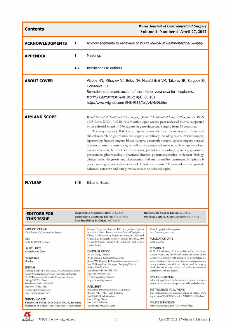
96 Resectionandreconstructionoftheinferiorvenacavaforneoplasms
Vladov NN, Mihaylov VI, Belev NV, Mutafchiiski VM, Takorov IR, Sergeev SK,
Odisseeva EH
102 Lymphomapresentingasanecroticcolonicmass
Konstantinidis IT, Probstfeld MR
REVIEW
CASE REPORT
BRIEF ARTICLE
World Journal of Gastrointestinal SurgeryVolume 4 Number 4 April 27, 2012
FLYLEAF
ACKNOWLEDGMENTS
ABOUT COVER
EDITORS FOR THIS ISSUE
Responsible Assistant Editor: Jin-Lei Wang Responsible Science Editor: Jin-Lei WangResponsible Electronic Editor: Xiao-Mei Zheng Proofing Editorial Office Director: Jin-Lei WangProofing Editor-in-Chief: Lian-Sheng Ma
APPENDIX
NAMEOFJOURNALWorld Journal of Gastrointestinal Surgery
ISSNISSN 1948-9366 (online)
LAUNCHDATENovember 30, 2009
FREQUENCYMonthly
EDITINGEditorial Board of World Journal of Gastrointestinal SurgeryRoom 903, Building D, Ocean International Center, No. 62 Dongsihuan Zhonglu, Chaoyang District, Beijing 100025, ChinaTelephone: +86-10-85381891Fax: +86-10-85381893E-mail: [email protected]://www.wjgnet.com
EDITOR-IN-CHIEFTimothy M Pawlik, MD, MPH, FACS, Associate Professor of Surgery and Oncology, Hepatobiliary
Surgery Program Director, Director, Johns Hopkins Medicine Liver Tumor Center Multi-Disciplinary Clinic, Co-Director of Center for Surgical Trials and Outcomes Research, Johns Hopkins Hospital, 600 N. Wolfe Street, Harvey 611, Baltimore, MD 21287, United States
EDITORIALOFFICEJin-Lei Wang, DirectorWorld Journal of Gastrointestinal SurgeryRoom 903, Building D, Ocean International Center, No. 62 Dongsihuan Zhonglu, Chaoyang District, Beijing 100025, ChinaTelephone: +86-10-85381891Fax: +86-10-85381893E-mail: [email protected]://www.wjgnet.com
PUBLISHERBaishideng Publishing Group Co., LimitedRoom 1701, 17/F, Henan Building, No.90 Jaffe Road, Wanchai, Hong Kong, ChinaFax: +852-31158812Telephone: +852-58042046
E-mail: [email protected]://www.wjgnet.com
PUBLICATIONDATEApril 27, 2012
COPYRIGHT© 2012 Baishideng. Articles published by this Open-Access journal are distributed under the terms of the Creative Commons Attribution Non-commercial Li-cense, which permits use, distribution, and reproduction in any medium, provided the original work is properly cited, the use is non commercial and is otherwise in compliance with the license.
SPECIALSTATEMENTAll articles published in this journal represent the view-points of the authors except where indicated otherwise.
INSTRUCTIONSTOAUTHORSFull instructions are available online at http://www.wjgnet.com/1948-9366/g_info_20100305152206.htm
ONLINESUBMISSIONhttp://www.wjgnet.com/1948-9366office/
Contents
AIM AND SCOPE
WJGS|www.wjgnet.com II April 27, 2012|Volume 4|Issue 4|
I AcknowledgmentstoreviewersofWorldJournalofGastrointestinalSurgery
I Meetings
I-V Instructionstoauthors
VladovNN,MihaylovVI,BelevNV,MutafchiiskiVM,TakorovIR,SergeevSK,
OdisseevaEH.
Resectionandreconstructionoftheinferiorvenacavaforneoplasms.
WorldJGastrointestSurg 2012;4(4):96-101
http://www.wjgnet.com/1948-9366/full/v4/i4/96.htm
World Journal of Gastrointestinal Surgery (World J Gastrointest Surg, WJGS, online ISSN 1948-9366, DOI: 10.4240), is a monthly, open-access, peer-reviewed journal supported by an editorial board of 336 experts in gastrointestinal surgery from 35 countries.
The major task of WJGS is to rapidly report the most recent results in basic and clinical research on gastrointestinal surgery, specifically including micro-invasive surgery, laparoscopy, hepatic surgery, biliary surgery, pancreatic surgery, splenic surgery, surgical nutrition, portal hypertension, as well as the associated subjects such as epidemiology, cancer research, biomarkers, prevention, pathology, radiology, genetics, genomics, proteomics, pharmacology, pharmacokinetics, pharmacogenetics, molecular biology, clinical trials, diagnosis and therapeutics and multimodality treatment. Emphasis is placed on original research articles and clinical case reports. This journal will also provide balanced, extensive and timely review articles on selected topics.
I-III EditorialBoard

Interval routine appendectomy following conservative treatment of acute appendicitis: Is it really needed?
George H Sakorafas, Dimitrios Sabanis, Christos Lappas, Aikaterini Mastoraki, John Papanikolaou, Charalambos Siristatidis, Vasileios Smyrniotis
George H Sakorafas, Dimitrios Sabanis, Christos Lappas, Aikaterini Mastoraki, Vasileios Smyrniotis, 4th Department of Surgery, Athens University, Medical School, Attikon University Hospital, GR-115 26 Athens, GreeceJohn Papanikolaou, Department of Gastroenterology, Athens University, Medical School, Attikon University Hospital, GR-115 26 Athens, GreeceCharalambos Siristatidis, Department of Obstetrics and Gyne-cology, Athens University, Medical School, Attikon University Hospital, GR-115 26 Athens, GreeceAuthor contributions: Sakorafas GH designed and wrote the paper; Sabanis D, Lappas C and Mastoraki A performed the lit-erature research; Papanikolaou J and Siristatidis C analyzed bib-liographical data; Smyrniotis V edited the paper.Correspondence to: George H Sakorafas, MD, Assistant Professor, 4th Department of Surgery, Athens University, Medi-cal School, Attikon University Hospital, Arkadias 19-21, GR-115 26 Athens, Greece. [email protected]: +30-210-7487192 Fax: +30-210-7487192Received: January 9, 2011 Revised: March 24, 2012Accepted: March 30, 2012Published online: April 27, 2012
AbstractConservative management of acute appendicitis (AA) is gradually being adopted as a valuable therapeutic choice in the treatment of selected patients with AA. This approach is based on the results of many recent studies indicating that it is a valuable and effective alternative to routine emergency appendectomy. Exist-ing data do not support routine interval appendectomy following successful conservative management of AA; indeed, the risk of recurrence is low. Moreover, recur-rences usually exhibit a milder clinical course compared to the first episode of AA. The role of routine interval appendectomy is also questioned recently, even in pa-tients with AA complicated by plastron or localized ab-scess formation. Surgical judgment is required to avoid
misdiagnosis when selecting a conservative approach in patients with a presumed “appendiceal” mass.
© 2012 Baishideng. All rights reserved.
Key words: Appendicitis; Surgery; Antibiotics; Interval appendicectomy; Plastron; Abscess
Peer reviewer: Grigory G Karmazanovsky, Professor, Depart-ment of Radiology, Vishnevsky Istitute of Surgery, B Serpuk-hovskaya street 27, Moscow 117997, Russia
Sakorafas GH, Sabanis D, Lappas C, Mastoraki A, Papanikolaou J, Siristatidis C, Smyrniotis V. Interval routine appendectomy following conservative treatment of acute appendicitis: Is it re-ally needed? World J Gastrointest Surg 2012; 4(4): 83-86 Available from: URL: http://www.wjgnet.com/1948-9366/full/v4/i4/83.htm DOI: http://dx.doi.org/10.4240/wjgs.v4.i4.83
INTRODUCTIONSince the first publication on acute appendicitis (AA) by Fitz et al[1] in 1886, surgical management of AA has been considered as a classical dogma for over one century. Emergency appendectomy has the advantage of immedi-ate resolution of a surgical problem, which is dealt with by a single admission, at a time when the benefit is most apparent to the patient and his/her family; this approach eliminates the problem of possible recurrences of AA and the initial uncertainty about the effectiveness and the outcome of conservative treatment. Despite the fact that appendectomy still remains the “gold standard” in the management of AA, during the last two decades there has been an increasing body of evidence suggesting that conservative management is a valuable alternative to sur-gery in selected patients with suspected AA, which can be used as the first line therapy for AA. This approach has
EDITORIAL
Online Submissions: http://www.wjgnet.com/[email protected]:10.4240/wjgs.v4.i4.83
World J Gastrointest Surg 2012 April 27; 4(4): 83-86ISSN 1948-9366 (online)
© 2012 Baishideng. All rights reserved.
83 April 27, 2012|Volume 4|Issue 4|WJGS|www.wjgnet.com

been shown to be effective in many recent publications (including clinical trials and meta-analyses). The main ad-vantage of the conservative approach is the elimination of the early and late morbidity (and mortality, albeit low) of an abdominal operation and general anesthesia. The effectiveness of this approach has been increased by the availability of new efficient antibiotics[2].
In evaluating the role of conservative management of AA, it is important to consider the need for interval ap-pendectomy. Obviously, if routine interval appendectomy is required, then conservative management of AA would seem unattractive as a therapeutic option for most cases since its main advantage (e.g., avoidance of surgery) is eliminated. On the other hand, if interval appendectomy is not routinely needed, then conservative management of AA would be the treatment of choice in a large percent-age of patients with suspected AA. The aim of this review is to critically summarize currently available data regarding the role of interval appendectomy in the management of patients with AA who were conservatively treated.
CONSERVATIVE MANAGEMENT OF AA: HOW EFFECTIVE IS IT?Success and recurrence rates are the two main end points when evaluating the effectiveness and long-term results of conservative management of AA. Many recent studies have shown that conservative treatment is effective in a high percentage of patients with AA. Success rates range in the literature between 68% and 95%[2-8]. Recurrences following conservative management may be observed in about 5%-14% of patients[9-13]. Recently, Kaminski et al[14] reported a 5% recurrence rate with a median follow-up of 4 years in 864 patients treated with antibiotics alone. Interestingly, recurrent episodes exhibited a milder clinical course than the first episode[14]. Dixon et al[15] reported a similar low incidence of recurrent appendicitis and found that subsequent attacks were less frequent and less severe. As expected, the identification of factors associated with a high risk of recurrence of AA would be of great inter-est for the clinician since, when present, the effectiveness of conservative management of AA is diminished. These risk factors should be taken into consideration when se-lecting patients for conservative or surgical management and include retained fecal stones, increased (> 4 mg/dL) CRP levels, elevated percent bands, partial small bowel obstruction on admission, etc.[7,16-22]. In the presence of these “risk factors”, emergency appendectomy should be strongly considered.
INTERVAL APPENDECTOMY FOLLOWING SUCCESSFUL CONSERVATIVE MANAGEMENT OF UNCOMPLICATED AA: IS IT NECESSARY?Although there are some groups suggesting routine inter-val appendectomy for all patients who have had nonsur-
gical treatment of an episode of AA, in clinical practice most surgeons question its routine use. The basic ques-tion which should be answered is the following: is the risk of surgery and general anesthesia justified by the risk of recurrent AA? The clinician should keep in his/her mind that appendectomy is associated with a small, albeit sig-nificant, morbidity and even mortality, despite being con-sidered a “routine” surgical procedure. Indeed, following emergency appendectomy, mortality ranges from 0.07% to 0.7% in patients without and 0.5% to 2.4% in patients with perforation[23-25]. Operative mortality increases in the presence of co-morbidity (e.g., heart and lung diseases, morbid obesity, etc.) and in aged patients (< 0.1% in pa-tients younger than 40 years, 2.6% in septuagenarians, 6.8% in octogenarians and 16.4% in nonagenarians)[24]. Morbidity rates range between 10% and 20% for AA without perforation and reach up to 30% for perforated appendicitis[2,9,26]. Common complications after appen-dectomy include wound and (more rarely) intraabdominal septic complications, adhesive small bowel obstruction (a long term complication requiring surgery in about 1.5% of patients by 30 years)[4,27]. Even the less invasive laparoscopic appendectomy is also associated with its one morbidity and even mortality rates.
Interval appendectomy could, however, be justified if the risk of recurrence was too high. However, the risk of recurrence is low (see above) but increases in the pres-ence of the “risk factors” mentioned above. Moreover, recurrences are usually characterized by a milder clinical course than the primary attack[15]. Therefore routine inter-val appendicectomy is probably not warranted following successful management of uncomplicated AA, given the low risk of recurrent appendicitis and the potential early and late complications of an elective operation[8,28-30].
INTERVAL APPENDECTOMY FOLLOWING SUCCESSFUL CONSERVATIVE MANAGEMENT OF COMPLICATED AA: IS IT ROUTINELY NECESSARY?Occasionally, a patient’s defense mechanisms may restrict and enclose the inflammation, resulting in the forma-tion of an inflammatory mass (phlegmon or plastron) of a contained (circumscribed) abscess. Typically, these inflammatory changes are observed some days (usually more than 4 d) after the onset of symptoms and more commonly in children (especially < 5 years)[2,10].
Patients with plastron formation Emergency surgery in these cases is not warranted; in-deed, under these circumstances surgery may be techni-cally demanding because of the distorted anatomy and the difficulties of closing the appendiceal stump because of the inflamed tissues. The risk of injury of adjacent organs (i.e., intestinal loops) is increased due to the presence of inflammatory changes and adhesions[13,30]. Moreover, the overstimulation of an already primed inflammatory sys-
84 April 27, 2012|Volume 4|Issue 4|WJGS|www.wjgnet.com
Sakorafas GH et al . Interval appendectomy: Is it routinely needed?

tem, with extensive stimulation of the cytokine cascade, may further complicate the postoperative course[11,31]. As a result, immediate surgery in these patients is associated with over a 3-fold increase in morbidity compared with conservative management[2]. Occasionally, the exploration ends with an ileocecal resection or a right-sided hemico-lectomy (in about 3% of patients) due to technical prob-lems or a suspicion of malignancy because of the distort-ed inflamed tissues[2,32]. For these reasons, in patients with AA complicated by inflammatory mass (plastron) forma-tion, the classical and recommended initial treatment is conservative with antibiotics[33]. Interval appendectomy is traditionally performed about 6 wk after the episode of AA to prevent recurrences and remove the offending or-gan to permanently resolve infection[33,34]. During this time of about 6-8 wk, the local inflammatory changes usually have subsided, the edematous and inflamed bowel has recovered and the patient is appropriately prepared[32-35]. However, the need for interval appendectomy after a suc-cessful nonsurgical treatment has recently been questioned as the risk of recurrence is relatively small[12,35-37]. This is-sue remains highly debated, with others proposing either delayed (i.e., appendectomy during the same admission, mainly to diminish sick leave) or routine interval appen-dectomy[38-40].
Patients with localized abscess formationNon-operative management has been proposed for the management of patients with localized abscess forma-tion due to perforated appendicitis[11]. Antibiotic therapy is successful in about 93% of these patients; in about 20% of them, image-guided percutaneous drainage of the abscess will eventually be required[2]. Interestingly, Nadler et al[7] suggested that patients with a phlegmon on imaging tests as opposed to an abscess are more likely to respond to conservative treatment and that the presence of a phlegmon reflected improved host defenses. These authors also suggested that the need for abscess drainage increases the failure rate, perhaps because of inadequate source control[7]. To date, the role of interval appendec-tomy in these patients has not been adequately evaluated.
POTENTIAL PROBLEMS, CONCERNS AND DISADVANTAGES OF OMITTING INTERVAL APPENDECTOMYSome authors have stated that in patients with AA treated conservatively without interval appendectomy, there is a risk (about 2%) of missing pathological findings, such as Crohn’s disease or neoplasms (most commonly, ap-pendiceal carcinoids)[2,41]. Immediate surgery with a right sided hemicolectomy, if needed, to avoid this problem, proposed by some authors as the definitive treatment in patients with complicated AA, is too aggressive an ap-proach[42-44] and has not been adopted by most surgeons. Nowadays, the availability and wide use of modern di-agnostic tools (including computed tomography and in-
terval colonoscopy) in selected patients have diminished the risk of misdiagnosis. Most colon cancer cases occur in patients over the age of 40 years. Therefore, patients older than 40 years should be followed-up with colonos-copy or computed tomography to exclude malignancy, especially when initial symptoms were atypical or in the presence of other suspicious findings (for example, ane-mia).
The risk of recurrence of appendicitis is a concern in patients with AA treated conservatively and without interval appendectomy. These patients should be coun-seled about the possibility of a recurrence of appendicitis and encouraged to seek medical attention early should symptoms recur. Most surgeons would advocate appen-dectomy (emergency or interval) in patients with multiple (> 2) recurrences. Personal preferences of the patient should also be taken into consideration in the process of management decision-making.
In conclusion, interval appendectomy is not routinely required in patients treated conservatively for AA. The risk of recurrence is low; moreover, potential recurrences usually have a mild clinical course. Interval (or emergency) operation should be considered in selected patients (for example, in the presence of “risk factors” indicating a high probability of recurrence, such as the presence of a retained fecalith) or following multiple (> 2 or 3) epi-sodes of AA. Patients with AA complicated by plastron or localized abscess formation should be treated conser-vatively initially; image-guided percutaneous drainage may be required to achieve drainage in patients with localized abscess. Despite that interval appendectomy is still per-formed by the majority of surgeons around the world, there is evidence that, even in these cases, interval appen-dectomy could be avoided. Currently, the lack of a suffi-cient body of evidence precludes firm recommendations. Surgical judgment is required to avoid misdiagnosis if such a conservative approach is adopted; further diagnos-tic evaluation may be required in selected patients (for ex-ample in patients > 40 years with anemia and a presumed “appendiceal” mass) to exclude malignancy. Personal pref-erences and specific conditions (for example, people living in remote or isolated areas without easy access to health facilities) should also be taken into consideration when deciding about the optimal management of each patient with AA (complicated or not).
REFERENCES1 Fitz RH. Perforating inflammation of the vermiform appen-
dix: with special reference to its early diagnosis and treat-ment. Am J Med Sci 1886; 92: 321-346
2 Andersson RE, Petzold MG. Nonsurgical treatment of ap-pendiceal abscess or phlegmon: a systematic review and meta-analysis. Ann Surg 2007; 246: 741-748
3 Varadhan KK, Humes DJ, Neal KR, Lobo DN. Antibiotic therapy versus appendectomy for acute appendicitis: a meta-analysis. World J Surg 2010; 34: 199-209
4 Hansson J, Körner U, Khorram-Manesh A, Solberg A, Lundholm K. Randomized clinical trial of antibiotic therapy versus appendicectomy as primary treatment of acute ap-pendicitis in unselected patients. Br J Surg 2009; 96: 473-481
85 April 27, 2012|Volume 4|Issue 4|WJGS|www.wjgnet.com
Sakorafas GH et al . Interval appendectomy: Is it routinely needed?

5 Styrud J, Eriksson S, Nilsson I, Ahlberg G, Haapaniemi S, Neovius G, Rex L, Badume I, Granström L. Appendectomy versus antibiotic treatment in acute appendicitis. a prospec-tive multicenter randomized controlled trial. World J Surg 2006; 30: 1033-1037
6 Eriksson S, Granström L. Randomized controlled trial of ap-pendicectomy versus antibiotic therapy for acute appendici-tis. Br J Surg 1995; 82: 166-169
7 Nadler EP, Reblock KK, Vaughan KG, Meza MP, Ford HR, Gaines BA. Predictors of outcome for children with perfo-rated appendicitis initially treated with non-operative man-agement. Surg Infect (Larchmt) 2004; 5: 349-356
8 Oliak D, Yamini D, Udani VM, Lewis RJ, Vargas H, Arnell T, Stamos MJ. Nonoperative management of perforated appen-dicitis without periappendiceal mass. Am J Surg 2000; 179: 177-181
9 Liu K, Ahanchi S, Pisaneschi M, Lin I, Walter R. Can acute appendicitis be treated by antibiotics alone? Am Surg 2007; 73: 1161-1165
10 Oliak D, Yamini D, Udani VM, Lewis RJ, Arnell T, Vargas H, Stamos MJ. Initial nonoperative management for periappen-diceal abscess. Dis Colon Rectum 2001; 44: 936-941
11 Brown CV, Abrishami M, Muller M, Velmahos GC. Appen-diceal abscess: immediate operation or percutaneous drain-age? Am Surg 2003; 69: 829-832
12 Willemsen PJ, Hoorntje LE, Eddes EH, Ploeg RJ. The need for interval appendectomy after resolution of an appendiceal mass questioned. Dig Surg 2002; 19: 216-20; discussion 221
13 Lugo JZ, Avgerinos DV, Lefkowitz AJ, Seigerman ME, Zahir IS, Lo AY, Surick B, Leitman IM. Can interval appendectomy be justified following conservative treatment of perforated acute appendicitis? J Surg Res 2010; 164: 91-94
14 Kaminski A, Liu IL, Applebaum H, Lee SL, Haigh PI. Rou-tine interval appendectomy is not justified after initial non-operative treatment of acute appendicitis. Arch Surg 2005; 140: 897-901
15 Dixon MR, Haukoos JS, Park IU, Oliak D, Kumar RR, Arnell TD, Stamos MJ. An assessment of the severity of recurrent appendicitis. Am J Surg 2003; 186: 718-722; discussion 722
16 Aprahamian CJ, Barnhart DC, Bledsoe SE, Vaid Y, Harmon CM. Failure in the nonoperative management of pediatric ruptured appendicitis: predictors and consequences. J Pediatr Surg 2007; 42: 934-938; discussion 938
17 Bufo AJ, Shah RS, Li MH, Cyr NA, Hollabaugh RS, Hixson SD, Schropp KP, Lasater OE, Joyner RE, Lobe TE. Interval appendectomy for perforated appendicitis in children. J Laparoendosc Adv Surg Tech A 1998; 8: 209-214
18 Shindoh J, Niwa H, Kawai K, Ohata K, Ishihara Y, Tak-abayashi N, Kobayashi R, Hiramatsu T. Predictive factors for negative outcomes in initial non-operative management of suspected appendicitis. J Gastrointest Surg 2010; 14: 309-314
19 Kogut KA, Blakely ML, Schropp KP, Deselle W, Hixson SD, Davidoff AM, Lobe TE. The association of elevated percent bands on admission with failure and complications of inter-val appendectomy. J Pediatr Surg 2001; 36: 165-168
20 Tsai HM, Shan YS, Lin PW, Lin XZ, Chen CY. Clinical analy-sis of the predictive factors for recurrent appendicitis after initial nonoperative treatment of perforated appendicitis. Am J Surg 2006; 192: 311-316
21 Levin T, Whyte C, Borzykowski R, Han B, Blitman N, Har-ris B. Nonoperative management of perforated appendicitis in children: can CT predict outcome? Pediatr Radiol 2007; 37: 251-255
22 Nitecki S, Karmeli R, Sarr MG. Appendiceal calculi and fe-caliths as indications for appendectomy. Surg Gynecol Obstet
1990; 171: 185-18823 Blomqvist PG, Andersson RE, Granath F, Lambe MP,
Ekbom AR. Mortality after appendectomy in Sweden, 1987-1996. Ann Surg 2001; 233: 455-460
24 Blomqvist P, Ljung H, Nyrén O, Ekbom A. Appendectomy in Sweden 1989-1993 assessed by the Inpatient Registry. J Clin Epidemiol 1998; 51: 859-865
25 Luckmann R. Incidence and case fatality rates for acute ap-pendicitis in California. A population-based study of the ef-fects of age. Am J Epidemiol 1989; 129: 905-918
26 Hale DA, Molloy M, Pearl RH, Schutt DC, Jaques DP. Ap-pendectomy: a contemporary appraisal. Ann Surg 1997; 225: 252-261
27 Andersson RE. Small bowel obstruction after appendicec-tomy. Br J Surg 2001; 88: 1387-1391
28 Eriksson S, Styrud J. Interval appendicectomy: a retrospec-tive study. Eur J Surg 1998; 164: 771-774; discussion 775
29 Puapong D, Lee SL, Haigh PI, Kaminski A, Liu IL, Apple-baum H. Routine interval appendectomy in children is not indicated. J Pediatr Surg 2007; 42: 1500-1503
30 Tekin A, Kurtoğlu HC, Can I, Oztan S. Routine interval appendectomy is unnecessary after conservative treatment of appendiceal mass. Colorectal Dis 2008; 10: 465-468
31 Moore FA, Moore EE. Evolving concepts in the pathogen-esis of postinjury multiple organ failure. Surg Clin North Am 1995; 75: 257-277
32 Ahmed I, Deakin D, Parsons SL. Appendix mass: do we know how to treat it? Ann R Coll Surg Engl 2005; 87: 191-195
33 Nitecki S, Assalia A, Schein M. Contemporary management of the appendiceal mass. Br J Surg 1993; 80: 18-20
34 Thomas DR. Conservative management of the appendix mass. Surgery 1973; 73: 677-680
35 Karaca I, Altintoprak Z, Karkiner A, Temir G, Mir E. The management of appendiceal mass in children: is interval ap-pendectomy necessary? Surg Today 2001; 31: 675-677
36 Hurme T, Nylamo E. Conservative versus operative treat-ment of appendicular abscess. Experience of 147 consecutive patients. Ann Chir Gynaecol 1995; 84: 33-36
37 Tingstedt B, Bexe-Lindskog E, Ekelund M, Andersson R. Management of appendiceal masses. Eur J Surg 2002; 168: 579-582
38 Garg P, Dass BK, Bansal AR, Chitkara N. Comparative eval-uation of conservative management versus early surgical intervention in appendicular mass--a clinical study. J Indian Med Assoc 1997; 95: 179-180, 196
39 Kumar S, Jain S. Treatment of appendiceal mass: prospec-tive, randomized clinical trial. Indian J Gastroenterol 2004; 23: 165-167
40 Marya SK, Garg P, Singh M, Gupta AK, Singh Y. Is a long delay necessary before appendectomy after appendiceal mass formation? A preliminary report. Can J Surg 1993; 36: 268-270
41 Mazziotti MV, Marley EF, Winthrop AL, Fitzgerald PG, Walton M, Langer JC. Histopathologic analysis of interval appendectomy specimens: support for the role of interval appendectomy. J Pediatr Surg 1997; 32: 806-809
42 Thompson JE, Bennion RS, Schmit PJ, Hiyama DT. Cecec-tomy for complicated appendicitis. J Am Coll Surg 1994; 179: 135-138
43 Sarkar R, Bennion RS, Schmit PJ, Thompson JE. Emergent ileocecectomy for infection and inflammation. Am Surg 1997; 63: 874-877
44 Lane JS, Schmit PJ, Chandler CF, Bennion RS, Thompson JE. Ileocecectomy is definitive treatment for advanced appendi-citis. Am Surg 2001; 67: 1117-1122
S- Editor Wang JL L- Editor Roemmele A E- Editor Zheng XM
86 April 27, 2012|Volume 4|Issue 4|WJGS|www.wjgnet.com
Sakorafas GH et al . Interval appendectomy: Is it routinely needed?

Malignant ascites: A review of prognostic factors, pathophysiology and therapeutic measures
Suma L Sangisetty, Thomas J Miner
Suma L Sangisetty, Thomas J Miner, Department of Surgery, Warren Alpert Medical School of Brown University, Rhode Is-land Hospital, Providence, RI 02903, United StatesAuthor contributions: All the two authors contributed to this review.Correspondence to: Thomas J Miner, MD, FACS, Assistant Professor, Department of Surgery, Warren Alpert Medical School of Brown University, Rhode Island Hospital, 593 Eddy Street, APC443, Providence, RI 02903, United States. [email protected]: +1-401-444-2892 Fax: +1-401-444-6681Received: June 2, 2011 Revised: December 31, 2011Accepted: January 10, 2012Published online: April 27, 2012
AbstractMalignant ascites indicates the presence of malignant cells in the peritoneal cavity and is a grave prognostic sign. While survival in this patient population is poor, averaging about 20 wk from time of diagnosis, quality of life can be improved through palliative procedures. Selecting the appropriate treatment modality remains a careful process, which should take into account po-tential risks and benefits and the life expectancy of the patient. Traditional therapies, including paracentesis, peritoneovenous shunt placement and diuretics, are successful and effective in varying degrees. After care-ful review of the patient’s primary tumor origin, tumor biology, tumor stage, patient performance status and comorbidities, surgical debulking and intraperitoneal chemotherapy should be considered if the benefit of therapy outweighs the risk of operation because sur-vival curves can be extended and palliation of symp-tomatic malignant ascites can be achieved in select pa-tients. In patients with peritoneal carcinomatosis who do not qualify for surgical cytoreduction but suffer from the effects of malignant ascites, intraperitoneal chemo-therapy can be safely and effectively administered via laparoscopic techniques. Short operative times, short hospital stays, low complication rates and ultimately
symptomatic relief are the advantages of laparoscopi-cally administering heated intraperitoneal chemother-apy, making it not only a valuable treatment modality but also the most successful treatment modality for achieving palliative cure of malignant ascites.
© 2012 Baishideng. All rights reserved.
Key words: Carcinomatosis; Peritoneal; Paracentesis; Peritovenous shunts; HIPEC
Peer reviewers: Grigory G Karmazanovsky, Professor, Depart-ment of Radiology, Vishnevsky Istitute of Surgery, B Serpuk-hovskaya Street 27, Moscow 117997, Russia; Dr. Sreenivasan Karuparthi, Department of Surgical Gastroenterology, Sri Ram-achandra University, Chennai 600116, India
Sangisetty SL, Miner TJ. Malignant ascites: A review of prog-nostic factors, pathophysiology and therapeutic measures. World J Gastrointest Surg 2012; 4(4): 87-95 Available from: URL: http://www.wjgnet.com/1948-9366/full/v4/i4/87.htm DOI: http://dx.doi.org/10.4240/wjgs.v4.i4.87
INTRODUCTIONMalignant ascites is a sign of peritoneal carcinomatosis, the presence of malignant cells in the peritoneal cav-ity. Tumors causing carcinomatosis are more commonly secondary peritoneal surface malignancies which include: ovarian, colorectal, pancreatic and uterine; extra-abdom-inal tumors originating from lymphoma, lung and breast; and a small number of unknown primary tumors. Malig-nant ascites accounts for approximately 10% of all cases of ascites[1]. The presence of malignant ascites is a grave prognostic sign. While survival in this patient population is poor, averaging about 20 wk from time of diagnosis, quality of life can be improved through palliative proce-dures[2]. Currently no effective anti-tumor therapy exists for peritoneal carcinomatosis. Given the uncertainty sur-
REVIEW
Online Submissions: http://www.wjgnet.com/[email protected]:10.4240/wjgs.v4.i4.87
World J Gastrointest Surg 2012 April 27; 4(4): 87-95ISSN 1948-9366 (online)
© 2012 Baishideng. All rights reserved.
87 April 27, 2012|Volume 4|Issue 4|WJGS|www.wjgnet.com

rounding the disease process and formation of malignant ascites, the therapeutic options are limited and often the goal of treatment is to target palliation of symptoms, which can include abdominal pain, dyspnea, nausea, vomiting and anorexia. In this paper, we will provide a review of the prognostic factors of malignant ascites, the pathophysiology of ascites formation, current diagnostic modalities, traditional therapeutic measures and newer therapies, including current medical and surgical treat-ment options.
PATHO-PHYSIOLOGYThe pathophysiology of malignant ascites is multifacto-rial. It is postulated that ascites formation is related to a combination of altered vascular permeability and ob-structed lymphatic drainage. A careful understanding of the peritoneum, the lymphatic system and the dynamic flow of fluid are needed to elucidate the mechanisms of malignant ascites formation. Five microscopic barriers exist which prevent movement of proteins away from the intravascular space: capillary endothelium, capillary base-ment membrane, interstitial stroma, mesothelial basement membrane and mesothelial cells of the peritoneal lining. By means of a combination of mechanical and selective mechanisms, including tight junctions and anionic macro-molecules, an effective barrier is maintained, preventing leakage of protein molecules into the peritoneal cavity. In 1922, Putnam described the peritoneal membrane as a “living membrane,” of which crystalloid solutions instituted into the peritoneal cavity equilibrated between the peritoneal cavity and the serum. The movement of colloid was not well understood, however, described as being transmitted in one direction into the serum from the peritoneal cavity, by means of some “vital (membrane) activity”, possibly phagocytosis or mechanical filtration through intercellular spaces[3]. The relative impermeability of the capillary membrane to proteins is the basis for osmotic gradients, described by Starling’s equation of capillary forces, which states that the exchange of fluid between the plasma and interstitium is dependent on the hydraulic and oncotic pressure in each compartment. On-cotic pressure differences are the basis for fluid reabsorp-tion from the interstitial space and prevention of edema formation.
While macromolecules, proteins and cells do not preferentially leave the intravascular space, they do ac-cumulate in the peritoneal cavity and may return to the systemic circulation by means of the peritoneal lymphatic system. Recklinghausen first described lymphatic stomata, small openings of lymphatics that connect the body cav-ity and lymphatic lumen, responsible for movement of large particles into the vascular space[4]. Fukuo et al[5] dem-onstrated three lymphatic pathways in the abdomen using India ink injection and transmission electron microscopy. The principal pathway begins with the lymphatic stomata, entering the peritoneal lymphatics via networks in the diaphragm, undergoing filtration through regional lymph
nodes of the diaphragm, and eventually emptying into the thoracic duct[5]. These mechanisms of osmotic gra-dients and lymphatic drainage allow for a dynamic fluid balance between the peritoneal cavity and the intravascu-lar space, such that the osmolality of the peritoneal space is constantly changing.
As early as 1953, Holm-Nielson demonstrated that in mice with malignant ascites, India ink injected into the peritoneal cavity remained in the peritoneal cavity, sug-gesting lymphatic obstruction as a major factor in patho-genesis of malignant ascites[6]. Feldman later showed that in mice inoculated with tumor cells, radioactive labeled erythrocytes injected into the intra-peritoneal space failed to return to the intravascular space as they did in normal mice due to tumor infiltrating the lymphatics, confirmed by histological evaluation, and subsequent to these events was the formation of ascites[7]. Nagy et al[8] demonstrated that radioactive albumin transport into the intravascular space was reduced after tumor injection and that this reduction preceded any significant increases in tumor burden. Additionally, radio-labeled red blood cells did not enter the intraperitoneal space at any increased rates until tumor burden had increased by at least 10 fold. As-cites fluid accumulation did not occur until late stages of tumor growth[8]. These studies demonstrate the impor-tance of lymphatic obstruction in tumor related ascites. Although many authors have offered theories regarding tumor metastasis, it is not clear why cancer cells prefer-entially localize to the peritoneal cavity rather than other sites and cause malignant ascites[9,10].
The quality of fluid in patients with malignancy relat-ed ascites due to peritoneal carcinomatosis is distinctive, with positive cytology, high ascitic fluid protein concen-trations and low serum-ascites albumin gradient[11]. The high protein content of malignant ascites indicates that there is an alteration in vascular permeability to allow for large molecules to accumulate in the intraperitoneal space. Senger at al[12] showed that vessels of the perito-neal lining of experimental animals with tumor ascites were significantly more permeable, due to the presence of a permeability factor found only in tumor ascites. When Garrison et al[2] infused cell-free malignant asci-tes into the intraperitoneal space, an increase in edema formation in the omental vessels and an increase in the concentration of protein in the interstitial space were observed, thus implicating a tumor-induced factor that alters vessel permeability and promotes the formation of malignant ascites. This vascular permeability factor, known as vascular endothelial growth factor (VEGF), is responsible for allowing a varying degree of movement of micro and macromolecules across the vascular endo-thelium, in the setting of normal physiological states, in addition to pathological disease states, ranging from acute inflammation, wound healing and menstruation to tumor angiogenesis[13]. Zebrowski et al[14] showed that VEGF levels were significantly higher in malignant ascites when compared to nonmalignant ascites, and when cirrhotic as-cites was exposed to VEGF, endothelial cell permeability
88 April 27, 2012|Volume 4|Issue 4|WJGS|www.wjgnet.com
Sangisetty SL et al . A review of malignant ascites

increased. The addition of VEGF neutralizing antibodies to malignant ascites reduced this permeability. Of note, exposure of cirrhotic ascites to cells had a similar effect on endothelial permeability, suggesting factors other than VEGF have a role in malignant ascites formation[14]. Al-though not clearly a mechanism behind malignant ascites formation, ascites in cirrhotic patients has been associ-ated with splanchnic hyperemia, thought due perhaps to tumor necrosis factor[15,16].
Thus, it is apparent that the formation of malignant ascites is a complex, multifactorial process. The mecha-nism for fluid and protein accumulation in the intraperi-toneal space associated with cancer appears to be second-ary to a combination of impaired lymphatic drainage and increased vascular permeability. These processes are intertwined, allowing for net filtration that overwhelms the ability of the lymphatic system to drain the peritoneal space, particularly when obstructed by increasing tumor burden.
DIAGNOSISIn 52%-54% of cases of peritoneal carcinomatosis, asci-tes is the first detected sign of intra-abdominal malignan-cy[2,17]. The causes of intra-abdominal fluid production are many, including cirrhosis, congestive heart failure, nephrosis, pancreatitis, peritonitis, primary malignancy or hepatic metastases. It is not possible to distinguish benign ascites from malignant ascites by physical exam or radio-graphic techniques alone. Invasive testing is necessary to differentiate the two types. Abdominal paracentesis with ascitic fluid analyses can diagnose malignant causes of ascites production in most cases, but laparoscopic tissue sampling may be necessary. Ascitic fluid analysis consists of microscopic, chemical and cytological evaluation to help differentiate between infectious, inflammatory and malignancy induced ascites formation. In patients with peritoneal carcinomatosis, the ascites fluid has posi-tive cytology, elevated protein concentrations and a low serum-ascites albumin gradient[8]. While in some reports cytology is diagnostic in only 50%-60% of cases of ma-lignant ascites, it has been demonstrated that up to 97% of patients with peritoneal carcinomatosis have positive cytology, indicating that the tumor is shedding cells into the peritoneal cavity, making it a highly sensitive test and the gold standard for diagnosing peritoneal carcinoma-tosis[11,18]. In patients with peritoneal carcinomatosis and hepatic metastases, fluid cytology is positive and ascites protein concentrations are variable, but the serum-ascites albumin gradient remains elevated, with the addition of a markedly elevated serum alkaline phosphatase level (> 350 mg/dL)[11]. The addition of tumor markers, especially CEA, CA-125 and α fetoprotein, are not reliable in diag-nosing malignancy but they can aid in identifying the pri-mary tumor causing malignant ascites. The biochemical properties of ascites fluid, including fibronectin, choles-terol, lactate dehydrogenase, sialic acid, telomerase activ-ity and proteases, have been studied and, while clinically helpful, they have not yet been found to be reliable in dif-
ferentiating between malignant and benign ascites. Tumor and biochemical markers along with the morphological features of the cytological smear, immunohistochemical staining and clinical history are important in determining both the presence of malignancy related ascites and the primary sites of metastatic carcinomas[19].
If the diagnostic workup does not reveal the primary source of malignancy but confirms the presence of a malignancy, a search for the tumor of origin should be pursued. In male patients with positive cytology, whose diagnostic workup remains negative despite blood tests and radiological imaging, it may not be useful to pursue further investigations because knowing the tumor of origin may not affect management or outcome. However, in female patients, if the conventional methods have failed to demonstrate the tumor of origin, laparoscopy or laparotomy should be performed for tissue diagnosis, be-cause patients with an ovarian malignancy are responsive to tumor debulking and chemotherapy and their survival outcomes are better.
SURVIVAL The prognostic factors associated with malignant ascites have been poorly studied, further complicating manage-ment decisions. A retrospective review of 76 patients with malignant ascites performed by Mackey et al[20], where median survival was determined to be 11.1 wk from time of diagnosis, showed that significant predictors of poor prognosis included presence of edema, depressed serum albumin and liver metastases, while prolonged survival was found in patients with ovarian cancer. Survival curves did not differ between patients with known cancers and unknown primary malignancies or between patients with ascites as the initial presentation of malignancy and patients with a known prior malignancy[20]. In another study by Garrison et al[2], it was demonstrated that tumors originating from the female reproductive system had the longest survivals, with a mean survival of 19 wk, and foregut adenocarcinomas had the poorest survivals, with a mean survival of 10 wk from the onset of ascites. Addi-tionally, patients with high protein concentrations within the ascitic fluid did better than those with transudative ascitic fluid[2]. Ayantunde et al[17] showed that the presence of liver metastases and low levels of serum and ascites protein concentrations, although related, were indepen-dent prognostic factors associated with poorer outcomes. Furthermore, low protein levels are also associated with poor nutritional reserve and depressed immune function, adversely affecting this patient population. Malignant as-cites thus carries a grave prognosis. Although the clinical outcome cannot be altered and survival times are limited, a successful goal of treatment is to palliate the symptoms of malignant ascites.
TRADITIONAL THERAPYSeveral treatment modalities can alleviate the symptoms associated with malignant ascites. Because the natural
89 April 27, 2012|Volume 4|Issue 4|WJGS|www.wjgnet.com
Sangisetty SL et al . A review of malignant ascites

history of ascites formation is poorly understood, these measures and quality of life data is limited and the effica-cy of existing treatments is difficult to assess. Traditional modalities for managing malignant ascites include sodium restricted diets, diuretic therapy, serial paracentesis and peritoneovenous shunting. In a survey of practice mea-sures for managing malignant ascites, it was determined that paracentesis was most often utilized (98%) and it was perceived to be most effective (89%). Diuretics were used by 61% but were not felt to be as effective (45%)[21].
ParacentesisReview of the literature demonstrates a clear benefit from paracentesis in achieving symptomatic relief. Fischer described a simple, safe and effective method of insert-ing a 14-gauge needle with a 16-gauge catheter into the free peritoneal cavity, draining up to nine liters at a time with concurrent intravenous fluids running to prevent hypotension due to rapid vascular space depletion[22]. The durability of paracentesis remains an issue as symptoms often return within 72 h. Theoretically, therapeutic agents could be administered via the catheter but this method is not used anymore due to the potential for adhesion formation and intestinal obstruction[22]. Approximately 93% of patients show relief of nausea, vomiting, dys-pnea and/or abdominal discomfort[23,24]. Complications of therapeutic taps include pain, perforation, hypoten-sion and secondary peritonitis. Paracentesis is effective in relieving the symptoms associated with malignant ascites but it requires repeated treatments, leads to frequent hospitalizations, depletes the patients of protein and elec-trolytes, and exposes the patient to a small but significant risk of peritonitis.
Peritovenous shunts In 1974, LeVeen first introduced the peritoneovenous shunt to surgically treat patients with refractory ascites secondary to cirrhosis. The LeVeen shunt returns ascites fluid to the venous system via a one way pressure acti-vated valve shunt mechanism that mimics physiological mechanisms. The Denver shunt, originally designed to overcome the frequent complication of shunt occlusion occurring with the LeVeen shunt, features a compress-ible pump chamber bearing a pressure sensitive valve, which opens when positive pressure exceeds 1 cm of water[25]. There appears to be no particular type of Peri-tovenous shunts (PVS) shown to be more effective or superior, with complication rates similar between the two types[26,27].
Peritoneovenous shunts are used to reduce the need for repeated paracentesis and relieve the symptoms as-sociated with increased intra-abdominal pressure second-ary to ascites and the resulting protein and fluid deple-tion. Patients must be carefully selected for PVS. These patients typically have failed conservative therapies and have rapid production of ascites or poor response to diuretics. Patients benefit from PVS because its use pre-serves serum albumin levels. Quality of life is preserved
through less frequent need for paracentesis. In 75%-78% of patients, malignant ascites is controlled by PVS and the mean duration of shunt patency is 10-12 wk[23,24].
This treatment should be offered to patients judiciously as it does require perioperative hospitalization. Although overall days in hospital are reduced, PVS surgery carries an operative risk of mortality between 10% and 20% in an already tenuous patient[28]. In reviewing the literature, 20% of PVS are associated with complications; these are most frequently shunt occlusion (19%-26%), pul-monary edema (9.5%-12%) and pulmonary embolism (5%-7%)[19,20]. Other reported complications include ascitic leak from insertion site, subclinical disseminated intravascular coagulopathy (76%), clinical disseminated intravascular coagulopathy (2%), infection (5%) and gas-trointestinal bleeding[24,28]. In approximately 3%-7% of patients, tumor emboli were demonstrated at autopsy[23,24]. Despite the direct infusion of viable malignant cells into the circulation, tumor implants were generally uncom-mon and if present, these metastases were clinically as-ymptomatic and did not affect survival[29]. Hemorrhagic ascites and elevated ascitic fluid protein concentration are associated with higher risk of shunt occlusion and therefore are considered contraindications to PVS[24,28]. Patients with loculated malignant effusions do not ben-efit from PVS. Relative contraindications for PVS include advanced congestive heart failure or renal failure because PVS is associated with volume overload. Also demon-strated as a relative contraindication is the presence of positive cytology, with 75% of complications occurring in this group, including early shunt failure, postoperative coagulopathy, infection and tumor emboli[30].
PVS is not without risks and complications but in carefully selected patients, it can alleviate symptoms as-sociated with malignant ascites. Patients with breast and ovarian cancer had the best response rate (> 50%), while patients with gastrointestinal malignancies did worse (10%-15% response); therefore, it is often suggested that PVS should not be implemented in patients with GI can-cers[18,28].
Diuretics Diuretics benefit few patients with malignant ascites in a predictable fashion and when used in high doses, may cause systemic blood volume depletion, electrolyte ab-normalities and renal dysfunction. Diuretics appear to be successful in achieving symptomatic relief in 43%-44% of cases reported in the literature[23,24]. Greenway et al[31] described good symptomatic control of ascites with large doses of spironolactone (150-400 mg/d) in a small group of patients who showed a clear retention of sodium and elevated plasma renin activity, with the most common side effect encountered being nausea and vomiting and no occurrences of electrolyte imbalances or renal dys-function. It appears that patients with cancer who have ascites caused by portal hypertension secondary to hepat-ic metastases benefit most from diuretic therapy[32]. When peritoneal carcinomatosis is complicated by hepatic
90 April 27, 2012|Volume 4|Issue 4|WJGS|www.wjgnet.com
Sangisetty SL et al . A review of malignant ascites

metastases, the quality of the ascites fluid and the mecha-nism of fluid production differ and can be compared to fluid production in patients with cirrhosis. In cirrhotic patients, portal hypertension is present and is associated with an elevated serum-ascites albumin gradient, second-ary to the efflux of protein from the intravascular space into the peritoneal space, where the protein concentra-tion is related to the degree of portal pressure[33]. In both groups of patients, circulating blood volume is reduced and the renin-angiotensin-aldosterone system is activated, leading to sodium retention. Diuretics such as spirono-lactone serve as competitive antagonists to aldosterone, thereby decreasing the reabsorption of water and sodium in the renal collecting duct. Pockros et al[32] demonstrated elevated renin levels in patients with massive hepatic me-tastases compared to normal renin levels in patients with ascites secondary to peritoneal carcinomatosis. Further-more, diuretic use resulted in the mobilization of ascites fluid and approximately 1 kg/d in weight loss, without symptomatic hypotension or renal dysfunction in the hepatic metastases group compared to 0.5 kg/d in weight loss with subsequent hypotension and renal dysfunction occurring in the peritoneal carcinomatosis group[32].
NEWER THERAPYIn the cases of primary malignancies without metastases, surgical resection with completely negative microscopic margins confers a better survival and is the basis of surgi-cal oncology. Historically, operative intervention in cases of malignant ascites arising from peritoneal carcinoma-tosis was reserved for palliation of symptoms or emer-gent need to relieve obstruction or perforation. While clearance of tumor burden in patients with peritoneal carcinomatosis is often unachievable, investigations into aggressive cytoreductive surgery combined with intra-peritoneal chemotherapy, either in the intraoperative set-ting with hyperthermia (known as HIPEC) or/and in the early postoperative setting (known as EPIC), has served as a premise for improving survival benefit in addition to preventing or palliating future development of malignant ascites.
With regard to gastrointestinal cancer, peritoneal re-currence of tumor will occur in up to 29% of patients[34]. Prior to operative intervention, subclinical metastases, which escape preoperative CT scans and direct visu-alization during surgery, are present. These progress and spread further via hematogenous dissemination or lymphatic spread to distant sites of metastases and be-come clinically apparent months to years after resection. Tumor cells may enter the vascular or lymphatic spaces during surgical resection but these do not become clini-cally significant if the vessels remain intact, due to the high resistance of these endothelial lined channels to tumor proliferation, described by Weiss as the “theory of metastatic insufficiency”[35]. These tumor cells often die without harming the host. A separate mechanism exists to potentiate tumor recurrence at the resection site and in
the peritoneum. Even after aggressive attempts at resec-tion, tumor burden may remain at the microscopic level. The “tumor cell entrapment hypothesis” claims that local trauma during surgery is responsible for dislodging mi-croscopic tumor emboli by tumor manipulation or lym-phovascular vessel transection. These tumor cells then have the potential to implant onto the raw surfaces of neighboring peritoneum. Once this occurs, healing and restorative processes encase tumor cells within avascular intraperitoneal adhesions, precluding cancer from natural host defense mechanisms and systemic chemotherapy[36]. This theory led to the conception of perioperative intra-peritoneal chemotherapy, instilled into the abdomen up to 7 d postoperatively to target microscopic disseminated disease within the peritoneal cavity.
Direct intra-peritoneal administration of chemotherapy compared to systemic chemotherapy achieves higher tissue concentration, delivering cytotoxic agents up to 2-3 mm of the peritoneal layer without systemic absorption or toxicity[36]. Hyperthermia offers additional cytotoxic ef-fect by inhibiting cellular mechanisms of replication and repair and is synergistic, starting at a temperature of 39 degrees Celsius when used with chemotherapeutic agents. Hyperthermic intra-peritoneal chemotherapy is beneficial when timed directly after complete cytoreduction is first achieved, as the depth of penetration is further limited by postoperative fibrin deposition and adhesion formation. Intra-peritoneal chemotherapy can be administered via the open or closed techniques. The open technique is believed to distribute thermal energy homogenously employing the properties of spatial diffusion. Closed abdominal che-motherapy allows for increased intra-abdominal pressure, which is believed to drive deeper penetration of chemo-therapeutic agents without increasing the risk of exposure to the surgical team. There are no prospective trials that compare the efficacy of the open vs the closed techniques.
Selection criteria to determine the type of patient that will best benefit from perioperative intraperitoneal che-motherapy includes primary tumor origin, tumor biology, tumor stage, prior treatment with systemic chemotherapy or surgical resection and responses to those, patient performance status and comorbidity, and most impor-tant, effectiveness of surgical debulking. Roviello et al[37] showed that postoperative complications occurred in 44% of patients undergoing cytoreductive surgery with intraperitoneal chemotherapy. These complications most commonly included wound infection, hematological tox-icity, intestinal fistula and symptomatic pleural effusion requiring drainage. Reoperation was necessary in 8% of patients studied and mortality rate was 1.6%. Indepen-dent predictors of morbidity included residual tumor after resection and age. Probability of survival was higher in patients with ovarian or colorectal cancer compared to gastric cancer. Further review of the literature dem-onstrates morbidity rates associated with cytoreduction and intra-peritoneal chemotherapy ranging from 24.5% to 54% and mortality rates ranging from 1.5% to 4%[38]. When complete cytoreductive surgery was possible, me-
91 April 27, 2012|Volume 4|Issue 4|WJGS|www.wjgnet.com
Sangisetty SL et al . A review of malignant ascites

dian survival was 32.4 mo compared to 8.4 mo in the incomplete resection group. Independent prognostic indicators associated with favorable outcomes were com-plete cytoreduction, treatment by a second procedure, limited peritoneal carcinomatosis, age less than 65 years, and use of adjuvant chemotherapy. Negative independent prognostic factors included the use of neoadjuvant che-motherapy, involvement of lymph nodes, presence of he-patic metastases, and poor histological differentiation[39]. Two separate trials dedicated to the analysis of complica-tion rates and associated morbidity point to the duration of surgery and number of resections and peritonectomy procedures as being associated with the greatest predictor of complication[39,40].
A consensus statement was formed by seventy-five surgical oncologists regarding the use of cytoreductive surgery and hyperthermic intraperitoneal chemotherapy in the management of peritoneal malignancies of colonic origin. Review of the literature identified a subset of patients, in whom complete cytoreduction was achieved and combined with heated intraperitoneal mitomycin C and postoperative systemic chemotherapy. These patients had metastatic disease of colonic origin and were found to have a median survival up to 42 mo. Clinical and ra-diological evidence that were associated with successful complete cytoreduction (R0/R1 by the R scoring sys-tem or CC-0/CC-1 by the completion of cytoreduction score) included an Eastern Cooperative Oncology Group performance status of two or less, no evidence of extra-abdominal disease, up to three small, resectable parenchy-mal hepatic metastases, no evidence of biliary, ureteral or more than one site of intestinal obstruction, no small bowel involvement which included the mesentery, and a small volume of disease in the gastro-hepatic ligament. The treatment pathway to identify which patients would benefit most from surgical intervention was thus delin-eated. Those patients with recurrent and/or metastatic colon cancer with peritoneal involvement and a good performance status, a good response to systemic therapy, and/or limited liver involvement should be considered for cytoreductive surgery and hyperthermic intraperito-neal chemotherapy. If complete cytoreduction cannot be clearly achieved, surgical intervention should be reserved for circumstances in which palliation is the goal[41].
Although the amount of residual disease left after at-tempted cytoreduction has been demonstrated to predict prognosis, categorizing a resection as complete or incom-plete has become a focus of concern. Surgeons employ a variety of methodologies in determining the complete-ness of cytoreduction. Up to 74% of experts surveyed consider the completeness of cytoreduction (CC) score to be the best classification system for residual disease[42]. This score proposed by Sugarbaker is based on a maximal intratumoral penetration of cisplatin (2.5 mm). This value was obtained in a controlled experimental setting using a microscope that is not used at the time of operation and does not apply to other frequently used chemotherapeu-tic agents. Instead, residual disease is classified using the
CC score based on remaining macroscopic disease, thus leading to observer variability.
It is known that cytoreductive surgery and hyper-thermic intraperitoneal chemotherapy is associated with high morbidity. Several instruments were developed to assess quality of life in long-term survivors. In various forms, these measure physical, functional, social/family and emotional well-being. Piso et al[43] performed a review of short and long-term quality of life assessments in patients undergoing cytoreductive surgery followed by intra-peritoneal chemotherapy. Review of the literature shows that while quality of life is initially impaired by sur-gery and postoperative complications, functional status returns to baseline, with little to no limitations in most patients, beginning at 3 mo post-treatment[44]. There are no randomized clinical trials of cytoreductive surgery and intraperitoneal chemotherapy that also evaluate quality of life. Assessment of the quality of life in this patient population with an already limited life expectancy cannot be overlooked and should be included in clinical trials that assess the efficacy of this treatment.
A poorer overall survival has been reported in pa-tients with non-ovarian malignant ascites and evidence of malnutrition with a median survival of 23 mo compared to 89.9% 1 year survival when ascites was absent[45,46]. In a Phas Ⅰ/Ⅱ study conducted by Loggie et al[46], it was demonstrated that combined treatment of radical surgi-cal debulking and intra-peritoneal heated chemotherapy using mitomycin C was an effective means to provide pal-liation by preventing recurrence of ascites in up to 75% of patients for a median duration up to 7.5 mo. Radical debulking was scored as a R2 in 78% of these patients, but the association of R2 resection with the halting of ascites formation was not reported. Positive peritoneal cytology without gross ascites was observed in 35.3% of patients studied. Administration of intra-peritoneal heated chemotherapy prevented the development of ascites in all of these patients for a median duration up to 9.4 mo. Patients without positive cytology never developed ascites, suggesting that intraperitoneal administration of chemo-therapy can prevent formation of malignant ascites[46]. Patient selection criteria included absence of serious end organ dysfunction, absence of hepatic metastases, normal coagulation profile, albumin greater than 2.8 g/dL, liver function tests less than three times normal, and serum creatinine less than 2.0 mg/dL, which may account for the high success rate in this highly selected subgroup. In an-other Phase Ⅱ trial, Bitran showed that the intraperitoneal administration of Bleomycin was successful in completely eliminating malignancy related ascites to amounts unde-tectable by physical exam or radiological technique in 60% of patients. Primary malignancies in this 10 patient group included gastric, ovarian and pancreatic cancers previ-ously unresponsive to systemic chemotherapy. All patients had effective creatinine clearances greater than 70 mL/min. The effect of intraperitoneal Bleomycin lasted for a median of 8.6 mo and was overall well tolerated, with abdominal distension and pain being the most common
92 April 27, 2012|Volume 4|Issue 4|WJGS|www.wjgnet.com
Sangisetty SL et al . A review of malignant ascites

93 April 27, 2012|Volume 4|Issue 4|WJGS|www.wjgnet.com
post procedure complaint[47]. Schilsky et al[48] used intra-peritoneal cisplatin and fluorouracil without cytoreductive surgery in patients with advanced intra-abdominal cancer previously refractory to conventional systemic chemo-therapy and demonstrated a favorable response to therapy in the subgroup of patients with clinically apparent ma-lignant ascites and peritoneal tumor nodules less than one centimeter in diameter. After five cycles of intraperitoneal chemotherapy, one patient with malignant ascites and unknown primary malignancy displayed complete patho-logical remission, confirmed by second-look laparotomy. The six patients with intractable malignant ascites due to ovarian, colon or unknown primary malignancy received intraperitoneal chemotherapy and peritoneal fluid cytol-ogy became negative and ascites completely resolved after two or three cycles of chemotherapy[48].
In patients with peritoneal carcinomatosis with symp-tomatic malignant ascites who are excluded from cytore-ductive surgery, chemotherapy can be effectively admin-istered using laparoscopic techniques with the intent to achieve palliative cure. Benefits of laparoscopy include a less painful modality to diagnose and stage malignancy, offering shorter hospitalization and less pain when com-pared to exploratory laparotomy. Garofalo et al[49] studied patients with debilitating ascites originating from primary gastric, ovarian, breast or peritoneal mesothelioma ma-lignancies who were not candidates for resection due to extensive peritoneal carcinomatosis. After minimal viscerolysis laparoscopically to optimize contact of che-motherapy with peritoneal surfaces, intraperitoneal che-motherapy was administered via a 10-mm infusion trocar and collected via three 5-mm suctioning drains. Drains were left in place and removed postoperatively when drainage was minimal to allow for drainage of reactive fluid and prevent formation of fluid collections and/or infected ascites. Cisplatin and doxorubicin were used for ovarian cancer, peritoneal mesothelioma or breast cancer in equivalent doses used in current standard practices for these malignancies after cytoreduction. Colorectal or gastric malignancies received mitomycin C. Average temperature of the peritoneal cavity was 42 ℃. The op-erating table was tilted every 15 min with a total duration of perfusion time of 90 min. Resolution of ascites was observed in all cases. The mean survival of 10 of the 14 patients available for follow up was 29 wk. Neither mor-bidity nor mortality was associated with the procedure[49]. In a second study, laparoscopic HIPEC using mitomycin and cisplatin achieved successful palliation of symptoms related to malignant ascites from advanced, unresectable gastric cancer, with all patients no longer requiring para-centeses. Complication rate was low, with delayed gastric emptying occurring in one patient. Mean hospital stay was 8 d. Survey of quality of life improvement was not formally studied[50]. The largest series available to date is a multi-institutional analysis in fifty-two patients where laparoscopic HIPEC was employed using technique and chemotherapeutic agents similar to those previously de-scribed and resulted in a complete resolution of ascites
in 94% of patients. Underlying primary tumors included gastric, colon, ovarian, breast, peritoneal mesothelioma and melanoma. Median survival was 14 wk. Postoperative complications reported were two minor wound infections and one deep vein thrombosis. Mean hospital stay was 2.3 d[51]. Laparoscopic HIPEC is a valuable treatment mo-dality in palliating refractory malignant ascites regardless of underlying primary tumor and is not associated with major complication or treatment-related mortality, thus making it a safe and effective technique with well-demon-strated palliative cure of symptomatic malignant ascites.
Other newer treatments currently under investiga-tion to hinder formation of malignant ascites include: intraperitoneal administration of VEGF inhibitor; matrix metalloproteinase inhibitors such as Batimastat; immu-notherapeutic agents such as interferon, tumor necrosis factor, Corynebacterium parvum and Streptococcal preparation OK-432; and more recently, radioimmunotherapy utiliz-ing monoclonal antibody therapy[30]. Results from these methods are variable given that patient numbers are lim-ited. While these newer therapeutic options are promis-ing, further clinical evaluation in patients with malignant ascites is warranted.
CONCLUSIONMalignant ascites indicates the presence of malignant cells in the peritoneal cavity and is a grave prognostic sign. Survival in this patient population is poor. The for-mation of malignant ascites is a complex, multifactorial process involving a combination of impaired lymphatic drainage by tumor burden and increased vascular per-meability by several factors, which are currently under investigation. When approaching patients with malignant ascites, the goal remains early diagnosis and treatment of symptoms associated with increased intra-abdominal pressure without the intention to cure the disease. Be-cause the mechanisms of malignant ascites production are unclear and this is a small, heterogeneous patient population, which is often difficult to study, there are no validated guidelines for preventing or reducing the pro-duction or reaccumulation of malignant ascites. Selecting the appropriate treatment modality remains a careful pro-cess, which should take into account potential risks and benefits and the life expectancy of the patient. Traditional therapies, including paracentesis, peritoneovenous shunt placement and diuretics, are successful and effective in varying degrees. Paracentesis appears to be the most fre-quently employed traditional treatment modality second-ary to its low associated risk and effectiveness in relief of symptoms. Peritoneovenous shunting, while most closely emulating physiological mechanisms of returning fluid to the systemic circulation, carries a 20% risk of complica-tion in an already tenuous patient. In patients with cancer related ascites caused by portal hypertension secondary to hepatic metastases, diuretics should be considered. In these patients, the response and symptomatic control is more predictable.
Sangisetty SL et al . A review of malignant ascites

94 April 27, 2012|Volume 4|Issue 4|WJGS|www.wjgnet.com
Operative intervention in cases of malignant ascites arising from peritoneal carcinomatosis should no longer be reserved for emergent situations of obstruction or perforation. Early detection and attempts at complete cy-toreduction combined with intraperitoneal chemotherapy have served to improve survival benefit. Direct intraperi-toneal chemotherapy rather than systemic chemotherapy is implemented as it achieves higher tissue concentrations without systemic toxicity. After careful review of the pa-tient’s primary tumor origin, tumor biology, tumor stage, patient performance status and comorbidities, surgical debulking and intraperitoneal chemotherapy should be considered if the benefit of therapy outweighs the risk of operation because survival curves can be extended and palliation of symptomatic malignant ascites can be achieved in select patients. In patients with peritoneal carcinomatosis who do not qualify for surgical cytoreduc-tion but suffer from the effects of malignant ascites, in-traperitoneal chemotherapy can be safely and effectively administered via laparoscopic techniques with the intent to achieve palliative cure. Short operative times, short hospital stays, low complication rates and, ultimately, symptomatic relief are the advantages of laparoscopically administering heated intraperitoneal chemotherapy, mak-ing it not only a valuable treatment modality but also the most successful treatment modality for achieving pallia-tive cure of malignant ascites. Further investigations into surveying quality of life remain to be formally studied. Quality of life assessments should be carried out in all ongoing studies, with a necessity to include this assess-ment in a formal randomized control clinical trial, as this is a very important factor in assessing efficacy of treat-ment.
REFERENCES1 Runyon BA. Care of patients with ascites. N Engl J Med 1994;
330: 337-3422 Garrison RN, Kaelin LD, Galloway RH, Heuser LS. Malig-
nant ascites. Clinical and experimental observations. Ann Surg 1986; 203: 644-651
3 Putnam TJ. The living peritoneum as a dialyzing membrane. Am J Physiol 1923; 63: 548-565
4 Von Recklinghausen F. Zur Fettresorption. Virchows Arch Path Anat 1863; 26: 172-208
5 Fukuo Y, Shinohara H, Matsuda T. The distribution of lym-phatic stomata in the diaphragm of the golden hamster. J Anat 1990; 169: 13-21
6 Holm-Nielsen P. Pathogenesis of ascites in peritoneal carci-nomatosis. Acta Pathol Microbiol Scand 1953; 33: 10-21
7 Feldman GB, Knapp RC, Order SE, Hellman S. The role of lymphatic obstruction in the formation of ascites in a murine ovarian carcinoma. Cancer Res 1972; 32: 1663-1666
8 Nagy JA, Herzberg KT, Dvorak JM, Dvorak HF. Pathogene-sis of malignant ascites formation: initiating events that lead to fluid accumulation. Cancer Res 1993; 53: 2631-2643
9 Paget S. The distribution of secondary growths in cancer of the breast. 1889. Cancer Metastasis Rev 1989; 8: 98-101
10 Ewing J. Neoplastic disease. 3rd ed. Philadelphia: WB Saunders, 1928
11 Runyon BA, Hoefs JC, Morgan TR. Ascitic fluid analysis in malignancy-related ascites. Hepatology 1988; 8: 1104-1109
12 Senger DR, Galli SJ, Dvorak AM, Perruzzi CA, Harvey VS,
Dvorak HF. Tumor cells secrete a vascular permeability fac-tor that promotes accumulation of ascites fluid. Science 1983; 219: 983-985
13 Nagy JA, Benjamin L, Zeng H, Dvorak AM, Dvorak HF. Vascular permeability, vascular hyperpermeability and an-giogenesis. Angiogenesis 2008; 11: 109-119
14 Zebrowski BK, Liu W, Ramirez K, Akagi Y, Mills GB, Ellis LM. Markedly elevated levels of vascular endothelial growth factor in malignant ascites. Ann Surg Oncol 1999; 6: 373-378
15 Benoit JN, Barrowman JA, Harper SL, Kvietys PR, Granger DN. Role of humoral factors in the intestinal hyperemia as-sociated with chronic portal hypertension. Am J Physiol 1984; 247: G486-G493
16 Bomzon A, Blendis LM. Vascular reactivity in experimental portal hypertension. Am J Physiol 1987; 252: G158-G162
17 Ayantunde AA, Parsons SL. Pattern and prognostic factors in patients with malignant ascites: a retrospective study. Ann Oncol 2007; 18: 945-949
18 Parsons SL, Watson SA, Steele RJ. Malignant ascites. Br J Surg 1996; 83: 6-14
19 Tangkijvanich P, Tresukosol D, Sampatanukul P, Sakdikul S, Voravud N, Mahachai V, Mutirangura A. Telomerase assay for differentiating between malignancy-related and nonma-lignant ascites. Clin Cancer Res 1999; 5: 2470-2475
20 Mackey JR, Venner PM. Malignant ascites: demographics, therapeutic efficacy and predictors of survival. Can J Oncol 1996; 6: 474-480
21 Lee CW, Bociek G, Faught W. A survey of practice in man-agement of malignant ascites. J Pain Symptom Manage 1998; 16: 96-101
22 Fischer DS. Abdominal paracentesis for malignant ascites. Arch Intern Med 1979; 139: 235
23 Becker G, Galandi D, Blum HE. Malignant ascites: system-atic review and guideline for treatment. Eur J Cancer 2006; 42: 589-597
24 Smith EM, Jayson GC. The current and future management of malignant ascites. Clin Oncol (R Coll Radiol) 2003; 15: 59-72
25 Lund RH, Newkirk JB. Peritoneo-venous shunting system for surgical management of ascites. Contemp Surg 1979; 14: 31-45
26 Gough IR, Balderson GA. Malignant ascites. A comparison of peritoneovenous shunting and nonoperative manage-ment. Cancer 1993; 71: 2377-2382
27 Edney JA, Hill A, Armstrong D. Peritoneovenous shunts palliate malignant ascites. Am J Surg 1989; 158: 598-601
28 Adam RA, Adam YG. Malignant ascites: past, present, and future. J Am Coll Surg 2004; 198: 999-1011
29 Tarin D, Price JE, Kettlewell MG, Souter RG, Vass AC, Crossley B. Mechanisms of human tumor metastasis studied in patients with peritoneovenous shunts. Cancer Res 1984; 44: 3584-3592
30 Cheung DK, Raaf JH. Selection of patients with malig-nant ascites for a peritoneovenous shunt. Cancer 1982; 50: 1204-1209
31 Greenway B, Johnson PJ, Williams R. Control of malignant ascites with spironolactone. Br J Surg 1982; 69: 441-442
32 Pockros PJ, Esrason KT, Nguyen C, Duque J, Woods S. Mo-bilization of malignant ascites with diuretics is dependent on ascitic fluid characteristics. Gastroenterology 1992; 103: 1302-1306
33 Hoefs JC. Serum protein concentration and portal pressure determine the ascitic fluid protein concentration in patients with chronic liver disease. J Lab Clin Med 1983; 102: 260-273
34 Ströhlein MA, Heiss MM. Intraperitoneal immunotherapy to prevent peritoneal carcinomatosis in patients with ad-vanced gastrointestinal malignancies. J Surg Oncol 2009; 100: 329-330
35 Weiss L. Metastatic inefficiency: causes and consequences. Cancer Rev 1986; 3: 1-24
Sangisetty SL et al . A review of malignant ascites

95 April 27, 2012|Volume 4|Issue 4|WJGS|www.wjgnet.com
36 Sugarbaker PH, Cunliffe WJ, Belliveau J, de Bruijn EA, Graves T, Mullins RE, Schlag P. Rationale for integrating early postoperative intraperitoneal chemotherapy into the surgical treatment of gastrointestinal cancer. Semin Oncol 1989; 16: 83-97
37 Roviello F, Marrelli D, Neri A, Cerretani D, de Manzoni G, Pedrazzani C, Cioppa T, Nastri G, Giorgi G, Pinto E. Treat-ment of peritoneal carcinomatosis by cytoreductive surgery and intraperitoneal hyperthermic chemoperfusion (IHCP): postoperative outcome and risk factors for morbidity. World J Surg 2006; 30: 2033-2040; discussion 2041-2042
38 Glehen O, Osinsky D, Cotte E, Kwiatkowski F, Freyer G, Isaac S, Trillet-Lenoir V, Sayag-Beaujard AC, François Y, Vi-gnal J, Gilly FN. Intraperitoneal chemohyperthermia using a closed abdominal procedure and cytoreductive surgery for the treatment of peritoneal carcinomatosis: morbidity and mortality analysis of 216 consecutive procedures. Ann Surg Oncol 2003; 10: 863-869
39 Glehen O, Kwiatkowski F, Sugarbaker PH, Elias D, Levine EA, De Simone M, Barone R, Yonemura Y, Cavaliere F, Quenet F, Gutman M, Tentes AA, Lorimier G, Bernard JL, Bereder JM, Porcheron J, Gomez-Portilla A, Shen P, Deraco M, Rat P. Cytoreductive surgery combined with periopera-tive intraperitoneal chemotherapy for the management of peritoneal carcinomatosis from colorectal cancer: a multi-institutional study. J Clin Oncol 2004; 22: 3284-3292
40 Stephens AD, Alderman R, Chang D, Edwards GD, Es-quivel J, Sebbag G, Steves MA, Sugarbaker PH. Morbidity and mortality analysis of 200 treatments with cytoreductive surgery and hyperthermic intraoperative intraperitoneal chemotherapy using the coliseum technique. Ann Surg Oncol 1999; 6: 790-796
41 Esquivel J, Sticca R, Sugarbaker P, Levine E, Yan TD, Alex-ander R, Baratti D, Bartlett D, Barone R, Barrios P, Bieligk S, Bretcha-Boix P, Chang CK, Chu F, Chu Q, Daniel S, de Bree E, Deraco M, Dominguez-Parra L, Elias D, Flynn R, Foster J, Garofalo A, Gilly FN, Glehen O, Gomez-Portilla A, Gonza-lez-Bayon L, Gonzalez-Moreno S, Goodman M, Gushchin V, Hanna N, Hartmann J, Harrison L, Hoefer R, Kane J, Kecma-novic D, Kelley S, Kuhn J, Lamont J, Lange J, Li B, Loggie B, Mahteme H, Mann G, Martin R, Misih RA, Moran B, Morris D, Onate-Ocana L, Petrelli N, Philippe G, Pingpank J, Pitroff A, Piso P, Quinones M, Riley L, Rutstein L, Saha S, Alrawi S, Sardi A, Schneebaum S, Shen P, Shibata D, Spellman J, Sto-jadinovic A, Stewart J, Torres-Melero J, Tuttle T, Verwaal V, Villar J, Wilkinson N, Younan R, Zeh H, Zoetmulder F, Seb-
bag G. Cytoreductive surgery and hyperthermic intraperito-neal chemotherapy in the management of peritoneal surface malignancies of colonic origin: a consensus statement. Soci-ety of Surgical Oncology. Ann Surg Oncol 2007; 14: 128-133
42 González-Moreno S, Kusamura S, Baratti D, Deraco M. Postoperative residual disease evaluation in the locoregional treatment of peritoneal surface malignancy. J Surg Oncol 2008; 98: 237-241
43 Piso P, Glockzin G, von Breitenbuch P, Popp FC, Dahlke MH, Schlitt HJ, Nissan A. Quality of life after cytoreductive surgery and hyperthermic intraperitoneal chemotherapy for peritoneal surface malignancies. J Surg Oncol 2009; 100: 317-320
44 McQuellon RP, Loggie BW, Fleming RA, Russell GB, Lehm-an AB, Rambo TD. Quality of life after intraperitoneal hyper-thermic chemotherapy (IPHC) for peritoneal carcinomatosis. Eur J Surg Oncol 2001; 27: 65-73
45 Stewart JH, Shen P, Levine EA. Intraperitoneal hyperther-mic chemotherapy for peritoneal surface malignancy: cur-rent status and future directions. Ann Surg Oncol 2005; 12: 765-777
46 Loggie BW, Perini M, Fleming RA, Russell GB, Geisinger K. Treatment and prevention of malignant ascites associated with disseminated intraperitoneal malignancies by aggres-sive combined-modality therapy. Am Surg 1997; 63: 137-143
47 Bitran JD. Intraperitoneal bleomycin. Pharmacokinetics and results of a phase II trial. Cancer 1985; 56: 2420-2423
48 Schilsky RL, Choi KE, Grayhack J, Grimmer D, Guarnieri C, Fullem L. Phase I clinical and pharmacologic study of intraperitoneal cisplatin and fluorouracil in patients with ad-vanced intraabdominal cancer. J Clin Oncol 1990; 8: 2054-2061
49 Garofalo A, Valle M, Garcia J, Sugarbaker PH. Laparoscopic intraperitoneal hyperthermic chemotherapy for palliation of debilitating malignant ascites. Eur J Surg Oncol 2006; 32: 682-685
50 Facchiano E, Scaringi S, Kianmanesh R, Sabate JM, Castel B, Flamant Y, Coffin B, Msika S. Laparoscopic hyperthermic intraperitoneal chemotherapy (HIPEC) for the treatment of malignant ascites secondary to unresectable peritoneal car-cinomatosis from advanced gastric cancer. Eur J Surg Oncol 2008; 34: 154-158
51 Valle M, Van der Speeten K, Garofalo A. Laparoscopic hyperthermic intraperitoneal peroperative chemotherapy (HIPEC) in the management of refractory malignant ascites: A multi-institutional retrospective analysis in 52 patients. J Surg Oncol 2009; 100: 331-334
S- Editor Wang JL L- Editor Roemmele A E- Editor Zheng XM
Sangisetty SL et al . A review of malignant ascites

Resection and reconstruction of the inferior vena cava for neoplasms
Nikola Nikolov Vladov, Vassil Ivanov Mihaylov, Nikolai Vassilev Belev, Ventzislav Metodiev Mutafchiiski, Ivelin Rumenov Takorov, Sergei Kirilov Sergeev, Evelina Hristova Odisseeva
Nikola Nikolov Vladov, Vassil Ivanov Mihaylov, Nikolai Vassilev Belev, Ventzislav Metodiev Mutafchiiski, Ivelin Rumenov Takorov, Sergei Kirilov Sergeev, Evelina Hristova Odisseeva, Hepato-biliary, Pancreatic and Transplant Surgery, Military Medical Academy, Sofia 1606, BulgariaAuthor contributions: Vladov NN’s surgical team performed the operative intervention; Mihaylov VI, Belev NV and other authors were also involved in the follow up of the patients and editing the manuscript.Correspondence to: Nikola Nikolov Vladov, MD, PhD, Hep-ato-biliary, Pancreatic and Transplant Surgery, Military Medical Academy, Sofia 1606, Bulgaria. [email protected]: +359-888440565 Fax: +359-2-9225174Received: October 28, 2011 Revised: February 27, 2012Accepted: March 10, 2012Published online: April 27, 2012
AbstractAIM: To evaluate the results of an aggressive surgical approach of resection and reconstruction of the inferior vena cava (IVC).
METHODS: The approach to caval resection depends on the extent and location of tumor involvement. The supra- and infra-hepatic portion of the IVC was dis-sected and taped. Left and right renal veins were also taped to control the bleeding. In 12 of the cases with partial tangential resection of the IVC, the flow was reduced to less than 40% so that the vein was primar-ily closed with a running suture. In 3 of the cases, the lumen of the vein was significantly reduced, requiring the use of a polytetrafluoroethylene (PTFE) patch. In 2 of the cases with segmental resection of the IVC, a PTFE prosthesis was used and in 1 case, the IVC was resected without reconstruction due to shunting the blood through the azygos and hemiazygos veins.
RESULTS: The mean operation time was 266 min
(230-310 min) with an average intraoperative blood loss of 300 mL (200-2000 mL). The patients stayed in intensive care unit for 1.8 d (1-3 d). Mean hospital stay was 9 d (7-15 d). Twelve patients (66.7%) had no complications and 6 patients (33.3%) had the following complications: acute bleeding in 2 patients; bile leak in 2 patients; intra abdominal abscess in 1 patient; pul-monary embolism in 2 patients; and partial thrombosis of the patch in 1 patient. General complications such as pneumonia, pleural effusion and cardiac arrest were observed in the same group of patients. In all but 1 case, the complications were transient and successfully controlled. The mortality rate was 11.1% (n = 2). One patient died due to cardiac arrest and pulmonary em-bolism in the operation room and the second one died 2 d after surgery due to coagulopathy. With a median follow-up of 24 mo, 5 (27.8%) patients died of tumor recurrence and 11 (61.1%) are still alive, but three of them have a recurrence on computed tomography.
CONCLUSION: There are a variety of options for re-construction after resection of the IVC that offers a higher resectable rate and better prognosis in selected cases.
© 2012 Baishideng. All rights reserved.
Key words: Resection; Reconstruction; Inferior vena cava
Peer reviewer: Kuniya Tanaka, MD, PhD, Professor, Depart-ment of Gastroenterological Surgery, Yokohama City University, 3-9 Fukuura, Kanazawaku, Yokohama, Ktrj 112, Japan
Vladov NN, Mihaylov VI, Belev NV, Mutafchiiski VM, Takorov IR, Sergeev SK, Odisseeva EH. Resection and reconstruction of the inferior vena cava for neoplasms. World J Gastrointest Surg 2012; 4(4): 96-101 Available from: URL: http://www.wjgnet.com/1948-9366/full/v4/i4/96.htm DOI: http://dx.doi.org/10.4240/wjgs.v4.i4.96
BRIEF ARTICLE
Online Submissions: http://www.wjgnet.com/[email protected]:10.4240/wjgs.v4.i4.96
World J Gastrointest Surg 2012 April 27; 4(4): 96-101ISSN 1948-9366 (online)
© 2012 Baishideng. All rights reserved.
96 April 27, 2012|Volume 4|Issue 4|WJGS|www.wjgnet.com

Vladov NN et al . Resection of inferior vena cava
INTRODUCTIONInvolvement of the inferior vena cava (IVC) has tradi-tionally been considered as a contraindication for resec-tion of advanced liver and retroperitoneal tumors be-cause of the poor long-term prognosis and high surgical risks. The development of innovative surgical techniques, such as total hepatic vascular exclusion, veno-venous bypass and ex vivo hepatic resection, and the progress of liver transplantation has made a curative surgical ap-proach to tumors involving both the liver and the IVC possible. The resected IVC can be primarily be repaired or reconstructed with synthetic or autogenous grafts.
MATERIALS AND METHODSFrom January 2008 to September 2010, 18 patients re-quired resection of the IVC for malignancies presented in Table 1. There were 7 (38.9%) male patients and 11 (61.1%) female patients. The mean age of the patients was 58.8 years old (range 49 to 70). Tumor development was predominantly extracaval in 15 patients (83.3%) and 3 patients with leiomyosarcoma of the IVC. In most of the cases, the IVC was resected due to colorectal cancer (CRC) liver metastases (n = 8), infiltration of hepatocel-lular carcinoma (n = 2), cholangiocarcinoma (n = 1), gall bladder cancer (n = 1) and pheochromocytoma of the right suprarenal gland (n = 1). In 2 of the cases, there were infiltration and thrombosis of the IVC by renal cell carcinoma of the right kidney.
Clinical presentationThe most common presenting symptom was pain in the upper abdomen in 12 patients. Edema of the lower extremities was observed in only 2 patients (11.7%), due to rich collaterals, and one patient (5.8%) presented with Budd-Chiari syndrome, with hepatomegaly, ascites and jaundice.
Preoperative imagingAbdominal Doppler ultrasound and angio-computed tomography (CT) were performed in all patients (100%). Ascending cavography by the femoral route was per-formed in 6 patients (33.3%) and selective arteriography of the celiac trunk in 3 patients (16.7%). Trans-esoph-ageal echocardiography was performed in 4 patients (22.2%) in whom intracardiac extension was suspected. All the patients were thoroughly examined and preopera-tively staged.
Surgical proceduresSurgery was performed through a superior midline and bilateral subcostal incision. In 2 patients with involve-ment of the suprahepatic IVC, an additional midline thoracotomy and pericardiotomy was used. A staging laparoscopy was performed in 3 patients. After mobiliza-tion of the liver, intraoperative Doppler ultrasound was performed. The approach to caval resection depended on
the extent and location of tumor involvement. The su-pra- and infra-hepatic portion of the IVC was dissected and taped. Left and right renal veins were also taped to control the bleeding. In the cases of CRC metastases and liver resection, hepatic parenchyma was divided using the Ultrasonic Surgical Aspirator and bipolar pincettes. The parenchyma transection was performed with inflow occlusion (Pringle maneuver) in 8 of the cases. Central venous pressure was kept at or below 5 cm H2O during parenchymal transection to minimize blood loss. Total vascular exclusion of the liver was performed in 5 of the patients. Warming therapy was applied to 16 patients to minimize intraoperative hypothermia.
In one of the cases with cancer of sigmoid colon (T3N1M1H3) and liver metastases in Sg 2, 3, 4, 6, 7, 8, we performed resection of primary cancer combined with metastasectomies in the left liver and ligation of the right branch of the portal vein in the first operation. In the second operation, we performed a right hepatectomy with partial tangential resection of the IVC and resection of metastasis of segment 3.
In 12 of the cases with partial tangential resection of the IVC, the flow was reduced to less than 40% so that the vein was primarily closed with a running suture (66.7%) (Figure 1).
In 3 of the cases, the lumen of the vein was signifi-cantly reduced, requiring the use of a polytetrafluoroeth-ylene (PTFE) patch (16.7%). In 2 (11.1%) of the cases with segmental resection of the IVC, a PTFE prosthesis was used and in 1 case, the IVC was resected without reconstruction due to shunting the blood through the azygos and hemiazygous veins.
One of the patients with leiomyosarcoma of the IVC presented with edema of the lower extremities and Budd-Chiari syndrome. In this case, a resection of the retrohepatic vena cava with partial resection of Sg 8, thrombectomy from the middle and right hepatic veins was performed. For the reconstruction of the IVC, a PTFE prosthesis was used (Figure 2).
RESULTSThe mean operation time was 266 min (230-310 min) with an average intraoperative blood loss of 300 mL (200- 2000 mL). The patients stayed in the intensive care unit for 1.8 d (1-3 d). Mean hospital stay was 9 d (7-15 d). Twelve patients (66.7%) had no complications and 6 patients (33.3%) had the following complications: acute bleeding in 2 patients; bile leak in 2 patients; intra abdominal abscess in 1 patient; pulmonary embolism in 2 patients; and partial thrombosis of the patch in 1 patient.
In one of the cases with PTFE patch reconstruction, we found a thrombosis at the place of the patch on the second postoperative day, which was successfully treated with anticoagulation therapy (Figures 3 and 4).
In the patient with advanced cholangiocarcinoma, we first performed ligation of the right branch of the portal vein due to insufficient liver volume in the left liver. One
97 April 27, 2012|Volume 4|Issue 4|WJGS|www.wjgnet.com

month later, a right hemihepatectomy with partial tan-gential resection of the IVC was performed. Six months later there is no evidence of recurrence (Figures 5 and 6).
General complications, such as pneumonia, pleural effusion and cardiac arrest, were observed in the same
group of patients. In all but 1 case, the complications were transient and successfully controlled. The mortality rate was 11.1% (n = 2). One patient died due to cardiac arrest and pulmonary embolism in the operation room and the second one died 2 d after surgery due to coagu-
98 April 27, 2012|Volume 4|Issue 4|WJGS|www.wjgnet.com
Table 1 Patients with resection of the inferior vena cava
Diagnose Sex Age (yr) Operation Vascular resection
CRC metastases F 65 Right hepatectomy TangentialCRC metastases F 61 Right hepatectomy TangentialCRC metastases M 65 Right hepatectomy TangentialCRC metastases M 52 Sg Ⅴ, Ⅵ, Ⅶ TangentialCRC metastases F 56 Right hepatectomy TangentialCRC metastases F 70 Right hepatectomy TangentialCRC metastases F 61 Metastasectomy TangentialCRC metastases F 63 Right hepatectomy TangentialHCC F 65 Right hepatectomy TangentialHCC M 62 Sg Ⅶ and Ⅷ Segmental + PTFECholangiocarcinoma F 51 Right hepatectomy Tangential + patchGall bladder cancer F 68 Sg Ⅳ, Ⅴ and Ⅵ TangentialLeiomyosarcoma F 62 Right hepatectomy Tangential + patchLeiomyosarcoma M 57 Partial Sg Ⅷ Segmental + PTFELeiomyosarcoma M 49 Resection TangentialRenal cell carcinoma M 63 Right nephrectomy TangentialRenal cell carcinoma M 62 Right nephrectomy SegmentalPheochromocytoma F 28 Right suprarenalectomy Tangential + patch
CRC: Colorectal cancer; HCC: Hepatocellular carcinoma.
Figure 1 Partial tangential resection with a running suture of the inferior vena cava.
Figure 2 Polytetrafluoroethylene reconstruction of the inferior vena cava.
Figure 3 Reconstruction with a patch.
Figure 4 Thrombosis at the side of the patch on the second postoperative day.
Vladov NN et al . Resection of inferior vena cava
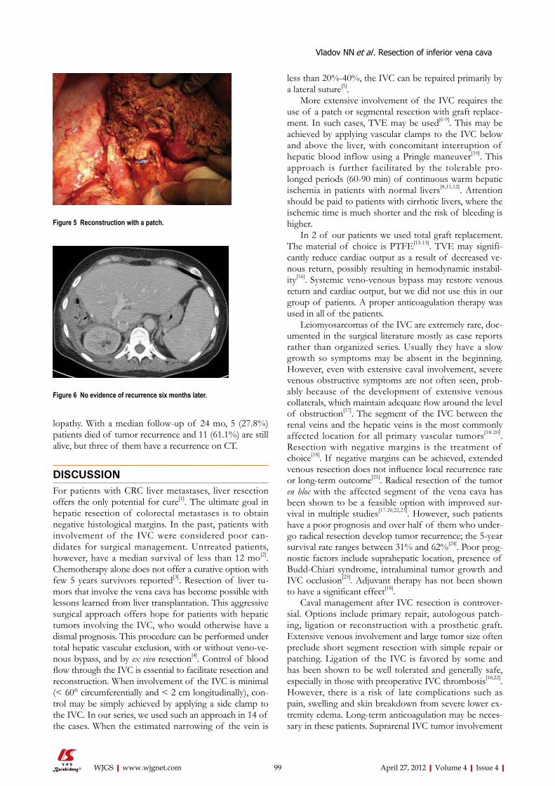
lopathy. With a median follow-up of 24 mo, 5 (27.8%) patients died of tumor recurrence and 11 (61.1%) are still alive, but three of them have a recurrence on CT.
DISCUSSIONFor patients with CRC liver metastases, liver resection offers the only potential for cure[1]. The ultimate goal in hepatic resection of colorectal metastases is to obtain negative histological margins. In the past, patients with involvement of the IVC were considered poor can-didates for surgical management. Untreated patients, however, have a median survival of less than 12 mo[2]. Chemotherapy alone does not offer a curative option with few 5 years survivors reported[3]. Resection of liver tu-mors that involve the vena cava has become possible with lessons learned from liver transplantation. This aggressive surgical approach offers hope for patients with hepatic tumors involving the IVC, who would otherwise have a dismal prognosis. This procedure can be performed under total hepatic vascular exclusion, with or without veno-ve-nous bypass, and by ex vivo resection[4]. Control of blood flow through the IVC is essential to facilitate resection and reconstruction. When involvement of the IVC is minimal (< 60° circumferentially and < 2 cm longitudinally), con-trol may be simply achieved by applying a side clamp to the IVC. In our series, we used such an approach in 14 of the cases. When the estimated narrowing of the vein is
less than 20%-40%, the IVC can be repaired primarily by a lateral suture[5].
More extensive involvement of the IVC requires the use of a patch or segmental resection with graft replace-ment. In such cases, TVE may be used[6-9]. This may be achieved by applying vascular clamps to the IVC below and above the liver, with concomitant interruption of hepatic blood inflow using a Pringle maneuver[10]. This approach is further facilitated by the tolerable pro-longed periods (60-90 min) of continuous warm hepatic ischemia in patients with normal livers[8,11,12]. Attention should be paid to patients with cirrhotic livers, where the ischemic time is much shorter and the risk of bleeding is higher.
In 2 of our patients we used total graft replacement. The material of choice is PTFE[13-15]. TVE may signifi-cantly reduce cardiac output as a result of decreased ve-nous return, possibly resulting in hemodynamic instabil-ity[16]. Systemic veno-venous bypass may restore venous return and cardiac output, but we did not use this in our group of patients. A proper anticoagulation therapy was used in all of the patients.
Leiomyosarcomas of the IVC are extremely rare, doc-umented in the surgical literature mostly as case reports rather than organized series. Usually they have a slow growth so symptoms may be absent in the beginning. However, even with extensive caval involvement, severe venous obstructive symptoms are not often seen, prob-ably because of the development of extensive venous collaterals, which maintain adequate flow around the level of obstruction[17]. The segment of the IVC between the renal veins and the hepatic veins is the most commonly affected location for all primary vascular tumors[18-20]. Resection with negative margins is the treatment of choice[18]. If negative margins can be achieved, extended venous resection does not influence local recurrence rate or long-term outcome[21]. Radical resection of the tumor en bloc with the affected segment of the vena cava has been shown to be a feasible option with improved sur-vival in multiple studies[17-20,22,23]. However, such patients have a poor prognosis and over half of them who under-go radical resection develop tumor recurrence; the 5-year survival rate ranges between 31% and 62%[24]. Poor prog-nostic factors include suprahepatic location, presence of Budd-Chiari syndrome, intraluminal tumor growth and IVC occlusion[25]. Adjuvant therapy has not been shown to have a significant effect[18].
Caval management after IVC resection is controver-sial. Options include primary repair, autologous patch-ing, ligation or reconstruction with a prosthetic graft. Extensive venous involvement and large tumor size often preclude short segment resection with simple repair or patching. Ligation of the IVC is favored by some and has been shown to be well tolerated and generally safe, especially in those with preoperative IVC thrombosis[18,22]. However, there is a risk of late complications such as pain, swelling and skin breakdown from severe lower ex-tremity edema. Long-term anticoagulation may be neces-sary in these patients. Suprarenal IVC tumor involvement
99 April 27, 2012|Volume 4|Issue 4|WJGS|www.wjgnet.com
Figure 5 Reconstruction with a patch.
Figure 6 No evidence of recurrence six months later.
Vladov NN et al . Resection of inferior vena cava

treated with IVC ligation can place a patient at serious risk for renal insufficiency. Restoration of flow to the right renal vein by reimplantation (or pelvic kidney au-totransplantation) is mandatory to maintain right kidney function, but optional for the left renal vein because of the left kidney’s considerable collateral drainage through the adrenal, inferior phrenic, gonadal and paravertebral vessels[26]. Kieffer et al[19] used a proximal pressure read-ing of 30 mm Hg or more in the IVC as an indication for caval reconstruction and found reconstruction to be necessary in most cases. PTFE is the most commonly used prosthetic material and has been shown to be a suit-able replacement for the IVC with excellent long-term patency[19,20,23,27,28]. Infection and graft thrombosis are the 2 major complications of this type of reconstruction but both are rare. Graft thrombosis may or may not have any clinical importance and methods used to decrease its in-cidence include the use of ring-reinforced PTFE to pre-vent compression, short-term anticoagulation and place-ment of an arterio-venous fistula to augment flow[19].
PTFE graft infection after IVC replacement has been shown to be a rare occurrence in several large se-ries[19,20,23,27]. The treatment is usually conservative but in some cases the graft must be removed.
Direct extension of renal cell carcinoma into the vena cava has been found in 4% to 10% of patients undergoing nephrectomy to treat cancer[29,30]. The prognosis of RCC with IVC tumor thrombosis is difficult to predict due to a wide variety in clinical behavior[31]. Although involvement of the IVC in renal cancer is generally not a vascular inva-sion by the neoplastic process but mostly an intraluminal extension of the tumor mass, such intravascular growth implies a heightened biological behavior of the tumor. Early pulmonary metastases are found in most cases.
Resection of intrahepatic cholangiocarcinoma with a negative microscopic margin improved survival. Thus, concomitant hepatic and IVC resection may provide a potentially curative operation. This aggressive surgical approach may offer hope for patients with intrahepatic cholangiocarcinoma involving the IVC[32,33].
In conclusion, it is apparent that application of com-bined resection of the liver and IVC expands the role of liver resection for malignancy and will benefit selected patients[34-36]. En bloc resection can be accomplished safely and confers an increase in survival for lesions often considered unresectable. There are a variety of options for replacement of the IVC if it cannot be primarily reconstructed. The use of various graft materials for re-construction of the hepatic great vessels offers a higher resectable rate and better prognosis in selected cases. Such an operation should be performed in a specialized center where surgeons are familiar with both aspects of complex hepatobiliary surgery and liver transplantation.
COMMENTSBackgroundInvolvement of the major vessels has traditionally been considered as a contra-indication for resection of advanced liver and retroperitoneal tumors because of
the poor long-term prognosis and high surgical risks. Research frontiersThe development of innovative surgical techniques, such as total hepatic vascular exclusion,veno-venous bypass and ex vivo hepatic resection, and the progress of liver transplantation has made a curative surgical approach to tumors involving both the liver and inferior vena cava (IVC) possible.Innovations and breakthroughsThe surgical removal of the tumor with clear margins is the only potential for cure. The advances in surgery and perioperative care lead to a more aggres-sive approach to malignancies involving major vessels which should be re-placed by different kinds of allogen artificial grafts.ApplicationsThe use of vascular grafts after block resection of tumor and major vessels should be performed in specialized HPB and Transplant centers. The type of the graft depends on the hospital protocol and personal preferences of the sur-geon.TerminologyAllograft is organ or tissue from one individual to another of the same species with a different genotype, including cadaveric or living related. A suitable vein is not always available and in this situation an artificial graft should be used.Peer reviewThe authors presented resection and reconstruction of the IVC for tumors. They conclude that the technique of combined resection of the liver and IVC is safe, expands the indication, and increases survival.
REFERENCES1 Adam R, Hoti E, Folprecht G, Benson AB. Accomplishments
in 2008 in the management of curable metastatic colorectal cancer. Gastrointest Cancer Res 2009; 3: S15-S22
2 Rougier P, Milan C, Lazorthes F, Fourtanier G, Partensky C, Baumel H, Faivre J. Prospective study of prognostic factors in patients with unresected hepatic metastases from colorectal cancer. Fondation Française de Cancérologie Digestive. Br J Surg 1995; 82: 1397-1400
3 Isenberg J, Fischbach R, Krüger I, Keller HW. Treatment of liver metastases from colorectal cancer. Anticancer Res 1996; 16: 1291-1295
4 Lodge JP, Ammori BJ, Prasad KR, Bellamy MC. Ex vivo and in situ resection of inferior vena cava with hepatectomy for colorectal metastases. Ann Surg 2000; 231: 471-479
5 Liao GS, Hsieh HF, Hsieh CB, Chen TW, Chen CJ, Yu JC, Li YC. Vessel reconstruction for great vessel invasion by hepato-biliary malignancy. J Med Sci 2005; 25: 309-312
6 Heaney JP, Stanton WK, Halbert DS, Seidel J, Vice T. An im-proved technic for vascular isolation of the liver: experimen-tal study and case reports. Ann Surg 1966; 163: 237-241
7 Fortner JG, Shiu MH, Kinne DW, Kim DK, Castro EB, Wat-son RC, Howland WS, Beattie EJ. Major hepatic resection us-ing vascular isolation and hypothermic perfusion. Ann Surg 1974; 180: 644-652
8 Huguet C, Nordlinger B, Galopin JJ, Bloch P, Gallot D. Nor-mothermic hepatic vascular exclusion for extensive hepatec-tomy. Surg Gynecol Obstet 1978; 147: 689-693
9 Berney T, Mentha G, Morel P. Total vascular exclusion of the liver for the resection of lesions in contact with the vena cava or the hepatic veins. Br J Surg 1998; 85: 485-488
10 Pringle JH. V. Notes on the Arrest of Hepatic Hemorrhage Due to Trauma. Ann Surg 1908; 48: 541-549
11 Nagasue N, Yukaya H, Ogawa Y, Hirose S, Okita M. Seg-mental and subsegmental resections of the cirrhotic liver un-der hepatic inflow and outflow occlusion. Br J Surg 1985; 72: 565-568
12 Huguet C, Gavelli A, Bona S. Hepatic resection with ischemia of the liver exceeding one hour. J Am Coll Surg 1994; 178: 454-458
13 Huguet C, Ferri M, Gavelli A. Resection of the suprarenal inferior vena cava. The role of prosthetic replacement. Arch
100 April 27, 2012|Volume 4|Issue 4|WJGS|www.wjgnet.com
COMMENTS
Vladov NN et al . Resection of inferior vena cava

Surg 1995; 130: 793-79714 Sarkar R, Eilber FR, Gelabert HA, Quinones-Baldrich WJ.
Prosthetic replacement of the inferior vena cava for malig-nancy. J Vasc Surg 1998; 28: 75-81; discussion 82-83
15 Bower TC, Nagorney DM, Toomey BJ, Gloviczki P, Pairolero PC, Hallett JW, Cherry KJ. Vena cava replacement for malig-nant disease: is there a role? Ann Vasc Surg 1993; 7: 51-62
16 Delva E, Barberousse JP, Nordlinger B, Ollivier JM, Vacher B, Guilmet C, Huguet C. Hemodynamic and biochemical monitoring during major liver resection with use of hepatic vascular exclusion. Surgery 1984; 95: 309-318
17 Dzsinich C, Gloviczki P, van Heerden JA, Nagorney DM, Pairolero PC, Johnson CM, Hallett JW, Bower TC, Cherry KJ. Primary venous leiomyosarcoma: a rare but lethal disease. J Vasc Surg 1992; 15: 595-603
18 Hollenbeck ST, Grobmyer SR, Kent KC, Brennan MF. Surgi-cal treatment and outcomes of patients with primary inferior vena cava leiomyosarcoma. J Am Coll Surg 2003; 197: 575-579
19 Kieffer E, Alaoui M, Piette JC, Cacoub P, Chiche L. Leiomyo-sarcoma of the inferior vena cava: experience in 22 cases. Ann Surg 2006; 244: 289-295
20 Hines OJ, Nelson S, Quinones-Baldrich WJ, Eilber FR. Leio-myosarcoma of the inferior vena cava: prognosis and com-parison with leiomyosarcoma of other anatomic sites. Cancer 1999; 85: 1077-1083
21 Mingoli A, Sapienza P, Cavallaro A, Di Marzo L, Burchi C, Giannarelli D, Feldhaus RJ. The effect of extend of caval resection in the treatment of inferior vena cava leiomyosar-coma. Anticancer Res 1997; 17: 3877-3881
22 Mingoli A, Feldhaus RJ, Cavallaro A, Stipa S. Leiomyosar-coma of the inferior vena cava: analysis and search of world literature on 141 patients and report of three new cases. J Vasc Surg 1991; 14: 688-699
23 Illuminati G, Calio’ FG, D’Urso A, Giacobbi D, Papaspyro-poulos V, Ceccanei G. Prosthetic replacement of the infrahe-patic inferior vena cava for leiomyosarcoma. Arch Surg 2006; 141: 919-924; discussion 924
24 Ito H, Hornick JL, Bertagnolli MM, George S, Morgan JA, Baldini EH, Wagner AJ, Demetri GD, Raut CP. Leiomyosar-
coma of the inferior vena cava: survival after aggressive man-agement. Ann Surg Oncol 2007; 14: 3534-3541
25 Mingoli A, Cavallaro A, Sapienza P, Di Marzo L, Feldhaus RJ, Cavallari N. International registry of inferior vena cava leiomyosarcoma: analysis of a world series on 218 patients. Anticancer Res 1996; 16: 3201-3205
26 Kraybill WG, Callery MP, Heiken JP, Flye MW. Radical resection of tumors of the inferior vena cava with vascular reconstruction and kidney autotransplantation. Surgery 1997; 121: 31-36
27 Dew J, Hansen K, Hammon J, McCoy T, Levine EA, Shen P. Leiomyosarcoma of the inferior vena cava: surgical manage-ment and clinical results. Am Surg 2005; 71: 497-501
28 Bower TC, Nagorney DM, Cherry KJ, Toomey BJ, Hallett JW, Panneton JM, Gloviczki P. Replacement of the inferior vena cava for malignancy: an update. J Vasc Surg 2000; 31: 270-281
29 Skinner DG, Vermillion CD, Colvin RB. The surgical man-agement of renal cell carcinoma. J Urol 1972; 107: 705-710
30 Kearney GP, Waters WB, Klein LA, Richie JP, Gittes RF. Re-sults of inferior vena cava resection for renal cell carcinoma. J Urol 1981; 125: 769-773
31 Kaplan S, Ekici S, Doğan R, Demircin M, Ozen H, Paşaoğlu I. Surgical management of renal cell carcinoma with inferior vena cava tumor thrombus. Am J Surg 2002; 183: 292-299
32 Kurosawa H, Kimura F, Ito H, Shimizu H, Togawa A, Otsuka M, Yoshidome H, Kato A, Miyazaki M. Right hepatectomy combined with retrohepatic caval resection, using a left renal vein patch graft for advanced cholangiocarcinoma. J Hepato-biliary Pancreat Surg 2004; 11: 362-365
33 Nakagohri T, Konishi M, Inoue K, Oda T, Kinoshita T. Ex-tended right hepatic lobectomy with resection of inferior vena cava and portal vein for intrahepatic cholangiocarci-noma. J Hepatobiliary Pancreat Surg 2000; 7: 599-602
34 Felix EL, Wood DK, Das Gupta TK. Tumors of the retroperi-toneum. Curr Probl Cancer 1981; 6: 1-47
35 Huguet C, Gavelli A. Total vascular exclusion for liver resec-tion. Hepatogastroenterology 1998; 45: 368-369
36 Scheele J, Stang R, Altendorf-Hofmann A, Paul M. Resection of colorectal liver metastases. World J Surg 1995; 19: 59-71
S- Editor Wang JL L- Editor Roemmele A E- Editor Zheng XM
101 April 27, 2012|Volume 4|Issue 4|WJGS|www.wjgnet.com
Vladov NN et al . Resection of inferior vena cava

Lymphoma presenting as a necrotic colonic mass
Ioannis T Konstantinidis, Michael R Probstfeld
Ioannis T Konstantinidis, Department of Surgery, The Univer-sity of Arizona College of Medicine, Tucson, AZ 85724-5058, United StatesMichael R Probstfeld, Department of Surgery, Tucson Medical Center, Tucson, AZ 85712, United States Author contributions: The entire two authors contributed to this case report.Correspondence to: Ioannis T Konstantinidis, MD, Depart-ment of Surgery, The University of Arizona College of Medi-cine, 1501 N. Campbell Avenue, PO Box 245058, Tucson, AZ 85724-5058, United States. [email protected] Telephone: +1-520-6267747 Fax: +1-520-6264334Received: May 15, 2011 Revised: November 27, 2011Accepted: December 10, 2011Published online: April 27, 2012
AbstractPrimary colonic lymphomas represent a rare minority among the colonic neoplasms. Their correct pre-opera-tive identification is crucial for the design of treatment. We herein describe a case of a colonic lymphoma pre-senting as a necrotic colonic mass and we discuss the current evidence about the presentation, diagnosis and treatment of lymphomas isolated to the colon.
© 2012 Baishideng. All rights reserved.
Key words: Colonic lymphoma; Necrotic colonic mass
Peer reviewer: John H Stewart, MD, Department of Surgery, Wake Forest University School of Medicine, Winston-Salem, NC 27157, United States
Konstantinidis IT, Probstfeld MR. Lymphoma presenting as a ne-crotic colonic mass. World J Gastrointest Surg 2012; 4(4): 102-103 Available from: URL: http://www.wjgnet.com/1948-9366/full/v4/i4/102.htm DOI: http://dx.doi.org/10.4240/wjgs.v4.i4.102
INTRODUCTIONColonic lymphomas represent a rare entity, comprising
less than 1% of colonic neoplasms. Their correct treat-ment is still an issue of debate as the rarity of this disease precludes randomized clinical trials. In this report, we describe a case of a colonic lymphoma presenting as a necrotic colonic mass and we emphasize the correct iden-tification of colonic lymphomas and the current evidence with regard to their treatment.
CASE REPORTA 70-year-old female presented with a 6-mo history of vague abdominal pain. The patient also complained of constipation, fatigue and night sweats but no nausea, vom-iting, weight loss or melena. The patient’s medical history included breast cancer status post lumpectomy 10 years ago and splenic lymphoma status post splenectomy 6 years ago. Her last colonoscopy was 6 years ago and was normal. The abdomen was soft, tender to palpation over the left lower quadrant with no rebound or guarding. An 8-10 cm ab-dominal mass was palpable at the left lower quadrant. The laboratory results showed a white cell count of 13 000 per cubic millimeter and a carcino-embryonic antigen level of 0.7 ng/mL. Abdominal computed tomography (CT) with oral and intravenous contrast medium showed a necrotic mass 8.7 cm × 9.4 cm at the left lower quadrant, encasing the distal descending and proximal sigmoid colon, with associated adenopathy in the retroperitoneum and left sided hydronephrosis secondary to ureteral obstruction by the mass (Figure 1A and B, arrows). A colonoscopy was consistent with a large necrotic and ulcerated mass in the sigmoid colon (Figure 2).
Biopsies obtained during the colonoscopy were con-sistent with B-cell lymphoma. The patient underwent sur-gical exploration and proximal colostomy with the plan to follow up with systemic chemotherapy and surgery after the conclusion of the chemotherapy.
DISCUSSIONPrimary colonic lymphomas account for only 0.2%-0.6% of colon cancers[1-5] and 10%-20% of the gastrointestinal
CASE REPORT
Online Submissions: http://www.wjgnet.com/[email protected]:10.4240/wjgs.v4.i4.102
World J Gastrointest Surg 2012 April 27; 4(4): 102-103ISSN 1948-9366 (online)
© 2012 Baishideng. All rights reserved.
102 April 27, 2012|Volume 4|Issue 4|WJGS|www.wjgnet.com

Konstantinidis IT et al . Lymphoma, necrotic colonic mass
lymphomas, with the stomach by far the most common site[1,6]. Colonic lymphomas are found more frequently in males in their sixth and seventh decade of life[1,3,5]. In-flammatory bowel disease and immunosuppressive states like HIV are known risk factors[1,5]. The most frequent presentation is abdominal pain and weight loss, whereas an abdominal mass, as in our case, is often palpable[1-3,5]. The most commonly involved site is the cecum, likely be-cause of the abundance of lymphoid tissue in the ileoce-cal region[1-5]. The predominant type is non-Hodgkins B cell lymphoma[1,3,4].
In our case, the lymphoma presented as a necrotic colonic mass on computerized tomography. In general, the CT appearance of lymphomas can be that of either a discrete mass, focal induration or diffuse colonic inva-sion[7]. The presence of extensive abdominal and/or pelvic lymphadenopathy places the lymphoma at the top of the differential diagnosis. Even in the absence of lymphadenopathy, imaging characteristics such as loca-tion at the cecum, demarcation from the peri-colonic fat with no invasion of surrounding viscera and the presence of perforation in the absence of desmoplastic reaction should raise the suspicion of a lymphoma[7]. The role of colonoscopy and biopsy is crucial for the correct pre-operative diagnosis.
Most of the tumors present in an advanced stage and the reported 5-year survival is thus relatively poor,
ranging between 27%-55%[1-6]. Most of the reported se-ries use a combination of surgery and chemotherapy[3]. Although the exact role of chemotherapy cannot be de-fined due to the rarity of the disease and the lack of ran-domized trials, some authors support that it is associated with a survival benefit[1,4]. In the presence of a colonic perforation, which may occur during the chemotherapy, the mortality is high[4]. In the report by Lai et al[4], the four patients who were operated emergently for perforation died within 30 d post-operatively. In our case, we elected to offer a proximal colostomy, given the extent of the disease, and to proceed with chemotherapy. We plan to resect the remnant tumor and restore the continuity of the GI tract after completion of the chemotherapy.
REFERENCES1 Dionigi G, Annoni M, Rovera F, Boni L, Villa F, Castano P,
Bianchi V, Dionigi R. Primary colorectal lymphomas: review of the literature. Surg Oncol 2007; 16 Suppl 1: S169-S171
2 Zighelboim J, Larson MV. Primary colonic lymphoma. Clinical presentation, histopathologic features, and outcome with combination chemotherapy. J Clin Gastroenterol 1994; 18: 291-297
3 Wong MT, Eu KW. Primary colorectal lymphomas. Colorec-tal Dis 2006; 8: 586-591
4 Lai YL, Lin JK, Liang WY, Huang YC, Chang SC. Surgical resection combined with chemotherapy can help achieve better outcomes in patients with primary colonic lymphoma. J Surg Oncol 2011; 104: 265-268
5 Fan CW, Changchien CR, Wang JY, Chen JS, Hsu KC, Tang R, Chiang JM. Primary colorectal lymphoma. Dis Colon Rectum 2000; 43: 1277-1282
6 Koch P, del Valle F, Berdel WE, Willich NA, Reers B, Hid-demann W, Grothaus-Pinke B, Reinartz G, Brockmann J, Temmesfeld A, Schmitz R, Rübe C, Probst A, Jaenke G, Bodenstein H, Junker A, Pott C, Schultze J, Heinecke A, Par-waresch R, Tiemann M. Primary gastrointestinal non-Hodg-kin’s lymphoma: I. Anatomic and histologic distribution, clinical features, and survival data of 371 patients registered in the German Multicenter Study GIT NHL 01/92. J Clin On-col 2001; 19: 3861-3873
7 Wyatt SH, Fishman EK, Hruban RH, Siegelman SS. CT of primary colonic lymphoma. Clin Imaging 1994; 18: 131-141
S- Editor Wang JL L- Editor Roemmele A E- Editor Zheng XM
103 April 27, 2012|Volume 4|Issue 4|WJGS|www.wjgnet.com
B
A
Figure 1 Axial (A) and coronal (B) computed tomography scan images of the colonic mass.
Figure 2 Endoscopic appearance of the colonic mass.

WJGS|www.wjgnet.com
Online Submissions: http://www.wjgnet.com/[email protected]
World J Gastrointest Surg 2012 April 27; 4(4): IISSN 1948-9366 (online)
© 2012 Baishideng. All rights reserved.
ACKNOWLEDGMENTS
Acknowledgments to reviewers of World Journal of Gastrointestinal Surgery
Many reviewers have contributed their expertise and time to the peer review, a critical process to ensure the quality of World Journal of Gastrointestinal Surgery. The editors and authors of the articles submitted to the journal are grateful to the following reviewers for evaluating the articles (including those published in this issue and those rejected for this issue) during the last editing time period.
Adnan Narci, Professor, Department of Pediatric Surgery, Afyon Ko-catepe University School of Medicine, Izmir Street, 7km, Afyonkarahisar 03200, Turkey
Caroline S Verbeke, MD, PhD, Department of Histopathology, Bexley Wing Level 5 St James’s University, Hospital Beckett Street, Leeds LS9 7TF, United Kingdom
Chien-Hung Chen, MD, PhD, Department of Internal Medicine, Na-tional Taiwan University Hospital and National Taiwan University, College of Medicine, No. 7, Chung-Shan South Road, Taipei 100, Taiwan, China
Christian Max Schmidt, MD, PhD, MBA, FACS, Departments of Surgery and Biochemistry/Molecular Biology, Indiana University School of Medicine, 980 W Walnut St C522, Indianapolis, IN 46202, United States
Chapel Alain, PhD, Department of Men Radioprotection, Laboratory of Radio Pathology and Innovative Therapy, Institute of Nuclear Safety and radioprotection, PO Box 17, Far 92262, France
Chen-Guo Ker, MD, PhD, Professor, Department of Surgery, Kaohsi-ung Medical University, No. 100, Tz-You 1st Rd, Kaohsiung, Taiwan, China
Douglas S Tyler, MD, Department of Surgery, Duke University Medical Center, Box 3118, Durham, NC 27710, United States
Gregory Peter Sergeant, MD, Department of General Surgery, Univer-sity Hospital Leuven, Herestraat 49, Leuven B-3000, Belgium
Helena M Isoniemi, MD, PhD, Professor, Transplantation and Liver Surgery Clinic, Helsinki University Hospital, Box 263, Helsinki 00029-HUCH, Finland
Marcelo AF Ribeiro, MD, PhD, TCBC, TCBCD, FACS, Department of Surgery, Santo Amaro University, Alameda Gregorio Bogossian So-brinho, 80/155, Santana de Parnaiba, SP 06543-385, Brazil
Manuela Santos, PhD, Associate Professor, Department of Medicine, University of Montreal, Montreal Cancer Institute, CRCHUM/Notre-Dame Hospital, Pavillon De Seve Y5625, 1560 Sherbrooke Est, Montreal, QC, H2L 4M1, Canada
Marcus VM Valadao, MD, Instituto Nacional de Cancer, Hospital do Cancer Unidade I, Hc2., Rua do Equador 831, Santo Cristo, Rio de Janeiro 20220-410, RJ, Brazil
Ned Abraham, MBBS, FRACS, FRCS, PhD, Coffs Colorectal and Capsule Endoscopy Centre, University of New South Wales, 187 Rose Av-enue, PO Box 2244, Coffs Harbour, NSW 2450, Australia
Stavros J Gourgiotis, MD, PhD, Department of Second Surgical, 401 General Army Hospital of Athens, 41 Zakinthinou Street, Papagou, Athens 15669, Greece
Sukamal Saha, MD, FACS, FRCS, FICS, Department of Orthopedics, 3500 Calkins Rd, Suite A, Flint, MI 48532, United States
Vollmar Brigitte, MD, Professor, Institute of Experimental Surgery, Uni-versity of Rostock, Schillingallee 69a, Rostock 18057, Germany
I April 27, 2012|Volume 4|Issue 4|

Events Calendar 2012January 19-21, 2012Gastrointestinal Cancers Symposium 2012San Francisco, CA, United States
January 25-29, 2012Alpine Liver and Pancreatic Surgery MeetingCarlo Magno Zeledria Hotel, Madonna di Campiglio, Italy
February 1-4, 2012 Society Of Laparoendoscopic Surgeons AsianAmerican Multi-Specialty Summit 2012 (SLS 2012) Honolulu, HI, United States February 4, 2012Radio ENT 2012Bangalore, India February 14-16, 20127th Annual Academic Surgical Conference Las Vegas, NV, United States February 22-24, 2012BTS 15th Annual Congress Glasgow, United Kingdom
February 20-25, 2012Minimally Invasive Surgery Symposium 2012The Grand America Hotel, Salt Lake City, UT, United States
March 7-10, 2012Society of American Gastrointestinal and Endoscopic Surgeons Annual Meeting 2012 (SAGES 2012)The San Diego Convention Center, San Diego, CA, United States
March 9-10, 2012Kieler Arthroskopiekurs Kniegelenk Kiel, Germany
March 29- April 1, 2012Endovienna 2012 - 5th World Congress for Endoscopic Surgery of the Brain Skull Base & Spine combined with The First Global Update on Fess, The Sinuses & The NoseVienna, Austria
March 7-11, 2012American Hepato-Pancreato Biliary Association Annual Meeting 2012 (AHPBA 2012)Eden Roc Resort, 4525 Collins Avenue,Miami Beach, FL, United States May 19-22, 2012The 2012 Digestive Disease WeekSan Diego, CA, United States
May 18-19, 2012 The American Pancreas Club Scientific MeetingSan Diego, CA, United States
June 1-5, 201248th American Society of Clinical Oncology Annual MeetingChicago, IL, United States
June 17-20, 2012Digestive Disorders Federation Conference - Combined meeting of BSG, AUGIS, BAPEN & BSLLiverpool, United Kingdom
June 20-23, 201244th meeting of European Pancreatic ClubPrague, Czech Republic
June 27-30, 2011ESMO 14th World Congress on Gastrointestinal CancerBarcelona, Spain
July 1-5, 201210th World Congress of the International Hepato-Pancreato-Biliary Association joined with the European HPBA CongressParis, France
September 15-16, 2012Current problems of gastroenterology and abdominal SurgeryKiev, Ukraine
September 19-21, 201232nd Congress of the European Society of Surgical Oncology (ESSO)Valencia, Spain
September 28 - October 2, 201237th European Society for Medical Oncology (ESMO) CongressVienna, Austria
November 4-7, 20128th National Cancer Research Institute ConferenceLiverpool, United Kingdom
November 14-16, 2012Pancreatic Society of Great Britain and Ireland Meeting 2012Cameron House Hotel, Glasgow
December 8, 2012IASGO 2012 - 22nd World Congress of the International Association of Surgeons, Gastroenterologists and OncologistsBangkok, Thailand
Online Submissions: http://www.wjgnet.com/[email protected]
WJGS|www.wjgnet.com I
World J Gastrointest Surg 2012 April 27; 4(4): IISSN 1948-9366 (online)
© 2012 Baishideng. All rights reserved.
April 27, 2012|Volume 4|Issue 4|
MEETINGS

GENERAL INFORMATION World Journal of Gastrointestinal Surgery (World J Gastrointest Surg, WJGS, online ISSN 1948-9366, DOI: 10.4240), is a monthly, open-access (OA), peer-reviewed journal supported by an editorial board of 336 experts in gastrointestinal surgery from 35 countries.
The biggest advantage of the OA model is that it provides free, full-text articles in PDF and other formats for experts and the pub-lic without registration, which eliminates the obstacle that traditional journals possess and usually delays the speed of the propagation and communication of scientific research results. The open access model has been proven to be a true approach that may achieve the ultimate goal of the journals, i.e. the maximization of the value to the readers, authors and society.
Maximization of personal benefitsThe role of academic journals is to exhibit the scientific levels of a country, a university, a center, a department, and even a scientist, and build an important bridge for communication between scientists and the public. As we all know, the significance of the publication of scientific articles lies not only in disseminating and communicat-ing innovative scientific achievements and academic views, as well as promoting the application of scientific achievements, but also in formally recognizing the "priority" and "copyright" of innovative achievements published, as well as evaluating research performance and academic levels. So, to realize these desired attributes of WJGS and create a well-recognized journal, the following four types of personal benefits should be maximized. The maximization of per-sonal benefits refers to the pursuit of the maximum personal ben-efits in a well-considered optimal manner without violation of the laws, ethical rules and the benefits of others. (1) Maximization of the benefits of editorial board members: The primary task of edito-rial board members is to give a peer review of an unpublished sci-entific article via online office system to evaluate its innovativeness, scientific and practical values and determine whether it should be published or not. During peer review, editorial board members can also obtain cutting-edge information in that field at first hand. As leaders in their field, they have priority to be invited to write articles and publish commentary articles. We will put peer reviewers’ names and affiliations along with the article they reviewed in the journal to acknowledge their contribution; (2) Maximization of the benefits of authors: Since WJGS is an open-access journal, readers around the world can immediately download and read, free of charge, high-quality, peer-reviewed articles from WJGS official website, thereby realizing the goals and significance of the communication between authors and peers as well as public reading; (3) Maximization of the benefits of readers: Readers can read or use, free of charge, high-quality peer-reviewed articles without any limits, and cite the arguments, viewpoints, concepts, theories, methods, results, conclu-sion or facts and data of pertinent literature so as to validate the innovativeness, scientific and practical values of their own research achievements, thus ensuring that their articles have novel arguments or viewpoints, solid evidence and correct conclusion; and (4) Maxi-mization of the benefits of employees: It is an iron law that a first-class journal is unable to exist without first-class editors, and only first-class editors can create a first-class academic journal. We insist on strengthening our team cultivation and construction so that ev-ery employee, in an open, fair and transparent environment, could contribute their wisdom to edit and publish high-quality articles,
thereby realizing the maximization of the personal benefits of edi-torial board members, authors and readers, and yielding the greatest social and economic benefits.
Aims and scopeThe major task of WJGS is to rapidly report the most recent results in basic and clinical research on gastrointestinal surgery, specifically including micro-invasive surgery, laparoscopy, hepatic surgery, biliary surgery, pancreatic surgery, splenic surgery, surgical nutrition, portal hypertension, as well as the associated subjects such as epidemiology, cancer research, biomarkers, prevention, pathology, radiology, genetics, genomics, proteomics, pharmacology, pharmacokinetics, pharmacogenetics, molecular biology, clinical trials, diagnosis and therapeutics and multimodality treatment. Emphasis is placed on original research articles and clinical case reports. This journal will also provide balanced, extensive and timely review articles on selected topics.
ColumnsThe columns in the issues of WJGS will include: (1) Editorial: To introduce and comment on major advances and developments in the field; (2) Frontier: To review representative achievements, comment on the state of current research, and propose directions for future research; (3) Topic Highlight: This column consists of three formats, including (A) 10 invited review articles on a hot topic, (B) a commentary on common issues of this hot topic, and (C) a commentary on the 10 individual articles; (4) Observation: To update the development of old and new questions, highlight unsolved problems, and provide strategies on how to solve the questions; (5) Guidelines for Basic Research: To provide guidelines for basic research; (6) Guidelines for Clinical Practice: To provide guidelines for clinical diagnosis and treatment; (7) Review: To review systemically progress and unresolved problems in the field, comment on the state of current research, and make suggestions for future work; (8) Original Article: To report innovative and original findings in gastrointestinal surgery; (9) Brief Article: To briefly report the novel and innovative findings in gastrointestinal surgery; (10) Case Report: To report a rare or typical case; (11) Letters to the Editor: To discuss and make reply to the contributions published in WJGS, or to introduce and comment on a controversial issue of general interest; (12) Book Reviews: To introduce and comment on quality monographs of gastrointestinal surgery; and (13) Guidelines: To introduce consensuses and guidelines reached by international and national academic authorities worldwide on basic research and clinical practice in gastrointestinal surgery.
Name of journalWorld Journal of Gastrointestinal Surgery
ISSNISSN 1948-9366 (online)
Editorial-in-ChiefTimothy M Pawlik, MD, MPH, FACS, Associate Professor of Surgery and Oncology, Hepatobiliary Surgery Program Director, Di-rector, Johns Hopkins Medicine Liver Tumor Center Multi-Disciplin-ary Clinic, Co-Director of Center for Surgical Trials and Outcomes Research, Johns Hopkins Hospital, 600 N. Wolfe Street, Harvey 611,
Instructions to authors
WJGS|www.wjgnet.com April 27, 2012|Volume 4|Issue 4|I
Online Submissions: http://www.wjgnet.com/[email protected]
World J Gastrointest Surg 2012 April 27; 4(4): I-VISSN 1948-9366 (online)
© 2012 Baishideng. All rights reserved.
INSTRUCTIONS TO AUTHORS

Instructions to authors
Baltimore, MD 21287, United States
Editorial OfficeWorld Journal of Gastrointestinal SurgeryEditorial Department: Room 903, Building D, Ocean International Center,No. 62 Dongsihuan Zhonglu, Chaoyang District, Beijing 100025, ChinaE-mail: [email protected]://www.wjgnet.comTelephone: +86-10-85381891Fax: +86-10-85381893
Indexing/abstractingPubMed Central, PubMed, Digital Object Identifer, and Directory of Open Access Journals.
Published byBaishideng Publishing Group Co., Limited
SPECIAL STATEMENTAll articles published in this journal represent the viewpoints of the authors except where indicated otherwise.
Biostatistical editingStatistical review is performed after peer review. We invite an expert in Biomedical Statistics from to evaluate the statistical method used in the paper, including t-test (group or paired comparisons), chi-squared test, Ridit, probit, logit, regression (linear, curvilinear, or stepwise), correlation, analysis of variance, analysis of covariance, etc. The reviewing points include: (1) Statistical methods should be described when they are used to verify the results; (2) Whether the statistical techniques are suitable or correct; (3) Only homoge-neous data can be averaged. Standard deviations are preferred to standard errors. Give the number of observations and subjects (n). Losses in observations, such as drop-outs from the study should be reported; (4) Values such as ED50, LD50, IC50 should have their 95% confidence limits calculated and compared by weighted probit analysis (Bliss and Finney); and (5) The word ‘significantly’ should be replaced by its synonyms (if it indicates extent) or the P value (if it indicates statistical significance).
Conflict-of-interest statementIn the interests of transparency and to help reviewers assess any poten-tial bias, WJGS requires authors of all papers to declare any compet-ing commercial, personal, political, intellectual, or religious interests in relation to the submitted work. Referees are also asked to indi-cate any potential conflict they might have reviewing a particular paper. Before submitting, authors are suggested to read “Uniform Requirements for Manuscripts Submitted to Biomedical Journals: Ethical Considerations in the Conduct and Reporting of Research: Conflicts of Interest” from International Committee of Medical Journal Editors (ICMJE), which is available at: http://www.icmje.org/ethical_4conflicts.html.
Sample wording: [Name of individual] has received fees for serv-ing as a speaker, a consultant and an advisory board member for [names of organizations], and has received research funding from [names of organization]. [Name of individual] is an employee of [name of or-ganization]. [Name of individual] owns stocks and shares in [name of organization]. [Name of individual] owns patent [patent identification and brief description].
Statement of informed consentManuscripts should contain a statement to the effect that all human studies have been reviewed by the appropriate ethics committee or it should be stated clearly in the text that all persons gave their informed consent prior to their inclusion in the study. Details that might disclose the identity of the subjects under study should be omitted. Authors should also draw attention to the Code of Ethics
of the World Medical Association (Declaration of Helsinki, 1964, as revised in 2004).
Statement of human and animal rightsWhen reporting the results from experiments, authors should fol-low the highest standards and the trial should conform to Good Clinical Practice (for example, US Food and Drug Administration Good Clinical Practice in FDA-Regulated Clinical Trials; UK Medi-cines Research Council Guidelines for Good Clinical Practice in Clinical Trials) and/or the World Medical Association Declaration of Helsinki. Generally, we suggest authors follow the lead investiga-tor’s national standard. If doubt exists whether the research was conducted in accordance with the above standards, the authors must explain the rationale for their approach and demonstrate that the institutional review body explicitly approved the doubtful as-pects of the study.
Before submitting, authors should make their study approved by the relevant research ethics committee or institutional review board. If human participants were involved, manuscripts must be accompa-nied by a statement that the experiments were undertaken with the understanding and appropriate informed consent of each. Any per-sonal item or information will not be published without explicit con-sents from the involved patients. If experimental animals were used, the materials and methods (experimental procedures) section must clearly indicate that appropriate measures were taken to minimize pain or discomfort, and details of animal care should be provided.
SUBMISSION OF MANUSCRIPTSManuscripts should be typed in 1.5 line spacing and 12 pt. Book Antiqua with ample margins. Number all pages consecutively, and start each of the following sections on a new page: Title Page, Ab-stract, Introduction, Materials and Methods, Results, Discussion, Acknowledgements, References, Tables, Figures, and Figure Leg-ends. Neither the editors nor the publisher are responsible for the opinions expressed by contributors. Manuscripts formally accepted for publication become the permanent property of Baishideng Publishing Group Co., Limited, and may not be reproduced by any means, in whole or in part, without the written permission of both the authors and the publisher. We reserve the right to copy-edit and put onto our website accepted manuscripts. Authors should follow the relevant guidelines for the care and use of laboratory animals of their institution or national animal welfare committee. For the sake of transparency in regard to the performance and reporting of clinical trials, we endorse the policy of the ICMJE to refuse to pub-lish papers on clinical trial results if the trial was not recorded in a publicly-accessible registry at its outset. The only register now avail-able, to our knowledge, is http://www.clinicaltrials.gov sponsored by the United States National Library of Medicine and we encour-age all potential contributors to register with it. However, in the case that other registers become available you will be duly notified. A letter of recommendation from each author’s organization should be provided with the contributed article to ensure the privacy and secrecy of research is protected.
Authors should retain one copy of the text, tables, photographs and illustrations because rejected manuscripts will not be returned to the author(s) and the editors will not be responsible for loss or damage to photographs and illustrations sustained during mailing.
Online submissionsManuscripts should be submitted through the Online Submission System at: http://www.wjgnet.com/1948-9366office. Authors are highly recommended to consult the ONLINE INSTRUCTIONS TO AUTHORS (ht tp ://www.wjgnet .com/1948-9366/g_info_20100305152206.htm) before attempting to submit online. For assistance, authors encountering problems with the Online Submi-ssion System may send an email describing the problem to [email protected], or by telephone: +86-10-85381891. If you submit your manuscript online, do not make a postal contribution. Repeated online submission for the same manuscript is strictly prohibited.
WJGS|www.wjgnet.com II April 27, 2012|Volume 4|Issue 4|

Instructions to authors
MANUSCRIPT PREPARATIONAll contributions should be written in English. All articles must be submitted using word-processing software. All submissions must be typed in 1.5 line spacing and 12 pt. Book Antiqua with ample margins. Style should conform to our house format. Required information for each of the manuscript sections is as follows:
Title pageTitle: Title should be less than 12 words.
Running title: A short running title of less than 6 words should be provided.
Authorship: Authorship credit should be in accordance with the standard proposed by International Committee of Medical Journal Editors, based on (1) substantial contributions to conception and design, acquisition of data, or analysis and interpretation of data; (2) drafting the article or revising it critically for important intellectual content; and (3) final approval of the version to be published. Au-thors should meet conditions 1, 2, and 3.Institution: Author names should be given first, then the complete name of institution, city, province and postcode. For example, Xu-Chen Zhang, Li-Xin Mei, Department of Pathology, Chengde Medical College, Chengde 067000, Hebei Province, China. One au-thor may be represented from two institutions, for example, George Sgourakis, Department of General, Visceral, and Transplantation Surgery, Essen 45122, Germany; George Sgourakis, 2nd Surgical Department, Korgialenio-Benakio Red Cross Hospital, Athens 15451, Greece
Author contributions: The format of this section should be: Author contributions: Wang CL and Liang L contributed equally to this work; Wang CL, Liang L, Fu JF, Zou CC, Hong F and Wu XM designed the research; Wang CL, Zou CC, Hong F and Wu XM performed the research; Xue JZ and Lu JR contributed new reagents/analytic tools; Wang CL, Liang L and Fu JF analyzed the data; and Wang CL, Liang L and Fu JF wrote the paper.
Supportive foundations: The complete name and number of supportive foundations should be provided, e.g. Supported by National Natural Science Foundation of China, No. 30224801
Correspondence to: Only one corresponding address should be provided. Author names should be given first, then author title, affiliation, the complete name of institution, city, postcode, province, country, and email. All the letters in the email should be in lower case. A space interval should be inserted between country name and email address. For example, Montgomery Bissell, MD, Professor of Medicine, Chief, Liver Center, Gastroenterology Division, University of California, Box 0538, San Francisco, CA 94143, United States. [email protected]
Telephone and fax: Telephone and fax should consist of +, country number, district number and telephone or fax number, e.g. Telephone: +86-10-85381891 Fax: +86-10-85381893
Peer reviewers: All articles received are subject to peer review. Normally, three experts are invited for each article. Decision for acceptance is made only when at least two experts recommend an article for publication. Reviewers for accepted manuscripts are acknowledged in each manuscript, and reviewers of articles which were not accepted will be acknowledged at the end of each issue. To ensure the quality of the articles published in WJGS, reviewers of accepted manuscripts will be announced by publishing the name, title/position and institution of the reviewer in the footnote accompanying the printed article. For example, reviewers: Professor Jing-Yuan Fang, Shanghai Institute of Digestive Disease, Shanghai, Affiliated Renji Hospital, Medical Faculty, Shanghai Jiaotong University, Shanghai, China; Professor Xin-Wei Han, Department of Radiology, The First Affiliated Hospital, Zhengzhou University,
Zhengzhou, Henan Province, China; and Professor Anren Kuang, Department of Nuclear Medicine, Huaxi Hospital, Sichuan University, Chengdu, Sichuan Province, China.
AbstractThere are unstructured abstracts (no less than 256 words) and structured abstracts (no less than 480). The specific requirements for structured abstracts are as follows:
An informative, structured abstracts of no less than 480 words should accompany each manuscript. Abstracts for original contri-butions should be structured into the following sections. AIM (no more than 20 words): Only the purpose should be included. Please write the aim as the form of “To investigate/study/…”; MATERI-ALS AND METHODS (no less than 140 words); RESULTS (no less than 294 words): You should present P values where appropri-ate and must provide relevant data to illustrate how they were ob-tained, e.g. 6.92 ± 3.86 vs 3.61 ± 1.67, P < 0.001; CONCLUSION (no more than 26 words).
Key wordsPlease list 5-10 key words, selected mainly from Index Medicus, which reflect the content of the study.
TextFor articles of these sections, original articles and brief articles, the main text should be structured into the following sections: INTRO-DUCTION, MATERIALS AND METHODS, RESULTS and DISCUSSION, and should include appropriate Figures and Tables. Data should be presented in the main text or in Figures and Tables, but not in both. The main text format of these sections, editorial, topic highlight, case report, letters to the editors, can be found at: http://www.wjgnet.com/1948-9366/g_info_list.htm.
IllustrationsFigures should be numbered as 1, 2, 3, etc., and mentioned clearly in the main text. Provide a brief title for each figure on a sepa-rate page. Detailed legends should not be provided under the figures. This part should be added into the text where the figures are applicable. Figures should be either Photoshop or Illustra-tor files (in tiff, eps, jpeg formats) at high-resolution. Examples can be found at: http://www.wjgnet.com/1007-9327/13/4520.pdf; http://www.wjgnet.com/1007-9327/13/4554.pdf; http://www.wjgnet.com/1007-9327/13/4891.pdf; http://www.wjgnet.com/1007-9327/13/4986.pdf; http://www.wjgnet.com/1007-9327/13/4498.pdf. Keeping all elements compiled is necessary in line-art image. Scale bars should be used rather than magnification factors, with the length of the bar defined in the leg-end rather than on the bar itself. File names should identify the fig-ure and panel. Avoid layering type directly over shaded or textured areas. Please use uniform legends for the same subjects. For exam-ple: Figure 1 Pathological changes in atrophic gastritis after treat-ment. A: ...; B: ...; C: ...; D: ...; E: ...; F: ...; G: …etc. It is our principle to publish high resolution-figures for the printed and E-versions.
TablesThree-line tables should be numbered 1, 2, 3, etc., and mentioned clearly in the main text. Provide a brief title for each table. Detailed legends should not be included under tables, but rather added into the text where applicable. The information should complement, but not duplicate the text. Use one horizontal line under the title, a second under column heads, and a third below the Table, above any footnotes. Vertical and italic lines should be omitted.
Notes in tables and illustrationsData that are not statistically significant should not be noted. aP < 0.05, bP < 0.01 should be noted (P > 0.05 should not be noted). If there are other series of P values, cP < 0.05 and dP < 0.01 are used. A third series of P values can be expressed as eP < 0.05 and fP < 0.01. Other notes in tables or under illustrations should be expressed as 1F, 2F, 3F; or sometimes as other symbols with a superscript (Arabic
WJGS|www.wjgnet.com III April 27, 2012|Volume 4|Issue 4|

Instructions to authors
numerals) in the upper left corner. In a multi-curve illustration, each curve should be labeled with ●, ○, ■, □, ▲, △, etc., in a certain sequence.
AcknowledgmentsBrief acknowledgments of persons who have made genuine con-tributions to the manuscript and who endorse the data and conclu-sions should be included. Authors are responsible for obtaining written permission to use any copyrighted text and/or illustrations.
REFERENCESCoding systemThe author should number the references in Arabic numerals according to the citation order in the text. Put reference numbers in square brackets in superscript at the end of citation content or after the cited author’s name. For citation content which is part of the narration, the coding number and square brackets should be typeset normally. For example, “Crohn’s disease (CD) is associated with increased intestinal permeability[1,2]”. If references are cited directly in the text, they should be put together within the text, for example, “From references[19,22-24], we know that...”.
When the authors write the references, please ensure that the order in text is the same as in the references section, and also ensure the spelling accuracy of the first author’s name. Do not list the same citation twice.
PMID and DOIPleased provide PubMed citation numbers to the reference list, e.g. PMID and DOI, which can be found at http://www.ncbi.nlm.nih.gov/sites/entrez?db=pubmed and http://www.crossref.org/SimpleTextQuery/, respectively. The numbers will be used in E-version of this journal.
Style for journal referencesAuthors: the name of the first author should be typed in bold-faced letters. The family name of all authors should be typed with the initial letter capitalized, followed by their abbreviated first and middle initials. (For example, Lian-Sheng Ma is abbreviated as Ma LS, Bo-Rong Pan as Pan BR). The title of the cited article and italicized journal title (journal title should be in its abbreviated form as shown in PubMed), publication date, volume number (in black), start page, and end page [PMID: 11819634 DOI: 10.3748/wjg.13.5396].
Style for book referencesAuthors: the name of the first author should be typed in bold-faced letters. The surname of all authors should be typed with the initial letter capitalized, followed by their abbreviated middle and first initials. (For example, Lian-Sheng Ma is abbreviated as Ma LS, Bo-Rong Pan as Pan BR) Book title. Publication number. Publication place: Publication press, Year: start page and end page.
FormatJournals English journal article (list all authors and include the PMID where applicable)1 Jung EM, Clevert DA, Schreyer AG, Schmitt S, Rennert J,
Kubale R, Feuerbach S, Jung F. Evaluation of quantitative con-trast harmonic imaging to assess malignancy of liver tumors: A prospective controlled two-center study. World J Gastroenterol 2007; 13: 6356-6364 [PMID: 18081224 DOI: 10.3748/wjg.13. 6356]
Chinese journal article (list all authors and include the PMID where applicable)2 Lin GZ, Wang XZ, Wang P, Lin J, Yang FD. Immunologic
effect of Jianpi Yishen decoction in treatment of Pixu-diar-rhoea. Shijie Huaren Xiaohua Zazhi 1999; 7: 285-287
In press3 Tian D, Araki H, Stahl E, Bergelson J, Kreitman M. Signature
of balancing selection in Arabidopsis. Proc Natl Acad Sci USA 2006; In press
Organization as author4 Diabetes Prevention Program Research Group. Hyperten-
sion, insulin, and proinsulin in participants with impaired glu-cose tolerance. Hypertension 2002; 40: 679-686 [PMID: 12411462 PMCID:2516377 DOI:10.1161/01.HYP.0000035706.28494. 09]
Both personal authors and an organization as author 5 Vallancien G, Emberton M, Harving N, van Moorselaar RJ;
Alf-One Study Group. Sexual dysfunction in 1, 274 European men suffering from lower urinary tract symptoms. J Urol 2003; 169: 2257-2261 [PMID: 12771764 DOI:10.1097/01.ju. 0000067940.76090.73]
No author given6 21st century heart solution may have a sting in the tail. BMJ
2002; 325: 184 [PMID: 12142303 DOI:10.1136/bmj.325. 7357.184]
Volume with supplement7 Geraud G, Spierings EL, Keywood C. Tolerability and safety
of frovatriptan with short- and long-term use for treatment of migraine and in comparison with sumatriptan. Headache 2002; 42 Suppl 2: S93-99 [PMID: 12028325 DOI:10.1046/j.1526-4610.42.s2.7.x]
Issue with no volume8 Banit DM, Kaufer H, Hartford JM. Intraoperative frozen
section analysis in revision total joint arthroplasty. Clin Orthop Relat Res 2002; (401): 230-238 [PMID: 12151900 DOI:10.1097/00003086-200208000-00026]
No volume or issue9 Outreach: Bringing HIV-positive individuals into care. HRSA
Careaction 2002; 1-6 [PMID: 12154804]
BooksPersonal author(s)10 Sherlock S, Dooley J. Diseases of the liver and billiary system.
9th ed. Oxford: Blackwell Sci Pub, 1993: 258-296Chapter in a book (list all authors)11 Lam SK. Academic investigator’s perspectives of medical
treatment for peptic ulcer. In: Swabb EA, Azabo S. Ulcer disease: investigation and basis for therapy. New York: Marcel Dekker, 1991: 431-450
Author(s) and editor(s)12 Breedlove GK, Schorfheide AM. Adolescent pregnancy.
2nd ed. Wieczorek RR, editor. White Plains (NY): March of Dimes Education Services, 2001: 20-34
Conference proceedings13 Harnden P, Joffe JK, Jones WG, editors. Germ cell tumours V.
Proceedings of the 5th Germ cell tumours Conference; 2001 Sep 13-15; Leeds, UK. New York: Springer, 2002: 30-56
Conference paper14 Christensen S, Oppacher F. An analysis of Koza's computa-
tional effort statistic for genetic programming. In: Foster JA, Lutton E, Miller J, Ryan C, Tettamanzi AG, editors. Genetic programming. EuroGP 2002: Proceedings of the 5th Euro-pean Conference on Genetic Programming; 2002 Apr 3-5; Kinsdale, Ireland. Berlin: Springer, 2002: 182-191
Electronic journal (list all authors)15 Morse SS. Factors in the emergence of infectious diseases.
Emerg Infect Dis serial online, 1995-01-03, cited 1996-06-05; 1(1): 24 screens. Available from: URL: http://www.cdc.gov/ncidod/eid/index.htm
Patent (list all authors)16 Pagedas AC, inventor; Ancel Surgical R&D Inc., assignee.
Flexible endoscopic grasping and cutting device and positioning tool assembly. United States patent US 20020103498. 2002 Aug 1
Statistical dataWrite as mean ± SD or mean ± SE.
Statistical expressionExpress t test as t (in italics), F test as F (in italics), chi square test as
WJGS|www.wjgnet.com IV April 27, 2012|Volume 4|Issue 4|

Instructions to authors
χ2 (in Greek), related coefficient as r (in italics), degree of freedom as υ (in Greek), sample number as n (in italics), and probability as P (in italics).
UnitsUse SI units. For example: body mass, m (B) = 78 kg; blood pres-sure, p (B) = 16.2/12.3 kPa; incubation time, t (incubation) = 96 h, blood glucose concentration, c (glucose) 6.4 ± 2.1 mmol/L; blood CEA mass concentration, p (CEA) = 8.6 24.5 mg/L; CO2 volume fraction, 50 mL/L CO2, not 5% CO2; likewise for 40 g/L formal-dehyde, not 10% formalin; and mass fraction, 8 ng/g, etc. Arabic numerals such as 23, 243, 641 should be read 23 243 641.
The format for how to accurately write common units and quantums can be found at: http://www.wjgnet.com/1948-9366/g_info_20100312191949.htm.
AbbreviationsStandard abbreviations should be defined in the abstract and on first mention in the text. In general, terms should not be abbreviated unless they are used repeatedly and the abbreviation is helpful to the reader. Permissible abbreviations are listed in Units, Symbols and Abbreviations: A Guide for Biological and Medical Editors and Authors (Ed. Baron DN, 1988) published by The Royal Society of Medicine, London. Certain commonly used abbreviations, such as DNA, RNA, HIV, LD50, PCR, HBV, ECG, WBC, RBC, CT, ESR, CSF, IgG, ELISA, PBS, ATP, EDTA, mAb, can be used directly without further explanation.
ItalicsQuantities: t time or temperature, c concentration, A area, l length, m mass, V volume.Genotypes: gyrA, arg 1, c myc, c fos, etc.Restriction enzymes: EcoRI, HindI, BamHI, Kbo I, Kpn I, etc.Biology: H. pylori, E coli, etc.
Examples for paper writingEditorial: http://www.wjgnet.com/1948-9366/g_info_20100312190249.htm
Frontier: http://www.wjgnet.com/1948-9366/g_info_20100312190321.htm
Topic highlight: http://www.wjgnet.com/1948-9366/g_info_20100312190447.htm
Observation: http://www.wjgnet.com/1948-9366/g_info_20100312190550.htm
Guidelines for basic research: http://www.wjgnet.com/1948-9366/g_info_20100312190653.htm
Guidelines for clinical practice: http://www.wjgnet.com/ 1948-9366/g_info_20100312190758.htm
Review: http://www.wjgnet.com/1948-9366/g_info_20100312190907.htm
Original articles: http://www.wjgnet.com/1948-9366/g_info_20100312191047.htm
Brief articles: http://www.wjgnet.com/1948-9366/g_info_20100312191203.htm
Case report: http://www.wjgnet.com/1948-9366/g_info_20100312191328.htm
Letters to the editor: http://www.wjgnet.com/1948-9366/g_info_20100312191431.htm
Book reviews: http://www.wjgnet.com/1948-9366/g_info_20100312191548.htm
Guidelines: http://www.wjgnet.com/1948-9366/g_info_20100312191635.htm
SUBMISSION OF THE REVISED MANUSCRIPTS AFTER ACCEPTEDPlease revise your article according to the revision policies of WJGS. The revised version including manuscript and high-resolution image figures (if any) should be re-submited online (http://www.wjgnet.com/1948-9366office/). The author should send the copyright transfer letter, responses to the reviewers, English language Grade B certificate (for non-native speakers of English) and final manuscript checklist to [email protected].
Language evaluation The language of a manuscript will be graded before it is sent for revision. (1) Grade A: priority publishing; (2) Grade B: minor lan-guage polishing; (3) Grade C: a great deal of language polishing needed; and (4) Grade D: rejected. Revised articles should reach Grade A or B.
Copyright assignment formPlease download a Copyright assignment form from http://www.wjgnet.com/1948-9366/g_info_20100312191901.htm.
Responses to reviewersPlease revise your article according to the comments/suggestions provided by the reviewers. The format for responses to the reviewers’ comments can be found at: http://www.wjgnet.com/1948-9366/g_info_20100312191818.htm.
Proof of financial supportFor paper supported by a foundation, authors should provide a copy of the document and serial number of the foundation.
Links to documents related to the manuscript WJGS will be initiating a platform to promote dynamic interactions between the editors, peer reviewers, readers and authors. After a man-uscript is published online, links to the PDF version of the submitted manuscript, the peer-reviewers’ report and the revised manuscript will be put on-line. Readers can make comments on the peer reviewer’s report, authors’ responses to peer reviewers, and the revised manu-script. We hope that authors will benefit from this feedback and be able to revise the manuscript accordingly in a timely manner.
Science news releasesAuthors of accepted manuscripts are suggested to write a science news item to promote their articles. The news will be released rap-idly at EurekAlert/AAAS (http://www.eurekalert.org). The title for news items should be less than 90 characters; the summary should be less than 75 words; and main body less than 500 words. Science news items should be lawful, ethical, and strictly based on your original content with an attractive title and interesting pictures.
Publication feeWJGS is an international, peer-reviewed, Open-Access, online jour-nal. Articles published by this journal are distributed under the terms of the Creative Commons Attribution Non-commercial License, which permits use, distribution, and reproduction in any medium, provided the original work is properly cited, the use is non commercial and is otherwise in compliance with the license. Authors of accepted articles must pay a publication fee. The related standards are as follows. Publication fee: 1300 USD per article. Edi-torial, topic highlights, original articles, brief articles, book reviews and letters to the editor are published free of charge.
WJGS|www.wjgnet.com V April 27, 2012|Volume 4|Issue 4|


