Embed Size (px)
Citation preview
World Journal of Gastroenterology
ISSN 1007-9327CN 14-1219/R
A Weekly Journal of Gastroenterology and Hepatology
Indexed and Abstracted in:Current Contents®/Clinical Medicine, Science Citation Index Expanded (also known as SciSearch®) and Journal Citation Reports/Science Edition, Index Medicus, MEDLINE and PubMed, Chemical Abstracts, EMBASE/Excerpta Medica, Abstracts Journals, Nature Clinical Practice Gastroenterology and Hepatology, CAB Abstracts and Global Health. ISI JCR 2003-2000 IF: 3.318, 2.532, 1.445 and 0.993.
Volume 14 Number 34September 14, 2008
World J Gastroenterol2008 September 14; 14(34): 5233-5360
Online Submissionswjg.wjgnet.com
www.wjgnet.com Printed on Acid-free Paper
™©Baishideng百世登
Published by The WJG Press and BaishidengRoom 903, Ocean International Center, Building D
No. 62 Dongsihuan Zhonglu, Chaoyang District, Beijing 100025, ChinaFax: +86-10-8538-1893 E-mail: [email protected] http://www.wjgnet.com
HONORARY EDITORS-IN-CHIEFMontgomery Bissell, San FranciscoJames L Boyer, New HavenChao-Long Chen, KaohsiungKe-Ji Chen, BeijingLi-Fang Chou, TaipeiJacques V Dam, StanfordMartin H Floch, New HavenGuadalupe Garcia-Tsao, New HavenZhi-Qiang Huang, BeijingShinn-Jang Hwang, TaipeiIra M Jacobson, New YorkDerek Jewell, OxfordEmmet B Keeffe, Palo AltoMin-Liang Kuo, TaipeiNicholas F LaRusso, RochesterJie-Shou Li, NanjingGeng-Tao Liu, BeijingLein-Ray Mo, TainanBo-Rong Pan, Xi'anFa-Zu Qiu, WuhanEamonn M Quigley, CorkDavid S Rampton, LondonRafi q A Sheikh, SacramentoRudi Schmid, Kentfi eld[1]
Nicholas J Talley, RochesterSun-Lung Tsai, Young-Kang CityGuido NJ Tytgat, AmsterdamHsiu-Po Wang, TaipeiJaw-Ching Wu, TaipeiMeng-Chao Wu, ShanghaiMing-Shiang Wu, TaipeiJia-Yu Xu, ShanghaiTa-Sen Yeh, TaoyuanMing-Lung Yu, Kaohsiung
PRESIDENT AND EDITOR-IN-CHIEFLian-Sheng Ma, Beijing
STRATEGY ASSOCIATE EDITORS-IN-CHIEFPeter Draganov, FloridaRonnie Fass, TucsonHugh J Freeman, Vancouver John P Geibel, New Haven Maria Concepción Gutiérrez-Ruiz, MéxicoKazuhiro Hanazaki, KochiAkio Inui, KagoshimaKalpesh Jani, VadodaraSanaa M Kamal, CairoIoannis E Koutroubakis, HeraklionJose JG Marin, SalamancaJavier S Martin, Punta del EsteNatalia A Osna, OmahaJose Sahel, Marseille Ned Snyder, GalvestonNathan Subramaniam, BrisbaneWei Tang, TokyoAlan BR Thomson, EdmontonPaul Joseph Thuluvath, BaltimoreJames F Trotter, DenverShingo Tsuji, Osaka Harry HX Xia, HanoverYoshio Yamaoka, HoustonJesue K Yamamoto-Furusho, México
ASSOCIATE EDITORS-IN-CHIEFGianfranco D Alpini, TempleBruno Annibale, Roma
www.wjgnet.com I
The World Journal of Gastroenterology Editorial Board consists of 1208 members, representing a team of worldwide experts in gastroenterology and hepatology. They are from 60 countries, including Albania (1), Argentina (4), Australia (39), Austria (10), Belarus (1), Belgium (15), Brazil (2), Bulgaria (1), Canada (28), Chile (1), China (60), Croatia (2), Cuba (1), Czech (2), Denmark (7), Egypt (4), Estonia (1), Finland (4), France (44), Germany (108), Greece (9), Hungary (2), Iceland (1), India (12), Iran (3), Ireland (4), Israel (8), Italy (96), Japan (176), Lebanon (3), Lithuania (1), Macedonia (1), Malaysia (3), Mexico (6), Monaco (1), Morocco (1), The Netherlands (26), New Zealand (1), Nigeria (1), Norway (3), Pakistan (2), Peru (1), Poland (6), Portugal (1), Russia (3), Saudi Arabia (2), Serbia (1), Singapore (4), Slovakia (2), Slovenia (1), South Africa (2), South Korea (14), Spain (38), Sweden (15), Switzerland (13), Turkey (8), United Arab Emirates (1), United Kingdom (83), United States (316) and Uruguay (2).
Roger W Chapman, OxfordChi-Hin Cho, Hong KongAlexander L Gerbes, MunichShou-Dong Lee, TaipeiWalter E Longo, New HavenYou-Yong Lu, BeijingMasao Omata, Tokyo
BIOSTATISTICAL EDITORLiang-Ping Hu, Beijing
MEMBERS OF THE EDITORIAL BOARD
Bashkim Resuli, Tirana
Julio H Carri, Córdoba Carlos J Pirola, Buenos AiresSilvia Sookoian, Buenos AiresAdriana M Torres, Rosario
Leon Anton Adams, NedlandsMinoti V Apte, Liverpool Richard B Banati, Lidcombe Michael R Beard, Adelaide Patrick Bertolino, Sydney
Albania
Argentina
Australia
World Journal of Gastroenterology
Editorial Board2007-2009
Andrew V Biankin, SydneyFilip Braet, SydneyAndrew D Clouston, SydneyGraham Cooksley, QueenslandDarrell HG Crawford, BrisbaneAdrian G Cummins, Woodville SouthGuy D Eslick, SydneyMichael A Fink, MelbourneRobert JL Fraser, Daw ParkPeter Raymond Gibson, VictoriaJacob George, WestmeadMark D Gorrell, SydneyYik-Hong Ho, TownsvilleGerald J Holtmann, AdelaideMichael Horowitz, AdelaideJohn E Kellow, SydneyRupert Leong, ConcordGeoffrey W McCaughan, SydneyFinlay A Macrae, VictoriaDaniel Markovich, BrisbanePhillip S Oates, PerthJacqui Richmond, VictoriaStephen M Riordan, SydneyIan C Roberts-Thomson, AdelaideDevanshi Seth, CamperdownArthur Shulkes, MelbourneRoss C Smith, SydneyKevin J Spring, BrisbaneHuy A Tran, New South WalesDebbie Trinder, FremantleMartin J Veysey, GosfordDaniel L Worthley, Bedford
Peter Ferenci, ViennaValentin Fuhrmann, ViennaAlfred Gangl, ViennaChristoph Gasche, ViennaKurt Lenz, LinzMarkus Peck-Radosavljevic, ViennaRudolf E Stauber, AuenbruggerplatzHerbert Tilg, InnsbruckMichael Trauner, GrazHarald Vogelsang, ViennaGuenter Weiss, Innsbruck
Belarus
Rudi Beyaert, GentBart Rik De Geest, LeuvenInge I Depoortere, LeuvenOlivier Detry, LiègeBenedicte Y De Winter, AntwerpKarel Geboes, LeuvenThierry Gustot, BrusselsYves J Horsmans, BrusselsGeert G Leroux-Roels, GhentLouis Libbrecht, LeuvenEtienne M Sokal, BrusselsMarc Peeters, De PintelaanGert A Van Assche, LeuvenYvan Vandenplas, BrusselsEddie Wisse, Keerbergen
Heitor Rosa, GoianiaAna Cristina Simões e Silva, Belo Horizonte
Zahariy Krastev, Sofi a
Fernando Alvarez, QuébecDavid Armstrong, OntarioJeffrey P Baker, TorontoOlivier Barbier, QuébecNancy Baxter, TorontoMatthew Bjerknes, TorontoFrank J Burczynski, ManitobaMichael F Byrne, VancouverWang-Xue Chen, OttawaChantal Guillemette, QuébecSamuel S Lee, CalgaryGary A Levy, TorontoAndrew L Mason, AlbertaJohn K Marshall, OntarioDonna-Marie McCafferty, CalgaryThomas I Michalak, St. John'sGerald Y Minuk, ManitobaPaul Moayyedi, HamiltonKostas Pantopoulos, QuébecWilliam G Paterson, KingstonEldon Shaffer, CalgaryMorris Sherman, TorontoMartin Storr, CalgaryElena F Verdu, OntarioJohn L Wallace, CalgaryEric M Yoshida, Vancouver
Silvana Zanlungo, Santiago
Henry LY Chan, HongkongXiao-Ping Chen, WuhanZong-Jie Cui, BeijingDa-Jun Deng, BeijingEr-Dan Dong, BeijingSheung-Tat Fan, Hong KongJin Gu, BeijingXin-Yuan Guan, PokfulamDe-Wu Han, TaiyuanMing-Liang He, Hong KongWayne HC Hu, Hong KongChee-Kin Hui, Hong KongChing-Lung Lai, Hong KongKam Chuen Lai, Hong KongJames YW Lau, Hong KongYuk-Tong Lee, Hong KongSuet-Yi Leung, Hong KongWai-Keung Leung, Hong KongJohn M Luk, PokfulamChung-Mau Lo, Hong KongJing-Yun Ma, BeijingRonnie Tung Ping Poon, Hong KongLun-Xiu Qin, ShanghaiYu-Gang Song, GuangzhouQin Su, BeijingWai-Man Wong, Hong Kong
Hong Xiao, ShanghaiDong-Liang Yang, WuhanWinnie Yeo, Hong KongYuan Yuan, ShenyangMan-Fung Yuen, Hong KongJian-Zhong Zhang, BeijingXin-Xin Zhang, ShanghaiBo-Jian Zheng, Hong Kong Shu Zheng, Hangzhou
Tamara Cacev, ZagrebMarko Duvnjak, Zagreb
Damian C Rodriguez, Havana
Milan Jirsa, PrahaPavel Trunečka, Prague
Peter Bytzer, CopenhagenAsbjørn M Drewes, AalborgHans Gregersen, AalborgJens H Henriksen, HvidovreClaus P Hovendal, OdenseFin S Larsen, CopenhagenSøren Møller, Hvidovre
Abdel-Rahman El-Zayadi, GizaAmr M Helmy, CairoAyman Yosry, Cairo
Riina Salupere, Tartu
Irma E Jarvela, HelsinkiKatri M Kaukinen, TampereMinna Nyström, HelsinkiPentti Sipponen, Espoo
Bettaieb Ali, DijonCorlu Anne, RennesDenis Ardid, Clermont-FerrandCharles P Balabaud, BordeauxSoumeya Bekri, RouenJacques Belghiti, ClichyJacques Bernuau, Clichy CedexPierre Brissot, RennesPatrice P Cacoub, ParisFranck Carbonnel, BesanconLaurent Castera, PessacBruno Clément, RennesBenoit Coffi n, ColombesJacques Cosnes, ParisThomas Decaens, Cedex
Austria
Yury K Marakhouski, Minsk
Belgium
Brazil
Bulgaria
Canada
Chile
China
Croatia
Cuba
Czech
Denmark
Egypt
Estonia
Finland
France
www.wjgnet.comⅡ
www.wjgnet.com Ⅲ
Francoise L Fabiani, AngersGérard Feldmann, ParisJean Fioramonti, ToulouseJean-Noël Freund, StrasbourgJean-Paul Galmiche, NantesCatherine Guettier, VillejuifChantal Housset, ParisJuan L Iovanna, MarseilleRene Lambert, LyonPatrick Marcellin, ParisPhilippe Mathurin, LilleTamara Matysiak–Budnik, ParisFrancis Mégraud, BordeauxRichard Moreau, ClichyThierry Piche, NiceRaoul Poupon, ParisJean Rosenbaum, BordeauxDominique Marie Roulot, BobignyThierry Poynard, ParisJean-Philippe Salier, RouenDidier Samuel, VillejuifJean-Yves Scoazec, LyonKhalid A Tazi, ClichyEmmanuel Tiret, ParisBaumert F Thomas, StrasbourgMarie-Catherine Vozenin-brotons, VillejuifJean-Pierre H Zarski, GrenobleJessica Zucman-Rossi, Paris
Hans-Dieter Allescher, G-PartenkirchenMartin Anlauf, KielRudolf Arnold, MarburgMax G Bachem, UlmThomas F Baumert, FreiburgDaniel C Baumgart, BerlinHubert Blum, FreiburgThomas Bock, TuebingenKatja Breitkopf, MannheimDunja Bruder, BraunschweigMarkus W Büchler, HeidelbergChrista Buechler, RegensburgReinhard Buettner, BonnElke Cario, EssenUta Dahmen, EssenChristoph F Dietrich, Bad MergentheimArno J Dormann, Koeln Rainer J Duchmann, BerlinVolker F Eckardt, WiesbadenPaul Enck, TuebingenFred Fändrich, KielUlrich R Fölsch, KielHelmut Friess, HeidelbergPeter R Galle, MainzNikolaus Gassler, AachenAndreas Geier, AachenMarkus Gerhard, MunichWolfram H Gerlich, GiessenDieter Glebe, GiessenBurkhard Göke, MunichFlorian Graepler, TuebingenAxel M Gressner, AachenVeit Gülberg, MunichRainer Haas, MunichEckhart G Hahn, ErlangenStephan Hellmig, KielMartin Hennenberg, BonnJohannes Herkel, HamburgKlaus R Herrlinger, StuttgartEva Herrmann, Homburg/SaarEberhard Hildt, BerlinJoerg C Hoffmann, BerlinFerdinand Hofstaedter, Regensburg
Werner Hohenberger, ErlangenJörg C Kalff, BonnRalf Jakobs, LudwigshafenJutta Keller, HamburgAndrej Khandoga, MunichSibylle Koletzko, MünchenStefan Kubicka, HannoverJoachim Labenz, SiegenFrank Lammert, BonnThomas Langmann, RegensburgChristian Liedtke, AachenMatthias Löhr, MannheimChristian Maaser, MuensterAhmed Madisch, DresdenPeter Malfertheiner, MagdeburgMichael P Manns, HannoverHelmut Messmann, AugsburgStephan Miehlke, DresdenSabine Mihm, GöttingenSilvio Nadalin, EssenMarkus F Neurath, MainzJohann Ockenga, BerlinFlorian Obermeier, RegensburgGustav Paumgartner, MunichUlrich KS Peitz, MagdeburgMarkus Reiser, BochumEmil C Reisinger, RostockSteffen Rickes, MagdeburgTilman Sauerbruch, BonnDieter Saur, MunichHans Scherubl, BerlinJoerg Schirra, MunichRoland M Schmid, MünchenVolker Schmitz, BonnAndreas G Schreyer, RegensburgTobias Schroeder, EssenHenning Schulze-Bergkamen, MainzHans Seifert, OldenburgNorbert Senninger, MuensterManfred V Singer, MannheimGisela Sparmann, RostockChristian J Steib, MünchenJurgen M Stein, FrankfurtUlrike S Stein, BerlinManfred Stolte, BayreuthChristian P Strassburg, HannoverWolfgang R Stremmel, HeidelbergHarald F Teutsch, UlmRobert Thimme, FreiburgHans L Tillmann, LeipzigTung-Yu Tsui, RegensburgAxel Ulsenheimer, MunichPatrick Veit-Haibach, EssenClaudia Veltkamp, HeidelbergSiegfried Wagner, DeggendorfHenning Walczak, HeidelbergHeiner Wedemeyer, HannoverFritz von Weizsacker, BerlinJens Werner, HeidelbergBertram Wiedenmann, BerlinReiner Wiest, RegensburgStefan Wirth, WuppertalStefan JP Zeuzem, Homburg
Alexandra A Alexopoulou, AthensGeorge N Dalekos, LarissaChristos Dervenis, AthensMelanie Maria Deutsch, AthensTsianos Epameinondas, IoanninaElias A Kouroumalis, HeraklionGeorge Papatheodoridis, AthensSpiros Sgouros, Athens
Peter L Lakatos, BudapestZsuzsa Szondy, Debrecen
Hallgrimur Gudjonsson, Reykjavik
Philip Abraham, MumbaiRakesh Aggarwal, LucknowKunissery A Balasubramanian, VelloreDeepak Kumar Bhasin, ChandigarhSujit K Bhattacharya, KolkataYogesh K Chawla, ChandigarhRadha K Dhiman, ChandigarhSri Prakash Misra, AllahabadRamesh Roop Rai, JaipurNageshwar D Reddy, HyderabadRakesh Kumar Tandon, New Delhi
Seyed-Moayed Alavian, TehranReza Malekzadeh, TehranSeyed A Taghavi, Shiraz
Billy Bourke, DublinRonan A Cahill, CorkAnthony P Moran, Galway
Simon Bar-Meir, HashomerAbraham R Eliakim, HaifaZvi Fireman, HaderaYaron Ilan, JerusalemAvidan U Neumann, Ramat-GanYaron Niv, PardesiaRan Oren, Tel AvivAmi D Sperber, Beer-Sheva
Germany
Greece
Hungary
Iceland
India
Iran
Ireland
Israel
ItalyGiovanni Addolorato, RomaLuigi E Adinolfi , NaplesDomenico Alvaro, Mario Angelico, RomeVito Annese, San Giovanni RotondFilippo Ansaldi, GenoaAdolfo F Attili, RomaGiovanni Barbara, BolognaClaudio Bassi, VeronaGabrio Bassotti, PerugiaPier M Battezzati, MilanStefano Bellentani, CarpiAntomio Benedetti, AnconaMauro Bernardi, BolognaLivia Biancone, RomeLuigi Bonavina, Milano Flavia Bortolotti, PadovaGiuseppe Brisinda, RomeElisabetta Buscarini, CremaGiovanni Cammarota, Roma
www.wjgnet.comIV
Kyoichi Adachi, IzumoYasushi Adachi, SapporoTaiji Akamatsu, MatsumotoSk Md Fazle Akbar, EhimeTakafumi Ando, NagoyaAkira Andoh, OtsuTaku Aoki, TokyoMasahiro Arai, TokyoTetsuo Arakawa, OsakaYasuji Arase, TokyoMasahiro Asaka, SapporoHitoshi Asakura, TokyoTakeshi Azuma, Fukui Yoichi Chida, FukuokaTakahiro Fujimori, TochigiJiro Fujimoto, HyogoKazuma Fujimoto, SagaMitsuhiro Fujishiro, TokyoYoshihide Fujiyama, OtsuHiroyuki Fukui, TochigiHiroyuki Hanai, HamamatsuNaohiko Harada, FukuokaMakoto Hashizume, FukuokaTetsuo Hayakawa, NagoyaToru Hiyama, HigashihiroshimaKazuhide Higuchi, OsakaKeisuke Hino, UbeKeiji Hirata, KitakyushuYuji Iimuro, NishinomiyaKenji Ikeda, TokyoToru Ikegami, Fukuoka Kenichi Ikejima, Bunkyo-kuFumio Imazeki, ChibaYutaka Inagaki, KanagawaYasuhiro Inokuchi, YokohamaHaruhiro Inoue, YokohamaMasayasu Inoue, OsakaHiromi Ishibashi, NagasakiShunji Ishihara, IzumoToru Ishikawa, NiigataKei Ito, SendaiMasayoshi Ito, TokyoHiroaki Itoh, AkitaRyuichi Iwakiri, SagaYoshiaki Iwasaki, OkayamaTerumi Kamisawa, TokyoHiroshi Kaneko, Aichi-GunShuichi Kaneko, KanazawaTakashi Kanematsu, NagasakiMitsuo Katano, FukuokaJunji Kato, SapporoMototsugu Kato, SapporoShinzo Kato, TokyoNorifumi Kawada, OsakaSunao Kawano, OsakaMitsuhiro Kida, KanagawaYoshikazu Kinoshita, IzumoTsuneo Kitamura, ChibaSeigo Kitano, OitaKazuhiko Koike, TokyoNorihiro Kokudo, TokyoSatoshi Kondo, SapporoShoji Kubo, OsakaShigeki Kuriyama, Kagawa[2]
Katsunori Iijima, SendaiMasato Kusunoki, Tsu MieShin Maeda, Tokyo Shigeru Marubashi, SuitaMasatoshi Makuuchi, TokyoOsamu Matsui, KanazawaYasuhiro Matsumura, ChibaYasushi Matsuzaki, TsukubaKiyoshi Migita, Omura
Antonino Cavallari, BolognaGiuseppe Chiarioni, ValeggioMichele Cicala, RomeMassimo Colombo, MilanAmedeo Columbano, CagliariMassimo Conio, SanremoDario Conte, MilanoGino R Corazza, PaviaFrancesco Costa, PisaAntonio Craxi, PalermoSilvio Danese, Milan Roberto de Franchis, MilanoRoberto De Giorgio, BolognaMaria Stella De Mitri, BolognaGiovanni D De Palma, NaplesFabio Farinati, PaduaGiammarco Fava, AnconaFrancesco Feo, SassariFiorucci Stefano, PerugiaAndrea Galli, FirenzeValeria Ghisett, TurinGianluigi Giannelli, BariEdoardo G Giannini, GenoaPaolo Gionchetti, BolognaFabio Grizzi, MilanSalvatore Gruttadauria, PalermoMario Guslandi, MilanoPietro Invernizzi, MilanEzio Laconi, CagliariGiacomo Laffi , FirenzeGiovanni Maconi, MilanLucia Malaguarnera, CataniaEmanuele D Mangoni, NapoliPaolo Manzoni, TorinoGiulio Marchesini, BolognaFabio Marra, FlorenceMarco Marzioni, AnconaGiuseppe Mazzella, BolognaMario U Mondelli, PaviaGiuseppe Montalto, PalermoGiovanni Monteleone, RomeGiovanni Musso, TorinoGerardo Nardone, NapoliValerio Nobili, RomeFabio Pace, MilanoLuisi Pagliaro, PalermoFrancesco Pallone, RomeFabrizio R Parente, MilanMaurizio Parola, TorinoFrancesco Perri, San Giovanni RotondoRaffaele Pezzilli, BolognaAlberto Pilotto, San Giovanni RotondoAlberto Piperno, MonzaMario Pirisi, NovaraAnna C Piscaglia, RomaPaolo Del Poggio, TreviglioGabriele B Porro, MilanoPiero Portincasa, BariCosimo Prantera, RomaBernardino Rampone, SienaOliviero Riggio, RomeClaudio Romano, MessinaMarco Romano, NapoliGerardo Rosati, PotenzaMario Del Tacca, PisaGloria Taliani, RomePier A Testoni, MilanEnrico Roda, BolognaDomenico Sansonno, BariVincenzo Savarino, GenovaVincenzo Stanghellini, BolognaGiovanni Tarantino, NaplesRoberto Testa, GenoaDino Vaira, BolognaAnna Linda Zignego, Florence
Japan
Kenji Miki, TokyoTetsuya Mine, KanagawaHiroto Miwa, Hyogo Masashi Mizokami, NagoyaYoshiaki Mizuguchi, TokyoMotowo Mizuno, HiroshimaMorito Monden, SuitaHisataka S Moriwaki, GifuYasuaki Motomura, IizukaYoshiharu Motoo, KanazawaNaofumi Mukaida, KanazawaKazunari Murakami, OitaKunihiko Murase, TusimaHiroaki Nagano, SuitaMasahito Nagaki, GifuMasaki Nagaya, KawasakiYujl Naito, KyotoAtsushi Nakajima, YokohamaHisato Nakajima, TokyoHiroki Nakamura, Yamaguchi Shotaro Nakamura, FukuokaMikio Nishioka, NiihamaShuji Nomoto, NagoyaSusumu Ohmada, MaebashiHirohide Ohnishi, AkitaMasayuki Ohta, OitaTetsuo Ohta, KanazawaKazuichi Okazaki, OsakaKatsuhisa Omagari, Nagasaki Saburo Onishi, NankokuMorikazu Onji, EhimeSatoshi Osawa, HamamatsuMasanobu Oshima, KanazawaHiromitsu Saisho, Chiba Hidetsugu Saito, TokyoYutaka Saito, TokyoIsao Sakaida, Yamaguchi Michiie Sakamoto, TokyoYasushi Sano, ChibaHiroki Sasaki, TokyoIwao Sasaki, SendaiMotoko Sasaki, KanazawaChifumi Sato, TokyoShuichi Seki, OsakaHiroshi Shimada, YokohamaMitsuo Shimada, TokushimaTomohiko Shimatan, HiroshimaHiroaki Shimizu, ChibaIchiro Shimizu, TokushimaYukihiro Shimizu, KyotoShinji Shimoda, FukuokaTooru Shimosegawa, SendaiTadashi Shimoyama, HirosakiKen Shirabe, Iizuka CityYoshio Shirai, NiigataKatsuya Shiraki, MieYasushi Shiratori, OkayamaMasayuki Sho, NaraYasuhiko Sugawara, TokyoHidekazu Suzuki, TokyoMinoru Tada, TokyoTadatoshi Takayama, TokyoTadashi Takeda, OsakaKoji Takeuchi, KyotoKiichi Tamada, Tochigi Akira Tanaka, KyotoEiji Tanaka, MatsumotoNoriaki Tanaka, Okayama Shinji Tanaka, Hiroshima Hideki Taniguchi, YokohamaKyuichi Tanikawa, KurumeAkira Terano, ShimotsugagunHitoshi Togash, YamagataShinji Togo, YokohamaKazunari Tominaga, OsakaTakuji Torimura, Fukuoka
www.wjgnet.com Ⅴ
Minoru Toyota, SapporoAkihito Tsubota, ChibaTakato Ueno, KurumeNaomi Uemura, TokyoShinichi Wada, TochigiHiroyuki Watanabe, KanazawaToshio Watanabe, OsakaYuji Watanabe, EhimeToshiaki Watanabe, TokyoChun-Yang Wen, NagasakiSatoshi Yamagiwa, NiigataKoji Yamaguchi, FukuokaTakayuki Yamamoto, YokkaichiTakashi Yao, FukuokaMasashi Yoneda, TochigiHiroshi Yoshida, TokyoMasashi Yoshida, TokyoNorimasa Yoshida, KyotoHitoshi Yoshiji, NaraKentaro Yoshika, ToyoakeYasunobu Yoshikai, FukuokaMasahide Yoshikawa, KashiharaKatsutoshi Yoshizato, Higashihiroshima
LebanonBassam N Abboud, BeirutAla I Sharara, BeirutJoseph D Boujaoude, Beirut
LithuaniaLimas Kupcinskas, Kaunas
MacedoniaVladimir C Serafi moski, Skopje
MalaysiaAndrew Seng Boon Chua, IpohKhean-Lee Goh, Kuala LumpurJayaram Menon, Sabah
MexicoDiego Garcia-Compean, MonterreyEduardo R Marin-Lopez, Jesús GarcíaNahum Méndez-Sánchez, MexicoSaúl Villa-Treviño, México
MonacoPatrick Rampal, Monaco
MoroccoAbdellah Essaid, Rabat
The NetherlandsUlrich Beuers, AmsterdamGerd Bouma, AmsterdamLee Bouwman, LeidenJ Bart A Crusius, AmsterdamNKH de Boer, AmsterdamKoert P de Jong, GroningenHenrike Hamer, MaastrichtFrank Hoentjen, HaarlemJanine K Kruit, Groningen
Ernst J Kuipers, RotterdamCBHW Lamers, LeidenTon Lisman, UtrechtYi Liu, AmsterdamJeroen Maljaars, MaastrichtServaas Morré, AmsterdamChris JJ Mulder, AmsterdamMichael Müller, WageningenAmado S Peña, AmsterdamRobert J Porte, GroningenIngrid B Renes, RotterdamAndreas Smout, UtrechtPaul E Sijens, GroningenReinhold W Stockbrugger, MaastrichtLuc JW van der Laan, RotterdamKarel van Erpecum, UtrechtGerard P VanBerge-Henegouwen,Utrecht
New ZealandIan D Wallace, Auckland
NigeriaSamuel B Olaleye, Ibadan
Norway Trond Berg, Oslo Tom H Karlsen, OsloHelge L Waldum, Trondheim
PakistanMuhammad S Khokhar, LahoreSyed MW Jafri, Karachi
PeruHector H Garcia, Lima
PolandTomasz Brzozowski, Cracow Robert Flisiak, BialystokHanna Gregorek, WarsawDariusz M Lebensztejn, BialystokWojciech G Polak, WroclawMarek Hartleb, Katowice
Portugal Miguel C De Moura, Lisbon
Russia Vladimir T Ivashkin, Moscow Leonid Lazebnik, Moscow Vasiliy I Reshetnyak, Moscow
Saudi ArabiaIbrahim A Al Mofl eh, RiyadhAhmed Helmy, Riyadh
SerbiaDusan M Jovanovic, Sremska Kamenica
Singapore Bow Ho, SingaporeKhek-Yu Ho, SingaporeFock Kwong Ming, SingaporeFrancis Seow-Choen, Singapore
SlovakiaSilvia Pastorekova, BratislavaAnton Vavrecka, Bratislava
SloveniaSasa Markovic, Ljubljana
South AfricaRosemar Joyce Burnett, PretoriaMichael C Kew, Parktown
South KoreaByung Ihn Choi, SeoulHo Soon Choi, SeoulMarie Yeo, SuwonSun Pyo Hong, Gyeonggi-doJae J Kim, SeoulJin-Hong Kim, Suwon Myung-Hwan Kim, Seoul Chang Hong Lee, SeoulJong Kyun Lee, SeoulEun-Yi Moon, SeoulJae-Gahb Park, Seoul Dong Wan Seo, Seoul Dong Jin Suh, SeoulByung Chul Yoo, Seoul
SpainJuan G Abraldes, Barcelona Agustin Albillos, MadridRaul J Andrade, MálagaLuis Aparisi, ValenciaFernando Azpiroz, Barcelona Ramon Bataller, Barcelona Josep M Bordas, Barcelona Xavier Calvet, Sabadell Jordi Camps, CatalunyaAndres Cardenas, BarcelonaVicente Carreño, MadridJose Castellote, BarcelonaAntoni Castells, Barcelona Vicente Felipo, ValenciaJuan C Garcia-Pagán, Barcelona Jaime B Genover, BarcelonaJavier P Gisbert, MadridJaime Guardia, Barcelona Isabel Fabregat, BarcelonaMercedes Fernandez, Barcelona Angel Lanas, Zaragoza Juan-Ramón Larrubia, GuadalajaraLaura Lladóa, BarcelonaMaría IT López, JaénJuan R Malagelada, BarcelonaJosé M Mato, DerioJuan F Medina, PamplonaMiguel A Muñoz-Navas, PamplonaJulian Panes, Barcelona Miguel M Perez, ValenciaMiguel Perez-Mateo, Alicante
Josep M Pique, BarcelonaJesús M Prieto, PamplonaSabino Riestra, Pola De SieroLuis Rodrigo, OviedoManuel Romero-Gómez, SevillaJoan Roselló-Catafau, Barcelona
SwedenEinar S Björnsson, GothenburgCurt Einarsson, Huddinge Per M Hellström, StockholmUlf Hindorf, LundElisabeth Hultgren-Hörnquist, Örebro Anders E Lehmann, MölndalHanns-Ulrich Marschall, StockholmLars C Olbe, Molndal Lars A Pahlman, UppsalaMatti Sallberg, StockholmMagnus Simrén, GöteborgXiao-Feng Sun, Linköping Ervin Tóth, MalmöWeimin Ye, StockholmChrister S von Holstein, Lund
SwitzerlandChrish Beglinger, Basel Pierre A Clavien, ZurichJean-Francois Dufour, BernFranco Fortunato, ZürichJean L Frossard, GenevaGerd A Kullak-Ublick, ZurichPierre Michetti, LausanneFrancesco Negro, GenèveBruno Stieger, Zurich Radu Tutuian, ZurichStephan R Vavricka, ZurichGerhard Rogler, ZurichArthur Zimmermann, Berne
TurkeyYusuf Bayraktar, Ankara Figen Gurakan, Ankara Aydin Karabacakoglu, KonyaSerdar Karakose, KonyaHizir Kurtel, IstanbulOsman C Ozdogan, IstanbulÖzlem Yilmaz, IzmirCihan Yurdaydin, Ankara
United Arab EmiratesSherif M Karam, Al-Ain
United KingdomDavid H Adams, BirminghamSimon Afford, Birmingham Navneet K Ahluwalia, StockportAhmed Alzaraa, ManchesterLesley A Anderson, BelfastCharalambos G Antoniades, LondonAnthony TR Axon, Leeds Qasim Aziz, ManchesterNicholas M Barnes, BirminghamJim D Bell, LondonMairi Brittan, LondonAlastair D Burt, NewcastleSimon S Campbell, Manchester
Simon R Carding, LeedsPaul J Ciclitira, LondonEithne Costello, LiverpoolTatjana Crnogorac-Jurcevic, LondonHarry Dalton, TruroAmar P Dhillon, LondonWilliam Dickey, LondonderryJames E East, LondonEmad M El-Omar, AberdeenAhmed M Elsharkawy, Newcastle Upon TyneAnnette Fristscher-Ravens, LondonElizabeth Furrie, DundeeDaniel R Gaya, EdinburghSubrata Ghosh, London William Greenhalf, LiverpoolIndra N Guha, SouthamptonPeter C Hayes, EdinburghGwo-Tzer Ho, EdinburghAnthony R Hobson, SalfordLesley A Houghton, ManchesterStefan G Hübscher, BirminghamRobin Hughes, LondonPali Hungin, StocktonDavid P Hurlstone, Sheffi eldRajiv Jalan, LondonJanusz AZ Jankowski, OxfordBrian T Johnston, BelfastDavid EJ Jones, NewcastleRoger Jones, LondonMichael A Kamm, HarrowPeter Karayiannis, LondonLaurens Kruidenier, HarlowPatricia F Lalor, BirminghamChee Hooi Lim, MidlandsHong-Xiang Liu, Cambridge Yun Ma, LondonKenneth E L McColl, GlasgowStuart AC McDonald, LondonDermot P Mcgovern, OxfordGiorgina Mieli-Vergani, LondonNikolai V Naoumov, London John P Neoptolemos, Liverpool James Neuberger, BirminghamPhilip Noel Newsome, BirminghamMark S Pearce, Newcastle Upon TyneStephen P Pereira, LondonD Mark Pritchard, LiverpoolSakhawat Rahman, LondonStephen E Roberts, SwanseaMarco Senzolo, PadovaSoraya Shirazi-Beechey, LiverpoolRobert Sutton, LiverpoolSimon D Taylor-Robinson, LondonParis P Tekkis, LondonUlrich Thalheimer, LondonDavid G Thompson, SalfordNick P Thompson, NewcastleDavid Tosh, BathFrank I Tovey, London Chris Tselepis, BirminghamDiego Vergani, LondonGeoffrey Warhurst, SalfordAlastair John Watson, LiverpoolPeter J Whorwell, ManchesterRoger Williams, LondonKaren L Wright, BathMin Zhao, Foresterhill
Golo Ahlenstiel, BethesdaBS Anand, HoustonFrank A Anania, AtlantaM Ananthanarayanan, New YorkGavin E Arteel, LouisvilleJasmohan S Bajaj, Milwaukee Subhas Banerjee, Palo AltoPeter A Banks, BostonJamie S Barkin, Miami BeachKim E Barrett, San DiegoMarc D Basson, DetroitAnthony J Bauer, PittsburghWallace F Berman, DurhamTimothy R Billiar, PittsburghEdmund J Bini, New YorkDavid G Binion, MilwaukeeJennifer D Black, Buffalo Herbert L Bonkovsky, CharlotteCarla W Brady, DurhamAndrea D Branch, New YorkRobert S Bresalier, HoustonAlan L Buchman, ChicagoRonald W Busuttil, Los AngelesAlan Cahill, PhiladelphiaJohn M Carethers, San DiegoDavid L Carr-Locke, BostonMaurice A Cerulli, New YorkRavi S Chari, NashvilleJiande Chen, GalvestonXian-Ming Chen, OmahaXin Chen, San FranciscoRamsey Chi-man Cheung, Palo AltoWilliam D Chey, Ann ArborJohn Y Chiang, RootstownParimal Chowdhury, ArkansasRaymond T Chung, BostonJames M Church, ClevelandRam Chuttani, BostonMark G Clemens, CharlotteAna J Coito, Los AngelesVincent Coghlan, BeavertonDavid Cronin II, New HavenJohn Cuppoletti, CincinnatiMark J Czaja, New YorkPeter V Danenberg, Los Angeles Kiron M Das, New Brunswick Conor P Delaney, ClevelandJose L del Pozo, RochesterSharon DeMorrow, TempleDeborah L Diamond, SeattleDouglas A Drossman, Chapel HillKaterina Dvorak, TucsonBijan Eghtesad, ClevelandHala El-Zimaity, HoustonMichelle Embree-Ku, ProvidenceSukru Emre, New HavenDouglas G Farmer, Los AngelesAlessio Fasano, BaltimoreMark A Feitelson, PhiladelphiaAriel E Feldstein, ClevelandAlessandro Fichera, ChicagoRobert L Fine, New YorkMagali Fontaine, StanfordChris E Forsmark, GainesvilleGlenn T Furuta, AuroraChandrashekhar R Gandhi, PittsburghSusan L Gearhart, BaltimoreXupeng Ge, BostonXin Geng, New BrunswickM Eric Gershwin, SuiteJean-Francois Geschwind, BaltimoreIgnacio Gil-Bazo, New YorkShannon S Glaser, TempleAjay Goel, DallasRichard M Green, ChicagoJulia B Greer, Pittsburgh
United StatesManal F Abdelmalek, DurhamGary A Abrams, BirminghamMaria T Abreu, New YorkReid B Adams, Virginia
www.wjgnet.comⅥ

James H Grendell, New YorkDavid R Gretch, SeattleStefano Guandalini, ChicagoAnna S Gukovskaya, Los Angeles Sanjeev Gupta, BronxDavid J Hackam, PittsburghStephen B Hanauer, ChicagoGavin Harewood, Rochester Margaret M Heitkemper, WashingtonAlan W Hemming, GainesvilleSamuel B Ho, San DiegoPeter R Holt, New YorkColin W Howden, ChicagoHongjin Huang, AlamedaJamal A Ibdah, ColumbiaAtif Iqbal, Omaha Hajime Isomoto, RochesterHartmut Jaeschke, TucsonDennis M Jensen, Los AngelesCheng Ji, Los AngelesLeonard R Johnson, MemphisMichael P Jones, ChicagoPeter J Kahrilas, Chicago Anthony N Kalloo, BaltimoreMarshall M Kaplan, BostonNeil Kaplowitz, Los AngelesSerhan Karvar, Los AngelesRashmi Kaul, TulsaJonathan D Kaunitz, Los AngelesAli Keshavarzian, ChicagoMiran Kim, ProvidenceJoseph B Kirsner, Chicago Leonidas G Koniaris, MiamiBurton I Korelitz, New YorkRobert J Korst, New York Richard A Kozarek, Seattle Alyssa M Krasinskas, PittsburghMichael Kremer, Chapel HillShiu-Ming Kuo, Buffalo Paul Y Kwo, IndianapolisDaryl Tan Yeung Lau, GalvestoStephen J Lanspa, OmahaJoel E Lavine, San DiegoBret Lashner, ClevelandDirk J van Leeuwen, LebanonGlen A Lehman, IndianapolisAlex B Lentsch, CincinnatiAndreas Leodolter, La Jolla Gene LeSage, HoustonJosh Levitsky, ChicagoCynthia Levy, GainesvilleMing Li, New Orleans Zhiping Li, BaltimoreZhe-Xiong Lian, DavisLenard M Lichtenberger, HoustonGary R Lichtenstein, PhiladelphiaOtto Schiueh-Tzang Lin, SeattleMartin Lipkin, New York Chen Liu, GainesvilleEdward V Loftus, RochesteRobin G Lorenz, BirminghamMichael R Lucey, Madison James D Luketich, PittsburghGuangbin Luo, ChevelandHenry T Lynch, OmahaPatrick M Lynch, HoustonJohn S Macdonald, New YorkBruce V MacFadyen, AugustaWillis C Maddrey, DallasAshok Malani, Los AngelesMercedes Susan Mandell, AuroraPeter J Mannon, BethesdaCharles M Mansbach, TennesseeJohn F Di Mari, Texas
John M Mariadason, BronxJorge A Marrero, Ann ArborPaul Martin, New YorkPaulo Ney Aguiar Martins, BostonWendy M Mars, PittsburghLaura E Matarese, PittsburghRichard W McCallum, KansasBeth A McCormick, CharlestownLynne V McFarland, WashingtonKevin McGrath, PittsburghHarihara Mehendale, MonroeAli Mencin, New YorkFanyin Meng, OhioStephan Menne, New YorkDidier Merlin, AtlantaHoward Mertz, NashvilleGeorge W Meyer, SacramentoGeorge Michalopoulos, PittsburghJames M Millis, ChicagoFabrizio Michelassi, New YorkAlbert D Min, New YorkPramod K Mistry, New HavenEmiko Mizoguchi, BostonSmruti R Mohanty, ChicagoSatdarshan S Monga, PittsburghTimothy H Moran, Baltimore Peter L Moses, BurlingtonSteven F Moss, ProvidenceAndrew J Muir, DurhamMilton G Mutchnick, DetroitMasaki Nagaya, BostonVictor Navarro, PhiladelphiaLaura E Nagy, ClevelandHiroshi Nakagawa, PhiladelphiaDouglas B Nelson, Minneapolis Justin H Nguyen, FloridaPatrick G Northup, CharlottesvilleChristopher O'Brien, MiamiRobert D Odze, BostonBrant K Oelschlager, WashingtonCurtis T Okamoto, Los AngelesStephen JD O’Keefe, PittsburghDimitry Oleynikov, OmahaStephen J Pandol, Los AngelesGeorgios Papachristou, PittsburghPankaj J Pasricha, GalvestonZhiheng Pei, New York Michael A Pezzone, PittsburghCS Pitchumoni, New BrunswiucPaul J Pockros, La JollaJay Pravda, GainesvilleMassimo Raimondo, JacksonvilleGS Raju, GalvestonRaymund R Razonable, MinnesotaMurray B Resnick, ProvidenceAdrian Reuben, Charleston Douglas K Rex, IndianapolisVictor E Reyes, GalvestonBasil Rigas, New YorkYehuda Ringel, Chapel HillRichard A Rippe, Chapel HillMaribel Rodriguez-Torres, SanturceMarcos Rojkind, WashingtonPhilip Rosenthal, San FranciscoBarry Rosser, Jacksonville FloridaHemant K Roy, EvanstonSammy Saab, Los AngelesShawn D Safford, NorfolkDushyant V Sahani, BostonBruce E Sands, BostonJames M Scheiman, Ann ArborEugene R Schiff, MiamiNicholas J Shaheen, Chapel HillVanessa M Shami, Charlottesville
Prateek Sharma, Kansas CityHarvey L Sharp, MinneapolisStuart Sherman, Indianapolis Shivendra Shukla, ColumbiaAlphonse E Sirica, VirginiaShanthi V Sitaraman, AtlantaStuart J Spechler, DallasShanthi Srinivasan, AtlantaMichael Steer, BostonPeter D Stevens, New YorkCharmaine A Stewart, RochesterChristian D Stone, Saint LouisGary D Stoner, Columbus R Todd Stravitz, RichmondLiping Su, ChicagoChristina Surawicz, SeattleRobert W Summers, Iowa CityWing-Kin Syn, DurhamGyongyi Szabo, WorcesterYvette Taché, Los AngelesSeng-Lai Tan, SeattleAndrzej S Tarnawski, OrangeK-M Tchou-Wong, New YorkJonathan P Terdiman, San FranciscoNeil D Theise, New YorkChristopher C Thompson, BostonSwan N Thung, New YorkMichael Torbenson, BaltimoreNatalie J Torok, SacramentoRA Travagli, Baton RougeGeorge Triadafi lopoulos, Stanford Chung-Jyi Tsai, LexingtonJanet Elizabeth Tuttle-Newhall, DurhamAndrew Ukleja, FloridaMichael F Vaezi, NashvilleHugo E Vargas, ScottsdaleArnold Wald, WisconsinScott A Waldman, PhiladelphiaJian-Ying Wang, Baltimore Timothy C Wang, New YorkIrving Waxman, ChicagoSteven A Weinman, GalvestonSteven D Wexner, WestonKeith T Wilson, BaltimoreJacqueline L Wolf, BostonJackie Wood, OhioGeorge Y Wu, FarmingtonJian Wu, SacramentoSamuel Wyllie, HoustonWen Xie, PittsburghVijay Yajnik, BostonVincent W Yang, AtlantaFrancis Y Yao, San FranciscoHal F Yee, San FranciscoXiao-Ming Yin, PittsburghMin You, TampaZobair M Younossi, VirginiaLiqing Yu, Winston-SalemDavid Yule, RochesterRuben Zamora, PittsburghMichael E Zenilman, New YorkZhi Zhong, Chapel HillMichael A Zimmerman, ColoradoStephen D Zucker, Cincinnati
UruguayHenry Cohen, Montevideo
[1]Passed away on October 20, 2007[2]Passed away on June 11, 2007
www.wjgnet.com Ⅶ
5233 Heterogeneity of endoscopy negative heartburn: Epidemiology and
natural history
Pace F, Casini V, Pallotta S
5237 Capsule endoscopy in non-steroidal anti-inflammatory drugs-enteropathy
and miscellaneous, rare intestinal diseases
Gay G, Delvaux M, Frederic M
5245 Capsule endoscopy in neoplastic diseases
Pennazio M, Rondonotti E, de Franchis R
5254 Esophageal capsule endoscopy
Fernandez-Urien I, Carretero C, Armendariz R, Muñoz-Navas M
5261 Role of videocapsule endoscopy for gastrointestinal bleeding
Carretero C, Fernandez-Urien I, Betes M, Muñoz-Navas M
5265 Colon capsule endoscopy
Fernandez-Urien I, Carretero C, Borda A, Muñoz-Navas M
5269 Patency© and agile© capsules
Caunedo-Álvarez Á, Romero-Vazquez J, Herrerias-Gutierrez JM
5274 Active chinese mistletoe lectin-55 enhances colon cancer surveillance
through regulating innate and adaptive immune responses
Ma YH, Cheng WZ, Gong F, Ma AL, Yu QW, Zhang JY, Hu CY, Chen XH, Zhang DQ
5282 Incidence of gallstone disease in Italy: Results from a multicenter,
population-based Italian study (the MICOL project)
Festi D, Dormi A, Capodicasa S, Staniscia T, Attili AF, Loria P, Pazzi P, Mazzella G,
Sama C, Roda E, Colecchia A
5290 Ileal lesions in patients with ulcerative colitis after ileo-rectal anastomosis:
Relationship with colonic metaplasia
Biancone L, Calabrese E, Palmieri G, Petruzziello C, Onali S, Sica GS, Cossignani M,
Condino G, Das KM, Pallone F
Contents
National Journal Award2005
Weekly Established in October 1995
World Journal ofGastroenterology
Volume 14 Number 34September 14, 2008
www.wjgnet.com
EDITORIAL
™©Baishideng百世登
TOPIC HIGHLIGHTS
COLORECTAL CANCER
CLINICAL RESEARCH

ContentsWorld Journal of Gastroenterology
Volume 14 Number 34 September 14, 2008
5301 Continuous wound infusion of local anaesthetic agents following colorectal
surgery: Systematic review and meta-analysis
Karthikesalingam A, Walsh SR, Markar SR, Sadat U, Tang TY, Malata CM
5306 mRNA levels of TLR4 and TLR5 are independent of H pylori
Garza-González E, Bocanegra-García V, Bosques-Padilla FJ, Flores-Gutiérrez JP,
Moreno F, Perez-Perez GI
5311 Chemoradiotherapy with twice-weekly administration of low-dose
gemcitabine for locally advanced pancreatic cancer
Igarashi H, Ito T, Kawabe K, Hisano T, Arita Y, Kaku T, Takayanagi R
5316 Endoscopic findings can predict the efficacy of leukocytapheresis for
steroid-naive patients with moderately active ulcerative colitis
Umehara Y, Kudo M, Kawasaki M
5322 Halothane hepatitis in Iran: A review of 59 cases
Eghtesadi-Araghi P, Sohrabpour A, Vahedi H, Saberi-Firoozi M
5327 Anti-HBc screening in Indian blood donors: Still an unresolved issue
Dhawan HK, Marwaha N, Sharma RR, Chawla Y, Thakral B, Saluja K, Sharma SK,
Thakur MK, Jain A
5331 Characteristics of paraesophageal varices: A study with 64-row
multidetector computed tomograghy portal venography
Zhao LQ, He W, Chen G
5336 Effect of music on procedure time and sedation during colonoscopy: A
meta-analysis
Tam WWS, Wong ELY, Twinn SF
5344 Effect of mutant p27kip1 gene on human cholangiocarcinoma cell line,
QBC939
Luo J, Chen YJ, Wang WY, Zou SQ
5349 A “false positive” octreoscan in ileal Crohn’s disease
Fernandez A, Tabuenca O, Peteiro A
www.wjgnet.com
CASE REPORT
RAPID COMMUNICATION
5353 Inflammatory myoglandular polyp causing hematochezia
Hirasaki S, Okuda M, Kudo K, Suzuki S, Shirakawa A
5356 Acknowledgments to Reviewers of World Journal of Gastroenterology
5357 Meetings
5358 Instructions to authors
I-VII Editorial Board
Online Submissions
Online Submissions
FLYLEAF
INSIDE FRONT COVER
INSIDE BACK COVER
RESPONSIBLE EDITORSFOR THIS ISSUE
Assistant Editor: Hui Li Review Editor: Lin Tian Electronic Page Editor: Wei-Bing Zhang Editor-in-Charge: Lin-Lin Xiao Copy Editor: Dr. Bernardino Rampone Associate Senior Editor: Jian-Xia Cheng Layout Editor: Lian-Sheng Ma
ACKNOWLEDGMENTS
APPENDIX
NAME OF JOURNAL World Journal of Gastroenterology
RESPONSIBLE INSTITUTIONDepartment of Science and Technology of Shanxi Province
SPONSOR Taiyuan Research and Treatment Center for Digestive Diseases, 77 Shuangta Xijie, Taiyuan 030001, Shanxi Province, China
EDITINGEditorial Board of World Journal of Gastroenterolog y, Room 903, Ocean International Center, Building D, No. 62 Dongsihuan Zhonglu, Chaoyang District, Beijing 100025, ChinaTelephone: +86-10-59080039Fax: +86-10-85381893E-mail: [email protected]://www.wjgnet.com
PUBLISHINGThe WJG Press and Beijing Baishideng BioMed Scientific Co., Ltd., Room 903, Ocean International Center, Building D, No. 62 Dongsihuan Zhonglu, Chaoyang District, Beijing 100025, ChinaTelephone: +86-10-59080039Fax: +86-10-85381893E-mail: [email protected]://www.wjgnet.com
PRINTINGBeijing Kexin Printing House
OVERSEAS DISTRIBUTORBeijing Bureau for Distribution of Newspapers and Journals (Code No. 82-261)China International Book Trading Corporation PO Box 399, Beijing, China (Code No. M4481)
PUBLICATION DATESeptember 14, 2008
EDITOR-IN-CHIEFLian-Sheng Ma, Beijing
SUBSCRIPTION RMB 50 Yuan for each issue, RMB 2400 Yuan for one year
CSSNISSN 1007-9327CN 14-1219/R
HONORARY EDITORS-IN-CHIEFMontgomery Bissell, San FranciscoJames L Boyer, New HavenChao-Long Chen, KaohsiungKe-Ji Chen, BeijingLi-Fang Chou, TaipeiJacques V Dam, StanfordMartin H Floch, New HavenGuadalupe Garcia-Tsao, New HavenZhi-Qiang Huang, BeijingShinn-Jang Hwang, TaipeiIra M Jacobson, New YorkDerek Jewell, OxfordEmmet B Keeffe, Palo AltoMin-Liang Kuo, TaipeiNicholas F LaRusso, RochesterJie-Shou Li, NanjingGeng-Tao Liu, BeijingLein-Ray Mo, TainanBo-Rong Pan, Xi'anFa-Zu Qiu, WuhanEamonn M Quigley, CorkDavid S Rampton, LondonRafiq A Sheikh, SacramentoRudi Schmid, Kentfield[1]
Nicholas J Talley, RochesterSun-Lung Tsai, Young-Kang CityGuido NJ Tytgat, AmsterdamHsiu-Po Wang, TaipeiJaw-Ching Wu, TaipeiMeng-Chao Wu, ShanghaiMing-Shiang Wu, TaipeiJia-Yu Xu, ShanghaiTa-Sen Yeh, TaoyuanMing-Lung Yu, Kaohsiung
STRATEGY ASSOCIATE EDITORS-IN-CHIEFPeter Draganov, FloridaRonnie Fass, TucsonHugh J Freeman, Vancouver John P Geibel, New Haven Maria C Gutiérrez-Ruiz, México
Kazuhiro Hanazaki, KochiAkio Inui, KagoshimaKalpesh Jani, VadodaraSanaa M Kamal, CairoIoannis E Koutroubakis, HeraklionJose JG Marin, SalamancaJavier S Martin, Punta del EsteNatalia A Osna, OmahaJose Sahel, Marseille Ned Snyder, GalvestonNathan Subramaniam, BrisbaneWei Tang, TokyoAlan BR Thomson, EdmontonPaul Joseph Thuluvath, BaltimoreJames F Trotter, DenverShingo Tsuji, Osaka Harry HX Xia, HanoverYoshio Yamaoka, HoustonJesue K Yamamoto-Furusho, México
ASSOCIATE EDITORS-IN-CHIEFGianfranco D Alpini, TempleBruno Annibale, RomaRoger William Chapman, OxfordChi-Hin Cho, Hong KongAlexander L Gerbes, MunichShou-Dong Lee, TaipeiWalter Edwin Longo, New HavenYou-Yong Lu, BeijingMasao Omata, Tokyo
EDITORIAL OFFICEDirector: Jian-Xia Cheng, BeijingDeputy Director: Jian-Zhong Zhang, Beijing
LANGUAGE EDITORSDirector: Jing-Yun Ma, BeijingDeputy Director: Xian-Lin Wang, Beijing
MEMBERSGianfranco D Alpini, TempleBS Anand, HoustonManoj Kumar, NepalPatricia F Lalor, BirminghamMing Li, New OrleansMargaret Lutze, ChicagoSabine Mihm, GöttingenFrancesco Negro, GenèveBernardino Rampone, SienaRichard A Rippe, Chapel HillStephen E Roberts, Swansea
COPY EDITORSGianfranco D Alpini, TempleSujit Kumar Bhattacharya, KolkataFilip Braet, SydneyKirsteen N Browning, Baton RougeRadha K Dhiman, ChandigarhJohn Frank Di Mari, TexasShannon S Glaser, TempleEberhard Hildt, BerlinPatricia F Lalor, BirminghamMing Li, New OrleansMargaret Lutze, ChicagoMI Torrs, JaénSri Prakash Misra, AllahabadGiovanni Monteleone, RomeGiovanni Musso, TorinoValerio Nobili, RomeOsman Cavit Ozdogan, IstanbulFrancesco Perri, San Giovanni RotondoThierry Piche, NiceBernardino Rampone, SienaRichard A Rippe, Chapel HillRoss C Smith, SydneyDaniel Lindsay Worthley, BedfordGeorge Y Wu, FarmingtonJian Wu, Sacramento
COPYRIGHT© 2008 Published by The WJG Press. All rights reserved; no part of this publication may be reproduced, stored in a retrieval system, or transmitted in any form or by any means, electronic, mechanical, photocopying, recording, or otherwise without the prior permission of WJG. Authors are required to grant WJG an exclusive licence to publish.
SPECIAL STATEMENT All articles published in this journal represent the viewpoints of the authors except where indicated otherwise.
INSTRUCTIONS TO AUTHORSFull instructions are available online at http://www.wjgnet.com/wjg/help/instructions.jsp. If you do not have web access please contact the editorial office.
ONLINE SUBMISSION http://wjg.wjgnet.com
ContentsWorld Journal of Gastroenterology
Volume 14 Number 34 September 14, 2008
www.wjgnet.com
Fabio Pace, Valentina Casini, Stefano Pallotta
Heterogeneity of endoscopy negative heartburn: Epidemiology and natural history
EDITORIAL
Online Submissions: wjg.wjgnet.com World J Gastroenterol 2008 September 14; 14(34): [email protected] World Journal of Gastroenterology ISSN 1007-9327doi:10.3748/wjg.14.5233 © 2008 The WJG Press. All rights reserved.
Fabio Pace, Department of Clinical Sciences “L. Sacco” University Hospital, Milan 20157, ItalyValentina Casini, Stefano Pallotta, Division of Gastroen-terology, “L. Sacco” University Hospital, Milan 20157, ItalyAuthor contributions: Pace F had the idea and wrote the outline of the manuscript; Casini V and Pallotta S assisted in editorial work.Correspondence to: Fabio Pace, Professor, Department of Clinical Sciences “L. Sacco” University Hospital, Milan 20157, Italy. [email protected]: +39-2-39042943 Fax: +39-2-39042337Received: June 5, 2008 Revised: August 13, 2008Accepted: August 20, 2008Published online: September 14, 2008
AbstractIt has now become clear that only about 40% or less of patients with heartburn and/or regurgitation have esophagitis, and that the majority of them lack visible distal esophageal mucosa breaks. These subjects are referred to as non-erosive gastroesophageal reflux disease (NERD) patients. It has been estimated that in the Western world at least one tenth of the general population has at least weekly heartburn. This proportion seems to be lower in Asia, while prevalence is rapidly increasing. Although it would be extremely useful to have prospective information regarding the fate of such patients, the natural history of NERD is largely unknown, and very few studies in the literature have addressed this issue. These studies are for the greater part old, not well conducted, and suffer from methodological drawbacks including ill-defined entry criteria. However, a review of these studies indicates that a consistent minority of NERD patients may develop erosive disease at an approximate rate of about 10% per year.
© 2008 The WJG Press. All rights reserved.
Key words: Gastroesophageal reflux disease; Non-erosive gastroesophageal reflux disease; Esophagitis; Proton pump inhibitor
Peer reviewer: Tomohiko Shimatani, Assistant Professor, Department of General Medicine, Hiroshima University Hospital, 1-2-3 Kasumi, Minami-ku, Hiroshima 7348551, Japan
Pace F, Casini V, Pallotta S. Heterogeneity of endoscopy
negative heartburn: Epidemiology and natural history. World J Gastroenterol 2008; 14(34): 5233-5236 Available from: URL: http://www.wjgnet.com/1007-9327/14/5233.asp DOI: http://dx.doi.org/10.3748/wjg.14.5233
INTRODUCTIONThe recently published Montreal Criteria, dealing with a global classification of gastroesophageal reflux disease (GERD), define heartburn as a burning sensation in the retrosternal area (behind the breastbone) (level of agreement = 100%), claim gastroesophageal reflux (GER) as the most common cause of heartburn (level of agreement = 100%), but admit that heartburn can have a number of non-reflux related causes (level of agreement 98%) and that the prevalence of these is unknown[1]. Moreover, these criteria state that the typical reflux syndrome can be diagnosed on the basis of the presence of characteristic symptoms, i.e. heartburn and regurgitation, without diagnostic testing (level of agreement = 100%). The epidemiology of heartburn shows a clear geographical variation; in North America heartburn occurring at least weekly ranges between 13.2% and 27%; it is slightly lower in Europe ranging between 7.7% and 15%, whereas the prevalence remains definitely lower of in Asia (3.1%)[2].
It is now clear that only about 40% of patients with heartburn and/or regurgitation have visible distal esophageal mucosal breaks caused by gastroesophageal reflux[3,4]. The remaining approximately 60% suffer from non-erosive reflux disease (NERD) or, according with the Montreal criteria, a typical reflux syndrome[2], i.e. the presence of heartburn and/or regurgitation without esophageal injury.
This negative etiologic definition is not satisfactory: it has been suggested that this may lead to a rather heterogeneous group of patients, including both patients with and without pathological esophageal acid exposure[5]. Thus, subcategorization of NERD relies primarily on the results of 24-h esophageal pH monitoring. Patients with GER symptoms and abnormal esophageal acid exposure during 24-h esophageal pH monitoring can be classified as NERD; additionally, even patients with a normal esophageal acid exposure but a positive symptom-reflux association may be defined as NERD. The remainder patient may be defined as
www.wjgnet.com
having “functional heartburn”[5]. Recently, the Rome Ⅲ Committee added that functional heartburn patients also have to demonstrate a negative response to standard course of proton pump inhibitor (PPI) treatment[6].
Since these definitions appear to be useful only at a research setting, and not at a primary care level, in this review we will describe the epidemiology and natural history of NERD patients solely defined on the basis of their symptoms and the absence of endoscopic injury.
EPIDEMIOLOGYBy far, the best study available up to now is the Kalixanda study[3]. The aim of this study was to estimate the prevalence of, and to identify risk factors for gastroesophageal reflux symptoms and esophagitis in the adult population of two Swedish municipalities, Kalix and Haparanda (‘‘the Kalixanda study’’), with roughly 30 000 inhabitants, chosen because the distribution of age and gender in this area was similar to the national average in Sweden. In the two communities, upper endoscopies were provided by both primary and secondary care physicians and by two endoscopy units involved in the study. By using the computerized Swedish national population register, consisting of all citizens in order of date of birth, the adult population living in the two municipalities was identified and defined as the target population (n = 21 610). Subsequently, a systematic sample (every seventh) of the target population (13.9% of the target population) was enrolled as the study population (n = 3000), and one-third of them were submitted to an esophago-gastroduodenoscopy (EGD) on a voluntary basis, and this formed the study population, i.e. 1000 individuals in random order, representing 4.6% of the target population. The primary symptom analysis in this study was based on the presence of troublesome heartburn and/or acid regurgitation over the past 3 mo.
Four hundred subjects (40%, CI = 37.0-43.0) reported at the time of the EGD visit that they had been bothered by troublesome heartburn and/or acid regurgitation over the past 3 mo. There was no statistically significant difference in prevalence between the sexes, except in the oldest age group, where women had more symptoms (P < 0.01).
Weekly symptoms were reported by 200 (20%, CI = 17.5-22.5, mean age 52.4, 45% M) and daily symptoms by 59 individuals (5.9%, CI = 4.4-7.4, mean age 52.8, 44.1% M). There was no statistically significant difference in age or gender between these two groups. Erosive esophagitis (EE) was found in 155 subjects (15.5%, CI = 13.2-17.7) with a mean age of 52.6 years and was most prevalent in men (22%) especially in the youngest age group (32%), and most often mild esophagitis (L-A grade A or B in 95.5% of cases) was diagnosed. The esophagus was macroscopically normal in 769 subjects (76.9%, CI = 74.3-79.5) in the EGD study sample. These subjects had a mean age of 53.5 years and 340 of them (44.1%) were men. This group also includes 123 individuals who had a hiatus
hernia as the only finding. Overall, a hiatus hernia was observed in 239 individuals (23.9%, CI = 21.2-26.5) with a mean age of 55.6 years, 54.4% being men. Thus, in this study, 40% of subjects reported typical GER symptoms during the last 3 mo (half of them on a weekly basis), and of these 15.5% had esophagitis whereas 76.9% had absence of esophagitis (NERD) at upper endoscopy. Globally, about 10% of the study population had erosive esophagitis (n = 98), whereas almost 27% of the sample had typical GER symptoms but no esophagitis (n = 271); if only cases with weekly symptoms were considered, the rate cuts down to 12.5% (n = 125).
In a preliminary report of an Italian endoscopic study, the Loiano-Monghidoro project, conducted on 892 adult subjects belonging to the general population, the prevalence of esophagitis was 8.2%, and 24.8% of those had no symptoms[4]. The prevalence of at least weekly heartburn in the same population was 21.5%.
Therefore, from these two population studies, we can estimate that in Europe at least one tenth of the general population has at least weekly heartburn.
NATURAL HISTORYEvaluating the natural history of NERD is useful for a number of reasons[7], this knowledge may help (1) to discern the percentage of the population that will progress from non-erosive to erosive disease and possibly to its complications, such as stricture, Barrett’s oesophagus, and esophageal adenocarcinoma, or from exclusively esophageal to supraesophageal manifestations, (2) to define, assess, and validate productivity of risk factors for such complicated forms of the disease, (3) to determine if medical or other therapies are able to positively modify the natural course of the disease, and (4) to determine the need for maintenance therapy to prevent complications and persistent symptoms in such patients.
Until recently, patients with NERD were considered to suffer from a milder disease[8], i .e. requiring less intensive/prolonged treatment and possibly characterized by a better long-term prognosis. This concept was subsequently proven to be incorrect, since the impairment in disease-related quality of life (HRQoL), for example, appears to be similar in GERD patients with or without endoscopic esophagitis and is related in both instances to symptom severity[9]. Also, the symptomatic acute response to PPI drugs in patients with or without endoscopic mucosal damage seems not to be different, and in fact might be worse in NERD[10,11]. Finally, after discontinuation of acute treatment, symptomatic relapse within 6 months appears to affect a similarly high proportion of both GERD groups[12].
We reported one of the first natural history studies of symptomatic GERD patients without endoscopic esophagitis but with a pathological esophageal pH-metry[13]. In that study we showed that 5 of 33 such patients treated with antacids or prokinetic agents developed endoscopic esophagitis within 6 mo, and that the extent of esophageal acid exposure at entry was not
www.wjgnet.com
5234 ISSN 1007-9327 CN 14-1219/R World J Gastroenterol September 14, 2008 Volume 14 Number 34
predictive for this complication. In a subsequent study[14], we extended the observation of the original patient group up to a median duration of 10 years. The first interesting observation regarding this patient sample is that almost all patients that we were able to trace (28/29) are affected by GERD symptoms when anti-secretory drugs are discontinued, and therefore the majority (75%) were on such therapy due to GERD symptoms. Secondly, a very high proportion (89%) of our patients in whom repeat endoscopy was performed (n = 18) showed an erosive esophagitis. Thus, a considerable proportion of the original patient cohort indeed showed a progression from non-erosive to erosive disease.
Schindlbeck et al[15], in a study investigating the fate of GERD patients with and without esophagitis, reported on 16 patients with pH-documented GERD and no esophagitis 3 years after the diagnosis. During this period, four patients (25%) developed reflux esophagitis, while the majority of the patient population, which also included patients with esophagitis at entry, was still taking medications on a daily basis because of their GERD symptoms. Symptoms were rated to be equal or worse than at entry by 70% of patients in the absence of treatment.
In a Finnish study, 57 consecutive referrals with symptoms of GERD were treated by modification of lifestyle/antacids[16]. Initial assessment included endoscopy and esophageal pH recording, and patients were then followed up for a median of 19.5 years. Of the 30 patients with no evidence of erosive esophagitis at presentation, five (17%) developed grade 1 esophagitis according to Savary-Miller classification. In the study by McDougall et al[17], 71% of the 17 patients with a pH-metry documented NERD complained of frequent heartburn 3 to 4.5 years after initial diagnosis, 59% were on daily acid suppressive therapy, and 24% of those patients who had repeat endoscopy developed esophagitis. Again, a progression from non-erosive to erosive GERD was observed, at least in a proportion of patients.
More recently, we have performed a study on patients with typical GERD symptoms presenting to our laboratory to undergo 24-h esophageal pH-monitoring. We have analyzed patients (n = 35) with a pathological investigation, defined as a 24-h % of GER exceeding 5.0% of the total recording time, and with a negative upper GI endoscopy. These NERD patients have been interviewed by mean of a structured questionnaire on average three years after the initial diagnosis, in order to assess the presence and severity of GERD symptoms, the therapy (if any) received during this period of follow-up, and the results of any subsequent endoscopic examination performed.
The results of this retrospective survey show that 14% of those NERD patients who underwent repeat endoscopy developed erosive esophagitis during the 3-year follow-up, despite the fact that almost all of them received effective symptomatic treatment, i.e. H2-RA or PPI therapy[18].
Finally, in a recent multicenter trial[19] conducted on 588 patients with NERD and assessing the effectiveness
of continuous vs on demand PPI maintenance therapy, it was observed that a proportion as high as 5% of patients treated “on-demand” developed erosive changes within 6 mo of study, as compared with 0% in the continuous treatment arm.
A study has been conducted in a cohort of 3894 patients with predominant heartburn, with or without esophagitis, (1717 NERD, 1512 Los Angeles grade A/B and 278 LA grade C/D, and 387 had Barrett’s esophagus) under routine clinical care in Germany, Austria, and Switzerland (ProGERD study)[20]. After initial treatment with esomeprazole, they were followed up for two years, regardless of their response. Medical therapy or endoscopy was initiated at the discretion of their primary care physician, in line with routine care. At two years, endoscopy with biopsy was performed according to the protocol. The results were as follows: 25% of patients who had NERD at baseline progressed to LA A/B and 0.6% to LA C/D. At 2 years, 22% of patients had been off medication for at least 3 mo. The conclusions of the authors were that GERD does not seem to be a categorical disease. Progression and regression (the latter likely due to therapy) between grades was observed in this large cohort of patients under routine clinical care.
Another recent study has examined the possible progression in 47 subjects with symptomatic GERD without endoscopic evidence of esophagitis, out of a group of 497 patients undergoing upper GI endoscopy for various reasons[21]; all those patients (47 + 450) were endoscopically assessed annually for 5 years. Esophagitis developed in 36.2% of patients with NERD, as compared with 11.3% in the control group, with a hazard ratio of developing esophagitis in the former group of 3.07. The authors concluded that the condition of symptomatic GERD carries a high risk of developing esophagitis, which increases steadily with time and was more frequent in those NERD patients with hiatus hernia, who smoke and drink alcohol, and who are without H pylori infection[21].
All these studies indicate that some patients with NERD may indeed develop erosive disease, at an approximate rate of about 10% per year. If this rate remains stable with time, a substantial proportion of patients with NERD may develop ERD within 10 years, which is a rate close to what we observed in our 10-year follow-up study of NERD patients[14].
These conclusions are in accordance with results of a recently published systematic review of 22 publications on the endoscopic assessment of erosive or non-erosive GERD over periods larger than 12 months[22]. In this review, authors conclude that the observed progression rate from NERD to ERD ranges in the literature from 0% to 30%. The variability may be related to the duration of follow-up and other factors as H pylori infection.
CONCLUSIONNERD is a heterogeneous condition, presently defined on the basis of the presence of typical GERD symptoms and the absence of esophageal damage as
Pace F et al . Epidemiology and natural history of NERD 5235
www.wjgnet.com
judged by upper endoscopy. This definition is for various reasons unsatisfactory. The prevalence of at least weekly heartburn in the general population in Europe can be estimated to range from 10% to 20%.
A consistent proportion of this group will develop an erosive esophagitis (progression), even under routine therapeutic care, with a rate probably around 10% per year within a 10-year frame.
REFERENCES1 Vakil N, van Zanten SV, Kahrilas P, Dent J, Jones R. The
Montreal definition and classification of gastroesophageal reflux disease: a global evidence-based consensus. Am J Gastroenterol 2006; 101: 1900-1920; quiz 1943
2 Dent J , El-Serag HB, Wallander MA, Johansson S. Epidemiology of gastro-oesophageal reflux disease: a systematic review. Gut 2005; 54: 710-717
3 Ronkainen J, Aro P, Storskrubb T, Johansson SE, Lind T, Bolling-Sternevald E, Graffner H, Vieth M, Stolte M, Engstrand L, Talley NJ, Agreus L. High prevalence of gastroesophageal reflux symptoms and esophagitis with or without symptoms in the general adult Swedish population: a Kalixanda study report. Scand J Gastroenterol 2005; 40: 275-285
4 Zagari RM, Fuccio L, Wallander MA, Johansson S, Fiocca R, Casanova S, Farahmand BY, Winchester CC, Roda E, Bazzoli F. Gastro-oesophageal reflux symptoms, oesophagitis and Barrett's oesophagus in the general population: Loiano-Monghidoro study. Gut 2008; 57: 1354-1359
5 Tack J, Fass R. Review article: approaches to endoscopic-negative reflux disease: part of the GERD spectrum or a unique acid-related disorder? Aliment Pharmacol Ther 2004; 19 Suppl 1: 28-34
6 Galmiche JP, Clouse RE, Balint A, Cook IJ, Kahrilas PJ, Paterson WG, Smout AJ. Functional esophageal disorders. Gastroenterology 2006; 130: 1459-1465
7 Locke GR 3rd. Natural history of nonerosive reflux disease. Is all gastroesophageal reflux disease the same? What is the evidence? Gastroenterol Clin North Am 2002; 31: S59-S66
8 Quigley EM, DiBaise JK. Non-erosive reflux disease: the real problem in gastro-oesophageal reflux disease. Dig Liver Dis 2001; 33: 523-527
9 Glise H, Hallerback B, Wiklund I. Quality of life: a reflection of symptoms and concerns. Scand J Gastroenterol Suppl 1996; 221: 14-17
10 Smout AJPM. Endoscopy-negative acid reflux disease.
Aliment Pharmacol Ther 1997; 11 Suppl 2: 81-8511 Fass R, Fennerty MB, Vakil N. Nonerosive reflux disease-
current concepts and dilemmas. Am J Gastroenterol 2001; 96: 303-314
12 Carlsson R, Dent J, Watts R, Riley S, Sheikh R, Hatlebakk J, Haug K, de Groot O, van Oudvorst A, Dalvag A, Junghard O, Wiklund I. Gastro-oesophageal reflux disease in primary care: an international study of different treatment strategies with omeprazole. Eur J Gastroenterol Hepatol 1998; 10: 119-124
13 Pace F, Santalucia F, Bianchi Porro G. Natural history of gastro-oesophageal reflux disease without oesophagitis. Gut 1991; 32: 845-848
14 Pace F, Bollani S, Molteni P, Bianchi Porro G. Natural history of gastro-oesophageal reflux disease without oesophagitis (NERD)--a reappraisal 10 years on. Dig Liver Dis 2004; 36: 111-115
15 Schindlbeck NE, Klauser AG, Berghammer G, Londong W, Mueller-Lissner SA. Three year follow up of patients with gastrooesophageal reflux disease. Gut 1992; 33: 1016-1019
16 Isolauri J, Luostarinen M, Isolauri E, Reinikainen P, Viljakka M, Keyrilainen O. Natural course of gastroesophageal reflux disease: 17-22 year follow-up of 60 patients. Am J Gastroenterol 1997; 92: 37-41
17 McDougall NI, Johnston BT, Collins JS, McFarland RJ, Love AH. Three- to 4.5-year prospective study of prognostic indicators in gastro-oesophageal reflux disease. Scand J Gastroenterol 1998; 33: 1016-1022
18 Pace F, Pallotta S, Molteni P, Zentilin P, Russo L, Savarino V, Bianchi Porro G, Grossi E, Cuomo R. Natural history of NERD in 3 Italian tertiary referral centres after 5 years of follow up. Gut 2006; 55 suppl: A62
19 Bayerdörffer E, Sipponen P, Bigard M, Weiss W, Mearin F, Rodrigo L, Dominguez-Munoz J, Grundling H, Nauclér E, Svedberg L, Keeling N, Eklund S. Esomeprazole 20 mg continous versus on demand treatment of patients with endoscopy-negative reflux disease (ENRD). Gut 2004; 53 (Suppl 4): A106
20 Labenz J , Nocon M, Lind T, Leodolter A, Jaspersen D, Meyer-Sabellek W, Stolte M, Vieth M, Willich SN, Malfertheiner P. Prospective Follow-Up data from the ProGERD Study Suggest that GERD Is Not a categorial disease. Am J Gastroenterol 2006; 101: 2457-2462
21 Kawanishi M. Will symptomatic gastroesophageal reflux disease develop into reflux esophagitis? J Gastroenterol 2006; 41: 440-443
22 Fullard M, Kang JY, Neild P, Poullis A, Maxwell JD. Systematic review: does gastrooesophageal reflux disease progress? Aliment Pharmacol Ther 2006; 24: 33-45
S- Editor Li DL L- Editor Mihm S E- Editor Yin DH
www.wjgnet.com
5236 ISSN 1007-9327 CN 14-1219/R World J Gastroenterol September 14, 2008 Volume 14 Number 34
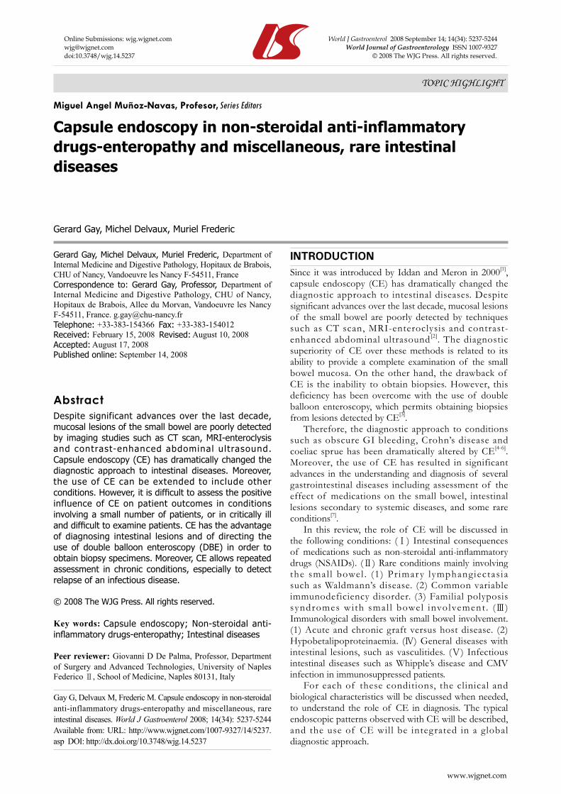
Gerard Gay, Michel Delvaux, Muriel Frederic, Department of Internal Medicine and Digestive Pathology, Hopitaux de Brabois, CHU of Nancy, Vandoeuvre les Nancy F-54511, France Correspondence to: Gerard Gay, Professor, Department of Internal Medicine and Digestive Pathology, CHU of Nancy, Hopitaux de Brabois, Allee du Morvan, Vandoeuvre les Nancy F-54511, France. [email protected]: +33-383-154366 Fax: +33-383-154012Received: February 15, 2008 Revised: August 10, 2008Accepted: August 17, 2008Published online: September 14, 2008
AbstractDespite significant advances over the last decade, mucosal lesions of the small bowel are poorly detected by imaging studies such as CT scan, MRI-enteroclysis and contrast-enhanced abdominal ultrasound. Capsule endoscopy (CE) has dramatically changed the diagnostic approach to intestinal diseases. Moreover, the use of CE can be extended to include other conditions. However, it is difficult to assess the positive influence of CE on patient outcomes in conditions involving a small number of patients, or in critically ill and difficult to examine patients. CE has the advantage of diagnosing intestinal lesions and of directing the use of double balloon enteroscopy (DBE) in order to obtain biopsy specimens. Moreover, CE allows repeated assessment in chronic conditions, especially to detect relapse of an infectious disease.
© 2008 The WJG Press. All rights reserved.
Key words: Capsule endoscopy; Non-steroidal anti-inflammatory drugs-enteropathy; Intestinal diseases
Peer reviewer: Giovanni D De Palma, Professor, Department of Surgery and Advanced Technologies, University of Naples Federico Ⅱ, School of Medicine, Naples 80131, Italy
Gay G, Delvaux M, Frederic M. Capsule endoscopy in non-steroidal anti-inflammatory drugs-enteropathy and miscellaneous, rare intestinal diseases. World J Gastroenterol 2008; 14(34): 5237-5244 Available from: URL: http://www.wjgnet.com/1007-9327/14/5237.asp DOI: http://dx.doi.org/10.3748/wjg.14.5237
TOPIC HIGHLIGHT
Capsule endoscopy in non-steroidal anti-inflammatory drugs-enteropathy and miscellaneous, rare intestinal diseases
Gerard Gay, Michel Delvaux, Muriel Frederic
Miguel Angel Muñoz-Navas, Profesor, Series Editors
Online Submissions: wjg.wjgnet.com World J Gastroenterol 2008 September 14; 14(34): [email protected] World Journal of Gastroenterology ISSN 1007-9327doi:10.3748/wjg.14.5237 © 2008 The WJG Press. All rights reserved.
INTRODUCTIONSince it was introduced by Iddan and Meron in 2000[1], capsule endoscopy (CE) has dramatically changed the diagnostic approach to intestinal diseases. Despite significant advances over the last decade, mucosal lesions of the small bowel are poorly detected by techniques such as CT scan, MRI-enteroclysis and contrast-enhanced abdominal ultrasound[2]. The diagnostic superiority of CE over these methods is related to its ability to provide a complete examination of the small bowel mucosa. On the other hand, the drawback of CE is the inability to obtain biopsies. However, this deficiency has been overcome with the use of double balloon enteroscopy, which permits obtaining biopsies from lesions detected by CE[3].
Therefore, the diagnostic approach to conditions such as obscure GI bleeding, Crohn’s disease and coeliac sprue has been dramatically altered by CE[4-6]. Moreover, the use of CE has resulted in significant advances in the understanding and diagnosis of several gastrointestinal diseases including assessment of the effect of medications on the small bowel, intestinal lesions secondary to systemic diseases, and some rare conditions[7].
In this review, the role of CE will be discussed in the following conditions: (Ⅰ) Intestinal consequences of medications such as non-steroidal anti-inflammatory drugs (NSAIDs). (Ⅱ) Rare conditions mainly involving the smal l bowel . (1) Pr imar y lymphangiectas ia such as Waldmann’s disease. (2) Common variable immunodeficiency disorder. (3) Familial polyposis syndromes wi th smal l bowel involvement . (Ⅲ) Immunological disorders with small bowel involvement. (1) Acute and chronic graft versus host disease. (2) Hypobetalipoproteinaemia. (Ⅳ) General diseases with intestinal lesions, such as vasculitides. (Ⅴ) Infectious intestinal diseases such as Whipple’s disease and CMV infection in immunosuppressed patients.
For each of these conditions, the clinical and biological characteristics will be discussed when needed, to understand the role of CE in diagnosis. The typical endoscopic patterns observed with CE will be described, and the use of CE will be integrated in a global diagnostic approach.
www.wjgnet.com
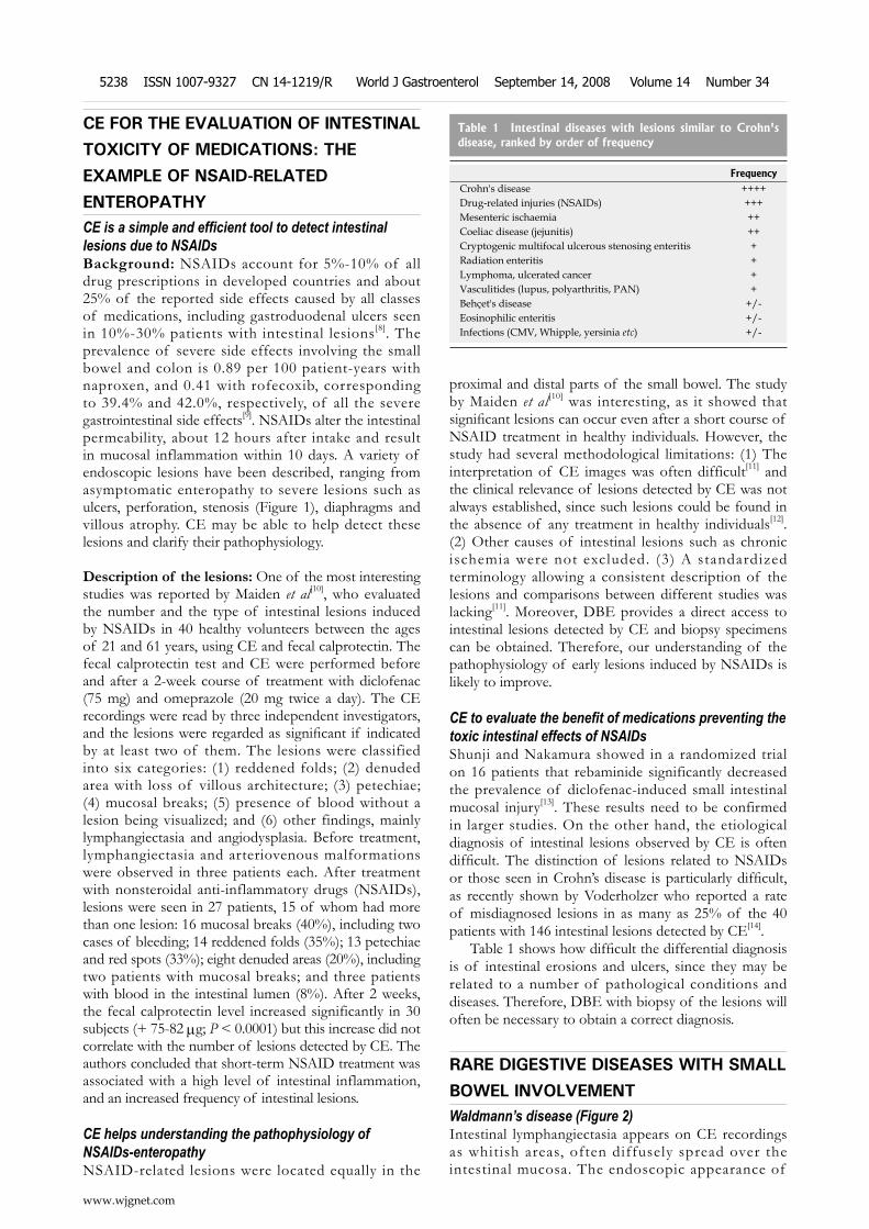
CE FOR THE EVALUATION OF INTESTINAL TOXICITY OF MEDICATIONS: THE EXAMPLE OF NSAID-RELATED ENTEROPATHYCE is a simple and efficient tool to detect intestinal lesions due to NSAIDsBackground: NSAIDs account for 5%-10% of all drug prescriptions in developed countries and about 25% of the reported side effects caused by all classes of medications, including gastroduodenal ulcers seen in 10%-30% patients with intestinal lesions[8]. The prevalence of severe side effects involving the small bowel and colon is 0.89 per 100 patient-years with naproxen, and 0.41 with rofecoxib, corresponding to 39.4% and 42.0%, respectively, of all the severe gastrointestinal side effects[9]. NSAIDs alter the intestinal permeability, about 12 hours after intake and result in mucosal inflammation within 10 days. A variety of endoscopic lesions have been described, ranging from asymptomatic enteropathy to severe lesions such as ulcers, perforation, stenosis (Figure 1), diaphragms and villous atrophy. CE may be able to help detect these lesions and clarify their pathophysiology.
Description of the lesions: One of the most interesting studies was reported by Maiden et al[10], who evaluated the number and the type of intestinal lesions induced by NSAIDs in 40 healthy volunteers between the ages of 21 and 61 years, using CE and fecal calprotectin. The fecal calprotectin test and CE were performed before and after a 2-week course of treatment with diclofenac (75 mg) and omeprazole (20 mg twice a day). The CE recordings were read by three independent investigators, and the lesions were regarded as significant if indicated by at least two of them. The lesions were classified into six categories: (1) reddened folds; (2) denuded area with loss of villous architecture; (3) petechiae; (4) mucosal breaks; (5) presence of blood without a lesion being visualized; and (6) other findings, mainly lymphangiectasia and angiodysplasia. Before treatment, lymphangiectasia and arteriovenous malformations were observed in three patients each. After treatment with nonsteroidal anti-inflammatory drugs (NSAIDs), lesions were seen in 27 patients, 15 of whom had more than one lesion: 16 mucosal breaks (40%), including two cases of bleeding; 14 reddened folds (35%); 13 petechiae and red spots (33%); eight denuded areas (20%), including two patients with mucosal breaks; and three patients with blood in the intestinal lumen (8%). After 2 weeks, the fecal calprotectin level increased significantly in 30 subjects (+ 75-82 µg; P < 0.0001) but this increase did not correlate with the number of lesions detected by CE. The authors concluded that short-term NSAID treatment was associated with a high level of intestinal inflammation, and an increased frequency of intestinal lesions.
CE helps understanding the pathophysiology of NSAIDs-enteropathyNSAID-related lesions were located equally in the
proximal and distal parts of the small bowel. The study by Maiden et al[10] was interesting, as it showed that significant lesions can occur even after a short course of NSAID treatment in healthy individuals. However, the study had several methodological limitations: (1) The interpretation of CE images was often difficult[11] and the clinical relevance of lesions detected by CE was not always established, since such lesions could be found in the absence of any treatment in healthy individuals[12]. (2) Other causes of intestinal lesions such as chronic ischemia were not excluded. (3) A standardized terminology allowing a consistent description of the lesions and comparisons between different studies was lacking[11]. Moreover, DBE provides a direct access to intestinal lesions detected by CE and biopsy specimens can be obtained. Therefore, our understanding of the pathophysiology of early lesions induced by NSAIDs is likely to improve.
CE to evaluate the benefit of medications preventing the toxic intestinal effects of NSAIDsShunji and Nakamura showed in a randomized trial on 16 patients that rebaminide significantly decreased the prevalence of diclofenac-induced small intestinal mucosal injury[13]. These results need to be confirmed in larger studies. On the other hand, the etiological diagnosis of intestinal lesions observed by CE is often difficult. The distinction of lesions related to NSAIDs or those seen in Crohn’s disease is particularly difficult, as recently shown by Voderholzer who reported a rate of misdiagnosed lesions in as many as 25% of the 40 patients with 146 intestinal lesions detected by CE[14].
Table 1 shows how difficult the differential diagnosis is of intestinal erosions and ulcers, since they may be related to a number of pathological conditions and diseases. Therefore, DBE with biopsy of the lesions will often be necessary to obtain a correct diagnosis.
RARE DIGESTIVE DISEASES WITH SMALL BOWEL INVOLVEMENTWaldmann’s disease (Figure 2)Intestinal lymphangiectasia appears on CE recordings as whitish areas, often diffusely spread over the intestinal mucosa. The endoscopic appearance of
Table 1 Intestinal diseases with lesions similar to Crohn's disease, ranked by order of frequency
FrequencyCrohn's disease ++++Drug-related injuries (NSAIDs) +++Mesenteric ischaemia ++Coeliac disease (jejunitis) ++Cryptogenic multifocal ulcerous stenosing enteritis +Radiation enteritis +Lymphoma, ulcerated cancer +Vasculitides (lupus, polyarthritis, PAN) +Behçet's disease +/-Eosinophilic enteritis +/-Infections (CMV, Whipple, yersinia etc) +/-
www.wjgnet.com
5238 ISSN 1007-9327 CN 14-1219/R World J Gastroenterol September 14, 2008 Volume 14 Number 34
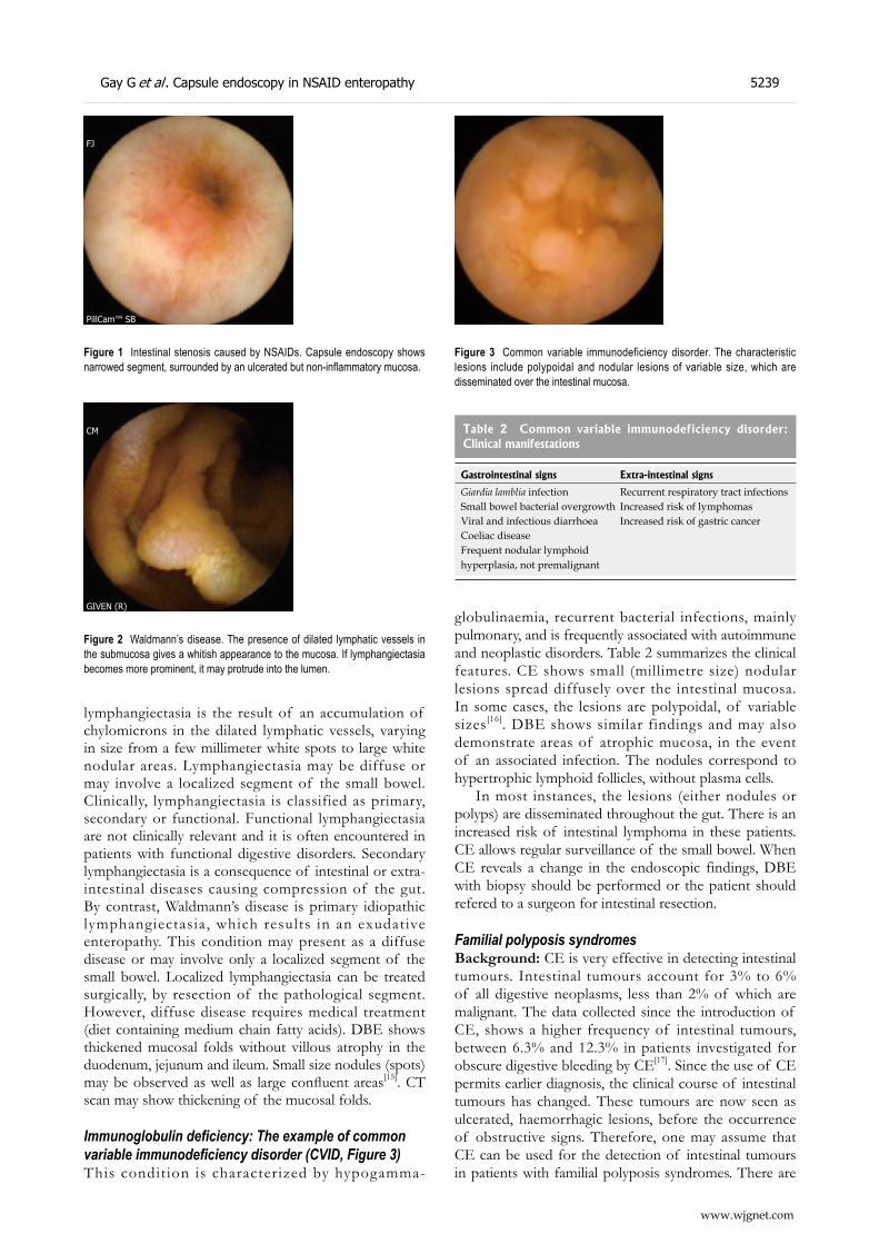
lymphangiectasia is the result of an accumulation of chylomicrons in the dilated lymphatic vessels, varying in size from a few millimeter white spots to large white nodular areas. Lymphangiectasia may be diffuse or may involve a localized segment of the small bowel. Clinically, lymphangiectasia is classified as primary, secondary or functional. Functional lymphangiectasia are not clinically relevant and it is often encountered in patients with functional digestive disorders. Secondary lymphangiectasia is a consequence of intestinal or extra-intestinal diseases causing compression of the gut. By contrast, Waldmann’s disease is primary idiopathic lymphangiectasia, which results in an exudative enteropathy. This condition may present as a diffuse disease or may involve only a localized segment of the small bowel. Localized lymphangiectasia can be treated surgically, by resection of the pathological segment. However, diffuse disease requires medical treatment (diet containing medium chain fatty acids). DBE shows thickened mucosal folds without villous atrophy in the duodenum, jejunum and ileum. Small size nodules (spots) may be observed as well as large confluent areas[15]. CT scan may show thickening of the mucosal folds.
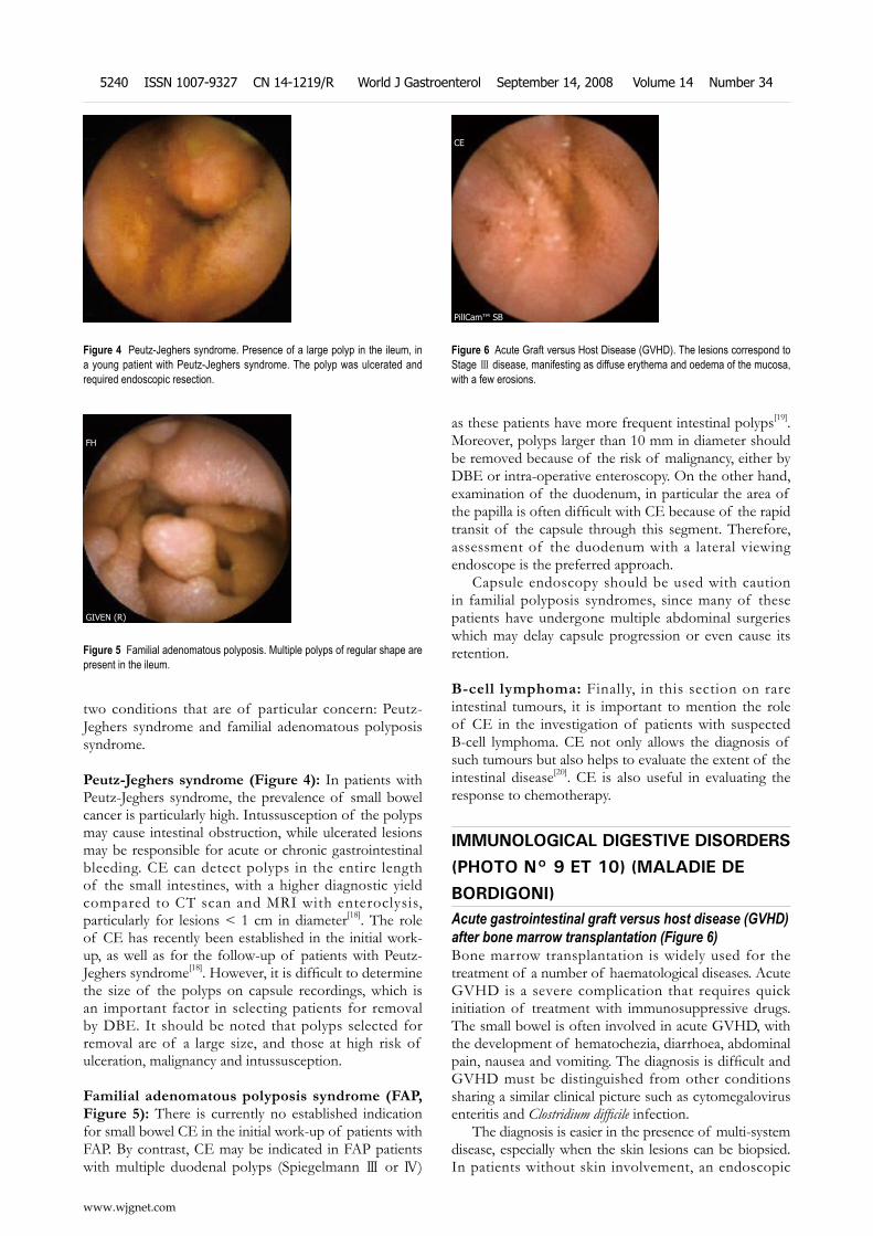
Immunoglobulin deficiency: The example of common variable immunodeficiency disorder (CVID, Figure 3)This condition is characterized by hypogamma-
globulinaemia, recurrent bacterial infections, mainly pulmonary, and is frequently associated with autoimmune and neoplastic disorders. Table 2 summarizes the clinical features. CE shows small (millimetre size) nodular lesions spread diffusely over the intestinal mucosa. In some cases, the lesions are polypoidal, of variable sizes[16]. DBE shows similar findings and may also demonstrate areas of atrophic mucosa, in the event of an associated infection. The nodules correspond to hypertrophic lymphoid follicles, without plasma cells.
In most instances, the lesions (either nodules or polyps) are disseminated throughout the gut. There is an increased risk of intestinal lymphoma in these patients. CE allows regular surveillance of the small bowel. When CE reveals a change in the endoscopic findings, DBE with biopsy should be performed or the patient should refered to a surgeon for intestinal resection.
Familial polyposis syndromes Background: CE is very effective in detecting intestinal tumours. Intestinal tumours account for 3% to 6% of all digestive neoplasms, less than 2% of which are malignant. The data collected since the introduction of CE, shows a higher frequency of intestinal tumours, between 6.3% and 12.3% in patients investigated for obscure digestive bleeding by CE[17]. Since the use of CE permits earlier diagnosis, the clinical course of intestinal tumours has changed. These tumours are now seen as ulcerated, haemorrhagic lesions, before the occurrence of obstructive signs. Therefore, one may assume that CE can be used for the detection of intestinal tumours in patients with familial polyposis syndromes. There are
PillCam™ SB
FJ
Figure 1 Intestinal stenosis caused by NSAIDs. Capsule endoscopy shows narrowed segment, surrounded by an ulcerated but non-inflammatory mucosa.
GIVEN (R)
CM
Figure 2 Waldmann’s disease. The presence of dilated lymphatic vessels in the submucosa gives a whitish appearance to the mucosa. If lymphangiectasia becomes more prominent, it may protrude into the lumen.
Figure 3 Common variable immunodeficiency disorder. The characteristic lesions include polypoidal and nodular lesions of variable size, which are disseminated over the intestinal mucosa.
Table 2 Common variable immunodeficiency disorder: Clinical manifestations
Gastrointestinal signs Extra-intestinal signs
Giardia lamblia infection Recurrent respiratory tract infectionsSmall bowel bacterial overgrowth Increased risk of lymphomasViral and infectious diarrhoea Increased risk of gastric cancerCoeliac diseaseFrequent nodular lymphoid hyperplasia, not premalignant
Gay G et al . Capsule endoscopy in NSAID enteropathy 5239
www.wjgnet.com
two conditions that are of particular concern: Peutz-Jeghers syndrome and familial adenomatous polyposis syndrome.
Peutz-Jeghers syndrome (Figure 4): In patients with Peutz-Jeghers syndrome, the prevalence of small bowel cancer is particularly high. Intussusception of the polyps may cause intestinal obstruction, while ulcerated lesions may be responsible for acute or chronic gastrointestinal bleeding. CE can detect polyps in the entire length of the small intestines, with a higher diagnostic yield compared to CT scan and MRI with enteroclysis, particularly for lesions < 1 cm in diameter[18]. The role of CE has recently been established in the initial work-up, as well as for the follow-up of patients with Peutz-Jeghers syndrome[18]. However, it is difficult to determine the size of the polyps on capsule recordings, which is an important factor in selecting patients for removal by DBE. It should be noted that polyps selected for removal are of a large size, and those at high risk of ulceration, malignancy and intussusception.
Familial adenomatous polyposis syndrome (FAP, Figure 5): There is currently no established indication for small bowel CE in the initial work-up of patients with FAP. By contrast, CE may be indicated in FAP patients with multiple duodenal polyps (Spiegelmann Ⅲ or Ⅳ)
as these patients have more frequent intestinal polyps[19]. Moreover, polyps larger than 10 mm in diameter should be removed because of the risk of malignancy, either by DBE or intra-operative enteroscopy. On the other hand, examination of the duodenum, in particular the area of the papilla is often difficult with CE because of the rapid transit of the capsule through this segment. Therefore, assessment of the duodenum with a lateral viewing endoscope is the preferred approach.
Capsule endoscopy should be used with caution in familial polyposis syndromes, since many of these patients have undergone multiple abdominal surgeries which may delay capsule progression or even cause its retention.
B-cell lymphoma: Finally, in this section on rare intestinal tumours, it is important to mention the role of CE in the investigation of patients with suspected B-cell lymphoma. CE not only allows the diagnosis of such tumours but also helps to evaluate the extent of the intestinal disease[20]. CE is also useful in evaluating the response to chemotherapy.
IMMUNOLOGICAL DIGESTIVE DISORDERS (PHOTO N° 9 ET 10) (MALADIE DE BORDIGONI)Acute gastrointestinal graft versus host disease (GVHD) after bone marrow transplantation (Figure 6)Bone marrow transplantation is widely used for the treatment of a number of haematological diseases. Acute GVHD is a severe complication that requires quick initiation of treatment with immunosuppressive drugs. The small bowel is often involved in acute GVHD, with the development of hematochezia, diarrhoea, abdominal pain, nausea and vomiting. The diagnosis is difficult and GVHD must be distinguished from other conditions sharing a similar clinical picture such as cytomegalovirus enteritis and Clostridium difficile infection.
The diagnosis is easier in the presence of multi-system disease, especially when the skin lesions can be biopsied. In patients without skin involvement, an endoscopic
Figure 4 Peutz-Jeghers syndrome. Presence of a large polyp in the ileum, in a young patient with Peutz-Jeghers syndrome. The polyp was ulcerated and required endoscopic resection.
GIVEN (R)
FH
Figure 5 Familial adenomatous polyposis. Multiple polyps of regular shape are present in the ileum.
PillCam™ SB
CE
Figure 6 Acute Graft versus Host Disease (GVHD). The lesions correspond to Stage Ⅲ disease, manifesting as diffuse erythema and oedema of the mucosa, with a few erosions.
www.wjgnet.com
5240 ISSN 1007-9327 CN 14-1219/R World J Gastroenterol September 14, 2008 Volume 14 Number 34
work-up is needed, with esogastroduodenoscopy and ileo-colonoscopy. Until recently, esogastroduodenoscopy was regarded as the gold standard for the examination of the duodenum, with findings of mucosal oedema, denuded mucosa, erosions, erythema and bleeding ulcers. These lesions are classified into 4 grades, grade Ⅰ refers to focal erythema, grade Ⅱ diffuse erythema and oedema, grade Ⅲ severe oedema and friable mucosa with erosions, and grade Ⅳ presence of exudate, ulcers and active bleeding[21].
The clinical usefulness of CE in acute GVHD was demonstrated in two studies. Neumann et al[22] evaluated 14 patients with clinical signs of acute GVHD after bone marrow transplantation, and observed typical intestinal lesions with histological confirmation on biopsy obtained by endoscopy, performed subsequently. This study also showed that the lesions involved the entire length of the small bowel and were more intense in the ileum compared to in the jejunum. The most important finding was the very high negative predictive value of CE since patients with a negative CE did not develop acute GVHD during the 2-month follow-up period after CE. These findings were confirmed by Aghai et al[23] in a study on a larger cohort of patients.
Although it is worth mentioning that CE is well tolerated by such critically ill patients, certain limitations should be noted. In some patients, the capsule may be retained in the stomach. The Rapidview system can be used to monitor the progression of the capsule[24]. In the event of delayed gastric clearance, erythromycin may be administered. Moreover, it should be emphasized that in some patients, CE was normal but these patients had acute GVHD[25].
Despite the limited data available in the literature, CE should now be regarded as an alternative to esogastroduodenoscopy in the workup of patients with suspected GVHD, particularly in critically ill patients requiring quick therapeutic decisions. Indeed, CE appears to be as effective as esogastroduodenoscopy with biopsy for the diagnosis of acute GVHD[25-27].
Hypobetalipoproteinemia (Figure 7)Background: Hypobetalipoproteinemia is an autosomal dominant disorder caused by a mutation or deletion in the apoB gene which produces a truncated apolipoprotein B. The plasma concentrations of the two forms of apolipoprotein B, that is, Apo B-100 synthesized by the liver and Apo-B48 synthesized by the intestine, are undetectable. The biological and clinical picture is similar to that of abetalipoproteinemia. Heterozygous hypobetalipoproteinemia is present in 0.1%-0.8% of the genera l popula t ion , whi le homozyg ous hypobetalipoproteinemia is rare. The prognosis of Apo B-related disorders depends mainly on the consequences of the malabsorption syndrome, and on the progression of neurological alterations caused by the deficiency of soluble vitamin E[28].
Diagnostic procedure: Laboratory tests show hypocholesterolemia, acanthocytosis and undetectable
ApoB lipoprotein. CT scan shows massive and diffuse infiltration of the liver. CE demonstrates diffuse whitish pattern of the intestinal mucosa with occasional yellow areas seen in the entire length of the small bowel, but without any villous atrophy. Enteroscopy, formerly performed with push enteroscopes and currently with DBE shows a similar pattern as CE and moreover, biopsy specimens can be obtained. These lesions are produced by the accumulation of fat vesicles in the enterocytes in the jejunum and ileum, without causing vil lous atrophy [29]. Immunohistchemistr y shows migration of truncated Apo-B and the absence of normal Apo-B hypobetalipoproteinemia.
INTESTINAL EXPRESSION OF VASCULITIDESBackgroundGeneral considerations: Systemic diseases include several very heterogeneous pathological conditions which share some common features: (1) symptoms suggesting the involvement of multiple organs, (2) specific immunological and biological alterations, (3) a tendency to chronicity, and (4) clinical response to immunosuppressive therapy. Systemic diseases are divided into two main categories[30,31]: connective tissue or collagenous diseases with predominant abnormal production or deregulated synthesis of collagen, and vasculitides with predominant inflammation of the blood vessels resulting in abnormal vascular permeability, thrombosis and tissue ischaemia. These conditions share some pathophysiological mechanisms such as abnormal production of proteins or cytokines, and abnormal humoral or cellular immunity that results in muscular inflammation, atrophy and fragmentation. In the end, the vascular endothelial changes become predominant and are responsible for the muscular, neurological and digestive abnormalities.
Digestive manifestations of vasculitides and systemic diseases include a number of clinical conditions (Table 3)[32]. The development of GI symptoms raises three main questions: Do the digestive symptoms correspond to the initial manifestation of the condition or to a new acute phase of the disease? Are the digestive symptoms secondary to a complication of the treatment? Is the observed complication a component of the primary disease? To answer these questions, the clinician may use diagnostic techniques such as CT scan, biological and immunological tests, and CE. The use of CE will be illustrated by a few examples.
Specific characteristics of diseasesBehçet’s disease: Behçet’s disease is a systemic inflammatory condition, with 10% to 25% patients developing GI manifestations. The symptoms consist mainly of abdominal pain, diarrhoea and acute or chronic bleeding. CE may show erosions and aphthous ulcers in the small bowel, as reported by Fylejk in a series of 20 patients[33]. Intestinal lesions may be present
Gay G et al . Capsule endoscopy in NSAID enteropathy 5241
www.wjgnet.com
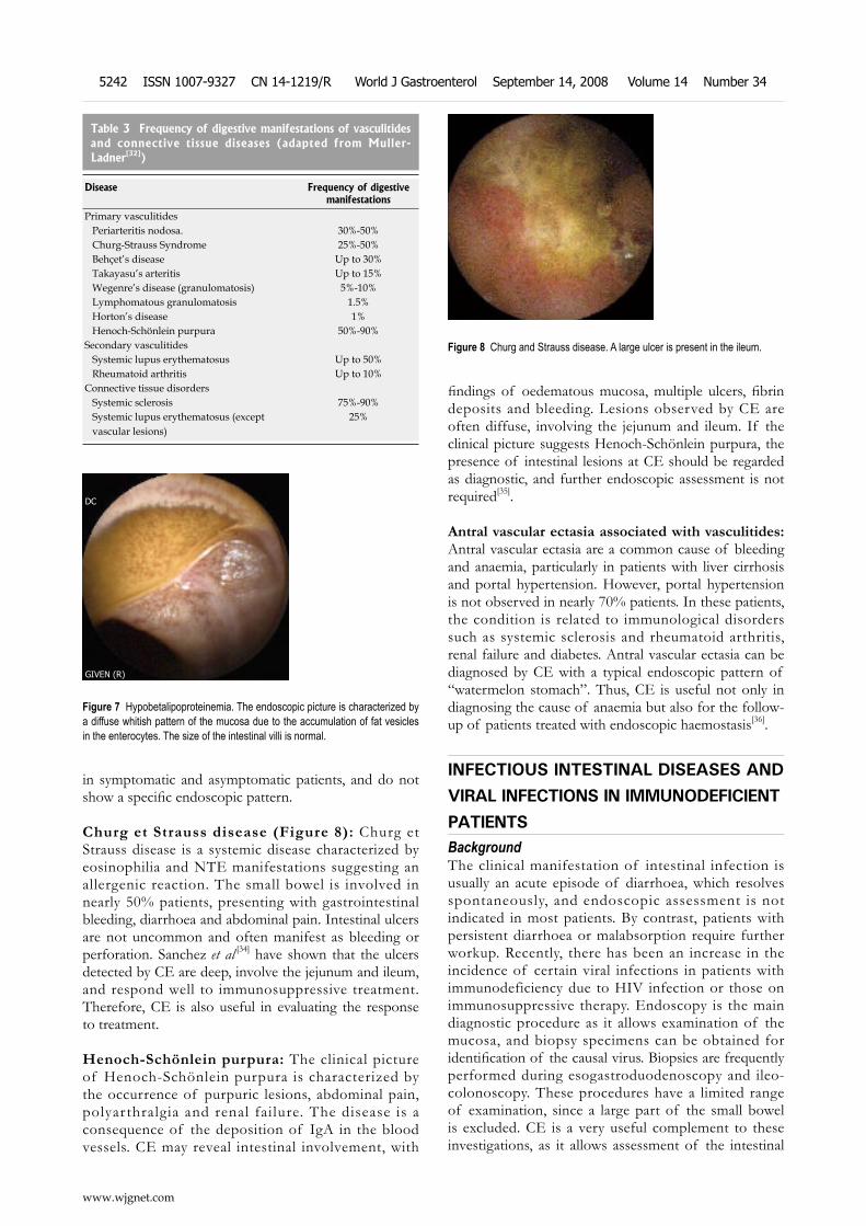
in symptomatic and asymptomatic patients, and do not show a specific endoscopic pattern.
Churg et Strauss disease (Figure 8): Churg et Strauss disease is a systemic disease characterized by eosinophilia and NTE manifestations suggesting an allergenic reaction. The small bowel is involved in nearly 50% patients, presenting with gastrointestinal bleeding, diarrhoea and abdominal pain. Intestinal ulcers are not uncommon and often manifest as bleeding or perforation. Sanchez [34] have shown that the ulcers detected by CE are deep, involve the jejunum and ileum, and respond well to immunosuppressive treatment. Therefore, CE is also useful in evaluating the response to treatment.
Henoch-Schönlein purpura: The clinical picture of Henoch-Schönlein purpura is characterized by the occurrence of purpuric lesions, abdominal pain, polyarthralgia and renal failure. The disease is a consequence of the deposition of IgA in the blood vessels. CE may reveal intestinal involvement, with
findings of oedematous mucosa, multiple ulcers, fibrin deposits and bleeding. Lesions observed by CE are often diffuse, involving the jejunum and ileum. If the clinical picture suggests Henoch-Schönlein purpura, the presence of intestinal lesions at CE should be regarded as diagnostic, and further endoscopic assessment is not required[35].
Antral vascular ectasia associated with vasculitides: Antral vascular ectasia are a common cause of bleeding and anaemia, particularly in patients with liver cirrhosis and portal hypertension. However, portal hypertension is not observed in nearly 70% patients. In these patients, the condition is related to immunological disorders such as systemic sclerosis and rheumatoid arthritis, renal failure and diabetes. Antral vascular ectasia can be diagnosed by CE with a typical endoscopic pattern of “watermelon stomach”. Thus, CE is useful not only in diagnosing the cause of anaemia but also for the follow-up of patients treated with endoscopic haemostasis[36].
INFECTIOUS INTESTINAL DISEASES AND VIRAL INFECTIONS IN IMMUNODEFICIENT PATIENTSBackgroundThe clinical manifestation of intestinal infection is usually an acute episode of diarrhoea, which resolves spontaneously, and endoscopic assessment is not indicated in most patients. By contrast, patients with persistent diarrhoea or malabsorption require further workup. Recently, there has been an increase in the incidence of certain viral infections in patients with immunodeficiency due to HIV infection or those on immunosuppressive therapy. Endoscopy is the main diagnostic procedure as it allows examination of the mucosa, and biopsy specimens can be obtained for identification of the causal virus. Biopsies are frequently performed during esogastroduodenoscopy and ileo-colonoscopy. These procedures have a limited range of examination, since a large part of the small bowel is excluded. CE is a very useful complement to these investigations, as it allows assessment of the intestinal
GIVEN (R)
DC
Figure 7 Hypobetalipoproteinemia. The endoscopic picture is characterized by a diffuse whitish pattern of the mucosa due to the accumulation of fat vesicles in the enterocytes. The size of the intestinal villi is normal.
Figure 8 Churg and Strauss disease. A large ulcer is present in the ileum.
Table 3 Frequency of digestive manifestations of vasculitides and connective tissue diseases (adapted from Muller-Ladner[32])
Disease Frequency of digestive manifestations
Primary vasculitides Periarteritis nodosa. 30%-50% Churg-Strauss Syndrome 25%-50% Behçet’s disease Up to 30% Takayasu’s arteritis Up to 15% Wegenre’s disease (granulomatosis) 5%-10% Lymphomatous granulomatosis 1.5% Horton’s disease 1% Henoch-Schönlein purpura 50%-90%Secondary vasculitides Systemic lupus erythematosus Up to 50% Rheumatoid arthritis Up to 10%Connective tissue disorders Systemic sclerosis 75%-90% Systemic lupus erythematosus (except vascular lesions)
25%
www.wjgnet.com
5242 ISSN 1007-9327 CN 14-1219/R World J Gastroenterol September 14, 2008 Volume 14 Number 34
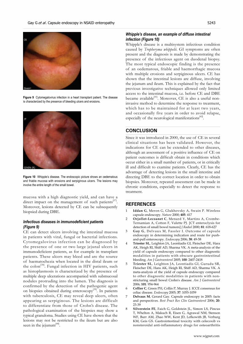
mucosa with a high diagnostic yield, and can have a direct impact on the management of such patients[37]. Moreover, lesions detected by CE can be subsequently biopsied during DBE.
Infectious diseases in immunodeficient patients (Figure 9)CE can detect ulcers involving the intestinal mucosa in patients with viral, fungal or bacterial infections. Cytomegalovirus infection can be diagnosed by the presence of one or two large jejunal ulcers in immunodeficient patients, as for example in transplant patients. These ulcers may bleed and are the source of haematochezia when located in the distal ileum or the colon[38]. Fungal infection in HIV patients, such as histoplasmosis is characterized by the presence of multiple deep ulcerations accompanied with submucosal nodules protruding into the lumen. The diagnosis is confirmed by the detection of the pathogenic agent on biopsies obtained during enteroscopy[39]. In patients with tuberculosis, CE may reveal deep ulcers, often appearing as serpiginous. The lesions are difficult to differentiate from those of Crohn’s disease. The pathological examination of the biopsies may show a typical granuloma. Studies using CE have shown that the lesions may not be restricted to the ileum but are also seen in the jejunum[40].
Whipple’s disease, an example of diffuse intestinal infection (Figure 10)Whipple’s disease is a multisystem infectious condition caused by Tropheryma whippelii. GI symptoms are often present and the diagnosis is made by demonstrating the presence of the infectious agent on duodenal biopsy. The most typical endoscopic finding is the presence of an oedematous, friable and haemorrhagic mucosa with multiple erosions and serpiginous ulcers. CE has shown that the intestinal lesions are diffuse, involving the jejunum and ileum. This is explained by the fact that previous investigative techniques allowed only limited access to the intestinal mucosa, i.e. before CE and DBE became available[41]. Moreover, CE is also a useful non-invasive method to determine the response to treatment, which has to be maintained for at least two years, and occasionally five years in order to avoid relapse, especially of the neurological manifestations[42].
CONCLUSIONSince it was introduced in 2000, the use of CE in several clinical situations has been validated. However, the indications for CE can be extended to other diseases, although an assessment of a positive influence of CE on patient outcomes is difficult obtain in conditions which occur either in a small number of patients, or in critically ill and difficult to examine patients. Clearly, CE has the advantage of detecting lesions in the small intestine and directing DBE to the correct location in order to obtain biopsies. Moreover, repeated assessment can be made in chronic conditions, especially to detect the response to treatment.
REFERENCES1 Iddan G, Meron G, Glukhovsky A, Swain P. Wireless
capsule endoscopy. Nature 2000; 405: 4172 Orjollet-Lecoanet C, Menard Y, Martins A, Crombe-
Ternamian A, Cotton F, Valette PJ. [CT enteroclysis for detection of small bowel tumors] J Radiol 2000; 81: 618-627
3 Gay G , Delvaux M, Fassler I . Outcome of capsule endoscopy in determining indication and route for push-and-pull enteroscopy. Endoscopy 2006; 38: 49-58
4 Triester SL, Leighton JA, Leontiadis GI, Fleischer DE, Hara AK, Heigh RI, Shiff AD, Sharma VK. A meta-analysis of the yield of capsule endoscopy compared to other diagnostic modalities in patients with obscure gastrointestinal bleeding. Am J Gastroenterol 2005; 100: 2407-2418
5 Triester SL, Leighton JA, Leontiadis GI, Gurudu SR, Fleischer DE, Hara AK, Heigh RI, Shiff AD, Sharma VK. A meta-analysis of the yield of capsule endoscopy compared to other diagnostic modalities in patients with non-stricturing small bowel Crohn's disease. Am J Gastroenterol 2006; 101: 954-964
6 Cellier C, Green PH, Collin P, Murray J. ICCE consensus for celiac disease. Endoscopy 2005; 37: 1055-1059
7 Delvaux M, Gerard Gay. Capsule endoscopy in 2005: facts and perspectives. Best Pract Res Clin Gastroenterol 2006; 20: 23-39
8 Silverstein FE, Faich G, Goldstein JL, Simon LS, Pincus T, Whelton A, Makuch R, Eisen G, Agrawal NM, Stenson WF, Burr AM, Zhao WW, Kent JD, Lefkowith JB, Verburg KM, Geis GS. Gastrointestinal toxicity with celecoxib vs nonsteroidal anti-inflammatory drugs for osteoarthritis
Given®
CK
Figure 9 Cytomegalovirus infection in a heart transplant patient. The disease is characterized by the presence of bleeding ulcers and erosions.
GIVEN (R)
JT
Figure 10 Whipple’s disease. The endoscopic picture shows an oedematous and friable mucosa with erosions and serpiginous ulcers. The lesions may involve the entire length of the small bowel.
Gay G et al . Capsule endoscopy in NSAID enteropathy 5243
www.wjgnet.com
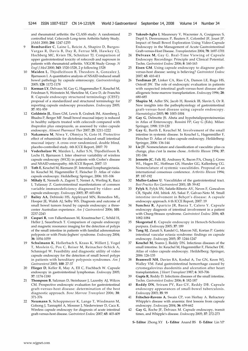
and rheumatoid arthritis: the CLASS study: A randomized controlled trial. Celecoxib Long-term Arthritis Safety Study. JAMA 2000; 284: 1247-1255
9 Bombardier C, Laine L, Reicin A, Shapiro D, Burgos-Vargas R, Davis B, Day R, Ferraz MB, Hawkey CJ, Hochberg MC, Kvien TK, Schnitzer TJ. Comparison of upper gastrointestinal toxicity of rofecoxib and naproxen in patients with rheumatoid arthritis. VIGOR Study Group. N Engl J Med 2000; 343: 1520-1528, 2 p following 1528
10 Maiden L , Thjodleifsson B, Theodors A, Gonzalez J, Bjarnason I. A quantitative analysis of NSAID-induced small bowel pathology by capsule enteroscopy. Gastroenterology 2005; 128: 1172-1178
11 Korman LY, Delvaux M, Gay G, Hagenmuller F, Keuchel M, Friedman S, Weinstein M, Shetzline M, Cave D, de Franchis R. Capsule endoscopy structured terminology (CEST): proposal of a standardized and structured terminology for reporting capsule endoscopy procedures. Endoscopy 2005; 37: 951-959
12 Goldstein JL, Eisen GM, Lewis B, Gralnek IM, Aisenberg J, Bhadra P, Berger MF. Small bowel mucosal injury is reduced in healthy subjects treated with celecoxib compared with ibuprofen plus omeprazole, as assessed by video capsule endoscopy. Aliment Pharmacol Ther 2007; 25: 1211-1222
13 Nakamura M, Niwa Y, Ohmiya N, Goto H. Protective effect of rebaminide for dictofenac-induced small intestinal mucosal injury: A cross over randomized, double blind, placebo-controlled study. 6th ICCE Report, 2007: 70
14 Voderholzer W, Maiden L, Adler S.N, Thjodleifsson B, Lochs H, Bjarnason I. Inter observer variability of wireless capsule endoscopy (WCE) in patients with Crohn’s disease and NSAID enteropathy. 6th ICCE Report, 2007: 13
15 Toth E, Keuchel M, Riemann JF. Intestinal lymphangiectasia. In: Keuchel M, Hagenmüller F, Fleischer D. Atlas of video capsule endoscopy. Heildelberg: Springer, 2006: 101-106
16 Mihaly E, Nemeth A, Zagoni T, Nemet A, Werling K, Racz I, Tulassay Z. Gastrointestinal manifestations of common variable immunodeficiency diagnosed by video- and capsule endoscopy. Endoscopy 2005; 37: 603-604
17 Bailey AA, Debinski HS, Appleyard MN, Remedios ML, Hooper JE, Walsh AJ, Selby WS. Diagnosis and outcome of small bowel tumors found by capsule endoscopy: a three-center Australian experience. Am J Gastroenterol 2006; 101: 2237-2243
18 Caspari R, von Falkenhausen M, Krautmacher C, Schild H, Heller J, Sauerbruch T. Comparison of capsule endoscopy and magnetic resonance imaging for the detection of polyps of the small intestine in patients with familial adenomatous polyposis or with Peutz-Jeghers' syndrome. Endoscopy 2004; 36: 1054-1059
19 Schulmann K, Hollerbach S, Kraus K, Willert J, Vogel T, Moslein G, Pox C, Reiser M, Reinacher-Schick A, Schmiegel W. Feasibility and diagnostic utility of video capsule endoscopy for the detection of small bowel polyps in patients with hereditary polyposis syndromes. Am J Gastroenterol 2005; 100: 27-37
20 Flieger D, Keller R, May A, Ell C, Fischbach W. Capsule endoscopy in gastrointestinal lymphomas. Endoscopy 2005; 37: 1174-1180
21 Thompson B, Salzman D, Steinhauer J, Lazenby AJ, Wilcox CM. Prospective endoscopic evaluation for gastrointestinal graft-versus-host disease: determination of the best diagnostic approach. Bone Marrow Transplant 2006; 38: 371-376
22 Neumann S, Schoppmeyer K, Lange T, Wiedmann M, Golsong J, Tannapfel A, Mossner J, Niederwieser D, Caca K. Wireless capsule endoscopy for diagnosis of acute intestinal graft-versus-host disease. Gastrointest Endosc 2007; 65: 403-409
23 Yakoub-Agha I, Maunoury V, Wacrenier A, Couignoux S, Depil S, Desreumaux P, Bauters F, Colombel JF, Jouet JP. Impact of Small Bowel Exploration Using Video-Capsule Endoscopy in the Management of Acute Gastrointestinal Graft-versus-Host Disease. Transplantation 2004; 78: 1697-1701
24 Delvaux M , Gay G. Real-Time Viewing of Capsule Endoscopy Recordings: Principle and Clinical Potential. Techn. Gastrointest Endosc 2006; 8: 160-163
25 Eisen GM. Using capsule endoscopy to diagnose graft-versus-host disease: seeing is believing? Gastrointest Endosc 2007; 65: 410-411
26 Terdiman JP, Linker CA, Ries CA, Damon LE, Rugo HS, Ostroff JW. The role of endoscopic evaluation in patients with suspected intestinal graft-versus-host disease after allogeneic bone-marrow transplantation. Endoscopy 1996; 28: 680-685
27 Shapira M, Adler SN, Jacob H, Resnick IB, Slavin S, Or R. New insights into the pathophysiology of gastrointestinal graft-versus-host disease using capsule endoscopy. Haematologica 2005; 90: 1003-1004
28 Gay G, Delmotte JS. Abeta and hypobetalipoproteinemias in Atlas of Enteroscopy. Rossini FP, Gay G (Eds). Milan: Springer, 1998: 119-120
29 Gay G, Barth E, Keuchel M. Involvement of the small intestine in systemic disease. In Keuchel L, Hagenmüller F, Fleischer D. Atlas of video capsule endoscopy. Heildelberg: Springer, 2006: 136-144
30 Lie JT. Nomenclature and classification of vasculitis: plus ca change, plus c'est la meme chose. Arthritis Rheum 1994; 37: 181-186
31 Jennette JC, Falk RJ, Andrassy K, Bacon PA, Churg J, Gross WL, Hagen EC, Hoffman GS, Hunder GG, Kallenberg CG. Nomenclature of systemic vasculitides. Proposal of an international consensus conference. Arthritis Rheum 1994; 37: 187-192
32 Muller-Ladner U. Vasculitides of the gastrointestinal tract. Best Practice Res Gastroenterol 2001; 15: 59-82
33 Fylyk S, Fylyk SN, Safatle-Riberio AV, Neves F, Goncalves CR, Sipahi AM, Ishiok AS, Sakai P, Galvao-Neto M. Small intestine involvement in Behçet’s disease: A capsule endoscopy approach. 6 th ICCE Report, 2007: 19
34 Sanchez R , Aparicio JR, Baeza T, Calero Y. Capsule endoscopy diagnosis of intestinal involvement in a patient with Churg-Strauss syndrome. Gastrointest Endosc 2006; 63: 1082-1084
35 Skogestad E. Capsule endoscopy in Henoch-Schonlein purpura. Endoscopy 2005; 37: 189
36 Tang SJ, Zanati S, Kandel G, Marcon NE, Kortan P. Gastric intestinal vascular ectasia syndrome: findings on capsule endoscopy. Endoscopy 2005; 37: 1244-1247
37 Keuchel M, Soares J, Reddy DN. Infectious diseases of the small intestine. In: Keuchel M, Hagenmüller F, Fleischer DE. Atlas of video capsule endoscopy. Heildelberg: Springer, 2006: 126-135
38 Bramwell NH, Davies RA, Koshal A, Tse GN, Keon WJ, Walley VM. Fatal gastrointestinal hemorrhage caused by cytomegalovirus duodenitis and ulceration after heart transplantation. J Heart Transplant 1987; 6: 303-706
39 Gupta R, Reddy D. Infections diseases of the small intestine. Techn. Gastrointest Endosc 2006; 8: 182-187
40 Reddy DN , Sriram PV, Rao GV, Reddy DB. Capsule endoscopy appearances of small-bowel tuberculosis. Endoscopy 2003; 35: 99
41 Fritscher-Ravens A, Swain CP, von Herbay A. Refractory Whipple's disease with anaemia: first lessons from capsule endoscopy. Endoscopy 2004; 36: 659-662
42 Gay G, Roche JF, Delvaux M. Capsule endoscopy, transit times, and Whipple's disease. Endoscopy 2005; 37: 272-273
S- Editor Zhong XY L- Editor Anand BS E- Editor Lin YP
www.wjgnet.com
5244 ISSN 1007-9327 CN 14-1219/R World J Gastroenterol September 14, 2008 Volume 14 Number 34
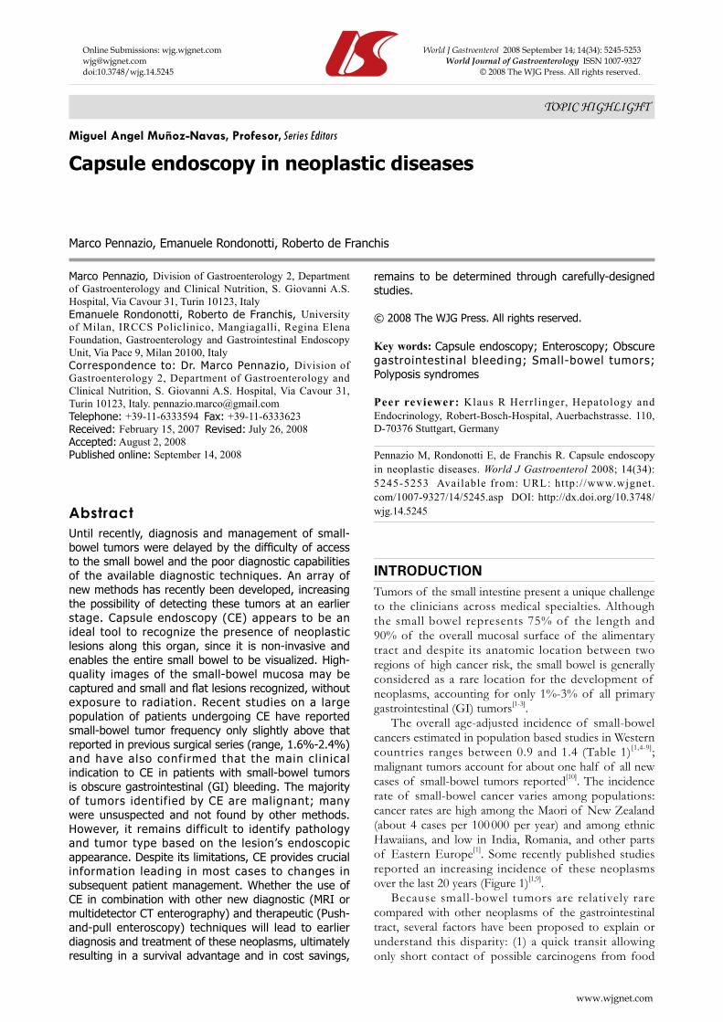
TOPIC HIGHLIGHT
Capsule endoscopy in neoplastic diseases
Marco Pennazio, Emanuele Rondonotti, Roberto de Franchis
Miguel Angel Muñoz-Navas, Profesor, Series Editors
Online Submissions: wjg.wjgnet.com World J Gastroenterol 2008 September 14; 14(34): [email protected] World Journal of Gastroenterology ISSN 1007-9327doi:10.3748/wjg.14.5245 © 2008 The WJG Press. All rights reserved.
Marco Pennazio, Division of Gastroenterology 2, Department of Gastroenterology and Clinical Nutrition, S. Giovanni A.S. Hospital, Via Cavour 31, Turin 10123, ItalyEmanuele Rondonotti, Roberto de Franchis, University of Milan, IRCCS Policlinico, Mangiagalli, Regina Elena Foundation, Gastroenterology and Gastrointestinal Endoscopy Unit, Via Pace 9, Milan 20100, ItalyCorrespondence to: Dr. Marco Pennazio, Division of Gastroenterology 2, Department of Gastroenterology and Clinical Nutrition, S. Giovanni A.S. Hospital, Via Cavour 31, Turin 10123, Italy. [email protected]: +39-11-6333594 Fax: +39-11-6333623Received: February 15, 2007 Revised: July 26, 2008Accepted: August 2, 2008Published online: September 14, 2008
AbstractUntil recently, diagnosis and management of small-bowel tumors were delayed by the difficulty of access to the small bowel and the poor diagnostic capabilities of the available diagnostic techniques. An array of new methods has recently been developed, increasing the possibility of detecting these tumors at an earlier stage. Capsule endoscopy (CE) appears to be an ideal tool to recognize the presence of neoplastic lesions along this organ, since it is non-invasive and enables the entire small bowel to be visualized. High-quality images of the small-bowel mucosa may be captured and small and flat lesions recognized, without exposure to radiation. Recent studies on a large population of patients undergoing CE have reported small-bowel tumor frequency only slightly above that reported in previous surgical series (range, 1.6%-2.4%) and have also confirmed that the main cl inical indication to CE in patients with small-bowel tumors is obscure gastrointestinal (GI) bleeding. The majority of tumors identified by CE are malignant; many were unsuspected and not found by other methods. However, it remains difficult to identify pathology and tumor type based on the lesion’s endoscopic appearance. Despite its limitations, CE provides crucial information leading in most cases to changes in subsequent patient management. Whether the use of CE in combination with other new diagnostic (MRI or multidetector CT enterography) and therapeutic (Push-and-pull enteroscopy) techniques will lead to earlier diagnosis and treatment of these neoplasms, ultimately resulting in a survival advantage and in cost savings,
remains to be determined through carefully-designed studies.
© 2008 The WJG Press. All rights reserved.
Key words: Capsule endoscopy; Enteroscopy; Obscure gastrointestinal bleeding; Small-bowel tumors; Polyposis syndromes
Peer reviewer: Klaus R Herrlinger, Hepatology and Endocrinology, Robert-Bosch-Hospital, Auerbachstrasse. 110, D-70376 Stuttgart, Germany
Pennazio M, Rondonotti E, de Franchis R. Capsule endoscopy in neoplastic diseases. World J Gastroenterol 2008; 14(34): 5245-5253 Available from: URL: http://www.wjgnet.com/1007-9327/14/5245.asp DOI: http://dx.doi.org/10.3748/wjg.14.5245
INTRODUCTIONTumors of the small intestine present a unique challenge to the clinicians across medical specialties. Although the small bowel represents 75% of the length and 90% of the overall mucosal surface of the alimentary tract and despite its anatomic location between two regions of high cancer risk, the small bowel is generally considered as a rare location for the development of neoplasms, accounting for only 1%-3% of all primary gastrointestinal (GI) tumors[1-3].
The overall age-adjusted incidence of small-bowel cancers estimated in population based studies in Western countries ranges between 0.9 and 1.4 (Table 1)[1,4-9]; malignant tumors account for about one half of all new cases of small-bowel tumors reported[10]. The incidence rate of small-bowel cancer varies among populations: cancer rates are high among the Maori of New Zealand (about 4 cases per 100 000 per year) and among ethnic Hawaiians, and low in India, Romania, and other parts of Eastern Europe[1]. Some recently published studies reported an increasing incidence of these neoplasms over the last 20 years (Figure 1)[1,9].
Because small-bowel tumors are relatively rare compared with other neoplasms of the gastrointestinal tract, several factors have been proposed to explain or understand this disparity: (1) a quick transit allowing only short contact of possible carcinogens from food
www.wjgnet.com
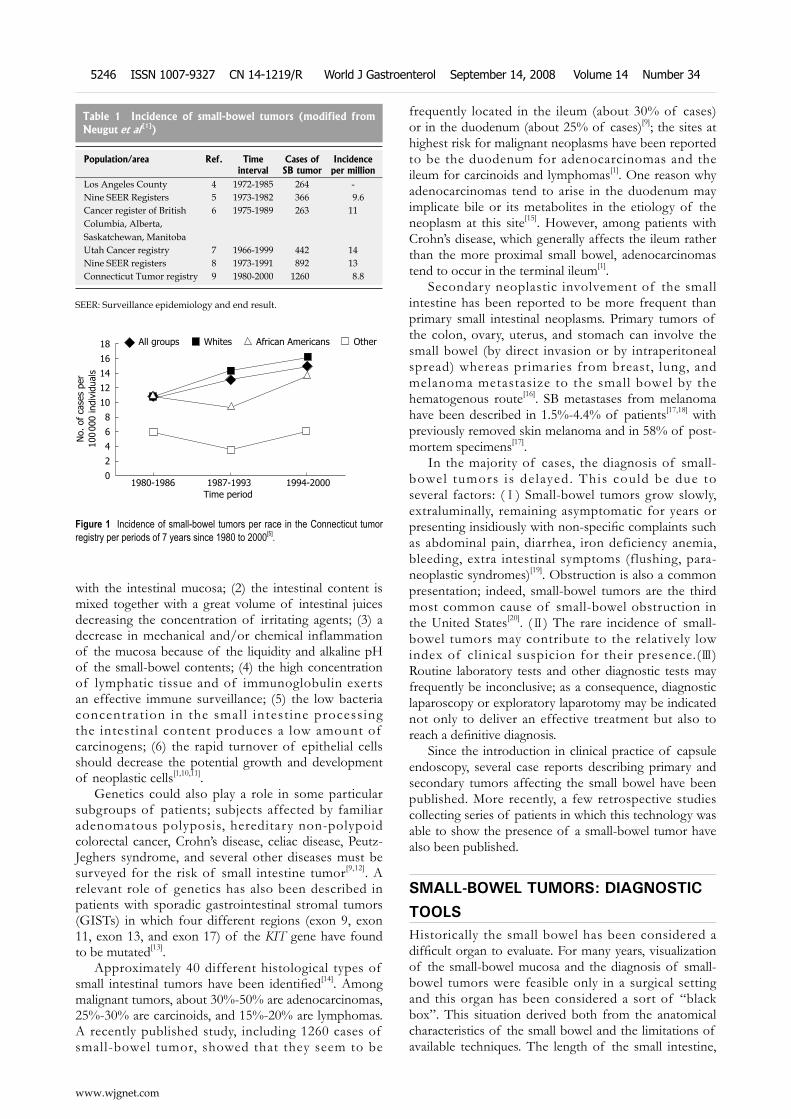
with the intestinal mucosa; (2) the intestinal content is mixed together with a great volume of intestinal juices decreasing the concentration of irritating agents; (3) a decrease in mechanical and/or chemical inflammation of the mucosa because of the liquidity and alkaline pH of the small-bowel contents; (4) the high concentration of lymphatic tissue and of immunoglobulin exerts an effective immune surveillance; (5) the low bacteria concentrat ion in the smal l intest ine processing the intestinal content produces a low amount of carcinogens; (6) the rapid turnover of epithelial cells should decrease the potential growth and development of neoplastic cells[1,10,11].
Genetics could also play a role in some particular subgroups of patients; subjects affected by familiar adenomatous polyposis, hereditary non-polypoid colorectal cancer, Crohn’s disease, celiac disease, Peutz-Jeghers syndrome, and several other diseases must be surveyed for the risk of small intestine tumor[9,12]. A relevant role of genetics has also been described in patients with sporadic gastrointestinal stromal tumors (GISTs) in which four different regions (exon 9, exon 11, exon 13, and exon 17) of the KIT gene have found to be mutated[13].
Approximately 40 different histological types of small intestinal tumors have been identified[14]. Among malignant tumors, about 30%-50% are adenocarcinomas, 25%-30% are carcinoids, and 15%-20% are lymphomas. A recently published study, including 1260 cases of small-bowel tumor, showed that they seem to be
frequently located in the ileum (about 30% of cases) or in the duodenum (about 25% of cases)[9]; the sites at highest risk for malignant neoplasms have been reported to be the duodenum for adenocarcinomas and the ileum for carcinoids and lymphomas[1]. One reason why adenocarcinomas tend to arise in the duodenum may implicate bile or its metabolites in the etiology of the neoplasm at this site[15]. However, among patients with Crohn’s disease, which generally affects the ileum rather than the more proximal small bowel, adenocarcinomas tend to occur in the terminal ileum[1].
Secondary neoplastic involvement of the small intestine has been reported to be more frequent than primary small intestinal neoplasms. Primary tumors of the colon, ovary, uterus, and stomach can involve the small bowel (by direct invasion or by intraperitoneal spread) whereas primaries from breast, lung, and melanoma metastasize to the small bowel by the hematogenous route[16]. SB metastases from melanoma have been described in 1.5%-4.4% of patients[17,18] with previously removed skin melanoma and in 58% of post-mortem specimens[17].
In the majority of cases, the diagnosis of small-bowel tumors is de layed. This could be due to several factors: (Ⅰ) Small-bowel tumors grow slowly, extraluminally, remaining asymptomatic for years or presenting insidiously with non-specific complaints such as abdominal pain, diarrhea, iron deficiency anemia, bleeding, extra intestinal symptoms (flushing, para-neoplastic syndromes)[19]. Obstruction is also a common presentation; indeed, small-bowel tumors are the third most common cause of small-bowel obstruction in the United States[20]. (Ⅱ) The rare incidence of small-bowel tumors may contribute to the relatively low index of clinical suspicion for their presence.(Ⅲ) Routine laboratory tests and other diagnostic tests may frequently be inconclusive; as a consequence, diagnostic laparoscopy or exploratory laparotomy may be indicated not only to deliver an effective treatment but also to reach a definitive diagnosis.
Since the introduction in clinical practice of capsule endoscopy, several case reports describing primary and secondary tumors affecting the small bowel have been published. More recently, a few retrospective studies collecting series of patients in which this technology was able to show the presence of a small-bowel tumor have also been published.
SMALL-BOWEL TUMORS: DIAGNOSTIC TOOLSHistorically the small bowel has been considered a difficult organ to evaluate. For many years, visualization of the small-bowel mucosa and the diagnosis of small-bowel tumors were feasible only in a surgical setting and this organ has been considered a sort of “black box”. This situation derived both from the anatomical characteristics of the small bowel and the limitations of available techniques. The length of the small intestine,
Table 1 Incidence of small-bowel tumors (modified from Neugut et al [1])
Population/area Ref. Time interval
Cases of SB tumor
Incidence per million
Los Angeles County 4 1972-1985 264 -Nine SEER Registers 5 1973-1982 366 9.6Cancer register of British Columbia, Alberta, Saskatchewan, Manitoba
6 1975-1989 263 11
Utah Cancer registry 7 1966-1999 442 14Nine SEER registers 8 1973-1991 892 13Connecticut Tumor registry 9 1980-2000 1260 8.8
SEER: Surveillance epidemiology and end result.
Time period1980-1986 1987-1993 1994-2000
18
16
14
12
10
8
6
4
2
0
No.
of
case
s pe
r 10
0 00
0 in
divi
dual
s
OtherAfrican AmericansWhitesAll groups
Figure 1 Incidence of small-bowel tumors per race in the Connecticut tumor registry per periods of 7 years since 1980 to 2000[5].
www.wjgnet.com
5246 ISSN 1007-9327 CN 14-1219/R World J Gastroenterol September 14, 2008 Volume 14 Number 34
the distance between the organ and external orifices (mouth and anus), its sinuousness, its ability to produce huge amounts of fluids and the continuous contractions long hampered accurate inspection of the small-bowel mucosa.
Traditional radiological techniques, including small-bowel follow-through and small-bowel enteroclysis, allow an indirect evaluation of the entire small bowel, however the difficulty to place a specific catheter in the right position (enteroclysis), the low pressure and the dilution of the contrast medium (small-bowel follow-through) contribute to a high miss rate for small and/or flat lesions. Conventional cross-sectional imaging techniques, such as computed tomography (CT) and magnetic resonance imaging (MRI), can be helpful in identifying large small-bowel masses or extraintestinal disease, but are unable to provide precise data about the intestinal wall. Endoscopy has the advantage of visualizing intestinal mucosa directly and, above all, of carrying out targeted biopsies. Upper GI endoscopy, when performed to the ligament of Treitz, is suited for identifying duodenal tumors; however, lesions located distally pose a unique diagnostic challenge. Push enteroscopy (PE) is an effective diagnostic and therapeutic procedure which entails the oral insertion of a dedicated enteroscope; however, it only allows thorough examination of the distal duodenum and proximal jejunum to approximately 50-100 cm beyond the ligament of Treitz. Because of its ability to examine the entire small intestine, sonde enteroscopy has been utilized to diagnose small-bowel tumors[21]. However, this technically-challenging procedure has been today completely abandoned. Intraoperative enteroscopy (IOE) is the most complete, but also the most invasive means of examining the small bowel. It is a difficult, time-consuming technique, often traumatic to the bowel, with a substantial risk of complications and even mortality.
The development and the introduction in the clinical practice of the capsule endoscopy (CE) has revolutionized the field of small-bowel imaging, not only opening up this sort of “Pandora’s box”, but also stimulating the development of other imaging techniques aimed at studying the small bowel.
Magnetic resonance enteroclysis (MRI-enteroclysis) combines the advantages of cross-sectional resonance with those of volume challenge of conventional enteroclysis in the detection and characterization of small-bowel wall abnormalities, such as initial neoplasms. Small-bowel tumors usually exhibit moderate signal intensity on true-FISP images, as opposed to the high signal intensity of the distended lumen and the mesenteric fat. Post-gadolinium 3D FLASH with fat saturation may be the most important sequence for the identification and characterization of small-bowel tumors by their enhancement pattern. The degree of prestenotic dilatation, the peritoneal extension of the neoplasm and associated lymphadenopathy is well visualized in all MRI-enteroclysis sequences[22,23]. The multidetector row computed tomography (MRCT) has the potential to provide high-resolution images and
a precise delineation of pathology. The multiplanar reformatted images obtained using MRCT have spatial resolution similar to that of the axial plane without any loss of information. These advantages of MDCT imaging lead to a more accurate demonstration of the site of the tumor and possible complications of underlying small-bowel tumors including small-bowel obstruction, intussusception, perforation and bleeding[24]. The administration of methyl-cellulose as a neutral luminal contrast material in a 4%-15% water-soluble solution or a diluted (1%) barium solution as positive luminal contrast in patients undergoing MRCT results in a computed tomography enteroclysis (CT-enteroclysis). As previously described for MRI-enteroclysis, this technique combines the advantages of enteral volume challenge and the ability of cross-sectional imaging to depict extra intestinal manifestations of the disease[25]. Both MRI enteroclysis and CT-enteroclysis require the placement of a specific catheter into the third part of the duodenum (fluoroscopic monitored), administration of medications (anti-motility agents and sedative medications) and small-bowel preparation with laxatives (PEG-based solutions, 2 to 4 L)[24]. Up to now, there are only few, but promising, publications about the role of these three techniques in the diagnostic algorithm of small-bowel tumors.
In the attempt to design a new scope that would allow a large part of the small-bowel mucosa to be visualized, overcoming the limits of PE and IOE, Yamamoto et al[25] developed a new method of push-and-pull enteroscopy (PPE) using a double-balloon technique. PPE affords inspection of the entire small bowel, combining the oral and anal approaches, with the advantage of enabling biopsies and endoscopic interventions to be performed in all parts of the small bowel without laparotomy. It is, however, invasive, time-consuming, and requires conscious sedation[26].
CAPSULE ENDOSCOPY IN THE DIAGNOSIS OF SMALL-BOWEL TUMORSIn a recently published paper, the hypothesis of an increased incidence of small-bowel tumors in recent years was put forward, based on the increasing number of cases diagnosed by means a non-invasive methods such as CE and small-bowel ultrasound[11]. In fact, compared with previously mentioned diagnostic techniques for the study of the small bowel, CE seems to be an ideal tool to recognize the presence of neoplastic lesions along the small bowel. The potential of CE for the diagnosis of small-bowel tumors, as well as for the surveillance of subjects at increased risk of developing them, depends largely on the technical characteristics of this diagnostic device. CE is a non-invasive tool, well accepted by patients, who can allow the visualization of the entire small bowel; high-quality images of the small-bowel mucosa may be captured and small and flat lesions recognized, without exposure to radiation.
In fact, since the introduction of CE in clinical
www.wjgnet.com
Pennazio M et al . Capsule endoscopy in neoplastic diseases 5247
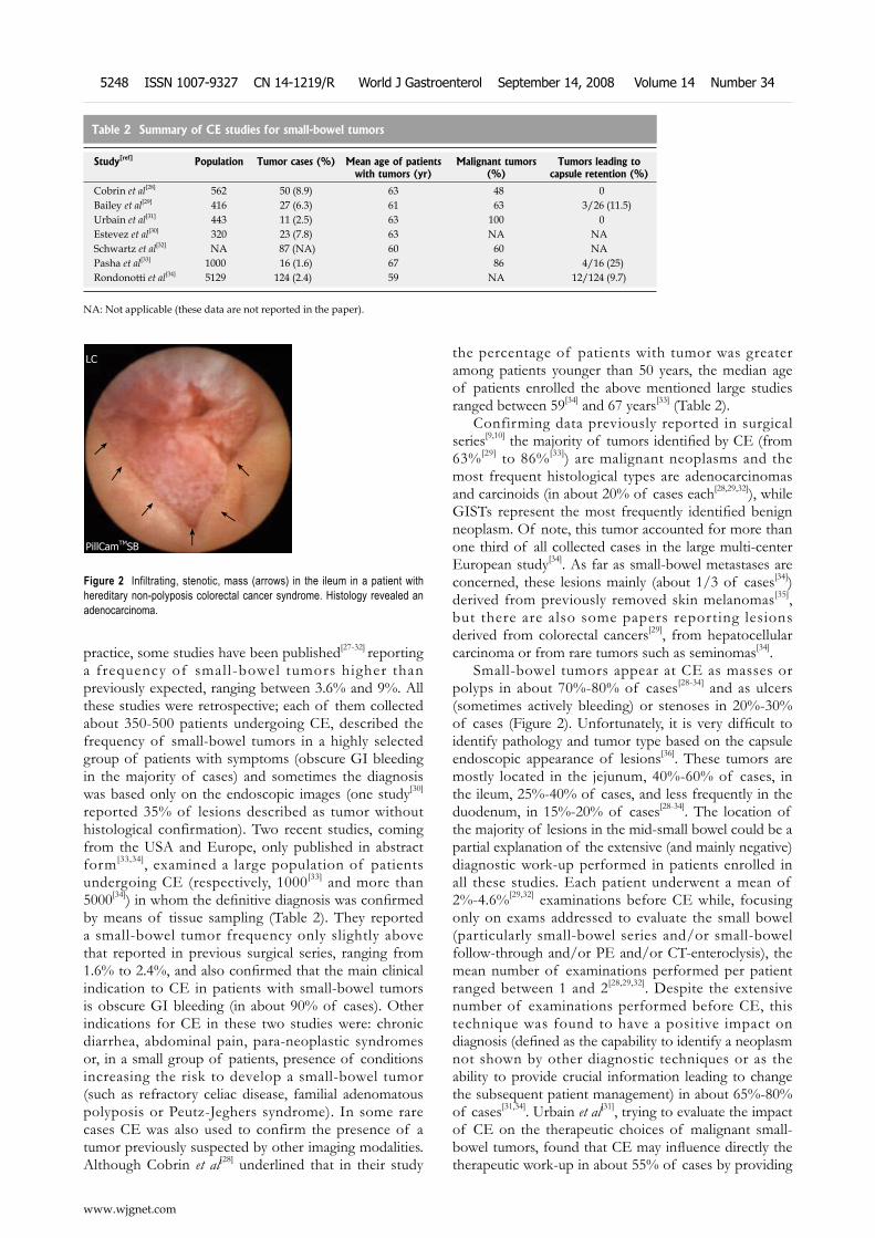
practice, some studies have been published[27-32] reporting a frequency of small-bowel tumors higher than previously expected, ranging between 3.6% and 9%. All these studies were retrospective; each of them collected about 350-500 patients undergoing CE, described the frequency of small-bowel tumors in a highly selected group of patients with symptoms (obscure GI bleeding in the majority of cases) and sometimes the diagnosis was based only on the endoscopic images (one study[30] reported 35% of lesions described as tumor without histological confirmation). Two recent studies, coming from the USA and Europe, only published in abstract form[33,34], examined a large population of patients undergoing CE (respectively, 1000[33] and more than 5000[34]) in whom the definitive diagnosis was confirmed by means of tissue sampling (Table 2). They reported a small-bowel tumor frequency only slightly above that reported in previous surgical series, ranging from 1.6% to 2.4%, and also confirmed that the main clinical indication to CE in patients with small-bowel tumors is obscure GI bleeding (in about 90% of cases). Other indications for CE in these two studies were: chronic diarrhea, abdominal pain, para-neoplastic syndromes or, in a small group of patients, presence of conditions increasing the risk to develop a small-bowel tumor (such as refractory celiac disease, familial adenomatous polyposis or Peutz-Jeghers syndrome). In some rare cases CE was also used to confirm the presence of a tumor previously suspected by other imaging modalities. Although Cobrin et al[28] underlined that in their study
the percentage of patients with tumor was greater among patients younger than 50 years, the median age of patients enrolled the above mentioned large studies ranged between 59[34] and 67 years[33] (Table 2).
Confirming data previously reported in surgical series[9,10] the majority of tumors identified by CE (from 63%[29] to 86%[33]) are malignant neoplasms and the most frequent histological types are adenocarcinomas and carcinoids (in about 20% of cases each[28,29,32]), while GISTs represent the most frequently identified benign neoplasm. Of note, this tumor accounted for more than one third of all collected cases in the large multi-center European study[34]. As far as small-bowel metastases are concerned, these lesions mainly (about 1/3 of cases[34]) derived from previously removed skin melanomas[35], but there are also some papers reporting lesions derived from colorectal cancers[29], from hepatocellular carcinoma or from rare tumors such as seminomas[34].
Small-bowel tumors appear at CE as masses or polyps in about 70%-80% of cases[28-34] and as ulcers (sometimes actively bleeding) or stenoses in 20%-30% of cases (Figure 2). Unfortunately, it is very difficult to identify pathology and tumor type based on the capsule endoscopic appearance of lesions[36]. These tumors are mostly located in the jejunum, 40%-60% of cases, in the ileum, 25%-40% of cases, and less frequently in the duodenum, in 15%-20% of cases[28-34]. The location of the majority of lesions in the mid-small bowel could be a partial explanation of the extensive (and mainly negative) diagnostic work-up performed in patients enrolled in all these studies. Each patient underwent a mean of 2%-4.6%[29,32] examinations before CE while, focusing only on exams addressed to evaluate the small bowel (particularly small-bowel series and/or small-bowel follow-through and/or PE and/or CT-enteroclysis), the mean number of examinations performed per patient ranged between 1 and 2[28,29,32]. Despite the extensive number of examinations performed before CE, this technique was found to have a positive impact on diagnosis (defined as the capability to identify a neoplasm not shown by other diagnostic techniques or as the ability to provide crucial information leading to change the subsequent patient management) in about 65%-80% of cases[31,34]. Urbain et al[31], trying to evaluate the impact of CE on the therapeutic choices of malignant small-bowel tumors, found that CE may influence directly the therapeutic work-up in about 55% of cases by providing
Table 2 Summary of CE studies for small-bowel tumors
Study[ref] Population Tumor cases (%) Mean age of patients with tumors (yr)
Malignant tumors (%)
Tumors leading to capsule retention (%)
Cobrin et al[28] 562 50 (8.9) 63 48 0Bailey et al[29] 416 27 (6.3) 61 63 3/26 (11.5)Urbain et al[31] 443 11 (2.5) 63 100 0Estevez et al[30] 320 23 (7.8) 63 NA NASchwartz et al[32] NA 87 (NA) 60 60 NA Pasha et al[33] 1000 16 (1.6) 67 86 4/16 (25)Rondonotti et al[34] 5129 124 (2.4) 59 NA 12/124 (9.7)
NA: Not applicable (these data are not reported in the paper).
www.wjgnet.com
5248 ISSN 1007-9327 CN 14-1219/R World J Gastroenterol September 14, 2008 Volume 14 Number 34
Figure 2 Infiltrating, stenotic, mass (arrows) in the ileum in a patient with hereditary non-polyposis colorectal cancer syndrome. Histology revealed an adenocarcinoma.
PillCamTMSB
LC
←
←
←
←
←
←
information about size, location and appearance of the lesion.
Because the early diagnosis and treatment of cancer usually affects outcome, some authors[28,29] suggest that the capability of CE to discover small-bowel tumors at an early stage may have an impact on prognosis for patients with these lesions. All the papers previously mentioned reported that in patients with small-bowel neoplasm identified by CE, surgery alone or surgery plus chemotherapy is the treatment of choice in about 85%-90% of the cases[28-30,33,34] but, to date, there is only one published paper describing the follow-up of these patients. Bailey et al[29] reported that surgical treatment was performed in 88% of patients with small-bowel tumor, in half of the cases with curative aim. None of the patients who underwent a curative resection developed tumor recurrence at follow-up (range, 26-51 mo). These authors also reported that none of the patients with benign tumors discovered by CE and treated according to CE findings had recurrence of either overt or occult obscure GI bleeding at follow-up (3-51 mo).
CAPSULE ENDOSCOPY FOR SPECIFIC SMALL-BOWEL TUMORS Thanks to its capability to identify a small-bowel lesion in most patients with a prior negative diagnostic work-up, several case reports, but also some small series, aimed at evaluating the possible role of the CE in the diagnosis of specific tumors in particular clinical conditions, have been published over the last few years.
Van Tuyl et al[37], in a prospective descriptive study, evaluated 20 patients with liver metastases, mesenteric metastases or both, originated from a neuroendocrine tumor (NET) with unknown primary location. All these patients had undergone several examinations including small-bowel enteroclysis, abdominal CT, pentetreotide scintigraphy and laboratory tests. In this particular subset of patients, CE showed a diagnostic yield (60%) significantly higher than enteroclysis and CT scan. Pentetreotide scintigraphy had an even higher diagnostic yield than CE, but without differentiation between intestinal and mesenteric localization. In this study, the absence of findings at CE in patients with abnormalities at nuclear imaging was interpreted to be related to the presence of NET restricted to the mesentery or to a false-negative CE. On the ground of these data, the authors suggested that patients with a metastatic NET and an unknown primary tumor should undergo CE. Conversely, in a small retrospective study of 8 patients[38], CE detected NETs of the small bowel with high specificity, but slightly lower sensitivity than did CT enteroclysis. It was concluded that CE should not be used as a routine method for diagnosing NET in the small bowel.
As far as small-bowel metastases are concerned, Prakoso and Selby[35] performed a retrospective analysis of a prospective database identifying 13 patients with
previous or recurrent malignant melanoma referred for CE. The indication for CE were overt GI bleeding in three patients, anemia in six, abnormal imaging in two, abdominal pain in one, and one patient had positive fecal occult blood test. In these patients, CE was able not only to show small-bowel metastases (in 5 patients), but also to provide a different possible explanation of symptoms in three other patients (NSAID-related ulcers, artero-venous malformation or aphtoid lesions). The authors concluded that since the optimal investigation for the detection of small-bowel metastases in patients with melanoma has still to be determined, CE can be considered an ideal method to do so because it appears to be more sensitive than small-bowel follow-through and CT scan.
Flieger et al[39] explored the potential contribution of CE to the diagnosis and staging of gastrointestinal lymphomas describing capsule endoscopic features of these tumors. They studied with CE a total of 27 consecutive patients with newly diagnosed gastroin-testinal lymphoma: 20 patients with histologically confirmed gastric lymphoma and seven patients with intestinal lymphoma. All seven patients with primary intestinal lymphomas were found to have pathological findings at CE (ulcerations, nodules or villous atrophy), while 5 of the 20 patients with gastric lymphoma had pathological findings in the small bowel (including abnormal villi, white nodules or villous atrophy). In this study, the authors found that CE is able to identify pathological intestinal findings in patients with gastrointestinal lymphoma more frequently than previously thought and suggest that knowledge of small-bowel involvement can lead to changes in the therapeutic strategy in individual cases.
Lymphomatous polyposis (LP), first described by Cornes in 1961[40,41], is a rare condition; however, since the introduction of CE and PPE in clinical practice, a few reports[42,43] have been published on this topic. LP is defined as polypoid mucosal involvement of long segments of the GI tract by neoplastic lymphoid cells[40]. For many years LP has been considered the macroscopic appearance of the mantle cell lymphoma, but it has recently been suggested that it can be also the macroscopic manifestation of mucosa-associated lymphoid tissue (MALT) lymphoma and follicular B cell lymphoma[44]. In patients with LP, CE is a valuable tool because it may recognize the presence of nodules, evaluate the extent of the small-bowel involvement and drive further investigations (i.e. the decision about the PPE approach).
Another peculiar clinical condition is represented by patients with refractory celiac disease. It is known that these patients have an increased risk to develop small-bowel neoplasms, mainly enteropathy associated T-cell lymphoma (EATL). However, in this particular subgroup of patients CE is aimed at identifying not only a malignant neoplasm, but also some other possible complications such as ulcerative jejunitis. To date, two papers have been published on this topic[45,46] showing that CE is a useful tool in the assessment of complicated
www.wjgnet.com
Pennazio M et al . Capsule endoscopy in neoplastic diseases 5249
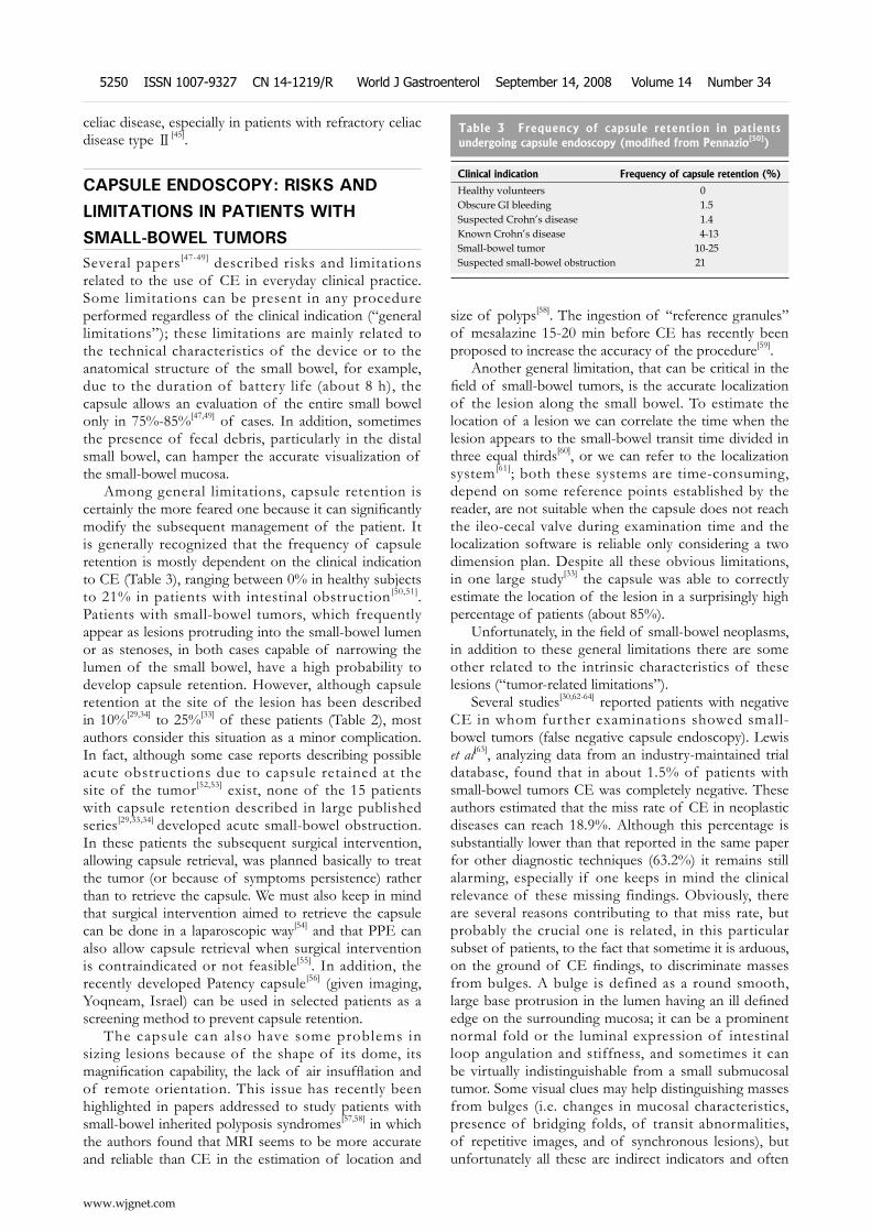
celiac disease, especially in patients with refractory celiac disease type Ⅱ[45].
CAPSULE ENDOSCOPY: RISKS AND LIMITATIONS IN PATIENTS WITH SMALL-BOWEL TUMORSSeveral papers[47-49] described risks and limitations related to the use of CE in everyday clinical practice. Some limitations can be present in any procedure performed regardless of the clinical indication (“general limitations”); these limitations are mainly related to the technical characteristics of the device or to the anatomical structure of the small bowel, for example, due to the duration of battery life (about 8 h), the capsule allows an evaluation of the entire small bowel only in 75%-85%[47,49] of cases. In addition, sometimes the presence of fecal debris, particularly in the distal small bowel, can hamper the accurate visualization of the small-bowel mucosa.
Among general limitations, capsule retention is certainly the more feared one because it can significantly modify the subsequent management of the patient. It is generally recognized that the frequency of capsule retention is mostly dependent on the clinical indication to CE (Table 3), ranging between 0% in healthy subjects to 21% in patients with intestinal obstruction[50,51]. Patients with small-bowel tumors, which frequently appear as lesions protruding into the small-bowel lumen or as stenoses, in both cases capable of narrowing the lumen of the small bowel, have a high probability to develop capsule retention. However, although capsule retention at the site of the lesion has been described in 10%[29,34] to 25%[33] of these patients (Table 2), most authors consider this situation as a minor complication. In fact, although some case reports describing possible acute obstructions due to capsule retained at the site of the tumor[52,53] exist, none of the 15 patients with capsule retention described in large published series[29,33,34] developed acute small-bowel obstruction. In these patients the subsequent surgical intervention, allowing capsule retrieval, was planned basically to treat the tumor (or because of symptoms persistence) rather than to retrieve the capsule. We must also keep in mind that surgical intervention aimed to retrieve the capsule can be done in a laparoscopic way[54] and that PPE can also allow capsule retrieval when surgical intervention is contraindicated or not feasible[55]. In addition, the recently developed Patency capsule[56] (given imaging, Yoqneam, Israel) can be used in selected patients as a screening method to prevent capsule retention.
The capsule can also have some problems in sizing lesions because of the shape of its dome, its magnification capability, the lack of air insufflation and of remote orientation. This issue has recently been highlighted in papers addressed to study patients with small-bowel inherited polyposis syndromes[57,58] in which the authors found that MRI seems to be more accurate and reliable than CE in the estimation of location and
size of polyps[58]. The ingestion of “reference granules” of mesalazine 15-20 min before CE has recently been proposed to increase the accuracy of the procedure[59].
Another general limitation, that can be critical in the field of small-bowel tumors, is the accurate localization of the lesion along the small bowel. To estimate the location of a lesion we can correlate the time when the lesion appears to the small-bowel transit time divided in three equal thirds[60], or we can refer to the localization system[61]; both these systems are time-consuming, depend on some reference points established by the reader, are not suitable when the capsule does not reach the ileo-cecal valve during examination time and the localization software is reliable only considering a two dimension plan. Despite all these obvious limitations, in one large study[33] the capsule was able to correctly estimate the location of the lesion in a surprisingly high percentage of patients (about 85%).
Unfortunately, in the field of small-bowel neoplasms, in addition to these general limitations there are some other related to the intrinsic characteristics of these lesions (“tumor-related limitations”).
Several studies[30,62-64] reported patients with negative CE in whom further examinations showed small-bowel tumors (false negative capsule endoscopy). Lewis et al[63], analyzing data from an industry-maintained trial database, found that in about 1.5% of patients with small-bowel tumors CE was completely negative. These authors estimated that the miss rate of CE in neoplastic diseases can reach 18.9%. Although this percentage is substantially lower than that reported in the same paper for other diagnostic techniques (63.2%) it remains still alarming, especially if one keeps in mind the clinical relevance of these missing findings. Obviously, there are several reasons contributing to that miss rate, but probably the crucial one is related, in this particular subset of patients, to the fact that sometime it is arduous, on the ground of CE findings, to discriminate masses from bulges. A bulge is defined as a round smooth, large base protrusion in the lumen having an ill defined edge on the surrounding mucosa; it can be a prominent normal fold or the luminal expression of intestinal loop angulation and stiffness, and sometimes it can be virtually indistinguishable from a small submucosal tumor. Some visual clues may help distinguishing masses from bulges (i.e. changes in mucosal characteristics, presence of bridging folds, of transit abnormalities, of repetitive images, and of synchronous lesions), but unfortunately all these are indirect indicators and often
Clinical indication Frequency of capsule retention (%)
Healthy volunteers 0Obscure GI bleeding 1.5Suspected Crohn’s disease 1.4Known Crohn’s disease 4-13Small-bowel tumor 10-25Suspected small-bowel obstruction 21
Table 3 Frequency of capsule retention in patients undergoing capsule endoscopy (modified from Pennazio[50])
www.wjgnet.com
5250 ISSN 1007-9327 CN 14-1219/R World J Gastroenterol September 14, 2008 Volume 14 Number 34
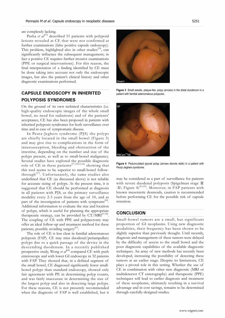
are completely lacking.Pasha et al[33] described 51 patients with polypoid
lesions revealed at CE that were not confirmed at further examinations (false positive capsule endoscopy). This problem, highlighted also in other studies[30], can significantly influence the subsequent management; in fact a positive CE requires further invasive examinations (PPE or surgical interventions). For this reason, the final interpretation of a finding identified by CE must be done taking into account not only the endoscopic images, but also the patient’s clinical history and other diagnostic examinations performed.
CAPSULE ENDOSCOPY IN INHERITED POLYPOSIS SYNDROMESOn the ground of its own technical characteristics (i.e. high-quality endoscopic images of the whole small bowel, no need for radiations) and of the patients' acceptance, CE has also been proposed in patients with inherited polyposis syndromes for both surveillance over time and in case of symptomatic disease.
In Peutz-Jeghers syndrome (PJS) the polyps are chief ly located in the small bowel (Figure 3) and may give rise to complications in the form of intussusception, bleeding and obstruction of the intestine, depending on the number and size of the polyps present, as well as to small-bowel malignancy. Several studies have explored the possible diagnostic role of CE in these patients[57,58,65,66] showing that this tool seems to be superior to small-bowel follow-through[57]. Unfortunately, the same studies also underlined that CE (as discussed above) is not reliable for accurate sizing of polyps. At the present time, it is suggested that CE should be performed at diagnosis in all patients with PJS, as the primary surveillance modality every 2-3 years from the age of 10, and as part of the investigation of patients with symptoms[50]. Additional information to evaluate the size and location of polyps, which is useful for planning the appropriate therapeutic strategy, can be provided by CT/MRI[57,58]. The coupling of CE with PPE and polypectomy may offer an ideal follow-up and treatment method for these patients, possibly avoiding surgery[67].
The role of CE is less clear in familial adenomatous polyposis (FAP). CE may miss duodenal/periampullary polyps due to a quick passage of the device in the descending duodenum. In a recent ly publ ished prospective study, Wong et al[68] compared CE with push enteroscopy and with lower GI endoscopy in 32 patients with FAP. They showed that, in a defined segment of the small bowel, CE diagnosed significantly fewer small-bowel polyps than standard endoscopy, showed only fair agreement with PE in determining polyp counts, and was fairly inaccurate in determining the size of the largest polyp and also in detecting large polyps. For these reasons, CE is not presently recommended when the diagnosis of FAP is well established, but it
may be considered as a part of surveillance for patients with severe duodenal polyposis (Spigelman stage Ⅲ-Ⅳ; Figure 4)[65,66]. Moreover, in FAP patients with known mesenteric desmoids, caution is recommended before performing CE for the possible risk of capsule retention.
CONCLUSION Small-bowel tumors are a small , but s ignif icant proportion of GI neoplasms. Using new diagnostic modalities, their frequency has been shown to be slightly superior than previously thought. Until recently, diagnosis and management of these tumors were delayed by the difficulty of access to the small bowel and the poor diagnostic capabilities of the available diagnostic techniques. An array of new methods has recently been developed, increasing the possibility of detecting these tumors at an earlier stage. Despite its limitations, CE plays a pivotal role in this setting. Whether the use of CE in combination with other new diagnostic (MRI or multidetector CT enterography) and therapeutic (PPE) techniques will lead to earlier diagnosis and treatment of these neoplasms, ultimately resulting in a survival advantage and in cost savings, remains to be determined through carefully-designed studies.
←
←
Given®
Figure 3 Small sessile, plaque-like, polyp (arrows) in the distal duodenum in a patient with familial adenomatous polyposis.
PillCamTMSB
NP
←
←
←
←
←
Figure 4 Pedunculated jejunal polyp (arrows denote stalk) in a patient with Peutz-Jeghers syndrome.
www.wjgnet.com
Pennazio M et al . Capsule endoscopy in neoplastic diseases 5251
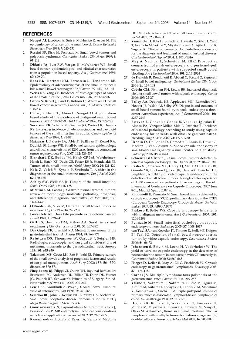
REFERENCES1 Neugut AI, Jacobson JS, Suh S, Mukherjee R, Arber N. The
epidemiology of cancer of the small bowel. Cancer Epidemiol Biomarkers Prev 1998; 7: 243-251
2 Rossini FP, Risio M, Pennazio M. Small bowel tumors and polyposis syndromes. Gastrointest Endosc Clin N Am 1999; 9: 93-114
3 DiSario JA, Burt RW, Vargas H, McWhorter WP. Small bowel cancer: epidemiological and clinical characteristics from a population-based registry. Am J Gastroenterol 1994; 89: 699-701
4 Ross RK , Hartnett NM, Bernstein L, Henderson BE. Epidemiology of adenocarcinomas of the small intestine: is bile a small bowel carcinogen? Br J Cancer 1991; 63: 143-145
5 Weiss NS, Yang CP. Incidence of histologic types of cancer of the small intestine. J Natl Cancer Inst 1987; 78: 653-656
6 Gabos S, Berkel J, Band P, Robson D, Whittaker H. Small bowel cancer in western Canada. Int J Epidemiol 1993; 22: 198-206
7 Chow JS, Chen CC, Ahsan H, Neugut AI. A population-based study of the incidence of malignant small bowel tumours: SEER, 1973-1990. Int J Epidemiol 1996; 25: 722-728
8 Severson RK, Schenk M, Gurney JG, Weiss LK, Demers RY. Increasing incidence of adenocarcinomas and carcinoid tumors of the small intestine in adults. Cancer Epidemiol Biomarkers Prev 1996; 5: 81-84
9 Hatzaras I, Palesty JA, Abir F, Sullivan P, Kozol RA, Dudrick SJ, Longo WE. Small-bowel tumors: epidemiologic and clinical characteristics of 1260 cases from the connecticut tumor registry. Arch Surg 2007; 142: 229-235
10 Blanchard DK, Budde JM, Hatch GF 3rd, Wertheimer-Hatch L, Hatch KF, Davis GB, Foster RS Jr, Skandalakis JE. Tumors of the small intestine. World J Surg 2000; 24: 421-429
11 Kala Z , Valek V, Kysela P, Svoboda T. A shift in the diagnostics of the small intestine tumors. Eur J Radiol 2007; 62: 160-165
12 Ashley SW, Wells SA Jr. Tumors of the small intestine. Semin Oncol 1988; 15: 116-128
13 Miettinen M, Lasota J. Gastrointestinal stromal tumors: review on morphology, molecular pathology, prognosis, and differential diagnosis. Arch Pathol Lab Med 2006; 130: 1466-1478
14 O'Riordan BG, Vilor M, Herrera L. Small bowel tumors: an overview. Dig Dis 1996; 14: 245-257
15 Lowenfels AB. Does bile promote extra-colonic cancer? Lancet 1978; 2: 239-241
16 Gill SS , Heuman DM, Mihas AA. Small intest inal neoplasms. J Clin Gastroenterol 2001; 33: 267-282
17 Das Gupta TK, Brasfield RD. Metastatic melanoma of the gastrointestinal tract. Arch Surg 1964; 88: 969-973
18 Reintgen DS , Thompson W, Garbutt J , Seigler HF. Radiologic, endoscopic, and surgical considerations of melanoma metastatic to the gastrointestinal tract. Surgery 1984; 95: 635-639
19 Talamonti MS, Goetz LH, Rao S, Joehl RJ. Primary cancers of the small bowel: analysis of prognostic factors and results of surgical management. Arch Surg 2002; 137: 564-570; discussion 570-571
20 Fitzgibbons RJ, Filippi CJ, Quinn TH. Inguinal hernias. In: Brunicardi FC, Andersen DK, Billiar TR, Dunn DL, Hunter JG, Pollock RE. Schwartz’s Principles of Surgery. 8th ed. New York: McGraw-Hill, 2005: 230-246
21 Lewis BS, Kornbluth A, Waye JD. Small bowel tumours: yield of enteroscopy. Gut 1991; 32: 763-765
22 Semelka RC, John G, Kelekis NL, Burdeny DA, Ascher SM. Small bowel neoplastic disease: demonstration by MRI. J Magn Reson Imaging 1996; 6: 855-860
23 Gourtsoyiannis N, Papanikolaou N, Grammatikakis J, Prassopoulos P. MR enteroclysis: technical considerations and clinical applications. Eur Radiol 2002; 12: 2651-2658
24 Ramachandran I, Sinha R, Rajesh A, Verma R, Maglinte
DD. Multidetector row CT of small bowel tumours. Clin Radiol 2007; 62: 607-614
25 Yamamoto H, Kita H, Sunada K, Hayashi Y, Sato H, Yano T, Iwamoto M, Sekine Y, Miyata T, Kuno A, Ajibe H, Ido K, Sugano K. Clinical outcomes of double-balloon endoscopy for the diagnosis and treatment of small-intestinal diseases. Clin Gastroenterol Hepatol 2004; 2: 1010-1016
26 May A , Nachbar L, Schneider M, Ell C. Prospective comparison of push enteroscopy and push-and-pull enteroscopy in patients with suspected small-bowel bleeding. Am J Gastroenterol 2006; 101: 2016-2024
27 de Franchis R, Rondonotti E, Abbiati C, Beccari G, Signorelli C. Small bowel malignancy. Gastrointest Endosc Clin N Am 2004; 14: 139-148
28 Cobrin GM, Pittman RH, Lewis BS. Increased diagnostic yield of small bowel tumors with capsule endoscopy. Cancer 2006; 107: 22-27
29 Bailey AA, Debinski HS, Appleyard MN, Remedios ML, Hooper JE, Walsh AJ, Selby WS. Diagnosis and outcome of small bowel tumors found by capsule endoscopy: a three-center Australian experience. Am J Gastroenterol 2006; 101: 2237-2243
30 Estevez E , Gonzalez-Conde B, Vazquez-Iglesias JL, Alonso PA, Vazquez-Millan Mde L, Pardeiro R. Incidence of tumoral pathology according to study using capsule endoscopy for patients with obscure gastrointestinal bleeding. Surg Endosc 2007; 21: 1776-1780
31 Urbain D, De Looze D, Demedts I, Louis E, Dewit O, Macken E, Van Gossum A. Video capsule endoscopy in small-bowel malignancy: a multicenter Belgian study. Endoscopy 2006; 38: 408-411
32 Schwartz GD, Barkin JS. Small-bowel tumors detected by wireless capsule endoscopy. Dig Dis Sci 2007; 52: 1026-1030
33 Pasha SF, Sharma VK, Carey EJ, Shiff AD, Heigh RI, Gurudu SR, Erickson PJ, Post JK, Hara AK, Fleischer DE, Leighton JA. Utility of video capsule endoscopy in the detection of small bowel tumors. A single center experience of 1000 consecutive patients. Proceedings of the 6th International Conference on Capsule Endoscopy, 2007 June 8-10; Madrid, Spain, 2007: 45
34 Rondonotti E, Pennazio M. Small bowel tumors detected by capsule endoscopy (VCE): preliminary data from the ECEG (European Capsule Endoscopy Group) database. Gastroint Endosc 2007: 65: AB90-AB371
35 Prakoso E, Selby WS. Capsule endoscopy in patients with malignant melanoma. Am J Gastroenterol 2007; 102: 1204-1208
36 Pennazio M . Small-intestinal pathology on capsule endoscopy: tumors. Endoscopy 2005; 37: 1008-1017
37 van Tuyl SA, van Noorden JT, Timmer R, Stolk MF, Kuipers EJ, Taal BG. Detection of small-bowel neuroendocrine tumors by video capsule endoscopy. Gastrointest Endosc 2006; 64: 66-72
38 Johanssen S, Boivin M, Lochs H, Voderholzer W. The yield of wireless capsule endoscopy in the detection of neuroendocrine tumors in comparison with CT enteroclysis. Gastrointest Endosc 2006; 63: 660-665
39 Flieger D, Keller R, May A, Ell C, Fischbach W. Capsule endoscopy in gastrointestinal lymphomas. Endoscopy 2005; 37: 1174-1180
40 Cornes JS. Multiple lymphomatous polyposis of the gastrointestinal tract. Cancer 1961; 14: 249-257
41 Yatabe Y, Nakamura S, Nakamura T, Seto M, Ogura M, Kimura M, Kuhara H, Kobayashi T, Taniwaki M, Morishima Y, Koshikawa T, Suchi T. Multiple polypoid lesions of primary mucosa-associated lymphoid-tissue lymphoma of colon. Histopathology 1998; 32: 116-125
42 Higuchi K , Komatsu K, Wakamatsu H, Kawasaki H, Murata M, Miyazaki K, Oikawa K, Ohwada M, Nanjo H, Otaka M, Watanabe S, Komatsu K. Small intestinal follicular lymphoma with multiple tumor formations diagnosed by double-balloon enteroscopy. Intern Med 2007; 46: 705-709
www.wjgnet.com
5252 ISSN 1007-9327 CN 14-1219/R World J Gastroenterol September 14, 2008 Volume 14 Number 34
43 Esaki M, Matsumoto T, Nakamura S, Suekane H, Ohji Y, Yao T, Iida M. Capsule endoscopy findings in intestinal follicular lymphoma. Endoscopy 2007; 39 Suppl 1: E86-E87
44 Kodama T, Ohshima K, Nomura K, Taniwaki M, Nakamura N, Nakamura S, Kohno S, Yamamoto J, Karube K, Yamasita Y, Shirakusa T, Kikuchi M. Lymphomatous polyposis of the gastrointestinal tract, including mantle cell lymphoma, follicular lymphoma and mucosa-associated lymphoid tissue lymphoma. Histopathology 2005; 47: 467-478
45 Daum S, Wahnschaffe U, Glasenapp R, Borchert M, Ullrich R, Zeitz M, Faiss S. Capsule endoscopy in refractory celiac disease. Endoscopy 2007; 39: 455-458
46 Culliford A, Daly J, Diamond B, Rubin M, Green PH. The value of wireless capsule endoscopy in patients with complicated celiac disease. Gastrointest Endosc 2005; 62: 55-61
47 Ho KK, Joyce AM. Complications of capsule endoscopy. Gastrointest Endosc Clin N Am 2007; 17: 169-178, viii-ix
48 Cheifetz AS, Lewis BS. Capsule endoscopy retention: is it a complication? J Clin Gastroenterol 2006; 40: 688-691
49 Rondonotti E, Herrerias JM, Pennazio M, Caunedo A, Mascarenhas-Saraiva M, de Franchis R. Complications, limitations, and failures of capsule endoscopy: a review of 733 cases. Gastrointest Endosc 2005; 62: 712-716; quiz 752, 754
50 Pennazio M. Capsule endoscopy: where are we after 6 years of clinical use? Dig Liver Dis 2006; 38: 867-878
51 Cave D, Legnani P, de Franchis R, Lewis BS. ICCE consensus for capsule retention. Endoscopy 2005; 37: 1065-1067
52 Strosberg JR, Shibata D, Kvols LK. Intermittent bowel obstruction due to a retained wireless capsule endoscope in a patient with a small bowel carcinoid tumour. Can J Gastroenterol 2007; 21: 113-115
53 Lin OS, Brandabur JJ, Schembre DB, Soon MS, Kozarek RA. Acute symptomatic small bowel obstruction due to capsule impaction. Gastrointest Endosc 2007; 65: 725-728
54 Dominguez EP , Choi Y , Rai jman IL , Sweeney JF . Laparoscopic approach for the retrieval of retained video capsule endoscopy. JSLS 2006; 10: 496-498
55 Tanaka S, Mitsui K, Shirakawa K, Tatsuguchi A, Nakamura T, Hayashi Y, Sakamoto C, Terano A. Successful retrieval of video capsule endoscopy retained at ileal stenosis of Crohn's disease using double-balloon endoscopy. J Gastroenterol Hepatol 2006; 21: 922-923
56 Spada C, Shah SK, Riccioni ME, Spera G, Marchese M, Iacopini F, Familiari P, Costamagna G. Video capsule endoscopy in patients with known or suspected small bowel stricture previously tested with the dissolving patency capsule. J Clin Gastroenterol 2007; 41: 576-582
57 Brown G, Fraser C, Schofield G, Taylor S, Bartram C, Phillips R, Saunders B. Video capsule endoscopy in peutz-jeghers syndrome: a blinded comparison with barium follow-through for detection of small-bowel polyps. Endoscopy 2006; 38: 385-390
58 Caspari R, von Falkenhausen M, Krautmacher C, Schild H, Heller J, Sauerbruch T. Comparison of capsule endoscopy and magnetic resonance imaging for the detection of polyps of the small intestine in patients with familial adenomatous polyposis or with Peutz-Jeghers' syndrome. Endoscopy 2004; 36: 1054-1059
59 Racz I, Janoki M, Kovacs V. Measurement of small-bowel polyp size in patients with Peutz-Jeghers syndrome by using reference granules during video capsule endoscopy. Endoscopy 2007; 39 Suppl 1: E41
60 Biagi F, Rondonotti E, Campanella J, Villa F, Bianchi PI, Klersy C, De Franchis R, Corazza GR. Video capsule endoscopy and histology for small -bowel mucosa evaluation: a comparison performed by blinded observers. Clin Gastroenterol Hepatol 2006; 4: 998-1003
61 Fischer D , Schreiber R, Levi D, Eliakim R. Capsule endoscopy: the localization system. Gastrointest Endosc Clin N Am 2004; 14: 25-31
62 Mehdizadeh S, Ross A, Gerson L, Leighton J, Chen A, Schembre D, Chen G, Semrad C, Kamal A, Harrison EM, Binmoeller K, Waxman I, Kozarek R, Lo SK. What is the learning curve associated with double-balloon enteroscopy? Technical details and early experience in 6 U.S. tertiary care centers. Gastrointest Endosc 2006; 64: 740-750
63 Lewis BS, Eisen GM, Friedman S. A pooled analysis to evaluate results of capsule endoscopy trials. Endoscopy 2005; 37: 960-965
64 Chong AK, Chin BW, Meredith CG. Clinically significant small-bowel pathology identified by double-balloon enteroscopy but missed by capsule endoscopy. Gastrointest Endosc 2006; 64: 445-449
65 Schulmann K, Hollerbach S, Kraus K, Willert J, Vogel T, Moslein G, Pox C, Reiser M, Reinacher-Schick A, Schmiegel W. Feasibility and diagnostic utility of video capsule endoscopy for the detection of small bowel polyps in patients with hereditary polyposis syndromes. Am J Gastroenterol 2005; 100: 27-37
66 Burke CA, Santisi J, Church J, Levinthal G. The utility of capsule endoscopy small bowel surveillance in patients with polyposis. Am J Gastroenterol 2005; 100: 1498-1502
67 Ohmiya N, Taguchi A, Shirai K, Mabuchi N, Arakawa D, Kanazawa H, Ozeki M, Yamada M, Nakamura M, Itoh A, Hirooka Y, Niwa Y, Nagasaka T, Ito M, Ohashi S, Okamura S, Goto H. Endoscopic resection of Peutz-Jeghers polyps throughout the small intestine at double-balloon enteroscopy without laparotomy. Gastrointest Endosc 2005; 61: 140-147
68 Wong RF, Tuteja AK, Haslem DS, Pappas L, Szabo A, Ogara MM, DiSario JA. Video capsule endoscopy compared with standard endoscopy for the evaluation of small-bowel polyps in persons with familial adenomatous polyposis (with video). Gastrointest Endosc 2006; 64: 530-537
S- Editor Zhong XY L- Editor Rippe RA E- Editor Lin YP
www.wjgnet.com
Pennazio M et al . Capsule endoscopy in neoplastic diseases 5253
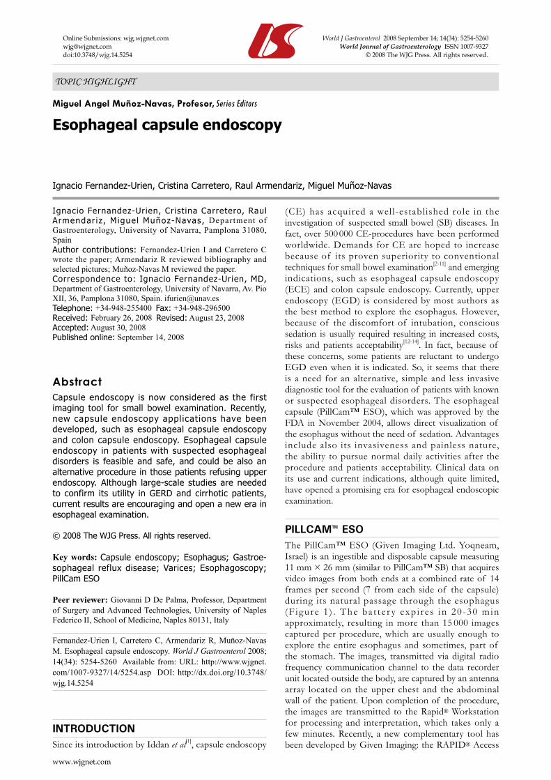
www.wjgnet.com
TOPIC HIGHLIGHT
Esophageal capsule endoscopy
Ignacio Fernandez-Urien, Cristina Carretero, Raul Armendariz, Miguel Muñoz-Navas
Miguel Angel Muñoz-Navas, Profesor, Series Editors
Online Submissions: wjg.wjgnet.com World J Gastroenterol 2008 September 14; 14(34): [email protected] World Journal of Gastroenterology ISSN 1007-9327doi:10.3748/wjg.14.5254 © 2008 The WJG Press. All rights reserved.
Ignacio Fernandez-Urien, Cristina Carretero, Raul Armendar iz , Miguel Muñoz-Navas, Department of Gastroenterology, University of Navarra, Pamplona 31080, SpainAuthor contributions: Fernandez-Urien I and Carretero C wrote the paper; Armendariz R reviewed bibliography and selected pictures; Muñoz-Navas M reviewed the paper.Correspondence to: Ignacio Fernandez-Urien, MD, Department of Gastroenterology, University of Navarra, Av. Pio XII, 36, Pamplona 31080, Spain. [email protected]: +34-948-255400 Fax: +34-948-296500Received: February 26, 2008 Revised: August 23, 2008Accepted: August 30, 2008Published online: September 14, 2008
AbstractCapsule endoscopy is now considered as the first imaging tool for small bowel examination. Recently, new capsule endoscopy applications have been developed, such as esophageal capsule endoscopy and colon capsule endoscopy. Esophageal capsule endoscopy in patients with suspected esophageal disorders is feasible and safe, and could be also an alternative procedure in those patients refusing upper endoscopy. Although large-scale studies are needed to confirm its utility in GERD and cirrhotic patients, current results are encouraging and open a new era in esophageal examination.
© 2008 The WJG Press. All rights reserved.
Key words: Capsule endoscopy; Esophagus; Gastroe-sophageal reflux disease; Varices; Esophagoscopy; PillCam ESO
Peer reviewer: Giovanni D De Palma, Professor, Department of Surgery and Advanced Technologies, University of Naples Federico II, School of Medicine, Naples 80131, Italy
Fernandez-Urien I, Carretero C, Armendariz R, Muñoz-Navas M. Esophageal capsule endoscopy. World J Gastroenterol 2008; 14(34): 5254-5260 Available from: URL: http://www.wjgnet.com/1007-9327/14/5254.asp DOI: http://dx.doi.org/10.3748/wjg.14.5254
INTRODUCTIONSince its introduction by Iddan et al[1], capsule endoscopy
(CE) has acquired a well-established role in the investigation of suspected small bowel (SB) diseases. In fact, over 500 000 CE-procedures have been performed worldwide. Demands for CE are hoped to increase because of its proven superiority to conventional techniques for small bowel examination[2-11] and emerging indications, such as esophageal capsule endoscopy (ECE) and colon capsule endoscopy. Currently, upper endoscopy (EGD) is considered by most authors as the best method to explore the esophagus. However, because of the discomfort of intubation, conscious sedation is usually required resulting in increased costs, risks and patients acceptability[12-14]. In fact, because of these concerns, some patients are reluctant to undergo EGD even when it is indicated. So, it seems that there is a need for an alternative, simple and less invasive diagnostic tool for the evaluation of patients with known or suspected esophageal disorders. The esophageal capsule (PillCam™ ESO), which was approved by the FDA in November 2004, allows direct visualization of the esophagus without the need of sedation. Advantages include also its invasiveness and painless nature, the ability to pursue normal daily activities after the procedure and patients acceptability. Clinical data on its use and current indications, although quite limited, have opened a promising era for esophageal endoscopic examination.
PILLCAM™ ESOThe PillCam™ ESO (Given Imaging Ltd. Yoqneam, Israel) is an ingestible and disposable capsule measuring 11 mm × 26 mm (similar to PillCam™ SB) that acquires video images from both ends at a combined rate of 14 frames per second (7 from each side of the capsule) during its natural passage through the esophagus (F i gu re 1 ) . T he ba t t e r y exp i r e s i n 20 -30 min approximately, resulting in more than 15 000 images captured per procedure, which are usually enough to explore the entire esophagus and sometimes, part of the stomach. The images, transmitted via digital radio frequency communication channel to the data recorder unit located outside the body, are captured by an antenna array located on the upper chest and the abdominal wall of the patient. Upon completion of the procedure, the images are transmitted to the Rapid® Workstation for processing and interpretation, which takes only a few minutes. Recently, a new complementary tool has been developed by Given Imaging: the RAPID® Access
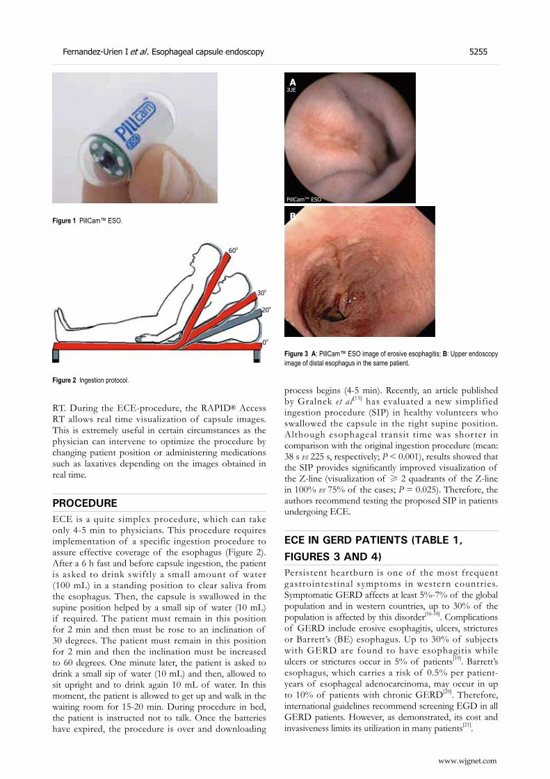
Fernandez-Urien I et al . Esophageal capsule endoscopy 5255
RT. During the ECE-procedure, the RAPID® Access RT allows real time visualization of capsule images. This is extremely useful in certain circumstances as the physician can intervene to optimize the procedure by changing patient position or administering medications such as laxatives depending on the images obtained in real time.
PROCEDUREECE is a quite simplex procedure, which can take only 4-5 min to physicians. This procedure requires implementation of a specific ingestion procedure to assure effective coverage of the esophagus (Figure 2). After a 6 h fast and before capsule ingestion, the patient is asked to drink swiftly a small amount of water (100 mL) in a standing position to clear saliva from the esophagus. Then, the capsule is swallowed in the supine position helped by a small sip of water (10 mL) if required. The patient must remain in this position for 2 min and then must be rose to an inclination of 30 degrees. The patient must remain in this position for 2 min and then the inclination must be increased to 60 degrees. One minute later, the patient is asked to drink a small sip of water (10 mL) and then, allowed to sit upright and to drink again 10 mL of water. In this moment, the patient is allowed to get up and walk in the waiting room for 15-20 min. During procedure in bed, the patient is instructed not to talk. Once the batteries have expired, the procedure is over and downloading
process begins (4-5 min). Recently, an article published by Gralnek et al [15] has evaluated a new simplified ingestion procedure (SIP) in healthy volunteers who swallowed the capsule in the right supine position. Although esophageal transit time was shorter in comparison with the original ingestion procedure (mean: 38 s vs 225 s, respectively; P < 0.001), results showed that the SIP provides significantly improved visualization of the Z-line (visualization of ≥ 2 quadrants of the Z-line in 100% vs 75% of the cases; P = 0.025). Therefore, the authors recommend testing the proposed SIP in patients undergoing ECE.
ECE IN GERD PATIENTS (TABLE 1, FIGURES 3 AND 4)Persistent heartburn is one of the most frequent gastrointestinal symptoms in western countries. Symptomatic GERD affects at least 5%-7% of the global population and in western countries, up to 30% of the population is affected by this disorder[16-18]. Complications of GERD include erosive esophagitis, ulcers, strictures or Barrett’s (BE) esophagus. Up to 30% of subjects with GERD are found to have esophagitis while ulcers or strictures occur in 5% of patients[19]. Barrett’s esophagus, which carries a risk of 0.5% per patient-years of esophageal adenocarcinoma, may occur in up to 10% of patients with chronic GERD[20]. Therefore, international guidelines recommend screening EGD in all GERD patients. However, as demonstrated, its cost and invasiveness limits its utilization in many patients[21].
Figure 1 PillCam™ ESO.
Figure 2 Ingestion protocol.
60o
30o
20o
0o
JUE
PillCam™ ESO
A
B
Figure 3 A: PillCam™ ESO image of erosive esophagitis; B: Upper endoscopy image of distal esophagus in the same patient.
www.wjgnet.com
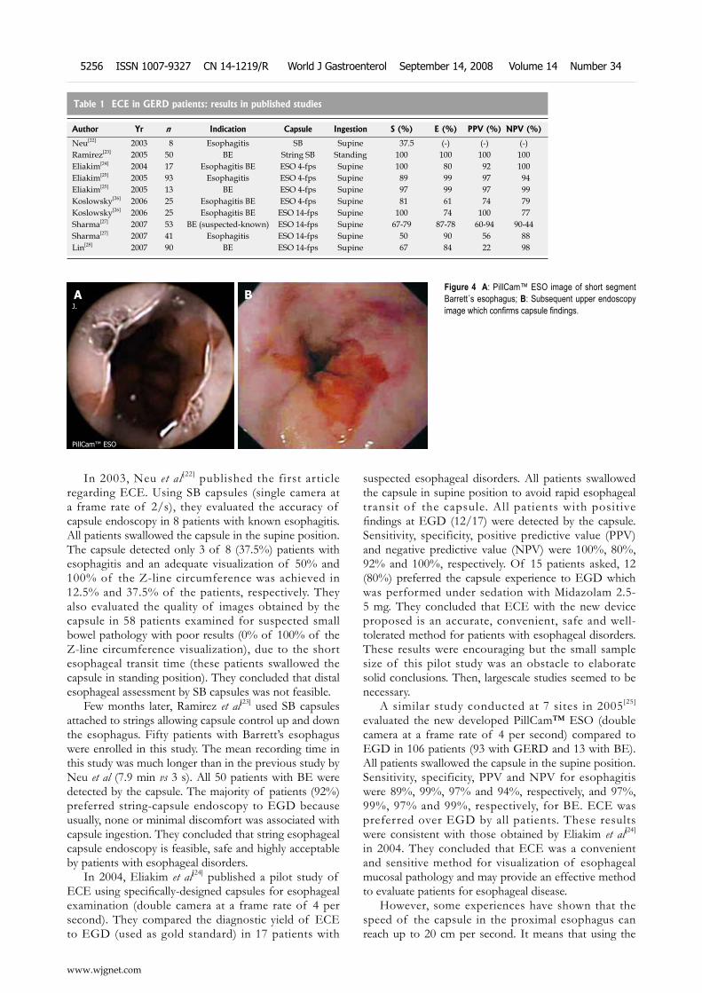
In 2003, Neu et al [22] published the first article regarding ECE. Using SB capsules (single camera at a frame rate of 2/s), they evaluated the accuracy of capsule endoscopy in 8 patients with known esophagitis. All patients swallowed the capsule in the supine position. The capsule detected only 3 of 8 (37.5%) patients with esophagitis and an adequate visualization of 50% and 100% of the Z-line circumference was achieved in 12.5% and 37.5% of the patients, respectively. They also evaluated the quality of images obtained by the capsule in 58 patients examined for suspected small bowel pathology with poor results (0% of 100% of the Z-line circumference visualization), due to the short esophageal transit time (these patients swallowed the capsule in standing position). They concluded that distal esophageal assessment by SB capsules was not feasible.
Few months later, Ramirez et al[23] used SB capsules attached to strings allowing capsule control up and down the esophagus. Fifty patients with Barrett’s esophagus were enrolled in this study. The mean recording time in this study was much longer than in the previous study by Neu et al (7.9 min vs 3 s). All 50 patients with BE were detected by the capsule. The majority of patients (92%) preferred string-capsule endoscopy to EGD because usually, none or minimal discomfort was associated with capsule ingestion. They concluded that string esophageal capsule endoscopy is feasible, safe and highly acceptable by patients with esophageal disorders.
In 2004, Eliakim et al[24] published a pilot study of ECE using specifically-designed capsules for esophageal examination (double camera at a frame rate of 4 per second). They compared the diagnostic yield of ECE to EGD (used as gold standard) in 17 patients with
suspected esophageal disorders. All patients swallowed the capsule in supine position to avoid rapid esophageal transit of the capsule. All patients with positive findings at EGD (12/17) were detected by the capsule. Sensitivity, specificity, positive predictive value (PPV) and negative predictive value (NPV) were 100%, 80%, 92% and 100%, respectively. Of 15 patients asked, 12 (80%) preferred the capsule experience to EGD which was performed under sedation with Midazolam 2.5- 5 mg. They concluded that ECE with the new device proposed is an accurate, convenient, safe and well- tolerated method for patients with esophageal disorders. These results were encouraging but the small sample size of this pilot study was an obstacle to elaborate solid conclusions. Then, largescale studies seemed to be necessary.
A similar study conducted at 7 sites in 2005[25] evaluated the new developed PillCam™ ESO (double camera at a frame rate of 4 per second) compared to EGD in 106 patients (93 with GERD and 13 with BE). All patients swallowed the capsule in the supine position. Sensitivity, specificity, PPV and NPV for esophagitis were 89%, 99%, 97% and 94%, respectively, and 97%, 99%, 97% and 99%, respectively, for BE. ECE was preferred over EGD by all patients. These results were consistent with those obtained by Eliakim et al[24] in 2004. They concluded that ECE was a convenient and sensitive method for visualization of esophageal mucosal pathology and may provide an effective method to evaluate patients for esophageal disease.
However, some experiences have shown that the speed of the capsule in the proximal esophagus can reach up to 20 cm per second. It means that using the
Table 1 ECE in GERD patients: results in published studies
Author Yr n Indication Capsule Ingestion S (%) E (%) PPV (%) NPV (%)
Neu[22] 2003 8 Esophagitis SB Supine 37.5 (-) (-) (-)Ramirez[23] 2005 50 BE String SB Standing 100 100 100 100Eliakim[24] 2004 17 Esophagitis BE ESO 4-fps Supine 100 80 92 100Eliakim[25] 2005 93 Esophagitis ESO 4-fps Supine 89 99 97 94Eliakim[25] 2005 13 BE ESO 4-fps Supine 97 99 97 99Koslowsky[26] 2006 25 Esophagitis BE ESO 4-fps Supine 81 61 74 79Koslowsky[26] 2006 25 Esophagitis BE ESO 14-fps Supine 100 74 100 77Sharma[27] 2007 53 BE (suspected-known) ESO 14-fps Supine 67-79 87-78 60-94 90-44Sharma[27] 2007 41 Esophagitis ESO 14-fps Supine 50 90 56 88Lin[28] 2007 90 BE ESO 14-fps Supine 67 84 22 98
Figure 4 A: PillCam™ ESO image of short segment Barrett´s esophagus; B: Subsequent upper endoscopy image which confirms capsule findings.
A BJ.
PillCam™ ESO
www.wjgnet.com
5256 ISSN 1007-9327 CN 14-1219/R World J Gastroenterol September 14, 2008 Volume 14 Number 34
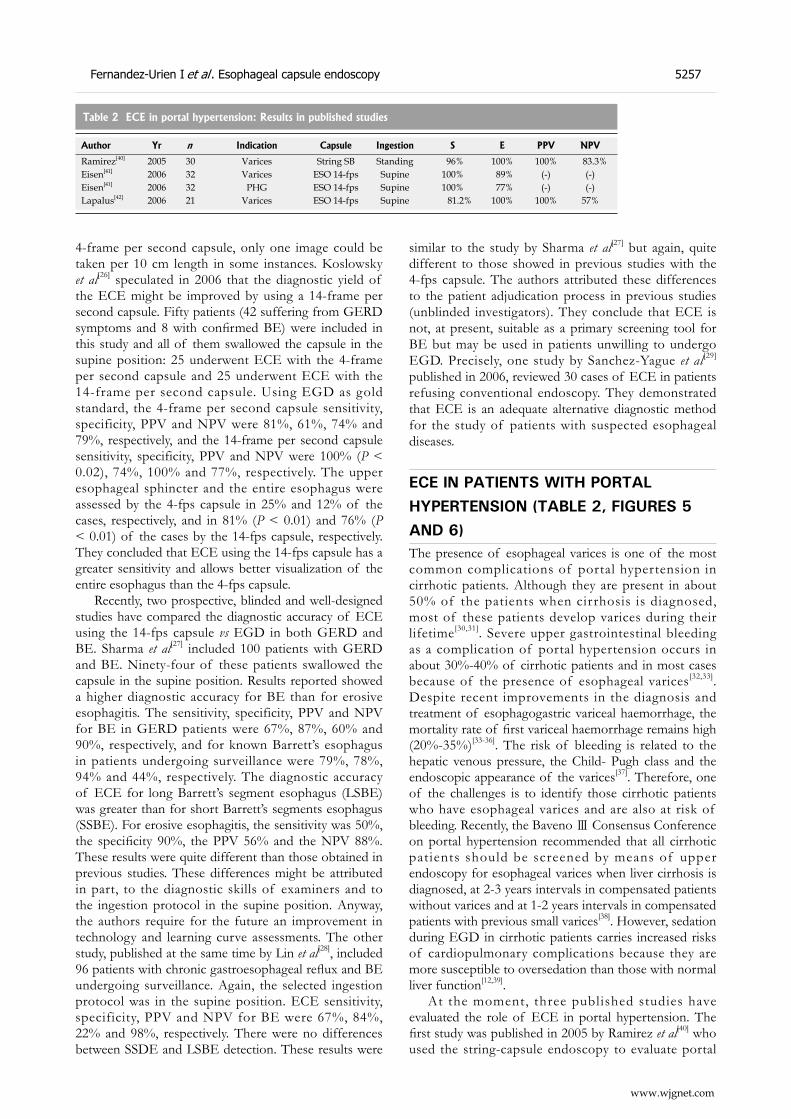
4-frame per second capsule, only one image could be taken per 10 cm length in some instances. Koslowsky et al[26] speculated in 2006 that the diagnostic yield of the ECE might be improved by using a 14-frame per second capsule. Fifty patients (42 suffering from GERD symptoms and 8 with confirmed BE) were included in this study and all of them swallowed the capsule in the supine position: 25 underwent ECE with the 4-frame per second capsule and 25 underwent ECE with the 14-frame per second capsule. Using EGD as gold standard, the 4-frame per second capsule sensitivity, specificity, PPV and NPV were 81%, 61%, 74% and 79%, respectively, and the 14-frame per second capsule sensitivity, specificity, PPV and NPV were 100% (P < 0.02), 74%, 100% and 77%, respectively. The upper esophageal sphincter and the entire esophagus were assessed by the 4-fps capsule in 25% and 12% of the cases, respectively, and in 81% (P < 0.01) and 76% (P < 0.01) of the cases by the 14-fps capsule, respectively. They concluded that ECE using the 14-fps capsule has a greater sensitivity and allows better visualization of the entire esophagus than the 4-fps capsule.
Recently, two prospective, blinded and well-designed studies have compared the diagnostic accuracy of ECE using the 14-fps capsule vs EGD in both GERD and BE. Sharma et al[27] included 100 patients with GERD and BE. Ninety-four of these patients swallowed the capsule in the supine position. Results reported showed a higher diagnostic accuracy for BE than for erosive esophagitis. The sensitivity, specificity, PPV and NPV for BE in GERD patients were 67%, 87%, 60% and 90%, respectively, and for known Barrett’s esophagus in patients undergoing surveillance were 79%, 78%, 94% and 44%, respectively. The diagnostic accuracy of ECE for long Barrett’s segment esophagus (LSBE) was greater than for short Barrett’s segments esophagus (SSBE). For erosive esophagitis, the sensitivity was 50%, the specificity 90%, the PPV 56% and the NPV 88%. These results were quite different than those obtained in previous studies. These differences might be attributed in part, to the diagnostic skills of examiners and to the ingestion protocol in the supine position. Anyway, the authors require for the future an improvement in technology and learning curve assessments. The other study, published at the same time by Lin et al[28], included 96 patients with chronic gastroesophageal reflux and BE undergoing surveillance. Again, the selected ingestion protocol was in the supine position. ECE sensitivity, specificity, PPV and NPV for BE were 67%, 84%, 22% and 98%, respectively. There were no differences between SSDE and LSBE detection. These results were
similar to the study by Sharma et al[27] but again, quite different to those showed in previous studies with the 4-fps capsule. The authors attributed these differences to the patient adjudication process in previous studies (unblinded investigators). They conclude that ECE is not, at present, suitable as a primary screening tool for BE but may be used in patients unwilling to undergo EGD. Precisely, one study by Sanchez-Yague et al[29] published in 2006, reviewed 30 cases of ECE in patients refusing conventional endoscopy. They demonstrated that ECE is an adequate alternative diagnostic method for the study of patients with suspected esophageal diseases.
ECE IN PATIENTS WITH PORTAL HYPERTENSION (TABLE 2, FIGURES 5 AND 6)The presence of esophageal varices is one of the most common complications of portal hypertension in cirrhotic patients. Although they are present in about 50% of the patients when cirrhosis is diagnosed, most of these patients develop varices during their lifetime[30,31]. Severe upper gastrointestinal bleeding as a complication of portal hypertension occurs in about 30%-40% of cirrhotic patients and in most cases because of the presence of esophageal varices[32,33]. Despite recent improvements in the diagnosis and treatment of esophagogastric variceal haemorrhage, the mortality rate of first variceal haemorrhage remains high (20%-35%)[33-36]. The risk of bleeding is related to the hepatic venous pressure, the Child- Pugh class and the endoscopic appearance of the varices[37]. Therefore, one of the challenges is to identify those cirrhotic patients who have esophageal varices and are also at risk of bleeding. Recently, the Baveno Ⅲ Consensus Conference on portal hypertension recommended that all cirrhotic patients should be screened by means of upper endoscopy for esophageal varices when liver cirrhosis is diagnosed, at 2-3 years intervals in compensated patients without varices and at 1-2 years intervals in compensated patients with previous small varices[38]. However, sedation during EGD in cirrhotic patients carries increased risks of cardiopulmonary complications because they are more susceptible to oversedation than those with normal liver function[12,39].
At the moment, three published studies have evaluated the role of ECE in portal hypertension. The first study was published in 2005 by Ramirez et al[40] who used the string-capsule endoscopy to evaluate portal
Table 2 ECE in portal hypertension: Results in published studies
Author Yr n Indication Capsule Ingestion S E PPV NPV
Ramirez[40] 2005 30 Varices String SB Standing 96% 100% 100% 83.3%Eisen[41] 2006 32 Varices ESO 14-fps Supine 100% 89% (-) (-)Eisen[41] 2006 32 PHG ESO 14-fps Supine 100% 77% (-) (-)Lapalus[42] 2006 21 Varices ESO 14-fps Supine 81.2% 100% 100% 57%
Fernandez-Urien I et al . Esophageal capsule endoscopy 5257
www.wjgnet.com
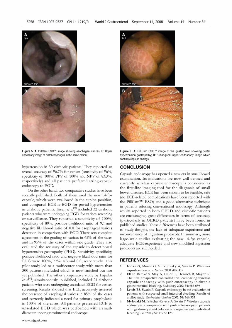
hypertension in 30 cirrhotic patients. They reported an overall accuracy of 96.7% for varices (sensitivity of 96%, specificity of 100%, PPV of 100% and NPV of 83.3%, respectively) and all patients preferred string-capsule endoscopy to EGD.
On the other hand, two comparative studies have been recently published. Both of them used the new 14-fps capsule, which were swallowed in the supine position, and compared ECE vs EGD for portal hypertension in cirrhotic patients. Eisen et al[41] included 32 cirrhotic patients who were undergoing EGD for varices screening or surveillance. They reported a sensitivity of 100%, specificity of 89%, positive likelihood ratio of 9.1 and negative likelihood ratio of 0.0 for esophageal varices detection in comparison with EGD. There was complete agreement in the grading of varices in 65% of the cases and in 95% of the cases within one grade. They also evaluated the accuracy of the capsule to detect portal hypertension gastropathy (PHG). Sensitivity, specificity, positive likelihood ratio and negative likelihood ratio for PHG were 100%, 77%, 4.3 and 0.0, respectively. This pilot study led to a multicenter study with more than 300 patients included which is now finished but not yet published. The other comparative study by Lapalus et al[42], simultaneously published, included 21 cirrhotic patients who were undergoing unsedated EGD for varices screening. Results showed that ECE accurately assessed the presence of esophageal varices in 85% of the cases and correctly indicated a need for primary prophylaxis in 100% of the cases. All patients preferred ECE to unsedated EGD which was performed with a small-diameter upper gastrointestinal endoscope.
CONCLUSIONCapsule endoscopy has opened a new era in small bowel examination. Its indications are now well-defined and currently, wireless capsule endoscopy is considered as the first-line imaging tool for the diagnosis of small bowel diseases. ECE has been shown to be feasible, safe (no ECE-related complications have been reported with the PillCam™ ESO) and a good alternative technique in patients refusing conventional endoscopy. Although results reported in both GERD and cirrhotic patients are encouraging, great differences in terms of accuracy (particularly in GERD patients) have been found in published studies. These differences have been attributed to study designs, the lack of adequate experience and inconvenience of ingestion protocols. In summary, more large-scale studies evaluating the new 14-fps capsule, adequate ECE-experience and new modified ingestion protocols are still needed.
REFERENCES1 Iddan G, Meron G, Glukhovsky A, Swain P. Wireless
capsule endoscopy. Nature 2000; 405: 4172 Ell C, Remke S, May A, Helou L, Henrich R, Mayer G.
The first prospective controlled trial comparing wireless capsule endoscopy with push enteroscopy in chronic gastrointestinal bleeding. Endoscopy 2002; 34: 685-689
3 Lewis BS, Swain P. Capsule endoscopy in the evaluation of patients with suspected small intestinal bleeding: Results of a pilot study. Gastrointest Endosc 2002; 56: 349-353
4 Mylonaki M, Fritscher-Ravens A, Swain P. Wireless capsule endoscopy: a comparison with push enteroscopy in patients with gastroscopy and colonoscopy negative gastrointestinal bleeding. Gut 2003; 52: 1122-1126
Figure 5 A: PillCam ESO™ image showing esophageal varices; B: Upper endoscopy image of distal esophagus in the same patient.
B
AAPP
PillCam™ ESO
Figure 6 A: PillCam ESO™ image of the gastric wall showing portal hypertension gastropathy; B: Subsequent upper endoscopy image which confirms capsule findings.
B
AJMB
PillCam™ ESO
www.wjgnet.com
5258 ISSN 1007-9327 CN 14-1219/R World J Gastroenterol September 14, 2008 Volume 14 Number 34
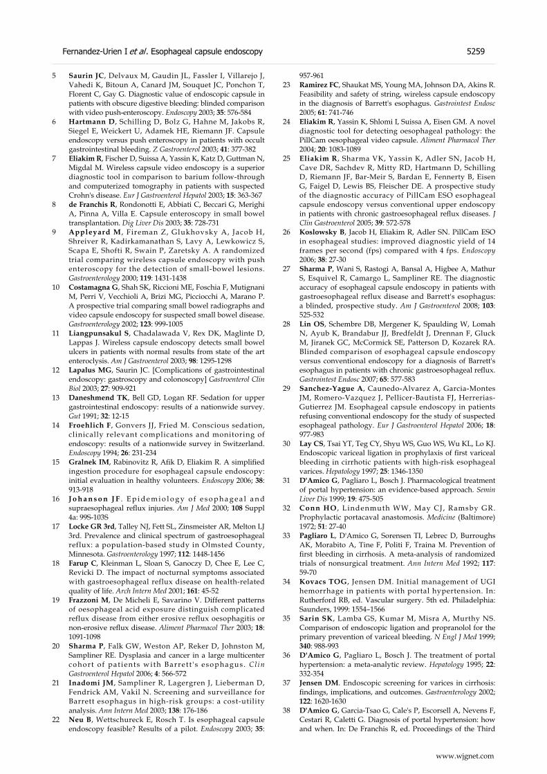
5 Saurin JC, Delvaux M, Gaudin JL, Fassler I, Villarejo J, Vahedi K, Bitoun A, Canard JM, Souquet JC, Ponchon T, Florent C, Gay G. Diagnostic value of endoscopic capsule in patients with obscure digestive bleeding: blinded comparison with video push-enteroscopy. Endoscopy 2003; 35: 576-584
6 Hartmann D, Schilling D, Bolz G, Hahne M, Jakobs R, Siegel E, Weickert U, Adamek HE, Riemann JF. Capsule endoscopy versus push enteroscopy in patients with occult gastrointestinal bleeding. Z Gastroenterol 2003; 41: 377-382
7 Eliakim R, Fischer D, Suissa A, Yassin K, Katz D, Guttman N, Migdal M. Wireless capsule video endoscopy is a superior diagnostic tool in comparison to barium follow-through and computerized tomography in patients with suspected Crohn's disease. Eur J Gastroenterol Hepatol 2003; 15: 363-367
8 de Franchis R, Rondonotti E, Abbiati C, Beccari G, Merighi A, Pinna A, Villa E. Capsule enteroscopy in small bowel transplantation. Dig Liver Dis 2003; 35: 728-731
9 Appleyard M , Fireman Z, Glukhovsky A, Jacob H, Shreiver R, Kadirkamanathan S, Lavy A, Lewkowicz S, Scapa E, Shofti R, Swain P, Zaretsky A. A randomized trial comparing wireless capsule endoscopy with push enteroscopy for the detection of small-bowel lesions. Gastroenterology 2000; 119: 1431-1438
10 Costamagna G, Shah SK, Riccioni ME, Foschia F, Mutignani M, Perri V, Vecchioli A, Brizi MG, Picciocchi A, Marano P. A prospective trial comparing small bowel radiographs and video capsule endoscopy for suspected small bowel disease. Gastroenterology 2002; 123: 999-1005
11 Liangpunsakul S, Chadalawada V, Rex DK, Maglinte D, Lappas J. Wireless capsule endoscopy detects small bowel ulcers in patients with normal results from state of the art enteroclysis. Am J Gastroenterol 2003; 98: 1295-1298
12 Lapalus MG, Saurin JC. [Complications of gastrointestinal endoscopy: gastroscopy and colonoscopy] Gastroenterol Clin Biol 2003; 27: 909-921
13 Daneshmend TK, Bell GD, Logan RF. Sedation for upper gastrointestinal endoscopy: results of a nationwide survey. Gut 1991; 32: 12-15
14 Froehlich F, Gonvers JJ, Fried M. Conscious sedation, clinically relevant complications and monitoring of endoscopy: results of a nationwide survey in Switzerland. Endoscopy 1994; 26: 231-234
15 Gralnek IM, Rabinovitz R, Afik D, Eliakim R. A simplified ingestion procedure for esophageal capsule endoscopy: initial evaluation in healthy volunteers. Endoscopy 2006; 38: 913-918
16 J o h a n s o n J F . E p i d e m i o l o g y o f e s o p h a g e a l a n d supraesophageal reflux injuries. Am J Med 2000; 108 Suppl 4a: 99S-103S
17 Locke GR 3rd, Talley NJ, Fett SL, Zinsmeister AR, Melton LJ 3rd. Prevalence and clinical spectrum of gastroesophageal reflux: a population-based study in Olmsted County, Minnesota. Gastroenterology 1997; 112: 1448-1456
18 Farup C, Kleinman L, Sloan S, Ganoczy D, Chee E, Lee C, Revicki D. The impact of nocturnal symptoms associated with gastroesophageal reflux disease on health-related quality of life. Arch Intern Med 2001; 161: 45-52
19 Frazzoni M, De Micheli E, Savarino V. Different patterns of oesophageal acid exposure distinguish complicated reflux disease from either erosive reflux oesophagitis or non-erosive reflux disease. Aliment Pharmacol Ther 2003; 18: 1091-1098
20 Sharma P, Falk GW, Weston AP, Reker D, Johnston M, Sampliner RE. Dysplasia and cancer in a large multicenter cohort of pat ients with Barret t ' s esophagus . Cl in Gastroenterol Hepatol 2006; 4: 566-572
21 Inadomi JM, Sampliner R, Lagergren J, Lieberman D, Fendrick AM, Vakil N. Screening and surveillance for Barrett esophagus in high-risk groups: a cost-utility analysis. Ann Intern Med 2003; 138: 176-186
22 Neu B, Wettschureck E, Rosch T. Is esophageal capsule endoscopy feasible? Results of a pilot. Endoscopy 2003; 35:
957-96123 Ramirez FC, Shaukat MS, Young MA, Johnson DA, Akins R.
Feasibility and safety of string, wireless capsule endoscopy in the diagnosis of Barrett's esophagus. Gastrointest Endosc 2005; 61: 741-746
24 Eliakim R, Yassin K, Shlomi I, Suissa A, Eisen GM. A novel diagnostic tool for detecting oesophageal pathology: the PillCam oesophageal video capsule. Aliment Pharmacol Ther 2004; 20: 1083-1089
25 Eliakim R, Sharma VK, Yassin K, Adler SN, Jacob H, Cave DR, Sachdev R, Mitty RD, Hartmann D, Schilling D, Riemann JF, Bar-Meir S, Bardan E, Fennerty B, Eisen G, Faigel D, Lewis BS, Fleischer DE. A prospective study of the diagnostic accuracy of PillCam ESO esophageal capsule endoscopy versus conventional upper endoscopy in patients with chronic gastroesophageal reflux diseases. J Clin Gastroenterol 2005; 39: 572-578
26 Koslowsky B, Jacob H, Eliakim R, Adler SN. PillCam ESO in esophageal studies: improved diagnostic yield of 14 frames per second (fps) compared with 4 fps. Endoscopy 2006; 38: 27-30
27 Sharma P, Wani S, Rastogi A, Bansal A, Higbee A, Mathur S, Esquivel R, Camargo L, Sampliner RE. The diagnostic accuracy of esophageal capsule endoscopy in patients with gastroesophageal reflux disease and Barrett's esophagus: a blinded, prospective study. Am J Gastroenterol 2008; 103: 525-532
28 Lin OS, Schembre DB, Mergener K, Spaulding W, Lomah N, Ayub K, Brandabur JJ, Bredfeldt J, Drennan F, Gluck M, Jiranek GC, McCormick SE, Patterson D, Kozarek RA. Blinded comparison of esophageal capsule endoscopy versus conventional endoscopy for a diagnosis of Barrett's esophagus in patients with chronic gastroesophageal reflux. Gastrointest Endosc 2007; 65: 577-583
29 Sanchez-Yague A, Caunedo-Alvarez A, Garcia-Montes JM, Romero-Vazquez J, Pellicer-Bautista FJ, Herrerias-Gutierrez JM. Esophageal capsule endoscopy in patients refusing conventional endoscopy for the study of suspected esophageal pathology. Eur J Gastroenterol Hepatol 2006; 18: 977-983
30 Lay CS, Tsai YT, Teg CY, Shyu WS, Guo WS, Wu KL, Lo KJ. Endoscopic variceal ligation in prophylaxis of first variceal bleeding in cirrhotic patients with high-risk esophageal varices. Hepatology 1997; 25: 1346-1350
31 D'Amico G, Pagliaro L, Bosch J. Pharmacological treatment of portal hypertension: an evidence-based approach. Semin Liver Dis 1999; 19: 475-505
32 Conn HO , Lindenmuth WW, May CJ, Ramsby GR. Prophylactic portacaval anastomosis. Medicine (Baltimore) 1972; 51: 27-40
33 Pagliaro L, D'Amico G, Sorensen TI, Lebrec D, Burroughs AK, Morabito A, Tine F, Politi F, Traina M. Prevention of first bleeding in cirrhosis. A meta-analysis of randomized trials of nonsurgical treatment. Ann Intern Med 1992; 117: 59-70
34 Kovacs TOG, Jensen DM. Initial management of UGI hemorrhage in patients with portal hypertension. In: Rutherford RB, ed. Vascular surgery. 5th ed. Philadelphia: Saunders, 1999: 1554–1566
35 Sarin SK, Lamba GS, Kumar M, Misra A, Murthy NS. Comparison of endoscopic ligation and propranolol for the primary prevention of variceal bleeding. N Engl J Med 1999; 340: 988-993
36 D'Amico G, Pagliaro L, Bosch J. The treatment of portal hypertension: a meta-analytic review. Hepatology 1995; 22: 332-354
37 Jensen DM. Endoscopic screening for varices in cirrhosis: findings, implications, and outcomes. Gastroenterology 2002; 122: 1620-1630
38 D'Amico G, Garcia-Tsao G, Cale's P, Escorsell A, Nevens F, Cestari R, Caletti G. Diagnosis of portal hypertension: how and when. In: De Franchis R, ed. Proceedings of the Third
Fernandez-Urien I et al . Esophageal capsule endoscopy 5259
www.wjgnet.com
Baveno International Consensus Workshop on Definitions, Methology and Therapeutic Strategies. Oxford: Blackwell Science, 2001: 36-63
39 Madhotra R, Mokhashi M, Willner I, Hawes RH, Reuben A. Prospective evaluation of a 3.1-mm battery-powered esophagoscope in screening for esophageal varices in cirrhotic patients. Am J Gastroenterol 2003; 98: 807-812
40 Ramirez FC, Hakim S, Tharalson EM, Shaukat MS, Akins R. Feasibility and safety of string wireless capsule endoscopy in the diagnosis of esophageal varices. Am J Gastroenterol 2005; 100: 1065-1071
41 Eisen GM, Eliakim R, Zaman A, Schwartz J, Faigel D, Rondonotti E, Villa F, Weizman E, Yassin K, deFranchis R. The accuracy of PillCam ESO capsule endoscopy versus conventional upper endoscopy for the diagnosis of esophageal varices: a prospective three-center pilot study. Endoscopy 2006; 38: 31-35
42 Lapalus MG, Dumortier J, Fumex F, Roman S, Lot M, Prost B, Mion F, Ponchon T. Esophageal capsule endoscopy versus esophagogastroduodenoscopy for evaluating portal hypertension: a prospective comparative study of performance and tolerance. Endoscopy 2006; 38: 36-41
S- Editor Zhong XY E- Editor Lin YP
www.wjgnet.com
5260 ISSN 1007-9327 CN 14-1219/R World J Gastroenterol September 14, 2008 Volume 14 Number 34
TOPIC HIGHLIGHT
Role of videocapsule endoscopy for gastrointestinal bleeding
Cristina Carretero, Ignacio Fernandez-Urien, Maite Betes, Miguel Muñoz-Navas
Miguel Angel Muñoz-Navas, Profesor, Series Editors
Online Submissions: wjg.wjgnet.com World J Gastroenterol 2008 September 14; 14(34): [email protected] World Journal of Gastroenterology ISSN 1007-9327doi:10.3748/wjg.14.5261 © 2008 The WJG Press. All rights reserved.
Cristina Carretero, Ignacio Fernandez-Urien, Maite Betes, Miguel Muñoz-Navas, Department of Gastroenterology, University of Navarra, Pamplona 31008, SpainAuthor contributions: Carretero C and Fernandez-Urien I contributed equally to this paper, both performing research and writing the paper; Betes M and Muñoz-Navas M reviewed the paper.Correspondence to: Cristina Carretero, MD, Department of Gastroenterology, University of Navarra, Av. Pio Ⅻ, 36, Pamplona 31008, Spain. [email protected]: +34-948-255400 Fax: +34-948-296500Received: March 31, 2008 Revised: July 26, 2008Accepted: August 2, 2008Published online: September 14, 2008
AbstractObscure gastrointestinal bleeding (OGIB) is defined as bleeding of an unknown origin that persists or recurs after negative initial upper and lower endoscopies. Several techniques, such as endoscopy, arteriography, scintigraphy and barium radiology are helpful for recognizing the bleeding source; nevertheless, in about 5%-10% of cases the bleeding lesion cannot be determined. The development of videocapsule endoscopy (VCE) has permitted a direct visualization of the small intestine mucosa. We will analyze those techniques in more detail. The diagnostic yield of CE for OGIB varies from 38% to 93%, being in the higher range in those cases with obscure-overt bleeding.
© 2008 The WJG Press. All rights reserved.
Key words: Capsule endoscopy; Bleeding; Small bowel; Obscure; Hemorrhage
Peer reviewer: Ronan A Cahill, Department of General Surgery, Waterford Regional Hospital, Waterford, Cork, Ireland
Carretero C, Fernandez-Urien I, Betes M, Muñoz-Navas M. Role of videocapsule endoscopy for gastrointestinal bleeding. World J Gastroenterol 2008; 14(34): 5261-5264 Available from: URL: http://www.wjgnet.com/1007-9327/14/5261.asp DOI: http://dx.doi.org/10.3748/wjg.14.5261
INTRODUCTIONObscure gastrointestinal bleeding (OGIB) is defined as
bleeding of an unknown origin that persists or recurs after negative initial upper and lower endoscopies[1]. It can be subclassified as either obscure-occult, detected only by positive fecal occult blood tests (FOBT) and/or iron deficiency anemia (IDA), or obscure-overt with recurrent or persistent visible episodes of bleeding[2].
Approximately 5% of all gastrointestinal bleeding[3]
are of obscure origin, and the most frequent causes are found among these: esophagitis, Cameron ulcers, Dieulafoy lesions, angiodysplasias, GAVE, portal hypertensive gastropathy, small bowel neoplasms, hemobilia, Meckel diverticulum, Crohn’s disease, medication-induced mucosal lesions and a few others[2].
Several techniques, such as endoscopy, arteriography, scintigraphy and barium radiology, are helpful for recognizing the bleeding source; nevertheless, in about 5%-10% of cases the bleeding lesion cannot be determined[4]. Before 2001, the study of OGIB was deficient as the small bowel could not be reviewed reliably in its whole, but the development of videocapsule endoscopy (VCE) has permitted a direct visualization of the small intestine mucosa.
DIAGNOSTIC TECHNIQUESCapsule endoscopy (CE)Capsule endoscopy (CE) is a disposable 26 mm × 11 mm plastic capsule (Figure 1) consisting of an optical dome, 4 light-emitting electrodes, a sensor, 2 batteries and a micro transmitter. It acquires and transmits 2 frames per second (until the battery expires after 7 h ± 1 h[5]) to a sensor array attached to the patient[2]. Image features include a 140-degree field of view, 1:8 magnification, 1 to 30 mm depth of field and a minimum size for detection of about 0.1 mm[5].
It is passively propelled by peristalsis and it captures images of the entire length of the small intestine. The main limitations are the lack of air insufflation, the unavailability of rinsing, taking biopsies or/and treating lesions[2].
The incidence of capsule retention accounts of less than 1%[2] and is generally related to the presence of endoluminal narrowing. For that reason, and intending to predict the risk of retention, patients with high suspicion of bowel narrowing (history of NSAID’s intake, Crohn’s Disease, occlusive symptoms or ischemic bowel disease) should undergo capsule endoscopy exploration after performing other techniques such as CT scan, small
www.wjgnet.com
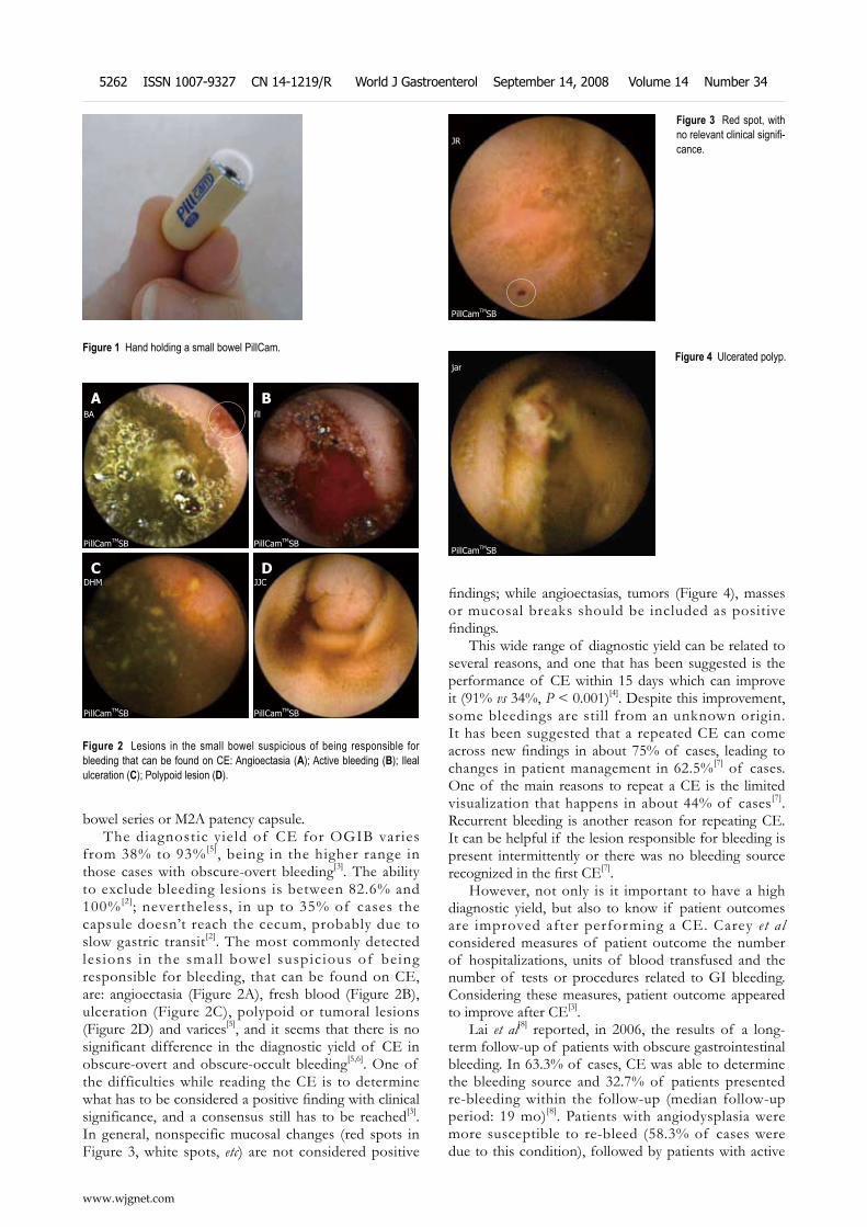
bowel series or M2A patency capsule.The diagnostic yield of CE for OGIB varies
from 38% to 93%[5], being in the higher range in those cases with obscure-overt bleeding[3]. The ability to exclude bleeding lesions is between 82.6% and 100%[2]; nevertheless, in up to 35% of cases the capsule doesn’t reach the cecum, probably due to slow gastric transit[2]. The most commonly detected les ions in the smal l bowel suspic ious of being responsible for bleeding, that can be found on CE, are: angioectasia (Figure 2A), fresh blood (Figure 2B), ulceration (Figure 2C), polypoid or tumoral lesions (Figure 2D) and varices[5], and it seems that there is no significant difference in the diagnostic yield of CE in obscure-overt and obscure-occult bleeding[5,6]. One of the difficulties while reading the CE is to determine what has to be considered a positive finding with clinical significance, and a consensus still has to be reached[3]. In general, nonspecific mucosal changes (red spots in Figure 3, white spots, etc) are not considered positive
findings; while angioectasias, tumors (Figure 4), masses or mucosal breaks should be included as positive findings.
This wide range of diagnostic yield can be related to several reasons, and one that has been suggested is the performance of CE within 15 days which can improve it (91% vs 34%, P < 0.001)[4]. Despite this improvement, some bleedings are still from an unknown origin. It has been suggested that a repeated CE can come across new findings in about 75% of cases, leading to changes in patient management in 62.5%[7] of cases. One of the main reasons to repeat a CE is the limited visualization that happens in about 44% of cases[7]. Recurrent bleeding is another reason for repeating CE. It can be helpful if the lesion responsible for bleeding is present intermittently or there was no bleeding source recognized in the first CE[7].
However, not only is it important to have a high diagnostic yield, but also to know if patient outcomes are improved after performing a CE. Carey et al considered measures of patient outcome the number of hospitalizations, units of blood transfused and the number of tests or procedures related to GI bleeding. Considering these measures, patient outcome appeared to improve after CE[3].
Lai et al[8] reported, in 2006, the results of a long-term follow-up of patients with obscure gastrointestinal bleeding. In 63.3% of cases, CE was able to determine the bleeding source and 32.7% of patients presented re-bleeding within the follow-up (median follow-up period: 19 mo)[8]. Patients with angiodysplasia were more susceptible to re-bleed (58.3% of cases were due to this condition), followed by patients with active
Figure 1 Hand holding a small bowel PillCam.
DC
BA
PillCamTMSB PillCamTMSB
PillCamTMSB PillCamTMSB
DHM JJC
BA fll
Figure 2 Lesions in the small bowel suspicious of being responsible for bleeding that can be found on CE: Angioectasia (A); Active bleeding (B); Ileal ulceration (C); Polypoid lesion (D).
PillCamTMSB
jarFigure 4 Ulcerated polyp.
PillCamTMSB
JR
Figure 3 Red spot, with no relevant clinical signifi-cance.
www.wjgnet.com
5262 ISSN 1007-9327 CN 14-1219/R World J Gastroenterol September 14, 2008 Volume 14 Number 34

bleeding during the CE procedure with no identified bleeding source (53.8%)[8]. They found that the probability of re-bleeding was significantly higher in patients with positive CE than in those with negative CE (P = 0.003)[8].
Repeat upper endoscopy and colonoscopyBetween 35% to 75% of patients can be under-diagnosed at the initial endoscopic study[2]. Missed lesions occur due to their size, location, presence of clots or the absence of active bleeding while the endoscopy is being performed. Other possible causes are anemia, volume contraction, the effect of sedatives, as they can result in paleness of vascular lesions, and the timing of the endoscopy, as it is more probably to identify the bleeding source if the endoscopy is performed within 48 hours of the acute event[2].
It has been recommended to repeat upper and lower endoscopy (“second-look endoscopy”)[2], nevertheless, about 5% to 10% of patients will remain undiagnosed.
Push enteroscopyEnteroscopy consists of a peroral insertion into the jejunum of a long endoscope, using either a pediatric colonoscope or an enteroscope, and with or without the use of an overtube in order to avoid gastric looping[2]. This technique allows the examination of 15 to 160 cm beyond the ligament of Treitz[5] and it is generally considered the next diagnostic step after upper and lower negative endoscopic studies.
In 38%-75% of cases, the lesion causing OGIB can be identified with this technique[2,5]. However, in 28% to 75% of cases, it is reachable with a gastroscope[5]. Common findings are angiodysplasias (20%-46%), peptic ulcer disease, benign and malignant jejunal tumors, diverticulum, esophagitis and varices[2]. A meta-analysis has been published showing that the yield of CE for all findings is 63% vs 28% for Push Enteroscopy, with an incremental yield of 35%, P < 0.00001[9]. Yield of clinically significant findings is 56% for CE vs 26% for Push Enteroscopy[9].
Small bowel series and enteroclysisBefore the development of CE, a radiologic study of the small bowel was mandatory for the study of OGIB. The diagnostic yield is about 6%[2,5], and the most common missing lesions include angioectasia, ulcers and erosions[5]. The diagnostic yield can be improved (diagnostic yield of 10%-20%)[5] by performing enteroclysis. This technique consists in the introduction of a catheter into the small intestine followed by the injection of barium and methylcellulose. The barium coats the intestine and the methylcellulose distends the lumen to give a double contrast exam that allows for fluoroscopic visualization of the entire small bowel. The sensitivity of enteroclysis for small bowel tumors is higher than small bowel series[5].
A recent meta-analysis has shown that the diagnostic yield for all findings for CE is 67%, compared with an 8% for small bowel series. Diagnostic yield for
significant findings is 42% for CE vs 6% for small bowel radiography[9].
Bleeding scanning with technetium-99-labeled RBC and angiographyThis test can be useful if active bleeding is present as it may detect the source of hemorrhage if the bleeding rate ranges from 0.1 to 0.4 mL/min. Angiography can also be performed for diagnosis, as it is able to detect bleeding rates over 0.5 mL/min[2]. If the bleeding scanning is positive, an angiography should be performed to detect bleeding lesions and treat them if possible[2]. The diagnostic yield of these techniques varies from 44% to 68%[2].
Computed tomographic angiography (CTA)Angiographic images can be obtained, not only by routine angiographic techniques, but also by CT scan. CTA is noninvasive and potentially useful for the diagnosis of GI bleeding, as it avoids the risks of standard angiography[10]. The CTA is able to identify the bleeding source in 24% of patients[10]. As has been recently published, CE is able to identify the bleeding source in a higher proportion of patients than CTA (72% vs 24%)[10].
Intraoperative enteroscopy (IE)IE is the final diagnostic procedure for OGIB. It consists of performing an enteroscopy with the help of the surgeon, who helps by pushing the bowel over the enteroscope as the endoscopist examines it. The insertion can be done transoral or directly through a small enterotomy, but the last is more likely to achieve a complete examination of the small bowel. One of the drawbacks of this technique is that the manipulation of the bowel can create artifacts, which can be considered as bleeding sources although they are really not[2]. The direct effect of this handicap is that the examination of the mucosa has to be done while the intubation and not while the withdrawal of the endoscope. IE can achieve a diagnostic yield of a 70 to 93 which is comparable to CE[9].
Double balloon enteroscopy (DBE)DBE is a novel endoscopic technique that allows for visualization of the entire small bowel, tissue sampling and therapeutic interventions. It consists of an enteroscope with 2 latex balloons, one attached to the endoscope and the other attached to an overtube. The procedure is performed by inflating and deflating the balloons, allowing the deep intubation through the small bowel. The route of insertion (perorally or transanally) selected depends on the patients’ symptoms (hematochezia, melena etc).
A potential bleeding cause can be found in 75.7% of patients[11] and approximately a quarter of patients might need both anterograde and retrograde approaches[11]. DBE can change patient management in about 83.5% of cases, with an average therapeutic endoscopic intervention of 15.7%[11]. Hadithi et al[12] have published that CE is able to detect the presence of a possible
www.wjgnet.com
Carretero C et al . Capsule endoscopy for GI bleeding 5263
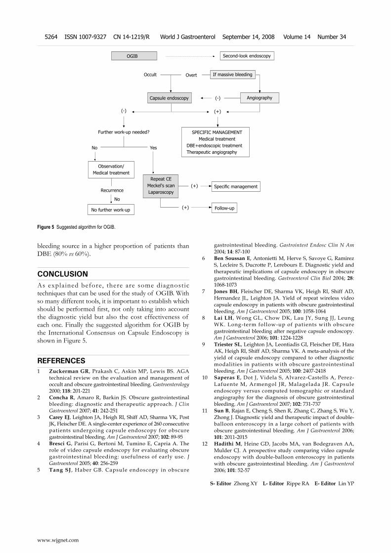
bleeding source in a higher proportion of patients than DBE (80% vs 60%).
CONCLUSIONAs explained before, there are some diagnostic techniques that can be used for the study of OGIB. With so many different tools, it is important to establish which should be performed first, not only taking into account the diagnostic yield but also the cost effectiveness of each one. Finally the suggested algorithm for OGIB by the International Consensus on Capsule Endoscopy is shown in Figure 5.
REFERENCES1 Zuckerman GR, Prakash C, Askin MP, Lewis BS. AGA
technical review on the evaluation and management of occult and obscure gastrointestinal bleeding. Gastroenterology 2000; 118: 201-221
2 Concha R, Amaro R, Barkin JS. Obscure gastrointestinal bleeding: diagnostic and therapeutic approach. J Clin Gastroenterol 2007; 41: 242-251
3 Carey EJ, Leighton JA, Heigh RI, Shiff AD, Sharma VK, Post JK, Fleischer DE. A single-center experience of 260 consecutive patients undergoing capsule endoscopy for obscure gastrointestinal bleeding. Am J Gastroenterol 2007; 102: 89-95
4 Bresci G, Parisi G, Bertoni M, Tumino E, Capria A. The role of video capsule endoscopy for evaluating obscure gastrointestinal bleeding: usefulness of early use. J Gastroenterol 2005; 40: 256-259
5 Tang SJ , Haber GB. Capsule endoscopy in obscure
gastrointestinal bleeding. Gastrointest Endosc Clin N Am 2004; 14: 87-100
6 Ben Soussan E, Antonietti M, Herve S, Savoye G, Ramirez S, Lecleire S, Ducrotte P, Lerebours E. Diagnostic yield and therapeutic implications of capsule endoscopy in obscure gastrointestinal bleeding. Gastroenterol Clin Biol 2004; 28: 1068-1073
7 Jones BH, Fleischer DE, Sharma VK, Heigh RI, Shiff AD, Hernandez JL, Leighton JA. Yield of repeat wireless video capsule endoscopy in patients with obscure gastrointestinal bleeding. Am J Gastroenterol 2005; 100: 1058-1064
8 Lai LH, Wong GL, Chow DK, Lau JY, Sung JJ, Leung WK. Long-term follow-up of patients with obscure gastrointestinal bleeding after negative capsule endoscopy. Am J Gastroenterol 2006; 101: 1224-1228
9 Triester SL, Leighton JA, Leontiadis GI, Fleischer DE, Hara AK, Heigh RI, Shiff AD, Sharma VK. A meta-analysis of the yield of capsule endoscopy compared to other diagnostic modalities in patients with obscure gastrointestinal bleeding. Am J Gastroenterol 2005; 100: 2407-2418
10 Saperas E, Dot J, Videla S, Alvarez-Castells A, Perez-Lafuente M, Armengol JR, Malagelada JR. Capsule endoscopy versus computed tomographic or standard angiography for the diagnosis of obscure gastrointestinal bleeding. Am J Gastroenterol 2007; 102: 731-737
11 Sun B, Rajan E, Cheng S, Shen R, Zhang C, Zhang S, Wu Y, Zhong J. Diagnostic yield and therapeutic impact of double-balloon enteroscopy in a large cohort of patients with obscure gastrointestinal bleeding. Am J Gastroenterol 2006; 101: 2011-2015
12 Hadithi M, Heine GD, Jacobs MA, van Bodegraven AA, Mulder CJ. A prospective study comparing video capsule endoscopy with double-balloon enteroscopy in patients with obscure gastrointestinal bleeding. Am J Gastroenterol 2006; 101: 52-57
S- Editor Zhong XY L- Editor Rippe RA E- Editor Lin YP
Second-look endoscopyOGIB
Occult Overt If massive bleeding
AngiographyCapsule endoscopy
Further work-up needed?
No Yes
Observation/Medical treatment
SPECIFIC MANAGEMENTMedical treatment
DBE+endoscopic treatmentTherapeutic angiography
Repeat CE Meckel's scan Laparoscopy
No further work-up
Specific management
Follow-up
(+)
(+)
(+)
(-)
(-)
No
Recurrence
Figure 5 Suggested algorithm for OGIB.
www.wjgnet.com
5264 ISSN 1007-9327 CN 14-1219/R World J Gastroenterol September 14, 2008 Volume 14 Number 34
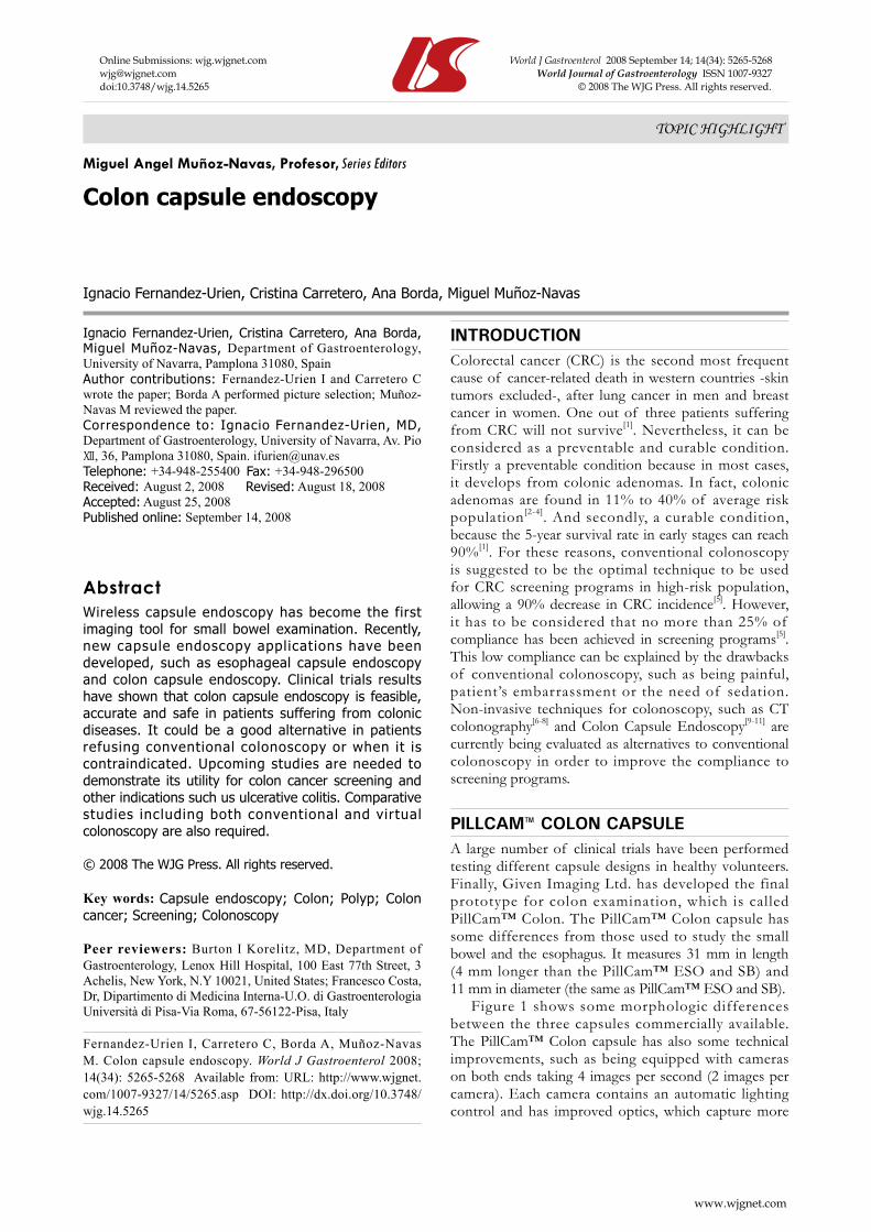
TOPIC HIGHLIGHT
Colon capsule endoscopy
Ignacio Fernandez-Urien, Cristina Carretero, Ana Borda, Miguel Muñoz-Navas
Miguel Angel Muñoz-Navas, Profesor, Series Editors
Online Submissions: wjg.wjgnet.com World J Gastroenterol 2008 September 14; 14(34): [email protected] World Journal of Gastroenterology ISSN 1007-9327doi:10.3748/wjg.14.5265 © 2008 The WJG Press. All rights reserved.
Ignacio Fernandez-Urien, Cristina Carretero, Ana Borda, Miguel Muñoz-Navas, Department of Gastroenterology, University of Navarra, Pamplona 31080, SpainAuthor contributions: Fernandez-Urien I and Carretero C wrote the paper; Borda A performed picture selection; Muñoz-Navas M reviewed the paper.Correspondence to: Ignacio Fernandez-Urien, MD, Department of Gastroenterology, University of Navarra, Av. Pio Ⅻ, 36, Pamplona 31080, Spain. [email protected]: +34-948-255400 Fax: +34-948-296500Received: August 2, 2008 Revised: August 18, 2008Accepted: August 25, 2008Published online: September 14, 2008
AbstractWireless capsule endoscopy has become the first imaging tool for small bowel examination. Recently, new capsule endoscopy applications have been developed, such as esophageal capsule endoscopy and colon capsule endoscopy. Clinical trials results have shown that colon capsule endoscopy is feasible, accurate and safe in patients suffering from colonic diseases. It could be a good alternative in patients refusing conventional colonoscopy or when it is contraindicated. Upcoming studies are needed to demonstrate its utility for colon cancer screening and other indications such us ulcerative colitis. Comparative studies including both conventional and virtual colonoscopy are also required.
© 2008 The WJG Press. All rights reserved.
Key words: Capsule endoscopy; Colon; Polyp; Colon cancer; Screening; Colonoscopy
Peer reviewers: Burton I Korelitz, MD, Department of Gastroenterology, Lenox Hill Hospital, 100 East 77th Street, 3 Achelis, New York, N.Y 10021, United States; Francesco Costa, Dr, Dipartimento di Medicina Interna-U.O. di Gastroenterologia Università di Pisa-Via Roma, 67-56122-Pisa, Italy
Fernandez-Urien I, Carretero C, Borda A, Muñoz-Navas M. Colon capsule endoscopy. World J Gastroenterol 2008; 14(34): 5265-5268 Available from: URL: http://www.wjgnet.com/1007-9327/14/5265.asp DOI: http://dx.doi.org/10.3748/wjg.14.5265
INTRODUCTIONColorectal cancer (CRC) is the second most frequent cause of cancer-related death in western countries -skin tumors excluded-, after lung cancer in men and breast cancer in women. One out of three patients suffering from CRC will not survive[1]. Nevertheless, it can be considered as a preventable and curable condition. Firstly a preventable condition because in most cases, it develops from colonic adenomas. In fact, colonic adenomas are found in 11% to 40% of average risk population[2-4]. And secondly, a curable condition, because the 5-year survival rate in early stages can reach 90%[1]. For these reasons, conventional colonoscopy is suggested to be the optimal technique to be used for CRC screening programs in high-risk population, allowing a 90% decrease in CRC incidence[5]. However, it has to be considered that no more than 25% of compliance has been achieved in screening programs[5]. This low compliance can be explained by the drawbacks of conventional colonoscopy, such as being painful, patient’s embarrassment or the need of sedation. Non-invasive techniques for colonoscopy, such as CT colonography[6-8] and Colon Capsule Endoscopy[9-11] are currently being evaluated as alternatives to conventional colonoscopy in order to improve the compliance to screening programs.
PILLCAM™ COLON CAPSULEA large number of clinical trials have been performed testing different capsule designs in healthy volunteers. Finally, Given Imaging Ltd. has developed the final prototype for colon examination, which is called PillCam™ Colon. The PillCam™ Colon capsule has some differences from those used to study the small bowel and the esophagus. It measures 31 mm in length (4 mm longer than the PillCam™ ESO and SB) and 11 mm in diameter (the same as PillCam™ ESO and SB).
Figure 1 shows some morphologic differences between the three capsules commercially available. The PillCam™ Colon capsule has also some technical improvements, such as being equipped with cameras on both ends taking 4 images per second (2 images per camera). Each camera contains an automatic lighting control and has improved optics, which capture more
www.wjgnet.com
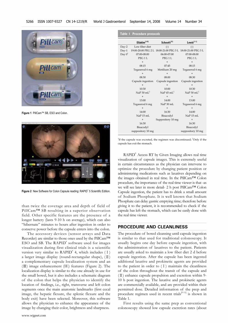
than twice the coverage area and depth of field of PillCam™ SB resulting in a superior observation field. Other specific features are the presence of a longer battery (lasts 9-10 h on average), which can also “hibernate” minutes to hours after ingestion in order to conserve power before the capsule enters into the colon.
The accessory devices (sensor arrays and Data Recorder) are similar to those ones used by the PillCam™ ESO and SB. The RAPID® software used for images visualization during first clinical trials is a scientific version very similar to RAPID® 4, which includes (Ⅰ) a larger image display (round-rectangular shape), (Ⅱ) a complementary capsule localization system and an (Ⅲ) image enhancement (IE) features (Figure 2). The localization display is similar to the one already in use for the small bowel, but it also includes a schematic diagram of the colon that helps the physician to identify the location of findings, i.e., right, transverse and left colon segments once the main anatomic landmarks (first cecal image, the hepatic flexure, the splenic flexure and the body exit) have been selected. Moreover, this software allows the physician to enhance the appearance of the image by changing their color, brightness and sharpness.
RAPID® Access RT by Given Imaging allows real time visualization of capsule images. This is extremely useful in certain circumstances as the physician can intervene to optimize the procedure by changing patient position or administering medications such as laxatives depending on the images obtained in real time. In the PillCam™ Colon procedure, the importance of the real time viewer is that -as we will see later in more detail- 2 h post PillCam™ Colon Capsule ingestion, the patient has to drink a small amount of Sodium Phosphate. It is well known that Sodium Phosphate can delay gastric emptying time; therefore before giving it to the patient, it is recommended to check if the capsule has left the stomach, which can be easily done with the real time viewer.
PROCEDURE AND CLEANLINESSThe procedure of bowel cleansing until capsule ingestion is similar to that used for traditional colonoscopy. It usually begins one day before capsule ingestion, with the administration of laxatives to the patient. Patients are usually asked to maintain a low fiber diet 2 d before capsule ingestion. After the capsule has been ingested additional laxative and prokinetic agents are provided to the patient in order to (Ⅰ) maintain the cleanliness of the colon throughout the transit of the capsule and (Ⅱ) enhance capsule propulsion and excretion within 9- 10 h post ingestion. The laxative and prokinetic agents are commercially available, and are provided within their permitted dose. Detailed information of the prep and procedure regimen used in recent trials[9-11] is shown in Table 1.
First results using the same prep as conventional colonoscopy showed low capsule excretion rates (about
Figure 1 PillCam™ SB, ESO and Colon.
Figure 2 New Software for Colon Capsule reading: RAPID® 5 Scientific Edition.
Table 1 Procedure protocols
Eliakim[10] Schoofs[9] Lewis[11]
Day-2 Low fiber diet (-) (-)Day-1 19:00-20:00 PEG 2 L 18:00-21:00 PEG 3 L 18:00-21:00 PEG 3 LDay 01 07:00-08:00
PEG 1 L+
08:15 Tegaserod 6 mg
+08:30
Capsule ingestion+
10:30 NaP 30 mL2
+13:00
Tegaserod 6 mg+
14:00 NaP 15 mL
+16:30
Bisacodyl suppository 10 mg
06:00-07:00 PEG 1 L
+07:45
Motilium 20 mg+
08:00 Capsule ingestion
+10:00
NaP 45 mL2
+14:00
NaP 30 mL+
16:30 Bisacodyl
Suppository 10 mg
07:00-08:00 PEG 1 L
+08:15
Tegaserod 6 mg+
08:30 Capsule ingestion
+10:30
NaP 30 mL2
+13:00
Tegaserod 6 mg+
14:00 NaP 15 mL
+16:30
Bisacodyl suppository 10 mg
1If the capsule was excreted, the regimen was discontinued; 2Only if the capsule has exit the stomach.
www.wjgnet.com
5266 ISSN 1007-9327 CN 14-1219/R World J Gastroenterol September 14, 2008 Volume 14 Number 34
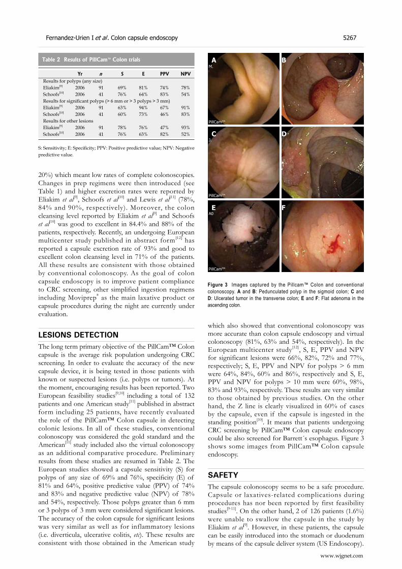
20%) which meant low rates of complete colonoscopies. Changes in prep regimens were then introduced (see Table 1) and higher excretion rates were reported by Eliakim et al[9], Schoofs et al[10] and Lewis et al[11] (78%, 84% and 90%, respectively). Moreover, the colon cleansing level reported by Eliakim et al[9] and Schoofs et al[10] was good to excellent in 84.4% and 88% of the patients, respectively. Recently, an undergoing European multicenter study published in abstract form[12] has reported a capsule excretion rate of 93% and good to excellent colon cleansing level in 71% of the patients. All these results are consistent with those obtained by conventional colonoscopy. As the goal of colon capsule endoscopy is to improve patient compliance to CRC screening, other simplified ingestion regimens including Moviprep® as the main laxative product or capsule procedures during the night are currently under evaluation.
LESIONS DETECTIONThe long term primary objective of the PillCam™ Colon capsule is the average risk population undergoing CRC screening. In order to evaluate the accuracy of the new capsule device, it is being tested in those patients with known or suspected lesions (i.e. polyps or tumors). At the moment, encouraging results has been reported. Two European feasibility studies[9,10] including a total of 132 patients and one American study[11] published in abstract form including 25 patients, have recently evaluated the role of the PillCam™ Colon capsule in detecting colonic lesions. In all of these studies, conventional colonoscopy was considered the gold standard and the American[11] study included also the virtual colonoscopy as an additional comparative procedure. Preliminary results from these studies are resumed in Table 2. The European studies showed a capsule sensitivity (S) for polyps of any size of 69% and 76%, specificity (E) of 81% and 64%, positive predictive value (PPV) of 74% and 83% and negative predictive value (NPV) of 78% and 54%, respectively. Those polyps greater than 6 mm or 3 polyps of 3 mm were considered significant lesions. The accuracy of the colon capsule for significant lesions was very similar as well as for inflammatory lesions (i.e. diverticula, ulcerative colitis, etc). These results are consistent with those obtained in the American study
which also showed that conventional colonoscopy was more accurate than colon capsule endoscopy and virtual colonoscopy (81%, 63% and 54%, respectively). In the European multicenter study[12], S, E, PPV and NPV for significant lesions were 66%, 82%, 72% and 77%, respectively; S, E, PPV and NPV for polyps > 6 mm were 64%, 84%, 60% and 86%, respectively and S, E, PPV and NPV for polyps > 10 mm were 60%, 98%, 83% and 93%, respectively. These results are very similar to those obtained by previous studies. On the other hand, the Z line is clearly visualized in 60% of cases by the capsule, even if the capsule is ingested in the standing position[10]. It means that patients undergoing CRC screening by PillCam™ Colon capsule endoscopy could be also screened for Barrett´s esophagus. Figure 3 shows some images from PillCam™ Colon capsule endoscopy.
SAFETYThe capsule colonoscopy seems to be a safe procedure. Capsule or laxatives-related complications during procedures has nor been reported by first feasibility studies[9-11]. On the other hand, 2 of 126 patients (1.6%) were unable to swallow the capsule in the study by Eliakim et al[9]. However, in these patients, the capsule can be easily introduced into the stomach or duodenum by means of the capsule deliver system (US Endoscopy).
Table 2 Results of PillCam™ Colon trials
Yr n S E PPV NPVResults for polyps (any size)Eliakim[9] 2006 91 69% 81% 74% 78%Schoofs[10] 2006 41 76% 64% 83% 54%Results for significant polyps (> 6 mm or > 3 polyps > 3 mm)Eliakim[9] 2006 91 63% 94% 67% 91%Schoofs[10] 2006 41 60% 73% 46% 83%Results for other lesionsEliakim[9] 2006 91 78% 76% 47% 93%Schoofs[10] 2006 41 76% 63% 82% 52%
S: Sensitivity; E: Specificity; PPV: Positive predictive value; NPV: Negative predictive value.
Fernandez-Urien I et al . Colon capsule endoscopy 5267
www.wjgnet.com
DC
BA
E F
Figure 3 Images captured by the Pillcam™ Colon and conventional colonoscopy. A and B: Pedunculated polyp in the sigmoid colon; C and D: Ulcerated tumor in the transverse colon; E and F: Flat adenoma in the ascending colon.
M.
PillCam™
J.
PillCam™
A0
PillCam™
←
←←←
←
←
←
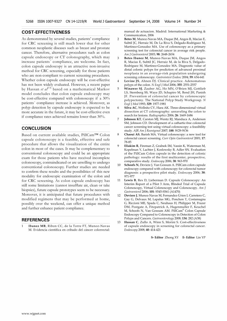
COST-EFFECTIVENESSAs demonstrated by several studies, patients’ compliance for CRC screening is still much lower that for other common neoplastic diseases such as breast and prostate cancer. Therefore, alternative procedures such as colon capsule endoscopy or CT colonography, which may increase patients´ compliance, are welcome. In fact, colon capsule endoscopy is an attractive non-invasive method for CRC screening, especially for those patients who are non-compliant to current screening procedures. Whether colon capsule endoscopy will be cost-effective has not been widely evaluated. However, a recent paper by Hassan et al[13] based on a mathematical Markov model concludes that colon capsule endoscopy may be cost-effective compared with colonoscopy if a 30% patients´ compliance increase is achieved. Moreover, as polyp detection by capsule endoscopy is expected to be more accurate in the future, it may be cost-effective even if compliance rates achieved remains lower than 30%.
CONCLUSIONBased on current available studies, PillCam™ Colon capsule colonoscopy is a feasible, effective and safe procedure that allows the visualization of the entire colon in most of the cases. It may be complementary to conventional colonoscopy and could be an appropriate exam for those patients who have received incomplete colonoscopy, contraindicated or are unwilling to undergo conventional colonoscopy. Further studies are needed to confirm these results and the possibilities of this new modality for endoscopic examination of the colon and for CRC screening. As colon capsule endoscopy has still some limitations (cannot insufflate air, clean or take biopsies), future capsule prototypes seem to be necessary. Moreover, it is anticipated that future procedures with modified regimens that may be performed at home, possibly over the weekend, can offer a unique method and further enhance patient compliance.
REFERENCES1 Ibanez MB, Ribon CC, de la Torre FT, Munoz-Navas
M. Evidencia científica en cribado del cáncer colorrectal:
manual de actuacion. Madrid: International Marketing & Communication, 2006
2 Betes M, Munoz-Navas MA, Duque JM, Angos R, Macias E, Subtil JC, Herraiz M, De La Riva S, Delgado-Rodriguez M, Martinez-Gonzalez MA. Use of colonoscopy as a primary screening test for colorectal cancer in average risk people. Am J Gastroenterol 2003; 98: 2648-2654
3 Betes Ibanez M, Munoz-Navas MA, Duque JM, Angos R, Macias E, Subtil JC, Herraiz M, de la Riva S, Delgado-Rodriguez M, Martinez-Gonzalez MA. Diagnostic value of distal colonic polyps for prediction of advanced proximal neoplasia in an average-risk population undergoing screening colonoscopy. Gastrointest Endosc 2004; 59: 634-641
4 Levine JS, Ahnen DJ. Clinical practice. Adenomatous polyps of the colon. N Engl J Med 2006; 355: 2551-2557
5 Winawer SJ, Zauber AG, Ho MN, O'Brien MJ, Gottlieb LS, Sternberg SS, Waye JD, Schapiro M, Bond JH, Panish JF. Prevention of colorectal cancer by colonoscopic polypectomy. The National Polyp Study Workgroup. N Engl J Med 1993; 329: 1977-1981
6 Silva AC, Wellnitz CV, Hara AK. Three-dimensional virtual dissection at CT colonography: unraveling the colon to search for lesions. Radiographics 2006; 26: 1669-1686
7 Johnson KT, Carston MJ, Wentz RJ, Manduca A, Anderson SM, Johnson CD. Development of a cathartic-free colorectal cancer screening test using virtual colonoscopy: a feasibility study. AJR Am J Roentgenol 2007; 188: W29-W36
8 Chaoui AS, Barish MA. Virtual colonoscopy: a new tool for colorectal cancer screening. Curr Opin Gastroenterol 2001; 17: 78-85
9 Eliakim R, Fireman Z, Gralnek IM, Yassin K, Waterman M, Kopelman Y, Lachter J, Koslowsky B, Adler SN. Evaluation of the PillCam Colon capsule in the detection of colonic pathology: results of the first multicenter, prospective, comparative study. Endoscopy 2006; 38: 963-970
10 Schoofs N, Deviere J, Van Gossum A. PillCam colon capsule endoscopy compared with colonoscopy for colorectal tumor diagnosis: a prospective pilot study. Endoscopy 2006; 38: 971-977
11 Lewis B, Rex D, Lieberman D. Capsule Colonoscopy: An Interim Report of a Pilot 3 Arm, Blinded Trial of Capsule Colonoscopy, Virtual Colonoscopy and Colonoscopy. Am J Gastroenterol 2006; 101: S545-S561 (A1470)
12 Deviere J, Munoz-Navas M, Fernandez-Urien I, Carretero C, Gay G, Delvaux M, Lapalus MG, Ponchon T, Costamagna G, Riccioni ME, Spada C, Neuhaus H, Philipper M, Frazer DM, Postgate A, Fitzpatrick A, Hagenmuller F, Keuchel M, Schoofs N, Van Gossum AM. PillCam® Colon Capsule Endoscopy Compared to Colonoscopy in Detection of Colon Polyps and Cancers. Gastroenterology 2008; 134: 282 (A38)
13 Hassan C, Zullo A, Winn S, Morini S. Cost-effectiveness of capsule endoscopy in screening for colorectal cancer. Endoscopy 2008; 40: 414-421
S- Editor Zhong XY E- Editor Lin YP
www.wjgnet.com
5268 ISSN 1007-9327 CN 14-1219/R World J Gastroenterol September 14, 2008 Volume 14 Number 34
Ángel Caunedo-Álvarez, Javier Romero-Vazquez, Juan M Herrerias-Gutierrez, Gastroenterology Service, “Virgen Macarena” University Hospital, Av Dr. Fedriani, Seville 41071, SpainCorrespondence to: Ángel Caunedo Álvarez, MD, Gastroenterology Service, “Virgen Macarena” University Hospital, Avda. Dr. Fedriani, Seville 41071, Spain. [email protected]: +34-955-008801 Fax: +34-955-008805Received: February 15, 2008 Revised: July 26, 2008Accepted: August 2, 2008Published online: September 14, 2008
AbstractSmall bowel strictures can be missed by current diagnostic methods. The Patency capsule is a new non-endoscopic dissolvable capsule which has as an objective of checking the patency of digestive tract, in a non-invasive manner. The available clinical trials have demonstrated that the Patency© capsule is a good tool for assessment of the functional patency of the small bowel, and it allows identification of those patients who can safely undergo a capsule endoscopy, despite clinical and radiographic evidence of small-bowel obstruction. Some cases of intestinal occlusion have been reported with the Patency© capsule, four of them needed surgery. So, a new capsule with two timer plugs (Agile© capsule) has been recently developed in order to minimize the risk of occlusion. This new device stars its dissolution process earlier (30 h after ingestion) and its two timer plugs have been designed to begin the disintegration even when the device is blocked in a tight stricture.
© 2008 The WJG Press. All rights reserved.
Key words: Capsule endoscopy; Patency capsule; Agile capsule; Small bowel strictures
Peer reviewer: Amado S Peña, Professor, Department of Pathology, Immunogenetics, VU University Medical Centre, De Boelelaan 1117, PO Box 7057, Amsterdam 1007 MB, The Netherlands
Caunedo-Álvarez Á, Romero-Vazquez J, Herrerias-Gutierrez JM. Patency© and agile© capsules. World J Gastroenterol 2008; 14(34): 5269-5273 Available from: URL: http://www.wjgnet.com/1007-9327/14/5269.asp DOI: http://dx.doi.org/10.3748/wjg.14.5269
TOPIC HIGHLIGHT
Patency© and agile© capsules
Ángel Caunedo-Álvarez, Javier Romero-Vazquez, Juan M Herrerias-Gutierrez
Miguel Angel Muñoz-Navas, Profesor, Series Editors
Online Submissions: wjg.wjgnet.com World J Gastroenterol 2008 September 14; 14(34): [email protected] World Journal of Gastroenterology ISSN 1007-9327doi:10.3748/wjg.14.5269 © 2008 The WJG Press. All rights reserved.
INTRODUCTION AND DEVICE DESCRIPTIONSince Iddan et al[1] reported the new wireless endoscopy system the capsule endoscopy (CE) has become one of the most significant technical innovations of Gastroenterology in recent years. Regarding the complications of the techniques, some incidental cases of impaction in a Meckel´s diverticulum[2], a Zenker´s diverticulum[3], or in the cricopharyngeal muscle[4], as well as aspiration of the capsule[5-7], solved without complications, have been reported. However, the most frequent side effect is undoubtedly the non-natural excretion (NNE) of the capsule due to a stricture or a tumor in the small bowel. The retrospective analysis of some series indicates that the incidence of NNE depends on the indication for the capsule exam: 0% in healthy controls[8], 1.4% in obscure gastrointestinal bleeding[9-13], 1.48% in suspected Crohn´s disease[14-17], 5%-13% in known Crohn´s disease[17-19] and 21% in suspected small bowel obstruction[20].
In the vast majority of cases, capsule retention, regardless of its cause, is asymptomatic, being evidenced by the absence of excretion and confirmed by means of radiology. On the other hand, capsule retention frequently allows for the identification of the bowel pathology which caused the symptoms in the patient and which could not have been diagnosed by means of standard methods, furthermore facilitating the location of the stricture or the tumor for the surgeon after “milking” the capsule through the bowel. For these reasons, many authors have described NNE as a “therapeutic complication” of the CE[21-23]. However, although this consideration is valid for patients with tumors or those with stricture due to NSAIDs, radiotherapy or previous abdominal surgery, it may not be valid in patients with stenoses secondary to Crohn’s disease. Unlike the previously mentioned groups, in patients with Crohn’s disease, surgery is not curative and may present more complications, thus in this case, NNE of the capsule can become more an “undesirable circumstance” than a “therapeutic complication”. The same can be said of those patients showing high surgical risk or who would not be willing to undergo surgery in the event that NNE occurs.
Current imaging techniques can show long or medium stenoses, with great reduction of the lumen size; however, short stenoses usually cannot be detected by standard methods. This fact explains that in most
www.wjgnet.com
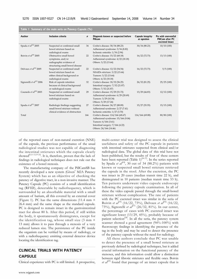
of the reported cases of non-natural excretion (NNE) of the capsule, the previous performance of the usual radiological studies was not capable of diagnosing the intestinal strictures which the capsule clearly showed[9,11-13,19-21]. It is, therefore, proven that the lack of findings in radiological techniques does not rule out the existence of a bowel stenosis.
The manufacturing company of the PillCamSB has recently developed a new system (Given® M2A Patency System) which has as an objective of checking the patency of digestive tract, in a non-invasive manner. The Patency Capsule (PC) consists of a small identification tag (RFID), detectable by radiofrequency, which is surrounded by an absorbable material with a small amount of barium, all this covered by an external cover (Figure 1). PC has the same dimensions (11.4 mm × 26.4 mm) and the same shape as the standard capsule. PC is designed to remain intact in the gastrointestinal tract for about 80 h. After this period, if still within the body, it spontaneously disintegrates, except for the identification tag, whose small size (3 mm × 13 mm) allows it to pass through a stenosis of a very reduced lumen size. The persistence of the PC inside the organism can be verified by means of radiology, or with a radiofrequency emitting external detector device locating the identification tag.
CLINICAL TRIALS WITH PATENCY CAPSULEClinical experience with PC is still limited. A prospective,
multi-center trial was designed to assess the clinical usefulness and safety of the PC capsule in patients with intestinal strictures suspected from clinical and/or radiological data. The global data of this trial have not been published, but the results of four of these centers have been reported (Table 1)[24-27]. In the series reported by Spada et al[24], 30 out of 34 (88.2%) patients with known or suspected small bowel stricture retrieved the capsule in the stool. After the excretion, the PC was intact in 20 cases (median transit time 22 h), and disintegrated in 10 patients (median transit time 53 h). Ten patients underwent video capsule endoscopy following the patency capsule examination. In all of these the video capsule passed through the small-bowel stricture without complication. The rate of patients with the PC excreted intact was similar in the seria of Boivin et al[25] (16/22, 73%), Delvaux et al[26] (16/22, 73%), Signorelli et al[27] (26/32, 81%). In our center, the percentage of cases with PC excreted intact was significant lower (13/29, 45%), probably because of patient selection[28]. In all the seria, the patency system scanner showed a good agreement (94%-100%) with fluoroscopy findings in identifying the presence of the tag in the body and may be used to detect the presence of the patency capsule without the need for radiology.
All these authors conclude that PC was unable to detect the presence of a small bowel stricture as previously defined by radiological techniques, but it added crucial information on the functional patency of the stenoses, and this information could allow a distinction between rigid fibrotic strictures and flexible ones. Boivin et al[25] found that passage of an intact capsule that is
Table 1 Summary of the main seria on Patency Capsule (%)
Author Inclusion criteria n Diagnosis known or suspected before Pillcam
Capsule integrity at egestion
Pts with uneventful PillCam after PC excreted intact
Spada et al[24] 2005 Suspected or confirmed small bowel stricture based on radiological exams
34 Crohn's disease: 30/34 (88.23)Adhesional syndrome: 3/34 (8.82)Ischemic enteritis: 1/34 (2.94)
30/34 (88.23) 10/10 (100)
Boivin et al[25] 2005 Obstructive small bowel symptoms, and/or radiographic evidence of structuring small bowel disease
22 Crohn's disease: 15/22 (68.18)Adhesional syndrome: 4/22 (18.18)Others: 3/22 (13.64)
16/22 (72.73) 13/13 (100)
Delvaux et al[26] 2005 Suspected or confirmed small bowel stricture based on either clinical background or radiological exams
22 Crohn's disease: 12/22 (54.54)NSAIDs stricture: 3/22 (13.64)Tumors: 3/22 (13.64)Others: 4/22 (18.18)
16/22 (72.73) 5/5 (100)
Signorelli et al[27] 2006 Risk of capsule retention because of clinical background or radiological exams
32 Crohn's disease: 18/32 (56.25)Intestinal surgery: 7/32 (21.87)Others: 7/32 (21.87)
26/32 (81.25) 25/25 (100)
Caunedo et al[28] 2003 Suspected or confirmed small bowel stricture based on radiological exams
29 Crohn's disease: 15/29 (51.72)Adhesional syndrome: 6/29 (20.69)Tumors: 3/29 (10.34)Others: 5/29 (17.24)
13/29 (44.83) 12/12 (100)
Spada et al[31] 2007 Radiologic findings suggesting small bowel stricture without clinical evidence of obstruction
27 Crohn's disease: 24/27 (88.89)Adhesional syndrome: 2/27 (7.41)Ischemic enteritis: 1/27 (3.70)
15/27 (55.55) 15/15 (100)
Total 166 Crohn's disease: 114/166 (68.67)Adhesional syndrome: 15/166 (9.04)Tumors: 6/166 (3.61)Intestinal surgery: 7/166 (4.22)Others: 24/166 (14.46)
116/166 (69.88) 80/80 (100)
www.wjgnet.com
5270 ISSN 1007-9327 CN 14-1219/R World J Gastroenterol September 14, 2008 Volume 14 Number 34
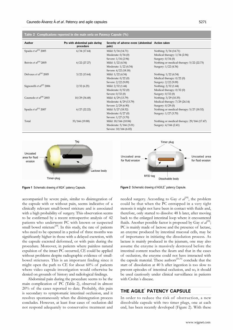
accompanied by severe pain, similar to disintegration of the capsule with or without pain, seems indicative of a clinically relevant small-bowel stricture and is associated with a high probability of surgery. This observation seems to be confirmed by a recent retrospective analysis of 42 patients who underwent PC with known or suspected small bowel stricture[29]. In this study, the rate of patients who need to be operated in a period of three months was significantly higher in those with a delayed excretion, with the capsule excreted deformed, or with pain during the procedure. Moreover, in patients where painless natural expulsion of the intact PC occurred, CE could be applied without problems despite radiographic evidence of small-bowel strictures. This is an important finding since it might open the path to CE for about 60% of patients where video capsule investigation would otherwise be denied on grounds of history and radiological findings.
Abdominal pain during the procedure seems to be the main complication of PC (Table 2), observed in almost 20% of the cases reported to date. Probably, this pain is secondary to symptomatic intestinal occlusion, and it resolves spontaneously when the disintegration process concludes. However, at least four cases of occlusion did not respond adequately to conservative treatment and
needed surgery. According to Gay et al[30], the problem could be that when the PC entrapped in a very tight stenosis it might not have been in contact with fluids and, therefore, only started to dissolve 48 h later, after moving back to the enlarged intestinal loop where it encountered fluids. Another possible factor is proposed by Gay et al[30]; PC is mainly made of lactose and the presence of lactase, an enzyme produced by intestinal mucosal cells, may be of importance in initiating the dissolution process. As lactase is mainly produced in the jejunum, one may also assume the enzyme is massively destroyed before the intestinal content reaches the ileum and that in the cases of occlusion, the enzyme could not have interacted with the capsule material. These authors[26,31] conclude that the start of dissolution at 40 h after ingestion is too slow to prevent episodes of intestinal occlusion, and so, it should be used cautiously under clinical surveillance in patients with Crohn´s disease.
THE AGILE© PATENCY CAPSULEIn order to reduce the risk of obstruction, a new dissolvable capsule with two timer plugs, one at each end, has been recently developed (Figure 2). With these
Table 2 Complications reported in the main seria on Patency Capsule (%)
Author Pts with abdominal pain during procedure
Severity of adverse event (abdominal pain)
Action taken
Spada et al[24] 2005 6/34 (17.64) Mild: 5/34 (14.71)Moderate: 0/34 (0)Severe: 1/34 (2.94)
Nothing: 5/34 (14.71)Medical therapy: 1/34 (2.94)Surgery: 0/34 (0)
Boivin et al[25] 2005 6/22 (27.27) Mild: 1/22 (4.54)Moderate: 1/22 (4.54)Severe: 4/22 (18.18)
Nothing or medical therapy: 5/22 (22.73)Surgery: 1/22 (4.54)
Delvaux et al[26] 2005 3/22 (13.64) Mild: 1/22 (4.54)Moderate: 0/22 (0)Severe: 2/22 (9.09)
Nothing: 1/22 (4.54)Medical therapy: 0/22 (0)Surgery: 2/22 (9.09)
Signorelli et al[27] 2006 2/32 (6.25) Mild: 2/32 (1.44)Moderate: 0/32 (0)Severe: 0/32 (0)
Nothing: 2/32 (1.44)Medical therapy: 0/32 (0)Surgery: 0/32 (0)
Caunedo et al[28] 2003 10/29 (34.48) Mild: 4/29 (13.79)Moderate: 4/29 (13.79)Severe: 2/29 (6.90)
Nothing: 3/29 (10.35)Medical therapy: 7/29 (24.14)Surgery: 0/29 (0)
Spada et al[31] 2007 6/27 (22.22) Mild: 5/27 (18.52)Moderate: 0/27 (0)Severe: 1/27 (3.70)
Nothing or medical therapy: 5/27 (18.52)Surgery: 1/27 (3.70)
Total 33/166 (19.88) Mild: 18/166 (10.84)Moderate: 5/166 (3.01)Severe: 10/166 (6.02)
Nothing or medical therapy: 29/166 (17.47)Surgery: 4/166 (2.41)
Timer plug
Uncoated area for fluid erosion
Uncoated area for fluid erosion
Dissolvable bodyRFID tag
Figure 2 Schematic drawing of AGILE® patency Capsule.
Uncoated area for fluid
erosion
Tag
Lactose body
Timer-plug
Figure 1 Schematic drawing of M2A® patency Capsule.
Caunedo-Álvarez Á et al . Patency and agile capsules 5271
www.wjgnet.com
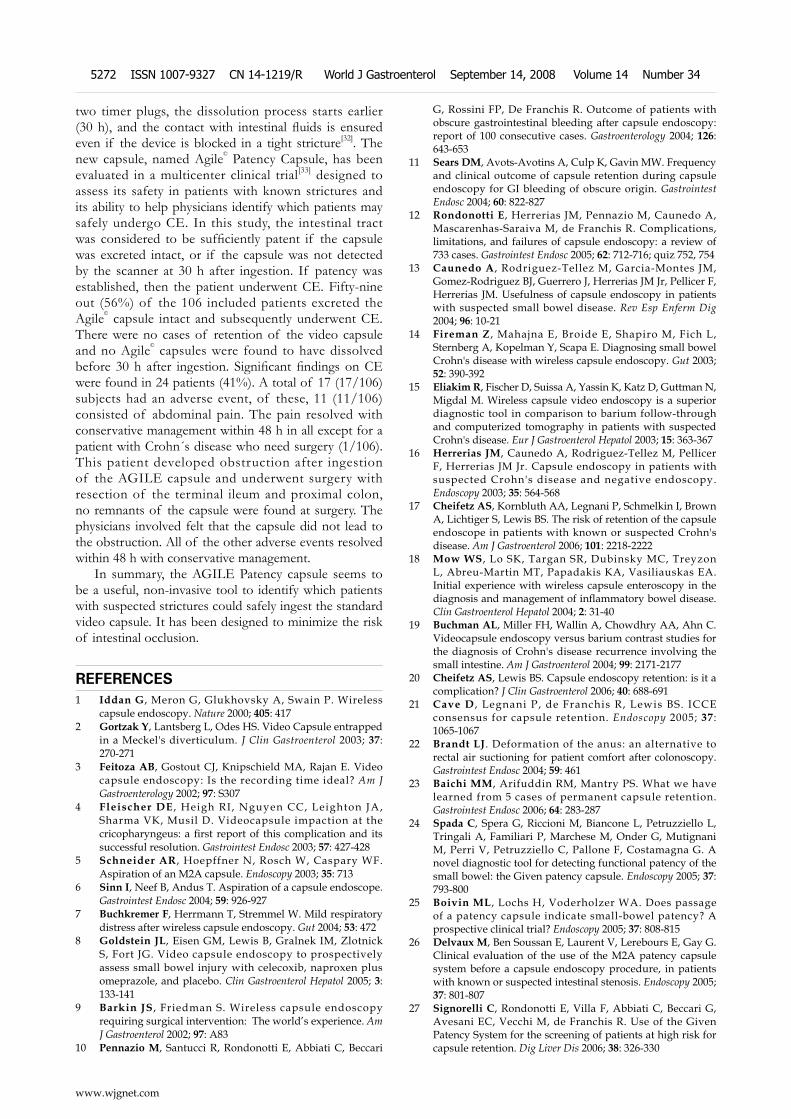
two timer plugs, the dissolution process starts earlier (30 h), and the contact with intestinal fluids is ensured even if the device is blocked in a tight stricture[32]. The new capsule, named Agile© Patency Capsule, has been evaluated in a multicenter clinical trial[33] designed to assess its safety in patients with known strictures and its ability to help physicians identify which patients may safely undergo CE. In this study, the intestinal tract was considered to be sufficiently patent if the capsule was excreted intact, or if the capsule was not detected by the scanner at 30 h after ingestion. If patency was established, then the patient underwent CE. Fifty-nine out (56%) of the 106 included patients excreted the Agile© capsule intact and subsequently underwent CE. There were no cases of retention of the video capsule and no Agile© capsules were found to have dissolved before 30 h after ingestion. Significant findings on CE were found in 24 patients (41%). A total of 17 (17/106) subjects had an adverse event, of these, 11 (11/106) consisted of abdominal pain. The pain resolved with conservative management within 48 h in all except for a patient with Crohn´s disease who need surgery (1/106). This patient developed obstruction after ingestion of the AGILE capsule and underwent surgery with resection of the terminal ileum and proximal colon, no remnants of the capsule were found at surgery. The physicians involved felt that the capsule did not lead to the obstruction. All of the other adverse events resolved within 48 h with conservative management.
In summary, the AGILE Patency capsule seems to be a useful, non-invasive tool to identify which patients with suspected strictures could safely ingest the standard video capsule. It has been designed to minimize the risk of intestinal occlusion.
REFERENCES1 Iddan G, Meron G, Glukhovsky A, Swain P. Wireless
capsule endoscopy. Nature 2000; 405: 4172 Gortzak Y, Lantsberg L, Odes HS. Video Capsule entrapped
in a Meckel's diverticulum. J Clin Gastroenterol 2003; 37: 270-271
3 Feitoza AB, Gostout CJ, Knipschield MA, Rajan E. Video capsule endoscopy: Is the recording time ideal? Am J Gastroenterology 2002; 97: S307
4 Fleischer DE , Heigh RI, Nguyen CC, Leighton JA, Sharma VK, Musil D. Videocapsule impaction at the cricopharyngeus: a first report of this complication and its successful resolution. Gastrointest Endosc 2003; 57: 427-428
5 Schneider AR, Hoepffner N, Rosch W, Caspary WF. Aspiration of an M2A capsule. Endoscopy 2003; 35: 713
6 Sinn I, Neef B, Andus T. Aspiration of a capsule endoscope. Gastrointest Endosc 2004; 59: 926-927
7 Buchkremer F, Herrmann T, Stremmel W. Mild respiratory distress after wireless capsule endoscopy. Gut 2004; 53: 472
8 Goldstein JL, Eisen GM, Lewis B, Gralnek IM, Zlotnick S, Fort JG. Video capsule endoscopy to prospectively assess small bowel injury with celecoxib, naproxen plus omeprazole, and placebo. Clin Gastroenterol Hepatol 2005; 3: 133-141
9 Barkin JS , Friedman S. Wireless capsule endoscopy requiring surgical intervention: The world’s experience. Am J Gastroenterol 2002; 97: A83
10 Pennazio M, Santucci R, Rondonotti E, Abbiati C, Beccari
G, Rossini FP, De Franchis R. Outcome of patients with obscure gastrointestinal bleeding after capsule endoscopy: report of 100 consecutive cases. Gastroenterology 2004; 126: 643-653
11 Sears DM, Avots-Avotins A, Culp K, Gavin MW. Frequency and clinical outcome of capsule retention during capsule endoscopy for GI bleeding of obscure origin. Gastrointest Endosc 2004; 60: 822-827
12 Rondonotti E, Herrerias JM, Pennazio M, Caunedo A, Mascarenhas-Saraiva M, de Franchis R. Complications, limitations, and failures of capsule endoscopy: a review of 733 cases. Gastrointest Endosc 2005; 62: 712-716; quiz 752, 754
13 Caunedo A, Rodriguez-Tellez M, Garcia-Montes JM, Gomez-Rodriguez BJ, Guerrero J, Herrerias JM Jr, Pellicer F, Herrerias JM. Usefulness of capsule endoscopy in patients with suspected small bowel disease. Rev Esp Enferm Dig 2004; 96: 10-21
14 Fireman Z , Mahajna E, Broide E, Shapiro M, Fich L, Sternberg A, Kopelman Y, Scapa E. Diagnosing small bowel Crohn's disease with wireless capsule endoscopy. Gut 2003; 52: 390-392
15 Eliakim R, Fischer D, Suissa A, Yassin K, Katz D, Guttman N, Migdal M. Wireless capsule video endoscopy is a superior diagnostic tool in comparison to barium follow-through and computerized tomography in patients with suspected Crohn's disease. Eur J Gastroenterol Hepatol 2003; 15: 363-367
16 Herrerias JM, Caunedo A, Rodriguez-Tellez M, Pellicer F, Herrerias JM Jr. Capsule endoscopy in patients with suspected Crohn's disease and negative endoscopy. Endoscopy 2003; 35: 564-568
17 Cheifetz AS, Kornbluth AA, Legnani P, Schmelkin I, Brown A, Lichtiger S, Lewis BS. The risk of retention of the capsule endoscope in patients with known or suspected Crohn's disease. Am J Gastroenterol 2006; 101: 2218-2222
18 Mow WS, Lo SK, Targan SR, Dubinsky MC, Treyzon L, Abreu-Martin MT, Papadakis KA, Vasiliauskas EA. Initial experience with wireless capsule enteroscopy in the diagnosis and management of inflammatory bowel disease. Clin Gastroenterol Hepatol 2004; 2: 31-40
19 Buchman AL, Miller FH, Wallin A, Chowdhry AA, Ahn C. Videocapsule endoscopy versus barium contrast studies for the diagnosis of Crohn's disease recurrence involving the small intestine. Am J Gastroenterol 2004; 99: 2171-2177
20 Cheifetz AS, Lewis BS. Capsule endoscopy retention: is it a complication? J Clin Gastroenterol 2006; 40: 688-691
21 Cave D , Legnani P, de Franchis R, Lewis BS. ICCE consensus for capsule retention. Endoscopy 2005; 37 : 1065-1067
22 Brandt LJ. Deformation of the anus: an alternative to rectal air suctioning for patient comfort after colonoscopy. Gastrointest Endosc 2004; 59: 461
23 Baichi MM, Arifuddin RM, Mantry PS. What we have learned from 5 cases of permanent capsule retention. Gastrointest Endosc 2006; 64: 283-287
24 Spada C, Spera G, Riccioni M, Biancone L, Petruzziello L, Tringali A, Familiari P, Marchese M, Onder G, Mutignani M, Perri V, Petruzziello C, Pallone F, Costamagna G. A novel diagnostic tool for detecting functional patency of the small bowel: the Given patency capsule. Endoscopy 2005; 37: 793-800
25 Boivin ML, Lochs H, Voderholzer WA. Does passage of a patency capsule indicate small-bowel patency? A prospective clinical trial? Endoscopy 2005; 37: 808-815
26 Delvaux M, Ben Soussan E, Laurent V, Lerebours E, Gay G. Clinical evaluation of the use of the M2A patency capsule system before a capsule endoscopy procedure, in patients with known or suspected intestinal stenosis. Endoscopy 2005; 37: 801-807
27 Signorelli C, Rondonotti E, Villa F, Abbiati C, Beccari G, Avesani EC, Vecchi M, de Franchis R. Use of the Given Patency System for the screening of patients at high risk for capsule retention. Dig Liver Dis 2006; 38: 326-330
www.wjgnet.com
5272 ISSN 1007-9327 CN 14-1219/R World J Gastroenterol September 14, 2008 Volume 14 Number 34
28 Caunedo A, Rodriguez-Tellez M, Romero J, Hernandez-Duran M, Romero R, Pellicer FJ, Herrerias JM. Evaluation of the M2A Patency capsule in the gastrointestinal tract: one-centre preliminary data from a multicentre prospective trial. Endoscopy 2003; 35: A182
29 Caunedo-Alvarez A, Romero-Vázquez J, Gomez-Rodriguez BJ, Sanchez-Yague A, Castro-Laria L, Herrerias-Gutierrez JM. Prognostic Factors of Short-Term Surgery in Patients with Known or Suspected Stricture Undergone to Patency Capsule. Gastrointes Endoscopy 2007; 65: AB340
30 Gay G , Delvaux M, Laurent V, Reibel N, Regent D, Grosdidier G, Roche JF. Temporary intestinal occlusion induced by a "patency capsule" in a patient with Crohn's disease. Endoscopy 2005; 37: 174-177
31 Spada C, Shah SK, Riccioni ME, Spera G, Marchese M, Iacopini F, Familiari P, Costamagna G. Video capsule
endoscopy in patients with known or suspected small bowel stricture previously tested with the dissolving patency capsule. J Clin Gastroenterol 2007; 41: 576-582
32 Caunedo-Alvarez A, Romero-Vazquez J, Gomez-Rodriguez BJ, Sanchez-Yague A, Castro-Laria L, Herrerias-Gutierrez JM. Evaluation of a new double-headed biodegradable device (AGILE Patency Capsule) for detecting functional patency of the small intestine: A prospective clinical trial. Proceedings of the 5th International Conference on Capsule. Endoscopy 2006
33 Herrerias JM, Leighton JA, Costamagna G, Infantolino A, Eliakim R, Fischer D, Rubin DT, Manten HD, Scapa E, Morgan DR, Bergwerk AJ, Koslowsky B, Adler SN. Agile patency system eliminates risk of capsule retention in patients with known intestinal strictures who undergo capsule endoscopy. Gastrointest Endosc 2008; 67: 902-909
S- Editor Zhong XY L- Editor Rippe RA E- Editor Lin YP
Caunedo-Álvarez Á et al . Patency and agile capsules 5273
www.wjgnet.com
Yan-Hui Ma, Wei-Zhi Cheng, Fang Gong, An-Lun Ma, Qi-Wen Yu, Ji-Ying Zhang, Chao-Ying Hu, Xue-Hua Chen, Dong-Qing Zhang
Online Submissions: wjg.wjgnet.com World J Gastroenterol 2008 September 14; 14(34): [email protected] World Journal of Gastroenterology ISSN 1007-9327doi:10.3748/wjg.14.5274 © 2008 The WJG Press. All rights reserved.
Active chinese mistletoe lectin-55 enhances colon cancer surveillance through regulating innate and adaptive immune responses
COLORECTAL CANCER
www.wjgnet.com
Yan-Hui Ma, Wei-Zhi Cheng, Fang Gong, An-Lun Ma, Qi-Wen Yu, Ji-Ying Zhang, Chao-Ying Hu, Shanghai Jiao Tong University School of Medicine, Shanghai Institute of Immunology, Shanghai 200025, ChinaXue-Hua Chen, Shanghai Institute of Digestive Surgery, Ruijin Hospital, Shanghai 200025, ChinaAn-Lun Ma, Centre de recherche du CHUM, Hôpital Notre-Dame, Université de Montréal, Montréal, Québec H2L 4M1, CanadaAuthor contributions: Ma YH and Cheng WZ contributed equally to this work; Zhang DQ, Ma YH and Cheng WZ designed research; Ma YH, Cheng WZ, Gong F, Ma AL, Yu QW, Zhang JY, Hu CY and Chen XH performed research; Chen XH contributed new reagents/analytic tools; Ma YH, Cheng WZ and Zhang DQ wrote the paper.Supported by The National Natural Science Foundation of China, No. 30471593 and No. 30670939; the Shanghai Leading Academic Discipline Project, No. T0206; the Shanghai Commission of Science and Technology, No. 07JC14033; the Shanghai Institute of Immunology Project, No. 07-A02Correspondence to: Dong-Qing Zhang, Shanghai Institute of Immunology,Shanghai Jiao Tong University School of Medicine, Shanghai 200025, China. [email protected]: +86-21-64453049 Fax: +86-21-64453049Received: May 26, 2008 Revised: July 17, 2008Accepted: July 24, 2008Published online: September 14, 2008
AbstractAIM: To investigate the potential role of Active Chinese mistletoe lectin-55 (ACML-55) in tumor immune surveillance. METHODS: In this study, an experimental model was established by hypodermic inoculating the colon cancer cell line CT26 (5 × 10⁵ cells) into BALB/c mice. The experimental treatment was orally administered with ACML-55 or PBS, followed by the inoculation of colon cancer cell line CT26. Intracellular cytokine staining was used to detect IFN-γ production by tumor antigen specific CD8+ T cells. FACS analysis was employed to profile composition and activation of CD4+, CD8+, γδ T and NK cells.RESULTS: Our results showed, compared to PBS treated mice, ACML-55 treatment significantly delayed colon cancer development in colon cancer -bearing
Balb/c mice in vivo . Treatment with ACML-55 enhanced both Ag specific activation and proliferation of CD4+ and CD8+ T cells, and increased the number of tumor Ag specific CD8+ T cells. It was more important to increase the frequency of tumor Ag specific IFN-γ producing-CD8+ T cells. Interestingly, ACML-55 treatment also showed increased cell number of NK, and γδT cells, indicating the role of ACML-55 in activation of innate lymphocytes. CONCLUSION: Our results demonstrate that ACML-55 therapy can enhance function in immune surveillance in colon cancer-bearing mice through regulating both innate and adaptive immune responses.
© 2008 The WJG Press. All rights reserved.
Key words: Active chinese mistletoe lectin-55; Colon cancer; Immune surveillance; Tumor therapy; Ag-specific-CD8+ T cell
Peer reviewer: Yoshiharu Motoo, MD, PhD, FACP, FACG,Professor and Chairman, Department of Medical Oncology, Kanazawa Medical University,1-1 Daigaku, Uchinada, Ishikawa 920-0293, Japan
Ma YH, Cheng WZ, Gong F, Ma AL, Yu QW, Zhang JY, Hu CY, Chen XH, Zhang DQ. Active chinese mistletoe lectin-55 enhances colon cancer surveillance through regulating innate and adaptive immune responses. World J Gastroenterol 2008; 14(34): 5274-5281 Available from: URL: http://www.wjgnet.com/1007-9327/14/5274.asp DOI: http://dx.doi.org/10.3748/wjg.14.5274
INTRODUCTIONMistletoe (Viscum album) is a semiparasitic plant with many unusual properties. It was used as a kind of herbal remedy in the ancient Chinese Pharmacopoeia and has been used in traditional Chinese medicine for diseases, such as gonorrhea, syphilis, hypertension and rheumatism for thousands of years. The aqueous extract of European Mistletoe (EM) has been used in conventional cancer therapy for decades[1]. Therapeutic efficacy is mostly attributed to the mistletoe lectins
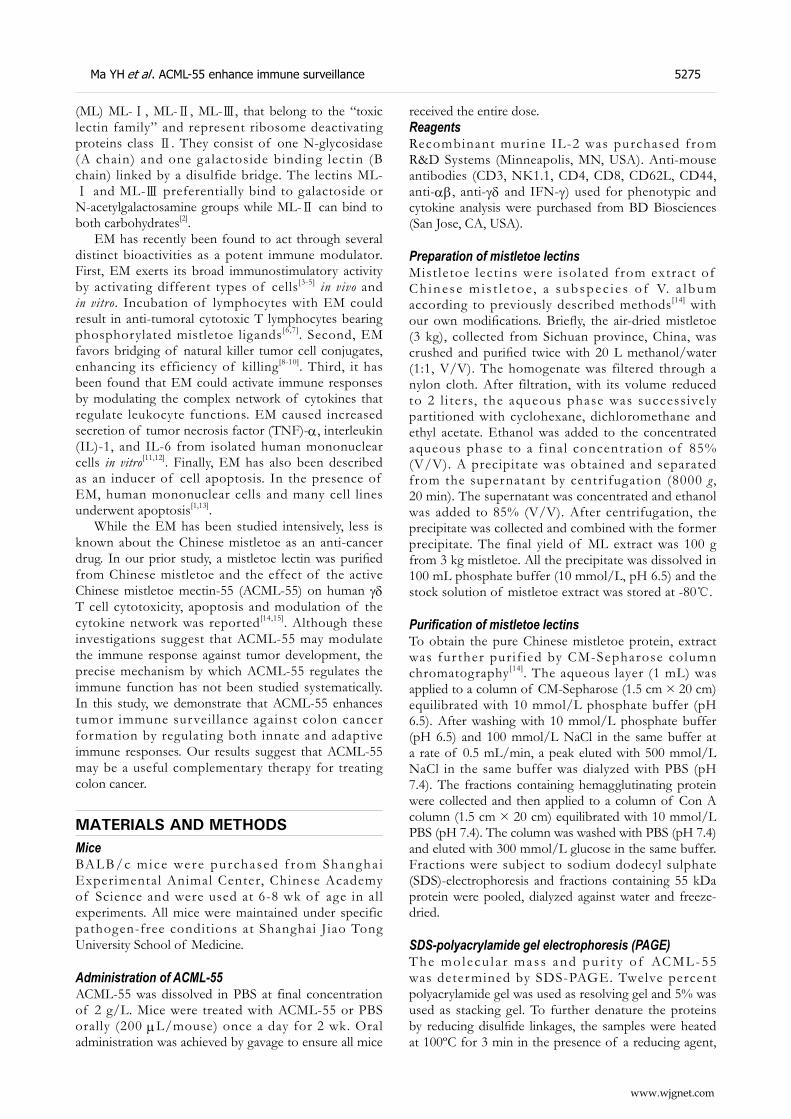
www.wjgnet.com
Ma YH et al . ACML-55 enhance immune surveillance 5275
(ML) ML-Ⅰ, ML-Ⅱ, ML-Ⅲ, that belong to the “toxic lectin family” and represent ribosome deactivating proteins class Ⅱ. They consist of one N-glycosidase (A chain) and one galactoside binding lectin (B chain) linked by a disulfide bridge. The lectins ML-Ⅰ and ML-Ⅲ preferentially bind to galactoside or N-acetylgalactosamine groups while ML-Ⅱ can bind to both carbohydrates[2].
EM has recently been found to act through several distinct bioactivities as a potent immune modulator. First, EM exerts its broad immunostimulatory activity by activating different types of cells[3-5] in vivo and in vitro. Incubation of lymphocytes with EM could result in anti-tumoral cytotoxic T lymphocytes bearing phosphorylated mistletoe ligands[6,7]. Second, EM favors bridging of natural killer tumor cell conjugates, enhancing its efficiency of killing[8-10]. Third, it has been found that EM could activate immune responses by modulating the complex network of cytokines that regulate leukocyte functions. EM caused increased secretion of tumor necrosis factor (TNF)-α, interleukin (IL)-1, and IL-6 from isolated human mononuclear cells in vitro[11,12]. Finally, EM has also been described as an inducer of cell apoptosis. In the presence of EM, human mononuclear cells and many cell lines underwent apoptosis[1,13].
While the EM has been studied intensively, less is known about the Chinese mistletoe as an anti-cancer drug. In our prior study, a mistletoe lectin was purified from Chinese mistletoe and the effect of the active Chinese mistletoe mectin-55 (ACML-55) on human γδ T cell cytotoxicity, apoptosis and modulation of the cytokine network was reported[14,15]. Although these investigations suggest that ACML-55 may modulate the immune response against tumor development, the precise mechanism by which ACML-55 regulates the immune function has not been studied systematically. In this study, we demonstrate that ACML-55 enhances tumor immune surveillance against colon cancer formation by regulating both innate and adaptive immune responses. Our results suggest that ACML-55 may be a useful complementary therapy for treating colon cancer.
MATERIALS AND METHODSMiceBALB/c mice were purchased f rom Shangha i Experimental Animal Center, Chinese Academy of Science and were used at 6-8 wk of age in all experiments. All mice were maintained under specific pathogen-free conditions at Shanghai Jiao Tong University School of Medicine.
Administration of ACML-55ACML-55 was dissolved in PBS at final concentration of 2 g/L. Mice were treated with ACML-55 or PBS orally (200 μL/mouse) once a day for 2 wk. Oral administration was achieved by gavage to ensure all mice
received the entire dose.ReagentsRecombinant murine IL-2 was purchased from R&D Systems (Minneapolis, MN, USA). Anti-mouse antibodies (CD3, NK1.1, CD4, CD8, CD62L, CD44, anti-αβ, anti-γδ and IFN-γ) used for phenotypic and cytokine analysis were purchased from BD Biosciences (San Jose, CA, USA).
Preparation of mistletoe lectinsMist letoe lect ins were isolated from extract of Ch ine se m i s t l e toe , a subspec i e s o f V. a l bum according to previously described methods[14] with our own modifications. Briefly, the air-dried mistletoe (3 kg), collected from Sichuan province, China, was crushed and purified twice with 20 L methanol/water (1:1, V/V). The homogenate was filtered through a nylon cloth. After filtration, with its volume reduced to 2 l i ters, the aqueous phase was success ively partitioned with cyclohexane, dichloromethane and ethyl acetate. Ethanol was added to the concentrated aqueous phase to a f inal concentrat ion of 85% (V/V). A precipitate was obtained and separated from the supernatant by centrifugation (8000 g , 20 min). The supernatant was concentrated and ethanol was added to 85% (V/V). After centrifugation, the precipitate was collected and combined with the former precipitate. The final yield of ML extract was 100 g from 3 kg mistletoe. All the precipitate was dissolved in 100 mL phosphate buffer (10 mmol/L, pH 6.5) and the stock solution of mistletoe extract was stored at -80.
Purification of mistletoe lectinsTo obtain the pure Chinese mistletoe protein, extract was fur ther pur if ied by CM-Sepharose column chromatography[14]. The aqueous layer (1 mL) was applied to a column of CM-Sepharose (1.5 cm × 20 cm) equilibrated with 10 mmol/L phosphate buffer (pH 6.5). After washing with 10 mmol/L phosphate buffer (pH 6.5) and 100 mmol/L NaCl in the same buffer at a rate of 0.5 mL/min, a peak eluted with 500 mmol/L NaCl in the same buffer was dialyzed with PBS (pH 7.4). The fractions containing hemagglutinating protein were collected and then applied to a column of Con A column (1.5 cm × 20 cm) equilibrated with 10 mmol/L PBS (pH 7.4). The column was washed with PBS (pH 7.4) and eluted with 300 mmol/L glucose in the same buffer. Fractions were subject to sodium dodecyl sulphate (SDS)-electrophoresis and fractions containing 55 kDa protein were pooled, dialyzed against water and freeze-dried.
SDS-polyacrylamide gel electrophoresis (PAGE)T he mo lecu l a r mas s and pur i t y o f ACML-55 was determined by SDS-PAGE. Twelve percent polyacrylamide gel was used as resolving gel and 5% was used as stacking gel. To further denature the proteins by reducing disulfide linkages, the samples were heated at 100ºC for 3 min in the presence of a reducing agent,
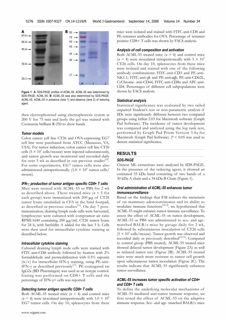
then electrophoresed using electrophoresis system at 200 V for 75 min and lastly the gel was stained with Coomassie brilliant R-250 to show bands.
Tumor modelsColon cancer cell line CT26 and OVA-expressing EG7 cell line were purchased from ATCC (Manassas, VA, USA). For tumor induction, colon cancer cell line CT26 cells (5 × 10⁵ cells/mouse) were injected subcutaneously, and tumor growth was monitored and recorded daily for over 3 wk as described in our previous studies[15]. For some experiments, EG7 tumor cells were also administered intraperitoneally (1.0 × 10⁶ tumor cells/mouse).
IFN-γ production of tumor antigen specific CD8+ T cellsMice were treated with ACML-55 or PBS for 2 wk as described above. These treated mice (n = 5 for each group) were immunized with 200 μg of CT26 tumor lysate emulsified in CFA in the hind footpad, as described in previous studies[16]. On day 7 post-immunization, draining lymph node cells were harvested, lymphocytes were cultured with comptumor air ratio RPMI-1640 containing 200 μg/mL CT26 tumor lysate for 24 h, with brefeldin A added for the last 3 h. Cells were then used for intracellular cytokine staining as described below.
Intracellular cytokine stainingCultured draining lymph node cells were stained with FITC-anti-CD8 antibody followed by fixation with 2% formaldehyde and permeabilization with 0.5% saponin (w/v) for intracellular IFN-γ staining, using PE-anti-IFN-γ as described previously[17]. PE-conjugated rat IgG2a (BD Pharmingen) was used as an isotype control. Gating was performed on CD8+ T cells and the percentage of IFN-γ+ cells was reported.
Detecting tumor antigen specific CD8+ T cellsBoth ACML-55 treated mice (n = 4) and control mice (n = 4) were inoculated intraperitoneally with 1.0 × 10⁶
EG7 tumor cells. On day 10, splenocytes from these
mice were isolated and stained with FITC-anti-CD8 and PE-tetramer antibodies for OVA. Percentage of tetramer positive CD8+ T cells was shown by FACS analysis.
Analysis of cell composition and activationBoth ACML-55 treated mice (n = 4) and control mice (n = 4) were inoculated intraperitoneally with 5 × 10⁵ CT26 cells. On day 10, splenocytes from these mice were isolated and stained with one of the following antibody combinations: FITC-anit-CD3 and PE-anti-NK1.1; FITC-anti-γδ and PE-anti-αβ; PE-anti-CD62L, CyChrome- anti-CD44, FITC-anti-CD8a and APC-anti-CD4. Percentages of different cell subpopulations were shown by FACS analysis.
Statistical analysisStatistical significance was evaluated by two tailed unpaired Student’s test or non-parametric analysis if SDs were significantly different between two compared groups using InStat 2.03 for Macintosh software (Graph Pad Software). The incidence of tumor development was compared and analyzed using the log rank test, performed by Graph Pad Prism Version 3.0a for Macintosh (Graph Pad Software). P < 0.05 was used to denote statistical significance.
RESULTSSDS-PAGEChinese ML extractions were analyzed by SDS-PAGE. In the presence of the reducing agent, it showed an estimated 55 kDa band consisting of two bands of a 30 kDa A chain and a 34 kDa B Chain (Figure 1).
Oral administration of ACML-55 enhances tumor immunesurveillanceBased on the findings that EM reduces the metastasis of rat mammary adenocarcinomas and its ability to modulate immune functions[18,12], we hypothesized that ACML-55 might enhance tumor immune surveillance. To assess the effect of ACML-55 on tumor development, ACML-55 or PBS was administered to sex- and age- matched BALB/c mice by gavage daily for 2 wk, followed by subcutaneous inoculation of CT26 cells (5 × 10⁵ cells/mouse). Tumor growth was observed and recorded daily as previously described[16,19]. Compared to control group (PBS treated), ACML-55 treated mice showed delayed tumor development (Figure 2A) as well as reduced tumor size (Figrue 2B). ACML-55 treated mice were much more resistant to tumor cell growth upon subcutaneous tumor inoculation (Figrue 2C). The results indicate that ACML-55 significantly enhances tumor surveillance.
ACML-55 increases tumor specific activation of CD4+ and CD8+ T cells To define the underlying molecular mechanisms of ACML-55 mediated anti-tumor immune response, we first tested the effect of ACML-55 on the adaptive immune response. Sex- and age- matched BALB/c mice
97.0
←
66.0
←
45.0
←
30.0
←
20.1
←
M 1
A 95
←
72
←
55
←
43
←
26
←
34
←
17
←
M 1 2
B
Figure 1 A: SDS-PAGE profiles of ACML-55. ACML-55 was determined by SDS-PAGE; ACML-55. B: ACML-55 was also determined by SDS-PAGE. ACML-55. ACML-55 in presence (lane 1) and absence (lane 2) of reducing agent.
www.wjgnet.com
5276 ISSN 1007-9327 CN 14-1219/R World J Gastroenterol September 14, 2008 Volume 14 Number 34
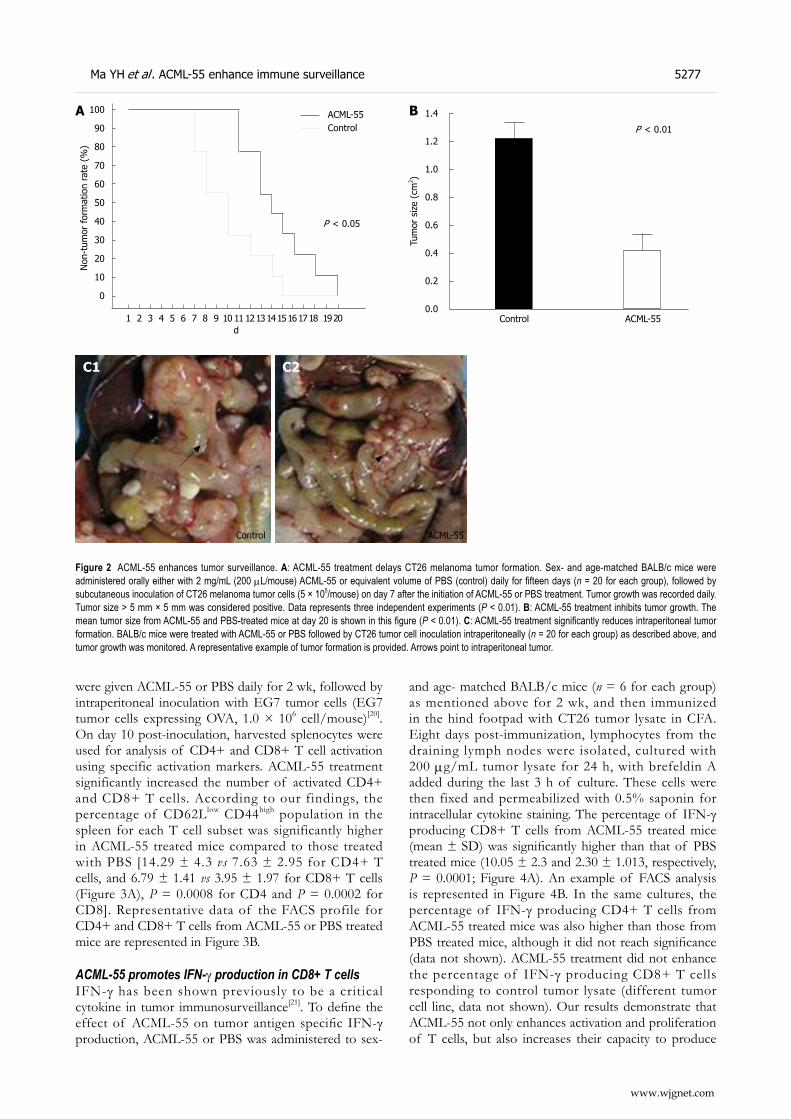
were given ACML-55 or PBS daily for 2 wk, followed by intraperitoneal inoculation with EG7 tumor cells (EG7 tumor cells expressing OVA, 1.0 × 10⁶ cell/mouse)[20]. On day 10 post-inoculation, harvested splenocytes were used for analysis of CD4+ and CD8+ T cell activation using specific activation markers. ACML-55 treatment significantly increased the number of activated CD4+ and CD8+ T cells. According to our findings, the percentage of CD62Llow CD44high population in the spleen for each T cell subset was significantly higher in ACML-55 treated mice compared to those treated with PBS [14.29 ± 4.3 vs 7.63 ± 2.95 for CD4+ T cells, and 6.79 ± 1.41 vs 3.95 ± 1.97 for CD8+ T cells (Figure 3A), P = 0.0008 for CD4 and P = 0.0002 for CD8]. Representative data of the FACS profile for CD4+ and CD8+ T cells from ACML-55 or PBS treated mice are represented in Figure 3B.
ACML-55 promotes IFN-γ production in CD8+ T cellsIFN-γ has been shown previously to be a critical cytokine in tumor immunosurveillance[21]. To define the effect of ACML-55 on tumor antigen specific IFN-γ production, ACML-55 or PBS was administered to sex-
and age- matched BALB/c mice (n = 6 for each group) as mentioned above for 2 wk, and then immunized in the hind footpad with CT26 tumor lysate in CFA. Eight days post-immunization, lymphocytes from the draining lymph nodes were isolated, cultured with 200 μg/mL tumor lysate for 24 h, with brefeldin A added during the last 3 h of culture. These cells were then fixed and permeabilized with 0.5% saponin for intracellular cytokine staining. The percentage of IFN-γ producing CD8+ T cells from ACML-55 treated mice (mean ± SD) was significantly higher than that of PBS treated mice (10.05 ± 2.3 and 2.30 ± 1.013, respectively, P = 0.0001; Figure 4A). An example of FACS analysis is represented in Figure 4B. In the same cultures, the percentage of IFN-γ producing CD4+ T cells from ACML-55 treated mice was also higher than those from PBS treated mice, although it did not reach significance (data not shown). ACML-55 treatment did not enhance the percentage of IFN-γ producing CD8+ T cells responding to control tumor lysate (different tumor cell line, data not shown). Our results demonstrate that ACML-55 not only enhances activation and proliferation of T cells, but also increases their capacity to produce
1.4
1.2
1.0
0.8
0.6
0.4
0.2
0.0
P < 0.01
Control ACML-55
Tum
or s
ize
(cm
2 )
B
Figure 2 ACML-55 enhances tumor surveillance. A: ACML-55 treatment delays CT26 melanoma tumor formation. Sex- and age-matched BALB/c mice were administered orally either with 2 mg/mL (200 μL/mouse) ACML-55 or equivalent volume of PBS (control) daily for fifteen days (n = 20 for each group), followed by subcutaneous inoculation of CT26 melanoma tumor cells (5 × 10⁵/mouse) on day 7 after the initiation of ACML-55 or PBS treatment. Tumor growth was recorded daily. Tumor size > 5 mm × 5 mm was considered positive. Data represents three independent experiments (P < 0.01). B: ACML-55 treatment inhibits tumor growth. The mean tumor size from ACML-55 and PBS-treated mice at day 20 is shown in this figure (P < 0.01). C: ACML-55 treatment significantly reduces intraperitoneal tumor formation. BALB/c mice were treated with ACML-55 or PBS followed by CT26 tumor cell inoculation intraperitoneally (n = 20 for each group) as described above, and tumor growth was monitored. A representative example of tumor formation is provided. Arrows point to intraperitoneal tumor.
www.wjgnet.com
Ma YH et al . ACML-55 enhance immune surveillance 5277
C1
Control
C2
ACML-55
1 2 3 4 5 6 7 8 9 10 11 12 13 14 15 16 17 18 19 20
100
90
80
70
60
50
40
30
20
10
0
Non
-tum
or for
mat
ion
rate
(%
)
d
P < 0.05
ACML-55Control
A
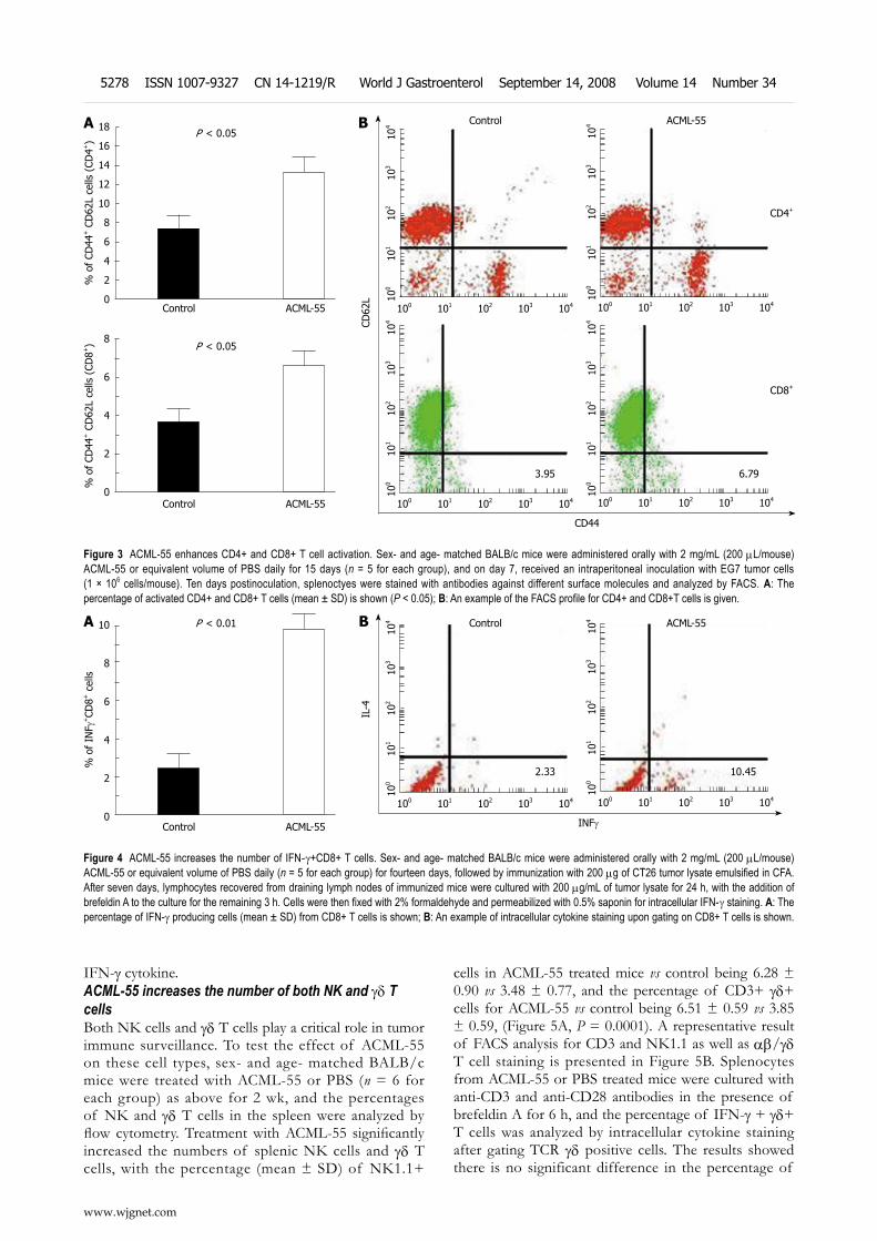
IFN-γ cytokine.ACML-55 increases the number of both NK and γδ T cellsBoth NK cells and γδ T cells play a critical role in tumor immune surveillance. To test the effect of ACML-55 on these cell types, sex- and age- matched BALB/c mice were treated with ACML-55 or PBS (n = 6 for each group) as above for 2 wk, and the percentages of NK and γδ T cells in the spleen were analyzed by flow cytometry. Treatment with ACML-55 significantly increased the numbers of splenic NK cells and γδ T cells, with the percentage (mean ± SD) of NK1.1+
cells in ACML-55 treated mice vs control being 6.28 ± 0.90 vs 3.48 ± 0.77, and the percentage of CD3+ γδ+ cells for ACML-55 vs control being 6.51 ± 0.59 vs 3.85 ± 0.59, (Figure 5A, P = 0.0001). A representative result of FACS analysis for CD3 and NK1.1 as well as αβ/γδ T cell staining is presented in Figure 5B. Splenocytes from ACML-55 or PBS treated mice were cultured with anti-CD3 and anti-CD28 antibodies in the presence of brefeldin A for 6 h, and the percentage of IFN-γ + γδ+ T cells was analyzed by intracellular cytokine staining after gating TCR γδ positive cells. The results showed there is no significant difference in the percentage of
www.wjgnet.com
5278 ISSN 1007-9327 CN 14-1219/R World J Gastroenterol September 14, 2008 Volume 14 Number 34
Figure 3 ACML-55 enhances CD4+ and CD8+ T cell activation. Sex- and age- matched BALB/c mice were administered orally with 2 mg/mL (200 μL/mouse) ACML-55 or equivalent volume of PBS daily for 15 days (n = 5 for each group), and on day 7, received an intraperitoneal inoculation with EG7 tumor cells (1 × 10⁶ cells/mouse). Ten days postinoculation, splenoctyes were stained with antibodies against different surface molecules and analyzed by FACS. A: The percentage of activated CD4+ and CD8+ T cells (mean ± SD) is shown (P < 0.05); B: An example of the FACS profile for CD4+ and CD8+T cells is given.
8
6
4
2
0
P < 0.05
% o
f CD
44+ C
D62
L ce
lls (
CD8+
)
Control ACML-55
18
16
14
12
10
8
6
4
2
0
P < 0.05
% o
f CD
44+ C
D62
L ce
lls (
CD4+
)
A
Control ACML-55
100 101 102 103 104
100
10
1 10
2 10
3 10
4
100 101 102 103 104
100
10
1 10
2 10
3 10
4
100 101 102 103 104
100
10
1 10
2 10
3 10
4
100 101 102 103 104
100
10
1 10
2 10
3 10
4
CD4+
CD8+
CD44
CD62
L
B
3.95 6.79
Control ACML-55
Figure 4 ACML-55 increases the number of IFN-γ+CD8+ T cells. Sex- and age- matched BALB/c mice were administered orally with 2 mg/mL (200 μL/mouse) ACML-55 or equivalent volume of PBS daily (n = 5 for each group) for fourteen days, followed by immunization with 200 μg of CT26 tumor lysate emulsified in CFA. After seven days, lymphocytes recovered from draining lymph nodes of immunized mice were cultured with 200 μg/mL of tumor lysate for 24 h, with the addition of brefeldin A to the culture for the remaining 3 h. Cells were then fixed with 2% formaldehyde and permeabilized with 0.5% saponin for intracellular IFN-γ staining. A: The percentage of IFN-γ producing cells (mean ± SD) from CD8+ T cells is shown; B: An example of intracellular cytokine staining upon gating on CD8+ T cells is shown.
10
8
6
4
2
0
P < 0.01
% o
f IN
F γ+CD
8+ c
ells
Control ACML-55
A
100 101 102 103 104
100
10
1
102
10
3
104
100 101 102 103 104
100
10
1
102
10
3
104
INFγ
IL-4
Control ACML-55B
10.452.33
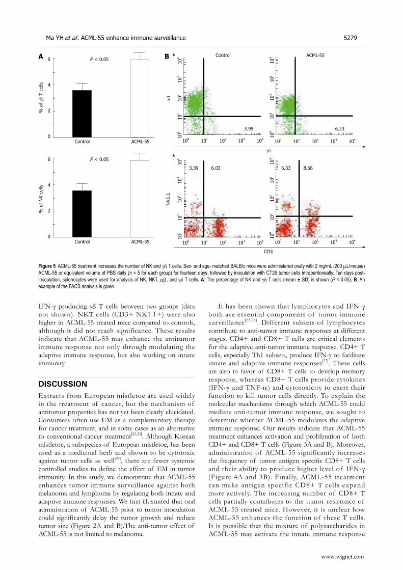
IFN-γ producing γδ T cells between two groups (data not shown). NKT cells (CD3+ NK1.1+) were also higher in ACML-55 treated mice compared to controls, although it did not reach significance. These results indicate that ACML-55 may enhance the antitumor immune response not only through modulating the adaptive immune response, but also working on innate immunity.
DISCUSSIONExtracts from European mistletoe are used widely in the treatment of cancer, but the mechanism of antitumor properties has not yet been clearly elucidated. Consumers often use EM as a complementary therapy for cancer treatment, and in some cases as an alternative to conventional cancer treatment[22,23]. Although Korean mistletoe, a subspecies of European mistletoe, has been used as a medicinal herb and shown to be cytotoxic against tumor cells as well[24], there are fewer systemic controlled studies to define the effect of EM in tumor immunity. In this study, we demonstrate that ACML-55 enhances tumor immune surveillance against both melanoma and lymphoma by regulating both innate and adaptive immune responses. We first illustrated that oral administration of ACML-55 prior to tumor inoculation could significantly delay the tumor growth and reduce tumor size (Figure 2A and B).The anti-tumor effect of ACML-55 is not limited to melanoma.
It has been shown that lymphocytes and IFN-γ both are essential components of tumor immune surveillance[25,26]. Different subsets of lymphocytes contribute to anti-tumor immune responses at different stages. CD4+ and CD8+ T cells are critical elements for the adaptive anti-tumor immune response. CD4+ T cells, especially Th1 subsets, produce IFN-γ to facilitate innate and adaptive immune responses[27]. These cells are also in favor of CD8+ T cells to develop memory response, whereas CD8+ T cells provide cytokines (IFN-γ and TNF-α) and cytotoxicity to exert their function to kill tumor cells directly. To explain the molecular mechanisms through which ACML-55 could mediate anti-tumor immune response, we sought to determine whether ACML-55 modulates the adaptive immune response. Our results indicate that ACML-55 treatment enhances activation and proliferation of both CD4+ and CD8+ T cells (Figure 3A and B). Moreover, administration of ACML-55 significantly increases the frequency of tumor antigen specific CD8+ T cells and their ability to produce higher level of IFN-γ (Figure 4A and 3B). Finally, ACML-55 treatment can make antigen specific CD8+ T cells expand more actively. The increasing number of CD8+ T cells partially contributes to the tumor resistance of ACML-55 treated mice. However, it is unclear how ACML-55 enhances the function of these T cells. It is possible that the mixture of polysaccharides in ACML-55 may activate the innate immune response
A
www.wjgnet.com
Ma YH et al . ACML-55 enhance immune surveillance 5279
Figure 5 ACML-55 treatment increases the number of NK and γδ T cells. Sex- and age- matched BALB/c mice were administered orally with 2 mg/mL (200 μL/mouse) ACML-55 or equivalent volume of PBS daily (n = 5 for each group) for fourteen days, followed by inoculation with CT26 tumor cells intraperitoneally. Ten days post-inoculation, splenocytes were used for analysis of NK, NKT, αβ, and γδ T cells. A: The percentage of NK and γδ T cells (mean ± SD) is shown (P < 0.05); B: An example of the FACS analysis is given.
6
4
2
0
P < 0.05
% o
f γδ
T c
ells
Control ACML-55
% o
f N
K ce
lls
6
4
2
0
P < 0.05
Control ACML-55
B
100 101 102 103 104
100
10
1 10
2 10
3 10
4
100 101 102 103 104
100
10
1 10
2 10
3 10
4
CD3
NK1
.13.39 6.03 6.33 8.66
100 101 102 103 104
100
10
1 10
2 10
3 10
4
100 101 102 103 104
100
10
1 10
2 10
3 10
4
Control ACML-55
αβ
γδ
3.95 6.23
through undefined signaling pathways, such as Toll-like receptors and the downstream NF-κB pathway, which in turn helps to regulate the adaptive immune response. Consistently, it has been reported that ACML-55 enhances IL-12 production from macrophages[10] and increases nitric oxide concentration[12]. Further studies are needed to clarify the underlying mechanisms that mediate the effect of ACML-55 on the adaptive immune response.
A potential target of ACML-55 modulation within the innate immune system may be γδ T cells. These cells belong to a unique subset of T cells. They recognize protein or peptide independent of antigen presentation and function as innate like cells[28]. Our earlier studies have demonstrated that γδT cells predominantly produce IFN-γ upon activation[12,29] and play a critical role in tumor immune surveillance by providing an early source of IFN-γ[14]. Interestingly, ACML-55 treatment significantly increases the number of γδ T cells compared to those of PBS-treated mice (Figure 5). Since ACML-55 was given orally, it is possible that the effective components in ACML-55 might directly encounter γδ T cells lining the epithelial layer of the intestine resulting in their activation. In addition to γδ T cells, it has been well established that NK cells play an essential role in tumor immune surveillance. Interestingly, we found that treatment with ACML-55 also upregulates the number of NK cells upon tumor inoculation. Although the changes of NK T cells did not reach significance, the trend is clear. These results indicate that ACML-55 has multiple effects on the immune system.
Given the findings that ACML-55 could efficiently enhance several immune parameters (CD4+, CD8+ and γδ T cells), which were shown previously to be positive for tumor immune surveillance, it is likely that ACML-55 mediates its potential effects on tumor surveillance, at least in part, by upregulation these particular parameters. Future studies using different T cell subset from deficient mice will help to illuminate these questions.
In summary, we have presented clear picture that, as an active lectin from Chinese mistletoe, ACML-55 enhances tumor immune surveillance by regulating both the innate and the adaptive immune responses. Further studies are needed to define the molecular mechanisms mediating the effect of ACML-55 in tumor immunity.
COMMENTSBackgroundMistletoe is a semiparasitic plant with many unusual properties. In our prior study, a mistletoe lectin was purified from Chinese mistletoe and the effect of the active Chinese mistletoe lectin-55 (ACML-55 ) on human γδ T cell cytotoxicity, apoptosis and modulation of the cytokine network was reported. Although these investigations suggest that ACML-55 may modulate the immune response against tumor development, the precise mechanism by which ACML-55 regulates the immune function has not been studied systematically.Research frontiersIn this study, an experimental model was established by hypodermically inoculating the colon cancer cell line CT26 into Balb/c mice. Intracellular cytokine staining used to detect tumor antigen specific CD8+ T cell IFN-γ production. The FACS profile for CD4+ and CD8+ T cells and NK or γδ T Cells composition and activation.Innovations and breakthroughs
This study is investigates the potential effect of active Chinese mistletoe lectin-55 to enhance colon cancer immune surveillance through regulating both innate and adaptive immune responses.ApplicationsThe results demonstrate that ACML-55 can enhance colon cancer immune surveillance through regulating both innate and adaptive immune responses and also suggest that ACML-55 may be a useful complementary therapy for treating colon cancer.Peer reviewThe study investigates the potential effect of ACML-55 to enhance colon cancer immune surveillance through regulating both innate and adaptive immune responses. Its scientific contents can reflect the advanced levels of basic research and the first report on Chinese mistletoe lectin-55.
REFERENCES 1 Bussing A, Suzart K, Bergmann J, Pfuller U, Schietzel M,
Schweizer K. Induction of apoptosis in human lymphocytes treated with Viscum album L. is mediated by the mistletoe lectins. Cancer Lett 1996; 99: 59-72
2 Hajto T. Immunomodulatory effects of iscador: a Viscum album preparation. Oncology 1986; 43 Suppl 1: 51-65
3 Ribereau-Gayon G, Dumont S, Muller C, Jung ML, Poindron P, Anton R. Mistletoe lectins I, II and III induce the production of cytokines by cultured human monocytes. Cancer Lett 1996; 109: 33-38
4 Beuth J. Clinical relevance of immunoactive mistletoe lectin-I. Anticancer Drugs 1997; 8 Suppl 1: S53-S55
5 Stein GM, Berg PA. Mistletoe extract-induced effects on immunocompetent cells: in vitro studies. Anticancer Drugs 1997; 8 Suppl 1: S39-S42
6 Fischer S, Scheffler A, Kabelitz D. Activation of human gamma delta T-cells by heat-treated mistletoe plant extracts. Immunol Lett 1996; 52: 69-72
7 Fischer S, Scheffler A, Kabelitz D. Stimulation of the specific immune system by mistletoe extracts. Anticancer Drugs 1997; 8 Suppl 1: S33-S37
8 Hauer J, Anderer FA. Mechanism of stimulation of human natural killer cytotoxicity by arabinogalactan from Larix occidentalis. Cancer Immunol Immunother 1993; 36: 237-244
9 Mueller EA, Anderer FA. A Viscum album oligosaccharide activating human natural cytotoxicity is an interferon gamma inducer. Cancer Immunol Immunother 1990; 32: 221-227
10 Mueller EA, Anderer FA. Synergistic action of a plant rhamnogalacturonan enhancing antitumor cytotoxicity of human natural killer and lymphokine-activated killer cells: chemical specificity of target cell recognition. Cancer Res 1990; 50: 3646-3651
11 Mockel B, Schwarz T, Zinke H, Eck J, Langer M, Lentzen H. Effects of mistletoe lectin I on human blood cell lines and peripheral blood cells. Cytotoxicity, apoptosis and induction of cytokines. Arzneimittelforschung 1997; 47: 1145-1151
12 Thies A, Nugel D, Pfuller U, Moll I, Schumacher U. Influence of mistletoe lectins and cytokines induced by them on cell proliferation of human melanoma cells in vitro. Toxicology 2005; 207: 105-116
13 Harmsma M, Gromme M, Ummelen M, Dignef W, Tusenius KJ, Ramaekers FC. Differential effects of Viscum album extract IscadorQu on cell cycle progression and apoptosis in cancer cells. Int J Oncol 2004; 25: 1521-1529
14 Lee HS, Kim YS, Kim SB, Choi BE, Woo BH, Lee KC. Isolation and characterization of biologically active lectin from Korean mistletoe, Viscum album var. Coloratum. Cell Mol Life Sci 1999; 55: 679-682
15 Gong F, Ma Y, Ma A, Yu Q, Zhang J, Nie H, Chen X, Shen B, Li N, Zhang D. A lectin from Chinese mistletoe increases gammadelta T cell-mediated cytotoxicity through induction of caspase-dependent apoptosis. (Shanghai) 2007; 39: 445-452
16 Gao Y, Yang W, Pan M, Scully E, Girardi M, Augenlicht LH,
www.wjgnet.com
5280 ISSN 1007-9327 CN 14-1219/R World J Gastroenterol September 14, 2008 Volume 14 Number 34
Craft J, Yin Z. Gamma delta T cells provide an early source of interferon gamma in tumor immunity. J Exp Med 2003; 198: 433-442
17 Yin Z, Zhang DH, Welte T, Bahtiyar G, Jung S, Liu L, Fu XY, Ray A, Craft J. Dominance of IL-12 over IL-4 in gamma delta T cell differentiation leads to default production of IFN-gamma: failure to down-regulate IL-12 receptor beta 2-chain expression. J Immunol 2000; 164: 3056-3064
18 Jung ML , Baudino S, Ribereau-Gayon G, Beck JP. Characterization of cytotoxic proteins from mistletoe (Viscum album L.). Cancer Lett 1990; 51: 103-108
19 Gao Y, Tao J, Li MO, Zhang D, Chi H, Henegariu O, Kaech SM, Davis RJ, Flavell RA, Yin Z. JNK1 is essential for CD8+ T cell-mediated tumor immune surveillance. J Immunol 2005; 175: 5783-5789
20 Gorelik L, Flavell RA. Immune-mediated eradication of tumors through the blockade of transforming growth factor-beta signaling in T cells. Nat Med 2001; 7: 1118-1122
21 Ikeda H, Old LJ, Schreiber RD. The roles of IFN gamma in protection against tumor development and cancer immunoediting. Cytokine Growth Factor Rev 2002; 13: 95-109
22 deVere White RW, Hackman RM, Soares SE, Beckett LA, Li Y, Sun B. Effects of a genistein-rich extract on PSA levels in men with a history of prostate cancer. Urology 2004; 63:
259-26323 deVere White RW, Hackman RM, Soares SE, Beckett LA,
Sun B. Effects of a mushroom mycelium extract on the treatment of prostate cancer. Urology 2002; 60: 640-644
24 Khil LY, Kim W, Lyu S, Park WB, Yoon JW, Jun HS. Mechanisms involved in Korean mistletoe lectin-induced apoptosis of cancer cells. World J Gastroenterol 2007; 13: 2811-2818
25 Dunn GP, Old LJ, Schreiber RD. The immunobiology of cancer immunosurveillance and immunoediting. Immunity 2004; 21: 137-148
26 Li NL, Nie H, Yu QW, Zhang JY, Ma AL, Shen BH, Wang L, Bai J, Chen XH, Zhou T, Zhang DQ. Role of soluble Fas ligand in autoimmune diseases. World J Gastroenterol 2004; 10: 3151-3156
27 Shankaran V, Ikeda H, Bruce AT, White JM, Swanson PE, Old LJ, Schreiber RD. IFNgamma and lymphocytes prevent primary tumour development and shape tumour immunogenicity. Nature 2001; 410: 1107-1111
28 Carding SR, Egan PJ. Gammadelta T cells: functional plasticity and heterogeneity. Nat Rev Immunol 2002; 2: 336-345
29 Yin Z, Chen C, Szabo SJ, Glimcher LH, Ray A, Craft J. T-Bet expression and failure of GATA-3 cross-regulation lead to default production of IFN-gamma by gammadelta T cells. J Immunol 2002; 168: 1566-1571
S- Editor Zhong XY L- Editor Negro F E- Editor Zhang WB
www.wjgnet.com
Ma YH et al . ACML-55 enhance immune surveillance 5281
Davide Festi, Ada Dormi, Simona Capodicasa, Tommaso Staniscia, Adolfo F Attili, Paola Loria, Paolo Pazzi, Giuseppe Mazzella, Claudia Sama, Enrico Roda, Antonio Colecchia
Incidence of gallstone disease in Italy: Results from a multicenter, population-based Italian study (the MICOL project)
CLINICAL RESEARCH
www.wjgnet.com
Online Submissions: wjg.wjgnet.com World J Gastroenterol 2008 September 14; 14(34): [email protected] World Journal of Gastroenterology ISSN 1007-9327doi:10.3748/wjg.14.5282 © 2008 The WJG Press. All rights reserved.
Davide Festi, Giuseppe Mazzella, Claudia Sama, Enrico Roda, Antonio Colecchia, Department of Internal Medicine and Gastroenterology, University of Bologna, Bologna 40138, ItalyAda Dormi, Department of Clinical Medicine and Biotech-nology, University of Bologna, Bologna 40138, ItalySimona Capodicasa, Tommaso Staniscia, Department of Medicine and Aging, University of Chieti, Chieti 66100, ItalyAdolfo F Attili, Department of Gastroenterology, University “La Sapienza”, Roma 00010, ItalyPaola Loria, Department of Internal Medicine, University of Modena, Modena 41100, ItalyPaolo Pazzi, Department of Internal Medicine, S. Anna Hospital, Ferrara 44100, ItalyAuthor contributions: Festi D, Attili AF, Pazzi P, Loria P, Sama C, Roda E designed research; Festi D, Attili AF, Pazzi P, Loria P, Colecchia A performed research; Capodicasa S, Staniscia T, Dormi A analyzed data; Festi D wrote the paper.Correspondence to: Davide Festi, MD, Dipartimento di Medicina Interna e Gastroenterologia, Policlinico S. Orsola-Malpighi, Via Massarenti 9, Bologna 40138, Italy. [email protected]: +39-51-6364123 Fax: +39-51-6364123Received: January 3, 2008 Revised: August 13, 2008Accepted: August 20, 2008Published online: September 14, 2008
AbstractAIM: To evaluate gallstone incidence and risk factors in a large population-based study.METHODS: Gallstone incidence and risk factors, were evaluated by structured questionnaire and physical examination, respectively, in 9611 of 11 109 (86.5%) subjects who were gallstone-free at the cross-sectional study.RESULTS: Six centers throughout Italy enrolled 9611 subjects (5477 males, 4134 females, aged 30- 79 years), 9517 of whom were included into analysis: 424 subjects (4.4%) had gallstones and 61 (0.6%) had been cholecystectomized yielding a cumulative incidence of 0.67% per year (0.66% in males, 0.81% in females). Increasing age, a high body mass index (BMI), a history of diabetes, peptic ulcer and angina, and low cholesterol and high triglyceride levels were identified as risk factors in men while, in females, the only risk factors were increasing age and a high BMI.
Increasing age and pain in the right hypochondrium in men and increasing age in females were identified as predictors of gallstones. Pain in the epigastrium/right hypochondrium was the only symptom related to gallstones; furthermore, some characteristics of pain (forcing to rest, not relieved by bowel movements) were significantly associated with gallstones. No correlation was found between gallstone characteristics and clinical manifestations, while increasing age in men and increasing age and BMI in females were predictors of pain.CONCLUSION: Increasing age and BMI represent true risk factors for gallstone disease (GD); pain in the right hypochondrium and/or epigastrium is confirmed as the only symptom related to gallstones.
© 2008 The WJG Press. All rights reserved.
Key words: Gallstone disease; Ultrasonography; Epidemiology; Prevalence; Incidence; Abdominal pain; Cholecystectomy; Body mass index
Peer reviewer: Serdar Karakose, PhD, Professor, Department of Radiology, Meram Medical Faculty, Selcuk University, Konya 42080, Turkey
Festi D, Dormi A, Capodicasa S, Staniscia T, Attili AF, Loria P, Pazzi P, Mazzella G, Sama C, Roda E, Colecchia A. Incidence of gallstone disease in Italy: Results from a multicenter, population-based Italian study (the MICOL project). World J Gastroenterol 2008; 14(34): 5282-5289 Available from: URL: http://www.wjgnet.com/1007-9327/14/5282.asp DOI: http://dx.doi.org/10.3748/wjg.14.5282
INTRODUCTIONGallstone disease (GD) is a very common gastroin-testinal disorder, presents mainly in the Western world[1,2]. Although this disease has a low mortality rate, its economic and health impact is significant due to its high morbidity. In fact, GD is one of the most common abdominal conditions for which patients in developed countries are admitted to hospitals[3], and this frequency has increased in Western countries since the 1950s[4]. GD is considered “a surgical disease”
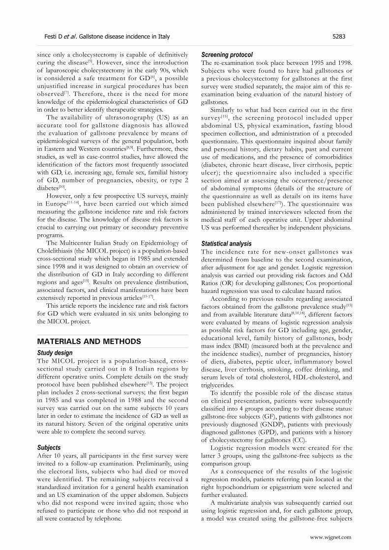
Festi D et al . Gallstone disease incidence in Italy 5283
www.wjgnet.com
since only a cholecystectomy is capable of definitively curing the disease[5]. However, since the introduction of laparoscopic cholecystectomy in the early 90s, which is considered a safe treatment for GD[6], a possible unjustified increase in surgical procedures has been observed[7]. Therefore, there is the need for more knowledge of the epidemiological characteristics of GD in order to better identify therapeutic strategies.
The availability of ultrasonography (US) as an accurate tool for gallstone diagnosis has allowed the evaluation of gallstone prevalence by means of epidemiological surveys of the general population, both in Eastern and Western countries[8,9]. Furthermore, these studies, as well as case-control studies, have allowed the identification of the factors most frequently associated with GD, i.e. increasing age, female sex, familial history of GD, number of pregnancies, obesity, or type 2 diabetes[10].
However, only a few prospective US surveys, mainly in Europe[11-14], have been carried out which aimed measuring the gallstone incidence rate and risk factors for the disease. The knowledge of disease risk factors is crucial to carrying out primary or secondary preventive programs.
The Multicenter Italian Study on Epidemiology of Cholelithiasis (the MICOL project) is a population-based cross-sectional study which began in 1985 and extended since 1998 and it was designed to obtain an overview of the distribution of GD in Italy according to different regions and ages[15]. Results on prevalence distribution, associated factors, and clinical manifestations have been extensively reported in previous articles[15-17].
This article reports the incidence rate and risk factors for GD which were evaluated in six units belonging to the MICOL project.
MATERIALS AND METHODSStudy designThe MICOL project is a population-based, cross-sectional study carried out in 8 Italian regions by different operative units. Complete details on the study protocol have been published elsewhere[15]. The project plan includes 2 cross-sectional surveys; the first began in 1985 and was completed in 1988 and the second survey was carried out on the same subjects 10 years later in order to estimate the incidence of GD as well as its natural history. Seven of the original operative units were able to complete the second survey.
SubjectsAfter 10 years, all participants in the first survey were invited to a follow-up examination. Preliminarily, using the electoral lists, subjects who had died or moved were identified. The remaining subjects received a standardized invitation for a general health examination and an US examination of the upper abdomen. Subjects who did not respond were invited again; those who refused to participate or those who did not respond at all were contacted by telephone.
Screening protocolThe re-examination took place between 1995 and 1998. Subjects who were found to have had gallstones or a previous cholecystectomy for gallstones at the first survey were studied separately, the major aim of this re-examination being evaluation of the natural history of gallstones.
Similarly to what had been carried out in the first survey[15], the screening protocol included upper abdominal US, physical examination, fasting blood specimen collection, and administration of a precoded questionnaire. This questionnaire inquired about family and personal history, dietary habits, past and current use of medications, and the presence of comorbidities (diabetes, chronic heart disease, liver cirrhosis, peptic ulcer); the questionnaire also included a specific section aimed at assessing the occurrence/presence of abdominal symptoms (details of the structure of the questionnaire as well as details on its items have been published elsewhere[17]). The questionnaire was administered by trained interviewers selected from the medical staff of each operative unit. Upper abdominal US was performed thereafter by independent physicians.
Statistical analysisThe incidence rate for new-onset gallstones was determined from baseline to the second examination, after adjustment for age and gender. Logistic regression analysis was carried out providing risk factors and Odd Ratios (OR) for developing gallstones; Cox proportional hazard regression was used to calculate hazard ratios.
According to previous results regarding associated factors obtained from the gallstone prevalence study[15] and from available literature data[8,10,18], different factors were evaluated by means of logistic regression analysis as possible risk factors for GD including age, gender, educational level, family history of gallstones, body mass index (BMI) (measured both at the prevalence and the incidence studies), number of pregnancies, history of diets, diabetes, peptic ulcer, inflammatory bowel disease, liver cirrhosis, smoking, coffee drinking, and serum levels of total cholesterol, HDL-cholesterol, and triglycerides.
To identify the possible role of the disease status on clinical presentation, patients were subsequently classified into 4 groups according to their disease status: gallstone-free subjects (GF), patients with gallstones not previously diagnosed (GNDP), patients with previously diagnosed gallstones (GPD), and patients with a history of cholecystectomy for gallstones (CC).
Logistic regression models were created for the latter 3 groups, using the gallstone-free subjects as the comparison group.
As a consequence of the results of the logistic regression models, patients referring pain located at the right hypochondrium or epigastrium were selected and further evaluated.
A multivariate analysis was subsequently carried out using logistic regression and, for each gallstone group, a model was created using the gallstone-free subjects
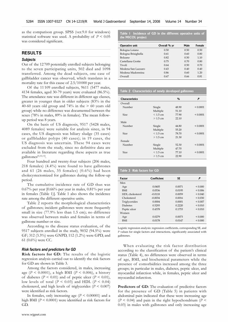
as the comparison group. SPSS (ver.9.0 for windows) statistical software was used. A probability of P < 0.05 was considered significant.
RESULTSSubjectsOut of the 12 709 potentially enrolled subjects belonging to the seven participating units, 502 died and 1098 transferred. Among the dead subjects, one case of gallbladder cancer was observed, which translates in a mortality rate for this cause of 2.5/10 000 per year.
Of the 11 109 enrolled subjects, 9611 (5477 males, 4134 females, aged 30-79 years) were evaluated (86.5%). The attendance rate was different in different age classes, greater in younger than in older subjects (83% in the 40-60 years old group and 74% in the > 60 years old group) while no difference was documented between the sexes (78% in males, 89% in females). The mean follow-up period was 8 years.
On the basis of US diagnosis, 9517 (5428 males, 4089 females) were suitable for analysis since, in 94 cases, the US diagnosis was biliary sludge (35 cases) or gallbladder polyps (40 cases); in 19 cases, the US diagnosis was uncertain. These 94 cases were excluded from the study, since no definitive data are available in literature regarding these aspects as true gallstones[5,10,11].
Four hundred and twenty-four subjects (206 males, 218 females) (4.4%) were found to have gallstones and 61 (26 males, 35 females) (0.6%) had been cholecystectomized for gallstones during the follow-up period.
The cumulative incidence rate of GD thus was 0.67% per year [0.66% per year in males, 0.81% per year in females (Table 1)]. Table 1 also shows the incidence rate among the different operative units.
Table 2 reports the morphological characteristics of gallstones; incident gallstones were more frequently small in size (77.9% less than 1.5 cm); no difference was observed between males and females in terms of gallstone number or size.
According to the disease status evaluation, of the 9517 subjects enrolled in the study, 9032 (94.5%) were GF; 312 (3.3%) were GNPD, 112 (1.2%) were GPD, and 61 (0.6%) were CC.
Risk factors and predictors for GDRisk factors for GD: The results of the logistic regression analysis carried out to identify the risk factors for GD are shown in Table 3.
Among the factors considered, in males, increasing age (P < 0.0001), a high BMI (P < 0.006), a history of diabetes (P < 0.01) and of peptic ulcer (P < 0.01), low levels of total (P < 0.03) and HDL (P < 0.04) cholesterol, and high levels of triglycerides (P < 0.007) were identified as risk factors.
In females, only increasing age (P < 0.00001) and a high BMI (P < 0.0001) were identified as risk factors for GD.
When evaluating the risk factor distribution according to the classification of the patient’s clinical status (Table 4), no differences were observed in terms of age, BMI, and biochemical parameters while the presence of comorbidities increased among the three groups; in particular in males, diabetes, peptic ulcer, and myocardial infarction while, in females, peptic ulcer and myocardial infarction.
Predictors of GD: The evaluation of predictive factors for the presence of GD (Table 5) in patients with abdominal pain indicated that these were increasing age (P < 0.04) and pain in the right hypochondrium (P < 0.03) in males with gallstones and only increasing age
Operative unit Overall % yr Male Female
Bologna Loiano 0.50 0.50 0.50Bologna Brisighella 0.61 0.60 0.80Bolzano 0.82 0.50 1.10Castellana Grotte 0.75 0.70 0.80Tivoli 0.64 0.50 0.70Modena San Lazzaro 0.43 0.40 0.40Modena Madonnina 0.86 0.60 1.20Overall 0.67 0.66 0.81
Table 1 Incidence of GD in the different operative units of the MICOL project
Characteristics % POverall Number Single 48.90 < 0.0001
Multiple 51.10 Size ≤ 1.5 cm 77.90 < 0.0001
≥ 1.5 cm 22.10Male Number Single 44.80 < 0.0001 Multiple 55.20 Size ≤ 1.5 cm 78.70 < 0.0001
≥ 1.5 cm 21.30Female Number Single 52.30 < 0.0001
Multiple 47.70 Size ≤ 1.5 cm 77.10 < 0.0001
≥ 1.5 cm 22.90
Table 2 Characteristics of newly developed gallstones
Factor Coefficient SE PMen Age 0.0405 0.0071 < 0.000 BMI 0.0536 0.0195 < 0.006 HDL cholesterol -0.0118 0.0059 < 0.040 Cholesterol -0.0034 0.0016 < 0.030 Triglycerides 0.0004 0.0001 < 0.007 Diabetes 0.5293 0.2220 < 0.010 Peptic ulcer 0.4378 0.1753 < 0.010Women Age 0.0279 0.0073 < 0.000 BMI 0.0178 0.0147 < 0.000
Table 3 Risk factors for GD
Logistic regression analysis: regression coefficients, corresponding SE, and P values for single factors and interactions, significantly associated with gallstones.
www.wjgnet.com
5284 ISSN 1007-9327 CN 14-1219/R World J Gastroenterol September 14, 2008 Volume 14 Number 34
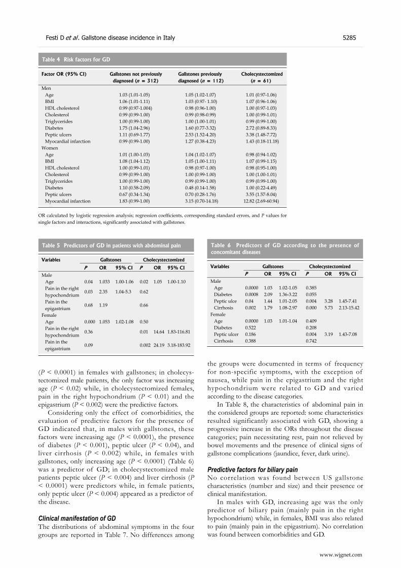
(P < 0.0001) in females with gallstones; in cholecys-tectomized male patients, the only factor was increasing age (P < 0.02) while, in cholecystectomized females, pain in the right hypochondrium (P < 0.01) and the epigastrium (P < 0.002) were the predictive factors.
Considering only the effect of comorbidities, the evaluation of predictive factors for the presence of GD indicated that, in males with gallstones, these factors were increasing age (P < 0.0001), the presence of diabetes (P < 0.001), peptic ulcer (P < 0.04), and liver cirrhosis (P < 0.002) while, in females with gallstones, only increasing age (P < 0.0001) (Table 6) was a predictor of GD; in cholecystectomized male patients peptic ulcer (P < 0.004) and liver cirrhosis (P < 0.0001) were predictors while, in female patients, only peptic ulcer (P < 0.004) appeared as a predictor of the disease.
Clinical manifestation of GDThe distributions of abdominal symptoms in the four groups are reported in Table 7. No differences among
the groups were documented in terms of frequency for non-specific symptoms, with the exception of nausea, while pain in the epigastrium and the right hypochondrium were related to GD and varied according to the disease categories.
In Table 8, the characteristics of abdominal pain in the considered groups are reported: some characteristics resulted significantly associated with GD, showing a progressive increase in the ORs throughout the disease categories; pain necessitating rest, pain not relieved by bowel movements and the presence of clinical signs of gallstone complications (jaundice, fever, dark urine).
Predictive factors for biliary painNo correlation was found between US gallstone characteristics (number and size) and their presence or clinical manifestation.
In males with GD, increasing age was the only predictor of biliary pain (mainly pain in the right hypochondrium) while, in females, BMI was also related to pain (mainly pain in the epigastrium). No correlation was found between comorbidities and GD.
Factor OR (95% CI) Gallstones not previously diagnosed (n = 312)
Gallstones previously diagnosed (n = 112)
Cholecystectomized (n = 61)
Men Age 1.03 (1.01-1.05) 1.05 (1.02-1.07) 1.01 (0.97-1.06) BMI 1.06 (1.01-1.11) 1.03 (0.97- 1.10) 1.07 (0.96-1.06) HDL cholesterol 0.99 (0.97-1.004) 0.98 (0.96-1.00) 1.00 (0.97-1.03) Cholesterol 0.99 (0.99-1.00) 0.99 (0.98-0.99) 1.00 (0.99-1.01) Triglycerides 1.00 (0.99-1.00) 1.00 (1.00-1.01) 0.99 (0.99-1.00) Diabetes 1.75 (1.04-2.96) 1.60 (0.77-3.32) 2.72 (0.89-8.33) Peptic ulcers 1.11 (0.69-1.77) 2.53 (1.52-4.20) 3.38 (1.48-7.72) Myocardial infarction 0.99 (0.99-1.00) 1.27 (0.38-4.23) 1.43 (0.18-11.18)Women Age 1.01 (1.00-1.03) 1.04 (1.02-1.07) 0.98 (0.94-1.02) BMI 1.08 (1.04-1.12) 1.05 (1.00-1.11) 1.07 (0.99-1.15) HDL cholesterol 1.00 (0.99-1.01) 0.98 (0.97-1.00) 0.98 (0.95-1.00) Cholesterol 0.99 (0.99-1.00) 1.00 (0.99-1.00) 1.00 (1.00-1.01) Triglycerides 1.00 (0.99-1.00) 0.99 (0.99-1.00) 0.99 (0.99-1.00) Diabetes 1.10 (0.58-2.09) 0.48 (0.14-1.58) 1.00 (0.22-4.49) Peptic ulcers 0.67 (0.34-1.34) 0.70 (0.28-1.76) 3.55 (1.57-8.04) Myocardial infarction 1.83 (0.99-1.00) 3.15 (0.70-14.18) 12.82 (2.69-60.94)
Table 4 Risk factors for GD
OR calculated by logistic regression analysis; regression coefficients, corresponding standard errors, and P values for single factors and interactions, significantly associated with gallstones.
Variables Gallstones Cholecystectomized
P OR 95% CI P OR 95% CIMale Age 0.04 1.033 1.00-1.06 0.02 1.05 1.00-1.10 Pain in the right hypochondrium
0.03 2.35 1.04-5.3 0.62
Pain in the epigastrium
0.68 1.19 0.66
Female Age 0.000 1.053 1.02-1.08 0.50 Pain in the right hypochondrium
0.36 0.01 14.64 1.83-116.81
Pain in the epigastrium
0.09 0.002 24.19 3.18-183.92
Table 5 Predictors of GD in patients with abdominal pain
Variables Gallstones Cholecystectomized
P OR 95% CI P OR 95% CIMale Age 0.0000 1.03 1.02-1.05 0.385 Diabetes 0.0008 2.09 1.36-3.22 0.055 Peptic ulce 0.04 1.44 1.01-2.05 0.004 3.28 1.45-7.41 Cirrhosis 0.002 1.79 1.08-2.97 0.000 5.73 2.13-15.42Female Age 0.0000 1.03 1.01-1.04 0.409 Diabetes 0.522 0.208 Peptic ulcer 0.186 0.004 3.19 1.43-7.08 Cirrhosis 0.388 0.742
Table 6 Predictors of GD according to the presence of concomitant diseases
Festi D et al . Gallstone disease incidence in Italy 5285
www.wjgnet.com
DISCUSSIONThe present study evaluated incidence and risk factors for GD in a large population from various Italian regions, thus providing a more detailed picture of the epidemiological characteristics of this disease. Incidence was higher in females than in males and increased with age. In our population, the risk factors for GD in males were increasing age, BMI, concomitant diseases such as diabetes, liver cirrhosis, peptic ulcer, coronary disease, HDL and total cholesterol, and high levels of triglycerides while, in females, only increasing age and BMI. Increasing age, pain in the right hypochondrium/epigastrium, and the presence of concomitant diseases are predictors of GD. Pain in the right hypochondrium or epigastrium was the only symptom associated with GD; symptom severity increased as a function of the natural history of the disease. Increasing age in men and aging and BMI in females were the only predictive factors for the eventual presence of symptoms.
GD is a very common gastrointestinal disorder mainly in the Western world[1,2]; although this disease has a low mortality rate, its economic and health impact is significant due to its high morbidity. In fact, GD is one of the most common abdominal conditions for which patients are admitted to hospitals in developed countries[3]. Knowledge of disease epidemiology is therefore crucial in managing this disorder, not only for planning preventive programs, but also for the identification of the best therapeutic strategy. Several US-based surveys have been carried out in Europe[19-23] and in North[24,25] and South[26] America as well as in Asia[27,28], indicating prevalence rates for GD ranging
from 5.9%[20] to 21.9%[22]. However, few studies[11-14] have been carried out to evaluate incidence and risk factors of GD, mainly due to the difficulties in following up large populations for several years. The MICOL study was designed to obtain a general overview of GD in Italy, investigating GD in terms of prevalence, incidence, risk factors, and natural history[15]. In the present study, a large general population was evaluated with the objective of identifying gallstone incidence and risk factors as well as the morphological and clinical characteristics of newly developed gallstones. Incidence was higher than that measured in previous Italian studies[12-14]; these differences could be related to the small population sample evaluated in the earlier studies. Furthermore, prevalence was also higher than in Denmark (4.5% and 5.8%, in males and females, respectively)[11]. Differences in research design may justify these differences even it is not possible to exclude the role of environmental factors.
In the present study, the response rate was higher (79%) than that reported in other GD incidence surveys performed in the north (63.7)[12] and in the centre (73.5%)[14] of Italy and similar to that observed in Denmark (82.8%)[11] and in southern Italy (87.7%)[13]; this percentage indicates a high adherence of the target population to the epidemiological study.
The participation rate was also higher in the present incidence study (79%) than in the previous study evaluating GD prevalence (64.4%)[15]; this difference could be related to a possible self-selection of patients or to an effect of dilution in the prevalence study since, in that study, 14 units participated while, in the present study, only 7 units were able to adhere to the protocol.
Symptoms (%) OR (95% CI) Gallstone-free subjects (n = 9001)
Gallstones not previously diagnosed (n = 312)
Gallstones previously diagnosed (n = 112)
Cholecystectomized (n = 61)
Belching 19.1 19.5 27.2 23.1 1.03 (0.75-1.39) 1.27 (0.86-1.89) 0.92 (0.48-1.75)
Heartburn 32.2 33.1 37.5 44.2 1.12 (0.85-1.39) 0.89 (0.61-1.30) 1.29 (0.72-2.03)
Nausea 12.3 14.3 18.4 26.9 1.22 (0.82-1.82) 1.48 (0.90-2.42) 2.40 (1.19-4.83)
Vomiting 7.2 7.7 9.6 9.6 0.99 (0.59-1.64) 0.79 (0.41-1.53) 0.97 (0.40-2.33)
Bloated feeling after meals 37.2 38.6 44.9 48.1 1.24 (0.95-1.61) 1.12 (0.78-1.63) 1.24 (0.69-2.21)
Epigastric discomfort 33.8 33.1 40.4 38.5 0.96 (0.72-1.26) 1.26 (0.87-1.83) 0.87 (0.48-1.59)
Bitter taste in the morning 36.4 35.4 41.2 44.2 0.93 (0.71-1.21) 0.88 (0.61-1.26) 1.08 (0.61-1.88)
Heavy feeling on the right side 19.7 18.8 25.7 28.8 0.94 (0.68-1.29) 1.14 (0.77-1.71) 1.09 (0.58-2.05)
Heavy feeling in the epigastrium 22.7 21.3 25.0 26.9 0.83 (0.60-1.15) 0.81 (0.53-1.24) 0.67 (0.34-1.33)
Pain in the epigastrium 11.2 24.7 39.0 51.2 1.5 (1.17-2.4) 3.0 (2.6-5.7) 29.9 (7.0-221.3)
Pain in the right hipocondrium 15.8 23.7 39.0 46.5 1.7 (1.3-2.8) 3.9 (3.1-7.4) 32.1 (9.3-238.9)
Intolerance to fatty or fried foods 24.0 5 4.4 3.4 0.99 (0.63-1.55) 0.94 (0.19-1.79) 1.04 (0.5-2.14)
Table 7 Distribution of symptoms (and unadjusted ORs) in gallstone-free subjects, patients with gallstones not previously diagnosed, patients with a previous diagnosis of gallstones, and patients with a history of cholecystectomy for gallstones
www.wjgnet.com
5286 ISSN 1007-9327 CN 14-1219/R World J Gastroenterol September 14, 2008 Volume 14 Number 34
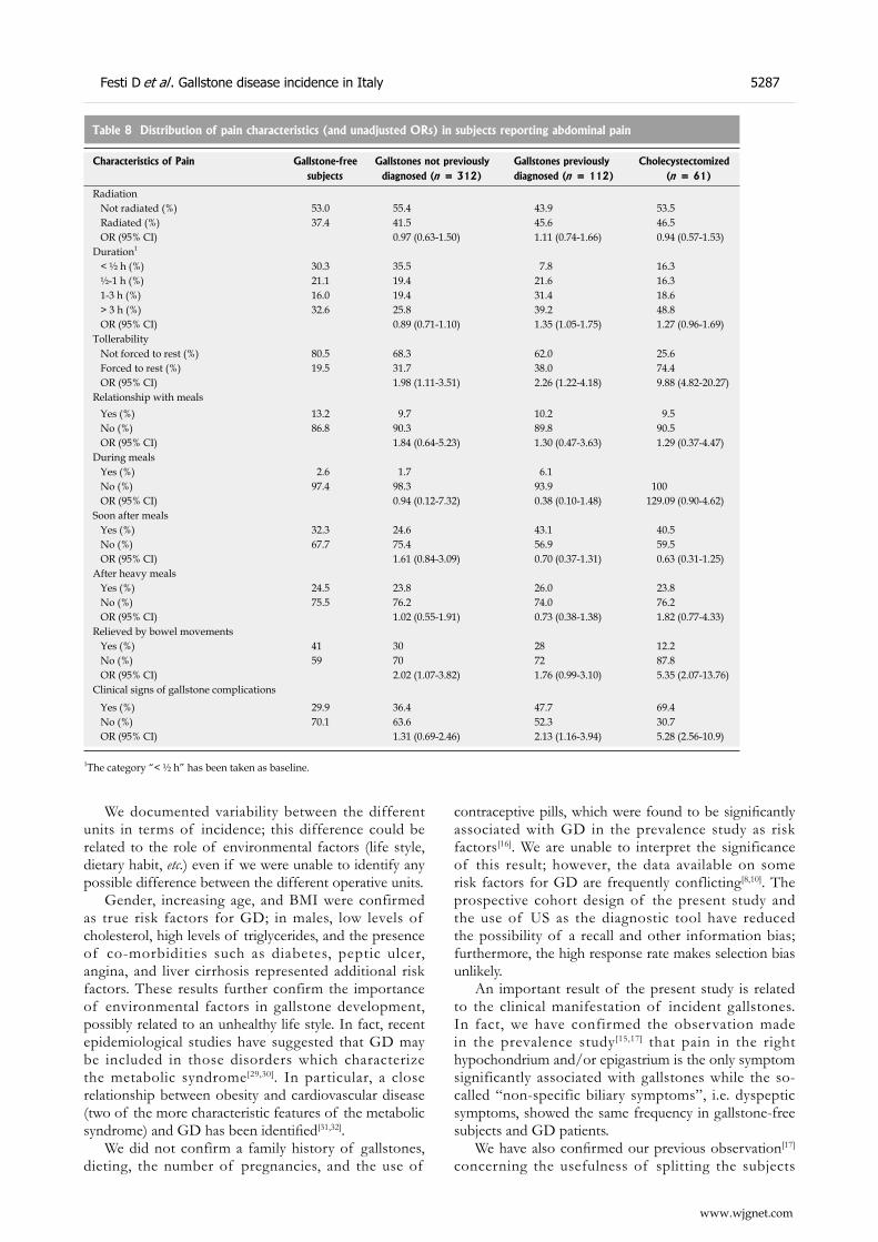
We documented variability between the different units in terms of incidence; this difference could be related to the role of environmental factors (life style, dietary habit, etc.) even if we were unable to identify any possible difference between the different operative units.
Gender, increasing age, and BMI were confirmed as true risk factors for GD; in males, low levels of cholesterol, high levels of triglycerides, and the presence of co-morbidities such as diabetes, peptic ulcer, angina, and liver cirrhosis represented additional risk factors. These results further confirm the importance of environmental factors in gallstone development, possibly related to an unhealthy life style. In fact, recent epidemiological studies have suggested that GD may be included in those disorders which characterize the metabolic syndrome[29,30]. In particular, a close relationship between obesity and cardiovascular disease (two of the more characteristic features of the metabolic syndrome) and GD has been identified[31,32].
We did not confirm a family history of gallstones, dieting, the number of pregnancies, and the use of
contraceptive pills, which were found to be significantly associated with GD in the prevalence study as risk factors[16]. We are unable to interpret the significance of this result; however, the data available on some risk factors for GD are frequently conflicting[8,10]. The prospective cohort design of the present study and the use of US as the diagnostic tool have reduced the possibility of a recall and other information bias; furthermore, the high response rate makes selection bias unlikely.
An important result of the present study is related to the clinical manifestation of incident gallstones. In fact, we have confirmed the observation made in the prevalence study[15,17] that pain in the right hypochondrium and/or epigastrium is the only symptom significantly associated with gallstones while the so-called “non-specific biliary symptoms”, i.e. dyspeptic symptoms, showed the same frequency in gallstone-free subjects and GD patients.
We have also confirmed our previous observation[17] concerning the usefulness of splitting the subjects
Characteristics of Pain Gallstone-free subjects
Gallstones not previously diagnosed (n = 312)
Gallstones previously diagnosed (n = 112)
Cholecystectomized (n = 61)
Radiation Not radiated (%) 53.0 55.4 43.9 53.5 Radiated (%) 37.4 41.5 45.6 46.5 OR (95% CI) 0.97 (0.63-1.50) 1.11 (0.74-1.66) 0.94 (0.57-1.53)Duration1
< ½ h (%) 30.3 35.5 7.8 16.3 ½-1 h (%) 21.1 19.4 21.6 16.3 1-3 h (%) 16.0 19.4 31.4 18.6 > 3 h (%) 32.6 25.8 39.2 48.8 OR (95% CI) 0.89 (0.71-1.10) 1.35 (1.05-1.75) 1.27 (0.96-1.69)Tollerability Not forced to rest (%) 80.5 68.3 62.0 25.6 Forced to rest (%) 19.5 31.7 38.0 74.4 OR (95% CI) 1.98 (1.11-3.51) 2.26 (1.22-4.18) 9.88 (4.82-20.27)Relationship with meals Yes (%) 13.2 9.7 10.2 9.5 No (%) 86.8 90.3 89.8 90.5 OR (95% CI) 1.84 (0.64-5.23) 1.30 (0.47-3.63) 1.29 (0.37-4.47)During meals Yes (%) 2.6 1.7 6.1 No (%) 97.4 98.3 93.9 100 OR (95% CI) 0.94 (0.12-7.32) 0.38 (0.10-1.48) 129.09 (0.90-4.62)Soon after meals Yes (%) 32.3 24.6 43.1 40.5 No (%) 67.7 75.4 56.9 59.5 OR (95% CI) 1.61 (0.84-3.09) 0.70 (0.37-1.31) 0.63 (0.31-1.25)After heavy meals Yes (%) 24.5 23.8 26.0 23.8 No (%) 75.5 76.2 74.0 76.2 OR (95% CI) 1.02 (0.55-1.91) 0.73 (0.38-1.38) 1.82 (0.77-4.33)Relieved by bowel movements Yes (%) 41 30 28 12.2 No (%) 59 70 72 87.8 OR (95% CI) 2.02 (1.07-3.82) 1.76 (0.99-3.10) 5.35 (2.07-13.76)Clinical signs of gallstone complications
Yes (%) 29.9 36.4 47.7 69.4 No (%) 70.1 63.6 52.3 30.7 OR (95% CI) 1.31 (0.69-2.46) 2.13 (1.16-3.94) 5.28 (2.56-10.9)
Table 8 Distribution of pain characteristics (and unadjusted ORs) in subjects reporting abdominal pain
1The category “< ½ h” has been taken as baseline.
Festi D et al . Gallstone disease incidence in Italy 5287
www.wjgnet.com
enrolled in the study into 4 categories reproducing the different stages of GD (absence of disease, silent disease, overt disease, severe disease); in fact, the frequency and severity of the clinical symptoms and the signs of GD increased throughout the 3 disease categories.
Fur ther more, for those character ist ics that are an expression of the degree of pain severity (pain forcing to rest, presence of clinical signs of gallstone complications) they progressively increased from silent gallstones to symptomatic gallstones to cholecystectomized patients for gallstones, suggesting that the natural history of GD moves from a silent to a clinically evident stage; this is also true for newly developed gallstones. This information may be useful in choosing the best therapeutic strategy in gallstone patients since surgical treatment is indicated only in true symptomatic gallstone patients[5,33].
Finally, we were unable to identify any predictive factor for the presence of biliary pain in terms of gallstone characteristics (number and size), while increasing age in males and a high BMI in females were related to the presence of biliary pain. These results are in agreement with some[34], but in disagreement with others[35,36].
In conclusion, this study provides data on the gallstone incidence and risk factors for GD in a large free-living population; the incidence rate is higher in females and it increases with age. Increasing age and BMI represent true risk factors for GD; pain in the right hypochondrium and/or epigastrium is confirmed as the only symptom related to GD; pain, as well as its characteristics of disease severity, increases in severity and frequency throughout the different stages of GD (from silent to severe disease). This information may help physicians in clinical decision making.
COMMENTSBackgroundGallstone incidence and risk factors are poorly understood and the relationship between gallstone presence and its clinical manifestations is debated.Research frontiersGallstone incidence, risk factors, and clinical manifestations.Innovations and breakthroughsThe present paper provides important information on gallstone incidence and risk factors and clearly identifies the clinical manifestations of the disease.ApplicationsThe results of the present paper can be useful for the early recognition of gallstones and for deciding the most appropriate therapeutic management according to the clinical presentation. Furthermore, preventive strategies can be identified and planned according to these results.Peer reviewThis manuscript is a multicenter Italian study giving information about the incidence of gallstone disease in Italy. The results of this study can provide information for the comparison of Italian population results with the other nations' results.
REFERENCES1 Sandler RS, Everhart JE, Donowitz M, Adams E, Cronin
K, Goodman C, Gemmen E, Shah S, Avdic A, Rubin R. The
burden of selected digestive diseases in the United States. Gastroenterology 2002; 122: 1500-1511
2 Aerts R, Penninckx F. The burden of gallstone disease in Europe. Aliment Pharmacol Ther 2003; 18 Suppl 3: 49-53
3 Russo MW, Wei JT, Thiny MT, Gangarosa LM, Brown A, Ringel Y, Shaheen NJ, Sandler RS. Digestive and liver diseases statistics, 2004. Gastroenterology 2004; 126: 1448-1453
4 Bateson MC. Gallstones and cholecystectomy in modern Britain. Postgrad Med J 2000; 76: 700-703
5 Roda E, Festi D, Lezoche E, Leushner U, Paumgartner G, Sauerbruch T. Strategies in the treatment of biliary stones. Gastroenterol Int 2000; 13: 7-15
6 National Institutes of Health Consensus Development Conference Statement: Gallstones and Laparoscopic Cholecystectomy. September 14-16, 1992. J Laparoendosc Surg 1993; 3: 77-90
7 Legorreta AP, Silber JH, Costantino GN, Kobylinski RW, Zatz SL. Increased cholecystectomy rate after the introduction of laparoscopic cholecystectomy. JAMA 1993; 270: 1429-1432
8 Shaffer EA. Epidemiology and risk factors for gallstone disease: has the paradigm changed in the 21st century? Curr Gastroenterol Rep 2005; 7: 132-140
9 Kratzer W, Mason RA, Kachele V. Prevalence of gallstones in sonographic surveys worldwide. J Clin Ultrasound 1999; 27: 1-7
10 Diehl AK. Epidemiology and natural history of gallstone disease. Gastroenterol Clin North Am 1991; 20: 1-19
11 Jensen KH, Jorgensen T. Incidence of gallstones in a Danish population. Gastroenterology 1991; 100: 790-794
12 Barbara L, Sama C, Morselli Labate AM, Taroni F, Rusticani G, Festi D. A 10-year incidence of gallstone disease: the Sirmione study. J Hepatol 1993; 18 Suppl 1: 104A
13 Misciagna G, Leoci C, Elba S, Petruzzi J, Guerra V, Mossa A, Noviello M, Coviello A, Capece-Minutolo M, Giorgiot T. The epidemiology of cholelithiasis in southern Italy. Eur J Gastroenterol Hepatol 1994; 6: 937-941
14 Angelico F, Del Ben M, Barbato A, Conti R, Urbinati G. Ten-year incidence and natural history of gallstone disease in a rural population of women in central Italy. The Rome Group for the Epidemiology and Prevention of Cholelithiasis (GREPCO). Ital J Gastroenterol Hepatol 1997; 29: 249-254
15 Attili AF, Carulli N, Roda E, Barbara B, Capocaccia L, Menotti A, Okoliksanyi L, Ricci G, Capocaccia R, Festi D. Epidemiology of gallstone disease in Italy: prevalence data of the multicenter Italian study on cholelithiasis (MICOL). Am J Epidemiol 1995; 141: 158-165
16 Attili AF, Capocaccia R, Carulli N, Festi D, Roda E, Barbara L, Capocaccia L, Menotti A, Okolicsanyi L, Ricci G, Lalloni L, Mariotti S, Sama C, Scafato E. Factors associated with gallstone disease in the MICOL experience. Multicenter Italian Study on Epidemiology of Cholelithiasis. Hepatology 1997; 26: 809-818
17 Festi D, Sottili S, Colecchia A, Attili A, Mazzella G, Roda E, Romano F. Clinical manifestations of gallstone disease: evidence from the multicenter Italian study on cholelithiasis (MICOL). Hepatology 1999; 30: 839-846
18 Misciagna G, Leoci C, Guerra V, Chiloiro M, Elba S, Petruzzi J, Mossa A, Noviello MR, Coviello A, Minutolo MC, Mangini V, Messa C, Cavallini A, De Michele G, Giorgio I. Epidemiology of cholelithiasis in southern Italy. Part II: Risk factors. Eur J Gastroenterol Hepatol 1996; 8: 585-593
19 Barbara L, Sama C, Morselli Labate AM, Taroni F, Rusticali AG, Festi D, Sapio C, Roda E, Banterle C, Puci A. A population study on the prevalence of gallstone disease: the Sirmione Study. Hepatology 1987; 7: 913-917
20 Loria P, Dilengite MA, Bozzoli M, Carubbi F, Messora R, Sassatelli R, Bertolotti M, Tampieri A, Tartoni PL, Cassinadri M. Prevalence rates of gallstone disease in Italy. The Chianciano population study. Eur J Epidemiol 1994; 10: 143-150
www.wjgnet.com
5288 ISSN 1007-9327 CN 14-1219/R World J Gastroenterol September 14, 2008 Volume 14 Number 34
21 Jorgensen T . Prevalence of gallstones in a Danish population. Am J Epidemiol 1987; 126: 912-921
22 Glambek I, Kvaale G, Arnesjo B, Soreide O. Prevalence of gallstones in a Norwegian population. Scand J Gastroenterol 1987; 22: 1089-1094
23 Heaton KW, Braddon FE, Mountford RA, Hughes AO, Emmett PM. Symptomatic and silent gall stones in the community. Gut 1991; 32: 316-320
24 Everhart JE, Khare M, Hill M, Maurer KR. Prevalence and ethnic differences in gallbladder disease in the United States. Gastroenterology 1999; 117: 632-639
25 Everhart JE, Yeh F, Lee ET, Hill MC, Fabsitz R, Howard BV, Welty TK. Prevalence of gallbladder disease in American Indian populations: findings from the Strong Heart Study. Hepatology 2002; 35: 1507-1512
26 Miquel JF, Covarrubias C, Villaroel L, Mingrone G, Greco AV, Puglielli L, Carvallo P, Marshall G, Del Pino G, Nervi F. Genetic epidemiology of cholesterol cholelithiasis among Chilean Hispanics, Amerindians, and Maoris. Gastroenterology 1998; 115: 937-946
27 Khuroo MS, Mahajan R, Zargar SA, Javid G, Sapru S. Prevalence of biliary tract disease in India: a sonographic study in adult population in Kashmir. Gut 1989; 30: 201-205
28 Nomura H, Kashiwagi S, Hayashi J, Kajiyama W, Ikematsu H, Noguchi A, Tani S, Goto M. Prevalence of gallstone disease in a general population of Okinawa, Japan. Am J Epidemiol 1988; 128: 598-605
29 Mendez-Sanchez N, Chavez-Tapia NC, Motola-Kuba D, Sanchez-Lara K, Ponciano-Rodriguez G, Baptista H, Ramos
MH, Uribe M. Metabolic syndrome as a risk factor for gallstone disease. World J Gastroenterol 2005; 11: 1653-1657
30 Grundy SM. Cholesterol gallstones: a fellow traveler with metabolic syndrome? Am J Clin Nutr 2004; 80: 1-2
31 Tsai CJ, Leitzmann MF, Willett WC, Giovannucci EL. Prospective study of abdominal adiposity and gallstone disease in US men. Am J Clin Nutr 2004; 80: 38-44
32 Mendez-Sanchez N, Bahena-Aponte J, Chavez-Tapia NC, Motola-Kuba D, Sanchez-Lara K, Conciano-Rodriguez G, Ramos MH, Uribe M. Strong association between gallstones and cardiovascular disease. Am J Gastroenterol 2005; 100: 827-830
33 Guidelines for the treatment of gallstones. American College of Physicians. Ann Intern Med 1993; 119: 620-622
34 Gruppo Romano per la Epidemiologia e la Prevenzione della Colelitiasi (GREPCO). Radiologic appearance of gallstones and its relationship with biliary symptoms and awareness of having gallstones. Observations during epidemiological studies. Rome Group for the Epidemiology and Prevention of Cholelithiasis (GREPCO). Dig Dis Sci 1987; 32: 349-353
35 R o s E , V a l d e r r a m a R , B r u C , B i a n c h i L , T e r e s J . Symptomatic versus silent gallstones. Radiographic features and eligibility for nonsurgical treatment. Dig Dis Sci 1994; 39: 1697-1703
36 Juvonen T , Niemela O, Makela J , Kairaluoma MI. Characteristics of symptomatic gallbladder disease in patients with either solitary or multiple cholesterol gallstones. Hepatogastroenterology 1994; 41: 263-266
S- Editor Li DL L- Editor Mihm S E- Editor Yin DH
Festi D et al . Gallstone disease incidence in Italy 5289
www.wjgnet.com
Livia Biancone, Emma Calabrese, Giampiero Palmieri, Carmelina Petruzziello, Sara Onali, Giuseppe Sigismondo Sica, Marta Cossignani, Giovanna Condino, Kiron Moy Das, Francesco Pallone
Online Submissions: wjg.wjgnet.com World J Gastroenterol 2008 September 14; 14(34): [email protected] World Journal of Gastroenterology ISSN 1007-9327doi:10.3748/wjg.14.5290 © 2008 The WJG Press. All rights reserved.
Ileal lesions in patients with ulcerative colitis after ileo-rectal anastomosis: Relationship with colonic metaplasia
CLINICAL RESEARCH
www.wjgnet.com
Livia Biancone, Emma Calabrese, Carmelina Petruzziello, Sara Onali, Marta Cossignani, Giovanna Condino, Francesco Pallone, Cattedra di Gastroenterologia, Dipartimento di Medicina Interna, Università di “Tor Vergata”, Roma 00133, ItalyGiampiero Palmieri, Cattedra di Anatomia ed Istologia Patologica, Università di “Tor Vergata”, Roma 00133, ItalyGiuseppe Sigismondo Sica, Cattedra di Chirurgia Generale, Università di “Tor Vergata”, Roma 00133, ItalyKiron Moy Das, GI Unit, Department of Gastroenterology & Hepatology, University of Medicine & Dentistry of New Brunswick, New Brunswick NJ 08903, United StatesAuthor contributions: Biancone L designed research, performed endoscopies and wrote the paper; Calabrese E, Petruzziello C, Onali S, Condino G performed the clinical assessment and recruitment of patients; Cossignani M performed research and collected the data; Palmieri G performed immunohistochemistry; Sica GS gave his surgical support; Pallone F contributed to writing the paper; Das KM designed research, provided reagents and contributed to wrote the paper.Supported by The Fondazione Umberto Di Mario, Largo Marchiafava n. 1, ZIP code 00161, Roma, ItalyCorrespondence to: Livia Biancone, MD, PhD, Cattedra di Gastroenterologia, Dipartimento di Medicina Interna, Università “Tor Vergata”, Via Montpellier, 1, Rome 00133,Italy. [email protected]: +39-6-20903737 Fax: +39-6-20903738Received: May 2, 2008 Revised: June 20, 2008Accepted: June 27, 2008Published online: September 14, 2008
AbstractAIM: To assess whether in ulcerative colitis (UC) patients with ileo-rectal anastomosis (IRA), ileal lesions may develop in the neo-terminal-ileum and their possible relation with phenotypic changes towards colonic epithelium. METHODS: A total of 19 patients with IRA under regular follow up were enrolled, including 11 UC and 8 controls (6 Crohn’s disease, CD; 1 familial adenomatous polyposis, FAP; 1 colon cancer, colon K). Ileal lesions were identified by ileoscopy with biopsies taken from the ileum (involved and uninvolved) and from the rectal stump. Staining included HE and immunohistochemistry using monoclonal antibodies against colonic epithelial protein CEP (Das-1) and human tropomyosin isoform 5, hTM5 (CG3). Possible relation between development of colonic metaplasia and ileal lesions was investigated.
RESULTS: Stenosing adenocarcinoma of the rectal stump was detected in 1 UC patient. The neo-terminal ileum was therefore investigated in 10/11 UC patients. Ileal ulcers were detected in 7/10 UC, associated with colonic metaplasia in 4/7 (57.1%) and Das-1 and CG3 reactivity in 3/4 UC. In controls, recurrence occurred in 4/6 CD, associated with colonic metaplasia in 3/4 and reactivity with Das-1 and CG3 in 2/3. CONCLUSION: Present findings suggest that in UC, ileal lesions associated with changes towards colonic epithelium may develop also after IRA. Changes of the ileal content after colectomy may contribute to the development of colonic metaplasia, leading to ileal lesions both in the pouch and in the neo-terminal ileum after IRA.
© 2008 The WJG Press. All rights reserved.
Key words: Ulcerative colitis; Ileo-rectal anastomosis; Ileal lesions; Colonic metaplasia
Peer reviewer: Jay Pravda, MD, Inflammatory Disease Research Center, Gainesville, Florida 32614-2181, United States
Biancone L, Calabrese E, Palmieri G, Petruzziello C, Onali S, Sica GS, Cossignani M, Condino G, Das KM, Pallone F. Ileal lesions in patients with ulcerative colitis after ileo-rectal anastomosis: Relationship with colonic metaplasia. World J Gastroenterol 2008; 14(34): 5290-5300 Available from: URL: http://www.wjgnet.com/1007-9327/14/5290.asp DOI: http://dx.doi.org/10.3748/wjg.14.5290
INTRODUCTIONInflammatory changes of the distal ileum in ulcerative colitis (UC) may be observed in backwash ileitis [1-3] and after total proctocolectomy with ileal pouch (“pouchitis”)[4-8]. Chronic inflammation of the ileal pouch has been described not only in patients with UC[9-13], but also after total proctocolectomy for other conditions, including Familial Adenomatous Polyposis (FAP). The frequency of pouchitis is more frequent in UC (60%-70% at 1 years)[14-16] than in FAP (< 50% at 1 years), thus suggesting that the underlying UC may be involved in the pathogenesis of pouchitis[8,14,17]. Common bacterial flora also appears to play a major role in
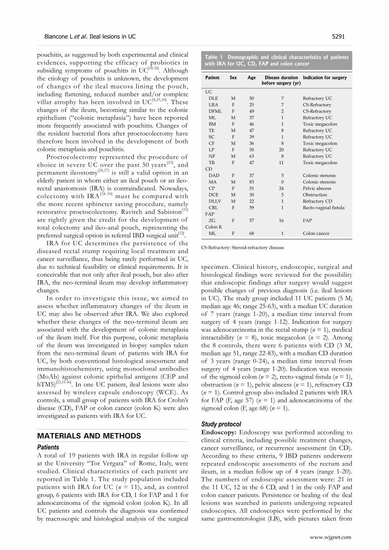
www.wjgnet.com
Biancone L et al . Ileal lesions in UC 5291
pouchitis, as suggested by both experimental and clinical evidences, supporting the efficacy of probiotics in subsiding symptoms of pouchitis in UC[18-22]. Although the etiology of pouchitis is unknown, the development of changes of the ileal mucosa lining the pouch, including flattening, reduced number and/or complete villar atrophy has been involved in UC[4,23,24]. These changes of the ileum, becoming similar to the colonic epithelium (“colonic metaplasia”) have been reported more frequently associated with pouchitis. Changes of the resident bacterial flora after proctocolectomy have therefore been involved in the development of both colonic metaplasia and pouchitis.
Proctocolectomy represented the procedure of choice in severe UC over the past 30 years[25], and permanent ileostomy[26,27] is still a valid option in an elderly patient in whom either an ileal pouch or an ileo-rectal anastomosis (IRA) is contraindicated. Nowadays, colectomy with IRA[28-30] must be compared with the more recent sphincter saving procedure, namely restorative proctocolectomy. Ravitch and Sabiston[31] are rightly given the credit for the development of total colectomy and ileo-anal pouch, representing the preferred surgical option in referral IBD surgical unit[32].
IRA for UC determines the persistence of the diseased rectal stump requiring local treatment and cancer surveillance, thus being rarely performed in UC, due to technical feasibility or clinical requirements. It is conceivable that not only after ileal pouch, but also after IRA, the neo-terminal ileum may develop inflammatory changes.
In order to investigate this issue, we aimed to assess whether inflammatory changes of the ileum in UC may also be observed after IRA. We also explored whether these changes of the neo-terminal ileum are associated with the development of colonic metaplasia of the ileum itself. For this purpose, colonic metaplasia of the ileum was investigated in biopsy samples taken from the neo-terminal ileum of patients with IRA for UC, by both conventional histological assessment and immunohistochemistry, using monoclonal antibodies (MoAb) against colonic epithelial antigens (CEP and hTM5)[23,33-36]. In one UC patient, ileal lesions were also assessed by wireless capsule endoscopy (WCE). As controls, a small group of patients with IRA for Crohn’s disease (CD), FAP or colon cancer (colon K) were also investigated as patients with IRA for UC.
MATERIALS AND METHODSPatientsA total of 19 patients with IRA in regular follow up at the University “Tor Vergata” of Rome, Italy, were studied. Clinical characteristics of each patient are reported in Table 1. The study population included patients with IRA for UC (n = 11), and, as control group, 6 patients with IRA for CD, 1 for FAP and 1 for adenocarcinoma of the sigmoid colon (colon K). In all UC patients and controls the diagnosis was confirmed by macroscopic and histological analysis of the surgical
specimen. Clinical history, endoscopic, surgical and histological findings were reviewed for the possibility that endoscopic findings after surgery would suggest possible changes of previous diagnosis (i.e. ileal lesions in UC). The study group included 11 UC patients (5 M; median age 46; range 25-63), with a median UC duration of 7 years (range 1-20), a median time interval from surgery of 4 years (range 1-12). Indication for surgery was adenocarcinoma in the rectal stump (n = 1), medical intractability (n = 8), toxic megacolon (n = 2). Among the 8 controls, there were 6 patients with CD (3 M, median age 51, range 22-83), with a median CD duration of 3 years (range 0-24), a median time interval from surgery of 4 years (range 1-20). Indication was stenosis of the sigmoid colon (n = 2), recto-vaginal fistula (n = 1), obstruction (n = 1), pelvic abscess (n = 1), refractory CD (n = 1). Control group also included 2 patients with IRA for FAP (F, age 57) (n = 1) and adenocarcinoma of the sigmoid colon (F, age 68) (n = 1).
Study protocolEndoscopy: Endoscopy was performed according to clinical criteria, including possible treatment changes, cancer surveillance, or recurrence assessment (in CD). According to these criteria, 9 IBD patients underwent repeated endoscopic assessments of the rectum and ileum, in a median follow up of 4 years (range 1-20). The numbers of endoscopic assessment were: 21 in the 11 UC, 12 in the 6 CD, and 1 in the only FAP and colon cancer patients. Persistence or healing of the ileal lesions was searched in patients undergoing repeated endoscopies. All endoscopies were performed by the same gastroenterologist (LB), with pictures taken from
Table 1 Demographic and clinical characteristics of patients with IRA for UC, CD, FAP and colon cancer
Patient Sex Age Disease duration before surgery (yr)
Indication for surgery
UC DLE M 50 7 Refractory UC LRA F 25 7 CS-Refractory DFML F 49 2 CS-Refractory ML M 37 1 Refractory UC BM F 46 1 Toxic megacolon TE M 47 8 Refractory UC SC F 39 1 Refractory UC CF M 36 8 Toxic megacolon LP F 50 20 Refractory UC NF M 63 8 Refractory UC TR F 47 11 Toxic megacolonCD DAD F 37 5 Colonic stenosis MA M 83 0 Colonic stenosis CP F 51 24 Pelvic abscess DCE M 30 5 Obstruction DLLV M 22 1 Refractory CD CRL F 59 1 Recto-vaginal fistulaFAP ZG F 57 16 FAPColon K ML F 68 1 Colon cancer
CS-Refractory: Steroid-refractory disease.
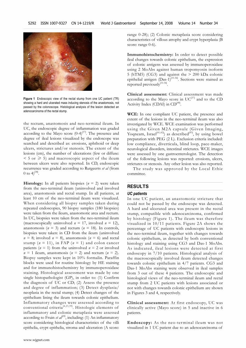
the rectum, anastomosis and neo-terminal ileum. In UC, the endoscopic degree of inflammation was graded according to the Mayo score (0-4)[37]. The presence and degree of ileal lesions visualized by the endoscope was searched and described as: erosions, aphthoid or deep ulcers, strictures and/or stenosis. The extent of the lesions (cm), the number of ulcerations (few or diffuse: < 5 or ≥ 5) and macroscopic aspect of the ileum between ulcers were also reported. In CD, endoscopic recurrence was graded according to Rutgeerts et al (from 0 to 4)[38].
Histology: In all patients biopsies (n = 2) were taken from the neo-terminal ileum (uninvolved and involved area), anastomosis and rectal stump. In all patients, at least 10 cm of the neo-terminal ileum were visualized. When considering all biopsy samples taken during repeated endoscopies, 90 biopsy samples (180 biopsies) were taken from the ileum, anastomotic area and rectum. In UC, biopsies were taken from the neo-terminal ileum (macroscopically uninvolved n = 17, involved n = 12), anastomosis (n = 3) and rectum (n = 18). In controls, biopsies were taken: in CD from the ileum (uninvolved n = 8; involved n = 8), anastomosis (n = 6) and rectal stump (n = 11), in FAP (n = 1) and colon cancer patients (n = 1) from the uninvolved n = 2 or involved n = 1 ileum, anastomosis (n = 2) and rectum (n = 2). Biopsy samples were kept in 10% formalin. Paraffin blocks were used for routine histology by HE staining and for immunohistochemistry by immunoperoxidase staining. Histological assessment was made by one single histopathologist (GP), in order to: (1) Confirm the diagnosis of UC or CD; (2) Assess the presence and degree of inflammation; (3) Detect dysplasia/neoplasia in the rectal stump; (4) Detect changes of the epithelium lining the ileum towards colonic epithelium. Inflammatory changes were assessed according to conventional criteria [4,23,39]. Histologic elements of inflammatory and colonic metaplasia were assessed according to Fruin et al[24], including: (1) An inflammatory score considering histological characteristics of the villi epithelia, crypt epithelia, stroma and ulceration (A score:
range 0-28); (2) Colonic metaplasia score considering characteristics of villous atrophy and crypt hyperplasia (B score: range 0-6).
Immunohistochemistry: In order to detect possible ileal changes towards colonic epithelium, the expression of colonic antigens was assessed by immunoperoxidase using 2 MoAbs against human tropomyosin isoform 5 (hTM5) (CG3) and against the > 200 kDa colonic epithelial antigen (Das-1)[33-36]. Sections were stained as reported previously[33-36].
Clinical assessment: Clinical assessment was made according to the Mayo score in UC[37] and to the CD Activity Index (CDAI) in CD[40].
WCE: In one compliant UC patient, the presence and extent of the lesions in the neo-terminal ileum was also investigated by WCE. WCE examination was performed us ing the Given M2A capsule (Given Imaging, Yoqneam, Israel[41,42]) as described[43], by using bowel preparation with PEG (2 L). Exclusion criteria included: low compliance, diverticula, blind loop, pace-maker, neurological disorders, intestinal strictures. WCE images were assessed by one gastroenterologist. The detection of the following lesions was reported: erosions, ulcers, strictures or stenosis. Any other lesion was also reported.
The study was approved by the Local Ethic committee.
RESULTSUC patientsIn one UC patient, an anastomotic stricture that could not be passed by the endoscope was detected. A hard and ulcerated area was present in the rectal stump, compatible with adenocarcinoma, confirmed by histology (Figure 1). The ileum was therefore visualized in 10/11 patients. Figure 2A shows the percentage of UC patients with endoscopic lesions in the neo-terminal ileum, together with changes towards colonic epithelium, as detected by both conventional histology and staining using CG3 and Das-1 MoAbs. As indicated, i leal lesions were detected at f irst endoscopy in 7/10 patients. Histological analysis of the macroscopically involved ileum detected changes towards colonic epithelium in 4/7 patients. CG3 and Das-1 MoAbs staining were observed in ileal samples from 3 out of these 4 patients. The endoscopic and histological views of the neo-terminal ileum and rectal stump from 2 UC patients with lesions associated or not with changes towards colonic epithelium are shown in Figures 3 and 4, respectively.
Clinical assessment: At first endoscopy, UC was clinically active (Mayo score) in 5 and inactive in 6 patients.
Endoscopy: As the neo-terminal i leum was not visualized in 1 UC patient due to an adenocarcinoma of
Figure 1 Endoscopic view of the rectal stump from one UC patient (TR) showing a hard and ulcerated mass inducing stenosis of the anastomosis, not passed by the colonoscope. Histological analysis of the lesion detected an adenocarcinoma of the rectal stump.
www.wjgnet.com
5292 ISSN 1007-9327 CN 14-1219/R World J Gastroenterol September 14, 2008 Volume 14 Number 34
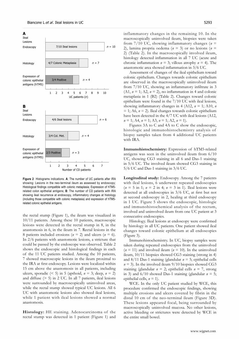
the rectal stump (Figure 1), the ileum was visualized in 10/11 patients. Among these 10 patients, macroscopic lesions were detected in the rectal stump in 8, in the anastomosis in 6, in the ileum in 7. Rectal lesions in the 8 patients included erosions (n = 2) and ulcers (n = 6). In 2/6 patients with anastomotic lesions, a stricture that could be passed by the endoscope was observed. Table 2 shows the endoscopic and histological findings in each of the 11 UC patients studied. Among the 10 patients, 7 showed macroscopic lesions in the ileum proximal to the IRA at first endoscopy. Lesions were localized within 15 cm above the anastomosis in all patients, including ulcers, sporadic (< 5) in 5 (apthoid, n = 3; deep, n = 2) and diffuse (> 5) in 2 UC. In all 7 patients, ileal lesions were surrounded by macroscopically uninvolved areas, while the rectal stump showed typical UC lesions. All 6 UC with anastomotic lesions also showed ileal lesions, while 1 patient with ileal lesions showed a normal anastomosis.
Histology: HE staining. Adenocarcinoma of the rectal stump was detected in 1 patient (Figure 1) and
inflammatory changes in the remaining 10. In the macroscopically uninvolved ileum, biopsies were taken from 7/10 UC, showing inflammatory changes (n = 2), lamina propria oedema (n = 3) or no lesions (n = 2) (Table 2). In the macroscopically involved ileum, histology detected inflammation in all 7 UC (acute and chronic inflammation n = 3; villous atrophy n = 4). The anastomotic area showed inflammation in 3/6 UC.
Assessment of changes of the ileal epithelium toward colonic epithelium. Changes towards colonic epithelium are observed in the macroscopically uninvolved ileum from 7/10 UC, showing an inflammatory infiltrate in 3 (A1, n = 1; A2, n = 2), no inflammation in 4 and colonic metaplasia in 1 (B2) (Table 2). Changes toward colonic epithelium were found in the 7/10 UC with ileal lesions, showing inflammatory changes in 4 (A12, n = 1; A10, n = 1; A6, n = 2). Ileal changes towards colonic epithelium have been detected in the 4/7 UC with ileal lesions (A12, n = 1; A4, n = 1; A3, n = 1; A2, n = 1).
Figures 3A to C and 4A to C show the endoscopic, histologic and immunohistochemistry analysis of biopsy samples taken from 4 additional UC patients with IRA.
Immunohistochemistry: Expression of hTM5-related antigens was seen in the uninvolved ileum from 6/10 UC, showing CG3 staining in all 6 and Das-1 staining in 5/6 UC. The involved ileum showed CG3 staining in 5/6 UC and Das-1 staining in 3/6 UC.
Longitudinal study: Endoscopy. Among the 7 patients with ileal lesions, 6 underwent repeated endoscopies (n = 5 in 1; n = 2 in 4; n = 3 in 1). Ileal lesions were detected at all endoscopies in 3/6 UC, at first but not at second endoscopy in 2, healing at third endoscopy in 1 UC. Figure 5 shows the endoscopic, histologic and immunohistochemical analysis of the rectum, involved and uninvolved ileum from one UC patient at 3 consecutive endoscopies.
Histology. Ileal lesions at endoscopy were confirmed by histology in all UC patients. One patient showed ileal changes toward colonic epithelium at all endoscopies (Figure 3).
Immunohistochemistry. In UC, biopsy samples were taken during repeated endoscopies from the uninvolved (n = 11) and involved ileum (n = 10). In the uninvolved ileum, 10/11 biopsies showed CG3 staining (strong in 4) and 8/11 Das-1 staining (glandular n = 5; epithelial cells n = 3). In the involved ileum 9/10 biopsies showed CG3 staining (glandular n = 2; epithelial cells n = 7, strong in 3) and 6/10 showed Das-1 staining (glandular n = 5; epithelial cells, n = 1).
WCE. In the only UC patient studied by WCE, this procedure confirmed the endoscopic findings, showing multiple erosions and ulcers covered by fibrin in the distal 10 cm of the neo-terminal ileum (Figure 3D). These lesions appeared focal, being surrounded by macroscopically uninvolved mucosa. No other lesions, active bleeding or strictures were detected by WCE in the entire small bowel.
2/3 Posltive n = 3
3/4 Col. Met. n = 4
4/6 Ileal lesions n = 6
1 2 3 4 5 6 7
IlealLesions
Endoscopy
Histology
Expression ofcolonic epithelialantigens (hTM5)
Number of CD patients
B
3/4 Posltive n = 4
4/7 Colonic Metaplasia n = 7
7/10 Ileal lesions n = 10
1 2 3 4 5 6 7 8 9 10
IlealLesions
Endoscopy
Histology
Expression ofcolonic epithelialantigens (hTM5)
UC patients (n )
A
Figure 2 Histograms indications. A: The number of UC patients after IRA showing: Lesions in the neo-terminal ileum as assessed by endoscopy; Histological findings compatible with colonic metaplasia; Expression of hTM5-related colon epithelial antigens; B: The number of CD patients with IRA showing ileal recurrence at endoscopy, inflammatory changes at histology (including those compatible with colonic metaplasia) and expression of hTM5-related colonic epithelial antigens.
www.wjgnet.com
Biancone L et al . Ileal lesions in UC 5293
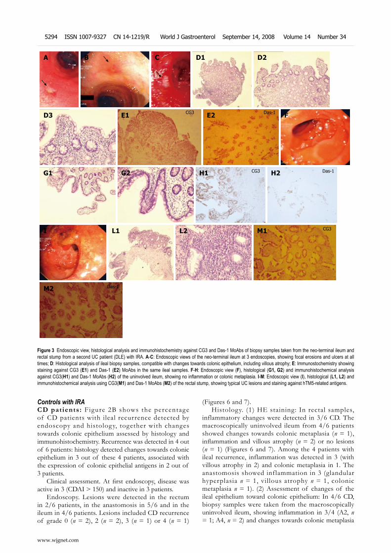
Controls with IRACD patients: F igure 2B shows the percentage of CD patients with ileal recurrence detected by endoscopy and histology, together with changes towards colonic epithelium assessed by histology and immunohistochemistry. Recurrence was detected in 4 out of 6 patients: histology detected changes towards colonic epithelium in 3 out of these 4 patients, associated with the expression of colonic epithelial antigens in 2 out of 3 patients.
Clinical assessment. At first endoscopy, disease was active in 3 (CDAI > 150) and inactive in 3 patients.
Endoscopy. Lesions were detected in the rectum in 2/6 patients, in the anastomosis in 5/6 and in the ileum in 4/6 patients. Lesions included CD recurrence of grade 0 (n = 2), 2 (n = 2), 3 (n = 1) or 4 (n = 1)
(Figures 6 and 7). Histology. (1) HE staining: In rectal samples,
inflammatory changes were detected in 3/6 CD. The macroscopically uninvolved ileum from 4/6 patients showed changes towards colonic metaplasia (n = 1), inflammation and villous atrophy (n = 2) or no lesions (n = 1) (Figures 6 and 7). Among the 4 patients with ileal recurrence, inflammation was detected in 3 (with villous atrophy in 2) and colonic metaplasia in 1. The anastomosis showed inflammation in 3 (glandular hyperplasia n = 1, villous atrophy n = 1, colonic metaplasia n = 1). (2) Assessment of changes of the ileal epithelium toward colonic epithelium: In 4/6 CD, biopsy samples were taken from the macroscopically uninvolved ileum, showing inflammation in 3/4 (A2, n = 1; A4, n = 2) and changes towards colonic metaplasia
Figure 3 Endoscopic view, histological analysis and immunohistochemistry against CG3 and Das-1 MoAbs of biopsy samples taken from the neo-terminal ileum and rectal stump from a second UC patient (DLE) with IRA. A-C: Endoscopic views of the neo-terminal ileum at 3 endoscopies, showing focal erosions and ulcers at all times; D: Histological analysis of ileal biopsy samples, compatible with changes towards colonic epithelium, including villous atrophy; E: Immunostochemistry showing staining against CG3 (E1) and Das-1 (E2) MoAbs in the same ileal samples. F-H: Endoscopic view (F), histological (G1, G2) and immunohistochemical analysis against CG3(H1) and Das-1 MoAbs (H2) of the uninvolved ileum, showing no inflammation or colonic metaplasia. I-M: Endoscopic view (I), histological (L1, L2) and immunohistochemical analysis using CG3(M1) and Das-1 MoAbs (M2) of the rectal stump, showing typical UC lesions and staining against hTM5-related antigens.
M2 Das-1
M1 CG3
H2 Das-1H1 CG3
E2 Das-1E1 CG3
C
←
B ←A
←
L2L1I
G2G1
FD3
D2D1
www.wjgnet.com
5294 ISSN 1007-9327 CN 14-1219/R World J Gastroenterol September 14, 2008 Volume 14 Number 34
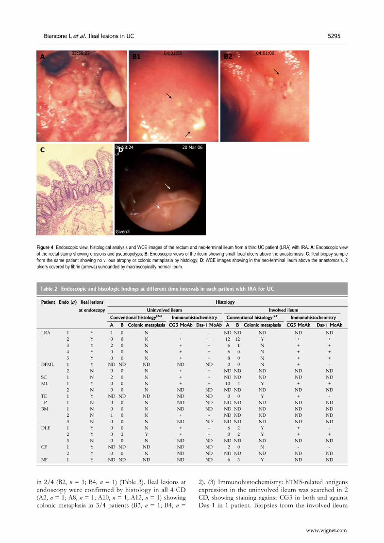
in 2/4 (B2, n = 1; B4, n = 1) (Table 3). Ileal lesions at endoscopy were confirmed by histology in all 4 CD (A2, n = 1; A8, n = 1; A10, n = 1; A12, n = 1) showing colonic metaplasia in 3/4 patients (B3, n = 1; B4, n =
2). (3) Immunohistochemistry: hTM5-related antigens expression in the uninvolved ileum was searched in 2 CD, showing staining against CG3 in both and against Das-1 in 1 patient. Biopsies from the involved ileum
C
Figure 4 Endoscopic view, histological analysis and WCE images of the rectum and neo-terminal ileum from a third UC patient (LRA) with IRA. A: Endoscopic view of the rectal stump showing erosions and pseudopolyps; B: Endoscopic views of the ileum showing small focal ulcers above the anastomosis; C: Ileal biopsy sample from the same patient showing no villous atrophy or colonic metaplasia by histology; D: WCE images showing in the neo-terminal ileum above the anastomosis, 2 ulcers covered by fibrin (arrows) surrounded by macroscopically normal ileum.
D
←
←
06:58:24al
20 Mar 06
Given®
A 03:56:27 B1
←
←
04:01:56 B2
←
04:01:06
Table 2 Endoscopic and histologic findings at different time intervals in each patient with IRA for UC
Patient Endo (n) Ileal lesions Histology
at endoscopy Uninvolved ileum Involved ileum
Conventional histology[23] Immunohistochemistry Conventional histology[23] Immunohistochemistry
A B Colonic metaplasia CG3 MoAb Das-1 MoAb A B Colonic metaplasia CG3 MoAb Das-1 MoAb
LRA 1 Y 1 0 N - - ND ND ND ND ND2 Y 0 0 N + + 12 12 Y + +3 Y 2 0 N + + 6 1 N + +4 Y 0 0 N + + 6 0 N + +5 Y 0 0 N + + 8 0 N + +
DFML 1 Y ND ND ND ND ND 0 0 N + -2 N 0 0 N + + ND ND ND ND ND
SC 1 N 2 0 N + + ND ND ND ND NDML 1 Y 0 0 N + + 10 4 Y + +
2 N 0 0 N ND ND ND ND ND ND NDTE 1 Y ND ND ND ND ND 0 0 Y + -LP 1 N 0 0 N ND ND ND ND ND ND NDBM 1 N 0 0 N ND ND ND ND ND ND ND
2 N 1 0 N + - ND ND ND ND ND3 N 0 0 N ND ND ND ND ND ND ND
DLE 1 Y 0 0 N + - 6 2 Y + -2 Y 0 2 Y + + 0 2 Y + +3 N 0 0 N ND ND ND ND ND ND ND
CF 1 Y ND ND ND ND ND 2 0 N - -2 Y 0 0 N ND ND ND ND ND ND ND
NF 1 Y ND ND ND ND ND 6 3 Y ND ND
www.wjgnet.com
Biancone L et al . Ileal lesions in UC 5295
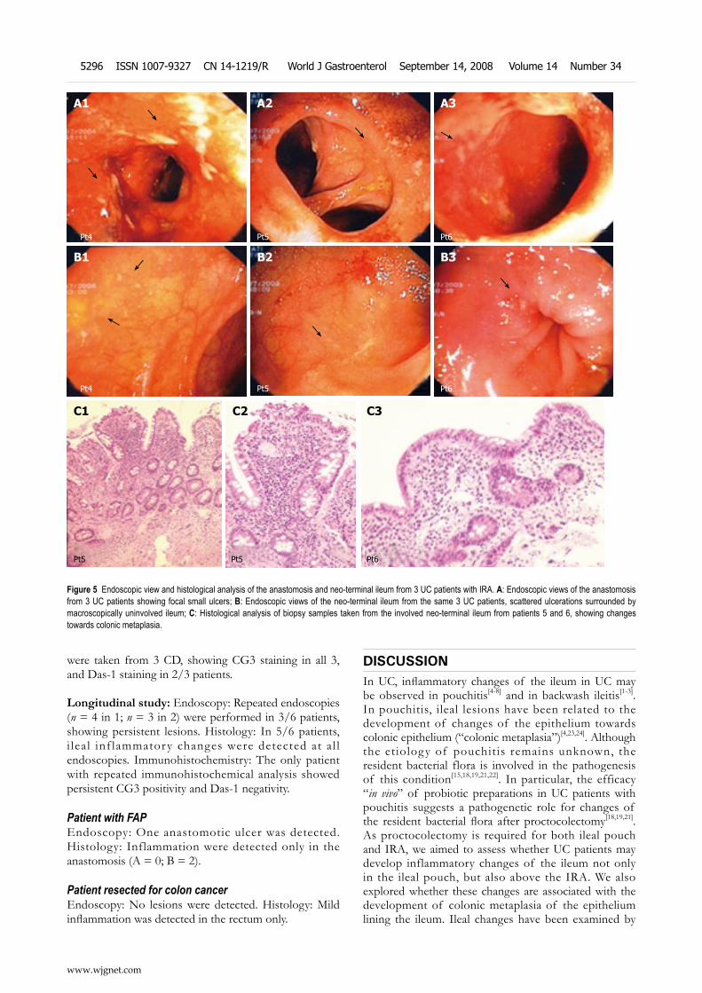
were taken from 3 CD, showing CG3 staining in all 3, and Das-1 staining in 2/3 patients.
Longitudinal study: Endoscopy: Repeated endoscopies (n = 4 in 1; n = 3 in 2) were performed in 3/6 patients, showing persistent lesions. Histology: In 5/6 patients, i leal inf lammatory changes were detected at a l l endoscopies. Immunohistochemistry: The only patient with repeated immunohistochemical analysis showed persistent CG3 positivity and Das-1 negativity.
Patient with FAPEndoscopy: One anastomotic ulcer was detected. Histology: Inflammation were detected only in the anastomosis (A = 0; B = 2).
Patient resected for colon cancerEndoscopy: No lesions were detected. Histology: Mild inflammation was detected in the rectum only.
DISCUSSIONIn UC, inflammatory changes of the ileum in UC may be observed in pouchitis[4-8] and in backwash ileitis[1-3]. In pouchitis, ileal lesions have been related to the development of changes of the epithelium towards colonic epithelium (“colonic metaplasia”)[4,23,24]. Although the etiology of pouchitis remains unknown, the resident bacterial flora is involved in the pathogenesis of this condition[15,18,19,21,22]. In particular, the efficacy “in vivo” of probiotic preparations in UC patients with pouchitis suggests a pathogenetic role for changes of the resident bacterial flora after proctocolectomy[18,19,21]. As proctocolectomy is required for both ileal pouch and IRA, we aimed to assess whether UC patients may develop inflammatory changes of the ileum not only in the ileal pouch, but also above the IRA. We also explored whether these changes are associated with the development of colonic metaplasia of the epithelium lining the ileum. Ileal changes have been examined by
Figure 5 Endoscopic view and histological analysis of the anastomosis and neo-terminal ileum from 3 UC patients with IRA. A: Endoscopic views of the anastomosis from 3 UC patients showing focal small ulcers; B: Endoscopic views of the neo-terminal ileum from the same 3 UC patients, scattered ulcerations surrounded by macroscopically uninvolved ileum; C: Histological analysis of biopsy samples taken from the involved neo-terminal ileum from patients 5 and 6, showing changes towards colonic metaplasia.
C3
Pt6
C1
Pt5
C2
Pt5
B3
Pt6
←
B2
Pt5
←
B1
Pt4
←
←
A3
Pt6
←
A2
Pt5
←
A1←
←
Pt4
www.wjgnet.com
5296 ISSN 1007-9327 CN 14-1219/R World J Gastroenterol September 14, 2008 Volume 14 Number 34
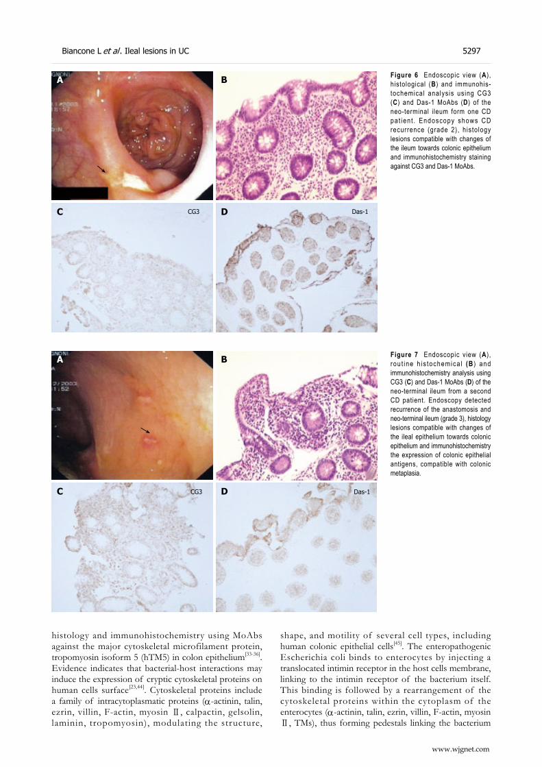
histology and immunohistochemistry using MoAbs against the major cytoskeletal microfilament protein, tropomyosin isoform 5 (hTM5) in colon epithelium[33-36]. Evidence indicates that bacterial-host interactions may induce the expression of cryptic cytoskeletal proteins on human cells surface[23,44]. Cytoskeletal proteins include a family of intracytoplasmatic proteins (α-actinin, talin, ezrin, villin, F-actin, myosin Ⅱ, calpactin, gelsolin, laminin, tropomyosin), modulating the structure,
shape, and motility of several cell types, including human colonic epithelial cells[45]. The enteropathogenic Escherichia coli binds to enterocytes by injecting a translocated intimin receptor in the host cells membrane, linking to the intimin receptor of the bacterium itself. This binding is followed by a rearrangement of the cytoskeletal proteins within the cytoplasm of the enterocytes (α-actinin, talin, ezrin, villin, F-actin, myosin Ⅱ, TMs), thus forming pedestals linking the bacterium
Figure 6 Endoscopic view (A), histological (B) and immunohis-tochemical analysis using CG3 (C) and Das-1 MoAbs (D) of the neo-terminal i leum form one CD pa t ien t . Endoscopy shows CD recurrence (grade 2), histology lesions compatible with changes of the ileum towards colonic epithelium and immunohistochemistry staining against CG3 and Das-1 MoAbs.
BA
←
C CG3 D Das-1
Figure 7 Endoscopic view (A), rou t ine h is tochemica l (B ) and immunohistochemistry analysis using CG3 (C) and Das-1 MoAbs (D) of the neo-terminal ileum from a second CD patient. Endoscopy detected recurrence of the anastomosis and neo-terminal ileum (grade 3), histology lesions compatible with changes of the ileal epithelium towards colonic epithelium and immunohistochemistry the expression of colonic epithelial antigens, compatible with colonic metaplasia.
D Das-1C CG3
A
←
B
www.wjgnet.com
Biancone L et al . Ileal lesions in UC 5297
itself to colonic epithelial cells[44,45]. These observations prompted us to also assess whether hTM5 may be expressed on the epithelial cells lining the neo-terminal ileum in UC patients with IRA, due to changes of the ileum towards colonic epithelium after colectomy and the possible relation between the development of colonic metaplasia and ileal lesions. Our findings from a limited number of patients with IBD suggest that ileal lesions may be observed after IRA, although more frequently in UC (7 out of 10 patients). The presence of scattered erosions and ulcers in the neo-terminal ileum above the anastomosis was confirmed by WCE images, acquired as described[41-43], from the only patient performing this procedure. Despite the focal inflammation of the ileum, histological analysis of the surgical colonic specimen confirmed the diagnosis of UC in all 7 patients. Ileal lesions were associated with epithelial changes towards colonic epithelium in 4 out of these 7 patients, associated with the expression of hTM5-related antigens in 3 out of these 4 patients. The same findings were not detected in any of the 7 UC patients with no ileal lesions. Surprisingly, colonic metaplasia and the expression of colonic epithelial antigens were also observed in some of the few tested patients with IRA for CD. Although “colonization” of the ileum after total colectomy for any indication has been described, its possible relation with the development of ileal lesions is unknown. The score from Fruin et al[24] was used for assessing colonic metaplasia, as for pouchitis. Major limits of our study include the low number of patients and the cross-sectional study plan, not allowing comparisons before versus after surgery. Although it is said that colectomy cures UC, this study underscores the fact that surgery is not the final answer due to the high
incidence of pouchitis and other functional problems experience by these patients. Of particular concern is the asymptomatic neoplasia that can arise in the residual rectal stump required for these surgical procedures, which was seen in one of the study patients.
Although not conclusive, our findings suggest that lesions may be observed in the neo-terminal ileum of UC and CD patients following IRA. These changes are towards colonic epithelium phenotype and with the expression of colonic epithelial antigens in some patients. Longitudinal studies are ongoing for further characterization of the molecular mechanisms leading to ileal changes in UC. Present findings suggest that changes of the ileal content after colectomy may contribute to the development of colonic type of metaplasia, leading to ileal lesions both in the pouch and in the neo-terminal ileum after IRA.
COMMENTSBackgroundInflammatory changes of the distal ileum in ulcerative colitis (UC) may be observed in backwash ileitis and after total proctocolectomy with ileal pouch (“pouchitis”). Although total proctocolectomy with ileal pouch currently represents the most frequently performed surgical procedure in UC, colectomy with ileo-rectal anastomosis (IRA) is still in these patients. The persistence of the diseased rectal stump after IRA requires endoscopical surveillance. Ileal inflammation may be observed in UC patients with pouchitis, being related to changes of the ileal epithelium towards colonic epithelium (“colonic metaplasia”). Whether the ileum above IRA in patients with IRA for UC may develop inflammatory changes as observed in pouchitis is unknown and this observation may be useful for proper follow up of patients after surgery. Research frontiersThe etiology of pouchitis in patients with UC is unknown. However, the development of changes of the ileal mucosa lining the pouch, including flattening, reduced number and/or complete villar atrophy has been involved in the pathogenesis of pouchitis. These changes of the ileum, becoming similar
www.wjgnet.com
5298 ISSN 1007-9327 CN 14-1219/R World J Gastroenterol September 14, 2008 Volume 14 Number 34
Table 3 Endoscopical and histologic findings of patients with IRA for CD, FAP or colonic K
Patient Endo (n) Ileal lesions at endoscopy Histology
Uninvolved ileum Involved ileumConventional histology[23] Immunohistochemistry Conventional histology[23] Immunohistochemistry
Score Score Colonic CG3 Das-1 Score Score Colonic CG3 Das-1
A B metaplasia MoAb MoAb A B metaplasia MoAb MoAb
DAD 1 Y ND ND ND ND ND 4 0 N + -(CD-1) 2 Y ND ND ND ND ND 0 0 N + -
3 Y 0 0 N + - 0 0 N + -4 Y 0 0 N + - 8 4 Y + -
MA 1 N 4 4 Y + + 0 3 Y + +(CD-2)DCE 1 N ND ND ND ND ND ND ND ND ND ND(CD-3)CP 1 N 0 0 N ND ND ND ND ND ND ND(CD-4) 2 N 2 0 N ND ND ND ND ND ND ND
3 Y 1 1 N ND ND 2 1 N ND NDDLV 1 Y ND ND ND ND ND 4 1 Y + +(CD-5) 2 N 2 2 Y ND ND
3 Y 4 2 Y ND ND 12 4 Y ND NDCRL 1 Y ND ND ND ND ND 10 3 Y ND ND(CD-6)ZG 1 N 0 0 N ND ND 8 2 N ND ND(FAP)ML 1 N 0 0 N ND ND ND ND ND ND ND(K)
to the colonic epithelium (“colonic metaplasia”) have been reported more frequently associated with pouchitis. Changes of the resident bacterial flora after proctocolectomy therefore have been involved in the development of both colonic metaplasia and pouchitis. It is conceivable that after total colectomy for UC and related changes of the common bacterial flora, ileal lesions may develop not only after ileal pouch, but also after IRA.Innovations and breakthroughsThe present study showed that In UC, ileal lesions associated with changes towards colonic epithelium may develop after IRA. Changes of the ileal content after colectomy may contribute to the development of colonic metaplasia, leading to ileal lesions also after IRA. ApplicationsResults from our study suggest that patients with IRA for UC need endoscopical assessment not only for cancer surveillance but also for assessing the possible development of ileal lesions above anastomosis. Present findings also add new insights regarding the natural history of UC after IRA.TerminologyTropomyosin isoform 5 (hTM5) is a cytoskeletal microfilament protein present in the epithelium from human colon, but not from the ileum. Mucosal and circulating antibodies against hTM5 have been demonstrated in patients with UC. Evidences indicate that bacterial-host interactions may induce the expression of cryptic cytoskeletal proteins on human cells surface. Cytoskeletal proteins include a family of intracytoplasmatic proteins (α-actinin, talin, ezrin, villin, F-actin, myosin Ⅱ, calpactin, gelsolin, laminin, tropomyosin), modulating the structure, shape, and motility of several cell types, including human colonic epithelial cells.Peer reviewThis is a clinical and immunohistochemical study supporting the need of continued endoscopical follow up of UC patients after IRA.
REFERENCES1 Abdelrazeq AS, Wilson TR, Leitch DL, Lund JN, Leveson
SH. Ileitis in ulcerative colitis: is it a backwash? Dis Colon Rectum 2005; 48: 2038-2046
2 Gustavsson S, Weiland LH, Kelly KA. Relationship of backwash ileitis to ileal pouchitis after ileal pouch-anal anastomosis. Dis Colon Rectum 1987; 30: 25-28
3 Haskell H, Andrews CW Jr, Reddy SI, Dendrinos K, Farraye FA, Stucchi AF, Becker JM, Odze RD. Pathologic features and clinical significance of "backwash" ileitis in ulcerative colitis. Am J Surg Pathol 2005; 29: 1472-1481
4 Apel R, Cohen Z, Andrews CW Jr, McLeod R, Steinhart H, Odze RD. Prospective evaluation of early morphological changes in pelvic ileal pouches. Gastroenterology 1994; 107: 435-443
5 Arai K, Koganei K, Kimura H, Akatani M, Kitoh F, Sugita A, Fukushima T. Incidence and outcome of complications following restorative proctocolectomy. Am J Surg 2005; 190: 39-42
6 Brunel M, Penna C, Tiret E, Balladur P, Parc R. Restorative proctocolectomy for distal ulcerative colitis. Gut 1999; 45: 542-545
7 Chambers WM, McC Mortensen NJ. Should ileal pouch-anal anastomosis include mucosectomy? Colorectal Dis 2007; 9: 384-392
8 Madden MV, Farthing MJ, Nicholls RJ. Inflammation in ileal reservoirs: 'pouchitis'. Gut 1990; 31: 247-249
9 Meagher AP, Farouk R, Dozois RR, Kelly KA, Pemberton JH. J ileal pouch-anal anastomosis for chronic ulcerative colitis: complications and long-term outcome in 1310 patients. Br J Surg 1998; 85: 800-803
10 Meuwissen SG, Hoitsma H, Boot H, Seldenrijk CA. Pouchitis (pouch ileitis). Neth J Med 1989; 35 Suppl 1: S54-S66
11 Michelassi F, Lee J, Rubin M, Fichera A, Kasza K, Karrison T, Hurst RD. Long-term functional results after ileal pouch anal restorative proctocolectomy for ulcerative colitis: a prospective observational study. Ann Surg 2003; 238: 433-441; discussion 442-445
12 Sagar PM, Pemberton JH. Ileo-anal pouch function and dysfunction. Dig Dis 1997; 15: 172-188
13 Salemans JM, Nagengast FM, Lubbers EJ, Kuijpers JH. Postoperative and long-term results of ileal pouch-anal anastomosis for ulcerative colitis and familial polyposis coli. Dig Dis Sci 1992; 37: 1882-1889
14 Sandborn WJ, Tremaine WJ, Batts KP, Pemberton JH, Phillips SF. Pouchitis after ileal pouch-anal anastomosis: a Pouchitis Disease Activity Index. Mayo Clin Proc 1994; 69: 409-415
15 Shen B, Fazio VW, Remzi FH, Lashner BA. Clinical approach to diseases of ileal pouch-anal anastomosis. Am J Gastroenterol 2005; 100: 2796-2807
16 Zuccaro G Jr, Fazio VW, Church JM, Lavery IC, Ruderman WB, Farmer RG. Pouch ileitis. Dig Dis Sci 1989; 34: 1505-1510
17 Shen B, Fazio VW, Remzi FH, Delaney CP, Bennett AE, Achkar JP, Brzezinski A, Khandwala F, Liu W, Bambrick ML, Bast J, Lashner B. Comprehensive evaluation of inflammatory and noninflammatory sequelae of ileal pouch-anal anastomoses. Am J Gastroenterol 2005; 100: 93-101
18 Gionchetti P, Rizzello F, Venturi A, Brigidi P, Matteuzzi D, Bazzocchi G, Poggioli G, Miglioli M, Campieri M. Oral bacteriotherapy as maintenance treatment in patients with chronic pouchitis: a double-blind, placebo-controlled trial. Gastroenterology 2000; 119: 305-309
19 Gionchetti P, Rizzello F, Helwig U, Venturi A, Lammers KM, Brigidi P, Vitali B, Poggioli G, Miglioli M, Campieri M. Prophylaxis of pouchitis onset with probiotic therapy: a double-blind, placebo-controlled trial. Gastroenterology 2003; 124: 1202-1209
20 Hurst RD, Molinari M, Chung TP, Rubin M, Michelassi F. Prospective study of the incidence, timing and treatment of pouchitis in 104 consecutive patients after restorative proctocolectomy. Arch Surg 1996; 131: 497-500; discussion 501-502
21 Mimura T, Rizzello F, Helwig U, Poggioli G, Schreiber S, Talbot IC, Nicholls RJ, Gionchetti P, Campieri M, Kamm MA. Once daily high dose probiotic therapy (VSL#3) for maintaining remission in recurrent or refractory pouchitis. Gut 2004; 53: 108-114
22 Sandborn WJ, McLeod R, Jewell DP. Medical therapy for induction and maintenance of remission in pouchitis: a systematic review. Inflamm Bowel Dis 1999; 5: 33-39
23 Biancone L, Palmieri G, Lombardi A, Colantoni A, Tonelli F, Das KM, Pallone F. Tropomyosin expression in the ileal pouch: a relationship with the development of pouchitis in ulcerative colitis. Am J Gastroenterol 2003; 98: 2719-2726
24 Fruin AB, El-Zammer O, Stucchi AF, O'Brien M, Becker JM. Colonic metaplasia in the ileal pouch is associated with inflammation and is not the result of long-term adaptation. J Gastrointest Surg 2003; 7: 246-253; discussion 253-254
25 Kettlewell MGW. Proctocolectomy for ulcerative colitis. In: Allan RN, Keighley MRB, Alexander-Williams J & Hawkins C. Inflammatory Bowel Diseases. Edinburgh: Churchill Livingstone, 1991: 439-445
26 Brooke BN. Outcome of surgery for ulcerative colitis. Lancet 1956; 271: 532-536
27 Larson DW , Pemberton JH. Current concepts and controversies in surgery for IBD. Gastroenterology 2004; 126: 1611-1619
28 Bulow C, Vasen H, Jarvinen H, Bjork J, Bisgaard ML, Bulow S. Ileorectal anastomosis is appropriate for a subset of patients with familial adenomatous polyposis. Gastroenterology 2000; 119: 1454-1460
29 Lepisto A, Jarvinen HJ. Fate of the rectum after colectomy with ileorectal anastomosis in ulcerative colitis. Scand J Surg 2005; 94: 40-42
30 Watts JM, Hughes ES. Ulcerative colitis and Crohn's disease: results after colectomy and ileorectal anastomosis. Br J Surg 1977; 64: 77-83
31 Roggo A, Brunner U, Ottinger LW, Largiader F. The
www.wjgnet.com
Biancone L et al . Ileal lesions in UC 5299
continuing challenge of aneurysms of the popliteal artery. Surg Gynecol Obstet 1993; 177: 565-572
32 Nicholls RJ , Holt SD, Lubowski DZ. Restorat ive proctocolectomy with ileal reservoir. Comparison of two-stage vs. three-stage procedures and analysis of factors that might affect outcome. Dis Colon Rectum 1989; 32: 323-326
33 Kesari KV , Yoshizaki N, Geng X, Lin JJ , Das KM. Externalization of tropomyosin isoform 5 in colon epithelial cells. Clin Exp Immunol 1999; 118: 219-227
34 Das KM, Dasgupta A, Mandal A, Geng X. Autoimmunity to cytoskeletal protein tropomyosin. A clue to the pathogenetic mechanism for ulcerative colitis. J Immunol 1993; 150: 2487-2493
35 Lin JJ, Hegmann TE, Lin JL. Differential localization of tropomyosin isoforms in cultured nonmuscle cells. J Cell Biol 1988; 107: 563-572
36 Takahashi F, Das KM. Isolation and characterization of a colonic autoantigen specifically recognized by colon tissue-bound immunoglobulin G from idiopathic ulcerative colitis. J Clin Invest 1985; 76: 311-318
37 Tremaine WJ, Sandborn WJ, Wolff BG, Carpenter HA, Zinsmeister AR, Metzger PP. Bismuth carbomer foam enemas for active chronic pouchitis: a randomized, double-blind, placebo-controlled trial. Aliment Pharmacol Ther 1997; 11: 1041-1046
38 Rutgeerts P, Geboes K, Vantrappen G, Beyls J, Kerremans R, Hiele M. Predictability of the postoperative course of Crohn's disease. Gastroenterology 1990; 99: 956-963
39 Sandborn WJ, Feagan BG, Hanauer SB, Lochs H, Lofberg R, Modigliani R, Present DH, Rutgeerts P, Scholmerich J, Stange EF, Sutherland LR. A review of activity indices and efficacy endpoints for clinical trials of medical therapy in adults with Crohn's disease. Gastroenterology 2002; 122: 512-530
40 Best WR, Becktel JM, Singleton JW, Kern F Jr. Development of a Crohn's disease activity index. National Cooperative Crohn's Disease Study. Gastroenterology 1976; 70: 439-444
41 Costamagna G, Shah SK, Riccioni ME, Foschia F, Mutignani M, Perri V, Vecchioli A, Brizi MG, Picciocchi A, Marano P. A prospective trial comparing small bowel radiographs and video capsule endoscopy for suspected small bowel disease. Gastroenterology 2002; 123: 999-1005
42 Iddan G, Meron G, Glukhovsky A, Swain P. Wireless capsule endoscopy. Nature 2000; 405: 417
43 Biancone L, Calabrese E, Petruzziello C, Onali S, Caruso A, Palmieri G, Sica GS, Pallone F. Wireless capsule endoscopy and small intestine contrast ultrasonography in recurrence of Crohn's disease. Inflamm Bowel Dis 2007; 13: 1256-1265
44 Vallance BA, Finlay BB. Exploitation of host cells by enteropathogenic Escherichia coli. Proc Natl Acad Sci USA 2000; 97: 8799-8806
45 Luo Y, Frey EA, Pfuetzner RA, Creagh AL, Knoechel DG, Haynes CA, Finlay BB, Strynadka NC. Crystal structure of enteropathogenic Escherichia coli intimin-receptor complex. Nature 2000; 405: 1073-1077
S- Editor Li DL L- Editor Li M E- Editor Zhang WB
www.wjgnet.com
5300 ISSN 1007-9327 CN 14-1219/R World J Gastroenterol September 14, 2008 Volume 14 Number 34
to -0.029; P = 0.04, day 3 weighted mean difference: -0.61; 95% CI: 1.01 to -0.20; P = 0.0038). Local an-aesthetic wound infusion was associated with a signifi-cant decrease in total opioid consumption (weighted mean difference: -40.13; 95% CI: -76.74 to -3.53; P = 0.03). There was no significant decrease in length of stay (weighted mean difference: -20.87; 95% CI: -46.96 to 5.21; P = 0.12) or return of bowel function (weighted mean difference: -9.40; 95% CI: -33.98 to 15.17; P = 0.45).CONCLUSION: The results of this systematic re-view and meta-analysis suggest that local anaesthetic wound infusion following laparotomy for major color-ectal surgery is a promising technique but do not pro-vide conclusive evidence of benefit. Further research is required including cost-effectiveness analysis.
© 2008 The WJG Press. All rights reserved.
Key words: Colorectal surgery; Laparotomy; Local anaesthesia; Infusion; Wound healing
Peer reviewer: Conor P Delaney, MD, MCh, PhD, FRCSI, FACS, Professor of Surgery, Case Western Reserve University, Chief, Division of Colorectal Surgery, Vice-Chairman, Department of Surgery, Director, Institute for Surgery and Innovation, University Hospitals, Case Medical Center, 11100 Euclid Avenue Cleveland, Cleveland 44106-5047, United States
Karthikesalingam A, Walsh SR, Markar SR, Sadat U, Tang TY, Malata CM. Continuous wound infusion of local anaes-thetic agents following colorectal surgery: Systematic review and meta-analysis. World J Gastroenterol 2008; 14(34): 5301-5305 Available from: URL: http://www.wjgnet.com/1007-9327/14/5301.asp DOI: http://dx.doi.org/10.3748/wjg.14.5301
INTRODUCTIONOpen surgery comprising colonic resection and primary bowel anastomosis accounts for up to a third of elective general surgical admissions[1]. The control of pain following these operations represents a major challenge as highly complex nociceptive pathways are involved[2-4]. Pain control following abdominal laparotomy and bowel anastomosis is therefore not amenable to
Alan Karthikesalingam, Stewart R Walsh, Sheraz R Markar, Umar Sadat, Tjun Y Tang, Department of General Surgery, Addenbrooke’s Hospital, Cambridge University Hospitals NHS Foundation Trust, Cambridge CB2 2QQ, United KingdomCharles M Malata, Department of Plastic and Reconstructive Surgery, Addenbrooke’s Hospital, Cambridge University Hospi-tals Foundation Trust, Cambridge CB2 2QQ, United KingdomAuthor contributions: Karthikesalingam A and Malata CM designed research; Karthikesalingam A, Markar SR, Walsh SR and Sadat U performed research; Walsh SR, Tang TY and Sadat U analysed data; and Karthikesalingam A, Walsh SR, Sadat U, Markar SR and Malata CM wrote the paper.Correspondence to: Stewart R Walsh, Department of General Surgery, Addenbrooke’s Hospital, Cambridge University Hospitals NHS Foundation Trust, Specialist Registrar in General Surgery, Box 201, Level 7, Hills Road, Cambridge CB2 2QQ, United Kingdom. [email protected]: +44-1223-217246 Fax: +44-1223-216015Received: May 25, 2008 Revised: July 3, 2008Accepted: July 10, 2008Published online: September 14, 2008
AbstractAIM: To provide a specific review and meta-analysis of the available evidence for continuous wound infusion of local anaesthetic agents following midline laparoto-my for major colorectal surgery.METHODS: Medline, Embase, trial registries, con-ference proceedings and article reference lists were searched to identify randomised, controlled trials of continuous wound infusion of local anaesthetic agents following colorectal surgery. The primary outcomes were opioid consumption, pain visual analogue scores (VASs), return to bowel function and length of hospital stay. Weighted mean difference were calculated for continuous outcomes.RESULTS: Five trials containing 542 laparotomy wounds were eligible for inclusion. There was a sig-nificant decrease in post-operative pain VAS at rest on day 3 (weighted mean difference: -0.43; 95% CI: -0.81 to -0.04; P = 0.03) but not on post-operative day 1 and 2. Local anaesthetic infusion was associated with a significant reduction in pain VAS on movement on all three post-operative days (day 1 weighted mean difference: -1.14; 95% CI: -2.24 to -0.041; P = 0.04, day 2 weighted mean difference: -0.97, 95% CI: -1.91
Continuous wound infusion of local anaesthetic agents following colorectal surgery: Systematic review and meta-analysis
Alan Karthikesalingam, Stewart R Walsh, Sheraz R Markar, Umar Sadat, Tjun Y Tang, Charles M Malata
RAPID COMMUNICATION
Online Submissions: wjg.wjgnet.com World J Gastroenterol 2008 September 14; 14(34): [email protected] World Journal of Gastroenterology ISSN 1007-9327doi:10.3748/wjg.14.5301 © 2008 The WJG Press. All rights reserved.
www.wjgnet.com
pharmacological monotherapy and modern analgesic strategies following major colorectal surgery involve the combination of many agents including parenteral opiates, nonsteroidal anti-inflammatory drugs (NSAIDs), paracetamol and epidural infusion techniques[5].
Unfortunately, there is no ideal analgesic regimen - all current techniques have disadvantages in the form of important side-effects, cost, patient compliance, proce-dural complications and delays in discharge[6]. Subopti-mal post-operative pain control is of great clinical conse-quence and has been associated with cardiovascular and respiratory complications and increased gastrointestinal paralysis[5].
A recent systematic review[7] has revealed the prom-ise of continuous wound infusion of local anaesthetic agents to provide improved pain control following thoracic[8-10], abdominal[11-13], gynaecological[14-16], and or-thopaedic[17-19] operations, but there is a need for a more focused review of the evidence specific to colorectal laparotomy.
MATERIALS AND METHODSAn electronic search was performed using the Embase and Medline databases from 1966 until 2007. The search terms “postoperative pain”, “postoperative analgesia”, “local anesthetics”, “continuous”, “infusion”, “per-fusion”, “irrigation”, “patient-controlled”, and MeSH headings “Colorectal Surgery” (MeSH), “Laparotomy” (MeSH), were used in combination with the Boolean operators AND or OR. Two authors independently performed electronic searches in March 2008. The electronic search was supplemented by a hand search of published abstracts from meetings of the Surgical Research Society, the Society of Academic and Research Surgery, the American Society of Anesthesiologists, the Anaesthetic Research Society and the Association of Surgeons of Great Britain and Ireland from 1980 to 2007. The reference lists of articles obtained were also searched to identify further relevant citations. Finally, the search included the Current Controlled Trials Register (www.controlled-trials.com) and the Cochrane Database of Controlled Trials.
Abstracts of the citations identified by the search were then scrutinised by two observers (SRW and AK) in order to determine eligibility for inclusion in the meta-analysis. Studies were included if they met each of the following criteria: randomised controlled trial, patients undergoing midline laparotomy for colorectal surgery, randomisation to groups with or without continuous wound infusion of local anaesthetic.
The primary outcome measure for the meta-analysis was the opioid consumption in each arm. Data from eli-gible trials were entered into a computerized spreadsheet for analysis. The quality of each trial was assessed using the Jadad scoring system[20]. The statistical analysis was performed using Statsdirect 2.5.7 (Statsdirect Ltd., UK). Weighted mean difference were calculated for the effect of local anaesthetic infusion on opioid consumption and linear analogue pain scores on post-operative days 1, 2
and 3. Further pooled outcome measures were duration of hospital stay and time to return of bowel function. All pooled outcome measures were determined using random-effects models as described by Der Simonian and Laird[21]. Heterogeneity amongst the trials was as-sessed by Cochran’s Q statistic, a null hypothesis test in which P < 0.05 is taken to indicate the presence of sig-nificant heterogeneity. The Egger test was used to assess the funnel plot for significant asymmetry, indicating pos-sible publication or other biases.
RESULTSThe initial search identified 590 papers. After screening, 5 randomised controlled trials were identified[22-26]. The five trials included 542 laparotomy wounds, of which 259 were randomised to infusion of local anaesthetic agents.
Outcome measuresOpioid consumption: Four of the five trials reported total opioid consumption with or without local anaes-thetic wound infusions[22-25] (Figure 1A). Local anaes-thetic wound infusion was associated with a significant decrease in total opioid consumption (weighted mean difference: -40.13; 95% CI: -76.74 to -3.53; P = 0.03). This outcome measure was associated with significant statistical heterogeneity (Cochran’s Q = 45.31, P = 0.02) but not significant bias (Egger Test = -4.69, P = 0.27).
Four of the five trials reported separate data for opio-id consumption with or without local anaesthetic wound infusion on post-operative day 1[22,23,25,26] (Figure 1B). Local anaesthetic wound infusion was associated with a significant decrease in opioid consumption on post-operative day 1 (weighted mean difference: -8.34; 95% CI: -16.38 to -0.31; P = 0.04). There was significant statistical heterogeneity (Cochran’s Q = 9.98, P = 0.019) but not significant bias (Egger test: -2.11, P = 0.48).
Three trials reported opioid consumption on post-operative days 2 and 3[22,23,26] (Table 1). There was no significant effect on opioid consumption (d 2 weighted mean difference: -9.49; 95% CI: -20.37 to 1.39; P = 0.087; day 3 weighted mean difference: -4.80; 95% CI: -11.72 to 2.13; P = 0.17). Two trials did not report this outcome measure rendering calculation of statistical heterogeneity or bias impossible.
Visual analogue pain scores at restFour of the five trials reported visual analogue scores (VASs) of pain on post-operative days 1, 2 and 3[22-24,26]. Post-operative pain was reduced with local anaesthetic infusion on d 1 and 2 but the difference was not signifi-cant (Table 1) (d 1 weighted mean difference: -0.18; 95% CI: -1.31 to 0.95; P = 0.75 and d 2 weighted mean differ-ence: -0.20; 95% CI: -1.06 to 0.66; P = 0.65). However, these outcome measures were associated with significant statistical heterogeneity (Cochran’s Q 18.15 and 15.42, P < 0.05). The use of local anaesthetic wound infu-sions was associated with a significant decrease in post-operative pain at rest on d 3 (Figure 1C) (weighted mean
www.wjgnet.com
5302 ISSN 1007-9327 CN 14-1219/R World J Gastroenterol September 14, 2008 Volume 14 Number 34
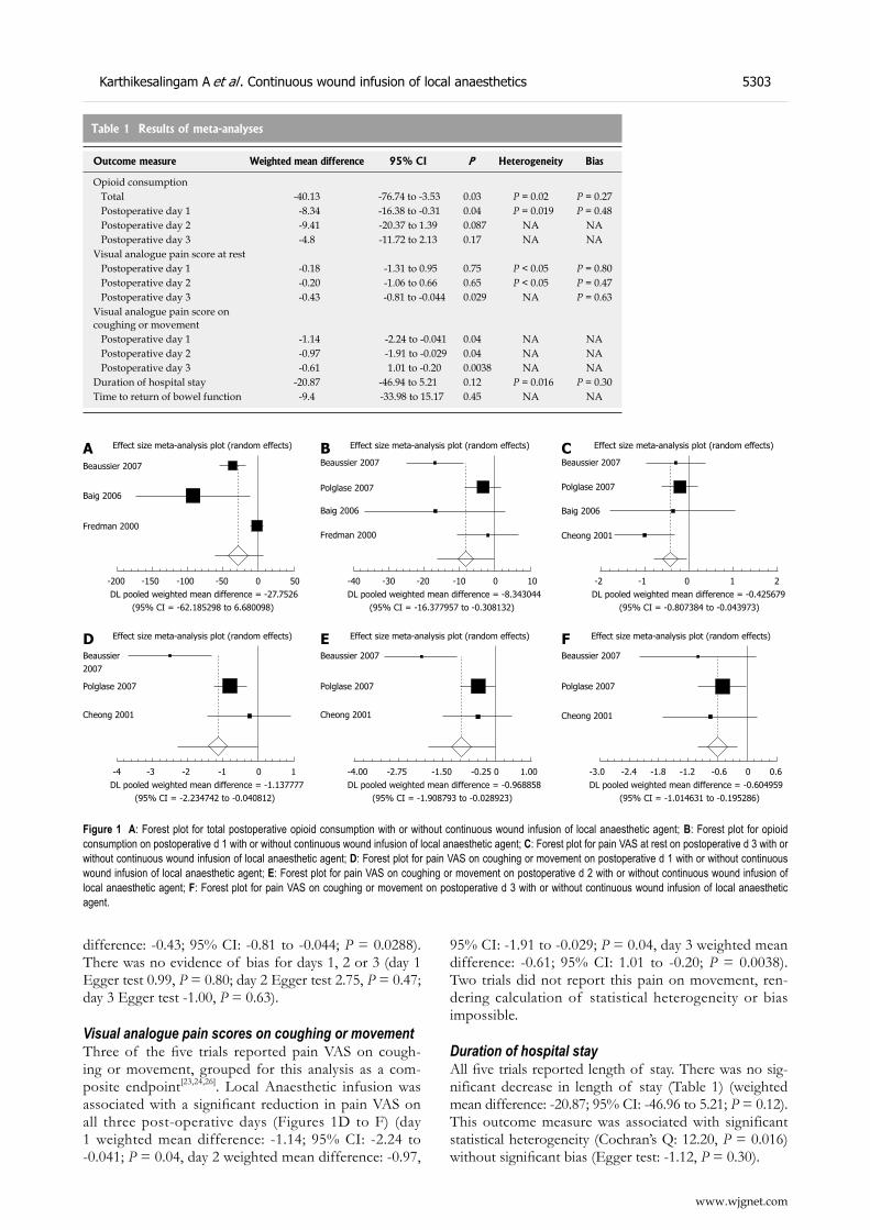
difference: -0.43; 95% CI: -0.81 to -0.044; P = 0.0288). There was no evidence of bias for days 1, 2 or 3 (day 1 Egger test 0.99, P = 0.80; day 2 Egger test 2.75, P = 0.47; day 3 Egger test -1.00, P = 0.63).
Visual analogue pain scores on coughing or movementThree of the five trials reported pain VAS on cough-ing or movement, grouped for this analysis as a com-posite endpoint[23,24,26]. Local Anaesthetic infusion was associated with a significant reduction in pain VAS on all three post-operative days (Figures 1D to F) (day 1 weighted mean difference: -1.14; 95% CI: -2.24 to -0.041; P = 0.04, day 2 weighted mean difference: -0.97,
95% CI: -1.91 to -0.029; P = 0.04, day 3 weighted mean difference: -0.61; 95% CI: 1.01 to -0.20; P = 0.0038). Two trials did not report this pain on movement, ren-dering calculation of statistical heterogeneity or bias impossible.
Duration of hospital stayAll five trials reported length of stay. There was no sig-nificant decrease in length of stay (Table 1) (weighted mean difference: -20.87; 95% CI: -46.96 to 5.21; P = 0.12). This outcome measure was associated with significant statistical heterogeneity (Cochran’s Q: 12.20, P = 0.016) without significant bias (Egger test: -1.12, P = 0.30).
Table 1 Results of meta-analyses
Outcome measure Weighted mean difference 95% CI P Heterogeneity Bias
Opioid consumption Total -40.13 -76.74 to -3.53 0.03 P = 0.02 P = 0.27 Postoperative day 1 -8.34 -16.38 to -0.31 0.04 P = 0.019 P = 0.48 Postoperative day 2 -9.41 -20.37 to 1.39 0.087 NA NA Postoperative day 3 -4.8 -11.72 to 2.13 0.17 NA NAVisual analogue pain score at rest Postoperative day 1 -0.18 -1.31 to 0.95 0.75 P < 0.05 P = 0.80 Postoperative day 2 -0.20 -1.06 to 0.66 0.65 P < 0.05 P = 0.47 Postoperative day 3 -0.43 -0.81 to -0.044 0.029 NA P = 0.63Visual analogue pain score on coughing or movement Postoperative day 1 -1.14 -2.24 to -0.041 0.04 NA NA Postoperative day 2 -0.97 -1.91 to -0.029 0.04 NA NA Postoperative day 3 -0.61 1.01 to -0.20 0.0038 NA NADuration of hospital stay -20.87 -46.94 to 5.21 0.12 P = 0.016 P = 0.30Time to return of bowel function -9.4 -33.98 to 15.17 0.45 NA NA
www.wjgnet.com
Karthikesalingam A et al . Continuous wound infusion of local anaesthetics 5303
Figure 1 A: Forest plot for total postoperative opioid consumption with or without continuous wound infusion of local anaesthetic agent; B: Forest plot for opioid consumption on postoperative d 1 with or without continuous wound infusion of local anaesthetic agent; C: Forest plot for pain VAS at rest on postoperative d 3 with or without continuous wound infusion of local anaesthetic agent; D: Forest plot for pain VAS on coughing or movement on postoperative d 1 with or without continuous wound infusion of local anaesthetic agent; E: Forest plot for pain VAS on coughing or movement on postoperative d 2 with or without continuous wound infusion of local anaesthetic agent; F: Forest plot for pain VAS on coughing or movement on postoperative d 3 with or without continuous wound infusion of local anaesthetic agent.
Effect size meta-analysis plot (random effects)
Beaussier 2007
Polglase 2007
Effect size meta-analysis plot (random effects)
Beaussier 2007
Polglase 2007
Cheong 2001
-3.0 -2.4 -1.8 -1.2 -0.6 0 0.6 DL pooled weighted mean difference = -0.604959 (95% CI = -1.014631 to -0.195286)
Effect size meta-analysis plot (random effects)
Beaussier 2007
Polglase 2007
Cheong 2001
-4.00 -2.75 -1.50 -0.25 0 1.00 DL pooled weighted mean difference = -0.968858 (95% CI = -1.908793 to -0.028923)
Cheong 2001
-4 -3 -2 -1 0 1 DL pooled weighted mean difference = -1.137777 (95% CI = -2.234742 to -0.040812)
Effect size meta-analysis plot (random effects)
Beaussier 2007
Polglase 2007
Baig 2006
-2 -1 0 1 2 DL pooled weighted mean difference = -0.425679 (95% CI = -0.807384 to -0.043973)
Cheong 2001
Effect size meta-analysis plot (random effects)
Beaussier 2007
Polglase 2007
Baig 2006
-40 -30 -20 -10 0 10 DL pooled weighted mean difference = -8.343044 (95% CI = -16.377957 to -0.308132)
Fredman 2000
Effect size meta-analysis plot (random effects)
Beaussier 2007
Baig 2006
Fredman 2000
-200 -150 -100 -50 0 50 DL pooled weighted mean difference = -27.7526 (95% CI = -62.185298 to 6.680098)
A B C
D E F
Time to return of bowel functionMean time to production of faeces was reported by three trials[22-24]. There was no significant effect of local anaesthetic wound infusion (Table 1) (weighted mean difference: -9.40; 95% CI: -33.98 to 15.17; P = 0.45). Two trials did not report this outcome measure render-ing calculation of statistical heterogeneity or bias impos-sible.
DISCUSSIONThe results of our meta-analysis suggest that wound infusions are a promising adjunct to existing analgesic regimens following laparotomy for major colorectal sur-gery. The results do not, however, provide conclusive evidence of significant benefit conferred by this tech-nique and it is doubtful whether the data gathered are sufficient to support generalisation of this conclusion to routine practice. The number of eligible trials (5) and to-tal abdominal wounds (542) is small, and meta-analyses on small samples may be vulnerable to confounding if one or two of the eligible trials demonstrate a strong trend for or against the intervention under investigation.
For the purpose of this meta-analysis, the outcome measure “opioid consumption” was chosen to reflect opioid-sparing effect provided by local anaesthetic infu-sions. However, the significant statistical heterogeneity associated with this outcome measure reflects a variety of background analgesic regimens used in both control and treatment groups. Polglase et al utilized a multimodal analgesic regimen whereas the other trials studied used only patient controlled opioid analgesia to provide back-ground analgesia. This degree of methodological hetero-geneity between the trials may have influenced the meta-analysis.
Analysis of pain VAS may also have been affected by methodological heterogeneity between the trials studied. Furthermore, pain VAS is a non-parametric variable whereas the meta-analysis models used assume paramet-ric distribution of the variables under study. The vari-able “length of stay” reflects a composite endpoint that may have been affected by several factors other than the presence of local anaesthetic infusions, and therefore it is not possible to draw causative inferences from the re-sults of this pooled outcome measure with great validity. It was not possible to obtain sufficient data for all the trials under study to provide a reliable analysis of return to bowel function.
An economic analysis of local anaesthetic wound infusions is also needed - it seems likely that a greater amount of data is needed to clarify any trends in post-operative complications that may support the use of these infusions. Further large randomised controlled trials are required to investigate the promise of local anaesthetic wound infusions in major colorectal surgery, using standardized local anaesthetic agents, background analgesic regimens, experimental protocols, discharge criteria and anatomical site for wound infusion delivery.
In conclusion, Although suboptimal postoperative pain control is associated with cardiovascular, respira-
tory and gastrointestinal complications, many multimo-dal regimens for analgesia following major colorectal laparotomy provide inadequate pain relief. Although the number of trials available for meta-analysis is small, the available data demonstrate potential benefit in terms of reduction in opioid consumption following laparotomy for major colorectal surgery. Further large-scale studies will be needed to ascertain if any clear benefit or harm is conferred by the prophylactic use of local anaesthetic wound infusions in major colorectal surgery. Future re-search on this topic should also address the inaccuracies introduced by the methodological heterogeneity pre-viously addressed in available trials, and provide a cost-effectiveness analysis of the use of continuous wound infusions in colorectal surgery.
COMMENTSBackgroundPain control following abdominal laparotomy and bowel anastomosis in color-ectal surgery is a complex challenge not amenable to pharmacological mono-therapy. Modern multimodal analgesic regimens may provide suboptimal post-operative pain control, which is associated with cardiovascular and respiratory complications and increased gastrointestinal paralysis.Research frontiersContinuous wound infusions of local anaesthetic agents have been suggested to provide improved pain control following a broad range of surgical incisions, both alone and as part of a multimodal analgesic regimen.Innovations and breakthroughsThere is a need for a focused and quantitative review of the evidence for the analgesic benefit of continuous wound infusion of local anaesthetics specific to colorectal laparotomy, which is provided by this meta-analysis.ApplicationsThe meta-analysis demonstrates potential benefit in terms of reduction in opioid consumption following laparotomy for major colorectal surgery. The review highlights the need for future research on this topic and identifies that such fu-ture research should address the inaccuracies introduced by the methodologi-cal heterogeneity identified in available trials, and provides a cost-effectiveness analysis of the use of continuous wound infusions in colorectal surgery.TerminologyVisual analogue scale (VAS) is a validated research tool used to quantitatively assess the subjective experience of patients’ pain perception.Peer reviewThe authors present a systematic analysis of local anaesthetics in wounds after open colorectal surgery. This is an area that has not been addressed by sys-tematic analysis previously. It is a well-done and timely review.
REFERENCES1 Gendall KA, Kennedy RR, Watson AJ, Frizelle FA. The
effect of epidural analgesia on postoperative outcome after colorectal surgery. Colorectal Dis 2007; 9: 584-598; discussion 598-600
2 Kehlet H. Surgical stress: the role of pain and analgesia. Br J Anaesth 1989; 63: 189-195
3 Stein C. Peripheral mechanisms of opioid analgesia. Anesth Analg 1993; 76: 182-191
4 Dahl JB, Kehlet H. Non-steroidal anti-inflammatory drugs: rationale for use in severe postoperative pain. Br J Anaesth 1991; 66: 703-712
5 PROSPECT working group. Procedure specific post-operative pain management: open colonic resection. Available from: URL: http://www.postoppain.org/frameset.htm Accessed 28th January 2008
6 Kehlet H, Liu SS. Continuous local anesthetic wound infusion to improve postoperative outcome: back to the
www.wjgnet.com
5304 ISSN 1007-9327 CN 14-1219/R World J Gastroenterol September 14, 2008 Volume 14 Number 34
COMMENTS
periphery? Anesthesiology 2007; 107: 369-3717 Liu SS, Richman JM, Thirlby RC, Wu CL. Efficacy of
continuous wound catheters delivering local anesthetic for postoperative analgesia: a quantitative and qualitative systematic review of randomized controlled trials. J Am Coll Surg 2006; 203: 914-932
8 Barron DJ, Tolan MJ, Lea RE. A randomized controlled trial of continuous extra-pleural analgesia post-thoracotomy: efficacy and choice of local anaesthetic. Eur J Anaesthesiol 1999; 16: 236-245
9 Dowling R, Thielmeier K, Ghaly A, Barber D, Boice T, Dine A. Improved pain control after cardiac surgery: results of a randomized, double-blind, clinical trial. J Thorac Cardiovasc Surg 2003; 126: 1271-1278
10 Francois T, Blanloeil Y, Pillet F, Moren J, Mazoit X, Geay G, Douet MC. Effect of interpleural administration of bupivacaine or lidocaine on pain and morphine requirement after esophagectomy with thoracotomy: a randomized, double-blind and controlled study. Anesth Analg 1995; 80: 718-723
11 Chester JF, Ravindranath K, White BD, Shanahan D, Taylor RS, Lloyd-Williams K. Wound perfusion with bupivacaine: objective evidence for efficacy in postoperative pain relief. Ann R Coll Surg Engl 1989; 71: 394-396
12 Lau H, Patil NG, Lee F. Randomized clinical trial of postoperative subfascial infusion with bupivacaine following ambulatory open mesh repair of inguinal hernia. Dig Surg 2003; 20: 285-289
13 Levack ID, Holmes JD, Robertson GS. Abdominal wound perfusion for the relief of postoperative pain. Br J Anaesth 1986; 58: 615-619
14 Fredman B, Shapiro A, Zohar E, Feldman E, Shorer S, Rawal N, Jedeikin R. The analgesic efficacy of patient-controlled ropivacaine instillation after Cesarean delivery. Anesth Analg 2000; 91: 1436-1440
15 Gupta A, Perniola A, Axelsson K, Thorn SE, Crafoord K, Rawal N. Postoperative pain after abdominal hysterectomy: a double-blind comparison between placebo and local anesthetic infused intraperitoneally. Anesth Analg 2004; 99: 1173-1179, table of contents
16 Kushner DM, LaGalbo R, Connor JP, Chappell R, Stewart SL, Hartenbach EM. Use of a bupivacaine continuous wound infusion system in gynecologic oncology: a randomized trial. Obstet Gynecol 2005; 106: 227-233
17 Alford JW , Fadale PD. Evaluation of postoperative bupivacaine infusion for pain management after anterior cruciate ligament reconstruction. Arthroscopy 2003; 19: 855-861
18 Barber FA, Herbert MA. The effectiveness of an anesthetic continuous-infusion device on postoperative pain control. Arthroscopy 2002; 18: 76-81
19 Bianconi M, Ferraro L, Ricci R, Zanoli G, Antonelli T, Giulia B, Guberti A, Massari L. The pharmacokinetics and efficacy of ropivacaine continuous wound instillation after spine fusion surgery. Anesth Analg 2004; 98: 166-172, table of contents
20 Jadad AR, Moore RA, Carroll D, Jenkinson C, Reynolds DJ, Gavaghan DJ, McQuay HJ. Assessing the quality of reports of randomized clinical trials: is blinding necessary? Control Clin Trials 1996; 17: 1-12
21 DerSimonian R, Laird N. Meta-analysis in clinical trials. Control Clin Trials 1986; 7: 177-188
22 Baig MK, Zmora O, Derdemezi J, Weiss EG, Nogueras JJ, Wexner SD. Use of the ON-Q pain management system is associated with decreased postoperative analgesic requirement: double blind randomized placebo pilot study. J Am Coll Surg 2006; 202: 297-305
23 Beaussier M, El'Ayoubi H, Schiffer E, Rollin M, Parc Y, Mazoit JX, Azizi L, Gervaz P, Rohr S, Biermann C, Lienhart A, Eledjam JJ. Continuous preperitoneal infusion of ropivacaine provides effective analgesia and accelerates recovery after colorectal surgery: a randomized, double-blind, placebo-controlled study. Anesthesiology 2007; 107: 461-468
24 Cheong WK, Seow-Choen F, Eu KW, Tang CL, Heah SM. Randomized clinical trial of local bupivacaine perfusion versus parenteral morphine infusion for pain relief after laparotomy. Br J Surg 2001; 88: 357-359
25 Fredman B, Zohar E, Tarabykin A, Shapiro A, Mayo A, Klein E, Jedeikin R. Bupivacaine wound instillation via an electronic patient-controlled analgesia device and a double-catheter system does not decrease postoperative pain or opioid requirements after major abdominal surgery. Anesth Analg 2001; 92: 189-193
26 Polglase AL, McMurrick PJ, Simpson PJ, Wale RJ, Carne PW, Johnson W, Chee J, Ooi CW, Chong JW, Kingsland SR, Buchbinder R. Continuous wound infusion of local anesthetic for the control of pain after elective abdominal colorectal surgery. Dis Colon Rectum 2007; 50: 2158-2167
S- Editor Li DL L- Editor Lutze M E- Editor Ma WH
www.wjgnet.com
Karthikesalingam A et al . Continuous wound infusion of local anaesthetics 5305
there were no differences in TLR4 or TLR5 mRNA lev-els among the different clinical presentations/histologi-cal findings (P > 0.05). In the in vitro assay, the mRNA levels of TLR4 or TLR5 in AGS cells were not influ-enced by the vacAs1 status or the clinical condition as-sociated with the strains (P > 0.05 for both TLR4 and TLR5).CONCLUSION: The results of this study show that the mRNA levels of TLR4 and TLR5 in gastric cells, both in vivo and in vitro , are independent of H py-lori colonization and suggest that vacA may not be a significant player in the first step of innate immune recognition mediated by TLR4 or TLR5.
© 2008 The WJG Press. All rights reserved.
Key words: H pylori ; Toll-like receptor 4; Toll-like receptor 5; AGS cells; mRNA
Peer reviewer: Dr. Limas Kupcinskas, Professor, Gastro-enterology of Kaunas University of Medicine, Mickeviciaus 9, Kaunas LT 44307, Lithuania
Garza-González E, Bocanegra-García V, Bosques-Padilla FJ, Flores-Gutiérrez JP, Moreno F, Perez-Perez GI. mRNA levels of TLR4 and TLR5 are independent of H pylori. World J Gastroenterol 2008; 14(34): 5306-5310 Available from: URL: http://www.wjgnet.com/1007-9327/14/5306.asp DOI: http://dx.doi.org/10.3748/wjg.14.5306
INTRODUCTIONH pylori is a Gram-negative, flagellated bacterium that colonizes the gastric mucosa of approximately two-thirds of the world’s population and is the primary cause of peptic ulcers and gastric adenocarcinoma. The complex interactions between different H pylori strains, the host immune system or environmental factors (or combinations thereof) are responsible for the significant variability in disease presentation associated with H pylori infection[1].
Vacuolating cytotoxin (VacA) and the CagA protein are the two major virulence markers usually associated with H pylori pathogenicity. VacA induces the forma-tion of intracellular vacuoles in epithelial cell lines. Aside from its direct cell-damaging effect in vitro, VacA also plays a major role in inducing cytoskeletal changes, apoptosis and suppression of epithelial cell prolifera-
Elvira Garza-González, Francisco Moreno, Departmento de Microbiología, Facultad de Medicina, Universidad Autónoma de Nuevo León, Monterrey 64460, MexicoVirgi l io Bocanegra-García, Departmento de Biología Molecular y Bioingeniería, UAM Reynosa Aztlán, Universidad Autónoma de Tamaulipas, Reynosa, Tamaulipas 88740, MexicoFrancisco Javier Bosques-Padilla, Juan Pablo Flores-Gutiérrez, Hospital Universitario “Dr. José Eleuterio González”, Universidad Autónoma de Nuevo León, Monterrey 64460, MexicoGuillermo Ignacio Perez-Perez, Department of Medicine and Microbiology, New York University School of Medicine, New York NY 10010, United StatesAuthor contributions: Garza-González E designed and coordinated the study, optimized PCR conditions, and drafted the manuscript; Bocanegra-García V and Moreno F extracted RNAs and performed molecular assays; Flores-Gutiérrez JP performed histological examination and interpretation of results; Bosques-Padilla FJ recruited patients and carried out endoscopies; Pérez-Perez GI participated in the design, coordination of the study and revised the drafted manuscript.Correspondence to: Elvira Garza-González, Departmento de Microbiología, Facultad de Medicina, Universidad Autónoma de Nuevo León, Av. Madero y Dr. Aguirre s/n, Colonia Mitras Centro, Monterrey 64460, Mexico. [email protected]: +52-81-83294166 Fax: +52-81-83294166Received: May 4, 2008 Revised: August 18, 2008Accepted: August 25, 2008Published online: September 14, 2008
AbstractAIM: To determine if the presence H pylori or its viru-lence affect toll-like receptor 4 (TLR4) and TLR5 mRNA expression levels.METHODS: For the in vivo assays, gastric biopsies were obtained from 40 patients and H pylori status was determined. For the in vitro assays, human gastric adenocarcinoma mucosal cells (AGS) were cultured in the presence or absence of twelve selected H pylori strains. H pylori strains isolated from culture-positive patients and selected strains were genotyped for cagA and vacA . The cDNA was obtained from mRNA extract-ed from biopsies and from infected AGS cells. TLR4 and TLR5 mRNA levels were examined by real-time PCR.RESULTS: The presence of H pylori did not affect the mRNA levels of TLR4 or TLR5 in gastric biopsies. The mRNA levels of both receptors were not influenced by the vacA status (P > 0.05 for both receptors) and
mRNA levels of TLR4 and TLR5 are independent of H pylori
Elvira Garza-González, Virgilio Bocanegra-García, Francisco Javier Bosques-Padilla, Juan Pablo Flores-Gutiérrez, Francisco Moreno, Guillermo Ignacio Perez-Perez
www.wjgnet.com
RAPID COMMUNICATION
Online Submissions: wjg.wjgnet.com World J Gastroenterol 2008 September 14; 14(34): [email protected] World Journal of Gastroenterology ISSN 1007-9327doi:10.3748/wjg.14.5306 © 2008 The WJG Press. All rights reserved.
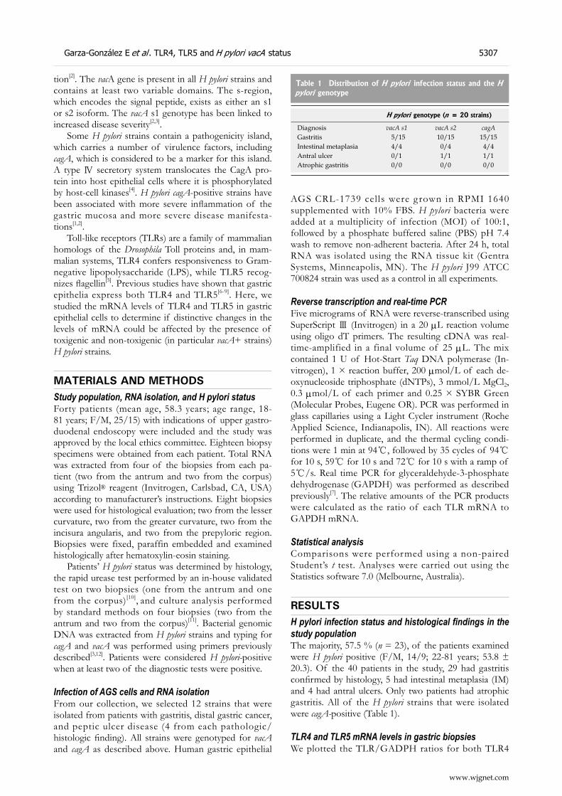
www.wjgnet.com
Garza-González E et al . TLR4, TLR5 and H pylori vacA status 5307
tion[2]. The vacA gene is present in all H pylori strains and contains at least two variable domains. The s-region, which encodes the signal peptide, exists as either an s1 or s2 isoform. The vacA s1 genotype has been linked to increased disease severity[2,3].
Some H pylori strains contain a pathogenicity island, which carries a number of virulence factors, including cagA, which is considered to be a marker for this island. A type Ⅳ secretory system translocates the CagA pro-tein into host epithelial cells where it is phosphorylated by host-cell kinases[4]. H pylori cagA-positive strains have been associated with more severe inflammation of the gastric mucosa and more severe disease manifesta-tions[1,2].
Toll-like receptors (TLRs) are a family of mammalian homologs of the Drosophila Toll proteins and, in mam-malian systems, TLR4 confers responsiveness to Gram-negative lipopolysaccharide (LPS), while TLR5 recog-nizes flagellin[5]. Previous studies have shown that gastric epithelia express both TLR4 and TLR5[6-9]. Here, we studied the mRNA levels of TLR4 and TLR5 in gastric epithelial cells to determine if distinctive changes in the levels of mRNA could be affected by the presence of toxigenic and non-toxigenic (in particular vacA+ strains) H pylori strains.
MATERIALS AND METHODSStudy population, RNA isolation, and H pylori status Forty patients (mean age, 58.3 years; age range, 18- 81 years; F/M, 25/15) with indications of upper gastro-duodenal endoscopy were included and the study was approved by the local ethics committee. Eighteen biopsy specimens were obtained from each patient. Total RNA was extracted from four of the biopsies from each pa-tient (two from the antrum and two from the corpus) using Trizol® reagent (Invitrogen, Carlsbad, CA, USA) according to manufacturer’s instructions. Eight biopsies were used for histological evaluation; two from the lesser curvature, two from the greater curvature, two from the incisura angularis, and two from the prepyloric region. Biopsies were fixed, paraffin embedded and examined histologically after hematoxylin-eosin staining.
Patients’ H pylori status was determined by histology, the rapid urease test performed by an in-house validated test on two biopsies (one from the antrum and one from the corpus)[10], and culture analysis performed by standard methods on four biopsies (two from the antrum and two from the corpus)[11]. Bacterial genomic DNA was extracted from H pylori strains and typing for cagA and vacA was performed using primers previously described[3,12]. Patients were considered H pylori-positive when at least two of the diagnostic tests were positive.
Infection of AGS cells and RNA isolationFrom our collection, we selected 12 strains that were isolated from patients with gastritis, distal gastric cancer, and peptic ulcer disease (4 from each pathologic/histologic finding). All strains were genotyped for vacA and cagA as described above. Human gastric epithelial
AGS CRL-1739 cel ls were grown in RPMI 1640 supplemented with 10% FBS. H pylori bacteria were added at a multiplicity of infection (MOI) of 100:1, followed by a phosphate buffered saline (PBS) pH 7.4 wash to remove non-adherent bacteria. After 24 h, total RNA was isolated using the RNA tissue kit (Gentra Systems, Minneapolis, MN). The H pylori J99 ATCC 700824 strain was used as a control in all experiments.
Reverse transcription and real-time PCRFive micrograms of RNA were reverse-transcribed using SuperScript Ⅲ (Invitrogen) in a 20 μL reaction volume using oligo dT primers. The resulting cDNA was real-time-amplified in a final volume of 25 μL. The mix contained 1 U of Hot-Start Taq DNA polymerase (In-vitrogen), 1 × reaction buffer, 200 μmol/L of each de-oxynucleoside triphosphate (dNTPs), 3 mmol/L MgCl2, 0.3 μmol/L of each primer and 0.25 × SYBR Green (Molecular Probes, Eugene OR). PCR was performed in glass capillaries using a Light Cycler instrument (Roche Applied Science, Indianapolis, IN). All reactions were performed in duplicate, and the thermal cycling condi-tions were 1 min at 94, followed by 35 cycles of 94 for 10 s, 59 for 10 s and 72 for 10 s with a ramp of 5/s. Real time PCR for glyceraldehyde-3-phosphate dehydrogenase (GAPDH) was performed as described previously[7]. The relative amounts of the PCR products
were calculated as the ratio of each TLR mRNA to GAPDH mRNA.
Statistical analysisComparisons were performed using a non-paired Student’s t test. Analyses were carried out using the Statistics software 7.0 (Melbourne, Australia).
RESULTSH pylori infection status and histological findings in the study populationThe majority, 57.5 % (n = 23), of the patients examined were H pylori positive (F/M, 14/9; 22-81 years; 53.8 ± 20.3). Of the 40 patients in the study, 29 had gastritis confirmed by histology, 5 had intestinal metaplasia (IM) and 4 had antral ulcers. Only two patients had atrophic gastritis. All of the H pylori strains that were isolated were cagA-positive (Table 1).
TLR4 and TLR5 mRNA levels in gastric biopsiesWe plotted the TLR/GADPH ratios for both TLR4
Table 1 Distribution of H pylori infection status and the H pylori genotype
H pylori genotype (n = 20 strains)
Diagnosis vacA s1 vacA s2 cagAGastritis 5/15 10/15 15/15Intestinal metaplasia 4/4 0/4 4/4Antral ulcer 0/1 1/1 1/1Atrophic gastritis 0/0 0/0 0/0
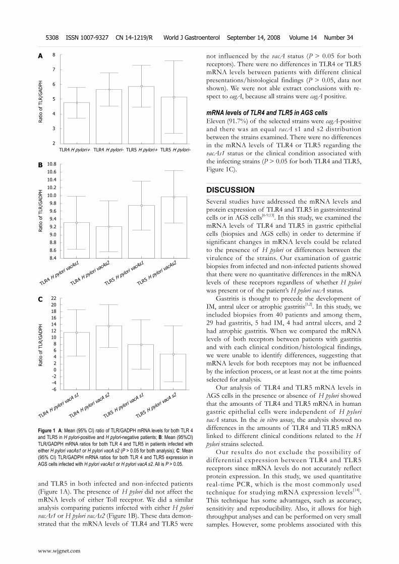
and TLR5 in both infected and non-infected patients (Figure 1A). The presence of H pylori did not affect the mRNA levels of either Toll receptor. We did a similar analysis comparing patients infected with either H pylori vacAs1 or H pylori vacAs2 (Figure 1B). These data demon-strated that the mRNA levels of TLR4 and TLR5 were
not influenced by the vacA status (P > 0.05 for both receptors). There were no differences in TLR4 or TLR5 mRNA levels between patients with different clinical presentations/histological findings (P > 0.05, data not shown). We were not able extract conclusions with re-spect to cagA, because all strains were cagA positive.
mRNA levels of TLR4 and TLR5 in AGS cellsEleven (91.7%) of the selected strains were cagA-positive and there was an equal vacA s1 and s2 distribution between the strains examined. There were no differences in the mRNA levels of TLR4 or TLR5 regarding the vacAs1 status or the clinical condition associated with the infecting strains (P > 0.05 for both TLR4 and TLR5, Figure 1C).
DISCUSSIONSeveral studies have addressed the mRNA levels and protein expression of TLR4 and TLR5 in gastrointestinal cells or in AGS cells[6-9,13]. In this study, we examined the mRNA levels of TLR4 and TLR5 in gastric epithelial cells (biopsies and AGS cells) in order to determine if significant changes in mRNA levels could be related to the presence of H pylori or differences between the virulence of the strains. Our examination of gastric biopsies from infected and non-infected patients showed that there were no quantitative differences in the mRNA levels of these receptors regardless of whether H pylori was present or of the patient’s H pylori vacA status.
Gastritis is thought to precede the development of IM, antral ulcer or atrophic gastritis[1,2]. In this study, we included biopsies from 40 patients and among them, 29 had gastritis, 5 had IM, 4 had antral ulcers, and 2 had atrophic gastritis. When we compared the mRNA levels of both receptors between patients with gastritis and with each clinical condition/histological findings, we were unable to identify differences, suggesting that mRNA levels for both receptors may not be influenced by the infection process, or at least not at the time points selected for analysis.
Our analysis of TLR4 and TLR5 mRNA levels in AGS cells in the presence or absence of H pylori showed that the amounts of TLR4 and TLR5 mRNA in human gastric epithelial cells were independent of H pylori vacA status. In the in vitro assay, the analysis showed no differences in the amounts of TLR4 and TLR5 mRNA linked to different clinical conditions related to the H pylori strains selected.
Our results do not exclude the possibil ity of differential expression between TLR4 and TLR5 receptors since mRNA levels do not accurately reflect protein expression. In this study, we used quantitative real-time PCR, which is the most commonly used technique for studying mRNA expression levels[14]. This technique has some advantages, such as accuracy, sensitivity and reproducibility. Also, it allows for high throughput analyses and can be performed on very small samples. However, some problems associated with this
8
7
6
5
4
3
2
Ratio
of
TLR/G
ADPH
TLR4 H pylori+ TLR4 H pylori- TLR5 H pylori+ TLR5 H pylori-
A
10.8
10.6
10.4
10.2
10.0
9.8
9.6
9.4
9.2
9.0
8.8
8.6
8.4
Ratio
of
TLR/G
ADPH
B
TLR4 H pylori vacA
s1
TLR4 H pylori vacA
s2
TLR5 H pylori vacA
s1
TLR5 H pylori vacA
s2
2220181614121086420
-2-4-6
Ratio
of
TLR/G
ADPH
C
TLR4 H pylori va
cA s1
TLR4 H pylori vacA
s2
TLR5 H pylori vacA
s1
TLR5 H pylori vacA
s2
Figure 1 A: Mean (95% CI) ratio of TLR/GADPH mRNA levels for both TLR 4 and TLR5 in H pylori-positive and H pylori-negative patients; B: Mean (95%CI) TLR/GADPH mRNA ratios for both TLR 4 and TLR5 in patients infected with either H pylori vacAs1 or H pylori vacA s2 (P > 0.05 for both analysis); C: Mean (95% CI) TLR/GADPH mRNA ratios for both TLR 4 and TLR5 expression in AGS cells infected with H pylori vacAs1 or H pylori vacA s2. All is P > 0.05.
www.wjgnet.com
5308 ISSN 1007-9327 CN 14-1219/R World J Gastroenterol September 14, 2008 Volume 14 Number 34
technique must be addressed, such as the effects of different amounts of starting material, especially when analyzing biopsies. To deal with this variation, an internal control (housekeeping gene) must be simultaneously amplified with the gene of interest for normalization purposes[15]. In this study, even though we included analysis of GADPH as an internal control we could not assume that protein expression levels of both receptors were equal based on the levels of detected mRNA.
An additional caveat was that the in vivo mRNA analysis of pooled biopsies from for ty patients minimized the ability to identify biopsy-to-biopsy or patient-to-patient differences. Since RNA was extracted from all four antrum and corpus biopsies in the same vial, different gene expression profiles associated with different tissues would not have been detected. By analyzing four combined biopsies, it was possible that mRNA differences between corpus and antrum mucosal samples could have been missed. However, in the in vitro assay the mRNA levels obtained following infections with 12 different H pylori strains showed no differences in TLR expression. Although this is a uniform tissue, the observation that at least different H pylori strains did not affect TLR mRNA levels suggested that the same results would be observed in biopsy samples.
Some invest ig ators have sug gested that the subcellular distribution of receptors, rather than TLR expression level, could be relevant in the pathogenesis of inflammatory diseases because the expression of these receptors seems to be constitutive[7]. Our results are consistent with this view. Nonetheless, a study examining the cellular distribution of expression in relation to H pylori vacA status would be an interesting issue to address.
Schmausser et al[9], reported that gastric epithelium with intestinal metaplasia and dysplasia expressed TLR4 and TLR5. They demonstrated that 17 out of 22 patients strongly expressed TLR4 (77.27%) and all 22 patients with gastric carcinoma expressed TLR5. Our study confirmed the presence of TLR4 and TLR5 mRNA, which preceded the expression of both receptors.
It is likely that evaluating the roles of other Toll-like receptors would help elucidate differences in disease manifestation and severity of diseases caused by H pylori infections. Valuable information regarding the recognition of whole H pylori or its LPS by TLR2 has been reported. Smith et al[16] demonstrated that gastric epithelial cells recognized and responded to H pylori infection, at least in part, via TLR2 and that H pylori LPS was a TLR2 agonist.
Additionally, Mandell et al [17] demonstrated that cytokine responses to whole H pylori were mediated by TLR2. Based on these investigations, more work examining the role of TLR2 in relation to the H pylori vacA status is needed. The results of this study show that the TLR4 and TLR5 mRNA levels in gastric cells both in vivo and in vitro are independent of H pylori and suggest that vacA may not be involved in the first steps of innate immune-recognition of H pylori.
COMMENTSBackgroundH pylori is the primary cause of peptic ulcers and gastric adenocarcinoma. The variability of clinical manifestations is associated with bacterial, host immune responses and environmental factors.Research frontiersGastric epithelia express toll-like receptor 4 (TLR4) and TLR5. We studied the mRNA levels of TLR4 and TLR5 in gastric epithelial cells to determine if distinc-tive changes in mRNA levels could be influenced by the presence of toxigenic H pylori strains.Innovations and breakthroughsIn this study, we analyzed the mRNA levels of both TLR4 and TLR5 in gastric biopsies from infected and non-infected patients and in AGS cells infected with H pylori. We correlated these results with the vacA status of the strains. There were no quantitative differences in the mRNA levels of these receptors regardless of H pylori presence or the H pylori vacA status both in gastric biopsies and in AGS cells.ApplicationsThe mRNA levels of TLR4 and TLR5 in gastric cells both in vivo and in vitro are independent of H pylori or their vacA status. Peer reviewThe results show that the mRNA levels of TLR4 and TLR5 in gastric cells are not influenced by H pylori vacA status and suggest that vacA may not be a sig-nificant player in the first step of innate immune recognition mediated by TLR4 or TLR5. It seems innovative and very interesting.
REFERENCES1 Blaser MJ, Berg DE. Helicobacter pylori genetic diversity
and risk of human disease. J Clin Invest 2001; 107: 767-7732 Figueiredo C, Machado JC, Yamaoka Y. Pathogenesis of
Helicobacter pylori Infection. Helicobacter 2005; 10 Suppl 1: 14-20
3 Atherton JC, Cao P, Peek RM Jr, Tummuru MK, Blaser MJ, Cover TL. Mosaicism in vacuolating cytotoxin alleles of Helicobacter pylori. Association of specific vacA types with cytotoxin production and peptic ulceration. J Biol Chem 1995; 270: 17771-17777
4 Odenbreit S, Puls J, Sedlmaier B, Gerland E, Fischer W, Haas R. Translocation of Helicobacter pylori CagA into gastric epithelial cells by type IV secretion. Science 2000; 287: 1497-1500
5 Ferrero RL. Innate immune recognition of the extracellular mucosal pathogen, Helicobacter pylori. Mol Immunol 2005; 42: 879-885
6 Su B , Ceponis PJ, Lebel S, Huynh H, Sherman PM. Helicobacter pylori activates Toll-like receptor 4 expression in gastrointestinal epithelial cells. Infect Immun 2003; 71: 3496-3502
7 Schmausser B, Andrulis M, Endrich S, Lee SK, Josenhans C, Muller-Hermelink HK, Eck M. Expression and subcellular distribution of toll-like receptors TLR4, TLR5 and TLR9 on the gastric epithelium in Helicobacter pylori infection. Clin Exp Immunol 2004; 136: 521-526
8 Backhed F, Rokbi B, Torstensson E, Zhao Y, Nilsson C, Seguin D, Normark S, Buchan AM, Richter-Dahlfors A. Gastric mucosal recognition of Helicobacter pylori is independent of Toll-like receptor 4. J Infect Dis 2003; 187: 829-836
9 Schmausser B, Andrulis M, Endrich S, Muller-Hermelink HK, Eck M. Toll-like receptors TLR4, TLR5 and TLR9 on gastric carcinoma cells: an implication for interaction with Helicobacter pylori. Int J Med Microbiol 2005; 295: 179-185
10 Flores-Orta D, Bosques-Padilla F, Gomez-Leija G, Frederick F. Comparative study of rapid urease test (Hazell test) vs CLO-test in the diagnosis of Helicobacter pylori infection. Gut 1997; 41 Suppl 3: A160
11 Perez-Perez GI. Accurate diagnosis of Helicobacter pylori. Culture, including transport. Gastroenterol Clin North Am
www.wjgnet.com
Garza-González E et al . TLR4, TLR5 and H pylori vacA status 5309
COMMENTS
2000; 29: 879-88412 Rugge M, Busatto G, Cassaro M, Shiao YH, Russo V,
Leandro G, Avellini C, Fabiano A, Sidoni A, Covacci A. Patients younger than 40 years with gastric carcinoma: Helicobacter pylori genotype and associated gastritis phenotype. Cancer 1999; 85: 2506-2511
13 Su B , Ceponis PJ, Lebel S, Huynh H, Sherman PM. Helicobacter pylori activates Toll-like receptor 4 expression in gastrointestinal epithelial cells. Infect Immun 2003; 71: 3496-3502
14 Bustin SA, Nolan T. Pitfalls of quantitative real-time reverse-transcription polymerase chain reaction. J Biomol Tech 2004; 15: 155-166
15 Bustin SA. Absolute quantification of mRNA using real-
time reverse transcription polymerase chain reaction assays. J Mol Endocrinol 2000; 25: 169-193
16 Smith MF Jr, Mitchell A, Li G, Ding S, Fitzmaurice AM, Ryan K, Crowe S, Goldberg JB. Toll-like receptor (TLR) 2 and TLR5, but not TLR4, are required for Helicobacter pylori-induced NF-kappa B activation and chemokine expression by epithelial cells. J Biol Chem 2003; 278: 32552-32560
17 Mandell L, Moran AP, Cocchiarella A, Houghton J, Taylor N, Fox JG, Wang TC, Kurt-Jones EA. Intact gram-negative Helicobacter pylori, Helicobacter felis, and Helicobacter hepaticus bacteria activate innate immunity via toll-like receptor 2 but not toll-like receptor 4. Infect Immun 2004; 72: 6446-6454
S- Editor Zhong XY L- Editor Negro F E- Editor Ma WH
www.wjgnet.com
5310 ISSN 1007-9327 CN 14-1219/R World J Gastroenterol September 14, 2008 Volume 14 Number 34
CONCLUSION: Chemoradiotherapy with low-dose gemcitabine administered twice weekly could be effective to patients with locally advanced pancreatic cancer; however, patients developing liver metastases had a worse prognosis. Another chemoradiotherapy strategy might be needed for those patients, such as administrating one or two cycles of chemotherapy initially, followed by chemoradiotherapy for the cases with no distant metastases.
© 2008 The WJG Press. All rights reserved.
Key words: Advanced pancreatic cancer; Chemoradio-therapy; Gemcitabine; Radiosensitizer; Tumor marker
Peer reviewer: Michael E Zenilman, MD, Clarence and Mary Dennis Professor and Chairman, Department of Surgery, SUNY Downstate Medical Center, Box40, 450, Clarkson Avenue, Brooklyn, NY 11202, United States
Igarashi H, Ito T, Kawabe K, Hisano T, Arita Y, Kaku T, Takayanagi R. Chemoradiotherapy with twice-weekly administration of low-dose gemcitabine for locally advanced pancreatic cancer. World J Gastroenterol 2008; 14(34): 5311-5315 Available from: URL: http://www.wjgnet.com/1007-9327/14/5311.asp DOI: http://dx.doi.org/10.3748/wjg.14.5311
INTRODUCTIONPancreatic cancer is one of the leading causes of cancer death in the world and in most patients the tumor is surgically unresectable at the time of diagnosis[1]. Even in the patient with complete surgical resection, both distant and local patterns of recurrence are common[2]. In approximately 50% of resected pancreatic tumors, the surgical margins are involved with tumor cells, so it can be assumed that most patients are harboring occult metastases at the time of diagnosis[3]. Recently, studies for adjuvant chemotherapy or chemoradiotherapy, a n d th o s e fo r n eo a d juva n t ch emo th e r a p y o r chemoradiotherapy have been investigated[3-6].
For patients with locally advanced pancreatic cancer, chemoradiotherapy has been accepted as a standard treatment[7]. The results of previous randomized trials
Hisato Igarashi, Tetsuhide Ito, Ken Kawabe, Terumasa Hisano, Yoshiyuki Arita, Toyoma Kaku, Ryoichi Takayanagi, Department of Medicine and Bioregulatory Science, Graduate School of Medical Science, Kyushu University, Fukuoka 812-8582, JapanAuthor contributions: Igarashi H analyzed the data and wrote the paper; Ito T designed the study and revised the paper; Kawabe K, Hisano T, Arita Y and Kaku T collected and analyzed the data; Takayanagi R revised the paper. Correspondence to: Hisato Igarashi, MD, PhD, Department of Medicine and Bioregulatory Science, Graduate School of Medical Science, Kyushu University 3-1-1 Maidashi Higashi-ku Fukuoka 812-8582 Japan. [email protected]: +81-92-6425285 Fax: +81-92-6425287Received: May 13, 2008 Revised: July 28, 2008Accepted: August 4, 2008Published online: September 14, 2008
AbstractAIM: To evaluate the chemoradiotherapy for locally advanced pancreatic cancer ut i l iz ing low dose gemcitabine as a radiation sensitizer administered twice weekly. METHODS: We performed a retrospective analysis of chemoradiotherapy utilizing gemcitabine administered twice weekly at a dose of 40 mg/m2. After that, maintenance systemic chemotherapy with gemcitabine, at a dose of 1000 mg/m2, was administered weekly for 3 wk with 1-wk rest until disease progression or unacceptable toxicity developed. RESULTS: Eighteen patients with locally advanced unresectable pancreatic cancer were enrolled. Three of those patients could not continue with the therapy; one patient had interstitial pneumonia during radiation therapy and two other patients showed liver metastasis or peritoneal metastasis during an early stage of the therapy. The median survival was 15.0 mo and the overall 1-year survival rate was 60%, while the median progression-free survival was 8.0 mo. The subgroup which showed the reduction of tumor development, more than 50% showed a tendency for a better prognosis; however, other parameters including age, gender and performance status did not correlate with survival. The median survival of the groups that died of liver metastasis and peritoneal metastasis were 13.0 mo and 27.7 mo, respectively.
Online Submissions: wjg.wjgnet.com World J Gastroenterol 2008 September 14; 14(34): [email protected] World Journal of Gastroenterology ISSN 1007-9327doi:10.3748/wjg.14.5311 © 2008 The WJG Press. All rights reserved.
Chemoradiotherapy with twice-weekly administration of low-dose gemcitabine for locally advanced pancreatic cancer
Hisato Igarashi, Tetsuhide Ito, Ken Kawabe, Terumasa Hisano, Yoshiyuki Arita, Toyoma Kaku, Ryoichi Takayanagi
RAPID COMMUNICATION
www.wjgnet.com
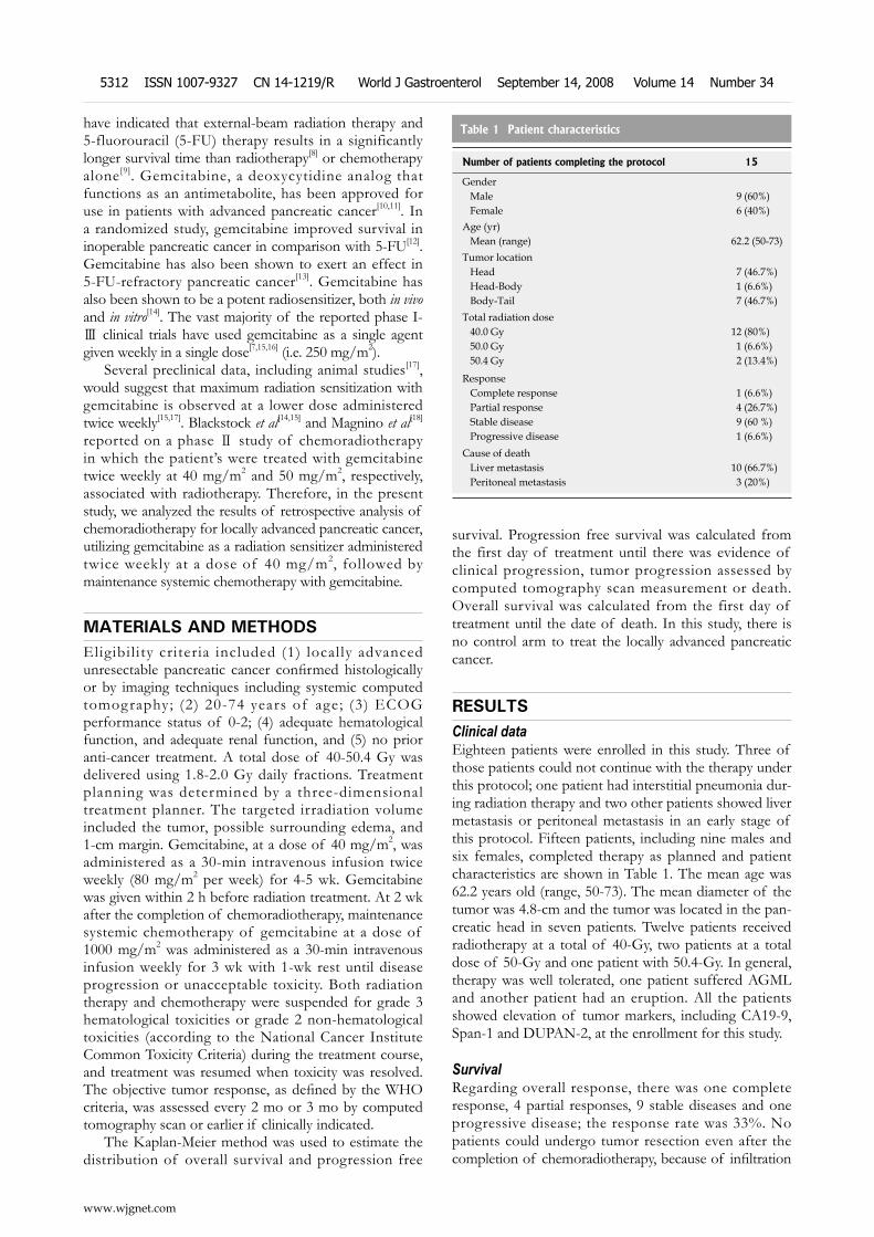
have indicated that external-beam radiation therapy and 5-fluorouracil (5-FU) therapy results in a significantly longer survival time than radiotherapy[8] or chemotherapy alone[9]. Gemcitabine, a deoxycytidine analog that functions as an antimetabolite, has been approved for use in patients with advanced pancreatic cancer[10,11]. In a randomized study, gemcitabine improved survival in inoperable pancreatic cancer in comparison with 5-FU[12]. Gemcitabine has also been shown to exert an effect in 5-FU-refractory pancreatic cancer[13]. Gemcitabine has also been shown to be a potent radiosensitizer, both in vivo and in vitro[14]. The vast majority of the reported phase I-Ⅲ clinical trials have used gemcitabine as a single agent given weekly in a single dose[7,15,16] (i.e. 250 mg/m2).
Several preclinical data, including animal studies[17], would suggest that maximum radiation sensitization with gemcitabine is observed at a lower dose administered twice weekly[15,17]. Blackstock et al[14,15] and Magnino et al[18] reported on a phase Ⅱ study of chemoradiotherapy in which the patient’s were treated with gemcitabine twice weekly at 40 mg/m2 and 50 mg/m2, respectively, associated with radiotherapy. Therefore, in the present study, we analyzed the results of retrospective analysis of chemoradiotherapy for locally advanced pancreatic cancer, utilizing gemcitabine as a radiation sensitizer administered twice weekly at a dose of 40 mg/m2, followed by maintenance systemic chemotherapy with gemcitabine.
MATERIALS AND METHODSEligibil i ty criteria included (1) local ly advanced unresectable pancreatic cancer confirmed histologically or by imaging techniques including systemic computed tomography; (2) 20-74 years of age; (3) ECOG performance status of 0-2; (4) adequate hematological function, and adequate renal function, and (5) no prior anti-cancer treatment. A total dose of 40-50.4 Gy was delivered using 1.8-2.0 Gy daily fractions. Treatment planning was determined by a three-dimensional treatment planner. The targeted irradiation volume included the tumor, possible surrounding edema, and 1-cm margin. Gemcitabine, at a dose of 40 mg/m2, was administered as a 30-min intravenous infusion twice weekly (80 mg/m2 per week) for 4-5 wk. Gemcitabine was given within 2 h before radiation treatment. At 2 wk after the completion of chemoradiotherapy, maintenance systemic chemotherapy of gemcitabine at a dose of 1000 mg/m2 was administered as a 30-min intravenous infusion weekly for 3 wk with 1-wk rest until disease progression or unacceptable toxicity. Both radiation therapy and chemotherapy were suspended for grade 3 hematological toxicities or grade 2 non-hematological toxicities (according to the National Cancer Institute Common Toxicity Criteria) during the treatment course, and treatment was resumed when toxicity was resolved. The objective tumor response, as defined by the WHO criteria, was assessed every 2 mo or 3 mo by computed tomography scan or earlier if clinically indicated.
The Kaplan-Meier method was used to estimate the distribution of overall survival and progression free
survival. Progression free survival was calculated from the first day of treatment until there was evidence of clinical progression, tumor progression assessed by computed tomography scan measurement or death. Overall survival was calculated from the first day of treatment until the date of death. In this study, there is no control arm to treat the locally advanced pancreatic cancer.
RESULTSClinical dataEighteen patients were enrolled in this study. Three of those patients could not continue with the therapy under this protocol; one patient had interstitial pneumonia dur-ing radiation therapy and two other patients showed liver metastasis or peritoneal metastasis in an early stage of this protocol. Fifteen patients, including nine males and six females, completed therapy as planned and patient characteristics are shown in Table 1. The mean age was 62.2 years old (range, 50-73). The mean diameter of the tumor was 4.8-cm and the tumor was located in the pan-creatic head in seven patients. Twelve patients received radiotherapy at a total of 40-Gy, two patients at a total dose of 50-Gy and one patient with 50.4-Gy. In general, therapy was well tolerated, one patient suffered AGML and another patient had an eruption. All the patients showed elevation of tumor markers, including CA19-9, Span-1 and DUPAN-2, at the enrollment for this study.
SurvivalRegarding overall response, there was one complete response, 4 partial responses, 9 stable diseases and one progressive disease; the response rate was 33%. No patients could undergo tumor resection even after the completion of chemoradiotherapy, because of infiltration
Table 1 Patient characteristics
Number of patients completing the protocol 15
Gender Male 9 (60%) Female 6 (40%)Age (yr) Mean (range) 62.2 (50-73)Tumor location Head 7 (46.7%) Head-Body 1 (6.6%) Body-Tail 7 (46.7%)Total radiation dose 40.0 Gy 12 (80%) 50.0 Gy 1 (6.6%) 50.4 Gy 2 (13.4%)
Response Complete response 1 (6.6%) Partial response 4 (26.7%) Stable disease 9 (60 %) Progressive disease 1 (6.6%)Cause of death Liver metastasis 10 (66.7%) Peritoneal metastasis 3 (20%)
www.wjgnet.com
5312 ISSN 1007-9327 CN 14-1219/R World J Gastroenterol September 14, 2008 Volume 14 Number 34
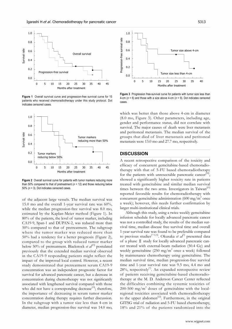
of the adjacent large vessels. The median survival was 15.0 mo and the overall 1-year survival rate was 60%, while the median progression-free survival was 8.0 mo, estimated by the Kaplan-Meier method (Figure 1). In 80% of the patients, the level of tumor marker, including CA19-9, Span-1 and DUPAN-2, was reduced more than 50% compared to that of pretreatment. The subgroup where the tumor marker was reduced more than 50% had a tendency for a better prognosis (Figure 2), compared to the group with reduced tumor marker below 50% of pretreatment. Blackstock et al[15] postulated previously that the extended median survival observed in the CA19-9 responding patients might reflect the impact of the improved local control. However, a recent study demonstrated that pretreatment serum CA19-9 concentration was an independent prognostic factor for survival for advanced pancreatic cancer, but a decrease in concentration during chemotherapy was not significantly associated with lengthened survival compared with those who did not have a corresponding decrease[19]; therefore, the importance of decreasing in serum tumor marker concentration during therapy requires further discussion. In the subgroup with a tumor size less than 4-cm in diameter, median progression-free survival was 14.0 mo,
which was better than those above 4-cm in diameter (8.0 mo, Figure 3). Other parameters, including age, gender and performance status, did not correlate with survival. The major causes of death were liver metastasis and peritoneal metastasis. The median survival of the groups that died of liver metastasis and peritoneal metastasis were 13.0 mo and 27.7 mo, respectively.
DISCUSSIONA recent retrospective comparison of the toxicity and efficacy of concurrent gemcitabine-based chemoradio-therapy with that of 5-FU based chemoradiotherapy for the patients with unresectable pancreatic cancer[20], showed a significantly higher toxicity rate in patients treated with gemcitabine and similar median survival times between the two arms. Investigators in Taiwan[21] reported favorable results for chemoradiotherapy with concurrent gemcitabine administration (600 mg/m2 once a week); however, this needs further confirmation by larger multi-institutional clinical trials.
Although this study, using a twice weekly gemcitabine infusion schedule for locally advanced pancreatic cancer was not a controlled study, the results of the median sur-vival time, median disease free survival time and overall 1-year survival rate was found to be preferable compared to previous studies[7-9,15]. Okusaka et al[7] presented data of a phase Ⅱ study for locally advanced pancreatic can-cer treated with external-beam radiation (50.4 Gy) and weekly gemcitabine (250 mg/m2 once a week) followed by maintenance chemotherapy using gemcitabine. The median survival time, median progression-free survival time and 1-year survival rate was 9.5 mo, 4.4 mo and 28%, respectively[7]. An expanded retrospective review of patients receiving gemcitabine-based chemoradio-therapy at the M. D. Anderson Cancer Center reflected the difficulties combining the systemic toxicities of 200-500 mg/m2 doses of gemcitabine with the local-regional toxicities associated with chemoradiotherapy to the upper abdomen[22]. Furthermore, in the original GITSG trial of radiation and 5-FU based chemotherapy, 18% and 21% of the patients randomized into the
1.0
0.8
0.6
0.4
0.2
0.0
Surv
ival
rat
e
Overall survival
Progression-free survival
0 5 10 15 20 25 30 35 40 45
Months after treatment
Figure 1 Overall survival curve and progression-free survival curve for 15 patients who received chemoradiotherapy under this study protocol. Dot indicates censored cases.
Figure 2 Overall survival curve for patients with tumor markers reducing more than 50% compared to that of pretreatment (n = 12) and those reducing below 50% (n = 3). Dot indicates censored cases.
1.0
0.8
0.6
0.4
0.2
0.0
Surv
ival
rat
e
0 5 10 15 20 25 30 35 40 45
Months after treatment
Tumor markersreducing more than 50%
Tumor markersreducing below 50%
1.0
0.8
0.6
0.4
0.2
0.0
Prog
ress
ion-
free
sur
viva
l rat
e
Tumor size above 4-cm
Tumor size less than 4-cm
0 5 10 15 20 25 30 35 40
Months after treatment
Figure 3 Progression free-survival curve for patients with tumor size less than 4-cm (n = 6) and those with a size above 4-cm (n = 9). Dot indicates censored cases.
www.wjgnet.com
Igarashi H et al . Chemoradiotherapy for pancreatic cancer 5313
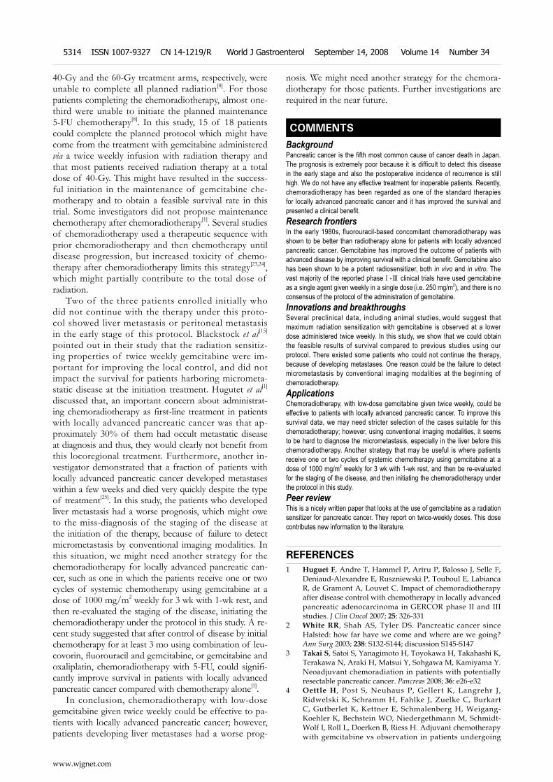
40-Gy and the 60-Gy treatment arms, respectively, were unable to complete all planned radiation[8]. For those patients completing the chemoradiotherapy, almost one-third were unable to initiate the planned maintenance 5-FU chemotherapy[8]. In this study, 15 of 18 patients could complete the planned protocol which might have come from the treatment with gemcitabine administered via a twice weekly infusion with radiation therapy and that most patients received radiation therapy at a total dose of 40-Gy. This might have resulted in the success-ful initiation in the maintenance of gemcitabine che-motherapy and to obtain a feasible survival rate in this trial. Some investigators did not propose maintenance chemotherapy after chemoradiotherapy[1]. Several studies of chemoradiotherapy used a therapeutic sequence with prior chemoradiotherapy and then chemotherapy until disease progression, but increased toxicity of chemo-therapy after chemoradiotherapy limits this strategy[23,24], which might partially contribute to the total dose of radiation.
Two of the three patients enrolled initially who did not continue with the therapy under this proto-col showed liver metastasis or peritoneal metastasis in the early stage of this protocol. Blackstock et al[15] pointed out in their study that the radiation sensitiz-ing properties of twice weekly gemcitabine were im-portant for improving the local control, and did not impact the survival for patients harboring micrometa-static disease at the initiation treatment. Hugutet et al[1]
discussed that, an important concern about administrat-ing chemoradiotherapy as first-line treatment in patients with locally advanced pancreatic cancer was that ap-proximately 30% of them had occult metastatic disease at diagnosis and thus, they would clearly not benefit from this locoregional treatment. Furthermore, another in-vestigator demonstrated that a fraction of patients with locally advanced pancreatic cancer developed metastases within a few weeks and died very quickly despite the type of treatment[25]. In this study, the patients who developed liver metastasis had a worse prognosis, which might owe to the miss-diagnosis of the staging of the disease at the initiation of the therapy, because of failure to detect micrometastasis by conventional imaging modalities. In this situation, we might need another strategy for the chemoradiotherapy for locally advanced pancreatic can-cer, such as one in which the patients receive one or two cycles of systemic chemotherapy using gemcitabine at a dose of 1000 mg/m2 weekly for 3 wk with 1-wk rest, and then re-evaluated the staging of the disease, initiating the chemoradiotherapy under the protocol in this study. A re-cent study suggested that after control of disease by initial chemotherapy for at least 3 mo using combination of leu-covorin, fluorouracil and gemcitabine, or gemcitabine and oxaliplatin, chemoradiotherapy with 5-FU, could signifi-cantly improve survival in patients with locally advanced pancreatic cancer compared with chemotherapy alone[1].
In conclusion, chemoradiotherapy with low-dose gemcitabine given twice weekly could be effective to pa-tients with locally advanced pancreatic cancer; however, patients developing liver metastases had a worse prog-
nosis. We might need another strategy for the chemora-diotherapy for those patients. Further investigations are required in the near future.
COMMENTSBackgroundPancreatic cancer is the fifth most common cause of cancer death in Japan. The prognosis is extremely poor because it is difficult to detect this disease in the early stage and also the postoperative incidence of recurrence is still high. We do not have any effective treatment for inoperable patients. Recently, chemoradiotherapy has been regarded as one of the standard therapies for locally advanced pancreatic cancer and it has improved the survival and presented a clinical benefit. Research frontiersIn the early 1980s, fluorouracil-based concomitant chemoradiotherapy was shown to be better than radiotherapy alone for patients with locally advanced pancreatic cancer. Gemcitabine has improved the outcome of patients with advanced disease by improving survival with a clinical benefit. Gemcitabine also has been shown to be a potent radiosensitizer, both in vivo and in vitro. The vast majority of the reported phaseⅠ-Ⅲ clinical trials have used gemcitabine as a single agent given weekly in a single dose (i.e. 250 mg/m2), and there is no consensus of the protocol of the administration of gemcitabine. Innovations and breakthroughsSeveral preclinical data, including animal studies, would suggest that maximum radiation sensitization with gemcitabine is observed at a lower dose administered twice weekly. In this study, we show that we could obtain the feasible results of survival compared to previous studies using our protocol. There existed some patients who could not continue the therapy, because of developing metastases. One reason could be the failure to detect micrometastasis by conventional imaging modalities at the beginning of chemoradiotherapy. ApplicationsChemoradiotherapy, with low-dose gemcitabine given twice weekly, could be effective to patients with locally advanced pancreatic cancer. To improve this survival data, we may need stricter selection of the cases suitable for this chemoradiotherapy; however, using conventional imaging modalities, it seems to be hard to diagnose the micrometastasis, especially in the liver before this chemoradiotherapy. Another strategy that may be useful is where patients receive one or two cycles of systemic chemotherapy using gemcitabine at a dose of 1000 mg/m2 weekly for 3 wk with 1-wk rest, and then be re-evaluated for the staging of the disease, and then initiating the chemoradiotherapy under the protocol in this study. Peer reviewThis is a nicely written paper that looks at the use of gemcitabine as a radiation sensitizer for pancreatic cancer. They report on twice-weekly doses. This dose contributes new information to the literature.
REFERENCES1 Huguet F, Andre T, Hammel P, Artru P, Balosso J, Selle F,
Deniaud-Alexandre E, Ruszniewski P, Touboul E, Labianca R, de Gramont A, Louvet C. Impact of chemoradiotherapy after disease control with chemotherapy in locally advanced pancreatic adenocarcinoma in GERCOR phase II and III studies. J Clin Oncol 2007; 25: 326-331
2 White RR, Shah AS, Tyler DS. Pancreatic cancer since Halsted: how far have we come and where are we going? Ann Surg 2003; 238: S132-S144; discussion S145-S147
3 Takai S, Satoi S, Yanagimoto H, Toyokawa H, Takahashi K, Terakawa N, Araki H, Matsui Y, Sohgawa M, Kamiyama Y. Neoadjuvant chemoradiation in patients with potentially resectable pancreatic cancer. Pancreas 2008; 36: e26-e32
4 Oettle H, Post S, Neuhaus P, Gellert K, Langrehr J, Ridwelski K, Schramm H, Fahlke J, Zuelke C, Burkart C, Gutberlet K, Kettner E, Schmalenberg H, Weigang-Koehler K, Bechstein WO, Niedergethmann M, Schmidt-Wolf I, Roll L, Doerken B, Riess H. Adjuvant chemotherapy with gemcitabine vs observation in patients undergoing
www.wjgnet.com
5314 ISSN 1007-9327 CN 14-1219/R World J Gastroenterol September 14, 2008 Volume 14 Number 34
COMMENTS
curative-intent resection of pancreatic cancer: a randomized controlled trial. JAMA 2007; 297: 267-277
5 Regine WF, Winter KA, Abrams RA, Safran H, Hoffman JP, Konski A, Benson AB, Macdonald JS, Kudrimoti MR, Fromm ML, Haddock MG, Schaefer P, Willett CG, Rich TA. Fluorouracil vs gemcitabine chemotherapy before and after fluorouracil-based chemoradiation following resection of pancreatic adenocarcinoma: a randomized controlled trial. JAMA 2008; 299: 1019-1026
6 Vento P, Mustonen H, Joensuu T, Karkkainen P, Kivilaakso E, Kiviluoto T. Impact of preoperative chemoradiotherapy on survival in patients with resectable pancreatic cancer. World J Gastroenterol 2007; 13: 2945-2951
7 Okusaka T, Ito Y, Ueno H, Ikeda M, Takezako Y, Morizane C, Kagami Y, Ikeda H. Phase II study of radiotherapy combined with gemcitabine for locally advanced pancreatic cancer. Br J Cancer 2004; 91: 673-677
8 Moertel CG, Frytak S, Hahn RG, O'Connell MJ, Reitemeier RJ, Rubin J, Schutt AJ, Weiland LH, Childs DS, Holbrook MA, Lavin PT, Livstone E, Spiro H, Knowlton A, Kalser M, Barkin J, Lessner H, Mann-Kaplan R, Ramming K, Douglas HO Jr, Thomas P, Nave H, Bateman J, Lokich J, Brooks J, Chaffey J, Corson JM, Zamcheck N, Novak JW. Therapy of locally unresectable pancreatic carcinoma: a randomized comparison of high dose (6000 rads) radiation alone, moderate dose radiation (4000 rads + 5-fluorouracil), and high dose radiation + 5-fluorouracil: The Gastrointestinal Tumor Study Group. Cancer 1981; 48: 1705-1710
9 Treatment of locally unresectable carcinoma of the pancreas: comparison of combined-modality therapy (chemotherapy plus radiotherapy) to chemotherapy alone. Gastrointestinal Tumor Study Group. J Natl Cancer Inst 1988; 80: 751-755
10 Berlin JD, Catalano P, Thomas JP, Kugler JW, Haller DG, Benson AB 3rd. Phase III study of gemcitabine in combination with fluorouracil versus gemcitabine alone in patients with advanced pancreatic carcinoma: Eastern Cooperative Oncology Group Trial E2297. J Clin Oncol 2002; 20: 3270-3275
11 Sakamoto H, Kitano M, Suetomi Y, Takeyama Y, Ohyanagi H, Nakai T, Yasuda C, Kudo M. Comparison of standard-dose and low-dose gemcitabine regimens in pancreatic adenocarcinoma patients: a prospective randomized trial. J Gastroenterol 2006; 41: 70-76
12 Burris HA 3rd , Moore MJ, Andersen J, Green MR, Rothenberg ML, Modiano MR, Cripps MC, Portenoy RK, Storniolo AM, Tarassoff P, Nelson R, Dorr FA, Stephens CD, Von Hoff DD. Improvements in survival and clinical benefit with gemcitabine as first-line therapy for patients with advanced pancreas cancer: a randomized trial. J Clin Oncol 1997; 15: 2403-2413
13 Rothenberg ML. New developments in chemotherapy for patients with advanced pancreatic cancer. Oncology (Williston Park) 1996; 10: 18-22
14 Blackstock AW, Bernard SA, Richards F, Eagle KS, Case LD, Poole ME, Savage PD, Tepper JE. Phase I trial of twice-weekly gemcitabine and concurrent radiation in patients with advanced pancreatic cancer. J Clin Oncol 1999; 17: 2208-2212
15 Blackstock AW, Tepper JE, Niedwiecki D, Hollis DR, Mayer RJ, Tempero MA. Cancer and leukemia group B (CALGB) 89805: phase II chemoradiation trial using gemcitabine in patients with locoregional adenocarcinoma of the pancreas. Int J Gastrointest Cancer 2003; 34: 107-116
16 Crane CH, Varadhachary G, Pisters PW, Evans DB, Wolff RA. Future chemoradiation strategies in pancreatic cancer. Semin Oncol 2007; 34: 335-346
17 Fields MT, Eisbruch A, Normolle D, Orfali A, Davis MA, Pu AT, Lawrence TS. Radiosensitization produced in vivo by once- vs. twice-weekly 2'2'-difluoro-2'-deoxycytidine (gemcitabine). Int J Radiat Oncol Biol Phys 2000; 47: 785-791
18 Magnino A, Gatti M, Massucco P, Sperti E, Faggiuolo R, Regge D, Capussotti L, Gabriele P, Aglietta M. Phase II trial of primary radiation therapy and concurrent chemotherapy for patients with locally advanced pancreatic cancer. Oncology 2005; 68: 493-499
19 Hess V, Glimelius B, Grawe P, Dietrich D, Bodoky G, Ruhstaller T, Bajetta E, Saletti P, Figer A, Scheithauer W, Herrmann R. CA 19-9 tumour-marker response to chemotherapy in patients with advanced pancreatic cancer enrolled in a randomised controlled trial. Lancet Oncol 2008; 9: 132-138
20 Crane CH, Abbruzzese JL, Evans DB, Wolff RA, Ballo MT, Delclos M, Milas L, Mason K, Charnsangavej C, Pisters PW, Lee JE, Lenzi R, Vauthey JN, Wong AB, Phan T, Nguyen Q, Janjan NA. Is the therapeutic index better with gemcitabine-based chemoradiation than with 5-fluorouracil-based chemoradiation in locally advanced pancreatic cancer? Int J Radiat Oncol Biol Phys 2002; 52: 1293-1302
21 Li CP, Chao Y, Chi KH, Chan WK, Teng HC, Lee RC, Chang FY, Lee SD, Yen SH. Concurrent chemoradiotherapy treatment of local ly advanced pancreat ic cancer : gemcitabine versus 5-fluorouracil, a randomized controlled study. Int J Radiat Oncol Biol Phys 2003; 57: 98-104
22 Crane CH, Janjan NA, Evans DB, Wolff RA, Ballo MT, Milas L, Mason K, Charnsangavej C, Pisters PW, Lee JE, Lenzi R, Vauthey JN, Wong A, Phan T, Nguyen Q, Abbruzzese JL. Toxicity and efficacy of concurrent gemcitabine and radiotherapy for locally advanced pancreatic cancer. Int J Pancreatol 2001; 29: 9-18
23 Epelbaum R, Rosenblatt E, Nasrallah S, Faraggi D, Gaitini D, Mizrahi S, Kuten A. Phase II study of gemcitabine combined with radiation therapy in patients with localized, unresectable pancreatic cancer. J Surg Oncol 2002; 81: 138-143
24 Schneider BJ, Ben-Josef E, McGinn CJ, Chang AE, Colletti LM, Normolle DP, Hejna GF, Lawrence TS, Zalupski MM. Capecitabine and radiation therapy preceded and followed by combination chemotherapy in advanced pancreatic cancer. Int J Radiat Oncol Biol Phys 2005; 63: 1325-1330
25 Andre T, Balosso J, Louvet C, Hannoun L, Houry S, Huguier M, Colonna M, Lotz JP, De Gramont A, Bellaiche A, Parc R, Touboul E, Izrael V. Combined radiotherapy and chemotherapy (cisplatin and 5-fluorouracil) as palliative treatment for localized unresectable or adjuvant treatment for resected pancreatic adenocarcinoma: results of a feasibility study. Int J Radiat Oncol Biol Phys 2000; 46: 903-911
S- Editor Zhong XY L- Editor Rippe RA E- Editor Ma WH
www.wjgnet.com
Igarashi H et al . Chemoradiotherapy for pancreatic cancer 5315
Online Submissions: wjg.wjgnet.com World J Gastroenterol 2008 September 14; 14(34): [email protected] World Journal of Gastroenterology ISSN 1007-9327doi:10.3748/wjg.14.5316 © 2008 The WJG Press. All rights reserved.
Endoscopic findings can predict the efficacy of leukocytapheresis for steroid-naive patients with moderately active ulcerative colitis
Yasushi Umehara, Masatoshi Kudo, Masanori Kawasaki
RAPID COMMUNICATION
www.wjgnet.com
Yasushi Umehara, Masatoshi Kudo, Masanori Kawasaki, Division of Gastroenterology and Hepatology, Department of Internal Medicine Kinki University School of Medicine, 377-2 Ohno-Higashi, Osaka-Sayama 589-8511, JapanAuthor contributions: Umehara Y, Kudo M and Kawasaki M contributed equally to this work; Umehara Y and Kudo M desined research; Umehara Y and Kawasaki M performed research; Umehara Y and Kudo M contributed new reagents/analytic tools; Umehara Y and Kudo M analyzed data and Umehara Y wrote the paper.Correspondence to: Masatoshi Kudo, MD, PhD, Division of Gastroenterology and Hepatology, Department of Internal Medicine Kinki University School of Medicine, 377-2 Ohno-Higashi, Osaka-Sayama 589-8511, Japan. [email protected]: +81-723-660221-3525 Fax: +81-723-672880Received: June 21, 2008 Revised: August 11, 2008Accepted: August 18, 2008Published online: September 14, 2008
AbstractAIM: To investigate the therapeutic usefulness of leukocytapheresis (LCAP; Cellsoba) in steroid-naive patients with moderately active ulcerative colitis (UC).METHODS: Eighteen steroid-naive patients with moderately active UC received one LCAP session every week for five consecutive weeks.RESULTS: The remission rate 8 weeks after the last LCAP session was 61.1% (11/18). All three patients with deep ulcers showed worsening after LCAP. For the remaining 15 patients, who had erosions or geographic ulcers, the average clinical activity index (CAI) score dropped significantly from 9.4 to 3.8 eight weeks after the last LCAP session (t = 4.89, P = 0.001). The average C-reactive protein (CRP) levels before and after LCAP were 1.2 mg/dL and 1.0 mg/dL, respectively. Of the patients with erosions, geographic ulcers, and deep ulcers, 100% (9/9), 33.3% (2/6), and 0% (0/3) were in remission 8 weeks after the last LCAP session, respectively (χ2 = 7.65, P < 0.005). Forty-eight weeks after the last LCAP session, the remission rates for patients with erosions and geographic ulcers were 44.4% (4/9) and 16.7% (1/6), respectively. Only one patient suffered a mild adverse event after LCAP (nausea).CONCLUSION: LCAP is a useful and safe therapy
for steroid-naive UC patients with moderate disease activity. Moreover, the efficacy of the treatment can be predicted on the basis of endoscopic findings.
© 2008 The WJG Press. All rights reserved.
Key words: Ulcera t ive co l i t i s ; S te ro id -na ive; Leukocytapheresis; Efficacy; Endoscopic findings
Peer reviewer: Alastair JM Watson, Professor, Department of Gastroenterology, University of Liverpool, the Henry Wellcome Laboratory, Nuffield Bldg, Crown St, Liverpool L69 3GE, United Kingdom
Umehara Y, Kudo M, Kawasaki M. Endoscopic findings can predict the efficacy of leukocytapheresis for steroid-naive patients with moderately active ulcerative colitis. World J Gastroenterol 2008; 14(34): 5316-5321 Available from: URL: http://www.wjgnet.com/1007-9327/14/5316.asp DOI: http://dx.doi.org/10.3748/wjg.14.5316
INTRODUCTIONAlthough the etiology of ulcerative colitis (UC) is still unknown, it is believed that an immune abnormality may be involved in its development[1,2]. It is characterized by chronic over-activation of the colonic mucosal immune system. Consequently, if remission cannot be achieved by salazosulfapyridine or mesalazine treatment, the second line of treatment has conventionally been to administer steroids[2-6]. However, steroid administration can increase susceptibility to infections, diabetes mellitus and osteoporosis. Recently, it was reported that steroid-refractory or steroid-dependent patients with UC can be effectively treated by cytapheresis[7-10]. To determine whether leukocytapheresis (LCAP) may also be useful with other UC patients, we administered LCAP to 18 steroid-naive UC patients. We also assessed whether the efficacy of LCAP can be predicted on the basis of endoscopic findings.
MATERIALS AND METHODSFrom January 2005 to April 2007, 33 UC patients were treated with LCAP at our hospital. All patients were
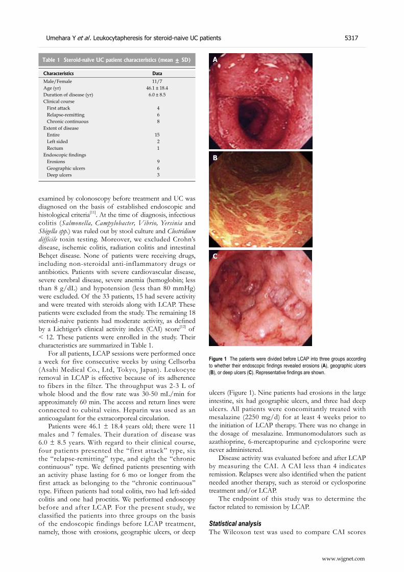
www.wjgnet.com
Umehara Y et al . Leukocytapheresis for steroid-naive UC patients 5317
examined by colonoscopy before treatment and UC was diagnosed on the basis of established endoscopic and histological criteria[11]. At the time of diagnosis, infectious colitis (Salmonella, Campylobacter, Vibrio, Yersinia and Shigella spp.) was ruled out by stool culture and Clostridium difficile toxin testing. Moreover, we excluded Crohn’s disease, ischemic colitis, radiation colitis and intestinal Behçet disease. None of patients were receiving drugs, including non-steroidal anti-inflammatory drugs or antibiotics. Patients with severe cardiovascular disease, severe cerebral disease, severe anemia (hemoglobin; less than 8 g/dL) and hypotension (less than 80 mmHg) were excluded. Of the 33 patients, 15 had severe activity and were treated with steroids along with LCAP. These patients were excluded from the study. The remaining 18 steroid-naive patients had moderate activity, as defined by a Lichtiger’s clinical activity index (CAI) score[12] of < 12. These patients were enrolled in the study. Their characteristics are summarized in Table 1.
For all patients, LCAP sessions were performed once a week for five consecutive weeks by using Cellsorba (Asahi Medical Co., Ltd, Tokyo, Japan). Leukocyte removal in LCAP is effective because of its adherence to fibers in the filter. The throughput was 2-3 L of whole blood and the flow rate was 30-50 mL/min for approximately 60 min. The access and return lines were connected to cubital veins. Heparin was used as an anticoagulant for the extracorporeal circulation.
Patients were 46.1 ± 18.4 years old; there were 11 males and 7 females. Their duration of disease was 6.0 ± 8.5 years. With regard to their clinical course, four patients presented the “first attack” type, six the “relapse-remitting” type, and eight the “chronic continuous” type. We defined patients presenting with an activity phase lasting for 6 mo or longer from the first attack as belonging to the “chronic continuous” type. Fifteen patients had total colitis, two had left-sided colitis and one had proctitis. We performed endoscopy before and after LCAP. For the present study, we classified the patients into three groups on the basis of the endoscopic findings before LCAP treatment, namely, those with erosions, geographic ulcers, or deep
ulcers (Figure 1). Nine patients had erosions in the large intestine, six had geographic ulcers, and three had deep ulcers. All patients were concomitantly treated with mesalazine (2250 mg/d) for at least 4 weeks prior to the initiation of LCAP therapy. There was no change in the dosage of mesalazine. Immunomodulators such as azathioprine, 6-mercaptopurine and cyclosporine were never administered.
Disease activity was evaluated before and after LCAP by measuring the CAI. A CAI less than 4 indicates remission. Relapses were also identified when the patient needed another therapy, such as steroid or cyclosporine treatment and/or LCAP.
The endpoint of this study was to determine the factor related to remission by LCAP.
Statistical analysisThe Wilcoxon test was used to compare CAI scores
Table 1 Steroid-naïve UC patient characteristics (mean ± SD)
Characteristics Data
Male/Female 11/7Age (yr) 46.1 ± 18.4Duration of disease (yr) 6.0 ± 8.5Clinical course First attack 4 Relapse-remitting 6 Chronic continuous 8Extent of disease Entire 15 Left sided 2 Rectum 1Endoscopic findings Erosions 9 Geographic ulcers 6 Deep ulcers 3
A
B
C
Figure 1 The patients were divided before LCAP into three groups according to whether their endoscopic findings revealed erosions (A), geographic ulcers (B), or deep ulcers (C). Representative findings are shown.
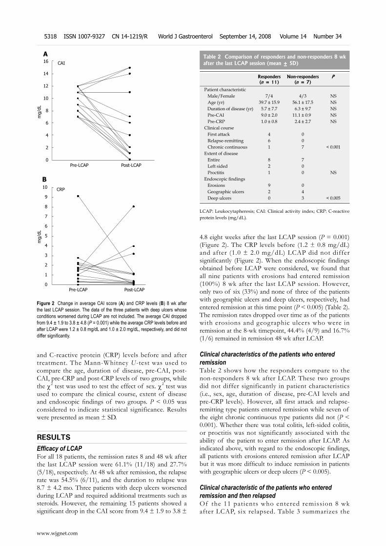
and C-reactive protein (CRP) levels before and after treatment. The Mann-Whitney U-test was used to compare the age, duration of disease, pre-CAI, post-CAI, pre-CRP and post-CRP levels of two groups, while the χ2 test was used to test the effect of sex. χ2 test was used to compare the clinical course, extent of disease and endoscopic findings of two groups. P < 0.05 was considered to indicate statistical significance. Results were presented as mean ± SD.
RESULTSEfficacy of LCAPFor all 18 patients, the remission rates 8 and 48 wk after the last LCAP session were 61.1% (11/18) and 27.7% (5/18), respectively. At 48 wk after remission, the relapse rate was 54.5% (6/11), and the duration to relapse was 8.7 ± 4.2 mo. Three patients with deep ulcers worsened during LCAP and required additional treatments such as steroids. However, the remaining 15 patients showed a significant drop in the CAI score from 9.4 ± 1.9 to 3.8 ±
4.8 eight weeks after the last LCAP session (P = 0.001) (Figure 2). The CRP levels before (1.2 ± 0.8 mg/dL) and after (1.0 ± 2.0 mg/dL) LCAP did not differ significantly (Figure 2). When the endoscopic findings obtained before LCAP were considered, we found that all nine patients with erosions had entered remission (100%) 8 wk after the last LCAP session. However, only two of six (33%) and none of three of the patients with geographic ulcers and deep ulcers, respectively, had entered remission at this time point (P < 0.005) (Table 2). The remission rates dropped over time as of the patients with erosions and geographic ulcers who were in remission at the 8-wk timepoint, 44.4% (4/9) and 16.7% (1/6) remained in remission 48 wk after LCAP.
Clinical characteristics of the patients who enteredremissionTable 2 shows how the responders compare to the non-responders 8 wk after LCAP. These two groups did not differ significantly in patient characteristics (i.e., sex, age, duration of disease, pre-CAI levels and pre-CRP levels). However, all first attack and relapse-remitting type patients entered remission while seven of the eight chronic continuous type patients did not (P < 0.001). Whether there was total colitis, left-sided colitis, or proctitis was not significantly associated with the ability of the patient to enter remission after LCAP. As indicated above, with regard to the endoscopic findings, all patients with erosions entered remission after LCAP but it was more difficult to induce remission in patients with geographic ulcers or deep ulcers (P < 0.005).
Clinical characteristic of the patients who entered remission and then relapsedOf the 11 patients who entered remission 8 wk after LCAP, six relapsed. Table 3 summarizes the
Figure 2 Change in average CAI score (A) and CRP levels (B) 8 wk after the last LCAP session. The data of the three patients with deep ulcers whose conditions worsened during LCAP are not included. The average CAI dropped from 9.4 ± 1.9 to 3.8 ± 4.8 (P = 0.001) while the average CRP levels before and after LCAP were 1.2 ± 0.8 mg/dL and 1.0 ± 2.0 mg/dL, respectively, and did not differ significantly.
16
14
12
10
8
6
4
2
0
ACAI
Pre-LCAP Post-LCAP
mg/
dL
10
9
8
7
6
5
4
3
2
1
0
BCRP
Pre-LCAP Post-LCAP
mg/
dL
Table 2 Comparison of responders and non-responders 8 wk after the last LCAP session (mean ± SD)
Responders(n = 11)
Non-responders(n = 7)
P
Patient characteristic Male/Female 7/4 4/3 NS Age (yr) 39.7 ± 15.9 56.1 ± 17.5 NS Duration of disease (yr) 5.7 ± 7.7 6.3 ± 9.7 NS Pre-CAI 9.0 ± 2.0 11.1 ± 0.9 NS Pre-CRP 1.0 ± 0.8 2.4 ± 2.7 NSClinical course First attack 4 0 Relapse-remitting 6 0 Chronic continuous 1 7 < 0.001Extent of disease Entire 8 7 Left sided 2 0 Proctitis 1 0 NSEndoscopic findings Erosions 9 0 Geographic ulcers 2 4 Deep ulcers 0 3 < 0.005
LCAP: Leukocytapheresis; CAI: Clinical activity index; CRP: C-reactive protein levels (mg/dL).
www.wjgnet.com
5318 ISSN 1007-9327 CN 14-1219/R World J Gastroenterol September 14, 2008 Volume 14 Number 34
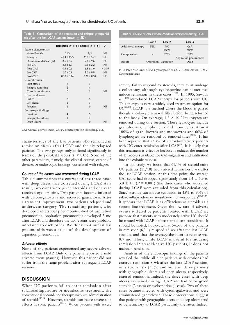
characteristics of the five patients who remained in remission 48 wk after LCAP and the six relapsed patients. The two groups only differed significantly in terms of the post-CAI scores (P < 0.05). None of the other parameters, namely, the clinical course, extent of disease, or endoscopic findings, correlated with relapse.
Course of the cases who worsened during LCAPTable 4 summarizes the courses of the three cases with deep ulcers that worsened during LCAP. As a result, two cases were given steroids and one case received cyclosporine. Two patients became infected with cytomegalovirus and received ganciclovir. After a transient improvement, two patients relapsed and underwent surgery. The remaining patient, who developed interstitial pneumonitis, died of aspiration pneumonitis. Aspiration pneumonitis developed 3 mo after LCAP, and therefore the two events were probably unrelated to each other. We think that interstitial pneumonitis was a cause of the development of aspiration pneumonitis.
Adverse effectsNone of the patients experienced any severe adverse effects from LCAP. Only one patient reported a mild adverse event (nausea). However, this patient did not suffer from the same problem after subsequent LCAP sessions.
DISCUSSIONWhen UC pat ients fa i l to enter remiss ion after salazosulfapyridine or mesalazine treatment, the conventional second-line therapy involves administration of steroids[13,14]. However, steroids can cause severe side effects in some patients[15,16]. When patients with severe
activity fail to respond to steroids, they must undergo a colectomy, although cyclosporine can sometimes induce remission in these cases[17-20]. In 1995, Sawada et al[21] introduced LCAP therapy for patients with UC. This therapy is now a widely used treatment option for UC[22,23]. LCAP is a method where the blood is passed though a leukocyte removal filter before being returned to the body. On average, 1.6 × 10¹⁰ leukocytes are removed during one session. These leukocytes include granulocytes, lymphocytes and monocytes. Almost 100% of granulocytes and monocytes and 60% of lymphocytes are removed by removal filter[24,25]. It has been reported that 73.3% of steroid-refractory patients with UC enter remission after LCAP[26]. It is likely that this treatment is effective because it reduces the number of leukocytes available for transmigration and infiltration into the colonic mucosa.
In this study, we found that 61.1% of steroid-naive UC patients (11/18) had entered remission 8 wk after the last LCAP session. At this time point, the average CAI score had dropped significantly from 9.4 ± 1.9 to 3.8 ± 4.8 (P = 0.001) (the three cases who worsened during LCAP were excluded from this calculation). Since steroids can induce remission in 45% to 90% of salazosulfapyridine or mesalazine non-responders[15,27-29], it appears that LCAP is as efficacious as steroids as a second-line treatment. Given the low rate of adverse events suffered by patients treated with LCAP, we propose that patients with moderately active UC should be treated with LCAP before steroids are considered. It should be noted, however, that 54.5% of the patients in remission (6/11) relapsed 48 wk after the last LCAP session, and that the average duration to relapse was 8.7 mo. Thus, while LCAP is useful for inducing remission in steroid-naive UC patients, it does not maintain remission.
Analysis of the endoscopic findings of the patients revealed that while all nine patients with erosions had entered remission 8 wk after the last LCAP session, only two of six (33%) and none of three patients with geographic ulcers and deep ulcers, respectively, entered remission. Indeed, the three cases with deep ulcers worsened during LCAP and had to be given steroids (2 cases) or cyclosporine (1 case). Two of these cases became infected with cytomegalovirus and were administered ganciclovir. These observations suggest that patients with geographic ulcers and deep ulcers tend to be refractory to LCAP, particularly the latter. Indeed,
Table 3 Comparison of the remission and relapse groups 48 wk after the last LCAP session (mean ± SD)
Remission (n = 5) Relapse (n = 6) PPatient characteristic Male/Female 2/3 5/1 NS Age (yr) 45.4 ± 13.5 35.0 ± 16.1 NS Duration of disease (yr) 3.5 ± 3.2 7.6 ± 9.6 NS Pre-CAI 8.8 ± 1.7 9.1 ± 2.2 NS Post-CAI 0.6 ± 0.4 1.8 ± 1.0 < 0.05 Pre-CRP 1.0 ± 0.9 1.0 ± 0.8 NS Post-CRP 0.18 ± 0.14 0.32 ± 0.39 NSClinical course First attack 3 1 Relapse-remitting 2 4 Chronic continuous 0 1 NSExtent of disease Entire 3 5 Left sided 1 1 Proctitis 1 0 NSEndoscopic findings Erosions 4 5 Geographic ulcers 1 1 Deep ulcers 0 0 NS
CAI: Clinical activity index; CRP: C-reactive protein levels (mg/dL).
Table 4 Course of cases whose condition worsened during LCAP
Case 1 Case 2 Case 3
Additional therapy PSL PSL CsAGCV GCV
Complication - CMV CMVAspiration pneumonitis
Result Operation Operation Dead
PSL: Prednisolone; CsA: Cyclospoline; GCV: Ganciclovir; CMV: Cytomegalovirus.
www.wjgnet.com
Umehara Y et al . Leukocytapheresis for steroid-naive UC patients 5319
LCAP may not improve the situation for patients with deep ulcers given their higher risk of developing cytomegalovirus infections[30]. Such patients should perhaps be treated with an intensive therapy such as cyclosporine at a more early stage[31]. However, since all patients with erosion entered remission 8 wk after LCAP and many (44.4%) remained in remission at the 48-wk time point, LCAP is strongly recommended for patients with erosion.
The post-CAI was the only factor that predicted a relapse. In other words, if the post-CAI could be maintained at < 1 by LCAP, it may be possible to maintain long duration remission.
In conclusion, LCAP is a useful and safe therapy for steroid-naive UC patients with moderate activity. Moreover, endoscopic findings help to predict the efficacy of this treatment.
COMMENTSBackgroundAn immune dysfunction is believed to be involved in the development of ulcerative colitis (UC). For a long time, steroids have represented the second line therapy for the induction to remission in UC, if remission cannot be achieved by salazosulfapyridine or mesalazine treatment. However, steroid administration can cause several side effects. Leukocytapheresis (LCAP) have been reported to be effective for steroid-refractory or steroid-dependent patients with UC; however, the data of LCAP for steroid-naïve patient with UC is limited.Research frontiersTo determine whether LCAP may also be useful in steroid-naive UC patients, these authors administered LCAP to steroid-naive UC patients. They also assessed whether the efficacy of LCAP can be predicted on the basis of endoscopic findings.Innovations and breakthroughsThe authors found that 61.1% of steroid-naive UC patients (11/18) had entered remission 8 wk after the last LCAP session. Since steroids can induce remission in 45% to 90% of salazosulfapyridine or mesalazine non-responders, it appears that LCAP is as efficacious as steroids as a second-line treatment. Analysis of the endoscopic findings of the patients revealed that the remission rate of the patients with erosion was extremely high after LCAP, compared to the extremely low rate observed in patients with geographic ulcers and deep ulcers. None of the patients experienced any severe adverse effects from LCAP.ApplicationsGiven the low rate of adverse events suffered by patients treated with LCAP, authors propose that patients with moderately active UC should be treated with LCAP before steroids are considered.TerminologyLCAP is a treatment procedure where leukocytes are removed through their adherence to fibers in the filter.Peer reviewLCAP is a useful and safe therapy for steroid-naive UC patients with moderate activity. Moreover, endoscopic findings help to predict the efficacy of this treatment. This is an interesting study.
REFERENCES1 Arai F, Takahashi T, Furukawa K, Matsushima K, Asakura
H. Mucosal expression of interleukin-6 and interleukin-8 messenger RNA in ulcerative colitis and in Crohn's disease. Dig Dis Sci 1998; 43: 2071-2079
2 Podolsky DK. Inflammatory bowel disease (1). N Engl J Med 1991; 325: 928-937
3 Truelove SC, Jewell DP. Intensive intravenous regimen for severe attacks of ulcerative colitis. Lancet 1974; 1: 1067-1070
4 Meyers S , Sachar DB, Goldberg JD, Janowitz HD.
Corticotropin versus hydrocortisone in the intravenous treatment of ulcerative colitis. A prospective, randomized, double-blind clinical trial. Gastroenterology 1983; 85: 351-357
5 Hanauer SB. Inflammatory bowel disease. N Engl J Med 1996; 334: 841-848
6 Jarnerot G, Rolny P, Sandberg-Gertzen H. Intensive intravenous treatment of ulcerative colitis. Gastroenterology 1985; 89: 1005-1013
7 Naganuma M, Funakoshi S, Sakuraba A, Takagi H, Inoue N, Ogata H, Iwao Y, Ishi H, Hibi T. Granulocytapheresis is useful as an alternative therapy in patients with steroid-refractory or -dependent ulcerative colitis. Inflamm Bowel Dis 2004; 10: 251-257
8 Giampaolo B, Giuseppe P, Michele B, Alessandro M, Fabrizio S, Alfonso C. Treatment of active steroid-refractory inflammatory bowel diseases with granulocytapheresis: Our experience with a prospective study. World J Gastroenterol 2006; 12: 2201-2204
9 Emmrich J, Petermann S, Nowak D, Beutner I, Brock P, Klingel R, Mausfeld-Lafdhiya P, Liebe S, Ramlow W. Leukocytapheresis (LCAP) in the management of chronic active ulcerative colitis--results of a randomized pilot trial. Dig Dis Sci 2007; 52: 2044-2053
10 Domenech E, Hinojosa J, Esteve-Comas M, Gomollon F, Herrera JM, Bastida G, Obrador A, Ruiz R, Saro C, Gassull MA. Granulocyteaphaeresis in steroid-dependent inflammatory bowel disease: a prospective, open, pilot study. Aliment Pharmacol Ther 2004; 20: 1347-1352
11 Garland CF, Lilienfeld AM, Mendeloff AI, Markowitz JA, Terrell KB, Garland FC. Incidence rates of ulcerative colitis and Crohn's disease in fifteen areas of the United States. Gastroenterology 1981; 81: 1115-1124
12 Lichtiger S, Present DH, Kornbluth A, Gelernt I, Bauer J, Galler G, Michelassi F, Hanauer S. Cyclosporine in severe ulcerative colitis refractory to steroid therapy. N Engl J Med 1994; 330: 1841-1845
13 Podolsky DK. Inflammatory bowel disease. N Engl J Med 2002; 347: 417-429
14 Kornbluth A, Sachar DB. Ulcerative colitis practice guidelines in adults (update): American College of Gastroenterology, Practice Parameters Committee. Am J Gastroenterol 2004; 99: 1371-1385
15 Lennard-Jones JE. Toward optimal use of corticosteroids in ulcerative colitis and Crohn's disease. Gut 1983; 24: 177-181
16 Baron JH, Connell AM, Kanaghinis TG, Lennard-Jones JE, Jones AF. Out-patient treatment of ulcerative colitis. Comparison between three doses of oral prednisone. Br Med J 1962; 2: 441-443
17 Rowe FA, Walker JH, Karp LC, Vasiliauskas EA, Plevy SE, Targan SR. Factors predictive of response to cyclosporin treatment for severe, steroid-resistant ulcerative colitis. Am J Gastroenterol 2000; 95: 2000-2008
18 Stack WA, Long RG, Hawkey CJ. Short- and long-term outcome of patients treated with cyclosporin for severe acute ulcerative colitis. Aliment Pharmacol Ther 1998; 12: 973-978
19 Sandborn WJ. A critical review of cyclosporin therapy in inflammatory bowel disease. Inframm Bowel Dis 1995; 1: 48-63
20 Santos J, Baudet S, Casellas F, Guarner L, Vilaseca J, Malagelada JR. Efficacy of intravenous cyclosporine for steroid refractory attacks of ulcerative colitis. J Clin Gastroenterol 1995; 20: 285-289
21 Sawada K, Ohnishi K, Fukui S, Yamada K, Yamamura M, Amano K, Amano K, Wada M, Tanida N, Satomi M. Leukocytapheresis therapy, performed with leukocyte removal f i l ter , for inflammatory bowel disease. J Gastroenterol 1995; 30: 322-329
22 Sawada K, Shimoyama T. Therapeutic cytapheresis for inflammatory bowel disease. Ther Apher 1998; 2: 90-92
23 Sawada K, Muto T, Shimoyama T, Satomi M, Sawada T, Nagawa H, Hiwatashi N, Asakura H, Hibi T. Multicenter
www.wjgnet.com
5320 ISSN 1007-9327 CN 14-1219/R World J Gastroenterol September 14, 2008 Volume 14 Number 34
COMMENTS
randomized controlled trial for the treatment of ulcerative colitis with a leukocytapheresis column. Curr Pharm Des 2003; 9: 307-321
24 Shirokaze J. Leukocytapheresis using a leukocyte removal filter. Ther Apher 2002; 6: 261-266
25 Yamaji K, Tsuda H, Hashimoto H. Current topics on cytapheresis technologies. Ther Apher 2001; 5: 287-292
26 Kohgo Y , Hibi H, Chiba T, Shimoyama T, Muto T, Yamamura K, Popovsky MA. Leukocyte apheresis using a centrifugal cell separator in refractory ulcerative colitis: a multicenter open label trial. Ther Apher 2002; 6: 255-260
27 Lennard-Jones JE, Longmore AJ, Newell AC, Wilson CW, Jones FA. An assessment of prednisone, salazopyrin, and topical hydrocortisone hemisuccinate used as out-patient treatment for ulcerative colitis. Gut 1960; 1: 217-222
28 Powell-Tuck J, Bown RL, Lennard-Jones JE. A comparison
of oral prednisolone given as single or multiple daily doses for active proctocolitis. Scand J Gastroenterol 1978; 13: 833-837
29 Faubion WA Jr, Loftus EV Jr, Harmsen WS, Zinsmeister AR, Sandborn WJ. The natural history of corticosteroid therapy for inflammatory bowel disease: a population-based study. Gastroenterology 2001; 121: 255-260
30 Nishimoto Y, Matsumoto T, Suekane H, Shimizu M, Mikami Y, Iida M. Cytomegalovirus infection in a patient with ulcerative colitis: colonoscopic findings. Gastrointest Endosc 2001; 53: 816-818
31 D'Haens G, Lemmens L, Geboes K, Vandeputte L, Van Acker F, Mortelmans L, Peeters M, Vermeire S, Penninckx F, Nevens F, Hiele M, Rutgeerts P. Intravenous cyclosporine versus intravenous corticosteroids as single therapy for severe attacks of ulcerative colitis. Gastroenterology 2001; 120: 1323-1329
S- Editor Li DL L- Editor Negro F E- Editor Zhang WB
www.wjgnet.com
Umehara Y et al . Leukocytapheresis for steroid-naive UC patients 5321
Payam Eghtesadi-Araghi, Amir-Ali Sohrabpour, Homayoon Vahedi, Mehdi Saberi-Firoozi
Halothane hepatitis in Iran: A review of 59 cases
RAPID COMMUNICATION
www.wjgnet.com
Online Submissions: wjg.wjgnet.com World J Gastroenterol 2008 September 14; 14(34): [email protected] World Journal of Gastroenterology ISSN 1007-9327doi:10.3748/wjg.14.5322 © 2008 The WJG Press. All rights reserved.
Payam Eghtesadi-Araghi, Homayoon Vahedi, Digestive Disease Research Center, Shariati Hospital, Medical Sciences/University of Tehran, Tehran 14117, IranAmir-Ali Sohrabpour, Mehdi Saberi-Firoozi, Gastroenterology Research Center, Shiraz University of Medical Sciences, Shiraz 14117, IranAuthor contributions: Eghtesadi-Araghi P designed and performed research, analyzed data and wrote the paper; Vahedi H contributed in providing cases and critical review of the project; Sohrabpour A and Saberi-Firoozi M contributed in providing cases.Supported by A grant from the Digestive Disease Research Center, Tehran University of Medical Sciences, Iran, No. 83.36Correspondence to: Dr. Payam Eghtesadi-Araghi, Digestive Disease Research Center, Shariati Hospital, North Karegar Avenue, Medical Sciences/University of Tehran, Tehran 14117, Iran. [email protected]: +98-21-44454814 Fax: +98-21-44454814Received: June 11, 2008 Revised: July 27, 2008Accepted: August 3, 2008Published online: September 14, 2008
AbstractAIM: To study halothane hepatitis (HH) in Iran and its associated risk factors.METHODS: We retrospectively studied files of all cases diagnosed with HH referred to three referral hospitals and four private centers in Iran from April 1994 to September 2006. Information on age at surgery, gender, medications history, obesity, history of previous exposure, previous reaction to halothane, familial history, type of surgery, perioperative hypoxia or sepsis, morbidity and mortality were recorded and analyzed.RESULTS: A total of 59 cases were identified. Forty-eight (81%) were women. The median age at the time of surgery was 44 years (range, 18 to 80 years). Sixty percent of patients were above 40-year-old. Obesity was observed in 22.2%. Previous history of exposures to halothane was noted in 61% of which 50% had history of post-exposure reaction. Coronary artery bypass graft (CABG), cholecystectomy, and cosmetic surgeries (mainly weight reduction) were the most frequent surgeries. The mortality rate was 12.2%. In patients developing encephalopathy, it was as high as 50%.CONCLUSION: HH remains an important cause of morbidity and mortality in centers still using this anesthetic. However, a large percentage of these cases
could have been avoided. To lessen occurrence of further cases of HH, the authors suggest that in female patients having a history of surgery (or delivery) with general anesthesia, the use of halothane should be absolutely avoided. Utilization of proper substitutes in adults’ anesthesia is advocated.
© 2008 The WJG Press. All rights reserved.
Key words: Anesthesia; Inhalation; Halothane; Hepatitis; Drug-induced
Peer reviewer: Jacques Bernuau, MD, Beaujon Hospital, 100, boulevard du Général Leclerc, Cedex 92118, France
Eghtesadi-Araghi P, Sohrabpour A, Vahedi H, Saberi-Firoozi M. Halothane hepatitis in Iran: A review of 59 cases. World J Gastroenterol 2008; 14(34): 5322-5326 Available from: URL: http://www.wjgnet.com/1007-9327/14/5322.asp DOI: http://dx.doi.org/10.3748/wjg.14.5322
INTRODUCTIONHalothane was first introduced to clinical practice in 1956 and was immediately recognized as a great advance in anesthesia[1]. First reports of postoperative liver necrosis with halothane began to appear in 1958[2,3] followed by further anecdotal reports[4-7]. By 1963, 7 years after the introduction of halothane, at least 350 cases of “halothane hepatitis” (HH) had been reported[8]. These reports led to the National Halothane Study, which estimated fatal hepatic necrosis following halothane anesthesia to be approximately 1 in 35 000 in the US[9]. Other retrospective studies confirmed that halothane was associated with severe liver dysfunction, with an incidence ranging from 1 in 6000 to 1 in 35 000[10].
Two major types of hepatotoxicity are associated with halothane administration: type Ⅰ (mild) and type Ⅱ (fulminant). Type Ⅱ hepatotoxicity is associated with massive centrilobular liver cell necrosis that leads to fulminant liver failure and is clinically characterized by fever, jaundice, and grossly elevated serum transaminase levels. It appears to be immune mediated and initiated by oxidative halothane metabolism by cytochrome P450 to an intermediate compound. This compound binds to trifluoroacetylate proteins in the hepatic endoplasmic reticulum, thought to occur in genetically predisposed
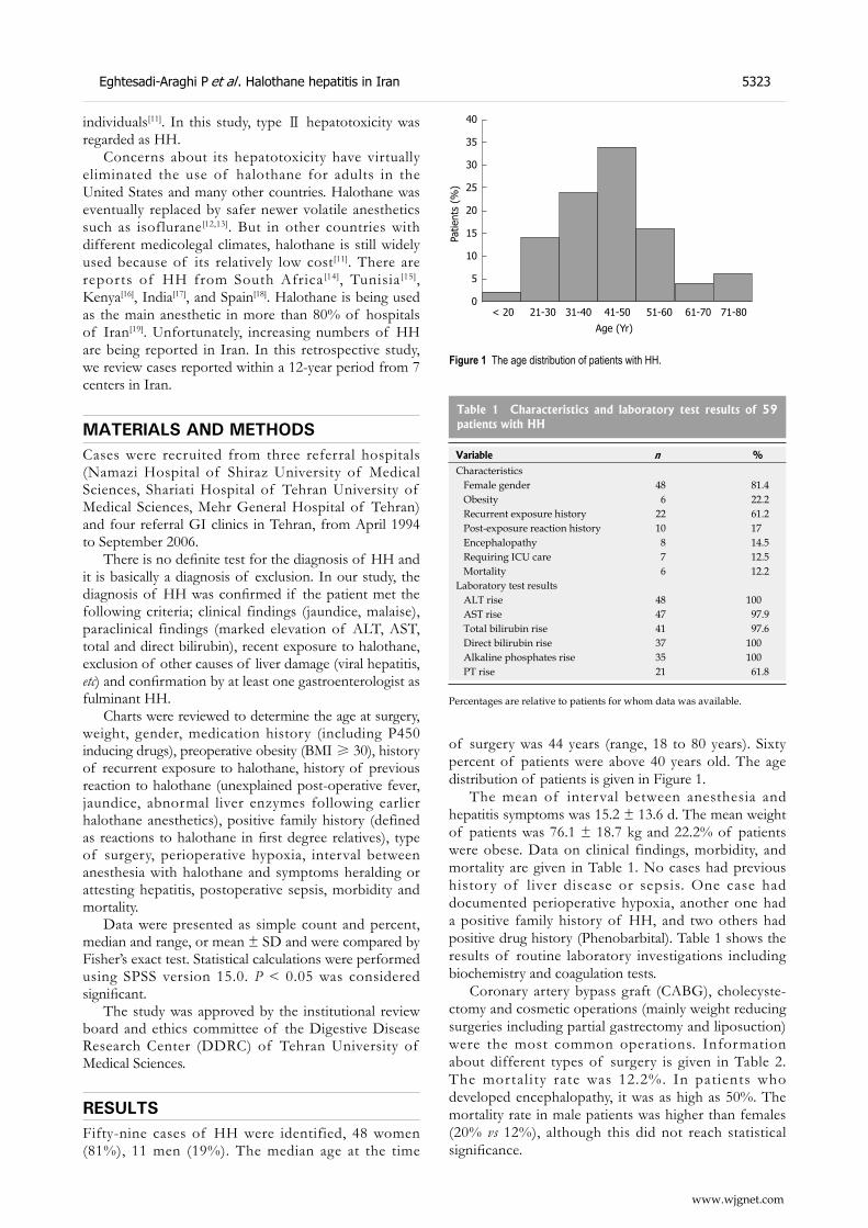
Eghtesadi-Araghi P et al . Halothane hepatitis in Iran 5323
www.wjgnet.com
individuals[11]. In this study, type Ⅱ hepatotoxicity was regarded as HH.
Concerns about its hepatotoxicity have virtually eliminated the use of halothane for adults in the United States and many other countries. Halothane was eventually replaced by safer newer volatile anesthetics such as isoflurane[12,13]. But in other countries with different medicolegal climates, halothane is still widely used because of its relatively low cost[11]. There are reports of HH from South Africa[14], Tunisia[15], Kenya[16], India[17], and Spain[18]. Halothane is being used as the main anesthetic in more than 80% of hospitals of Iran[19]. Unfortunately, increasing numbers of HH are being reported in Iran. In this retrospective study, we review cases reported within a 12-year period from 7 centers in Iran.
MATERIALS AND METHODSCases were recruited from three referral hospitals (Namazi Hospital of Shiraz University of Medical Sciences, Shariati Hospital of Tehran University of Medical Sciences, Mehr General Hospital of Tehran) and four referral GI clinics in Tehran, from April 1994 to September 2006.
There is no definite test for the diagnosis of HH and it is basically a diagnosis of exclusion. In our study, the diagnosis of HH was confirmed if the patient met the following criteria; clinical findings (jaundice, malaise), paraclinical findings (marked elevation of ALT, AST, total and direct bilirubin), recent exposure to halothane, exclusion of other causes of liver damage (viral hepatitis, etc) and confirmation by at least one gastroenterologist as fulminant HH.
Charts were reviewed to determine the age at surgery, weight, gender, medication history (including P450 inducing drugs), preoperative obesity (BMI ≥ 30), history of recurrent exposure to halothane, history of previous reaction to halothane (unexplained post-operative fever, jaundice, abnormal liver enzymes following earlier halothane anesthetics), positive family history (defined as reactions to halothane in first degree relatives), type of surgery, perioperative hypoxia, interval between anesthesia with halothane and symptoms heralding or attesting hepatitis, postoperative sepsis, morbidity and mortality.
Data were presented as simple count and percent, median and range, or mean ± SD and were compared by Fisher’s exact test. Statistical calculations were performed using SPSS version 15.0. P < 0.05 was considered significant.
The study was approved by the institutional review board and ethics committee of the Digestive Disease Research Center (DDRC) of Tehran University of Medical Sciences.
RESULTSFifty-nine cases of HH were identified, 48 women (81%), 11 men (19%). The median age at the time
of surgery was 44 years (range, 18 to 80 years). Sixty percent of patients were above 40 years old. The age distribution of patients is given in Figure 1.
The mean of interval between anesthesia and hepatitis symptoms was 15.2 ± 13.6 d. The mean weight of patients was 76.1 ± 18.7 kg and 22.2% of patients were obese. Data on clinical findings, morbidity, and mortality are given in Table 1. No cases had previous history of liver disease or sepsis. One case had documented perioperative hypoxia, another one had a positive family history of HH, and two others had positive drug history (Phenobarbital). Table 1 shows the results of routine laboratory investigations including biochemistry and coagulation tests.
Coronary artery bypass graft (CABG), cholecyste-ctomy and cosmetic operations (mainly weight reducing surgeries including partial gastrectomy and liposuction) were the most common operations. Information about different types of surgery is given in Table 2. The mortality rate was 12.2%. In patients who developed encephalopathy, it was as high as 50%. The mortality rate in male patients was higher than females (20% vs 12%), although this did not reach statistical significance.
40
35
30
25
20
15
10
5
0< 20 21-30 31-40 41-50 51-60 61-70 71-80
Age (Yr)
Patie
nts
(%)
Figure 1 The age distribution of patients with HH.
Table 1 Characteristics and laboratory test results of 59 patients with HH
Variable n %Characteristics Female gender 48 81.4 Obesity 6 22.2 Recurrent exposure history 22 61.2 Post-exposure reaction history 10 17 Encephalopathy 8 14.5 Requiring ICU care 7 12.5 Mortality 6 12.2Laboratory test results ALT rise 48 100 AST rise 47 97.9 Total bilirubin rise 41 97.6 Direct bilirubin rise 37 100 Alkaline phosphates rise 35 100 PT rise 21 61.8
Percentages are relative to patients for whom data was available.
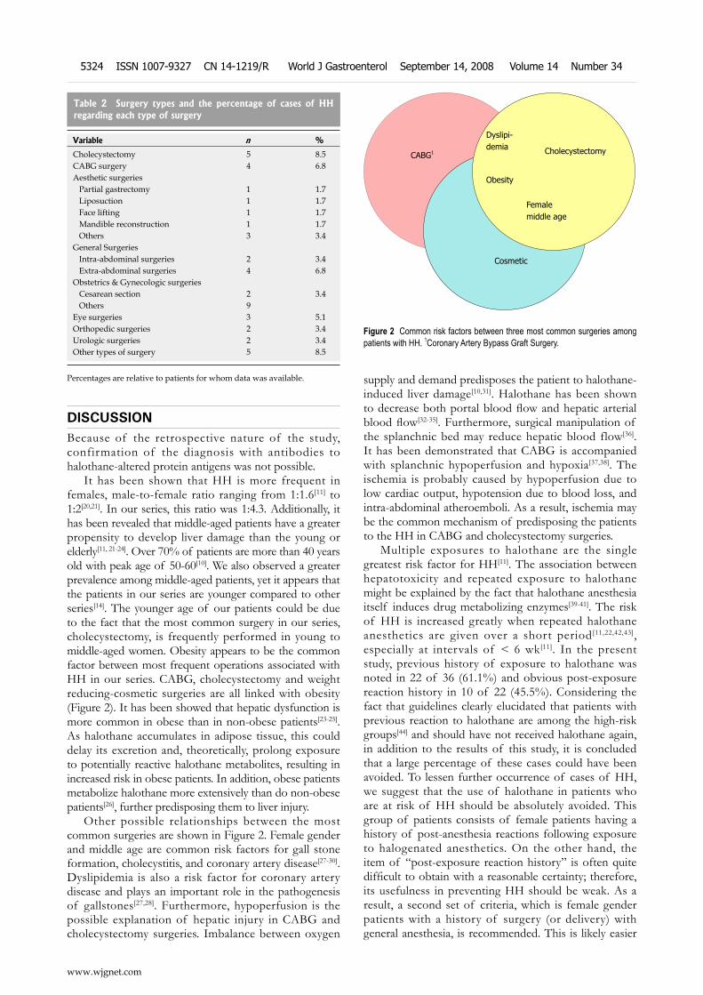
DISCUSSIONBecause of the retrospective nature of the study, confirmation of the diagnosis with antibodies to halothane-altered protein antigens was not possible.
It has been shown that HH is more frequent in females, male-to-female ratio ranging from 1:1.6[11] to 1:2[20,21]. In our series, this ratio was 1:4.3. Additionally, it has been revealed that middle-aged patients have a greater propensity to develop liver damage than the young or elderly[11, 21-24]. Over 70% of patients are more than 40 years old with peak age of 50-60[10]. We also observed a greater prevalence among middle-aged patients, yet it appears that the patients in our series are younger compared to other series[14]. The younger age of our patients could be due to the fact that the most common surgery in our series, cholecystectomy, is frequently performed in young to middle-aged women. Obesity appears to be the common factor between most frequent operations associated with HH in our series. CABG, cholecystectomy and weight reducing-cosmetic surgeries are all linked with obesity (Figure 2). It has been showed that hepatic dysfunction is more common in obese than in non-obese patients[23-25]. As halothane accumulates in adipose tissue, this could delay its excretion and, theoretically, prolong exposure to potentially reactive halothane metabolites, resulting in increased risk in obese patients. In addition, obese patients metabolize halothane more extensively than do non-obese patients[26], further predisposing them to liver injury.
Other possible relationships between the most common surgeries are shown in Figure 2. Female gender and middle age are common risk factors for gall stone formation, cholecystitis, and coronary artery disease[27-30]. Dyslipidemia is also a risk factor for coronary artery disease and plays an important role in the pathogenesis of gallstones[27,28]. Furthermore, hypoperfusion is the possible explanation of hepatic injury in CABG and cholecystectomy surgeries. Imbalance between oxygen
supply and demand predisposes the patient to halothane-induced liver damage[10,31]. Halothane has been shown to decrease both portal blood flow and hepatic arterial blood flow[32-35]. Furthermore, surgical manipulation of the splanchnic bed may reduce hepatic blood flow[36]. It has been demonstrated that CABG is accompanied with splanchnic hypoperfusion and hypoxia[37,38]. The ischemia is probably caused by hypoperfusion due to low cardiac output, hypotension due to blood loss, and intra-abdominal atheroemboli. As a result, ischemia may be the common mechanism of predisposing the patients to the HH in CABG and cholecystectomy surgeries.
Multiple exposures to halothane are the single greatest risk factor for HH[11]. The association between hepatotoxicity and repeated exposure to halothane might be explained by the fact that halothane anesthesia itself induces drug metabolizing enzymes[39-41]. The risk of HH is increased greatly when repeated halothane anesthetics are given over a short period[11,22,42,43], especially at intervals of < 6 wk[11]. In the present study, previous history of exposure to halothane was noted in 22 of 36 (61.1%) and obvious post-exposure reaction history in 10 of 22 (45.5%). Considering the fact that guidelines clearly elucidated that patients with previous reaction to halothane are among the high-risk groups[44] and should have not received halothane again, in addition to the results of this study, it is concluded that a large percentage of these cases could have been avoided. To lessen further occurrence of cases of HH, we suggest that the use of halothane in patients who are at risk of HH should be absolutely avoided. This group of patients consists of female patients having a history of post-anesthesia reactions following exposure to halogenated anesthetics. On the other hand, the item of “post-exposure reaction history” is often quite difficult to obtain with a reasonable certainty; therefore, its usefulness in preventing HH should be weak. As a result, a second set of criteria, which is female gender patients with a history of surgery (or delivery) with general anesthesia, is recommended. This is likely easier
Variable n %
Cholecystectomy 5 8.5CABG surgery 4 6.8Aesthetic surgeries Partial gastrectomy 1 1.7 Liposuction 1 1.7 Face lifting 1 1.7 Mandible reconstruction 1 1.7 Others 3 3.4General Surgeries Intra-abdominal surgeries 2 3.4 Extra-abdominal surgeries 4 6.8Obstetrics & Gynecologic surgeries Cesarean section 2 3.4 Others 9Eye surgeries 3 5.1Orthopedic surgeries 2 3.4Urologic surgeries 2 3.4Other types of surgery 5 8.5
Table 2 Surgery types and the percentage of cases of HH regarding each type of surgery
Percentages are relative to patients for whom data was available.
www.wjgnet.com
5324 ISSN 1007-9327 CN 14-1219/R World J Gastroenterol September 14, 2008 Volume 14 Number 34
Figure 2 Common risk factors between three most common surgeries among patients with HH. 1Coronary Artery Bypass Graft Surgery.
CABG1
Cosmetic
Femalemiddle age
Cholecystectomy
Dyslipi-demia
Obesity
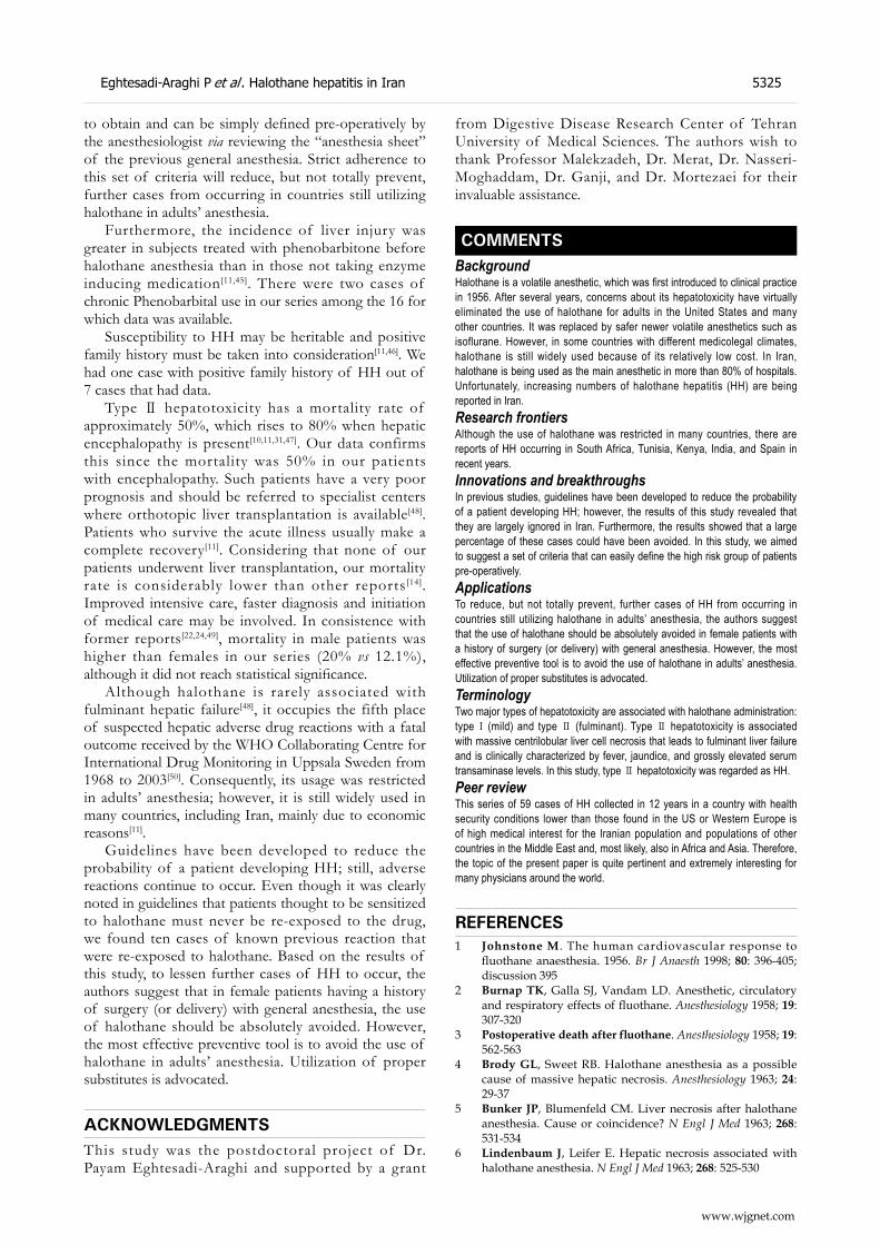
to obtain and can be simply defined pre-operatively by the anesthesiologist via reviewing the “anesthesia sheet” of the previous general anesthesia. Strict adherence to this set of criteria will reduce, but not totally prevent, further cases from occurring in countries still utilizing halothane in adults’ anesthesia.
Furthermore, the incidence of liver injury was greater in subjects treated with phenobarbitone before halothane anesthesia than in those not taking enzyme inducing medication[11,45]. There were two cases of chronic Phenobarbital use in our series among the 16 for which data was available.
Susceptibility to HH may be heritable and positive family history must be taken into consideration[11,46]. We had one case with positive family history of HH out of 7 cases that had data.
Type Ⅱ hepatotoxicity has a mortality rate of approximately 50%, which rises to 80% when hepatic encephalopathy is present[10,11,31,47]. Our data confirms this since the mortality was 50% in our patients with encephalopathy. Such patients have a very poor prognosis and should be referred to specialist centers where orthotopic liver transplantation is available[48]. Patients who survive the acute illness usually make a complete recovery[11]. Considering that none of our patients underwent liver transplantation, our mortality rate is considerably lower than other reports[14]. Improved intensive care, faster diagnosis and initiation of medical care may be involved. In consistence with former reports[22,24,49], mortality in male patients was higher than females in our series (20% vs 12.1%), although it did not reach statistical significance.
Although halothane is rarely associated with fulminant hepatic failure[48], it occupies the fifth place of suspected hepatic adverse drug reactions with a fatal outcome received by the WHO Collaborating Centre for International Drug Monitoring in Uppsala Sweden from 1968 to 2003[50]. Consequently, its usage was restricted in adults’ anesthesia; however, it is still widely used in many countries, including Iran, mainly due to economic reasons[11].
Guidelines have been developed to reduce the probability of a patient developing HH; still, adverse reactions continue to occur. Even though it was clearly noted in guidelines that patients thought to be sensitized to halothane must never be re-exposed to the drug, we found ten cases of known previous reaction that were re-exposed to halothane. Based on the results of this study, to lessen further cases of HH to occur, the authors suggest that in female patients having a history of surgery (or delivery) with general anesthesia, the use of halothane should be absolutely avoided. However, the most effective preventive tool is to avoid the use of halothane in adults’ anesthesia. Utilization of proper substitutes is advocated.
ACKNOWLEDGMENTSThis study was the postdoctoral project of Dr. Payam Eghtesadi-Araghi and supported by a grant
from Digestive Disease Research Center of Tehran University of Medical Sciences. The authors wish to thank Professor Malekzadeh, Dr. Merat, Dr. Nasseri-Moghaddam, Dr. Ganji, and Dr. Mortezaei for their invaluable assistance.
COMMENTSBackgroundHalothane is a volatile anesthetic, which was first introduced to clinical practice in 1956. After several years, concerns about its hepatotoxicity have virtually eliminated the use of halothane for adults in the United States and many other countries. It was replaced by safer newer volatile anesthetics such as isoflurane. However, in some countries with different medicolegal climates, halothane is still widely used because of its relatively low cost. In Iran, halothane is being used as the main anesthetic in more than 80% of hospitals. Unfortunately, increasing numbers of halothane hepatitis (HH) are being reported in Iran.Research frontiersAlthough the use of halothane was restricted in many countries, there are reports of HH occurring in South Africa, Tunisia, Kenya, India, and Spain in recent years.Innovations and breakthroughsIn previous studies, guidelines have been developed to reduce the probability of a patient developing HH; however, the results of this study revealed that they are largely ignored in Iran. Furthermore, the results showed that a large percentage of these cases could have been avoided. In this study, we aimed to suggest a set of criteria that can easily define the high risk group of patients pre-operatively.ApplicationsTo reduce, but not totally prevent, further cases of HH from occurring in countries still utilizing halothane in adults’ anesthesia, the authors suggest that the use of halothane should be absolutely avoided in female patients with a history of surgery (or delivery) with general anesthesia. However, the most effective preventive tool is to avoid the use of halothane in adults’ anesthesia. Utilization of proper substitutes is advocated.TerminologyTwo major types of hepatotoxicity are associated with halothane administration: type Ⅰ (mild) and type Ⅱ (fulminant). Type Ⅱ hepatotoxicity is associated with massive centrilobular liver cell necrosis that leads to fulminant liver failure and is clinically characterized by fever, jaundice, and grossly elevated serum transaminase levels. In this study, type Ⅱ hepatotoxicity was regarded as HH.Peer reviewThis series of 59 cases of HH collected in 12 years in a country with health security conditions lower than those found in the US or Western Europe is of high medical interest for the Iranian population and populations of other countries in the Middle East and, most likely, also in Africa and Asia. Therefore, the topic of the present paper is quite pertinent and extremely interesting for many physicians around the world.
REFERENCES1 Johnstone M. The human cardiovascular response to
fluothane anaesthesia. 1956. Br J Anaesth 1998; 80: 396-405; discussion 395
2 Burnap TK, Galla SJ, Vandam LD. Anesthetic, circulatory and respiratory effects of fluothane. Anesthesiology 1958; 19: 307-320
3 Postoperative death after fluothane. Anesthesiology 1958; 19: 562-563
4 Brody GL, Sweet RB. Halothane anesthesia as a possible cause of massive hepatic necrosis. Anesthesiology 1963; 24: 29-37
5 Bunker JP, Blumenfeld CM. Liver necrosis after halothane anesthesia. Cause or coincidence? N Engl J Med 1963; 268: 531-534
6 Lindenbaum J, Leifer E. Hepatic necrosis associated with halothane anesthesia. N Engl J Med 1963; 268: 525-530
α β μ γ κ χ ∆ Ω
Ⅰ Ⅱ Ⅲ Ⅳ Ⅴ Ⅵ Ⅶ Ⅷ Ⅸ Ⅹ Ⅺ Ⅻ± ×
Eghtesadi-Araghi P et al . Halothane hepatitis in Iran 5325
www.wjgnet.com
7 Temple RL, Cote RA, Gorens SW. Massive hepatic necrosis following general anesthesia. Anesth Analg 1962; 41: 586-592
8 Brown BR Jr, Gandolfi AJ. Adverse effects of volatile anaesthetics. Br J Anaesth 1987; 59: 14-23
9 Summary of the national Halothane Study. Possible association between halothane anesthesia and postoperative hepatic necrosis. JAMA 1966; 197: 775-788
10 Ray DC, Drummond GB. Halothane hepatitis. Br J Anaesth 1991; 67: 84-99
11 P e r a l t a R , P o t e r a c k K A , G u z o f s k i S . H a l o t h a n e Hepatotoxicity. Last modified date: May 25, 2006. Available from URL: http://www.emedicine.com
12 Allan LG, Hussey AJ, Howie J, Beckett GJ, Smith AF, Hayes JD, Drummond GB. Hepatic glutathione S-transferase release after halothane anaesthesia: open randomised comparison with isoflurane. Lancet 1987; 1: 771-774
13 Hussey AJ, Aldridge LM, Paul D, Ray DC, Beckett GJ, Allan LG. Plasma glutathione S-transferase concentration as a measure of hepatocellular integrity following a single general anaesthetic with halothane, enflurane or isoflurane. Br J Anaesth 1988; 60: 130-135
14 Voigt MD, Workman B, Lombard C, Kirsch RE. Halothane hepatitis in a South African population--frequency and the influence of gender and ethnicity. S Afr Med J 1997; 87: 882-885
15 Daghfous R, el Aidli S, Sfaxi M, Daghfous M, Kastalli S, Srairi S, Loueslati MH, Belkahia C. [Halothane-induced hepatitis. 8 case reports] Tunis Med 2003; 81: 874-878
16 Otedo AE. Halothane induced hepatitis: case report. East Afr Med J 2004; 81: 538-539
17 Kumar GP, Bhat VJ, Sowdi V. Fulminant hepatic failure following halothane anaesthesia. J Clin Forensic Med 2005; 12: 271-273
18 Rodes J, Bruguera M. The uses of error: iatrogenic hepatitis. Lancet 2001; 357: 791
19 The "Halothane" will be replaced with newer anesthetic drugs in Iran. Date Accessed: July 17, 2008. Available from URL: http://www.reporter.ir/archives/86/10/005402.php
20 Benjamin SB, Goodman ZD, Ishak KG, Zimmerman HJ, Irey NS. The morphologic spectrum of halothane-induced hepatic injury: analysis of 77 cases. Hepatology 1985; 5: 1163-1171
21 Neuberger J, Williams R. Halothane anaesthesia and liver damage. Br Med J (Clin Res Ed) 1984; 289: 1136-1139
22 Bottiger LE, Dalen E, Hallen B. Halothane-induced liver damage: an analysis of the material reported to the Swedish Adverse Drug Reaction Committee, 1966-1973. Acta Anaesthesiol Scand 1976; 20: 40-46
23 Carney FM, Van Dyke RA. Halothane hepatitis: a critical review. Anesth Analg 1972; 51: 135-160
24 Walton B, Simpson BR, Strunin L, Doniach D, Perrin J, Appleyard AJ. Unexplained hepatitis following halothane. Br Med J 1976; 1: 1171-1176
25 Peters RL, Edmondson HA, Reynolds TB, Meister JC, Curpey TJ. Hepatic necrosis associated with halothane anesthesia. Am J Med 1969; 47: 748-764
26 Bentley JB, Vaughan RW, Gandolfi AJ, Cork RC. Halothane biotransformation in obese and nonobese patients. Anesthesiology 1982; 57: 94-97
27 Boland LL, Folsom AR, Rosamond WD. Hyperinsulinemia, dyslipidemia, and obesity as risk factors for hospitalized gallbladder disease. A prospective study. Ann Epidemiol 2002; 12: 131-140
28 Grunhage F, Lammert F. Gallstone disease. Pathogenesis of gallstones: A genetic perspective. Best Pract Res Clin Gastroenterol 2006; 20: 997-1015
29 Wild SH, Byrne CD. ABC of obesity. Risk factors for
diabetes and coronary heart disease. BMJ 2006; 333 : 1009-1011
30 Franzosi MG . Should we continue to use BMI as a cardiovascular risk factor? Lancet 2006; 368: 624-625
31 Cousins MJ , Plummer JL, Hall PD. Risk factors for halothane hepatitis. Aust N Z J Surg 1989; 59: 5-14
32 Gelman S, Fowler KC, Smith LR. Liver circulation and function during isoflurane and halothane anesthesia. Anesthesiology 1984; 61: 726-730
33 Hursh D, Gelman S, Bradley EL Jr. Hepatic oxygen supply during halothane or isoflurane anesthesia in guinea pigs. Anesthesiology 1987; 67: 701-706
34 Gelman S, Rimerman V, Fowler KC, Bishop SP, Bradley EL Jr. The effect of halothane, isoflurane, and blood loss on hepatotoxicity and hepatic oxygen availability in phenobarbital-pretreated hypoxic rats. Anesth Analg 1984; 63: 965-972
35 Hughes RL, Campbell D, Fitch W. Effects of enflurane and halothane on liver blood flow and oxygen consumption in the greyhound. Br J Anaesth 1980; 52: 1079-1086
36 Seltzer JL, Goldberg ME, Larijani GE, Ritter DE, Starsnic MA, Stahl GL, Lefer AM. Prostacyclin mediation of vasodilation following mesenteric traction. Anesthesiology 1988; 68: 514-518
37 D'Ancona G, Baillot R, Poirier B, Dagenais F, de Ibarra JI , Bauset R, Mathieu P, Doyle D. Determinants of gastrointestinal complications in cardiac surgery. Tex Heart Inst J 2003; 30: 280-285
38 Zacharias A, Schwann TA, Parenteau GL, Riordan CJ, Durham SJ, Engoren M, Fenn-Buderer N, Habib RH. Predictors of gastrointestinal complications in cardiac surgery. Tex Heart Inst J 2000; 27: 93-99
39 Nimmo WS, Thompson PG, Prescott LF. Microsomal enzyme induction after halothane anaesthesia. Br J Clin Pharmacol 1981; 12: 433-434
40 St Haxholdt O, Loft S, Clemmensen A, Hjortso E. Increased hepatic microsomal activity after halothane anaesthesia in children. Anaesthesia 1986; 41: 579-581
41 Neuberger JM . Halothane and hepatitis. Incidence, predisposing factors and exposure guidelines. Drug Saf 1990; 5: 28-38
42 Inman WH, Mushin WW. Jaundice after repeated exposure to halothane: an analysis of Reports to the Committee on Safety of Medicines. Br Med J 1974; 1: 5-10
43 Inman WH, Mushin WW. Jaundice after repeated exposure to halothane: a further analysis of reports to the Committee on Safety of Medicines. Br Med J 1978; 2: 1455-1456
44 Neuberger J, Williams R. Halothane hepatitis. Dig Dis 1988; 6: 52-64
45 Nomura F, Hatano H, Ohnishi K, Akikusa B, Okuda K. Effects of anticonvulsant agents on halothane-induced liver injury in human subjects and experimental animals. Hepatology 1986; 6: 952-956
46 Hoft RH, Bunker JP, Goodman HI, Gregory PB. Halothane hepatitis in three pairs of closely related women. N Engl J Med 1981; 304: 1023-1024
47 Bjornsson E, Jerlstad P, Bergqvist A, Olsson R. Fulminant drug-induced hepatic failure leading to death or liver transplantation in Sweden. Scand J Gastroenterol 2005; 40: 1095-1101
48 Neuberger J. Halothane hepatitis. Eur J Gastroenterol Hepatol 1998; 10: 631-633
49 Moult PJ, Sherlock S. Halothane-related hepatitis. A clinical study of twenty-six cases. Q J Med 1975; 44: 99-114
50 Bjornsson E, Olsson R. Suspected drug-induced liver fatalities reported to the WHO database. Dig Liver Dis 2006; 38: 33-38
S- Editor Li DL L- Editor Li M E- Editor Yin DH
www.wjgnet.com
5326 ISSN 1007-9327 CN 14-1219/R World J Gastroenterol September 14, 2008 Volume 14 Number 34
anti-HBc screening and discarding large number of blood units versus considering ID NAT (Individual donor nuclic acid testing) needs to be assessed.
© 2008 The WJG Press. All rights reserved.
Key words: Hepatitis B core antigen; Hepatitis B surface antigen; Hepatitis B virus; Transfusion-associated hepatitis B virus; Blood donors
Peer reviewer: Dr. Deepak Narayan Amarapurkar, Department Of Gastroenterology, Bombay Hospital & Medical Research Centre, D 401 Ameya Soc, New Prabhadevi Road, Prabhadevi, Mumbai 400025, India
Dhawan HK, Marwaha N, Sharma RR, Chawla Y, Thakral B, Saluja K, Sharma SK, Thakur MK, Jain A. Anti-HBc screening in Indian blood donors: Still an unresolved issue. World J Gastroenterol 2008; 14(34): 5327-5330 Available from: URL: http://www.wjgnet.com/1007-9327/14/5327.asp DOI: http://dx.doi.org/10.3748/wjg.14.5327
INTRODUCTIONHepatitis B virus (HBV) infection is a serious global health problem affecting two billion people worldwide, and 350 million people suffer from chronic HBV infection[1]. Despite mandatory screening for HBsAg by ELISA for over 20 years, transfusion-associated HBV (TAHBV) continues to be a major problem in India, more so in patients receiving repeated transfusions[2]. The incidence of transfusion-associated hepatitis (TAH) after cardiac surgery (4 + 2.4 units transfusion) was estimated at 6.9%, of which TAHBV constituted 20%[2]. The incidence of TAHBV in patients receiving multiple transfusions, such as thalassemia, ranged from 17.9% in the first year to 69.2% by the third year. Patients on renal dialysis showed similar rates of infection with HBV[2]. It has been demonstrated that some HBsAg-negative individuals and those reactive for anti-HBc continue to replicate HBV[3,4]. Thus the absence of HBsAg in the blood of apparently healthy individuals may not be sufficient to ensure lack of circulating HBV. Blood containing anti-HBc with or without detectable presence of HBsAg might be infectious; therefore, routine blood donor screening for anti-HBc has been implemented
Hari Krishan Dhawan, Neelam Marwaha, Ratti Ram Sharma, Beenu Thakral, Karan Saluja, Manish K Thakur, Ashish Jain, Department of Transfusion Medicine PGIMER, Chandigarh 160012, IndiaYogesh Chawla, Sanjeev Kumar Sharma, Department of Hepatology PGIMER, Chandigarh 160012, IndiaAuthor contributions: Marwaha N, Sharma RR and Chawla Y designed the research; Dhawan HK and Sharma SK performed the research; Thakral B, Saluja K, Thakur MK and Jain A analyzed data; Dhawan HK wrote the paper.Correspondence to: Hari Krishan Dhawan, Department of Transfusion Medicine PGIMER, Chandigarh 160012, India. [email protected]: + 91-172-2756481 Fax: + 91-172-2744401Received: June 20, 2008 Revised: July 14, 2008Accepted: July 21, 2008Published online: September 14, 2008
AbstractAIM: To study the seroprevalence of antibody to hepatitis B core antigen (anti-HBc) in healthy blood donors negative for HBsAg and to evaluate whether anti-HBc detection could be adopted in India as a screening assay for HBV in addition to HBsAg.METHODS: A total of 1700 serum samples collected from HBsAg-negative healthy blood donors were tested for the presence of anti-HBc antibody (IgM + IgG). All samples reactive for anti-HBc antibody were then investigated for presence of anti-HBs and for liver function tests (LFTs). One hundred serum samples reactive for anti-HBc were tested for HBV DNA by PCR method. RESULTS: Out of 1700 samples tested, 142 (8.4%) blood samples were found to be reactive for anti-HBc. It was significantly lower in voluntary (6.9%) as compared to replacement donors (10.4%, P = 0.011). Seventy-two (50.7%) anti-HBc reactive samples were also reactive for anti-HBs with levels > 10 mIU/mL and 70 (49.3%) samples were non-reactive for anti-HBs, these units were labeled as anti-HBc-only. These 142 anti-HBc reactive units were also tested for liver function test. HBV DNA was detected in only 1 of 100 samples tested. CONCLUSION: Keeping in view that 8%-18% of donor population in India is anti-HBc reactive, inclusion of anti-HBc testing will lead to high discard rate. Anti-HBs as proposed previously does not seem to predict clearance of the virus. Cost effectiveness of introducing universal
Anti-HBc screening in Indian blood donors: Still an unresolved issue
Hari Krishan Dhawan, Neelam Marwaha, Ratti Ram Sharma, Yogesh Chawla, Beenu Thakral, Karan Saluja, Sanjeev Kumar Sharma, Manish K Thakur, Ashish Jain
RAPID COMMUNICATION
Online Submissions: wjg.wjgnet.com World J Gastroenterol 2008 September 14; 14(34): [email protected] World Journal of Gastroenterology ISSN 1007-9327doi:10.3748/wjg.14.5327 © 2008 The WJG Press. All rights reserved.
www.wjgnet.com
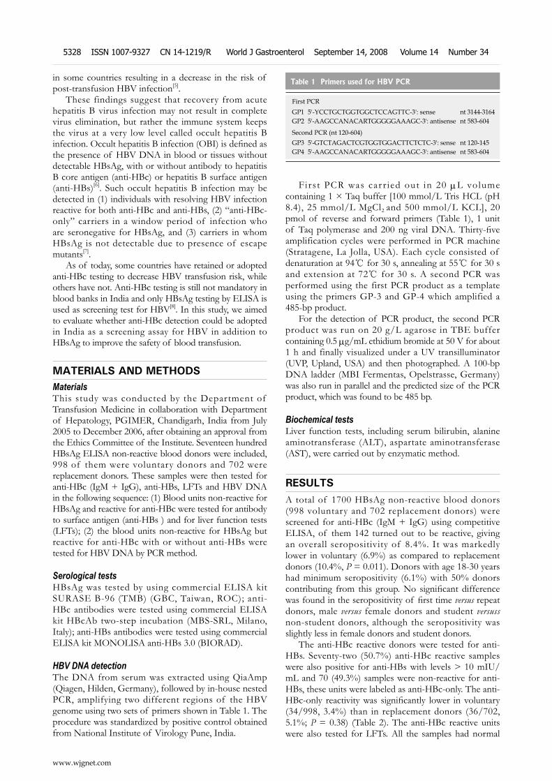
in some countries resulting in a decrease in the risk of post-transfusion HBV infection[5].
These findings suggest that recovery from acute hepatitis B virus infection may not result in complete virus elimination, but rather the immune system keeps the virus at a very low level called occult hepatitis B infection. Occult hepatitis B infection (OBI) is defined as the presence of HBV DNA in blood or tissues without detectable HBsAg, with or without antibody to hepatitis B core antigen (anti-HBc) or hepatitis B surface antigen (anti-HBs)[6]. Such occult hepatitis B infection may be detected in (1) individuals with resolving HBV infection reactive for both anti-HBc and anti-HBs, (2) “anti-HBc-only” carriers in a window period of infection who are seronegative for HBsAg, and (3) carriers in whom HBsAg is not detectable due to presence of escape mutants[7].
As of today, some countries have retained or adopted anti-HBc testing to decrease HBV transfusion risk, while others have not. Anti-HBc testing is still not mandatory in blood banks in India and only HBsAg testing by ELISA is used as screening test for HBV[8]. In this study, we aimed to evaluate whether anti-HBc detection could be adopted in India as a screening assay for HBV in addition to HBsAg to improve the safety of blood transfusion.
MATERIALS AND METHODSMaterialsThis study was conducted by the Department of Transfusion Medicine in collaboration with Department of Hepatology, PGIMER, Chandigarh, India from July 2005 to December 2006, after obtaining an approval from the Ethics Committee of the Institute. Seventeen hundred HBsAg ELISA non-reactive blood donors were included, 998 of them were voluntary donors and 702 were replacement donors. These samples were then tested for anti-HBc (IgM + IgG), anti-HBs, LFTs and HBV DNA in the following sequence: (1) Blood units non-reactive for HBsAg and reactive for anti-HBc were tested for antibody to surface antigen (anti-HBs ) and for liver function tests (LFTs); (2) the blood units non-reactive for HBsAg but reactive for anti-HBc with or without anti-HBs were tested for HBV DNA by PCR method.
Serological testsHBsAg was tested by using commercial ELISA kit SURASE B-96 (TMB) (GBC, Taiwan, ROC); anti-HBc antibodies were tested using commercial ELISA kit HBcAb two-step incubation (MBS-SRL, Milano, Italy); anti-HBs antibodies were tested using commercial ELISA kit MONOLISA anti-HBs 3.0 (BIORAD).
HBV DNA detectionThe DNA from serum was extracted using QiaAmp (Qiagen, Hilden, Germany), followed by in-house nested PCR, amplifying two different regions of the HBV genome using two sets of primers shown in Table 1. The procedure was standardized by positive control obtained from National Institute of Virology Pune, India.
F irs t PCR was car r ied out in 20 μL volume containing 1 × Taq buffer [100 mmol/L Tris HCL (pH 8.4), 25 mmol/L MgCl2 and 500 mmol/L KCL], 20 pmol of reverse and forward primers (Table 1), 1 unit of Taq polymerase and 200 ng viral DNA. Thirty-five amplification cycles were performed in PCR machine (Stratagene, La Jolla, USA). Each cycle consisted of denaturation at 94 for 30 s, annealing at 55 for 30 s and extension at 72 for 30 s. A second PCR was performed using the first PCR product as a template using the primers GP-3 and GP-4 which amplified a 485-bp product.
For the detection of PCR product, the second PCR product was run on 20 g/L agarose in TBE buffer containing 0.5 μg/mL ethidium bromide at 50 V for about 1 h and finally visualized under a UV transilluminator (UVP, Upland, USA) and then photographed. A 100-bp DNA ladder (MBI Fermentas, Opelstrasse, Germany) was also run in parallel and the predicted size of the PCR product, which was found to be 485 bp.
Biochemical testsLiver function tests, including serum bilirubin, alanine aminotransferase (ALT), aspartate aminotransferase (AST), were carried out by enzymatic method.
RESULTSA total of 1700 HBsAg non-reactive blood donors (998 voluntary and 702 replacement donors) were screened for anti-HBc (IgM + IgG) using competitive ELISA, of them 142 turned out to be reactive, giving an overall seropositivity of 8.4%. It was markedly lower in voluntary (6.9%) as compared to replacement donors (10.4%, P = 0.011). Donors with age 18-30 years had minimum seropositivity (6.1%) with 50% donors contributing from this group. No significant difference was found in the seropositivity of first time versus repeat donors, male versus female donors and student versuss non-student donors, although the seropositivity was slightly less in female donors and student donors.
The anti-HBc reactive donors were tested for anti-HBs. Seventy-two (50.7%) anti-HBc reactive samples were also positive for anti-HBs with levels > 10 mIU/mL and 70 (49.3%) samples were non-reactive for anti-HBs, these units were labeled as anti-HBc-only. The anti-HBc-only reactivity was significantly lower in voluntary (34/998, 3.4%) than in replacement donors (36/702, 5.1%; P = 0.38) (Table 2). The anti-HBc reactive units were also tested for LFTs. All the samples had normal
Table 1 Primers used for HBV PCR
First PCR GP1 5'-YCCTGCTGGTGGCTCCAGTTC-3': sense nt 3144-3164GP2 5'-AAGCCANACARTGGGGGAAAGC-3': antisense nt 583-604
Second PCR (nt 120-604)GP3 5'-GTCTAGACTCGTGGTGGACTTCTCTC-3': sense nt 120-145GP4 5'-AAGCCANACARTGGGGGAAAGC-3': antisense nt 583-604
www.wjgnet.com
5328 ISSN 1007-9327 CN 14-1219/R World J Gastroenterol September 14, 2008 Volume 14 Number 34
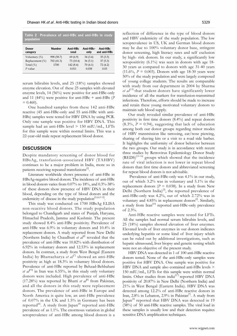
serum bilirubin levels, and 25 (18%) samples showed enzyme elevation. Out of these 25 samples with elevated enzyme levels, 14 (56%) were positive for anti-HBc-only and 11 (44%) were positive for anti-HBc + anti-HBs (P = 0.460).
One hundred samples from these 142 anti-HBc reactive (45 anti-HBc-only and 55 anti-HBc with anti-HBs) samples were tested for HBV DNA by using PCR. Only one sample was positive for HBV DNA. This sample had an anti-HBs level > 150 mIU/mL, LFTs for this sample were within normal limits. This was a 22-year-old male repeat replacement blood donor.
DISCUSSIONDespite mandatory screening of donor blood for HBsAg, transfusion-associated HBV (TAHBV) continues to be a major problem in India, more so in patients receiving repeated transfusions[2].
Literature worldwide shows presence of anti-HBc in HBsAg-negative blood donors. The incidence of anti-HBc in blood donors varies from 0.07% to 18%, and 0.3%-38% of these donors show presence of HBV DNA in their blood, depending on the type of blood donors and the endemicity of disease in the study population[2,4,6,9-12].
This study was conducted on 1700 HBsAg ELISA non-reactive blood donors. The study population belonged to Chandigarh and states of Punjab, Haryana, Himachal Pradesh, Jammu and Kashmir. The present study showed 8.4% anti-HBc positivity. Prevalence of anti-HBc was 6.9% in voluntary donors and 10.4% in replacement donors. A study reported from New Delhi (Northern India) by Chaudhuri et al[2] revealed that the prevalence of anti-HBc was 10.82% with distribution of 6.92% in voluntary donors and 12.53% in replacement donors. In contrast, a study from West Bengal (Eastern India) by Bhattacharya et al [7] showed an anti-HBc positivity as high as 18.3% in voluntary blood donors. Prevalence of anti-HBc reported by Behzad-Behbahani et al[10] in Iran was 6.55%, in this study only voluntary donors were included. High prevalence of anti-HBc (17.28%) was reported by Bhatti et al[6] from Pakistan, and all the donors in this study were replacement donors. The prevalence of anti-HBc in Europe and North America is quite low, an anti-HBc prevalence of 0.07% in the UK and 1.5% in Germany has been reported[6]. A study from Japan[4] reported anti-HBc prevalence of as 1.1%. The enormous variation in global seroprevalence of anti-HBc among blood donors is a
reflection of difference in the type of blood donors and HBV endemicity of the study population. The low seroprevalence in US, UK and German blood donors may be due to 100% voluntary donor base, stringent donor screening, high literacy rates and self exclusion by high- risk donors. In our study, a significantly low seropositivity (6.1%) was seen in donors with age 18- 30 years as compared to donors with age 31-40 years (11.6%, P = 0.003). Donors with age 18-30 years were 50% of the study population and were largely composed of young college students. The results are comparable with study from our department in 2004 by Sharma et al [13] that student donors have significantly lower incidence of all the markers for transfusion-transmitted infections. Therefore, efforts should be made to increase and retain these young motivated voluntary donors to maintain safe blood supply.
Our study revealed similar prevalence of anti-HBc positivity in first time donors (8.4%) and repeat donors (8.3%, P = 0.94), suggesting that lack of education among both our donor groups regarding minor modes of HBV transmission like tattooing, ear/nose piercing, sharing of shaving kits or a visit to a road side barber. It highlights the uniformity of donor behavior between the two groups. Our study is in accordance with recent three studies by Retrovirus Epidemiology Donor Study (REDS)[14-16] groups which showed that the incidence rate of viral infection is not lower in repeat blood donors than first time donors and abbreviated screening for repeat blood donors is not advisable.
Prevalence of anti-HBc-only was 4.1% in our study, out of which 3.2% was in voluntary and 5.1% in the replacement donors (P = 0.038). In a study from New Delhi (Northern India)[2], the reported prevalence of anti-HBc-only was 4.2%, out of which 2.72% was in voluntary and 4.85% in replacement donors[2]. Similarly, a study from Iran[10] reported anti-HBc-only prevalence of 2.3%.
Anti-HBc reactive samples were tested for LFTs. All the samples had normal serum bilirubin levels, and 25 (18%) samples showed elevation of liver enzymes. Elevated levels of liver enzymes in our donors indicates underlying hepatitis or some kind of liver injury which can be ruled out by additional investigations, such as hepatic ultrasound, liver biopsy and genetic testing which were not an objective of the present study.
HBV DNA was detected in 1 of 100 anti-HBc reactive donors tested. None of the anti-HBc-only samples were positive for HBV DNA. One sample was positive for HBV DNA and sample also contained anti-HBs levels > 150 mIU/mL, LFTs for this sample were within normal limits. Other studies from india[2,7] reported HBV DNA positivity of 20.87% in New Delhi (Northern India) and 21% in West Bengal (Eastern India). HBV DNA was detected among 12.2% of anti-HBc reactive donors in Iran, 2.8% in Lebanon, 2.9% in Pakistan[7]. A study from Japan[4] reported that HBV DNA was detected in 19 (38%) of 50 anti-HBc reactive samples. The viral load in these samples is usually low and their detection requires sensitive DNA amplification techniques.
Table 2 Prevalence of anti-HBc and anti-HBs in study population
Donor category
Number Anti-HBc total
Anti-HBc-only
Anti-HBc and anti-HBs
Voluntary (%) 998 (58.7) 69 (6.9) 34 (3.4) 35 (3.5)Replacement (%) 702 (41.3) 73 (10.4) 36 (5.1) 37 (5.3)Total (%) 1700 142 (8.4) 70 (4.1) 72 (4.2)P value 0.011 0.038 0.03
Dhawan HK et al . Anti-HBc testing in Indian blood donors 5329
www.wjgnet.com
In our study, low incidence of HBV DNA in anti-HBc reactive samples may be due to limitation of sensitivity of HBV DNA amplification technique. Another reason of low HBV DNA positivity in our study could be due to type of blood donors and the endemicity of disease in the study population, in our study 50% of donors were between 18-30 years, largely belong to young college students. This group being better educated can understand and cooperate in answering to the risk factors in acquiring HBV infection during pre-donation questionnaire. Another study from Chandigarh (Northwestern India) by Duseja et al[17] showed 0% prevalence of HBV DNA in 100 adult healthy blood donors, non-reactive for HBsAg.
Keeping in view that 8% to 18% of donor population in India is anti-HBc reactive, inclusion of anti-HBc testing will lead to high discard rate. Anti-HBs as proposed previously does not seem to predict clearance of the virus as the single donor, who tested reactive for HBV DNA in our study, had high anti-HBs titers. Cost effectiveness of introducing universal anti-HBc screening and discarding large number of blood units versus considering ID NAT (Individual donor nuclic acid testing) needs to be assessed. Awareness and education of donors is required regarding minor modes of HBV transmission, modification of the donor questionnaire to eliminate all donors with a history of jaundice in adult life and more stringent one-to-one donor screening to elicit such information should be implemented.
COMMENTSBackgroundDespite mandatory screening of donor blood for HBsAg, transfusion-associated HBV (TAHBV) continues to be a major problem in India, more so in patients receiving repeated transfusions. Research frontiersThe present study was undertaken to assess the seroprevalence of antibody to hepatitis B core antigen (anti-HBc) in serum samples of healthy blood donors negative for HBsAg and to evaluate whether anti-HBc detection could be adopted in India as a screening assay for HBV in addition to HBsAg.Innovations and breakthroughsThere is high prevalence of anti-HBc in Indian blood donors. Voluntary donors have lower incidence of anti-HBc than replacement donors. Anti-HBs does not seem to predict clearance of the virus in the blood donors. ApplicationsKeeping in view that 8%-18% of donor population in India is anti-HBc reactive, inclusion of anti-HBc testing will lead to high discard rate. Cost effectiveness of introducing universal anti-HBc screening and discarding large number of blood units versus considering ID NAT needs to be assessed.Peer reviewThis is an important and timely paper assessing the role of anti-HBc testing to detect occult HBV infectivity for addressing the important issue of TAHBV in Indian blood donors. The research was done at PGIMER Chandigarh, India, one of the premier institutes in the country. This paper deserves publication.
REFERENCES1 Schmidt M, Nubling CM, Scheiblauer H, Chudy M, Walch
LA, Seifried E, Roth WK, Hourfar MK. Anti-HBc screening
of blood donors: a comparison of nine anti-HBc tests. Vox Sang 2006; 91: 237-243
2 Chaudhuri V, Nanu A, Panda SK, Chand P. Evaluation of serologic screening of blood donors in India reveals a lack of correlation between anti-HBc titer and PCR-amplified HBV DNA. Transfusion 2003; 43: 1442-1448
3 Nanu A, Sharma SP, Chatterjee K, Jyoti P. Markers for transfusion-transmissible infections in north Indian voluntary and replacement blood donors: prevalence and trends 1989-1996. Vox Sang 1997; 73: 70-73
4 Yotsuyanagi H, Yasuda K, Moriya K, Shintani Y, Fujie H, Tsutsumi T, Nojiri N, Juji T, Hoshino H, Shimoda K, Hino K, Kimura S, Iino S, Koike K. Frequent presence of HBV in the sera of HBsAg-negative, anti-HBc-positive blood donors. Transfusion 2001; 41: 1093-1099
5 Kleinman SH, Kuhns MC, Todd DS, Glynn SA, McNamara A, DiMarco A, Busch MP. Frequency of HBV DNA detection in US blood donors testing positive for the presence of anti-HBc: implications for transfusion transmission and donor screening. Transfusion 2003; 43: 696-704
6 Bhatti FA, Ullah Z, Salamat N, Ayub M, Ghani E. Anti-hepatits B core antigen testing, viral markers, and occult hepatitis B virus infection in Pakistani blood donors: implications for transfusion practice. Transfusion 2007; 47: 74-79
7 Bhattacharya P , Chandra PK, Datta S, Banerjee A, Chakraborty S, Rajendran K, Basu SK, Bhattacharya SK, Chakravarty R. Significant increase in HBV, HCV, HIV and syphilis infections among blood donors in West Bengal, Eastern India 2004-2005: exploratory screening reveals high frequency of occult HBV infection. World J Gastroenterol 2007; 13: 3730-3733
8 Malik V. Drugs and Cosmetics Act, 1940. 16th ed. Lucknow: Eastern Book Company, 2003: 279-303
9 Kleinman SH, Busch MP. HBV: amplified and back in the blood safety spotlight. Transfusion 2001; 41: 1081-1085
10 Behzad-Behbahani A, Mafi-Nejad A, Tabei SZ, Lankarani KB, Torab A, Moaddeb A. Anti-HBc & HBV-DNA detection in blood donors negative for hepatitis B virus surface antigen in reducing risk of transfusion associated HBV infection. Indian J Med Res 2006; 123: 37-42
11 Allain JP. Occult hepatitis B virus infection: implications in transfusion. Vox Sang 2004; 86: 83-91
12 Comanor L, Holland P. Hepatitis B virus blood screening: unfinished agendas. Vox Sang 2006; 91: 1-12
13 Sharma RR, Cheema R, Vajpayee M, Rao U, Kumar S, Marwaha N, Agnihotri SK. Prevalence of markers of transfusion transmissible diseases in voluntary and replacement blood donors. Natl Med J India 2004; 17: 19-21
14 Glynn SA , Schreiber GB, Busch MP, Kleinman SH, Williams AE, Nass CC, Ownby HE, Smith JW. Demographic characteristics, unreported risk behaviors, and the prevalence and incidence of viral infections: a comparison of apheresis and whole-blood donors. The Retrovirus Epidemiology Donor Study. Transfusion 1998; 38: 350-358
15 Williams AE, Thomson RA, Schreiber GB, Watanabe K, Bethel J, Lo A, Kleinman SH, Hollingsworth CG, Nemo GJ. Estimates of infectious disease risk factors in US blood donors. Retrovirus Epidemiology Donor Study. JAMA 1997; 277: 967-972
16 Schreiber GB, Glynn SA, Busch MP, Sharma UK, Wright DJ, Kleinman SH. Incidence rates of viral infections among repeat donors:are frequent donors safer? Transfusion 2001; 41: 730-735
17 Duseja A, Sharma S, Subramanian PG, Agnihotri SK, Chakraborti A, Chawla Y. Occult hepatitis B virus (HBV) infection in healthy blood donors. Indian J Pathol Microbiol 2003; 46: 690-692
S- Editor Zhong XY L- Editor Kumar M E- Editor Ma WH
www.wjgnet.com
5330 ISSN 1007-9327 CN 14-1219/R World J Gastroenterol September 14, 2008 Volume 14 Number 34
Li-Qin Zhao, Wen He, Guang Chen
Characteristics of paraesophageal varices: A study with 64-row multidetector computed tomography portal venography
RAPID COMMUNICATION
Online Submissions: wjg.wjgnet.com World J Gastroenterol 2008 September 14; 14(34): [email protected] World Journal of Gastroenterology ISSN 1007-9327doi:10.3748/wjg.14.5331 © 2008 The WJG Press. All rights reserved.
Li-Qin Zhao, Wen He, Guang Chen, Department of Radiology, Beijing Friendship Hospital, Capital Medical University, Bei-jing 100050, ChinaAuthor contributions: He W and Zhao LQ designed the research; Zhao LQ, Chen G performed the data collection; Zhao LQ and He W performed the post-processing of the images and analyzed data; Zhao LQ wrote the manuscript; He W revised the paper.Supported by The Science Technology Program of Beijing Education Committee, No. KM200810025002Correspondence to: Wen He, MD, Department of Radiology, Beijing Friendship Hospital, Capital Medical University, 95 Yong’An Road, Xuan Wu District, Beijing 100050, China. [email protected]: + 86-10-63138470 Fax: + 86-10-63134411Received: June 19, 2008 Revised: July 27, 2008Accepted: August 3, 2008Published online: September 14, 2008
AbstractAIM: To identify the characteristics of morphology, loca-tion and collateral circulation involved in paraesophageal varices (para-EV) of portal hypertension patients with 64-row multidetector computed tomography (MDCT).METHODS: Fifty-two of 501 patients with portal hypertensive cirrhosis accompanied with esophageal varices were selected for 64-row MDCT examination af-ter the observation of para-EV. The CT protocol includ-ed unenhanced, arterial and portal phases with a slice thickness of 0.625 mm and a scanning field of 2 cm above the bifurcation to the lower edge of kidney. The CT portal venography (CTPV) was reformatted on AW4.3 workstation. The characteristics of origina-tion, location, morphology and collateral circulation in para-EV were observed.RESULTS: Among the 52 cases of para-EV, 50 showed the originations from the posterior branch of left gastric vein, while the others from the anterior branch. Fifty cases demonstrated their locations close to the esoph-ageal-gastric junction, and the other two cases were extended to the inferior bifurcation of the trachea. The circuitous pattern was observed in 16 cases, while reticulated pattern was seen in 36 cases. Collateral circulation identified 4 cases of single periesophageal varices (peri-EV) communication, 3 cases of single hemiazygous vein, one case of single inferior vena cava, 41 cases of mixed type (collateral communica-
tions of at least 2 of above mentioned types) and 3 cases of undetermined communications. Among all the cases, 43 patients showed the communications between para-EV and peri-EV, while hemiazygous vein (43 cases) and inferior vena cava (5 cases) were also involved.CONCLUSION: Sixty-four-row multidetector computed tomography portal venography could display the loca-tion, morphology, origin, and collateral types of para-EV, which provides important and referable information for clinical management and disease prognosis.
© 2008 The WJG Press. All rights reserved.
Key words: Computer tomography; Portal venogra-phy; Paraesophageal varices; Hepatic cirrhosis; Portal hypertension
Peer reviewers: Juan G Abraldes, MD, Hepatic Hemodynamic Laboratory, Liver Unit, Hospital Clinic, University of Barcelo-na, Villarroel 170, Barcelona 08036, Spain; Aydin Karabacako-glu, PhD, Assistant Professor, Department of Radiology, Meram Medical Faculty, Selcuk University, Konya 42080, Turkey
Zhao LQ, He W, Chen G. Characteristics of paraesophageal varices: A study with 64-row multidetector computed to-mography portal venography. World J Gastroenterol 2008; 14(34): 5331-5335 Available from: URL: http://www.wjgnet.com/1007-9327/14/5331.asp DOI: http://dx.doi.org/10.3748/wjg.14.5331
INTRODUCTIONEsophageal varice is a common collateral circulation manifestation in portal hypertension, and may cause a severe complication if ruptured[1-3]. Endoscopic therapy is an effective approach that is being applied worldwide[4,5], although the recurrences have been reported in many cases[6]. Some authors reported that the existence of perforating veins, the communicating branches between periesophageal varices (peri-EV) and paraesophageal varices (para-EV), and their blood flow found by endoscopic color Doppler ultrasonography (ECDUS) affected the recurrences of hemorrhage caused by rupturing[7]. However, ECDUS is an invasive technique, requires specialized equipment
www.wjgnet.com
and technology, and is incapable of demonstrating the drainage types and morphologic features of esophageal varices. Multidetector computed tomography portal venography (MDCTPV) has been used widely in collateral circulation studies of esophageal and gastric varices[8-11]. The drainage veins of esophageal varices can be clearly displayed on MDCTPV[10]. Our study focuses on the utilization of MDCTPV in observing para-EV drainage types to provide referable information for clinical management selection and prognosis evaluation.
MATERIALS AND METHODSPatient populationA total of 501 portal hypertensive cirrhotic patients with esophageal varices were investigated from April 2007 to May 2008. Among them, 52 patients with presence of para-EV were selected for this study, including 35 male and 17 female, aged 21-71 years, averaging 45.2 years. There were 27 cases of hepatitis B, 3 hepatitis C, 16 alcoholic, 2 primary biliary and 4 cases of cryptogenic cirrhosis. Based on Child’s grading, 9 cases were Grade A, 28 cases Grade B and 15 cases of Grade C. Among the 52 patients, 50 were accompanied with gastric varices. Fifty patients were not treated with esophageal varice ligation (EVL), esophageal varice sclerotherapy (EVS) or any other endoscopic therapies, while 1 case received post-EVL therapy and 1 case received post-EVS therapy. Liver cirrhosis was confirmed in all patients through general consideration from clinical histories, laboratory findings, as well as CT, sonography or MRI examinations. Informed consent for CT scans and the use of contrast media were obtained from all patients before scanning procedures.
Imaging acquisitionA GE VCT 64-row MDCT scanner was applied to perform unenhanced, arterial and portal vein phase enhanced scans on all patients. The scanning range covers from 2 cm above the tracheal bifurcation to the lower edge of the kidney. One hundred mL of non-ionic contrast medium (Omnipaque 350) was introduced with an infusion rate of 4.0 mL/s. The arterial phase scanning time delay was determined with Smart technology, and portal phase scanning was initiated at 25 s after the beginning of arterial phase. Slice thickness was set at 5.0 mm, and reconstitution thickness was 0.625 mm.
Image analysisPost-processing methods: vertebrae, costal bones and other structures with bony densities were removed using GE AW4.3 workstation, and then maximum intensity projection (MIP), multi-planner reformation (MPR) technique were applied to reveal the origin, location, morphology and communicating veins of para-EV. The 3D structures of esophageal varices were displayed with volume rendering (VR) technique.
Observation item and indicationsOrigin of para-EV: The relationship between para-EV
and anterior or posterior branches of the left gastric vein were examined and determined.
Morphology and location of para-EV: The morpho-logical patterns of para-EV were categorized into circuitous pattern and reticulated pattern, in which circuitous pattern was denoted as para-EV linear, near-parallel pathways, while reticulated pattern was denoted as prominent para-EV winding and distortion, forming meshwork or loops. In case of the presence of both circuitous and reticulated patterns, the categorization was determined according to their diameter ratios and then was defined as “mainly circuitous pattern” or “mainly reticulated pattern”. Large-diameter vessels with a ratio of over 1 were defined as dominant type. The para-EV was divided into 3 sections according to the extent, including the upper section located at a level superior to the tracheal bifurcation; the middle section located at or near the tracheal bifurcation level, and the lower section located at the abdominal as well as the lower thoracic segment of the esophageal periphery.
Communicating veins of para-EV: The communica-tions between para-EV and peri-EV, hemiazygous vein, subphrenic vein, inferior vena cava and other vessels were investigated. The criteria set for CTPV judgment on the communication of para-EV and its collaterals were as follows: (1) Para-EV was directly conjoint with the above-mentioned vessels; (2) Para-EV and the vessels mentioned above were conjoint with their peripheral circuitous and reticulated blood vessels, respectively.
RESULTSOrigination of para-EVThere were 50 patients with their varices originating from the posterior branch of the left gastric vein (50/52, 96.15%) (Figure 1A) while the varices of the other 2 patients originated from the anterior branch of the left gastric vein (2/52, 3.85%), in which the posterior branch of the left gastric vein was absent and the anterior branch of left gastric vein routes up and enters the fundus through the gastroesophageal junction to form gastric fundal varices. The latter routes up to form peri-EV, then penetrate through esophageal wall to form para-EV.
Morphology and location of para-EVSixteen cases of circuitous morphological pattern (16/52, 36.54%) (Figure 1B) and 36 cases of reticulated morphological pattern (36/52, 63.46%) (Figure 1C) were observed. The locations of para-EV were identified as follows: 50 cases in lower section (50/52, 96.15%) (Figure 1C), 2 cases in middle section (2/52, 3.85%), and none in upper section.
Communicating veins of para-EVAmong the 52 cases, 3 showed undeter mined communicating vessels (3/52, 5.77%) with 1 of circuitous morphological pattern, while the others were of reticulated
www.wjgnet.com
5332 ISSN 1007-9327 CN 14-1219/R World J Gastroenterol September 14, 2008 Volume 14 Number 34
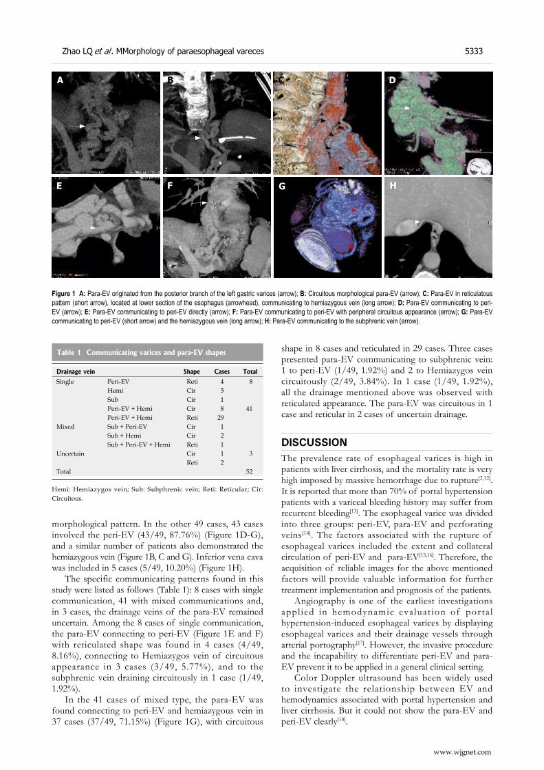
morphological pattern. In the other 49 cases, 43 cases involved the peri-EV (43/49, 87.76%) (Figure 1D-G), and a similar number of patients also demonstrated the hemiazygous vein (Figure 1B, C and G). Inferior vena cava was included in 5 cases (5/49, 10.20%) (Figure 1H).
The specific communicating patterns found in this study were listed as follows (Table 1): 8 cases with single communication, 41 with mixed communications and, in 3 cases, the drainage veins of the para-EV remained uncertain. Among the 8 cases of single communication, the para-EV connecting to peri-EV (Figure 1E and F) with reticulated shape was found in 4 cases (4/49, 8.16%), connecting to Hemiazygos vein of circuitous appearance in 3 cases (3/49, 5.77%), and to the subphrenic vein draining circuitously in 1 case (1/49, 1.92%).
In the 41 cases of mixed type, the para-EV was found connecting to peri-EV and hemiazygous vein in 37 cases (37/49, 71.15%) (Figure 1G), with circuitous
shape in 8 cases and reticulated in 29 cases. Three cases presented para-EV communicating to subphrenic vein: 1 to peri-EV (1/49, 1.92%) and 2 to Hemiazygos vein circuitously (2/49, 3.84%). In 1 case (1/49, 1.92%), all the drainage mentioned above was observed with reticulated appearance. The para-EV was circuitous in 1 case and reticular in 2 cases of uncertain drainage.
DISCUSSIONThe prevalence rate of esophageal varices is high in patients with liver cirrhosis, and the mortality rate is very high imposed by massive hemorrhage due to rupture[2,12]. It is reported that more than 70% of portal hypertension patients with a variceal bleeding history may suffer from recurrent bleeding[13]. The esophageal varice was divided into three groups: peri-EV, para-EV and perforating veins[14]. The factors associated with the rupture of esophageal varices included the extent and collateral circulation of peri-EV and para-EV[15,16]. Therefore, the acquisition of reliable images for the above mentioned factors will provide valuable information for further treatment implementation and prognosis of the patients.
Angiography is one of the earliest investigations appl ied in hemodynamic eva luat ion of por ta l hypertension-induced esophageal varices by displaying esophageal varices and their drainage vessels through arterial portography[17]. However, the invasive procedure and the incapability to differentiate peri-EV and para-EV prevent it to be applied in a general clinical setting.
Color Doppler ultrasound has been widely used to investigate the relationship between EV and hemodynamics associated with portal hypertension and liver cirrhosis. But it could not show the para-EV and peri-EV clearly[18].
Figure 1 A: Para-EV originated from the posterior branch of the left gastric varices (arrow); B: Circuitous morphological para-EV (arrow); C: Para-EV in reticulatous pattern (short arrow), located at lower section of the esophagus (arrowhead), communicating to hemiazygous vein (long arrow); D: Para-EV communicating to peri-EV (arrow); E: Para-EV communicating to peri-EV directly (arrow); F: Para-EV communicating to peri-EV with peripheral circuitous appearance (arrow); G: Para-EV communicating to peri-EV (short arrow) and the hemiazygous vein (long arrow); H: Para-EV communicating to the subphrenic vein (arrow).
A B C D
E F G H
Drainage vein Shape Cases Total
Single Peri-EV Reti 4 8Hemi Cir 3Sub Cir 1Peri-EV + Hemi Cir 8 41Peri-EV + Hemi Reti 29
Mixed Sub + Peri-EV Cir 1Sub + Hemi Cir 2Sub + Peri-EV + Hemi Reti 1
Uncertain Cir 1 3 Reti 2
Total 52
Table 1 Communicating varices and para-EV shapes
Hemi: Hemiazygos vein; Sub: Subphrenic vein; Reti: Reticular; Cir: Circuitous.
Zhao LQ et al . MMorphology of paraesophageal vareces 5333
www.wjgnet.com
Endoscopy is a more popular approach for the observation of morphological patterns, extension and severity of esophageal submucotic varices with the limitations to intramural and para-EV[19].
ECDUS is gradually becoming a useful tool for esophageal varicose inspection, which is able to display peri-EV, para-EV, perforating veins and the blood flow directions of perforating veins[7,14,20]. However, it is unable to clearly display the pathway and collaterals of para-EV. On the other hand, endoscopic sonography is costly, and also an invasive procedure that requires a skillful operator, which limited its application in clinical settings.
MDCTPV has already been a matured investigation technique used to examine esophageal and gastric varices. It is competent in displaying the types of morphological pattern, origin and collateral circulations of esophageal and gastric varices[10,11]. Sixty-four-row MDCT consists of thin slice of 0.625 mm, and a high spatial resolution, which makes it capable of displaying the communicating patterns of para-EV and peri-EV. Among the 52 cases in our study, this pattern of collateral circulation was shown in 49 (94.23%) cases. Generally, it is a non-invasive technique, and hence, it can be used to evaluate para-EV conveniently. However, displaying of this sort of collaterals is relatively rough and unsatisfactory in detail if compared with EUS. In addition, it is incapable to assess the blood flow directions of paraesophageal-periesophageal collateral circulation due to the characters of CTPV.
According to our study, vast majority of para-EV originate from the posterior branch of left gastric vein, and only 2 cases originated from the anterior branch of left gastric vein. The latter is highly associated with gastric fundal varices. A previous report suggested that[21], in cases of coexisting gastric fundal varices and esophageal varices in lower esophageal segment, the extent of esophageal varices could be aggravated after imposing obliteration therapy for gastric fundal varices. In our study, there was one patient who experienced alleviation of gastric varices with aggravated para-EV and peri-EV after receiving gastroesophageal varices treatment. The origination of para-EV may be related directly to its anatomical formation.
In our study, para-EV was found more abundant near the superior part of the gastroesophageal junction, which is also the lower esophageal segment (96.15%) than in the middle segment (3.85%). There was no para-EV involved in the upper segment. This suggests that para-EV usually occurs in the middle and lower esophageal segment and drains into the vena cava system through collateral vessels. The involved drainage vessels that we have noted were peri-EV, hemiazygous veins and subphrenic veins.
The communicating branches of para-EV and hemiazygous vein manifest as drainage “detour” veins bypassing along the anterior descending aorta to the hemiazygous vein, and subsequently drain into the superior vena cava. Ibukuro et al[22] described the above anatomic changes as preaortic vein using CT and CTAP
application. This “detour” is a commonly seen drainage route, and has an occurrence rate of 87.76%.
The hemodynamics of para-EV communicating peri-EV is relatively complicated. The communicating vessels of para-EV and peri-EV are mostly situated close to the top of the cardia level. Sato et al[7] observed the presence of perforating veins between peri-EV and para-EV with Levovist ECDUS, and meanwhile, he found the bidirectional blood flow of peri-EV and para-EV, which included the directions from para-EV to peri-EV and from peri-EV to para-EV, as well as mixed type that presented both blood flow directions. This may explain why previous para-EV studies about the recurrence risk of post-esophageal varices treatment remain controversial[20,23,24].
Para-EV can still drain into the inferior vena cava through the subphrenic vein, or directly communicate with the inferior vena cava. The subphrenic veins are the bilateral vessels which end up at inferior vena cava at the diaphragm level[25]. The communication between para-EV and subphrenic veins shown on CTPV was consistent to the precaval route and morphological pattern as reported by Ibukuro et al[25] from their CTAP and autopsy findings.
The degree of varices will be more severe if the morphological patterns of para-EV are reticulated. Under such condition, para-EV and peri-EV are individually communicated, or may be accompanied with hemiazygous vein drainage. They will mostly drain into the hemiazygous veins or subphrenic veins if the collaterals of para-EV are circuitous. Explanations about this correlation and its pathological significance require further studies.
Even though CTPV is incapable of displaying the blood flow directions of para-EV, it has a certain guidance value for EUS application. If para-EV is detected on CTPV, EUS should be further implemented to confirm the presence of specific communicating branches between peri-EV and para-EV as well as the location and hemodynamic characteristics of their communicating branches so as to provide relevant referable information for treatment selections.
COMMENTSBackgroundThe existence of para-EV and perforating veins and the direction of their blood flow can affect the recurrence of hemorrhage caused by rupturing.Research frontiersThe morphology, location and collateral circulation characteristics of para-EV in portal hypertensive patients were studied with 64-row multidetector computer tomography portal venography (MDCTPV), which is a non-invasive method.Innovations and breakthroughsIn majority cases of this study, the collateral circulation pattern of para-EV and the morphological characteristics of para-EV were revealed thanks to the high spatial resolution images of the advanced MDCT and the appropriate image post-processing.ApplicationsMDCTPV is a noninvasive method to display the morphological characteristics of para-EV, which is useful in clinical management and disease prognosis.TerminologyParaesophageal varices are the varices that exist outside the esophagus. Peri-
www.wjgnet.com
5334 ISSN 1007-9327 CN 14-1219/R World J Gastroenterol September 14, 2008 Volume 14 Number 34
EV is the varices that exist in the esophageal wall or the submucotic varices. Perforating vein is the communicating branch between peri-EV and para-EV. The subphrenic vein is the bilateral vessel that ends up at inferior vena cava at the diaphragm level. CTPV is the abbreviation of CT portal venography.Peer reviewThis is a good review with excellent images. MDCTPV is a convenient method to display the morphological and collateral circulations of the para-EV. It is of important value in the selection of clinical therapy and evaluation of prognosis.
REFERENCES1 Bhasin DK , Malhi NJ. Variceal bleeding and portal
hypertension: much to learn, much to explore. Endoscopy 2002; 34: 119-128
2 Bratovic I, Lacevic N. Management of esophageal varices. Med Arh 2002; 56: 11-12
3 Brandenburger LA, Regenstein FG. Variceal Hemorrhage. Curr Treat Options Gastroenterol 2002; 5: 73-80
4 Gotoh Y , Iwakiri R, Sakata Y, Koyama T, Noda T, Matsunaga C, Ogata SI, Ishibashi S, Sakata H, Tsunada S, Fujimoto K. Evaluation of endoscopic variceal ligation in prophylactic therapy for bleeding of oesophageal varices: a prospective, controlled trial compared with endoscopic injection sclerotherapy. J Gastroenterol Hepatol 1999; 14: 241-244
5 Mizumoto H, Matsutani S, Fukuzawa T, Ishii H, Sato G, Maruyama H, Saisho H. Hemodynamics in the left gastric vein after endoscopic ligation of esophageal varices combined with sclerotherapy. J Gastroenterol Hepatol 2001; 16: 495-500
6 Grace ND, Groszmann RJ, Garcia-Tsao G, Burroughs AK, Pagliaro L, Makuch RW, Bosch J, Stiegmann GV, Henderson JM, de Franchis R, Wagner JL, Conn HO, Rodes J. Portal hypertension and variceal bleeding: an AASLD single topic symposium. Hepatology 1998; 28: 868-880
7 Sato T , Yamazaki K, Toyota J, Karino Y, Ohmura T, Akaike J, Kuwata Y, Suga T. Perforating veins in recurrent esophageal varices evaluated by endoscopic color Doppler ultrasonography with a galactose-based contrast agent. J Gastroenterol 2004; 39: 422-428
8 Kang HK, Jeong YY, Choi JH, Choi S, Chung TW, Seo JJ, Kim JK, Yoon W, Park JG. Three-dimensional multi-detector row CT portal venography in the evaluation of portosystemic collateral vessels in liver cirrhosis. Radiographics 2002; 22: 1053-1061
9 Agarwal A, Jain M. Multidetector CT portal venography in evaluation of portosystemic collateral vessels. J Med Imaging Radiat Oncol 2008; 52: 4-9
10 Zhang LQ, He W. CT portal venography of collateral veins in esophageal varices. Zhongguo Yixue Yingxiang Jishu 2007; 23: 242-245
11 Zhao LQ , He W, Zhao H, Yu YZ. The value of CT portalvenography in the diagnosis of collateral veins in patients with gastric varices. Zhonghua Fangshexue Zazhi 2006; 40: 1175-1178
12 Seewald S, Seitz U, Yang AM, Soehendra N. Variceal bleeding and portal hypertension: still a therapeutic challenge? Endoscopy 2001; 33: 126-139
13 Grace ND, Groszmann RJ, Garcia-Tsao G, Burroughs AK,
Pagliaro L, Makuch RW, Bosch J, Stiegmann GV, Henderson JM, de Franchis R, Wagner JL, Conn HO, Rodes J. Portal hypertension and variceal bleeding: an AASLD single topic symposium. Hepatology 1998; 28: 868-880
14 Irisawa A , Shibukawa G, Obara K, Saito A, Takagi T, Shishido H, Odajima H, Abe M, Sugino T, Suzuki T, Kasukawa R, Sato Y. Collateral vessels around the esophageal wall in patients with portal hypertension: comparison of EUS imaging and microscopic findings at autopsy. Gastrointest Endosc 2002; 56: 249-253
15 Irisawa A, Obara K, Sato Y, Saito A, Takiguchi F, Shishido H, Sakamoto H, Kasukawa R. EUS analysis of collateral veins inside and outside the esophageal wall in portal hypertension. Gastrointest Endosc 1999; 50: 374-380
16 Irisawa A, Saito A, Obara K, Shibukawa G, Takagi T, Yamamoto G, Sakamoto H, Takiguchi F, Shishido H, Hikichi T, Oyama H, Sato N, Katakura K, Kasukawa R, Sato Y. Usefulness of endoscopic ultrasonographic analysis of variceal hemodynamics for the treatment of esophageal varices. Fukushima J Med Sci 2001; 47: 39-50
17 Watanabe K, Kimura K, Matsutani S, Ohto M, Okuda K. Portal hemodynamics in patients with gastric varices. A study in 230 patients with esophageal and/or gastric varices using portal vein catheterization. Gastroenterology 1988; 95: 434-440
18 Li FH, Hao J, Xia JG, Li HL, Fang H. Hemodynamic analysis of esophageal varices in patients with liver cirrhosis using color Doppler ultrasound. World J Gastroenterol 2005; 11: 4560-4565
19 Chikamori F, Kuniyoshi N, Shibuya S, Takase Y. Correlation between endoscopic and angiographic findings in patients with esophageal and isolated gastric varices. Dig Surg 2001; 18: 176-181
20 Choudhuri G, Dhiman RK, Agarwal DK. Endosonographic evaluation of the venous anatomy around the gastro-esophageal junction in patients with portal hypertension. Hepatogastroenterology 1996; 43: 1250-1255
21 Cho SK, Shin SW, Lee IH, Do YS, Choo SW, Park KB, Yoo BC. Balloon-occluded retrograde transvenous obliteration of gastric varices: outcomes and complications in 49 patients. AJR Am J Roentgenol 2007; 189: W365-W372
22 Ibukuro K, Tsukiyama T, Mori K, Inoue Y. Preaortic esophageal veins: CT appearance. AJR Am J Roentgenol 1998; 170: 1535-1538
23 Dhiman RK , Choudhuri G, Saraswat VA, Agarwal DK, Naik SR. Role of paraoesophageal collaterals and perforating veins on outcome of endoscopic sclerotherapy for oesophageal varices: an endosonographic study. Gut 1996; 38: 759-764
24 Hino S, Kakutani H, Ikeda K, Uchiyama Y, Sumiyama K, Kuramochi A, Kitamura Y, Matsuda K, Arakawa H, Kawamura M, Masuda K, Suzuki H. Hemodynamic assessment of the left gastric vein in patients with esophageal varices with color Doppler EUS: factors affecting development of esophageal varices. Gastrointest Endosc 2002; 55: 512-517
25 Ibukuro K, Tsukiyama T, Mori K, Inoue Y. Precaval draining vein from paraesophageal varices: radiologic-anatomic correlation. AJR Am J Roentgenol 1999; 172: 651-654
S- Editor Li DL L- Editor Ma JY E- Editor Yin DH
Zhao LQ et al . MMorphology of paraesophageal vareces 5335
www.wjgnet.com
Online Submissions: wjg.wjgnet.com World J Gastroenterol 2008 September 14; 14(34): [email protected] World Journal of Gastroenterology ISSN 1007-9327doi:10.3748/wjg.14.5336 © 2008 The WJG Press. All rights reserved.
Effect of music on procedure time and sedation during colonoscopy: A meta-analysis
Wilson WS Tam, Eliza LY Wong, Sheila F Twinn
RAPID COMMUNICATION
www.wjgnet.com
Wilson WS Tam, Sheila F Twinn, Nethersole School of Nursing, 8/F Esther Lee Building, The Chinese University of Hong Kong, Shatin, Hong Kong, ChinaEliza LY Wong, School of Public Health, 4/F Postgraduate Education Centre, Prince of Wales Hospital, Shatin, Hong Kong, ChinaAuthor contributions: Tam WWS, Wong ELY and Twinn SF designed the study; Tam WWS and Wong ELY performed the search and data (papers) collection; Tam WWS performed the analysis; Tam WWS, Wong ELY and Twinn SF wrote the paper.Correspondence to: Wilson WS Tam, Nethersole School of Nursing, Room 830, Esther Lee Building, The Chinese University of Hong Kong, Shatin, Hong Kong, China. [email protected]: +86-852-26098172 Fax: +86-852-26035935Received: July 10, 2008 Revised: August 25, 2008Accepted: September 2, 2008Published online: September 14, 2008
AbstractAIM: To integrate results from different studies in examining the effectiveness of music in reducing the procedure time and the amount of sedation used during colonoscopic procedure. METHODS: An electronic search in various databases was performed to identify related articles. Study quality was evaluated by the Jadad’s scale. The random effect model was used to pool the effect from individual trials and the Cohen Q-statistic was used to determine heterogeneity. Egger’s regression was used to detect publication bias. RESULTS: Eight studies with 722 subjects were included in this meta-analysis. The combined mean difference for the time taken for the colonoscopy procedure between the music and control groups was -2.84 with 95% CI (-5.61 to -0.08), implying a short time for the music group. The combined mean difference for the use of sedation was -0.46 with 95%CI (-0.91 to -0.01), showing a significant reduction in the use of sedation in the music group. Heterogeneity was observed in both analyses but no publication bias was detected. CONCLUSION: Listening to music is effective in reducing procedure time and amount of sedation during colonoscopy and should be promoted.
© 2008 The WJG Press. All rights reserved.
Key words: Colonoscopy; Colon cancer; Meta-analysis; Music therapy; Screening
Peer reviewer: Yoshiharu Motoo, MD, PhD, FACP, FACG, Professor and Chairman, Department of Medical Oncology, Kanazawa Medical University,1-1 Daigaku, Uchinada, Ishikawa 920-0293, Japan
Tam WWS, Wong ELY, Twinn SF. Effect of music on procedure time and sedation during colonoscopy: A meta-analysis. World J Gastroenterol 2008; 14(34): 5336-5343 Available from: URL: http://www.wjgnet.com/1007-9327/14/5336.asp DOI: http://dx.doi.org/10.3748/wjg.14.5336
INTRODUCTIONIn 2005, colorectal cancer was the fourth most frequent cancer type worldwide for both sexes and colon cancer accounted for about 655 000 deaths per year[1]. Most colon cancers develop from polyps that grow abnormally in the colon. If polyps grow unnoticed and are not removed, they may become cancerous. Indeed, colorectal cancer is among the most preventable and curable cancers and with early detection 75%-90% of them can be prevented[2]. The aim of screening is to find precancerous polyps so they can be removed before they become cancerous. Screening for colon cancer has been shown to be effective in reducing mortality[3].
Colonoscopy is now the recommended method for screening colon cancer[4]. During a colonoscopy procedure, physicians insert a colonoscope into the rectum of a patient from the anus and slowly guide it into the colon for direct visualization and diagnosis[5]. As can be imagined, it is not a comfortable experience. In fact, many people refuse to undergo colonoscopy because of discomfort and anxiety[6], some feel out of control with what would be happening during the procedure and were fearful of looking foolish[7]. Even those who agree to undergo a colonoscopy feel frightened and anxious before the procedure[8]. After it, they express it was an unpleasant and stressful experience[9]. Therefore, most physicians prefer to
www.wjgnet.com
Tam WWS et al . Music and colonoscopy: A meta-analysi 5337
perform colonoscopy with conscious sedation[10]
although some still start the procedure without sedation, particularly in non-anxious patients[11].
Since the use of sedation is risky-it may contribute to the occur rence of cardiovascular events and is associated with the risk of cardio-respiratory complication[12,13] especially for elderly patients[14]-pand costly, non-pharmacological methods for alleviating patients’ discomfort and anxiety have been developed and music therapy is one of them. The use of music therapy to promote relaxation has a long history in medicine[15,16]. The ancient Chinese medical reference, Yellow Emperor’s Classics of Internal Medicine, mentioned the use of music for treatment[17], while ancient Indian treatises, like Samaveda, stated the therapeutic utility of music[18]. Earlier studies show the effectiveness of music on patients with acute myocardial infarctions[19] and receiving intensive medical/surgical care[20]. The effects of music on different screening procedures were examined during the early 1990s. For example, Fullhart[21] and Palakanis[6] s tudied anxiety in pat ients undergoing f lexible sigmoidoscopy and Bampton[22] assessed the role of relaxation music on patient tolerance of gastrointestinal endoscopic procedures. Since then, more and more studies have examined the effect of music during colonoscopy, but no conclusion has yet been reached about its effectiveness. Recently, a meta-analysis[23] focusing on the general endoscopic procedure was published and the authors reported significantly lower anxiety levels and shorter procedure times. The aim of this meta-analysis is to integrate results from different studies in examining the effectiveness of music in reducing procedure time and amount of sedation used during colonoscopy.
MATERIALS AND METHODSSearch strategiesWe identified studies to evaluate the effect of listening to music during colonoscopy. The electronic search was conducted on six databases, namely, AMED-Allied and Complementary Medicine (1985 to 2007), ACP journal club (1991 to 2007), CINAHL (1982 to March 2007), Cochrane Central Register of Controlled trials (up to 1st quarter of 2007), EMBASE (1980 to March 2007), and MEDLINE (1966 to March 2007). Only two sets of keywords, i.e. Colonoscopy and Music/Music, were used to include as many articles as possible. We also attempted to identify any potential unpublished studies such as theses and dissertations using the above keywords through the ProQuest Dissertations and Theses database, and the web using Internet search engines, Google and Yahoo, with thesis or dissertation as additional keywords. Reference lists of the collected papers were also checked for any potential articles.
Inclusion criteriaOnly randomized controlled studies reported in English
were included. At least one of the comparison groups included listening to music during colonoscopy as part of the intervention.
Data extractionFor each eligible study, we extracted information on author/s, year of publication, age distribution, gender proportion, other demographic data, intervention, outcome measures, and results. The data extraction form was modified from the data extraction form of van Tulder[24]. Two investigators (WT and EW) of the study independently extracted the data, which was then summarized by one of them. Agreement was reached on the extracted data before proceeding to data analysis.
Methodological qualityThe methodological quality of the included studies was assessed using the Jadad’s scale[25]. The quality var iab les recorded in the cr i ter ia l i s t inc luded the procedure of patient allocation, information regarding withdrawals and dropouts, blinding of patients, and outcome assessment. A maximum of five points can be obtained using the scale. However, as blinding of patients is difficult while assessing behavioral interventions[26], only blinding of outcome assessors was considered in the scale. Furthermore, as withdrawals and attrition of patients were unlikely in such a short period, such item in the Jadad’s scale was also ignored. Therefore, the maximum points obtainable for each study became four. Besides the Jadad’s scale, one additional question was extracted from van Tulder[24] for assessing the comparability of the two comparison groups.
Statistical analysisRandom-effect models were used to combine the outcome effect, i.e. the (standardized) difference between treated and control groups, for each study[27]. Heterogeneity was examined using the Q-test and I2[27]. All analyses were conducted using RevMan[27]. Publication b ias was examined by funnel p lot and Eg ger ’s regression[28].
RESULTSSearch strategiesThe electronic search was conducted in May 2007 and 19 records were found. The title and abstracts of these papers were examined and it was found that one was written in Portuguese[29], one was a systematic review focus on pain[30], one was a discussion paper on the use of sedation[31], one was an editorial[32], and two were news[33,34]. One paper[35] occurred twice in our search as the surname of the first author was recorded differently in two electronic databases (EBASE and MEDLINE), so one was removed. Hence, only 12 studies[35-46] were included and the full articles were obtained for further examination. A study[37] was found to be a non-randomized study
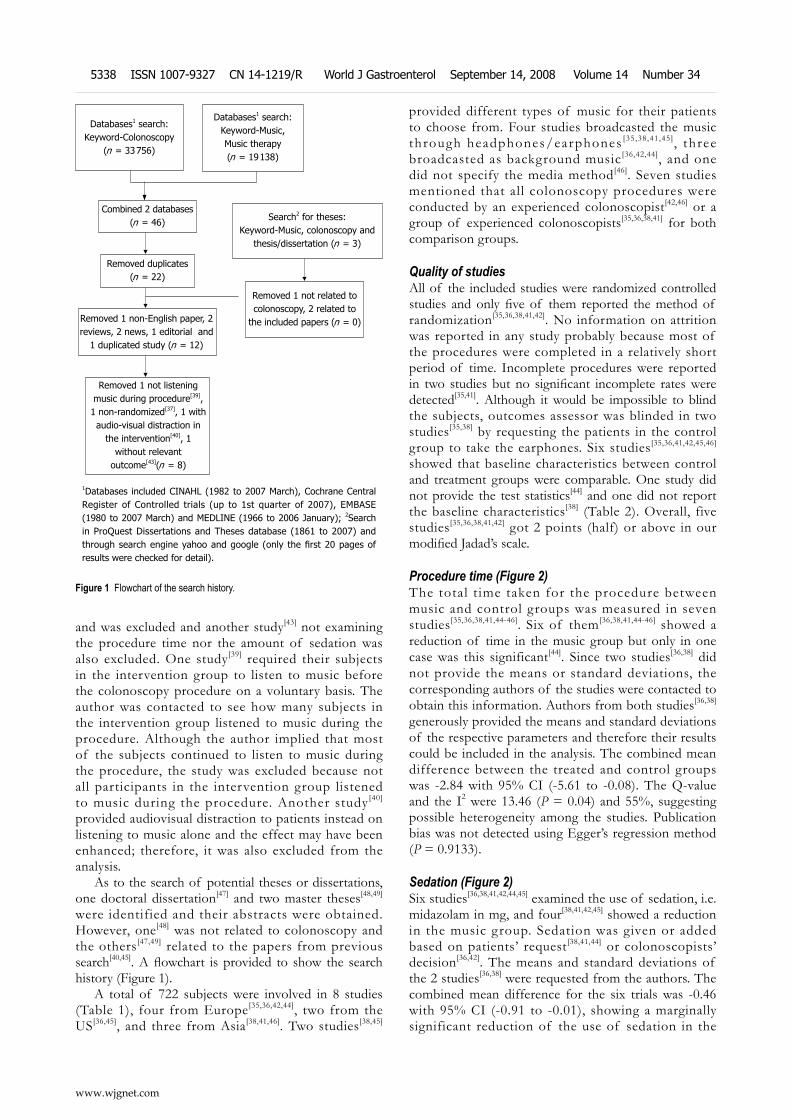
and was excluded and another study[43] not examining the procedure time nor the amount of sedation was also excluded. One study[39] required their subjects in the intervention group to listen to music before the colonoscopy procedure on a voluntary basis. The author was contacted to see how many subjects in the intervention group listened to music during the procedure. Although the author implied that most of the subjects continued to listen to music during the procedure, the study was excluded because not all participants in the intervention group listened to music during the procedure. Another study[40] provided audiovisual distraction to patients instead on listening to music alone and the effect may have been enhanced; therefore, it was also excluded from the analysis.
As to the search of potential theses or dissertations, one doctoral dissertation[47] and two master theses[48,49]
were identified and their abstracts were obtained. However, one[48] was not related to colonoscopy and the others[47,49] related to the papers from previous search[40,45]. A flowchart is provided to show the search history (Figure 1).
A total of 722 subjects were involved in 8 studies (Table 1), four from Europe[35,36,42,44], two from the US[36,45], and three from Asia[38,41,46]. Two studies[38,45]
provided different types of music for their patients to choose from. Four studies broadcasted the music through headphones/earphones [35,38 ,41 ,45], three broadcasted as background music[36,42,44], and one did not specify the media method[46]. Seven studies mentioned that all colonoscopy procedures were conducted by an experienced colonoscopist[42,46] or a group of experienced colonoscopists[35,36,38,41] for both comparison groups.
Quality of studiesAll of the included studies were randomized controlled studies and only five of them reported the method of randomization[35,36,38,41,42]. No information on attrition was reported in any study probably because most of the procedures were completed in a relatively short period of time. Incomplete procedures were reported in two studies but no significant incomplete rates were detected[35,41]. Although it would be impossible to blind the subjects, outcomes assessor was blinded in two studies[35,38] by requesting the patients in the control group to take the earphones. Six studies[35,36,41,42,45,46] showed that baseline characteristics between control and treatment groups were comparable. One study did not provide the test statistics[44] and one did not report the baseline characteristics[38] (Table 2). Overall, five studies[35,36,38,41,42] got 2 points (half) or above in our modified Jadad’s scale.
Procedure time (Figure 2)The total t ime taken for the procedure between music and control groups was measured in seven studies[35,36,38,41,44-46]. Six of them[36,38,41,44-46] showed a reduction of time in the music group but only in one case was this significant[44]. Since two studies[36,38] did not provide the means or standard deviations, the corresponding authors of the studies were contacted to obtain this information. Authors from both studies[36,38] generously provided the means and standard deviations of the respective parameters and therefore their results could be included in the analysis. The combined mean difference between the treated and control groups was -2.84 with 95% CI (-5.61 to -0.08). The Q-value and the I2 were 13.46 (P = 0.04) and 55%, suggesting possible heterogeneity among the studies. Publication bias was not detected using Egger’s regression method (P = 0.9133).
Sedation (Figure 2)Six studies[36,38,41,42,44,45] examined the use of sedation, i.e. midazolam in mg, and four[38,41,42,45] showed a reduction in the music group. Sedation was given or added based on patients’ request[38,41,44] or colonoscopists’ decision[36,42]. The means and standard deviations of the 2 studies[36,38] were requested from the authors. The combined mean difference for the six trials was -0.46 with 95% CI (-0.91 to -0.01), showing a marginally significant reduction of the use of sedation in the
Databases1 search:Keyword-Colonoscopy
(n = 33 756)
Databases1 search:Keyword-Music,Music therapy(n = 19 138)
Combined 2 databases (n = 46)
Removed duplicates (n = 22)
Removed 1 non-English paper, 2 reviews, 2 news, 1 editorial and
1 duplicated study (n = 12)
Removed 1 not listeningmusic during procedure[39],1 non-randomized[37], 1 withaudio-visual distraction in
the intervention[40], 1 without relevant
outcome[43](n = 8)
Search2 for theses:Keyword-Music, colonoscopy and
thesis/dissertation (n = 3)
Removed 1 not related to colonoscopy, 2 related to
the included papers (n = 0)
1Databases included CINAHL (1982 to 2007 March), Cochrane Central Register of Controlled trials (up to 1st quarter of 2007), EMBASE (1980 to 2007 March) and MEDLINE (1966 to 2006 January); 2Search in ProQuest Dissertations and Theses database (1861 to 2007) and through search engine yahoo and google (only the first 20 pages of results were checked for detail).
Figure 1 Flowchart of the search history.
www.wjgnet.com
5338 ISSN 1007-9327 CN 14-1219/R World J Gastroenterol September 14, 2008 Volume 14 Number 34
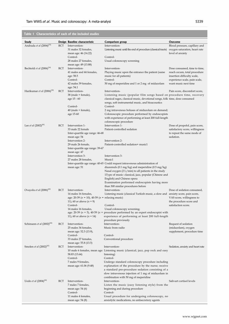
www.wjgnet.com
Tam WWS et al . Music and colonoscopy: A meta-analysi 5339
Table 1 Characteristics of each of the included studies
Study Design Baseline characteristic Comparison group OutcomeAndrada et al (2004)[35] RCT Intervention- Intervention- Blood pressure, capillary and
31 males 32 females, Listening music until the end of procedure (classical tracts) oxygen saturation, heart rate mean age: 46 (14.22) level of anxietyControl- Control-28 males 27 females, Usual colonoscopy screeningmean age: 49 (13.88)
Bechtold et al (2006)[36] RCT Intervention- Intervention- Dose consumed, time to time, 41 males and 44 females, Playing music upon the entrance the patient (same reach cecum, total procedure age: 58.5 music for all patients) insertion difficulty scale, Control- Control- experience scale, pain scale, 42 males 39 females, 50 mg of meperidine and 1 or 2 mg of midazolam want music next timeage: 54.1
Harikumar et al (2006)[38] RCT Intervention- Intervention- Pain score, discomfort score, 38 (male + female), age 15 - 60
Listening music (popular film songs based on classical rages, classical music, devotional songs, folk songs, soft instrumental music, and bioacoustics
procedure time, recovery time, dose consumed
Control- Control-40 (male + female),age 15-60
2 mg intravenous boluses of midazolam on demand; Colonoscopic procedure performed by endoscopists with experience of performing at least 200 full-length colonoscopic procedure
Lee et al (2002)[41] RCT Intervention 1- Intervention 1- Dose of propofol, pain score, 33 male 22 female Patient-controlled sedation satisfactory score, willingness Inter-quartile age range: 46-68 to repeat the same mode of mean age: 54 sedation.Intervention 2- Intervention 2-29 male 26 female, Patient-controlled sedation+ music1Inter-quartile age range: 39-67mean age: 47Intervention 3- Intervention 3-27 males 28 females, Music1Inter-quartile age range: 40-65 Could request intravenous administration of mean age: 51 diazemuls (0.1 mg/kg) and meperidine (0.5 mg/kg)
Nasal oxygen (2 L/min) to all patients in the study1Type of music: classical, jazz, popular (Chinese and English) and Chinese operaExamination performed endoscopists having more than 300 similar procedures before
Ovayolu (2006)[42] RCT Intervention- Intervention- Dose of sedation consumed, 14 males 16 females, Listening music (classical Turkish music, a slow and anxiety score, pain score, age: 20-39 (n = 10), 40-59 (n = 11), 60 or above (n = 9)
relaxing music) VAS score, willingness to the procedure score and
Control- Control- satisfaction score.14 males 16 females, Usual colonoscopy screening;age: 20-39 (n = 5), 40-59 (n = 11), 60 or above (n = 14)
procedure performed by an expert endoscopist with experience of performing at least 200 full-length procedure previously
Schimann et al (2002)[44] RCT Intervention- Intervention- Request of sedation 25 males 34 females, Music from radio (midazolam), oxygen mean age: 52.3 (13.9), supplement, procedure timeControl- Control-33 males 27 females, Conventional proceduremean age: 55.8 (13.5)
Smolen et al (2002)[45] RCT Intervention- Intervention- Sedation, anxiety and heart rate10 male 6 females, mean age: 58.83 (13.64)
Listening music (classical, jazz, pop rock and easy listening)
Control- Control-7 males 9 females,mean age: 61.06 (9.48)
Undergo standard colonoscopy procedure including explanation of the procedure by the nurse; receive a standard pre-procedure sedation consisting of a slow intravenous injection of 1 mg of midazolam in combination with 50 mg of meperidine
Uedo et al (2004)[46] RCT Intervention- Intervention- Salivart cortisol levels7 males 7 females, mean age: 54 (6)
Listen the music (easy listening style) from the beginning and during procedure
Control- Control-11 males 4 females,mean age: 54 (8)
Usual procedure for undergoing colonoscopy, no anxiolytic medications, no antisecretory agents
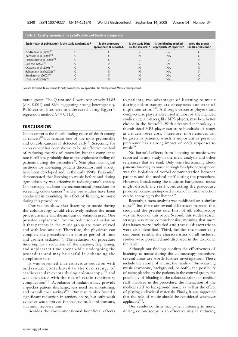
music group. The Q-test and I2 were respectively 34.83 (P < 0.001) and 86% suggesting strong heterogeneity. Publication bias was not detected using Egger’s regression method (P = 0.1150).
DISCUSSIONColon cancer is the fourth leading cause of death among all cancers[1] but remains one of the most preventable and curable cancers if detected early[2]. Screening for colon cancer has been shown to be an effective method of reducing the risk of mortality, but the compliance rate is still low probably due to the unpleasant feeling of patients during the procedure[9]. Non-pharmacological methods for alleviating patients discomfort and anxiety have been developed and, in the early 1990s, Palakanis[6]
demonstrated that listening to music before and during sigmoidoscopy was effective in reducing one’s anxiety. Colonoscopy has been the recommended procedure for screening colon cancer[4] and more studies have been conducted in examining the effect of listening to music during this procedure.
Our results show that listening to music during the colonoscopy would effectively reduce the mean procedure time and the amount of sedation used. One possible explanation for the reduction of sedation is that patients in the music group are more relaxed and with less anxiety. Therefore, the physician can complete the procedure in a shorter period of time and use less sedation[45]. The reduction of procedure time implies a reduction of the anxious, frightening, and unpleasant time spent while undergoing the procedure and may be useful in enhancing the compliance rate.
It was reported that conscious sedation with midazo l am cont r ibu ted to the occur rence o f cardiovascular events during colonoscopy[12] and was associated with the risk of cardio-respiratory complication[14]. Avoidance of sedation may provide a quicker patient discharge, less need for monitoring, and overall cost savings[50]. Our results also found a significant reduction in anxiety score, but only weak evidence was observed for pain score, blood pressure, and mean recovery time.
Besides the above-mentioned beneficial effects
to patients, two advantages of listening to music dur ing colonoscopy are cheapness and ease of implementation[51]. Although cassette players and compact disc players were used in most of the included studies, digital players, like MP3 players, may be a better choice in the future[52]. With advanced technology, a thumb-sized MP3 player can store hundreds of songs at a much lower cost. Therefore, more choices can be given to patients, which is important as personal preference has a strong impact on one’s responses to music[53].
No harmful effects from listening to music were reported in any study in the meta-analysis and other references that we read. Only one shortcoming about patients listening to music through headphone/earphone was the isolation of verbal communication between patients and the medical staff during the procedure. However, broadcasting the music as background music might disturb the staff conducting the procedure probably because an imposed choice of musical selection can be annoying to the listener[53].
Recently, a meta-analysis was published on a similar topic[23] but there are several differences between that study and the present one. First of all, colonoscopy was the focus of this paper. Second, this study’s search strategy was more comprehensive, meaning that more databases were included and theses/dissertations were also identified. Third, besides the numerically combined results, the characteristics of all included studies were presented and discussed in the text or in the table.
Although our findings confirm the effectiveness of listening to music during the colonoscopy procedure, several areas are worth further investigation. These include the choice of music, the mode of broadcasting music (earphone, background, or both), the possibility of using placebo to the patients in the control group, the possibility of blinding to the colonoscopist/s or medical staff involved in the procedure, the interaction of the medical staff to background music as well as the effect of playing audiovisual materials. Finally, it was suggested that the role of music should be considered whenever applicable[54].
Our results confirm that patient listening to music during colonoscopy is an effective way in reducing
Table 2 Quality assessment by Jadad’s scale and baseline comparison
Study (year of publication) Is the study randomized? Is the procedure appropriate & reported?
Is the study blind to the assessors?
Is the blinding method appropriate & reported?
Were the groups similar at baseline?
Andrada et al (2004)[35] C C C C CBechtold et al (2006)[36] C C N NA CHarikumar et al (2006)[38] C C C C P1
Lee et al (2002)[41] C P C N COvayolu et al (2006)[42] C C N NA CSchiemann et al (2002)[44] C N N NA P2
Smolen et al (2002)[45] C N N NA CUedo et al (2004)[46] C N N NA C
Remark: C, correct; N, not correct; P, partly correct; NA, not applicable. 1No result provided; 2No test result provided.
www.wjgnet.com
5340 ISSN 1007-9327 CN 14-1219/R World J Gastroenterol September 14, 2008 Volume 14 Number 34
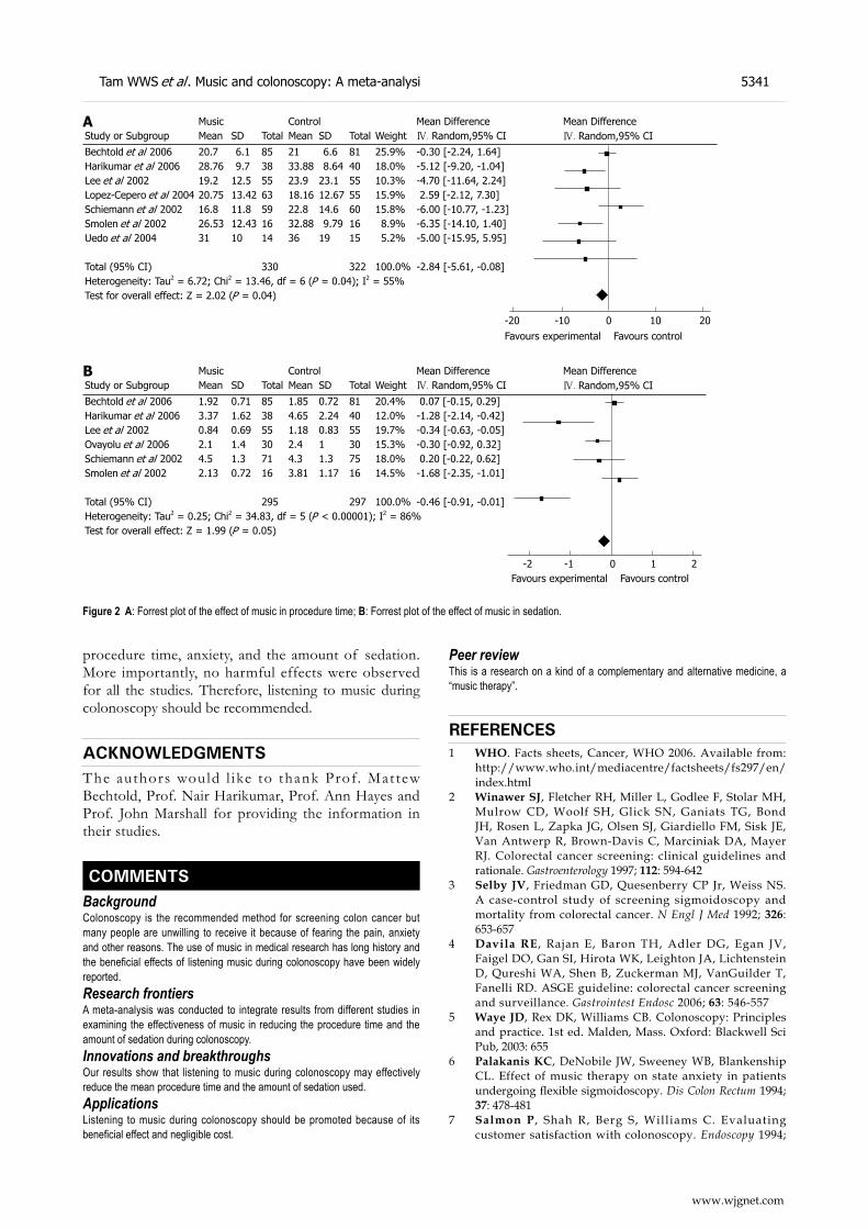
procedure time, anxiety, and the amount of sedation. More importantly, no harmful effects were observed for all the studies. Therefore, listening to music during colonoscopy should be recommended.
ACKNOWLEDGMENTSThe authors would l ike to thank Prof. Mattew Bechtold, Prof. Nair Harikumar, Prof. Ann Hayes and Prof. John Marshall for providing the information in their studies.
COMMENTSBackgroundColonoscopy is the recommended method for screening colon cancer but many people are unwilling to receive it because of fearing the pain, anxiety and other reasons. The use of music in medical research has long history and the beneficial effects of listening music during colonoscopy have been widely reported.Research frontiersA meta-analysis was conducted to integrate results from different studies in examining the effectiveness of music in reducing the procedure time and the amount of sedation during colonoscopy. Innovations and breakthroughsOur results show that listening to music during colonoscopy may effectively reduce the mean procedure time and the amount of sedation used.Applications Listening to music during colonoscopy should be promoted because of its beneficial effect and negligible cost.
Peer reviewThis is a research on a kind of a complementary and alternative medicine, a “music therapy”.
REFERENCES1 WHO. Facts sheets, Cancer, WHO 2006. Available from:
http://www.who.int/mediacentre/factsheets/fs297/en/index.html
2 Winawer SJ, Fletcher RH, Miller L, Godlee F, Stolar MH, Mulrow CD, Woolf SH, Glick SN, Ganiats TG, Bond JH, Rosen L, Zapka JG, Olsen SJ, Giardiello FM, Sisk JE, Van Antwerp R, Brown-Davis C, Marciniak DA, Mayer RJ. Colorectal cancer screening: clinical guidelines and rationale. Gastroenterology 1997; 112: 594-642
3 Selby JV, Friedman GD, Quesenberry CP Jr, Weiss NS. A case-control study of screening sigmoidoscopy and mortality from colorectal cancer. N Engl J Med 1992; 326: 653-657
4 Davila RE, Rajan E, Baron TH, Adler DG, Egan JV, Faigel DO, Gan SI, Hirota WK, Leighton JA, Lichtenstein D, Qureshi WA, Shen B, Zuckerman MJ, VanGuilder T, Fanelli RD. ASGE guideline: colorectal cancer screening and surveillance. Gastrointest Endosc 2006; 63: 546-557
5 Waye JD, Rex DK, Williams CB. Colonoscopy: Principles and practice. 1st ed. Malden, Mass. Oxford: Blackwell Sci Pub, 2003: 655
6 Palakanis KC, DeNobile JW, Sweeney WB, Blankenship CL. Effect of music therapy on state anxiety in patients undergoing flexible sigmoidoscopy. Dis Colon Rectum 1994; 37: 478-481
7 Salmon P , Shah R, Berg S, Williams C. Evaluating customer satisfaction with colonoscopy. Endoscopy 1994;
Music Control Mean DifferenceStudy or Subgroup Mean SD Total Mean SD Total Weight Ⅳ. Random,95% CI
Bechtold et al 2006 20.7 6.1 85 21 6.6 81 25.9% -0.30 [-2.24, 1.64]Harikumar et al 2006 28.76 9.7 38 33.88 8.64 40 18.0% -5.12 [-9.20, -1.04]Lee et al 2002 19.2 12.5 55 23.9 23.1 55 10.3% -4.70 [-11.64, 2.24]Lopez-Cepero et al 2004 20.75 13.42 63 18.16 12.67 55 15.9% 2.59 [-2.12, 7.30]Schiemann et al 2002 16.8 11.8 59 22.8 14.6 60 15.8% -6.00 [-10.77, -1.23]Smolen et al 2002 26.53 12.43 16 32.88 9.79 16 8.9% -6.35 [-14.10, 1.40]Uedo et al 2004 31 10 14 36 19 15 5.2% -5.00 [-15.95, 5.95]
Total (95% CI) 330 322 100.0% -2.84 [-5.61, -0.08]Heterogeneity: Tau2 = 6.72; Chi2 = 13.46, df = 6 (P = 0.04); I2 = 55%Test for overall effect: Z = 2.02 (P = 0.04)
Mean DifferenceⅣ. Random,95% CI
-20 -10 0 10 20Favours experimental Favours control
A
Music Control Mean DifferenceStudy or Subgroup Mean SD Total Mean SD Total Weight Ⅳ. Random,95% CI
Bechtold et al 2006 1.92 0.71 85 1.85 0.72 81 20.4% 0.07 [-0.15, 0.29]Harikumar et al 2006 3.37 1.62 38 4.65 2.24 40 12.0% -1.28 [-2.14, -0.42]Lee et al 2002 0.84 0.69 55 1.18 0.83 55 19.7% -0.34 [-0.63, -0.05]Ovayolu et al 2006 2.1 1.4 30 2.4 1 30 15.3% -0.30 [-0.92, 0.32]Schiemann et al 2002 4.5 1.3 71 4.3 1.3 75 18.0% 0.20 [-0.22, 0.62]Smolen et al 2002 2.13 0.72 16 3.81 1.17 16 14.5% -1.68 [-2.35, -1.01]
Total (95% CI) 295 297 100.0% -0.46 [-0.91, -0.01]Heterogeneity: Tau2 = 0.25; Chi2 = 34.83, df = 5 (P < 0.00001); I2 = 86%Test for overall effect: Z = 1.99 (P = 0.05)
Mean DifferenceⅣ. Random,95% CI
-2 -1 0 1 2Favours experimental Favours control
B
Figure 2 A: Forrest plot of the effect of music in procedure time; B: Forrest plot of the effect of music in sedation.
www.wjgnet.com
Tam WWS et al . Music and colonoscopy: A meta-analysi 5341
26: 342-3468 Salmore RG, Nelson JP. The effect of preprocedure
teaching, relaxation instruction, and music on anxiety as measured by blood pressures in an outpatient gastrointestinal endoscopy laboratory. Gastroenterol Nurs 2000; 23: 102-110
9 Kim LS, Koch J, Yee J, Halvorsen R, Cello JP, Rockey DC. Comparison of patients' experiences during imaging tests of the colon. Gastrointest Endosc 2001; 54: 67-74
10 Balsells F, Wyllie R, Kay M, Steffen R. Use of conscious s e d a t i o n f o r l o w e r a n d u p p e r g a s t r o i n t e s t i n a l endoscopic examinations in children, adolescents, and young adults: a twelve-year review. Gastrointest Endosc 1997; 45: 375-380
11 Fisher NC, Bailey S, Gibson JA. A prospective, randomized controlled trial of sedation vs. no sedation in outpatient diagnostic upper gastrointestinal endoscopy. Endoscopy 1998; 30: 21-24
12 Ristikankare M, Julkunen R, Laitinen T, Wang SX, Heikkinen M, Janatuinen E, Hartikainen J. Effect of conscious sedation on cardiac autonomic regulation during colonoscopy. Scand J Gastroenterol 2000; 35 : 990-996
13 Bell GD. Premedication, preparation, and surveillance. Endoscopy 2002; 34: 2-12
14 Yuno K , I ishi H, Tatsuta M, Hifumi K, Omori M. Intravenous midazolam as a sedative for colonoscopy: a randomized, double-blind clinical trial. Aliment Pharmacol Ther 1996; 10: 981-984
15 Rorke MA. Music therapy in the age of enlightenment. J Music Ther 2001; 38: 66-73
16 West M. Music Therapy in Antiquity. Horden P, editor. Music as medicine: the history of music therapy since antiquity. Aldershot; Brookfield, Vt: Ashgate, 2000: 51-68
17 Ni MS. The Yellow Emperor’s Classic of internal medicine: A new translation of the Neijing Suwen with commentary. Boston: Shambhala Pub, 1995: 14-17
18 Katz JB. Music Therapy: Some Possibilities in the Indian Tradition. Horden P, editor. Music as medicine: the history of music therapy since antiquity. Aldershot; Brookfield, Vt: Ashgate, 2000: 84-102
19 White JM. Music therapy: an intervention to reduce anxiety in the myocardial infarction patient. Clin Nurse Spec 1992; 6: 58-63
20 Updike P. Music therapy results for ICU patients. Dimens Crit Care Nurs 1990; 9: 39-45
21 Fullhart JW. Preparatory information and anxiety before sigmoidoscopy: a comparative study. 1992; 14: 286-290
22 Bampton P, Draper B. Effect of relaxation music on patient tolerance of gastrointestinal endoscopic procedures. J Clin Gastroenterol 1997; 25: 343-345
23 Rudin D, Kiss A, Wetz RV, Sottile VM. Music in the endoscopy suite: a meta-analysis of randomized controlled studies. Endoscopy 2007; 39: 507-510
24 van Tulder M , Furlan A, Bombardier C, Bouter L. Updated method guidelines for systematic reviews in the cochrane collaboration back review group. Spine 2003; 28: 1290-1299
25 Jadad AR, Moore RA, Carroll D, Jenkinson C, Reynolds DJ, Gavaghan DJ, McQuay HJ. Assessing the quality of reports of randomized clinical trials: is blinding necessary? Control Clin Trials 1996; 17: 1-12
26 Monninkhof E , van der Valk P, van der Palen J , van Herwaarden C, Partridge MR, Zielhuis G. Self-management education for patients with chronic obstructive pulmonary disease: a systematic review. Thorax 2003; 58: 394-398
27 Review Manager (RevMan) [Computer program] Version 5.0. Copenhagen: The Nordic Cochrane Centre, The
Cochrane Collaboration, 200828 Egger M, Davey Smith G, Schneider M, Minder C. Bias
in meta-analysis detected by a simple, graphical test. BMJ 1997; 315: 629-634
29 Arruda Alves PR, Roncaratti E, Habr Gama A. Reduction in salivary levels of cortisol by music therapy during colonoscopy. GED-Gastrenterologia Endoscopia Digestiva 2004; 23: 83-84
30 Cepeda MS, Carr DB, Lau J, Alvarez H. Music for pain relief. Cochrane Database Syst Rev 2006; CD004843
31 Lazzaroni M, Bianchi Porro G. Preparation, premedication, and surveillance. Endoscopy 2005; 37: 101-109
32 Lotufo PA. The noise stops you from hearing good music: the possibilities for a mortality reduction program for cancer of the colon and rectum in Sao Paulo. Sao Paulo Med J 2003; 121: 95-96
33 Kurth T, Krevvsky B, Chiu C. Late-breaking news. Bottom Line Health 2007; 21: 1
34 Anonymous. Tuned up for colon test. Nur Standard 2006; 21: 8
35 Lopez-Cepero Andrada JM, Amaya Vidal A, Castro Aguilar-Tablada T, Garcia Reina I , S i lva L, Ruiz Guinaldo A, Larrauri De la Rosa J, Herrero Cibaja I, Ferre Alamo A, Benitez Roldan A. Anxiety during the performance of colonoscopies: modification using music therapy. Eur J Gastroenterol Hepatol 2004; 16: 1381-1386
36 Bechtold ML, Perez RA, Puli SR, Marshall JB. Effect of music on patients undergoing outpatient colonoscopy. World J Gastroenterol 2006; 12: 7309-7312
37 Binek J, Sagmeister M, Borovicka J, Knierim M, Magdeburg B, Meyenberger C. Perception of gastrointestinal endoscopy by patients and examiners with and without background music. Digestion 2003; 68: 5-8
38 Harikumar R , Raj M, Paul A, Harish K, Kumar SK, Sandesh K, Asharaf S, Thomas V. Listening to music d e c r e a s e s n e e d f o r s e d a t i v e m e d i c a t i o n d u r i n g colonoscopy: a randomized, controlled trial. Indian J Gastroenterol 2006; 25: 3-5
39 Hayes A, Buffum M, Lanier E, Rodahl E, Sasso C. A music intervention to reduce anxiety prior to gastrointestinal procedures. Gastroenterol Nurs 2003; 26: 145-149
40 Lee DW, Chan AC, Wong SK, Fung TM, Li AC, Chan SK, Mui LM, Ng EK, Chung SC. Can visual distraction decrease the dose of patient-controlled sedation required during colonoscopy? A prospective randomized controlled trial. Endoscopy 2004; 36: 197-201
41 Lee DW, Chan KW, Poon CM, Ko CW, Chan KH, Sin KS, Sze TS, Chan AC. Relaxation music decreases the dose of patient-controlled sedation during colonoscopy: a prospective randomized controlled trial. Gastrointest Endosc 2002; 55: 33-36
42 O v a y o l u N , U c a n O , P e h l i v a n S , P e h l i v a n Y , Buyukhatipoglu H, Savas MC, Gulsen MT. Listening to Turkish classical music decreases patients' anxiety, pain, dissatisfaction and the dose of sedative and analgesic drugs during colonoscopy: a prospective randomized controlled trial. World J Gastroenterol 2006; 12: 7532-7536
43 Salmore RG, Nelson JP. The effect of preprocedure teaching, relaxation instruction, and music on anxiety as measured by blood pressures in an outpatient gastrointestinal endoscopy laboratory. Gastroenterol Nurs 2000; 23: 102-110
44 Schiemann U, Gross M, Reuter R, Kellner H. Improved procedure of colonoscopy under accompanying music therapy. Eur J Med Res 2002; 7: 131-134
45 Smolen D, Topp R, Singer L. The effect of self-selected music during colonoscopy on anxiety, heart rate, and blood pressure. Appl Nurs Res 2002; 15: 126-136
46 Uedo N, Ishikawa H, Morimoto K, Ishihara R, Narahara H, Akedo I , Ioka T, Kaj i I , Fukuda S . Reduct ion in salivary cortisol level by music therapy during colonoscopic examination. Hepatogastroenterology 2004;
www.wjgnet.com
5342 ISSN 1007-9327 CN 14-1219/R World J Gastroenterol September 14, 2008 Volume 14 Number 34
51: 451-45347 Lee DWH. The use of patient-controlled and adjunct
sedation for colonoscopy. Thesis. Chinese University of Hong Kong 2005
48 Parker DB. The effect of Music Therapy for pain and anxiety versus literature on the immediate and future perceptions of cardiac patients. Thesis. The Florida State University, 2004
49 Smolen D, Topp R, Singer L. The effect of self-selected music during colonoscopy on anxiety, heart rate, and blood
pressure. Appl Nurs Res 2002; 15: 126-13650 Takahashi Y, Tanaka H, Kinjo M, Sakumoto K. Sedation-
free colonoscopy. Dis Colon Rectum 2005; 48: 855-85951 Kemper KJ, Danhauer SC. Music as therapy. South Med J
2005; 98: 282-28852 Maag M. Podcasting and MP3 players: emerging education
technologies. Comput Inform Nurs 2006; 24: 9-1353 Bonny HL. Music and healing. Music Ther 1986; 6A: 3-12. 54 Thaut MH. The future of music in therapy and medicine.
Ann N Y Acad Sci 2005; 1060: 303-308
S- Editor Zhong XY L- Editor Negro F E- Editor Zhang WB
www.wjgnet.com
Tam WWS et al . Music and colonoscopy: A meta-analysi 5343
Online Submissions: wjg.wjgnet.com World J Gastroenterol 2008 September 14; 14(34): [email protected] World Journal of Gastroenterology ISSN 1007-9327doi:10.3748/wjg.14.5344 © 2008 The WJG Press. All rights reserved.
Effect of mutant p27kip1 gene on human cholangiocarcinoma cell line, QBC939
Jian Luo, Yong-Jun Chen, Wei-Yu Wang, Sheng-Quan Zou
RAPID COMMUNICATION
www.wjgnet.com
Jian Luo, Yong-Jun Chen, Wei-Yu Wang, Sheng-Quan Zou, Department of General Surgery, Tongji Hospital, Tongji Medical College, Huazhong University of Science and Technology, Wuhan 430030, Hubei Province, ChinaAuthor contributions: Luo J, Chen YJ, Wang WY and Zou SQ designed the research; Luo J and Chen YJ performed the research; Wang WY contributed to the new reagents/analytic tools; Wang WY analyzed the data; Luo J wrote the paper.Supported by The National High Technology Research and Development Program of China (863 Program), No. Z2002AA214061Correspondence to: Sheng-Quan Zou, Department of General Surgery, Tongji Hospital, Tongji Medical College, Huazhong University of Science and Technology, Wuhan 430030, Hubei Province, China. [email protected]: +86-27-83662398 Fax: +86-27-83662851Received: December 16, 2007 Revised: August 8, 2008Accepted: August 15, 2008Published online: September 14, 2008
AbstractAIM: To investigate the effects of exogenously mutated p27kip1 (p27) on proliferation and apoptosis of human cholangiocarcinoma cell line, QBC939 in vivo .METHODS: Adenoviral vectors were used to transfect mutated p27 cDNA into human QBC939 cell l ine. Expression of p27 was detected by RT-PCR. Western blot. Cell growth, morphological change, cell cycle, apoptosis and cloning formation were determined by MTT assay and flow cytometry.RESULTS: The express ion of p27 protein and mRNA was increased significantly in QBC939 cell line transfected with Ad-p27mt. The transfer of Ad-p27mt could significantly inhibit the growth of QBC939 cells, decrease the cloning formation rate and induce apoptosis. p27 over expression caused cell cycle arrest at G0/G1 phase 72 h after infection with Ad-p27mt.CONCLUSION: p27 may cause cell cycle arrest at G0/G1 phase and subsequently lead to apoptosis. Recombinant adenovirus expressing mutant p27 may be potent ia l ly useful in gene therapy for cholangiocarcinoma.
© 2008 The WJG Press. All rights reserved.
Key words: Adenovirus; Cholangiocarcinoma; Gene therapy; Cell cycle; Apoptosis
Peer reviewer: Gustav Paumgartner, Professor, University of Munich, Klinikum Grosshadern, Marchioninistr. 15, Munich, D-81377, Germany
Luo J, Chen YJ, Wang WY, Zou SQ. Effect of mutant p27kip1
gene on human cholangiocarcinoma cell line, QBC939. World J Gastroenterol 2008; 14(34): 5344-5348 Available from: URL: http://www.wjgnet.com/1007-9327/14/5344.asp DOI: http://dx.doi.org/10.3748/wjg.14.5344
INTRODUCTIONIt is well known that cell cycle progression is governed by cyclin-dependent kinases (CDKs). P27kip1 (p27), a key inhibitor of CDKs, can directly inhibit the entry of cell cycle from G1 phase into S phase. A major mechanism underlying the regulation of p27 is proteolysis by the ubiquitin–proteasome pathway. Phosphorylation of p27 on threonine 187 (T187) by Cdk2 creates a binding site for a Skp2-containing E3 ubiquitin-protein ligase, SCF. Ubiquitylation of p27 by SCF results in degradation of p27 by the proteasome.
In this study, a replication-deficient adenovirus vector encoding a mutated p27 at the Thr-187/pyrophosphorylat ion s i te was constr ucted and transfected into the cultured human cholangiocarcinoma cell line QBC939, in order to investigate the effects of adenovirus-mediated p27 on proliferation and apoptosis of cholangiocarcinoma cells.
MATERIALS AND METHODSMaterialsHuman cholangiocarcinoma cell l ine QBC939 was kindly donated by Professor Wang Shu-Guang of the Hepatobiliary Department of Xinan Hospital, Third Military Medical University. Tetramethyl-azo-zole-cyan (MTT) and iodized-dine (PI) were purchased from Sigma Ltd. Human-source rat anti-p27 monoclonal antibody was purchased from Beijing Zhongshan Ltd. Sense and anti-sense primers of p27 were synthesized by Shanghai Sangon Bioengineering and Technology Service Co. Ltd. Recombinant adenovirus vehicle Ad-p27mt and adenovirus control vehicle Ad-LacZ were kindly donated by Professor Xu Shao-Yong at Digestive Medical Department and Doctor Wang Jia-Ning at
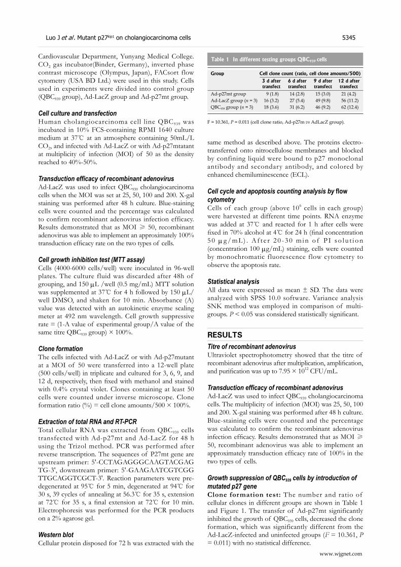
same method as described above. The proteins electro-transferred onto nitrocellulose membranes and blocked by confining liquid were bound to p27 monoclonal antibody and secondary antibody, and colored by enhanced chemiluminescence (ECL).
Cell cycle and apoptosis counting analysis by flow cytometryCells of each group (above 106 cells in each group) were harvested at different time points. RNA enzyme was added at 37 and reacted for 1 h after cells were fixed in 70% alcohol at 4 for 24 h (final concentration 5 0 μg / m L ) . A f t e r 2 0 - 3 0 m i n o f P I s o l u t i o n (concentration 100 μg/mL) staining, cells were counted by monochromatic fluorescence flow cytometry to observe the apoptosis rate.
Statistical analysisAll data were expressed as mean ± SD. The data were analyzed with SPSS 10.0 software. Variance analysis SNK method was employed in comparison of multi-groups. P < 0.05 was considered statistically significant.
RESULTSTitre of recombinant adenovirusUltraviolet spectrophotometry showed that the titre of recombinant adenovirus after multiplication, amplification, and purification was up to 7.95 × 10¹² CFU/mL.
Transduction efficacy of recombinant adenovirusAd-LacZ was used to infect QBC939 cholangiocarcinoma cells. The multiplicity of infection (MOI) was 25, 50, 100 and 200. X-gal staining was performed after 48 h culture. Blue-staining cells were counted and the percentage was calculated to confirm the recombinant adenovirus infection efficacy. Results demonstrated that as MOI ≥ 50, recombinant adenovirus was able to implement an approximately transduction efficacy rate of 100% in the two types of cells.
Growth suppression of QBC939 cells by introduction of mutated p27 geneClone formation test: The number and ratio of cellular clones in different groups are shown in Table 1 and Figure 1. The transfer of Ad-p27mt significantly inhibited the growth of QBC939 cells, decreased the clone formation, which was significantly different from the Ad-LacZ-infected and uninfected groups (F = 10.361, P = 0.011) with no statistical difference.
www.wjgnet.com
Luo J et al . Mutant p27kip1 on cholangiocarcinoma cells 5345
Cardiovascular Department, Yunyang Medical College. CO2 gas incubator(Binder, Germany), inverted phase contrast microscope (Olympus, Japan), FACsort flow cytometry (USA BD Ltd.) were used in this study. Cells used in experiments were divided into control group (QBC939 group), Ad-LacZ group and Ad-p27mt group.
Cell culture and transfectionHuman cholangiocarcinoma cell l ine QBC939 was incubated in 10% FCS-containing RPMI 1640 culture medium at 37 at an atmosphere containing 50mL/L CO2, and infected with Ad-LacZ or with Ad-p27mtatant at multiplicity of infection (MOI) of 50 as the density reached to 40%-50%.
Transduction efficacy of recombinant adenovirusAd-LacZ was used to infect QBC939 cholangiocarcinoma cells when the MOI was set at 25, 50, 100 and 200. X-gal staining was performed after 48 h culture. Blue-staining cells were counted and the percentage was calculated to confirm recombinant adenovirus infection efficacy. Results demonstrated that as MOI ≥ 50, recombinant adenovirus was able to implement an approximately 100% transduction efficacy rate on the two types of cells.
Cell growth inhibition test (MTT assay) Cells (4000-6000 cells/well) were inoculated in 96-well plates. The culture fluid was discarded after 48h of grouping, and 150 μL /well (0.5 mg/mL) MTT solution was supplemented at 37 for 4 h followed by 150 μL/well DMSO, and shaken for 10 min. Absorbance (A) value was detected with an autokinetic enzyme scaling meter at 492 nm wavelength. Cell growth suppressive rate = (1-A value of experimental group/A value of the same titre QBC939 group) × 100%.
Clone formationThe cells infected with Ad-LacZ or with Ad-p27mutant at a MOI of 50 were transferred into a 12-well plate (500 cells/well) in triplicate and cultured for 3, 6, 9, and 12 d, respectively, then fixed with methanol and stained with 0.4% crystal violet. Clones containing at least 50 cells were counted under inverse microscope. Clone formation ratio (%) = cell clone amounts/500 × 100%.
Extraction of total RNA and RT-PCRTotal cellular RNA was extracted from QBC939 cells transfected with Ad-p27mt and Ad-LacZ for 48 h using the Trizol method. PCR was performed after reverse transcription. The sequences of P27mt gene are upstream primer: 5'-CCTAGAGGGCAAGTACGAGTG-3', downstream primer: 5'-GAAGAATCGTCGGTTGCAGGTCGCT-3'. Reaction parameters were pre-degenerated at 95 for 5 min, degenerated at 94 for 30 s, 39 cycles of annealing at 56.3 for 35 s, extension at 72 for 35 s, a final extension at 72 for 10 min. Electrophoresis was performed for the PCR products on a 2% agarose gel.
Western blotCellular protein disposed for 72 h was extracted with the
Table 1 In different testing groups QBC939 cells
Group Cell clone count (ratio, cell clone amounts/500)
3 d after transfect
6 d after transfect
9 d after transfect
12 d after transfect
Ad-p27mt group 9 (1.8) 14 (2.8) 15 (3.0) 21 (4.2)Ad-LacZ group (n = 3) 16 (3.2) 27 (5.4) 49 (9.8) 56 (11.2)QBC939 group (n = 3) 18 (3.6) 31 (6.2) 46 (9.2) 62 (12.4)
F = 10.361, P = 0.011 (cell clone ratio, Ad-p27m vs AdLacZ group).
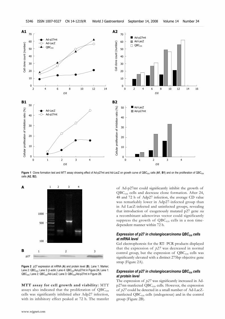
MTT assay for cell growth and viability: MTT assays also indicated that the proliferation of QBC939 cells was significantly inhibited after Adp27 infection, with its inhibitory effect peaked at 72 h. The transfer
of Ad-p27mt could significantly inhibit the growth of QBC939 cells and decrease clone formation. After 24, 48 and 72 h of Adp27 infection, the average CD value was remarkably lower in Adp27-infected group than in Ad LacZ-infected and uninfected groups, revealing that introduction of exogenously mutated p27 gene via a recombinant adenovirus vector could significantly suppress the growth of QBC939 cells in a non time-dependent manner within 72 h.
Expression of p27 in cholangiocarcinoma QBC939 cells at mRNA level Gel electrophoresis for the RT- PCR products displayed that the expression of p27 was decreased in normal control group, but the expression of QBC939 cells was significantly elevated with a distinct 275bp objective gene strap (Figure 2A).
Expression of p27 in cholangiocarcinoma QBC939 cells at protein level The expression of p27 was significantly increased in Ad-p27mt-tranfected QBC939 cells. However, the expression of p27 could be detected in a small number of Ad-LacZ-tranfected QBC939 cells (endogenous) and in the control group (Figure 2B).
Figure 1 Clone formation test and MTT assay showing effect of Ad-p27mt and Ad-LacZ on growth curve of QBC939 cells (A1, B1) and on the proliferation of QBC939 cells (A2, B2).
2 4 6 8 10 12 14
t /d
70
60
50
40
30
20
10
0
Cell
clon
e co
unt
(num
ber)
Ad-p27mtAd-LacZQBC939
0 2 4 6 8 10 12 14 16
t /d
70
60
50
40
30
20
10
0
Cell
clon
e co
unt
(num
ber)
Ad-p27mtAd-LacZQBC939
A1
B1
0 1 2 3 4
t /d
50
40
30
20
10
0Cellu
lar
prol
ifera
tion
of in
hibi
tion
ratio
(%
)
Ad-p27mtAd-LacZ
0 1 2 3 4
t /d
50
40
30
20
10
0
Cellu
lar
prol
ifera
tion
of in
hibi
tion
ratio
(%
)
Ad-p27mtAd-LacZ
A2
B2
www.wjgnet.com
5346 ISSN 1007-9327 CN 14-1219/R World J Gastroenterol September 14, 2008 Volume 14 Number 34
B
1 2 3 4
1000
500
100
1 2 3
p27
Figure 2 p27 expression at mRNA (A) and protein level (B). Lane 1: Marker; Lane 2: QBC939; Lane 3: b-actin; Lane 4: QBC939/Ad-p27mt in Figure 2A; Lane 1: QBC939; Lane 2: QBC939/Ad-LacZ; Lane 3: QBC939/Ad-p27mt in Figure 2B.
A
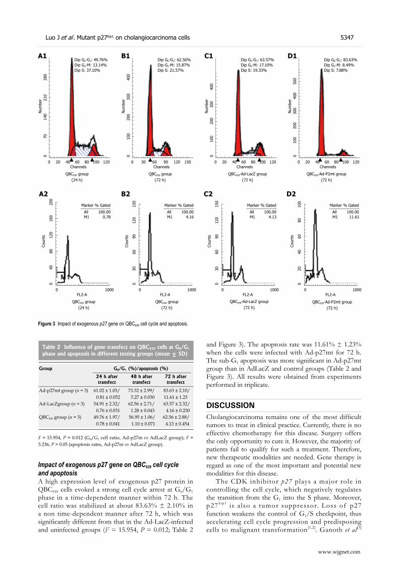
Impact of exogenous p27 gene on QBC939 cell cycle and apoptosisA high expression level of exogenous p27 protein in QBC939 cells evoked a strong cell cycle arrest at G0/G1 phase in a time-dependent manner within 72 h. The cell ratio was stabilized at about 83.63% ± 2.10% in a non time-dependent manner after 72 h, which was significantly different from that in the Ad-LacZ-infected and uninfected groups (F = 15.954, P = 0.012; Table 2
and Figure 3). The apoptosis rate was 11.61% ± 1.23% when the cells were infected with Ad-p27mt for 72 h. The sub-G1 apoptosis was more significant in Ad-p27mt group than in AdLacZ and control groups (Table 2 and Figure 3). All results were obtained from experiments performed in triplicate.
DISCUSSION Cholangiocarcinoma remains one of the most difficult tumors to treat in clinical practice. Currently, there is no effective chemotherapy for this disease. Surgery offers the only opportunity to cure it. However, the majority of patients fail to qualify for such a treatment. Therefore, new therapeutic modalities are needed. Gene therapy is regard as one of the most important and potential new modalities for this disease.
The CDK inhibitor p27 plays a major role in controlling the cell cycle, which negatively regulates the transition from the G1 into the S phase. Moreover, p27kip1 is a lso a tumor suppressor. Loss of p27 function weakens the control of G1/S checkpoint, thus accelerating cell cycle progression and predisposing cells to malignant transformation[1,2]. Ganoth et al[3]
Table 2 Influence of gene transfect on QBC939 cells at G0/G1 phase and apoptosis in different testing groups (mean ± SD)
Group G0/G1 (%)/apoptosis (%)
24 h after transfect
48 h after transfect
72 h after transfect
Ad-p27mt group (n = 3) 61.02 ± 1.03/ 0.81 ± 0.052
73.32 ± 2.99/ 5.27 ± 0.030
83.63 ± 2.10/ 11.61 ± 1.23
Ad-LacZgroup (n = 3) 54.91 ± 2.32/ 0.76 ± 0.031
62.56 ± 2.71/ 1.28 ± 0.043
63.57 ± 2.32/ 4.16 ± 0.230
QBC939 group (n = 3) 49.76 ± 1.97/ 0.78 ± 0.041
56.95 ± 1.06/ 1.10 ± 0.071
62.56 ± 2.88/ 4.13 ± 0.454
F = 15.954, P = 0.012 (G0/G1 cell ratio, Ad-p27m vs AdLacZ group); F = 3.236, P > 0.05 (apoptosis rates, Ad-p27m vs AdLacZ group).
Dip G0-G1: 49.76%
Num
ber
0 70
1
40 21
0 28
0
0 20 40 60 80 100 120Channels
QBC939 group (24 h)
Dip G2-M: 13.14%Dip S: 37.10%
A1Dip G0-G1: 62.56%
Num
ber
0 1
00 20
0 30
0
400
0 30 60 90 120 150Channels
QBC939 group (72 h)
Dip G2-M: 15.87%Dip S: 21.57%
B1Dip G0-G1: 63.57%
Num
ber
0 20 40 60 80 100 120Channels
QBC939-Ad-LacZ group (72 h)
Dip G2-M: 17.10%Dip S: 19.33%
C1
0 1
00 2
00 30
0
400
Marker % Gated
Coun
ts
0 4
0
80
120
1
60 2
00
0 1000FL2-A
QBC939 group (24 h)
All 100.00
A2
M1 0.78
Marker % Gated
Coun
ts
0 3
0
60
90 1
20 1
50
0 1000FL2-A
QBC939 group (72 h)
All 100.00
B2
M1 4.16
Marker % GatedCo
unts
0 3
0
60
90 1
20 1
50
0 1000FL2-A
All 100.00
C2
M1 4.13
QBC939-Ad-LacZ group (72 h)
Marker % Gated
Coun
ts
0 2
0
40
60 8
0
100
0 1000FL2-A
All 100.00
D2
M1 11.61
QBC939-Ad-P2mt group (72 h)
Figure 3 Impact of exogenous p27 gene on QBC939 cell cycle and apoptosis.
Dip G0-G1: 83.63%
Num
ber
0 10
0 2
00 3
00 4
00
500
0 20 40 60 80 100 120Channels
Dip G2-M: 8.49%Dip S: 7.88%
D1
QBC939-Ad-P2mt group (72 h)
www.wjgnet.com
Luo J et al . Mutant p27kip1 on cholangiocarcinoma cells 5347
and Troncone et al[4] reported that the degradation of p27 is mainly regulated by post-translational ubiquitin-proteasome-mediated proteolysis of phosphorylation in threonine (Thr) 187. In order to inhibit the degradation of p27 and restore the function of G1/S checkpoint, we transfected mutated p27 into cholangiocarcinoma QBC939 cell line, which has a mutation of Thr-187/Pro-188 (ACGCCC) to Met-187/Ile-188(ATGATC).
Western blot analysis showed that 72 h after infection with Ad-p27mt, p27 in the CBC939 cells expressed a strong band, whereas Ad-LacZ-infected QBC939 cells showed a faint p27 protein product in the uninfected groups, suggesting that tansgenes can be successfully induced and expressed. The elevated level of p27 expression demonstrated that mutated p27 was resistant to degradation and more stable than wild p27, indicating that phosphorylation of threonine (Thr) 187 can trigger degradation of wild p27. The transfer of Ad-p27mt significantly inhibited the proliferation of QBC939 cells, decreased clone formation, strongly induced cell cycle arrest and apoptosis at G1/S phase within 72 h after infection, which is consistent with the previous findings[5].
It has been well documented that over expression of wild p27 via adenoviral gene transfer on p27-deficient tumor cells could strongly inhibit cell cycle arrest and even lead to significantly apoptosis in disparate types of human cancers, such as spongiocytoma, lung cancer, leukaemia. It is a common phenomenon that recombinant adenovirus-mediated p27 can eliminate carcinoma cells through apoptosis. Although there was no significant difference in apoptosis between Adp27mt- and AdLacZ-infected cells, uninfected cells at any time point, our data show that 72 h after infection with Adp27mt, the typical sub-G1 apoptotic peak could be observed by flow cytometry, which was more apparent than in AdLacZ-infected and uninfected cells. Although the precise mechanism by which p27 induces apoptosis is unclear, transfer of p27 is associated with a moderate level of apoptosis as shown by FACS analysis. Since QBC939 cells have mutated p53, the mechanism underlying apoptosis induced by transfer of p27 must be p53-independent. Further investigation is needed on how p27 regulates and induces apoptosis.
In conclusion, Ad-p27mt at Thr-187 can be used as a novel, potent, tumor-suppressing gene therapy tool in the treatment of cholangiocarcinoma.
ACKNOWLEDGMENTSThe authors thank Doctors Chen YJ and Wang WY for their long, concerted, and fruitful efforts, Professor Wang SG for kindly donation of human cholangiocarcinoma cell line QBC939, Professors Xu SY and Wang JN for their recombinant adenovirus vehicle Ad-p27mt and adenovirus control vehicle Ad-LacZ, Professor Zou SQ for his useful suggestions, and a great debt of gratitude to its organizer, Professor Zou SQ.
COMMENTSBackgroundAs a cyclin-dependent kinase inhibitor, p27Kip1 (p27) regulates cell cycle progression by transcriptional, translational and proteolytic mechanisms. G1/S cell cycle progression requires p27 proteolysis, which is triggered by its phosphorylation of threonine (Thr) 187. Increased p27 causes proliferating cells to exit from the cell cycle, while decreased p27 is required for quiescent cells to resume cell division. Low levels of p27 are associated with excessive cell proliferation in pathological conditions such as inflammation and cancers. High levels of p27 are observed in such conditions of diminished cell proliferation as in late stages of arterial wound repair in atherosclerosis. Interestingly, in many types of tumors such as gastric, prostate and breast carcinomas, the expression of p27 gene is down-regulated. Loss of p27 expression may result in tumor development and/or progression.Research frontiersThe research involved cell morphologyoncology, cell morphology, molecular biology and gene therapy for cholangiocarcinoma. Innovations and breakthroughsIt is well know that the degradation of p27 is mainly regulated by post-translational ubiquitin-proteasome-mediated proteolysis during phosphorylation of threonine (Thr) 187. In order to inhibit the degradation of p27 and restore the function of G1/S checkpoint, we transfected mutated p27 into cholangiocarcinoma QBC939 cell line, which can mutate from Thr-187/Pro-188 (ACGCCC) to Met-187/Ile-188(ATGATC). The recombinant adenoviral vector cannot replicate in target cells because it lacks the E1 gene, thus only expressing the inserted gene. Because the foreign gene fragment is not incorporated into the genome of target cells, the danger of mutations affecting treatment is reduced. Meanwhile, adenoviral vectors are stable and easy to purify. This technique can effectively affect both proliferating and quiescent cells ex vivo. The potential for gene therapy by using the recombinant adenovirus is worthy of extensive attention. The results of our study suggest that adenovirus-mediated p27 gene transfection can be used as a novel gene therapy for cholangiocarcinoma.Applications The prognosis of cholangiocarcinoma is extremely poor although aggressive multidisciplinary cancer therapies have been used in clinical practice. Thus, it is imperative to develop new and effective treatment modalities for cholangiocarcinoma, such as gene therapy.Peer reviewThe authors showed that transfection of a human cholangiocarcinoma cell line (QBC939) could cause cell cycle arrest and apoptosis, which are of interest in developing new treatment modalities for cholangiocarcinoma. The methods used were well described. The results are of scientific interest.
REFERENCES1 Milde-Langosch K, Hagen M, Bamberger AM, Loning T.
Expression and prognostic value of the cell-cycle regulatory proteins, Rb, p16MTS1, p21WAF1, p27KIP1, cyclin E, and cyclin D2, in ovarian cancer. Int J Gynecol Pathol 2003; 22: 168-174
2 Fukunaga M. Immunohistochemical characterization of cyclin E and p27KIP1 expression in early hydatidiform moles. Int J Gynecol Pathol 2004; 23: 259-264
3 Ganoth D, Bornstein G, Ko TK, Larsen B, Tyers M, Pagano M, Hershko A. The cell-cycle regulatory protein Cks1 is required for SCF(Skp2)-mediated ubiquitinylation of p27. Nat Cell Biol 2001; 3: 321-324
4 Troncone G, Martinez JC, Iaccarino A, Zeppa P, Caleo A, Russo M, Migliaccio I, Motti ML, Califano D, Palmieri EA, Palombini L. p27Kip1 is expressed in proliferating cells in its form phosphorylated on threonine 187. BMC Clin Pathol 2005; 5: 3
5 Katner AL, Gootam P, Hoang QB, Gnarra JR, Rayford W. A recombinant adenovirus expressing p7(Kip1) induces cell cycle arrest and apoptosis in human 786-0 renal carcinoma cells. J Urol 2002; 168: 766-773
S- Editor Zhong XY L- Editor Wang XL E- Editor Lin YP
www.wjgnet.com
5348 ISSN 1007-9327 CN 14-1219/R World J Gastroenterol September 14, 2008 Volume 14 Number 34
Online Submissions: wjg.wjgnet.com World J Gastroenterol 2008 September 14; 14(34): [email protected] World Journal of Gastroenterology ISSN 1007-9327doi:10.3748/wjg.14.5349 © 2008 The WJG Press. All rights reserved.
A “false positive” octreoscan in ileal Crohn’s disease
Alberto Fernandez, Olga Tabuenca, Angeles Peteiro
CASE REPORT
Alberto Fernandez, Gastroenterology Department, POVISA Hospital, Vigo 36211, SpainOlga Tabuenca, Nuclear Medicine Department, POVISA Hospital, Vigo 36211, SpainAngeles Peteiro, Pathology Department, POVISA Hospital, Vigo 36211, SpainAuthor contributions: Fernandez A, Tabuenca O and Peteiro A contributed equally to this work and wrote the paper.Correspondence to: Alberto Fernandez, Servicio de Aparato Digestivo, Hospital POVISA, c/ Salamanca 5, Vigo (Pontevedra) 36211. Spain. [email protected]: +34-98-6413144 Fax: +34-98-6421439Received: May 24, 2008 Revised: July 28, 2008Accepted: August 4, 2008Published online: September 14, 2008
AbstractWe present a case report of a patient with a suspicious ileal carcinoid tumour. Clinical examination as well as computer tomography (CT) scan suggested a tumour. Octeotride scan showed uptake in the same bowel loop reported as pathological in CT. The patient underwent surgery and biopsy which reported Crohn’s disease (CD). The interest in the case is due to the fact that this is, to the best of our knowledge, the second report of Crohn’sdisease as a cause of false positive octeotride scan. Unfortunately, no somatostatin receptors could be found in the sample, so further studies should be performed.
© 2008 The WJG Press. All rights reserved.
Key words: Crohn’s disease; Carcinoid tumour; Octreoscan; Somatostatin receptor scintigraphy; 111In- DTPA- octreotido
Peer reviewer: Gert De Hertogh, Ph.D, Department of Mor-phology and Molecular Pathology, University Hospitals KU-Leuven, Minderbroedersstraat 12, Leuven 3000, Belgium
Fernandez A, Tabuenca O, Peteiro A. A “false positive” octreoscan in ileal Crohn’s disease. World J Gastroenterol 2008; 14(34): 5349-5352 Available from: URL: http://www.wjgnet.com/1007-9327/14/5349.asp DOI: http://dx.doi.org/10.3748/wjg.14.5349
INTRODUCTIONSomatostat in receptors (SS-Rs) are membrane
glycoproteins spreading over a large number of body tissues and can be found in normal and pathological conditions[1]. Many benign and malignant tumours over express SS-R in their membranes especially in neuroendocrine tumours (NETs) but there are other benign conditions showing an increase number of SS-Rs, such as granulomatous or inflammatory disease[2].
Octeotride is an analogue whose molecule is a shortened version of somatostatin’s, from 8 to 14 aminoacides, sharing its active nuclei and allowing the 111Indium-DPTA molecule to bind to the N-terminal (d-phe).
Thus, 111In- DTPA-D- Phe1- octreótido (111In- DTPAOC) is the labelled form of octreotide. There are 5 subtypes of SS-R, and somatostatin shows a high affinity for all of them. However, octeotride has a high affinity for only SS-R 2 subtype and a low affinity for SS-R 3 and SS-R 5 subtypes. Therefore, pathological conditions over expressing these receptors are able to be imaged in 111In- DTPAOC scans.
The sensitivity of 111In- DTPAOC in detecting these pathological conditions is variable being very high in carcinoid tumour (86%)[3]. Uptake in other tissues can lead to false-positive findings when studying suspected NETs. Only one case of Crohn’s disease (CD) has been recently reported as a cause of false positive scan[4].
We report a case of a false positive 111In- DTPAOC scintigraphy in a patient with CD mimicking an ileal carcinoid tumour. A discussion about SS-R potential alterations in CD was included.
CASE REPORTA 40-year-old man was admitted to our outpatient clinic with an 8-mo history of dull abdominal pain and weight loss of 20 kg, but no other gastrointestinal symptoms. In terms of past medical or surgery history, the patient denied of smoking or other diseases. Physical examination only revealed a painful site in the right lower quadrant of the abdomen and a body temperature of 37.3. Laboratory test only revealed an increased erythrocyte sedimentation rate of 35 mm/h (normal range: 0- 25 mm/h) and C-reactive protein rate of 2.37 mg/dL (normal range: 0-0.5 mg/dL). A computer tomography (CT) scan was performed (Figure 1) and showed thickening of the ileum distal wall and the presence of a 4 cm solid mass with irregular border adjacent to the affected ileum, conditioning retraction, with prominent mesenteric lymph nodes. All these findings suggested a carcinoid tumour.
www.wjgnet.com
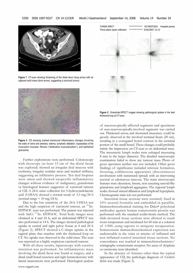
Further explorations were performed. Colonoscopy with ileoscopy (at least 15 cm of the distal ileum was explored) showed an irregular ileal mucosa with erythema, irregular nodular areas and marked stiffness, suggesting an infiltrative process. Ten ileal biopsies were taken and showed unspecific inf lammatory changes without evidence of malignancy, granulomas or histological features suggestive of carcinoid tumour or CD. A 24-h urine collection for 5-hydroxyindolacetic acid (5-HIAA) showed a normal result of 3.3 mg/24 h (normal range < 10 mg/24 h).
Due to the low sensitivity of the 24-h 5-HIAA test and the high suspicion of carcinoid tumour, an 111In- DTPAOC scan was performed. The patient was injected with 3mCi 111In- DTPAOC. Total body images were obtained at 4 and 24 h, and an abdominal SPECT was also performed at 14 h. The images showed pathological uptake in central and right pelvic fossae at 4 and 24 h (Figure 2). SPECT showed a C-shape uptake in the sagittal plane that matches with the thickened loop on CT. No uptake was observed in the solid mass. The scan was reported as a highly suspicious carcinoid tumour.
With all these results, laparoscopy with curative intention was performed. An inf lammatory mass involving the distal ileon was found at surgery, and a distal small bowel resection and right hemicolectomy with lateral anastomosis were performed. Histological analysis
of macroscopically-affected segments and specimens of non-macroscopically-involved segments was carried out. Thickened serosa and shortened mesentery could be grossly observed in the involved terminal ileum (20 cm), resulting in a corrugated bowel contour in the terminal portion of the small bowel. These changes could probably mimic the impression on CT-scan to an abdominal mass. The mesenteric lymph nodes were enlarged measuring 8 mm in the larger diameter. The detailed macroscopic examination failed to show any tumour mass. Photo of gross specimen neither was nor included. Other gross findings of significance included stricture formation, fissuring, cobblestone appearance (discontinuous involvement with transmural spread) with an intervening normal or edematous mucosa. The main microscopic features were ulceration, fissure, non caseating sarcoid-like granuloma and lymphoid aggregates. The regional lymph nodes showed sinusal dilatation and lymphoid hyperplasia. Chromogranin stain was not performed.
Intestinal tissue sections were routinely fixed in 10% neutral formalin and embedded in paraffin. Immunohistochemical staining for Dako® polyclonal antibodies against human somatostatin protein was performed with the standard avidin-biotin method. The slide-mounted tissue sections were allowed to reach room temperature and incubated for 60 min at a solution of 1:900, using saponic to antigenic recuperation. Somatostatin immunohistochemical expression was undetectable in the veins or arteries of inflamed and non inflamed control intestinal tissue sections. No concordance was reached in immunohistochemistry/scintigraphy somatostatin receptor. No areas of dysplasia or carcinoma were identified.
In the absence of features other than the typical appearance of CD, the pathologic diagnosis of Crohn’s ileitis was made (Figure 3).
Figure 1 CT-scan showing thickening of the distal ileum (long arrow) with an adjacent solid mass (short arrow), suggesting a carcinoid tumour.
Figure 2 Octeotride-SPECT images showing pathological uptake in the ileal thickened loop at CT-scan.
TUMOR SPECT ************* OCTREOTIDO Hospital povisaThree plane spots collection ************* 5/30/2007 12:13
Figure 3 CD showing marked transmural inflammatory changes (involving the walls of veins and arteries): edema, lymphatic dilatation, hyperplasia of the muscularis mucosae, fibrosis (“obliterative muscularization”), and epithelioid granuloma.
www.wjgnet.com
5350 ISSN 1007-9327 CN 14-1219/R World J Gastroenterol September 14, 2008 Volume 14 Number 34
After surgery, azathioprine was started for prevention of postoperative recurrence. The patient underwent a complete clinical recovery and 6 mo later he was asymptomatic. A surveillance/follow-up colonoscopy did not show any abnormality and inflammatory signs of recurrence.
DISCUSSIONThere are many cases of carcinoid tumour misdiagnosed as CD only discovered when treatment is not effective or surgery is performed. It was reported that approximately 2.3% of patients with ileal carcinoid are first diagnosed and treated as CD[5]. However, the reverse is a very uncommon situation.
In our case, the clinic features as well as the image of CT-scan strongly suggested a carcinoid tumour. In order to confirm the suspected disease, 111In- DTPAOC scan was performed and showed a C-shape uptake in the bowel loop that corresponded to that reported as pathologic in CT, but there was no uptake in the solid mass. The patient underwent surgery and the final pathologic report was CD. The mass that did not show uptake was reported as inflammatory.
111In- DTPAOC is a radio-labelled octreotide analogue that binds to SS-R expressed in cell membranes. Many benign and malignant diseases overexpress SS-R and thus, can be imaged with this radioligand[6]. Carcinoid tumour, one of the malignant tumours, shows more uptakes in the bowel loop due to its high density of SS-R. Reported values for the detection of known carcinoid tumour localizations vary from 80% to nearly 100%[1]. Uptake is related to SS-R density and even tumours smaller than 1 cm in diameter can be detected. This is the reason why somatostatin receptor scintigraphy (SRS) plays a central role in locating and assessing the primary gastroenteropancreatic NET[7] and has a marked effect in the clinical management of these malignancies[8,9]. However, 12% of SRS examinations result in false-positive localization of NET, understanding a false-positive as an uptake not related with the tested pathology. Renal parapelvic cysts, accessory spleens, ventral hernias, thyroid or breast disease are the most frequent cases of false positive localizations[10]. SS-R is over expressed in activated peripheric lymphocytes and macrophages in granulomatous and inflammatory diseases, allowing obtaining images[11,12].
I n n o r m a l b o w e l , S S - R i s e x p r e s s e d i n gastrointestinal mucosa, peripheral nervous system and lymphoid tissue. Several tumours, such as carcinoid tumour, express SS-R in peritumoral veins[13]. In intestinal inflammatory disease, a high density of SS-R is detected by autoradiography in intestinal intramural veins but not in normal tissues. SS-R is seen in small muscular veins of submucosa, tunica muscularis and subserosa, and the thickness of veins is labelled. These veins are histologically normal and seldom have a minimal lymphocyte infiltration although label intensity does not seem to depend on them as the surrounding
tissues are infiltrated with these cells and do not show labelling[14-16].
Usually no 111In- DTPAOC uptake is found in inflammatory bowel disease. There is only one recent report of 111In- DTPAOC uptake in Crohn’s disease in literature although there is no evidence that SS-R is determined[4]. In our case, the samples were studied to assess the existence of SS-R. Immunohistochemical staining of somatostatin receptors was performed and somatostatin immunohistochemical expression was undetectable in the veins or arteries of inflamed and non inflamed control intestines.
Unfortunately, SS-R could not be found in the tissues studied, so we could not explain this abnormal uptake. No concordance was reached in immunohistochemistry/scintigraphy somatostatin receptor, possibly as a consequence of somatostatin receptor heterogeneity. Further studies should be made to assess the possible causes of uptake in CD.
However, there are severa l repor ts of smal l carcinoid tumours (a few millimetres in diameter ) found in patients with CD, suggesting a correlation between the pathogenesis of both disorders[17]. It has been theorized that inflammation creates a favourable environment for the development of carcinoid tumour, although most of the carcinoid tumours reported are found in non-inflamed bowel. Other theories include the hyperstimulation of enteroendocrine cells by inflammation[18] and the role of proinflammatory cytokines such as PTHrP or IL-6[19].
We would like to encourage physicians to be aware of CD as a cause of false positive results when performing 111In- DTPAOC scans.
REFERENCES1 Kwekkeboom D, Krenning EP, de Jong M. Peptide receptor
imaging and therapy. J Nucl Med 2000; 41: 1704-17132 van Hagen PM. Somatostatin receptor expression in clinical
immunology. Metabolism 1996; 45: 86-873 Kwekkeboom DJ, Krenning EP. Somatostatin receptor
imaging. Semin Nucl Med 2002; 32: 84-914 Marko J, Lamba R, Miller F, Buchman A, Spies S, Nikolaidis
P. OctreoScan positive Crohn's disease mimicking an ileal carcinoid tumor. J Clin Gastroenterol 2008; 42: 66-68
5 Hsu EY, Feldman JM, Lichtenstein GR. Ileal carcinoid tumors stimulating Crohn's disease: incidence among 176 consecutive cases of ileal carcinoid. Am J Gastroenterol 1997; 92: 2062-2065
6 Warner RR, O'dorisio TM. Radiolabeled peptides in diagnosis and tumor imaging: clinical overview. Semin Nucl Med 2002; 32: 79-83
7 Ramage JK , Davies AH, Ardill J, Bax N, Caplin M, Grossman A, Hawkins R, McNicol AM, Reed N, Sutton R, Thakker R, Aylwin S, Breen D, Britton K, Buchanan K, Corrie P, Gillams A, Lewington V, McCance D, Meeran K, Watkinson A. Guidelines for the management of gastroenteropancreatic neuroendocrine (including carcinoid) tumours. Gut 2005; 54 Suppl 4: iv1-iv16
8 Lebtahi R, Cadiot G, Sarda L, Daou D, Faraggi M, Petegnief Y, Mignon M, le Guludec D. Clinical impact of somatostatin receptor scintigraphy in the management of patients with neuroendocrine gastroenteropancreatic tumors. J Nucl Med 1997; 38: 853-858
9 Termanini B, Gibril F, Reynolds JC, Doppman JL, Chen CC,
www.wjgnet.com
Fernandez A et al . False positive octreoscan 5351
Stewart CA, Sutliff VE, Jensen RT. Value of somatostatin receptor scintigraphy: a prospective study in gastrinoma of its effect on clinical management. Gastroenterology 1997; 112: 335-347
10 Gibril F, Reynolds JC, Chen CC, Yu F, Goebel SU, Serrano J, Doppman JL, Jensen RT. Specificity of somatostatin receptor scintigraphy: a prospective study and effects of false-positive localizations on management in patients with gastrinomas. J Nucl Med 1999; 40: 539-553
11 Vanhagen PM, Krenning EP, Reubi JC, Kwekkeboom DJ, Bakker WH, Mulder AH, Laissue I, Hoogstede HC, Lamberts SW. Somatostatin analogue scintigraphy in granulomatous diseases. Eur J Nucl Med 1994; 21: 497-502
12 Tabuenca Dopico O, Gutierrez Mendiguchia C, Rego Iraeta A. [Incidental diagnosis of lymph node tuberculosis in an 111-indium-octreotide scintigraphy during a study of acromegaly] Rev Esp Med Nucl 2006; 25: 193-197
13 Reubi JC, Horisberger U, Laissue J. High density of somatostatin receptors in veins surrounding human cancer tissue: role in tumor-host interaction? Int J Cancer 1994; 56: 681-688
14 Reubi JC, Mazzucchelli L, Laissue JA. Intestinal vessels express a high density of somatostatin receptors in human
inflammatory bowel disease. Gastroenterology 1994; 106: 951-959
15 Denzler B, Reubi JC. Expression of somatostatin receptors in peritumoral veins of human tumors. Cancer 1999; 85: 188-198
16 Reubi JC, Waser B, Schaer JC, Laissue JA. Somatostatin receptor sst1-sst5 expression in normal and neoplastic human tissues using receptor autoradiography with subtype-selective ligands. Eur J Nucl Med 2001; 28 : 836-846
17 West NE, Wise PE, Herline AJ, Muldoon RL, Chopp WV, Schwartz DA. Carcinoid tumors are 15 times more common in patients with Crohn's disease. Inflamm Bowel Dis 2007; 13: 1129-1134
18 Le Marc'hadour F, Bost F, Peoc'h M, Roux JJ, Pasquier D, Pasquier B. Carcinoid tumour complicating inflammatory bowel disease. A study of two cases with review of the literature. Pathol Res Pract 1994; 190: 1185-1192; discussion 1193-1200
19 Barhoum M , Hutchins L, Fonseca VA. Intractable hypercalcemia due to a metastatic carcinoid secreting parathyroid hormone-related peptide and interleukin-6: response to octreotide. Am J Med Sci 1999; 318: 203-205
S- Editor Li DL L- Editor Wang XL E- Editor Zhang WB
www.wjgnet.com
5352 ISSN 1007-9327 CN 14-1219/R World J Gastroenterol September 14, 2008 Volume 14 Number 34
Online Submissions: wjg.wjgnet.com World J Gastroenterol 2008 September 14; 14(34): [email protected] World Journal of Gastroenterology ISSN 1007-9327doi:10.3748/wjg.14.5353 © 2008 The WJG Press. All rights reserved.
Inflammatory myoglandular polyp causing hematochezia
Shoji Hirasaki, Masato Okuda, Kenichiro Kudo, Seiyuu Suzuki, Atsuko Shirakawa
CASE REPORT
Shoji Hirasaki, Masato Okuda, Kenichiro Kudo, Seiyuu Suzuki, Department of Internal Medicine, Sumitomo Besshi Hospital, Niihama 7928543, JapanAtsuko Shirakawa, Department of Pathology, Sumitomo Besshi Hospital, Niihama 7928543, JapanAuthor contributions: Hirasaki S, Okuda M, Kudo K and Suzuki S were involved in the care of the patient; Shirakawa A studied the specimen; Hirasaki S performed the endoscopic resection; Hirasaki S wrote the paper.Correspondence to: Shoji Hirasaki, Department of Internal Medicine, Sumitomo Besshi Hospital, 3-1 Ohji-cho, Niihama 7928543, Japan. [email protected] Telephone: +81-897-377111 Fax: +81-897-377121 Received: April 18, 2008 Revised: August 11, 2008Accepted: August 18, 2008Published online: September 14, 2008
AbstractA case of inflammatory myoglandular polyp (IMGP) causing hematochezia is reported. The patient was a 33-year-old man who visited our hospital for further evaluation of hematochezia. Colonoscopy revealed a red, hard, spherical peduncular polyp with erosion and mucous exudation, about 20 mm in diameter, in the descending colon. Excluding the polyp, there was no lesion in the colorectum. Endoscopic polypectomy was performed. Histological examination of the specimen revealed inflammatory granulation tissue in the lamina propria, proliferation of smooth muscle, and hyperplastic glands with variable cystic changes. This polyp was diagnosed as an IMGP. The symptom of hematochezia was resolved after endoscopic resection. Our case shows that treatment is necessary for IMGP if intestinal bleeding occurs and endoscopists should be aware of the endoscopic characteristics of IMGP.
© 2008 The WJG Press. All rights reserved.
Key words: Inf lammatory myoglandular polyp; Colonoscopy; Endoscopic polypectomy; Gastrointestinal bleeding
P e e r re v i e w e r : D r. J a m e s E E a s t , D e p a r t m e n t o f Gastroenterology, St. Mary’s Hospital, Clarence Wing, Praed Street, London W2 1NY, United Kingdom
Hirasaki S, Okuda M, Kudo K, Suzuki S, Shirakawa A. Inflammatory myoglandular polyp causing hematochezia. World J Gastroenterol 2008; 14(34): 5353-5355 Available from: URL:
http://www.wjgnet.com/1007-9327/14/5353.asp DOI: http://dx.doi.org/10.3748/wjg.14.5353
INTRODUCTIONIn f l ammator y myog landu la r po lyp ( IMGP) i s characterized by inflammatory granulation tissue in the lamina propria[1], proliferation of smooth muscle[2], and hyperplastic glands with variable cystic changes[3-7]. Only a small number of cases have been reported and the pathogenesis and natural history remain unclear[8-12]. Herein, we describe a relatively rare case of inflammatory myoglandular polyp causing hematochezia.
CASE REPORTA 33-year-old man presented with the symptom of hematochezia. He was in good health with no specific family or past medical history. His body temperature was 36.7, blood pressure was 148/82 mmHg, and radial pulse rate was 70 beats/min and regular. He had neither anemia nor jaundice. Neurological examination revealed no abnormal findings. Abdominal palpation revealed tenderness in the left lower quadrant. Routine hematological examination and biochemical tests were within normal limits. Colonoscopy revealed a red, hard, spherical peduncular polyp with erosion and mucous exudation, about 20 mm in diameter, in the descending colon (Figure 1). With conventional colonoscopy, the lesion did not show type Ⅲ or Ⅳ pit pattern although magnifying colonoscopy was not performed. We speculated that this polyp was non-neoplastic. It was suspected to be an inflammatory polyp from endoscopic findings although it should be distinguished from a juvenile polyp. An air contrast barium enema also revealed a pedunculated polyp in the descending colon (Figure 2). Excluding the polyp, there was no lesion in the colorectum. We speculated that the polyp in the descending colon was the causative lesion of hematochezia. Endoscopic polypectomy was performed. At polypectomy, polyp erosion was healed (Figure 3). Histological examination of the specimen revealed inflammatory granulation tissue in the lamina propria, proliferation of smooth muscle, and hyperplastic glands with variable cystic changes (Figure 4). The lesion was diagnosed as an IMGP. After endoscopic polypectomy, the symptom of hematochezia was resolved.
www.wjgnet.com
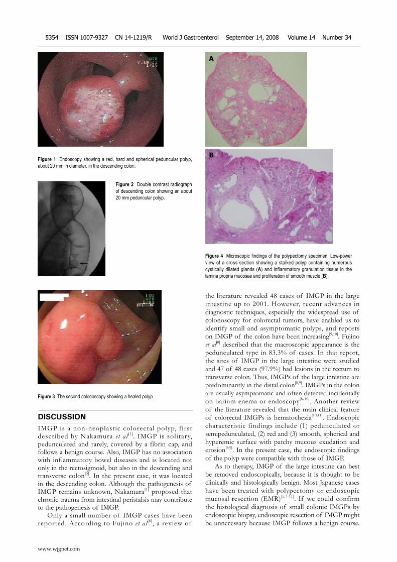
DISCUSSIONIMGP is a non-neoplastic colorectal polyp, first described by Nakamura et al [1]. IMGP is solitary, pedunculated and rarely, covered by a fibrin cap, and follows a benign course. Also, IMGP has no association with inflammatory bowel diseases and is located not only in the rectosigmoid, but also in the descending and transverse colon[3]. In the present case, it was located in the descending colon. Although the pathogenesis of IMGP remains unknown, Nakamura[1] proposed that chronic trauma from intestinal peristalsis may contribute to the pathogenesis of IMGP.
Only a small number of IMGP cases have been reported. According to Fujino et al [8], a review of
the literature revealed 48 cases of IMGP in the large intestine up to 2001. However, recent advances in diagnostic techniques, especially the widespread use of colonoscopy for colorectal tumors, have enabled us to identify small and asymptomatic polyps, and reports on IMGP of the colon have been increasing[9,10]. Fujino et al[8] described that the macroscopic appearance is the pedunculated type in 83.3% of cases. In that report, the sites of IMGP in the large intestine were studied and 47 of 48 cases (97.9%) had lesions in the rectum to transverse colon. Thus, IMGPs of the large intestine are predominantly in the distal colon[8,9]. IMGPs in the colon are usually asymptomatic and often detected incidentally on barium enema or endoscopy[8-10]. Another review of the literature revealed that the main clinical feature of colorectal IMGPs is hematochezia[10,11]. Endoscopic characteristic findings include (1) pedunculated or semipedunculated, (2) red and (3) smooth, spherical and hyperemic surface with patchy mucous exudation and erosion[8,9]. In the present case, the endoscopic findings of the polyp were compatible with those of IMGP.
As to therapy, IMGP of the large intestine can best be removed endoscopically, because it is thought to be clinically and histologically benign. Most Japanese cases have been treated with polypectomy or endoscopic mucosal resection (EMR)[5,7-11]. If we could confirm the histological diagnosis of small colonic IMGPs by endoscopic biopsy, endoscopic resection of IMGP might be unnecessary because IMGP follows a benign course.
Figure 1 Endoscopy showing a red, hard and spherical peduncular polyp, about 20 mm in diameter, in the descending colon.
Figure 2 Double contrast radiograph of descending colon showing an about 20 mm peduncular polyp.
←
Figure 3 The second colonoscopy showing a healed polyp.
Figure 4 Microscopic findings of the polypectomy specimen. Low-power view of a cross section showing a stalked polyp containing numerous cystically dilated glands (A) and inflammatory granulation tissue in the lamina propria mucosae and proliferation of smooth muscle (B).
A
B
www.wjgnet.com
5354 ISSN 1007-9327 CN 14-1219/R World J Gastroenterol September 14, 2008 Volume 14 Number 34
However, the diagnosis of colorectal IMGPs could seldom be made by endoscopic biopsy and the final diagnosis of colonic IMGP depends on the pathological findings of EMR or endoscopic polypectomy specimens. Endoscopic or surgical treatment is necessary if gastrointestinal bleeding[9] or colonic intussusception occurs. Local excision of the polyp is curative. Kayhan et al[12] have reported a case of large IMGP (> 6 cm) that was too large to be removed endoscopically, and was thus treated with surgical resection. We consider that the percentage of patients with colonic IMGP who undergo surgical resection will decrease and endoscopic resection will increase in the future because of recent advances in diagnostic technologies such as improved endoscopic images.
In conclusion, we report a case of IMGP causing hematochezia. IMGP should generally be taken into consideration as a differential diagnosis of peduncular polyp of the colon. IMGP of the large intestine is not fatal and patients remain asymptomatic in their daily lives except for gastrointestinal bleeding or bowel obstruction. Therefore, it is likely that there are many latent patients with IMGP who might be incidentally discovered in the future. Endoscopists should be aware of that IMGP may exhibit the aforementioned endoscopic characteristics and may cause hematochezia. The causes of IMGP are still obscure, and further accumulation of cases may disclose their pathogenesis.
REFERENCES1 Nakamura S, Kino I, Akagi T. Inflammatory myoglandular
polyps of the colon and rectum. A clinicopathological study of 32 pedunculated polyps, distinct from other types of
polyps. Am J Surg Pathol 1992; 16: 772-7792 Griffiths AP, Hopkinson JM, Dixon MF. Inflammatory
myoglandular polyp causing ileo-ileal intussusception. Histopathology 1993; 23: 596-598
3 Bhathal PS, Chetty R, Slavin JL. Myoglandular polyps. Am J Surg Pathol 1993; 17: 852-853
4 Gomez Navarro E, del Rio Martin JV, Sarasa Corral JL, Melero Calleja E. [Myoglandular inflammatory polyp located in the distal end of the rectum] Rev Esp Enferm Dig 1994; 85: 45-46
5 Nagata S, Sumioka M, Sato O, Miyamoto M, Watanabe C, Yamada H, Hirata K, Imagawa M, Haruma K, Kajiyama G. [Five cases of inflammatory myoglandular polyp] Nippon Shokakibyo Gakkai Zasshi 1998; 95: 145-150
6 Bhardwaj K, Mohan H, Chopra R, Bhardwaj S, Sachdev A. Inflammatory myoglandular polyp of rectum. Indian J Gastroenterol 1998; 17: 63-64
7 Harada N , Chijiiwa Y, Yao T, Koyanagi M, Ono Y, Motomura S. Inflammatory myoglandular polyp. J Clin Gastroenterol 1999; 29: 104-105
8 Fujino Y, Orii S, Nakamura S, Sugai T, Saito S, Yamaguchi T, Noro S, Ishii M, Inomata M, Suzuki K. Five cases of colorectal inflammatory myoglandular polyps. Gastroenterological Endoscopy 2001; 43: 1281-1286
9 Moriyama T, Matsumoto T, Hizawa K, Tada S, Fuchigami T, Iwai K, Yao T, Iida M. Inflammatory myoglandular colorectal polyps: a case series of nine patients. Endoscopy 2003; 35: 363-365
10 Tashiro M, Yoshikawa I, Matsuhashi T, Yamasaki T, Nishikawa S, Taguchi M, Yamasaki M, Kume K, Otsuki M. Images of interest. Gastrointestinal: inflammatory myoglandular polyp of the colon. J Gastroenterol Hepatol 2005; 20: 1123
11 Becheanu G, Stamm B. Inflammatory myoglandular polyp--a rare but distinct type of colorectal polyps. Pathol Res Pract 2003; 199: 837-839
12 Kayhan B, Kucukel F, Akdogan M, Ozaslan E, Kucukbas TA, Atoglu O. Inflammatory myoglandular polyp: a rare cause of hematochezia. Turk J Gastroenterol 2004; 15: 117-119
S- Editor Zhong XY L- Editor Wang XL E- Editor Zhang WB
www.wjgnet.com
Hirasaki S et al . IMGP causing hematochezia 5355
ACKNOWLEDGMENTS
Acknowledgments to Reviewers of World Journal of Gastroenterology
Many reviewers have contributed their expertise and time to the peer review, a critical process to ensure the quality of World Journal of Gastroenterology. The editors and authors of the articles submitted to the journal are grateful to the following reviewers for evaluating the articles (including those published in this issue and those rejected for this issue) during the last editing time period.
Arno J Dormann, PD, MEDHabil, Medizinische Klinik, Krankenhaus Holweide, Kliniken der Stadt Köln gGmbH, Neufelder St. 32, 51067 Köln, Germany
Yik-Hong Ho, ProfessorDepartment of Surgery, School of Medicine, James Cook University, Townsville 4811, Australia
Dr. Bernardino RamponeDepartment of General Surgery and Surgical Oncology, University of Siena, viale Bracci, Siena 53100, Italy
Taku Aoki, MDDivision of Hepato-Biliary-Pancreatic and Transplantation Surgery, Department of Surgery, Graduate School of Medicine, University of Tokyo,7-3-1 Hongo, Bunkyo-ku, Tokyo, 113-8655, Japan
Tadashi Shimoyama, MDHirosaki University, 5 Zaifu-cho, Hirosaki 036-8562, Japan
Yoshio Yamaoka, MD, PhD, Associate ProfessorDepartment of Medicine/Gastroenterology, Baylor College of Medicine and VA Medical Center (111D), 2002 Holcombe Blvd, Houston, Texas 77030, United States
Rakesh Aggarwal, Additional ProfessorDepartment of Gastroenterology, Sanjay Gandhi Postgraduate Institute of Medical Sciences, Lucknow 226014, India
Rosemar Joyce Burnett, PhDDepartment of Epidemiology National School of Public Health, University of Limpopo, Medunsa Campus PO Box 173, MEDUNSA, Pretoria 0204, South Africa
Seyed A Taghavi, Associate ProfessorDepartment of Internal Medicine, Nemazee Hospital, No.23, 59th Alley, Ghasrodasht St., Shiraz 71838-95453, Iran
Yik-Hong Ho, ProfessorDepartment of Surgery, School of Medicine, James Cook University, Townsville 4811, Australia
Alastair John Watson, ProfessorThe Henry Wellcome Laboratory, Nuffield Building, University of Liverpool, Crown St., Liverpool, L69 3GE, United Kingdom
Satoshi Osawa, MDFirst Department of Medicine, Hamamatsu University School of Medicine, 1-20-1 Handayama, Hamamatsu, 431-3192, Japan
Elke Cario, MDDivision of Gastroenterology and Hepatology, University Hospital of Essen, Institutsgruppe I, Virchowstr. 171, Essen D-45147, Germany
Yoshio Yamaoka, MD, PhD, Associate ProfessorDepartment of Medicine/Gastroenterology, Baylor College of Medicine and VA Medical Center (111D), 2002 Holcombe Blvd, Houston, Texas 77030, United States
Walter E Longo, ProfessorDepartment of Surgery, Yale University School of Medicine, 205 Cedar Street, New Haven 06510, United States
Conor P Delaney, MD, MCh, PhD, FRCSI, FACS, Professor of SurgeryCase Western Reserve University, Chief, Division of Colorectal Surgery, Vice-Chairman, Department of Surgery, Director, Institute for Surgery and Innovation, University Hospitals, Case Medical Center, 11100 Euclid Avenue Cleveland, OH 44106-5047. United States
Yuan Yuan, ProfessorCancer Institute of China Medical University, 155 North Nanjing Street, Heping District, Shenyang 110001, Liaoning Province,China
Jose JG Marin, ProfessorHead of the Departamento Physiology and Pharmacology, University of Salamanca, CIBERehd, Campus Miguel de Unamuno, ED-S09, Salamanca 37007, Spain
Mercedes Susan Mandell, MD, PhDDepartment of Anesthesiology, University of Colorado Health Sciences Ctr., 12401 E. 17th Ave, B113 Aurora, CO 80045, United States
Dr. Martin HennenbergDipl-Biol, Medizinische Klinik & Poliklinik I, Uni-Klinik Bonn, Sigmund-Freud Str. 25, 53105 Bonn, Germany
Kurt Lenz, ProfessorDepartment of Internal Medicine, Konventhospital Barmherzige Brueder, A-4020 Linz, Austria
Atsushi Nakajima, ProfessorDivision of Gastroenterology, Yokohama City University Graduate School of Medicine, 3-9 Fuku-ura, Kanazawa-ku, Yokohama 236-0004, Japan
Ana Cristina Simões e Silva, MD, PhD, ProfessorFaculdade de Medicina UFMG, Departamento de Pediatria, sala 267, Avenida Professor Alfredo Balena, 190, Bairro Santa Efigênia, Belo Horizonte, Minas Gerais 30130-100, Brazil
Oliviero Riggio, ProfessorDipartimento di Medicina Clinica, Sapienza Università di Roma,Viale dell'Università 37, 00185 Roma, Italy
Robert J Korst, MDDepartment of Cardiothoracic Surgery, Weill Medical College of Cornell University, Room M404, 525 East 68th Street, New York 10032, United States
Zsuzsa Szondy, ProfessorDepartment of Biochemistry and Molecular Biol, University of Debrecen, Debrecen H-4012, Hungary
Leonidas G Koniaris, ProfessorAlan Livingstone Chair in Surgical Oncology, 3550 Sylvester Comprehensive Cancer Center (310T), 1475 NW 12th Ave., Miami, FL 33136, United States
D Mark Pritchard, PhD, FRCPGastroenterology of University of Liverpool, 5th Floor UCD Building, Daulby St, Liverpool L69 3GA, United Kingdom
Dr. Bijan Eghtesad, Associate ProfessorDepartment of General Surgery, Cleveland Clinic Foundation, 9500 Euclid Avenue, Cleveland OH 44195, United States
Katsutoshi Yoshizato, PhDAcademic Advisor , Phoenix Bio Co., Ltd., 3-4-1 Kagamiyama, Higashihiroshima, 739-0046, Japan
Mitsuo Shimada, ProfessorDepartment of Digestive and Pediatric Surgery, Tokushima University, Kuramoto 3-18-15, Tokushima 770-8503, Japan
Peter L Lakatos, MD, PhD, Assistant Professor1st Department of Medicine, Semmelweis University, Koranyi S 2A, Budapest H1083, Hungary
Lesley A Anderson, PhDMPHe, BSc (Hons), Academic Fellow in Cancer Prevention, Cancer Epidemiology and Prevention Research Group, Centre for Clinical and Population Sciences, Mulhouse Building Grosvenor Road, Belfast BT12 6BJ, United Kingdom
Online Submissions: wjg.wjgnet.com World J Gastroenterol 2008 September 14; 14(34): [email protected] World Journal of Gastroenterology ISSN 1007-9327www.wjgnet.com © 2008 The WJG Press. All rights reserved.
Meetings
Events Calendar 2008-2009FALK SYMPOSIA 2008January 24-25, Frankfurt, GermanyFalk Workshop: Perspectives inLiver Transplantation
International Gastroenterological Congresses 2008February 14-16, Paris, FranceEASL-AASLD-APASL-ALEH-IASLConference Hepatitis B and C virus resistance to antiviral therapieswww.easl.ch/hepatitis-conference
February 14-17, Berlin, Germany8th International Conference on New Trends in Immunosuppression and Immunotherapywww.kenes.com/immuno
February 28, Lyon, France3rd Congress of ECCO - the European Crohn’s and Colitis OrganisationInflammatory Bowel Diseases 2008www.ecco-ibd.eu
February 29, Québec, CanadaCanadian Association ofGastroenterologyE-mail: [email protected]
March 10-13, Birmingham, UKBritish Society of Gastroenterology Annual MeetingE-mail: [email protected]
March 14-15, HangZhou, ChinaFalk Symposium 163: Chronic Inflammation of Liver and Gut
March 23-26, Seoul, KoreaAsian Pacific Association for the Study of the Liver18th Conference of APASL: New Horizons in Hepatologywww.apaslseoul2008.org
March 29-April 1, Shanghai, ChinaShanghai-Hong Kong International Liver Congresswww.livercongress.org
April 05-09, Monte-Carlo (Grimaldi Forum), Monaco OESO 9th World Congress, The Gastro-esophageal Reflux Disease: from Reflux to MucosalInflammation-Management of Adeno-carcinomasE-mail: [email protected]
April 9-12, Los Angeles, USASAGES 2008 Annual Meeting - partof Surgical Spring Weekwww.sages.org/08program/html/
April 18-22, Buenos Aires, Argentina9th World Congress of the International Hepato-Pancreato Biliary AssociationAssociation for the Study of the Liverwww.ca-ihpba.com.ar
April 23-27, Milan, Italy43rd Annual Meeting of the European Association for the Study of theLiverwww.easl.ch
May 2-3, Budapest, HungaryFalk Symposium 164: Intestinal
Online Submissions: wjg.wjgnet.com World J Gastroenterol 2008 September 14; 14(34): [email protected] World Journal of Gastroenterology ISSN 1007-9327www.wjgnet.com © 2008 The WJG Press. All rights reserved.
Disorders
May 18-21, San Diego, California, USADigestive Disease Week 2008
May 21-22, California, USAASGE Annual Postgraduate CourseEndoscopic Practice 2008: At theInterface of Evidence and ExpertOpinionE-mail: education@asge.org
June 4-7, Helsinki, FinlandThe 39th Nordic Meeting of Gastroenterologywww.congrex.com/ngc2008
June 5-8, Sitges (Barcelona), SpainSemana de las EnfermedadesDigestivasE-mail: [email protected]
June 6-8, Prague, Czech Republic3rd Annual European Meeting: Perspectives in Inflammatory Bowel DiseasesE-mail: [email protected]
June 10-13, Istanbul, TurkeyESGAR 2008 19th Annual Meeting and Postgraduate Course E-mail: [email protected]
June 11-13, Stockholm, Sweden16th International Congress ofthe European Association forEndoscopic SurgeryE-mail: info@eaes-eur.org
June 13-14, Amsterdam, NetherlandsFalk Symposium 165: XX International Bile Acid Meeting. Bile Acid Biology and Therapeutic Actions
June 13-14, Prague, Czech RepublicCentral and Eastern European Conference on Colorectal "Cancer" Screening, Prevention and ManagementE-mail: [email protected]
June 25-28, Barcelona, Spain 10th World Congress on Gastrointestinal CancerImedex and ESMOE-mail: [email protected]
June 25-28, Lodz, PolandJoint Meeting of the European Pancreatic Club (EPC) and the International Association of Pancreatology (IAP)E-mail: [email protected] www.pancreatology.org
June 26-28, Bratislava, Slovakia5th Central European Gastroenterology Meetingwww.ceurgem2008.cz
July 9-12, Paris, FranceILTS 14th Annual InternationalCongresswww.ilts.org
September 10-13, Budapest, Hungary11th World Congress of the International Society for Diseases of the Esophagus E-mail: [email protected]
September 13-16, New Delhi, IndiaAsia Pacific Digestive WeekE-mail: [email protected]
APDW 2008September 13-16, New Delhi, IndianOrganized: Indian Society ofGastroenterology
III FALK GASTRO-CONFERENCE
September 17, Mainz, GermanyFalk Workshop: Strategies of Cancer Prevention in Gastroenterology
September 18-19, Mainz, GermanyFalk Symposium 166:GI Endoscopy - Standards &Innovations
September 18-20, Prague, Czech RepublicPrague Hepatology Meeting 2008www.czech-hepatology.cz/phm2008
September 20-21, Mainz, GermanyFalk Symposium 167:Liver Under Constant Attack - From Fat to Viruses
September 24-27, Nantes, FranceThird Annual MeetingEuropean Society of Coloproctologywww.escp.eu.com
October 8-11, Istanbul, Turkey18th World Congress of theInternational Association ofSurgeons,Gastroenterologists and OncologistsE-mail: [email protected]
October 18-22, Vienna, Austria16th United European Gastroenterology Weekwww.negf.org www.acv.at
October 22-25, Minnesota, USAAnstralian Gastroenterology Week 2008E-mail: [email protected]
October 22-25, Brisbane, Australia 71st Annual Colon and Rectal SurgeryConferenceE-mail: [email protected]
October 31-November 4, Moscone West Convention Center, San Francisco, CA 59th AASLD Annual Meeting and Postgraduate CourseThe Liver MeetingInformation: www.aasld.org
November 6-9, Lucerne, SwitzerlandNeurogastroenterology & MotilityJoint International Meeting 2008E-mail: [email protected]
November 12, Santiago de Chile, ChileFalk Workshop: Digestive Diseases: State of the Art and Daily Practice
November 28-29, Cairo, Egypt1st Hepatology and Gastroenterology Post Graduate Course www.egyptgastrohep.com
December 7-9, Seoul, Korea6th International Meeting Hepatocellular Carcinoma: Eastern and Western ExperiencesE-mail: [email protected]
INFORMATION FOR ALLFALK FOUNDATION e.V.E-mail: [email protected] www.falkfoundation.de
Advanced Courses - European
Institute of Telesurgery EITS - 2008Strasbourg, FranceJanuary 18-19, March 28-29, June 6-7, October 3-4
N.O.T.E.SApril 3-5, November 27-29Laparoscopic Digestive Surgery
June 27-28, November 7-8 Laparoscopic Colorectal Surgery
July 3-5Interventional GI Endoscopy TechniquesContact address for all courses: E-mail: [email protected]
International Gastroenterological Congresses 2009March 23-26, Glasgow, ScotlandMeeting of the British Society of Gastroenterology (BSG)E-mail: [email protected]
May 17-20, Denver, Colorado, USADigestive Disease Week 2009
November 21-25, London, UKGastro 2009 UEGW/World Congress of Gastroenterologywww.gastro2009.org
Global Collaboration forGastroenterologyFor the first time in the history of gastroenterology, an international conference will take place which joins together the forces of four pre-eminent organisations: Gastro 2009, UEGW/WCOG London. The United European Gastroenterology Federation (UEGF) and the World Gastroenterology Organisation (WGO), together with the World O r g a n i s a t i o n o f D i g e s t i v e E n d o s c o p y ( O M E D ) a n d t h e British Society of Gastroenterology (BSG), are jointly organising a landmark meet ing in London from November 21-25, 2009. This collaboration will ensure the perfect balance of basic science and clinical practice, will cover all disciplines in gastroenterology (endoscopy, digest ive oncology, nutr i t ion, digestive surgery, hepatology, gastroenterology) and ensure a truly global context; all presented in the exciting setting of the city of London. Attendance is expected to reach record heights as participants are provided with a compact “all-in-one” programme merging the best of several GI meetings. Faculty and participants from all corners of the earth will merge to provide a truly global environment conducive to the exchange of ideas and the forming of friendships and collaborations.
Instructions to authors
GENERAL INFORMATIONWorld Journal of Gastroenterology (World J Gastroenterol ISSN 1007-9327 CN 14-1219/R) is a weekly open access peer-reviewed journal supported by an editorial board consisting of 1208 experts in gastroenterology and hepatology from 60 countries. The aim of the journal is to deliver the most clinically relevant original and commentary articles to readers, and to make the full text publicly available to all clinicians, scientists, patients and biomedical students on an unrestricted platform, so that they can access and learn about the most recent key advances in the field.
In addition to the open access nature, another key characteristic of WJG is its reading guidance for each article which includes back-ground, research frontier, related reports, breakthroughs, applications, terminology, and comments of peer reviewers for the general readers.
WJG publishes articles on esophageal, gastrointestinal, hepatobiliary and pancreatic tumors, and other esophageal, gastrointestinal, hepatic-biliary and pancreatic diseases in relation to epidermiology, immunology, microbiology, motility & nerve-gut interaction, endocrinology, nutrition & obesity, endoscopy, imaging and advanced hi-technology.
The main goal of WJG is to publish high quality commentary articles contributed by leading experts in gastroenterology and hepatology and original articles that combine the clinical practice and advanced basic research, to provide an interactive platform for clinicians and researchers in internal medicine, surgery, infectious diseases, traditional Chinese medicine, oncology, integrated Chinese and Western medicine, imaging, endoscopy, interventional therapy, pathology and other basic medical specialities, and thus eventually improving the clinical practice and healthcare for patients.
Indexed and abstracted inCurrent Contents®/Clinical Medicine, Science Citation Index Expanded (also known as SciSearch®) and Journal Citation Reports/Science Edition, Index Medicus, MEDLINE and PubMed, Chemical Abstracts, EMBASE/Excerpta Medica, Abstracts Journals, Nature Clinical Practice Gastroenterology and Hepatology, CAB Abstracts and Global Health. ISI JCR 2003-2000 IF: 3.318, 2.532, 1.445 and 0.993.
Published byThe WJG Press
SUBMISSION OF MANUSCRIPTSManuscripts should be typed in 1.5 line spacing and 12 pt. Book Antiqua with ample margins. Number all pages consecutively, and start each of the following sections on a new page: Title Page, Abstract, Introduction, Materials and Methods, Results, Discussion, Acknowledgements, References, Tables, Figures, and Figure Legends. Neither the editors nor the publisher are responsible for the opinions expressed by contributors. Manuscripts formally accepted for publication become the permanent property of The WJG Press, and may not be reproduced by any means, in whole or in part, without the written permission of both the authors and the publisher. We reserve the right to copy-edit and put onto our website accepted manuscripts. Authors should follow the relevant guidelines for the care and use of laboratory animals of their institution or national animal welfare committee. For the sake of transparency in regard to the performance and reporting of clinical trials, we endorse the policy of the International Committee of Medical Journal Editors to refuse to publish papers on clinical trial results if the trial was not recorded in a publicly-accessible registry at its outset. The only register now available, to our knowledge, is http://www. clinicaltrials.gov sponsored by the United States National Library of Medicine and we encourage all potential contributors to register with it. However, in the case that other registers become available you will be duly notified. A letter of recommendation from each author’s organization should be provided with the contributed article to ensure the privacy and secrecy of research is protected.
Authors should reta in one copy of the text , tables, photographs and illustrations because rejected manuscripts will not be returned to the author(s) and the editors will not be responsible for loss or damage to photographs and illustrations sustained during mailing.
Online submissionsManuscripts should be submitted through the Online Submission System at: http://wjg.wjgnet.com/wjg. Authors are highly recommended to consult the ONLINE INSTRUCTIONS TO AUTHORS (http://www.wjgnet.com/wjg/help/instructions.jsp) before attempting to submit online. For assistance, authors encountering problems with the Online Submission System may send an email describing the problem to [email protected], or by telephone: +86-10-85381892. If you submit your manuscript online, do not make a postal contribution. Repeated online submission for the same manuscript is strictly prohibited.
MANUSCRIPT PREPARATIONAll contributions should be written in English. All articles must be submitted using word-processing software. All submissions must be typed in 1.5 line spacing and 12 pt. Book Antiqua with ample margins. Style should conform to our house format. Required information for each of the manuscript sections is as follows:
Title pageFull manuscript title, running title, all author(s) name(s), affiliations, institution(s) and/or department(s) where the work was carried out; author contributions; disclosure of any financial support for the research; and the name, full address, telephone and fax numbers and email address of the corresponding author should be included. Titles should be concise and informative (remove all unnecessary words), emphasize what is new, and avoid abbreviations. A short running title of less than 40 letters should be provided. List the author(s)’ name(s) as follows: initial and/or first name, middle name or initial(s), and full family name.
Author controbutions: The format of this section should be like this: Author contributions: Wang CL and Liang L contributed equally to this work; Wang CL, Liang L, Fu JF, Zou CC, Hong F and Wu XM designed research; Wang CL, Zou CC, Hong F and Wu XM performed research; Xue JZ and Lu JR contributed new reagents/analytic tools; Wang CL, Liang L and Fu JF analyzed data; and Wang CL, Liang L and Fu JF wrote the paper.
Peer reviewers: All articles received are subject to peer review. Normally, three experts are invited for each article. Decision for acceptance is made only when at least two experts recommend an article for publication. Reviewers for accepted manuscripts are acknowledged in each manuscript, and reviewers of articles which were not accepted will be acknowledged at the end of each issue. To ensure the quality of the articles published in WJG, reviewers of accepted manuscripts will be announced by publishing the name, title/position and institution of the reviewer in the footnote accompanying the printed article. For example, reviewers: Professor Jing-Yuan Fang, Shanghai Institute of Digestive Disease, Shanghai, Affiliated Renji Hospital, Medical Faculty, Shanghai Jiaotong University, Shanghai, China; Professor Xin-Wei Han, Department of Radiology, The First Affiliated Hospital, Zhengzhou University, Zhengzhou, Henan Province, China; and Professor Anren Kuang, Department of Nuclear Medicine, Huaxi Hospital, Sichuan University, Chengdu, Sichuan Province, China.
AbstractAn informative, structured abstract of no more than 350 words should accompany each manuscript. Abstracts for original contributions should be structured into the following sections: AIM: Only the purpose should be included. METHODS: The materials, techniques, instruments and equipment, and the experimental procedures should be included. RESULTS: The observed and experimental results, including data, effects, outcome, etc. should be included. Authors should present P value where necessary, and also include any significant data. CONCLUSION: Accurate view and the value of the results should be included.
The format for structured abstracts can be found at: http://www.wjgnet.com/wjg/help/11.doc.
Key wordsPlease list 5-10 key words, selected mainly from Index Medicus, which reflect the content of the study.
TextFor articles of these sections, original articles, rapid communication
Online Submissions: wjg.wjgnet.com World J Gastroenterol 2008 September 14; 14(34): [email protected] World Journal of Gastroenterology ISSN 1007-9327www.wjgnet.com © 2008 The WJG Press. All rights reserved.
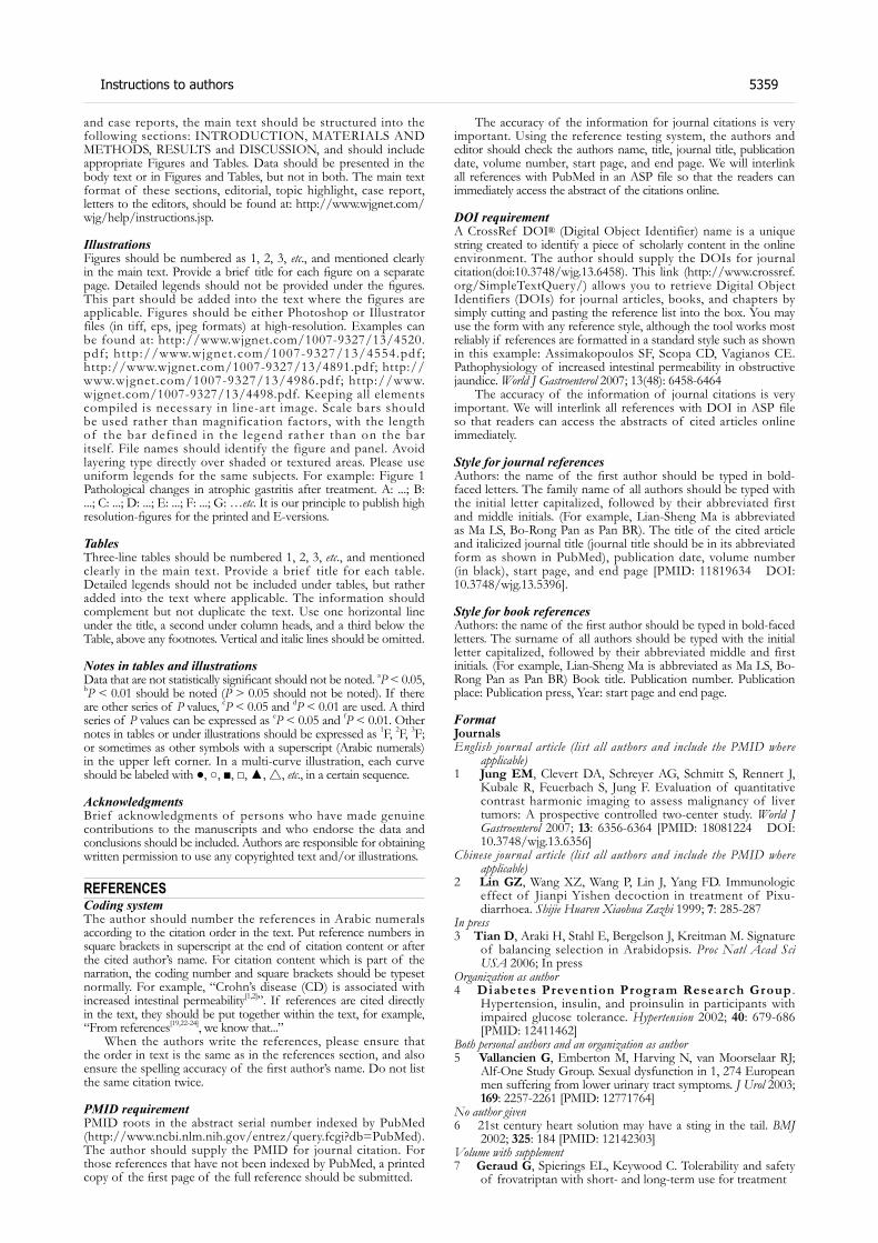
and case reports, the main text should be structured into the following sections: INTRODUCTION, MATERIALS AND METHODS, RESULTS and DISCUSSION, and should include appropriate Figures and Tables. Data should be presented in the body text or in Figures and Tables, but not in both. The main text format of these sections, editorial, topic highlight, case report, letters to the editors, should be found at: http://www.wjgnet.com/wjg/help/instructions.jsp.
IllustrationsFigures should be numbered as 1, 2, 3, etc., and mentioned clearly in the main text. Provide a brief title for each figure on a separate page. Detailed legends should not be provided under the figures. This part should be added into the text where the figures are applicable. Figures should be either Photoshop or Illustrator files (in tiff, eps, jpeg formats) at high-resolution. Examples can be found at: http://www.wjgnet.com/1007-9327/13/4520.pdf; http://www.wjgnet .com/1007-9327/13/4554.pdf; http://www.wjgnet.com/1007-9327/13/4891.pdf; http://www.wjgnet.com/1007-9327/13/4986.pdf; http://www.wjgnet.com/1007-9327/13/4498.pdf. Keeping all elements compiled is necessary in line-art image. Scale bars should be used rather than magnification factors, with the length of the bar defined in the legend rather than on the bar itself. File names should identify the figure and panel. Avoid layering type directly over shaded or textured areas. Please use uniform legends for the same subjects. For example: Figure 1 Pathological changes in atrophic gastritis after treatment. A: ...; B: ...; C: ...; D: ...; E: ...; F: ...; G: …etc. It is our principle to publish high resolution-figures for the printed and E-versions.
TablesThree-line tables should be numbered 1, 2, 3, etc., and mentioned clearly in the main text. Provide a brief title for each table. Detailed legends should not be included under tables, but rather added into the text where applicable. The information should complement but not duplicate the text. Use one horizontal line under the title, a second under column heads, and a third below the Table, above any footnotes. Vertical and italic lines should be omitted.
Notes in tables and illustrationsData that are not statistically significant should not be noted. aP < 0.05, bP < 0.01 should be noted (P > 0.05 should not be noted). If there are other series of P values, cP < 0.05 and dP < 0.01 are used. A third series of P values can be expressed as eP < 0.05 and fP < 0.01. Other notes in tables or under illustrations should be expressed as 1F, 2F, 3F; or sometimes as other symbols with a superscript (Arabic numerals) in the upper left corner. In a multi-curve illustration, each curve should be labeled with , , , , , , etc., in a certain sequence.
AcknowledgmentsBrief acknowledgments of persons who have made genuine contributions to the manuscripts and who endorse the data and conclusions should be included. Authors are responsible for obtaining written permission to use any copyrighted text and/or illustrations.
REFERENCESCoding systemThe author should number the references in Arabic numerals according to the citation order in the text. Put reference numbers in square brackets in superscript at the end of citation content or after the cited author’s name. For citation content which is part of the narration, the coding number and square brackets should be typeset normally. For example, “Crohn’s disease (CD) is associated with increased intestinal permeability[1,2]”. If references are cited directly in the text, they should be put together within the text, for example, “From references[19,22-24], we know that...”
When the authors write the references, please ensure that the order in text is the same as in the references section, and also ensure the spelling accuracy of the first author’s name. Do not list the same citation twice.
PMID requirementPMID roots in the abstract serial number indexed by PubMed (http://www.ncbi.nlm.nih.gov/entrez/query.fcgi?db=PubMed). The author should supply the PMID for journal citation. For those references that have not been indexed by PubMed, a printed copy of the first page of the full reference should be submitted.
The accuracy of the information for journal citations is very important. Using the reference testing system, the authors and editor should check the authors name, title, journal title, publication date, volume number, start page, and end page. We will interlink all references with PubMed in an ASP file so that the readers can immediately access the abstract of the citations online.
DOI requirementA CrossRef DOI® (Digital Object Identifier) name is a unique string created to identify a piece of scholarly content in the online environment. The author should supply the DOIs for journal citation(doi:10.3748/wjg.13.6458). This link (http://www.crossref.org/SimpleTextQuery/) allows you to retrieve Digital Object Identifiers (DOIs) for journal articles, books, and chapters by simply cutting and pasting the reference list into the box. You may use the form with any reference style, although the tool works most reliably if references are formatted in a standard style such as shown in this example: Assimakopoulos SF, Scopa CD, Vagianos CE. Pathophysiology of increased intestinal permeability in obstructive jaundice. World J Gastroenterol 2007; 13(48): 6458-6464
The accuracy of the information of journal citations is very important. We will interlink all references with DOI in ASP file so that readers can access the abstracts of cited articles online immediately.
Style for journal referencesAuthors: the name of the first author should be typed in bold-faced letters. The family name of all authors should be typed with the initial letter capitalized, followed by their abbreviated first and middle initials. (For example, Lian-Sheng Ma is abbreviated as Ma LS, Bo-Rong Pan as Pan BR). The title of the cited article and italicized journal title (journal title should be in its abbreviated form as shown in PubMed), publication date, volume number (in black), start page, and end page [PMID: 11819634 DOI: 10.3748/wjg.13.5396].
Style for book referencesAuthors: the name of the first author should be typed in bold-faced letters. The surname of all authors should be typed with the initial letter capitalized, followed by their abbreviated middle and first initials. (For example, Lian-Sheng Ma is abbreviated as Ma LS, Bo-Rong Pan as Pan BR) Book title. Publication number. Publication place: Publication press, Year: start page and end page.
FormatJournals English journal article (list all authors and include the PMID where
applicable)1 Jung EM, Clevert DA, Schreyer AG, Schmitt S, Rennert J,
Kubale R, Feuerbach S, Jung F. Evaluation of quantitative contrast harmonic imaging to assess malignancy of liver tumors: A prospective controlled two-center study. World J Gastroenterol 2007; 13: 6356-6364 [PMID: 18081224 DOI: 10.3748/wjg.13.6356]
Chinese journal article (list all authors and include the PMID where applicable)
2 Lin GZ, Wang XZ, Wang P, Lin J, Yang FD. Immunologic effect of Jianpi Yishen decoction in treatment of Pixu-diarrhoea. Shijie Huaren Xiaohua Zazhi 1999; 7: 285-287
In press3 Tian D, Araki H, Stahl E, Bergelson J, Kreitman M. Signature
of balancing selection in Arabidopsis. Proc Natl Acad Sci USA 2006; In press
Organization as author4 Diabetes Prevention Program Research Group .
Hypertension, insulin, and proinsulin in participants with impaired glucose tolerance. Hypertension 2002; 40: 679-686 [PMID: 12411462]
Both personal authors and an organization as author 5 Vallancien G, Emberton M, Harving N, van Moorselaar RJ;
Alf-One Study Group. Sexual dysfunction in 1, 274 European men suffering from lower urinary tract symptoms. J Urol 2003; 169: 2257-2261 [PMID: 12771764]
No author given6 21st century heart solution may have a sting in the tail. BMJ
2002; 325: 184 [PMID: 12142303]Volume with supplement7 Geraud G, Spierings EL, Keywood C. Tolerability and safety
of frovatriptan with short- and long-term use for treatment
Instructions to authors 5359
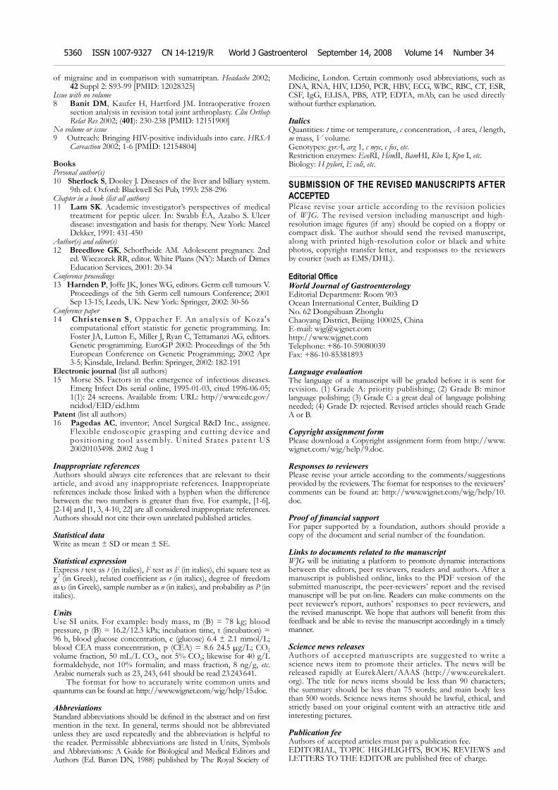
of migraine and in comparison with sumatriptan. Headache 2002; 42 Suppl 2: S93-99 [PMID: 12028325]
Issue with no volume8 Banit DM, Kaufer H, Hartford JM. Intraoperative frozen
section analysis in revision total joint arthroplasty. Clin Orthop Relat Res 2002; (401): 230-238 [PMID: 12151900]
No volume or issue9 Outreach: Bringing HIV-positive individuals into care. HRSA
Careaction 2002; 1-6 [PMID: 12154804]
BooksPersonal author(s)10 Sherlock S, Dooley J. Diseases of the liver and billiary system.
9th ed. Oxford: Blackwell Sci Pub, 1993: 258-296Chapter in a book (list all authors)11 Lam SK. Academic investigator’s perspectives of medical
treatment for peptic ulcer. In: Swabb EA, Azabo S. Ulcer disease: investigation and basis for therapy. New York: Marcel Dekker, 1991: 431-450
Author(s) and editor(s)12 Breedlove GK, Schorfheide AM. Adolescent pregnancy. 2nd
ed. Wieczorek RR, editor. White Plains (NY): March of Dimes Education Services, 2001: 20-34
Conference proceedings13 Harnden P, Joffe JK, Jones WG, editors. Germ cell tumours V.
Proceedings of the 5th Germ cell tumours Conference; 2001 Sep 13-15; Leeds, UK. New York: Springer, 2002: 30-56
Conference paper14 Christensen S , Oppacher F. An analysis of Koza's
computational effort statistic for genetic programming. In: Foster JA, Lutton E, Miller J, Ryan C, Tettamanzi AG, editors. Genetic programming. EuroGP 2002: Proceedings of the 5th European Conference on Genetic Programming; 2002 Apr 3-5; Kinsdale, Ireland. Berlin: Springer, 2002: 182-191
Electronic journal (list all authors)15 Morse SS. Factors in the emergence of infectious diseases.
Emerg Infect Dis serial online, 1995-01-03, cited 1996-06-05; 1(1): 24 screens. Available from: URL: http//www.cdc.gov/ncidod/EID/eid.htm
Patent (list all authors)16 Pagedas AC, inventor; Ancel Surgical R&D Inc., assignee.
Flexible endoscopic grasping and cutting device and posit ioning tool assembly. United States patent US 20020103498. 2002 Aug 1
Inappropriate referencesAuthors should always cite references that are relevant to their article, and avoid any inappropriate references. Inappropriate references include those linked with a hyphen when the difference between the two numbers is greater than five. For example, [1-6], [2-14] and [1, 3, 4-10, 22] are all considered inappropriate references. Authors should not cite their own unrelated published articles.
Statistical dataWrite as mean ± SD or mean ± SE.
Statistical expressionExpress t test as t (in italics), F test as F (in italics), chi square test as χ2 (in Greek), related coefficient as r (in italics), degree of freedom as υ (in Greek), sample number as n (in italics), and probability as P (in italics).
UnitsUse SI units. For example: body mass, m (B) = 78 kg; blood pressure, p (B) = 16.2/12.3 kPa; incubation time, t (incubation) = 96 h, blood glucose concentration, c (glucose) 6.4 ± 2.1 mmol/L; blood CEA mass concentration, p (CEA) = 8.6 24.5 mg/L; CO2 volume fraction, 50 mL/L CO2, not 5% CO2; likewise for 40 g/L formaldehyde, not 10% formalin; and mass fraction, 8 ng/g, etc. Arabic numerals such as 23, 243, 641 should be read 23 243 641.
The format for how to accurately write common units and quantums can be found at: http://www.wjgnet.com/wjg/help/15.doc.
AbbreviationsStandard abbreviations should be defined in the abstract and on first mention in the text. In general, terms should not be abbreviated unless they are used repeatedly and the abbreviation is helpful to the reader. Permissible abbreviations are listed in Units, Symbols and Abbreviations: A Guide for Biological and Medical Editors and Authors (Ed. Baron DN, 1988) published by The Royal Society of
Medicine, London. Certain commonly used abbreviations, such as DNA, RNA, HIV, LD50, PCR, HBV, ECG, WBC, RBC, CT, ESR, CSF, IgG, ELISA, PBS, ATP, EDTA, mAb, can be used directly without further explanation.
ItalicsQuantities: t time or temperature, c concentration, A area, l length, m mass, V volume.Genotypes: gyrA, arg 1, c myc, c fos, etc.Restriction enzymes: EcoRI, HindI, BamHI, Kbo I, Kpn I, etc.Biology: H pylori, E coli, etc.
SUBMISSION OF THE REVISED MANUSCRIPTS AFTER ACCEPTEDPlease revise your article according to the revision policies of WJG. The revised version including manuscript and high-resolution image figures (if any) should be copied on a floppy or compact disk. The author should send the revised manuscript, along with printed high-resolution color or black and white photos, copyright transfer letter, and responses to the reviewers by courier (such as EMS/DHL).
Editorial Office World Journal of GastroenterologyEditorial Department: Room 903 Ocean International Center, Building D No. 62 Dongsihuan Zhonglu Chaoyang District, Beijing 100025, ChinaE-mail: [email protected]://www.wjgnet.comTelephone: +86-10-59080039Fax: +86-10-85381893
Language evaluation The language of a manuscript will be graded before it is sent for revision. (1) Grade A: priority publishing; (2) Grade B: minor language polishing; (3) Grade C: a great deal of language polishing needed; (4) Grade D: rejected. Revised articles should reach Grade A or B.
Copyright assignment formPlease download a Copyright assignment form from http://www.wjgnet.com/wjg/help/9.doc.
Responses to reviewersPlease revise your article according to the comments/suggestions provided by the reviewers. The format for responses to the reviewers’ comments can be found at: http://www.wjgnet.com/wjg/help/10.doc.
Proof of financial supportFor paper supported by a foundation, authors should provide a copy of the document and serial number of the foundation.
Links to documents related to the manuscript WJG will be initiating a platform to promote dynamic interactions between the editors, peer reviewers, readers and authors. After a manuscript is published online, links to the PDF version of the submitted manuscript, the peer-reviewers’ report and the revised manuscript will be put on-line. Readers can make comments on the peer reviewer’s report, authors’ responses to peer reviewers, and the revised manuscript. We hope that authors will benefit from this feedback and be able to revise the manuscript accordingly in a timely manner.
Science news releasesAuthors of accepted manuscripts are suggested to write a science news item to promote their articles. The news will be released rapidly at EurekAlert/AAAS (http://www.eurekalert.org). The title for news items should be less than 90 characters; the summary should be less than 75 words; and main body less than 500 words. Science news items should be lawful, ethical, and strictly based on your original content with an attractive title and interesting pictures.
Publication feeAuthors of accepted articles must pay a publication fee.EDITORIAL, TOPIC HIGHLIGHTS, BOOK REVIEWS and LETTERS TO THE EDITOR are published free of charge.
5360 ISSN 1007-9327 CN 14-1219/R World J Gastroenterol September 14, 2008 Volume 14 Number 34