Embed Size (px)
Citation preview

J Med Genet 1995;32:383-388
X linked fatal infantile cardiomyopathy maps toXq28 and is possibly allelic to Barth syndrome
A K Gedeon, M J Wilson, A C Colley, D 0 Sillence, J C Mulley
Centre for MedicalGenetics, Departnentof Cytogenetics andMolecular Genetics,Women's andChildren's Hospital,North Adelaide,South Australia 5006A K GedeonJ C Mulley
Department ofGenetics, University ofAdelaide, Adelaide,South AustraliaA K Gedeon
Department ofGenetics, RoyalAlexandra Hospitalfor Children,Camperdown,New South Wales,AustraliaM J WilsonD 0 Sillence
Regional MedicalGenetics Unit,Newcastle WesternSuburbs Hospital,Waratah, New SouthWales, AustraliaA C Colley
Correspondence to:Dr Gedeon.Received 5 October 1994Revised version accepted forpublication9 December 1994
AbstractA number offamilies with X linked dilatedcardiomyopathy with onset in infancy orchildhood have now been described, withvarying clinical and biochemical features.Of these, one condition, Barth syndrome(BTHS), can be diagnosed clinically bythe characteristic associated features ofskeletal myopathy, short stature, and neu-tropenia, but not all of these features arealways present. Molecular genetic studieshave delineated the gene for BTHS, whichmaps to distal Xq28, from the gene forso called X linked dilated cardiomyopathy(XLCM), a teenage onset dilated car-diomyopathy, recently mapped to the 5'portion of the dystrophin locus at Xp2l.We report a large family in which male
infants have died with congenital dilatedcardiomyopathy, and there is a strongfamily history of unexplained death in in-fant males over at least four generations.Death always occurred in early infancy,without development of the characteristicfeatures associated with Barth syndrome.Molecular analysis localised the gene inthis family to Xq28 with lod scores of 2-3atO = 0 0 with dinucleotide repeat markers,p26 and p39, near DXS15 and at F8C. Theproximal limit to the localisation of thegene in this family is defined by a re-combinant at DXS296, while the distallimit could not be differentiated from thetelomere. This localisation is consistentwith a hypothesis of allelic and clinicalheterogeneity at the BTHS locus in Xq28.
( Med Genet 1995;32:383-388)
The aetiology of familial cardiomyopathy, par-ticularly those forms that are autosomally in-herited, is heterogeneous, and cannot alwaysbe determined from the clinical or biochemicalphenotype. Gene mapping of autosomal dom-inant hypertrophic cardiomyopathy (HCM) hasimplicated chromosomal regions 1 q3, 1 ipi 3-q13, 14ql 1, and 15q2, and a number of mis-sense point mutations in the ,B cardiac myosinheavy chain gene on chromosome 14 have beendescribed.' HCM can, however, be sporadic.Matemally inherited cardiomyopathies as-sociated with mtDNA mutations can presentwith marked clinical intrafamilial variation,from fatal infantile cardiopathy with lactic acid-
osis to adult onset skeletal and cardiac myo-pathy. Several mtDNA and tRNA mutationshave been described.2 The distinct X linkedinheritance of dilated cardiomyopathy in somefamilies has lead to regional localisation ofthesegenes by linkage mapping to the X chro-mosome.The familial X linked cardiomyopathies so
far reported tend to have infantile or childhoodonset of symptoms.3-8 The best delineated clin-ically is Barth syndrome (BTHS) which fea-tures X linked cardiomyopathy with variableskeletal myopathy, short stature, and neu-tropenia.4 The clinical picture is quite variableboth within and between families.89 Mostaffected people present with congestive cardiacfailure and on echocardiogram have ventriculardilatation and decreased left ventricular ejec-tion fraction (LVEF); thus they can be char-acterised as having a dilated cardiomyopathy.Reported laboratory findings have includedmitochondrial ultrastructural abnormalities,decreased levels of respiratory chain enzymes,decreased plasma or muscle carnitine, andincreased urinary excretion of 3-methyl-glutaconic and 2-ethylhydracyclic acids, butthese are inconsistent. Two independent stud-ies have recently localised the gene for BTHSto distal Xq28.'1'
Berko and Swift'2 reported a large kindredwith X linked dilated cardiomyopathy (nowdesignated XLCM) in which affected malespresented with rapidly progressive dilated car-diomyopathy in their teens to early twenties,with much later onset and slower course inmanifesting female carriers. This disorder isassociated with raised muscle creatine kinaseand abnormalities of cardiac muscle dy-strophin, but with no skeletal muscle in-volvement. XLCM has recently been linked tothe 5' portion ofthe dystrophin locus at Xp2 1.13These two major loci of X linked car-diomyopathy, BTHS and XLCM, have onlybeen clearly delineated by linkage analysis al-though they do show some clinical differences.We report a large family with apparent X
linked inheritance of a fatal infantile car-diomyopathy, in which we have regionally loc-alised the gene responsible to Xq28. Theclinical features in this family are insufficientto permit a definite diagnosis of Barth syn-drome, and the cardiomyopathy is consistentlyof congenital onset and fatal in infancy. Thepossibility that this represents allelic hetero-geneity within the BTHS locus is discussedwith respect to these findings.
383
group.bmj.com on July 12, 2011 - Published by jmg.bmj.comDownloaded from

Gedeon, Wilson, Colley, Sillence, Mulley
." ..z.'
/.
Z.
/'7
/1
Figure 1 Pedigree of the family with fatal infantile cardiomyopathy.
Materials and methodsCLINICAL REPORTThe pedigree (fig 1) comprises several maleinfants (IV-22, IV-23, VI1, V-3, V 5, and V-9,known or believed to have died with hy-pertrophic cardiomyopathy. These affectedmales, with clinical findings given below, are
related through apparently healthy women
(III 1, III-8, III 10, IV 1 and IV-8) in a pattern
consistent with X linked inheritance. Mito-chondrial inheritance seems unlikely in the ab-sence of any affected female offspring of carriermothers and is also supported by the lack ofclinical symptoms of cardiomyopathy in ob-ligate carriers.
V-1This infant was born at term, birth weight2440 g, Apgar scores 3 and 6. He was lethargicand mildly cyanosed from birth with periodicdeeper cyanosis. He had mild talipes equi-novarus but no dysmorphic features and no
obvious skeletal myopathy. Echocardiographyon day 11 showed a dilated, poorly contractingheart, with left ventricular hypertrophy (LVH)and LV dilatation; the interventricular septumwas not hypertrophied. He was treated withdigoxin, and showed some initial improvementbut died at the age of 6 weeks. Necropsy showeda globular heart with biventricular hypertrophyand left ventricular dilatation. The en-
docardium was pale and thickened. Microscopyshowed myocardial hyperplasia and en-
docardial fibroelastosis in the LV and right
ventricle and atrium. Skeletal muscle was re-ported as normal on light microscopy.
V-3The brother of V- 1 was born at term, Apgarscores 6 and 8. He had occasional cyanosisfrom birth and fed poorly at times. Echo-cardiogram at 1 week and 3 weeks showedminimal left ventricular hypertrophy, but nor-mal LV function. He died suddenly at 3-5weeks; no necropsy was performed.
VS5He was born at term, birth weight 3000 g,Apgar scores 3 and 9. He was lethargic andcyanosed from birth and had mild talipes equi-novarus. Echocardiography showed markedbiventricular hypertrophy with tricuspid re-gurgitation and a right to left shunt at theforamen ovale. He required assisted ventilationfor several days but was able to be extubatedby day 4. Investigations included electrolytes,urea, creatinine, calcium, urine metabolicscreen, lactate, pyruvate, full blood count anddifferential, carnitines, plasma amino acids,and very long chain fatty acids. Total, free, andacylcarnitines were normal as was his neu-trophil count. There was no evidence ofmethyl-glutaconic aciduria. He was maintained onantifailure treatment and stabilised for severalmonths, but had suboptimal weight gain andgradually deteriorated and died at the age of 5months.
384
E-1
group.bmj.com on July 12, 2011 - Published by jmg.bmj.comDownloaded from

X linked fatal infantile cardiomyopathy maps to Xq28
Birth weight was 2700 g and Apgar scores 8and 10. He fed poorly from birth but was firstinvestigated at 6 weeks when he presentedcyanosed and in cardiac failure; chest x rayshowed cardiomegaly. He was clinically muchimproved after starting antifailure therapy, butthe cardiomegaly persisted. He died at 5months; no necropsy was performed, and norecord of echocardiographic findings was avail-able.
The available history for the following twoinfants was taken from medical records kept atthe small country hospital where they were
born.
IV-22A birth weight of 2990 g was recorded at term.Dusky cyanotic episodes and some difficulty infeeding were documented in the nursing notesfrom birth and over the first 3 days of life,but he improved and was discharged homeapparently well at 6 days of age; no in-vestigations were documented. He died at theage of 2 to 3 weeks, cause unknown, and nonecropsy was performed.
IV23Birth weight was 2710 g at term. He had fre-quent episodes of cyanosis from birth, withlethargy. He deteriorated rapidly and died atthe age of 2 days; no investigations or necropsywere performed.
This clinical presentation is consistent withboth babies (IV-22 and IV-23) having had con-
genital cardiomyopathy. No investigations or
recognisable cause for the cyanotic episodesnor necropsy confirmation of these diagnosesis available.There is also a strong family history of un-
explained death of infant males, all less than6 months old, occurring over at least fourgenerations, including III-6, III-9, III11,III 13, IV-2, IV-3, IV 9, and IV-16. Medicalrecords for these and other possibly affectedinfants were either uninformative or not found.
DNA ANALYSIS
Genotyping of several dinucleotide repeat andRFLP markers in Xq27-28 was undertaken(table 1). The order of markers used was: cen-DXS548-DXS296(VK21A)-DXSI 1 13-GAB-
Table 1 Markers used for the localisation offatal infantile cardiomyopathy
Locus Markerlsymbol Polymorphism Heterozygosity Reference
DXS296 VK21A TaqI 0-22 14DXS1113 - (TC)IO(AC)2 0 75 15GABRA3 MGD34 (CA) 5 0-29 16DXS52 StI4-1 TaqI 0 77 14
p26 (CA)21 0-81 17p39 (CA)l6 0-84 17
DXS15 DX13 BgIII 0 5 14DXS707 2-55 MspI 0-42 18DXS605 2-19 EcoRI 0-48 18F8C intron 13 (CA)20 0-69 19DXS1108 sDF-2 (GT)5GC(GT)I3 0-75 20DXYS154 sDF-I (CS)18 0-70 20
RA3-DXS52(St-14)-p26, p39,DXS 15(DX1 3)-DXS707(2-55)-DXS605(2-19)-F8C-DXSI 1-08(SDF-2)-DXYS154(SDF-1)-tel.17182021 Themarkers p26 and p39 are dinucleotide repeatmarkers subcloned from a 500 kb YAC XY845,containing loci DXS52 and DXS15, and lie220kb and 10kb proximal to DXS15 re-spectively.'7 They are considered to be at thesame genetic location as DXS15 for the pur-poses ofmultipoint linkage analysis. Genotypesat DXS296 (TaqI), DXS52 (TaqI), and DXS15(BglII) were determined by Southern blot hy-bridisation of the relevant restriction digestrequired to detect the RFLP. The markers2-19 at DXS605 and 2-55 at DXS707 weredescribed as RFLPs'8; however, oligonucleo-tide sequences flanking each restrictionsite (supplied by D Toniolo) permitted am-plification by PCR before digestion. Remainingmarkers were analysed by PCR amplificationin the presence of [c_-32P] dCTP as previouslydescribed.22 The most distal polymorphicmarkers in Xq, DXS 1108 within 500 kb of thetelomere and DXYS154,20 were genotyped inthe family in an attempt to identify recombinantmeioses that would define the localisation inXq28 as distinct from the telomere. TheSTR44 and STR50 dinucleotide repeats, re-cently assigned D numbers DXS1238 andDXS1235 respectively,23 lie within introns ofthe dystrophin locus and were analysed to showexclusion of this cardiomyopathy locus fromXp2l.
LINKAGE ANALYSISLinkage analysis was performed using the com-puter programs MLINK for two point lodscores and LINKMAP for multipoint analysis.The analyses included all people from whomDNA samples were collected. The recom-bination fractions between loci included inthe multipoint analysis were: DXS296-0 001-DXS 1113-0 10-DXS52-0-008-p26, p39, DX-S 15-0 001-DXS707-0*001-DXS605-0 001-F-8C-0 005-DXS1108. The disease gene fre-quency was set at 1/10 000 and analysis as-sumed X linked recessive inheritance. Fordiseases with an X linked mode of inheritancethe critical value of the lod score is ZO>2-0 fordemonstration of linkage.24
ResultsPairwise lod scores at 10 marker loci in Xq28and at STR44 in Xp21 are given in table 2. Themaximum lod score of 2-30 at a recombinationfrequency of 0-0 was generated at the p26 andp39 markers near DXS15 and at F8C. TheSTR50, DXS548, GABRA3, and DXYS154loci were uninformative in this family. A re-combination event was observed with DXS296(VK21A) in subject III-8. Her status as a carrieris based on clinical records showing that hersons had a clinical presentation consistent withthe suspected cause of death, congenital car-diomyopathy.
All of the people from whom DNA sampleswere collected were included in the analysis.The genotypes of III-3 and III-5, for example,
385
group.bmj.com on July 12, 2011 - Published by jmg.bmj.comDownloaded from

Gedeon, Wilson, Colley, Sillence, Mulley
Table 2 Two point lod scores between the fatal infantile cardiomyopathy gene andmarkers within Xp2l and Xq28
0
Locus 0 01 0 05 0.1 0 2 0 3 0 4 Zmax Omax
Xp21STR44 -3-42 -1-44 -0-69 -0-09 0-12 0-13 0-15 0-36Xq28DXS296 (VK21A) 0-67 1-23 1-34 1-23 0-95 0-54 1-35 0-11DXS1113 1-66 1-52 1-33 0-96 0-58 0-21 1-70 0-0DXS52 (St-14) 2-17 2-00 1-78 1-33 0-86 0-38 2-21 0-0p26 2-26 2-07 1-84 1-36 0-87 0-37 2-30 0-0p39 2-26 2-07 1-84 1-36 0-87 0-37 2-30 0-0DXS15 (DX13) 1-43 1-31 1-16 0-85 0-51 0-18 1-45 0-0DXS707 (2-55) 1-92 1-75 1-54 1-10 0-63 0-20 1-96 0-0DXS605 (2-19) 1-39 1-26 1-10 0-77 0-43 0-11 1-42 0-0F8C 2-26 2-07 1-84 1-36 0-87 0-37 2-30 0-0DXS1108 (SDF-2) 1-65 1-51 1-32 0-95 0-57 0-21 1-69 0-0
could contribute to the inference of parentalgenotypes in generation II. Sufficient clinicalrecords could not be found to confirm causeof death and include the probably affectedmales IV 2, IV 3, IV 9, and IV-16 into theanalysis. Their inclusion under the assumptionthat they died of cardiomyopathy, increases thepeak two point lod score to 2-84 at 0=0 0 forthe markers p26 and p39 and at F8C. Severalmales in generation III have also died ofuntraceable causes and descendants of thegrandmaternal branch are said to have hadboys with endocardial fibroelastosis: however,none of these was included in the analyses.The proximal limit for the localisation of this
gene is defined by the recombination in subjectIII-8 at DXS296. The DXYS154 locus wasnot informative so that the distal limit to thislocalisation is not differentiated from the te-lomere at Xqter, since recombination was notobserved in any markers distal to DXS296.The regional localisation covers approximately11 cM, virtually all of the Xq28 band to the
30
20
a,
0C.)
cn
co0
-i
10
0
-10 F
-20-0-1 0 0.1
Recombination fraction
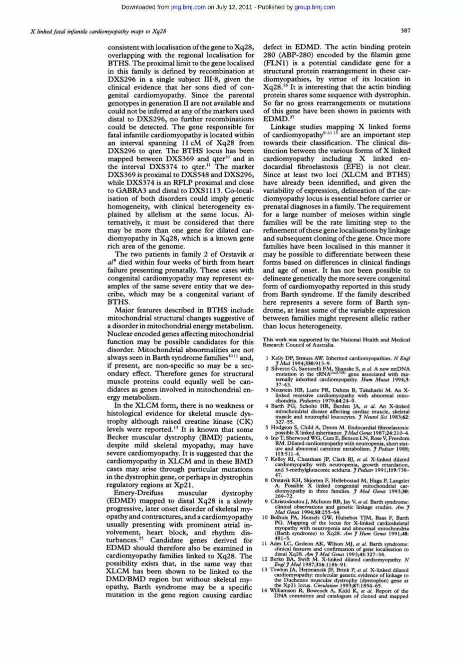
Figure 2 Location of the gene for fatal infantile cardiomyopathy on the background mapofX chromosome markers. The location score reached a peak at 12 27, equivalent to amultipoint lod score of 2-67.
telomere. The peak location score is 12X27(equivalent to a lod score of 2-67) at the p26,p39, and DXS 15 cluster between the lociDXS52 to DXS707 (fig 2). This multipointanalysis did not include the probably affectedmales IV2, IV 3, IV 9, and IV 16. The sig-nificant two point lod scores of 2-3 with p26,p39, and F8C, and 2-2 with DXS52 dependupon the assumption that IV-22 and IV 23were affected with fatal infantile cardio-myopathy. The peak two point lod scoreobtained by exclusion of III-8 and her sons isreduced to 2-21 at 0=0 0 at p26 and p39, butremains significant in support of linkage toXq28.
DiscussionThe large family described here has a severefamilial form of cardiomyopathy with early,often congenital onset which is invariably fatalin infancy. The clinical diagnosis of Barth syn-drome (BTHS) is based on the presence ofthe characteristic triad of cardiac and skeletalmyopathy in association with short stature andneutropenia, but BTHS could not be diagnosedclinically in this family as none of the patientsdeveloped other features. The cardiomyopathyin BTHS is of congenital, infantile, or child-hood onset and very variable progression,sometimes fatal in the neonatal period, but inothers within the same family may stabiliseor improve in early childhood. The cardiacfindings are of a predominantly dilated car-diomyopathy often with ventricular hy-pertrophy and sometimes associated withechocardiographic or histological evidence ofendocardial fibroelastosis. The cardiac findingsin the patients described here were of thistype, but although there was some response toantifailure medication in the two treated boys,death still ensued within months, and no knownaffected male has survived past infancy. Aswider family investigation has been limited,it is not known whether there are survivingasymptomatic affected boys.None of the affected infants in this family
had definite skeletal myopathy, and creatinekinase, measured in V 1 and V 5, was notraised. It is interesting to note that two infantshad mild talipes equinovarus, which has beenreported in patients with BTHS,"1 and couldbe a consequence ofeither congenital myopathyor in utero hypotonia. Mild neutropenia waspresent on one occasion in V 1, but was notobserved in other affected infants, nor was therea history of skin or other infections. Others9'have commented on the relatively low birthweight for gestational age, although this doesnot apply to all patients with BTHS; we alsonoted birth weights under the 10th centile inthree of the six infants reported. Informationregarding birth length and subsequent lineargrowth was lacking, but poor feeding and fail-ure to thrive, consistent with symptomatic car-diomyopathy occurred in the two infants whosurvived the longest (V 5 and V 9 survived to5 months).Although the clinical features were in-
conclusive, linkage analysis in this family was
CY)~ 00a) r- C1 ' o o rCN - LO a-rtCO(flU) tJ) ' ( ) U)XX XCN X )X
00 0 100 u. 0 cU~~~~~~~~~~~~~~~~~~~~~~~~~~~~~~~~~~~~~~~~~~~~~~~~~~~~~~~~~~~
386
_
group.bmj.com on July 12, 2011 - Published by jmg.bmj.comDownloaded from
X linked fatal infantile cardiomyopathy maps to Xq28
consistent with localisation ofthe gene to Xq28,overlapping with the regional localisation forBTHS. The proximal limit to the gene localisedin this family is defined by recombination atDXS296 in a single subject III-8, given theclinical evidence that her sons died of con-genital cardiomyopathy. Since the parentalgenotypes in generation II are not available andcould not be inferred at any ofthe markers useddistal to DXS296, no further recombinationscould be detected. The gene responsible forfatal infantile cardiomyopathy is located withinan interval spanning 11 cM of Xq28 fromDXS296 to qter. The BTHS locus has beenmapped between DXS369 and qter"' and inthe interval DXS374 to qter.11 The markerDXS369 is proximal to DXS548 and DXS296,while DXS374 is an RFLP proximal and closeto GABRA3 and distal to DXS1 113. Co-local-isation of both disorders could imply genetichomogeneity, with clinical heterogeneity ex-plained by allelism at the same locus. Al-ternatively, it must be considered that theremay be more than one gene for dilated car-diomyopathy in Xq28, which is a known generich area of the genome.The two patients in family 2 of Orstavik et
a18 died within four weeks of birth from heartfailure presenting prenatally. These cases withcongenital cardiomyopathy may represent ex-amples of the same severe entity that we des-cribe, which may be a congenital variant ofBTHS.Major features described in BTHS include
mitochondrial structural changes suggestive ofa disorder in mitochondrial energy metabolism.Nuclear encoded genes affecting mitochondrialfunction may be possible candidates for thisdisorder. Mitochondrial abnormalities are notalways seen in Barth syndrome families"101 and,if present, are non-specific so may be a sec-ondary effect. Therefore genes for structuralmuscle proteins could equally well be can-didates as genes involved in mitochondrial en-ergy metabolism.
In the XLCM form, there is no weakness orhistological evidence for skeletal muscle dys-trophy although raised creatine kinase (CK)levels were reported."3 It is known that someBecker muscular dystrophy (BMD) patients,despite mild skeletal myopathy, may havesevere cardiomyopathy. It is suggested that thecardiomyopathy in XLCM and in these BMDcases may arise through particular mutationsin the dystrophin gene, or perhaps in dystrophinregulatory regions at Xp2 1.
Emery-Dreifuss muscular dystrophy(EDMD) mapped to distal Xq28 is a slowlyprogressive, later onset disorder of skeletal my-opathy and contractures, and a cardiomyopathyusually presenting with prominent atrial in-volvement, heart block, and rhythm dis-turbances.25 Candidate genes derived forEDMD should therefore also be examined incardiomyopathy families linked to Xq28. Thepossibility exists that, in the same way thatXLCM has been shown to be linked to theDMD/BMD region but without skeletal my-opathy, Barth syndrome may be a specificmutation in the gene region causing cardiac
defect in EDMD. The actin binding protein280 (ABP-280) encoded by the filamin gene(FLN1) is a potential candidate gene for astructural protein rearrangement in these car-diomyopathies, by virtue of its location inXq28.26 It is interesting that the actin bindingprotein shares some sequence with dystrophin.So far no gross rearrangements or mutationsof this gene have been shown in patients withEDMD.27Linkage studies mapping X linked forms
of cardiomyopathy9'13' are an important steptowards their classification. The clinical dis-tinction between the various forms ofX linkedcardiomyopathy including X linked en-docardial fibroelastosis (EFE) is not clear.Since at least two loci (XLCM and BTHS)have already been identified, and given thevariability of expression, delineation of the car-diomyopathy locus is essential before carrier orprenatal diagnoses in a family. The requirementfor a large number of meioses within singlefamilies will be the rate limiting step to therefinement ofthese gene localisations by linkageand subsequent cloning ofthe gene. Once morefamilies have been localised in this manner itmay be possible to differentiate between theseforms based on differences in clinical findingsand age of onset. It has not been possible todelineate genetically the more severe congenitalform of cardiomyopathy reported in this studyfrom Barth syndrome. If the family describedhere represents a severe form of Barth syn-drome, at least some of the variable expressionbetween families might represent allelic ratherthan locus heterogeneity.
This work was supported by the National Health and MedicalResearch Council of Australia.
1 Kelly DP, Strauss AW. Inherited cardiomyopathies. N EnglJt Med 1994;330:913-9.
2 Silvestri G, Santorelli FM, Shanske S, et al. A new mtDNAmutation in the tRNAL'U-=R gene associated with ma-ternally inherited cardiomyopathy. Hum Mutat 1994;3:37-43.
3 Neustein HB, Lurie PR, Dahms B, Takahashi M. An X-linked recessive cardiomyopathy with abnormal mito-chondria. Pediatrics 1979;64:24-9.
4 Barth PG, Scholte HR, Berden JA, et al. An X-linkedmitochondrial disease affecting cardiac muscle, skeletalmuscle and neutrophil leucocytes. J Neurol Sci 1983;62:327-55.
5 Hodgson S, Child A, Dyson M. Endocardial fibroelastosis:possible X linked inheritance.JMed Genet 1987;24:210-4.
6 Ino T, SherwoodWG, Cutz E, Benson LN, Rose V, FreedomRM. Dilated cardiomyopathy with neutropenia, short stat-ure and abnormal carnitine metabolism. Jf Pediatr 1988;113:511-4.
7 Kelley RI, Cheatham JP, Clark BJ, et al. X-linked dilatedcardiomyopathy with neutropenia, growth retardation,and 3-methylglutaconic aciduria. J Pediatr 199 1;119:738-47.
8 Orstavik KH, Skjorten F, Hellebostad M, Haga P, LangsletA. Possible X linked congenital mitochondrial car-diomyopathy in three families. J Med Genet 1993;30:269-72.
9 Christodoulou J, McInnes RR, Jay V, et al. Barth syndrome:clinical observations and genetic linkage studies. Am JMed Genet 1994;50:255-64.
10 Bolhuis PA, Hensels GW, Hulsebos TJM, Baas F, BarthPG. Mapping of the locus for X-linked cardioskeletalmyopathy with neutropenia and abnormal mitochondria(Barth syndrome) to Xq28. Am J Hum Genet 1991;48:481-5.
11 Ades LC, Gedeon AK, Wilson MJ, et al. Barth syndrome:clinical features and confirmation of gene localisation todistal Xq28. Am J Med Genet 1993;45:327-34.
12 Berko BA, Swift M. X-linked dilated cardiomyopathy. NEnglJ Med 1987;316:1186-91.
13 Towbin JA, Hejtmancik JF, Brink P, et al. X-linked dilatedcardiomyopathy: molecular genetic evidence of linkage tothe Duchenne muscular dystrophy (dystrophin) gene atthe Xp2l locus. Circulation 1993;87:1854-65.
14 Williamson R, Bowcock A, Kidd K, et al. Report of theDNA committee and catalogues of cloned and mapped
387
group.bmj.com on July 12, 2011 - Published by jmg.bmj.comDownloaded from

Gedeon, Wilson, Colley, Sillence, Mulley
genes, markers formatted for PCR and DNA poly-morphisms. Cytogenet Cell Genet 1991;58:1190-832.
15 Weber C, Oudet C, Johnson S, Pilia G, Schlessinger D,Hanauer A. Dinucleotide repeat polymorphism close to
IDS gene in Xq27.3-q28 (DXS1113). Hum Mol Genet1993;2:612.
16 Hicks AA, Johnson KJ, Barnard EA, Darlison MG. Di-nucleotide repeat polymorphism in the human X-linkedGABAA receptor a3-subunit gene. Nucleic Acids Res 199 1;19:4016.
17 Wehnert M, Reiner 0, Caskey CT. Four STR poly-morphisms map to a 500 kb region between DXS 15 andDXS134. Hum Mol Genet 1993;2:1503.
18 Maestrini E, Rivella S, Tribioli C, et al. Identification ofnovel RFLPs in the vicinity of CpG islands in Xq28:application to the analysis of the pattern ofX chromosomeinactivation. Am Hum Genet 1992;50:156-63.
19 Lalloz MRA, McVey JH, Pattinson JK, Tuddenham EGD.Haemophilia A diagnosis by analysis of a hypervariabledinucleotide repeat within the factor VIII gene. Lancet199 1;338:207-1 1.
20 Freije D, Helms C, Watson MS, Donis-Keller H. Iden-tification of a second pseudoautosomal region near theXq and Yq telomeres. Science 1992;258:1784-7.
21 Schlessinger D, Mandel JL, Monaco AP, Nelson DL, WillardHF. Report of the Fourth International Workshop on
Human X Chromosome Mapping, 1993. Cytogenet CellGenet 1993;64: 147-94.
22 Gedeon AK, Richards RI, Mulley JC. Dinucleotide repeatpolymorphisms at the DXS294 and DXS300 loci in Xq26.Nucleic Acids Res 1991;19:5087.
23 Willard HF, Cremers F, Mandel JL, Monaco AP, NelsonDL, Schlessinger D. Report of the Fifth InternationalWorkshop on Human X Chromosome Mapping 1994.Cytogenet Cell Genet 1994;67:296-359.
24 Ott J. Analysis of Human Genetic Linkage. Revised edition.Baltimore: The John Hopkins University Press, 1991.
25 Yates JRW, Warner JP, Smith JA, et al. Emery-Dreifussmuscular dystrophy: linkage to markers in distal Xq28.Med Genet 1993;30:108-11.
26 Gorlin JB, Henske E, Warren ST, et al. Actin-binding protein(ABP-280) filamin gene (FLN) maps telomeric to thecolor vision locus (R/GCP) and centromeric to G6PD inXq28. Genomics 1993;17:496-8.
27 Maestrini E, Patrosso C, Mancini M, et al. Mapping of twogenes encoding isoforms of the actin binding proteinABP-280, a dystrophin like protein, to Xq28 and tochromosome 7. Hum Mol Genet 1993;2:761-6.
388
group.bmj.com on July 12, 2011 - Published by jmg.bmj.comDownloaded from

doi: 10.1136/jmg.32.5.383 1995 32: 383-388J Med Genet
A K Gedeon, M J Wilson, A C Colley, et al. syndrome.to Xq28 and is possibly allelic to Barth X linked fatal infantile cardiomyopathy maps
http://jmg.bmj.com/content/32/5/383Updated information and services can be found at:
These include:
References http://jmg.bmj.com/content/32/5/383#related-urls
Article cited in:
serviceEmail alerting
the box at the top right corner of the online article.Receive free email alerts when new articles cite this article. Sign up in
Notes
http://group.bmj.com/group/rights-licensing/permissionsTo request permissions go to:
http://journals.bmj.com/cgi/reprintformTo order reprints go to:
http://group.bmj.com/subscribe/To subscribe to BMJ go to:
group.bmj.com on July 12, 2011 - Published by jmg.bmj.comDownloaded from


