Embed Size (px)
Citation preview

Immunity, Vol. 21, 81–93, July, 2004, Copyright 2004 by Cell Press
XBP1, Downstream of Blimp-1, Expands the SecretoryApparatus and Other Organelles, and IncreasesProtein Synthesis in Plasma Cell Differentiation
restructuring that allows them to secrete large quantitiesof immunoglobulin (Ig) (Calame et al., 2003; Wiest et al.,1990). Two transcriptional regulators are essential forplasma cell differentiation: Blimp-1, a transcriptional re-pressor encoded by the prdm1 gene, and XBP1, a b-ZIP
A.L. Shaffer,1 Miriam Shapiro-Shelef,2
Neal N. Iwakoshi,3 Ann-Hwee Lee,3
Shu-Bing Qian,4 Hong Zhao,1 Xin Yu,1
Liming Yang,1 Bruce K. Tan,1 Andreas Rosenwald,1
Elaine M. Hurt,1 Emmanuel Petroulakis,5
family transcriptional activator (see below; Lin et al.,Nahum Sonenberg,5 Jonathan W. Yewdell,4
2003).Kathryn Calame,2 Laurie H. Glimcher,3
In the B cell lineage, Blimp-1 is predominantly ex-and Louis M. Staudt1,*pressed at the plasma cell stage where it directly re-1Metabolism Branchpresses genes encoding other transcription factors,Center for Cancer Researchsuch as c-myc, cIIta, pax5, spiB, and id3 (Lin et al., 1997,National Cancer Institute2002; Piskurich et al., 2000; Reljic et al., 2000; ShafferNational Institutes of Healthet al., 2002). Global gene expression analysis identified9000 Rockville Pikeover 250 genes whose expression was extinguished byBethesda, Maryland 20892Blimp-1, including genes mediating BCR expression and2 Department of Microbiology and Integrated Programsignaling, B cell activation and homing, and classin Biophysical, Cellular and Molecular Studiesswitching (Shaffer et al., 2002). Blimp-1 also decreasedColumbia University College ofproliferation and increased Ig synthesis, hallmarks ofPhysicians and Surgeonsplasma cell differentiation (Lin et al., 1997; Shaffer et al.,New York, New York 100322002). Recent work has shown that prdm1-deficient B3 Department of Immunology and Infectious Diseasescells proliferate rapidly upon LPS stimulation in vitro butHarvard School of Public Healthdo not secrete immunoglobulin and fail to produce theBoston, Massachusetts 02115secreted form of Ig heavy chain mRNA (Shapiro-Shelef4 Laboratory of Viral Diseaseset al., 2003). In vivo, B cell development is grossly normalNational Institute of Allergy and Infectious Diseasesin prdm1-deficient mice, but these B cells fail to developBethesda, Maryland 20892into Ig-secreting plasma cells upon immunization with5 McGill UniversityT-dependent and -independent antigens.Department of Biochemistry
XBP1 is a positively-acting transcription factor in theMontreal, Quebec, H3G 1Y6CREB/ATF family that is expressed at a high levels inCanadaplasma cells (Iwakoshi et al., 2003a; Reimold et al.,1996). Since disruption of xbp1 in the mouse germ lineresults in embryonic lethality (Reimold et al., 2000),Summaryxbp1�/�, rag2�/� chimeric mice were used to assessXBP1 function within the B cell lineage (Reimold et al.,The differentiation of B cells into immunoglobulin-2001). These mice possessed B cells that proliferatedsecreting plasma cells is controlled by two transcrip-and formed germinal centers normally but were dramati-tion factors, Blimp-1 and XBP1. By gene expressioncally impaired in their ability to secrete immunoglobulinprofiling, we defined a set of genes whose inductionin vitro and in vivo in response to T-dependent orduring mouse plasmacytic differentiation is dependent-independent antigens. Most importantly, these xbp1-on Blimp-1 and/or XBP1. Blimp-1-deficient B cellsdeficient mice were devoid of plasma cells, demonstra-failed to upregulate most plasma cell-specific genes,ting the requirement for XBP1 in plasmacytic differenti-
including xbp1. Differentiating xbp1-deficient B cellsation.
induced Blimp-1 normally but failed to upregulate XBP1 is also associated with the unfolded proteingenes encoding many secretory pathway compo- response (UPR; Calfon et al., 2002; Fewell et al., 2001;nents. Conversely, ectopic expression of XBP1 in- Harding et al., 2000; Mori, 2000; Shen et al., 2001; Yo-duced a wide spectrum of secretory pathway genes shida et al., 2001), a coordinated change in gene expres-and physically expanded the endoplasmic reticulum. sion that is triggered by perturbations in the functionIn addition, XBP1 increased cell size, lysosome con- of the endoplasmic reticulum (ER). Experimentally, thetent, mitochondrial mass and function, ribosome num- UPR can be induced by DTT, which disrupts proteinbers, and total protein synthesis. Thus, XBP1 coordi- folding in the ER; tunicamycin, which disrupts glycosyla-nates diverse changes in cellular structure and tion and folding in the ER; and thapsigargin, which de-function resulting in the characteristic phenotype of pletes ER calcium stores (Fewell et al., 2001; Hardingprofessional secretory cells. et al., 2001). Overexpression or misfolding of ER proteins
can also elicit the UPR (Kozutsumi et al., 1988).Introduction The UPR was first characterized in yeast, where a
single signaling pathway governs the response to ERWhen B cells become plasma cells, they lose expression stress (Cox et al., 1993; Patil and Walter, 2001). In thisof most B cell characteristics and undergo a radical pathway, the ER stress signal is transduced by a type I
transmembrane ER protein, IRE1. The ER lumenal por-tion of IRE1 encodes a kinase for which IRE1 itself ap-*Corresponding: [email protected]
Immunity82
pears to be the only substrate. Interaction of IRE1 with ATF6 is not clear at present since recent studies showedthe ER chaperone Kar2p/BiP prevents IRE1 multimeriza- that the activation of several UPR genes (BiP, chop,tion and autophosphorylation. Current models propose grp94, and xbp1) was unaffected in cells in which ATF6that during an UPR, BiP is preferentially bound to mis- levels were diminished by RNA interference (Lee etfolded client proteins, thereby releasing IRE1 to multi- al., 2003).merize and autophosphorylate. In the present study, we used gene expression profil-
IRE1 phosphorylation is accompanied by activation of ing to study the terminal differentiation of B cells derivedan endoribonuclease activity in its cytoplasmic domain, from Blimp-1-deficient and XBP1-deficient mice in orderwhich mediates the posttranscriptional processing of to elucidate the roles of these transcription factors inthe mRNA encoding HAC1, the yeast ortholog of XBP1. plasma cell development. We observed that XBP1 actedIRE1 removes a 252 nucleotide internal sequence from downstream of Blimp-1 to regulate a broad complementHAC1 mRNA, and tRNA ligase rejoins the two fragments of genes encoding ER-associated proteins, many ofof HAC1 mRNA. This new version of HAC1 mRNA is which are involved in protein secretion. XBP1 inducedtranslated more efficiently than the unprocessed form a dramatic physical expansion of the ER but also, unex-and encodes a more stable protein with greater tran- pectedly, increased cell size, organelle biogenesis, andscriptional activation potential (Chapman and Walter, total protein synthesis, thus demonstrating that XBP11997; Cox and Walter, 1996; Kawahara et al., 1998; Mori plays a central role in defining the secretory cell phe-et al., 2000; Sidrauski and Walter, 1997). This linear UPR notype.pathway is solely responsible for the upregulation ofyeast UPR genes, which encode proteins involved in Resultsnearly every aspect of the secretory pathway, includingprotein entry into the ER, folding, glycosylation, ER- Regulation of Plasma Cell Gene Expressionassociated degradation (ERAD), and vesicular traffick- by Blimp-1 and XBP1ing (Fewell et al., 2001; Travers et al., 2000). Lipopolysaccharide (LPS) treatment of mouse mature
In higher eukaryotes, ER stress stimulates three dis- splenic B cells in vitro is an established experimentaltinct but overlapping signaling pathways by activating system for studying plasma cell differentiation (LafrenzIRE1, PERK, and ATF6 (Harding et al., 2002; Ma and et al., 1982; Schliephake and Schimpl, 1996). After 4Hendershot, 2003). The IRE1 UPR pathway has been days in culture, approximately 30% of cells achieve amaintained in higher eukaryotes with some modification. plasma cell phenotype (surface expression of synde-Activated IRE1 functions in mammals by removing 26 can-1 and immunoglobulin secretion; M.S.-S. and N.N.I.,nucleotides from the XBP1 mRNA. This new mRNA en- unpublished data). To identify genes induced during dif-codes a protein with increased transcriptional activation ferentiation and to determine their dependence onpotential (Calfon et al., 2002; Shen et al., 2001; Yoshida Blimp-1 and XBP1, we compared gene expression pro-et al., 2001). XBP1 induces several UPR response genes, files from LPS-treated wild-type (wt), prdm1-deficient,similar to those induced by HAC1 in yeast (Lee et al., and xbp1-deficient B cells (Reimold et al., 2001; Shapiro-2003; Yoshida et al., 2003). However, mouse embryo
Shelef et al., 2003). For these studies, we constructedfibroblasts lacking IRE1 still upregulate several UPR
the mouse Lymphochip, a specialized DNA microarraygenes in response to ER stress, including BiP and chop,
analogous to the human Lymphochip (Alizadeh et al.,indicating that IRE1 is not the sole mediator of the UPR
1999), which is enriched for genes expressed in normalin higher eukaryotes (Harding et al., 2002).
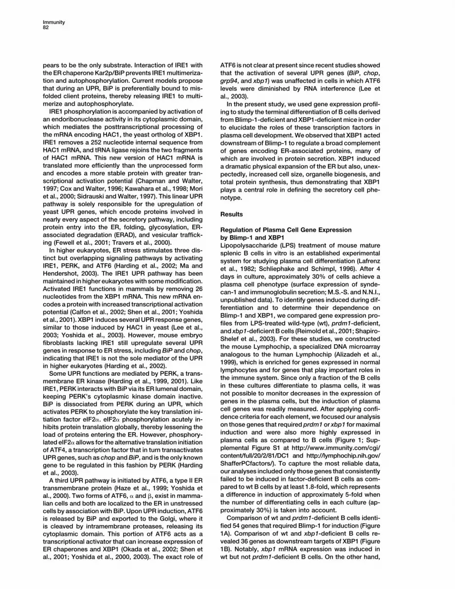
lymphocytes and for genes that play important roles inSome UPR functions are mediated by PERK, a trans-the immune system. Since only a fraction of the B cellsmembrane ER kinase (Harding et al., 1999, 2001). Likein these cultures differentiate to plasma cells, it wasIRE1, PERK interacts with BiP via its ER lumenal domain,not possible to monitor decreases in the expression ofkeeping PERK’s cytoplasmic kinase domain inactive.genes in the plasma cells, but the induction of plasmaBiP is dissociated from PERK during an UPR, whichcell genes was readily measured. After applying confi-activates PERK to phosphorylate the key translation ini-dence criteria for each element, we focused our analysistiation factor eIF2�. eIF2� phosphorylation acutely in-on those genes that required prdm1 or xbp1 for maximalhibits protein translation globally, thereby lessening theinduction and were also more highly expressed inload of proteins entering the ER. However, phosphory-plasma cells as compared to B cells (Figure 1; Sup-lated eIF2� allows for the alternative translation initiationplemental Figure S1 at http://www.immunity.com/cgi/of ATF4, a transcription factor that in turn transactivatescontent/full/20/2/81/DC1 and http://lymphochip.nih.gov/UPR genes, such as chop and BiP, and is the only knownShafferPCfactors/). To capture the most reliable data,gene to be regulated in this fashion by PERK (Hardingour analyses included only those genes that consistentlyet al., 2003).failed to be induced in factor-deficient B cells as com-A third UPR pathway is initiated by ATF6, a type II ERpared to wt B cells by at least 1.8-fold, which representstransmembrane protein (Haze et al., 1999; Yoshida eta difference in induction of approximately 5-fold whenal., 2000). Two forms of ATF6, � and �, exist in mamma-the number of differentiating cells in each culture (ap-lian cells and both are localized to the ER in unstressedproximately 30%) is taken into account.cells by association with BiP. Upon UPR induction, ATF6
Comparison of wt and prdm1-deficient B cells identi-is released by BiP and exported to the Golgi, where itfied 54 genes that required Blimp-1 for induction (Figureis cleaved by intramembrane proteases, releasing its1A). Comparison of wt and xbp1-deficient B cells re-cytoplasmic domain. This portion of ATF6 acts as avealed 36 genes as downstream targets of XBP1 (Figuretranscriptional activator that can increase expression of1B). Notably, xbp1 mRNA expression was induced inER chaperones and XBP1 (Okada et al., 2002; Shen et
al., 2001; Yoshida et al., 2000, 2003). The exact role of wt but not prdm1-deficient B cells. On the other hand,

XBP1 Regulates Secretory Cell Phenotype83
Figure 1. The Roles of prdm1 and xbp1 in Promoting Gene Expression during Plasma Cell Differentiation
RNA extracted from purified, LPS-treated wt, prdm1-, or xbp1-deficient splenic B cells was converted to labeled cDNA (Cy5, red) andcohybridized on mouse lymphochips with cDNA generated from a common reference pool of mouse cell line RNA (Cy3, green). Hybridizationwas measured by laser scanning (Axon Genepix 4000) and converted to a gene expression ratio (Cy5 experimental/Cy3 control), permittingthe direct comparison of all samples by hierarchical clustering. Data were normalized to unstimulated (time zero) controls for each time course.When genes were represented by multiple array elements, data for a single representative feature is displayed. A color bar depicts themagnitude of gene expression differences. (A) Genes whose induction was diminished in prdm1-deficient B cells by at least 1.8-fold versuswt at days 2 or 4 in 2 of 3 experiments. (B) Genes whose induction was diminished in xbp1-deficient B cells versus wt by at least 1.8-fold inboth experiments at days 2 or 4 of culture. *, prdm1 and Ig� light chain expression is affected less than 1.8-fold by the absence of xbp1(see Results).
prdm1 mRNA expression was induced equally in wt and noglobulin secretion (Figures 1 and 2). Some of thesegenes have previously been identified as XBP1 targetsxbp1-deficient B cells (Figure 1B). These observations
confirm that Blimp-1 is upstream of XBP1 in the regula- and are induced during the UPR (Edem, DnaJc3, Armet,Cai, Hspa5; Lee et al., 2003; Yoshida et al., 2003). How-tory cascade of terminal B cell differentiation, consistent
with previous observations (Shaffer et al., 2002; Shapiro- ever, many of these genes have not been formally asso-ciated with the response to ER stress and encode aShelef et al., 2003).
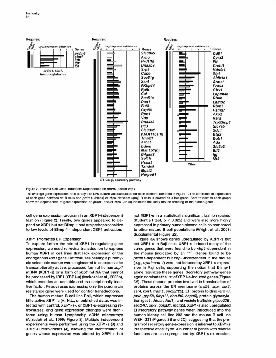
To better assess the contribution of Blimp-1 and XBP1 variety of secretory pathway proteins that play roles intargeting proteins to the ER (srp9), translocation of newlyto the induction of differentiation-associated genes, we
calculated the difference in expression of each gene synthesized proteins into the ER (sec61a, sec61g, ssr4),folding of ER proteins (fkbp14, ppib, grp58, txndc5,from Figure 1 between wt B cells and prdm1- or xbp1-
deficient B cells at day 4 following LPS stimulation. Fig- dnaJb9), ER protein degradation by the ERAD pathway(edem, sel1h, hrd1), protein glycosylation (dad1, fut8,ure 2 depicts the difference in expression for each gene
between wt and prdm1-deficient B cells (black bars) slc33a1, man1b1, b4galt2, mgat2), and vesicle traffick-ing (arhq, cope, vdp, arcn1). The expression of most (28and wt and xbp1-deficient B cells (gray bars). Most dif-
ferentiation genes (46 of 63) required both prdm1 and of 31) of the secretory pathway genes depended onboth prdm1 and xbp1, suggesting that XBP1, actingxbp1, and these are most likely downstream targets of
XBP1, since xbp1 itself was not induced in the absence downstream of Blimp-1, plays a critical role in coordinat-ing secretory function.of prdm1. Secretory pathway genes represented the
largest functional category of prdm1- and xbp1-depen- Several (16 of 63) plasma cell-associated genes (e.g.,syndecan-1, sdc1) required only prdm1 for their induc-dent genes (28), emphasizing the importance of these
regulatory factors in preparing cells for high-level immu- tion, suggesting that Blimp-1 controls part of the plasma
Immunity84
Figure 2. Plasma Cell Gene Induction: Dependence on prdm1 and/or xbp1
The average gene expression ratio at day 4 of LPS culture was calculated for each element identified in Figure 1. The difference in expressionof each gene between wt B cells and prdm1- (black) or xbp1-deficient (gray) B cells is plotted as a bar graph. Bars to next to each graphshow the dependence of gene expression on prdm1 and/or xbp1. An (h) indicates the likely mouse ortholog of the human gene.
cell gene expression program in an XBP1-independent not XBP1-u in a statistically significant fashion (pairedStudent’s t test, p � 0.025) and were also more highlyfashion (Figure 2). Finally, two genes appeared to de-
pend on XBP1 but not Blimp-1 and are perhaps sensitive expressed in primary human plasma cells as comparedto other mature B cell populations (Wright et al., 2003;to low levels of Blimp-1-independent XBP1 activation.Supplemental Figure S2).
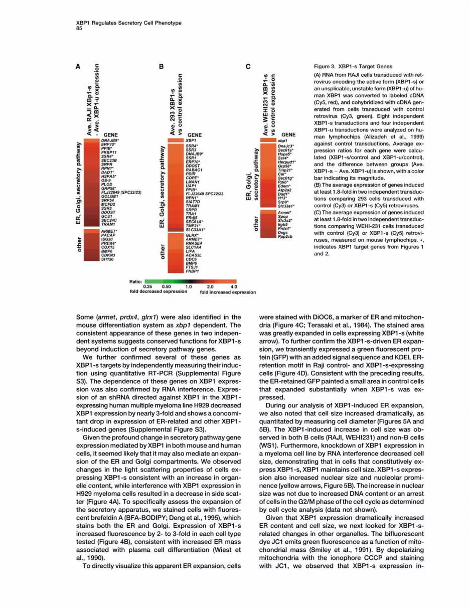
Figure 3A shows genes upregulated by XBP1-s butXBP1 Promotes ER ExpansionTo explore further the role of XBP1 in regulating gene not XBP1-u in Raji cells. XBP1-s induced many of the
same genes that were found to be xbp1-dependent inexpression, we used retroviral transduction to expresshuman XBP1 in cell lines that lack expression of the the mouse (indicated by an “*”). Genes found to be
prdm1-dependent but xbp1-independent in the mouseendogenous xbp1 gene. Retroviruses bearing a puromy-cin selectable marker were engineered to coexpress the (e.g., syndecan-1) were not induced by XBP1-s expres-
sion in Raji cells, supporting the notion that Blimp-1transcriptionally active, processed form of human xbp1mRNA (XBP1-s) or a form of xbp1 mRNA that cannot alone regulates these genes. Secretory pathway genes
again dominate the list of XBP1-s-induced genes (Figurebe processed by IRE1 (XBP1-u) (Iwakoshi et al., 2003b),which encodes an unstable and transcriptionally inac- 3A). These encode proteins involved in translocation of
proteins across the ER membrane (srp54, srpr, ssr3,tive factor. Retroviruses expressing only the puromycinresistance gene were used for control transductions. ssr4, rpn1, tram1, spc22/23), ER protein folding (erp70,
ppib, grp58, fkbp11, dnaJb9, hspa5), protein glycosyla-The human mature B cell line Raji, which expresseslittle active XBP1-s (A.-H.L., unpublished data), was in- tion (gcs1, ddost, dad1), and vesicle trafficking (sec23B,
sec24C, os-9, golgB1, mcfd2). XBP1-s also upregulatedfected with control, XBP1-s-, or XBP1-u-expressing re-troviruses, and gene expression changes were moni- ER/secretory pathway genes when introduced into the
human kidney cell line 293 and the mouse B cell linetored using human Lymphochip cDNA microarrays(Alizadeh et al., 1999; Figure 3). Multiple independent WEHI-231 (Figures 3B and 3C), suggesting that the pro-
gram of secretory gene expression is inherent to XBP1-sexperiments were performed using the XBP1-s (8) andXBP1-u retroviruses (4), allowing the identification of irrespective of cell type. A number of genes with diverse
functions are also upregulated by XBP1-s expression.genes whose expression was altered by XBP1-s but
XBP1 Regulates Secretory Cell Phenotype85
Figure 3. XBP1-s Target Genes
(A) RNA from RAJI cells transduced with ret-rovirus encoding the active form (XBP1-s) oran unsplicable, unstable form (XBP1-u) of hu-man XBP1 was converted to labeled cDNA(Cy5, red), and cohybridized with cDNA gen-erated from cells transduced with controlretrovirus (Cy3, green). Eight independentXBP1-s transductions and four independentXBP1-u transductions were analyzed on hu-man lymphochips (Alizadeh et al., 1999)against control transductions. Average ex-pression ratios for each gene were calcu-lated (XBP1-s/control and XBP1-u/control),and the difference between groups (Ave.XBP1-s � Ave. XBP1-u) is shown, with a colorbar indicating its magnitude.(B) The average expression of genes inducedat least 1.8-fold in two independent transduc-tions comparing 293 cells transduced withcontrol (Cy3) or XBP1-s (Cy5) retroviruses.(C) The average expression of genes inducedat least 1.8-fold in two independent transduc-tions comparing WEHI-231 cells transducedwith control (Cy3) or XBP1-s (Cy5) retrovi-ruses, measured on mouse lymphochips. *,indicates XBP1 target genes from Figures 1and 2.
Some (armet, prdx4, glrx1) were also identified in the were stained with DiOC6, a marker of ER and mitochon-dria (Figure 4C; Terasaki et al., 1984). The stained areamouse differentiation system as xbp1 dependent. The
consistent appearance of these genes in two indepen- was greatly expanded in cells expressing XBP1-s (whitearrow). To further confirm the XBP1-s-driven ER expan-dent systems suggests conserved functions for XBP1-s
beyond induction of secretory pathway genes. sion, we transiently expressed a green fluorescent pro-tein (GFP) with an added signal sequence and KDEL ER-We further confirmed several of these genes as
XBP1-s targets by independently measuring their induc- retention motif in Raji control- and XBP1-s-expressingcells (Figure 4D). Consistent with the preceding results,tion using quantitative RT-PCR (Supplemental Figure
S3). The dependence of these genes on XBP1 expres- the ER-retained GFP painted a small area in control cellsthat expanded substantially when XBP1-s was ex-sion was also confirmed by RNA interference. Expres-
sion of an shRNA directed against XBP1 in the XBP1- pressed.During our analysis of XBP1-induced ER expansion,expressing human multiple myeloma line H929 decreased
XBP1 expression by nearly 3-fold and shows a concomi- we also noted that cell size increased dramatically, asquantitated by measuring cell diameter (Figures 5A andtant drop in expression of ER-related and other XBP1-
s-induced genes (Supplemental Figure S3). 5B). The XBP1-induced increase in cell size was ob-served in both B cells (RAJI, WEHI231) and non-B cellsGiven the profound change in secretory pathway gene
expression mediated by XBP1 in both mouse and human (WS1). Furthermore, knockdown of XBP1 expression ina myeloma cell line by RNA interference decreased cellcells, it seemed likely that it may also mediate an expan-
sion of the ER and Golgi compartments. We observed size, demonstrating that in cells that constitutively ex-press XBP1-s, XBP1 maintains cell size. XBP1-s expres-changes in the light scattering properties of cells ex-
pressing XBP1-s consistent with an increase in organ- sion also increased nuclear size and nucleolar promi-nence (yellow arrows, Figure 5B). The increase in nuclearelle content, while interference with XBP1 expression in
H929 myeloma cells resulted in a decrease in side scat- size was not due to increased DNA content or an arrestof cells in the G2/M phase of the cell cycle as determinedter (Figure 4A). To specifically assess the expansion of
the secretory apparatus, we stained cells with fluores- by cell cycle analysis (data not shown).Given that XBP1 expression dramatically increasedcent brefeldin A (BFA-BODIPY; Deng et al., 1995), which
stains both the ER and Golgi. Expression of XBP1-s ER content and cell size, we next looked for XBP1-s-related changes in other organelles. The bifluorescentincreased fluorescence by 2- to 3-fold in each cell type
tested (Figure 4B), consistent with increased ER mass dye JC1 emits green fluorescence as a function of mito-chondrial mass (Smiley et al., 1991). By depolarizingassociated with plasma cell differentiation (Wiest et
al., 1990). mitochondria with the ionophore CCCP and stainingwith JC1, we observed that XBP1-s expression in-To directly visualize this apparent ER expansion, cells

Immunity86
Figure 4. XBP1-s Induces ER Expansion
Phenotypic changes in cells transduced with control, XBP1-s-, or shRNA XBP1-expressing retroviruses.(A) Side scatter of live cells.(B) ER/Golgi content measured by staining with BFA-BODIPY.(C) ER expansion in Raji visualized by microscopy after staining with DiOC6 (white arrow indicates expanded ER).(D) Control and XBP1-s-expressing Raji cells were transduced with an expression vector for an ER-targeted form of GFP, counterstained withDAPI to identify nuclei (blue), and analyzed by fluorescent microscopy.
creased mitochondrial mass by approximately 40% (Fig- which are glycosylated, we attempted to distinguish be-tween the synthesis of glycoproteins and nonglycopro-ure 5C). Using another vital dye to assess mitochondrial
function (mitotracker red; Poot and Pierce, 1999), we teins. We removed glycoproteins based on their abilityto bind to the agarose-coupled lectin concanavalin Asaw a concomitant 2.5-fold increase in mitochondrial
respiration (Figure 5D). Cells expressing XBP1-s also (ConA), and the specificity of glycoprotein capture wasconfirmed by elution of ConA binding proteins withhad an increase in perinuclear, punctate red staining
with acridine orange, a hallmark of lysosomes (Klint- �-methyl mannoside (data not shown). By SDS-PAGEanalysis, however, it appeared that some nonglycopro-worth et al., 1979; data not shown). The XBP1-induced
increase in lysosomal content was confirmed by flow teins (e.g., actin) also bound to ConA, presumablythrough their interactions with glycoproteins (data notcytometry of lysotracker red-stained cells (Figure 5E).
These striking changes in cellular structure prompted shown). With this in mind, we observed that XBP1 in-creased the synthesis of non-ConA binding, nonglyco-us to investigate whether XBP1 expression also altered
cellular protein content. Raji cells expressing XBP1-s sylated proteins to the same extent as observed for allproteins (Figure 6B), leading us to conclude that expres-had a 50% increase in protein content per cell compared
to control cells (Figure 6A). We next measured how the sion of XBP1-s increases total protein synthesis, notonly the synthesis of glycosylated, ER-targeted pro-expression of XBP1-s affected protein synthesis. Pro-
teins were labeled by incubating cells with 3H-leucine for teins.The increase in total protein synthesis mediated byincreasing times. To minimize the confounding effects of
protein degradation, radiolabeling was performed in the XBP1-s was associated with an increase in the numberof assembled ribosomes (80S) per cell compared to con-presence of MG132, a potent inhibitor of proteasomes,
lysosomal proteases, and calpains. Raji cells expressing trol or XBP1-u-expressing cells (Figure 6C). This wasnot accompanied by an overall induction of ribosomalXBP1-s had approximately 50% higher protein synthesis
than control cells (Figure 6B), whereas expression of gene expression (data not shown). It is possible thatthe increase in assembled ribosomes is related to theXBP1-u had little effect. Since XBP1-s induces the ex-
pression of many ER-targeted gene products, many of condensation and prominence of nucleoli, the sites of

XBP1 Regulates Secretory Cell Phenotype87
Figure 5. XBP1-s Induces Changes in Cell Size, Structure, and Organelle Content
(A) The average maximum diameter of control and XBP1-s-expressing cells was measured from four to six independent fields of at least fivecells each.(B) Cells transduced with control or XBP1-s retroviruses were cytospun onto glass slides, fixed, and stained with DIFFQUIK reagents formicroscopy: white arrow, increased cytoplasmic volume; black arrows, perinuclear vacuoles; and yellow arrows, expanded nucleus withprominent nucleoli.(C) Mitochondrial mass measured by flow cytometry after depolarization using CCCP followed by staining with JC1.(D) Mitochondrial function assessed by flow cytometry after incubating cells with mitotracker red.(E) Lysosomal content measured by flow cytometry after staining cells with lysotracker red.
ribosomal biogenesis, in XBP1-s-expressing cells (Fig- tween glycosylated and nonglycosylated proteins, wefound that XBP1-s expression had little effect on nongly-ure 5B, yellow arrows). However, it is not clear at present
whether this increase in ribosomal content can account coprotein degradation, which remained near 9% acrossall samples (Figure 6D). This suggests that the XBP1-s-for the increased protein synthesis, since the overall
loading of mRNAs onto polysomes in these cells was associated decrease in protein degradation is the resultof a specific decrease in glycoprotein degradation.extremely low and therefore difficult to accurately quan-
titate by ultracentrifugation (data not shown).We next examined the effects of XBP1-s on protein
degradation in Raji cells by radiolabeling cells for 5 min Discussionwith 35S-methionine and chasing with unlabeled methio-nine for 30 min. Protein degradation was monitored by We have used gene expression profiling to understand
how two transcription factors, Blimp-1 and XBP1, con-measuring the loss of cell-associated and secreted TCA-insoluble radioactivity in the presence and absence of trol plasma cell differentiation. Most plasma cell genes
were under the control of Blimp-1. Many of these werethe proteosome inhibitor MG132 over time. In controlcells, over 30% of labeled proteins were degraded dur- also targets of XBP1 and promote the entry, processing,
and movement of proteins through the secretory path-ing the 30 min chase period (Figure 6D), which reflectsthe rapid proteasome-mediated destruction of newly way. Furthermore, XBP1 expression was sufficient to
induce many phenotypic changes that characterizesynthesized proteins (Schubert et al., 2000). Expressionof XBP1-s reduced the fraction of rapidly degraded plasmacytic differentiation: increased cell size, ex-
panded organelle mass and function, and increased pro-newly synthesized proteins by approximately 40%, from31% in control cells to 19% with XBP1-s expression, tein synthesis. These findings suggest that the role of
XBP1 in higher eukaryotes has been extended beyondwhereas expression of XBP1-u had little effect on therate of degradation. Using ConA to discriminate be- its role in the ER stress response and that XBP1 may
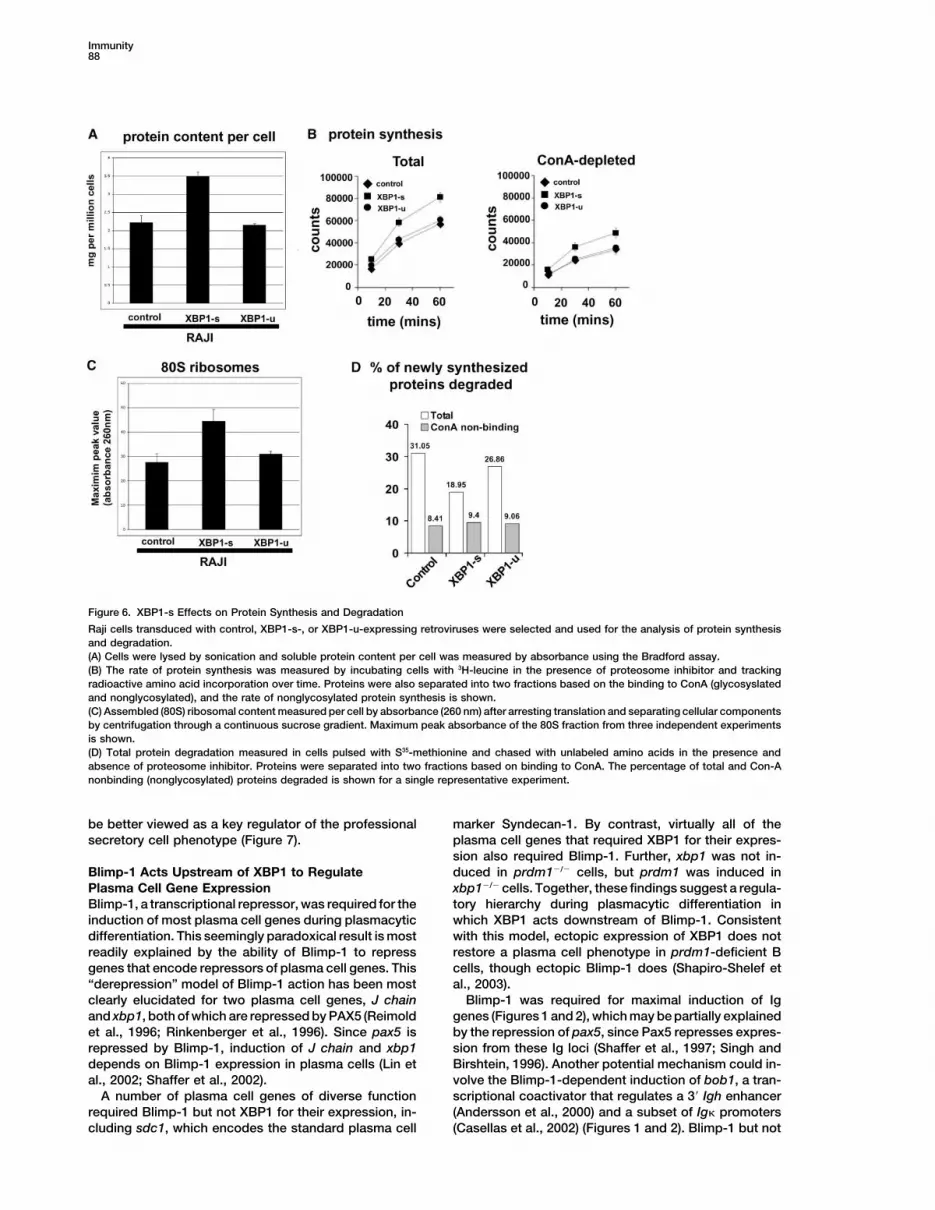
Immunity88
Figure 6. XBP1-s Effects on Protein Synthesis and Degradation
Raji cells transduced with control, XBP1-s-, or XBP1-u-expressing retroviruses were selected and used for the analysis of protein synthesisand degradation.(A) Cells were lysed by sonication and soluble protein content per cell was measured by absorbance using the Bradford assay.(B) The rate of protein synthesis was measured by incubating cells with 3H-leucine in the presence of proteosome inhibitor and trackingradioactive amino acid incorporation over time. Proteins were also separated into two fractions based on the binding to ConA (glycosyslatedand nonglycosylated), and the rate of nonglycosylated protein synthesis is shown.(C) Assembled (80S) ribosomal content measured per cell by absorbance (260 nm) after arresting translation and separating cellular componentsby centrifugation through a continuous sucrose gradient. Maximum peak absorbance of the 80S fraction from three independent experimentsis shown.(D) Total protein degradation measured in cells pulsed with S35-methionine and chased with unlabeled amino acids in the presence andabsence of proteosome inhibitor. Proteins were separated into two fractions based on binding to ConA. The percentage of total and Con-Anonbinding (nonglycosylated) proteins degraded is shown for a single representative experiment.
be better viewed as a key regulator of the professional marker Syndecan-1. By contrast, virtually all of theplasma cell genes that required XBP1 for their expres-secretory cell phenotype (Figure 7).sion also required Blimp-1. Further, xbp1 was not in-duced in prdm1�/� cells, but prdm1 was induced inBlimp-1 Acts Upstream of XBP1 to Regulate
Plasma Cell Gene Expression xbp1�/� cells. Together, these findings suggest a regula-tory hierarchy during plasmacytic differentiation inBlimp-1, a transcriptional repressor, was required for the
induction of most plasma cell genes during plasmacytic which XBP1 acts downstream of Blimp-1. Consistentwith this model, ectopic expression of XBP1 does notdifferentiation. This seemingly paradoxical result is most
readily explained by the ability of Blimp-1 to repress restore a plasma cell phenotype in prdm1-deficient Bcells, though ectopic Blimp-1 does (Shapiro-Shelef etgenes that encode repressors of plasma cell genes. This
“derepression” model of Blimp-1 action has been most al., 2003).Blimp-1 was required for maximal induction of Igclearly elucidated for two plasma cell genes, J chain
and xbp1, both of which are repressed by PAX5 (Reimold genes (Figures 1 and 2), which may be partially explainedby the repression of pax5, since Pax5 represses expres-et al., 1996; Rinkenberger et al., 1996). Since pax5 is
repressed by Blimp-1, induction of J chain and xbp1 sion from these Ig loci (Shaffer et al., 1997; Singh andBirshtein, 1996). Another potential mechanism could in-depends on Blimp-1 expression in plasma cells (Lin et
al., 2002; Shaffer et al., 2002). volve the Blimp-1-dependent induction of bob1, a tran-scriptional coactivator that regulates a 3� Igh enhancerA number of plasma cell genes of diverse function
required Blimp-1 but not XBP1 for their expression, in- (Andersson et al., 2000) and a subset of Ig� promoters(Casellas et al., 2002) (Figures 1 and 2). Blimp-1 but notcluding sdc1, which encodes the standard plasma cell

XBP1 Regulates Secretory Cell Phenotype89
Figure 7. A Model for the Regulatory Biology of Plasma Cell Differentiation
Blimp-1 acts upstream of XBP1 to initiate plasma cell differentiation. ER stress in a nonsecretory cell induces an “ER-stress” UPR in whichXBP1, PERK, and ATF6 are activated, leading to decreased ER stress or apoptosis (red box). Alternatively, XBP1 activation in a differentiatingsecretory cell initiates a “physiological” UPR that antagonizes the effects of PERK on translation, perhaps by inducing DNAJC3. XBP1 alsocoordinately alters the cell structure and function to create a professional secretory cell. Together, the activities of Blimp-1 and XBP1 driveplasma cell differentiation (green box).
XBP1 is also required for the switch in the usage of Igh suggests that XBP1 regulates essentially every stage ofthe secretory process, including the targeting of proteinsmRNA polyadenylation sites, shifting expression fromto the ER (srp9, srp54, rpn1), translocation of proteinsthe membrane-bound to the secreted form of Ig heavyacross the ER membrane (ssr1, ssr3, ssr4, srpr, tram1,chain (Alt et al., 1980; Early et al., 1980; Reimold et al.,sec61a, sec61g), cleavage of signal peptides (spc22/23,2001; Rogers et al., 1980; Shapiro-Shelef et al., 2003).h13/spp), folding of ER proteins (dnaJb9, hspa5/BiP,Although the factor(s) mediating this important switcherp70, grp58, pdir, txndc5, fkbp11, ppib), degradationare unknown, an interesting candidate is the RNA poly-of misfolded ER proteins by the ERAD pathway (edem,merase II processivity factor ELL2 (Shilatifard et al.,sel1h), protein glycosylation (slc33a1, ddost, gcs1,1997) that we found to be induced during plasmacyticman1b1, siat7d, dad1, slc33a1, b4galt2, mgat2, fut8),differentiation in a Blimp-1-dependent fashion (FiguresER-Golgi vesicular trafficking (cope, os-9, sec24C,1 and 2). The substantial number of genes, like ell2, thatsec23B, golgb1, mcdf2, vdp, arcn1), endosomal traffick-depend exclusively on Blimp-1 for induction (Figure 2)ing (rabac1), and targeting of secretory vesicles to thepoint to its unique and essential role in promotingplasma membrane (arhq/tc10). Importantly, XBP1 in-plasma cell differentiation.duces the expression of many components of the multi-protein chaperone complex that binds and processes
XBP1 and Biogenesis of the Secretory Apparatus nascent Ig heavy chains (Erp72, Hsp40 homologs, PDI,Previous work has demonstrated that XBP1, as well as PPIB, Bip) (Meunier et al., 2002), which may specificallyits yeast homolog HAC1p, regulate ER stress-induced contribute to the increased efficiency of Ig secretion.genes that promote folding of ER proteins and degrada- Interestingly, gene expression profiling experiments intion of misfolded ER proteins through the ERAD pathway yeast cells experiencing ER stress suggested a similarly(Lee et al., 2003; Mori et al., 1998; Yoshida et al., 2003). broad role for HAC1 in the secretory pathway (TraversRecent studies have also shown that there is overlap in et al., 2000). During the yeast UPR, HAC1 induces a hostthe genes induced during plasmacytic differentiation of genes involved in protein translocation into the ER,and those induced by ER stress (Gass et al., 2002; Iwa- glycosylation, ER protein folding, ER protein degrada-koshi et al., 2003a; Lee et al., 2003; van Anken et al., tion in the ERAD pathway, vesicular transport, and phos-2003). pholipid biosynthesis. Since not every secretory path-
The present study defines an expanded role for XBP1 way gene was upregulated by the UPR in yeast, HAC1in enhancing the secretory capacity of plasma cells. A may be devoted to remodeling the secretory pathwayremarkably consistent set of XBP1 target genes to specifically cope with an excess of unfolded proteinsemerged from studies of xbp1-deficient mouse B cells (Travers et al., 2000). Another interesting aspect of thisand ectopic XBP1 expression, many of which encode study was the finding that mutations in the UPR pathway
and the ERAD pathway were synthetically lethal underproteins that function in the secretory pathway. Our data

Immunity90
normal growth conditions, revealing a role for HAC1 and Therefore, the increase in protein synthesis caused byXBP1 cannot be explained by upregulation of mRNAsthe “UPR” in the absence of overt ER stress.
XBP1 has not only retained the broad, UPR-related, encoding glycosylated proteins in the secretory path-way but must be due to a more general mechanism.transcriptional program of HAC1 but also has the ability
to physically expand the ER compartment. In both One possible scenario involves the XBP1 target geneDNAJC3 (this study and Lee et al. [2003]), which encodeslymphoid and epithelial cell lines, ectopic XBP1 dramati-
cally augmented the ER, and in myeloma cells, knock- p58IPK, an inhibitor of PERK (van Huizen et al., 2003; Yanet al., 2002). When PERK is activated, it phosphorylatesdown of XBP1 diminished the ER. Notably, IRE1 and
HAC1 are required for the induction of a specialized form eIF2�, leading to decreased translation initiation of mostproteins. Therefore, the upregulation of p58IPK by XBP1of ER known as karmellae that is formed in response to
forced overexpression of ER proteins (Cox et al., 1997). could increase total protein synthesis by antagonizingPERK (Figure 7). XBP1 also increased the abundanceIn multicellular eukaryotes, unlike yeast, terminal differ-
entiation in some cell lineages results in a “professional” of assembled ribosomes per cell, which might contributeto the increase in total protein synthesis, and it is possi-secretory cell that has an expanded ER as a fixed attri-
bute. We propose that the function of HAC1 in inducing ble that the condensation of nucleoli that we observedin XBP1-expressing cells may play a role in this process.ER biogenesis under stress conditions has been
adapted in evolution to allow XBP1 to stably increase Interestingly, plasma cells are characterized by increasednuclear size and the presence of a single prominentER size during secretory cell differentiation. In keeping
with this hypothesis, XBP1 is not only highly expressed nucleolus (Benjamin et al., 1984), features that may beattributable to XBP1.in plasma cells but also in secretory tissues such as
the exocrine pancreas and salivary glands (Clauss etal., 1993). Evolution of XBP1 as a Master Regulator
of Secretory Cell DifferentiationWhereas yeast have a single pathway that senses un-XBP1: Beyond the Endoplasmic Reticulum
Unexpectedly, ectopic expression of processed XBP1 folded proteins in the ER, multicellular eukaryotes haveevolved three parallel pathways: IRE1/XBP1, PERK/in diverse cell lines increased overall cell size and, con-
versely, knockdown of XBP1 expression by RNA inter- ATF4, and ATF6. In evolution, the creation of parallelpathways by genome duplication can set the stage forference in a myeloma cell line decreased cell size. In
part, these findings could be explained by the ER expan- subsequent functional specialization. It has thereforebeen proposed that the three mammalian UPR pathwayssion that is induced by XBP1. However, XBP1 expres-
sion also increased nuclear size, mitochondrial mass, have diverged functionally during evolution (Figure 7;Calfon et al., 2002; Harding et al., 2002), and our dataand lysosomal content, suggesting that the cell size
phenotype is part of a coordinated increase in multiple strongly support this hypothesis.XPB1 has retained important features of its yeast or-intracellular structures. These functional capabilities of
XBP1 have not been reported for HAC1, suggesting that tholog HAC1, while developing additional functions thatpromote development of the secretory phenotype. LikeXBP1 has acquired additional regulatory roles during
evolution. HAC1, XBP1 transactivated a host of genes encodingproteins that function throughout the secretory path-The mechanism by which XBP1 exerts such pleiotro-
pic effects on cellular organelles is not clear at present. way. Further, our experiments revealed that XBP1 issufficient to expand the ER in a wide variety of mamma-Not only did mitochondrial mass increase but so did
mitochondrial function, as assessed by the mitochon- lian cells, which may be functionally analogous theHAC1-mediated formation of ER-like karmellae.drial membrane potential. Two XBP1 target genes,
cox15 and ndufa1, encode proteins that are required for However, XBP1 has acquired new functional capabili-ties that have not been reported for HAC1. Most notably,electron transport in the mitochondrion (Antonicka et
al., 2003; Yadava et al., 2002), and the XBP1 target gene we found that XBP1 increased overall protein synthesis,which clearly would favor high-level protein secretion.acas2l encodes a mitochondrial enzyme that synthe-
sizes acetyl-CoA for use in oxidation. However, it is In marked contrast, PERK signaling inhibits proteintranslation, a clearly undesirable attribute for a secretorynot know whether the increased expression of these
mitochondrial proteins would stimulate respiration or cell. Indeed, it is possible that XBP1 evolved in orderfor professional secretory cells to modulate the activitywhether they play a role in the XBP1-induced increase
in mitochondrial mass. Three XBP1 target genes encode of PERK. In this regard, it is notable that the PERKpathway upregulates chop (Harding et al., 2000; Yan etlysosomal proteins involved in lipid hydrolysis (lipa), fu-
sion of autophagic vesicles with the lysosome (lamp2), al., 2002), which encodes a mediator of apoptosis duringER stress, whereas the IRE1/XBP1 pathway does notand possibly in small molecule transport into the lyso-
some (laptm4a). Again, it is not known whether upregula- (this study and Gass et al. [2002]; Lee et al. [2003]). Thus,PERK serves as a checkpoint protein during ER stresstion of these lysosomal proteins can account for the
increased lysosomal biogenesis in XBP1-expressing that can decrease the unfolded protein load in the ERby inhibiting translation or eliminate the cell if the ERcells.
Another unanticipated observation was that XBP1 in- stress is severe and prolonged. XBP1, on the other hand,has apparently evolved to promote translation by antag-creased protein synthesis globally by 30%–50%, which
could contribute to the increase in organelle content onizing PERK, thereby allowing cells to differentiate intoprofessional secretory cells that can tolerate a constitu-and cell size. XBP1-s increased the synthesis of both
nonglycosylated and glycosylated proteins equivalently. tively high throughput of ER proteins.

XBP1 Regulates Secretory Cell Phenotype91
Flow Cytometry and MicroscopyIt may therefore be helpful to define a “physiological”Please see http://lymphochip.nih.gov/ShafferPCfactors/. For deter-UPR (Gass et al., 2002) that is activated constitutively inmination of protein synthesis, degradation, and ribosome content,professional secretory cells through the action of XBP1,please see http://lymphochip.nih.gov/ShafferPCfactors/.
and an “ER stress” UPR that is activated by unfoldedproteins in nonsecretory cells through the action of AcknowledgmentsPERK (Figure 7). Though activated during in vitro plasma
We would like to thank the members of all contributing labs forcell differentiation (Gass et al., 2002), the role of ATF6helpful discussions and generosity with reagents. We would like toin the physiological UPR remains unclear and will requirethank all those who have thusfar contributed RNA samples andthe analysis of ATF6-deficient animals to clarify this fac-cDNA clones to the Mouse Lymphochip project (especially H. Morse,
tor’s role in plasma cell differentiation. The physiological M. Potter, S. Janz, Y. Tagaya); Drs. E. Snapp and J. Lippincott-UPR that XBP1 coordinates promotes secretion and is Schwartz for the gift of the ER-targeted gfp vector; and Bob Straus-characterized by increased protein synthesis and bio- berg and the Cancer Genome Anatomy Project (National Cancer
Institute) as well as the Leukemia and Lymphoma Society for fund-genesis of ER, mitochondria, and lysosomes. The in-ing, in part, the creation of the Mouse Lymphochip Microarray. Thiscreased mitochondrial mass and respiration in the phys-work was in part supported by NIH grants AI32412 (LHG), AI50659iological UPR could be used by secretory cells to meet(KC) and AI43576 (KC), an Award from the Multiple Myeloma Re-
the energy demands of sustained high-level secretion. search Foundation (L.H.G.), and an Irvington Institute PostdoctoralThe benefit to a secretory cell of lysosomal biogenesis Fellowship Award (N.N.I.).is less clear, but it is possible that autophagy, which
Received: March 3, 2004requires lysosome function, may contribute biosyntheticRevised: April 23, 2004precursors needed for high-level protein secretion. TheAccepted: May 19, 2004present study has expanded our view of XBP1 functionPublished: July 20, 2004from that of a transcription factor that activates a subset
of ER stress genes to that of a key regulator responsible Referencesfor many of the cellular changes that occur during differ-entiation of secretory cells. Alizadeh, A., Eisen, M., Davis, R.E., Ma, C., Sabet, H., Tran, T.,
Powell, J., Yang, L., Marti, G., Moore, T., et al. (1999). The lympho-chip: a specialized cDNA microarray for the genomic-scale analysisExperimental Proceduresof gene expression in normal and malignant lymphocytes. ColdSpring Harb. Symp. Quant. Biol. 64, 71–78.Mice and In Vitro LPS B Cell DifferentiationAlt, F.W., Bothwell, A.L., Knapp, M., Siden, E., Mather, E., Koshland,Please see http://lymphochip.nih.gov/ShafferPCfactors/.M., and Baltimore, D. (1980). Synthesis of secreted and membrane-bound immunoglobulin mu heavy chains is directed by mRNAs thatCell Lines and Retroviral Constructsdiffer at their 3� ends. Cell 20, 293–301.Cell lines were grown as described at http://lymphochip.nih.gov/Andersson, T., Samuelsson, A., Matthias, P., and Pettersson, S.ShafferPCfactors/. Retroviral transduction of human and mouse cell(2000). The lymphoid-specific cofactor OBF-1 is essential for thelines was performed as described (Shaffer et al., 2000, 2002) usingexpression of a V(H) promoter/HS1,2 enhancer-linked transgene inconstructs expressing only the puromycin resistance gene (control)late B cell development. Mol. Immunol. 37, 889–899.or vectors expressing a form of XBP1 and the puromycin resistance
gene as part of an IRES-containing bicistronic vector (see http:// Antonicka, H., Mattman, A., Carlson, C.G., Glerum, D.M., Hoffbuhr,lymphochip.nih.gov/ShafferPCfactors/). The pRETROsuper retrovi- K.C., Leary, S.C., Kennaway, N.G., and Shoubridge, E.A. (2003).ral construct with only a puromycin resistance gene was used as a Mutations in COX15 produce a defect in the mitochondrial hemecontrol vector. An shRNA against human XBP1 was designed using biosynthetic pathway, causing early-onset fatal hypertrophic car-the Dharmacon search engine, converted to a hairpin loop with diomyopathy. Am. J. Hum. Genet. 72, 101–114.resistriction enzyme overhanging ends, and cloned via BglII/ Benjamin, D., Magrath, I.T., Triche, T.J., Schroff, R.W., Jensen, J.P.,HindIII sites into pRETROSUPER (see http://lymphochip.nih.gov/ and Korsmeyer, S.J. (1984). Induction of plasmacytoid differentiationShafferPCfactors/). After infection, pools of cells were selected with by phorbol ester in B-cell lymphoma cell lines bearing 8;14 translo-puromycin at 1 �g/ml. cations. Proc. Natl. Acad. Sci. USA 81, 3547–3551.
Calame, K.L., Lin, K.I., and Tunyaplin, C. (2003). Regulatory mecha-RNA Preparation nisms that determine the development and function of plasma cells.For mouse lymphochip analysis, the total RNA was prepared using Annu. Rev. Immunol. 21, 205–230.Trizol (Invitrogen), and for human lymphochip analysis, polyA� RNA Calfon, M., Zeng, H., Urano, F., Till, J.H., Hubbard, S.R., Harding,was prepared using FastTrack kits (Invitrogen). H.P., Clark, S.G., and Ron, D. (2002). IRE1 couples endoplasmic
reticulum load to secretory capacity by processing the XBP-1Microarray Analysis mRNA. Nature 415, 92–96.The Mouse Lymphochip Microarray Casellas, R., Jankovic, M., Meyer, G., Gazumyan, A., Luo, Y., Roeder,The mouse lymphochip is a spotted cDNA array similar in construc- R., and Nussenzweig, M. (2002). OcaB is required for normal tran-tion to the human lymphochip (Alizadeh et al., 1999). For murine scription and V(D)J recombination of a subset of immunoglobulinmicroarray analysis, experimental sample RNAs were converted to kappa genes. Cell 110, 575–585.Cy5-labeled cDNA and cohybridized with RNA from a reference pool
Chapman, R.E., and Walter, P. (1997). Translational attenuation me-labeled with Cy3 (http://lymphochip.nih.gov/ShafferPCfactors/).
diated by an mRNA intron. Curr. Biol. 7, 850–859.The Human Lymphochip Microarray
Clauss, I.M., Gravallese, E.M., Darling, J.M., Shapiro, F., Glimcher,Gene expression analysis using human lymphochips has been pre-M.J., and Glimcher, L.H. (1993). In situ hybridization studies suggestviously described (Alizadeh et al., 1999). RNA from control anda role for the basic region-leucine zipper protein hXBP-1 in exocrineXBP1-transduced cells were compared directly on arrays withgland and skeletal development during mouse embryogenesis. Dev.RNAs labeled as described in the figure legends. Microarray dataDyn. 197, 146–156.analysis (hierarchical clustering) and presentation was performed
using the Cluster and Treeview (Alizadeh et al., 1999). Data files for Cox, J.S., and Walter, P. (1996). A novel mechanism for regulatingactivity of a transcription factor that controls the unfolded proteinall experiments can be found at http://lymphochip.nih.gov/
ShafferPCfactors/. response. Cell 87, 391–404.

Immunity92
Cox, J.S., Shamu, C.E., and Walter, P. (1993). Transcriptional induc- a subset of endoplasmic reticulum resident chaperone genes in theunfolded protein response. Mol. Cell. Biol. 23, 7448–7459.tion of genes encoding endoplasmic reticulum resident proteins
requires a transmembrane protein kinase. Cell 73, 1197–1206. Lin, Y., Wong, K., and Calame, K. (1997). Repression of c-myc tran-scription by Blimp-1, an inducer of terminal B cell differentiation.Cox, J.S., Chapman, R.E., and Walter, P. (1997). The unfolded proteinScience 276, 596–599.response coordinates the production of endoplasmic reticulum pro-
tein and endoplasmic reticulum membrane. Mol. Biol. Cell 8, 1805– Lin, K.-I., Angelin-Duclos, C., Kuo, T.C., and Calame, K. (2002).1814. Blimp-1-dependent repression of Pax-5 is required for differentia-
tion of B cells to IgM secreting plasma cells. Mol. Cell Biol. 22, 4771–Deng, Y., Bennink, J.R., Kang, H.C., Haugland, R.P., and Yewdell,4780.J.W. (1995). Fluorescent conjugates of brefeldin A selectively stain
the endoplasmic reticulum and Golgi complex of living cells. J. Histo- Lin, K.I., Tunyaplin, C., and Calame, K. (2003). Transcriptional regula-chem. Cytochem. 43, 907–915. tory cascades controlling plasma cell differentiation. Immunol. Rev.
194, 19–28.Early, P., Rogers, J., Davis, M., Calame, K., Bond, M., Wall, R.,and Hood, L. (1980). Two mRNAs can be produced from a single Ma, Y., and Hendershot, L.M. (2003). The stressful road to antibodyimmunoglobulin mu gene by alternative RNA processing pathways. secretion. Nat. Immunol. 4, 310–311.Cell 20, 313–319.
Meunier, L., Usherwood, Y.K., Chung, K.T., and Hendershot, L.M.Fewell, S.W., Travers, K.J., Weissman, J.S., and Brodsky, J.L. (2001). (2002). A subset of chaperones and folding enzymes form multipro-The action of molecular chaperones in the early secretory pathway. tein complexes in endoplasmic reticulum to bind nascent proteins.Annu. Rev. Genet. 35, 149–191. Mol. Biol. Cell 13, 4456–4469.Gass, J.N., Gifford, N.M., and Brewer, J.W. (2002). Activation of Mori, K. (2000). Tripartite management of unfolded proteins in thean unfolded protein response during differentiation of antibody- endoplasmic reticulum. Cell 101, 451–454.secreting B cells. J. Biol. Chem. 277, 49047–49054.
Mori, K., Ogawa, N., Kawahara, T., Yanagi, H., and Yura, T. (1998).Harding, H.P., Zhang, Y., and Ron, D. (1999). Protein translation and Palindrome with spacer of one nucleotide is characteristic of thefolding are coupled by an endoplasmic-reticulum-resident kinase. cis-acting unfolded protein response element in SaccharomycesNature 397, 271–274. cerevisiae. J. Biol. Chem. 273, 9912–9920.Harding, H.P., Novoa, I., Zhang, Y., Zeng, H., Wek, R., Schapira, M., Mori, K., Ogawa, N., Kawahara, T., Yanagi, H., and Yura, T. (2000).and Ron, D. (2000). Regulated translation initiation controls stress- mRNA splicing-mediated C-terminal replacement of transcriptioninduced gene expression in mammalian cells. Mol. Cell 6, 1099– factor Hac1p is required for efficient activation of the unfolded pro-1108. tein response. Proc. Natl. Acad. Sci. USA 97, 4660–4665.Harding, H.P., Novoa, I., Bertolotti, A., Zeng, H., Zhang, Y., Urano, Okada, T., Yoshida, H., Akazawa, R., Negishi, M., and Mori, K. (2002).F., Jousse, C., and Ron, D. (2001). Translational regulation in the Distinct roles of activating transcription factor 6 (ATF6) and double-cellular response to biosynthetic load on the endoplasmic reticulum. stranded RNA-activated protein kinase-like endoplasmic reticulumCold Spring Harb. Symp. Quant. Biol. 66, 499–508. kinase (PERK) in transcription during the mammalian unfolded pro-
tein response. Biochem. J. 366, 585–594.Harding, H.P., Calfon, M., Urano, F., Novoa, I., and Ron, D. (2002).Transcriptional and translational control in the Mammalian unfolded Patil, C., and Walter, P. (2001). Intracellular signaling from the endo-protein response. Annu. Rev. Cell Dev. Biol. 18, 575–599. plasmic reticulum to the nucleus: the unfolded protein response in
yeast and mammals. Curr. Opin. Cell Biol. 13, 349–355.Harding, H.P., Zhang, Y., Zeng, H., Novoa, I., Lu, P.D., Calfon, M.,Sadri, N., Yun, C., Popko, B., Paules, R., et al. (2003). An integrated Piskurich, J.F., Lin, K.I., Lin, Y., Wang, Y., Ting, J.P., and Calame,stress response regulates amino acid metabolism and resistance K. (2000). BLIMP-I mediates extinction of major histocompatibilityto oxidative stress. Mol. Cell 11, 619–633. class II transactivator expression in plasma cells. Nat. Immunol.
1, 526–532.Haze, K., Yoshida, H., Yanagi, H., Yura, T., and Mori, K. (1999).Mammalian transcription factor ATF6 is synthesized as a transmem- Poot, M., and Pierce, R.H. (1999). Detection of changes in mitochon-brane protein and activated by proteolysis in response to endoplas- drial function during apoptosis by simultaneous staining with multi-mic reticulum stress. Mol. Biol. Cell 10, 3787–3799. ple fluorescent dyes and correlated multiparameter flow cytometry.
Cytometry 35, 311–317.Iwakoshi, N.N., Lee, A.H., and Glimcher, L.H. (2003a). The X-boxbinding protein-1 transcription factor is required for plasma cell Reimold, A.M., Ponath, P.D., Li, Y.S., Hardy, R.R., David, C.S., Strom-differentiation and the unfolded protein response. Immunol. Rev. inger, J.L., and Glimcher, L.H. (1996). Transcription factor B cell194, 29–38. lineage-specific activator protein regulates the gene for human
X-box binding protein 1. J. Exp. Med. 183, 393–401.Iwakoshi, N.N., Lee, A.H., Vallabhajosyula, P., Otipoby, K.L., Rajew-sky, K., and Glimcher, L.H. (2003b). Plasma cell differentiation and Reimold, A.M., Etkin, A., Clauss, I., Perkins, A., Friend, D.S., Zhang,the unfolded protein response intersect at the transcription factor J., Horton, H.F., Scott, A., Orkin, S.H., Byrne, M.C., et al. (2000). AnXBP-1. Nat. Immunol. 4, 321–329. essential role in liver development for transcription factor XBP-1.
Genes Dev. 14, 152–157.Kawahara, T., Yanagi, H., Yura, T., and Mori, K. (1998). Unconven-tional splicing of HAC1/ERN4 mRNA required for the unfolded pro- Reimold, A.M., Iwakoshi, N.N., Manis, J., Vallabhajosyula, P., Szo-tein response. Sequence-specific and non-sequential cleavage of molanyi-Tsuda, E., Gravallese, E.M., Friend, D., Grusby, M.J., Alt,the splice sites. J. Biol. Chem. 273, 1802–1807. F., and Glimcher, L.H. (2001). Plasma cell differentiation requires
the transcription factor XBP-1. Nature 412, 300–307.Klintworth, G.K., Hawkins, H.K., and Smith, C.F. (1979). Acridineorange particles in cultured fibroblasts. A comparative study of mac- Reljic, R., Wagner, S.D., Peakman, L.J., and Fearon, D.T. (2000).ular corneal dystrophy, systemic mucopolysaccharidoses types I-H Suppression of signal transducer and activator of transcriptionand II, and normal controls. Arch. Pathol. Lab. Med. 103, 297–299. 3-dependent B lymphocyte terminal differentiation by BCL-6. J. Exp.
Med. 192, 1841–1848.Kozutsumi, Y., Segal, M., Normington, K., Gething, M.J., and Sam-brook, J. (1988). The presence of malfolded proteins in the endoplas- Rinkenberger, J.L., Wallin, J.J., Johnson, K.W., and Koshland, M.E.mic reticulum signals the induction of glucose-regulated proteins. (1996). An interleukin-2 signal relieves BSAP (Pax5)-mediated re-Nature 332, 462–464. pression of the immunoglobulin J chain gene. Immunity 5, 377–386.
Lafrenz, D., Koretz, S., Stratte, P.T., Ward, R.B., and Strober, S. Rogers, J., Early, P., Carter, C., Calame, K., Bond, M., Hood, L., and(1982). LPS-induced differentiation of a murine B cell leukemia Wall, R. (1980). Two mRNAs with different 3� ends encode mem-(BCL1): changes in surface and secreted IgM. J. Immunol. 129, 1329– brane-bound and secreted forms of immunoglobulin mu chain. Cell1335. 20, 303–312.
Schliephake, D.E., and Schimpl, A. (1996). Blimp-1 overcomes theLee, A.H., Iwakoshi, N.N., and Glimcher, L.H. (2003). XBP-1 regulates

XBP1 Regulates Secretory Cell Phenotype93
block in IgM secretion in lipopolysaccharide/anti-mu F(ab�) Yoshida, H., Okada, T., Haze, K., Yanagi, H., Yura, T., Negishi, M.,and Mori, K. (2000). ATF6 activated by proteolysis binds in the pres-2-co-stimulated B lymphocytes. Eur. J. Immunol. 26, 268–271.ence of NF-Y (CBF) directly to the cis-acting element responsibleSchubert, U., Anton, L.C., Gibbs, J., Norbury, C.C., Yewdell, J.W.,for the mammalian unfolded protein response. Mol. Cell. Biol. 20,and Bennink, J.R. (2000). Rapid degradation of a large fraction of6755–6767.newly synthesized proteins by proteasomes. Nature 404, 770–774.Yoshida, H., Matsui, T., Yamamoto, A., Okada, T., and Mori, K. (2001).Shaffer, A.L., Peng, A., and Schlissel, M.S. (1997). In vivo occupancyXBP1 mRNA is induced by ATF6 and spliced by IRE1 in responseof the kappa light chain enhancers in primary pro- and pre-B cells:to ER stress to produce a highly active transcription factor. Cella model for kappa locus activation. Immunity 6, 131–143.107, 881–891.
Shaffer, A.L., Yu, X., He, Y., Boldrick, J., Chan, E., and Staudt, L.Yoshida, H., Matsui, T., Hosokawa, N., Kaufman, R.J., Nagata, K.,(2000). BCL-6 represses genes that function in lymphocyte differen-and Mori, K. (2003). A time-dependent phase shift in the mammaliantiation, inflammation and cell cycle control. Immunity 13, 199–212.unfolded protein response. Dev. Cell 4, 265–271.
Shaffer, A.L., Lin, K.-I., Kuo, T.C., Yu, X., Hurt, E.M., Rosenwald, A.,Giltnane, J.M., Yang, L., Zhao, H., Calame, K., et al. (2002). Blimp-1 orchestrates plasma cell differentiation by extinguishing the ma-ture B cell gene expression program. Immunity 17, 51–62.
Shapiro-Shelef, M., Lin, K.I., McHeyzer-Williams, L.J., Liao, J.,McHeyzer-Williams, M.G., and Calame, K. (2003). Blimp-1 is requiredfor the formation of immunoglobulin secreting plasma cells and pre-plasma memory B cells. Immunity 19, 607–620.
Shen, X., Ellis, R.E., Lee, K., Liu, C.Y., Yang, K., Solomon, A., Yoshida,H., Morimoto, R., Kurnit, D.M., Mori, K., et al. (2001). Complementarysignaling pathways regulate the unfolded protein response and arerequired for C. elegans development. Cell 107, 893–903.
Shilatifard, A., Duan, D.R., Haque, D., Florence, C., Schubach, W.H.,Conaway, J.W., and Conaway, R.C. (1997). ELL2, a new member ofan ELL family of RNA polymerase II elongation factors. Proc. Natl.Acad. Sci. USA 94, 3639–3643.
Sidrauski, C., and Walter, P. (1997). The transmembrane kinase Ire1pis a site-specific endonuclease that initiates mRNA splicing in theunfolded protein response. Cell 90, 1031–1039.
Singh, M., and Birshtein, B.K. (1996). Concerted repression of animmunoglobulin heavy-chain enhancer, 3� alpha E(hs1,2). Proc. Natl.Acad. Sci. USA 93, 4392–4397.
Smiley, S.T., Reers, M., Mottola-Hartshorn, C., Lin, M., Chen, A.,Smith, T.W., Steele, G.D., Jr., and Chen, L.B. (1991). Intracellularheterogeneity in mitochondrial membrane potentials revealed by aJ-aggregate-forming lipophilic cation JC-1. Proc. Natl. Acad. Sci.USA 88, 3671–3675.
Terasaki, M., Song, J., Wong, J.R., Weiss, M.J., and Chen, L.B.(1984). Localization of endoplasmic reticulum in living and glutaral-dehyde-fixed cells with fluorescent dyes. Cell 38, 101–108.
Travers, K.J., Patil, C.K., Wodicka, L., Lockhart, D.J., Weissman,J.S., and Walter, P. (2000). Functional and genomic analyses revealan essential coordination between the unfolded protein responseand ER-associated degradation. Cell 101, 249–258.
van Anken, E., Romijn, E.P., Maggioni, C., Mezghrani, A., Sitia, R.,Braakman, I., and Heck, A.J. (2003). Sequential waves of functionallyrelated proteins are expressed when B cells prepare for antibodysecretion. Immunity 18, 243–253.
van Huizen, R., Martindale, J.L., Gorospe, M., and Holbrook, N.J.(2003). P58IPK, a novel endoplasmic reticulum stress-inducible pro-tein and potential negative regulator of eIF2alpha signaling. J. Biol.Chem. 278, 15558–15564.
Wiest, D.L., Burkhardt, J.K., Hester, S., Hortsch, M., Meyer, D.I., andArgon, Y. (1990). Membrane biogenesis during B cell differentiation:most endoplasmic reticulum proteins are expressed coordinately.J. Cell Biol. 110, 1501–1511.
Wright, G., Tan, B., Rosenwald, A., Hurt, E.H., Wiestner, A., andStaudt, L.M. (2003). A gene expression-based method to diagnoseclinically distinct subgroups of diffuse large B cell lymphoma. Proc.Natl. Acad. Sci. USA 100, 9991–9996.
Yadava, N., Potluri, P., Smith, E.N., Bisevac, A., and Scheffler, I.E.(2002). Species-specific and mutant MWFE proteins. Their effect onthe assembly of a functional mammalian mitochondrial complex I.J. Biol. Chem. 277, 21221–21230.
Yan, W., Frank, C.L., Korth, M.J., Sopher, B.L., Novoa, I., Ron, D.,and Katze, M.G. (2002). Control of PERK eIF2alpha kinase activityby the endoplasmic reticulum stress-induced molecular chaperoneP58IPK. Proc. Natl. Acad. Sci. USA 99, 15920–15925.


