Embed Size (px)
Citation preview

Zebrafish mutations affecting cilia motility share similar cysticphenotypes and suggest a mechanism of cyst formation thatdiffers from pkd2 morphants
Jessica Sullivan-Brown1, Jodi Schottenfeld1, Noriko Okabe1, Christine L. Hostetter1,Fabrizio C. Serluca2, Stephan Y. Thiberge1, and Rebecca D. Burdine1*
1Department of Molecular Biology, Princeton University, NJ, USA
2Novartis Institutes for Biomedical Research. Developmental and Molecular Pathways. Cambridge, MA02139
AbstractZebrafish are an attractive model for studying the earliest cellular defects occurring during renal cystformation because its kidney (the pronephros) is simple and genes that cause cystic kidney diseases(CKD) in humans, cause pronephric dilations in zebrafish. By comparing phenotypes in threedifferent mutants, locke, swt and kurly, we find that dilations occur prior to 48 hpf in the medialtubules, a location similar to where cysts form in some mammalian diseases. We demonstrate thatthe first observeable phenotypes associated with dilation include cilia motility and luminalremodeling defects. Importantly, we show that some phenotypes common to human CKD, such asan increased number of cells, are secondary consequences of dilation. Despite having differences incilia motility, locke, swt and kurly share similar cystic phenotypes, suggesting that they function ina common pathway. To begin to understand the molecular mechanisms involved in cyst formation,we have cloned the swt mutation and find that it encodes a novel leucine rich repeat containing protein(LRRC50), which is thought to function in correct dynein assembly in cilia. Finally, we show thatknockdown of polycystic kidney disease 2 (pkd2) specifically causes glomerular cysts and does notaffect cilia motility, suggesting multiple mechanisms exist for cyst formation.
Keywordspronephros; cyst; kidney; zebrafish; cilia; locke; switch hitter; lrrc50; kurly; pkd2; oda7; nephron
IntroductionRenal cystic kidney diseases are pleiotrophic disorders that are clinically variable andgenetically diverse. Depending on the type of disease, cysts can occur in different locations ofthe kidney and can present at various stages of life ranging from fetal development to adulthood.The most prevalent form of renal cystic disease is Autosomal Dominant Polycystic Kidney
*Author for correspondence, Rebecca D. Burdine Department of Molecular Biology, Princeton University, Washington Road, Mof 433,Princeton, NJ 08544 USA, Phone 1-609-258-7515, Fax 1-609-258-1343, Email [email protected]'s Disclaimer: This is a PDF file of an unedited manuscript that has been accepted for publication. As a service to our customerswe are providing this early version of the manuscript. The manuscript will undergo copyediting, typesetting, and review of the resultingproof before it is published in its final citable form. Please note that during the production process errors may be discovered which couldaffect the content, and all legal disclaimers that apply to the journal pertain.Cover Figure Legend — Jodi Schottenfeld Wildtype anterior pronephric tubule showing the Na+/K+ ATPase (red) with Z0-1 (green),counterstained with Hoechst nuclear dye (blue) in a 48 hour zebrafish embryo. The picture beautifully illustrates the basolaterallocalization of the Na+/K+ ATPase opposed to the apical localization of ZO-1.
NIH Public AccessAuthor ManuscriptDev Biol. Author manuscript; available in PMC 2009 February 15.
Published in final edited form as:Dev Biol. 2008 February 15; 314(2): 261–275.
NIH
-PA Author Manuscript
NIH
-PA Author Manuscript
NIH
-PA Author Manuscript

Disease (ADPKD), which can occur in 1:1000 individuals and is caused by mutations in thegenes PKD1 or PKD2 (1994;1995;Burn et al., 1995;Gabow, 1993). ADPKD is furtherassociated with extra-renal pathologies such as liver cysts and cardiac deformities (Perrone,1997). The onset of clinical symptoms begins later in adulthood and cysts have been shown toarise from all areas of the nephron, with a majority of focal cysts occurring in the collectingducts (Baert, 1978;Verani and Silva, 1988). By contrast, Autosomal Recessive PKD (ARPKD)is rare, occurring in 1:20,000 live births, and cysts form mainly in the collecting ducts early inutero (Zerres et al., 1998). The pathological presentation of cysts in ARPKD shows fusiformdilations, while in ADPKD the cysts are focally derived and bud from the epithelium (Wilson,2004). ARPKD is caused by mutations in the gene fibrocystin (PKHD1), which is alsoassociated with congenital defects in the liver (Onuchic et al., 2002;Ward et al., 2002).Nephronophthisis disorders are characterized by renal cysts occurring in the corticomedullaryjunction of the kidney, which present later during childhood or early adulthood (Hildebrandtand Zhou, 2007). Bardet-Biel Syndrome is associated with a number of clinical presentationssuch as retinal degeneration, obesity, polydactyly and in some cases renal cysts (Blacque andLeroux, 2006). Despite the diversity of these syndromes, the majority of gene products mutatedin each of these cases are known to associate with the cilium, centrosome or basal bodies(reviewed in (Torres and Harris, 2006). The discoveries that these diseases stem from afunctional or structural defect related to cilia are so prevalent that these groups of disordershave been collectively described as “ciliopathies” (Badano et al., 2006;Hildebrandt and Zhou,2007).
Although the location and timing of cyst development varies in different disease states, thecellular defects observed in renal cysts are similar. Cystic dilations are characterized byincreased cellular proliferation and increased fluid secretion possibly due to the apicalmistargeting of ion channel proteins like the Na+/K+ATPase or EGFR/ErbB2 heterodimers(reviewed in (Wilson, 2004). Since the majority of proteins known to be involved in renalcystic diseases are found associated with the cilium, much research has been focused onuncovering the physiological significance of this (once thought to be vestigial) organelle. Themechanosensory hypothesis is the most common functional role assigned to renal cilia, inwhich the physical bending of cilia results in the elevation of cytoplasmic calciumconcentrations (Praetorius et al., 2003; Praetorius and Spring, 2001). It is thought that this risein calcium could participate in cellular signaling events that are critical for tubular integrity.Defects in cilia may prevent these processes, leading to unregulated cell growth and a lessdifferentiated epithelium.
The zebrafish pronephros provides an attractive model system for studying cyst formation atthe cellular level. The pronephros is the first kidney to form in all vertebrates and is replacedby a more advanced kidney in mammals (Vize et al., 1997). Although the pronephros inmammals is thought to be non-functional, the pronephros in zebrafish is active, acts as a closedsystem, and mimics mammalian kidneys in terms of gene expression and maintaining osmotichomeostasis (Drummond et al., 1998). In essence, the pronephros is a simple version of themore advanced mammalian kidney. The functional unit of the pronephros is termed the nephronand is composed of two tubules connecting to a fused glomerulus. The glomerulus functionsas the site of filtration, where hydrostatic pressure from the capillaries and the glomerularcapsule work to force fluid, salts and other small inorganic molecules nonselectively intoBowman’s space. The tubules and ducts are generally composed of highly reabsorptive cellsthat further refine the filtrate by allowing ions and salts back into the blood. The zebrafishpronephros forms very early in development, as cells fated to become the glomerulus, tubulesand ducts are specified by the 8 somite stage (12 hours post-fertilization (hpf)) (Serluca andFishman, 2001). Similar to the mammalian system, expression of wt1 (wilm’s tumor 1) marksthe glomerular fated tissue and pax2.1 marks the tubule and duct precursors (Serluca andFishman, 2001). By 24 hpf the glomerular fated tissue, which began as bilateral progenitor
Sullivan-Brown et al. Page 2
Dev Biol. Author manuscript; available in PMC 2009 February 15.
NIH
-PA Author Manuscript
NIH
-PA Author Manuscript
NIH
-PA Author Manuscript

populations on either side of midline, migrates to the midline and fuses at 50 hpf (Drummondet al., 1998). Glomerular filtration begins between 36–48 hpf as evidenced by injectingrhodamine dextran into the circulatory system and monitoring when the dye first appears inthe pronephric lumen (Drummond et al., 1998). Thus, the zebrafish pronephros developsquickly, and with a simple anatomy, allowing for earlier phenotypes to be more easilyaddressed.
In the zebrafish pronephros, cilia lining the tubules and ducts differ from mammalian renalcilia. Cells of the pronephros show a “salt and pepper” array of both single ciliated cells andmulticiliated cells (Liu et al., 2007; Ma and Jiang, 2007). The cilia on the multiciliated cellshave a 9+2 microtubule arrangement, a striking difference from mammalian tubules whichpossess immotile 9+0 cilia. Furthermore, cilia in the zebrafish pronephros, and in thepronephros of other fish species, are motile and this motility is thought to be important formoving fluid through the kidney (Kramer-Zucker et al., 2005; Lacy et al., 1989). It has beenshown by other laboratories that cilia motility is affected in the cystic kidneys of zebrafish at2–3 days post-fertilization (dpf) (Kramer-Zucker et al., 2005; Omori and Malicki, 2006).Despite these differences, the prevalence of ciliary defects related to cystic kidney phenotypesis highly conserved from mammals to teleosts.
Here we provide a detailed analysis of the spatial and temporal progression of pronephric cystdevelopment in zebrafish in three mutants with cilia motility defects; locke, switch hitter(swt) and kurly. From this time course, our results indicate that cystic dilations occur first inthe medial region of the pronephric tubules and may involve defects in luminal remodeling.This is in contrast to the pkd2 morphant, in which the dilation is restricted to the glomerularregion. Furthermore, we uncovered different functional roles of each gene product in regulatingciliogenesis or cilia motility that precede or act in parallel to cystic expansion. We have beenable to clone the swt mutation to a gene encoding a novel leucine rich repeat containing protein(LRRC50) which is expressed in tissues that contain motile cilia. By undertaking this timecourse of cyst formation in the zebrafish, we provide the foundation to further understand theprimary cause of tubular dilation, better characterize the cellular defects occurring in cysticcells, and study ciliary defects related to cystogenesis.
MATERIAL AND METHODSZebrafish Strains
locke (to237b), switch hitter (tm317) and kurly (tm304) were obtained from a large scale ENUmutagenesis screen (Brand et al., 1996; Haffter et al., 1996). locke, switch hittertm317 andkurly alleles were generated in the Tü strain and maintained by outcrossing to AB, WIK andPWT. switch hitterfk03a was isolated in a recessive genetic screen for visual kidney defects.The mutant allele was generated in the TL background and crossed to the WIK line to generateheterozyous mapping pairs. The kidney phenotypes associated with each allele have beenobserved across multiple generations. Embryos were collected and raised per standardprotocols.
switch hitter Positional CloningThe swt locus was mapped using SSLP markers (Liao and Zon, 1999). The swt genomic intervalwas narrowed between markers z11119 and z15270 on chromosome 7, where lrrc50 wasidentified as a candidate gene. To identify mutations we amplified each exon from mutantgenomic DNA and compared the sequences obtained with wildtype sibilings and Tü sequence.
Sullivan-Brown et al. Page 3
Dev Biol. Author manuscript; available in PMC 2009 February 15.
NIH
-PA Author Manuscript
NIH
-PA Author Manuscript
NIH
-PA Author Manuscript

HistologyEmbryos were fixed in 4% paraformaldehyde (PFA; Sigma P6148)/Phosphate Buffered Saline(PBS) overnight at 4°C. After gradual dehydration into ethanol, embryos were infiltrated andembedded in JB-4 plastic resin (JB-4 Embedding Kit, EMS #14270-00) according the protocolprovided by Electron Microscopy Sciences. All embryos were sectioned at 4µm on a LeicaRM2255 Rotary Microtome. Sections were then stained with Haematoxylin and Eosin dyesaccording to our standard laboratory protocols (available upon request).
Counting Cell NumberTo perform this analysis, embryos were sectioned in a transverse orientation at 4µm in JB-4plastic resin. Sections were collected in consecutive order and stained with Haematoxylin andEosin which readily distinguishes nuclei for counting. We counted nuclei in 15 consecutivesections (roughly 60µm) from the glomerular/anterior region and medial region of the kidney.The start of the glomerular/anterior region began when both the glomerulus and a cross-sectionthrough a tubule were observed in the same section, and ended in the anterior tubular region(Figure 2, B,C). The medial tubule sections were defined as a point posterior to the visceralorgans where the gut had migrated toward the middle of the body (Figure 2D). Cells werecounted from the anterior region at 2 dpf and 3 dpf. We did not count kidney cells in the anteriorregion at later time points because the tubules extending from the glomerulus becomeconvoluted, making the analysis inconsistent.
ImmunofluorescenceStaining for F-actin was performed on cryosections. Embryos were fixed in 4% PFA at 4°Covernight, washed once with PBS, and soaked in 10% sucrose/PBS at 4°C until the embryossank to the bottom of the epitube. Next, embryos were soaked in 25% sucrose/PBS at 4°C untilthe embryos sank. Embryos were then embedded in Tissue-Tek® O.C.T. compound whichwas allowed to solidify on dry ice. The embryos were sectioned at 8µm using the Leica CM3050S. Sections were washed in PBS with 0.005% saponin (PBS-saponin) twice and blockedin 5% normal goat serum (NGS) in PBS-saponin at room temperature for 30 min. Sampleswere incubated with rhodamine-phalloidin at room temperature for 1 hour. After washing threetimes for 20 min each, samples were equilibrated in 50% glycerol/PBS.
ZO-1 and Na+/K+ ATPase stainings were performed in whole mount embryos, then embeddedand sectioned in JB-4 plastic resin. Embryos were fixed in Dent’s solution (80% MeOH; 20%DMSO) overnight at 4°C. Embryos were slowly rehydrated into PBDT (1% DMSO, 0.1%Tween 20/PBS) and blocked for 2 hours in PBDT containing 10% NGS. Antibodies to ZO-1(Zymed) and Na+/K+ ATPase (α6F; Developmental Studies Hybridoma Bank, U. of Iowa)were added at a 1:200 and 1:25 concentration, respectively. Primary antibodies were added at4°C overnight. Samples were washed 5 times, 30 minutes each wash in 1%NGS/PBDT. ZO-1was detected using the secondary antibody FITC goat anti-mouse IgG1 (Southern BioTech,Birmingham, AL) and the Na+/K+ ATPase was detected using Alexa Fluor® 568 goat anti-mouse IgG2a antibody, both at a concentration of 1:500. To visualize the nuclei, Hoechst dyewas added with the secondary antibody at a 12.96 µM concentration overnight. The next day,embryos were washed with 1% NGS/PBDT buffer for 5 times, 30 minutes each wash. Embryoswere embedded according to the JB-4 histology protocol provided by the manufacturer,however the dehydration steps were performed quickly and the embedding steps wereperformed at 4°C. Sections were collected at 4µm. Both cryosection and JB-4 plastic sectionswere coverslipped in a 50% glycerol/PBS solution and imaged with a ZEISS LSM 510 confocalmicroscope.
The acetylated tubulin immunostaining was performed on embryos fixed either in Dent’s or4% PFA overnight. All staining procedures were performed in whole mount in a manner similar
Sullivan-Brown et al. Page 4
Dev Biol. Author manuscript; available in PMC 2009 February 15.
NIH
-PA Author Manuscript
NIH
-PA Author Manuscript
NIH
-PA Author Manuscript

to the protocol mentioned above. The anti-acetylated tubulin antibody (Sigma #T6793) wasadded at a concentration of 1:400 and the goat anti-mouse IgG2b secondary antibody (SouthernBioTech, Birmingham, AL) was added at a concentration of 1:500. Embryos were mountedand imaged in AquaPolyMount (Polysciences, Inc, Warrington, PA) with a ZEISS LSM510confocal microscope.
Video MicroscopyEmbryos were rinsed in double distilled H2O to remove any residual methlylene blue or saltsfrom the embryo medium. A glass depression slide (Ward’s Natural Science) was prepared byfilling the depression with 3% methylcellulose (Sigma M-0387) and 2 drops of 0.4% Tricaine(MS-222; Sigma A-5040). Embryos were oriented along their lateral sides and cilia motilitywas recorded starting in the cloaca and posterior regions (data not shown). We continuedimaging the tubule from posterior to anterior until we reached the medial segments. All moviesshown were recorded in the medial segments. The tubules were imaged with an Olympus BX51up-right microscope equipped with a 60X water immersion objective and differentialinterference contrast (DIC) optics. Video was recorded with an iXon camera (AndorTechnology) and Luca camera (Andor Technology). The Luca camera incorporates frametransfer technology which allows relatively fast image acquisition. We recorded movies withthe iXon camera between 10–20 frames per second (fps) and with the Luca camera at 158 fps.All movies were slowed down to 8 fps, except supplementary video #3 with was slowed to 15fps.
Cilia Frequency MeasurementsAs frequency measurements obtained from movie analysis could be impaired by a low framerate, we built a specialized set-up to perform high precision cilia frequency measurements. Ashutter controlled infra-red laser beam at 900nm, entering the microscope, was focused on thecilia and a small portion of the scattered light was collected by an optical fiber connected toan avalanche photodiode. As the cilia moves in and out of the focused beam during the periodicmovement, the amount of scattered light entering the fiber varies accordingly. The entrance ofthe fiber was brought to close proximity of the fish (~1mm). The detector output was recordedfor several seconds with a sampling rate of one millisecond. Finally, a Fourier transformanalysis of the recorded trace was made using Matlab to uncover the frequency content of thedata. While the beam was focused on the cilia, the frequency spectrum revealed very sharppics that disappeared when focusing the laser elsewhere in the animal. Care was taken to uselow laser intensity. We found that 10mW power entering the back aperture of the objectivewas sufficient for high signal collection and harmless to the animal. As we could not appreciateany systematic variation of the cilia frequency over the time the shutter was opened, weconcluded the laser has no indirect effect on the cilia movement.
RNA whole-mount in situ hybridizationDIG-labeled RNA probes were used in RNA in situ hybridization using standard methods. Theswt cDNA was amplified from a zebrafish 24 hpf cDNA library and cloned into Bluescript KS(Stratagene). For the sense probe, the plasmid was linearized with KpnI and transcribed withT3 enzyme and for the antisense probe the plasmid was linearized with SacI and transcribedwith T7 enzyme.
RESULTSlocke, switch hitter and kurly mutants develop pronephric cysts
locke, switch hitter (swt) and kurly mutants were originally generated in the Tübingenmutagenesis screen as recessive mutations that displayed a “curly tail down” phenotype and
Sullivan-Brown et al. Page 5
Dev Biol. Author manuscript; available in PMC 2009 February 15.
NIH
-PA Author Manuscript
NIH
-PA Author Manuscript
NIH
-PA Author Manuscript

developed cystic dilations in the pronephros (Brand et al., 1996; Haffter et al., 1996). The curlytail phenotype is not linked to the development of pronephric cysts, as some mutants with acurly tail down phenotype do not develop pronephric dilations (Brand et al., 1996) and JSB-personal observations). The curly tail down phenotype is first observed at 26 hpf, which is thefirst time point where we can morphologically distinguish the mutants from their siblings. By3 dpf, both locke and swt exhibit a strong downward curve in their body axis, however thephenotype observed in kurly mutants is less severe (Figure 1, A–H). At 3 dpf, cystic dilationsare observable by light microscopy in a region slightly posterior to the ear, shown by the blackarrowheads. By 5 dpf, the cystic dilations have increased in size (Figure 1, I–P). Although thesize of the cysts varies in mutants from the same clutch, swt and kurly mutants tend to developlarger cysts than locke mutant embryos. Despite their cystic developments, locke, swt andkurly mutants rarely become grossly edemic. Because edema is a hallmark sign of glomerularfiltration failure, we believe that the kidneys are functioning in these mutant embryos.
Cystic dilations first appear in the medial tubules in locke, swt and kurly mutantsThe temporal and spatial development of pronephric cystic phenotypes was assessed byperforming a histological time course. Figure 2 shows representative histological sections ofa 2 dpf wildtype embryo, highlighting different regions of the pronephros along the anteriorto posterior axis. Throughout the text, we will be referring to 5 distinct regions; the glomerulus,anterior tubules, medial tubules, posterior tubules and the cloaca region (Figure 2). Cysticdilations are first observed under simple light microscopy in the glomerular region at 2.5 dpf.However, we can detect earlier tubular dilation by histology and DIC at 2 dpf. At 2 dpf, themedial tubules are clearly dilated in all embryos examined as evidenced by histology (Figure3, E–H). Interestingly, in these mutants the the glomerular region and posterior portions of thekidney appear normal (Figure 3, A–D, data not shown). Although some embryos have begunto develop dilations in the glomerulus around 2 dpf, this is always accompanied by dilationsin the medial tubular region. These cystic dilations appear fusiform in nature, as they are widerat a specific area of the kidney and tapered at either end This is similar architecture to thatobserved in ARPKD cysts (Menezes and Onuchic, 2006). We first consistently observeglomerular dilations at 2.5 dpf which progress over time (Figure 3, I–L). Increased filtratevolumes into Bowman’s capsule are clearly evident, causing a disorganization of the capillariesand the associated podocytes. The extended glomerulus is observed both along the proximal-distal axis and anterior-posterior axis. Although the medial tubules continue to become dilated(Figure 3, M–P), the more posterior tubules are less affected, approaching the size of thewildtype tubules (Figure 3, Q–T). Cloaca development in locke, swt and kurly mutants appearsnormal, as the two pronephric ducts fuse in the posterior segment of the kidney and the lumenremains intact (data not shown). By performing this histological time course, we find thatdilations first occur by 2dpf in the medial tubules which are analogous to the proximalconvoluted and straight tubules in higher vertebrates (Wingert et al., 2007), a region wheredilations are observed in PKD.
Increased number of cells around the cystic kidney tubules is secondary to dilationIncreased cell proliferation is a characteristic trait of nephron dilation in mammalian renalcystic diseases (Nadasdy et al., 1995; Ramasubbu et al., 1998). Since cell proliferation has notbeen examined in zebrafish pronephric cyst formation, we determined whether there wereincreased numbers of cells surrounding mutant pronephric tubules. Data collected at 2 dpf didnot show a significant difference in cell number in the glomerular/anterior region despiteobserved dilations (Figure 4). However, by 3 dpf, locke and kurly showed an increase in cellnumber surrounding the tubules. The anterior region at 5 dpf could not be examined becausethe pronephric tubules become convoluted. The medial region of the kidney was then examinedat 2 dpf, 3 dpf and 5 dpf. At 2 dpf, all mutants showed dilations in the medial region, howeverthe cell number surrounding the kidney tubule was not statistically significant from the
Sullivan-Brown et al. Page 6
Dev Biol. Author manuscript; available in PMC 2009 February 15.
NIH
-PA Author Manuscript
NIH
-PA Author Manuscript
NIH
-PA Author Manuscript

wildtype tubules. By 3 dpf there was a significant increase in cell number when compared tothe wildtype tubules which became more pronounced at 5 dpf (Table 1 and Figure 4). This isthe first time an increased number of cells surrounding cystic mutant tubules has been reportedin zebrafish. However, this increase occurs after the tubules have become dilated, (see above)and indicates that an increase in cell number is a secondary effect and not the primary causeof cyst formation in zebrafish.
Pronephric lumen size decreases over time in wildtype embryos, while mutant lumen sizeincreases
As stated previously, we first observe cystic dilations in the medial tubules by 48 hpf throughhistology and DIC microscopy. At earlier time points (26–30 hpf) we found that the wildtypemedial tubules had a larger luminal diameter compared to the posterior tubules (Figure 5, Eand G). Over time, the lumen size in the medial tubules of wildtype embyos decreases, whichis supported by our observation of decreased cell number surrounding the medial tubules from2 dpf to 3–5 dpf (Figure 4 and Table 1).
When mutant embryos were examined at 26–30 hpf, the size of the medial and posterior tubulelumens were similar to wildtype (Figure 5, F and H). However, in mutant embryos there is aconsistent increase in the medial lumen diameter size by 48 hpf when compared to wildtypetubule lumens (Figure 5 I,J). These data suggest that wildtype tubules undergo a remodelingprocess to refine tubule size, and that these processes are affected in the mutant tubules.
Localization of the Na+/K+ ATPase is disrupted in mutant kidney cells, while apical polarityis intact
Cysts are classically defined as fluid-filled cavities lined by an epithelium. As observed inFigure 3, the cells lining the cysts have altered morphology and often appear flattened.Therefore, we sought to determine if epithelial polarity is affected in locke, swt and kurly mutantembryos by staining for F-actin and the tight junction marker ZO-1. At 2 dpf cystic expansionshave begun in the medial positions of the kidney, however, F-actin and ZO-1 localization inthese cells appeared normal (Figure 6 A–D and data not shown). At 4 dpf, although the tubulesare largely expanded, the localization of F-actin and ZO-1 surrounding the kidney tubules atthe apical surface remained normal (Figure 6, E–H and data not shown). The dedifferentiationof renal cells from a highly absorptive state to a secretory state is thought to be a leading factorin cystic fluid accumulation (Thomson et al., 2003). Some studies in mammals and zebrafishhave shown that the Na+/K+ ATPase is mistargeted to the apical surface of renal cystic cells,where secretion of sodium into the lumen can result in fluid accumulation (Avner et al.,1992;Drummond, 2003;Wilson et al., 2000). However this theory is controversial, and otherstudies involving both human and mouse cystic tissue failed to show altered polarity of theNa+/K+ ATPase (Brill et al., 1996;Thomson et al., 2003). Therefore, we sought to determineif the Na+/K+ ATPase was mislocalized to the apical surface in locke, swt and kurly mutantembryos. We analyzed Na+/K+ ATPase localization at 4 dpf in the anterior tubules, as thislocation showed the clearest basolateral staining in wildtype embryos. In locke, swt andkurly mutants, Na+/K+ ATPase localization was significantly altered as the strict baso-lateralstaining observed in the wildtype was not evident. In some cells, apical expression of theNa+/K+ ATPase was also observed (Figure 6, I,L, white arrows). Because the cells at this timepoint have abnormal morphology, we believe the defects in Na+/K+ ATPase localization aresecondary to increased fluid secretion in the tubules. Furthermore in kurly mutant embryos wedid observe correct basolateral localization of the Na+/K+ ATPase at 30hpf, before tubuledilation begins (data not shown).
Sullivan-Brown et al. Page 7
Dev Biol. Author manuscript; available in PMC 2009 February 15.
NIH
-PA Author Manuscript
NIH
-PA Author Manuscript
NIH
-PA Author Manuscript

Cilia motility is affected prior to tubule dilation in locke, swt and kurly mutantsThe connection of ciliary defects, whether structural or functional, with renal cystic diseaseshas been repeatedly established. However, we wanted to determine if cilia motility was affectedprior to tubule dilation. We found that locke, swt and kurly mutants have unique cilia motilitydefects at 26–30 hpf by taking video recordings of cilia movement (Material and Methods,Supplementary Data). In wildtype embryos, pronephric cilia are motile at 26 hpf, but they donot appear to bundle or show the coordinated cilia movement seen in our recordings at 2 and3 dpf (Supplementary Video #1 – #3). Cilia motility was evident from the cloaca region to themedial part of the kidney (data not shown). Analysis of more anterior cilia is not possiblebecause the yolk obstructs imaging. At 26–30 hpf, locke mutants had motile cilia that exhibiteda quick flickering movement, but with a reduced range of motion compared to wildtype(Supplementary Video #4). In contrast to locke mutants, swt mutants displayed completelyimmotile cilia (Supplementary Video #5). kurly mutants have plieotropic cilia motilityphenotypes ranging from immotile cilia to cilia that are motile but beat irregularly(Supplementary Video #6). Cilia were also examined at these early stages by immunostainingwith acetylated tubulin. Although swt and kurly mutants showed grossly normal lengths ofcilia, locke mutants had shortened cilia (Figure 5 A–D). Our results on cilia length in locke arein agreement with those recently published (Zhao and Malicki, 2007)
The cilia motility phenotypes observed in locke and swt mutants at 26–30 hpf remain consistentat 2 dpf and 3 dpf (Supplementary Video #7–Supplementary Video #8 and data not shown).Interestingly, we found that kurly mutants at 2 dpf had some cilia that bundled and movedsimilar to wildtype (Supplementary Video #2 and Supplementary Video #9). To moreaccurately compare the cilia motility in kurly mutants to that observed in wildtype embryos,we developed a method to determine the frequency of cilia movement at high precision bycollecting and measuring scattered infra-red light (see Material and Methods). Through thisanalysis we determined that both wildtype and kurly mutant ciliary bundles move atapproximately 42 beats/second (Hz) at 48–52 hpf (Figure 5, K,L). This is in contrast to thepreviously determined frequency of 20 Hz obtained by counting cilia beats at a later time point(60 hpf) by Kramer-Zucker et. al. (Kramer-Zucker et al, 2005). The infra-red recordingtechnique could not be applied to swt or locke embryos because the mutants never formedciliary bundles and the amount of light scattered by a single cilium was too low to detect.Importantly, the cystic phenotypes we observed in all three mutants are similar despite eachmutant having striking differences in cilia motility.
swt encodes a novel leucine rich repeat containing protein (LRRC50) that is required forciliary motility in zebrafish
To better understand how these gene products are affecting cilia motility and cyst formation,it will be important to determine the molecular nature of the affected genes. To this end, wehave cloned swt and find it encodes a novel leucine rich repeat containing protein (lrrc50;Figure 7). LRRC50 in zebrafish is a 562 amino acid protein with a predicted molecular weightof 63kDa. This protein contains six leucine rich repeats (LRR) at the N-terminus followed bya leucine cap domain and a coiled-coil domain. These domains are proposed to function asprotein-protein interaction domains, however the function of these motifs in swt are unknown.We have two alleles of swt which display similar pronephric cyst phenotypes and defects inleft-right patterning (J.S.B and R.D.B. in preparation). A missense mutation in the swttm317
allele changes an asparagine (AAC) to lysine (AAA) at amino acid position 172 within one ofthe leucine rich repeats (Figure 7, A,B). This mutation may affect the ability of the LRR domainto interact with appropriate targets. The second allele, swtfk03a, creates a stop codon at position259 which is predicted to truncate the protein before the coiled-coil domain (Figure 7, A,B).lrrc50 is an ortholog to ODA7, a gene previously cloned in the green alga Chlamydomonasthat was found to prevent axonemal outer row dynein assembly (Freshour et al., 2007;Kamiya,
Sullivan-Brown et al. Page 8
Dev Biol. Author manuscript; available in PMC 2009 February 15.
NIH
-PA Author Manuscript
NIH
-PA Author Manuscript
NIH
-PA Author Manuscript

1988). Mutations in ODA7 were originally identified in Chlamydomonas by selecting formutant strains showing reduced swimming velocity and reduced flagellar beat frequency.However unlike swt mutants, the cilia/flagella in ODA7 mutants were not completely immotile(Kamiya, 1988). Based on a series of biochemical fractionation studies, Freshour et. al. reportthat ODA7 serves as a structural bridge between the outer and inner row dynein motors in theaxoneme, suggesting a role for coordinating cilia motility (Freshour et al., 2007). Becauseswt mutants show immotile cilia in the kidney and neural tube (Supplementary Video #5 andSupplementary Video #8, data not shown), we were interested to determine where the geneproduct was expressed. swt RNA is maternally deposited in the embryo, as shown by RT-PCR(data not shown). By RNA in situ analysis, we observed swt expression in Kupffer’s vesicleat the tailbud stage (Figure 7, C). Kupffer’s vesicle is analogous to the mouse node in terms ofhaving a critical role in left-right patterning and possessing motile cilia (Bisgrove and Yost,2006). At the 12ss, the swt RNA expression is found in the floorplate of the neural tube, in theintermediate mesoderm that gives rise to the pronephros, and the otic vesicle (Figure 7, D–G).At 24 hpf, swt RNA is observed in the kidney tubules and in the neural tube concentrated atthe chordo-neural hinge (Figure 7, H). Thus, swt RNA was found in multiple tissues that areknown to have motile cilia, further demonstrating the role of LRRC50 in cilia motility.
Zebrafish pkd2 morphants develop glomerular dilations, but not tubule dilationsMutations in polycystic kidney disease2 (pkd2) are the second leading cause of ADPKD(Gabow, 1993). However pkd2 mutants in zebrafish do not develop any type of tubule dilationsor kidney cysts (Schottenfeld et al., 2007). It has widely been reported that morpholinos topkd2 in zebrafish do cause kidney cysts (Bisgrove et al., 2005; Obara et al., 2006; Schottenfeldet al., 2007; Sun et al., 2004). Because locke, swt and kurly all displayed defects in cilia motility,we were interested if pkd2 morphants also had defects in cilia motility. To test if cilia motilitywas affected in pkd2 morphants, we examined renal cilia by video microscopy at 2dpf (seeMaterial and Methods). In agreement with previous data from Obara et. Al, we found that thecilia in the pkd2 morphants beat in a similar coordinated fashion to wildtype cilia, indicatingthat cilia motility is not affected in pkd2 morphants (data not shown). However, our resultsindicate that the defect observed in pkd2 morphants is different than the cysts characterized inlocke, swt and kurly mutants. pkd2 morphants develop glomerular dilations similar to lockemutants at 3 dpf (Figure 8, A–D), but the tubules and ducts never become dilated. Figure 8 (E–H) depicts the anterior tubule region, which is slightly posterior to the glomerulus, where organssuch as the gut, swim bladder and liver are apparent. Although the tubules are clearly dilatedin locke mutant embryos (Figure 8,H) the tubules in pkd2 morphants are similar in size towildtype embryos (Figure 8, E–G). This phenotype is further evident in the medial region ofthe tubule (Figure 8, I–L). These data demonstrate that the pkd2 morphants have glomerulardilations, but morphologically do not develop the same dilations observed in the other threegenetic backgrounds with cilia motility defects. This suggests that there are differentmechanisms involved in generating pronephric dilations. These data demonstrate that the cysticdilations in pkd2 morphants are restricted to the glomerular tissue which differs from themutants examined with cilia motility defects. This suggests that different types of pronephriccysts are formed by distinct mechanisms in zebrafish.
DISCUSSIONUtilizing zebrafish phenotypes to provide insights into human cystic diseases
One of the most powerful aspects of using zebrafish to understand human disease is the abilityto perform detailed analyses to determine the first relevant phenotypes that occur. This isespecially important in complex diseases such as PKD, where a network of cellular phenotypeshave been detailed in the disease process, but the primary causes versus secondaryconsequences have not been elucidated. Based on our histological and cellular analysis, we
Sullivan-Brown et al. Page 9
Dev Biol. Author manuscript; available in PMC 2009 February 15.
NIH
-PA Author Manuscript
NIH
-PA Author Manuscript
NIH
-PA Author Manuscript

present a model of when and where relevant phenotypes occur during cyst formation inzebrafish (Figure 9). The first phenotypes we observe affect cilia motility (discussed below).We find that cystic dilations occur first in the medial tubules prior to 48 hpf, while glomerulardilations are not consistently observed until 2.5 dpf. This result is significant because it showsthat cystic dilations in these mutants are initially occurring in the medial tubules, which shareconsiderable homology to the proximal convoluted and straight tubules (Wingert et al.,2007). This location is important as it further underscores the homology between zebrafishcyst formation and human disease. For example, in ARPKD, the cysts have been shown totransiently arise from the proximal tubules, then over time predominantly affect the collectingducts (Nakanishi et al., 2000). Furthermore, it has been shown that the glomerulus can becomedilated in human renal cystic diseases and in mouse models of ADPKD as a secondary effectto the tubular dilation, similar to what we observed in zebrafish cystic dilations (Bernstein,1993;Tanner et al., 2002).
Defects in cellular remodeling may contribute to cystic dilations in locke, swt and kurlymutants
Although cystic dilations have been characterized in zebrafish embryos, this is the first reportto show an increase number of cells surrounding the tubules. Since cellular overproliferationis a phenotype known to be associated with human PKD, we counted the number of cellssurrounding the tubular lumens in wildtype and in each mutant background. By performingthis experiment, we made two interesting observations. First, we found that at 3 dpf, the cellnumber increases significantly in the mutant medial tubules compared to wildtype. However,we did not observe an increased number of cells in the medial tubules at 2 dpf, indicating thatthe increased cell number is not the initial cause of the cystic dilations. It should be noted thatat 2 dpf, a few tubules in locke mutants did appear to have an increased number of cellscompared to age matched siblings. However, in these mutant tubules the lumens appearedlarger than in mutants without a cell number increase‥ Therefore, we believe that increases incell number likely occur soon after dilation begins and may be a direct consequence of cellularresponses to dilation. The increase in cell number that was observed at 3 dpf continued to riseat 5 dpf. This result suggests that the cystic cells observed in locke, swt and kurly mutants mayhave an overproliferation phenotype secondary to the initial cause of dilation. In human PKD,cystogenesis is hallmarked by increased rates of both cell proliferation and apoptosis (reviewedin (Simons and Walz, 2006) and upregulation of the ERK/BRAF/MAPK due to increasedcAMP levels (Yamaguchi et al., 2003). Currently, most therapeutic approaches directly targeteither regulation of cell cycle (CKD inhibitor roscovitine, EGFR tyrosine kinase inhibitors,)or apoptotic events (mTOR inhibitor rapamycin) (Ibraghimov-Beskrovnaya, 2007;Shillingford et al., 2006)reviewed in (Torres and Harris, 2007). Given that locke, swt andkurly have an increased number of cells that surround the cystic lumens, it will be interestingto determine in further studies if these drugs reduce cyst size in zebrafish embryos.
Our second interesting observation is that the number of cells surrounding the wildtype medialtubule decreases from 2 dpf to 3 dpf, while the number of cells increases in the mutant tubules.This result suggests that the wildtype tubule is undergoing a reorganization process to extendthe pronephros along the anterior to posterior axis and/or regulate the general size of lumendiameter. It has been shown recently that the lengthening of the renal tubule in mice is governedby a planar cell polarity process which orients the plane of mitotic division in parallel to thetubular axis (Fischer et al., 2006). These authors also found that this regulated mitoticorientation was significantly affected in murine polycystic kidney models (Fischer et al.,2006). It will be interesting to determine whether planar cell polarity processes regulatezebrafish pronephric development and if these processes are defective in locke, swt andkurly mutants.
Sullivan-Brown et al. Page 10
Dev Biol. Author manuscript; available in PMC 2009 February 15.
NIH
-PA Author Manuscript
NIH
-PA Author Manuscript
NIH
-PA Author Manuscript

The role of cilia in pronephric cyst formationAlthough cystic dilations in locke, swt and kurly mutants appear to be occurring at similar timepoints and in similar locations, the cilia motility defects associated with each mutant areremarkably different. It has been suggested that cilia motility defects in zebrafish mutantembryos creates a general back-up of fluid flow in the nephrons and that this is the primarycause of cystic dilations (Kramer-Zucker et al., 2005). Although fluid back-up may exaggeratethe phenotypes, we believe that this hypothesis does not fully explain the observed cysticdilations in locke, swt and kurly mutants for the following reasons. First, the mutant embryosrarely become grossly edemic. This implies that the kidney is still functioning and fluid isexiting the body. We have preliminary data that suggest when the cloaca does not open theembryos do not develop enlarged tubules, but do become grossly edemic. Second, swt andkurly display opposite cilia motility defects; swt have completely immotile cilia while kurlycilia are motile and can move at the same frequency as wildtype when bundled. However, bothmutants develop pronephric cysts. Interestingly, in swt the size of the cysts under lightmicroscopy varies significantly between mutant embryos even though these mutants showedthe most consistent phenotype in cilia motility defects. These data lead us to hypothesize thatthe cilia in the pronephric tubules may have other functions in addition to moving fluid throughthe pronephric duct. Furthermore, because the cystic phenotype in locke, swt and kurly embryosare highly similar, this suggests that a common pathway is affected in these mutants. Like inmammalian systems, these cilia could have roles in mechanosensation and regulation of cellcycle. Further work exploring additional roles for zebrafish pronephric cilia is a future focusof our laboratory.
As a first step towards understanding how our mutants contribute to cilia motility and cystformation, we have identified the gene product mutated in swt. swt encodes a novel leucinerich repeat containing protein (LRRC50) that may function in protein-protein interactions. Thisgene is the ortholog of the Oda7 gene in the green alga Chlamydomonas where it has beenshown to function in outer dynein assembly in axoneme structure. Mutations in Oda7 result inslower swimming behavior and reduced flagella beat frequency. This is in contrast to the swtmutant where cilia are completely immotile. It will be interesting to determine if the axonemestructure is disrupted in swt mutants to determine if the function of Lrrc50 is conserved. Wefind that swt RNA is expressed in cells that contain motile cilia including Kupffer’s vesicle,the neural tube, kidney and otic vesicle. In the kidney, swt is expressed along the entire lengthof the nephron and appears to be required there for cilia motility. This is supported by ourfindings that immotile cilia are observed in the swt mutants from the cloaca to the medial regionof the nephron. It is interesting however, that the dilations are not observed throughout theentire nephron as the more posterior and cloaca regions are unaffected. Thus it is likely thatthe medial tubule cells are more sensitive to the loss of swt function.
pkd2 morphants suggest an alternate mechanism for cyst formationBy providing a histological time course of pronephric cyst development, we were able tocompare the cystic dilations observed in pkd2 morphants with cysts in locke, swt and kurlymutants. Unlike locke, swt and kurly, pkd2 morphants have motile pronephric cilia that beatsimilar to wildtype cilia. Interestingly, the pkd2 morphants did not develop cysts analogous tothe ciliary motility mutants, as the dilations appeared to be restricted to the glomerular region.Likewise, pkd2 null mice develop a preponderance of glomerular cysts (Wu et al., 2000). Thus,the pkd2 morphants in zebrafish are more similar to pkd2 nulls in mice. This is in contrast tothe mouse unstable Pkd2WS25 allele that develops cysts in the distal nephron segments and isa more analogous model to human PKD. (Wu et al., 2000). The observed glomerular dilationsin pkd2 morphants may indicate a function of pkd2 specifically in the glomerulus or during thedevelopment of glomerular fated tissue. We note that in Obara et. al they report cystic dilationsin pkd2 morphants as residing in the glomerulus and tubules. However, Wingert et. al. has
Sullivan-Brown et al. Page 11
Dev Biol. Author manuscript; available in PMC 2009 February 15.
NIH
-PA Author Manuscript
NIH
-PA Author Manuscript
NIH
-PA Author Manuscript

recently redefined the area referred to as tubules in Obara et al as the neck segments. Ourdefinitions of anterior and medial tubules are in agreement with the regions designated astubules by Wingert et al. which are distinct from the neck segments and glomerulus. It will beinteresting to see if the phenotype induced by the morpholino in Obara et al affects what werefer to as the anterior and medial tubules. Overall, the differences we observe between thepkd2 morphants and the other three cystic mutants described in this report suggest that thereare alternate mechanisms for cyst formation in zebrafish.
By performing this detailed characterization of pronephric cystic dilations, we have shown thatzebrafish mutants show similar phenotypes to mammalian cystic diseases, particularlyARPKD. Additionally, we have provided a framework for further examination of cysticmutants in zebrafish. Future studies in zebrafish will broaden our understanding of cystformation as we continue to explore the primary cellular defects associated with cysticexpansion and the roles for the cilium in these processes.
Supplementary MaterialRefer to Web version on PubMed Central for supplementary material.
ACKNOWLEDGEMENTS
We thank Robert Geisler and Silke Geiger-Rudolph for the original bulked segregant analysis that placed switchhitter on chromosome 7, Bo Xu for assistance with MatLab and statistical analysis, David Tank for suggesting themethod to measure cilia frequency, Heather McAllister for zebrafish care, and the members of the Burdine lab forhelpful discussions and reading of the manuscript. R.D.B. is the 44th Scholar of the Edward Mallinckrodt Jr.Foundation, and funds from this award were used in support of this work. Funds from awards to R.D.B. from the NewJersey Commission on Cancer Research (04-2405-CCR-E0), the Polycystic Kidney Disease Foundation, (#117b2r)and the National Institutes of Child Health and Human Development (1R01HD048584) were used in support of thiswork. J.S.B. is supported by predoctoral award 05-2411-CCR-E0 from the New Jersey Commission on CancerResearch. S.Y.T. and the Princeton Imaging facility are supported by NIH/NIGMS P50GM071508.
References1. The European Polycystic Kidney Disease Consortium. The polycystic kidney disease 1 gene encodes
a 14 kb transcript and lies within a duplicated region on chromosome 16. Cell 1994;78:725.2. The International Polycystic Kidney Disease Consortium. Polycystic kidney disease: the complete
structure of the PKD1 gene and its protein. Cell 1995;81:289–298. [PubMed: 7736581]3. Avner ED, et al. Abnormal sodium pump distribution during renal tubulogenesis in congenital murine
polycystic kidney disease. Proc Natl Acad Sci U S A 1992;89:7447–7451. [PubMed: 1323837]4. Badano JL, et al. The Ciliopathies: An Emerging Class of Human Genetic Disorders. Annu Rev
Genomics Hum Genet 2006;7:125–148. [PubMed: 16722803]5. Baert L. Hereditary polycystic kidney disease (adult form): a microdissection study of two cases at an
early stage of the disease. Kidney Int 1978;13:519–525. [PubMed: 713285]6. Bernstein J. Glomerulocystic kidney disease--nosological considerations. Pediatr Nephrol 1993;7:464–
470. [PubMed: 8398663]7. Bisgrove BW, et al. Polaris and Polycystin-2 in dorsal forerunner cells and Kupffer's vesicle are
required for specification of the zebrafish left-right axis. Dev Biol 2005;287:274–288. [PubMed:16216239]
8. Bisgrove BW, Yost HJ. The roles of cilia in developmental disorders and disease. Development2006;133:4131–4143. [PubMed: 17021045]
9. Blacque OE, Leroux MR. Bardet-Biedl syndrome: an emerging pathomechanism of intracellulartransport. Cell Mol Life Sci 2006;63:2145–2161. [PubMed: 16909204]
10. Brand M, et al. Mutations affecting development of the midline and general body shape duringzebrafish embryogenesis. Development 1996;123:129–142. [PubMed: 9007235]
11. Brill SR, et al. Immunolocalization of ion transport proteins in human autosomal dominant polycystickidney epithelial cells. Proc Natl Acad Sci U S A 1996;93:10206–10211. [PubMed: 8816777]
Sullivan-Brown et al. Page 12
Dev Biol. Author manuscript; available in PMC 2009 February 15.
NIH
-PA Author Manuscript
NIH
-PA Author Manuscript
NIH
-PA Author Manuscript

12. Burn TC, et al. The American PKD1 Consortium (APKD1 Consortium). Analysis of the genomicsequence for the autosomal dominant polycystic kidney disease (PKD1) gene predicts the presenceof a leucine-rich repeat. Hum Mol Genet 1995;4:575–582. [PubMed: 7633406]
13. Drummond I. Making a zebrafish kidney: a tale of two tubes. Trends Cell Biol 2003;13:357–365.[PubMed: 12837606]
14. Drummond IA, et al. Early development of the zebrafish pronephros and analysis of mutationsaffecting pronephric function. Development 1998;125:4655–4667. [PubMed: 9806915]
15. Fischer E, et al. Defective planar cell polarity in polycystic kidney disease. Nat Genet 2006;38:21–23. [PubMed: 16341222]
16. Freshour J, et al. Chlamydomonas flagellar outer row dynein assembly protein ODA7 interacts withboth outer row and I1 inner row dyneins. J Biol Chem 2007;282:5404–5412. [PubMed: 17194703]
17. Gabow PA. Autosomal dominant polycystic kidney disease. N Engl J Med 1993;329:332–342.[PubMed: 8321262]
18. Haffter P, et al. The identification of genes with unique and essential functions in the developmentof the zebrafish, Danio rerio. Development 1996;123:1–36. [PubMed: 9007226]
19. Hildebrandt F, Zhou W. Nephronophthisis-associated ciliopathies. J Am Soc Nephrol 2007;18:1855–1871. [PubMed: 17513324]
20. Ibraghimov-Beskrovnaya O. Targeting dysregulated cell cycle and apoptosis for polycystic kidneydisease therapy. Cell Cycle 2007;6:776–779. [PubMed: 17377490]
21. Kamiya R. Mutations at twelve independent loci result in absence of outer dynein arms inChylamydomonas reinhardtii. J Cell Biol 1988;107:2253–2258. [PubMed: 2974040]
22. Kramer-Zucker AG, et al. Cilia-driven fluid flow in the zebrafish pronephros, brain and Kupffer'svesicle is required for normal organogenesis. Development 2005;132:1907–1921. [PubMed:15790966]
23. Lacy ER, et al. Flagellar cells and ciliary cells in the renal tubule of elasmobranchs. J Exp Zool Suppl1989;2:186–192. [PubMed: 2575649]
24. Liao EC, Zon LI. Simple sequence-length polymorphism analysis. Methods Cell Biol 1999;60:181–183. [PubMed: 9891337]
25. Liu Y, et al. Notch signaling controls the differentiation of transporting epithelia and multiciliatedcells in the zebrafish pronephros. Development 2007;134:1111–1122. [PubMed: 17287248]
26. Ma M, Jiang YJ. Jagged2a-notch signaling mediates cell fate choice in the zebrafish pronephric duct.PLoS Genet 2007;3:e18. [PubMed: 17257056]
27. Menezes LF, Onuchic LF. Molecular and cellular pathogenesis of autosomal recessive polycystickidney disease. Braz J Med Biol Res 2006;39:1537–1548. [PubMed: 17160262]
28. Nadasdy T, et al. Proliferative activity of cyst epithelium in human renal cystic diseases. J Am SocNephrol 1995;5:1462–1468. [PubMed: 7703384]
29. Nakanishi K, et al. Proximal tubular cysts in fetal human autosomal recessive polycystic kidneydisease. J Am Soc Nephrol 2000;11:760–763. [PubMed: 10752536]
30. Obara T, et al. Polycystin-2 immunolocalization and function in zebrafish. J Am Soc Nephrol2006;17:2706–2718. [PubMed: 16943304]
31. Omori Y, Malicki J. oko meduzy and related crumbs genes are determinants of apical cell featuresin the vertebrate embryo. Curr Biol 2006;16:945–957. [PubMed: 16713951]
32. Onuchic LF, et al. PKHD1, the polycystic kidney and hepatic disease 1 gene, encodes a novel largeprotein containing multiple immunoglobulin-like plexin-transcription-factor domains and parallelbeta-helix 1 repeats. Am J Hum Genet 2002;70:1305–1317. [PubMed: 11898128]
33. Perrone RD. Extrarenal manifestations of ADPKD. Kidney Int 1997;51:2022–2036. [PubMed:9186898]
34. Praetorius HA, et al. Bending the primary cilium opens Ca2+-sensitive intermediate-conductance K+ channels in MDCK cells. J Membr Biol 2003;191:193–200. [PubMed: 12571753]
35. Praetorius HA, Spring KR. Bending the MDCK cell primary cilium increases intracellular calcium.J Membr Biol 2001;184:71–79. [PubMed: 11687880]
36. Ramasubbu K. Increased epithelial cell proliferation and abnormal extracellular matrix in ratpolycystic kidney disease. J Am Soc Nephrol 1998;9:937–945. [PubMed: 9621276]
Sullivan-Brown et al. Page 13
Dev Biol. Author manuscript; available in PMC 2009 February 15.
NIH
-PA Author Manuscript
NIH
-PA Author Manuscript
NIH
-PA Author Manuscript

37. Schottenfeld J, et al. Zebrafish curly up encodes a Pkd2 ortholog that restricts left-side-specificexpression of southpaw. Development 2007;134:1605–1615. [PubMed: 17360770]
38. Serluca FC, Fishman MC. Pre-pattern in the pronephric kidney field of zebrafish. Development2001;128:2233–2241. [PubMed: 11493543]
39. Shillingford JM, et al. The mTOR pathway is regulated by polycystin-1, and its inhibition reversesrenal cystogenesis in polycystic kidney disease. Proc Natl Acad Sci U S A 2006;103:5466–5471.[PubMed: 16567633]
40. Simons M, Walz G. Polycystic kidney disease: cell division without a c(l)ue? Kidney Int 2006;70:854–864. [PubMed: 16816842]
41. Sun Z, et al. A genetic screen in zebrafish identifies cilia genes as a principal cause of cystic kidney.Development 2004;131:4085–4093. [PubMed: 15269167]
42. Tanner GA, et al. Atubular glomeruli in a rat model of polycystic kidney disease. Kidney Int2002;62:1947–1957. [PubMed: 12427119]
43. Thomson RB, et al. Histopathological analysis of renal cystic epithelia in the Pkd2WS25/- mousemodel of ADPKD. Am J Physiol Renal Physiol 2003;285:F870–F880. [PubMed: 12851251]
44. Torres VE, Harris PC. Mechanisms of Disease: autosomal dominant and recessive polycystic kidneydiseases. Nat Clin Pract Nephrol 2006;2:40–55. [PubMed: 16932388]quiz 55
45. Torres VE, Harris PC. Polycystic kidney disease: genes, proteins, animal models, disease mechanismsand therapeutic opportunities. J Intern Med 2007;261:17–31. [PubMed: 17222165]
46. Verani RR, Silva FG. Histogenesis of the renal cysts in adult (autosomal dominant) polycystic kidneydisease: a histochemical study. Mod Pathol 1988;1:457–463. [PubMed: 3065782]
47. Vize PD, et al. Model systems for the study of kidney development: use of the pronephros in theanalysis of organ induction and patterning. Dev Biol 1997;188:189–204. [PubMed: 9268568]
48. Ward CJ, et al. The gene mutated in autosomal recessive polycystic kidney disease encodes a large,receptor-like protein. Nat Genet 2002;30:259–269. [PubMed: 11919560]
49. Wilson PD. Polycystic kidney disease. N Engl J Med 2004;350:151–164. [PubMed: 14711914]50. Wilson PD, et al. Apical plasma membrane mispolarization of NaK-ATPase in polycystic kidney
disease epithelia is associated with aberrant expression of the beta2 isoform. Am J Pathol2000;156:253–268. [PubMed: 10623674]
51. Wingert RA, et al. The cdx genes and retinoic acid control the positioning and segmentation of thezebrafish pronephros. PLoS Genet 2007;3:1922–1938. [PubMed: 17953490]
52. Wu G, et al. Cardiac defects and renal failure in mice with targeted mutations in Pkd2. Nat Genet2000;24:75–78. [PubMed: 10615132]
53. Yamaguchi T, et al. Cyclic AMP activates B-Raf and ERK in cyst epithelial cells from autosomal-dominant polycystic kidneys. Kidney Int 2003;63:1983–1994. [PubMed: 12753285]
54. Zerres K, et al. Prenatal diagnosis of autosomal recessive polycystic kidney disease (ARPKD):molecular genetics, clinical experience, and fetal morphology. Am J Med Genet 1998;76:137–144.[PubMed: 9511976]
55. Zhao C, Malicki J. Genetic defects of pronephric cilia in zebrafish. Mech Dev. 2007
Sullivan-Brown et al. Page 14
Dev Biol. Author manuscript; available in PMC 2009 February 15.
NIH
-PA Author Manuscript
NIH
-PA Author Manuscript
NIH
-PA Author Manuscript

Figure 1. locke, swt and kurly mutants develop pronephric cysts(A–H) 3 dpf, (I–P) 5 dpf. At 3 dpf, locke, swt and kurly mutants are easily identified by their“curly-tail down” phenotype. (E–H) are higher magnification images of (A–D), showing thecystic dilations visible under light microscopy. At 5 dpf, the cystic dilations have increased insize shown in (I–L) and magnified in (M–P). The black arrowheads mark the location of thecysts, posterior to the eye and ear. In general, locke mutants have smaller cysts than swt orkurly.
Sullivan-Brown et al. Page 15
Dev Biol. Author manuscript; available in PMC 2009 February 15.
NIH
-PA Author Manuscript
NIH
-PA Author Manuscript
NIH
-PA Author Manuscript

Figure 2. Histological and schematic representations of the pronephros along the anterior toposterior axis(A) Depiction of the corresponding regions referred to in the text. (B,C,D,E,F) Histologicalsections stained with Haematoxylin and Eosin. Pictures taken with a 40x objective lens.(B’,C’,D’,E’,F’) Schematic diagrams highlighting regions of interest. (B,B’) Glomerulus: Theglomerulus (pink) is found ventral to the notochord (nc) and medial to either somite (s).Connecting to the glomerulus are tubules that extend laterally (blue). The tubules then turn atthe edge of the somites and extend towards the posterior (green). Also shown is the gut (red).(C,C’) Anterior tubules: The region designated as the anterior tubules is slightly posterior tothe glomerulus region in which the viscera can be observed. (D,D’) Medial tubules: In this
Sullivan-Brown et al. Page 16
Dev Biol. Author manuscript; available in PMC 2009 February 15.
NIH
-PA Author Manuscript
NIH
-PA Author Manuscript
NIH
-PA Author Manuscript
region, the gut (red) and tubules (green) are positioned toward the midline and ventral to thenotochord (nc). (E,E’) Posterior tubules: The gut has become smaller and the tubules (green)are positioned more medially. (F,F’) Cloaca: This is the most posterior section before thetubules fuse into a single opening outside the body
Sullivan-Brown et al. Page 17
Dev Biol. Author manuscript; available in PMC 2009 February 15.
NIH
-PA Author Manuscript
NIH
-PA Author Manuscript
NIH
-PA Author Manuscript
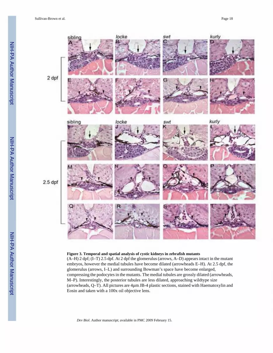
Figure 3. Temporal and spatial analysis of cystic kidneys in zebrafish mutants(A–H) 2 dpf; (I–T) 2.5 dpf. At 2 dpf the glomerulus (arrows, A–D) appears intact in the mutantembryos, however the medial tubules have become dilated (arrowheads E–H). At 2.5 dpf, theglomerulus (arrows, I–L) and surrounding Bowman’s space have become enlarged,compressing the podocytes in the mutants. The medial tubules are grossly dilated (arrowheads,M–P). Interestingly, the posterior tubules are less dilated, approaching wildtype size(arrowheads, Q–T). All pictures are 4µm JB-4 plastic sections, stained with Haematoxylin andEosin and taken with a 100x oil objective lens.
Sullivan-Brown et al. Page 18
Dev Biol. Author manuscript; available in PMC 2009 February 15.
NIH
-PA Author Manuscript
NIH
-PA Author Manuscript
NIH
-PA Author Manuscript

Figure 4. Average cell number surrounding the medial tubules in the mutant backgrounds isincreased at 3 dpf(A) Bar graphs display the average cell number surrounding the glomerular-anterior tubulelumen at 2 dpf and 3 dpf. At 2 dpf there is not a significant increase in cell number surroundingthe tubules between wildtype sibling and mutant populations. However, at 3 dpf, there is astatistical difference in cell number in locke and kurly mutant embryos. (B) Bar graphs depictthe average cell number surrounding the medial tubule lumen at 2 dpf, 3 dpf and 5 dpf. At 2dpf, there is not a significant increase in cell number between the wildtype sibling and mutantpopulations. However at 3 dpf and 5 dpf, the number of cells surrounding the lumen issignificantly increased when compared to wildtype siblings. Interestingly, the number of cellsin the wildtype sibling population decreases from 2dpf to 5dpf. In all graphs, the error barsrepresent standard error. Color code: black bars (sibling), striped pattern (locke), crossedpattern (swt), and dark gray (kurly)
Sullivan-Brown et al. Page 19
Dev Biol. Author manuscript; available in PMC 2009 February 15.
NIH
-PA Author Manuscript
NIH
-PA Author Manuscript
NIH
-PA Author Manuscript

Figure 5. Early pronephric cyst phenotypes(A–D) Acetylated tubulin staining in the pronephric tubules at 27 hpf. Cilia were detected usingan anti-acetylated tubulin antibody (green) and counterstained with the nuclear marker Hoechst(blue). Cilia appeared grossly normal in swt and kurly, but are shorter in locke when comparedto wildtype siblings. (E–J) DIC microscopy images of the pronephric tubules in wildtypesiblings and kurly mutants. Lumen sizes in posterior tubules from 26–30hpf are similar in bothwildtype siblings (E) and mutant embryos (F). Lumens in the medial tubules are larger in bothwildtype siblings (G) and mutant embryos (H) when compared in the posterior regions (E,F).By 2 dpf there is a clear dilation in the medial tubules of mutant embryos (J) as compared towildtype siblings (I). (K,L) Infra-red scattering measurements demonstrate that the frequency
Sullivan-Brown et al. Page 20
Dev Biol. Author manuscript; available in PMC 2009 February 15.
NIH
-PA Author Manuscript
NIH
-PA Author Manuscript
NIH
-PA Author Manuscript

of cilia movement in kurly mutants (L) is similar to the frequency observed in wildtype siblingembryos (K) (~42Hz). Both spectra show several pics. The first pic in each spectrumcorresponds to the fundamental frequency of cilia movement. The subsequent pic correspondsto the harmonics of the fundamental frequency (~n*42Hz with n = 1,2, and 3). E–J were takenwith a 60x objective water immersion lens using two different cameras. E–H were taken withan iXon camera (Andor); pixel size 16*16 µm. I,J were taken with a Luca camera (Andor);pixel size of 10*10 µm. Because of differences in pixel size, luminal size in E–H cannot bedirectly compared to I,J.
Sullivan-Brown et al. Page 21
Dev Biol. Author manuscript; available in PMC 2009 February 15.
NIH
-PA Author Manuscript
NIH
-PA Author Manuscript
NIH
-PA Author Manuscript

Figure 6. Na+/K+ ATPase localization is disrupted in cystic tissues, while apical polarity ismaintainedApical localization of F-actin (red) observed in wildtype siblings (A) is maintained in themedial tubules (white arrowheads) of mutants (B–D). The somites and gut are also stained byphalloidin (red). Correct apical localization of ZO-1 (green) is observed in wildtype sibling(E) and mutant (F–H) embryos. Sibling tubules are outlined by a white circle, and mutanttubules are distinguished by asterisks. Basolateral localization of the Na+/K+ ATPase (red)observed in wildtype siblings (I) is altered in cystic mutants at 4 dpf (J–L). Note in J–L thatNa+/K+ ATPase is adjacent to apical ZO-1 expression (green) which is not observed in wildtype(I). All confocal images were taken with a 40x water objective lens. A–D are cryosections; E–L are plastic sections. Nuclei (blue) are stained with either DRAQ5 (A–D) or Hoechst (E–L).ZO-1 staining basal to the pronephros comes from a different tissue (asterisks in K and L) anddoes not reflect an alteration in ZO-1 within the pronephros.
Sullivan-Brown et al. Page 22
Dev Biol. Author manuscript; available in PMC 2009 February 15.
NIH
-PA Author Manuscript
NIH
-PA Author Manuscript
NIH
-PA Author Manuscript
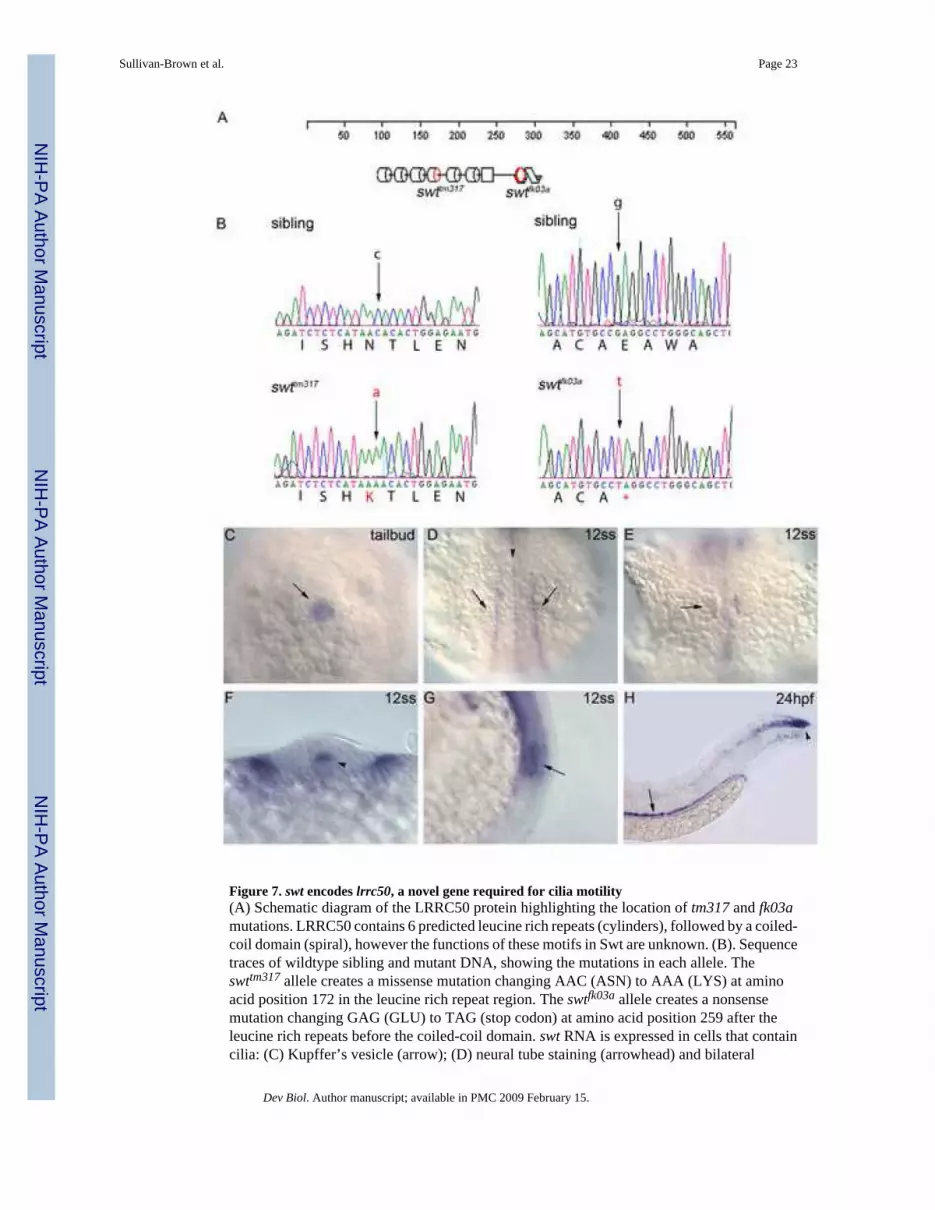
Figure 7. swt encodes lrrc50, a novel gene required for cilia motility(A) Schematic diagram of the LRRC50 protein highlighting the location of tm317 and fk03amutations. LRRC50 contains 6 predicted leucine rich repeats (cylinders), followed by a coiled-coil domain (spiral), however the functions of these motifs in Swt are unknown. (B). Sequencetraces of wildtype sibling and mutant DNA, showing the mutations in each allele. Theswttm317 allele creates a missense mutation changing AAC (ASN) to AAA (LYS) at aminoacid position 172 in the leucine rich repeat region. The swtfk03a allele creates a nonsensemutation changing GAG (GLU) to TAG (stop codon) at amino acid position 259 after theleucine rich repeats before the coiled-coil domain. swt RNA is expressed in cells that containcilia: (C) Kupffer’s vesicle (arrow); (D) neural tube staining (arrowhead) and bilateral
Sullivan-Brown et al. Page 23
Dev Biol. Author manuscript; available in PMC 2009 February 15.
NIH
-PA Author Manuscript
NIH
-PA Author Manuscript
NIH
-PA Author Manuscript
intermediate mesoderm staining (arrows); (E) otic vesicle (arrow); (F) cross section showingexpression in the floorplate of the neural tube (arrowhead); (G) lateral view of otic vesicleexpression (arrow); (H) pronephric duct (arrow, dorsal to the yolk extension) and chordo-neuralhinge expression (arrowhead, at the tip of the tail). Embryo in C is at tailbud stage (dorsal viewof posterior), D–G is at the 12 somite stage, and H is at 24hpf. In D, E anterior is up.
Sullivan-Brown et al. Page 24
Dev Biol. Author manuscript; available in PMC 2009 February 15.
NIH
-PA Author Manuscript
NIH
-PA Author Manuscript
NIH
-PA Author Manuscript

Figure 8. pkd2 morphants develop glomerular dilations, but do not become dilated in the tubularregion of the nephron(A–D) Glomerular region comparison at 3 dpf of a wildtype sibling embryo (A), pkd2morphants (B,C), and a locke mutant embryo (D). Black arrows mark the glomerulus, whichis clearly dilated in both pkd2 morphants and locke mutant embryos. (E–H) Anterior tubuleregion, immediately posterior to the glomerulus; although the tubules exhibit dilation inlocke mutant embryos (H), the tubules in pkd2 morphants (F,G) are not enlarged and resemblewildtype sibling tubules (E). (I–L) Medial tubule region; unlike locke mutant embryos (L), thelumens of the medial tubules are not dilated in pkd2 morphants (J,K), similar to wildtypesiblings (I). Black arrowheads indicate tubules. All pictures are from JB-4 plastic sections,stained with an H&E dye, and taken with 40x lens (A–H) and 100x oil lens (I–L).
Sullivan-Brown et al. Page 25
Dev Biol. Author manuscript; available in PMC 2009 February 15.
NIH
-PA Author Manuscript
NIH
-PA Author Manuscript
NIH
-PA Author Manuscript

Figure 9. Formation of pronephric cysts in zebrafish mutant embryosIn this model, longitudinal views of the pronephros along the anterior-posterior axis (left toright) are depicted and labeled. The 5 regions of interest are G=glomerulus, A=anterior tubule,M=medial tubule, P=posterior tubule, C=cloaca. At 30 hpf, the nephron of both wildtypesiblings (gray) and mutant (blue) embryos appear similar. At this stage, the cilia (red lines) inwildtype embryos are motile, but do not bundle. The mutant embryos each exhibit their ownunique cilia motility defects at this stage. At 48 hpf, cells in the wildtype sibling medial tubulesare multi-ciliated and the cilia have begun to bundle and move in a coordinated fashion. Cellsin the posterior and cloaca regions contain monocilia which are motile as well. It is at the medialtubules where the cystic dilations begin in the mutant embryos. Although the medial tubulesare drastically dilated in mutants, the lumens in the posterior and cloaca regions are similar insize to wildtype siblings. At 72hpf, the medial tubules in the mutant embryos continue to dilate,while the wildtype sibling medial tubules decrease in size. At this stage, the anterior tubuleand glomerular region of the mutant embryos are affected. The pkd2 morphants (yellow)display cystic dilations specifically in the glomerulus. The medial tubule in pkd2 morphantsdoes not dilate and shows normal cilia motility. The highlighted column depicts the medialtubules.
Sullivan-Brown et al. Page 26
Dev Biol. Author manuscript; available in PMC 2009 February 15.
NIH
-PA Author Manuscript
NIH
-PA Author Manuscript
NIH
-PA Author Manuscript

NIH
-PA Author Manuscript
NIH
-PA Author Manuscript
NIH
-PA Author Manuscript
Sullivan-Brown et al. Page 27
Table IThese data points are displayed in the bar graphs in Figure 4 and represent the means followed by standard error. Thesample size (n) refers to the number of tubules used for this statistical analysis. The number in parenthesis refers to thetotal number of embryos used to generate the sample size. Although each embryo contains two tubules, in some embryosboth of the tubules could not be accurately scored due to an incorrect position when sectioning.
Glomerular-Anterior Tubules2 dpf 3 dpf
sibling 4.22±.26 n=5 (3) 5.67±.25 n=5 (3)locke 4.73±.30 n=9 (6) 7.04±.46 n=7 (4) *swt 4.47±.18 n=7 (5) 7.75±.94 n=7 (4)kurly 4.54±.11 n=9 (5) 6.38±.06 n=4 (4) *
Medial Tubules2 dpf 3 dpf 5dpf
sibling 3.94±.12 n=6 (3) 2.64±.01 n=7 (4) * 2.83±.08 n=4 (2) *locke 4.25±.26 n=11 (6) 4.27±.19 n=8 (4) * 5.90±.99 n=4 (2) *swt 4.15±.22 n=8 (4) 4.33±.15 n=6 (3) * 6.79±.26 n=4 (2) *kurly 3.61±.12 n=10 (5) 4.65±.69 n=6 (3) * 7.18±.89 n=4 (2) **Asterisks indicate that the difference between cell number in wildtype siblings and mutant embryos is statistically significant by Student’s t-test (p-
value<0.05).
Dev Biol. Author manuscript; available in PMC 2009 February 15.


