Embed Size (px)
Citation preview

REVIEW PAPER
Zinc Oxide Nanoparticles: Green Synthesis and BiomedicalApplications
Sahana Sadhasivam1• Megala Shanmugam1
• Pillai Divya Umamaheswaran1 • Anbazhagan Venkattappan2 •
Anusuya Shanmugam1
Received: 18 June 2020 / Accepted: 11 October 2020� Springer Science+Business Media, LLC, part of Springer Nature 2020
AbstractNanoparticles refer to ultrafine particles with the particle size at nanoscale. When metals and metal oxides were synthe-
sized at nanoscale, by their unique properties such as smaller particle size, high strength, reactivity, sensitivity, specificity
and stability, they have established a wide variety of applications, and thus have gained popularity in various industries
such as food, textile, cosmetic, automobile and pharmaceuticals. Silver, gold, copper, aluminium, nickel, iron, zinc oxide,
titanium dioxide and copper oxide nanoparticles are few among them. This article presents a review on zinc oxide
nanoparticles, its green synthesis methods and various applications in biomedical fields such as antibacterial, antifungal,
anticancer, anti-inflammatory and wound healing.
Keywords Zinc oxide nanoparticles � Green synthesis method � Antibacterial � Anticancer � Anti-inflammatory �Wound healing
Introduction
Nanotechnology arises from nature, which influences the
substance through physio-chemical processes finding
momentous application in science field [1]. In 1959,
Richard Feynman was the first person conveyed the con-
cept of nanotechnology [2]. Nanotechnology is a field of
science which deals with the materials production and
manipulation in nanometers [3]. Nanomaterials are
microscopic materials that have a structural dimension of
100 nm or less [4]. They have defined properties such as
large surface area, quantum size effects which are con-
sidered to be a significant state of matter. Metal and metal
oxide nanoparticles stated various advantageous
applications in the field of sensors, catalyst, electronics,
optical fibers, agriculture, bio-labelling and in biomedical
areas [5].
Conventionally metal nanoparticle synthetic methods
are classified as top-down and bottom-up methods [6]. The
top-down methods transform the bulk materials into
nanostructures using several techniques such as sonication,
laser ablation, mechanical milling, and physical vapor
deposition [7]. This method has number of drawbacks such
as elaborate, time consuming, high cost of production, and
lack the ability to produce particles of nano size [8]. In
bottom-up methods, the metal precursors are used either in
solid, liquid or gas phases to produce metal nanoparticles.
The bottom-up chemical methods can be done using sol–
gel process, chemical co-precipitation, and chemical vapor
deposition, which often lead to the formation of hazardous
byproducts [9]. In addition, the chemical reducing reagents
used in the nanoparticle synthesis such as sodium dodecyl
sulfate, hydrazine and sodium borohydride are also stated
as toxic. Hence, in contrast to chemical methods, in recent
decades, eco-friendly green synthesis methods, one of the
bottom-up approaches, have gained greater attention for
synthesizing nanoparticles. This method uses various nat-
ural resources such as plants, and microorganisms for
& Anusuya Shanmugam
1 Department of Pharmaceutical Engineering, Vinayaka
Mission’s Kirupananda Variyar Engineering College,
Vinayaka Mission’s Research Foundation (Deemed to be
University), Salem, Tamil Nadu 636308, India
2 Department of Chemistry, Vinayaka Mission’s Kirupananda
Variyar Arts and Science College, Vinayaka Mission’s
Research Foundation (Deemed to be University), Salem,
Tamil Nadu 636308, India
123
Journal of Cluster Sciencehttps://doi.org/10.1007/s10876-020-01918-0(0123456789().,-volV)(0123456789().,- volV)

synthesizing nanoparticles [10]. In green synthesis, active
enzymes work as the capping and reducing agents for the
massive production of small nanoparticles [11]. Apart from
enzymes, several phytochemicals present in the plant
extract also act as reducing, stabilizing and capping agent
in the process of nanoparticles synthesis [12]. Microor-
ganisms such as bacteria, algae, fungi, yeast also have the
ability to reduce metal ions and hence are also used for the
massive production of nanoparticles [13]. The green syn-
thesized nanoparticles are far superior to physiochemical
synthesized nanoparticles, because of its reduced environ-
mental impacts and also large scale production of
nanoparticles of well-defined size and morphology [14].
Figure 1 illustrates the various synthesis methods of
nanoparticles.
Zinc oxide (ZnO) is a distinctive inorganic material
exhibiting various properties like piezoelectric, pyroelec-
tric, semiconducting, optoelctronics and catalysis [15].
ZnO is an n-type semiconductor with band gap energy of
3.37 eV at room temperature and 60 meV excitation
binding energy [16]. ZnO is non-toxic, compatible with
skin, and also used as a UV blocker in sunscreen products
[17]. Various researches reported ZnO as the strongest
antimicrobial agent, recognized by release of reactive
oxygen species (ROS) on its surface [18]. Meanwhile, ZnO
is also documented as bio-safe, biocompatible with various
unique applications in biomedical and drug delivery sys-
tems. Since, past two decades, ZnO nanoparticles (ZnO
NPs) have drawn researcher’s interest because of its
extensive properties. These nanoparticles are commonly
employed in several areas such as ethanol gas sensors,
photo-catalyst, UV-light emitting devices and pharmaceu-
tical and cosmetic industries [19–22].
Even though ZnO NPs are synthesized by various
means, this review emphasizes green synthesis method,
where the ZnO NPs are synthesized using both plants and
microbes, which not only increase the synthesis efficacy
and also reduce the production cost. This review also
elaborates various biomedical applications of ZnO NPs
such as antibacterial, antifungal, anticancer, anti-inflam-
matory and wound healing.
Plant Mediated Synthesis of ZnO NPs
The green synthesis of nanoparticles shows an alternate
way for the physical and chemical method of nanoparticle
synthesis. The preponderance of researchers is intensive
towards the green synthesis of metal and metal oxide
nanoparticles using various biological sources [23]. Among
those, plant sources are found to be rapid, eco-friendly, low
cost and are also safe for the humankind [24].
Plant parts such as bark, stem, leaves, fruits and seeds
have been widely used for the ZnO NP synthesis [25]. The
natural plant extract mediated synthesis is a cheap process
and it also doesn’t include any intermediate base group
formation [26]. It is time efficient and with less facility, it
can produce a very pure and quality product which is free
of impurities. Plants are the highest chosen sources for the
nanoparticles synthesis because they results in large scale
production of stable, and varied sized nanoparticles for
specific applications [27]. The green synthesis process
involves biological reduction of metal ions or metal oxides
to metal nanoparticles with the aid of plant phytochemicals
such as alkaloids, terpenoids, aminoacids, polysaccharides,
and polyphenolic compounds [28]. Table 1 summarizes the
different plant sources that are utilized for the production
of ZnO NPs.
Universally, green synthesis of ZnO NPs using plant
sources starts with sterilization of plant parts using double
distilled water or Tween 20 [29–32]. Then, the plant part is
dried at room temperature followed by grinding into
powder using mortar and pestle. The plant extract is pre-
pared by adding distilled water to the weighed powder and
mixture is boiled with continuous mixing using a magnetic
stirrer. Then, the solution is filtered using whatsman filter
paper and is used as a plant extract for further process
[33–37]. The specific volume of plant extract is mixed with
Zinc precursors such as Zinc nitrate, zinc acetate, zinc
sulphate, zinc chloride solution [38–43]. Then, the mixture
is continuously stirred at higher temperature resulting in
formation of ZnO NPs. The formed ZnO NPs are visually
confirmed by color change [44]. The UV–Vis spectroscopy
was used to further confirm the nanoparticles synthesized
(Fig. 2).
Subsequently, morphology of the nanoparticles is mea-
sured using electron microscopes such as Scanning Elec-
tron Microscope (SEM), Transmission Electron
Microscope (TEM), and Atomic Force Microscopy (AFM)
[33, 45, 46]. Then the crystal structure and chemicalFig. 1 Nanoparticle synthesis methods
S. Sadhasivam et al.
123

Table 1 Synthesis of ZnO NPs using Plants
Plant source Description Precursor Size and shape References
Lantana aculeate Leaves Zinc nitrate 12 ± 3 nm and spherical shaped [48]
Terminalia chebula Fruit Zinc nitrate 12 nm and spherical shaped [49]
Parthenium hysterophorous Leaves Zinc nitrate 16 nm and spherical shaped [29]
Boswellia ovalifoliolata Stem Bark Zinc nitrate 20.3 nm and spherical shaped [50]
Corymbia citriodora Leaves Zinc nitrate 64 nm and polyhedron shaped [51]
Spathodea campanulata Leaves Zinc nitrate 20–50 nm and spherical shaped [44]
Aloe barbadensis miller Leaves Zinc nitrate 25–40 nm and spherical shaped [30]
Limonia acidissima Leaves Zinc nitrate 53 nm and spherical shaped [52]
Parthenium hysterophorus Leaves Zinc nitrate 27 ± 5 nm and spherical shaped [31]
Allium sativum, Allium cepa, Petroselinum crispum Leaves
Bulb
Zinc nitrate 14 and 70 nm and Hexagonal
shaped
[32]
Nephelium lappaceum Fruit Peel Zinc nitrate 50 nm and needle shaped [33]
Sedum alfredii Shoot 100 nm and column shaped [34]
Artabotrys hexapetalu Bambusa vulgaris Leaves Zinc nitrate 20–30 nm and spherical, rod
shaped
[35]
Cassia fistula Leaves Zinc nitrate 5–15 nm and crystalline shaped [36]
Pongamia pinnata Leaves Zinc nitrate 100 nm and spherical, hexagonal
shaped
[37]
Carica papaya Leaves Zinc nitrate 10.2 ± 0.6 nm and hexagonal
shaped
[53]
Physalis alkekengi Roots, Leaves,
Stems, and Fruits
Zinc nitrate 50 to 200 nm and spherical shaped [54]
Hibiscus rosa-sinensis Leaves Zinc nitrate 23.4–48.5 nm and spherical
shaped
[55]
Rosa canina Fruits Zinc nitrate 50 nm and spherical shaped [56]
Plectranthus amboinicus Leaves Zinc nitrate 88 nm and rod shaped [57]
Vitex trifolia Leaves Zinc nitrate 15–46 nm and spherical shaped [58]
Aspalathus linearis Flower Zinc nitrate 1–8 nm and spherical shaped [59]
Anisochilus carnosus Leaves Zinc nitrate 20–40 nm and Spherical shaped [60]
Mimosa pudica Leaves Zinc nitrate 2.71 nm and Wurtzite shaped [61]
Coffee extract Powder Zinc nitrate 4.6 nm and hexagonal shaped [61]
Ocimum basilicum L. var. purpurascens Benth. Leaves Zinc nitrate 50 nm and hexagonal shaped [62]
Citrus aurantifolia Peel Zinc nitrate 50 nm and pyramid shaped [63]
Azadirachta indica Leaves Zinc nitrate 10–30 nm and hexagonal shaped [64]
Phyllanthus niruri Leaves Zinc nitrate 25.61 nm and crystalline shaped [65]
Artocarpus gomezianus Fruit Zinc nitrate 12 nm and spherical shaped [66]
croton sparsiflorus morong Leaves Zinc nitrate 22–52 nm and spherical shaped [67]
Plectranthus amboinicus Leaves Zinc nitrate 20–50 nm and spherical,
hexagonal shaped
[68]
Solanum nigrum Leaves Zinc nitrate 29.79 nm and quasi-spherical
shaped
[69]
Carissa edulis Fruit Zinc nitrate 50–55 nm and Flower shaped [70]
Vitex negundo Leaves Zinc nitrate 75–80 nm and spherical shaped [71]
Polygala tenuifolia Root Zinc nitrate 33.03–73.48 nm and spherical
shaped
[72]
Carica papaya Milk Zinc nitrate 11–26 nm and Crystalline shaped [73]
Azadirachta indica Leaves Zinc nitrate 40 nm and spherical shaped [74]
Nephelium lappaceum Peel Zinc nitrate 25–40 nm and spherical shaped [75]
Eucalyptus globulus Leaves Zinc nitrate 11.6 nm and Spherical shaped [76]
Peganum harmala Seed Zinc nitrate 40 nm and non-uniform shaped [77]
Zinc Oxide Nanoparticles: Green Synthesis and Biomedical Applications
123

Table 1 (continued)
Plant source Description Precursor Size and shape References
Lycopersicon esculentum, Citrus sinensis, Citrusparadisi, Citrus aurantifolia
Peel Zinc nitrate 9.7 nm and hexagonal shaped [78]
Camellia sinensis Leaves Zinc nitrate 8 ± .5 nm and Spherical shaped [79]
Limonia acidissima Leaves Zinc nitrate 12 nm–53 nm and Spherical
shaped
[52]
Solanum torvum Leaves Zinc nitrate 38.0 ± 2 nm and Spherical shaped [80]
Andrographis paniculata Leaves Zinc nitrate 57 ± 0.23 nm and spherical, oval,
hexagonal
[38]
Nigella Sativa Leaves Zinc nitrate 20 nm [81]
Scutellaria baicalensis Root Zinc nitrate 50 nm and sphere shaped [39]
Mangifera indica Leaves Zinc nitrate 45–60 nm and spherical,
hexagonal quartzite shaped
[82]
Rubus coreanus Fruit Zinc nitrate 23.16 nm and hexagonal wurtzite
shaped
[83]
Costus pictus D. Don Leaves Zinc nitrate 11–25 nm spherical, hexagonal
shaped
[84]
Albizia lebbeck Stem bark Zinc nitrate 66.25 nm and spherical shaped [85]
Mentha pulegium Leaves Zinc nitrate 38–49 nm and quasi spherical
shaped
[86]
Sesamum indicum Leaves, Stem and
Root
Zinc nitrate [87]
Lagenaria siceraria Pulp Zinc nitrate 25–55 nm and spherical shaped [88]
Populus ciliate Leaves Zinc nitrate 60–70 nm and spherical shaped [89]
Ceropegia candelabrum Leaves Zinc nitrate 12–35 nm and hexagonal wurtzite
shaped
[90]
Calotropis procera Latex Zinc acetate 5–40 nm and spherical shaped [46]
Black Tea Waste Zinc acetate 19.3 nm and rod shaped [91]
Lobelia leschenaultiana, Leaves Zinc acetate 20–65 nm and spherical,
hexagonal shaped
[92]
Passiflora caerulea Leaves Zinc acetate 70 nm and Spherical Shaped [93]
Ferulago angulata Boiss Zinc acetate 32 and 36 nm and spheroid shaped [94]
Couroupita guianensis Leaves Zinc acetate Hexagonal shaped [95]
Nyctanthes arbor-tristis Flower Zinc acetate 11–32 nm [96]
Ulva lactuca Seaweed Zinc acetate 10–50 nm and crystalline shaped [97]
Oak trees (Jaft) Fruit Zinc acetate 44 nm and hexagonal wurtzite
shaped
[98]
Juglans regia Leaves Zinc acetate 95–150 nm and spherical, flower
shaped
[99]
Cucurbita pepo Leaves Zinc acetate 8 nm [41]
Anchusa italic Flower Zinc acetate 8–14 nm and hexagonal shaped [100]
Abelmoschus esculentus okra crop Zinc acetate 29 nm and hexagonal wurtzite
shaped
[40]
Hyssops officinalis Whole plant Zinc acetate Pseudo spherical shaped [101]
Rehmanniae radix Root Zinc acetate 10–12 nm and hexagonal wurtzite
shaped
[102]
Catharanthus roseus Leaves Zinc acetate 50–90 nm and hexagonal wurtzite
shaped
[103]
Green tea Leaves Zinc acetate [104]
Bauhinia tomentosa Leaves Zinc sulphate 22–94 nm and hexagonal shaped [105]
Tecoma castanifolia Leaves Zinc sulphate 70–75 nm and spherical shaped [106]
Celosia argentea Leaves Zinc sulphate 25 nm and spherical shaped [107]
Trianthema portulacastrum Plant biomass Zinc sulphate 25–90 nm and spherical shaped [108]
S. Sadhasivam et al.
123

composition of the nanoparticles are characterized using
X-Ray Diffraction Spectroscopy (XRD) and Energy Dis-
persive X-ray analysis (EDX) respectively [34]. The
functional groups on the nanoparticles surface are descri-
bed using Fourier transform infrared spectroscopy (FTIR)
technique. The size and dispersity of nanoparticles in the
liquid medium are determined using the Dynamic Light
Scattering (DLS) and Zeta potential techniques respec-
tively [47]. The various plant sources used in the synthesis
of ZnO NPs with its morphological characteristics are lis-
ted in Table 1.
Microbes Mediated Synthesis of ZnO NPs
The nanoparticle synthesis using microorganisms such as
bacteria, fungi remained unexplored by researchers. Green
synthesis of nanoparticles using microbes shows added
benefits than using plant sources because of the microbial
reproducibility. Nevertheless, it has lots of disadvantages
such as screening of microorganism, need of contamination
free environment for the entire process, lack of control on
nanoparticle size, and shape and high cost of nutrient
media for microbes [112]. The existence of various
enzymes, biomolecules and proteins in microorganisms are
recognized as capping agents in the formation of multiple
sized nanoparticles. Commonly, microbial cultures are
grown in culture medium and then to that metal precursor
were introduced in the form of soluble salts [113]. After the
reaction time of few hours, the synthesized nanoparticles
precipitate at the bottom with a visible color change [114].
Additionally, reaction parameters such as pH, temperature,
concentration of metal precursor and reaction time deter-
mine the size and yield of the synthesized nanoparticles.
The nanoparticles synthesized are characterized using UV–
VIS spectroscopy, SEM, XRD, FTIR to determine the
shape, size, functional group and surface charge [115].
Table 2 summarizes the microbial mediated ZnO NPs
synthesis.
Biomedical Applications
Antibacterial Activity of ZnO NPs
Bacterial diseases pose serious health threats among man-
kind. Increased antibiotic resistance against bacteria,
emergence of new strains intended the researchers to focus
on the metal and metal oxide nanoparticles as antibacterial
Table 1 (continued)
Plant source Description Precursor Size and shape References
Aloe barbadensis Miller Leaves Zinc sulphate 8–18 nm and spherical, oval,
hexagonal shaped
[42]
Trifolium pretense Flower Zinc oxide 60–70 nm and Hexagonal shaped [109]
Tribulus terrestris Leaves Zinc oxide 6–10 nm and spherical shaped [44]
Lycopersicon esculentum Leaves Zinc oxide 10–50 nm and hexagonal wurtzite
shaped
[110]
Heritiera fomes, Sonneratia apetala Bark and leaves Zinc Chloride 20–50 nm [43]
Jacaranda mimosifolia Flower Zinc gluconate
hydrate
2–4 nm and spherical shaped [111]
Fig. 2 Schematic representation of green synthesis of zinc oxide
nanoparticles
Zinc Oxide Nanoparticles: Green Synthesis and Biomedical Applications
123

agents. Due to the unique physiochemical properties and
increased surface area, ZnO NPs are extensively explored
as antibacterial agents. In addition, ZnO NPs are also found
to be safe and compatible with the human system
[128, 129].
Numerous literatures explain about the antibacterial
mechanism of ZnO nanoparticles, which includes (i) pro-
duction of reactive oxygen species (ROS) such as super-
oxide anions, hydrogen peroxides and hydroxyl radicals (ii)
triggering Zn2? release and (iii) the released Zn2? ions
interacts with the bacterial cell, especially the cell mem-
brane, cytoplasm and nucleic acid, and thus disintegrates
the cellular integrity which ultimately results in cell death
[130–132] which is illustrated in Fig. 3. The green syn-
thesized ZnO NPs are used as effective antibacterial agent
against both gram positive and gram negative bacteria such
as, Bacillus subtilis, Salmonella typhimurium, Staphylo-
coccus aureus, Streptococcus pyogenes, Mycobacterium
tuberculosis, Escherichia coli, Klebsiella pneumonia,
Mycobacterium luteus, Vibrio cholera, Aspergillus niger,
Aspergillus fumigatus, Fusarium culmorum, Fusarium
oxysporum, Pseudomonas aeruginosa, and Salmonella
paratyphi. Table 3 summarizes the antibacterial activity of
green synthesized ZnO NPs against various bacterial
pathogens. In addition with the antibacterial activity of
ZnO NPs, the ZnO nanocomposites are also proven for
their antibacterial activity. Bacterial cellose/ZnO
nanocomposites [133], chitin/chitosan-ZnO nanocompos-
ites [134, 135] and carboxymethyl chitosan-ZnO
nanocomposites [136, 137].
Antifungal Activity of ZnO NPs
In addition to antibacterial activity, ZnO NPs are also been
reported for their antifungal activities against many of the
harmful yeasts and fungi, due to which it is widely used as
antifungal additives in food industries. There is a very good
review article by Qi et al. on the antifungal activities of
ZnO NPs, its mechanism of action and a wide variety of
applications [140].
Rajiv et al. synthesized ZnO NPs using Parthenium
hysterophorus L and validated its antifungal activity.
Highest inhibition was observed against Aspergillus flavus
and Aspergillus niger at 25 lg/ml and also reported that the
activity is size-dependent [31]. Vijayakumar et al. also
synthesized ZnO NPs using Lycopersicon esculentum leaf-
extract and proved its antifungal activity against Candida
Table 2 Synthesis of ZnO NPs using microorganisms
Microorganism Description Precursor Size and shape References
Bacillus subtillis Bacteria Zinc acetate 10–15 nm [116]
Bacillus licheniformis Bacteria Zinc acetate 200 nm and nanoflower shaped [117]
Serratia ureilytica Bacteria Zinc acetate 170–250 nm and spherical, nanoflower shaped [113]
Staphylococcus aureus Bacteria Zinc acetate 10–50 nm and circular shaped [114]
Pseudomonas aeruginosa Bacteria Zinc nitrate 35–80 nm and spherical shaped [115]
Sphingobacterium thalpophilum Bacteria Zinc nitrate 37 nm and spherical shaped [118]
Rhodococcus pyridinivorans Bacteria Zinc sulphate 100–120 nm and spherical, hexagonal shaped [119]
Lactobacillus plantarum Bacteria Zinc sulphate 7–19 nm and spherical clusters shaped [120]
Lactobacillus sporogens Bacteria Zinc chloride 5–15 nm and tubule shaped [121]
Lactobacillus johnsonii Bacteria Zinc oxide 4–9 nm and irregular shaped [122]
Aeromonas hydrophila Bacteria Zinc oxide 57.72 nm and Spherical, oval shaped [123]
Candida albicans Fungus Zinc oxide 20 nm and quasi spherical shaped [124]
Aspergillus fumigatus Fungus Zinc nitrate 1.2–6.8 nm and oblate spherical, hexagonal shaped [125]
Alternaria alternate Fungus Zinc sulfate 75 ± 5 nm and Spherical, Triangular, Hexagonal shaped [126]
Aspergillus fumigatus Fungus Zinc sulfate 60–80 nm and spherical shaped [127]
Fig. 3 Schematic representation of antibacterial mechanism of zinc
oxide nanoparticles
S. Sadhasivam et al.
123

Table 3 Antibacterial activity of green synthesized ZnO NPs
Plant source/
microorganism
Description Name of organism Antibacterial activity References
Cassia fistula Leaves Escherichia coli Antibacterial activity of ZnO NP
was 20 lg/mL
[36]
Pongamia pinnata Leaves Staphylococcus aureus, Escherichia coli [37]
Vitex trifolia Leaves Bacillus subtilis [58]
Phyllanthus niruri Leaves Staphylococcus saprophyticus. The Zone of inhibition of ZnO NP
was 2to 5 lm
[65]
Plectranthusamboinicus
Leaves Staphylococcus aureus Antibacterial activity of ZnO NP
was 8-10 lg/mL
[68]
Solanum nigrum Leaves Pseudomonas aeruginosa The Zone of inhibition of ZnO NP
was 2.7 to 4.9 lm
[69]
Vitex negunda Leaves Staphylococcus aureus, Staphylococcusparatyphi,
Escherichia coli
Antibacterial activity of ZnO NP
was 15-17 lg/mL
[71]
Costus pictus D. Don Leaves Staphylococcus aureus, Bacillus subtilis, Escherichiacoli, Salmonella paratyphi
The Zone of inhibition of ZnO NP
was 5 lm
[84]
Agro waste Solid
extract
Bacillus subtilis, Salmonella typhimurium [138]
Catharanthus roseus Leaves Staphylococcus aureus, Streptococcus pyogenes The Zone of inhibition of ZnO NP
was 2 to 7 lm
[103]
Trifolium pratense Flower
extract
Staphylococcus aureus, Pseudomonas aeruginosa,
Escherichia coliThe Zone of inhibition of ZnO NP
was 0.7 to 1.1 lm
[109]
Limonia acidissima Leaves Mycobacterium tuberculosis [52]
Ceropegiacandelabrum
Leaves Staphylococcus aureus,
Bacillus subtilis,
Escherichia coli,
Salmonella typhi
Antibacterial activity of ZnO NP
was 0.25 lg/mL
[90]
Celosia argentea Leaves Escherichia coli,
Salmonella, Acetobacter
Antibacterial activity of ZnO NP
was 3.1 lg/mL
[107]
Couroupitaguianensis
Leaves Bacillus cereus,
Klebsiella pneumonia,
Escherichia coli,
Mycobacterium luteus,
V. cholera
Antibacterial activity of ZnO NP
was 0.05-0.25 lg/mL
[95]
Parthenium
hysterophorus
Leaves Aspergillus flavus,
Aspergillus niger,
Aspergillus fumigatus,
Fusarium culmorum,
Fusarium oxysporum
[31]
Anisochilus carnosus Leaves Salmonella paratyphi,
Vibrio cholera,
Staphylococcus aureus, Escherichia coli
[60]
Jacarandamimosifolia
Flower Escherichia coli,
Enterococcus faecium
[111]
Green tea Leaves Staphylococcus aureus,
Escherichia coli
Antibacterial activity of ZnO NP
was 9.7 lg/mL
[104]
Caulerpa peltata,
Hypnea valencia,
Sargassummyriocystum
Leaves Staphylococcus aureus,
Streptococcus mutans,
Vibrio cholerae,
Neisseria gonorrhoeae,
Klebsiella pneumonia
Antibacterial activity of ZnO NP
was 0.10 lg/mL
[139]
Zinc Oxide Nanoparticles: Green Synthesis and Biomedical Applications
123

albicans, a most prevalent fungal pathogen at concentra-
tions 100 lg/ml [110].
Suresh et al. synthesized ZnO NPs using Costus pictus
leaf extract which was also proven its antifungal activity
using disc diffusion method against two of the fungal
pathogen such as Aspergillus niger and Candida albicans
[84]. In a similar study by Irshad et al. where ZnO NPs
were synthesized using green tea leaves and was verified
for its biocidal activity against Aspergillus niger using well
diffusion method. Both the studies revealed that due to the
electrostatic attraction between the negatively charged cell
membrane and the positively charged NPs, the NPs get
attached with the cell membrane which ultimately results in
membrane disruption and cell death [104].
ZnO NPs synthesized by Jamdagni et al. using flower
extract of Nyctanthes arbor-tristis also showed an anti-
fungal activity against five of the fungal pathogens of
plants such as Alternaria alternata, Aspergillus niger,
Botrytis cinerea, Fusarium oxysporum and Penicillium
expansum. This study finds the lowest minimum inhibitory
concentration of 16 lg/ml against A. niger [96].
Jayaseelan et al., successfully synthesized ZnO NPs
using Aeromonas hydrophila microoraganism and evalu-
ated its activity against various fungal pathogens such as
Aspergillus flavus, Aspergillus niger, Candida albicans.
The antifungal activity of the Aeromonas hydrophila ZnO
NPs was performed using well diffusion method and it
shows minimum inhibition zone around 19 ± 1.0 mm
[123].
Anticancer Activity of ZnO NPs
Zinc is an essential mineral which maintains homeostasis
by regulating enzyme activities [141], whereas, its defi-
ciency initiates and promotes the formations of cancerous
cells. Zinc plays an important role in maintaining the
activity of p53, a tumor suppressor gene, which regulates
apoptosis by activating caspase-6 enzyme [142]. ZnO NPs
also possess a unique electrostatic characteristic which
helps in selective targeting of cancer cells. Cancer cells
contain anionic phospholipids in large concentration on
their surface which results in electrostatic attraction with
ZnO NPs. This promotes cellular uptake of ZnO NPs by the
cancer cells and finally leads to cytotoxicity [143],
whereas, the small size of ZnO NPs enhances the perme-
ation and retention of NPs inside the tumorous cells and
thus helps to act upon it [144]. The mechanism behind the
selective cytotoxicity of ZnO NPs towards cancer cells is
the intracellular release of dissolved zinc ions, followed by
ROS induction [145]. ZnO NP generates large amount of
ROS in cancer cells compared to the ROS in normal cells,
which results in generation of large amounts of oxidative
Table 3 (continued)
Plant source/
microorganism
Description Name of organism Antibacterial activity References
Serratia ureilytica Bacteria Escherichia coli,
Staphylococcus aureus
[113]
Staphylococcusaureus
Bacteria Escherichia coli,
Staphylococcus aureus,
Staphylococcus epidermidis,
Listeria monocytogenes,
Klebsiella pneumonia,
Enterococcus faecalis,
Pseudomonas aeruginosa,
Methicillin-resistant Staphylococcus aureus (MRSA)
Antibacterial activity of ZnO NP
was 10 lg/mL
[114]
Sphingobacteriumthalpophilum
Bacteria Pseudomonas aeruginosa,
Enterobacter aerogens
The Zone of inhibition of ZnO NP
was 14.3 mm, 11.1 mm
[118]
Aeromonashydrophila
Bacteria Aeromonas hydrophila,
Pseudomonas aeruginosa,
Escherichia coli,
Staphylococcus aureus,
Enterococcus faecalis,
Streptococcus pyogenes
Antibacterial activity of ZnO NP
was 1.2 lg/mL
[123]
S. Sadhasivam et al.
123

stress in cancer cells. This leads to death of cancer cells
[146].
The anticancer activity of ZnO NPs prepared by green
synthesis method has been validated against a variety of
cell lines, such as Human Colon Adenocarcinoma (Caco-
2), Human Lung Adenocarcinoma (A549), Pancreatic
adenocarcinoma (PANC-1), Ovarian adenocarcinoma
(CaOV-3), Colonic adenocarcinoma (COLO205), Acute
promyelocytic leukemia (HL-60) cells, Breast adenocarci-
noma cell line(MDA-MB231) and are elaborated in
Table 4.
Anti-Inflammatory Activity of ZnO NPs
In the last few years, nanoparticles are broadly used as an
anti-inflammatory agent. The large surface area to volume
ratio converses the increased reactive properties of
nanoparticles, leading to the greater interaction with the
cell membrane and facilitating the transport inside the
membrane [153]. Because of ZnO NPs nanosize, Zn can be
effortlessly transferred through the cell membrane. The
earlier research article proposes the anti-inflammatory
activity of ZnO NP [154, 155]. ZnO NPs exert their anti-
inflammatory activity through various mechanisms namely,
inhibition of proinflammatory cytokines release [156],
inhibition of inducible nitric oxide synthase (iNOS)
enzyme expression [157], myeloperoxidase inhibition
[158], inhibition of the NF-jb pathway and inhibition of
mast cell degranulation [139]. The anti-inflammatory
activities of green synthesized ZnO NPs are summarized in
Table 5.
Wound Healing Activity of ZnO NPs
Skin is the largest organ of our body which protects us
from external invasion. Any damage happens to skin
results in wound. The wound will heal automatically, but
healing takes time. Often, wound healing may delay due to
microbial infection. Staphylococcus aureus and Pseu-
domonas aeruginosa are few such microorganisms which
cause severe wound infections. NPs are known for their
antibacterial activity and metal oxides NPs generate
hydrogen peroxide which can cause cell damage. Hence,
metal oxide NPs can be used to kill these pathogenic
organisms and thus can enhance the process of wound
healing. Several literatures evidenced the use of ZnO NPs
as successful wound healing agent [160–163]. However,
we summarize this wound healing property of ZnO NPs
prepared by green synthesis.
Khatami et al. prepared a cotton wound bandages by
impregnating them with ZnO NPs synthesized biologically
using coffee ground and experimented its antimicrobial
activities. Based on their minimum inhibitory
concentration (MIC), this study report the use of ZnO NPs
impregnated antibacterial bandages for treating wounds
and to cover infection sensitive wounds in diabetic patients
and wounds caused by burns [164].
Shao et al. synthesized ZnO NPs using Barleria gibsoni
leaf extract which exhibited excellent antibacterial activity
against pathogenic bacteria. In addition, they also devel-
oped a gel using this ZnO NPs which showed remarkable
wound healing property in rat and thus proved to be an
efficient topical antimicrobial formulation and wound
healing agent for general wounds and wounds due to burn
[165].
Ezealisiji et al. prepared ZnO NPs using Solanum tor-
vum (L) leaf extract, and made a ZnO nanoparticles–hy-
drogel composite and validated its toxicological profiles
using rats. This study predicted that there is a significant
increase in plasma concentration of zinc in dose and time
dependent manner. Due to this, there may be chances of
renal and hepatic failure upon its use for a prolonged
period. Hence, this study suggested the need to predict the
optimum dose of ZnO NPs which can be used as a wound
healing agent and also necessary to find the optimum time
it can be used in human [80].
ZnO NPs in Drug Delivery
The smaller particle size and the larger surface area of the
NPs facilitate its penetration via cell membrane and thus
get be absorbed into the cells, which leads to well distri-
bution. Hence, NPs are widely been used in drug delivery
where the drugs are loaded with NPs and thus are made to
reach their targets in sufficient quantity. In addition, when
the biodegradable materials are used for synthesizing NPs,
this prolongs the delivery of drugs in the target site [166].
By properly engineering the NPs, one can make the NPs
targeting specific cells. This property is applied to target
the cancerous cells and bacterial cells [107, 167].
Vaishnav et al. have synthesized ZnO NPs by green
synthesis method using the leaves of Celosia argentea and
have validated its drug delivery capacity using the drug
metronidazole benzoate. The results revealed that ZnO NPs
enhances the drug delivery of metronidazole [168]. Yuan
et al. synthesized a chitosan encapsulated ZnO NPs loaded
with doxorubicin and studied the effect of ZnO NPs in drug
delivery. The study showed that ZnO NPs helped in
releasing the drug at rapid rate initially which then grad-
ually become controlled release at the later period [169].
Zinc Oxide Nanoparticles: Green Synthesis and Biomedical Applications
123

Table 4 Anticancer activity of green synthesized ZnO NPs by MTT assay
Plant source/
microorganism
Description Cell lines tested Activity values Results Reference
Deverra tortuosa Aerial
parts
Human Colon
Adenocarcinoma
‘‘Caco-2’’
1. IC50 value: 50.81 lg/mL Remarkable selective
cytotoxicity was shown
[147]
Human Lung
Adenocarcinoma
‘‘A549’’
83.47 lg/mL
2. Activity were compared
with human lung
fibroblast cell line (WI38)
434.6 lg/mL
Sargassummuticum Seaweed Pancreatic
adenocarcinoma
(PANC-1)
IC50 value 10.8 ± 0.3 lg/
mL
1.Nano composite is most
toxic to HL-60 cells.
2.HA/ZnO nanocomposite
treatment for 72 h did not
cause toxicity to the
normal human lung
fibroblast (MRC-5) cell
line
[148]
Ovarian adenocarcinoma
(CaOV-3)
15.4 ± 1.2 lg/mL
Colonic adenocarcinoma
(COLO205)
12.1 ± 0.9 lg/mL
Acute
promyelocyticleukemia
cells (HL-60)
6.25 ± 0.5 lg/mL
Hyssops officinalis Whole
plant
Breast adenocarcinoma
cell line(MDA-MB231)
Incubated with different
concentrations of ZnO
NPs& evaluated at time of
24 h, 48 h and 72 h
Showed inhibitory effects
on the growth of Breast
cancer cells.
[101]
Rehmanniae radix Root Osteosarcoma cancer cell
line(MG-63)
Cells were cultured for 24 h
under different
concentrations (5 lg/ml,
10 lg/ml, 20 lg/ml,
30 lg/ml, 40 lg/ml,
50 lg/ml, 60 lg/ml,
70 lg/ml and 80 lg/ml)
1. With the increase in
concentration of NPs, the
survival of cancer cell was
decreased. 2. Study
displayed that ZnO NPs
was more effective in
minimizing the cancer
cell growth and survival
rate of MG-63 cells
[102]
Laurusnobilis Leaves Lung cancer cells (A549) 1.Evaluated at different
concentrations (10 mg/
mL, 20 mg/mL, 40 mg/
mL and 80 mg/mL) 2.
The toxicity of ZnO NPs
was also evaluated on
normal murine
macrophage RAW264.7
cells
1. NPs were found to be
non-toxic to normal
murine macrophage
RAW264.7 cells. 2. At
higher concentrations of
80lgmL-1, NPs were
found to be effective in
inhibiting the viability of
human A549 lung cancer
cells.
[149]
Cucumis melo inodorus Rough
shell
Murine breast cancer cell
lines(TUBO)
IC50 values (lg/mL)
At 24 h:20 At 48 h:18 At
72 h :16
Activity depends on both
time duration of treatment
and dose. Longer
treatment with higher
doses exhibited the
reduced cancer cell lines
viability.
[150]
Human breast cancer cell
lines (MCF7)
IC50 values (lg/mL)
At 24 h:40 At 48 h:33 At
72 h:31
Ecliptaprostrata Leaves Human Liver Carcinoma
Cell line(Hep-G2)
Samples evaluated in
different concentrations of
1 lg/mL, 10 lg/mL,
100 lg/mL, 250 lg/mL,
and 500 lg/mL, which
showed the cell necrosis
of 14.5%, 51.5%, 67%,
84%, and 86.5%, for ZnO
NPs, respectively
Dose dependent cytopathic
effects was observed.
Significant cytotoxic
effects was observed at
100 mg/mL
concentration.
[151]
S. Sadhasivam et al.
123
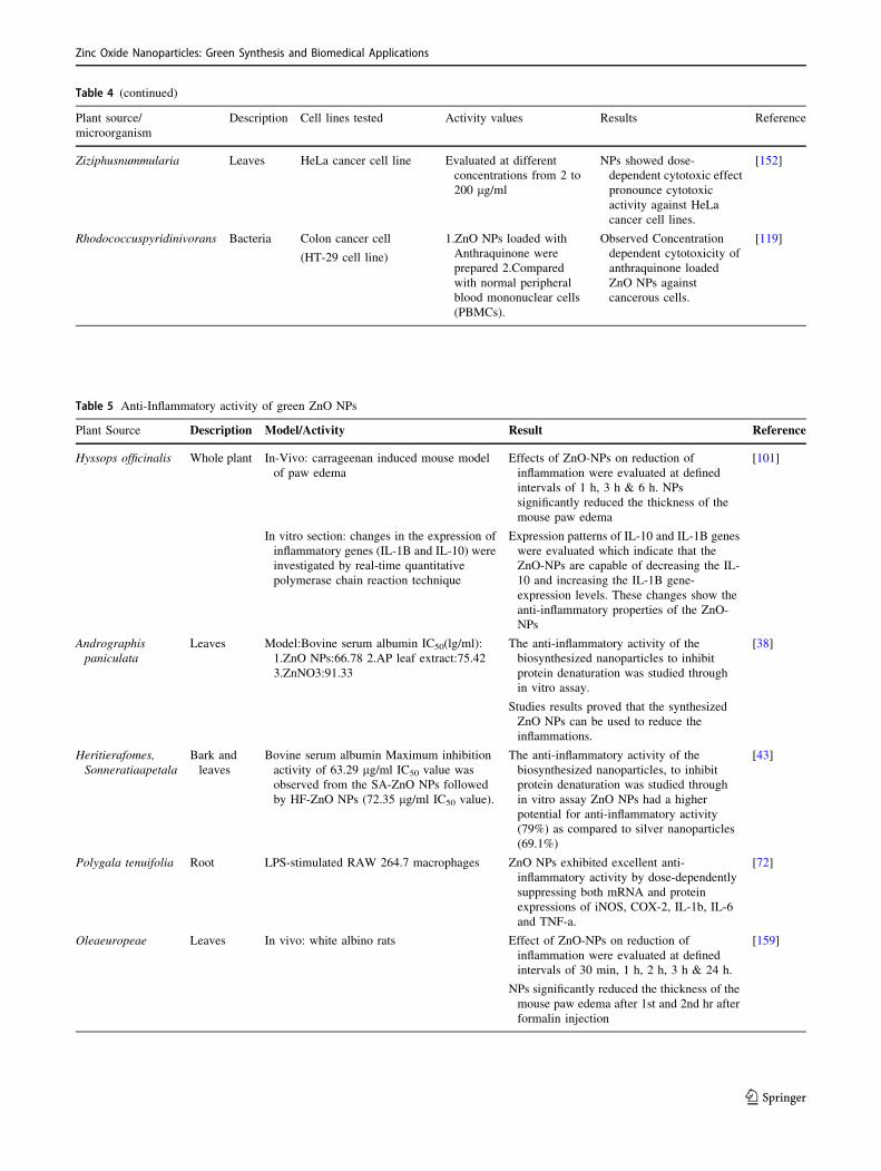
Table 4 (continued)
Plant source/
microorganism
Description Cell lines tested Activity values Results Reference
Ziziphusnummularia Leaves HeLa cancer cell line Evaluated at different
concentrations from 2 to
200 lg/ml
NPs showed dose-
dependent cytotoxic effect
pronounce cytotoxic
activity against HeLa
cancer cell lines.
[152]
Rhodococcuspyridinivorans Bacteria Colon cancer cell
(HT-29 cell line)
1.ZnO NPs loaded with
Anthraquinone were
prepared 2.Compared
with normal peripheral
blood mononuclear cells
(PBMCs).
Observed Concentration
dependent cytotoxicity of
anthraquinone loaded
ZnO NPs against
cancerous cells.
[119]
Table 5 Anti-Inflammatory activity of green ZnO NPs
Plant Source Description Model/Activity Result Reference
Hyssops officinalis Whole plant In-Vivo: carrageenan induced mouse model
of paw edema
Effects of ZnO-NPs on reduction of
inflammation were evaluated at defined
intervals of 1 h, 3 h & 6 h. NPs
significantly reduced the thickness of the
mouse paw edema
[101]
In vitro section: changes in the expression of
inflammatory genes (IL-1B and IL-10) were
investigated by real-time quantitative
polymerase chain reaction technique
Expression patterns of IL-10 and IL-1B genes
were evaluated which indicate that the
ZnO-NPs are capable of decreasing the IL-
10 and increasing the IL-1B gene-
expression levels. These changes show the
anti-inflammatory properties of the ZnO-
NPs
Andrographispaniculata
Leaves Model:Bovine serum albumin IC50(lg/ml):
1.ZnO NPs:66.78 2.AP leaf extract:75.42
3.ZnNO3:91.33
The anti-inflammatory activity of the
biosynthesized nanoparticles to inhibit
protein denaturation was studied through
in vitro assay.
Studies results proved that the synthesized
ZnO NPs can be used to reduce the
inflammations.
[38]
Heritierafomes,Sonneratiaapetala
Bark and
leaves
Bovine serum albumin Maximum inhibition
activity of 63.29 lg/ml IC50 value was
observed from the SA-ZnO NPs followed
by HF-ZnO NPs (72.35 lg/ml IC50 value).
The anti-inflammatory activity of the
biosynthesized nanoparticles, to inhibit
protein denaturation was studied through
in vitro assay ZnO NPs had a higher
potential for anti-inflammatory activity
(79%) as compared to silver nanoparticles
(69.1%)
[43]
Polygala tenuifolia Root LPS-stimulated RAW 264.7 macrophages ZnO NPs exhibited excellent anti-
inflammatory activity by dose-dependently
suppressing both mRNA and protein
expressions of iNOS, COX-2, IL-1b, IL-6
and TNF-a.
[72]
Oleaeuropeae Leaves In vivo: white albino rats Effect of ZnO-NPs on reduction of
inflammation were evaluated at defined
intervals of 30 min, 1 h, 2 h, 3 h & 24 h.
NPs significantly reduced the thickness of the
mouse paw edema after 1st and 2nd hr after
formalin injection
[159]
Zinc Oxide Nanoparticles: Green Synthesis and Biomedical Applications
123

Conclusion
Nanoparticles have diverse properties in comparison with
the bulk material due to its small size and also it proposes
various new innovations in biomedical, biosensor, cosmetic
and food industry. The production of metallic nanoparticles
by green synthesis method is environmental friendly, in-
expensive, non-toxic and can be easily scaled up. Green
sources acts as both capping and reducing agent to produce
nanoparticles of controlled shape and size. In biomedical
field, nanoparticles are used as antimicrobial, anticancer
and anti-inflammatory agents and various other fields are
also emerging. ZnO NPs are one of the important nano-
materials used extensively in biomedical field. Whole-
somely, this review focuses on the green production of
ZnO NPs and its emerging application in the biomedical
fields such as antibacterial, antifungal, anticancer, anti-in-
flammatory, wound healing and drug delivery are
addressed.
Acknowledgement The authors acknowledge the financial support
from Vinayaka Mission’s Research Foundation (Deemed to be
University), Salem (Research Grant No: VMRF/SeedMoney/2020/
VMKVEC-Salem/6).
Author’s Contributions SS reviewed the literature, wrote sections
on introduction, plant mediated and microbial mediated synthesis of
ZnO NPs. MS reviewed the literature and wrote sections on
antibacterial activity of ZnO NPs. PDU reviewed the literature and
wrote sections on anti-inflammatory and anticancer activities of ZnO
NPs. AV reviewed the literature and wrote sections on wound healing
activity of ZnO NPs. AS reviewed the literature and wrote sections on
antifungal activity of ZnO NPs. SS and AS conceived the manuscript.
All the authors read and approved the manuscript.
Funding The authors acknowledge the financial support from
Vinayaka Mission’s Research Foundation (Deemed to be University),
Salem (Research Grant No: VMRF/SeedMoney/2020/VMKVEC-
Salem/6).
Availability of Data and Materials Not applicable.
Compliance with Ethical Standards
Ethics Approval Not applicable.
Consent to Participate Not applicable.
Conflicts of Interest The authors declare that they have no conflict of
interest.
References
1. S. Mustapha, M. M. Ndamitso, A. S. Abdulkareem, J. O. Tijani,
D. T. Shuaib, A. O. Ajala, and A. K. Mohammed (2020). Appl.Water Sci. 10, 49.
2. K. Riehemann, S. W. Schneider, T. A. Luger, B. Godin, M.
Ferrari, and H. Fuchs (2009). Angew. Chem. Int. Ed. 48, 872.
3. C. Buzea, I.I. Pacheco, K. Robbie, Biointerphases. 2, MR17
(2007)
4. S. Iravani (2011). Green Chem. 13, 2638.
5. J. Jeevanandam, A. Barhoum, Y. S. Chan, A. Dufresne, and M.
K. Danquah (2018). Beilstein J. Nanotechnol. 9, 1050.
6. V. T. Arasu, D. Prabhu, and M. Soniya (2010). J. Biosci. Res. 1,
259.
7. X. Q. Li, D. W. Elliott, and W. X. Zhang (2006). Crit. Rev. SolidState Mater. Sci. 31, 111.
8. M. Suganeswari, A. Shering, M. P. Bharathi, and J. JayaSutha
(2011). Int J Pharm Biol Arch 2, 847.
9. M. Parashar, V. K. Shukla, and R. Singh (2020). J. Mater. Sci.:Mater. Elect. 31, 3729.
10. H. Mirzaei and M. Darroudi (2017). Ceram. Int. 43, 907.
11. J. Singh, T. Dutta, K. H. Kim, M. Rawat, P. Samddar, and P.
Kumar (2018). J. Nanobiotechnol. 16, 84.
12. M. S. Akhtar, J. Panwar, Y. S. Yun, and A. C. S. Sustain (2013).
Chem. Eng. 1, 591.
13. M. Ovais, A. T. Khalil, N. U. Islam, I. Ahmad, M. Ayaz, M.
Saravanan, Z. K. Shinwari, and S. Mukherjee (2018). Appl.Microbiol. Biotechnol. 102, 6799.
14. A. Gour and N. K. Jain (2019). Artif. Cells Nanomed. Biotech-nol. 47, 844.
15. Y. Liu, Y. Zhang, H. Lei, J. Song, H. Chen, and B. Li (2012).
Opt. Express. 20, 19404.
16. M. Rajalakshmi, S. Sohila, S. Ramya, R. Divakar, C. Ghosh, and
S. Kalavathi (2012). Opt. Mater. 34, 1241.
17. T. Krishnakumar, R. Jayaprakash, N. Pinna, V. N. Singh, B.
R. Mehta, and A. R. Phani (2009). Mater. Lett. 63, 242.
18. N. Padmavathy and R. Vijayaraghavan (2008). Sci. Technol.Adv. Mater. 9, 035004.
19. J. Xie, Y. Cao, D. Jia, Y. Li, and Y. Wang (2016). Ceram. Int.42, 90.
20. Z. Deng, M. Chen, G. Gu, and L. Wu (2008). J. Phys. Chem. B.112, 16.
21. P. J. Lu, S. C. Huang, Y. P. Chen, L. C. Chiueh, and D. Y. C.
Shih (2015). J. Food Drug Anal. 23, 587.
22. N. Izu, K. Shimada, T. Akamatsu, T. Itoh, W. Shin, K. Shiraishi,
and T. Usui (2014). Ceram. Int. 40, 8775.
23. P. Anastas and N. Eghbali (2010). Chem. Soc. Rev. 39, 301.
24. G. Rajakumar, T. Gomathi, M. Thiruvengadam, V. D. Ra-
jeswari, V. N. Kalpana, and I. M. Chung (2017). Microb.Pathog. 103, 123.
25. S. Fakhari and M. Jamzad (2019). H Kabiri Fard. Green Chem.Lett. Rev. 12, 19.
26. H. Agarwal, S. V. Kumar, and S. Rajeshkumar (2017). Resour.-Eff. Technol. 3, 406.
27. S. Ahmed, S. A. Chaudhry, and S. Ikram (2017). J. Photochem.Photobiol. B 166, 272.
28. A. G. Ingale and A. N. Chaudhari (2013). J. Nanomed. Nan-otechol. 4, 1.
29. K.S. Sindhura, T.N.V.K.V. Prasad, P.P. Selvam, O.M. Hussain,
Appl. Nanosci. 4, 819 (2014)
30. G. Sangeetha, S. Rajeshwari, and R. Venckatesh (2011). Mater.Res. Bull. 46, 2560.
31. P. Rajiv, S. Rajeshwari, and R. Venckatesh (2013). Spectrochim.Acta A Mol. Biomol. Spectrosc. 112, 384.
32. M. Stan, A. Popa, D. Toloman, A. Dehelean, I. Lung, and G.
Katona (2015). Mater. Sci. Semicond. Process 39, 23.
33. R. Yuvakkumar, J. Suresh, A. J. Nathanael, M. Sundrarajan, and
S. I. Hong (2014). Mater. Sci. Eng. C. 41, 17.
34. D. Wang, H. Liu, Y. Ma, J. Qu, J. Guan, N. Lu, Y. Lu, and X.
Yuan (2016). J. Alloys Compd. 680, 500.
35. S. Shanavas, J. Duraimurugan, G. S. Kumar, R. Ramesh, R.
Acevedo, P. M. Anbarasan, and P. M. Maadeswaran (2019).
Mater. Res. 6, 105098.
S. Sadhasivam et al.
123

36. D. Suresh, P. C. Nethravathi, H. Rajanaika, H. Nagabhushana,
and S. C. Sharma (2015). Mater. Sci. Semicond. Process. 31,
446.
37. M. Sundrarajan, S. Ambika, and K. Bharathi (2015). Adv.Powder Technol. 26, 1294.
38. G. Rajakumar, M. Thiruvengadam, G. Mydhili, T. Gomathi, and
I. M. Chung (2018). Bioprocess Biosyst. Eng. 41, 21.
39. L. Chen, I. Batjikh, J. Hurh, Y. Han, Y. Huo, H. Ali, J. F. Li, E.
J. Rupa, J. C. Ahn, R. Mathiyalagan, and D. C. Yang (2019).
Optik 184, 324.
40. A. R. Prasad, J. Garvasis, S. K. Oruvil, and A. Joseph (2019). J.Phys. Chem. Solids. 127, 265.
41. D. Hu, W. Si, W. Qin, J. Jiao, X. Li, X. Gu, and Y. Hao (2019).
J. Photochem. Photobiol. B 195, 12.
42. K. Ali, S. Dwivedi, A. Azam, Q. Saquib, M. S. Al-Said, A.
A. Alkhedhairy, and J. Musarrat (2016). J. Colloid Interface Sci.472, 145.
43. P. Thatoi, R. G. Kerry, S. Gouda, G. Das, K. Pramanik, H.
Thatoi, and J. K. Patra (2016). J. Photochem. Photobiol. B 163,
311.
44. P. E. Ochieng, E. Iwuoha, I. Michira, M. Masikini, J. Ondiek, P.
Githira, and G. N. Kamau (2015). Int. J. BioChem. Phys 23, 53.
45. Z. Y. Zhao, M. H. Wang, and T. T. Liu (2015). Mater. Lett. 158,
274.
46. R. P. Singh, V. K. Shukla, R. S. Yadav, P. K. Sharma, P.
K. Singh, and A. C. Pandey (2011). Biological approach of zinc
oxide nanoparticles formation and its characterization. Adv.Mater. Lett. 2, 313.
47. R. Xu (2008). Particuology 6, 112.
48. S. Narendhran and R. Sivaraj (2016). Bull. Mater. Sci. 39, 1.
49. N. Rana, S. Chand, and A. K. Gathania (2016). Int. Nano Lett. 6,
91.
50. N. Supraja, T. N. K. V. Prasad, T. G. Krishna, and E. David
(2016). Appl. Nanosci. 6, 581.
51. Y. Zheng, L. Fu, F. Han, A. Wang, W. Cai, J. Yu, J. Yang, and
F. Peng (2015). Green Chem. Lett. Rev. 8, 59.
52. B. N. Patil and T. C. Taranath (2016). Int. J. Mycobacteriol. 5,
197.
53. I. Saikia, M. Hazarika, and C. Tamuly (2015). Mater. Lett. 161,
29.
54. J. Qu, X. Yuan, X. Wang, and P. Shao (2011). Environ. Pollut.159, 1783.
55. K. Kombaiah, J. J. Vijaya, L. J. Kennedy, and M. Bououdina
(2016). Ceram. Int. 42, 2741.
56. S. Jafarirad, M. Mehrabi, B. Divband, and M. Kosari-Nasab
(2016). Mater. Sci. Eng. C 59, 296.
57. L. Fu and Z. Fu (2015). Ceram. Int. 41, 2492.
58. K. Elumalai, S. Velmurugan, S. Ravi, V. Kathiravan, and G.
A. Raj (2015). Adv. Powder Technol. 26, 1639.
59. A. Diallo, B. D. Ngom, E. Park, and M. Maaza (2015). J. AlloysCompd. 646, 425.
60. M. Anbuvannan, M. Ramesh, G. Viruthagiri, N. Shanmugam,
and N. Kannadasan (2015). Mater. Sci. Semicond. Process 39,
621.
61. I. Fatimah, R. Y. Pradita, and A. Nurfalinda (2016). ProcediaEng. 148, 43.
62. H. A. Salam, R. Sivaraj, and R. Venckatesh (2014). Mater. Lett.131, 16.
63. N. A. Samat and R. M. Nor (2013). Ceram. Int. 39, S545.
64. H. R. Madan, S. C. Sharma, D. Suresh, Y. S. Vidya, H.
Nagabhushana, H. Rajanaik, K. S. Anantharaju, S. C. Prashan-
tha, and P. S. Maiya (2016). Spectrochim. Acta A Mol. Biomol.Spectrosc. 152, 404.
65. M. Anbuvannan, M. Ramesh, G. Viruthagiri, N. Shanmugam,
and N. Kannadasan (2015). Spectrochim. Acta A Mol. Biomol.Spectrosc. 143, 304.
66. D. Suresh, R. M. Shobharani, P. C. Nethravathi, M. P. Kumar,
H. Nagabhushana, and S. C. Sharma (2015). Spectrochim. ActaA Mol. Biomol. Spectrosc. 141, 128.
67. V. Kathiravan, S. Ravi, S. Ashokkumar, S. Velmurugan, K.
Elumalai, and C. P. Khatiwada (2015). Spectrochim. Acta AMol. Biomol. Spectrosc. 139, 200.
68. S. Vijayakumar, G. Vinoj, B. Malaikozhundan, S. Shanthi, and
B. Vaseeharan (2015). Spectrochim. Acta A Mol. Biomol.Spectrosc. 137, 886.
69. M. Ramesh, M. Anbuvannan, G.J.S.A.P.A.M. Viruthagiri,
Spectrochim. Acta A Mol. Biomol. Spectrosc. 136, 864 (2015)
70. J. Fowsiya, G. Madhumitha, N. A. Al-Dhabi, and M. V. Arasu
(2016). J. Photochem. Photobiol. B: Biol. 162, 395.
71. S. Ambika and M. Sundrarajan (2015). J. Photochem. Photobiol.B 146, 52.
72. P. C. Nagajyothi, S. J. Cha, I. J. Yang, T. V. M. Sreekanth, K.
J. Kim, and H. M. Shin (2015). J. Photochem. Photobiol. B 146,
10.
73. S. C. Sharma (2016). Optik 127, 6498.
74. K. Elumalai and S. Velmurugan (2015). Appl. Surf. Sci. 345,
329.
75. T. Karnan and S. A. S. Selvakumar (2016). J. Mol. Struct. 1125,
358.
76. B. Siripireddy and B. K. Mandal (2017). Adv. Powder Technol.28, 785.
77. M. Fazlzadeh, R. Khosravi, and A. Zarei (2017). Ecol. Eng. 103,
180.
78. O. J. Nava, C. A. Soto-Robles, C. M. Gomez-Gutierrez, A.
R. Vilchis-Nestor, A. Castro-Beltran, A. Olivas, and P. A. Luque
(2017). J. Mol. Struct. 1147, 1.
79. O. J. Nava, P. A. Luque, C. M. Gomez-Gutierrez, A. R. Vilchis-
Nestor, A. Castro-Beltran, M. L. Mota-Gonzalez, and A. Olivas
(2017). J. Mol. Struct. 1134, 121.
80. K. M. Ezealisiji, X. Siwe-Noundou, B. Maduelosi, N. Nwa-
chukwu, and R. W. M. Krause (2019). Int. Nano Lett. 9, 99.
81. A. Alaghemand, S. Khaghani, M. R. Bihamta, M. Gomarian,
and M. Ghorbanpour (2018). J. Nanostruct. 8, 82.
82. S. Rajeshkumar, S. V. Kumar, A. Ramaiah, H. Agarwal, T.
Lakshmi, and S. M. Roopan (2018). Enzyme Microb. Technol.117, 91.
83. E. J. Rupa, G. Anandapadmanaban, R. Mathiyalagan, and D.
C. Yang (2018). Optik 172, 1179.
84. J. Suresh, G. Pradheesh, V. Alexramani, M. Sundrarajan, and S.
I. Hong (2018). Adv. Nat. Sci-Nanosci. 9, 015008.
85. H. Umar, D. Kavaz, and N. Rizaner (2019). Int. J. Nanomed. 14,
87.
86. S. S. Rad, A. M. Sani, and S. Mohseni (2019). Microb. Pathog.131, 239.
87. M. Manokari, R. Latha, S. Priyadharshini, R. M. Cokul, P.
Beniwal, and M. S. Shekhawat (2019). World News Nat. Sci. 23,
200.
88. V. N. Kalpana, C. Payel, and V. D. Rajeswari (2017). Res.J. Chem. Environ. 21, 14.
89. M. Hafeez, R. Arshad, M. U. Hameed, B. Akram, M. N. Ahmed,
S. A. Kazmi, I. Ahmad, and S. Ali (2019). Mater. Res. Express.6, 075064.
90. M. Murali, C. Mahendra, N. Rajashekar, M. S. Sudarshana, K.
A. Raveesha, and K. N. Amruthesh (2017). Spectrochim. Acta AMol. Biomol. Spectrosc. 179, 104.
91. S. S. Hassan, H. I. Abdel-Shafy, and M. S. Mansour (2019).
Arab. J. Chem. 12, 4074.
92. B. Banumathi, B. Malaikozhundan, and B. Vaseeharan (2016).
Vet. Parasitol. 216, 93.
93. J. Santhoshkumar, S. V. Kumar, and S. Rajeshkumar (2017).
Resour. Effic. Technol. 3, 459.
Zinc Oxide Nanoparticles: Green Synthesis and Biomedical Applications
123

94. E. S. Mehr, M. Sorbiun, A. Ramazani, and S. T. Fardood (2018).
J. Mater. Sci.: Mater. Electron. 29, 1333.
95. G. Sathishkumar, C. Rajkuberan, K. Manikandan, S. Prabuku-
mar, J. DanielJohn, and S. Sivaramakrishnan (2017). Mater.Lett. 188, 383.
96. P. Jamdagni, P. Khatri, and J. S. Rana (2018). J. King Saud.Univ. Sci. 30, 168.
97. R. Ishwarya, B. Vaseeharan, S. Kalyani, B. Banumathi, M.
Govindarajan, N. S. Alharbi, and G. Benelli (2018). J. Pho-tochem. Photobiol. B. 178, 249.
98. M. Sorbiun, E. S. Mehr, A. Ramazani, and S. T. Fardood (2018).
Int. J. Environ. Res. 12, 29.
99. E. Darvishi, D. Kahrizi, and E. Arkan (2019). J. Mol. Liq. 286,
110831.
100. S. Azizi, R. Mohamad, A. Bahadoran, S. Bayat, R. A. Rahim, A.
Ariff, and W. Z. Saad (2016). J. Photochem. Photobiol. B. 161,
441.
101. G. R. K. S. Mohammad, M. H. Tabrizi, T. Ardalan, S. Yada-
mani, and E. Safavi (2019). J. Biosci. 44, 30.
102. J. Cheng, X. Wang, L. Qiu, Y. Li, N. Marraiki, A. M. Elgorban,
and L. Xue (2020). J. Photochem. Photobiol. B. 202, 111644.
103. M. Gupta, R. S. Tomar, S. Kaushik, R. K. Mishra, and D.
Sharma (2018). Front. Microbiol. 9, 2030.
104. S. Irshad, A. Salamat, A. A. Anjum, S. Sana, R. S. Saleem, A.
Naheed, and A. Iqbal (2018). Cogent. Chem. 4, 1469207.
105. G. Sharmila, C. Muthukumaran, K. Sandiya, S. Santhiya, R.
S. Pradeep, N. M. Kumar, and M. Thirumarimurugan (2018). J.Nanostruct. Chem. 8, 293.
106. G. Sharmila, M. Thirumarimurugan, and C. Muthukumaran
(2019). Microchem. J. 145, 578.
107. J. Vaishnav, V. Subha, S. Kirubanandan, M. Arulmozhi, and S.
Renganathan (2017). J. Optoelectr. Biomed. Mater. 9, 59.
108. Z. U. H. Khan, H. M. Sadiq, N. S. Shah, A. U. Khan, N.
Muhammad, S. U. Hassan, and F. Ullah (2019). J. Photochem.Photobiol. B. 192, 147.
109. R. Dobrucka and J. Długaszewska (2016). Saudi. J. Biol. Sci. 23,
517.
110. S. Vijayakumar, B. Vaseeharan, R. Sudhakaran, J. Jeyakandan,
P. Ramasamy, A. Sonawane, and C. Faggio (2019). J. Clust. Sci.30, 1465.
111. D. Sharma, M. I. Sabela, S. Kanchi, P. S. Mdluli, G. Singh, T.
A. Stenstrom, and K. Bisetty (2016). J. Photochem. Photobiol.B. 162, 199.
112. M. Z. Hussein, W. H. W. N. Azmin, M. Mustafa, and A.
H. Yahaya (2009). J. Inorg. Biochem. 103, 1145.113. P. Dhandapani, A. S. Siddarth, S. Kamalasekaran, S. Mar-
uthamuthu, and G. Rajagopal (2014). Carbohydr. Polym. 103,
448.
114. M. A. Rauf, M. Owais, R. Rajpoot, F. Ahmad, N. Khan, and S.
Zubair (2017). RSC. Adv. 7, 36361.
115. B. N. Singh, A. K. S. Rawat, W. Khan, A. H. Naqvi, and B.
R. Singh (2014). PLoS One. 9, 9.
116. P. Dhandapani, A.A. Prakash, M.S. AlSalhi, S. Maruthamuthu,
S. Devanesan, A. Rajasekar, Mater. Chem. Phys. 122619 (2020)
117. R. M. Tripathi, A. S. Bhadwal, R. K. Gupta, P. Singh, A.
Shrivastav, and B. R. Shrivastav (2014). J. Photochem. Photo-biol. B. 141, 288.
118. N. Rajabairavi, C.S. Raju, C. Karthikeyan, K. Varutharaju, S.
Nethaji, A.S.H. Hameed, A. Shajahan, In Recent Trends in
Materials Science and Applications. 245 (2017)
119. D. Kundu, C. Hazra, A. Chatterjee, A. Chaudhari, and S. Mishra
(2014). J. Photochem. Photobiol. B. 140, 194.
120. E. Selvarajan and V. Mohanasrinivasan (2013). Mater. Lett. 112,
180.
121. K. Prasad, A.K. Jha. Nat. Sci. 1, (2009)
122. H. Al-Zahrani, A. El-Waseif, and D. El-Ghwas (2018). J. Innov.Pharm. Biol. Sci. 5, 16.
123. C. Jayaseelan, A. A. Rahuman, A. V. Kirthi, S. Marimuthu, T.
Santhoshkumar, A. Bagavan, and K. B. Rao (2012). Spec-
trochim. Acta AMol. Biomol. Spectrosc. 90, 78.
124. A. Mashrai, H. Khanam, and R. N. Aljawfi (2017). Arab.J. Chem. 10, S1530.
125. R. Raliya and J. C. Tarafdar (2013). Agric. Res. 2, 48.
126. J. Sarkar, M. Ghosh, A. Mukherjee, D. Chattopadhyay, and K.
Acharya (2014). Bioprocess. Biosyst. Eng. 37, 165.
127. A. Rajan, E. Cherian, and G. Baskar (2016). Int. J. Mod. Sci.Technol. 1, 52.
128. D. H. Sur and M. Mukhopadhyay (2019). Bioprocess. Biosyst.Eng. 42, 187.
129. K. R. Raghupathi, R. T. Koodali, and A. C. Manna (2011).
Langmuir. 27, 4020.
130. Z. Y. Zhang and H. M. Xiong (2015). Materials. 8, 3101.
131. L.E. Shi, Z.H. Li, W. Zheng, Y.F. Zhao, Y.F. Jin, Z.X.
Tang, Food Addit. Contam. Part A Chem. Anal. Control Expo.
Risk Assess. 31, 173 (2014)
132. Y. Jiang, L. Zhang, D. Wen, and Y. Ding (2016). Mater. Sci.Eng. C. 69, 1361.
133. F. Wahid, Y. X. Duan, X. H. Hu, L. Q. Chu, S. R. Jia, J. D. Cui,
and C. Zhong (2019). Int. J. Biol. Macromol. 132, 692.
134. M. M. AbdElhady (2012). Int. J. Carbohydr. Chem. 2012,
840591.
135. L. Al-Naamani, J. Dutta, and S. Dobretsov (2018). Nanomate-rials (Basel). 8, 479.
136. F. Wahid, J. J. Yin, D. D. Xue, H. Xue, Y. S. Lu, C. Zhong, and
L. Q. Chu (2016). Int. J. Biol. Macromol. 88, 273.
137. F. Wahid, H. S. Wang, C. Zhong, and L. Q. Chu (2017). Car-bohydr. Polym. 1, 165.
138. M. M. Chikkanna, S. E. Neelagund, and K. K. Rajashekarappa
(2019). Appl. Sci. 1, 117.
139. S. Nagarajan and K. A. Kuppusamy (2013). J. Nanobiotechnol.11, 39.
140. Q. Sun, J. Li, and T. Le (2018). J. Agric. Food. Chem. 66,
11209.
141. J. Zhou, N. S. Xu, and Z. L. Wang (2006). Adv. Mater. 18, 2432.
142. Y. Cho, S. Gorina, P. D. Jeffrey, and N. P. Pavletich (1994).
Science. 265, 346.
143. M. Abercrombie and E. J. Ambrose (1962). Cancer. Res. 22,
525.
144. J. W. Rasmussen, E. Martinez, P. Louka, and D. G. Wingett
(2010). Expert Opin. Drug Deliv. 7, 1063.
145. T. W. Turney, M. B. Duriska, V. Jayaratne, A. Elbaz, S.
J. O’Keefe, A. S. Hastings, and B. N. Feltis (2012). Chem. Res.Toxicol. 25, 2057.
146. G. Bisht and S. Rayamajhi (2016). Nanobiomedicine. 3, 9.
147. Y.A. Selim, M.A. Azb, I. Ragab, M.H. Abd El-Azim, Sci. Rep.
10, 1 (2020)
148. F. Namvar, S. Azizi, H. S. Rahman, R. Mohamad, A. Rasedee,
M. Soltani, and R. A. Rahim (2016). Onco. Targets. Ther. 9,
4549.
149. S. Vijayakumar, B. Vaseeharan, B. Malaikozhundan, and M.
Shobiya (2016). Biomed. Pharmacother. 84, 1213.
150. R. Mahdizadeh, M. Homayouni-Tabrizi, A. Neamati, S.M.R.
Seyedi, H.S. Tavakkol Afshari, J. Cell. Biochem. 120, 17984
(2019)
151. I. M. Chung, A. A. Rahuman, S. Marimuthu, A. V. Kirthi, K.
Anbarasan, and G. Rajakumar (2015). Nanomaterials 5, 1317.
152. H. Padalia and S. Chanda (2017). Artif. Cells Nanomed.
Biotechnol. 45, 1751.
153. H. Agarwal, A. Nakara, and V. K. Shanmugam (2019). Biomed.Pharmacother. 109, 2561.
S. Sadhasivam et al.
123

154. M. Ilves, J. Palomaki, M. Vippola, M. Lehto, K. Savolainen, T.
Savinko, and H. Aleniu (2014). Part. Fibre Toxicol. 11, 38.
155. M. H. Kim and H. J. Jeong (2015). J. Nanosci. 15, 6509.
156. B. Klosterhalfen, C. Tons, S. Hauptmann, L. Tietze, F.
A. Offner, W. Kupper, and C. J. Kirkpatrick (1996). Biochem.Pharmacol. 52, 1201.
157. M. M. Cortese-Krott, L. Kulakov, C. Oplander, V. Kolb-Ba-
chofen, K. D. Kroncke, and C. V. Suschek (2014). Redox. Biol.2, 945.
158. M. Navaei-Nigjeh, M. Gholami, M. S. Fakhri-Bafghi, M. Baeeri,
and M. Abdollahi (2018). Iran. J. Pharm. Res. 17, 927.
159. E. Mobarez, H. Azoz, N. Alkalamawy, and A. F. Nada (2018).
SVU-International Journal of Veterinary Sciences 1, 25.
160. M. Kaushik, R. Niranjan, R. Thangam, B. Madhan, V. Pandi-
yarasan, C. Ramachandran, D. H. Oh, and G. D. Venkatasubbu
(2019). Appl. Surf. Sci. 479, 1169.
161. S. Golbui Daghdari, M. Ahmadi, H. Dastmalchi Saei, A.A.
Tehrani, Nanomed. J. 4, 232 (2017)
162. Y. Gao, Y. Han, M. Cui, H. L. Tey, L. Wang, and C. Xu (2017).
J. Mater. Chem. B 5, 4535.
163. F. Oyarzun-Ampuero, A. Vidal, M. Concha, J. Morales, S.
Orellana, and I. Moreno-Villoslada (2015). Nanoparticles for the
treatment of wounds. Curr. Pharm. Des. 21, 4329.
164. M. Khatami, R. S. Varma, N. Zafarnia, H. Yaghoobi, M. Sarani,
and V. G. Kumar (2018). Sustain. Chem. Pharm. 10, 9.
165. F. Shao, A. Yang, D. M. Yu, J. Wang, X. Gong, and H. X. Tian
(2018). J. Photochem. Photobiol. B 189, 267.
166. V. N. Kalpana, V. Devi Rajeswari, Bioinorg. Chem. Appl.
3569758 (2018)
167. C. Hanley, J. Layne, A. Punnoose, K. M. Reddy, I. Coombs, A.
Coombs, K. Feris, and D. Wingett (2008). Nanotechnology 19,
295103.
168. H. Wang, D. Wingett, M. H. Engelhard, K. Feris, K. M. Reddy,
P. Turner, J. Layne, C. Hanley, J. Bell, D. Tenne, C. Wang, and
A. Punnoose (2009). J. Mater. Sci. Mater. Med. 20, 11.
169. Q. Yuan, S. Hein, and R. D. Misra (2010). Acta. Biomater. 6,
2732.
Publisher’s Note Springer Nature remains neutral with regard to
jurisdictional claims in published maps and institutional affiliations.
Zinc Oxide Nanoparticles: Green Synthesis and Biomedical Applications
123


