Embed Size (px)
Citation preview
1
© 1990-2006 J. Paul Robinson, Purdue University BMS 631- Flow Cytometry lecture0003.ppt
BMS 602A/631 - Lecture 3
Light and Matter
J. Paul Robinson, PhD
Professor of Immunopharmacology and Bioengineering
Reading materials:
(Shapiro 3rd ed. Pp 75-93; 4th Ed. Shapiro pp 101-136)
Note: The web version of these slides were converted to web slides by Microsoft PowerPoint directly. Microsoft made such a bad job of this process that all text boxes had to be eliminated because they did not translate at all – so forgive the problems – they are mostly bad Microsoft programming - - - thanks Bill!
All materials used in this course are available for download on the web at
http://tinyurl.com/2wkpp
Slide last modified January 9, 2006
2
© 1990-2006 J. Paul Robinson, Purdue University BMS 631- Flow Cytometry lecture0003.ppt
Learning Objectives
• Understand the basic properties of light• Understand basic principles of light propagation• Understand the constraints that are placed in
measurement systems• Understand how image formation, numerical aperture
and absorption impact instrument design
3
© 1990-2006 J. Paul Robinson, Purdue University BMS 631- Flow Cytometry lecture0003.ppt
Light and Matter• Energy
– joules, radiant flux (energy/unit time)– watts (1 watt=1 joule/second)
• Angles– steradians - sphere radius r - circumference is 2r2;
the angle that intercepts an arc r along the circumference is defined as 1 radian. (57.3 degrees) a sphere of radius r has a surface area of 4r2. One steradian is defined as the solid angle which intercepts as area equal; to r2 on the sphere surface
3rd Ed - Shapiro p 75
4th Ed – Shapiro p 101
4
© 1990-2006 J. Paul Robinson, Purdue University BMS 631- Flow Cytometry lecture0003.ppt
Terms• Side scatter, forward angle scatter, cell volume, coulter volume:• Understand light scattering concepts; intrinsic and extrinsic parameters• Photometry:• Light - what is it - wavelengths we can see 400-750 nm, most sensitive around 550 nm.
Below 400 nm essentially measuring radiant energy. Joules (energy) radiant flux (energy per unit time) is measured in watts (1 watt=1 joule/second).
• Steradian (sphere radius r has surface area of 4 r2; one steradian is defined as that solid angle which intercepts an area equal to r2 on the surface.
• Mole - contains Avogadro's number of molecules (6.02 x 10²³) and contains a mass in grams = molecular weight. Photons - light particles - waves - Photons are particles which have no rest mass - pure electromagnetic energy - these are absorbed and emitted by atoms and molecules as they gain or release energy. This process is quantized, is a discrete process involving photons of the same energy for a given molecule or atom. The sum total of this energy gain or loss is electromagnetic radiation propagating at the speed of light (3 x 10⁸ m/s). The energy (joules) of a photon is
• E=h and E=h/l [-frequency, l-wavelength, h-Planck's constant 6.63 x 10⁻³⁴ joule-seconds] • Energy - higher at short wavelengths - lower at longer wavelengths.
5
© 1990-2006 J. Paul Robinson, Purdue University BMS 631- Flow Cytometry lecture0003.ppt
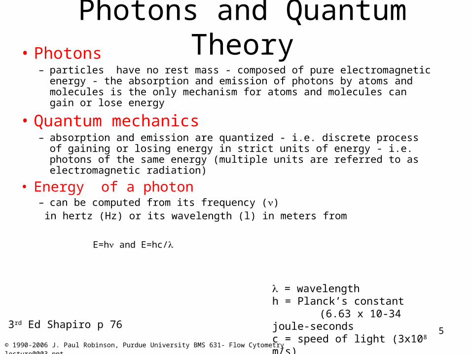
Photons and Quantum Theory• Photons
– particles have no rest mass - composed of pure electromagnetic energy - the absorption and emission of photons by atoms and molecules is the only mechanism for atoms and molecules can gain or lose energy
• Quantum mechanics– absorption and emission are quantized - i.e. discrete process of gaining or
losing energy in strict units of energy - i.e. photons of the same energy (multiple units are referred to as electromagnetic radiation)
• Energy of a photon– can be computed from its frequency () in hertz (Hz) or its wavelength (l) in meters from
E=h and E=hc/
= wavelengthh = Planck’s constant
(6.63 x 10⁻³⁴ joule-secondsc = speed of light (3x10⁸ m/s)3rd Ed Shapiro p 76
6
© 1990-2006 J. Paul Robinson, Purdue University BMS 631- Flow Cytometry lecture0003.ppt
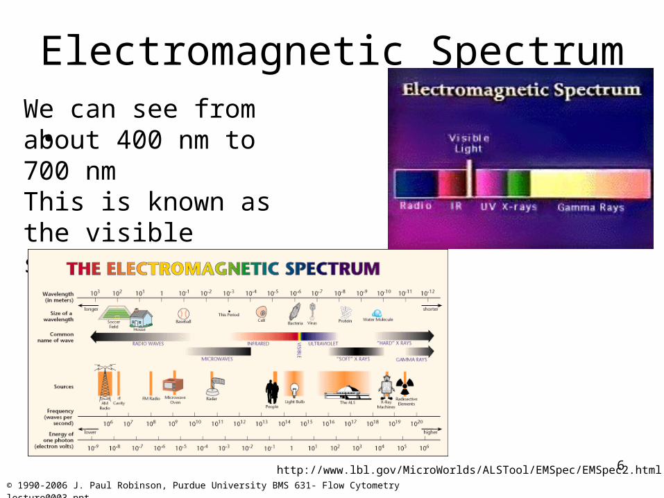
Electromagnetic Spectrum
• We can see from about 400 nm to 700 nmThis is known as the visible spectrum
http://www.lbl.gov/MicroWorlds/ALSTool/EMSpec/EMSpec2.html
7
© 1990-2006 J. Paul Robinson, Purdue University BMS 631- Flow Cytometry lecture0003.ppt
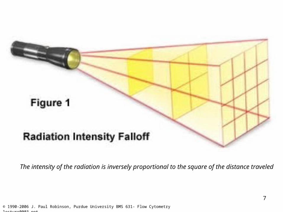
The intensity of the radiation is inversely proportional to the square of the distance traveled
8
© 1990-2006 J. Paul Robinson, Purdue University BMS 631- Flow Cytometry lecture0003.ppt
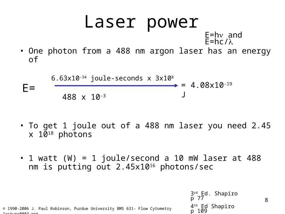
Laser power
• One photon from a 488 nm argon laser has an energy of
6.63x10⁻³⁴ joule-seconds x 3x10⁸
• To get 1 joule out of a 488 nm laser you need 2.45 x 10¹⁸ photons
• 1 watt (W) = 1 joule/second a 10 mW laser at 488 nm is putting out 2.45x10¹⁶ photons/sec
E=h and E=hc/
= 4.08x10⁻¹⁹ J
3rd Ed. Shapiro p 77
4th Ed Shapiro p 109
488 x 10-3E=
9
© 1990-2006 J. Paul Robinson, Purdue University BMS 631- Flow Cytometry lecture0003.ppt
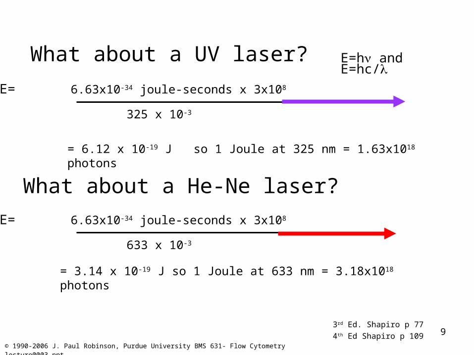
325 x 10-3
What about a UV laser?
E= 6.63x10⁻³⁴ joule-seconds x 3x108
= 6.12 x 10⁻¹⁹ J so 1 Joule at 325 nm = 1.63x10¹⁸ photons
What about a He-Ne laser?
633 x 10-3
E= 6.63x10⁻³⁴ joule-seconds x 3x108
= 3.14 x 10⁻¹⁹ J so 1 Joule at 633 nm = 3.18x10¹⁸ photons
3rd Ed. Shapiro p 77
4th Ed Shapiro p 109
E=h and E=hc/
10
© 1990-2006 J. Paul Robinson, Purdue University BMS 631- Flow Cytometry lecture0003.ppt
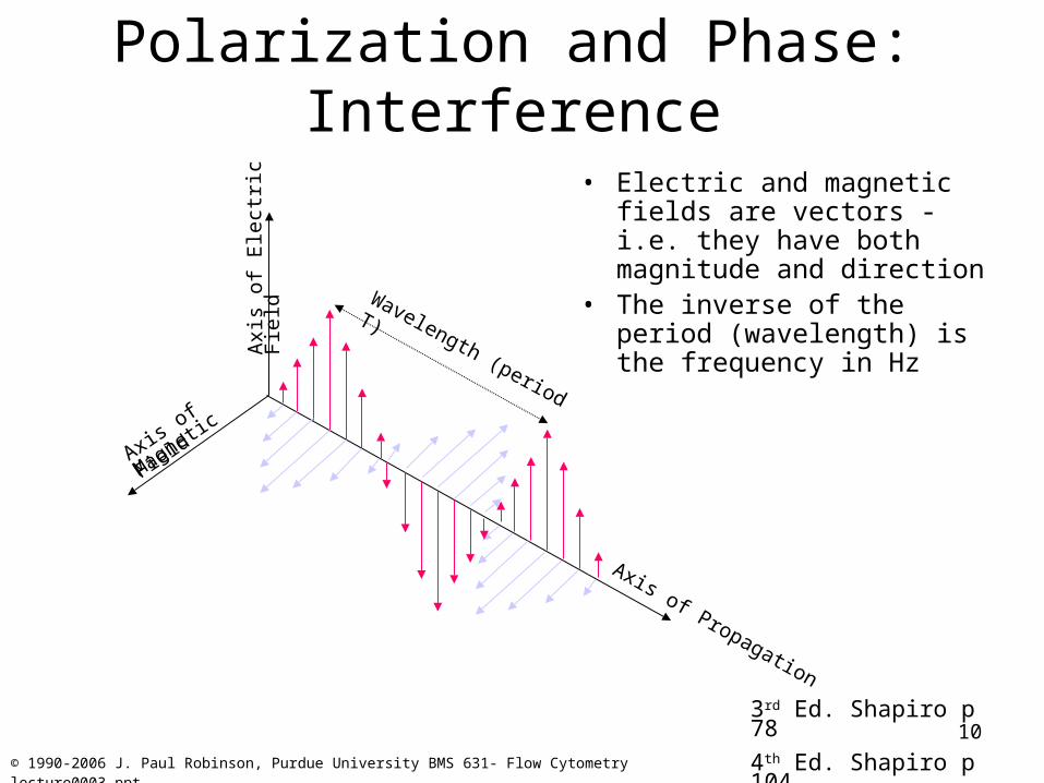
Polarization and Phase: Interference
• Electric and magnetic fields are vectors - i.e. they have both magnitude and direction
• The inverse of the period (wavelength) is the frequency in Hz
3rd Ed. Shapiro p 78
4th Ed. Shapiro p 104
Wavelength (period T)
Axis of
Magnetic F
ield
Axis of Propagation
Axi
s of
Ele
ctri
c F
ield
11
© 1990-2006 J. Paul Robinson, Purdue University BMS 631- Flow Cytometry lecture0003.ppt
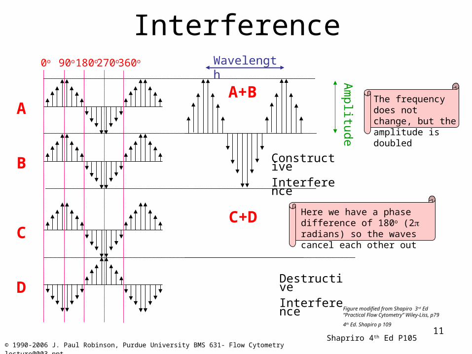
Interference
ConstructiveInterference
DestructiveInterference
A
B
C
D
A+B
C+D
Am
plitude
0o 90o 180o 270o 360o Wavelength
Figure modified from Shapiro 3rd Ed “Practical Flow Cytometry” Wiley-Liss, p79
4th Ed. Shapiro p 109
Here we have a phase difference of 180o (2 radians) so the waves cancel each other out
The frequency does not change, but the amplitude is doubled
Shapriro 4th Ed P105
12
© 1990-2006 J. Paul Robinson, Purdue University BMS 631- Flow Cytometry lecture0003.ppt
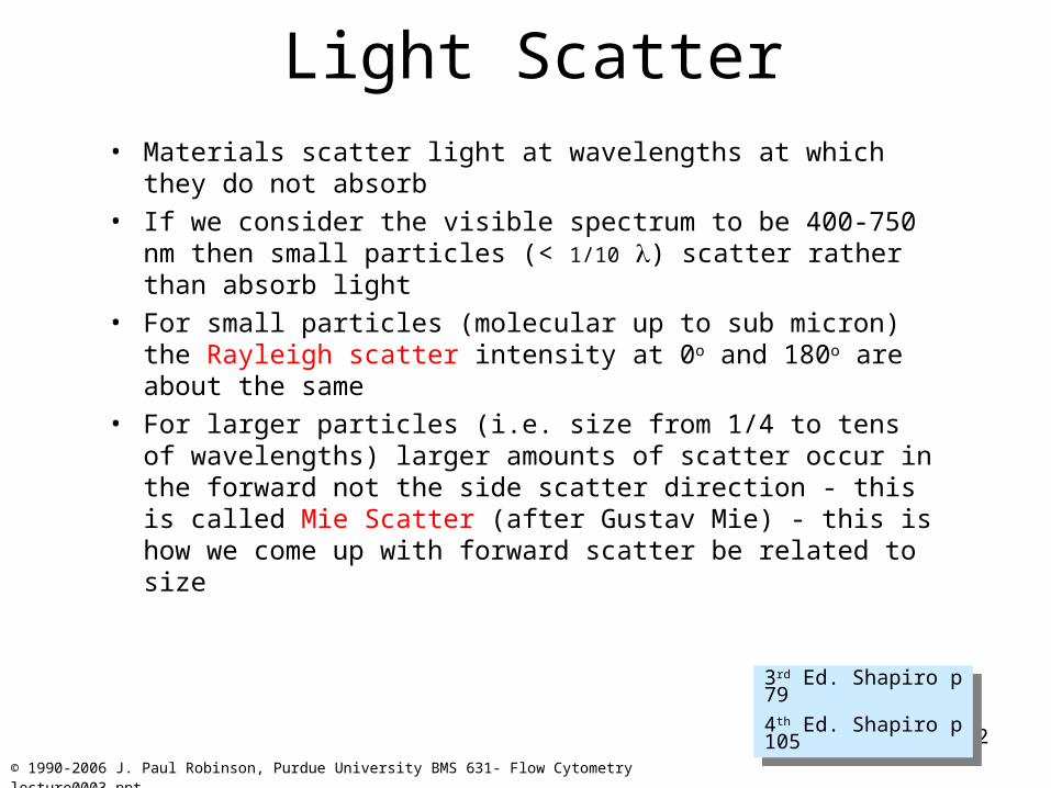
Light Scatter• Materials scatter light at wavelengths at which they do not
absorb• If we consider the visible spectrum to be 400-750 nm then
small particles (< 1/10 ) scatter rather than absorb light• For small particles (molecular up to sub micron) the Rayleigh
scatter intensity at 0o and 180o are about the same• For larger particles (i.e. size from 1/4 to tens of wavelengths)
larger amounts of scatter occur in the forward not the side scatter direction - this is called Mie Scatter (after Gustav Mie) - this is how we come up with forward scatter be related to size
3rd Ed. Shapiro p 79
4th Ed. Shapiro p 105
3rd Ed. Shapiro p 79
4th Ed. Shapiro p 105
13
© 1990-2006 J. Paul Robinson, Purdue University BMS 631- Flow Cytometry lecture0003.ppt
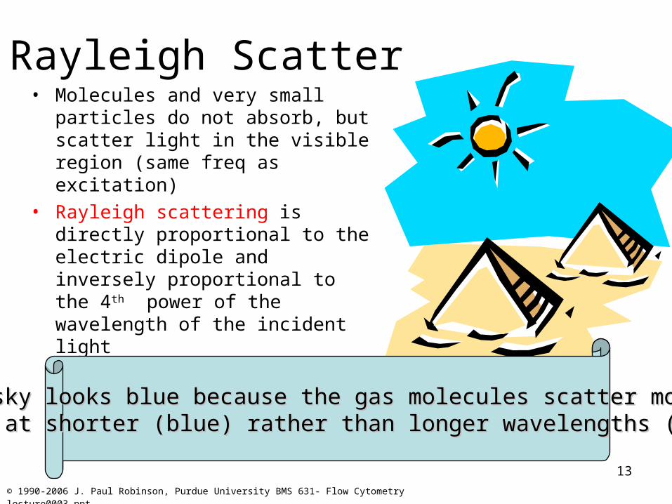
Rayleigh Scatter• Molecules and very small particles do
not absorb, but scatter light in the visible region (same freq as excitation)
• Rayleigh scattering is directly proportional to the electric dipole and inversely proportional to the 4th power of the wavelength of the incident light
the sky looks blue because the gas molecules scatter more the sky looks blue because the gas molecules scatter more light at shorter (blue) rather than longer wavelengths (red)light at shorter (blue) rather than longer wavelengths (red)
14
© 1990-2006 J. Paul Robinson, Purdue University BMS 631- Flow Cytometry lecture0003.ppt
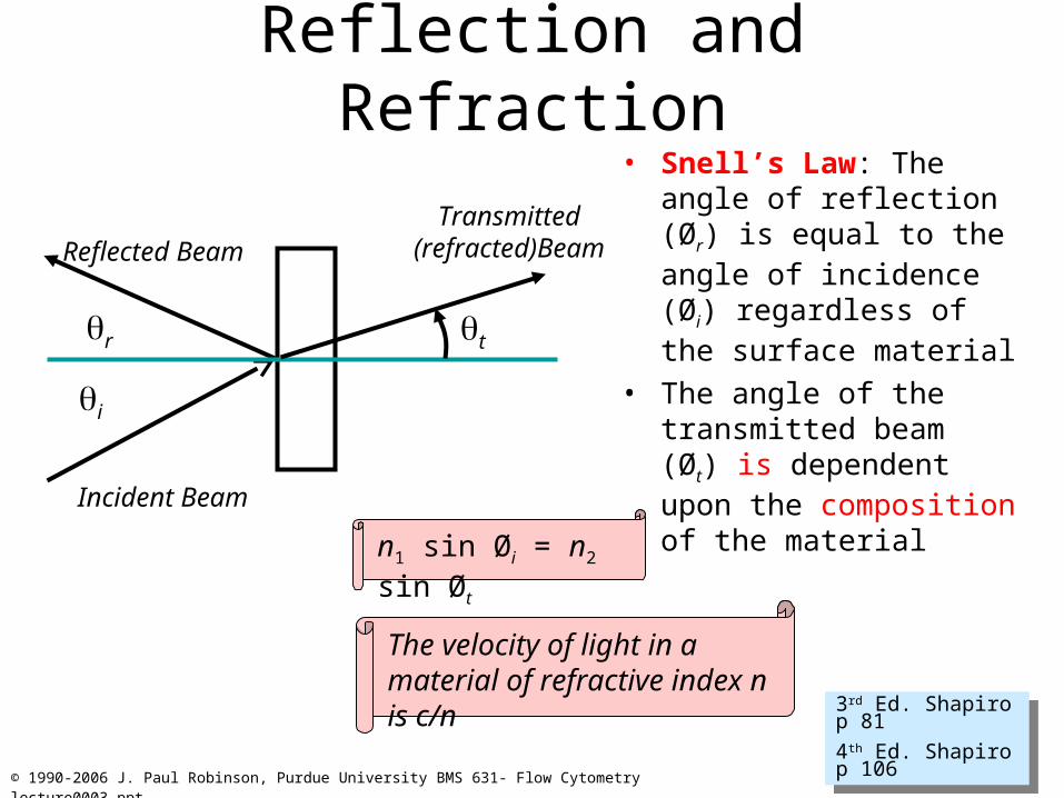
Reflection and Refraction• Snell’s Law: The angle of
reflection (Ør) is equal to the angle of incidence (Øi) regardless of the surface material
• The angle of the transmitted beam (Øt) is dependent upon the composition of the material
3rd Ed. Shapiro p 81
4th Ed. Shapiro p 106
3rd Ed. Shapiro p 81
4th Ed. Shapiro p 106
t
i
r
Reflected Beam
Incident Beam
Transmitted(refracted)Beam
n1 sin Øi = n2 sin Øt
The velocity of light in a material of refractive index n is c/n
15
© 1990-2006 J. Paul Robinson, Purdue University BMS 631- Flow Cytometry lecture0003.ppt
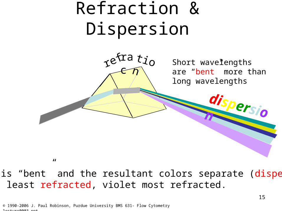
Refraction & Dispersion
Light is “bent” and the resultant colors separate (dispersion).Red is least refracted, violet most refracted.
dispersion
Short wavelengths are “bent” more than long wavelengths
refraction
16
© 1990-2006 J. Paul Robinson, Purdue University BMS 631- Flow Cytometry lecture0003.ppt
Light Propagation & Vergence
• Considering a point source emission of light, rays emanate over 4pi steradians
• If the ray of light travels through a length L of a medium of RI n, the optical path length S=Ln (thus S represents the distance light would have traveled in a vacuum in the same time it took to travel the distance L in the medium (RI n).
• Rays diverge (because the come from a point source• Vergence is measured in diopters
3rd Shapiro p 93
4th Shapiro p 119
3rd Shapiro p 93
4th Shapiro p 119
17
© 1990-2006 J. Paul Robinson, Purdue University BMS 631- Flow Cytometry lecture0003.ppt
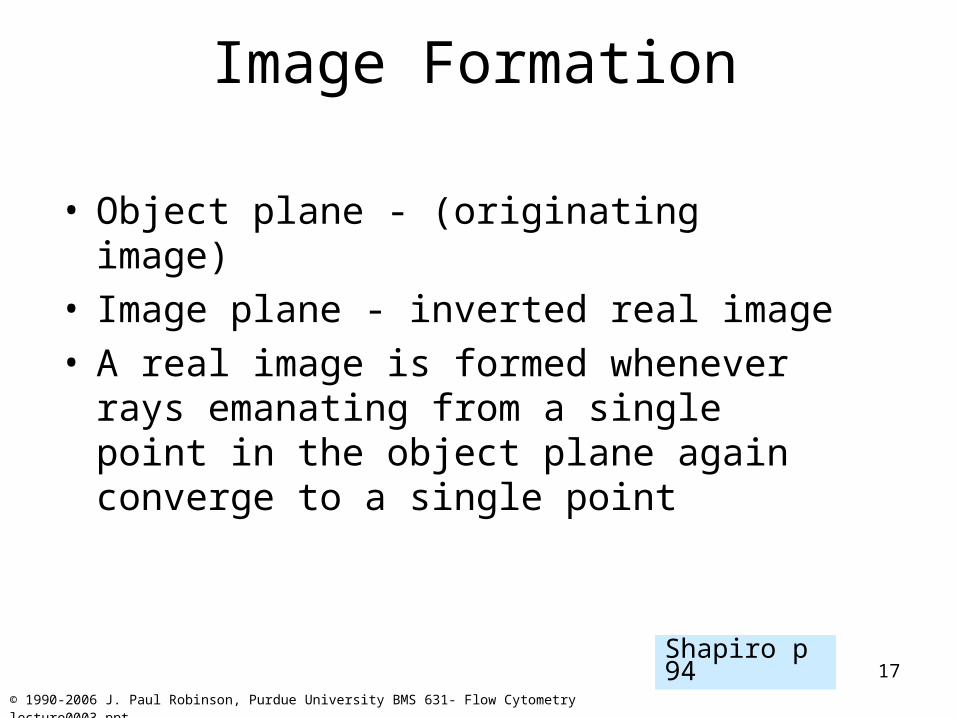
Image Formation
• Object plane - (originating image)• Image plane - inverted real image• A real image is formed whenever rays
emanating from a single point in the object plane again converge to a single point
Shapiro p 94
18
© 1990-2006 J. Paul Robinson, Purdue University BMS 631- Flow Cytometry lecture0003.ppt
Numerical Aperture
• The wider the angle the lens is capable of receiving light at, the greater its resolving power
• The higher the NA, the shorter the working distance
Shapiro p 96

19
© 1990-2006 J. Paul Robinson, Purdue University BMS 631- Flow Cytometry lecture0003.ppt
Numerical Aperture• Resolving power is directly related to numerical
aperture.• The higher the NA the greater the resolution• Resolving power:
The ability of an objective to resolve two distinct lines very close together
NA = n sin
– (n=the lowest refractive index between the object and first objective element) (hopefully 1)
– is 1/2 the angular aperture of the objective

20
© 1990-2006 J. Paul Robinson, Purdue University BMS 631- Flow Cytometry lecture0003.ppt
Numerical Aperture• For a narrow light beam (i.e. closed illumination aperture diaphragm)
the finest resolution is (at the brightest point of the visible spectrum i.e. 530 nm)…(closed condenser).
NA
2 x NA
.000532 x 1.00= 0.265 m
.000531.00 = 0.53 m
• With a cone of light filling the entire aperture the theoretical resolution is…(fully open condenser)..
=
=
21
© 1990-2006 J. Paul Robinson, Purdue University BMS 631- Flow Cytometry lecture0003.ppt
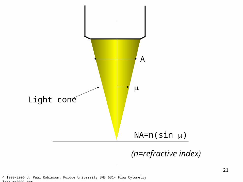
A
NA=n(sin )
Light cone
(n=refractive index)
22
© 1990-2006 J. Paul Robinson, Purdue University BMS 631- Flow Cytometry lecture0003.ppt
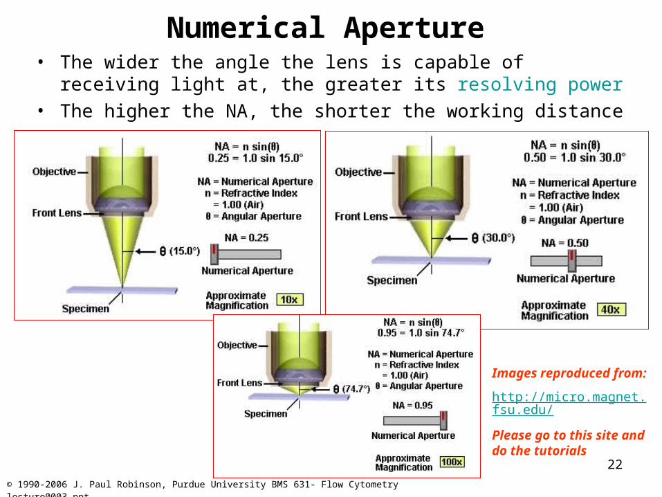
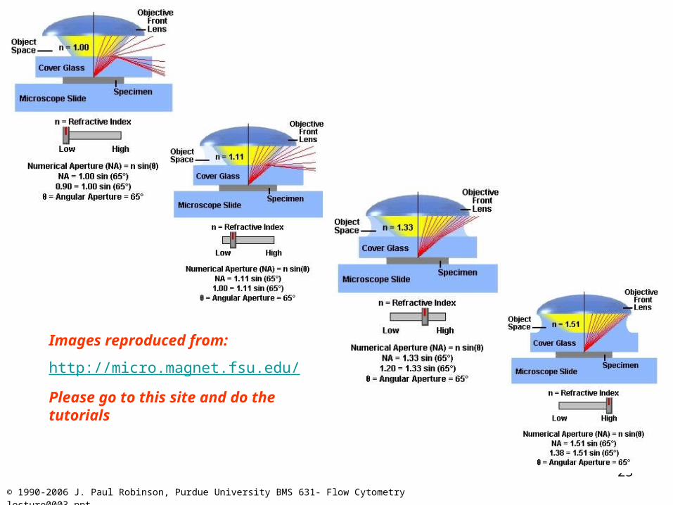
Numerical Aperture• The wider the angle the lens is capable of receiving light at, the
greater its resolving power• The higher the NA, the shorter the working distance
Images reproduced from:
http://micro.magnet.fsu.edu/
Please go to this site and do the tutorials
23
© 1990-2006 J. Paul Robinson, Purdue University BMS 631- Flow Cytometry lecture0003.ppt
Images reproduced from:
http://micro.magnet.fsu.edu/
Please go to this site and do the tutorials
24
© 1990-2006 J. Paul Robinson, Purdue University BMS 631- Flow Cytometry lecture0003.ppt
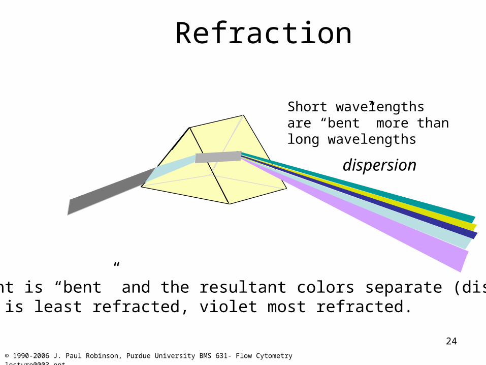
Refraction
Light is “bent” and the resultant colors separate (dispersion).Red is least refracted, violet most refracted.
dispersion
Short wavelengths are “bent” more than long wavelengths
25
© 1990-2006 J. Paul Robinson, Purdue University BMS 631- Flow Cytometry lecture0003.ppt
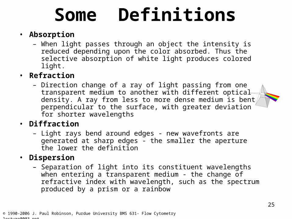
Some Definitions• Absorption
– When light passes through an object the intensity is reduced depending upon the color absorbed. Thus the selective absorption of white light produces colored light.
• Refraction– Direction change of a ray of light passing from one transparent
medium to another with different optical density. A ray from less to more dense medium is bent perpendicular to the surface, with greater deviation for shorter wavelengths
• Diffraction– Light rays bend around edges - new wavefronts are generated at
sharp edges - the smaller the aperture the lower the definition
• Dispersion– Separation of light into its constituent wavelengths when entering a
transparent medium - the change of refractive index with wavelength, such as the spectrum produced by a prism or a rainbow
26
© 1990-2006 J. Paul Robinson, Purdue University BMS 631- Flow Cytometry lecture0003.ppt
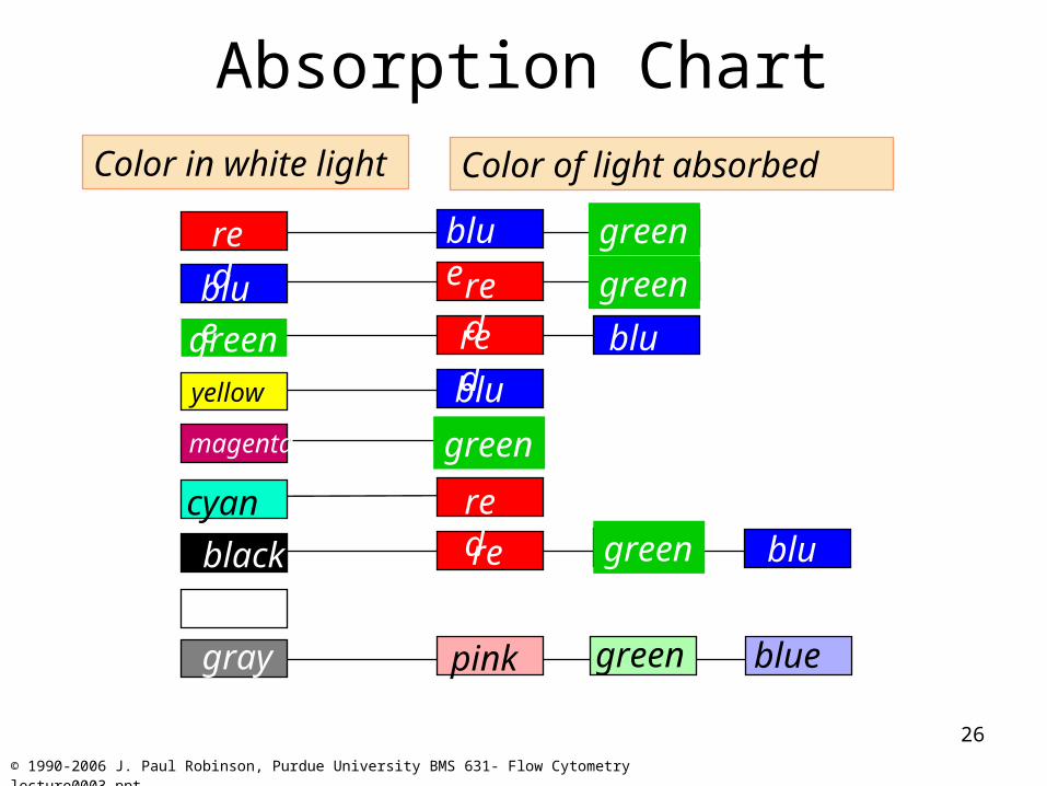
Absorption ChartColor in white light Color of light absorbed
red
blue
green
magenta
cyan
yellow
blue
blue
blue
blue
green
green
green
green
red
red
red
redblack
gray green bluepink

27
© 1990-2006 J. Paul Robinson, Purdue University BMS 631- Flow Cytometry lecture0003.ppt
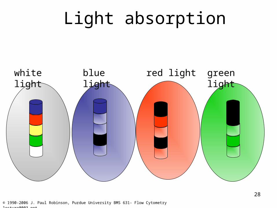
Light absorption
Absorption
Control
No blue/green light red filter
28
© 1990-2006 J. Paul Robinson, Purdue University BMS 631- Flow Cytometry lecture0003.ppt
Light absorption
white light blue light red light green light

29
© 1990-2006 J. Paul Robinson, Purdue University BMS 631- Flow Cytometry lecture0003.ppt
The light spectrumWavelength = Frequency
Blue light
488 nm
short wavelength
high frequency
high energy (2 times the red)
Red light
650 nm
long wavelength
low frequency
low energy
Photon as a wave packet of energy
30
© 1990-2006 J. Paul Robinson, Purdue University BMS 631- Flow Cytometry lecture0003.ppt
Lecture Summary
• Principles of light and matter
• Basic Optics to take into consideration
• Essentials of light measurement
• Absorption and optical properties