Embed Size (px)
Citation preview

© 2001 UMBC Neurological Management CCEMT-P SM 12/98 1
Intracranial Pressure

© 2001 UMBC Neurological Management CCEMT-P SM 12/98 2
Intracranial Pressure
• Definition– Pressure exerted by brain tissue, intracranial
blood, and cerebral spinal fluid (CSF) in a non-distended (closed) cavity

© 2001 UMBC Neurological Management CCEMT-P SM 12/98 3
Normal Contents of the Skull
• Brain - 80%
• Blood - 10%
• CSF - 10%
• Monroe-Kellie Doctrine

© 2001 UMBC Neurological Management CCEMT-P SM 12/98 4
Changes in Skull Contents
• Increased intracranial volume– Hyperemia– Bleeding, clots, bruises – Cerebral edema– Hydrocephalus– Foreign object– Tumor

© 2001 UMBC Neurological Management CCEMT-P SM 12/98 5
Intracranial Pressure
• Cerebral edema (localized or generalized) can develop from any injury to the brain
• Swelling peaks in 3-5 days

© 2001 UMBC Neurological Management CCEMT-P SM 12/98 6
Post-traumatic Hydrocephalia
• Communicating– CSF circulates but is not reabsorbed– Arachnoid villa are clogged
• Non-communicating– CSF circulation is obstructed– CSF can not reach the arachnoid villa to be
reabsorbed
© 2001 UMBC Neurological Management CCEMT-P SM 12/98 7
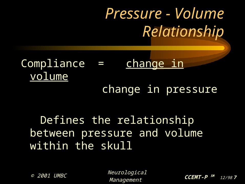
Pressure - Volume Relationship
Compliance = change in volume change in pressure
Defines the relationship between pressure and volume within the skull

© 2001 UMBC Neurological Management CCEMT-P SM 12/98 8
Pressure - Volume Relationship
• Three portions of the waveform curve– Flat– Curved– Vertical (inflection) point

© 2001 UMBC Neurological Management CCEMT-P SM 12/98 9
ICP Waveforms
• Normal waveform resembles a set of stairs
• With increasing ICP, waveform loses shape
© 2001 UMBC Neurological Management CCEMT-P SM 12/98
10
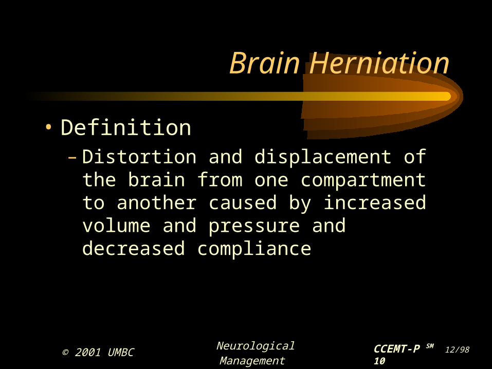
Brain Herniation
• Definition– Distortion and displacement of the brain from
one compartment to another caused by increased volume and pressure and decreased compliance

© 2001 UMBC Neurological Management CCEMT-P SM 12/98
11
Brain Herniation
• Three types– Central or transtentorial– Uncal or lateral transtentorial– Cingulate

© 2001 UMBC Neurological Management CCEMT-P SM 12/98
12
Mean Arterial Pressure (MAP)
• Calculate MAP– Subtracting diastolic pressure from systolic pressure– Divide by 3– And add diastolic pressure
140 - 100 = 40
3 = 13 + 100 = 113 MAP = 113

© 2001 UMBC Neurological Management CCEMT-P SM 12/98
13
Cerebral Perfusion Pressure (CPP)
• Pressure gradient driving blood flow and delivery of nutrients to the brain

© 2001 UMBC Neurological Management CCEMT-P SM 12/98
14
Cerebral Perfusion Pressure (CPP)
• Calculate CPP– Subtract ICP from MAP
Patient has an ICP of 80 and a MAP of 113 113 MAP
- 80 ICP 33 CPP

© 2001 UMBC Neurological Management CCEMT-P SM 12/98
15
Cerebral Perfusion Pressure (CPP)
CPP = MAP - ICP
Best if > 70 mmHg
< 60 mmHg = impaired blood flow to brain
© 2001 UMBC Neurological Management CCEMT-P SM 12/98
16
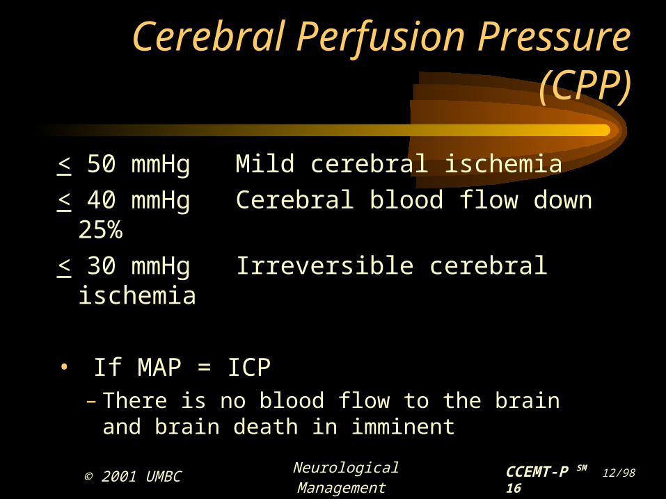
Cerebral Perfusion Pressure (CPP)
< 50 mmHg Mild cerebral ischemia
< 40 mmHg Cerebral blood flow down 25%
< 30 mmHg Irreversible cerebral ischemia
• If MAP = ICP – There is no blood flow to the brain
and brain death in imminent

© 2001 UMBC Neurological Management CCEMT-P SM 12/98
17
Increased ICP
• Neurological exam
• Motor function• Respiratory • Body temperature
• LOC • Pupil • Vision • Herniation

© 2001 UMBC Neurological Management CCEMT-P SM 12/98
18
Signs & Symptoms of ICP
• Vital signs changes - Cushing’s triad
• Widening pulse pressure
• Bradycardia
• Abnormal respiratory patterns

© 2001 UMBC Neurological Management CCEMT-P SM 12/98
19
Signs & Symptoms of ICP
• Respiratory changes– Cheyne-stokes– Central neurogenic hyperventilation– Biot’s– Kussmal

© 2001 UMBC Neurological Management CCEMT-P SM 12/98
20
ICP Monitoring

© 2001 UMBC Neurological Management CCEMT-P SM 12/98
21
ICP Monitoring
• Indications– Glasgow coma score <8 and positive CT– Paralytic and/or sedative medications are being
used

© 2001 UMBC Neurological Management CCEMT-P SM 12/98
22
Devices
• Interventricular cannula (IVC)
• Epidural catheter
• Subdural / subarachnoid monitoring devices
• Fiber optic transducer tipped probe

© 2001 UMBC Neurological Management CCEMT-P SM 12/98
23
Interventricular Cannula (IVC)
• Most commonly used monitor
• Placed within the ventricle
• Location of placement for some Caminos

© 2001 UMBC Neurological Management CCEMT-P SM 12/98
24
Interventricular Cannula (IVC)
• Advantages– Drain CSF to lower
ICP
– Obtain CSF cultures
– Increased accuracy in ICP monitoring
– Accurate and reliable
• Disadvantages– Infection
– Injury to brain
– Clot formation
– Hemorrhage risk
– Collapsed ventricle
– Placement may be impossible

© 2001 UMBC Neurological Management CCEMT-P SM 12/98
25
Interventricular Cannula (IVC)
• Transport considerations– System set-up – Charting ICP– Drainage orders – Movement – Pressure changes with air transport

© 2001 UMBC Neurological Management CCEMT-P SM 12/98
26
Epidural Catheter
• Lies beneath skull - above dura mater
• Advantages– Lesser rate of infection than IVC – Placement causes less injury to brain
• Disadvantages– Less accurate than IVC– Cannot be used to drain CSF

© 2001 UMBC Neurological Management CCEMT-P SM 12/98
27
Subdural / Subarachnoid Monitor
• Newer systems connect to fiber optics
• Called a “screw” or “bolt”
• Subdural - beneath dura and above pia
• Subarachnoid - placed beneath arachnoid and above pia

© 2001 UMBC Neurological Management CCEMT-P SM 12/98
28
Subdural / Subarachnoid Monitor
• Advantages– Not as invasive as
IVC or epidural
– Less rate of infection
– Less injury to brain
– Easier to place
• Disadvantages– Less accurate monitoring
– Cannot drain CSF
– Risk of bleeding and brain injury
– Higher rate of infection than epidural catheter
– Requires closed, intact skull

© 2001 UMBC Neurological Management CCEMT-P SM 12/98
29
Fiber Optic Transducer Tipped Probe
• Catheter with pressure sensing device placed into subdural space, brain parenchyma or ventricle
• Non fluid filled continuous intact system
© 2001 UMBC Neurological Management CCEMT-P SM 12/98
30
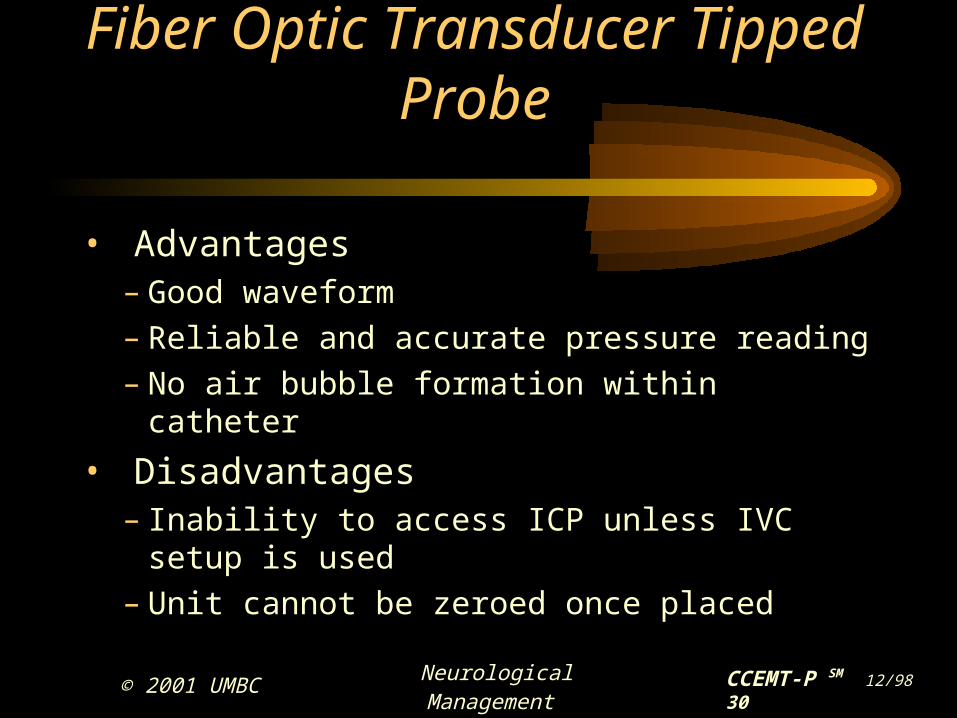
Fiber Optic Transducer Tipped Probe
• Advantages– Good waveform– Reliable and accurate pressure reading– No air bubble formation within catheter
• Disadvantages– Inability to access ICP unless IVC setup is used– Unit cannot be zeroed once placed

© 2001 UMBC Neurological Management CCEMT-P SM 12/98
31
Transport Considerations
• Avoid tension or kinking of cable
• Less problem with zeroing
• Maintain clean / intact dressing

© 2001 UMBC Neurological Management CCEMT-P SM 12/98
32
ICP Data
• Normal values 0-15 mmHg
• Normal waveforms first 3 waves– P1 - percussion
– P2 - tidal wave
– P3 - dicrotic wave
• Abnormal waveforms– C waves - think PACs
– B waves - think PVCs
– A waves - think V-fib

© 2001 UMBC Neurological Management CCEMT-P SM 12/98
33
Management of ICP
• Gas exchange– Optimize gas exchange to maintain ICP or
decrease ICP levels– Good pulmonary toilet– Ph changes– Hyperoxygenation– Hyperventilation– Positive end expiratory pressure (PEEP)

© 2001 UMBC Neurological Management CCEMT-P SM 12/98
34
Management of ICP
• Hyperventilation– Vasodilation occurs in brain tissue to increase
circulation and then increases ICP
• Goals of hyperventilation– Remove CO2 and cause vasoconstriction
– Cause - respiratory alkalosis– Effect - pace drops reducing cerebral blood
flow

© 2001 UMBC Neurological Management CCEMT-P SM 12/98
35
Hyperventilation Controversy
• What is optimal paco2 level?
– Old method - keep paco2 at 25 mmhg
– New method - paco2 range 28-32 mmhg

© 2001 UMBC Neurological Management CCEMT-P SM 12/98
36
Factors which Increase ICP
• Hip flexion (decreases venous return)
• Head and neck position• Changing level of height
of bed (especially flat)• External noxious stimuli
• Agitation• Pain• Coughing and
valsalva maneuver • Seizures

© 2001 UMBC Neurological Management CCEMT-P SM 12/98
37
Methods of Decreasing ICP
• Decrease external stimulation• Ensure a quiet environment• Pull slouching patients to the top of the bed• Use cervical collar with decreased neck
muscle tone• Shut off bright lights• Align head and neck• Surgical intervention

© 2001 UMBC Neurological Management CCEMT-P SM 12/98
38
Methods of Decreasing ICP
• If IVC in place, open and drain
• If too much CSF is lost, the ventricle can collapse

© 2001 UMBC Neurological Management CCEMT-P SM 12/98
39
Methods of Decreasing ICP
• Medications– Sedation – Paralytics– Diuretics – Steroids– Barbituate coma

© 2001 UMBC Neurological Management CCEMT-P SM 12/98
40
Medications
• Sedatives– Ventilator patients – Pentathol or fentanyl
• Paralytics– Decrease metabolism– Generally utilized with sedatives– ICP monitoring necessary for most medically
paralyzed patients

© 2001 UMBC Neurological Management CCEMT-P SM 12/98
41
Medications
• Diuretics – Mannitol and Lasix
• Steroids– Dexamethasone / methylprednisone– Controversial
• Some studies show no benefit, others show limited benefit if given within 8 hours of the injury

© 2001 UMBC Neurological Management CCEMT-P SM 12/98
42
Medications
• Barbiturate coma– Induced pharmacologically using barbiturates
• Barbiturate coma results– Neuro status - unresponsive with a GCS of 3– Respiratory - ventilator dependent– Cardiovascular - bradycardia and hypotensive
© 2001 UMBC Neurological Management CCEMT-P SM 12/98
43
Intracranial Pressure Monitoring
• Operative intervention– Surgical removal of blood clot or affected
portion of the brain

© 2001 UMBC Neurological Management CCEMT-P SM 12/98
44
Intracranial Pressure
Conclusion

© 2001 UMBC Neurological Management CCEMT-P SM 12/98
45
Neuromonitoring for Traumatic Brain Injury

© 2001 UMBC Neurological Management CCEMT-P SM 12/98
46
Intracranial Pressure Monitoring

© 2001 UMBC Neurological Management CCEMT-P SM 12/98
47
Jugular Venous Bulb Oximetry

© 2001 UMBC Neurological Management CCEMT-P SM 12/98
48
Transcranial Doppler Ultrasound
© 2001 UMBC Neurological Management CCEMT-P SM 12/98
49
Cerebral Function Monitoring

© 2001 UMBC Neurological Management CCEMT-P SM 12/98
50
Emerging Monitoring Technologies

© 2001 UMBC Neurological Management CCEMT-P SM 12/98
51
References