Embed Size (px)
Citation preview
Intracranial Mass Lesions and
Elevated Intracranial Pressure
Lissa C. Baird, MD Assistant Professor
Directory, Pediatric Surgical Neuro-Oncology Department of Neurological Surgery
Oregon Health & Science University

Conflict of Interest Disclosure
Disclosure
I do not have any financial relationships to
disclose.
I. General Principles of Intracranial Mass Lesions
Intracranial Mass Lesions Overview
• I. General Principles of Intracranial Mass Lesions
• II. Differential Diagnosis
• III. Signs and Symptoms
• IV. Clinical Management of Mass Lesions and Elevated Intracranial Pressure
1
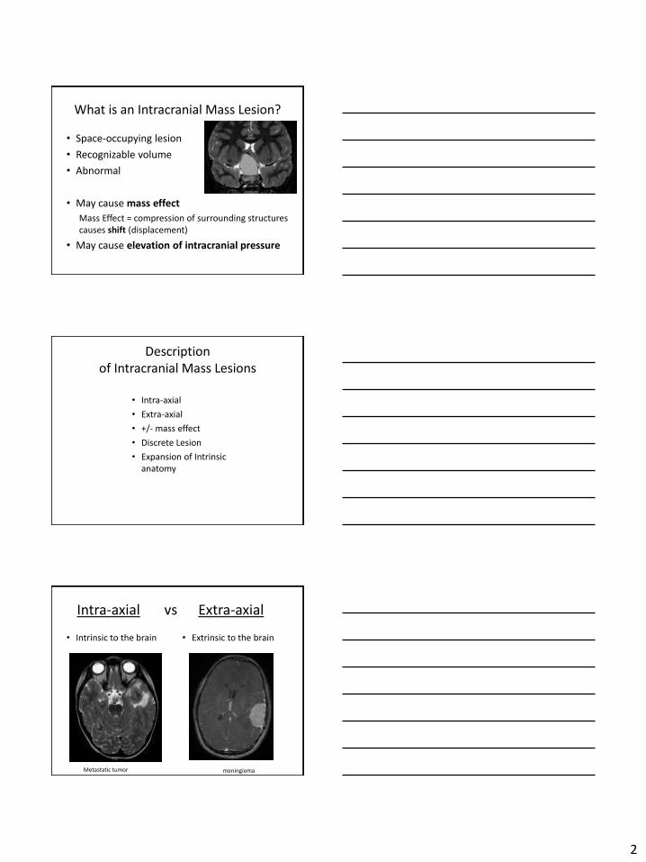
Intra-axial vs Extra-axial
• Intrinsic to the brain
Metastatic tumor
• Extrinsic to the brain
meningioma
What is an Intracranial Mass Lesion?
• Space-occupying lesion
• Recognizable volume
• Abnormal
• May c ause mass effect
Mass Effect = compression of surrounding structures causes shift (displacement)
• May c ause elevation of intracranial pressure
Description of Intracranial Mass Lesions
• Intra-axial
• Extra-axial
• +/- mass effect
• Discrete Lesion
• Expansion of In trinsic anatomy
2
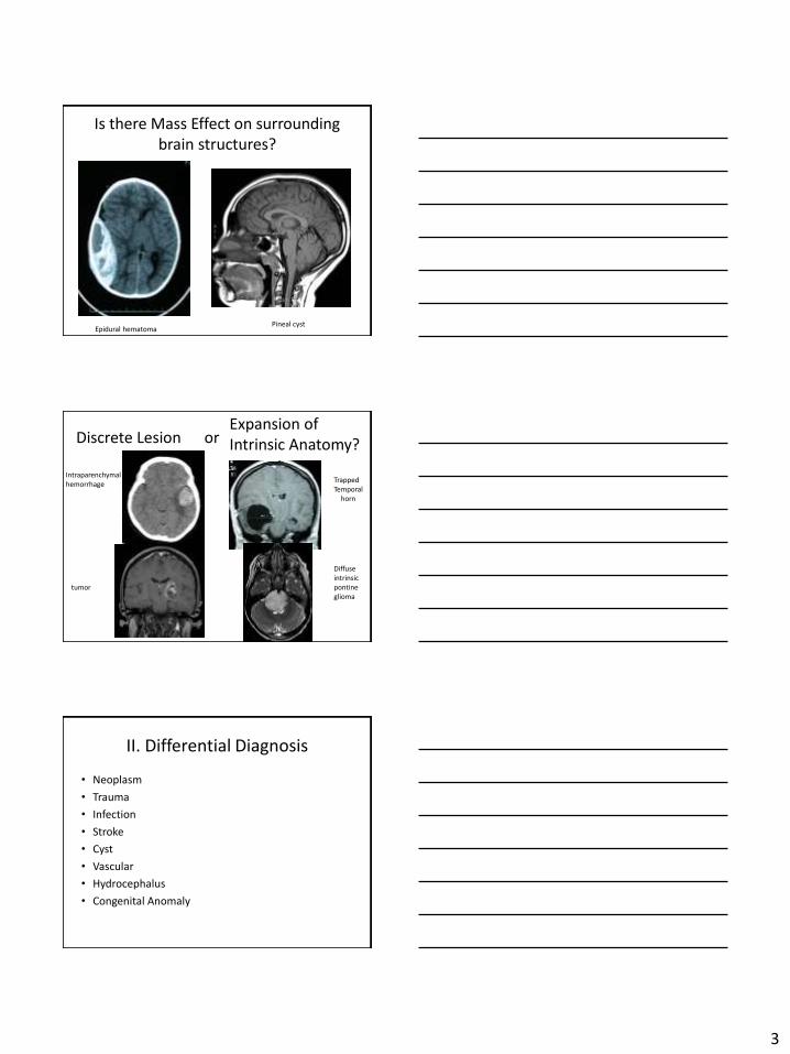
Is there Mass Effect on surrounding brain structures?
Epidural hematoma Pineal cyst
Discrete Lesion or Expansion of
Intrinsic Anatomy?
Intraparenchymalhemorrhage
tumor
Trapped Temporal horn
Diffuse intrinsic pontine glioma
II. Differential Diagnosis
• Neoplasm
• Trauma
• Infection
• Stroke
• Cyst
• Vascular
• Hydrocephalus
• Congenital Anomaly
3
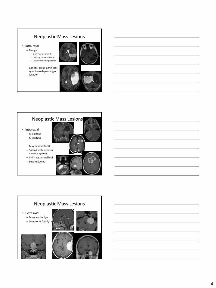
Neoplastic Mass Lesions
• Intra-axial
– Benign • Slow rate of growth
• Unlikely to metastasize
• Less surrounding edema
– Can still cause significant symptoms depending on location
Neoplastic Mass Lesions
• Intra-axial
– Malignant
– Metastatic
– May be multifocal
– Spread within central nervous system
– Infiltrate normal brain
– Severe Edema
Neoplastic Mass Lesions
• Extra-axial
– Most are benign
– Symptoms focally re lat
4
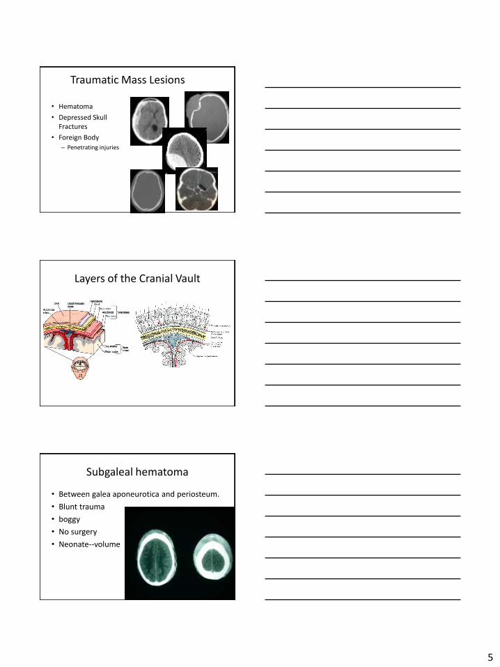
Traumatic Mass Lesions
• Hematoma
• Depressed S kull Fractures
• Foreign Body
– Penetrating injuries
Layers of the Cranial Vault
Subgaleal hematoma
• Between galea aponeurotica and periosteum.
• Blunt trauma
• boggy
• No surgery
• Neonate--volume
5
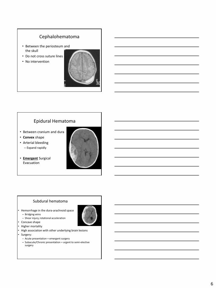
Cephalohematoma
• Between the periosteum andthe skull
• Do not cross suture lines
• No intervention
Epidural Hematoma
• Between cranium and dura
• Convex shape
• Arterial bleeding
– Expand rapidly
• Emergent SurgicalEvacuation
Subdural hematoma
• Hemorrhage in the dura-arachnoid space – Bridging veins
– Shear i njury, rotational acceleration
• Concave shape
• Higher mortality
• High association with other underlying brain lesions
• Surgery: – Acute presentation = emergent surgery
– Subacute/Chronic presentation = urgent to semi-elective surgery
6
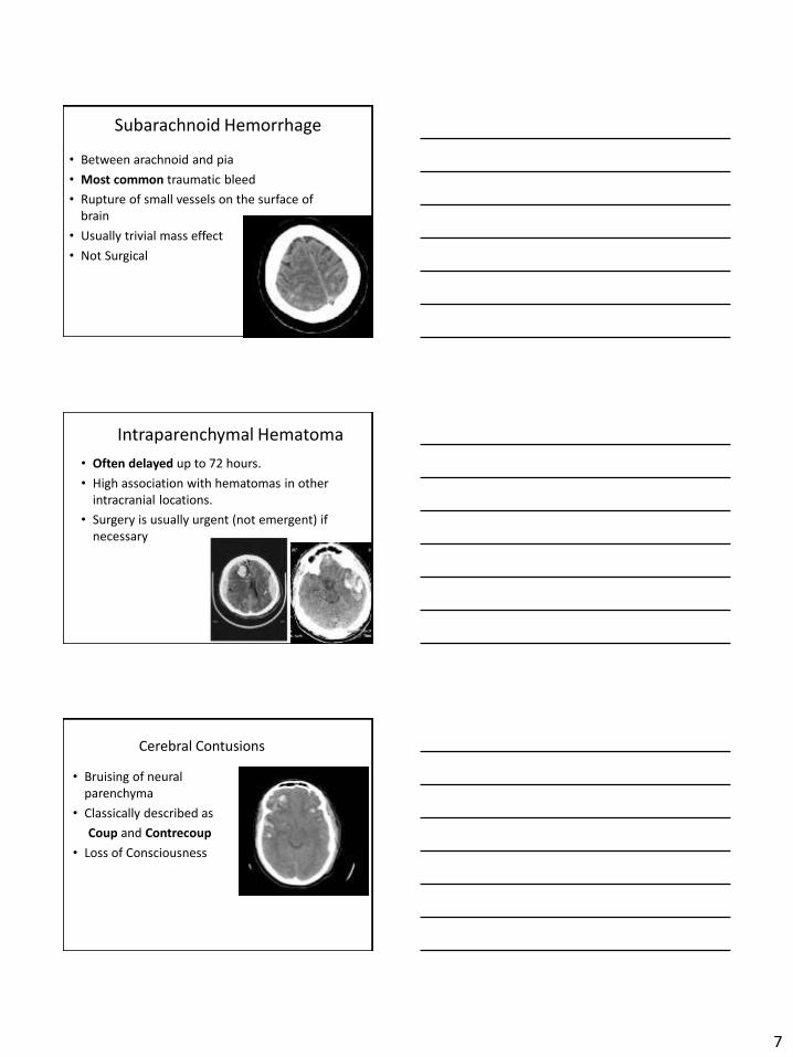
Subarachnoid Hemorrhage
• Between arachnoid and pia
• Most common traumatic bleed
• Rupture of small vessels on the surface ofbrain
• Usually trivial mass effect
• Not Surgical
Intraparenchymal Hematoma
• Often delayed up to 72 hours.
• High association with hematomas in otherintracranial locations.
• Surgery is usually urgent (not emergent) ifnecessary
Cerebral Contusions
• Bruising of neuralparenchyma
• Classically described as
Coup and Contrecoup
• Loss of Consciousness
7
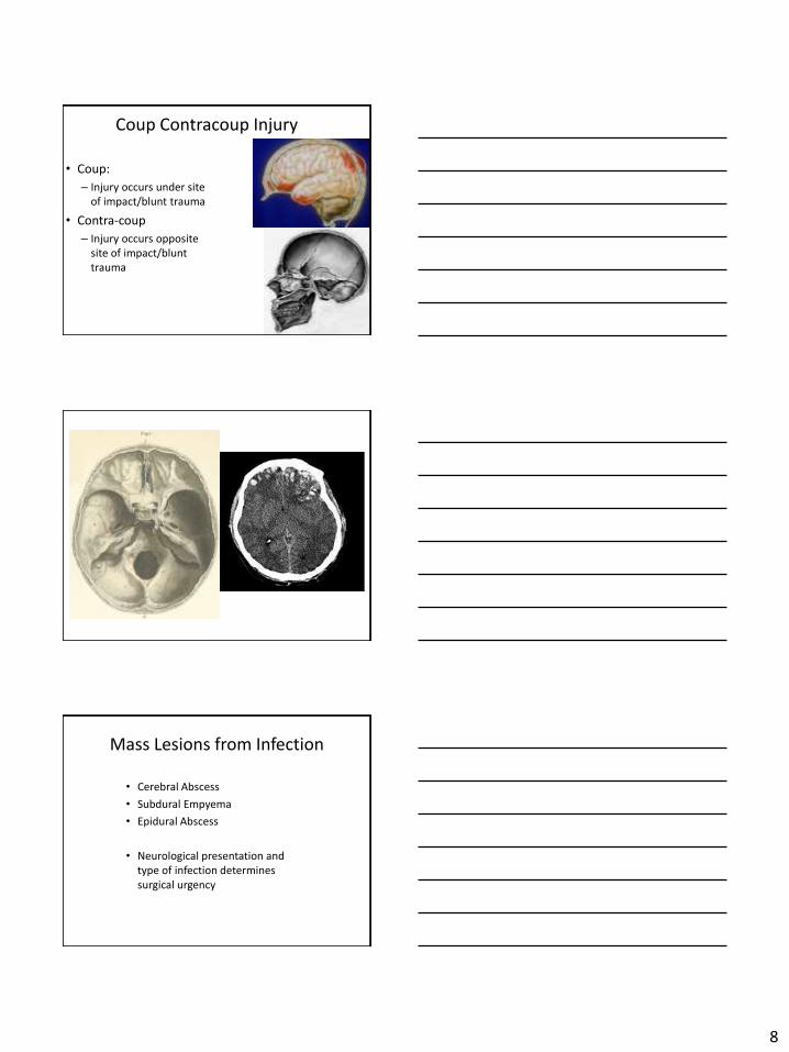
Coup Contracoup Injury
• Coup:
– Injury occurs under s ite of i mpact/blunt trauma
• Contra-coup
– Injury occurs opposite site of impact/blunt trauma
Mass Lesions from Infection
• Cerebral Abscess
• Subdural Empyema
• Epidural Abscess
• Neurological presentation and type of infection determines surgical urgency
8

Cerebral Abscess
Predisposing Conditions:
• Otitis Med ia/mastoiditis
• Sinusitis
• Dental Infection
• Penetrating Trauma
• Pulmonary Infection
• Endocarditis
• Immunocompromise
Cerebral Abscess
• Days 1 -4: early cerebritis
• Days 4 -9: late cerebritis
• Days 1 0-13: Early capsule formation
• Day 14 and later: Late capsule formation
• Meningitis
• Ventriculitis
Cerebral Abscess
• Symptoms:
• Headache
• Fever
• Focal neurologic deficits
• Altered mental status
• Seizures
• Nausea and Vomiting
• Nuchal rigidity
• Papilledema
• Surgical resection or aspiration (~2.5 cm)
• Antibiotics
• Mortality: 8-25%
• Neurological Sequelae: 20-70%
9
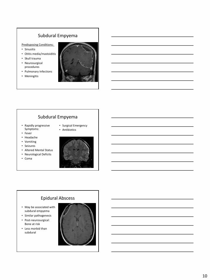
Subdural Empyema
Predisposing Conditions:
• Sinusitis
• Otitis m edia/mastoiditis
• Skull trauma
• Neurosurgical procedures
• Pulmonary Infections
• Meningitis
Subdural Empyema
• Rapidly progressive Symptoms:
• Fever
• Headache
• Vomiting
• Seizures
• Altered Mental Status
• Neurological Deficits
• Coma
• Surgical Emergency
• Antibiotics
Epidural Abscess
• May be associated w ith subdural empyema
• Similar pathogenesis
• Post-neurosurgical: Bone at risk
• Less m orbid than subdural
10
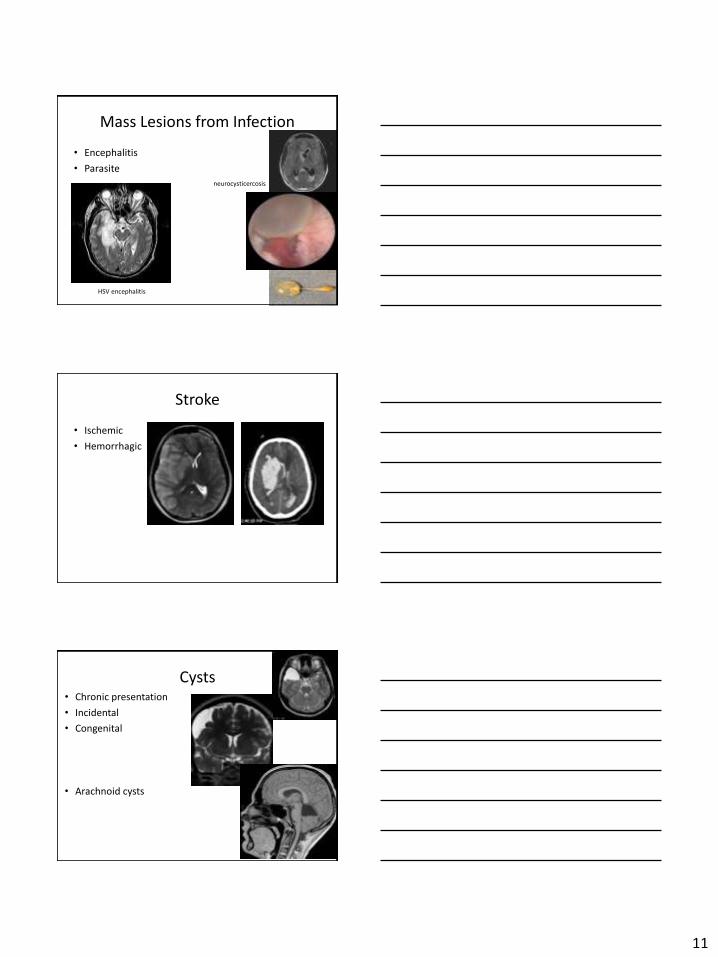
Mass Lesions from Infection
• Encephalitis
• Parasite
neurocysticercosis
HSV encephalitis
Stroke
• Ischemic
• Hemorrhagic
Cysts • Chronic presentation
• Incidental
• Congenital
• Arachnoid cysts
11
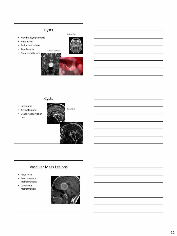
Cysts
• May be asymptomatic
• Headaches
• Endocrinopathies
• Papilledema
• Focal deficits r are
Colloid Cyst
Rathke’s Cleft Cyst
Cysts
• Incidental
• Asymptomatic
• Usually observation only
Pineal Cyst
Vascular Mass Lesions
• Aneurysm
• Arteriovenous malformations
• Cavernous malformation
12
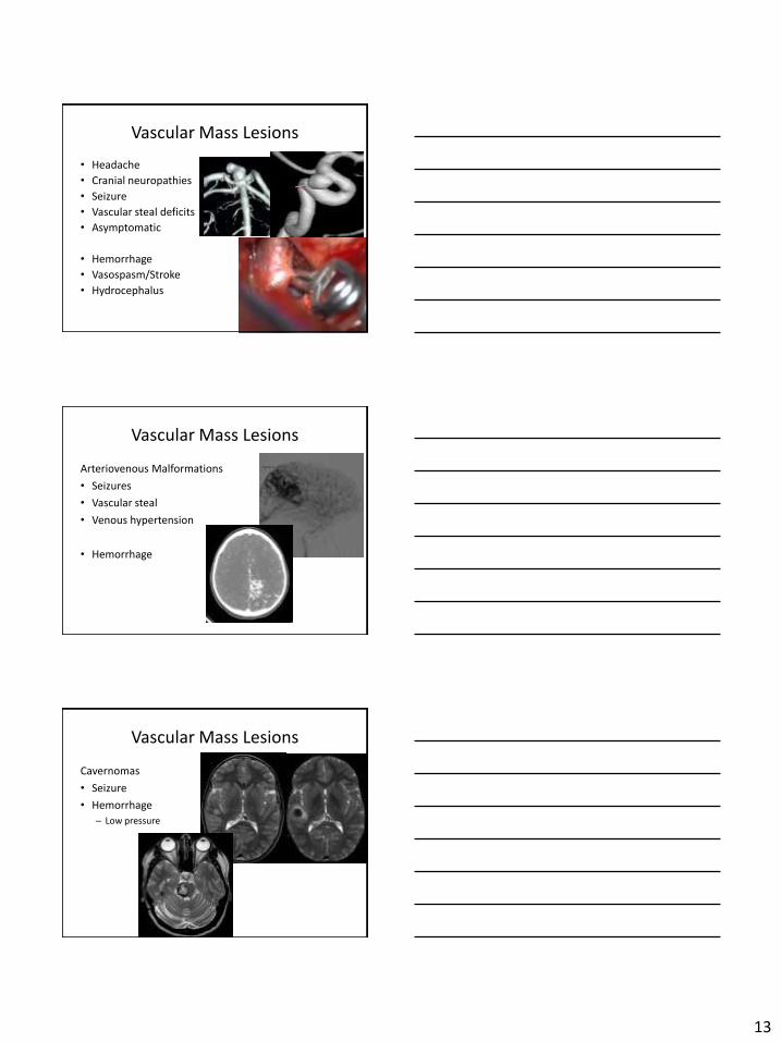
Vascular Mass Lesions
• Headache
• Cranial neuropathies
• Seizure
• Vascular steal deficits
• Asymptomatic
• Hemorrhage
• Vasospasm/Stroke
• Hydrocephalus
Vascular Mass Lesions
Arteriovenous Malformations
• Seizures
• Vascular steal
• Venous h ypertension
• Hemorrhage
Vascular Mass Lesions
Cavernomas
• Seizure
• Hemorrhage
– Low pressure
13
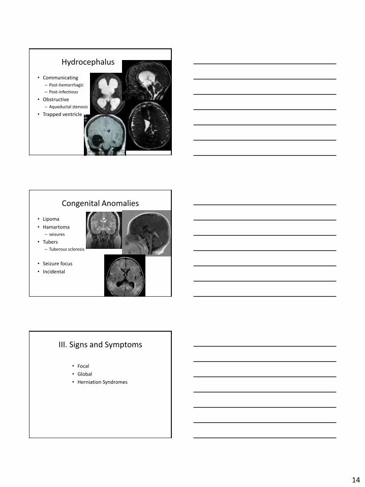
Hydrocephalus
• Communicating
– Post-hemorrhagic
– Post-infectious
• Obstructive
– Aqueductal stenosis
• Trapped v entricle
Congenital Anomalies
• Lipoma
• Hamartoma
– seizures
• Tubers
– Tuberous sclerosis
• Seizure focus
• Incidental
III. Signs and Symptoms
• Focal
• Global
• Herniation Syndromes
14
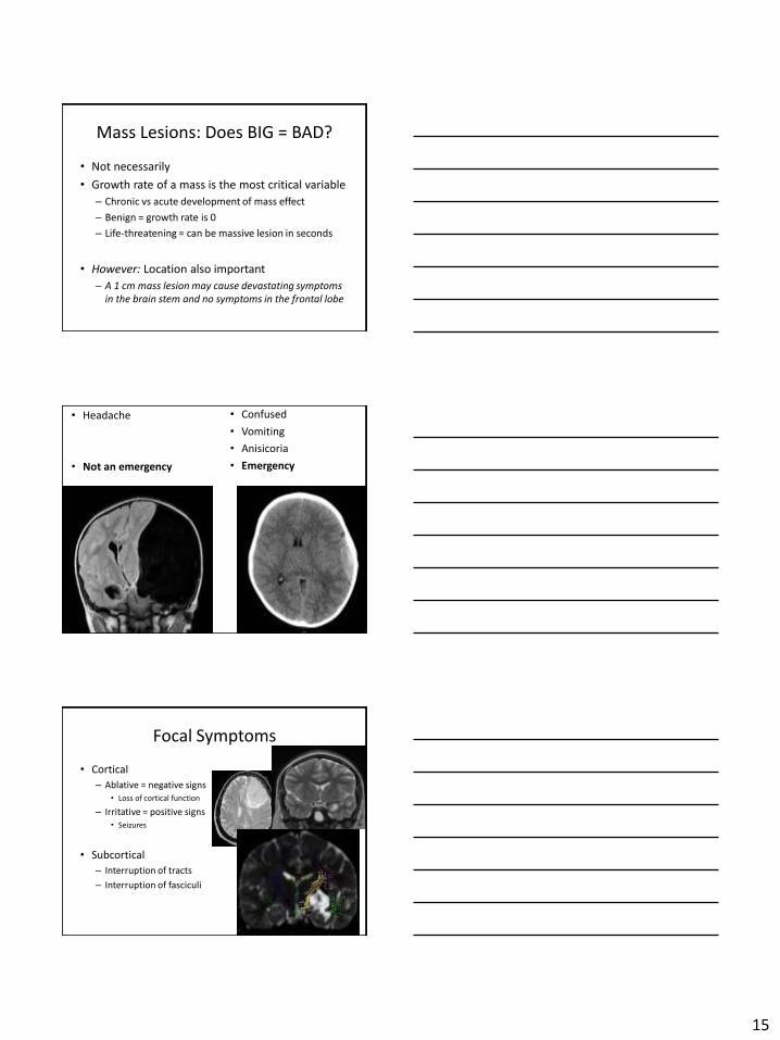
Mass Lesions: Does BIG = BAD?
• Not n ecessarily
• Growth rate of a mass is the most critical variable
– Chronic vs acute development of ma ss effect
– Benign = growth rate is 0
– Life-threatening = can be massive lesion in seconds
• However: Location also important
– A 1 cm mass lesion may cause devastating symptoms in the brain stem and no symptoms in the frontal lobe
• Headache
• Not a n emergency
• Confused
• Vomiting
• Anisicoria
• Emergency
Focal Symptoms
• Cortical
– Ablative = negative signs • Loss of cortical function
– Irritative = positive signs • Seizures
• Subcortical
– Interruption of tracts
– Interruption of fasciculi
15
•
Focal Effects
• Frontal Lobe – Personality changes
– Memory difficulties
– Cognitive difficulties
– Bladder incontinence
– Motor
• Temporal Lobe – Dysphasias
– Memory difficulties
– Seizures
Focal Effects
• Parietal Lobe
– Neglect syndroms
– Agnosia, astereognosia, dyslexia, dysgraphia, dyscalculia
– Sensory disturbance
Occipital Lobe
– Cortical blindness
– Visual field deficit
– Anton’s syndrome
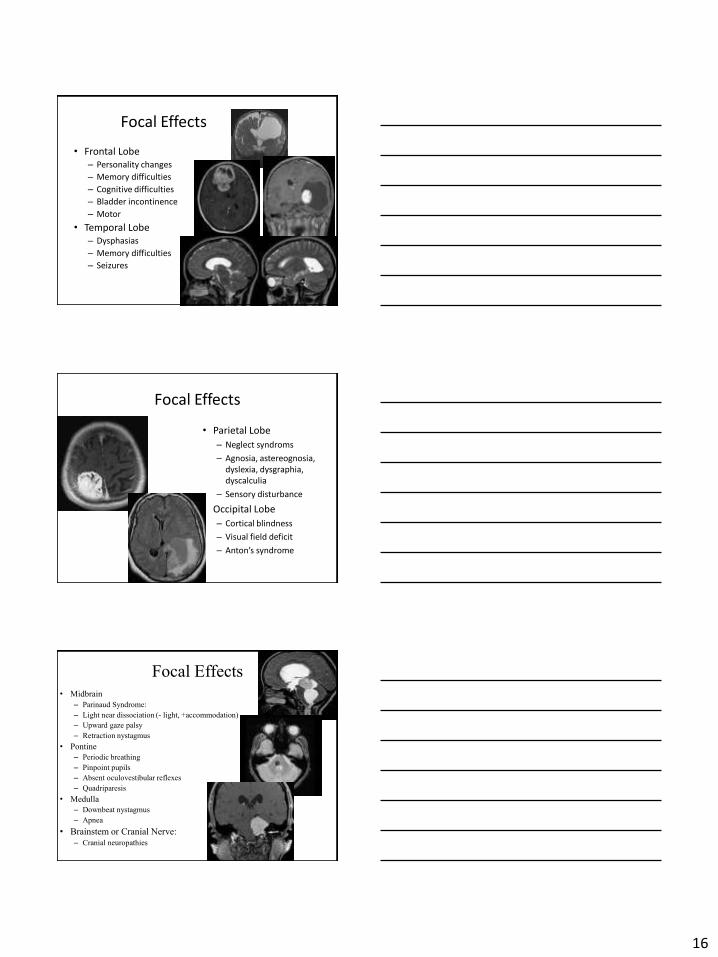
Focal Effects
• Midbrain
– Parinaud Syndrome:
– Light near dissociation (- light, +accommodation)
– Upward gaze palsy
– Retraction nystagmus
• Pontine
– Periodic breathing
– Pinpoint pupils
– Absent oculovestibular reflexes
– Quadriparesis
• Medulla
– Downbeat nystagmus
– Apnea
• Brainstem or Cranial Nerve: – Cranial neuropathies
16
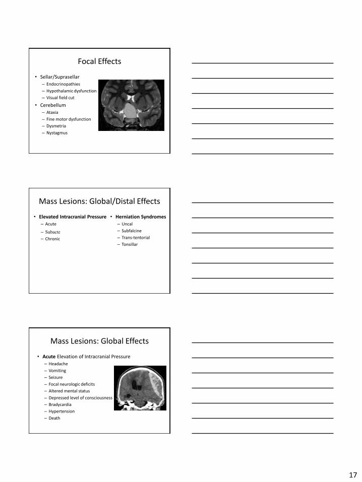
Focal Effects
• Sellar/Suprasellar
– Endocrinopathies
– Hypothalamic dysfunction
– Visual field cut
• Cerebellum
– Ataxia
– Fine motor dysfunction
– Dysmetria
– Nystagmus
Mass Lesions: Global/Distal Effects
• Elevated Intracranial Pressure • Herniation Syndromes
– Uncal
– Subfalcine
– Acute
– Subacte– Chronic – Trans-tentorial
– Tonsillar
Mass Lesions: Global Effects
• Acute Elevation of In tracranial Pressure
– Headache
– Vomiting
– Seizure
– Focal neurologic deficits
– Altered mental s tatus
– Depressed level of consciousness
– Bradycardia
– Hypertension
– Death
17
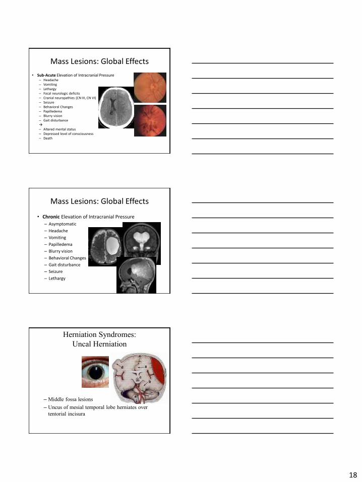
Mass Lesions: Global Effects
• Sub-Acute Elevation of Intracranial Pressure – Headache – Vomiting – Lethargy – Focal neurologic deficits – Cranial neuropathies (CN III, CN VI) – Seizure – Behavioral Changes – Papilledema – Blurry vision – Gait disturbance
– Altered mental status – Depressed level of consciousness – Death
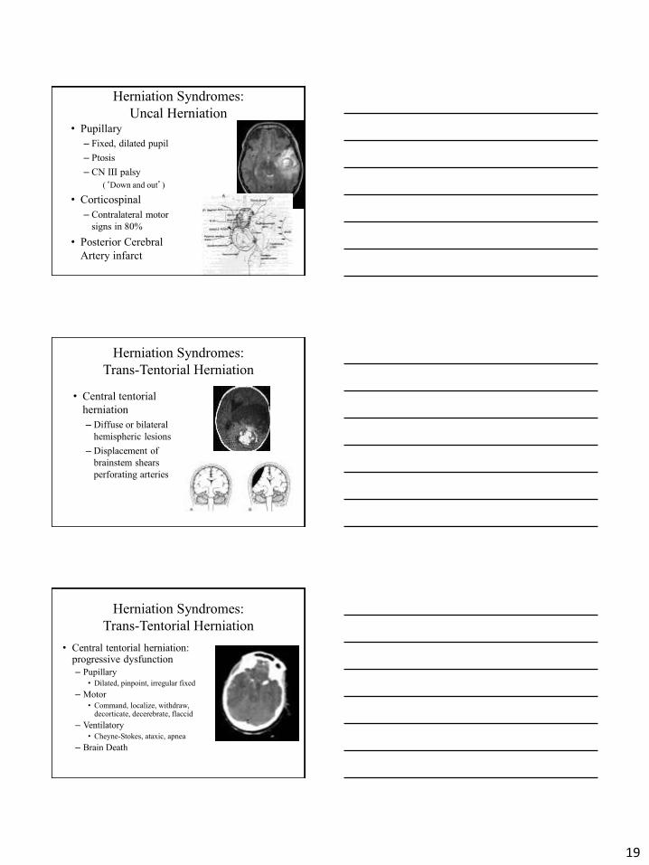
Mass Lesions: Global Effects
• Chronic Elevation of In tracranial Pressure
– Asymptomatic
– Headache
– Vomiting
– Papilledema
– Blurry vision
– Behavioral Changes
– Gait disturbance
– Seizure
– Lethargy
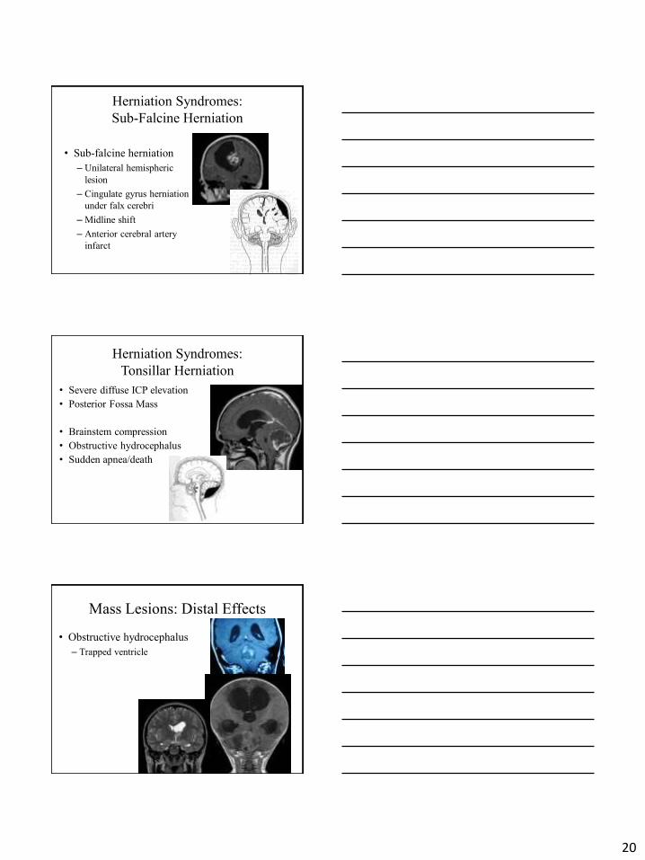
Herniation Syndromes:
Uncal Herniation
– Middle fossa lesions
– Uncus of mesial temporal lobe herniates over
tentorial incisura
18
Herniation Syndromes:
Uncal Herniation • Pupillary
– Fixed, dilated pupil
– Ptosis
– CN III palsy
(‘Down and out’)
• Corticospinal
– Contralateral motor
signs in 80%
• Posterior Cerebral
Artery infarct
Herniation Syndromes:
Trans-Tentorial Herniation
• Central tentorial
herniation
– Diffuse or bilateral
hemispheric lesions
– Displacement of
brainstem shears
perforating arteries
Herniation Syndromes:
Trans-Tentorial Herniation
• Central tentorial herniation: progressive dysfunction
– Pupillary
• Dilated, pinpoint, irregular fixed
– Motor
• Command, localize, withdraw, decorticate, decerebrate, flaccid
– Ventilatory
• Cheyne-Stokes, ataxic, apnea
– Brain Death
19
20
Herniation Syndromes:
Sub-Falcine Herniation
• Sub-falcine herniation
– Unilateral hemispheric
lesion
– Cingulate gyrus herniation
under falx cerebri
– Midline shift
– Anterior cerebral artery
infarct
Herniation Syndromes:
Tonsillar Herniation
• Severe diffuse ICP elevation
• Posterior Fossa Mass
• Brainstem compression
• Obstructive hydrocephalus
• Sudden apnea/death
Mass Lesions: Distal Effects
• Obstructive hydrocephalus
– Trapped ventricle
IV. Management ofIntracranial Mass Lesions
• Intracranial Pressure (ICP) Management
• Imaging
• Critical Care
• Medical Management
• Surgical Management
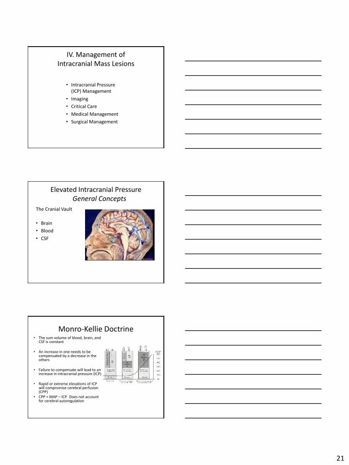
Elevated Intracranial Pressure General Concepts
The Cranial Vault
• Brain
• Blood
• CSF
Monro-Kellie Doctrine • The sum volume of blood, brain, and
CSF is constant
• An increase in one needs to be compensated by a decrease in the others
• Failure to compensate will lead to an increase in intracranial pressure (ICP)
• Rapid or extreme elevations of ICP will compromise cerebral perfusion (CPP)
• CPP = MAP – ICP. Does not account for cerebral autoregulation
21
• Cerebrospinal Fluid
• Brain
• Blood
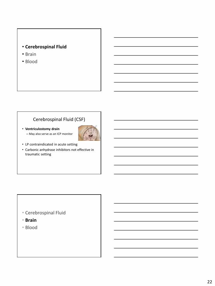
Cerebrospinal Fluid (CSF)
• Ventriculostomy drain
– May also serve as an ICP monitor
• LP contraindicated in acute setting
• Carbonic anhydrase inhibitors not effective in traumatic setting
• Cerebrospinal Fluid
• Brain
• Blood
22
Brain
• Osmotics
– Mannitol
• BBB
• Rebound edema
– Hypertonic saline
– Loop diuretics
• Lobectomies
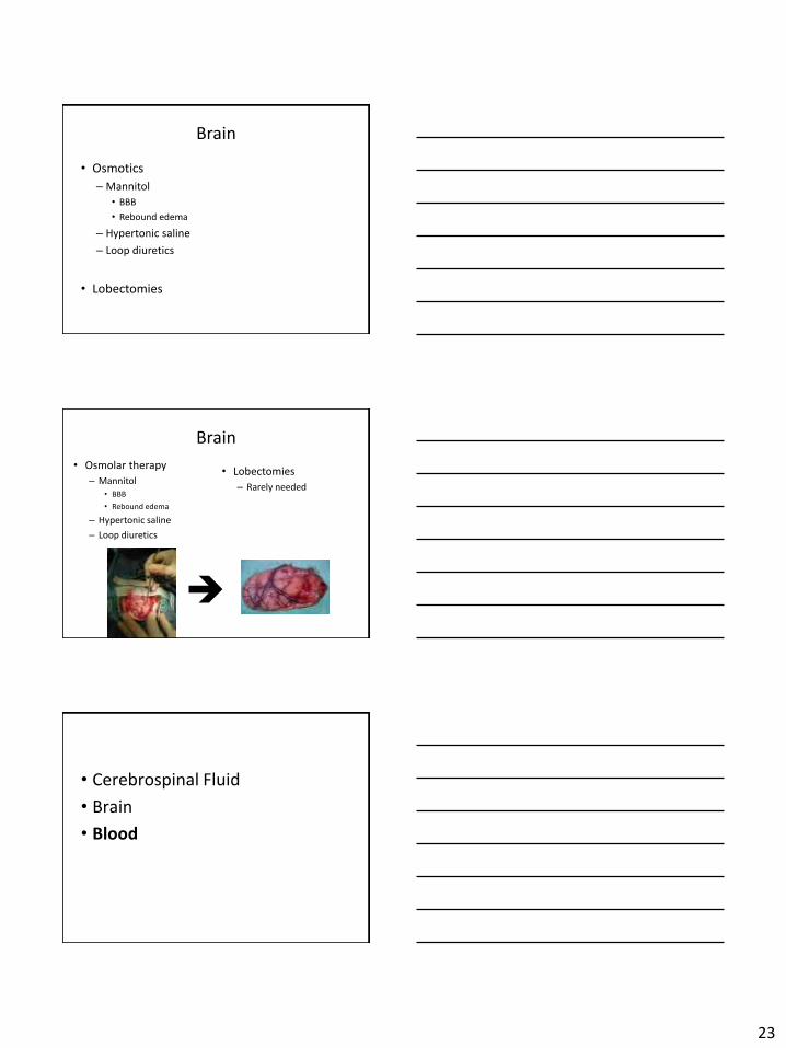
Brain
• Osmolar therapy
– Mannitol • BBB
• Rebound edema
– Hypertonic saline
– Loop diuretics
• Lobectomies
– Rarely needed
• Cerebrospinal Fluid
• Brain
• Blood
23
Blood
• Evacuate hematomas • Hyperventilation • Head of bed elevated
– Optimize venous outlet
• No cervical restriction – 2 fingers under c-collar – No jugular lines – Trach collars
• Minimize cerebral metabolic activity – Sedation – Muscle relaxants – Burst suppression
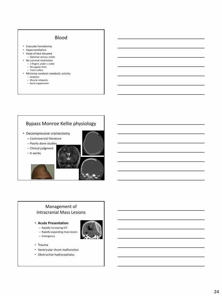
Bypass Monroe Kellie physiology
• Decompressive craniectomy
– Controversial literature
– Poorly done studies
– Clinical judgment
– It works
Management of Intracranial Mass Lesions
• Acute Presentation – Rapidly increasing ICP
– Rapidly expanding mass lesion
– Emergency
• Trauma
• Ventricular shunt malfunction
• Obstructive hydrocephalus
24
Emergent Management
1. ABCs
• Acute elevation of ICP can lead to: – Loss of airway protection – Suppression of respiratory drive – Bradycardia – Hemodynamic instability
2. Neurologic Assessment
• Level of Sensorium – Glascow Coma Score
Emergent Management
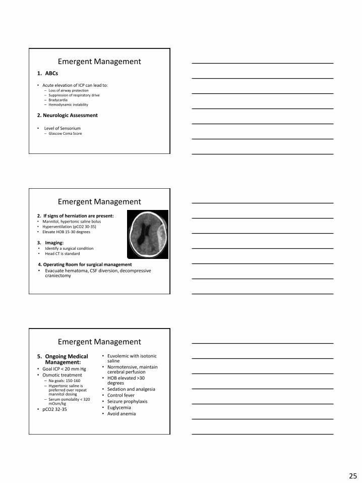
2. If signs of herniation are present: • Mannitol, hypertonic saline bolus • Hyperventilation (pCO2 30-35) • Elevate HOB 15-30 degrees
3. Imaging: • Identify a surgical condition • Head CT is standard
4. Operating Room for surgical management • Evacuate hematoma, CSF diversion, decompressive
craniectomy
Emergent Management
5. Ongoing Medical Management:
• Euvolemic with isotonic saline
• Normotensive, maintain cerebral perfusion
• Goal ICP < 20 mm Hg • Osmotic treatment
• HOB elevated >30 degrees – Na g oals: 150-160
– Hypertonic saline is preferred over repeat mannitol dosing
• Sedation and analgesia • Control fever
– Serum osmolality < 320 mOsm/kg • Seizure prophylaxis
• Euglycemia • pCO2 32-35 • Avoid anemia
25
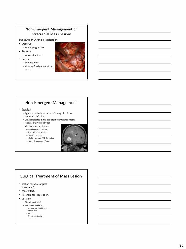
Non-Emergent Management of Intracranial Mass Lesions
Subacute or Chronic Presentation
• Observe
– Risk of progression
• Steroids
– Vasogenic edema
• Surgery
– Remove mass
– Alleviate focal pressure from mass
Non-Emergent Management
– Steroids
• Appropriate in the treatment of vasogenic edema
(tumor and infection)
• Contraindicated in the treatment of cytotoxic edema
(cranial injury and stroke)
• Mechanisms are obscure:
– membrane stabilization
– free radical quenching
– edema resolution
– slightly reduced CSF formation
– anti-inflammatory effects
Surgical Treatment of Mass Lesion
• Option for non-surgical treatment?
• Mass effect?
• Potential for Progression?
• Location – Risk of morbidity?
– Resources available?
• Technology: Stealth, MRI, endoscopy
• PICU
• Neuro-anesthesia
26
27
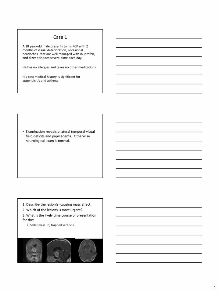
Case 1
A 28 year-old male presents to his PCP with 2 months of visual deterioration, occasional headaches that are well managed with ibuprofen, and dizzy episodes several time each day.
He has no allergies and takes no other medications
His past medical history is significant for appendicitis and asthma.
• Examination reveals bilateral temporal visual field deficits and papilledema. Otherwise neurological exam is normal.
1. Describe the lesion(s) causing mass effect.
2. Which of the lesions is most urgent?
3. What is the likely time course of presentation for the:
a) Sellar mass b) trapped ventricle
1
1. What is t he level of urgency?
2. Go through the appropriate initial management steps for this patient’s care.
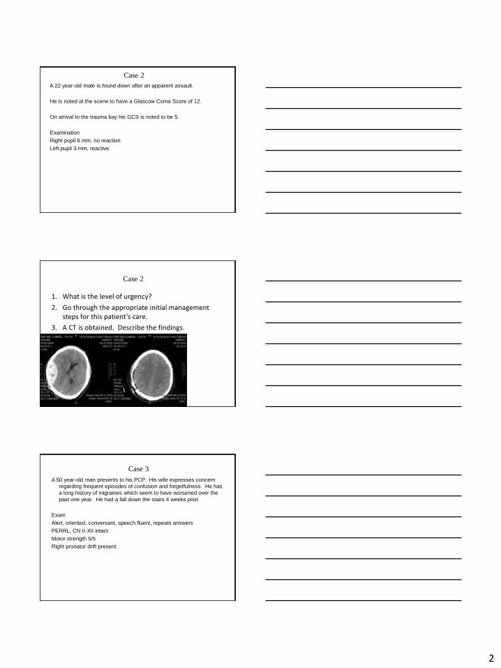
Case 2
A 22 year-old male is found down after an apparent assault.
He is noted at the scene to have a Glascow Coma Score of 12.
On arrival to the trauma bay his GCS is noted to be 5.
Examination
Right pupil 6 mm, no reaction
Left pupil 3 mm, reactive
Case 2
3. A CT is obtained. Describe the findings.
Case 3
A 50 year-old man presents to his PCP. His wife expresses concern
regarding frequent episodes of confusion and forgetfulness. He has
a long history of migraines which seem to have worsened over the
past one year. He had a fall down the stairs 4 weeks prior.
Exam
Alert, oriented, conversant, speech fluent, repeats answers
PERRL, CN II-XII intact
Motor strength 5/5
Right pronator drift present
2
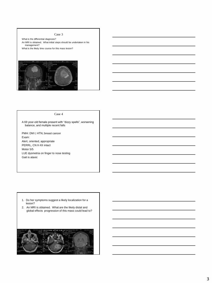
Case 3
What is the differential diagnosis?
An MRI is obtained. What initial steps should be undertaken in his
management?
What is the likely time course for this mass lesion?
Case 4
A 69 year-old female present with “dizzy spells”, worsening balance, and multiple recent falls
PMH: DM I; HTN; breast cancer
Exam:
Alert, oriented, appropriate
PERRL, CN II-XII intact
Motor 5/5
LUE dysmetria on finger to nose testing
Gait is ataxic
1. Do her symptoms suggest a likely localization for a
lesion?
2. An MRI is obtained. What are the likely distal and
global effects progression of this mass could lead to?
3


![Magnetic resonance imaging diagnostic features of giant ... · intracranial lesions [1, 2].Intracranial tuberculomas are potentially curable and its early differentiation from other](https://img.pdfslide.net/doc/110x75/5ec57aed7810c0214a0c2f34/magnetic-resonance-imaging-diagnostic-features-of-giant-intracranial-lesions.jpg)