Embed Size (px)
Citation preview
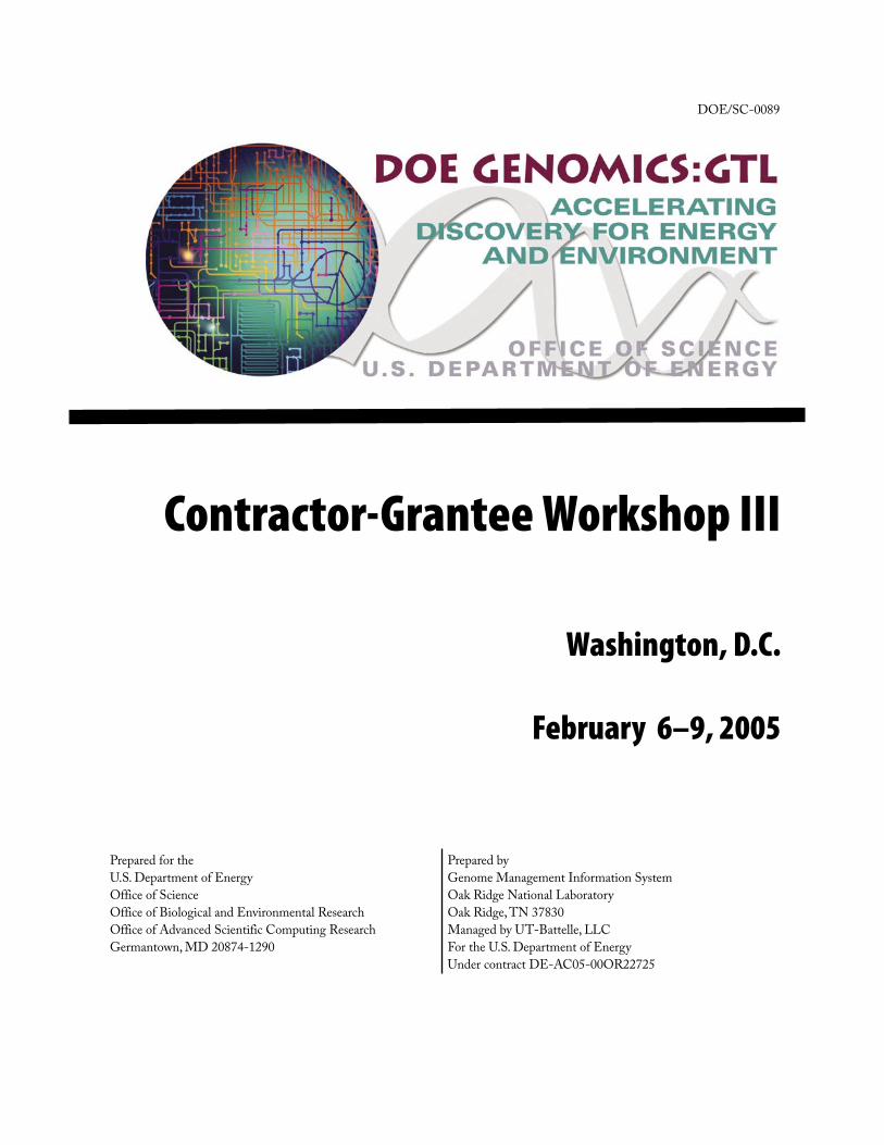
Contractor-Grantee Workshop III
Washington, D.C.
February 6–9, 2005
Prepared for the U.S. Department of Energy Office of Science Office of Biological and Environmental Research Office of Advanced Scientific Computing Research Germantown, MD 20874-1290
Prepared by Genome Management Information System Oak Ridge National Laboratory Oak Ridge, TN 37830 Managed by UT-Battelle, LLC For the U.S. Department of Energy Under contract DE-AC05-00OR22725
DOE/SC-0089

i* Presenting author
Contents
Welcome to Genomics:GTL Workshop III
Genomics:GTL Program Projects
Harvard Medical School
1 Metabolic Network Modeling of Prochlorococcus marinus ..........................................................3George M. Church* ([email protected]), Xiaoxia Lin, Daniel Segrè, Aaron Brandes, and Jeremy Zucker
2 Quantitative Proteomics of Prochlorococcus marinus ..................................................................4Kyriacos C. Leptos* ([email protected]), Jacob D. Jaffe, Eric Zinser, Debbie Lindell, Sallie W. Chisholm, and George M. Church
3 Genome Sequencing from Single Cells with Ploning ...............................................................5Kun Zhang* ([email protected]), Adam C. Martiny, Nikkos B. Reppas, Sallie W. Chisholm, and George M. Church
Lawrence Berkeley National Laboratory
4 VIMSS Computational Microbiology Core Research on Comparative and Functional Genomics .................................................................................................................................6Adam Arkin* ([email protected]), Eric Alm, Inna Dubchak, Mikhail Gelfand, Katherine Huang, Vijaya Natarajan, Morgan Price, and Yue Wang
5 The Virtual Institute of Microbial Stress and Survival (VIMSS): Deduction of Stress Response Pathways in Metal/Radionuclide Reducing Microbes ..............................................8Carl Abulencia, Eric Alm, Gary Andersen, Adam Arkin* ([email protected]), Kelly Bender, Sharon Borglin, Eoin Brodie, Swapnil Chhabra, Steve van Dien, Inna Dubchak, Matthew Fields, Sara Gaucher, Jil Geller, Masood Hadi, Terry Hazen, Qiang He, Zhili He, Hoi-Ying Holman, Katherine Huang, Rick Huang, Janet Jacobsen, Dominique Joyner, Jay Keasling, Keith Keller, Martin Keller, Aindrila Mukhopadhyay, Morgan Price, Joseph A. Ringbauer, Jr., Anup Singh, David Stahl, Sergey Stolyar, Jun Sun, Dorothea Thompson, Christopher Walker, Judy Wall, Jing Wei, Denise Wolf, Denise Wyborski, Huei-che Yen, Grant Zane, Jizhong Zhou, and Beto Zuniga
ii
PagePoster
* Presenting author
6 VIMSS Applied Environmental Microbiology Core Research on Stress Response Pathways in Metal-Reducers .................................................................................................. 11Terry C. Hazen* ([email protected]), Carl Abulencia, Gary Andersen, Sharon Borglin, Eoin Brodie, Steve van Dien, Matthew Fields, Jil Geller, Hoi-Ying Holman, Rick Huang, Janet Jacobsen, Dominique Joyner, Martin Keller, Aindrila Mukhopadhyay, David Stahl, Sergey Stolyar, Jun Sun, Dorothea Thompson, Judy Wall, Denise Wyborski, Huei-che Yen, Grant Zane, Jizhong Zhou, and Beto Zuniga
7 VIMSS Functional Genomics Core: Analysis of Stress Response Pathways in Metal-Reducing Bacteria ....................................................................................................... 14Aindrila Mukhopadhyay, Steven Brown, Swapnil Chhabra, Brett Emo, Weimin Gao, Sara Gaucher, Masood Hadi, Qiang He, Zhili He, Ting Li, Yongqing Liu, Alyssa Redding, Joseph Ringbauer, Jr., Dawn Stanek, Jun Sun, Lianhong Sun, Jing Wei, Liyou Wu, Huei-Che Yen, Wen Yu, Grant Zane, Matthew Fields, Martin Keller ([email protected]), Anup Singh ([email protected]), Dorothea Thompson, Judy Wall ([email protected]), Jizhong Zhou ([email protected]), and Jay Keasling* ([email protected])
Oak Ridge National Laboratory and Pacific Northwest National Laboratory
8 Center for Molecular and Cellular Systems: High-Throughput Identification and Characterization of Protein Complexes .................................................................................. 15Michelle Buchanan, Frank Larimer, Steven Wiley, Steven Kennel, Dale Pelletier, Brian Hooker, Gregory Hurst, Robert Hettich, Hayes McDonald* ([email protected]), Vladimir Kery, Mitchel Doktycz, Jenny Morrell, Bob Foote, Denise Schmoyer, Manesh Shah, and Bill Cannon
9 High-Throughput Analysis of Protein Complexes in the Center for Molecular and Cellular Systems ................................................................................................................................... 16Vladimir Kery* ([email protected]), Dale A. Pelletier, Joshua N. Adkins, Deanna L. Auberry, Frank R. Collart, Linda J. Foote, Brian S. Hooker, Peter Hoyt, Gregory B. Hurst, Stephen J. Kennel, Trish K. Lankford, Chiann-Tso Lin, Eric A. Livesay, Tse-Yuan S. Lu, Cathy K. McKeown, Priscilla A. Moore, Ronald J. Moore, and Kristin D. Victry
10 Investigating Gas Phase Dissociation Pathways of Crosslinked Peptides: Application to Protein Complex Determination ......................................................................................................... 17Sara P. Gaucher* ([email protected]), Masood Z. Hadi, and Malin M. Young
11 Center for Molecular and Cellular Systems: Statistical Screens for Datasets from High-Throughput Protein Pull-Down Assays ................................................................................. 18Frank W. Larimer* ([email protected]), Kenneth K. Anderson, Deanna L. Auberry, Don S. Daly, Vladimir Kery, Denise D. Schmoyer, Manesh B. Shah, and Amanda M. White
12 Center for Molecular and Cellular Systems: Analysis and Visualization of Data from a High-Throughput Protein Complex Identification Pipeline Using Modular and Automated Tools .. 19W. Hayes McDonald ([email protected]), Joshua N. Adkins, Deanna L. Auberry, Kenneth J.
Auberry, Gregory B. Hurst, Vladimir Kery, Frank W. Larimer, Manesh B. Shah, Denise D. Schmoyer, Eric F. Strittmatter, and Dave L. Wabb
iii
PagePoster
* Presenting author
Sandia National Laboratories
13 Carbon Sequestration in Synechococcus: A Computational Biology Approach to Relate the Genome to Ecosystem Response .......................................................................................20Grant S. Heffelfinger* ([email protected])
14 Integrating Heterogeneous Databases and Tools for High Throughput Microbial Analysis ... 21Nagiza Samatva* ([email protected]), Al Geist, Praveen Chandramohan, and Ramya Krishnamurthy
15 Toward Comprehensive Analysis of MS/MS Data Flows .......................................................22Andrey Gorin* ([email protected]), Nikita D. Arnold, Robert M. Day, and Tema Fridman
16 The Transcriptome of a Marine Cyanobacterium—Analysis Through Whole Genome Microarray Analyses ...............................................................................................................24Brian Palenik* ([email protected]), Ian Paulsen* ([email protected]), Bianca Brahamsha, Rob Herman, Katherine Kang, Ed Thomas, Jeri Timlin, and Dave Haaland
17 DEB: A Data Entry and Browsing Tool for Entering and Linking Synechococcus sp. WH8102 Whole Genome Microarray Metadata from Multiple Data Sources ....................................... 25Arie Shoshani* ([email protected]), Victor Havin, Vijaya Natarajan, Tony Martino, Jerilyn A. Timlin, Katherine Kang, Ian Paulsen, Brian Palenik, and Thomas Naughton
18 Microarray Analysis using VxInsight and PAM ......................................................................28George S. Davidson* ([email protected]), David Hanson, Shawn Martin, Margaret Werner-Washburne, and Mark D. Rintoul
19 Mapping of Biological Pathways and Networks across Microbial Genomes ............................30F. Mao, V. Olman, Z. Su, P. Dam, and Ying Xu* ([email protected])
20 Proteomic Analysis of the Synechococcus WH8102 CCM with Varying CO2 Concentrations ... 31Arlene Gonzales, Yooli K. Light, Zhaoduo Zhang, Michael D. Leavell, Rajat Sapra, Tahera Iqbal, Todd W. Lane, and Anthony Martino* ([email protected])
21 Predicting Protein-Protein Interactions Using Signature Products with an Application to β-Strand Ordering ............................................................................................................. 32Shawn Martin ([email protected]), W. Michael Brown, Charlie Strauss, Mark D. Rintoul*, and Jean-Loup Faulon
22 In Vivo Observation of the Native Pigments in Synechocystis sp. PCC 6803 Using a New Hyperspectral Confocal Microscope ......................................................................................33Michael B. Sinclair* ([email protected]), Jerilyn A. Timlin, David M. Haaland, Sawsan Hamad, and Wim F.J. Vermaas
iv
PagePoster
* Presenting author
23 Connecting Temperature and Metabolic Rate to Population Growth Rates in Marine Picophytoplankton .................................................................................................................34Andrea Belgrano* ([email protected]) and Damian Gessler
24 Deciphering Response Networks in Microbial Genomes through Data Mining and Computational Modeling .......................................................................................................34Z. Su, P. Dam, V. Olman, F. Mao, H. Wu, X. Chen, T. Jiang, B. Palenik, and Ying Xu* ([email protected])
25 BiLab – A New Tool that Combines the Ease-of-Use of MatLab and the Power of Multiple Computational Biology Libraries ........................................................................................... 37Al Geist* ([email protected]) and David Jung
26 Microbial Cell Modeling via Reacting/Diffusing Particles .................................................... 38Steve Plimpton* ([email protected]) and Alex Slepoy
27 Modeling RuBisCO’s Gating Mechanism Using Targeted Molecular Dynamics ................... 38Paul S. Crozier ([email protected]), Steven J. Plimpton, Mark D. Rintoul*, Christian Burisch, and Jürgen Schlitter
28 Selection of Ligands by Panning of Phage Display Peptide Libraries Reveals Potential Partners for TPR Domain and rbcS in Synechococcus WH8102 ...............................................40Zhaoduo Zhang* ([email protected]), Arlene D. Gonzales, Todd W. Lane, and Anthony Martino
University of Massachusetts, Amherst
29 Progress Toward Genome-Scale Monitoring of In Situ Gene Expression During Uranium Bioremediation and Electricity Harvesting ............................................................................40Dawn Holmes* ([email protected]), Kelly Nevin, Regina O’ Neil, Zhenya Shelbolina, Martin Lanthier, Jonathan Kaye, Brad Postier, and Derek Lovley
30 Integrating Phenotypic and Expression Data to Characterize Metabolism in G. sulfurreducens .....................................................................................................................42R. Mahadevan, C. H. Schilling, D. Segura, B. Yan, J. Krushkal, and D. R. Lovley* ([email protected])
31 Novel Regulatory Systems and Adaption of Some Well-Known Systems Controlling Respiration, Growth, and Chemotaxis of Geobactor Species ...................................................44Maddalena Coppi* ([email protected]), Byoung-Chan Kim, Laurie DiDonato, Julia Krushkal, Bin Yan, Richard Glaven, Regina O’ Neil, Suphan Bakkal, Allen Tsang, Hoa Tran, Abraham Esteve-Nunez, Cinthia Nunez, Ching Leang, Kuk-Jeong Chin, Barbara Methe , Robert Weis, Pablo Pomposiello, Kelly Nevin, and Derek Lovley
v
PagePoster
* Presenting author
32 Nanowires, Capacitors, and Other Novel Electron Transfer Mechanisms in Geobacter Species Elucidated from Genome-Scale Investigations ..........................................................46Gemma Reguera* ([email protected]), Teena Mehta, Dawn E. Holmes, Abraham Esteve-Núñez, Jessica Butler, Barbara Methe, Kelly Nevin, Swades K. Chaudhuri, Richard Glaven, Tunde Mester, Raymond DiDonato, Kevin McCarthy, Mark T. Tuominen, and Derek Lovley
33 Continued Progress in the use of Microarray Technology to Predict Gene Regulation and Function in Geobacter sulfurreducens ......................................................................................48Barbara Methé*([email protected]), Jennifer Webster, Kelly Nevin, and Derek Lovley
Shewanella Federation
34 The Shewanella Federation: Functional Genomic Investigations of Dissimilatory Metal- Reducing Shewanella ..............................................................................................................50James K. Fredrickson* ([email protected]), Carol S. Giometti, Eugene Kolker*, Kenneth H. Nealson, James M. Tiedje, Jizhong Zhou, Monica Riley, Shimon Weiss, James J. Collins, Frank Larimer, Frank Collart, Lee Ann McCue, Chip Lawrence, and Timothy S. Gardner
35 Global Profiling of Shewanella oneidensis MR-1: Expression of ‘Hypothetical’ Genes and Improved Functional Annotations ......................................................................................... 51Eugene Kolker* ([email protected]), Alex F. Picone, Michael Y. Galperin, Margaret F. Romine, Roger Higdon, Kira S. Makarova, Natali Kolker, Gordon A. Anderson, Xiaoyun Qiu, Kenneth J. Auberry, Gyorgy Babnigg, Alex S. Beliaev, Paul Edlefsen, Dwayne A. Elias, Yuri Gorby, Ted Holzman, Joel Klappenbach, Konstantinos T. Konstantinidis, Miriam L. Land, Mary S. Lipton, Lee-Ann McCue, Matthew Monroe, Ljiljana Pasa-Tolic, Grigoriy Pinchuk, Samuel Purvine, Margaret Serres, Sasha Tsapin, Brian A. Zakrajsek, Wenhong Zhu, Jizhong Zhou, Frank W. Larimer, Charles Lawrence, Monica Riley, Frank R. Collart, John R. Yates, III, Richard D. Smith, Carol Giometti, Kenneth Nealson, James K. Fredrickson, and James M. Tiedje
36 Respiratory Pathways and Regulatory Networks of Shewanella oneidensis Involved in Energy Metabolism and Environmental Sensing ................................................................... 52Alex Beliaev*, Yuri Gorby, Margie Romine, Jeff McLean, Grigoriy Pinchuk, Eric Hill, Jim Fredrickson, Jizhong Zhou, and Daad A. Saffarini
37 Functional Analysis of Shewanella, A Cross Genome Comparison .........................................54Margrethe H. Serres* ([email protected]) and Monica Riley
38 Optical Methods for Characterization of Expression Levels and Protein-Protein Interactions in Shewanella oneidensis MR-1 ............................................................................ 55Natalie R. Gassman* ([email protected]), Xiangxu Kong, Gopal Iyer, Younggyu Kim, and Shimon Weiss
vi
PagePoster
* Presenting author
39 Reverse-Engineering Microbial Networks in Escherichia coli and Shewanella oneidensis MR-1 via Large-Scale Perturbation Studies ........................................................................... 56G. Cottarel, M.E. Driscoll, J. Faith, M.K. Kohanski, J. Wierzbowski, C.B. Cantor, J.J. Collins, and T.S. Gardner* ([email protected])
40 Comparative Analysis of Gene Expression Profiles of Shewanella oneidensis MR-1 Following Exposure to Ionizing Radiation and Ultraviolet Radiation ................................... 57Xiaoyun Qiu* ([email protected]), George Sundin, Michael J. Daly, Alexander Vasilenko, Marina V. Omelchenko, Jizhong Zhou, Liyou Wu, Mary S. Lipton, and James M. Tiedje
41 The Microbial Proteome Project: A Database of Microbial Protein Expression in the Context of Genome Analysis .................................................................................................. 58Carol S. Giometti * ([email protected]), Gyorgy Babnigg, Sandra L. Tollaksen, Tripti Khare, Angela Ahrendt, Wenhong Zhu, Derek R. Lovley, James K. Fredrickson, and John R. Yates III
J. Craig Venter Institute
42 Estimation of the Minimal Mycoplasma Gene Set Using Global Transposon Mutagenesis and Comparative Genomics ...................................................................................................60John I. Glass* ([email protected]), Nina Alperovich, Nacyra Assad-Garcia, Shibu Yooseph, Mahir Maruf, Carole Lartigue, Cynthia Pfannkoch, Clyde A. Hutchison III, Hamilton O. Smith, and J. Craig Venter
43 Progress toward a Synthetic Cellular Genome ........................................................................ 61Hamilton O. Smith* ([email protected]), Cynthia Pfannkoch, Holly A. Baden-Tillson, Clyde A. Hutchison III, and J. Craig Venter
44 Development of a Deinococcus radiodurans Homologous Recombination System ....................62Sanjay Vashee*, Ray-Yuan Chuang* ([email protected]), Christian Barnes, Hamilton O. Smith, and J. Craig Venter
45 Development of a Novel Recombinant Cyanobacterial System for Hydrogen Production from Water .............................................................................................................................63Qing Xu, Shibu Yooseph, Hamilton O. Smith, and J. Craig Venter ([email protected])
46 Biotechnology For the Production of Ethanol and Butanol from Cellulose ............................64Prabha P. Iyer* ([email protected]), Hamilton O. Smith, and J. Craig Venter
vii
PagePoster
* Presenting author
Communication
47 Communicating Genomics:GTL ........................................................................................... 65Anne E. Adamson, Shirley H. Andrews, Jennifer L. Bownas, Denise K. Casey, Sherry A. Estes, Sheryl A. Martin, Marissa D. Mills, Kim Nylander, Judy M. Wyrick, Anita J. Alton, and Betty K. Mansfield* ([email protected])
Bioinformatics, Modeling, and Computation
48 SimPheny™: A Computational Infrastructure for Systems Biology ....................................... 67Christophe H. Schilling* ([email protected]), Sean Kane, Martin Roth, Jin Ruan, Kurt Stadsklev, Rajendra Thakar, Evelyn Travnik, Steve van Dien, and Sharon Wiback
49 Hybrid Bacterial Cell Models: Linking Genomics to Physiological Response ........................68Jordan C. Atlas* ([email protected]), Mariajose Castellanos, Anjali Dhiman, Bruce Church, and Michael L. Shuler
50 Identification of the Most Probable Biological Network Using Model Discrimination Analysis .................................................................................................................................. 69Andrea L. Knorr and Ranjan Srivastava* ([email protected])
51 Rhodopseudomonas palustris Regulons Detected by a Cross-Species Analysis of the α-Proteobacteria ..................................................................................................................... 70Sean Conlan* ([email protected]), Charles E. Lawrence, and Lee Ann McCue
52 Exploring Evolutionary Space ................................................................................................ 72Timothy G. Lilburn* ([email protected]), Yun Bai, Yuan Zhang, James R. Cole, and George M. Garrity
53 PhyloScan: A New Tool for Identifying Statistically Significant Transcription Factor Binding Sites by Combining Cross-Species Evidence ............................................................. 72Lee A. Newberg*, C. Steven Carmack, Lee Ann McCue ([email protected]), and Charles E. Lawrence
54 Predicting Protein Interactions via Docking Mesh Evaluator ................................................ 74Roummel F. Marcia, Susan D. Lindsey, Erick A. Butzlaff, and Julie C. Mitchell* ([email protected])
55 UC Merced Center for Computational Biology ....................................................................... 75Michael Colvin* ([email protected]), Arnold Kim, and Felice Lightstone
56 Biomic Approach to Predictive Cell Modeling ...................................................................... 76P. J. Ortoleva* ([email protected]), L. Ensman, J. Fan, K. Hubbard, A. Sayyed-Ahmad, F. Stanley, K. Tuncay, and K. Varala
viii
PagePoster
* Presenting author
57 The BioWarehouse System for Integration of Bioinformatics Databases ................................. 78Tom Lee, Valerie Wagner, Yannick Pouliot, and Peter D. Karp* ([email protected])
58 Building Large Biological Dynamic Models of Shewanella oneidensis from Incomplete Data . 79Ravishankar R. Vallabhajosyula* ([email protected]), Sri Paladugu, Klaus Maier, and Herbert M. Sauro
59 A Bayesian Method for Identifying Missing Enzymes in Predicted Metabolic Pathway Databases ............................................................................................................................... 81Michelle L. Green* ([email protected]) and Peter D. Karp
60 Does EcoCyc or KEGG Provide a Preferable Gold Standard for Training and Evaluation of Genome-Context Methods? ...............................................................................................82Peter D. Karp* ([email protected]) and Michelle L. Green
61 Towards a Physics and Systems Understanding of Ion Transport in Prokaryotes ....................83Shreedhar Natarajan, Asba Tasneem*, Sameer Varma, Lakshminarayan Iyer, L. Aravind, and Eric Jakobsson* ([email protected])
62 OptStrain: A Computational Framework for Redesign Microbial Production Systems ..........84Priti Pharkya and Costas D. Maranas* ([email protected])
63 DEMSIM: A Discrete Event Based Mechanistic Simulation Platform for Gene Expression and Regulation Dynamics ......................................................................................................84Madhukar Dasika and Costas D. Maranas* ([email protected])
64 On the Futility of Optima in Network Inferences and What Can Be Done About It .............. 86Charles (Chip) E. Lawrence* ([email protected])
Environmental Genomics
65 Whole Community Proteomics Study of an Acid Mine Drainage Biofilm Reveals Key Roles for “Hypothetical” Proteins in a Natural Microbial Biofilm ...................................................87Jill Banfield* ([email protected]),
Rachna J. Ram, Gene W. Tyson, Eric Allen, Nathan VerBerkmoes,
Michael P. Thelen, Brett J. Baker, Manesh Shah, Robert Hettich, and Robert C. Blake II
66 Application of High Throughput Microcapsule Culturing to Develop a Novel Genomics Technology Platform .............................................................................................................88Martin Keller* ([email protected]), Karsten Zengler, Marion Walcher, Carl Abulencia, Denise Wyborski, Sherman Chang, Imke Haller, Trevin Holland, Fred Brockman, Cheryl Kuske, and Susan Barns
ix
PagePoster
* Presenting author
67 Environmental Bacterial Diversity from Communities to Genomes ......................................89Janelle R. Thompson*, Silvia G. Acinas, Vanja Klepac-Ceraj, Sarah Pacocha, Chanathip Pharino, Dana E. Hunt, Luisa A. Marcelino, Jennifer Benoit, Ramahi Sarma-Rupavtarm, Daniel L. Distel, and Martin F. Polz ([email protected])
68 Distribution and Variation of Prochlorococcus Genotypes Across Multiple Oceanic Habitats ..90Adam C. Martiny* ([email protected]), P. K. Amos Tai, Anne W. Thompson, and Sallie W. Chisholm
69 From Perturbation Analysis to the Genomic Regulatory Code: the Sea Urchin Endomesoderm GRN ............................................................................................................. 91Paola Oliveri* ([email protected]), Pei-Yun Lee, Takuya Minokawa, Joel Smith, Qiang Tu, Meredith Howard, David McClay, and Eric H. Davidson
Microbial Genomics
70 The Genome of the Ammonia Oxidizing Bacterium Nitrosomonas europaea: Iron Metabolism and Barriers to Heterotrophy .................................................................................................. 93Xueming Wei, Neeraja Vajrala, Norman Hommes, Luis Sayavedra-Soto*, and Daniel Arp ([email protected])
71 Pelagibacter ubique: A Post-Genomic Investigation of Carbon Metabolism and Photochemistry in an Extraordinarily Abundant Oceanic Bacterium .................................... 95Stephen J. Giovannoni * ([email protected]), Lisa Bibbs, James Tripp, Scott Givan, Jang-Cheon Cho, Martha D. Stapels, Russell Desiderio, Mercha Podar, Kevin L. Vergin, Mick Noordeweir, Michael S. Rappé, Samuel Laney, Douglas F. Barofsky, and Eric Mathur
72 Does the Three Dimensional Organization of the Nucleoid of the Deinococcaceae Contribute to their Ionizing Radiation Resistance? ................................................................ 96J. M. Zimmerman and J. R. Battista* ([email protected])
73 Large Scale Genomic Analysis for Understanding Hydrogen Metabolism in Chlamydomonas reinhardtii ...............................................................................................................................97Michael Seibert* ([email protected]), Arthur R. Grossman, Maria L. Ghirardi, and Matthew C. Posewitz
74 Exploring the Genome and Proteome of Desulfitobacterium hafniense DCB2 for its Protein Complexes Involved in Metal Reduction and Dechlorination ....................................99James M. Tiedje*, Sang-Hoon Kim, Christina Harzman, John Davis, Brett Phinney, Michael Ngowe, Washington Mutatu, William Broderick, David DeWitt, Joan Broderick, and Terence L. Marsh
x
PagePoster
* Presenting author
75 An Integrative Approach to Energy, Carbon, and Redox Metabolism in the Cyanobacterium Synechocystis sp. PCC 6803 .........................................................................100Wim Vermaas* ([email protected]), Robert Roberson, Allison van de Meene, Bing Wang, Sawsan Hamad, Zhi Cai, Julian Whitelegge, Kym Faull, Sveta Gerdes, Andrei Osterman, and Ross Overbeek
76 Role of Cellulose Binding Modules in Cellulose Hydrolysis ................................................. 102David B. Wilson* ([email protected]) and Shaolin Chen
77 Three Prochlorococcus Cyanophage Genomes: Signature Features and Ecological Interpretation ...................................................................................................................... 103Matthew B. Sullivan* ([email protected]), Maureen Coleman, Peter Weigele, Forest Rohwer, and Sallie W. Chisholm
78 The Alternative Sigma Factor RpoN Regulon of Rhodopseudomonas palustris .......................104Yasuhiro Oda* ([email protected]), Sudip K. Samanta, Frank W. Larimer, and Caroline S. Harwood
79 Integrative Control of Key Metabolic Processes in Rhodopseudomonas palustris for the Enhancement of Carbon Sequestration and Biohydrogen Production .................................. 105F. Robert Tabita* ([email protected]), Janet L. Gibson, Caroline S. Harwood, Frank Larimer, J. Thomas Beatty, James C. Liao, and Jizhong (Joe) Zhou
80 Whole Genome Transcriptional Analysis of Toxic Metal Stresses in Caulobacter crescentus .. 107Gary L. Andersen* ([email protected]), Ping Hu, Eoin L. Brodie, and Harley H. McAdams
81 Systematic Analysis of Two-Component Signal Transduction Systems Regulating Cell Cycle Progression in Caulobacter crescentus ........................................................................... 108Michael Laub* ([email protected])
82 The U.S. DOE Joint Genome Institute Microbial Program ................................................. 109David Bruce* ([email protected]), Alla Lapidus, Patrick Chain, Jeremy Schmutz, Frank Larimer, Nikos Kyrpides, Paul Gilna, Eddy Rubin and Paul Richardson
83 Identification of Genes that are Required for Recycling Reducing Power during Photosynthetic Growth ....................................................................................................... 110Christine L. Tavano, Angela M. Podevels, and Timothy J. Donohue* ([email protected])
84 A Tightly-Regulated Oscillatory Circuit Formed by Conserved Master Regulator Proteins Controls the Caulobacter Cell Cycle ........................................................................ 110Harley McAdams* ([email protected]) and Lucy Shapiro
85 Dynamics and Control of Biofilms of the Oligotrophic Bacterium Caulobacter crescentus ..... 112Alfred M. Spormann ([email protected] ) and Plamena Entcheva-Dimitrov
xi
PagePoster
* Presenting author
86 Widespread and Abundant CelM Endoglucanases of Marine Cytophaga-like Bacteria Revealed by Whole Genome Shotgun Sequencing and Fosmid Cloning .............................. 112Matthew T. Cottrell and David L. Kirchman* ([email protected])
87 Data Analysis and Protein Identification Strategy for the Systems-Level Protein-Protein Interaction Networks of Shewanella oneidensis MR-1 ............................................................ 114Gordon A. Anderson* ([email protected]), James E. Bruce, Xiaoting Tang, Gerhard Munske, and Nikola Tolic
88 A Protein Interaction Reporter Strategy for Systems-Level Protein Interaction Networks of Shewanella oneidensis MR-1 .............................................................................................. 115James E. Bruce* ([email protected]), Xiaoting Tang, Harry Zhu, Saiful Chowdhury, Devi Adhikari, Gerhard Munske, Gordon A. Anderson, and Nikola Tolic
Technology Development and Use
Imaging, Molecular, and Cellular Analysis
89 Probing Single Microbial Proteins and Multi-Protein Complexes with Bioconjugated Quantum Dots ..................................................................................................................... 117Gang Bao* ([email protected]), Grant Jensen, Shuming Nie, and Phil LeDuc
90 Single-Molecule Imaging of Macromolecular Dynamics in a Cell ....................................... 119Jamie H. D. Cate ([email protected]) and Haw Yang* ([email protected])
91 Developing a High Resolution Method for Protein Localization in Whole Bacterium .........120Huilin Li* ([email protected]) and James Hainfeld ([email protected])
92 Novel Vibrational Nanoprobes for Microbiology at the Single Cell Level ............................. 121Thomas Huser* ([email protected]), Chad E. Talley, James W. Chan, Heiko Winhold, Ted Laurence, Anthony Esposito, Christopher W. Hollars, Christine A. Hara, Allen T. Christian, Michele H. Corzett, Rod Balhorn, and Stephen M. Lane
93 Instrumented Cell for Characterization of Mammalian and Microbial Cells ...................... 122Jane Bearinger* ([email protected]), Graham Bench, Jackie Crawford, Lawrence Dugan, Amy Hiddessen, Angela Hinz, Thomas Huser, Robin Miles, Magnus Palmblad, Chad Talley, Elizabeth Wheeler, and Allen Christian
94 Chemical Imaging of Biological Materials by NanoSIMS ....................................................123Peter K. Weber* ([email protected]), Ian D. Hutcheon, Radu Popa, and Ken Nealson
xii
PagePoster
* Presenting author
95 Direct Determination of Affinity in Individual Protein-Protein Complexes in Mono and Multivalent Configurations Using Dynamic Force Spectroscopy ..................................124Todd A. Sulchek, Kevin Langry, Raymond W. Friddle, Timothy V. Ratto, Sally DeNardo, Huguette Albrecht, Michael Colvin, and Aleksandr Noy* ([email protected])
96 Electron Tomography of Intact and Sectioned Microbial Cells ............................................ 126Kenneth H. Downing* ([email protected]), Luis Comolli, Haixin Sui, Hoi-Ying Holman, Ellen Judd, and Harley McAdams
97 Probing the High-Resolution Architecture and Environmental Dynamics of Microbial Surfaces by in vitro Atomic Force Microscopy ...................................................................... 127Alexander J. Malkin* ([email protected]), Marco Plomp, Terrance J. Leighton, and Katherine E. Wheeler
98 Real-Time Gene Expression Profiling of Single Live Cells of Shewanella oneidensis ............ 128X. Sunney Xie*, Jie Xiao, Ji Yu, Long Cai, Paul Choi*, Nir Friedman, Xiajia Ren, and Luying Xun*
99 High Throughput Fermentation and Cell Culture Device .................................................... 130David Klein ([email protected]), David Laidlaw, Gregory Andronaco, and Stephen Boyer
100 Immobilized Enzymes in Nanoporous Materials Exhibit Enhanced Stability and Activity .. 132Chenghong Lei, Yongsoon Shin, Jun Liu, and Eric J. Ackerman* ([email protected])
Protein Production and Molecular Tags
101 Towards High Throughput Selection of Binding Ligands: Using Flow Cytometry ............... 133Peter Pavlik, Milan Ovecka, Nileena Velappan, and Andrew Bradbury* ([email protected])
102 Efficient Chemical Methods for the Total Synthesis of Small Proteins: The First Crystallographic Structure of a Protein Diastereomer, [D-Gln35]-ubiquitin ...................... 134Duhee Bang* ([email protected]), George I. Makhatadze, and Stephen B. Kent ([email protected])
103 Development and Application of Multipurpose Affinity Probes to Isolate Intact Protein Complexes Associated with Metal Reduction from Shewanella oneidensis MR-1 ................... 135Liang Shi*, Thomas C. Squier* ([email protected]), M. Uljana Mayer*, Haishi Cao, Baowei Chen, Yuri A. Gorby, David F. Lowry, Jeff Mclean, Seema Verma, and Ping Yan
104 A Combined Informatics and Experimental Strategy for Improving Protein Expression ...... 137Osnat Herzberg, John Moult* ([email protected]), Fred Schwarz, and Harold Smith
105 High-Throughput Production and Analyses of Purified Proteins ......................................... 138F. William Studier* ([email protected]), John C. Sutherland, Lisa M. Miller, and Lin Yang
xiii
PagePoster
* Presenting author
106 Development of Genome-Scale Expression Methods ........................................................... 139Sarah Fey, Elizabeth Landorf, Yuri Londer, Terese Peppler, and Frank Collart* ([email protected])
107 Plate-Based Methods for Expression of Cytoplasmic Proteins from Shewanella oneidensis .... 140Elizabeth Landorf, Terese Peppler, Sarah Fey, Alexander Iakounine, Eugene Kolker, and Frank Collart*
108 Generating scFv and Protein Scaffolds to Protein Targets .................................................... 141Brian K. Kay* ([email protected]), Michael Scholle, Ushma Kriplani, John Kehoe, and Frank Collart
109 Cell Free Approaches for Protein Production ....................................................................... 141Gerald W. Becker*, Pavel Shiyanov, Yifei Wu, Sarah Fey, Elizabeth Landorf, Terese Peppler, and Frank Collart ([email protected])
110 Rapid Synthesis of Peptidic and Peptidomimetic Ligands for High-Throughput Protein Purification and Labeling ..................................................................................................... 142Jeffrey B.-H. Tok* ([email protected]), Priscilla Chan, David Smithson, Ted Tarasow, and Rod Balhorn
Proteomics and Metabolomics
111 Development and Application of New Technologies for Comprehensive and Quantitative High Throughput Microbial Proteomics .............................................................................. 143Richard D. Smith* ([email protected]), Mary S. Lipton, James K. Fredrickson, Matthew Monroe, Eric Livesay, Konstantinos Petritis, Joshua Adkins, Gordon A. Anderson, Kim Hixson, Ruihua Fang, Rui Zhao, Ronald J. Moore, and Yufeng Shen
112 Characterization of Rhodobacter sphaeroides by High Resolution Proteomic Measurements .144Mary S. Lipton* ([email protected]), Timothy Donohue* ([email protected]), Samuel Kaplan* ([email protected]), Stephen Callister, Matthew E. Monroe, Margie F. Romine, Ruihua Fang, Carrie D. Goddard, Nikola Tolic, Gordon A. Anderson, Richard D. Smith, Jim K. Fredrickson, Miguel Dominguez, Christine Tavano, Xiaihua Zeng, and Jung Hyeob Roh
113 Quantitative Metalloproteomics .......................................................................................... 146Patrick G. Grant* ([email protected]), Sharon Shields, Magnus Palmblad, and Graham Bench
114 New Technologies for Metabolomics .................................................................................... 147Jay D. Keasling* ([email protected]), Carolyn Bertozzi, Julie Leary, Michael Marletta, and David Wemmer
115 Characterization of Metal Reducing Microbial Systems by High Resolution Proteomic Measurements ...................................................................................................................... 148Mary S. Lipton* ([email protected]), Ruihua Fang, Dwayne A. Elias, Margie F. Romine, Alex Beliaev, Matthew E. Monroe, Kim K. Hixson, Yuri A. Gorby, Ljiljana Pasa-Tolic, Heather M. Mottaz, Gordon A. Anderson, Richard D. Smith, Jim K. Fredrickson, Derek Lovley, and Yanhuai R. Ding
xiv
PagePoster
* Presenting author
116 Protein Complexes and Pathways ......................................................................................... 150David Eisenberg* ([email protected]), Peter Bowers, Michael Strong, Huiying Li, Lukasz Salwinski, Robert Riley, Richard Llwellyn, Einat Sprinzak, Debnath Pal, and Todd Yeates
117 Metabolomic Functional Analysis of Bacterial Genomes ..................................................... 151Clifford J. Unkefer* ([email protected])
118 Dynameomics: Mass Annotation of Protein Dynamics through Molecular Dynamics Simulations of Fold-Space Representatives .......................................................................... 152David A. C. Beck* ([email protected]), Ryan Day, Kathryn A. Scott, R. Dustin Schaeffer, Robert E. Steward, Amanda L. Jonsson, Darwin O. V. Alonso, and Valerie Daggett
Ethical, Legal, and Societal Issues
119 The DNA Files® .................................................................................................................... 153Bari Scott* ([email protected])
120 Science Literacy Training for Public Radio Journalists ......................................................... 155Bari Scott* ([email protected])
Appendix 1: Attendees 157
Appendix 2: Web Sites 167
Author Index 169
Institution Index 177
Welcome to Genomics:GTL Workshop III
Welcome to the third Genomics:GTL Contractor-Grantee workshop. GTL continues to grow—sci-entifically, in DOE relevance, and as a program that needs all your diverse scientific, technical, and
intellectual efforts to make it a success. GTL is attracting broad and enthusiastic interest and support from scientists at universities, national laboratories, and industry; colleagues at other federal agencies; Depart-ment of Energy leadership; and Congress.
GTL’s challenge to the scientific community is to further develop and use a broad array of innovative tech-nologies and computational tools to systematically leverage the knowledge and capabilities brought to us by DNA sequencing projects. The goal is to seek a broad and predictive understanding of the functioning and control of complex systems in individual microbes and microbial communities. GTL’s prominent position at the interface of the physical, computational, and biological sciences is both a strength and a challenge. Microbes remain GTL’s principal biological focus. In the complex “simplicity” of microbes, we find capa-bilities needed by DOE and the nation for clean and secure energy, cleanup of environmental contamina-tion, and sequestration of atmospheric carbon dioxide that contributes to global warming. An ongoing challenge for the entire GTL community is to demonstrate that the fundamental science conducted in each of your research projects brings us a step closer to biology-based solutions for these important national energy and environmental needs.
This year brings two important milestones for GTL. First is the development of a roadmap that will help guide and justify the GTL program to a broad audience of scientists, policymakers, and the public. In the coming weeks we will be calling on many of you to provide critical review of this important document. Second is an important step forward in developing GTL user facilities: we are beginning the process of engineering and designing the Facility for Production and Characterization of Proteins and Molecular Tags.
GTL workshops are high-energy events that provide an opportunity for all of us to discuss, listen, and learn about exciting new advances in science; identify research needs and opportunities; form research partnerships; and share the excitement of this program with the broader scientific community. We look forward to a stimulating and productive meeting and offer our sincere thanks to all the organizers and to you, the scientists, whose vision and efforts will help us all to realize the promise of this exciting venture.
Ari Patrinos Associate Director of Science for Biological and Environmental Research Office of Science U.S. Department of Energy [email protected]
Ed Oliver Associate Director of Science for Advanced Scientific Computing Research Office of Science U.S. Department of Energy [email protected]

3* Presenting author
Genomics:GTL Program Projects
Harvard Medical School
1 Metabolic Network Modeling of Prochlorococcus marinus
George M. Church* ([email protected]), Xiaoxia Lin, Daniel Segrè, Aaron Brandes, and Jeremy Zucker
Harvard Medical School, Boston, MA
The marine cyanobacterium Prochlorococcus marinus dominates the phytoplankton in the tropical and subtropical oceans and contributes to a significant fraction of the global photosynthesis. Several strains of Prochlorococcus have been sequenced, which provides us a promising starting point for inves-tigating the relationship between genotype and phenotype at a genome scale and with a comparative approach. To achieve the ultimate goal of understanding the metabolism at a systems level, we are developing and utilizing new metabolic network models in several directions.
Comparison and connection of day-night metabolisms
Day-night cycles are known to play a central role in the metabolism of Prochlorococcus. We are explor-ing two approaches to model the difference and connection between day and night. One is to take the full metabolic network and formulate two separate models assuming different nutrient conditions and optimality criteria. Then the flux predictions can be compared to mRNA and protein expression data. In the other approach, we make use of the protein expression data, which helps to reduce the feasible flux space and leads to finer flux predictions.
Construction of metabolic networks
One major challenge in constructing complete and accurate in silico metabolic networks for quantita-tive analysis such as flux balance analysis (FBA) is to identify reactions that are “missed” in the anno-tation. We have been mainly using Pathway Tools software suite developed by SRI to identify meta-bolic reactions and are developing new algorithms to construct the “functional” metabolic network from a network perspective. Biochemical reactions with identified enzymes are included and then an “optimal” set of reactions are added such that the network produces the specified growth phenotype given corresponding nutrient conditions. Identification of the missing links will also help to refine the genome annotation. Another problem is that there exist “orphaned enzymes” — experimentally elucidated biochemical reactions whose enzyme has never been sequenced. To address this problem, we are utilizing a pathway hole-filling algorithm developed by SRI and developing bioinformatics techniques to identify candidate genes for these orphaned enzymes.
Analysis of metabolic networks with mass balance and energy balance
Conventional flux balance analysis (FBA) only considers mass balance. We are incorporating con-straints representing the second law of thermodynamics, which eliminates thermodynamically infea-
Genomics:GTL Program Projects
4 * Presenting author
sible fluxes. A subset of the additional constraints exhibits non-convexity, giving rise to substantial difficulty in the solution of the resulting optimization problem. We are developing new methods to overcome this challenge to make full use of combined FBA and EBA (energy balance analysis).
Construction and comparative study of whole-cell metabolic networks of MED4 and other strains
By combining a bioinformatics pipeline for generating metabolic network models from genome annotations and manual inspection/modification, we have constructed the in silico metabolic net-work of central carbon metabolism and amino acid biosynthesis for Prochlorococcus MED4, a high-light-adapted strain. We are extending it towards the genome-wide network. In addition, we will construct metabolic network models for the other sequenced strains, including the low-light-adapted MIT9313. Comparison of the structures of their metabolic networks and the calculated flux distri-butions under varying conditions will enable us to understand at a systems level how these different strains adapt their metabolisms to the different environments.
Project Web site: http://arep.med.harvard.edu/DOEGTL/
2 Quantitative Proteomics of Prochlorococcus marinus
Kyriacos C. Leptos1* ([email protected]), Jacob D. Jaffe1, Eric Zinser2, Debbie Lindell2, Sallie W. Chisholm2, and George M. Church1
1Harvard Medical School, Boston, MA and 2Massachusetts Institute of Technology, Cambridge, MA
With the capability of performing whole-cell proteome analysis, a need to extent the above capabil-ity to whole-cell protein quantitation has proven to be a necessity. For this purpose we developed MapQuant, a platform-independent open-source software, which given large amounts of mass-spec-trometry data, outputs quantitation for any organic species in the sample. We have previously applied MapQuant in the study of standardization samples at different concentrations on both LCQ and LTQ-FT spectrometers and also in the content of protein mixture of medium complexity and have showed linearity of signal with respect to the quantity of protein introduced.
The Prochlorococcus species is an abundant marine cyanobacterium that contributes significantly to the primary production of the ocean and whose life cycle is synchronized to the solar day (the “diel cycle”). In this study we leverage previously obtained protein identification data and the capabili-ties of MapQuant to quantify the proteins in a time-series dataset which includes 25 time points distributed along a 48-hour period (two diel cycles) of the strain MED4 of Prochlorococcus marinus. Protein samples from the growing culture were collected in duplicate and digested into peptides using trypsin, each time-point sample subjected to liquid chromatography coupled to hybrid linear ion trap-FTICR mass spectrometry, giving rise to a total 150 LC/MS experiments. The data acquisition took place on a Finnigan LTQ-FT mass spectrometer and it involved the acquisition of maximum two MS/MS spectra per MS spectrum. MS/MS spectra were interpreted using the program SEQUEST. The cross-correlation scores assigned to peptides that scored were filtered using thresholds to take into account false-positive results and the peptides were compiled into a summary list. This list of highly scored peptides was used as landmarks for evaluating MapQuant performance. MapQuant algorithms include morphological operations, noise filtering, watershed segmentation, peak finding and fitting, peak clustering and isotopic-cluster deconvolution and fitting using binomi-ally distributed clusters of gaussioid peaks.
Genomics:GTL Program Projects
5* Presenting author
MapQuant outputs a list of potential organic species, by reporting four physical attributes for each isotopic cluster that it deconvolves. Those attributes are the m/z and the retention time (RT) of the monoisotopic peak, its charge and its carbon content. We have employed an m/z, RT and charge matching approach to assigning MapQuant Isotopic Clusters (MQIC) to the landmark peptides identified by SEQUEST in the same run with 91% success. However, MQICs that were assigned to a peptide using SEQUEST constitute 3% of the total MQIC found in a 2-D map. We are in the process of developing a matching algorithm that will be able to assign identities to unassigned MQ-ICs. This approach will utilize SEQUEST peptides identified in the same organism Prochlorococcus marinus MED4 in five LC/LC/MS/MS experiments performed in the past, which correspond to five different environmental conditions. The matching algorithm should enable mapping of many of the remaining (97%) of the unidentified MQICs.
Our end goal is to be able to perform quantitation for most peptides found in the 25 time-points of the two diel cycles and hope to understand how carbon fixation, light-response and cell division are coordinated throughout the daily cycle.
Project Web site: http://arep.med.harvard.edu/DOEGTL/
3 Genome Sequencing from Single Cells with Ploning
Kun Zhang1* ([email protected]), Adam C. Martiny2, Nikkos B. Reppas1, Sallie W. Chisholm2, and George M. Church1
1Harvard Medical School, Boston, MA and 2Massachusetts Institute of Technology, Cambridge, MA
Currently genome sequencing is performed on cell populations because of the difficulty in preparing sequencing template from single cells. This makes the genome sequences of many difficult-to-culture organisms inaccessible or poorly assembled. We have developed a method that enables genome se-quencing from a single cell by performing polymerase cloning (ploning). In this method, we prepare sequencing templates from single cells with real-time multiple displacement amplification (rtMDA), which allows us to tackle the big technical challenge in single-cell whole genome analysis: to detect and suppress spurious amplification while targeting a single molecule of a microbial chromosome.
Experiments on Escherichia coli show that, (1) an amplification magnitude of 109 was achieved by rtMDA, (2) strain-specific genetic signatures were preserved, (3) neither spurious amplification product nor chimeric sequence was detected, (4) an estimated 97% of the target genome could be recovered from a polymerase clone (plone) at the 10X sequencing depth. The remaining regions are not missing, but present at lower copy numbers, and easily recovered by PCR. Since the low-coverage regions seem random, genome coverage can be improved by pooling the sequencing reads from two or more plones of the same type of cells during the assembly stage. Furthermore, we successfully per-formed ploning on both fresh and frozen Prochlorococcus cells, and obtained nearly complete coverage on both strains (MED4 and MIT9312) we tested. Plones of single cells from an ocean sample (from the Hawaii Ocean Time-series) are being screened for Prochlorococcus cells for genome sequencing. Initial results indicate successful amplification of single Prochlorococcus cells from this sample. After further screening of genome coverage, whole genome shot-gun sequencing will be performed on a few selected plones.
Genomics:GTL Program Projects
6 * Presenting author
Lawrence Berkeley National Laboratory
4 VIMSS Computational Microbiology Core Research on Comparative and Functional Genomics
Adam Arkin*1,2,3 ([email protected]), Eric Alm1, Inna Dubchak1, Mikhail Gelfand, Katherine Huang1, Vijaya Natarajan1, Morgan Price1, and Yue Wang2 1Lawrence Berkeley National Laboratory, Berkeley, CA; 2University of California, Berkeley, CA; and 3Howard Hughes Medical Institute, Chevy Chase, MD
Background. The VIMSS Computational Core group is tasked with data management, statisti-cal analysis, and comparative and evolutionary genomics for the larger VIMSS effort. In the early years of this project, we focused on genome sequence analysis including development of an operon prediction algorithm which has been validated across a number of phylogenetically diverse species. Recently, the Computational Core group has expanded its efforts, integrating large amounts of func-tional genomic data from several species into its comparative genomic framework.
Operon Prediction. To understand how bacteria work from genome sequences, before considering experimental data, we developed methods for identifying groups of functionally related genes. Many bacterial genes are organized in linear groups called operons. The problem of identifying operons had been well studied in model organisms such as E. coli, but we wished to predict operons in less studied bacteria such as D. vulgaris, where data to train the prediction method is not available. We used comparisons across dozens of genomes to identify likely conserved operons, and used these conserved operons instead of training data. The predicted operons from this approach show good agreement with known operons in model organisms and with gene expression data from diverse bacteria.
Statistical Modeling of Functional Genomics Experiments. These operon predictions give hints to the function and regulation of many genes, but they can also aid the analysis of gene expression data. Genes in the same operon generally have similar expression patterns, so the degree to which genes in the same operon have correlated measurements gives an indication of the reliability of the data. Although most analyses of gene expression data have assumed that there are no systematic biases, we found that many data sets have systematic biases – biases that can not be corrected simply by increasing the number of experimental replicates. Using a priori knowledge of operon structure from our predictions, we can measure and account for these systematic biases, and more accurately assign confidence levels to experimental measurements. Furthermore, if several genes in an operon have consistent measurements, we have developed novel statistical models that assign much higher confidence to those measurements.
Evolution of Microbial Genomes. Our analysis of operons also led us to discoveries about how bacteria evolve. First, a popular theory has been that operons are assembled by horizontal gene transfer, and that operons exist, in part, to facilitate such transfers. We showed that such transfers are not involved in operon formation, and instead argue that operons evolve because they improve gene regulation. Second, we discovered that operons are preferentially found on the leading strand of DNA replication. (In most bacteria, a majority of genes are on the leading strand.) This observation
Genomics:GTL Program Projects
7* Presenting author
is not explained the leading theories for strand bias. Instead, we note that genes, and especially long operons, are turned off during DNA replication, and these disruptions are shorter for operons on the leading strand. We believe that this mechanism can explain the known patterns of strand bias.
Metabolic Reconstruction of Delta-Proteobacteria. Species in the delta subgroup of the proteobacteria represent an important constituent of natural environmental diversity with key prop-erties such as the ability to reduce heavy metals that make them of particular relevance to DOE core missions. Recently, a number of delta-proteobacterial genomes were sequenced, yet little is known about the physiology and regulation of key pathways. We have completed a comprehensive survey of regulatory signals and metabolic reconstruction of metal-reducing delta-proteobacterial species using comparative genomic analysis. In our survey, we characterized the evolution of 15 distinct regulons across six species. Interestingly, these species shared as many regulatory pathways in common with B. subtilis, a gram-positive bacterium, as they did with E. coli, itself a member of the proteobacteria. In addition to previously characterized regulons, we discovered a new CRP-like transcription factor that controls the sulfate-reduction machinery in Desulfovibrio spp., and is generally present across anaero-bic species, which we have named HcpR.
Data Analysis. The Computational Core group also played a role in the interpretation of experi-mental data generated by the Functional Genomics Core group. In a recent experiment in which D. vulgaris cells were subject to nitrite stress, the Computation Core group developed a detailed biologi-cal model that explains the observed transcriptional responses at a molecular level. In particular, en-zymes involved in nitrite reduction to ammonia and incorporation of ammonia into glutamate were up-regulated, while the sulfate reduction machinery was down-regulated. In addition, iron uptake and oxidative stress genes were found to be up-regulated. Individual transcription factors along with their cognate DNA motifs were identified for each of these responses, and a model was proposed in which nitrite or other nitrogen intermediates play a role in oxidizing Fe(II), which in turn de-re-presses transcription from both the iron uptake and oxidative stress regulons.
Data Management. To support the larger VIMSS effort, the computational core group has deployed several new databases: the Biofiles database for rapid upload of arbitrary data types; the Experimental Data Staging and Experiment/Data Reporting Systems (EDSS/EDR) to automate the processing of key data types such as gene expression experiments; and the MicrobesOnline database which features a suite of analysis and visualization tools.
The EDSS database contains information and data from biomass production experiments (time points, stressor, direct cells counts, micrographs) and growth curve experiments. Several Web inter-faces have been developed to access the EDSS database, including, details about the biomass produc-tion experiments (lab procedures, sample allocations, shipping conditions), tables of QA data (direct counts), and plots of growth curve data. In addition, time points and information about stressors stored in EDSS are accessed when the results of other experiments (e.g., microarray experiments) are analyzed and results compared. The EDR database and Web interface were developed to provide a reporting system to track data generation from the starting point of biomass production through the entire suite of laboratory analyses performed on the biomass. The reporting system allows PIs to document each step in the experimental pipeline (e.g., sample preparation, QA measurements, etc.). A major component of the EDR system is a Web interface for writing and submitting reports about data being uploaded to the VIMSS file server. The interface requires users to describe the laboratory analysis that generated the data (type of analysis, dates data were generated, biomass source, etc.), content of the uploaded data, the file format and the format of the data within the file(s), and any reference information needed to fully understand the data file(s).
Genomics:GTL Program Projects
8 * Presenting author
The MicrobesOnline Database. The MicrobesOnline database currently hosts 180 genomes and features a full suite of software tools for browsing and comparing microbial genomes. Highlights include operon and regulon predictions, a multi-species genome browser, a multi-species Gene Ontology browser, a comparative KEGG metabolic pathway viewer and the VIMSS Bioinformatics Workbench for more in-depth sequence analysis. In addition, we provide an interface for genome annotation, which like all of the tools reported here, is freely available to the scientific community. To keep up with the ever-increasing rate at which microbial genomes are being sequenced, we have established an automated genome import pipeline. Since August 2004 this automated pipeline has allowed us to increase the number of hosted genomes from 100 to 180.
A number of outside groups are currently using the MicrobesOnline database for genome annotation projects. To facilitate the use of this community resource we are developing a sophisticated access control system, so individual research groups can use the power of the VIMSS annotation tools, while keeping data from their own particular genome project private until their analyses are ready to be made public.
Addition of Functional Genomics to MicrobesOnline. In addition to browsing comparative genomics, the MicrobesOnline database and website now allows users to browse and compare func-tional genomics data. In particular we have started with gene expression microarray data as a test case for high-throughput functional genomics measurements. Currently gene expression data from 262 experiments across four different species are hosted in the database. Software tools available from the MicrobesOnline functional genomics web portal allow users to overlay expression data on predicted operon structure or metabolic pathways. In addition, an operon-based estimate of microarray accu-racy has proven useful in determining the quality of experimental measurements.
5 The Virtual Institute of Microbial Stress and Survival (VIMSS): Deduction of Stress Response Pathways in Metal/Radionuclide Reducing Microbes
Carl Abulencia4, Eric Alm1, Gary Andersen1, Adam Arkin1* ([email protected]), Kelly Bender5, Sharon Borglin1, Eoin Brodie1, Swapnil Chhabra3, Steve van Dien6, Inna Dubchak1, Matthew Fields7, Sara Gaucher3, Jil Geller1, Masood Hadi3, Terry Hazen1, Qiang He2, Zhili He2, Hoi-Ying Holman1, Katherine Huang1, Rick Huang1, Janet Jacobsen1, Dominique Joyner1, Jay Keasling1, Keith Keller1, Martin Keller4, Aindrila Mukhopadhyay1, Morgan Price1, Joseph A. Ringbauer, Jr.5, Anup Singh3, David Stahl6, Sergey Stolyar6, Jun Sun4, Dorothea Thompson2, Christopher Walker6, Judy Wall5, Jing Wei4, Denise Wolf1, Denise Wyborski4, Huei-che Yen5, Grant Zane5, Jizhong Zhou2, and Beto Zuniga6
1Lawrence Berkeley National Laboratory, Berkeley, CA; 2Oak Ridge National Laboratory, Oak Ridge, TN; 3Sandia National Laboratories, Livermore, CA; 4Diversa, Inc., San Diego, CA; 5University of Missouri, Columbia, MO; 6University of Washington, Seattle, WA; and 7Miami University, Oxford, OH
Introduction
The mission of the Virtual Institute of Microbial Stress and Survival, is to understand the molecular basis for the survival and growth of microbes in the environment. Towards this end VIMSS has
Genomics:GTL Program Projects
9* Presenting author
designed a series of key protocols, experimental pipelines and computational analysis to support and coordinate research in this area. Our flagship project aims to elucidate the pathways and community interactions which underlie the ability of Desulfovibrio vulgaris Hildenborough (DvH) to survive in diverse, possibly contaminated environments and reduce metals. Their ability to reduce toxic Ura-nium and Chromium, major contaminants of industrial and DOE waste sites, to a less soluble form has made them attractive from the perspective of bioremediation.
We are discovering the molecular basis for the physiology of these organisms first through character-ization of the biogeochemical environment in which these microbes live and how different features of these environments affect their growth and reductive potential. We have created an integrated pro-gram through the creation of an experimental pipeline for the physiological and functional genomic characterization of microbes under diverse perturbations. This pipeline produced controlled biomass for a plethora of analyses as described below and is managed through workflow tools and a data man-agement and analysis system. The effort is broken into three interacting core activities: The Applied Environmental Microbiology Core; the Functional Genomics Core; and the Computational Core.
Accomplishments of the Applied Environmental Microbiology Core (AEMC)
Characterization of the Environment. The AEMC has collected or completed basic analysis of the stressors present at a number of NABIR FRC site, and characterized the microbial community before and after stimulation using 16SRNA microarrays. Large insert cloning was used to character-ize the enrichment of genomic functions in these environments. Diversity analysis of library clones revealed genes used in transport, small molecule binding, toxicity response and DNA synthesis, among others. We are now targeting primers for enrichment of signal transduction pathway compo-nents. In addition, nine D. vulgaris-like bacteria (DP1-9) were isolated from a metal impacted field site (Lake DePue, Illinois). All had identical 16S rRNA and dsrAB genes that were virtually identical to the orthologous genes of DvH. Complementary whole-genome microarray hybridization revealed that approximately 300 deleted genes were distributed in six regions of the chromosome, annotated as conserved/ hypothetical or phage related genes in DvH. We are now following up characterization of these phageless strains.
Biomass Production and Characterization: In the core pipeline experiments each microbe is first characterized physiologically using Omnilog phenotypic microarrays. A stressor condition is then applied to a large set of batch cultures and samples are collected periodically to obtain a time-series of cellular response. Each time-point is split so that the cells can be imaged, analyzed through syn-chrotron IR microscopy to measure the bulk physiological changes of the cells during their response, and determine the optimal time points to send to the functional genomics core (FGC) for transcript, protein and metabolite analysis. Response to oxygen stress, salt stress (shock and adaptation) and ni-trate have been fully characterized in this way. In related work, we are developing laboratory systems that simulate environmental conditions than can not be achieved in pure culture, initially focusing on co-cultures of two different Desulfovibrio species (DvH and Desulfovibrio sp. PT2) syntrophically coupled to a hydrogenotrophic methanogen (Methanococcus maripaludis). Transcriptional dynamics of the co-culture has been measured by the FGC. In addition, a metabolic stoichiometric model has been constructed using flux balance analysis (FBA) to complement and direct experimental studies on the physiology of DvH growing either alone or in co-culture.
Accomplishments of the Functional Genomics Core
Genetics: To improve the genetic accessibility of DvH, we found the cells to be sensitive to the an-tibiotic Geneticin or G418, therefore, allowing kanamycin resistance to be used as a genetic marker.
Genomics:GTL Program Projects
10 * Presenting author
Using the modified mini-Tn5 from Bill Metcalf, we have been able to generate a library of transpo-son mutants that appear to be randomly inserted throughout the genome. Several putative regulatory genes were among those mutated and we are screening for mutants of specific phenotypes. We have generated tagged hspC and rpoB genes in single copy controlled by their native promoters to use for development of assays for protein complexes. We have established a procedure for making gene deletions in non-essential genes that introduces a unique oligonucleotide that can be used for mutant identification. With this procedure, we have generated a putative fur deletion that is increased four fold over the wild type in its resistance to manganese. We are also generating a library of histidine kinase (HK) knockouts. DvH has 69 HKs that govern signal transduction. A suicide vector has been designed and created to enable gene deletion and concurrent “bar-coding” of the chromosome. Our preliminary results include 6 potential knock-out mutants.
Transcriptomics: We have, to date, characterized five stresses in DvH and five in S. oneidensis and results are integrated with the VIMSS MicrobesOnline Database. New regulons and their cis-regu-latory sequences have been discovered along with new hypotheses of the pathways by which both organisms respond to these different stressors. A number of papers are in press, submitted or are in preparation around this topic.
Proteomics: We have developed three complementary proteomics methods to characterize protein expression in our microbes Differential In Gel Electrophoresis followed by MALDI-TOF and nanLC-ISI-QTOF, Isotope coded affinity tagging with tandem LC mass spec, and direct MS-MS. In addition, to characterize protein complexes we have developed both a high throughput cloning & expression of DvH proteins in E. coli and methods for expression of genetically-modified proteins at their native levels in the host organism. These proteins are then used as bait proteins to enable “pull-down” of associated proteins.
Metabolomics: We have set up and optimized both Capillary electrophoresis (CE) and Liquid chromatography (LC) coupled with Mass spectrometry (MS) methods for characterization of me-tabolites. Metabolite extraction protocols have been developed for DvH.
Accomplishments of the Computational Core
During the past year the computational core has focused on building the comparative and func-tional genomic analysis tools to aid in the prediction of regulatory networks in microbes, elucidate their evolutionary relationships and extract the most meaning from the functional genomics and phenotypic data described in the last two sections. We have developed an increasingly sophisticated experimental and data management system that centralizes and serves all VIMSS data and tracks the progress through experimental runs of the pipeline. One of the key technologies we have developed is a set of web-accessible comparative genomic tools (http://vimss.org) designed to facilitate multi-species comparison among prokaryotes. Highlights of the system accessible through the VIMSS website include operon and regulon predictions based on novel methods we have proven to work on a wide diversity of micro-organisms, a multi-species genome browser, a multi-species Gene Ontology browser, a comparative KEGG metabolic pathway viewer and the VIMSS Bioinformatics Work-bench for in-depth sequence analysis. In addition, we provide an interface for genome annotation, which like all of the tools reported here, is freely available to the scientific community. This tool has been used successfully by a number of projects. In particular, an Joint Genome Institute Annotation Jamboree we ran to annotate D. desulfuricans G20 which will likely be reclassified as D. alaskensis. We have also been working on tools for modeling pathways and understanding how the molecular strate-gies we measure in the lab confer the ability to survive in the environment.
Genomics:GTL Program Projects
11* Presenting author
6 VIMSS Applied Environmental Microbiology Core Research on Stress Response Pathways in Metal-Reducers
Terry C. Hazen*1 ([email protected]), Carl Abulencia3, Gary Andersen1, Sharon Borglin1, Eoin Brodie1, Steve van Dien5, Matthew Fields6, Jil Geller1, Hoi-Ying Holman1, Rick Huang1, Janet Jacobsen1, Dominique Joyner1, Martin Keller3, Aindrila Mukhopadhyay1, David Stahl5, Sergey Stolyar5, Jun Sun3, Dorothea Thompson2, Judy Wall4, Denise Wyborski3, Huei-che Yen4, Grant Zane4, Jizhong Zhou2, and Beto Zuniga5
1Lawrence Berkeley National Laboratory, Berkeley, CA; 2Oak Ridge National Laboratory, Oak Ridge, TN; 3Diversa, Inc., San Diego, CA; 4University of Missouri, Columbia, MO; 5University of Washington, Seattle, WA; and 6Miami University, Oxford, OH
Field Studies
Identification of Different Relationships Between Contaminated Groundwater Samples Based Upon Geochemical Data or Multiple Gene Sequences from Microbial Communities. Factor analysis was used to identify a subset of variables that may explain a majority of the observed variance between the contaminated groundwater sites, and principal components analyses were then used to compare the sites based upon geochemistry, phylogenetic markers (n=353), and functional markers (n=432). The clonal libraries of the multiple genes (SSU rRNA gene, nirK, nirS, amoA, pmoA, and dsrAB) were constructed from groundwater samples (n=6) that varied in degrees of contamination. When geochemical characteristics were analyzed, the data suggested that the samples could be differenti-ated based upon pH, nitrate, sulfate, nickel, aluminum, and uranium. Similar relationships between the sites were observed when 107 analytes were used, but more resolution was achieved between the more contaminated sites. In addition, a majority of the variance between the acidic samples could be accounted for by tetrachloroethene, 99Tc, SO4, Al, Th and 1,1,2-trichloro-1,2,2-trifluoroethane. The analysis based on a phylogenetic marker resulted in different groupings for background and the two circumneutral sites compared to the geochemical analysis, and analyses of the OTU distributions for the functional genes each predicted different relationships between the sites. A tripartite PCA explained 76% of the variance and grouped the background sample with the three, heavily contami-nated sites. When all gene OTUs were used in the analyses, the sites were more similar than in any other comparison, 94% of the observed variance cold be explained, the background site was grouped with the contaminated sites, and possible key populations were identified by factor analysis. The data suggested that even though the background site was phylogenetically and geochemically distinct from the acidic sites, the extreme conditions of the acidic samples might be more analogous to the limited-nutrient conditions of the background site.
Biopanning/Clone libraries. Diversa extracted high molecular weight DNA from organisms present in contaminated soil sediment samples using a method that preserves the integrity of the DNA. Because the number of organisms in these samples was low, the genomic DNA was amplified using a phage polymerase amplification system. 16S rRNA analysis was then used to examine the microbial diversity of the samples. The amplified DNA was also used in the construction of large and small insert DNA libraries. These libraries were then screened for the presence of histidine kinase genes with homology to a subfamily of Desulfovibrio vulgaris histidine kinases. Genomic DNA has been extracted and amplified from nine different sites at the NABIR field research center. 16S rRNA analysis revealed the presence of distinct bacterial phyla, including proteobacteria, acidobacteria,
Genomics:GTL Program Projects
12 * Presenting author
and planctomycetes. Small and large insert libraries were constructed for all samples and examined for clonal diversity. Plaque hybridization of these libraries to histidine kinase homologous probes resulted in multiple positive clones. These clones will be compared and used to develop a better understanding of cellular responses to different environmental factors. These experiments have fur-thered the understanding of how the biological organisms in a contaminated system are organized, regulated and linked.
Enrichments. Nine D. vulgaris-like bacteria (DP1-9) were isolated from a metal impacted field site (Lake DePue, Illinois) as an additional reference set for comparative stress analyses. All had identi-cal 16S rRNA and dsrAB genes that were virtually identical to the orthologous genes of D. vulgaris Hildenborough (DvH). However, pulse field gel electrophoretic analysis of I-CeuI digests identified a large deletion in the genomes of all isolates. Complementary whole-genome microarray hybridiza-tion revealed that approximately 300 deleted genes were distributed in six regions of the chromo-some, annotated as conserved/ hypothetical or phage related genes in DvH. These deletions were also confirmed by PCR analysis, using primers complementary to regions flanking the deletions. Continuing collaboration with Judy Wall (U Missouri) has shown that one of the “phage-deficient” D. vulgaris strains (DP4) serves as host for latent viruses of D. vulgaris Hildenborough, identifying two phage morphotypes by EM. MPN enrichments from FRC area 2 sediments were developed using a PIPES buffered B2 medium supplemented with: 1) lactate, 2) lactate plus ethanol, 3) acetate, 4) propionate 5) pyruvate or 6) hydrogen plus carbon dioxide. All showed sulfate reducing activity within a range of 10-1 to 10-4 dilutions. Thirty isolates from the lactate medium were shown by 16S rRNA sequence to be affiliated with the “Firmicutes”. A Gram-negative sulfate reducer (curved-rod morphology) maintained on an H2/CO2 plus acetate medium was also isolated.
Dual culture systems. The kinetics and stoichiometry of syntrophic growth were determined in batch culture by quantifying each population, substrate consumption (lactate), evolution of metabolic intermediates (H2 and acetate), and end-product accumulation (CO2 and methane). D. vulgaris monocultures were grown at generation times comparable to syntrophic batch cultures (24 and 36 hours) in sulfate-limited chemostats for comparative transcription analyses. Fermentative growth D. vulgaris on a lactate medium (sulfate minus) with continuous headspace purging was also developed for comparison. Transcription analyses of co-cultures identified a preliminary set of D. vulgaris genes either up or down regulated with syntrophic association, including periplasmic and cytoplasmic hydrogenases. These analyses are now being replicated at ORNL. A metabolic stoichiometric model was constructed using flux balance analysis (FBA) to complement and direct experimental studies on the physiology of D. vulgaris growing either alone or in co-culture. The network for each organism was based primarily on the annotated genome sequences, supplemented by available biochemical knowledge. The Desulfovibrio model consists of 86 reactions and 73 internal metabolites, while that of the methanogen contains 84 reactions and 72 metabolites.
Stress Experiments
High Throughput Biomass Production. Producing large quantities of high quality and defensibly repro-ducible cells that have been exposed to specific environmental stressors is critical to high throughput and concomitant analyses using transcriptomics, proteomics, metabolomics, and lipidomics. Culture of D. vulgaris is made even more difficult because it is an obligate anaerobe and sulfate reducer. For the past two years, our Genomics:GTL VIMSS project has developed defined media, stock culture handling, scale-up protocols, bioreactors, and cell harvesting protocols to maximize throughput for simultaneous sampling for lipidomics, transcriptomics, proteomics, and metabolomics. All cells for every experiment, for every analysis are within two subcultures of the original ATCC culture of D.
Genomics:GTL Program Projects
13* Presenting author
vulgaris. In the past two years we have produced biomass for 38 integrated experiments (oxygen, NaCl, NO3, NO2, heat shock, cold shock, pH) each with as much as 30 liters of mid-log phase cells (3 x 10⁸ cells/ml). In addition, more than 40 adhoc experiments for supportive studies have been done each with 1-6 liters of culture. All cultures, all media components, all protocols, all analyses, all instruments, and all shipping records are completely documented using QA/QC level 1 for every experiment and made available to all investigators on the VIMSS Biofiles database (http://vimss.lbl.gov/perl/biofiles). To determine the optimal growth conditions and determine the minimum inhibitory concentration (MIC) of different stressors we adapted plate reader technology using Biolog and Omnilog readers using anaerobic bags and sealed plates. Since each well of the 96-well plate produces an automated growth curve, over more than 200 h, this has enabled us to do more than 4,000 growth curves over the last two years. Since the Omnilog can monitor 50 plates at a time, this allows us to do more than 5,000 growth curves in a year. We have also developed chemostat techniques using a specially made extremophile fermentor (FairManTech) that has no internal metal parts. With this system we can get D. vulgaris to steady state from the freezer in less than 80 h in turbidostat mode, with a dilution rate of 0.25 l/h. Each reactor has a useable volume of 3 liters, with our current two reactors this enables production of 6 liters of steady state culture twice a week. We have also developed new harvesting techniques to minimize the stress caused by sample preparation for shipping. Since the volumes being centrifuged are large, the cells were not cooling fast enough to ensure high quality samples, so we devised a sampling apparatus that draws the cells from the culture vessel through capillary tubing in a MgCl ice bath that lowers the sample to 4°C in less than 20 sec. These procedures have maximized our reproducibility and throughput for the 8 labs involved.
Phenotypic Responses. Phenotypic Microarray™ analysis is a recently developed analytical tool to de-termine the phenotype of an organism. The plates, which are commercially available from Biolog™ (Hayward, CA), consist of 20 96-well plates. The first eight plates test a variety of metabolic agents, including electron donors, acceptors, and amino acids. Plates 9 and 10 cover a pH and osmotic stress-ors, while plates 11-20 contain a variety of inhibitors, including toxic agents and antibiotics. We have developed the ability use these plates under anaerobic conditions by inoculating plates in an anaero-bic chamber, and heat-sealing them in polyethylene bags containing an anaerobic sachet. Using this technique, anaerobic conditions were maintained for up to a week. It was found that preconditioning of the cells in specialized media was required for the different types of plates in order to get a valid phenotype. The plates have been successfully used to characterize the phenotype of the D. vulgaris ATCC strain and are currently being applied to mutant strains to provide rapid screening of mutant phenotypic changes, for rapid pathway analyses and modeling. See (https://vimss.lbl.gov/~jsjacobsen/cgi-bin/Test/HazenLab/Omnilog/home.cgi) for sample data sets and analyses.
Synchrotron FTIR Spectromicroscopy for Real-Time Stress Analysis. LBNL’s newly developed synchro-tron radiation-based (SR) Fourier-transform infrared (FTIR) spectromicroscopy beamline allows the study of many biochemical and biophysical phenomena non-invasively as the processes are happening. It has been enabling for our high throughput determinations of optimal sampling times stress experiments. We can observe real-time changes of major biomolecule pools within cells as they are exposed to different stressors. This allows us to pick optimal sampling points during the stress response for transcriptome, protemome, meatabolome, and lipidome analyses. It also allows us to verify the purity and state of the culture for QA/QC.
Genomics:GTL Program Projects
14 * Presenting author
7 VIMSS Functional Genomics Core: Analysis of Stress Response Pathways in Metal-Reducing Bacteria
Aindrila Mukhopadhyay1, Steven Brown4, Swapnil Chhabra2, Brett Emo3, Weimin Gao4, Sara Gaucher2, Masood Hadi2, Qiang He4, Zhili He4, Ting Li4, Yongqing Liu4, Alyssa Redding1, Joseph Ringbauer, Jr.3, Dawn Stanek4, Jun Sun5, Lianhong Sun1, Jing Wei5, Liyou Wu4, Huei-Che Yen3, Wen Yu5, Grant Zane3, Matthew Fields4, Martin Keller5 ([email protected]), Anup Singh2 ([email protected]), Dorothea Thompson4, Judy Wall3 ([email protected]), Jizhong Zhou4
([email protected]), and Jay Keasling1* ([email protected])
1Lawrence Berkeley National Laboratory, Berkeley, CA; 2Sandia National Laboratories, Livermore, CA; 3University of Missouri, Columbia, MO; 4Oak Ridge National Laboratory, Oak Ridge, TN; and 5Diversa Inc., San Diego, CA
The Functional Genomics Core is part of the VIMSS project and there is a separate overview presentation of the VIMSS project. Environmental contamination by metals and radionuclides con-stitutes a serious problem in many ecosystems. Bioremediation schemes involving dissimilatory metal ion-reducing bacteria are attractive for their cost-effectiveness and limited physical detriment and disturbance on the environment. Desulfovibrio vulgaris, Shewanella oneidensis, and Geobacter metal-lireducens represent three different groups of organisms capable of metal and radionuclide reduction whose complete genome sequences were determined under the support of DOE-funded projects. Utilizing the available genome sequence information, we have focused our efforts on the experimen-tal analysis of various stress response pathways in D. vulgaris Hildenborough using a repertoire of functional genomic tools and mutational analysis.
Originally isolated in 1946, from the clay soils in Hildenborough, Kent (UK), Desulfovibrio vulgaris Hildenborough belongs to a class of sulfate reducing bacteria (SRB) that are found ubiquitously in nature. As with most soil bacteria that do not live permanently in hyperosmotic environments, a NaCl salt stress in a D .vulgaris can be expected to result in at least two primary responses, osmotic and that towards Na+ ions. The genomic sequence of D. vulgaris indicates that a variety of mecha-nisms may be employed to counter these two stresses. In order to understand these mechanisms at the physiological level an integrated functional genomics analysis was conducted. Data from micro-array analysis of the transcriptome, quantitative and qualitative proteomic analysis, PLFA profiling and IR studies reveal many interesting responses to stress. For example, with respect to hyperosmotic stress, the three gene operon that regulates the uptake of the osmoprotectant, glycine betaine is highly up-regulated. With respect to Na+ stress, Na+/H+ antiporters such as the dehydrogenase mnhA show upregulation in mRNA levels. As might be expected with cellular physiology, a myriad of other relevant responses were observed such as upregulation in ATP synthesis, down-regulation in flagellar systems. IR studies also indicate changes in cell wall composition. Moreover, several genes of unknown function were observed to be significantly and reproducibly changing, and may lead to the annotation of additional candidates involved in Salt stress. The study also attempts to understand the general correlation of proteomics vs. transcriptomics data. Results from this work will lead to further studies with metabolic profiling, and gene deletion mutants.
Genomics:GTL Program Projects
15* Presenting author
Oak Ridge National Laboratory and Pacific Northwest National Laboratory
8 Center for Molecular and Cellular Systems: High-Throughput Identification and Characterization of Protein Complexes
Michelle Buchanan1, Frank Larimer1, Steven Wiley2, Steven Kennel1, Dale Pelletier1, Brian Hooker2, Gregory Hurst1, Robert Hettich1, Hayes McDonald*1 ([email protected]), Vladimir Kery2, Mitchel Doktycz1, Jenny Morrell1, Bob Foote1, Denise Schmoyer1,Manesh Shah1, and Bill Cannon2
1Oak Ridge National Laboratory, Oak Ridge, TN and 2Pacific Northwest National Laboratory, Richland, WA
The Center for Molecular and Cellular Systems (CMCS) focuses on Goal 1 of the Genomics:GTL program, which aims to identify and characterize the complete set of protein complexes within a cell to provide a mechanistic basis of biochemical functions. Over the past two years, the CMCS has emphasized developing technologies that can be incorporated into a high throughput “pipeline” for the robust analysis of protein complexes. Several approaches for the isolation and identification of protein complexes from microbial cells were evaluated. Our experience has demonstrated that no single approach will be sufficient to handle the diverse types of complexes present in a cell. Thus, an integrated pipeline has been developed that uses two affinity-based approaches to isolate protein complexes in which tagged proteins are either expressed endogenously or exogenously. The individual technologies have been refined, validated and assembled into a semi-automated pipeline has been in operation for over 30 continuous weeks. A comprehensive laboratory information management system has been developed for sample tracking, process management, and data control. Experiments using over 200 tagged proteins have been conducted using Rhodopseudomonas palustris and Shewanel-la oneidensis cultures grown under different states. Data from the two types of pull-down approaches have been compared and have been found to provide complementary information. This suggests that both approaches are needed for comprehensive identification of protein complexes.
During the past year research tasks have been designed to improve the analysis pipeline. “Top-down” mass spectrometry has been used to identify modifications of constituent protein in the complexes. Several types of imaging tools have been employed to observe the complexes in live cells, including co-localization assays and fluorescence resonance energy transfer (FRET)-based assays. Additional effort has been placed on identifying new approaches for minimizing sample handling, such as microfluidic devices and automation. All of these research efforts have focused on development and validation of approaches to provide improved confidence of complex identification, increased sample throughput, and enhanced complex characterization.
Genomics:GTL Program Projects
16 * Presenting author
9 High-Throughput Analysis of Protein Complexes in the Center for Molecular and Cellular Systems
Vladimir Kery*2 ([email protected]), Dale A. Pelletier1, Joshua N. Adkins2, Deanna L. Auberry2, Frank R. Collart3, Linda J. Foote1, Brian S. Hooker2, Peter Hoyt1, Gregory B. Hurst1, Stephen J. Kennel1, Trish K. Lankford1, Chiann-Tso Lin2, Eric A. Livesay2, Tse-Yuan S. Lu1, Cathy K. McKeown1, Priscilla A. Moore2, Ronald J. Moore2, and Kristin D. Victry2
1Oak Ridge National Laboratory, Oak Ridge, TN; 2Pacific Northwest National Laboratory, Richland, WA; and 3Argonne National Laboratory, Argonne, IL
The Genomics:GTL Center for Molecular and Cellular Systems has implemented an integrated high-throughput “pipeline” for identifying the components of protein complexes from two bacterial species of interest to the DOE: Shewanella oneidensis, and Rhodopseudomonas palustris. This integrated pipeline uses two complementary approaches to isolate and identify protein complexes using affinity-tagged proteins—an endogenous approach, and an exogenous approach. In the exogenous approach, the targets of interest are cloned in a high-throughput procedure. Proteins are then expressed in E. coli and purified on Ni2+ agarose. Dialyzed purified tagged proteins are reattached to fresh Ni2+ agarose and exposed to lysate from the host cell of interest, thus forming protein complexes with host target proteins in vitro. In the endogenous approach, plasmids expressing the tagged protein of interest are transformed into the native host, and complexes are purified by tandem affinity purifica-tion using resins selective for the hexahistidine tag and the V5 epitope. In both approaches, the complexes are eluted from the beads under denaturing conditions, digested with trypsin and identi-fied using automated liquid chromatography/electrospray tandem mass spectrometry in combination with SEQUESTTM analysis of the data. All main liquid handling procedures in protein and protein complex purification as well as MS sample preparation and MS measurement are automated. We are completing automation of data processing and bioinformatics. A laboratory information manage-ment system (LIMS) has been implemented for integrating all aspects of sample tracking, analysis and data flow. Between ORNL and PNNL, we have to date attempted expression of nearly 400 different genes as affinity-tagged fusion proteins, completed over 5000 “pulldown” experiments on these genes (including replicates) and identified several hundred different proteins in the pulldown samples. Distinguishing authentic interactors from non-specific interactors in the identified proteins has been an important aspect of this work. Our process was validated on a number of well known bacterial protein complexes; RNA polymerase, RNA degradosome, F1F0-ATP synthase, GroESL, and others. Some interesting findings on composition of other newly identified protein complexes are being further validated and investigated (e. g. peptidoglycan biosynthesis complexes involving genes Mur A, Mur C and Mur E of S. oneidensis etc.). While we have initially focused our efforts on R. palustris and S. oneidensis, the processes that we have developed are universally applicable to any organism of interest. Our aim is to scale up this process to provide a fully automated capability for high-throughput analysis of protein complexes with the goal of increasing throughput that would allow characterization of greater than 5,000 complexes per year.
Genomics:GTL Program Projects
17* Presenting author
10 Investigating Gas Phase Dissociation Pathways of Crosslinked Peptides: Application to Protein Complex Determination
Sara P. Gaucher* ([email protected]), Masood Z. Hadi, and Malin M. Young
Sandia National Laboratories, Livermore, CA
Chemical crosslinking is an important tool for probing protein structure1 and protein-protein interactions.2-3 The approach usually involves crosslinking of specific amino acids within a folded protein or protein complex, enzymatic digestion of the crosslinked protein(s), and identification of the resulting crosslinked peptides by liquid chromatography/mass spectrometry (LC/MS). In this manner, distance constraints are obtained for residues that must be in close proximity to one another in the native structure or complex. As the complexity of the system under study increases, for example, a large multi-protein complex, simply measuring the mass of a crosslinked species will not always be sufficient to determine the identity of the crosslinked peptides. In such a case, tandem mass spectrometry (MS/MS) could provide the required information if the data can be properly interpreted. In MS/MS, a species of interest is isolated in the gas phase and allowed to undergo collision induced dissociation (CID). Because the gas-phase dissociation pathways of peptides have been well studied, methods are established for determining peptide sequence by MS/MS. However, although crosslinked peptides dissociate through some of the same pathways as isolated peptides, the additional dissociation pathways available to the former have not been studied in detail. Software such as MS2Assign4 has been written to assist in the interpretation of MS/MS from crosslinked peptide species, but it would be greatly enhanced by a more thorough understanding of how these species dissociate. We are thus systematically investigating the dissociation pathways open to cross-linked peptide species. A series of polyalanine and polyglycine model peptides have been synthesized containing one or two lysine residues to generate defined inter- and intra-molecular crosslinked spe-cies, respectively. Each peptide contains 11 total residues, and one arginine residue is present at the carboxy terminus to mimic species generated by tryptic digestion. The peptides have been allowed to react with a series of commonly used crosslinkers such as DSS, DSG, and DST. The tandem mass spectra acquired for these crosslinked species are being examined as a function of crosslinker identity, site(s) of crosslinking, and precursor charge state. Results from these model studies and observations from actual experimental systems are being incorporated into the MS2Assign software to enhance our ability to effectively use chemical crosslinking in protein complex determination.
This project is funded by the DOE Genomics:GTL Program Project lead by ORNL/PNNL.
ReferencesYoung, MM; Tang, N; Hempel, JC; Oshiro, CM; Taylor, EW; Kuntz, ID; Gibson, BW; Dollinger, G. “High throughput protein fold identification by using experimental constraints derived from intramolecular crosslinks and mass spectrometry.” PNAS 2000, 97, 5802-5806.Lanman, J; Lam, TT; Barnes, S; Sakalian, M; Emmett, MR; Marshall, AG; Prevelige, PE. “Identi-fication of novel interactions in HIV-1 capsid protein assembly by high-resolution mass spectrom-etry.” J. Mol. Biol. 2003, 325, 759-772.Rappsilber, J.; Siniossoglou, S; Hurt, EC; Mann, M. “A generic strategy to analyze the spatial orga-nization of multi-protein complexes by cross-linking and mass spectrometry.” Anal. Chem. 2000, 72, 267-275.Schilling, B; Row, RH; Gibson, BW; Guo, X; Young, MM. “MS2Assign, automated assignment and nomenclature of tandem mass spectra of chemically crosslinked peptides.” J. Am. Soc. Mass Spectrom. 2003, 14, 834-850.
1.
2.
3.
4.
Genomics:GTL Program Projects
18 * Presenting author
11 Center for Molecular and Cellular Systems: Statistical Screens for Datasets from High-Throughput Protein Pull-Down Assays
Frank W. Larimer*1 ([email protected]), Kenneth K. Anderson2, Deanna L. Auberry2, Don S. Daly2, Vladimir Kery2, Denise D. Schmoyer1, Manesh B. Shah1, and Amanda M. White2
1Oak Ridge National Laboratory, Oak Ridge, TN and 2Pacific Northwest National Laboratory, Richland, WA
The large-scale analysis pipeline developed at ORNL and PNNL to identify protein complexes uses a high-throughput affinity-tag “pulldown” isolation step followed by denaturing elution, trypsin digestion and combined liquid chromatography tandem mass spectrometry analysis. Each pulldown experiment identifies potential associations between the target proteins and the tagged protein in the isolation step. Any protein complex analysis method may miss some protein:protein interactions and identify artifactual associations.
We are developing informatics-based criteria for assigning significance to protein identifications and associations. Data from blanks and quality assurance standards are used identify analysis problems such as carry-over between samples. Wild-type controls and replicate pull-downs are used to esti-mate repeatability. Proteins that show up with statistically significant frequency in a large number of experiments are used to establish background profiles. As our Mass Spec dataset of pulldown experi-ments grows, it will facilitate validation of this approach. With an understanding of the experimental noise, a quantitative estimate of the significance of specific pulldown results can be estimated.
We are also evaluating published statistical frameworks for interpreting protein association data. In tandem, we have developed a statistical screen for high-throughput pulldown experiments to reduce labeling spurious associations and strengthen identification of true associations. Initial results, though promising, emphasize the difficulties in developing a valid estimator of the probability of association between two proteins.
Genomics:GTL Program Projects
19* Presenting author
12 Center for Molecular and Cellular Systems: Analysis and Visualization of Data from a High-Throughput Protein Complex Identification Pipeline Using Modular and Automated Tools
W. Hayes McDonald1 ([email protected]), Joshua N. Adkins2, Deanna L. Auberry2, Kenneth J. Auberry2, Gregory B. Hurst1, Vladimir Kery2, Frank W. Larimer1, Manesh B. Shah1, Denise D. Schmoyer1, Eric F. Strittmatter2, and Dave L. Wabb1
1Oak Ridge National Laboratory, Oak Ridge, TN and 2Pacific Northwest National Laboratory, Richland, WA
Global or systems level analysis of biological processes is becoming increasingly common and some of the best examples are emerging out the field of proteomics. The Center for Molecular and Cel-lular systems is focused on high-throughput isolation and characterization of protein complexes. The core experimental pipeline of this effort uses the parallel and complementary approaches of affinity purification of endogenously expressed tagged proteins (endogenous pulldown) and heterologously expressed tagged proteins which are then used to isolate interacting proteins out of a cell lysate (exogenous pulldown). This integrated pipeline is currently being applied to the study of protein complexes from R. palustris and S. oneidensis. After isolation, constituents of these complexes are identified using high performance liquid chromatography coupled to either tandem mass spectrom-etry (LC-MS/MS) or high resolution mass spectrometry (LC-MS).
The two affinity isolation and the two mass spectrometry protocols have differing analysis require-ments, therefore we have modularized our data analysis and visualization tools. This gives us not only the capabilities to automate and integrate data from these different sources, but also to “plug in” and evaluate new tools readily. Each of the following modules uses one or more software tools to accomplish its task: (a) MS data extraction and preparation - including extraction of data, filtering, and output to necessary format; (b) MS search – MS/MS database searches using SEQUEST or DBDigger or MS searches against an Accurate Mass Tag (AMT) database; (c) Search result filtering and summarization – protein identification and confidence; (d) Experimental filtering – reproduc-ibility and background subtraction built on statistical evaluation and expert analysis; (e) Network vi-sualization – using Cytoscape to view both simple and weighted networks of interactions. Currently, we have major modules automated; future work will require the seamless integration across modules and across PNNL and ORNL. Taken together this tool set gives us not only the ability to automate our data analysis, but also to quickly explore and compare relative strengths and utilities of both the experimental and data analysis pipelines.
Genomics:GTL Program Projects
20 * Presenting author
Sandia National Laboratories
13 Carbon Sequestration in Synechococcus: A Computational Biology Approach to Relate the Genome to Ecosystem Response
Grant S. Heffelfinger* ([email protected])
Sandia National Laboratories, Albuquerque, NM
This talk will provide an update on the progress to date of the Genomics:GTL project led by Sandia National Laboratories: “Carbon Sequestration in Synechococcus sp.: From Molecular Machines to Hierarchical Modeling.” This effort is focused on developing, prototyping, and applying new com-putational tools and methods to elucidate the biochemical mechanisms of the carbon sequestration of Synechococcus sp., an abundant marine cyanobacteria known to play an important role in the global carbon cycle. While much of our recent progress and results will be presented in detail in the seven or more posters submitted to this meeting (see Davidson et al., Geist et al., Martino et al., Plimpton et al., Samatova et al. Sinclair et al., Xu et al. and others), this talk will recap the larger focus and recent results of the project. Our project’s results include experimental data on the Synechococcus carboxysome and CO2 levels on growth rate and protein expression patterns in Synechococcus (sp. WH8102), the first characterizations of components of the proteome, and characterizations of the phosphorus and nitrogen regulatory pathways in conjunction with computationally derived predic-tions of these pathways. Our computational tool development efforts relative to processing high throughput experimental data have yielded new methods and algorithms for gene expression array analysis and a radically new tandem MS, MS/MS data analysis method which enables prototype assignment for large and diverse data sets (~60,000 spectra) with a surprising level of confidence. We have also developed and prototyped new computational tools for microbial systems biology, includ-ing methods for multi-scale characterization of protein interactions, methods for recognizing protein functional sites, and an integrating framework such tools. In addition to our work with Synechococcus Sp., we have applied these tools to other microbes in collaboration with other Genomics:GTL proj-ects including the ORNL-PNNL microbial proteomics effort for Rhodopseudomonas palustris (with F. Larimer and H. McDonald). Our efforts to develop and apply modeling and simulation tools have yielded structural insight into the specificity of the carbon fixing enzyme RuBisCO as well as a computational capability to track spatial and temporal variations in protein species concentrations in realistic cellular geometries for important cyanobacterial subcellular processes. Finally, we have constructed an integrated data infrastructure which allows advanced search and queries across a large, diverse set of data sources (e.g. databases of sequence, structure, pathway, protein interaction, and raw mass spectra and microarray data). Our “Synechococcus Encyclopedia,” contains all currently available database knowledge about this microbe and we are working now to create an encyclopedia for Rho-dopseudomonas palustris and Shewanella for use by the GTL Microbial Complexes Pipeline project and Shewanella Federation respectively. More detailed discussions of our results may be found in our project’s quarterly reports, available at www.genomes-to-life.org.
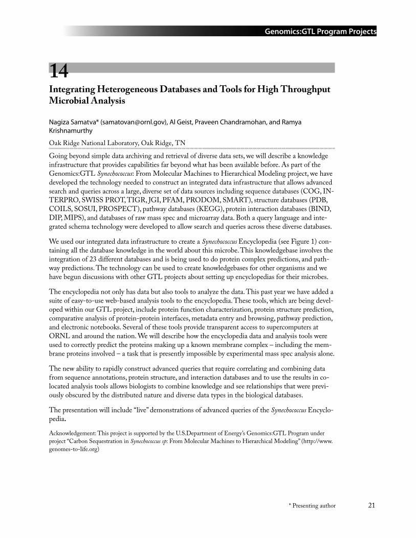
Genomics:GTL Program Projects
21* Presenting author
14 Integrating Heterogeneous Databases and Tools for High Throughput Microbial Analysis
Nagiza Samatva* ([email protected]), Al Geist, Praveen Chandramohan, and Ramya Krishnamurthy
Oak Ridge National Laboratory, Oak Ridge, TN
Going beyond simple data archiving and retrieval of diverse data sets, we will describe a knowledge infrastructure that provides capabilities far beyond what has been available before. As part of the Genomics:GTL Synechococcus: From Molecular Machines to Hierarchical Modeling project, we have developed the technology needed to construct an integrated data infrastructure that allows advanced search and queries across a large, diverse set of data sources including sequence databases (COG, IN-TERPRO, SWISS PROT, TIGR, JGI, PFAM, PRODOM, SMART), structure databases (PDB, COILS, SOSUI, PROSPECT), pathway databases (KEGG), protein interaction databases (BIND, DIP, MIPS), and databases of raw mass spec and microarray data. Both a query language and inte-grated schema technology were developed to allow search and queries across these diverse databases.
We used our integrated data infrastructure to create a Synechococcus Encyclopedia (see Figure 1) con-taining all the database knowledge in the world about this microbe. This knowledgebase involves the integration of 23 different databases and is being used to do protein complex predictions, and path-way predictions. The technology can be used to create knowledgebases for other organisms and we have begun discussions with other GTL projects about setting up encyclopedias for their microbes.
The encyclopedia not only has data but also tools to analyze the data. This past year we have added a suite of easy-to-use web-based analysis tools to the encyclopedia. These tools, which are being devel-oped within our GTL project, include protein function characterization, protein structure prediction, comparative analysis of protein-protein interfaces, metadata entry and browsing, pathway prediction, and electronic notebooks. Several of these tools provide transparent access to supercomputers at ORNL and around the nation. We will describe how the encyclopedia data and analysis tools were used to correctly predict the proteins making up a known membrane complex – including the mem-brane proteins involved – a task that is presently impossible by experimental mass spec analysis alone.
The new ability to rapidly construct advanced queries that require correlating and combining data from sequence annotations, protein structure, and interaction databases and to use the results in co-located analysis tools allows biologists to combine knowledge and see relationships that were previ-ously obscured by the distributed nature and diverse data types in the biological databases.
The presentation will include “live” demonstrations of advanced queries of the Synechococcus Encyclo-pedia.
Acknowledgement: This project is supported by the U.S.Department of Energy’s Genomics:GTL Program under project “Carbon Sequestration in Synechococcus sp: From Molecular Machines to Hierarchical Modeling” (http://www.genomes-to-life.org)
Genomics:GTL Program Projects
22 * Presenting author
Figure 1. Synechococcus sp. Encyclopedia. Advanced query and analysis interface. Search all Synechococcus databases. Browse experimental and analysis data. Download datasets.
15 Toward Comprehensive Analysis of MS/MS Data Flows
Andrey Gorin* ([email protected]), Nikita D. Arnold, Robert M. Day, and Tema Fridman
Oak Ridge National Laboratory, Oak Ridge, TN
Tandem mass spectrometry (MS/MS) is a powerful tool applied across several Genomics:GTL proj-ects for a variety of challenging proteomics projects: search for modified proteins, characterization of whole cell proteome, and identification of components of protein molecular machines. Despite great variety of the biological drivers, computational algorithms used “under the hood” face exactly the same challenges, and existing limitations of such algorithms are reproduced across many experi-mental designs. in ion trap devices under common conditions only ~20% of MS/MS spectra lead to peptide identifications that are worth to be considered, and misidentification rates remains to be high.
In certain range of score values the problem presents the tug-of-war alternative — boost of the reli-ability threshold (e.g. SEQUEST x-correlation value) rapidly decreases fraction of spectra that could be identified, while lowering it produces identifications from the “grey area”, which are of dubious quality. Algorithmically, the only way out is to increase information extraction from tandem MS data. If we could somehow retrieve total information content of a given spectrum, its fate can be decided unambiguously depending on our capacity to learn from it. Such capability could be useful for a
Genomics:GTL Program Projects
23* Presenting author
number of other interesting proteomics applications. The difficulties, of course, start with the defini-tion of something as unusual as information content of peptide spectrum.
Recently we proposed Probability Profile Method (PPM) — classification algorithm that infers identities of the individual spectral peaks examining their spectral neighborhoods under the “micro-scope” of Bayesian statistics. PPM results have the form of probabilistic statements, like peak number 123 is a b-ion with a 0.85 probability. Efficient identification of “noble” b- and y-ion peaks dramati-cally simplifies construction of de novo tags (partial peptides) for a particular spectrum. Relatively simple algorithmic advances allowed us to build PPM-chain – tool for de novo protein tagging based on our methodology. During this study we have realized that traditional separation of MS computa-tional algorithms into database search and de novo is very misleading. Our PPM-chain can be used in SEQUEST-emulation mode, taking full advantage of the known protein database, but at the same time has quite unique algorithmic capabilities, which include classical full-length de novo sequencing (it is not very good at the later task yet).
While capable of emulating SEQUEST our program works on entirely different mathematical and algorithmic principles. The laborious comparison between theoretical spectra and experimental spec-tra is the main CPU time consumer in database look-up algorithms, and correspondingly the perfor-mance typically scales linearly with the size of the search space, which grows exponentially in many situations (e.g., with the number of PTMs considered for each peptide). In de novo approach, almost all work is done up front, on the experimental spectra: peak labeling, finding of the tags, tag scoring. The need for the database comes very late in the process, involves very few candidate sequences and very simple procedures, which could be skipped all together for spectra with too little (no connect-able peaks) or a lot (direct de novo identification) in terms of the informational content.
De novo identification also has inherent flexibility, which is reflected in the suppleness of its output. For a given spectrum and given specifications for de novo tag (e.g. 3 residues are set as a minimum length) PPM-chain has three possible outcomes: (1) “no tag” - no satisfactory de novo tag could be constructed for the spectrum; (2) “tag-no-match” – there are good de novo tags, but they do not con-form to the available database; (3) “answer” – satisfactory de novo tag is found and mapped to a pro-tein in the database. In contrast, database look-up programs return the best match with an attached score, which slowly decreases from the confidently identified spectra toward definite identification failures. In this case the bad quality of the match (e.g., because the database protein contains se-quencing error) is hard to distinguish from the mediocre informational content of the spectrum (e.g., due to poor fragmentation). Such mix-up leads to all kinds of “grey area” situations, where valuable information - often indicative of unusual and interesting biological events - can be irrecoverably lost
We compared PPM-chain and SEQUEST using data sample obtained on the 54 ribosomal proteins of the Rhodopseudomonas palustris produced by Dr. Michael Strader at ORNL Center for Molecu-lar and Cellular System. We have explored results of both programs for three spectral sets separated by SEQUEST X-correlation score: “high quality” (>3.2), “medium” (from 2.2 to 3.2) and “low quality” (<2.2) spectra. For the high quality subset “no tag” outcome was obtained only for 21 spectra (1.4%) and out of 1263 “answer” results SEQUEST identification was confirmed for 99.9% spectra. “Tag-no-match” outcome was observed for 216 cases (14%) and this fraction kept increasing in me-dium and low quality subsets: (~ 38% in both). The “answer” outcomes were still excellently aligned with SEQUEST ids (99% and 96% precision values, correspondingly). The fraction of “no tag” cases has grown sharply: 18% for medium and 57% for low confidence sets, reflecting the absence of the differentiating information in many spectra belonging there.The result suggests an interesting speculation about possible sources of SEQUEST errors: it
Genomics:GTL Program Projects
24 * Presenting author
is feasible that a large fraction of such errors is due to the absence of the underlying correct answer in the database. In such cases the returned match bound to be an incorrect one, but still may have relatively high X-correlation value. In our approach such spectra immediately become candidates for further study, such as Post Translational Modification (PTM) search or further de novo processing. Summarizing, our testing indicates the following:
Even with the existing technology (which certainly could be improved) reliable de novo tags can be constructed for a large majority of MS/MS spectra – and virtually for all high quality spectra.
When de novo solution is compatible with the database, it is almost always the same as pro-vided by SEQUEST. This conclusion confirms a high reliability of the SEQUEST identifica-tions in the cases when the expected peptides are present in the protein database.
There is a significant fraction of spectra (~33% for medium X-correlation values, ~50% low X-corr values), where the PPM-Chain finds good de novo tags not compatible with anything in the target database. Some of these tags definitely reflect complex and interesting biological phenomena, where PTMs and point mutations are blocking the possibility of finding the cor-rect answers in the “plain vanilla” database searches.
For a future work we plan to apply PPM-chain for a comprehensive data extraction from the proteomics samples aiming at low abundance proteins as well as interesting biological facts, such as PTMs and mutated proteins. Our results strongly suggest that this approach will not only increase the output of useful information, but will also eliminate significant part of incorrect identification, further improving quality of the corresponding proteomics studies.
This work was funded in part by the US Department of Energy's Genomics:GTL program (www.doegenomestolife.org) under two projects, “Carbon Sequestration in Synechococcus Sp.: From Molecular Machines to Hierarchical Mod-eling” (www.genomes-to-life.org) and “Center for Molecular and Cellular Systems” (www.ornl.gov/GenomestoLife).
16 The Transcriptome of a Marine Cyanobacterium—Analysis Through Whole Genome Microarray Analyses
Brian Palenik1* ([email protected]), Ian Paulsen2* ([email protected]), Bianca Brahamsha1, Rob Herman1, Katherine Kang2, Ed Thomas3, Jeri Timlin3, and Dave Haaland3
1Scripps Institution of Oceanography, La Jolla, CA; 2The Institute for Genomic Research, Rockville, MD; and 3Sandia National Laboratories, Albuquerque, NM
Nitrogen and phosphorus abundance and type are thought to control photosynthesis and carbon se-questration in large areas of the world’s oceans. Little is known about the regulation in cyanobacteria of nitrogen and phosphorus metabolism and their interaction with other environmental variables such as light and micronutrients. We are using a whole genome microarray of Synechococcus sp. WH8102 to examine these issues.
We have used whole genome microarrays in a number or experiments, initially to compare cells grown with nitrate and cells grown with ammonia. We found that 247 genes were down-regulated
•
•
•
Genomics:GTL Program Projects
25* Presenting author
during growth under ammonia compared to nitrate. This included the NtcA transcriptional regulator known to control growth when ammonia is low and nitrogen sources other than ammonia are used. We also found that the use of a number of alternative nitrogen sources were down regulated (e.g. nitrate metabolism, cyanate transport, and urea metabolism). Some of these clearly have ntcA bind-ing sites upstream although a complete comparison of the microarray results with ntcA binding site predictions are still in progress by Zhengchang Su and Ying Xu (UGA) as part of our GTL project. In addition, a number of genes associated with stress conditions are down regulated. These include glutathione peroxidase and a number of proteases and heat shock like proteins. This supports our hy-pothesis that growth under nitrate is actually more stressful than on ammonia due to the requirement of additional electron transport activity (and electron leakage) to reduce nitrate to nitrite to ammonia. Thus, we currently hope to analyze and model these results as a combination of NtcA regulation and stress response regulation. Interestingly a number of hypotheticals and conserved hypotheticals are down regulated, giving us an initial clue as to their possible functions in the cell.
In addition, we have also characterized phosphate limitation in WH8102 and made knockout mutants in a number of the two-component regulatory systems of the cell. We are also examining these phosphate limitation experiments with the wild type and mutant cells using whole genome microarray analyses. Because of its relatively small number of regulatory systems compared to many microbes, Synechococcus sp. WH8102 is an ideal model system for preparing a complete picture of the regulatory networks of an environmentally significant microbe.
Sandia is a multi-program laboratory operated by Sandia Corporation, a Lockheed Martin Company, for the United States Department of Energy under Contract DE-ACO4-94AL85000.
This work was funded by the U.S.Department of Energy’s Genomics: GTL program (www.doegenomestolife.org) under project, “Carbon Sequestration in Synechococcus Sp.: From Molecular Machines to Hierarchical Modeling,” (www.genomes-to-life.org) and by its Microbial Genome Project under “Transport and its regulation in marine microorganisms—a genomics-based approach.”
17 DEB: a Data Entry and Browsing Tool for Entering and Linking Synechococcus sp. WH8102 Whole Genome Microarray Metadata from Multiple Data Sources
Arie Shoshani1* ([email protected]), Victor Havin1, Vijaya Natarajan1, Tony Martino2, Jerilyn A. Timlin2, Katherine Kang3, Ian Paulsen3, Brian Palenik4, and Thomas Naughton5
1Lawrence Berkeley National Laboratory, Berkeley, CA; 2Sandia National Laboratories, Albuquerque, NM; 3The Institute for Genomic Research, Rockville, MD; 4Scripps Institution of Oceanography, La Jolla, CA; and 5Oak Ridge National Laboratory, Oak Ridge, TN
The process of generating and analyzing microarray data for Synechococcus sp. WH8102 whole genome in the Sandia-led GTL project involves three collaborators, where each generates metadata about their operation as well as data files. The Synechococcus sp. microbes are cultured in the Scripps Institution of Oceanography in San Diego, then the sample pool is sent to TIGR in Rockville, Maryland for microarray hybridization, 2-color scanning, and analysis. The scanned files and slides are then sent to Sandia Lab in Albuquerque, New Mexico for analysis and additional scanning with
Genomics:GTL Program Projects
26 * Presenting author
a Hyperspectral Imaging instrument. Each of the institutes has an independent system for keeping track of metadata about their part of the operation and unfortunately these systems do not facilitate easy transfer of metadata details between institutions. This situation is typical of many biology proj-ects, and it begs for a solution.
In this sub-project we set out to develop a single system where such metadata can be collected and linked in an orderly fashion. We developed a web-based Data Entry and Browsing (DEB) tool that can capture the metadata from experiments and laboratories and store them in a database in a computer searchable form. The key need is to have an easy-to-use intuitive system that integrates the metadata of all the related activities in this project. The design of the DEB tool is based on inputs and insights from the biologists on the project and as such contains features that a biologist will find useful. The interface design mimics the familiar laboratory notebook format. The system is built on top of the Oracle database system. The main concept of the interface design is to expose the biologist to a single “object” and its attributes at a time, and presenting objects as pages in a notebook that can be “turned”, yet provide links between the objects in a simple intuitive fashion. An example of such a web-based screen is shown in the figure below.
The most powerful capability of the DEB system is that it is schema-driven, that is, all the interfaces to support all of the above features are generated automatically from the schema definition. There-fore, new metadata schemas can quickly be used to generate DEB interfaces as well as the underlying Oracle database for them. This feature makes this tool immediately applicable to new and/or chang-ing databases. This allowed us to generate databases based on schemas designed by the biologists. Specifically, the scientists from the three sites have defined schemas for the Nucleotide Pool of microbes, for the Microarray Hybridization (based on the MIAME concepts), and the Hyperspectral Imaging and analysis system. The design included the ability to link these schemas and thus allow a researcher from any area of the project to extract metadata from the various parts of the experi-ment. For example the microarray hybridization schema has “probe_source” that links (points to) the “nucleotide_pool_id” in the Nucleotide Pool schema.
Data entry to the databases is done in two different modes: 1) on-line web-based data entry, and 2) automated data uploading from another database source. The on-line mode is used by the people who culture the Nucleotide Pools (Scripps), and the people running the Hyperspectral Imaging (Sandia). The automated data uploading is used for the Microarray Hybridization metadata (TIGR) because they have their own well-developed internal database system. The automated metadata loading is performed by dumping the metadata into simple formatted files (similar the spreadsheet output format) and have schema-driven tools for loading the data into the common database.
The main features of the DEB system are:
It supports multiple inter-related object-classes, such as “experiment, materials, nucleotide-pool, samples, arrays, etc.
For each object-class, it displays a page that mimics a notebook, with pages that can be “turned” (i.e. selected by previous-next, or by number)
Objects can be linked to each other by simple connectors, such as a “sample” object linked to its “nucleotide-pool”.
Any file types (document, images, excel, etc.) can be uploaded to the system, and related to the metadata
•
•
•
•
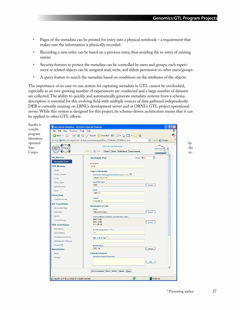
Genomics:GTL Program Projects
27* Presenting author
Pages of the metadata can be printed for entry into a physical notebook – a requirement that makes sure the information is physically recorded
Recording a new entry can be based on a previous entry, thus avoiding the re-entry of existing entries
Security features to protect the metadata can be controlled by users and groups; each experi-ment or related objects can be assigned read, write, and delete permission to other users/groups.
A query feature to search the metadata based on conditions on the attributes of the objects.
The importance of an easy-to-use system for capturing metadata in GTL cannot be overlooked, especially as an ever growing number of experiments are conducted and a large number of datasets are collected. The ability to quickly and automatically generate metadata systems from a schema description is essential for this evolving field with multiple sources of data gathered independently. DEB is currently running on LBNL’s development server and at ORNL’s GTL project operational server. While this system is designed for this project, its schema-driven architecture means that it can be applied to other GTL efforts.
Sandia is a multi-program laboratory operated by San- dia Corpo- ra-
•
•
•
•
Genomics:GTL Program Projects
28 * Presenting author
tion, a Lockheed Martin Company, for the United States Department of Energy under Contract DE-ACO4-94AL85000.
18 Microarray Analysis using VxInsight and PAM
George S. Davidson*1 ([email protected]), David Hanson2, Shawn Martin1, Margaret Werner-Washburne2, and Mark D. Rintoul1
1Sandia National Laboratories, Albuquerque, NM and 2University of New Mexico, Albuquerque, NM
In 2001 Hihara et al. [1] published a series of microarray experiments describing gene expression changes in the cyanobacterium Synechocystis sp. PCC 6803 in response to acclimation from a low light level (20 µmol photons m-2 sec-1 to 300 µmol photons m-2 sec-1). These data, which are publicly available from the KEGG database [2], have been reanalyzed using the VxInsight genome tools [3] from Sandia National Laboratories, and PAM [4] from Stanford. The analysis served as a test-bed for preparing the VxInsight tools to work with bacterial microarray data and associated databases. Here we present the results of clustering the individual experiments and the genes by co-expression. Interestingly, a number of experimental design issues are also raised. We examined lists of genes generated by PAM and by VxInsight that include significant differences in expression under the low light (LL) and the high light (HL) experimental conditions. We discuss the methodologies and the visual user interface linking these genes to online annotations and regulatory networks.
Hihara et al. measured total mRNA from HL conditions at 15 min, 1 hr, 6 hr, and 15 hr. These were compared to the mRNA levels measured under LL conditions, which served as control data. Expres-sion levels were measured with CyanoCHIP version 0.8 from TaKaRa. These experiments revealed 84 ORFs with up regulated expression and 80 ORFs with down regulated expression after exposure to HL. Almost all of the photosystem I genes were immediately down regulated, while genes associ-ated with photosystem II showed more complicated patterns. Both observations are consistent with the increasing PSII/PSI ratio in response to HL which involves an initial shift away from of PSI and the gradual construction of PSII (generally completed within 60 min.).
VxInsight found three groups of experiments, as shown in Figure 1, the first of which clearly reflects the stable, ongoing response to HL. The second captures the intermediate state when many of the PSII proteins are being synthesized, or have largely become available as the cells shift toward a higher metabolic plane. The third group contains a random mixture of arrays from time points throughout the experiment, which suggests that technical problems were confounding the measure-ments (something that is not uncommon in microarray experiments). We identified genes with significantly different expressions between late experimental conditions (first group) and the second group, which consisted of an equal number of measurements made at 15 minutes and at 60 minutes. VxInsight and PAM identified many of the same genes that Hihara et al. found; however the differ-ences offer opportunities for deeper study. We present these genes with their scores and demonstrate the interactive analysis of these lists, including the use of KEGG pathways.
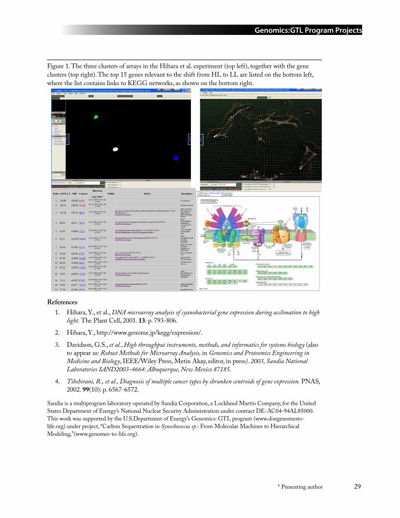
Genomics:GTL Program Projects
29* Presenting author
Figure 1. The three clusters of arrays in the Hihara et al. experiment (top left), together with the gene clusters (top right). The top 15 genes relevant to the shift from HL to LL are listed on the bottom left, where the list contains links to KEGG networks, as shown on the bottom right.
ReferencesHihara, Y., et al., DNA microarray analysis of cyanobacterial gene expression during acclimation to high light. The Plant Cell, 2001. 13: p. 793-806.
Hihara, Y., http://www.genome.jp/kegg/expression/.
Davidson, G.S., et al., High throughput instruments, methods, and informatics for systems biology (also to appear as: Robust Methods for Microarray Analysis, in Genomics and Proteomics Engineering in Medicine and Biology, IEEE/Wiley Press, Metin Akay, editor, in press). 2003, Sandia National Laboratories SAND2003-4664: Albuquerque, New Mexico 87185.
Tibshirani, R., et al., Diagnosis of multiple cancer types by shrunken centroids of gene expression. PNAS, 2002. 99(10): p. 6567-6572.
Sandia is a multiprogram laboratory operated by Sandia Corporation, a Lockheed Martin Company, for the United States Department of Energy’s National Nuclear Security Administration under contract DE-AC04-94AL85000. This work was supported by the U.S.Department of Energy’s Genomics: GTL program (www.doegenomesto-life.org) under project, “Carbon Sequestration in Synechococcus sp.: From Molecular Machines to Hierarchical Modeling,”(www.genomes-to-life.org).
1.
2.
3.
4.
Genomics:GTL Program Projects
30 * Presenting author
19 Mapping of Biological Pathways and Networks across Microbial Genomes
F. Mao, V. Olman, Z. Su, P. Dam, and Ying Xu* ([email protected])
University of Georgia, Athens, GA and Oak Ridge National Laboratory, Oak Ridge, TN
Homology exists beyond the individual gene level, and it could exist at the biological pathway and network level. There are a number of databases consisting of all experimentally validated and reliably predicted pathways/networks, providing a rich source of information for genome annotation and bio-logical studies at a systems level. A key to effectively use such information is to identify orthologous genes accurately. However existing methods for mapping these known pathways and networks have serious limitations, greatly limiting the utility of such very useful information. Virtually all exist-ing mapping methods are based on sequence similarity information, using tools such as reciprocal BLAST search or COG mapping. A fundamental problem with such methods is that sequence simi-larity information alone does NOT contain all the information needed to identify true orthologous genes!
We have recently developed a computational method and software, called P-MAP, for mapping a known pathway/network from one microbial organism to another by combining homology information and genomic structure information. The basic idea is that in microbes, genes working in the same pathway can generally be decomposed into a few operons or, in case of complex path-ways/networks, regulons. Such information has not been effectively used in pathway mapping. When mapping known pathways, we first predict all the operons in a genome using our operon prediction program. The predictions are then validated through comparing microarray data mainly to check for consistency between gene expression patterns for genes predicted to be in the same operons or adjacent operons. Our evaluation has indicated that our prediction accuracy is close to 90%. With such information, we then map genes in a pathway template to the target genome that simultane-ously gives relatively high sequence similarity between predicted orthologous gene pairs and has all the mapped genes grouped into a number of operons, preferably co-regulated operons based on the predicted cis regulatory elements and available microarray data. We have formulated the mapping problem as a linear integer programming (LIP) problem, and solved the problem using a commercial LIP solver, called COIN.
We have applied the P-MAP program to map known biological pathways in KEGG and MetaCyc to the cyanobacterial genomes and currently are mapping them to the Shewanella oneidensis MR-1 genome. Some of the mapping results could be found at http://csbl.bmb.uga.eddu/WH8102.
Acknowledgement: This project is supported by the U.S.Department of Energy’s Genomics:GTL Program under project “Carbon Sequestration in Synechococcus sp: From Molecular Machines to Hierarchical Modeling” (http://www.genomes-to-life.org).
Genomics:GTL Program Projects
31* Presenting author
20 Proteomic Analysis of the Synechococcus WH8102 CCM with Varying CO2 Concentrations
Arlene Gonzales, Yooli K. Light, Zhaoduo Zhang, Michael D. Leavell, Rajat Sapra, Tahera Iqbal, Todd W. Lane, and Anthony Martino* ([email protected])
Sandia National Laboratories, Livermore, CA
The genera Synechococcus and Prochlorococcus are oxygenic photoautotroph cyanobacteria. They are the most abundant picophytoplankton in the world’s oceans where they form the foundation of the ma-rine food web and are likely the largest contributors to primary production. Whole genome sequenc-es are now available for a number of cyanobacteria including Synechococcus WH8102, Prochlorococcus MED4, and Prochlorococcus MIT9313. The sequences make it possible to use comparative analysis and high-throughput functional genomics and proteomics experiments to help better understand global diversity involved in carbon fixation.
Synechococcus WH8102’s 2.4 Mb genome has yielded a number of interesting results regarding the carbon concentrating mechanism (CCM) in this organism. The carboxysome encoding operon in 8102 resembles that of β-proteobacteria rather than cyanobacteria. The operon most likely was acquired through horizontal gene transfer from phage. Carbonic anhydrase (CA) activity in the carboxysome shell protein csoS3 has been determined experimentally. Genome analysis indicates a putative β-CA and a ferripyochelin binding protein CA may also exist. Finally, transport of inorganic carbon in 8102 may occur through the low affinity CO2 uptake genes ndhD4, ndhF4, and chpX. In Prochlorococcus, uptake genes have not been observed. Perhaps a unique transport mechanism exists in oceanic cyanobacteria.
We will present a high-throughput proteomic approach using mass spectrometry (MS), 2-hybrid analysis, and phage display to deconstruct components of the CCM and determine the effect of changing CO2 levels in Synechococcus 8102. Protein expression levels of CCM components and pro-tein-protein interactions within the carboxysome will be presented. Protein fractions were separated in to particulate and soluble fractions, and western blots of the fractions indicated rbcL and car-boxysome shell proteins partitioned exclusively with the particulate fractions. Developing and fully mature carboxysomes were observed in the particulate fractions using electron microscopy. The two putative CAs partitioned separately in the particulate and soluble fractions. Changes in expression of specific proteins in cultures bubbled under different CO2 levels were determined using 2D electro-phoresis/MALDI-TOF MS. A synergistic whole proteome approach using capillary LC-MS/MS continues. Protein-protein interactions within the carboxysome have been determined using bacterial 2-hybrid techniques, and a number of pair wise interactions with be presented. Finally, rbcS-peptide interactions are being studied using phage display techniques.
Genomics:GTL Program Projects
32 * Presenting author
21 Predicting Protein-Protein Interactions Using Signature Products with an Application to β-Strand Ordering
Shawn Martin1 ([email protected]), W. Michael Brown1, Charlie Strauss2, Mark D. Rintoul*1, and Jean-Loup Faulon3
1Sandia National Laboratories, Albuquerque, NM; 2Los Alamos National Laboratory, Los Alamos, NM; and 3Sandia National Laboratories, Livermore, CA
As a part of the project entitled “Carbon Sequestration in Synechococcus sp.: From Molecular Machines to Hierarchical Modeling,” we have developed a computational method for predicting protein-protein interactions from amino acid sequence and experimental data (Martin, Roe et al. 2004). This method is based on the use of symmetric tensor products of amino acid sequence fragments, which we call signature products. These products occur with different frequencies when considering interacting versus non-interacting protein pairs. We can therefore predict when a protein pair interacts by comparing the frequency of the signature products in that pair to the correspond-ing frequencies in protein pairs known to interact. Computationally, these comparisons are encoded into a Support Vector Machine (SVM) framework (Cristianini and Shawe-Taylor 2000), where the signature products are implemented using kernel functions. The final result is an automated clas-sification system which can extrapolate from experimental results to complexes to entire proteomes, based only on experiment and primary sequence.
We have expended significant effort in benchmarking our method against competing techniques. In particular, we have compared the signature product method with methods based on products of InterPro signatures (Sprinzak and Margalit 2001; Mulder, Apweiler et al. 2003), concatenation of full-length amino acid sequences (Bock and Gough 2001), and a method combining multiple data sources ( Jansen, Yu et al. 2003). In all cases our method performed as well as or better than the competing methods, as measured by 10-fold cross validation. We have applied our method to the prediction of protein-protein interactions in the case of yeast SH3 domains (Tong, Drees et al. 2002), where we achieved 80.7% accuracy, the full yeast proteome (69% accuracy), and H. Pylori (Rain, Selig et al. 2001) (83.4% accuracy). In addition to these results, which appear in (Martin, Roe et al. 2004), we have applied our method using COG networks (Tatusov, Koonin et al. 1997) to Synechocystis sp. (91% accuracy), and Nostoc sp. (69% accuracy).
Using our signature product approach, we have gone on to develop a method for ordering β-strands, which can in turn be used to improve the results of ab initio protein folding. The first step in our method is to train a signature product model for predicting β-strand interactions. This model was trained by extracting all β-strands from the Protein Data Bank (PDB) (Berman, Westbrook et al. 2000) using the dictionary of protein secondary structure (DPSS) method (Kabsch and Sander, 1983). After removing identical sequences from the ~20,000 structures available in the PDB, we ob-tained ~90,000 β-strands with more than 3 residues. Using the product signature method, we trained a β-strand interaction predictor which achieved 77% accuracy on a randomly selected training/test set combination with 80% of the β-strands in the training set and the remaining 20% of the β-strands in the test set. The next step in our method, as yet unimplemented, applies to individual proteins. For a given protein, we will apply our model to every possible pair of β-strands within the protein, and then consider every possible ordering of these strands. We will use the ordering which gives the highest
Genomics:GTL Program Projects
33* Presenting author
average interaction score, as measured using our β-strand interaction predictor. We will validate our results by comparing our predicted ordering to the ordering actually present in the PDB.
Sandia is a multiprogram laboratory operated by Sandia Corporation, a Lockheed Martin Company, for the U.S. Department of Energy’s National Nuclear Security Administration under Contract DE-AC04-94AL85000.
22 In Vivo Observation of the Native Pigments in Synechocystis sp. PCC 6803 Using a New Hyperspectral Confocal Microscope
Michael B. Sinclair1* ([email protected]), Jerilyn A. Timlin1, David M. Haaland1, Sawsan Hamad2, and Wim F.J. Vermaas2
1Sandia National Laboratories, Albuquerque, NM and 2Arizona State University, Tempe, AZ
We have developed a new hyperspectral confocal microscope that combines the attributes of high spatial resolution (<0.5 µm), high speed acquisition (>8 MB/s) and single photon sensitivity. The new instrument records the emission spectrum from 500 nm to 800 nm for each voxel within the 3-dimensional sample. The acquisition of full spectral information, when coupled with modern multivariate data analysis techniques allows for quantification of the contribution of each of the emitting components present within any voxel. To demonstrate the advantages of this approach, we have obtained and analyzed in vivo hyperspectral images of wild type Synechocystis sp. PCC 6803, as well as two mutant strains: chlL–, which is incapable of light-independent synthesis of chlorophyll, and PS1-less/chlL–, which in addition to being incapable of light-independent synthesis of chloro-phyll does not assemble photosystem I. The raw emission spectra obtained from these specimens are quite complex, containing overlapping signatures from many pigments. Multivariate curve resolution analysis of the spectral images shows that each spectrum can be decomposed into independently varying contributions from phycocyanin, allophycocyanin, chlorophyll, a specific pool of “low-energy” chlorophyll associated with photosystem I, and protochlorophyllide. The relative contribution of each of these components varies from species to species in a manner consistent with expectations based on the genetic composition of the mutant strains. For example, we observe significant protochlorophyl-lide emission from the chlL– mutant which was grown in virtual darkness, while this emission is absent in the wild type. To our knowledge, this is the first ever demonstration of the coupling of rig-orous deconvolution methods with hyperspectral confocal microscopy to reveal multiple overlapping native pigment emissions from in vivo specimens. We have also observed evidence for an inhomo-geneous distribution of the emitting compounds within the cyanobacterium and are currently quan-titatively exploring this inhomogeneity in the concentration distributions both within and between cells. This presentation will describe the design, construction and performance of the hyperspectral confocal microscope. Our results for Synechocystis will be described in detail.
Sandia is a multi-program laboratory operated by Sandia Corporation, a Lockheed Martin Company, for the United States Department of Energy under Contract DE-ACO4-94AL85000. This work was funded in part by the U.S.Department of Energy’s Genomics: GTL program (www.doegenomestolife.org) under project, “Carbon Seques-tration in Synechococcus sp: From Molecular Machines to Hierarchical Modeling,” (www.genomes-to-life.org).
Genomics:GTL Program Projects
34 * Presenting author
23 Connecting Temperature and Metabolic Rate to Population Growth Rates in Marine Picophytoplankton
Andrea Belgrano* ([email protected]) and Damian Gessler
National Center for Genome Resources, Santa Fe, NM
Photosynthetic picophytoplankton bacteria such as Synechococcus can contribute up to more then 50% of the total water column primary production, thus playing an important role in controlling the net flux of CO2 between the atmosphere and the ocean, by the sequestration of carbon from the atmo-sphere via photosynthesis, and to global carbon cycling in marine systems.
We present how the rate of photosynthesis is regulated by changes in CO2 concentration, irradiance and temperature, including allometric scaling theory and Rubisco activity as the primary catalyst, for the fixation of carbon by picophytoplankton.
To integrate physiological, biogeochemical, and environmental data in a single model, we integrate ¾-power scaling laws, along with irradiance, temperature, and nutrient uptake functions. Body mass scaling provides the theoretical and empirical model for constraints on the supply and use of energetic resources for picophytoplankton. We use a Boltzamann factor approach to capture the temperature-dependence of metabolism, and additionally introduce a Michaelis-Menten approach for nutrient uptake that includes cell quotas in a single growth model for Synechococcus.
We present evidence that a rise in oceanic temperature may reduce the relative contribution that picophytoplankton plays in the ocean as a carbon pathway. This highlights the importance of under-standing shifts in the size composition of phytoplankton assemblages in relation to oceanic primary production of biomass, cell density, and nutrient status in the northwestern North Atlantic Ocean.
24 Deciphering Response Networks in Microbial Genomes through Data Mining and Computational Modeling
Z. Su3, P. Dam3, V. Olman3, F. Mao3, H. Wu3, X. Chen1, T. Jiang1, B. Palenik2, and Ying Xu3*
([email protected]) 1University of California, Riverside, CA; 2Scripps Institution of Oceanography, San Diego, CA; and 3University of Georgia, Athens, GA and Oak Ridge National Laboratory, Oak Ridge, TN
Deciphering of the “wiring diagrams” of biological networks (including metabolic, signaling and reg-ulatory networks) represents a highly challenging problem, due to our lack of general understanding about the conceptual framework of how biomolecules work together as a system and an insufficient amount of experimental data. The majority of on-going computational research has been focusing on developing general methodologies for deriving “functionally equivalent” networks that are consistent with the limited experimental data such as microarray kinetics data, possibly leading to network topologies that are not biologically meaningful.
Genomics:GTL Program Projects
35* Presenting author
We have been developing a computational framework, attempting to systematically derive network topologies that are most consistent with (a) information derived through mining genomic sequences and various genomic and proteomic data and (b) the kinetics data derived from microarray gene expression experiments. The framework consists of the following three key components:
Identification of genes involved in a particular biological process: To facilitate identification of genes possibly involved in a particular biological network, we first made genome-scale predictions of (a) gene functions, (b) operon structures and (c) cis regulatory elements at the genome scale. Gene function prediction is based on available genome annotation plus our own function predic-tion pipeline using additional information, including motif search and structure-based function prediction. Operons are predicted using our own program (see Section on operon/regulon predic-tions) Cis regulatory elements are predicted using our prediction program CUBIC, in conjunc-tion with microarray data when available, through identification of conserved sequence motifs and similar gene expression patterns. Based on the initial identification of genes possibly involved in a particular biological network, we then refine/expend the gene candidate list through compar-ing to the information collected in (a), (b) and (c) described above.
Prediction of interaction relationships among these candidate genes: Currently we attempt to predict two types of interactions: (a) protein-protein interactions, both physical interactions and functional associations, and (b) protein-DNA interactions. Protein-protein interactions are predicted using homology search against protein interaction databases such as DIP & BIND and also based on prediction methods such as gene fusion/fission analysis, and phylogenetic profile analysis. We have developed our own methods for protein (transcription factors)-DNA interac-tions, based on both sequence and structural information. The sequence-based method is mainly based on (a) homology search against known protein-DNA complexes, (b) identification of self-regulation events, and (c) co-evolution information of transcription factors and operons they regulate. When the 3D structures of transcription factors are available, our method can accurately predict the binding affinity between the structure and its predicted DNA binding motifs, provid-ing a highly effective tool for protein-DNA interaction prediction. Another piece of information for bio-molecular interactions comes from mapping known pathways from related organisms to the target genomes. Though not all such pathway mappings will provide complete pathway models in the target genome, the molecular interactions in the predicted pathways are useful and could be used for piecing together the “complete” network model in (3).
Prediction of wiring diagrams through computational optimization: We have developed two complementary methods for prediction of “complete” wiring diagrams of a target network, based on the predicted gene candidates and their (partial) interaction relationships and additional information. The first method connects the partially connected pieces predicted in (2) through mapping them to a genome-scale protein-protein interaction map we predicted in (2). The idea is to find the biologically most meaningful “paths” to connect the unconnected pieces (made of pro-tein-protein and protein-DNA interactions). An algorithm has been developed for accomplishing this. In addition, we are currently developing a new algorithm that connects all the interaction components that are most consistent with the available microarray kinetics data, generalizing the current popular methods. By doing so, we can get wiring diagrams that are consistent with both molecular interaction information derived through data mining and microarray kinetics data.
We now describe two key procedures needed to implement the above computational prediction pro-tocol because of the significance by their right.
1.
2.
3.
Genomics:GTL Program Projects
36 * Presenting author
Prediction of operons and regulons: We have recently developed a computational capability for prediction of operons in microbes, using multiple sources of information including (a) conserved gene neighborhoods across closely related organisms, (b) detected co-evolutionary information of genes, (c) functional relatedness of genes, (d) inter-genic distance information plus various types of other information. The overall prediction accuracy has reached 80% based on our test results on known operons in E. coli. We have applied this prediction program, in conjunction with available microarray data, to a number of genomes including E. coli, Shewanella, Pyrococcus and Synechococcus. The prediction procedure can be outlined as follows. We run the program to produce the initial operon candidate list and then we compare the predicted operons with available microarray data to check for consistency. Corrections will be made on the initial predictions if genes of the same operon exhibit significantly different expression patterns under any experimental condition, or genes from the neighboring operons have highly similar expression patterns under all known conditions and these operons are very close in the genomic sequence. In general about 5-10% of the original predic-tions are corrected based on the microarray data. We expect that the prediction accuracy could reach close to or even beyond 90% when sufficient microarray data is available. Based on the predicted operon structures, we then predicted regulons, based on available microarray data and genome-scale prediction of cis regulatory elements. The prediction procedure identifies operons that share similar expression patterns under the given experimental conditions and share conserved (predicted) binding motifs, and then clusters them into regulons. While this work is still in its early stage, we have identi-fied a number of interesting regulons in the genomes we have applied prediction programs.
Pathway mapping: We have recently developed a computational method and software P-MAP for mapping a known pathway/network from one microbial organism to another by combining homology information and genomic structure information. The basic idea is that in microbes, genes working in the same pathway can generally be decomposed into a few operons or, in case of complex pathways/networks, regulons. Such information has not been effectively used in pathway mapping. When mapping known pathways, we first predict all the operons in a genome using our operon prediction program. The predictions are then validated through comparing microarray data mainly to check for consistency between gene expression patterns for genes predicted to be in the same operons or adjacent operons. Our evaluation has indicated that our prediction accuracy is close to 90%. With such information, we then map genes in a pathway template to the target genome that simultane-ously gives relatively high sequence similarity between predicted orthologous gene pairs and has all the mapped genes grouped into a number of operons, preferably co-regulated operons based on the predicted cis regulatory elements and available microarray data. We have applied the P-MAP program to map known biological pathways in KEGG and MetaCyc to the cyanobacterial genomes and currently are mapping them to the Shewanella oneidensis MR-1 genome. Some of the mapping results could be found at http://csbl.bmg.uga.edu/WH8102.
Applications: We have applied this computational framework to predict the wiring diagrams of various response networks, which consists of signaling, regulatory and metabolic components. These include the carbon fixation, phosphorus assimilation and nitrogen assimilation networks in cyano-bacterial genomes. Research is on going to apply the framework to Shewanella oneidensis MR-1.
Acknowledgement: This project is supported by the U.S.Department of Energy’s Genomics:GTL Program under project “Carbon Sequestration in Synechococcus sp: From Molecular Machines to Hierarchical Modeling” (http://www.genomes-to-life.org)
Genomics:GTL Program Projects
37* Presenting author
25 BiLab – A New Tool that Combines the Ease-of-Use of MatLab and the Power of Multiple Computational Biology Libraries
Al Geist* ([email protected]) and David Jung
Oak Ridge National Laboratory, Oak Ridge, TN
As part of the Genomics:GTL Synechococcus: From Molecular Machines to Hierarchical Modeling project, we are developing a new tool called BiLab that we hope will revolutionize computational biology the way the MatLab revolutionized numerical linear algebra. MatLab is widely used to do analysis, to develop new algorithms, and to teach students. MatLab is easy enough for new users and powerful enough for sophisticated users. Yet under the covers it is just a scripting language that pro-vides easy access to the robust linear algebra functions in the LAPACK library. BiLab takes a similar approach except instead of only understanding matrices and doing linear algebra, BiLab understands biological objects such as DNA, proteins, and molecules and is able to manipulate them through any of the functions in a half-dozen standard computational biology libraries. And BiLab is able to display results in biologically relevant form, for example, a protein may be displayed as a molecule, a sequence alignment as stacked sequences.
We developed the BiLab scripting language to cater to different level of expertise in the user, from biologists who just want a quick way to use existing functions to bioinformatics programmers who want to write sophisticated programs in the BiLab scripting language. We have developed the tool to allow the easy addition of new biological objects and functions. Today BiLab provides access to all the functions in bioJava, bioPython, the text based NCBI tools, Jmol, JalView, and CDK. Developers can extend the scripting language to understand new biology. Thus the tool is designed to evolve with the Genomics: GTL program.
Like MatLab, data can be typed in manually or read in from files. BiLab understands the concept of remote biological databases and is able to dynamically load data from SwissProt, GenBank, FASTA, Protein Data Bank, EMBL, and other databases for analysis and study.
This presentation will describe how BiLab is built, how it can be extended, and hands-on demon-strations of the capabilities of the BiLab prototype.
Acknowledgement: This project is supported by the U.S.Department of Energy’s Genomics:GTL Program under project “Carbon Sequestration in Synechococcus sp: From Molecular Machines to Hierarchical Modeling” (http://www.genomes-to-life.org)
Genomics:GTL Program Projects
38 * Presenting author
26 Microbial Cell Modeling via Reacting/Diffusing Particles
Steve Plimpton* ([email protected]) and Alex Slepoy
Sandia National Laboratories, Albuquerque, NM
We have developed a simulator called ChemCell [1] that tracks protein interactions within cells and can be used to model signaling, metabolic, or regulatory response. Cell features for microbial cells are represented realistically by triangulated membrane surfaces. Particles represent proteins, complexes, or other biomolecules of interest. They diffuse via 3d Brownian motion within the cytoplasm, or in 2d within membrane surfaces. When particles are near each other, they interact in accord with Monte Carlo rules to perform biochemical reactions, which can represent protein complex formation and dissociation events, ligand binding, etc. ChemCell is similar in spirit to MCell [2] and Smoldyn [3].
In this poster, we focus on the underlying algorithms used for reaction rules. We have recently devel-oped a spatial version of the stochastic simulation algorithm (SSA) due to Gillespie [4] and discuss it’s implementation in ChemCell. We compare it to alternative approaches including the original SSA and the interaction rules recently developed by Andrews and Bray [3]. We also highlight issues with various reaction/diffusion algorithms relevant to parallel implementation within ChemCell, with the eventual goal of enabling whole-cell models of realistic numbers of proteins and other biomolecules.
References:S. J. Plimpton and A. Slepoy, SAND Report 2003-4509 (2003).
J. R. Stiles and T. M. Bartol, in Computational Neuroscience: Realistic modeling for experimental-ists, edited by E. De Schutter, published by CRC Press, 87-127 (2001).
S. S. Andrews and D. Bray, Phys Biology, 1, 137-151 (2004).
D. T. Gillespie, J Comp Phys, 22, 403-434 (1976).
27 Modeling RuBisCO’s Gating Mechanism Using Targeted Molecular Dynamics
Paul S. Crozier1 ([email protected]), Steven J. Plimpton1, Mark D. Rintoul1*, Christian Burisch2, and Jürgen Schlitter2
1Sandia National Laboratories, Albuquerque, NM and 2Ruhr-Universität Bochum, Bochum, Germany
RuBisCO is the enzymatic bottleneck of carbon sequestration in Synechococcus, which is partly due to its catalysis of a competing oxygenase reaction that limits its specificity and efficiency. The binding niche residues are highly conserved across RuBisCO species, yet experimentally-measured specifici-ties and carboxylation rates vary widely. The residues that make up the gate to the binding niche affect the gate’s opening and closing rates, and in turn, RuBisCO specificities and reaction rates. We
1.
2.
3.
4.
Genomics:GTL Program Projects
39* Presenting author
have performed molecular dynamics (MD) simulations of RuBisCO’s gating mechanism to gain insight into how residue-level changes in RuBisCO’s primary sequence affect enzyme performance.
Traditional MD is currently limited to the sub-microsecond timescale, but hardware and algorithm improvements continue to push the attainable timescales upward. We have recently developed several upgrades for our open-source parallel MD simulation package, LAMMPS (http://www.cs.sandia.gov/~sjplimp/lammps.html), which essentially double the algorithm’s performance on typical biomo-lecular simulations. Performance enhancements have come through implementation of the rRESPA hierarchical time-stepping method and a high-performance tabulation algorithm for rapid evaluation of CPU-intensive coulombic interatomic forces. The LAMMPS simulation package was officially released as an open-source parallel MD code available for download on the first of September. Since then, it has been downloaded 1,075 times.
In addition to improving the traditional MD capabilities in LAMMPS, we have implemented advanced MD methods that allow simulation of events that occur on much longer timescales. One such method, targeted molecular dynamics (TMD), allows simulation of user-specified transition events, like the RuBisCO gating mechanism, by imposing a dynamic holonomic constraint on the macromolecular complex. TMD yields the free energy profile of the transition event, which is related to the rate of the transition event.
We performed TMD simulations of the gating event of spinach and Synechococcus RuBisCO, each for WT and for mutant D473A. Our simple implicit solvent reduced-model predictions of gating free energy profiles have been encouraging since they have demonstrated the ability to discriminate between RuBisCO structural differences, and are in qualitative agreement with expected trends. For example, our TMD prediction shows a much higher gate opening barrier for Synechococcus than for spinach, which indicates more time in the closed state, more photorespiration, and lower specific-ity for Synechococcus RuBisCO. This is in qualitative agreement with the experimentally-measured specificities of Synechococcus RuBisCO (47) and spinach RuBisCO (92). Likewise, D473A muta-tions performed in silico for both RuBisCO species show a much lower free energy barrier for gate opening than do wild type RuBisCOs. Experiments show that D473A mutants are not catalytically competent, which is probably due to the fact that the binding niche gate can not properly close (and rapidly opens), without the D473 – R134 salt bridge.
This project is supported by the U.S.Department of Energy’s Genomics:GTL Program under project “Carbon Se-questration in Synechococcus sp: From Molecular Machines to Hierarchical Modeling” (http://www.genomes2life.org/). Sandia is a multiprogram laboratory operated by Sandia Corporation, a Lockheed Martin Company, for the United States Department of Energy’s National Nuclear Security Administration under contract DE-AC04-94AL85000.
Genomics:GTL Program Projects
40 * Presenting author
28 Selection of Ligands by Panning of Phage Display Peptide Libraries Reveals Potential Partners for TPR Domain and rbcS in Synechococcus WH8102
Zhaoduo Zhang* ([email protected]), Arlene D. Gonzales, Todd W. Lane, and Anthony Martino
Sandia National Laboratories, Livermore, CA
One of the goals of functional genomics is the identification of reliable protein interaction part-ners. The oceanic cyanobacterium Synechococcus WH8102 is an abundant marine microorganism important to global CO2 fixation. We have cloned, expressed and purified two TPR domains of a conserved hypothetical protein and the RuBiscO small subunit protein rbcS from Synechococcus WH8102. After immobilizing TPR domains and rbcS, selection of ligands were carried out by pan-ning of two phage libraries displayed random peptides. Peptides specifically binding to TPR domain or rbcS were selected and enriched after three panning processes from a 7-mer and a 12-mer library. A sequence of three amino acids TPR or TPS forms a consensus peptide specific for TPR domains, and APL or APR forms a consensus specific for rbcS. The binding of clones to the target protein was further confirmed by ELISA assay. Peptides specifically binding to rbcS were found in carboxy-some protein ccmK2, orfA, csoS3, csoS2 and rbcL, potential partners for rbcS.
University of Massachusetts, Amherst
29 Progress Toward Genome-Scale Monitoring of In Situ Gene Expression During Uranium Bioremediation and Electricity Harvesting
Dawn Holmes* ([email protected]), Kelly Nevin, Regina O’ Neil, Zhenya Shelbolina, Martin Lanthier, Jonathan Kaye, Brad Postier, and Derek Lovley
University of Massachusetts, Amherst, MA
The first two goals of our Genomics:GTL project are: 1) to determine the genome sequences of the Geobacteraceae that predominate during in situ bioremediation of uranium-contaminated waters and on the surface of electrodes harvesting electricity from waste organic matter and 2) to compre-hensively determine genome-wide patterns of the expression of these Geobacteraceae genes in the environments of interest. This data will be used to modify in silico models of Geobacteraceae, that are initially being developed with data from well-studied pure cultures of Geobacteraceae so that these models will be able to more accurately predict the growth and metabolism under a variety of condi-tions in subsurface environments or on electrodes.
Genomics:GTL Program Projects
41* Presenting author
Substantial progress was made in 2004 in field-scale genomic studies of in situ ura-nium bioremediation and harvesting electricity from aquatic sediments. The in situ uranium bioremediation studies were conducted at the DOE-NABIR UMTRA field study site in Rifle, Colorado. Acetate was added to the groundwater in order to stimulate the activity of dissimilatory metal-reducing microorganisms. As previously observed, uranium was rapidly removed from the groundwater as soluble U(VI) was reduced to insoluble U(IV). This is important because it demon-strates the ability to conduct reproducible field experiments at the site.
The composition of the microbial community and groundwater chemistry were monitored daily during the 27 day experiment. The expression of a suite of key genes was monitored every other day. Furthermore, about every 5 days large quantities of groundwater (> 500 liters) were collected for analysis of the genome sequences of the predominant microorganisms. This level of molecular analy-sis of a subsurface environment is unprecedented.
Analysis of 16S rRNA gene sequences demonstrated that within 13 days Geobacteraceae increased from less than 5% of the microbial community when acetate was first injected to over 99% of the community. Quantitative analysis of multiple highly conserved Geobacteraceae genes indicated that the number of Geobacteraceae increased more than 4 orders of magnitude in this short time. Further-more, the diversity of Geobacteraceae was extremely low. For example, during the height of uranium removal, 82% of the Geobacteraceae had 16S rRNA gene sequences that were 97.5-100% identical with 35% having an identical sequence within sequencing error. This is an incredible enrichment of closely related microorganisms in a natural environment. The finding that field experiments can be reproducibly conducted at the Rifle site and that the environment is highly enriched in a small clus-ter of highly related organisms demonstrates that this environment is ideal for the genome-enabled in silico environmental modeling we have proposed.
Furthermore, as the result of our detailed investigations of pure culture Geobacteraceae genomes in the first two years of our Genomics:GTL project, it was possible to use newly discovered Geobacte-raceae-specific gene sequences to conduct detailed studies on levels of in situ gene expression in the subsurface. For example, from the analysis of the six available Geobacteraceae genomes it was possible to identify Geobacteraceae-specific sequences for unique genes such as OmpB, a unique Geobactera-ceae-specific outer-membrane protein and GltA, a eukaryotic-like citrate synthase unique to Geobac-teraceae, as well as for genes involved in nutrient uptake and a diversity of housekeeping genes.
Studies on expression of gltA in chemostat cultures demonstrated a positive correlation between rates of acetate metabolism and levels of gltA transcripts, suggesting that levels of gltA transcripts might provide an indication of rates of metabolism. In the field experiment there was a remarkable correspondence between acetate levels in the groundwater and levels of gltA transcripts. As acetate rose gltA transcript levels increased. Both acetate and gltA transcript levels dropped during a rain event that diluted the acetate with rainwater recharge, and then gltA levels increased concurrent with a renewed increase in acetate over time. This contrasted with the constant expression, relative to total RNA, of Geobacteraceae housekeeping genes such as recA, rpoD, and proC. The pattern of expression of ompB was similar to that of the housekeeping genes, consistent with pure culture results indicating that this gene is constitutively expressed and its transcript levels are not correlated with rates of me-tabolism. Expression of Geobacteraceae nifD, which encodes for a portion of the nitrogenase complex, followed a pattern similar to that of gltA. This provided further evidence that the metabolism of the Geobacteraceae was controlled by the availability of acetate and also demonstrated that the growth of Geobacteraceae during bioremediation was limited by the availability of fixed nitrogen.
Genomics:GTL Program Projects
42 * Presenting author
These results demonstrate that high quality mRNA can be recovered from the subsurface and used to monitor the activity and metabolic state of Geobacteraceae during in situ uranium bioremediation. In order to monitor gene expression on a genome-wide basis a microarray approach is required. In order to develop this technique, pure cultures of Geobacter metallireducens were inoculated into steril-ized sediments from the Rifle site which were amended with acetate in order to simulate growth of Geobacter species during in situ uranium bioremediation. High quality mRNA in sufficient quantities for microarray analysis could be extracted from the sediments and are currently being analyzed with a whole-genome microarray.
In order to construct microarrays that represent the genomes of the Geobacteraceae that predominate in the subsurface the genome sequences of these organisms are being determined with three ap-proaches. Isolates with 16S rRNA gene sequences that are identical to those that predominate during in situ uranium bioremediation have been recovered and their genomes are being sequenced. High quality genomic DNA was extracted from the subsurface during the 2004 field experiment and is being used to construct both BAC and small insert libraries in order to obtain additional sequence from any Geobacteraceae that might not be isolated. Furthermore, samples have been preserved for single-cell genome sequencing. Sequence data from all three approaches should be available at the time of the meeting. This sequence data will be used to construct arrays for genome-scale analysis of gene expression using mRNA extracted for this purpose in the 2004 field experiment.
Field experiments on electricity harvesting were carried out in freshwater and marine sediments at the UMASS field station on Nantucket Island as well as with a swine waste digestor. Electrodes harvesting energy were highly enriched with Geobacteraceae of low diversity and representatives of the predominant Geobacteraceae were recovered in culture. Studies on gene expression and sequencing genomic DNA similar to those described above for the uranium bioremediation field experiment were carried out and will be presented.
30 Integrating Phenotypic and Expression Data to Characterize Metabolism in G. sulfurreducens
R. Mahadevan1, C. H. Schilling1, D. Segura2, B. Yan3, J. Krushkal3, and D. R. Lovley2* ([email protected])1Genomatica, Inc., San Diego, CA; 2University of Massachusetts, Amherst, MA; and 3University of Tennessee, Memphis, TN
Geobacteraceae have been shown to be important in bioremediation of uranium contaminated sub-surface environments, and in harvesting electricity from waste organic matter. These applications are intricately linked to cellular metabolism, and hence, motivating the need to understand metabolism in these metal reducing bacteria. An iterative approach of mathematical modeling followed by ex-perimentation was adopted to understand metabolism in these organisms.
A genome-scale metabolic model has been developed using the constraint-based modeling approach. Model-based analysis has revealed significant insights on the effect of global proton balance on the physiology of G. sulfurreducens and has provided explanation for the reduced yields during Fe (III) reduction. In addition, the comparison of the model predictions of the flux distributions with gene
Genomics:GTL Program Projects
43* Presenting author
expression data was valuable in elucidating the function of genes putatively annotated as encoding for NADPH dehydrogenase. The in silico analysis of the energetics of menaquinone secretion indi-cated a substantial reduction in the growth rate and suggested an explanation for why Geobacteraceae predominate over other bacteria that require such electron shuttles. The initial metabolic model pro-vided important physiological and ecological insights on the metabolism of Geobacteraceae. However, the analysis of metabolism revealed several redundant pathways in central metabolism around acetate utilization and pyruvate metabolism.
In order to further understand the role of these redundant pathways and their contribution to the overall robustness of metabolism, a combined computational and experimental approach was utilized to unravel the activity of the redundant pathways under different environmental conditions. The computational analysis of the metabolic network identified all the conditionally dependent metabolic pathways. A series of metabolic mutants in pyruvate oxidoreductase (POR), malate dehydrogenase, phosphoenol pyruvate carboxykinase (PPCK), phosphotransacetylase, was designed based on the computational analysis to resolve the activity of the redundant pathways. These mutants were char-acterized phenotypically under different growth conditions and the experimental data was compared with model predictions. This comparison revealed several interesting aspects of how central metabo-lism is regulated: POR is the only mechanism for the synthesis of pyruvate from acetate, PPCK is essential for growth on Fe(III) suggesting a potential for this enzyme to be regulated during Fe(III) reduction. These studies indicated the importance of incorporating the mechanism corresponding to the regulation of metabolism to refine the conceptual and in silico model.
Gene expression data corresponding to several environmental and genetic perturbations in G. sul-furreducens represents information that captures the activity of the regulatory network. Hence, gene expression data derived from several experiments were processed and assembled for further analysis. This expression data was filtered and then clustered based on expression similarities to identify co-expressed genes across the different perturbations. This was followed by sequence analysis including the searching the upstream regions of these co-expressed genes and operons for known transcription factor binding sites, and aligning the upstream regions to identify motifs that correspond to novel sites. This analysis revealed several potential regulatory interactions including a mechanism for regulating heat shock response, and motifs for regulation of sulfate metabolism. Further analysis with additional expression data that incorporates metabolic perturbations is expected to derive regulatory constraints for the metabolic model.
These studies reveal the potential of a combined computational and experimental strategy to itera-tively characterize metabolism and the associated regulatory network. Such highly refined conceptual and in silico models of cellular metabolism will be important to design and optimize efficient strate-gies for bioremediation and harvesting energy from organic substrates.
Genomics:GTL Program Projects
44 * Presenting author
31 Novel Regulatory Systems and Adaption of Some Well-Known Systems Controlling Respiration, Growth, and Chemotaxis of Geobactor Species
Maddalena Coppi1* ([email protected]), Byoung-Chan Kim1, Laurie DiDonato1, Julia Krushkal2, Bin Yan2, Richard Glaven1, Regina O’ Neil1, Suphan Bakkal1, Allen Tsang1, Hoa Tran1, Abraham Esteve-Nunez1, Cinthia Nunez1, Ching Leang1, Kuk-Jeong Chin1, Barbara Methe 3, Robert Weis1, Pablo Pomposiello1, Kelly Nevin1, and Derek Lovley1
1University of Massachusetts, Amherst, MA; 2University of Tennessee Health Science Center, Memphis, TN; and 3The Institute for Genomic Research, Rockville, MD
The goal of our Genomics:GTL project is to be able to predicatively model the growth and activ-ity of the Geobacter species that predominate during in situ bioremediation of uranium and on the surface of energy-harvesting electrodes in order to better understand these processes and to have the ability to predict the likely outcome of optimization strategies. This requires an understanding not only of the physiological capabilities of Geobacter species, but also of how the expression of those physiological capabilities is regulated under various environmental conditions. At last year’s meeting we reported on elucidation of global transcriptional regulatory systems in G. sulfurreducens, such as the RpoS, RpoE, and Fur regulons. In the past year studies have focused on genome-scale compu-tational and microarray analysis of transcriptional regulation as well as more in depth studies on the regulation of expression of genes known to encode for proteins important in extracellular electron transfer to metals and electrodes.
One of the most surprising findings was that some of the c-type cytochromes in G. sulfurreducens have a regulatory function and play a role in regulating the production of other c-type cytochromes via either transcriptional or post-translation regulatory functions. For example, the omcF gene is predicted to encode for a small outer-membrane mono-heme c-type cytochrome. Thus, its function as a potential electron transfer protein was evaluated. An OmcF-deficient mutant was deficient in its ability to reduce Fe(III) and this was associated with an absence of the outer-membrane c-type cytochrome, OmcB, which is known to be required for Fe(III) reduction. Reverse transcriptase PCR and northern blot analysis revealed that the omcB was not transcribed in the OmcF-deficient mutant. Expression of omcF in trans restored the expression of omcB as well as the ability of the mutant to reduce Fe(III). These results suggest that OmcF may play a role in transcriptional regulation of omcB. Deletion of another outer-membrane c-type cytochrome gene, designated omcG, which is predicted to contain 13 hemes, also greatly diminished levels of OmcB. However, unlike the omcF mutant, omcB transcription was not affected. These results indicate that OmcG is specifically in-volved in either the modification, stabilization, or maturation of OmcB. These are the first reports of cytochromes that are necessary for electron transfer to metals, but not directly involved in electron transfer process. Their more likely role is to serve as sensors that regulate cytochrome expression.
Expression of omcB is also controlled by RpoS and RelGsu, the G. sulfurreducens homolog of RelA, which are important in response to growth under nutrient-limited or stressful conditions. Another transcriptional regulator of omcB expression appears to be the product of the gene ofrR, which is immediately upstream of the operon that includes omcB. Levels of omcB transcripts increased orders of magnitude in response to a limitation in electron-acceptor availability or as rates of growth on Fe(III) increased. In a similar manner, expression of omcS, which encodes for an outer-membrane c-type cytochrome that is required for electricity production is regulated via multiple mechanisms.
Genomics:GTL Program Projects
45* Presenting author
Microarray studies have identified two-component systems that subsequent genetic studies have demonstrated control cytochrome production in response to changing environmental conditions. These results demonstrate that extracellular electron transfer is highly regulated in G. sulfurreducens.As outlined in a companion abstract, it has recently been determined that the pili of G. sulfurreducens function as nanowires that are required for electron transfer to Fe(III) oxides. Genome-scale studies of the regulation of pilin formation suggested that expression of pilA, the gene for the structural pilin protein, is regulated in response to electron acceptor availability, as well as redox and nutrient status. For example, levels of pilA transcripts were significantly higher in mutants in which one of the two Fnr-like genes was deleted or when cells were grown under electron-acceptor limiting conditions. Deleting the relGsu lowered pilA transcript levels. Genome analysis suggests that pilA expression is also controlled by a two component regulatory system and the sigma factor, RpoN. Additional mechanisms for pilin production will be reported.
It is expected that regulation of cell behavior in the form of chemotaxis plays an important role in the predominance of Geobacter species in subsurface environments. Previous studies have demon-strated that chemotaxis to iron is an important aspect of the reduction of Fe(III) oxides by Geobacter species. The genome sequence of G. sulfurreducens, contains multiple homologs of chemotaxis genes, including cheW (10), cheA (4), cheY (7), cheR (5), cheB (3), cheC (3), cheD (3) and cheV (1). This contrasts with the genome of E. coli which only contains a single copy of a subset of these genes. In order to elucidate factors controlling chemotaxis in Geobacter species the gene for a methyl-accept-ing chemotaxis protein (MCP), from Geobacter metallireducens was expressed in a strain of E. coli (HCB429) that lacks MCPs. This restored chemotaxis-like in the E. coli strain grown in soft agar. Evaluating the function of Geobacter chemotaxis proteins in E. coli shows promise as a versatile high throughput approach.
Computational analyses that integrated whole genome analyses, comparative genomics, and gene expression microarray data have identified thousands of sites potentially related to gene regulation in Geobacter species. A comprehensive resource has been developed that provides the predicted operon organization of the genomes and contig assemblies of Geobacter species, potential transcriptional regulatory motifs in the upstream regions of every predicted operon, the results of bi-directional similarity comparisons between E. coli regulatory proteins and proteins of G. sulfurreducens, and the genome locations of predicted transcription regulatory elements, palindromic motifs, and conserved bacterial elements. This is an invaluable resource for ongoing experimental studies of regulation of respiration and more recently initiated studies on regulation of central metabolism. As will be detailed at the meeting, many of the binding sites for transcriptional regulators that were predicted from analysis of multiple whole-genome gene expression studies with microarrays are in agreement with sequence-based predictions and with experimental results.
Details will also be provided on studies of other forms of regulation such as the response to oxidative stress and the coordinated regulation of the expression of central metabolism and respiratory genes under conditions simulating those expected during in situ uranium bioremediation as well as contin-ued studies of the global regulatory systems first described at last years meeting.
Genomics:GTL Program Projects
46 * Presenting author
32 Nanowires, Capacitors, and Other Novel Electron Transfer Mechanisms in Geobacter Species Elucidated from Genome-Scale Investigations
Gemma Reguera1* ([email protected]), Teena Mehta1, Dawn E. Holmes1, Abraham Esteve-Núñez1, Jessica Butler1, Barbara Methe2, Kelly Nevin1, Swades K. Chaudhuri1, Richard Glaven1, Tunde Mester1, Raymond DiDonato1, Kevin McCarthy1, Mark T. Tuominen1, and Derek Lovley1
1University of Massachusetts, Amherst, MA and 2The Institute for Genomic Research, Rockville, MD
Molecular ecology studies have demonstrated that Geobacteraceae are the predominant microorgan-isms involved in in situ bioremediation of uranium-contaminated groundwater and on the surface of electrodes harvesting electricity from waste organic matter. However, there has been little informa-tion on how these organisms transfer electrons outside the cell onto insoluble electron acceptors, such as metals and electrodes, or alternative mechanisms for respiration.
Insoluble Fe(III) oxides are the primary electron acceptor supporting the growth of Geobacter species in subsurface environments, including during uranium bioremediation. Previous studies on electron transfer to Fe(III) oxides in metal-reducing microorganisms have primarily focused on outer-mem-brane c-type cytochromes functioning as the terminal electron carriers that transfer electrons onto Fe(III) oxides. However, analysis of the genomes of two Pelobacter species, which are also members of the Geobacteraceae indicated that these organisms lacked genes for the outer-membrane cytochromes that are prevalent in Geobacter species. Yet, Pelobacter species can reduce Fe(III) oxide. If it is assumed that the same mechanism for extracellular electron transfer to Fe(III) oxide is conserved within the Geobacteraceae, then these results suggested that outer-membrane c-type cytochromes are not the Fe(III) oxide reductase.
Comparison of the available Geobacteraceae genomes indicated that the genes for pili, are highly conserved and expression studies indicated that PilA, the structural pilin protein, was expressed dur-ing growth on Fe(III) oxide, but not soluble, chelated Fe(III). A mutant of Geobacter sulfurreducens in which pilA was deleted could reduce soluble electron acceptors, including Fe(III) citrate, as well as wild-type but could not reduce Fe(III) oxide. Complementation with a functional pilA restored the capacity for Fe(III) oxide reduction. Based on the role of pili in other organisms, it was hypothesized that the pili were required for G. sulfurreducens to attach to Fe(III) oxides, but the pilA mutant at-tached to Fe(III) oxide as well as the wild-type, suggesting a novel role for pili in Fe(III) oxide reduc-tion. Conducting-probe atomic force microscopy revealed that the pili were highly conductive. In contrast, non-pilin proteins had no detectable conductivity and in instances in which the non-pilin proteins covered the pili filaments, they insulated the pili from the conductive tip. No conductivity was detected in the pili of Shewanella oneidensis, which, unlike Geobacter species, does not need to contact Fe(III) oxides in order to reduce them. These results suggest that the pili of G. sulfurreducens serve as biological nanowires, transferring electrons from the cell surface to the surface of Fe(III) oxides. Electron transfer via pili suggests possibilities for other unique cell-surface and cell-cell inter-actions, and for bioengineering of novel conductive materials.
The finding that pili, rather than c-type cytochromes, are likely to be responsible for the final elec-tron transfer to Fe(III) oxides leads to the question of why c-type cytochromes are so abundant in
Genomics:GTL Program Projects
47* Presenting author
Geobacter species, both in quantity and number of genes in the genomes. Some c-type cytochromes, such as the small, periplasmic PpcA, which are highly conserved in Geobacter and Pelobacter genomes, may be intermediaries in electron transfer to Fe(III). However, for many outer-membrane cyto-chromes, there is little similarity in gene sequences in even closely related Geobacter species. Whole genome gene microarray and proteomic studies revealed much higher expression of multiple cyto-chrome genes under electron-acceptor limiting conditions. Further investigation suggested that the cytochromes behave as a capacitor capable of accepting electrons from energy-generating electron transfer reactions in the inner membrane and storing these electrons until a suitable electron accep-tor is available. This explains how Geobacter species are able to thrive in subsurface environments in which insoluble Fe(III) oxides are heterogeneously dispersed because the capacitor cytochromes permit continued electron transfer during the search for new Fe(III) oxides followed by discharge to the Fe(III) oxide once a suitable source is found.
Surprisingly, the pilA mutant produced electricity as well as the wild-type, suggesting that electron transfer to electrodes proceeded via different mechanisms than electron transfer to Fe(III) oxides. Global analysis of gene expression in G. sulfurreducens with a whole-genome DNA microarray indi-cated that the outer-member c-type cytochrome gene, omcS, and its co-transcribed homolog, omcT, were the only genes coding for likely electron transfer proteins that were consistently up-regulated during growth on electrodes versus growth with Fe(III) as the electron acceptor. Quantitative PCR demonstrated that mRNA levels for these cytochromes increased as the amount of current harvested with the electrode increased. When omcS and omcT were deleted, current production decreased to ca. a third of the wild type and the potential of the anode went from –0.5 V in the wild type to only –0.15 in the mutant. Complementation of the omcS gene in the mutant restored current production and anode potential to values comparable to wild type. These results suggest that OmcS is likely to be the primary protein mediating electrical contact between the cell and the electrode surface. This finding offers several possibilities for engineering electrode surfaces and/or microorganisms to im-prove the function of microbial fuel cells.
Fumarate is an electron acceptor in some Geobacter species, such as G. sulfurreducens, but not in oth-ers, such as G. metallireducens. Yet the genomes of both organisms contained what appeared to be a heterotrimic type of fumarate reductase, frdCAB, homologous to the fumarate reductase of Wolinella succinogenes. Mutation of the putative catalytic subunit in G. sulfurreducens resulted in a strain that lacked fumarate reductase activity and was unable to respire fumarate. Furthermore, the mutant strain could not grow with acetate as the electron donor, regardless of electron acceptor, and lacked succinate dehydrogenase activity. Oxidation of acetate coupled to Fe(III) reduction was possible in the mutant strain if exogenous fumarate was provided, as fumarate could be converted to succinate through TCA cycle reactions and excreted. Highly similar genes were present in Geobacter metal-lireducens, which cannot respire fumarate. When a putative dicarboxylic acid transporter from G. sulfurreducens was expressed in G. metallireducens, growth with fumarate as the sole electron acceptor was possible. These results demonstrate that, unlike previously described organisms, Geobacter spe-cies use the same enzyme for both fumarate reduction and succinate oxidation in vivo. This also rep-resents the first example of genetic engineering of a Geobacter species for novel respiratory abilities.
Significant progress was also made in genome-enabled studies of oxygen respiration and novel outer-membrane proteins that were first reported at last year’s meeting and updates will be provided.
Genomics:GTL Program Projects
48 * Presenting author
33 Continued Progress in the use of Microarray Technology to Predict Gene Regulation and Function in Geobacter sulfurreducens
Barbara Methé1*([email protected]), Jennifer Webster1, Kelly Nevin2, and Derek Lovley2
1The Institute for Genomic Research, Rockville, MD and 2University of Massachusetts, Amherst, MA
Geobacter species represent a rare example in environmental microbiology in which microorgan-isms closely related to those which predominate in the environment and carry out environmental processes of interest can be readily cultivated in the laboratory. Molecular analyses designed to avoid culture bias, have demonstrated that microorganisms in the family Geobacteraceae are the dominant dissimilatory metal-reducing microorganisms in subsurface environments in which organic contami-nants are being degraded with the reduction of Fe(III) and in aquatic sediments where dissimilatory metal reduction is important. In addition to their importance in global carbon, nutrient and metal cycles interest in Geobacter spp. stems from their potential as agents of bioremediation and capacity to create electricity.
Since completion of the G. sulfurreducens genome sequence, global gene expression profiling has been undertaken through the application of microarray technology. Experiments for querying whole ge-nome PCR-based arrays currently being pursued include the examination of wild type G. sulfurredu-cens gene expression profiles under relevant physiological conditions and the testing of mutant strains in which a selected gene has been knocked out versus their wild type counterpart. Various data mining techniques including cluster analyses and analysis of variance are being employed to examine results from individual experiments and collectively across multiple experiments. These efforts have provided new insights into Geobacter physiology and regulatory networks.
For example, cells grown with chelated Fe(III) as the electron acceptor had higher levels of tran-scripts for omcB (GSU2737), an outer-membrane c-type cytochrome that is essential for Fe(III) re-duction. Several other c-type cytochrome genes also appeared to be up regulated including a putative c-type cytochrome (GSU1334) which based on current genome comparative analyses is unique to Geobacter lineages. A substantial proportion (30%) of the significantly expressed genes during Fe(III) reduction were genes of unknown function, or hypothetical proteins. These results suggest differences in the physiology of Fe(III) reduction among microorganisms which perform this metabolic process. An unexpected result was significantly higher levels of transcripts for genes which have a role in metal efflux, potentially suggesting the importance of maintaining metal homeostasis during release of soluble metals when reducing Fe(III). This includes at least six transporter genes that are members of the resistance-nodulation-division (RND) superfamily of efflux transporters such as representa-tives of the transmembrane spanning heavy metal efflux pump czcA family (GSU0830, GSU1332) and one gene encoding for the membrane fusion protein of the czcB family (GSU0829). In contrast, transcript levels for other members of the czcA and czcB families in the G. sulfurreducens genome (GSU2135, GSU2136, GSU3400) were comparable during growth on Fe(III) and fumarate. This suggests that these czc family members are paralogs with different physiological roles and regula-tion. Common themes appearing across multiple experiments include the importance of transporter expression and the expression of a group of genes related to protein folding which for example are down regulated under conditions such as growth of Geobacter as a biofilm and with Fe(III) as an electron acceptor, in contrast they are up regulated in a mutant strain in which the rpoE sigma factor has been knocked out.
Genomics:GTL Program Projects
49* Presenting author
The next phase of the microarray component is building upon these previous successes while extending the flexibility and power of this technique. One example is the adaptation of methods to effect linear amplification of total RNA. Development of this protocol is important to produc-ing high quality hybridizations from samples where the quantity of RNA that can ultimately be obtained from that sample is limited. High quality microarray hybridizations typically require several micrograms of total RNA per replicate. In order to obtain sufficient statistical power for meaning-ful analyses of microarray data it is necessary to replicate both biological samples as well as within sample replication (technical replication). Cell growth under conditions such as on poorly crystalline iron oxide media (an environmentally relevant growth condition of G. sulfurreducens) produces less total RNA upon extraction which can potentially hamper microarray efforts. Linear amplification (as opposed to geometric amplification with traditional PCR) of the total RNA allows the produc-tion of sufficient RNA quantities representative of the original proportions of the mRNA transcript population to facilitate the necessary replication of hybridizations to ensure a meaningful outcome post data analysis. The protocol described briefly here is currently being successfully tested on the G. sulfurreducens microarray and is a modification of the work of the classic “Eberwine” T7-Amplifica-tion method.
Amplified sense RNA is produced using random hexamers in a standard manner for first strand cDNA synthesis to create antisense cDNA. This product (antisense cDNA) is now used as the template in a second strand synthesis along with random nonamers to which a viral T3 promoter is attached. This results in a double-stranded cDNA in which the T3 promoter has been incorporated into the second strand. This second strand is in the sense orientation. An in vitro transcription (IVT) reaction can then be used to transcribe copious quantities of sense RNA from the T3 promoter sites. The resulting IVT product now serves as the template for standard cDNA synthesis and indirect fluorescent (Cy-Dye) labeling. These resulting targets can be used for hybridization to both PCR and oligonucleotide-based arrays.
One example where linear amplification has been applied is to the examination of gene expression patterns of G. sulfurreducens when grown using (insoluble) iron oxide as a sole electron acceptor. An overall improvement in hybridization intensity was realized with targets prepared from linear ampli-fied RNA in comparison to unamplified RNA targets. Further, quantitative RT-PCR of a subset of genes from each RNA population (amplified and unamplified) have revealed that the trend of each gene (either significant elevation, depression or no change in gene expression) was the same in both RNA populations. The use of this linear RNA amplification technique in future will include the examination of global gene expression patterns from RNA extracted directly from environments in which Geobacter spp. are dominant members of the microbial community.
Genomics:GTL Program Projects
50 * Presenting author
Shewanella Federation
34 The Shewanella Federation: Functional Genomic Investigations of Dissimilatory Metal-Reducing Shewanella
James K. Fredrickson*1([email protected]), Carol S. Giometti2, Eugene Kolker3*, Kenneth H. Nealson4, James M. Tiedje5, Jizhong Zhou6, Monica Riley7, Shimon Weiss8, James J. Collins9, Frank Larimer6, Frank Collart2, Lee Ann McCue10, Chip Lawrence10, and Timothy S. Gardner9
1Pacific Northwest National Laboratory, Richland, WA; 2Argonne National Laboratory, Argonne, IL; 3BIATECH, Bothell, WA; 4University of Southern California, Los Angeles, CA; 5Michigan State University, East Lansing, MI; 6Oak Ridge National Laboratory, Oak Ridge, TN; 7Marine Biological Laboratory, Woods Hole, MA; 8University of California, Los Angeles, and 9Boston University, Boston, MA; and 10Wadsworth Center, Troy, NY
Shewanella oneidensis MR-1 is a motile, facultative γ-Proteobacterium with remarkable metabolic versatility in regards to electron acceptor utilization; it can utilize O2, nitrate, fumarate, TMAO, DMSO, Mn, Fe, and S0 as terminal electron acceptors during anaerobic respiration. The ability to effectively reduce nitrate, polyvalent metals including solid phase Fe and Mn oxides and radionu-clides such as uranium and technetium has generated considerable interest in the potential role of this organism in metal biogeochemical cycling and bioremediation. The Shewanella Federation (SF), a collaborative scientific team assembled by DOE, is applying these approaches to achieve a system-level understanding of how Shewanella regulates energy and material flow and to utilize its versatile electron transport system to reduce metals and nitrate. The SF has developed an integrated approach to Shewanella functional genomics that capitalizes on the relative strengths, capabilities, and expertise of each group. SF members share information and resources and collaborate on projects that range from a few investigators focused on a defined topic to more complex “Federation-level” efforts. These intend to utilize combined SF capabilities in addressing more general scientific questions.
The SF is organized into various working groups that are engaged in a series of Shewanella-based collaborative sub-projects. The ongoing investigations include: (i) generation of deletion mutants in each of the 40 predicted c-type cytochromes in the MR-1 genome and characterization of their phenotype; (ii) global expression profiling using whole genome microarray and proteomic analyses and network modeling of steady-state and transitions between growth on various electron accep-tors; and (iii) analyses of global regulatory mutants involved in carbon and energy metabolism; and development of common standards and statistical models for numerous SF-wide studies. In addition, coordinate work is underway to revise the annotation of MR-1 based on new information generated by the SF and available in public databases. Supported by the database development, this re-annota-tion effort will provide the foundation for further functional experimentation, analysis, and modeling of MR-1. Additionally, it will facilitate comparative genomic of analyses of seven new strains of Shewanella recently completed by DOE’s Joint Genome Institute. These include S. amazonensis, S. putrefaciens strains CN32 and 200, S. denitrificans OS217T, S. baltica OS155, S. frigidimarina 400, S. sp. str. PV-4. In parallel, detailed physiological analyses are also being conducted with the sequenced strains to provide a basis for genomic comparisons and predictions. The overall goal of this col-laborative effort is to develop an evolutionary model for speciation in Shewanella as well as providing additional insights into electron transport and carbon metabolism.
Genomics:GTL Program Projects
51* Presenting author
35 Global Profiling of Shewanella oneidensis MR-1: Expression of ‘Hypothetical’ Genes and Improved Functional Annotations
Eugene Kolker*1 ([email protected]), Alex F. Picone1, Michael Y. Galperin2, Margaret F. Romine3, Roger Higdon1, Kira S. Makarova2, Natali Kolker1, Gordon A. Anderson3, Xiaoyun Qiu4, Kenneth J. Auberry3, Gyorgy Babnigg5, Alex S. Beliaev3, Paul Edlefsen1, Dwayne A. Elias3, Yuri Gorby3, Ted Holzman1, Joel Klappenbach4, Konstantinos T. Konstantinidis4, Miriam L. Land6, Mary S. Lipton3, Lee-Ann McCue7, Matthew Monroe3, Ljiljana Pasa-Tolic3, Grigoriy Pinchuk3, Samuel Purvine1,3, Margaret Serres8, Sasha Tsapin9, Brian A. Zakrajsek3, Wenhong Zhu10, Jizhong Zhou6, Frank W. Larimer6, Charles Lawrence7, Monica Riley8, Frank R. Collart5, John R. Yates, III10, Richard D. Smith3, Carol Giometti5, Kenneth Nealson9, James K. Fredrickson3, and James M. Tiedje4
1BIATECH, Bothell, WA; 2National Institutes of Health, Bethesda, MD; 3Pacific Northwest National Laboratory, Richland, WA; 4Michigan State University, East Lansing, MI; 5Argonne National Laboratory, Argonne, IL; 6Oak Ridge National Laboratory, Oak Ridge, TN; 7Wadsworth Center, Albany, NY; 8Marine Biological Laboratory, Woods Hole, MA; 9University of Southern California, Los Angeles, CA; and 10Scripps Research Institute, La Jolla, CA
The γ-proteobacterium Shewanella oneidensis strain MR-1 is a metabolically versatile organism that can reduce a wide range of organic compounds, metal ions, and radionuclides. Similar to most other sequenced organisms, approximately 40% of the predicted ORFs in the S. oneidensis genome were annotated as uncharacterized ‘hypothetical’ genes. We implemented an integrative approach using experimental and computational analyses to provide more detailed insight into gene function. Global expression profiles were determined for cells following UV irradiation and under aerobic and suboxic growth conditions. Transcriptomic and proteomic analyses confidently identified 538 ‘hypothetical’ genes as expressed in S. oneidensis cells both as mRNAs and proteins (33% of all predicted ‘hypo-thetical’ proteins). Publicly available analysis tools and databases and the expression data were applied to improve the annotation of these genes. The annotation results were scored using a seven-category schema that ranked both confidence and precision of the functional assignment. We were able to identify homologs for nearly all of these ‘hypothetical’ proteins (97%), but could confidently assign exact biochemical functions for only 16 proteins (category 1; 3%). Altogether, computational and experimental evidence provided functional assignments or insights for 240 more genes (categories 2-5; 45%). These functional annotations advance our understanding of genes involved in vital cellular processes including energy conversion, ion transport, secondary metabolism, and signal transduction. We propose that this integrative approach offers a valuable means to undertake the enormous challenge of characterizing the rapidly growing number of ‘hypothetical’ proteins with each newly sequenced genome.This work was carried out by the Shewanella Federation supported by the Genomics: GTL program of the Department of Energy’s Offices of Biological and Environmental Research and Advanced Scientific Computing Research (Grants DE-FG02-03ER63527, DE-FG02-02ER63427, and DE-FG02-02ER63342). Argonne, Oak Ridge, and Pacific Northwest National Laboratories are managed for the Department of Energy by University of Chicago (Contract W-31-109-ENG-38), University of Tennessee-Battelle LLC (Contract DE-AC05-00OR22725), and Battelle Memorial Institute (Contract DE-ACO6-76RLO1830), respectively.
Genomics:GTL Program Projects
52 * Presenting author
36 Respiratory Pathways and Regulatory Networks of Shewanella oneidensis Involved in Energy Metabolism and Environmental Sensing
Alex Beliaev*1, Yuri Gorby1, Margie Romine1, Jeff McLean1, Grigoriy Pinchuk1, Eric Hill1, Jim Fredrickson1, Jizhong Zhou2, and Daad A. Saffarini3
1Pacific Northwest National Laboratory, Richland, WA; 2Oak Ridge National Laboratory, Oak Ridge, TN; and 3University of Wisconsin, Milwaukee, WI
Shewanella oneidensis MR-1 is a facultative γ-Proteobacterium with remarkable metabolic versatil-ity in regards to electron acceptor utilization; it can utilize O2, nitrate, fumarate, Mn, Fe, and S0 as terminal electron acceptors during respiration. This versatility allows MR-1 to efficiently compete for resources in environments where electron acceptor type and concentration fluctuate in space and time. The ability to effectively reduce polyvalent metals and radionuclides, including solid phase Fe and Mn oxides, has generated considerable interest in the potential role of this organism in biogeochemical cycling and in the bioremediation of contaminant metals and radionuclides. The entire genome sequence of MR-1 has been determined and high throughput methods for measuring gene expression are being developed and applied. This project is part of the Shewanella Federation, a multi-investigator and cross-institutional consortium formed to achieve a systems level understand-ing of how S. oneidensis MR-1 senses and responds to its environment.
Electron Acceptor-Induced Shifts in S. oneidensis MR-1 Gene Expression Profiles. To define the repertoire of genes responding to both metal and non-metal electron acceptors and identify basic regulatory mechanisms governing anaerobic respiration in S. oneidensis, we compared mRNA expres-sion patterns of anaerobic cultures incubated with fumarate to those exposed to nitrate, thiosulfate, DMSO, TMAO, ferric citrate, hydrous HFO, manganese dioxide, colloidal manganese, and cobalt using whole-genome arrays. The extent of S. oneidensis transcriptome response to metal electron acceptors was revealed by hierarchical clustering analysis, where a high degree of similarity in global expression profiles was exhibited throughout all metal-reducing conditions which resulted in metals grouping separately from non-metal electron acceptors and forming a tight, well-defined branch. In accordance with the results of a principal component analysis, we identified two major expression groups that displayed activation and repression in the presence of metals and accounted for over 60% of the differentially expressed genes. While genes encoding hypothetical and conserved hypotheti-cal proteins dominated both clusters, there were several functional subgroups encoding putative components of electron transport chain, transcriptional regulators and detoxification/toxin resistance proteins that were characterized by their non-specific upregulation to all metal electron acceptors. Contrary to what was expected, the mtrCAB operon which encodes two deca-heme c-type cyto-chromes and an outer membrane protein essential for Fe(III) and Mn(IV) respiration in S. oneidensis showed 2- to 8- fold decrease in mRNA levels under metal-reducing conditions. In contrast, S. oneidensis demonstrated specific transcriptome responses to individual non-metal electron acceptors producing unique clusters of nitrate, thiosulfate and TMAO induced genes. While these observa-tions undoubtedly reflect the nature of metal and non-metal electron acceptors, the diversification and tighter regulatory control of the non-metal respiratory systems may be indicative of different evolutionary pathways taken by these respiratory systems. Moreover, the absence of upregulation for known genes involved in metal reduction may be due to the low-specificity and the opportunistic nature of the metal reduction pathways in S. oneidensis. This work represents an important step to-
Genomics:GTL Program Projects
53* Presenting author
wards understanding the anaerobic respiratory system of S. oneidensis MR-1 on a genomic scale and has yielded numerous candidate genes for more detailed functional analysis.
Autoaggregation of Shewanella oneidensis in Response to High Oxygen Concentrations. Despite the potential environmental importance of this phenomenon, little is known about the mechanisms inducing aggregate formation and subsequent impacts on cells inside the aggregates. Under aerobic conditions, S. oneidensis cells are highly adhesive to glass and in the presence of CaCl2 the cells ag-gregate into large multi-cellular structures. Microscopic analyses of these aggregates identified a presence of DNA, proteins and carbohydrate-like material in the extracellular matrix. In contrast, cells grown under suboxic conditions did not display any autoaggregation while their adhesion to surfaces was significantly reduced. Microarray expression analysis comparing samples of suboxically- vs. aerobically-grown cells identified a set of genes encoding cell-to-cell and cell-to-surface adhesion and colonization factors that positively responded to increased O2 concentrations. Of particular interest was the O2-dependent upregulation of S. oneidensis csgAB and csgDEF operons which are putatively involved in curli fimbrae formation. In other organisms, such Escherichia coli, these struc-tures confer attachment to inert surfaces such as glass and have also been implicated in cell-cell at-tachment. Although, when compared to suboxic conditions, both flocculated and unflocculated cells displayed some similarities in gene expression in response to elevated levels of O2, autoaggregation had a significant impact on gene expression in S. oneidensis. Direct comparison of aggregated versus unagreggated cells grown under 50% dissolved O2 tension (DOT) revealed remarkable differences in mRNA patterns between these two states. Unflocculated cells displayed significant increase of mRNA levels of genes involved in aerobic energy metabolism, intermediary carbon metabolism and gluconeogenesis as well as chemotaxis and motility. In contrast, several genes putatively involved in anaerobic metabolism, gene cluster encoding outer membrane proteins and cytochromes, and transcriptional regulation were upregulated under 50% DOT aggregated conditions. Remarkably, the majority (~90%) of genes located on the 50-kb megaplasmid of MR-1 displayed substantial levels of upregulation in flocculated cells. It is currently unclear whether this phenomenon is due to a global regulatory effect or to an increase in plasmid copy number. Although further studies are required for resolution, we speculate that autoaggregation in S. oneidensis MR-1 may serve as a mechanism to facilitate reduced O2 tensions within aggregate, leading to the expression of anaerobic genes under bulk aerobic conditions.
Cyclic AMP Signaling and cAMP Receptor Protein-Dependent Regulation of Anaerobic En-ergy Metabolism in Shewanella oneidensis MR-1. Unlike many bacteria studied to date, the ability of S. oneidensis to grow anaerobically with several electron acceptors is regulated by the cAMP-recep-tor protein (CRP). CRP-deficient mutants of MR-1 are impaired in anaerobic reduction and growth with Fe(III), Mn(IV), fumarate, nitrate, and DMSO. Loss of anaerobic respiration in crp mutants is due to loss of terminal anaerobic reductases and not due to deficiency in carbon metabolism. To further elucidate the role of CRP and to understand the mechanisms of cAMP-dependent gene expression under anaerobic conditions in S. oneidensis, DNA microarray analyses were performed. Comparison of mRNA expression profiles of wild-type and crp mutant cells grown anaerobically with different electron acceptors indicated that CRP positively regulates the expression of genes involved in energy generation and transcriptional regulation. These include the periplasmic nitrate reductase, the polysulfide reductase, anaerobic DMSO reductase genes, as well as the nitrate/nitrite sensor protein narQ. To identify the mechanisms and proteins that lead to CRP activation under anaerobic conditions, genes predicted to encode adenylate cyclases were analyzed. The genome sequence of S. oneidensis contains three putative adenylate cyclase genes, designated cyaA, cyaB, and cyaC. Deletion of cyaA or cyaB did not affect anaerobic growth with any of the electron acceptors
Genomics:GTL Program Projects
54 * Presenting author
tested while deletion of cyaC resulted in growth deficiency with DMSO. Surprisingly, deletions of both cyaA and cyaC resulted in anaerobic growth deficiency with DMSO, nitrate, Fe(III), Mn(IV), and fumarate. These phenotypes are similar to the phenotypes of the CRP-deficient mutants. Our results indicate that both CyaA and CyaC are needed for the production of cAMP under anaerobic conditions, and for activation of CRP. Further work to identify the cAMP signaling pathways in S. oneidensis is underway.
37 Functional Analysis of Shewanella, A Cross Genome Comparison
Margrethe H. Serres* ([email protected]) and Monica Riley
Marine Biological Laboratory, Woods Hole, MA
The genome sequence of Shewanella oneidensis MR-1, a microbe with unique metabolic and respira-tory properties including the use of metals as electron acceptors, has been published (Heidelberg et al. 2002). Recently six additional Shewanella genomes; S. putrefaciens CN-32, S. alga PV-4, S. amazonensis SB2B, S. baltica OS155, S. frigidimarina NCIMB400, and S. denitrificans OS2717 have been sequenced at the Joint Genome Institute. The new strains were selected as representa-tives of different ecological niches and redox environments ranging from terrestrial to marine and freshwater sediments. We are making use of the new sequences to do a comparative analysis between the protein coding sequences of S. oneidensis and MR-1 and those of the newly available Shewanella strains. In addition we are including the genomes of Escherichia coli, an experimentally well studied organism, and Geobacter sulfurreducens, an organism with metal reducing capabilities. Our aim is to detect variations in the protein content that may be related to differences in metabolic properties and environmental adaptation for the organisms.
In our previous work we have assembled groups of sequence similar proteins of S. oneidensis MR-1. We initially identified fused proteins (155) as these complicate the grouping process and separated them into stand-alone functional entities. The Darwin algorithm was used to detect sequence simi-larity among the protein sequences. A transitive grouping process was applied to generate sequence similar groups containing both closely related as well as more distantly related members. We also restrict the membership of a protein to one sequence similar group. As a result we identified 406 paralogous or sequence similar groups with memberships ranging from 2 to 64. The largest paralo-gous groups were found to encode for response regulators, ATP-binding component of the ABC su-perfamily transporters, transcriptional regulators of the LysR family, regulatory proteins, and sensory histidine kinases. The group sizes show a power-law distribution with most of the groups having few members and few groups having many members. The paralogous groups represent ancestral genes which have gone through duplication and divergence to generate today’s gene families. These gene families encode for proteins with related functions. Gene duplication and divergence is thought of as an important means by which an organism may specialize or generate functions.
We are making use of the paralogous group data for the S. oneidensis MR-1 genome to search for sequence matches in the newly sequenced Shewanella genomes and in E. coli and G. sulfurreducens. Data will be presented on the distribution of homologs to the members of the S. oneidensis MR-1 paralogous groups. The data will be analyzed to identify differences or similarities between the or-
Genomics:GTL Program Projects
55* Presenting author
ganisms and further use this information to shed light on functions that may be of importance to the metabolic properties and environmental fitness of the organisms.
One of the larger S. oneidensis MR-1 paralogous groups was found to contain a family of 27 methyl-accepting chemotaxis proteins (MCPs). These proteins are located in the membrane where they serve as signal receptors for the chemotaxis apparatus. Five MCPs are encoded in the E. coli genome and they are known to bind specific attractants or repellants. The expanded repertoire of MCPs in S. oneidensis MR-1 suggests that the chemotactic response and environmental sensing is highly special-ized in this organism. Sequence similarity between the S. oneidensis MR-1 MCPs and the proteins encoded in the newly sequenced Shewanella strains shows an expansion of the MCPs versus that of E. coli for all the strains. Interestingly the number of proteins with sequence matches varies widely in the different genomes from 19 to 40. G. sulfurreducens appears to contain 33 homologs of the S. oneidensis MR-1 MCP group. The sequence similarity and distribution of other chemotaxis related proteins will be presented.
38 Optical Methods for Characterization of Expression Levels and Protein-Protein Interactions in Shewanella oneidensis MR-1
Natalie R. Gassman1* ([email protected]), Xiangxu Kong1, Gopal Iyer1, Younggyu Kim1, and Shimon Weiss1,2
1University of California, Los Angeles, CA and 2University of California, Santa Barbara, CA
Biological networks are dependent on a delicate balance of cellular signaling and dynamic transcrip-tional response, and it has become increasingly important to unravel these networks by accurately quantifying gene expression levels and mapping protein-protein interactions. We have developed a single optical technique, Alternating Laser Excitation (ALEX), which can integrate the analysis of protein-protein interactions and gene expression levels in a sensitive and potentially high-throughput manner.
A protein-protein interaction rich system in MR-1 is transcriptional regulation. Using the sigma factor, σ24, and the DNA bending protein IHF, we are currently reconstructing an active transcription system from MR-1 and creating fluorescently labeled proteins for interaction analysis. When fluo-rescently labeled proteins are characterized, the ALEX method can be used to examine the mecha-nistic process of gene regulation by σ24 -RNA polymerase (RNAP) from the formation of open complex to transcription elongation, and the dynamics of DNA bending by IHF for transcriptional initiation can be resolved.
Additionally, we are currently expanding the capabilities of the ALEX technique for gene expression analysis. To demonstrate advances in this effort, we examined a DNA model system using two-color coincident detection. By hybridizing two spectrally distinct fluorophores to the target, we detected and quantified individual DNA molecules. Hybridized complexes were detected by colocalization of the probes to the target and specificity of the probes was determined by Forster resonance energy transfer (FRET) between the probes. With ALEX, we have detected and quantified interactions at the DNA level and are currently focusing our efforts at the mRNA level. Progress in both quantifica-tion of gene expression and protein-protein interactions will be reported.
Genomics:GTL Program Projects
56 * Presenting author
39 Reverse-Engineering Microbial Networks in Escherichia coli and Shewanella oneidensis MR-1 via Large-Scale Perturbation Studies
G. Cottarel, M.E. Driscoll, J. Faith, M.K. Kohanski, J. Wierzbowski, C.B. Cantor, J.J. Collins, and T.S. Gardner* ([email protected])
Boston University, Boston, MA
The impressive capabilities of microbes, ranging from energy transduction to signal processing, rival those of any engineered system. Functions of respiration, growth, and environmental sensing are principally regulated by transcriptional gene networks. Identifying the large-scale structure and dynamics of such networks is an important first step towards engineering microbes for applications in bioremediation and energy production.
Towards this goal, we have recently completed a pilot study validating an approach for rapid cell-wide reverse-engineering of transcriptional gene networks. In an extended study of the DNA-dam-age response network of Escherichia coli, we generated genome expression profiles of cells under 65 experimental conditions, encompassing both time-series profiles and genetic perturbations, in a background of antibiotic-induced DNA damage.
We succeeded in reconstructing a network map comprising over one hundred genes using an infer-ence algorithm developed previously in our lab [1,2]. This network provides a comprehensive picture of a major stress-response system in prokaryotes, buttressing and unifying evidence from previous studies. In addition, we have also identified several novel regulators in the network, for which we are pursuing further experimental validation. Our results establish the feasibility and scalability of our reverse-engineering approach, and have laid the groundwork for a similar study in Shewanella oneidensis MR-1.
S. oneidensis is a gram-negative microbe whose ability to reduce heavy-metals and other organic tox-ins has made it a promising candidate for use in environmental remediation. Using our reverse-engi-neering methods, we are conducting a broad series of growth-condition perturbations to reconstruct the transcriptional networks governing Shewanella’s respiratory system.To facilitate these studies, we have designed the first high-density Affymetrix oligonucleotide microarray for S. oneidensis. Beyond its use for genome-wide expression profiling, this microarray can also be used in chromatin immuno-precipitation studies, which will complement our expression-based inference techniques.
The knowledge we are deriving from our work both in E. coli and S. oneidensis has a variety of ap-plications, including the improvement of antibiotics, environmental remediation, and the prospect of biologically-derived energy sources.
ReferencesGardner TS, di Bernardo D, Lorenz D, Collins JJ. Inferring genetic networks and identifying compound mode of action via expression profiling. Science. 2003 Jul 4;301(5629):102-5.
Tegner J, Yeung MK, Hasty J, Collins JJ. Reverse engineering gene networks: Integrating genetic perturbations with dynamical modeling. Proc Natl Acad Sci U S A. 2003 May 13;100(10):5944-9.
1.
2.
Genomics:GTL Program Projects
57* Presenting author
40 Comparative Analysis of Gene Expression Profiles of Shewanella oneidensis MR-1 Following Exposure to Ionizing Radiation and Ultraviolet Radiation
Xiaoyun Qiu1* ([email protected]), George Sundin1, Michael J. Daly2, Alexander Vasilenko2, Marina V. Omelchenko3, Jizhong Zhou4, Liyou Wu4, Mary S. Lipton5, and James M. Tiedje1
1Michigan State University, East Lansing, MI; 2Uniformed Services University of the Health Sciences, Bethesda, MD; 3National Institutes of Health, Bethesda, MD; 4Oak Ridge National Laboratory, Oak Ridge, TN; and 5Pacific Northwest National Laboratory, Richland, WA
Shewanella oneidensis MR-1, a Gamma proteobacterium, is notable in the terminal electron acceptors it uses including some toxic metal ions and radionuclides. Thus it has potential for bioremediation. However, MR-1 is highly sensitive to both ionizing radiation (IR) and ultraviolet radiation (UVR). We delineated the genomic response of Shewanella oneidensis MR-1 to gamma ray, UVC (254 nm), UVB (290-320 nm), UVA (320-400 nm) and natural solar radiation. A total of 5.9-, 4.2-, 3.9-, 8.1-, and 28.0% of the MR-1 genome showed differential expression (P<0.05 and fold >2), respectively, at a dose that yielded about 20% survival rate. The gene expression profile of MR-1 in response to ionizing radiation is more similar to that of UVC, which is characterized by the induction of SOS response and prophage synthesis, plus a strong induction of antioxidant enzymes. Genomic response to UVB is a combination of the UVC and UVA patterns, which represents a shift from shorter wavelength of UVR-induced direct DNA damage and activation of prophages to longer wavelength of UVR-induced global photo-oxidative damage. We observed the traditional UVA-induced stress responses in MR-1 such as induction of antioxidant enzymes and proteins, sequestration of the tran-sition metals and activation of the degradative pathways, however, the induction of heavy metal and multidrug efflux pumps is a previously unknown phenotype for this stress. Consistent with natural solar UV radiation composition (about 95% UVA and 5% UVB), genomic response to solar radia-tion is more similar to that of UVA but with more genes induced for detoxification. In addition, the number of differentially expressed genes from most functional categories was much greater than for UVB or UVA or their sum. This unique gene expression profile indicates that natural solar radiation impacts biological processes in a much more complex way than previously thought.
Comparative genome analysis indicates that S. oneidensis MR-1 encodes a complex set of DNA repair and detoxification genes. For example, about 2.8% of the MR-1 genome is dedicated to DNA repair, replication and recombination, which is very similar to that of Escherichia coli K12 (2.7%) and the ex-tremely radiation resistant Deinococcus radiodurans R1 (3.1%). However, only about 5.8- and 13.9% of those genes were induced in MR-1 by UVC and gamma ray, respectively, which is much lower than in E. coli K12 (15.7% were induced by UVC) and in D. radiodurans (22.0% were induced by gamma ray). This result indicates that alteration in gene content and gene regulation, which may be the con-sequence of lack of recent natural selection, contribute to the high radiation sensitivity of MR-1.
Although we observed a strong induction of the SOS response in MR-1 following exposure to IR or short wavelength UVR (UVC and UVB), DNA damage caused during irradiation itself might not be the primary cause of cell death since there is relatively little DNA damage in MR-1 following 40 Gy or 3.3 J m-2 of UVC. MR-1 is a respiratory generalist and is very rich in cytochromes, which together with other respiratory chain components such as flavins and quinones are an important
Genomics:GTL Program Projects
58 * Presenting author
source of oxidative stress. A recent study by Daly et al. showed that in contrast to D. radiodurans, MR-1 accumulates exceptionally high Fe and low Mn levels. Accumulation of Mn in bacteria has been proposed to serve as non-enzymatic antioxidants during recovery after radiation exposure. Collectively, the results support that irradiated S. oneidensis is responding to oxidative stress elicited by metabolism-induced free radicals produced during recovery. For Fe-rich, Mn-poor cells such as S. oneidensis, death at low doses of IR might be caused by a combination of oxidative stress and the induction of lytic prophages, as proposed following recovery from UV radiation.
41 The Microbial Proteome Project: A Database of Microbial Protein Expression in the Context of Genome Analysis
Carol S. Giometti 1* ([email protected]), Gyorgy Babnigg1, Sandra L. Tollaksen1, Tripti Khare1, Angela Ahrendt1, Wenhong Zhu2, Derek R. Lovley3, James K. Fredrickson4, and John R. Yates III2
1Argonne National Laboratory, Argonne, IL; 2Scripps Research Institute, La Jolla, CA; 3University of Massachusetts, Amherst, MA; and 4Pacific Northwest National Laboratory, Richland, WA
This project, funded through the Microbial Genome and Genomics:GTL programs, is focused on the detection and characterization of differential protein expression in microbial systems relevant to the goals of the Office of Biological and Environmental Research. As part of this effort, relational databases are being used to assimilate and integrate the data collected. This growing knowledgebase can serve as a resource for comparison of proteomes across species and assessment of differential cellular responses to a variety of growth conditions. The Microbial Proteome Project knowledgebase currently includes experimental details and proteome data for 11 different microbes (Deinococcus radiodurans, Geobacter sulfurreducens, Geobacter metallireducens, Methanococcus jannaschii, Prochlorococcus marinus, Pyrococcus furiosus, Psychrobacter sp.5, Rhodopseudomonas palustris, Rhodobacter sphaeroides, Shewanella oneidensis, and Synechocystis sp. PC) and includes over 8000 protein pattern images that are accessible to authenticated users.
As partners in the Shewanella Federation and University of Massachusetts Genomics:GTL projects, our laboratory efforts in the past year have been focused on the proteomes of S. oneidensis MR-1, Geobacter sulfurreducens, and Geobacter metallireducens. Using two-dimensional electrophoresis (2DE) analyses, we have been able to complement the results generated using LC/MS-MS proteomics and microarray mRNA analysis methods, providing “snapshots” of protein components to confirm the expression of specific proteins, measure their relative abundance, and detect post-translational modi-fications. In addition to traditional 2DE methods, we have introduced approaches that capitalize on the sensitivity of fluorescence and chemiluminescence. Using these approaches, we have been able to detect differences in protein phosphorylation patterns and in heme protein expression. By combining affinity chromatography to enrich samples for phosphoproteins with detection using phosphate-spe-cific fluorescent dyes and immunoblotting with antibodies against specific phosphoamino acids, we have detected differences in the phosphoproteome of S. oneidensis when cells are grown aerobically compared to anaerobically. To optimize our detection of c-type cytochromes, we have used an iso-electric focusing fractionation protocol to concentrate G. sulfurreducens and G. metallireducens protein samples prior to electrophoresis and then detected the heme-positive proteins using a chemilumines-cence assay. This latter method has increased the number of heme-positive proteins detectable from
Genomics:GTL Program Projects
59* Presenting author
only four or five up to approximately 25 for each Geobacter species, and has provided the opportunity to do comparative analysis of the c-type cytochromes expressed by each of these microbes grown under different conditions.
In parallel with our investigation of subsets of the microbial proteomes, we continued our efforts to identify both constitutively expressed and induced proteins in S. oneidensis, G. sulfurreducens, and G. metallireducens. Our knowledgebase currently includes the identifications of 575 S. oneidensis MR-1, 400 G. sulfurreducens PCA, and 77 G. metallireducens proteins together with the peptide mass data used to obtain these identifications. These identifications represent the most abundant protein spots detected in 2DE patterns with either Coomassie Blue R250 or silver nitrate and include proteins ex-pressed in S. oneidensis cells grown with 50% dissolved oxygen (i.e., aerobic conditions), 0% dissolved oxygen (i.e., suboxic or hypoxic), Fe(III), nitrate, or fumarate, G. sulfurreducens cells grown with either fumarate or Fe(III), and G. metallireducens cells grown with either nitrate or Fe(III). When the functional categories of the identified S. oneidensis and G. sulfurreducens proteins are compared, the relative number of identified proteins in each category is quite similar. Exceptions are found in the categories of cellular processes and protein fate with a greater percentage of S. oneidensis proteins identified than G. sulfurreducens proteins. More proteins associated with regulatory functions, on the other hand, have been identified among the G. sulfurreducens proteins detected in 2DE patterns than among the S. oneidensis proteins. Also, a larger number of G. sulfurreducens proteins with no annotation or annotated as having unknown function have been identified whereas more proteins annotated as hypothetical proteins has been identified in the 2DE patterns of S. oneidensis. The latter observation could reflect the less mature annotation of the G. sulfurreducens genome compared to the S. oneidensis genome, i.e., proteins currently having no functional annotation could be reannotated as proteins with known function as improved methods for functional annotations become available. Of the 77 G. metallireducens proteins identified, 25 are currently annotated as unknown, hypothetical, or conserved hypothetical proteins.
Since the overall goal of this project is to provide a public resource of protein expression information for microbes in the context of genome sequence, in addition to a secure website shared by Shewanella and Geobacter co-investigators, public websites have been designed to provide access to proteome analysis results as they become validated and published. ProteomeWeb (http://ProteomeWeb.anl.gov) is an interactive public site that currently provides the identification of M. jannaschii proteins detected by 2DE with links to genome sequence information, tools for mining the proteome data, and links to metabolic pathways. GelBank (http://GelBank.anl.gov) includes the complete genome sequences of approximately 205 microbes and is designed to allow queries of proteome information. Numerous tools are provided, including the capability to search available sequence databases for specific protein functions and amino acid sequences. Web applications pertinent to 2DE analysis are provided on this site (e.g., titration curves for collections of proteins, 2DE pattern animations). The database is currently populated with protein identifications from the Argonne Microbial Proteomics studies and will accept data input from outside users interested in sharing and comparing results from proteome experiments.
This work is supported under Contract No. W-31-109-ENG-38 with the U.S. Department of Energy.
Genomics:GTL Program Projects
60 * Presenting author
J. Craig Venter Institute
42 Estimation of the Minimal Mycoplasma Gene Set Using Global Transposon Mutagenesis and Comparative Genomics
John I. Glass* ([email protected]), Nina Alperovich, Nacyra Assad-Garcia, Shibu Yooseph, Mahir Maruf, Carole Lartigue, Cynthia Pfannkoch, Clyde A. Hutchison III, Hamilton O. Smith, and J. Craig Venter
J. Craig Venter Institute, Rockville, MD
The Venter Institute aspires to make bacteria with specific metabolic capabilities encoded by artificial genomes. To achieve this we must develop technologies and strategies for creating bacterial cells from constituent parts of either biological or synthetic origin. Determining the minimal gene set needed for a functioning bacterial genome in a defined laboratory environment is a necessary step towards our goal. For our initial rationally designed cell we plan to synthesize a genome based on a mycoplasma blueprint (mycoplasma being the common name for the class Mollicutes). We chose this bacterial taxon because its members already have small, near minimal genomes that encode limited metabolic capacity and complexity. We took two approaches to determine what genes would need to be included in a truly minimal synthetic chromosome of a planned Mycoplasma laboratorium: determination of all the non-essential genes through random transposon mutagenesis of model mycoplasma species, Mycoplasma genitalium, and comparative genomics of a set of 15 mycoplasma genomes in order to identify genes common to all members of the taxon.
Global transposon mutagenesis has been used to predict the essential gene set for a number of bac-teria. In Bacillus subtilis all but 271 of bacterium’s ~4100 genes could be knocked out. Because of the redundancy of many genes and systems in B. subtilis and other conventional bacteria, in these bacteria disruptions of genes involved in essential functions are often not lethal. M. genitalium is a slow-grow-ing human urogenital pathogen that has the smallest known genome of any free-living cell at 580 kb. There is almost no redundancy in this genome, and as such M. genitalium is often used as a model of a minimal cell. In 1999 we published preliminary estimate of the M. genitalium minimal gene set. Global transposon mutagenesis identified 130 of the 484 M. genitalium protein-coding genes not es-sential for cell growth under laboratory conditions, and that work predicted that in a complete study still additional genes would likely be disrupted. In our current effort we have improved the technique to allow isolation and characterization of disruption mutants. Surprisingly, after attaining saturation mutagenesis of the M. genitalium genome we could only identify 98 disrupted genes, suggesting that for growth under our laboratory conditions the minimal mycoplasma essential gene set is ~386 protein coding genes. Some genes were disrupted that are involved in presumably critical metabolic processes, such as lactate, pyruvate and glycerol-3-phosphate dehydrogenases. This suggests that as has already been shown for some M. genitalium kinases, these dehydrogenases may be somewhat redundant due to less than stringent substrate specificity.
The 15 mycoplasma genomes comprise an excellent comparative genomics virtual laboratory. Previ-ous similar computational comparisons of genomes across diverse phyla of the eubacteria are of limited value. Because of non-orthologous gene displacement, pan-bacterial comparisons identified
Genomics:GTL Program Projects
61* Presenting author
less than 100 genes common to all bacteria; however determination of conserved genes within the narrow mycoplasma taxon is much more instructive. We identified 169 protein coding genes present in all of the complete mycoplasma genome sequences, and an expanded core set of 310 genes that are encoded in almost every member of our set of genomes. The additional genes in the expanded core gene set take into account that non-glycolytic mycoplasmas do not encode some glycolysis genes for instance, and that the obligate intracellular plant parasite, Phytoplasma asteris, has dispensed with many genes because the functions are provided by its host. At least 36 elements of the expanded core gene set are non-essential based on the gene disruption study. The combination of comparative genomics with the gene disruption data, and reports of specific enzymatic activities in different my-coplasma species enabled us to predict what elements are critical for this bacterial taxon. In addition to determining the consensus set of genes involved in different cellular functions, we identified 10 hypothetical genes conserved in almost all the genomes, and paralogous gene families likely involved in antigenic variation that comprise significant fractions of each genome and are presumably unnec-essary for cell viability under laboratory conditions.
43 Progress toward a Synthetic Cellular Genome
Hamilton O. Smith* ([email protected]), Cynthia Pfannkoch, Holly A. Baden-Tillson, Clyde A. Hutchison III, and J. Craig Venter
J. Craig Venter Institute, Rockville, MD
To test our understanding of the genetic requirements for cellular life, we proposed to construct a minimal cellular genome by chemical synthesis (Hutchison, et al., 1999). A number of technical hurdles remain before this can be accomplished and we report progress on these here.
We improved upon the methodology and shortened the time required for accurate assembly of 5- to 6-kb segments of DNA from synthetic oligonucleotides. We first tested our methodology by as-sembly of infectious phiX174 genomes (Smith, et al., 2003). The methods have since been tested by assembling three segments (4.6, 5.3, and 6.5 kb) of the mouse mitochondrial genome. In each case assembly was straightforward, so we feel the methods are quite robust. We have made improvements in these assembly methods aimed at improving sequence accuracy of the assembled DNA.
About one lethal error per 500 bp occurred in our phiX174 genome assembly, assuming a random distribution of errors among assembled genomes. We have approached the elimination of such errors at four levels: 1) Purifying oligonucleotides prior to assembly by high-throughput capillary electro-phoresis, to remove molecules containing errors that lead to mutations 2) Modifying assembly condi-tions to minimize DNA damage leading to mutations, 3) Global error correction of the assembled product by biochemical methods directed at producing the consensus sequence of the assembled product, and 4) Efficient correction of errors by oligonucleotide-directed mutagenesis following cloning and sequencing of the assembled product.
To drive development of improved methodology we are undertaking some larger synthetic projects. We are assembling the complete genome of the mouse mitochondrion. We are also synthesizing a region from the M. genitalium genome that encodes genes essential for translation of mRNA to produce proteins.
Genomics:GTL Program Projects
62 * Presenting author
44 Development of a Deinococcus radiodurans Homologous Recombination System
Sanjay Vashee*, Ray-Yuan Chuang* ([email protected]), Christian Barnes, Hamilton O. Smith, and J. Craig Venter
J. Craig Venter Institute, Rockville, MD
A major goal of our Institute is to rationally design synthetic microorganisms that are capable of carrying out the required functions. One of the requirements for this effort entails the packaging of the designed pathways into a cohesive genome. Our approach to this problem is to develop an efficient in vitro homologous recombination system based upon Deinococcus radiodurans (Dr). This bacterium was selected because it has the remarkable ability to survive 15,000 Gy of ionizing radiation. In contrast, doses below 10 Gy are lethal to almost all other organisms. Although hundreds of double-strand breaks are created, Dr is able to accurately restore its genome without evidence of mutation within a few hours after exposure, suggesting that the bacterium has a very efficient repair mechanism. The major repair pathway is thought to be homologous recombination, mainly because Dr strains containing mutations in recA, the bacterial recombinase, are sensitive to ionizing radiation.
Since the mechanism of homologous recombination is not yet well understood in Dr, we have un-dertaken two general approaches to study this phenomenon. First, we are establishing an endogenous extract that contains homologous recombination activity. This extract can then be fractionated to isolate and purify all proteins that perform homologous recombination. We are also utilizing infor-mation from the sequenced genome. For example, homologues of E. coli homologous recombination proteins, such as recD and ruvA, are present in Dr. Thus, another approach is to assemble the homol-ogous recombination activity by purifying and characterizing the analogous recombinant proteins. However, not all genes that play a major role in homologous recombination have been identified by annotation.
We have over-expressed and purified D. radiodurans homologues of repair proteins, including RecA, SSB, RecD, RuvB, and RuvC. We showed that the properties of DrSsb DNA binding and strand-exchange properties are very similar to that of E. coli SSB. In addition, using antibodies we have raised, we have determined that the amount of both DrSsb and RecA protein increases in the cell when exposed to a DNA damaging agent, whereas the level of DrRecD protein remains the same.
Genomics:GTL Program Projects
63* Presenting author
45 Development of a Novel Recombinant Cyanobacterial System for Hydrogen Production from Water
Qing Xu, Shibu Yooseph, Hamilton O. Smith, and J. Craig Venter ([email protected])
J. Craig Venter Institute, Rockville, MD
Hydrogen is a clean alternative to gasoline and other fossil fuels as it generates only water as a by-product. The development of cost-effective and renewable approaches to produce hydrogen fuel will lead to a new and efficient energy system, which will address both the adverse environmental impacts of fossil fuels and the need for energy independence. Photobiological processes are attractive routes to renewable hydrogen production. With solar energy, photosynthetic microbes such as cyanobacteria can extract energy from water via oxygenic photosynthesis. The resulting energy can be coupled to a hydrogenase system that yields hydrogen. However, one major drawback of this process is that most hydrogen-evolving hydrogenases are irreversibly inhibited by oxygen, which is an inherent byproduct of oxygenic photosynthesis.
The overall goal of our project is to develop a novel, O2-tolerant photobiological system in cyanobac-teria that can produce hydrogen continuously using water as the substrate. We have undertaken two general approaches to achieve this goal. Our first approach is to transfer known O2-tolerant NiFe-hydrogenase into cyanobacteria. It is reported that purple-sulfur photosynthetic bacterium Thiocapsa roseopersicina has an oxygen-tolerant evolving NiFe-hydrogenase, but this anoxygenic microbe can not split water. It is therefore logic to transfer the hydrogenase into cyanobacteria to construct a novel hybrid system that physically combines the most desirable properties of two bacteria. Thus far, we have cloned O2-tolerant hydrogenase genes hydS and hydL from T. roseopersicina into a cyano-bacterial expression vector to create plasmid pEX-Tr, which was then transformed into Synechococcus sp. PCC7942. Analysis of pEX-Tr Synechococcus transformants are currently in the process. Our second approach is to identify putative O2-tolerant NiFe-hydrogenases from marine microbes and transfer them into cyanobacteria. Screening hydrogenase genes from the environment has been a useful approach to find novel O2-tolerant hydrogenases. However, to date, only a few O2-tolerant hydrogenases have been identified. To take advantage of the environmental genetic information generated by our ongoing Global Ocean Sampling Project, we currently use probabilistic modeling methods such as HMMs to search our sequence databases for putative NiFe-hydrogenases. We will transfer them into cyanobacteria for heterologous expression, and then screen for novel O2-tolerant hydrogenases using a chemochromic screening method developed by scientists at NREL. Thus far, we have identified a putative NiFe-hydrogenase that shows strong homology to a known O2-tolerant hydrogenase. Cloning the genes is in the process.
Genomics:GTL Program Projects
64 * Presenting author
46 Biotechnology For the Production of Ethanol and Butanol from Cellulose
Prabha P. Iyer* ([email protected]), Hamilton O. Smith, and J. Craig Venter
J. Craig Venter Institute, Rockville, MD
Cellulose is the largest component of all biomass and is the most abundant organic polymer in nature. It also has the potential to be fermented into a number of useful products including ethanol and butanol. This has important implications for the production of renewable, biomass-based fuels. However, the crystalline structure of cellulose makes it a challenging substrate for most bacteria to break down. Further, most of the bacteria that do degrade cellulose are not able to produce large amounts of ethanol or butanol. The goal of this project is to construct a microorganism which can break cellulose down to glucose monomers and then ferment the glucose to appreciable quantities of alcohols. This would eliminate the current requirement for large amounts of enzymes to depolymer-ize cellulose before it can be fermented to alcohols.
We are in the process of cloning and heterologously expressing the entire cellulosome gene cluster of Clostridium cellulolyticum in Clostridium acetobutylicum. C. cellulolyticum is able to degrade crystalline cellulose; however, it is not able to produce large amounts of ethanol. On the other hand, C. acetobu-tylicum is a very well characterized organism which has traditionally been used to produce solvents in acetone-butanol-ethanol fermentations. Production of a cellulosome in C. acetobutylicum would constitute a major advance towards consolidated bioprocessing of cellulose to ethanol.
Expression of the cellulosome in organisms capable of producing butyrate such as Clostridium bu-tyricum and Clostridium tyrobutyricum is also being explored. This would facilitate the degradation of cellulose to butyrate which can then be converted into butanol via a second fermentation reaction.
65* Presenting author
Communication
47 Communicating Genomics:GTL
Anne E. Adamson, Shirley H. Andrews, Jennifer L. Bownas, Denise K. Casey, Sherry A. Estes, Sheryl A. Martin, Marissa D. Mills, Kim Nylander, Judy M. Wyrick, Anita J. Alton, and Betty K. Mansfield* ([email protected])
Oak Ridge National Laboratory, Oak Ridge, TN
Project Web site: DOEGenomeToLife.org
To help build the critical multidisciplinary research community needed to advance systems micro-biology research, the Genome Management Information System (GMIS) contributes to DOE Genomics:GTL program strategies. GMIS also communicates key scientific and technical concepts emanating from GTL and related programs to the scientific community and the public. We welcome ideas for extending and improving communications and program integration to represent GTL sci-ence more comprehensively to multidisciplinary audiences.
Accelerating GTL Science
For the past 16 years, we have focused on presenting Human Genome Project (HGP) information and on imparting knowledge to a wide variety of audiences. Our goal always has always been to help ensure that investigators could participate in and reap the scientific bounty of the genomic revolu-tion, new generations of students could be trained, and the public could make informed decisions regarding complicated genetics issues. Since 2000, GMIS has built on this experience to communi-cate about the DOE Office of Science’s Genomics:GTL program, sponsored jointly by the Office of Biological and Environmental Research and the Office of Advanced Scientific Computing Research.
GTL is a departure into a new territory of complexity and opportunity requiring contributions of interdisciplinary teams from the life, physical, and computing sciences and necessitating an un-precedented integrative communications approach. Because each discipline has its own perspective, effective communication is highly critical to the overall coordination and success of GTL. Part of the challenge is to help groups speak the same language, from team and research-community building and strategy development through program implementation and reporting of results to technical and lay audiences. Our mission is to inform and foster participation by the greater scientific community and administrators, educators, students, and the general public.
Specifically, our goals center on accelerating GTL science and subsequent applications. They include the following:
Foster information sharing, strategy development, and communication among scientists and across disciplines to accomplish synergies, innovation, and increased integration of knowledge. A new research community centered around the advanced concepts in GTL will emerge from this effort.
Help reduce duplication of work.
•
•
Communication
66 * Presenting author
Increase public awareness about the importance of understanding microbial systems and their capabilities. This information is critical not only to DOE missions in energy and environment but to the international community as well.
For the past year we have been working with DOE staff and teams of scientists to develop the next program and facilities roadmap for GTL. This roadmap, a planning and program-management tool, will be reviewed by the National Academy of Sciences. Tasks have included helping to organize workshops, capture workshop output for the roadmap, and conduct the myriad activities associated with creating a technical document of the roadmap’s size and importance.
In the last 12 months, we also overhauled the GTL website for the growing program. All GTL pub-lications are on the public Web site, which includes an image gallery, research abstracts, and links to program funding announcements and individual researcher Web sites. Additional site enhancements will be implemented once the GTL roadmap is published.
In addition to the GTL Web site, we produce such related sites as Human Genome Project Infor-mation, Microbial Genome Program, Microbial Genomics Gateway, Gene Gateway, Chromosome Launchpad, and the CERN Library on Genetics. Collectively, HGMIS Web sites receive more than 15 million hits per month. Over a million text-file hits from more than 300,000 user sessions last about 13 minutes—well above the average time for Web visits. We are leveraging this Web activity to increase visibility for the GTL program.
For outreach and to increase program input and grantee base, we identify venues for special GTL symposia and presentations by program managers and grantees. We present the GTL program via our exhibit at meetings of such organizations as the American Association for the Advancement of Science, American Society for Microbiology, American Chemical Society, National Science Teachers Association, National Association for Biology Teachers, and Biotechnology Industry Organization. We mail some 1600 packages of educational material each month to requestors and furnish handouts in bulk to meeting organizers who are hosting genomics educational events. We continue to create and update handouts, including a primer that explores the impact of genomics on science and society and flyers on careers in genetics and on relevant issues of concern to minority communities. We sup-ply educational materials in print and on the Web site about ethical, legal, and social issues (called ELSI) surrounding the increased availability of genetic information.
This research sponsored by Office of Biological and Environmental Research and Office of Advanced Scientific Computing Research, U.S. Department of Energy. Oak Ridge National Laboratory (ORNL) is managed by UT-Bat-telle, LLC, for the U. S. Department of Energy under Contract No. DE-AC05-00OR22725.
•
67* Presenting author
Bioinformatics, Modeling, and Computation
48 SimPheny™: A Computational Infrastructure for Systems Biology
Christophe H. Schilling* ([email protected]), Sean Kane, Martin Roth, Jin Ruan, Kurt Stadsklev, Rajendra Thakar, Evelyn Travnik, Steve van Dien, and Sharon Wiback
Genomatica, Inc., San Diego, CA
The Genomics:GTL (GTL) program has clearly stated a number of overall goals that will only be achieved if we develop “a computational infrastructure for systems biology that enables the develop-ment of computational models for complex biological systems that can predict the behavior of these complex systems and their responses to the environment.” At Genomatica we have developed the SimPhenyTM (for Simulating Phenotypes) platform as the computational infrastructure to support a model-driven systems biology research paradigm. SimPheny enables the efficient development of genome-scale metabolic models of microbial organisms and their simulation using a constraint-based modeling approach.
With SBIR grants from the DOE Genomics:GTL program we are in the process of enhancing the system-level functionality within SimPheny based on a collection of well qualified input from our pilot users. We have successfully deployed SimPheny into a series of academic laboratories and addressed key issues related to deployment strategies, collaboration requirements, training/support services, and experimental data integration needs, as well as modeling and simulation requirements. The findings that have resulted from these efforts have led to the identification of a series of high priority system-level requirements that need to be incorporated into the SimPheny platform in order to facilitate the use of such technologies to establish a model-driven research paradigm that can be broadly utilized by both expert and non-expert users.
We are now focusing on the software research and development activities necessary to achieve these system requirements. They include a major rework of the SimPheny underlying architecture and the related addition of functionality to improve remote connectivity for collaboration. Furthermore we are developing functionality to enable model and simulation data to be exported and imported within SimPheny. We are also developing capabilities to enable model comparison and the publishing of model content.
An integrated Fermentation Module has recently been added to SimPheny. This module allows users to manage, analyze, visualize, and integrate fermentation data within the existing modeling infrastructure. The module has the capability to check for the consistency of fermentation data by conducting elemental material balances and to convert raw concentration measurements into flux data that can be used to constrain and further validate simulation studies within SimPheny.
We are also in the process of integrating flux measurements within the context of genome-scale meta-bolic models to further our ability to analyze, interpret, and predict the behavior of biological systems. Designs are currently under way to incorporate data from 13C-labeling experiments into SimPheny and interpret the resulting flux measurements in the context of the predictive genome-scale models. Such functionality in SimPheny will provide experimental verification for our constraint-based
Bioinformatics, Modeling, and Computation
68 * Presenting author
predictions of metabolic flux distributions as well as lead to a streamlined method for isotope label tracing experiment analysis that can be implemented by the non-expert user.
These enhancements and overall system improvements will create an extremely stable foundation that will enable SimPheny to be deployed to a broader range of users with more targeted functionality. These efforts will lead to the creation of a comprehensive package of software, model content, and training/support for the delivery of cellular modeling technologies to the metabolic research com-munity.
49 Hybrid Bacterial Cell Models: Linking Genomics to Physiological Response
Jordan C. Atlas1* ([email protected]), Mariajose Castellanos1, Anjali Dhiman1,2, Bruce Church2, and Michael L. Shuler1
1Cornell University, Ithaca, NY and 2Gene Network Sciences, Ithaca, NY
A major challenge in the biological sciences is to relate cell physiology to genomic structure. Models that explicitly link genomic and proteomic data to physiology are necessary to take full advantage of bioinformatics. We have developed a whole cell hybrid model that captures the dynamics of a single celled chemoheterotrophic prokaryote. By hybrid model we refer to inserting a genomically/molecular-ly detailed sub-model into a coarse-grained model which is embedded in a representation of the cell’s environment. The initial step is to construct a coarse-grained model with lumped “pseudochemical species” (lumped components of similar chemical species). All subsystems of the coarse-grained model can be “de-lumped” into genomically complete, chemically distinct subsystems with corresponding genes and gene products. Using this coarse-grained host model structure we expect to quickly build a complete coarse-grained model of any given chemoheterotrophic bacteria using data from chemostat or other growth experiments. By combining molecularly detailed modules within the coarse grained host model, we capture not only the internal details of the dynamics of the molecular subsystem, but also can evaluate that mechanism within the context of a whole cell and its environment. The whole cell modeling approach presented here is being augmented by statistical mechanics methods for pa-rameter estimation that allow us to rapidly develop parameter sets for new modules as they are added.
This framework has been applied to create Cornell’s Minimal Cell Model (MCM). The MCM is a theoretical construction that attempts to develop our understanding of the relationship between cel-lular function and genetics using a “bottom up” approach; the necessary model functions are selected by rationally deciding what machinery a cell needs to live and reproduce. A “minimal cell” is a hypotheti-cal free living cell possessing the functions required for sustained growth and reproduction in a maxi-mally supportive culture environment. The MCM simulates the growth of a minimal cell. Ultimately, we aim to model the complete functionality of a minimal cell. The Shuler group has demonstrated the “modularity” of hybrid models by constructing a genomically and chemically detailed model of nucleo-tide metabolism within the MCM (PNAS v. 101(17), pp. 6681-6686). The current work focuses on incorporating amino acid supplementation into the coarse grained model. Another system of interest is Shewanella oneidensis, which has the potential to help remove metal pollutants from the environment. We believe that these techniques will ultimately allow us to build a model for Shewanella that creates a connection from the organism’s genomics, to its molecular functions, to the whole cell, and to the environment.
Bioinformatics, Modeling, and Computation
69* Presenting author
50 Identification of the Most Probable Biological Network Using Model Discrimination Analysis
Andrea L. Knorr and Ranjan Srivastava* ([email protected])
University of Connecticut, Storrs, CT
In seeking to understand the behavior of biological systems, whether at the molecular, cellular, or higher level, it is possible to develop multiple hypotheses of how the system of interest functions. These hypotheses may often be formulated into different network descriptions of the system. Using a Bayesian-based model discrimination technique, it is possible to determine which network, and as a consequence, which hypothesis is most probable. It is important to note that this method of network determination is not a data-mining approach, but rather is hypothesis-driven.
As an illustration of model discrimination, our work evaluating and identifying the most probable model of HIV-1 viral dynamics will be presented. Four different models of viral dynamics accounting for uninfected cells, infected cells, viral level, and/or the immune response were either taken from the literature or developed by our group. Parameters for the models were estimated from a cohort of 338 patients monitored for up to 2,484 days. Model discrimination was applied to determine which of the models, based on how they best captured overall viral dynamics, was most probable. The model determined as most likely was overwhelmingly favored relative to the remaining three models. It ac-counted for uninfected cells, infected cells, and cytotoxic T lymphocyte dynamics. Interestingly it was the only model that did not explicitly account for viral load, suggesting that none of the models to date have captured the appropriate network connectivity relating to viral load.
The technique of model discrimination is generic enough that it may easily be used to analyze bio-chemical kinetic pathways or identify the most likely genetic regulatory network in a given system of interest. To make such analysis readily available to a larger user base, we are in the process of develop-ing a software package for carrying out model discrimination. Specifically, the package will allow the user to enter their models using SBML, as well as the appropriate data. The package will then determine the most probable model, presenting statistical analysis and comparative graphical output of actual and predicted network behavior.
Bioinformatics, Modeling, and Computation
70 * Presenting author
51 Rhodopseudomonas palustris Regulons Detected by a Cross-Species Analysis of the α-Proteobacteria
Sean Conlan1,* ([email protected]), Charles E. Lawrence1,2, and Lee Ann McCue1
1The Wadsworth Center, Albany, NY and 2Brown University, Providence, RI
The objective of this study is to elucidate transcription regulatory mechanisms of the environmental-ly significant bacterium, Rhodopseudomonas palustris. This α-proteobacterial species carries out three of the chemical reactions that support life on this planet: the conversion of sunlight to chemical-po-tential energy, the absorption of carbon dioxide which it converts to cellular material, and the fixation of atmospheric nitrogen into ammonia. We predicted regulatory signals genome-wide by applying a Gibbs sampling algorithm1,2,3 to orthologous intergenic regions; specifically, those upstream of 2,044 genes/operons from R. palustris and seven other α-proteobacterial species (Bradyrhizobium japonicum, Brucella suis, Caulobacter crescentus, Rhodobacter sphaeroides, Rhodospirillum rubrum, Hyphomonas neptu-nium, and Novosphingobium aromaticivorans). A Bayesian motif clustering algorithm4 was then used to cluster the cross-species motifs to identify genes that are likely regulated by the same transcription factor (i.e., a regulon). Of the 101 putative regulons detected, several were of particular interest: an organic hydroperoxide resistance regulon, a flagellar regulon, a photosynthetic regulon, the LexA regulon, and four regulons involved in nitrogen metabolism (FixK2, NnrR, NtrC, σ54). In addition, a highly conserved repeat sequence was detected downstream of over 100 genes.
Cognate transcription factor identification: Organic hydroperoxide resistance
At the core of the transcription regulatory network is the interaction between a transcription factor and its cognate binding site. Currently, there is no reliable way to infer these cis-trans connections from sequence data alone. It has been esti-mated, however, that ~50% of transcription factors in E. coli are auto-regulatory. Given that, auto-regulatory site(s) for a transcription factor should cluster with sites for additional genes that are regulated. We investigated eight R. palustris clusters (mentioned above) with biochemical and genetic data in the literature and found that four of those clusters contain motifs upstream of the likely cognate tran-scription factor. In particular, a motif upstream of rpa0828, a MarR family transcription factor of unknown specificity, clustered with motifs upstream of two genes involved in resistance to organic hydroperoxides (ohr, rpa4101). RPA0828, and its orthologs, all contain a highly-conserved cysteine residue required for the activity of the organic hydroperoxide resistance regulator, OhrR, from Xan-thamonas campestris (Fig. 1). Therefore, it seems likely that RPA0828 is a regulator of hydroperoxide resistance in R. palustris. An additional OhrR homolog, RPA4102, was found in the rpa4101-4103 operon and may have a similar or redundant function.
Figure 1. Alignment of the oxidation sensitive re-gions of several OhrR orthologs.Organism Protein Alignment .X. campestris OhrR LDNQLCFALYSR. palustris RPA0828 LETQLCFALYSR. palustris RPA4102 LDRQVCFLLYAB. japonicum BLR0736 LDNQICFAVYSB. suis BRA0886 LADMLCFAVYSH. neptunium n.d. LDHALCFAIYS
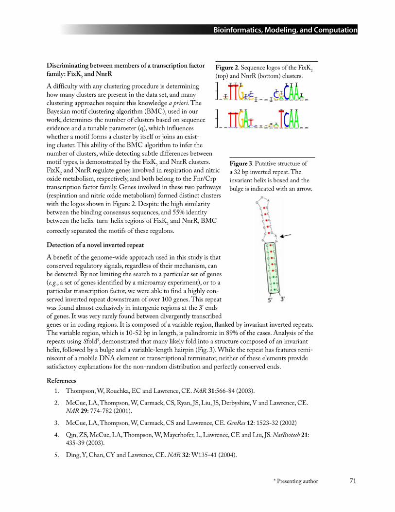
Bioinformatics, Modeling, and Computation
71* Presenting author
Discriminating between members of a transcription factor family: FixK2 and NnrR
A difficulty with any clustering procedure is determining how many clusters are present in the data set, and many clustering approaches require this knowledge a priori. The Bayesian motif clustering algorithm (BMC), used in our work, determines the number of clusters based on sequence evidence and a tunable parameter (q), which influences whether a motif forms a cluster by itself or joins an exist-ing cluster. This ability of the BMC algorithm to infer the number of clusters, while detecting subtle differences between motif types, is demonstrated by the FixK2 and NnrR clusters. FixK2 and NnrR regulate genes involved in respiration and nitric oxide metabolism, respectively, and both belong to the Fnr/Crp transcription factor family. Genes involved in these two pathways (respiration and nitric oxide metabolism) formed distinct clusters with the logos shown in Figure 2. Despite the high similarity between the binding consensus sequences, and 55% identity between the helix-turn-helix regions of FixK2 and NnrR, BMC correctly separated the motifs of these regulons.
Detection of a novel inverted repeat
A benefit of the genome-wide approach used in this study is that conserved regulatory signals, regardless of their mechanism, can be detected. By not limiting the search to a particular set of genes (e.g., a set of genes identified by a microarray experiment), or to a particular transcription factor, we were able to find a highly con-served inverted repeat downstream of over 100 genes. This repeat was found almost exclusively in intergenic regions at the 3' ends of genes. It was very rarely found between divergently transcribed genes or in coding regions. It is composed of a variable region, flanked by invariant inverted repeats. The variable region, which is 10-52 bp in length, is palindromic in 89% of the cases. Analysis of the repeats using Sfold5, demonstrated that many likely fold into a structure composed of an invariant helix, followed by a bulge and a variable-length hairpin (Fig. 3). While the repeat has features remi-niscent of a mobile DNA element or transcriptional terminator, neither of these elements provide satisfactory explanations for the non-random distribution and perfectly conserved ends.
ReferencesThompson, W, Rouchka, EC and Lawrence, CE. NAR 31:566-84 (2003).
McCue, LA, Thompson, W, Carmack, CS, Ryan, JS, Liu, JS, Derbyshire, V and Lawrence, CE. NAR 29: 774-782 (2001).
McCue, LA, Thompson, W, Carmack, CS and Lawrence, CE. GenRes 12: 1523-32 (2002)
Qin, ZS, McCue, LA, Thompson, W, Mayerhofer, L, Lawrence, CE and Liu, JS. NatBiotech 21: 435-39 (2003).
Ding, Y, Chan, CY and Lawrence, CE. NAR 32: W135-41 (2004).
1.
2.
3.
4.
5.
Figure 2. Sequence logos of the FixK2 (top) and NnrR (bottom) clusters.
Figure 3. Putative structure of a 32 bp inverted repeat. The invariant helix is boxed and the bulge is indicated with an arrow.
Bioinformatics, Modeling, and Computation
72 * Presenting author
52 Exploring Evolutionary Space
Timothy G. Lilburn1* ([email protected]), Yun Bai2, Yuan Zhang2, James R. Cole2, and George M. Garrity2
1American Type Culture Collection, Manassas, VA and 2Michigan State University, East Lansing, MI
The use of principal components analysis (PCA) to visualize the evolutionary relationships among thousands of sequences was developed by us as a tool for aiding in the definition of higher-level prokaryotic taxonomy. It not only helped define higher taxa by revealing naturally occurring clusters within the data, but also proved invaluable in highlighting errors in classification and annotation of sequences. Such PCA projections can be viewed as maps of evolutionary space for the sequences and, by extension, the organisms (and genomes) from which the sequences are obtained. Maps based on SSU rRNA sequences show large gaps between some phylogenetic groups. Presumably, this white space is due to the constraints on the evolution of these molecules that arise from their functional requirements. Sequences that might appear there either simply cannot occur in nature or belong to extinct species. Although PCA and other projection techniques can provide a reasonable approxima-tion of the topology hidden within a dataset, some distortion is inevitable and can be attributed to methodological biases and biases that may exist within the data. Previously, we had demonstrated that we could improve the accuracy of projections for a test case having a known topology and coordinate system by using a set of uniformly distributed external benchmarks. However, neither the true topology nor the coordinate system of the prokaryotic evolutionary space has been defined. Therefore, to understand the distortion, we would need to first define the limits of this space. In this study, we examine the use of a limited number (n=179) of internal reference points (benchmarks) on the transformation of the evolutionary distance data into the new coordinate system defined by PCA. We look at ways of making our maps of evolutionary space more accurate and explore why the white space exists. Methods explored include the generation of synthetic polychimeras, in silico ran-dom mutation, and complementation of a set of 179 proposed benchmark sequences. Our results are presented as a set of PCA plots that are evaluated in terms of their resolution and their concordance with the current taxonomy and with each other.
53 PhyloScan: a New Tool for Identifying Statistically Significant Transcription Factor Binding Sites by Combining Cross-Species Evidence
Lee A. Newberg1,2*, C. Steven Carmack1, Lee Ann McCue1 ([email protected]), and Charles E. Lawrence3
1Wadsworth Center, Albany, NY; 2Rensselaer Polytechnic Institute, Troy, NY; and 3Brown University, Providence, RI
If there are known transcription factor binding sites (TFBSs) for a particular transcription factor (TF), then it is possible to construct a motif model or position weight matrix with which to scan a
Bioinformatics, Modeling, and Computation
73* Presenting author
sequence database for additional sites, thereby predicting a regulon. However, scanning a genome for additional TFBSs typically results in finding few statistically significant sites. Specifically, the statistical significance of a sequence match (p-value) to a motif can be assessed by comparison with the probability of observing a match with a score as good or better in a randomly generated search space of identical size and nucleotide composition -- the smaller the p-value the greater the evidence that the match is not due to chance alone. Staden [1] presented an efficient method that exactly calculates this probability, and Neuwald et al. [2] described an implementation of this method. In practice, when scanning a genome or the promoter regions of a genome, it is frequently difficult to identify (below a chosen level of statistical significance) even the known TFBSs that were used in the construction of the motif, to say nothing of additional novel sites for that TF. Essentially, given the statistical nature of this approach, only a relatively small number of TFBSs will be identified that could possibly be considered significant (low sensitivity, high specificity).
With the goal of increasing the statistical power of scanning a genome sequence database with a regulatory motif, we have developed a scanning algorithm, PhyloScan, that combines evidence from matching sites found in orthologous data from several related species. Specifically, we have extended Staden’s method [1] to allow scanning of orthologous sequence data that is either multiply-aligned, unaligned or a combination thereof (aligned and unaligned). PhyloScan statistically accounts for the phylogenetic dependence of the species contributing aligned data and returns a p-value for the sequence match; importantly, the statistical significance is calculated directly, without employing training sets.
To evaluate this method we chose the Escherichia coli Crp and PurR motifs and gathered genome sequence data for several gamma-proteobacteria. Among the species chosen for this study (E. coli, Salmonella enterica Typhi, Yersinia pestis, Haemophilus influenzae, Vibrio cholerae, Shewanella oneidensis, and Pseudomonas aeruginosa), only E. coli and S. typhi exhibit extensive homology in the promoter regions [3]. Thus we aligned orthologous intergenic regions for these two species, and combine statistical evidence from scanning the aligned E. coli and S. typhi data with statistical evidence from scanning unaligned orthologous intergenic regions from the remaining five more distantly related species. This method enhances the identification of TFBSs in E. coli by several-fold over scanning the set of E. coli intergenic regions alone.
Staden, R. (1989) Methods for calculating the probabilities of finding patterns in sequences. Com-put Appl Biosci 5:89-96.
Neuwald, A. F., Liu, J. S. and Lawrence, C. E. (1995) Gibbs motif sampling: Detection of bacterial outer membrane protein repeats. Protein Sci 4:1618-32.
McCue, L. A., Thompson, W., Carmack, C. S. and Lawrence, C. E. (2002) Factors influencing the identification of transcription factor binding sites by cross-species comparison. Genome Res 12:1523-32.
1.
2.
3.
Bioinformatics, Modeling, and Computation
74 * Presenting author
54 Predicting Protein Interactions via Docking Mesh Evaluator
Roummel F. Marcia1, Susan D. Lindsey2, Erick A. Butzlaff1, and Julie C. Mitchell1* ([email protected])1University of Wisconsin, Madison, WI and 2University of California, San Diego, CA
Introduction
The Docking Mesh Evaluator (DoME) is a software package for protein docking and energy evalu-ation. It uses fast energy evaluation methods and optimization algorithms to predict the docked con-figurations of proteins with DNA, ligands, and other macromolecules. A fully parallelized package, DoME can run on supercomputers, clusters, and linked independent workstations.
Description
Previously, DoME’s energy model was based on solvent effects defined implicitly using adaptive mesh solutions to the Poisson-Boltzmann equation and van der Waals energy terms. While this earlier version achieved moderate success [1], more accurate results were obtained by incorporating hydrogen bond and solvation energy terms. The hydrogen bond term uses the spatial relationship between a potential acceptor atom and a donor-hydrogen pair both to determine the existence of a hydrogen bond and to compute the bond’s potential energy. The solvation term uses a modification to the method of Zhang et al. [2] to estimate the effective atomic contact energy of a complex in water. In addition, the energy function employs weighting and switching parameters to measure the individual contribution of each energy term to the overall interaction.
Global optimization methods were developed to determine the lowest function value of this energy model. In particular, the General Convex Quadratic Approximation [3] constructs a sequence of convex underestimators to a collection of local minima to predict possible areas of low energies. Yukawa potentials are used as analytic solutions to the linearized Poisson-Boltzmann equations so that gradient-based local optimization can be performed. Initial scanning of the energy landscape for favorable configurations as initial seeds for underestimation improves algorithm performance. This coupled use of scanning and optimizing is more effective in determining points of low energy values than scanning or optimizing alone [4].
We present results from the standard benchmarking test set of Chen et al. [5] for testing protein-protein docking algorithms. We consider both bound and unbound crystalline structures for deter-mining proper docked configurations. We also highlight which energy terms are significant in each test case. (Of the 59 the benchmarking test set contains, 22 are enzyme-inhibitor complexes, 19 are antibody-antigen complexes, 11 are various complexes, and 7 are difficult test cases whose solutions have considerable structural changes.)
ReferencesR.F. Marcia, J.C. Mitchell, and J.B. Rosen, Iterative convex quadratic approximation for global optimization in protein docking, Comput. Optim. Appl., Accepted for publication.
C. Zhang, G. Vasmatzis, J.L. Cornette, C. DeLisi, Determination of atomic desolvation energies from the structures of crystallized proteins, J. Mol. Biol, 1997 Apr. 4; 267(3); 707-26.
1.
2.
Bioinformatics, Modeling, and Computation
75* Presenting author
J.B. Rosen and R.F. Marcia, Convex quadratic approximation, Comput. Optim. Appl., 28, pp.173-184, 2004.
J.C. Mitchell, J.B. Rosen, A.T. Phillips, and L.F. Ten Eyck, Coupled optimization in protein dock-ing, in Proceedings of the Third Annual International Conference on Computational Molecular Biology, ACM Press, 1999, pp. 280-284.
R. Chen, J. Mintseris, J. Janin, and Z. Weng, A protein-protein docking benchmark, Prot. Struct. Fun. Gen., 52, pp. 88-91, 2003.
55 UC Merced Center for Computational Biology
Michael Colvin1* ([email protected]), Arnold Kim1, and Felice Lightstone2
1University of California, Merced, CA and 2Lawrence Livermore National Laboratory, Livermore, CA
We are establishing a Center for Computational Biology (CCB) at the newest campus of the Uni-versity of California. The CCB will sponsor multidisciplinary scientific projects in which biological understanding is guided by computational modeling. The center will also facilitate the development and dissemination of undergraduate and graduate course materials based on the latest research in computational biology. The Center is starting a number of activities that aim to recast biology as an information science:
Host multidisciplinary research projects in computational and mathematical biology that will provide a rich environment for graduate and undergraduate research.
Develop new mathematical and computational methods that are widely applicable to predictive modeling in the life sciences.
Develop and disseminate computational biology course materials that translate new research results into educational resources.
Extend the successes in achieving these objectives to other universities and “university-feeder” institutions such as community colleges.
This project is a multi-institutional collaboration including the new University of California campus at Merced, Rice University, Rensselaer Polytechnic Institute, and Lawrence Livermore National Laboratory, as well as individual collaborators at other sites. The CCB will foster a number of research projects that emphasize the role of predictive simulations in guiding biological understand-ing by funding graduate students and post-doctoral fellows with backgrounds in the mathematical and computational sciences to work on collaborative biology projects. Additionally, the center will work to translate this research into educational materials. UC Merced, as the first U.S. research university of the 21st century, offers many advantages for this new center, including an ideal venue for developing and implementing new courses and degree programs, a highly multidisciplinary faculty and organizational structure, and a strong commitment to educational outreach to diverse and under-represented groups. New computational biology courses will be used and assessed in the new UC Merced Biological Sciences major that will be accepting freshman and junior transfers in Fall 2005. Graduate courses will be implemented in the Quantitative Systems Biology Graduate Group that began accepting graduate students in Fall 2004. All course materials will be released under an open
3.
4.
5.
1.
2.
3.
4.
Bioinformatics, Modeling, and Computation
76 * Presenting author
public license using the Connexions courseware system developed at Rice University. We anticipate that this new biology curriculum will be effective in attracting students to biology who have an interest and aptitude in mathematics and computational sciences, as well as broaden the horizons of students expecting a traditional biology program. The electronic, modular course materials produced by the center will facilitate linkages to feeder schools at the state university, community college, and high school levels.
The long-term impact of the CCB will be to help train a new generation of biologists who bridge the gap between the computational and life sciences and to implement a new biology curriculum that can both influence and be adopted by other universities. Such scientists will be critical to the success of new approaches to biology, exemplified by the DOE Genomics:GTL program in which comprehensive datasets will be assembled with the goal of enabling predictive modeling of the behavior of microbes and microbial communities, as well as the biological components of life, such as multiprotein machines.
56 Biomic Approach to Predictive Cell Modeling
P. J. Ortoleva* ([email protected]), L. Ensman, J. Fan, K. Hubbard, A. Sayyed-Ahmad, F. Stanley, K. Tuncay, and K. Varala
Indiana University, Bloomington, IN
Replication, transcription, translation and metabolism, as well as physiological dynamics, are all strongly coupled and must be accounted for if predictive cell modeling is to be attained. Key barriers to doing so are that many processes are not yet understood in detail, and that the phenomenological parameters in a kinetic cell model are not yet calibrated. In this presentation we show how multiplex data (notably cDNA microarray time series) and incomplete cell models can be integrated via infor-mation theory to overcome these difficulties.
A kinetic cell model is expressed as (1)
where Ψ is a set of descriptive variables (RNA populations, metabolite, concentrations, etc.), Λ is a set of model parameters and Φ is a set of cell descriptive variables for which we do not have govern-ing equations (e.g. concentrations of species for which reactions have not yet been delineated). The problem is that (1) we cannot run such an incomplete model as the timecourse Φ(t) is required; and (2) therefore we cannot calibrate the model parameters Λ.
To overcome these difficulties, in our procedure we use information theory to construct the probabil-ity ρ that is a function of Λ and a functional of the timecourse Φ(t). We use available data (notably cDNA microarray, proteomics, NMR) in steady-state or time series to construct ρ. With this we seek the most probable values of Λ,Φ(t) by solving , the latter being a func-tional differential equation for the timecourse Φ(t). Regularization is also used to insure that there are no unphysically short timescale effects in Φ(t) created by the sparseness of, or noise in, the data.
The above methodology has been implemented for the case of transcription/translation modeling integrated with cDNA microarray time series analysis. The input of our software is microarray time series and a putative transcription control network (the latter being incomplete and possibly error-
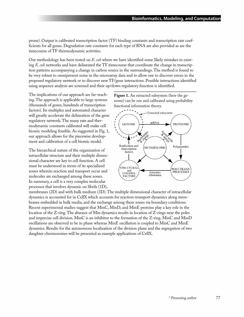
Bioinformatics, Modeling, and Computation
77* Presenting author
prone). Output is calibrated transcription factor (TF) binding constants and transcription rate coef-ficients for all genes. Degradation rate constants for each type of RNA are also provided as are the timecourse of TF thermodynamic activities.
Our methodology has been tested on E. coli where we have identified some likely mistakes in exist-ing E. coli networks and have delineated the TF timecourse that coordinate the change in transcrip-tion patterns accompanying a change in carbon source in the surroundings. The method is found to be very robust to omnipresent noise in the microarray data and to allow one to discover errors in the proposed regulatory network or to discover new TF/gene interactions. Possible interactions identified using sequence analysis are screened and their up/down regulatory function is identified.
The implications of our approach are far-reach-ing. The approach is applicable to large systems (thousands of genes, hundreds of transcription factors). Its multiplex and automated character will greatly accelerate the delineation of the gene regulatory network. The many rate and ther-modynamic constants calibrated will make cell biomic modeling feasible. As suggested in Fig. 1, our approach allows for the piecewise develop-ment and calibration of a cell biomic model.
The hierarchical nature of the organization of intracellular structure and their multiple dimen-sional character are key to cell function. A cell must be understood in terms of its specialized zones wherein reaction and transport occur and molecules are exchanged among these zones. In summary, a cell is a very complex molecular processor that involves dynamic on fibrils (1D), membranes (2D) and with bulk medium (3D). The multiple dimensional character of intracellular dynamics is accounted for in CellX which accounts for reaction-transport dynamics along mem-branes embedded in bulk media, and the exchange among these zones via boundary conditions. Recent experimental studies suggest that MinC, MinD, and MinE proteins play a key role in the location of the Z-ring. The absence of Min dynamics results in location of Z-rings near the poles and imprecise cell division. MinC is an inhibitor to the formation of the Z-ring. MinC and MinD oscillations are observed to be in phase whereas MinE oscillation is coupled to MinC and MinE dynamics. Results for the autonomous localization of the division plane and the segregation of two daughter chromosomes will be presented as example applications of CellX.
Figure 1. An extracted subsystem (here the ge-nome) can be run and calibrated using probability functional information theory.
Bioinformatics, Modeling, and Computation
78 * Presenting author
57 The BioWarehouse System for Integration of Bioinformatics Databases
Tom Lee, Valerie Wagner, Yannick Pouliot, and Peter D. Karp* ([email protected])
SRI International, Menlo Park, CA
BioWarehouse [1] is an open-source toolkit for constructing bioinformatics database (DB) ware-houses. It allows different users to integrate collections of DBs relevant to the problem at hand. BioWarehouse can integrate multiple public bioinformatics DBs into a common relational DB management system, facilitating a variety of DB integration tasks including comparative analysis and data mining. All data are loaded into a common schema to permit querying within a unified representation.
BioWarehouse currently supports the integration of Swiss-Prot, TrEMBL, ENZYME, KEGG, BioCyc, NCBI Taxonomy, CMR, and the microbial subset of Genbank. Loader tools implemented in the C and Java languages parse and load the preceding DBs into Oracle or MySQL instances of BioWarehouse.
The presentation will provide an overview of BioWarehouse goals, architecture, and implementation. The BioWarehouse schema supports the following bioinformatics datatypes: chemical compounds, biochemical reactions, metabolic pathways, proteins, genes, nucleic acid sequences, features on pro-tein and nucleic-acid sequences, organism taxonomies, and controlled vocabularies.
BioWarehouse is in use by several bioinformatics projects. An SRI project is developing algorithms for predicting which genes within a sequenced genome code for missing enzymes within metabolic pathways predicted for that genome [2]. BioWarehouse fills several roles within that project: it is used to construct a complete and nonredundant dataset of sequenced enzymes by combining pro-tein sequences from the UniProt and PIR DBs, and by removing from the resulting dataset those sequences that share a specified level of sequence similarity. Our current research involves extending the pathway hole filling algorithm with information from genome-context methods such as phyloge-netic signatures, which are obtained from BioWarehouse thanks to the large all-against-all BLAST results stored within CMR. Another SRI project is comparing the data content of the EcoCyc and KEGG DBs using BioWarehouse to access the KEGG data in a computable form.
BioWarehouse is supported by the Department of Energy and by DARPA through the DARPA BioSPICE program for biological simulation.
BioWarehouse Home Page http://bioinformatics.ai.sri.com/biowarehouse/
Green, M.L. and Karp, P.D., “A Bayesian method for identifying missing enzymes in predicted metabolic pathway databases,” BMC Bioinformatics 5(1):76 2004 http://www.biomedcentral.com/1471-2105/5/76.
1.
2.
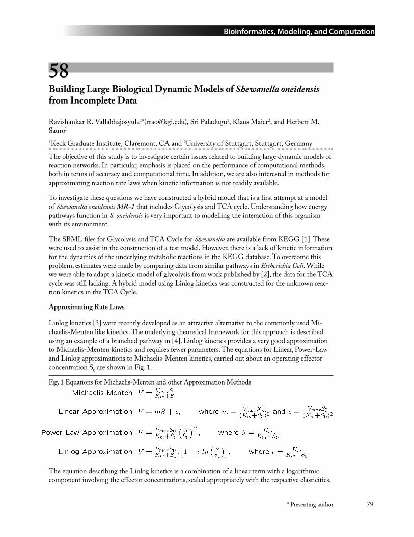
Bioinformatics, Modeling, and Computation
79* Presenting author
58 Building Large Biological Dynamic Models of Shewanella oneidensis from Incomplete Data
Ravishankar R. Vallabhajosyula1*([email protected]), Sri Paladugu1, Klaus Maier2, and Herbert M. Sauro1
1Keck Graduate Institute, Claremont, CA and 2University of Stuttgart, Stuttgart, Germany
The objective of this study is to investigate certain issues related to building large dynamic models of reaction networks. In particular, emphasis is placed on the performance of computational methods, both in terms of accuracy and computational time. In addition, we are also interested in methods for approximating reaction rate laws when kinetic information is not readily available.
To investigate these questions we have constructed a hybrid model that is a first attempt at a model of Shewanella oneidensis MR-1 that includes Glycolysis and TCA cycle. Understanding how energy pathways function in S. oneidensis is very important to modelling the interaction of this organism with its environment.
The SBML files for Glycolysis and TCA Cycle for Shewanella are available from KEGG [1]. These were used to assist in the construction of a test model. However, there is a lack of kinetic information for the dynamics of the underlying metabolic reactions in the KEGG database. To overcome this problem, estimates were made by comparing data from similar pathways in Escherichia Coli. While we were able to adapt a kinetic model of glycolysis from work published by [2], the data for the TCA cycle was still lacking. A hybrid model using Linlog kinetics was constructed for the unknown reac-tion kinetics in the TCA Cycle.
Approximating Rate Laws
Linlog kinetics [3] were recently developed as an attractive alternative to the commonly used Mi-chaelis-Menten like kinetics. The underlying theoretical framework for this approach is described using an example of a branched pathway in [4]. Linlog kinetics provides a very good approximation to Michaelis-Menten kinetics and requires fewer parameters. The equations for Linear, Power-Law and Linlog approximations to Michaelis-Menten kinetics, carried out about an operating effector concentration S0 are shown in Fig. 1.
Fig. 1 Equations for Michaelis-Menten and other Approximation Methods
The equation describing the Linlog kinetics is a combination of a linear term with a logarithmic component involving the effector concentrations, scaled appropriately with the respective elasticities.
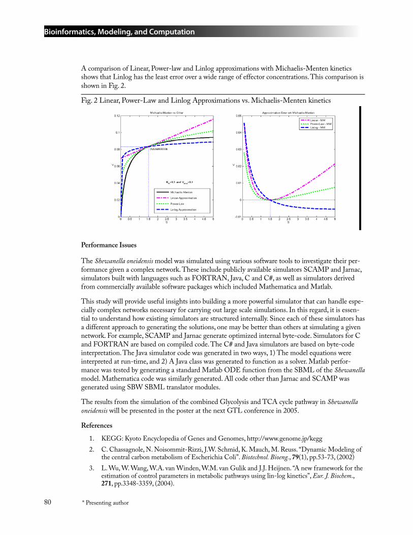
Bioinformatics, Modeling, and Computation
80 * Presenting author
A comparison of Linear, Power-law and Linlog approximations with Michaelis-Menten kinetics shows that Linlog has the least error over a wide range of effector concentrations. This comparison is shown in Fig. 2.
Fig. 2 Linear, Power-Law and Linlog Approximations vs. Michaelis-Menten kinetics
Performance Issues
The Shewanella oneidensis model was simulated using various software tools to investigate their per-formance given a complex network. These include publicly available simulators SCAMP and Jarnac, simulators built with languages such as FORTRAN, Java, C and C#, as well as simulators derived from commercially available software packages which included Mathematica and Matlab.
This study will provide useful insights into building a more powerful simulator that can handle espe-cially complex networks necessary for carrying out large scale simulations. In this regard, it is essen-tial to understand how existing simulators are structured internally. Since each of these simulators has a different approach to generating the solutions, one may be better than others at simulating a given network. For example, SCAMP and Jarnac generate optimized internal byte-code. Simulators for C and FORTRAN are based on compiled code. The C# and Java simulators are based on byte-code interpretation. The Java simulator code was generated in two ways, 1) The model equations were interpreted at run-time, and 2) A Java class was generated to function as a solver. Matlab perfor-mance was tested by generating a standard Matlab ODE function from the SBML of the Shewanella model. Mathematica code was similarly generated. All code other than Jarnac and SCAMP was generated using SBW SBML translator modules.
The results from the simulation of the combined Glycolysis and TCA cycle pathway in Shewanella oneidensis will be presented in the poster at the next GTL conference in 2005.
References
KEGG: Kyoto Encyclopedia of Genes and Genomes, http://www.genome.jp/kegg C. Chassagnole, N. Noisommit-Rizzi, J.W. Schmid, K. Mauch, M. Reuss. “Dynamic Modeling of the central carbon metabolism of Escherichia Coli”. Biotechnol. Bioeng., 79(1), pp.53-73, (2002)L. Wu, W. Wang, W.A. van Winden, W.M. van Gulik and J.J. Heijnen. “A new framework for the estimation of control parameters in metabolic pathways using lin-log kinetics”, Eur. J. Biochem., 271, pp.3348-3359, (2004).
1.2.
3.
Bioinformatics, Modeling, and Computation
81* Presenting author
D. Visser, J.J. Heijnen. “Dynamic Simulation and metabolic re-design of a branched pathway using Linlog kinetics”, Metabolic Engineering. 5, pp.164-176, (2003).
59 A Bayesian Method for Identifying Missing Enzymes in Predicted Metabolic Pathway Databases
Michelle L. Green* ([email protected]) and Peter D. Karp
SRI International, Menlo Park, CA
The PathoLogic program constructs Pathway/Genome databases by using a genome’s annotation to predict the set of metabolic pathways present in an organism. PathoLogic determines the set of reactions composing those pathways from the enzymes annotated in the organism’s genome. Many enzymes in a genome may be missed during the initial annotation effort or may be assigned a non-specific function (e.g., “thiolase family protein”). These missing or incomplete annotations can result in pathway holes. Pathway holes occur when a genome appears to lack the enzymes needed to catalyze reactions in a pathway. If a protein has not been assigned a specific function during the annotation process, any reaction catalyzed by that protein will appear as a missing enzyme or pathway hole in a Pathway/Genome database.
We have developed a method [1] that efficiently combines homology and pathway-based evidence using Bayesian methods to identify candidates for filling pathway holes in Pathway/Genome data-bases. Our program, which is now part of the Pathway Tools software, identifies potential candidate sequences for pathway holes, and combines data from multiple, heterogeneous sources to assess the likelihood that a candidate has the required function. By considering not only evidence from homology searches, but also genomic and functional context (e.g., are there functionally-related genes nearby in the genome?), our algorithm emulates the manual sequence annotation process to determine the posterior belief that a candidate has the required function. The method can be applied across an entire metabolic pathway network and is generally applicable to any pathway database. We achieved 71% precision at a probability threshold of 0.9 during cross-validation using known reac-tions in computationally-predicted pathway/genome databases.
After applying our method to 255 pathway holes in 99 pathways from the CauloCyc database, the predictions from this program completed fourteen additional pathways. The program made puta-tive assignments to 53 pathway holes, including annotation of 2 sequences of previously unknown function. The newly completed pathways include “fatty acid oxidation pathway”, “oxidative branch of the pentose phosphate pathway”, “peptidoglycan biosynthesis”, “pyridine nucleotide biosynthesis”, “pantothenate and coenzyme A biosynthesis”, “de novo biosynthesis of pyrimidine ribonucleotides”, “de novo biosynthesis of purine nucleotides II”, “histidine biosynthesis I”, “tyrosine biosynthesis I”, “phenylalanine biosynthesis II”, and “alanine biosynthesis I”.
Green, M.L. and Karp, P.D., “A Bayesian method for identifying missing enzymes in predicted metabolic pathway databases,” BMC Bioinformatics, 5:76 2004. http://www.biomedcentral.com/1471-2105/5/76.
4.
1.
Bioinformatics, Modeling, and Computation
82 * Presenting author
60 Does EcoCyc or KEGG Provide a Preferable Gold Standard for Training and Evaluation of Genome-Context Methods?
Peter D. Karp* ([email protected]) and Michelle L. Green
SRI International, Menlo Park, CA
Motivations: Genome-context methods such as phylogenetic profiles infer functional associations between genes and constitute a new approach to prediction of gene function. Most past developers of genome-context methods have trained and validated their algorithms against metabolic pathway DBs: primarily KEGG [1] and EcoCyc [2]. This work addresses the question of which of these two DBs is the optimal resource for training and evaluation of genome-context methods. Our hypothesis is that EcoCyc is the preferable resource to use because: (a) KEGG pathway maps con-tain many false-positive functional associations because KEGG maps tend to be much larger than EcoCyc pathways, and therefore contain genes from many different biological pathways, (b) KEGG pathways are less accurate because they are computationally predicted whereas EcoCyc pathways are curated from the biomedical literature, and (c) EcoCyc contains other types of functional relation-ships besides the metabolic and two-component signal transduction pathways that KEGG contains, such as descriptions of regulatory relationships between transcription factors and other genes.
Method: We evaluated this hypothesis by randomly choosing pairs of genes from the same KEGG and EcoCyc pathways, and counting the frequency with which those gene pairs show chromosomal adjacency, or similar phylogenetic profiles, since these basic methods for predicting functional as-sociations are shown to have utility by both EcoCyc and KEGG.
Results: The hypothesis is validated by the following results. Two genes chosen at random from the same EcoCyc pathway were 4.7 times more likely to be adjacent on the chromosome than two genes chosen at random from the same KEGG map. Furthermore, gene pairs chosen from the same KEGG map that are not in the same EcoCyc pathway are even less likely to be chromosomally adjacent or to exhibit similar phylogenetic profiles. Similar results were obtained for the BioCyc and KEGG datasets for seven other organisms. In addition, two genes chosen at random from the same EcoCyc pathway were 3.0 times more likely to have similar phylogenetic profiles than two genes chosen at random from the same E. coli KEGG map. In addition, we find that transcription factors and the genes that they regulate show significant chromosomal adjacency and similar phylogenetic profiles.
Summary: EcoCyc and the BioCyc DBs for other organisms are preferable resources for training and evaluation of genome-context methods because their pathways are more biologically meaningful, and because they contain a wider range of biological functional associations, such as those between transcription factors and the genes they regulate, thus allowing genome-context methods to recog-nize a wider set of biological relationships.
M. Kanehisa, S. Goto, S. Kawashima and A. Nakaya, “The KEGG databases at GenomeNet,” Nucleic Acids Research 40:42-6 2002.
I.M. Keseler, J. Collado-Vides, S. Gama-Castro, J. Ingraham, S. Paley, I.T. Paulsen, M. Peralta-Gil, and P.D. Karp, EcoCyc: a comprehensive database resource for E. coli, Nucleic Acids Research, Data-base Issue, 2005 (in press).
1.
2.
Bioinformatics, Modeling, and Computation
83* Presenting author
61 Towards a Physics and Systems Understanding of Ion Transport in Prokaryotes
Shreedhar Natarajan1, Asba Tasneem*1, Sameer Varma1, Lakshminarayan Iyer2, L. Aravind2, and Eric Jakobsson*2 ([email protected])1University of Illinois, Urbana, IL and 2National Institutes of Health, Bethesda, MD
Ion transport mechanisms play three fundamental roles in biological systems: 1) generation and sens-ing of electrochemical signals, 2) generation of osmotic force for regulating water flow, and 3) energy transduction. It is useful to study these functions in an integrated manner because: 1) Ion trans-porters as they perform all three functions pose similar issues in understanding the physical bases of those functions, and 2) it is common for the same transporter to be critical for more than one function. Indeed, it is universally true that transporters which are critical for one function will affect the functioning of networks of transporters critical to other functions, because every transporter in a membrane affects osmotic, chemical, and electrical driving forces for every other transporter in the same membrane.
In this paper we report on our efforts along three lines: 1) By bioinformatics means we have discovered homologues of chemo-sensitive postsynaptic ion channels in prokaryotes, including in Synechococcus, which is the organism whose GTL project we are associated with. This discovery may point to a previously unknown molecular mechanism for electrochemical signaling between prokaryotes, and may also facilitate determination of structures for the ligand-gated channel fam-ily of gene proteins. 2) We have developed improved methods for assigning protonation states of electrically interacting titratable residues in the lumen of bacterial porins. This is critical in order to make realistic models for ion permeation through these channels. 3) We are adapting phylogenetic profiling methods to infer transport and regulatory networks that govern ion and water homeostasis in prokaryotes.
Supported by grant # 0235792 from the National Science Foundation, the U.S. Department of Energy’s Genomics:GTL program (www.doegenomestolife.org) under project, “Carbon Sequestration in Synechococcus Sp.: From Molecu-lar Machines to Hierarchical Modeling,” (www.genomes-to-life.org), and the intramural program of the National Center for Biotechnology Information.
Bioinformatics, Modeling, and Computation
84 * Presenting author
62 OptStrain: A Computational Framework for Redesign Microbial Production Systems
Priti Pharkya and Costas D. Maranas* ([email protected])
Pennsylvania State University, University Park, PA
In this talk, we will discuss the hierarchical computational framework OptStrain aimed at guiding pathways modifications, through reaction additions and deletions, of microbial networks for the overproduction of targeted compounds. A comprehensive database of biotransformations, referred to as the Universal database (with over 5,700 reactions), is compiled and regularly updated by downloading and curating reactions from multiple biopathway database sources. Combinatorial optimization is then employed to elucidate the set(s) of non-native functionalities, extracted from this Universal database, to add to the examined production host for enabling the desired product for-mation. Subsequently, competing functionalities are identified and removed to ensure higher product yields coupled with growth. This work establishes an integrated computational framework capable of constructing stoichiometrically balanced pathways, imposing maximum product yield requirements, pinpointing the optimal substrate(s), and evaluating different microbial hosts. The range and utility of OptStrain is demonstrated by addressing a variety of product molecules and experimental verifica-tions.
63 DEMSIM: A Discrete Event Based Mechanistic Simulation Platform for Gene Expression and Regulation Dynamics
Madhukar Dasika and Costas D. Maranas* ([email protected])
Pennsylvania State University, University Park, PA
The advent of high-throughput technologies has provided a major impetus for developing sophisti-cated computational frameworks to unravel the underlying regulatory circuitry that governs the re-sponse of biological systems to environmental and genetic perturbations. A systems engineering view reveals that gene expression dynamics are governed by processes that are essentially event driven, i.e., many events have to take place in a predetermined order with uncertain start and execution times to accomplish a certain task. There are many parallels between gene expression and manufacturing systems. In analogy to a manufacturing facility which produces a certain amount of finished product at a particular time with a certain probability, the transcription process produces mRNA transcripts with probability determined by the cellular environment and availability of required components. Similarly, accumulating mRNA and protein levels in the cell are akin to product inventory held in warehouses in a manufacturing system. Motivated by the numerous parallels between these two seemingly different settings, we have used discrete event simulation, which is a powerful tool em-ployed to model and simulate supply chains and manufacturing systems, to model and simulate gene expression systems. In this talk, we will describe the DEMSIM tool that we have developed to test and hypothesize putative regulatory interactions.
Bioinformatics, Modeling, and Computation
85* Presenting author
The key feature of the DEMSIM platform is the abstracting of underlying transcription, translation and decay processes as stand-alone modules. A module is further characterized by a sequence of discrete events. For example, the transcription module is composed of the following sequence of dis-crete events: (i) binding of RNA polymerase to promoter sequence (ii) transcription elongation and (iii) transcription termination. Each module is described by a set of physical and model parameters. Physical parameters correspond to parameters which are known a priori from literature sources and are fixed within the simulation framework (e.g. length of gene, transcription rate, etc.). In contrast, model parameters are regression parameters that are fitted using the available experimental data. Subsequently, the simulation is driven by the communication between these modules in accordance with the specifics of the regulatory circuitry of the biological system being investigated. The stochas-ticity inherent to all events is captured using Monte Carlo based sampling.
The DEMSIM software implementation consists of the following three key components: (i) an event list that contains all the events that need to be executed along with their respective execution times, (ii) a global simulation clock that records the progress of simulation time as events are sequen-tially executed, and (iii) a set of state variables that characterize the system and which are updated every time an event is executed. At every time step, events corresponding to all active (non–termi-nated) modules in the system are included in the event list. Subsequently, the event list is sorted and the event having the smallest execution time is executed. The simulation clock is advanced and the execution time of all other events is updated. Such a sequential procedure prevents the occurrence of causality errors by ensuring that an event with a later time stamps is not executed before an event with an earlier time stamp. Furthermore, since the execution of certain events leads to the creation of new modules and the termination of existing ones, the number of active modules in the system is updated and new events are included in the event list. This procedure is then repeated for the dura-tion of the simulation horizon and state variables such as number of mRNA and protein molecules are recorded.
In this talk we will present the results for three biological systems of different levels of complexity that we have used to benchmark the DEMSIM platform. Simulation results for the relatively simple lac operon system of E. coli will demonstrate that the parameters embedded in the framework can indeed be trained to reproduce experimental data. Subsequently, the ability of the framework to serve as a predictive tool will be highlighted with reference to the SOS response system of E. coli. Simula-tion results will focus on DEMSIM’s ability to accurately predict the de novo response of the system to externally imposed perturbations. Finally, simulation studies for the araBAD system will dem-onstrate the framework’s ability to distinguish between different plausible regulatory mechanisms postulated to explain observed gene expression profiles. Overall, the presented results will highlight the broad applicability of the discrete-event paradigm, on which DEMSIM is based, to gene regula-tory systems.
Bioinformatics, Modeling, and Computation
86 * Presenting author
64 On the Futility of Optima in Network Inferences and What Can Be Done About It
Charles (Chip) E. Lawrence* ([email protected])
Brown University, Providence, RI
Inference of metabolic and regulatory networks has become a hot topic over the last few years, and a number of reports of efforts to infer such networks using genome scale data have appeared. One fac-tor often overlooked in these fledgling efforts stems from the large size of network solution spaces, which grow exponentially with the number of nodes in graphical representation of the network. The difficulty of drawing inferences in high dimensional setting is a well-recognized problem in statistical learning theory that is often enunciated as the “curse of dimensionality”. Most explanations of this curse don’t convey well its connection with inferences and tend to be abstract. Here I’ll use a Bayes-ian framework to directly illustrate how difficulties in network inference grows rapidly with network size, illustrate how the curse of dimensionality can so easily render even guaranteed optimal solutions unlikely, and show what can be done about it. Specifically, we will see that the curse stems from the large number of terms in the normalizing constant in Bayes rule, and that the individual terms define opportunities for limiting the curse’s ill effects. I’ll use a special class of planar networks, whose struc-ture allows for guaranteed optimal solutions and a guaranteed representative sampling, to show how optimal are often misleading, but nevertheless likely network connections and excluded connections can be strongly inferred based on samples from the solution space. I’ll also show how incomplete-ness or inaccuracy of an underlying model of a network can yield optimal solutions that are not even close to right. Background material on Bayesian inference and its connection with inferences based on optimality methods will be given as a aid for those who may not be fully familiar with statistical inference methods.
87* Presenting author
Environmental Genomics
65 Whole Community Proteomics Study of an Acid Mine Drainage Biofilm Reveals Key Roles for “Hypothetical” Proteins in a Natural Microbial Biofilm
Jill Banfield*1 ([email protected]), Rachna J. Ram1, Gene W. Tyson1, Eric Allen1, Nathan
VerBerkmoes2,3, Michael P. Thelen1, Brett J. Baker1, Manesh Shah3, Robert Hettich3, and Robert C. Blake II4
1University of California, Berkeley CA; 2University of Tennessee-Oak Ridge National Laboratory, Oak Ridge, TN; 3Oak Ridge National Laboratory, Oak Ridge, TN; and 4Xavier University of Louisiana, New Orleans, LA
We are studying relatively simple, low diversity microbial communities associated with extremely acidic, metal-rich mine drainage system to develop an understanding of adaptation, evolution, and the linkages between microbial activity and environmental geochemistry. In order to assess the genetic potential of an entire natural biofilm sample we partially reconstructed the genomes of five dominant organisms (Tyson et al. 2004). Comparative genomic analyses have revealed the structure of each population and provided insights into the processes that create and remove genome hetero-geneity. The genomes contain large blocks dominated by genes that encode hypothetical proteins. These are regions inserted in one species or strain relative to others and are inferred to be of phage origin. Phage insertion appears to be the most rapid process leading to strain heterogeneity, and pos-sibly diversification. Within strain populations, gene order is largely retained, and insertions or loss of single genes are rare events. Bacterial populations are dominated by a single clonal type. Homologous recombination is a key force shaping the genomes of archaeal populations in the system, but is rare between different species.
We have characterized the protein complement of a natural microbial community similar to that studied genomically in order to determine which genes are expressed and functionally important. By combining mass spectrometry-based “shotgun” proteomics with community genomics we confidently identified at least 1700 proteins from five dominant species. Proteins involved in protein refolding and response to oxidative stress were abundant, indicating that damage to biomolecules is a key challenge for survival. We validated more than 400 hypothetical proteins, a small subset of which are encoded within blocks of genes apparently acquired by lateral transfer. Entire operons encoding expressed, novel, lineage-specific proteins may be important for acid, metal and radical tolerance. 26% of the detected Leptospirillum group II proteins were hypothetical. An extracellular fraction was dominated by a novel protein shown to be a cytochrome central to iron oxidation and AMD formation. Sequencing of DNA encoding cytochrome regions for which peptides were not recovered revealed two amino acid substitutions. Using the strain variant sequence, 100% peptide coverage of the mature protein was achieved. Thus, an iterative genomic and proteomic approach analyses enable detailed in situ analyses of activity within natural microbial consortia.
Environmental Genomics
88 * Presenting author
66 Application of High Throughput Microcapsule Culturing to Develop a Novel Genomics Technology Platform
Martin Keller1* ([email protected]), Karsten Zengler1, Marion Walcher1, Carl Abulencia1, Denise Wyborski1, Sherman Chang1, Imke Haller1, Trevin Holland1, Fred Brockman2, Cheryl Kuske3, and Susan Barns3
1Diversa Corporation, San Diego, CA; 2Pacific Northwest National Laboratory, Richland, WA; and 3Los Alamos National Laboratory, Los Alamos, NM
Project Description
The overall goal of this project is to demonstrate the combination of high-throughput cultivation in microcapsules, which gives access to previously uncultivated microorganisms, with genome se-quencing from one to a few microcolony-containing microcapsules. This will allow direct access to physiological and genomic information from uncultured and/or difficult-to-culture microorganisms. This approach is fundamentally different than characterization and/or assembly of shotgun or BAC clones derived from community DNA or RNA. The units of analysis in our approach are living, pure microbial cultures in microcapsules, as opposed to the disassembled mixture of small fragments of genomes and cellular networks that have lost their biological context in studies using community nucleic acids. It is envisioned that the microcapsule based, high-throughput cultivation method will also be combined with Transcriptomics and Proteomics technology in the future.
Project achievements
A high-throughput cultivation method based on single cell encapsulation in microcapsules in combi-nation with flow cytometry has been applied to two soil samples. It has been demonstrated that mi-croorganisms belonging to a variety of bacterial phyla, such as Acidobacteria, Gemmatimonadetes and candidate division TM7, were growing in the microcapsules and subsequently formed microcolonies.
A fluorescent in situ hybridization (FISH) method has been optimized to selectively target and sort encapsulated microcolonies of interest using fluorescence activated cell sorting.
A whole-genome amplification technique has been employed to acquire a sufficient mass of DNA from targeted, encapsulated microcolonies (E. coli) to generate libraries for shotgun sequencing of entire genomes. The whole-genome amplification has been optimized so that DNA from as few as two cells can be amplified routinely. The genome coverage of amplified FISH-targeted microcolonies was evaluated using microarrays. Microarray data demonstrated a genome-coverage of 97% to 99% without high levels of bias towards certain genes. Subsequently, genomic libraries have been con-structed from these amplified DNA samples. Almost 300 clones of each library have been sequenced. Sequence analysis confirmed that 70% of the clones originated from E. coli DNA. Additional gene sequences were affiliated with certain beta-Proteobacteria typically found as experimental contami-nants.
This work was supported by the Office of Science (BER), U.S. Department of Energy, Grant No. DE-FG02-04ER63771.
Environmental Genomics
89* Presenting author
67 Environmental Bacterial Diversity from Communities to Genomes
Janelle R. Thompson1,2*, Silvia G. Acinas1, Vanja Klepac-Ceraj1,2, Sarah Pacocha1,2, Chanathip Pharino1, Dana E. Hunt1, Luisa A. Marcelino1, Jennifer Benoit1,2, Ramahi Sarma-Rupavtarm1, Daniel L. Distel3, and Martin F. Polz1 ([email protected])1Massachusetts Institute of Technology, Cambridge, MA; 2Woods Hole Oceanographic Institution, Woods Hole, MA; and 3New England Biolabs, Beverly, MA
We are studying the patterns of diversity among co-occurring coastal bacterioplankton from the level of the entire community to the individual genome. Our goal is to advance the understanding of structure-function relationships in microbial assemblages addressing questions including: What is the range of genomic diversity encompassed by functionally similar populations in specific environ-mental contexts? What mechanisms govern selection and diversification of natural microbial popula-tions? Using environmental 16S ribosomal RNA gene sequences (ribotypes) as a proxy for bacteria, we have shown that despite a high diversity in the environment, the majority of organisms fall into closely related clusters (<1% 16S rRNA divergence) (1). Such microdiverse sequence clusters are hypothesized to represent functionally-differentiated populations, which arise by selective sweeps (2) and persist because competitive mechanisms are too weak to purge diversity from within them (1).
To examine this hypothesis we quantitatively estimated the genomic diversity within one 16S rRNA microdiversity cluster (Vibrio splendidus). Quantitative PCR analysis (3) over an annual cycle indicated that V. splendidus was consistently present as a member of the coastal bacterioplankton community. Vibrio strains were isolated from representative months and the majority were identified as V. splendidus. Determination of sequence diversity of a universally distributed protein-coding gene (Hsp60) among all Vibrio isolates showed high heterogeneity but confirmed the monophyly of the V. splendidus strains. Still greater heterogeneity was revealed when the number of unique genotypes among strains was assayed by pulse field gel electrophoresis (PFGE), moreover, the PFGE analysis provided evidence that a large proportion of genotypes are differentiated by insertions and deletions of large genome fragments. In a set of 12 V. splendidus strains genome sizes ranged from 4.5 to 5.6 Mb with only weak correlation of genome size difference to Hsp60 sequence divergence (R = 0.37).
The high degree of heterogeneity among the V. splendidus genomes suggest that the average envi-ronmental concentration of individual genotypes is astoundingly small. To illustrate this, we divided the QPCR-based estimates of population size of V. splendidus in samples taken in Aug 03, Sept 03, and Oct 03 (1,890, 600, and 640 cells/ml, respectively) by the Chao-1 statistical estimates (4) for the number of Hsp60 alleles (125, 94 and 279) and genotypes (465, 553 and 901) in those same samples. The result suggests that unique Hsp60 alleles occurred in the monthly samples at average concentra-tions of 2 to 15 cells per ml (or at a frequency of 0.3 to 1%) while unique genotypes were present at ~10-fold lower frequency (average concentration for all samples estimated at <1 cell per ml).
The observed pattern of co-existing diversity suggests that purging of genotypes from the population is rare compared to processes introducing variation and that therefore variation persists because it is either favored (e.g., by balancing selection or resource specialization) or neutral. We present ecologi-cal considerations to suggest much of the observed diversity may in fact be neutral in an environmen-tal context. Such observations of extreme genomic heterogeneity among closely related individuals have significant implications for the assembly of genome sequences from environmental samples. In addition, if similar patterns of diversity are common to other bacterial populations caution should be exercised in interpreting the extent to which gene complements or even metabolic traits of individual
Environmental Genomics
90 * Presenting author
isolates may reflect the overall properties of populations. Indeed our results suggest that not only the gene content, but also quantitative abundance and dynamics of individual traits should be considered when evaluating the ecological significance of differences among coexisting genotypes.
References:S. G. Acinas et al., Nature 430, 551-554 (2004).
F. M. Cohan, Annu. Rev. Microbiol. 56, 457–487 (2002)
J. R. Thompson et al., Appl. Environ. Microbiol. 70, 4103-4110 (2004).
J. B. Hughes, J. J. Hellmann, T. H. Ricketts, B. J. M. Bohannan, Appl. Environ. Microbiol. 67, 4399-4406 (2001).
68 Distribution and Variation of Prochlorococcus Genotypes Across Multiple Oceanic Habitats
Adam C. Martiny* ([email protected]), P. K. Amos Tai, Anne W. Thompson, and Sallie W. Chisholm
Massachusetts Institute of Technology, Cambridge, MA
The cyanobacterium Prochlorococcus is very abundant in oligotrophic regions of the world’s oceans, constituting up to 50% of the cells in the euphotic zone. Thirty-two cultures have been isolated and based on these, a phylogenetic tree has been constructed showing the presence of 6 clades, four low light adapted and two high light adapted. That opens up two questions: (i) To what extent do these cultures represent the phylogenetic space of Prochlorococcus? (ii) Are the patterns of genotypic diver-sity within a given clade similar in field samples collected from different geographical locations?
We are using the sequence of the intergenic transcribed spacer region between the small and large subunit rRNA (ITS) as a neutral marker for genetic variation to describe the diversity of Prochlorococ-cus in field samples. We have cloned and sequenced 1200 ITS fragments from the North Pacific subtropical gyre (Hawaii Ocean Time Series), Sargasso Sea (Bermuda-Atlantic Time Series) at three depths – 25m, 80m and 160m and an upwelling region off the coast of Mexico at two depths (60m and 130m). The phylogenetic analysis showed a high frequency of sequences belonging to the 9312-clade from the 25m samples and 80m — consistent with the finding that the 9312 ‘ecotype’ numerically dominates the upper euphotic zone in many oceanic environments (Zinser et al, in prep.). A comparison using Mantel test of the microdiversity within the 9312 clade at 25m and 80m revealed that the populations were significantly different between these two depths. In addition, a significant amount of “yet to be cultured” diversity was discovered at both 80 and 160m depth including new lineages as well as sub-lineages within the 6 known clades. In a sample collected from a sub-oxic zone off the coast of Mexico, we found a group affiliated to the low light adapted 9313 ecotype, but forming an independent lineage not yet seen in other samples. A future goal is to target such new lineages, amplify their genome using the approach described by Zhang et al. (this meeting) and thereby expand our view on the Prochlorococcus physiology and evolution through comparative genome analysis..
1.
2.
3.
4.
Environmental Genomics
91* Presenting author
69 From Perturbation Analysis to the Genomic Regulatory Code: the Sea Urchin Endomesoderm GRN
Paola Oliveri*,1 ([email protected]), Pei-Yun Lee1, Takuya Minokawa2, Joel Smith1, Qiang Tu1, Meredith Howard1, David McClay3, and Eric H. Davidson1
1 California Institute of Technology, Pasadena, CA; 2 Tohoku University, Asamushi, Aomori, Japan; 3Duke University, Durham, NC
The sea urchin endomesoderm gene regulatory network (GRN) is the most comprehensively under-stood regulatory apparatus for control of spatial and temporal gene expression in any complex devel-opmental system. It contains almost 50 genes, mainly encoding regulatory proteins. It was initially constructed on the basis of spatial and temporal gene expression data, interpreted through a large scale, systematic, perturbation analysis in which expression of each gene was taken out or otherwise altered, and the effects on all other relevant genes measured quantitatively and with high sensitivity. The GRN provides the essential, overall, “transformation function” by which can be solved the causal relations between the genomic regulatory code that is hardwired into the DNA sequence, and the observed events of spatial and temporal gene expression. That is, it specifies the key cis-regulatory inputs into regulatory genes, and their key outputs terminating at other regulatory genes. Hence it is directly testable at the cis-regulatory level. In the last year, we identified by computational and experi-mental methods, and isolated, over a dozen of the central cis-regulatory nodes of the GRN. We then require that in gene transfer experiments that these genomic fragments display the same responses to the appropriate perturbations as do the endogenous genes in the whole embryo; and that when the genomic target sites for the relevant inputs are mutated, that the cis-regulatory constructs behave in the expected ways. This system wide task will be completed this year, and to date the results indicate that the perturbation analysis indicated the real encoded linkages with perhaps surprising accuracy. In addition: we have demonstrated for the first time that knowledge of the GRN can be used to reengineer the process of development; we have found ways to identify the modular “kernels” of the GRN, which consist of multiple genes recursively “wired” to one another and which are evolution-arily resistant to change; we have developed a new theory of logic processing within cis-regulatory modules, as a start on formalization of the genomic regulatory code; we have enlarged our knowledge of the GRN and are adding into it all regulatory genes encoded in the genome that are expressed in the appropriate time and place, so that it will approximate a complete regulatory treatment; and we have begun to extend GRN analysis to later processes.

Environmental Genomics
92 * Presenting author

93* Presenting author
Microbial Genomics
70 The Genome of the Ammonia Oxidizing Bacterium Nitrosomonas europaea: Iron Metabolism and Barriers to Heterotrophy
Xueming Wei, Neeraja Vajrala, Norman Hommes, Luis Sayavedra-Soto*, and Daniel Arp ([email protected])
Oregon State University, Corvallis, OR
Nitrosomonas europaea is an aerobic lithoautotrophic bacterium that uses ammonia (NH3) as its energy source (3). As a nitrifier, it is an important participant in the N cycle, which can also influence the C cycle. The genome sequence of N. europaea has been annotated and consists of approximately 2460 protein-encoding genes (1). We are continuing to use the genome sequence to explore the genetic structure and mechanisms underlying the lithoautotrophic growth style of N. europaea. Cur-rently, we are investigating its Fe requirements and its possible barriers to utilizing carbon sources different from CO2.
Because N. europaea has a relatively high content of hemes, sufficient Fe must be available in the me-dium for it to grow. The genome revealed that approximately 5% of the coding genes in N. europaea are dedicated to Fe transport and assimilation. Nonetheless, with the exception of citrate, N. europaea lacks genes for siderophore production (1). We have initiated the study on this intriguing facet by determining the Fe requirements for growth and are characterizing the expression of the putative membrane siderophore receptors.
N. europaea changes its heme composition when Fe is at a relatively low concentration. Biochemical analyses showed that cytochrome and heme contents of cells grown in Fe-limited medium were 4 fold lower than those from Fe-rich medium. Cellular Fe contents (in both membrane and soluble fractions) showed the same trend. The activity of hydroxylamine oxidoreductase was over three fold lower in cells grown in Fe-limited medium than that in full medium. The growth yields at 0.1 µM Fe and at 0.2 µM Fe were about 35% and 65% respectively of that observed at 10 µM Fe (full medium). N. europaea has the mechanisms to cope and grow under Fe limitation.
The N. europaea genome revealed that there are over 26 sets of genes that are organized similarly to the genes in a fecR/fecI system. Through similarity searches, we have identified possible TonB-depen-dent receptor genes up- or downstream of these sets. Some of these are similar to genes encoding the siderophore receptors for desferrioxamine (desferal), ferrichrome, and coprogen.
The addition of desferal in Fe-limiting medium promoted the growth of N. europaea, though with a longer lag phase, suggesting a necessary induction period of the corresponding receptor. A gene for the putative desferal outer membrane receptor was identified by similarity searches (NE1097, a foxA homologue). NE1097 was expressed at a higher level (>10 fold) in Fe-limiting, desferal-containing cultures than in Fe-sufficient cultures. The expression of NE1097 required the presence of desferal, since typical lag phases were observed when inoculants from desferal cultures were used. Several membrane proteins detected only in the cells grown in Fe-limited medium may be involved in Fe
Microbial Genomics
94 * Presenting author
transport. For example, a membrane peptide with the calculated MW of the putative desferal recep-tor was observed only in the cells grown in desferal-containing medium. Ferric citrate had an effect similar to that of desferal on N. europaea growth in Fe-limiting medium, but with a longer lag phase and a higher final cell density than that in the full medium. Ferrichrome, on the other hand, did not prolong the lag phase, yet increased total cell growth, suggesting that the genes for the ferrichrome receptors were expressed constitutively.
Consistent with the genome sequence data, no siderophores were detected in N. europaea culture filtrates under either Fe-limiting or Fe-sufficient conditions using a standard siderophore assay. We considered the possibility that citrate serves as a Fe chelator/siderophore, since N. europaea has the necessary genes to produce it. Citrate was detected (2 to 5 µM) in cell-free filtrates from both, low- and full Fe cultures. Surprisingly, cell-free filtrates from full Fe cultures had relatively higher concentrations (5 µM) of citrate than in low Fe cultures (2 to 3 µM). The role of citrate in Fe ac-quisition, if any, is yet to be determined. N. europaea apparently expresses siderophore receptors (i.e. NE1097) under low Fe conditions to scavenge Fe more efficiently. These results reinforce the notion that N. europaea uses siderophores produced by other organisms in natural habitats.
Genes encoding the putative outer membrane desferal receptor (NE1097 and NE1088, foxA homo-logues) have been cloned, insertional mutant constructs made, and mutant strains obtained through homologous recombination. Physiological and genetic characterization of these mutants is in progress.
In addition to the Fe experiments, analysis of the N. europaea genome has led to experiments probing the possible barriers to heterotrophy in N. europaea. The genome contains genes that are similar to the genes encoding fructose transport systems (PTS-type) in other bacteria. Furthermore, N. euro-paea can use fructose as the only source of carbon for growth (2). However, not all the genes required for an active PTS system are present in the genome. The inactivation of the two identified PTS genes did not affect growth on fructose or cause any other growth phenotype. Fructose may enter the cells by some other means. The role of the existing PTS genes remains unclear.
Historically, the activity of the enzyme 2-oxoglutarate dehydrogenase has not been detected in N. europaea. The lack of this activity was believed to be the cause for the obligate autotrophy of N. europaea. However, the genomic sequence reveals that the three genes necessary to encode this enzyme are present. We inactivated the first gene (odhA) in the operon of this enzyme. The mutant strain grew similarly to wild-type cells during exponential growth. However, in late stationary phase or under ammonia starvation (i.e. energy-limiting conditions), mutant strains lost viability faster and recovered more slowly upon addition of more ammonia as compared to the wild type. This suggests that 2-oxoglutarate dehydrogenase may be involved in processes occurring during the stationary growth phase of N. europaea.
A gene encoding a putative ammonia transporter (amt) is present in the genome. However, the strain with this gene inactivated showed no difference in growth to wild-type cells over a wide range of ammonium concentrations. The function of amt in N. europaea is still unknown.
We are exploring the idea that one barrier to heterotrophic growth in N. europaea may be due to a lack of transporters for alternative growth substrates. The genes encoding the enzymes to utilize glycerol as the carbon source are present, but the genes encoding a glycerol transporter are not. The heterologous expression of the gene for the glycerol permease from E. coli in N. europaea permits N. europaea to utilize glycerol as the carbon source.
Acknowledgment: This research is funded by grants from the Department of Energy (ER63149-1017305-0007065 and ER63781-1023295-0009824) and resources from the Oregon Agricultural Experiment Station.
Microbial Genomics
95* Presenting author
ReferencesChain, P., J. Lamerdin, F. Larimer, W. Regala, V. Lao, M. Land, L. Hauser, A. Hooper, M. Klotz, J. Norton, L. Sayavedra-Soto, D. Arciero, N. Hommes, M. Whittaker, and D. Arp. 2003. Complete genome sequence of the ammonia-oxidizing bacterium and obligate chemolithoautotroph Nitro-somonas europaea. J Bacteriol 185:2759-2773.
Hommes, N. G., L. A. Sayavedra-Soto, and D. J. Arp. 2003. Chemolithoorganotrophic growth of Nitrosomonas europaea on fructose. J Bacteriol 185:6809-6814.
Wood, P. M. 1986. Nitrification as a bacterial energy source, p. 39-62. In J. I. Prosser (ed.), Nitrifi-cation. Society for General Microbiology, IRL Press, Oxford.
71 Pelagibacter ubique: A Post-Genomic Investigation of Carbon Metabolism and Photochemistry in an Extraordinarily Abundant Oceanic Bacterium
Stephen J. Giovannoni1 * ([email protected]), Lisa Bibbs2, James Tripp1, Scott Givan1, Jang-Cheon Cho1, Martha D. Stapels3, Russell Desiderio1, Mercha Podar2, Kevin L. Vergin1, Mick Noordeweir2, Michael S. Rappé4, Samuel Laney1, Douglas F. Barofsky1, and Eric Mathur2 1Oregon State University, Corvallis, OR; 2 Diversa Corporation, San Diego, CA; 3Waters Corporation, Milford, MA; and 4Hawaii Institute of Marine Biology, Kaneohe, HI
The alphaproteobacterium SAR11, now known as Pelagibacter ubique, is arguably the most abundant organism in the oceans, where it accounts for approximately 25% of all microbial plankton cells. Dur-ing summer periods it may exceed 50% of the cells in the surface waters of temperate ocean gyres. Pelagibacter plays a key role in the oxidation of the oceanic dissolved organic carbon pool, which is approximately equivalent in size to the atmospheric carbon dioxide pool. The first cultured strains of Pelagibacter were isolated by high throughput methods for culturing cells by dilution into natural seawater media, and screening using a new cell array technology. Pelagibacter cultures are routinely propagated in autoclaved seawater, where they attain cell densities that are typical of native popula-tions (ca. 106 cells/ml). During the sequencing of the 1.3 million base pair genome of Pelagibacter ubique, in collaboration with Diversa Corp., it was discovered that this organism has a proteorho-dopsin (PR) gene. Liquid chromatography and tandem mass spectrometry were used to prove that the Pelagibacter PR gene is expressed in culture and that an identical protein is abundant in coastal Oregon seawater. Laser flash excitation experiments with whole, cultured cells revealed absorption transients with decay kinetics characteristic of retinylidene ion pumps, and light-dependent drops in pH provided confirmation that this PR is a light-dependent proton pump. Pelagibacter ubique is the first cultured bacterial isolate to exhibit the PR genes discovered by Bejá, Delong, and coworkers, and the only experimental choice at present for understanding how light-dependent proton pumps influence the efficiency of dissolved organic carbon (DOC) assimilation by heterotrophic bacteria in the ocean surface. The Pelagibacter genome is almost exactly the size of the genomes of the obli-gate intracellular parasites R. conorii and W. pipientis, but it appears to encode a relatively complete metabolic repertoire governed by unusually simple regulatory circuits. One objective of our current research is to predict the organic carbon sources used by Pelagibacter by metabolic reconstruction. Another major thrust of our research is the application of mass spectrometry methods to understand the regulatory responses of Pelagibacter to environmental variables, and to explore the proteome state
1.
2.
3.
Microbial Genomics
96 * Presenting author
of Pelagibacter cells in the oceans, so that they can be used as proxies to report the biological state of the system. Metabolic modeling of Pelagibacter is an attractive long range goal because it is one of the smallest and simplest cells known. Its remarkable success may be attributable to the integration and optimization of metabolic processes for efficiency at low nutrient fluxes..
72 Does the Three Dimensional Organization of the Nucleoid of the Deinococcaceae Contribute to their Ionizing Radiation Resistance?
J. M. Zimmerman and J. R. Battista* ([email protected])
Louisiana State University and A & M College, Baton Rouge, LA
Transmission electron micrographs of Deinococcus radiodurans R1 suggest that the nucleoid of this species exists as a toroidal ring, and have led to speculation that this structure facilitates the extreme radioresistance of this species. However, little direct evidence supports this contention. Since ex-treme radioresistance is characteristic of all the members of the Deinococcaceae, we hypothesize that if nucleoid morphology contributes to radioresistance, the genomic DNA of each species should form similar structures. Using epifluorescence and deconvolution microscopy, we evaluated the nucleoid morphologies of eight of the nine validly described species of Deinococcus, the radioresistant bacterium Rubrobacter radiotolerans, and the less radioresistant Thermus aquaticus, a distant relative of the deinococci. Although the nucleoids of Deinococcus murrayi, Deinococcus proteolyticus, Deinococ-cus radiophilus, and Deinococcus grandis have structures similar to D. radiodurans, the nucleoids of Deinococcus radiopugnans and Deinococcus geothermalis lack specific organization. The nucleoid of R. radiotolerans consists of multiple highly condensed spheres of DNA. Since only five of the seven recognized deinococcal species exhibit a structurally distinct nucleoid, we conclude there is no obvi-ous relationship between the three dimensional organization of genomic DNA and extreme radio-resistance. However, the genomic DNA of all extremely radioresistance species is highly condensed relative to the more radiosensitive species examined. We have examined nucleoid structure following the introduction of DNA double strand breaks and show that the shape of the nucleoid does not demonstrably change in radioresistant species even in strains incapable of repairing strand breaks, suggesting that DNA held in this tightly packed configuration contributes to the radioresistance of these bacteria.
Microbial Genomics
97* Presenting author
73 Large Scale Genomic Analysis for Understanding Hydrogen Metabolism in Chlamydomonas reinhardtii
Michael Seibert*1 ([email protected]), Arthur R. Grossman2, Maria L. Ghirardi1, and Matthew C. Posewitz3
1National Renewable Energy Laboratory, Golden, CO; 2Carnegie Institution of Washington, Stanford, CA; and 3Colorado School of Mines, Golden, CO
Many taxonomically diverse microorganisms have the ability to produce H2 anaerobically in path-ways coupled either to dark fermentation, dark CO-oxidation, light-dependent N2-fixation, or photosynthetic water oxidation. However, only certain photosynthetic organisms are able to directly couple water oxidation to the photoproduction of H2. Among these Chlamydomonas reinhardtii, a green alga, is able to convert the low potential reductant generated from water by photosynthesis into H2. A more fundamental understanding of the physiology, biochemistry and genetics of this proto-type alga might enable the future development of a sustainable system for H2 production. Recently, we and our collaborators announced the discovery of a physiological switch (sulfur deprivation), which attenuates photosystem II O2-evolution activity (Wykoff et al., 1998) and allows C. reinhardtii to metabolize O2 in sealed culture vessels (Melis et al., 2000). This produces an anaerobic environ-ment and leads to the photoproduction of volumetric amounts of H2 over a 4-day period in batch culture. Under sulfur-deprived conditions, algal cultures are subjected to a mixed metabolic state in which anaerobic fermentation, oxygenic photosynthesis and aerobic respiration co-occur. This physi-ological state provides us with a unique opportunity to further explore the gene-expression patterns and protein networks that sustain algal H2 production.
With the completion of the C. reinhardtii genome sequence as a part of the DOE Office of Science’s Genomics:GTL Program, it is now possible to thoroughly explore large-scale transcript profiles associated with H2 metabolism in this alga using gene microarrays. High-density DNA microarrays are being used to examine the ways in which WT and mutant strains of C. reinhardtii acclimate to conditions that allow for H2 production. Recently, an array with approximately 3,000 elements was used to examine sulfur deprivation responses in WT and mutant C. reinhardtii strains (Zhang et al., 2004). A new array based on specific synthetic 70 mers that represents approximately 10,000 genes has been developed at the Carnegie Institution (Stephan Eberhard and Arthur Grossman, unpub-lished); it should be ready for use by the beginning of 2005.
We have isolated several independent C. reinhardtii mutants with attenuated H2-photoproduction activity at NREL using a rapid screening technique and will compare gene expression profiles from these mutants with the WT under conditions that facilitate H2 production. One mutant lacks a functional HydEF gene (Posewitz et al., 2004), which encodes a radical SAM protein that is required to insert the metal catalytic center into the hydrogenase enzyme. The hydEF-1 mutant is the only reported C. reinhardtii strain that is unable to produce any H2 at all. The hydEF mutation specifically disrupts hydrogenase activity, but this mutant has no discernable phenotype in comparison to the WT when grown aerobically in the light. Nevertheless, the hydrogenase structural genes are induced during anaerobiosis. These data indicate that the necessary transcriptional, regulatory and signaling pathways required for hydrogenase induction remain intact in this mutant. Consequently, the genes differentially expressed in this mutant relative to the WT, should be a consequence of the mutant’s inability to photoproduce H2.
Microbial Genomics
98 * Presenting author
Another C. reinhardtii mutant, sta7-10 (Posewitz et al., 2004), is unable to accumulate intracellular starch. Interestingly, this mutant shows aberrant induction of hydrogenase-gene transcription and attenuated H2-photoproduction activity during anaerobiosis, which correlates with the redox state of its plastoquinone (PQ) pool. One reasonable hypothesis is that the redox state of the PQ pool may signal regulatory processes responsible for turning on or off specific genes involved in anaerobic fermentative pathways and H2 production.
Previous physiological studies have linked photosynthetic electron transport and fermentation to H2 production in C. reinhardtii. However, a more general knowledge of the metabolic and regulatory context that facilitates H2 production will be necessary to understand current limitations in H2-production yields. We will begin to develop a global understanding of the factors that promote H2 production during anaerobiosis by analyzing transcript profiles from WT and mutant cultures of C. reinhardtii. We will also investigate whether intracellular energy stores and/or redox carriers modu-late this activity and/or influence the expression of genes needed for H2 generation. This work will more fully elucidate the biochemical pathways utilized by C. reinhardtii during anaerobiosis and pro-vide insights into how mutants altered in normal fermentative metabolism acclimate to anaerobiosis. Genome-wide expression data will also facilitate the modeling of carbon and reductant fluxes, guid-ing future molecular and metabolic engineering approaches to improve H2 output by C. reinhardtii.
ReferencesMelis, A., Zhang, L., Forestier, M., Ghirardi, M. L. and Seibert, M. (2000). Sustained photobio-logical hydrogen gas production upon reversible inactivation of oxygen evolution in the green alga Chlamydomonas reinhardtii. Plant Physiol 122, 127-36.
Posewitz, M. C., King, P. W., Smolinski, S. L., Zhang, L., Seibert, M. and Ghirardi, M. L. (2004). Discovery of two novel radical S-adenosylmethionine proteins required for the assembly of an ac-tive [Fe] hydrogenase. J Biol Chem 279, 25711-20.
Posewitz, M. C., Smolinski, S. L., Kanakagiri, S., Melis, A., Seibert, M. and Ghirardi, M. L. (2004). Hydrogen Photoproduction Is Attenuated by Disruption of an Isoamylase Gene in Chlamydomonas reinhardtii. Plant Cell 16, 2151-63.
Wykoff, D. D., Davies, J. P., Melis, A. and Grossman, A. R. (1998). The regulation of photosyn-thetic electron transport during nutrient deprivation in Chlamydomonas reinhardtii. Plant Physiol 117, 129-39.
Zhang, Z., Shrager, J., Jain, M., Chang, C.W., Vallon, O. and Grossman, A. R. (2004). Insights into the survival of Chlamydomonas reinhardtii during sulfur starvation based on microarray analy-sis of gene expression. Eukaryot Cell.3, 1331-48.
1.
2.
3.
4.
5.
Microbial Genomics
99* Presenting author
74 Exploring the Genome and Proteome of Desulfitobacterium hafniense DCB2 for its Protein Complexes Involved in Metal Reduction and Dechlorination
James M. Tiedje1*, Sang-Hoon Kim1, Christina Harzman1, John Davis2, Brett Phinney1, Michael Ngowe1, Washington Mutatu1, William Broderick1, David DeWitt1, Joan Broderick1, and Terence L. Marsh1 ([email protected])
1Michigan State University, East Lansing, MI and 2Columbus State University, Columbus, GA
The strictly anaerobic bacterium Desulfitobacterium hafniense DCB-2 grows by pyruvate fermenta-tion and by alternate respiration using a wide range of electron acceptors including sulfur com-pounds, chlorinated compounds, and oxidized metals. This organism also grows with N2 as the sole nitrogen source. The metabolic versatility makes this organism useful for studies of the mechanism of dechlorination and metal reduction, and potentially useful in bioremediation. The sequence of the genome has been determined and seven ORFs similar to the cprA (reductive dehalogenase or RDase) gene of D. dehalogenans and four ORFs similar to nifH of nitrogenase have been detected. To identify the RDase genes induced during alternate respiration with 3-chloro-4-hydroxybenzoate (3C4HBA), 3,5-dichlorophenol (DCP), or ortho-bromophenol (o-BP), Xeotron® microarrays of the genome of DCB-2 were prepared. Competitive hybridization of cDNA prepared from cultures grown by py-ruvate fermentation and under three dehalorespiration conditions indicated that three RDase genes (designated as MENN, MFRS, and MSGV) were induced by 3C4HBA, and two genes (MSSA and VKMN) were induced by both DCP and o-BP. Also induced were genes within putative RDase operons, transporter/permease genes, and genes involved in electron transport systems. RT-PCR analysis targeting the seven cprA homologs revealed the same patterns of RDase gene expression. Northern hybridization assays targeting three RDase genes (MENN, MSSA, and VKMN) with RNAs from the four culture conditions showed a single mRNA species transcribed from MENN (1.8 kb) and VKMN (2 kb) that is long enough to contain genetic information for two linked genes, the RDase (cprA) and the adjacent docking protein (cprB) genes. However, Northern hybridization was not adequate as confirmation of the microarray results due to the cross-hybridization of probes to mRNAs from the multiple RDase genes that are phylogenetically related to varying degrees.
The physiology of heavy metal reduction under conditions where the oxidized metal was the only available electron acceptor was investigated with D. hafniense DCB-2 growing on a defined minimal freshwater media. The ability of DCB-2 to reduce Fe(III), Cu(II), U(VI), and Se(VI) were each tested separately. Bacterial growth under these metalorespiration conditions were observed for Fe(III), Cu(II), and U(VI), but not Se(VI). Reduction of Se(VI) did occur by DCB-2 when grown by pyruvate fermentation. SEM of DCB-2 morphology under fermentative growth with Se(VI) revealed that selenium is concentrated in polyps attached to the outside of the cell. The oxidation state of this selenium is unknown. Biofilm formation was observed for DCB-2 under conditions of fermentation (DCB-1 media) and respiration (ferric citrate media) when grown on either one of two different beads (Dupont and Siran™). Preliminary morphological examination under light micro-scope of planktonic cells grown in different heavy metal reduction conditions revealed considerable diversity in cellular morphologies, depending on the specific growth factors. This suggests a diverse adaptive repertoire to varying environmental conditions.
Microbial Genomics
100 * Presenting author
75 An Integrative Approach to Energy, Carbon, and Redox Metabolism in the Cyanobacterium Synechocystis sp. PCC 6803
Wim Vermaas1,* ([email protected]), Robert Roberson1, Allison van de Meene1, Bing Wang1, Sawsan Hamad1, Zhi Cai1, Julian Whitelegge2, Kym Faull2, Sveta Gerdes3, Andrei Osterman3, and Ross Overbeek3
1Arizona State University, Tempe, AZ; 2University of California, Los Angeles, CA; and 3Fellowship for the Integration of Genomes (FIG), Argonne, IL
The goal of this project is to merge knowledge from genomic, bioinformatic, proteomic, metabolic, ultrastructural and other perspectives to understand how cyanobacteria live, adapt and are regulated. This project focuses on the cyanobacterium Synechocystis sp. PCC 6803, which is spontaneously transformable and has a known genome sequence. Cyanobacteria contribute greatly to global photo-synthetic CO2 fixation and are related to the ancestors of chloroplasts.
Electron tomography and structure determinations. Cyanobacteria have a comprehensive internal membrane system, the thylakoids, where photosynthetic reactions take place. Using electron tomog-raphy, we have determined the 3-D structure of cyanobacterial cells with about 4 nm resolution in all directions (also see http://lsweb.la.asu.edu/synechocystis/). Also, we have been successful with high-resolution freeze fracture demonstrating the layered character of the thylakoid membranes and the presence of particles on the outer, cytoplasmic and thylakoid membranes that represent integral and peripheral membrane proteins. The glycocalyx of the cell wall was also exposed. Together these images further expand our 3-D insights in the structure of the cyanobacterial cell and the biogenesis of its components.
Studies on mutants that cannot synthesize chlorophyll in darkness show that such mutants do not have thylakoids in darkness, but will develop thylakoids within hours of illumination. Before il-lumination, cellular structures are observed that may be thylakoid precursors. In addition, the content of the storage compound polyhydroxyalkanoate (PHA) was determined in wild type and different mutants from 70 nm sections. Initial results were compared with the biochemical data obtained from GC/MS. Particularly in mutants that cannot respire due to the lack of terminal oxidases, polyhy-droxybutyrate accumulates, suggesting that this bioplastic serves as a storable fermentation product of this organism. High-resolution, 3-D structural investigations have also led to the discovery of new structures in cyanobacteria. For example, a fine, reticulate network of filaments in the cytoplasm was observed. The nature of these filaments is being determined.
Metabolic fluxes. Another important aspect that is investigated is the physiology of the organism in terms of its carbon fixation and utilization. The relative fluxes of the various central carbohydrate utilization pathways (glycolysis and the pentose phosphate pathway) are investigated by means of 13C-labeling in wild type and mutants impaired in either glycolysis or the pentose phosphate path-way, and grown under various conditions. 13C-glucose is rapidly taken up and converted. Upon isola-tion of metabolites at different times after 13C-glucose addition, labeled and unlabeled metabolite products are separated by LC and analyzed by MS. The resulting patterns are then used to determine metabolic fluxes through central carbon metabolism pathways. Labeling processes occur on the tim-escale of 5-40 min, with different compounds showing different labeling kinetics, demonstrating the usefulness of this approach to follow the path and rate of carbon metabolism in vivo.
Microbial Genomics
101* Presenting author
Proteome studies. Relative expression and protein turnover studies traditionally employ complete sub-stitution of stable isotopes. This approach has limitations, and we are investigating the use of labeling with 2-4% 13C to code samples for expression proteomics and turnover measurements. Altering the 13C abundance to ~2% yields a measurable effect on the peptide isotopic distribution and the inferred isotope ratio. Elevation of 13C abundance to 4% leads to extension of isotopic distribution and back-ground peaks across every unit of the mass range.
Subtle modification of the isotope ratio (~1-2% increase in 13C) had no effect upon either the ability of data-dependent acquisition software or database searching software to trigger tandem mass spec-trometry or match MSMS data to peptide sequences, respectively. More severe modification of the isotope ratio caused a significant drop in performance of both functionalities. Software for deconvo-lution of isotope ratio concomitant with protein identification using LC-MSMS has been developed (Isosolv). Subtle modification of isotope ratio proteomics (SMIRP) offers a convenient approach to in vivo isotope coding.
Bioinformatics. The work of the FIG research team resulted in the deployment of a new open source genomic platform, the SEED. The SEED represents a new generation of software for genome comparative analysis containing one of the largest (and permanently growing) genome collections. The complete system with annotations and tools is freely available. The SEED supports: (1) semi-automated genome comparative analysis and annotation, (2) pathway and subsystem reconstruction and analysis across multiple species, (3) community annotations and alternative assignments from major public integrations, (4) gene discovery using genome context analysis techniques, and (5) inte-gration, comparative analysis and interpretation of functional genomics data. The current integration (http://theseed.uchicago.edu/FIG/index.cgi) contains data from 470 bacterial (of these 261 are com-plete or near completion), 32 archaeal (21 more or less complete), 558 (16 complete) eukaryotic, and 1272 viral genomes, including complete and nearly complete genomes of 14 cyanobacteria, as well as anoxygenic phototrophic bacteria and higher plants – invaluable for comparative genomic studies of energy and carbon metabolism in Synechocystis sp. PCC 6803. An important unique feature of the SEED is the support of metabolic reconstruction and comparative genome analysis via encoding and projection of functional subsystems. The FIG research team has validated the new software by developing over 150 core subsystems, covering many aspects of central metabolism. Another impor-tant SEED feature is that it is readily editable and expandable by an inexperienced user. The editing of existing subsystems and the construction of new ones is straightforward and does not require any programming skills. This provides experimental biologists with unique opportunities of fully interac-tive in silico analysis of metabolic pathways at a whole-genome scale and distinguishes the SEED from other valuable resources, such as KEGG.
The SEED platform provided the foundation for the development of CyanoSEED (to be released in January 2005), a specialized portal to comparative analysis, community-based annotation, and meta-bolic reconstruction of all available cyanobacterial genomes. Many new subsystems covering areas specific for cyanobacteria were added to the CyanoSEED, including: cyanobacterial photosynthetic and respiratory membrane complexes, inorganic carbon concentration and fixation mechanisms, several pigment and cofactor biosynthetic pathways, etc. A comprehensive metabolic reconstruction effort supported by the CyanoSEED provides all components required for compiling stoichiometric matrices and starting flux-balance modeling.
This combination of molecular and cell biology, genomics, proteomics and metabolome analysis leads to comprehensive insight in cyanobacterial physiology and structure, and helps to elucidate the work-ings of this ecologically and evolutionarily important group of organisms.
Microbial Genomics
102 * Presenting author
76 Role of Cellulose Binding Modules in Cellulose Hydrolysis
David B. Wilson* ([email protected]) and Shaolin Chen
Cornell University, Ithaca, NY
Cellulose binding modules (CBM) are found on most cellulases that catalyze the hydrolysis of crys-talline cellulose, as well as on many hemicellulases produced by cellulolytic microorganisms. Removal of a CBM usually does not reduce activity on soluble substrates, such as CMC or cellulodextrins, but significantly reduces activity on crystalline cellulose (1). There is clear evidence that a major role of a CBM is to keep the catalytic domain close to its substrate, thus increasing its ability to bind to and hydrolyze individual cellulose chains (2). However, there also have been several reports that CBMs can disrupt the surface of cellulose, presumably by breaking some of the hydrogen bonds, which hold chains together (3,4). However, other workers have not seen this and thus this second role is still controversial (2, 5).
We had shown that addition of a hundred fold molar excess of a family 2 CBM to the Thermobifida fusca endocellulase Cel6A catalytic domain (lacking its CBM) did not give any stimulation of its activity on filter paper and thus I did not think that CBMs could disrupt cellulose (5). However, in recent experiments, we found that while the activity of Cel6Acd on filter paper is not stimulated by free CBM, the activity of native Cel6A was doubled by a one hundred-fold molar excess of a family 2 CBM. Furthermore, a twenty-fold excess of T. fusca E7 also doubled the activity of Cel6A on filter paper. E7 is a T. fusca 18K extracellular protein which is induced by growth on cellulose, is present in large amounts in the culture supernatant and that binds well to cellulose and to chitin. It has weak homology to some family 3 CBMs. These results do provide strong evidence that CBMs can alter the structure of cellulose in a way that makes it more easily hydrolyzed by an endocellulase. Experi-ments are under way to see if other cellulases are stimulated by E7 and if we can detect the nature of the change in cellulose structure caused by E7 binding.
ReferencesBoraston, A.B., Bolam, D.N., Gilbert, H.J., and Davies, G.J. Carbohydrate-binding modules: fine-tuning polysaccharide recognition. Biochem. J. 382: 769-781 (2004).
Bolam, D.N., Ciruela, A., McQueen-Mason, S., et al. Pseudomonas cellulose-binding domains mediate their effects by increasing enzyme substrate proximity. Biochem. J. 331: 775-781 (1998).
Din, N., Gilkes, N.R., Tekant, B., Miller, R.C. Jr., Warren, R.A.J., and Kilburn, D.G. Non–hydro-lytic disruption of cellulose fibres by the binding domain of a bacterial cellulase. Bio/Technology 9: 1096-1099 (1991).
Levy, I., Shani, Z., and Shoseyov, O. Modification of polysaccharides and plant cell wall by endo-1,4-beta-glucanase and cellulose-binding domains. Biomol. Eng. 19: 17-30 (2002).
Barr, B.K. Hydrolysis, specificity and active site binding of Thermobifida fusca cellulases. PhD Thesis Cornell University (1997)
1.
2.
3.
4.
5.
Microbial Genomics
103* Presenting author
77 Three Prochlorococcus Cyanophage Genomes: Signature Features and Ecological Interpretation
Matthew B. Sullivan1* ([email protected]), Maureen Coleman1, Peter Weigele1, Forest Rohwer2, and Sallie W. Chisholm1
1Massachusetts Institute of Technology, Cambridge, MA and 2San Diego State University, San Diego, CA
The oceanic cyanobacteria Prochlorococcus are globally important, ecologically diverse primary pro-ducers. Their viruses (phages) are thought to mediate population sizes and affect the evolutionary trajectories of their hosts. Here we present an analysis of genomes from three Prochlorococcus phages — a podovirus and two myoviruses. Although Prochlorococcus are evolutionarily and physiologically distinct from the majority of hosts of previously sequenced phages, the morphology, overall genome features and gene content suggest these phages are quite similar to the T7-like (P-SSP7) and T4-like (P-SSM2 and P-SSM4) phages. Using the existing phage taxonomic framework as a guideline, we examined genome sequences to establish ‘core’ genes for each phage group and found our cyano-phages contained 15 of 26 ‘core’ T7-like genes (P-SSP7) and 43 and 42 of 75 ‘core’ T4-like genes (P-SSM2 and P-SSM4). Outside of these ‘core’ phage genes, taxonomy of best hits analyses suggest each genome contains a significant number of ‘cyanobacterial’ genes – i.e., genes which are common in cyanobacteria, but have not been observed among phages outside the cyanophages – some of which we speculate represent ‘core’ cyanophage genes. For example, all three phage genomes contain photosynthetic genes (psbA, hliP) that are thought to help maintain host photosynthetic activity dur-ing infection, as well as an aldolase family gene (talC) that suggests alternative routes of carbon me-tabolism are important during cyanophage infection. The podovirus P-SSP7 genome also contains an integrase gene (int) and genome features that suggest it is capable of integrating into its host. If functional, this would be the first report of a cultured temperate T7-like phage or a temperate marine cyanophage, and would have significant evolutionary and ecological implications for both phage and host. Further, both myoviruses P-SSM2 and P-SSM4 contain phosphate-inducible genes (phoH and pstS) that may be important for phage and host responses to phosphate stress, a commonly limiting nutrient for marine cyanobacterial growth. Thus, these marine cyanophages appear to be variations of two well-known phages, but contain genes that, if functional, may document how cyanophages have specialized for infection of photosynthetic hosts in low nutrient oceanic environments.
Microbial Genomics
104 * Presenting author
78 The Alternative Sigma Factor RpoN Regulon of Rhodopseudomonas palustris
Yasuhiro Oda1* ([email protected]), Sudip K. Samanta1, Frank W. Larimer2, and Caroline S. Harwood1
1University of Iowa, Iowa City, IA and 2Oak Ridge National Laboratory, Oak Ridge, TN
The alternative RNA polymerase sigma factor RpoN activates a wide range of genes involved in various cellular processes in many different bacterial species. These include nitrogen and carbon metabolism, flagellar biosynthesis, and virulence. The photosynthetic bacterium Rhodopseudomonas palustris is an extremely metabolically versatile species. Under anaerobic conditions it can gener-ate energy from light and convert nitrogen gas to ammonia and hydrogen (a biofuel) by nitrogen fixation. It can also degrade lignin monomers. This metabolic versatility is reflected in the genome sequence of R. palustris strain CGA009. To address the question of what physiological processes are controlled by RpoN in R. palustris, we isolated an rpoN Tn5 transposon mutant. The mutant grew in medium containing ammonium as a nitrogen source with the same growth rate as wild-type, but did not grow under nitrogen-fixing conditions. In addition, rpoN appears to be involved in motility, hy-drogen recycling, and biofilm formation. Introduction of a plasmid containing the rpoN gene into the mutant in trans complemented all of these phenotypes. To assess the RpoN regulon of R. palustris in more detail with an emphasis on nitrogen metabolism, the whole genome gene expression profile of wild-type cells was compared to that of the rpoN mutant. The two strains were grown in medium containing yeast extract as the nitrogen source, which derepresses nitrogenases in wild-type cells and allows growth of the rpoN mutant. Wild-type cells expressed over 400 genes at levels of 2-fold or higher as compared to the rpoN mutant. Among these were the genes involved in nitrogen (e.g., nitrogenases, nitrogen regulatory proteins, ammonium transporters, and glutamine synthetases) and carbon (e.g., lignin monomers, fatty acids, and dicarboxylic acids) metabolism, flagellar biosynthesis, and various transport systems. These results and a computational analysis of the RpoN regulon have elucidated the functional role of the alternative sigma factor RpoN in the successful metabolic op-portunist R. palustris.
Microbial Genomics
105* Presenting author
79 Integrative Control of Key Metabolic Processes in Rhodopseudomonas palustris for the Enhancement of Carbon Sequestration and Biohydrogen Production
F. Robert Tabita1* ([email protected]), Janet L. Gibson1, Caroline S. Harwood2, Frank Larimer3, J. Thomas Beatty4, James C. Liao5, and Jizhong (Joe) Zhou3
1Ohio State University, Columbus, OH; 2University of Iowa, Iowa City, IA; 3Oak Ridge National Laboratory, Oak Ridge, TN; 4University of British Columbia, Vancouver, BC; and 5University of California, Los Angeles, CA
The nonsulfur purple (NSP) photosynthetic (PS) bacteria (1) are the most metabolically versatile organisms found on Earth and they have become model organisms to understand the biology of a number of important life processes. One bacterium, Rhodopseudomonas palustris, is unique in that it is able to catalyze more processes in a single cell than any other member of this versatile group. Thus, this organism probably catalyzes more fundamentally and environmentally significant metabolic pro-cesses than any known living organism on this planet. R. palustris is a common soil and water bacte-rium that can make its living by converting sunlight to cellular energy and by absorbing atmospheric carbon dioxide and converting it to biomass. It is often the most abundant NSP PS bacterium isolated in enrichments. Its abundance is most probably related to one of its unique characteristics; i.e., unlike other NSP PS bacteria, R. palustris can degrade and recycle components of the woody tissues of plants (wood contains the most abundant polymers on earth). R. palustris can do this both aerobically in the dark and anaerobically in the light. Recent work has shown that regulation of the processes of CO2 fixation, N2 fixation, and H2 metabolism is linked in NSP bacteria (2). Moreover, a different, yet uncharacterized regulatory mechanism operates under aerobic conditions (unpublished results). Now that its genome sequence is available through the efforts of the JGI and the members of this consortium (3), interactive metabolic regulation of the basic CO2, hydrogen, nitrogen, aro-matic acid, and sulfur pathways of R. palustris, as well as other important processes, can be probed at a level of sophistication that was not possible prior to the completion of the genomic sequence. We have pooled the collective expertise of several investigators, using a global approach to ascertain how all these processes are regulated in the cell at any one time. These studies take advantage of the fact that R. palustris is phototrophic, can fix nitrogen and evolve copious quantities of hydrogen gas, and is unique in its ability to use such a diversity of substrates for both autotrophic CO2 fixation (i.e., H2, H2S, S2O3
2-, formate) and heterotrophic carbon metabolism (i.e., sugars, dicarboxylic acids, and aromatics, plus many others) under both aerobic and anaerobic conditions.
With regard to the integrative control of metabolism, we have shown that the control of CO2 fixa-tion is superimposed on the control of nitrogen fixation and hydrogen metabolism in this organism. By interfering with the normal means by which R. palustris removes excess reducing equivalents gen-erated from the oxidation of organic carbon, strains were constructed in which much of the electron donor material required for growth was converted to hydrogen gas. The resultant strains were shown to be derepressed for hydrogen evolution such that copious quantities of H2 gas were produced under conditions where the wild-type would not normally do this. As R. palustris and related organ-isms have long been proposed to be useful for generating large amounts of hydrogen in bio-reactor systems, the advent of these newly isolated strains, in which hydrogen production is not subject to the normal control mechanisms that diminish the wild-type stain, is quite significant. Moreover,
Microbial Genomics
106 * Presenting author
R. palustris is unique amongst the nonsulfur purple bacteria in that it is capable of degrading lignin monomers and other waste aromatic acids both anaerobically and aerobically. Inasmuch as the degradation of these compounds may be coupled to the generation of hydrogen gas, by combining the properties of the hydrogen- producing derepressed strains, with waste organic carbon degrada-tion, there is much potential to apply these basic molecular manipulations to practical advances. To maximize this capability, the coordinated application of gene expression profiling (transcriptomics), proteomics, carbon flux analysis and bioinformatics approaches have been combined with traditional studies of mutants and physiological/biochemical characterization of cells. During the course of these studies, novel genes and regulators were identified from investigating control of specific processes by conventional molecular biology/biochemical techniques. These studies, along with the microarray studies discussed above, have shown that there are key protein regulators that control many different processes in this organism. In many instances, further surprises relative to the role of known regula-tors, such as the Reg system and CbbR, were noted in R. palustris. A novel phospho-relay system for controlling CO2 fixation gene expression was also identified and biochemically characterized and the means by which this system influences other aspects of metabolism is also under study. This latter system, where key regulators contain motifs that potentially respond to diverse metabolic and environmental perturbations, suggests an exquisite means for controlling key processes such as CO2 fixation. Likewise, interesting and important genes and proteins that control sulfur oxidation, nitrogen fixation, hydrogen oxidation, and photochemical energy generation have been identified and characterized, and the biochemistry of these systems is under intense study.
In summary, functional analysis of the R. palustris proteome and transcriptome, along with tradi-tional biochemical/physiological characterization, has led to considerable progress, placing our group in excellent position to address long term goals of computational modeling of metabolism such that carbon sequestration and hydrogen evolution might be maximized.
ReferencesTabita, F. R., and Hanson, T. E. Anoxygenic photosynthetic bacteria. 2004. In: Microbial Genom-ics. C. M. Fraser, K. E. Nelson, and T. D. Read (eds.). Humana Press, Inc., Totowa, NJ, pp. 225-243.
Dubbs, J. M., and Tabita, F. R. Regulators of nonsulfur purple phototrophic bacteria and the inter-active control of CO2 assimilation, nitrogen fixation, hydrogen metabolism and energy generation. FEMS Microbiol. Rev. 28 (2004) 353-376.
Larimer, F.W., Chain, P., Hauser, L., Lamerdin, J., Malfatti, S., Do, L., Land, M., Pelletier, D.A., Beatty, J.T., Lang, A.S., Tabita, F. R., Gibson, J.L., Hanson, T. E., Bobst, C., Torres y Torres, J., Peres, C., Harrison, F.H., Gibson, J., and Harwood, C.S. Complete genome sequence of the meta-bolically versatile photosynthetic bacterium Rhodopseudomonas palustris. Nature Biotechnology 22 (2004) 55-61.
1.
2.
3.
Microbial Genomics
107* Presenting author
80 Whole Genome Transcriptional Analysis of Toxic Metal Stresses in Caulobacter crescentus
Gary L. Andersen*1 ([email protected]), Ping Hu1, Eoin L. Brodie1, and Harley H. McAdams2 1Lawrence Berkeley National Laboratory, Berkeley, CA and 2Stanford University School of Medicine, Stanford, CA
Potentially hazardous levels of heavy metals have dispersed into subsurface sediment and ground-water in a number of metal contaminated DOE sites and represent a challenge for environmental restoration. Effective bioremediation of these sites requires knowledge of genetic pathways for resis-tance and biotransformation by component organisms within a microbial community. The aquatic bacterium Caulobacter crescentus is a ubiquitous organism with a distinctive ability to survive in low nutrient environments. It has been selected for extensive study by DOE because of its ability to sur-vive in broad environmental habitats where contamination may be present. The recently completed sequence of the strain CB15 has provided the information to study genome wide response to heavy metal stress. A customized 500,000-probe Affymetrix array has been designed by the McAdams laboratory at Stanford University to measure transcription levels of all 3763 putative ORFs, both strands of hypothetical proteins as well as the intervening intergenic regions. This study used this microarray to study transcriptional response to heavy metal stress.
We studied the toxic effect of six heavy metals (seven compound: methylmercury chloride, cadmium sulfate, sodium selenite, lead nitrate, potassium chromate, potassium dichromate and uranyl nitrate) on growth, survival and cell morphology. We unexpectedly found that strain CB15N was not signifi-cantly affected for growth at 1 mM uranium concentration. The highest level of uranium currently observed in ground water at the Oakridge FRC is 200 µM. Under the same conditions in our labora-tory, growth of E. coli K-12 was completely stopped and the growth of Pseudomonas putida (Pseudo-monas spp. has been reported to accumulate uranium) was drastically reduced. We believe this is the first study to identify C. crescentus as a uranium-resistant bacterium. Whole genome transcriptional analysis using the Affymetrix C. crescentus microarray revealed groups of genes, operons and path-ways, which were up regulated under different heavy metal stresses. Some of the up-regulated path-ways (such as DNA repair, removal of superoxide radicals, thio-group protection) confirmed what is known about heavy metal stress on other organisms. Nine transcripts were commonly up-regulated when the cells were stressed with four different toxic metals. We also observed the up-regulation of specific regulatory genes as well as genes and operons of unknown function in response to specific metal stresses. In cells stressed with uranium we observed the up-regulation of four proteins that belong to two different two-component signal transduction systems. Their involvement in uranium stress was confirmed in phenotypic studies by deletion mutants of one signaling pathway. We also identified groups of genes and operons of unknown functions, including transcripts from antisense strand of a predicted gene. Further studies may elucidate function of these transcripts and, ultimately, the mechanism used by C. crescentus to overcome uranium toxicity. Whole genome transcriptional analysis provided a powerful tool for the detection of candidate genes, with no prior knowledge, that may be involved in metal stress survival. Such analysis will be increasingly necessary as more micro-bial genome sequences are completed with only computational annotation to suggest function.
Microbial Genomics
108 * Presenting author
81 Systematic Analysis of Two-Component Signal Transduction Systems Regulating Cell Cycle Progression in Caulobacter crescentus
Michael Laub* ([email protected])
Harvard University, Cambridge, MA
Progression through the cell cycle requires the precise coordination of DNA replication, chromo-some segregation, cell division, and cell growth. How these processes are coordinated and regulated can be studied in the experimentally tractable model bacterium Caulobacter crescentus. Cell cycle progression in Caulobacter is accompanied by a series of morphological transitions which culminate in the production of two asymmetric daughter cells, a sessile stalk cell that immediately initiates a new round of DNA replication after cell division and a motile swarmer cell that must differenti-ate into a stalked cell before initiating DNA replication. We have begun a systematic analysis of the signaling and regulatory genes controlling the Caulobacter cell cycle, focusing primarily on the two-component signal transduction systems, comprised of histidine kinases and response regulators. Two-component signaling systems are ubiquitous regulatory pathways in prokaryotes that provide a versatile means of detecting and responding to changes in environmental, cellular, and developmental conditions.
We systematically generated deletion strains for each of the 107 two-component signaling genes (63 histidine kinases and 44 response regulators) encoded in the Caulobacter genome. The systematic phenotypic characterization of these mutants has identified four new two-component genes essential for viability and 16 others required for proper cell cycle progression. The deletion mutants generated were individually bar-coded to enable high-throughput analysis of growth and fitness under a variety of environmental conditions; use of this assay to identify the stimuli for specific two-component systems will be presented. Finally, we have developed a technique, termed kinase-substrate profiling, which allows the rapid and accurate delineation of phosphate flow through two-component signaling pathways. This technique has been employed to identify kinase-regulator pairs among the newly identified cell cycle regulatory proteins. Examples will be presented to demonstrate how the com-bination of these systematic genetic, biochemical, and genomic approaches can quickly lead to the identification of signaling pathways controlling key cellular processes and metabolic changes.
Microbial Genomics
109* Presenting author
82 The U.S. DOE Joint Genome Institute Microbial Program
David Bruce1* ([email protected]), Alla Lapidus2, Patrick Chain3, Jeremy Schmutz4, Frank Larimer5, Nikos Kyrpides2, Paul Gilna1, Eddy Rubin2 and Paul Richardson2 1JGI-Los Alamos National Laboratory, Los Alamos, NM; 2JGI-Production Genomics Facility and Lawrence Berkeley National Laboratory, Berkeley, CA; 3JGI-Lawrence Livermore National Laboratory, Livermore, CA; 4JGI-Stanford Human Genome Center, Palo Alto, CA; and 5JGI-Oak Ridge National Laboratory, Oak Ridge, TN
The Department of Energy initiated the Microbial Genome Program (MGP, http://microbialge-nome.org) in late 1994 as a spin-off of the Human Genome Program. The principle goal of the MGP is to fund research into microbes related to DOE interests. The DOE Joint Genome Institute ( JGI, www.jgi.doe.gov) is composed of affiliates from a number of national laboratories including Lawrence Berkeley National Laboratory, Lawrence Livermore National Laboratory, Los Alamos National Laboratory, Oak Ridge National Laboratory, as well as the Stanford Human Genome Center. The JGI’s Production Genomics Facility in Walnut Creek, California is a high throughput sequencing center and the principal engine for the JGI. Originally designed to sequence the DOE commitment to the Human Genome Program (Chromosomes 5, 16, and 19), this facility now routinely produces over 2.5 billion raw bases per month from a wide variety of organisms including microbes and microbial communities. The JGI is responsible for sequencing, assembling, and an-notating microbial genomes of interest to the DOE through the MGP and GTL programs.
The JGI Microbial Program was recently established to better coordinate and leverage the capabili-ties of its partner organizations. A work flow procedure has been formalized to process samples from DNA prep through sequencing, assembly, finishing, quality assurance, annotation and analysis. To date, the JGI has sequenced over 100 microbes to draft quality, finished over 30 and is currently working on more than 60 additional microbial projects. Most projects are now targeted for finishing at one of three JGI locations.
Virtually all microbial projects are sequenced by the whole genome shotgun method. The JGI randomly shears the purified DNA under different conditions and selects for three size populations. Fragments are end repaired and selected for inserts in the range of 3kb, 8kb, and 40kb. These are cloned into different vector systems and checked for quality by pcr or sequencing. Once libraries have passed the QC step, a total coverage of approximately 8.5X sequencing is performed on colonies from the three libraries. The resulting reads are trimmed for vector sequences and assembled. This production sequencing assembly is quality checked and then released to the collaborating PI as the initial Quality Draft assembly and is automatically annotated by the group at Oak Ridge National Laboratory. At this point, the draft assembly is then assigned to a finishing group that will close all gaps, resolve repeat discrepancies, and improve low quality regions. The final assembly is then passed to the Stanford Quality Assurance group to assess the integrity and overall quality of the genome sequence. The finished sequence then receives a final annotation and this package is used as the basis for analysis and publication.
Microbial Genomics
110 * Presenting author
83 Identification of Genes that are Required for Recycling Reducing Power during Photosynthetic Growth
Christine L. Tavano, Angela M. Podevels, and Timothy J. Donohue* ([email protected])
University of Wisconsin, Madison, WI
Photosynthetic organisms have the unique ability to conserve the energy in light and generate reducing power. We are studying what is required for photosynthesis in the α-proteobacterium, Rhodobacter sphaeroides. Global gene expression analysis has shown that RNA levels from ~50 genes of unknown function were regulated by changes in light intensity and oxygen tension, like those of known components of the R. sphaeroides photosynthetic apparatus. Several of these uncharacterized genes were located in the RSP4157-4164 gene cluster on plasmid P004. A mutant containing a polar insertion in RSP4157, CT01, was able to grow via photosynthesis under autotrophic condi-tions using H2 as an electron donor and CO2 as a carbon source. However, CT01 was unable to grow photoheterotrophically in a succinate based medium unless compounds that can be used to recycle reducing power (the external electron acceptor DMSO or CO2) were present, suggesting that this mutant was defective in recycling reducing power during photosynthetic growth when a fixed carbon source was present. CT01 had decreased levels of RNA for genes encoding putative glycolate degra-dation functions. Exogenous glycolate also rescued photoheterotrophic growth of CT01, leading us to propose that CO2 produced from glycolate metabolism can be used by the Calvin cycle to recycle reducing power generated in the photosynthetic apparatus. The ability of glycolate, CO2, or DMSO to support photoheterotrophic growth of CT01 suggests that products of the RSP4157 gene cluster serve a heretofore unknown role in recycling reducing power under photosynthetic conditions..
84 A Tightly-Regulated Oscillatory Circuit Formed by Conserved Master Regulator Proteins Controls the Caulobacter Cell Cycle
Harley McAdams* ([email protected]) and Lucy Shapiro
Stanford University School of Medicine, Stanford, CA
A complex oscillatory genetic circuit controls Caulobacter crescentus cell-cycle progression and asym-metric polar morphogenesis. Two tightly regulated master regulatory proteins, CtrA and GcrA, were recently shown to form the core oscillator.1 Two switches triggered by the progression of chromosome replication and cytoplasmic compartmentalization, respectively, act to create a type of escapement mechanism that paces and synchronizes progression of the cell cycle.1,2 The intracellular concentrations of GcrA and CtrA exhibit both temporal and spatial oscillations that act to activate or repress numerous cell cycle regulated genes. Many of these genes are themselves top-level regula-tors of modular functions that execute the functions involved in cell cycle progression (for example, replicating the chromosome, initiating the FtsZ ring, or constructing polar structures)
Microbial Genomics
111* Presenting author
The architecture of the bacterial cell’s regulatory control system is a hierarchical, modular, asyn-chronous, self-timed control system.3 In this system, synchronization of the ordering of cell cycle events is dependent on cross module dependencies, that is, checkpoints, often involving signaling via two-component systems (see Michael Laub’s abstract). Recent results elaborating this circuit include characterization of the regulons of two additional key Caulobacter cell cycle regulatory proteins, DnaA and DivK (publications in preparation). We have also identified and characterized two long-sought proteins centrally involved in active regulation of CtrA proteolysis (publication in preparation).
Caulobacter divides asymmetrically, producing daughter cells with differing polar structures, different cell fates, and asymmetric regulation of the initiation of chromosome replication. Complex intracel-lular signaling is required to keep the organelle developmental processes at the cell poles synchro-nized with other cell cycle events. Two recently characterized switch mechanisms controlling cell cycle progress are triggered by relatively large scale developmental events in the cell: the progress of the DNA replication fork and the physical compartmentalization of the cell that occurs well before division. These mechanisms invoke rapid, precisely timed, and even spatially differentiated regula-tory responses at important points in the cell cycle. Assays of the relative timing of cytoplasmic compartmentalization and cell division coupled with high resolution 3D tomographic images of the end stages of cell division has shown how breaking of phosphor-signaling paths by blocking the cytoplasmic diffusion of phosphorylated signal proteins leads to differential regulatory programming of the daughter cells.2,4
Holtzendorff, J., Hung, D., Brende, P., Reisenauer, A., Viollier, P. H., McAdams, H. H., Shapiro, L. Oscillating global regulators control the genetic circuit driving a bacterial cell cycle. Science 304, 983-7 (2004).
McGrath, P.T., Viollier, P. & McAdams, H.H. Setting the Pace: Mechanisms tying Caulobacter cell-cycle progression to macroscopic cellular events. Current Opinion in Mi-crobiology, 7(2):192-7. (2004).McAdams, H.H. & Shapiro, L. A bacterial cell-cycle regulatory network operating in time and space. Science 301, 1874-7 (2003).
Judd, E.M., Ryan, K., Moerner, W.E., Shapiro, L. & McAdams, H.H. Fluorescence bleaching reveals asymmetric compartment formation prior to cell division in Caulobacter. Proc. Natl. Acad. Aci. USA 100, 8235-8240 (2003).
1.
2.
3.
4.
Microbial Genomics
112 * Presenting author
85 Dynamics and Control of Biofilms of the Oligotrophic Bacterium Caulobacter crescentus
Alfred M. Spormann ([email protected] ) and Plamena Entcheva-Dimitrov
Stanford University, Stanford, CA
Caulobacter crescentus is an oligotrophic α-proteobacterium with a complex cell cycle involving sessile stalked and piliated, flagellated swarmer cells. Because the natural lifestyle of C. crescentus intrinsically involves a surface-associated, sessile state, we investigated the dynamics and control of C. crescentus biofilms developing on glass surfaces in a hydrodynamic system. In contrast to biofilms of the well studied Pseudomonas aeruginosa, E. coli, and Vibrio cholerae, C. crescentus CB15 cells form biphasic bio-films, consisting predominantly of a cell monolayer biofilm and a biofilm containing densely-packed, mushroom-shaped structures. Based on comparisons between the C. crescentus strain CB15 wild type and its holdfast (hfsA, ∆CC0095), pili (∆pilA-cpaF::Ωaac3), motility (motA), flagellum (flgH) mutants and a double mutant lacking holdfast and flagellum (hfsA; flgH), a model for biofilm formation in C. crescentus is proposed: For both biofilm forms, the holdfast structure at the tip of a stalked cell is crucial for mediating the initial attachment. Swimming motility by means of the single polar flagel-lum enhances initial attachment and enables progeny swarmer cells to escape from the monolayer biofilm. The flagellum structure contributes also to maintaining the mushroom structure. Type IV pili enhance but are not absolutely required for the initial adhesion phase. However, pili are essential for forming and maintaining the well-defined three-dimensional mushroom-shaped biofilm. The involvement of pili in mushroom architecture is a novel function for type IV pili in C. crescentus. These unique biofilm features demonstrate a spatial diversification of the C. crescentus population into a sessile, as ‘stem cells’ acting subpopulation (monolayer biofilm), which generates progeny cells capable of exploring the aqueous, oligotrophic environment by swimming motility, and a subpopula-tion accumulating in large mushroom structures.
86 Widespread and Abundant CelM Endoglucanases of Marine Cytophaga-like Bacteria Revealed by Whole Genome Shotgun Sequencing and Fosmid Cloning
Matthew T. Cottrell and David L. Kirchman* ([email protected])
University of Delaware, Lewes, DE
Culture-independent approaches for investigating the composition of marine bacterial communities have revealed that the most abundant members of natural bacterial communities differ from those that have been isolated in culture. Data obtained with a variety of approaches, including fluores-cence in situ hybridization (FISH) and fosmid cloning of environmental DNA, have identified Cytophaga-like as an abundant constituent of marine bacterial consortia. In our work we have been focusing the role of Cytophaga-like bacteria in carbon cycling in the ocean and their adaptations for
Microbial Genomics
113* Presenting author
the consumption of dissolved organic material (DOM) in the form of high molecular weight poly-saccharides. Previous investigations revealed that in natural marine consortia Cytophaga-like bacteria are superior competitors for the consumption of radiolabeled protein, chitin and possibly other polysaccharides. In this study we sought to identify what types of endoglucanases are employed by marine Cytophaga-like bacteria to utilize high molecular weight polysaccharides. Our approach was to search contigs bearing Cytophaga-like 16S rDNA in the recently published whole genome shotgun sequence database of the Sargasso Sea for endoglucanase genes.
We examined 27 contigs bearing Cytophaga-like 16S rDNA, including 11 annotated as such, plus 16 additional contigs we identified as having Cytophaga-like 16S rDNA by BLAST analysis of a 16S rDNA data base. BLAST analysis of the Cytophaga-like sequences revealed two contigs with open reading frames encoding proteins that are most similar to CelM in the Cytophaga hutchinsonii genome sequence. Subsequent BLAST analysis of the 1,001,987 conceptual peptides against a da-tabase of 113 cellulases (EC 3.2.1.4) obtained from Swiss-Prot revealed 30 contigs encoding CelM-like cellulases, which was the most prevalent cellulase detected. Cellulases belonging to glycosyl hydrolase family 5 were detected on 27 contigs, while 10 or fewer contigs possessed cellulases similar to those in glycosyl hydrolase families 8, 9, 10 and 12.
PCR primers that amplify a 1,132 bp fragment from selected environmental CelM sequences were used to screen a fosmid library of bacterial DNA from the Arctic Ocean. The CelM-positive fosmid bears Cytophaga-like 16S rDNA that places it in a separate cluster from the Sargasso Sea Cytophaga-like bacteria with CelM. Phylogenetic analysis of CelM genes from cultivated and uncultivated mi-crobes revealed that the marine environmental and the Cytophaga hutchinsonii genes cluster together and are distinct from the CelM genes identified in a wide variety of microbes, including Archaea and Gram-positive bacteria. Sequence analysis of the Arctic CelM revealed the expected conserved domains, including COG1363 and Pfam 05343, and evidence of a signal peptide.
The physiological role of CelM and related proteins is uncertain because there is experimental evidence for genes encoding similar proteins with endoglucanase activity in Clostridium thermocellum and peptidase activity in Lactococcus lactis. In order to test the hypothesis that CelM of Cytophaga-like bacteria plays a role in the consumption of high molecular weight DOM by Cytophaga-like bac-teria, we subcloned the Arctic Cytophaga-like CelM gene into an expression vector for endoglucanase and peptidase activity screening. We expect that the Arctic CelM will have endoglucanase activity because it is more similar to CelM in C. thermocellum gene than in L. lactis. In addition, the Arctic Cytophaga-like CelM is highly similarity to the CelM gene in C. hutchinsonii, which is an aggressive cellulose degrader. The activity of Arctic CelM is now being characterized and the complete fosmid is being sequenced by DOE-Joint Genome Institute.
Microbial Genomics
114 * Presenting author
87 Data Analysis and Protein Identification Strategy for the Systems-Level Protein-Protein Interaction Networks of Shewanella oneidensis MR-1
Gordon A. Anderson*1 ([email protected]), James E. Bruce2, Xiaoting Tang2, Gerhard Munske2, and Nikola Tolic1
1Pacific Northwest National Laboratory, Richland, WA and 2Washington State University, Pullman, WA
The Protein-Protein Interaction Networks research program aims to identify proteome wide protein interaction using cross-linking and high performance mass spectrometry. Cross-linking coupled with mass spectrometry is a widely used technique in protein interaction research. This technique can present a number of informatics challenges. Techniques that involve cross-linking of the proteins and then enzymatic digestion of these proteins into complex mixtures of peptides and cross linked peptides present a significant analysis challenge. The number of possible cross-linked peptides is the square of the number of tryptic peptides in the organism under study. This problem is amplified by other factors such as incomplete digestion and posttranslational modification.
This research project has developed unique cross-linker molecules called Protein Interaction Report-ers (PIRs) that contain bonds that can be cleaved with high specificity in the mass spectrometer. This allows the detection of the cross-linked peptide mass and then, after low energy CID, detection of the individual peptide masses. Using this mass information and the amino acid specificity of the cross-linker molecule peptide identification is possible in many cases without further mass spectrom-etry. An additional feature of this technique is the spacer chain of the cross-linker that is detected in the low energy CID spectrum. This provides a mass “signature” in the CID spectrum indicating cross-linked peptide data is present. This presentation will outline the data analysis steps required and outline an identification strategy.
High performance mass spectrometry will be used to enable high throughput identification of cross linked proteins. The cross-linked proteins will be extracted from the organism and purified. The proteins will be digested and prepared for analysis by mass spectrometry. High performance FTICR mass spectrometry will be used because of its high mass measurement accuracy. FTICR allows pep-tide mass measurements of 1 to 5 ppm. This mass measurement accuracy coupled with the peptide constraints due to this PIR approach will enable peptide and protein identification based on mass measurement accuracy alone.
The analysis of cross linked peptides consists of a mass spectrometry experiment that involves capturing a pre-cursor ms scan containing potential cross linked peptides. The next ms spectrum is a low energy CID of the same ions detected in the first pre-cursor spectrum. The second spectrum contains the cross-linked peptides with the cross-linker fragmented. This fragmentation occurs without affecting the peptide backbone. This pair of spectra (the pre-cursor and low energy CID) allows identification of the peptide masses that were cross linked. The low energy CID spectrum will also contain a “signature” indicating a cross linked peptide pair was in the pre-cursor spectrum. This “signature” is the mass of the cross linker’s core or spacer mass. This “signature” identifies spectra that potentially will identify a cross linked peptide pair. The two spectra can then be analyzed to identify the two peptide masses. The mass of the cross-linker core plus the two cross linked peptides must
Microbial Genomics
115* Presenting author
equal the mass of a ion detected in the pre-cursor spectrum. This analysis step will insure peptide identification will only be attempted for masses resulting from cross linked peptides.
The next step in the process is the identification of these peptides from their accurate masses. This identification is assisted by peptide constraints imposed by the cross-linker molecule. This presenta-tion will show the mass measurement accuracy needed for identification with and without the pep-tide constrains this methodology provides. This analysis is performed using the genomic data from Shewanella oneidensis. This data will be used to calculate all of the possible cross linkable peptides. These peptide sequences will be used to calculate the peptide masses. The resultant masses will be used to predict there uniqueness at various levels of mass measurement accuracy to determine the feasibility of identifying the peptides using mass accuracy alone.
This presentation, through simulation, demonstrates the analysis algorithms that will be used to identify cross-linked peptides and illustrate the mass spectrometry performance necessary for high throughput performance.
This research was supported by the Office of Science (BER), U.S. Department of Energy, Grant No. DE-FG02-04ER63924.
88 A Protein Interaction Reporter Strategy for Systems-Level Protein Interaction Networks of Shewanella oneidensis MR-1
James E. Bruce1* ([email protected]), Xiaoting Tang1, Harry Zhu1, Saiful Chowdhury1, Devi Adhikari1, Gerhard Munske1, Gordon A. Anderson2, and Nikola Tolic2
1Washington State University, Pullman, WA and 2Pacific Northwest National Laboratory, Richland, WA
A key challenge inherent in the utilization of genomic data is related to deciphering the network of protein-protein interactions that enable biological function. Chemical cross-linking has gained in-creasing interest as a tool for protein interaction profiling, yet the examples of successful application are relatively scarce. The difficulty inherent in mass spectral interpretation and protein identification resultant from cross-linking reaction mixtures is a significant barrier that has hindered many such efforts. The analysis of complex MS and MS/MS patterns resultant from various types of cross-linker products and multiple fragmentation pathways can present levels of complexity that preclude protein and protein-protein interaction identification. However, a general technique that can identify proteins based on a physical property common to protein-protein interactions, namely, the proximity of multiple protein species within a complex mixture, is still very desirable.
Our efforts have been devoted to the development of a novel approach for chemical cross-linking that can enable improved identification of protein interactions in complex systems. A key compo-nent of this research is the development of new compounds that can provide advanced features and additional information from cross-linking reaction mixtures. We call our approach that employs mass spectrometry-cleavable cross-linkers a “Protein Interaction Reporter” (PIR) strategy, since the fragments of the cross-linker themselves are encoded with additional information that enables im-proved analytical capabilities for protein interaction profiling. For example, our first-generation PIR structures have been developed with low-energy CAD cleavable bonds that, when activated, release a
Microbial Genomics
116 * Presenting author
reporter ion of specific m/z. These bonds can be efficiently fragmented at energy levels that preclude fragmentation of nearly all peptide amide backbone bonds. Thus, our initial MS/MS analyses of PIR-labeled products are less congested by complex multiple fragmentation pathways that are com-monly observed in most cross-linked peptide MS/MS spectra. Next-generation PIR structures em-ploy additional fragmentation schemes and features to allow even more information to be encoded in the compound. In all cases, the measured m/z’s of fragment ions resultant from PIR-peptide complex activation provide information that enables improved cross-link type and cross-linked peptide iden-tification.
This presentation will highlight our initial proof-of-principle PIR experiments that were performed with model noncovalent complexes. These applications of PIR technology showed that MS/MS data could be used to differentiate various product types from cross-linking reactions, and help pinpoint ions that are resultant from protein interactions. Since the complexity of products normally poses a significant impediment to successful cross-linker application even for model noncovalent complexes, PIR advancements allow improved capabilities for analysis of protein interactions with mass spec-trometry. This approach was able to produce protein-protein interaction structural data in excellent agreement with the known X-Ray crystal structure of Ribonuclease S, our model noncovalent com-plex. In addition, second-generation PIR structures were synthesized to incorporate affinity capture capabilities to allow enrichment of cross-linking products from complex mixtures. This feature will allow PIR cross-linked products to be enriched from digestion mixtures of proteins. Our initial investigations with this compound demonstrated that the sites of incorporation of a biotinylated PIR were nearly identical to those of the first generation structure. Thus, the affinity label seems to pose no major limitation to the reactivity of the PIR. We have also begun application of this PIR structure to complex protein mixtures, including Shewanella oneidensis MR-1 cell lysates in an effort to better define conditions for cross-linking studies with our compounds. The results of these initial applications, additional compound features, and our envisioned implementation of the PIR strategy with tandem accurate mass analyses for the characterization of protein interactions in Shewanella oneidensis MR-1 will be presented.
This research was supported by the Office of Science (BER), U.S. Department of Energy, Grant No. DE-FG02-04ER63924.
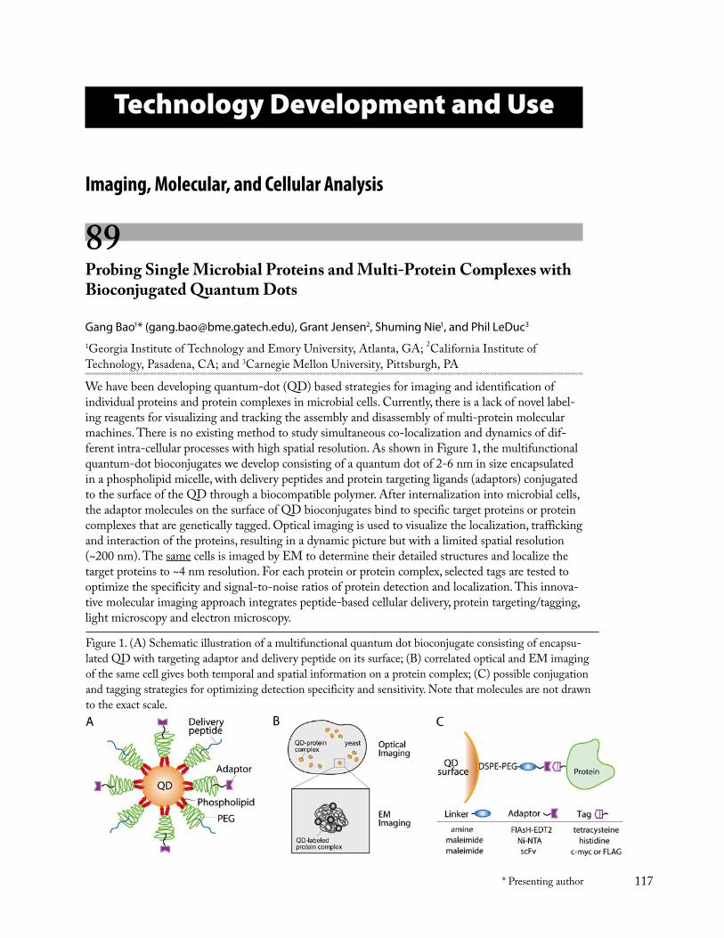
117* Presenting author
Technology Development and Use
Imaging, Molecular, and Cellular Analysis
89 Probing Single Microbial Proteins and Multi-Protein Complexes with Bioconjugated Quantum Dots
Gang Bao1* ([email protected]), Grant Jensen2, Shuming Nie1, and Phil LeDuc3
1Georgia Institute of Technology and Emory University, Atlanta, GA; 2California Institute of Technology, Pasadena, CA; and 3Carnegie Mellon University, Pittsburgh, PA
We have been developing quantum-dot (QD) based strategies for imaging and identification of individual proteins and protein complexes in microbial cells. Currently, there is a lack of novel label-ing reagents for visualizing and tracking the assembly and disassembly of multi-protein molecular machines. There is no existing method to study simultaneous co-localization and dynamics of dif-ferent intra-cellular processes with high spatial resolution. As shown in Figure 1, the multifunctional quantum-dot bioconjugates we develop consisting of a quantum dot of 2-6 nm in size encapsulated in a phospholipid micelle, with delivery peptides and protein targeting ligands (adaptors) conjugated to the surface of the QD through a biocompatible polymer. After internalization into microbial cells, the adaptor molecules on the surface of QD bioconjugates bind to specific target proteins or protein complexes that are genetically tagged. Optical imaging is used to visualize the localization, trafficking and interaction of the proteins, resulting in a dynamic picture but with a limited spatial resolution (~200 nm). The same cells is imaged by EM to determine their detailed structures and localize the target proteins to ~4 nm resolution. For each protein or protein complex, selected tags are tested to optimize the specificity and signal-to-noise ratios of protein detection and localization. This innova-tive molecular imaging approach integrates peptide-based cellular delivery, protein targeting/tagging, light microscopy and electron microscopy.
Figure 1. (A) Schematic illustration of a multifunctional quantum dot bioconjugate consisting of encapsu-lated QD with targeting adaptor and delivery peptide on its surface; (B) correlated optical and EM imaging of the same cell gives both temporal and spatial information on a protein complex; (C) possible conjugation and tagging strategies for optimizing detection specificity and sensitivity. Note that molecules are not drawn to the exact scale.
Technology Development and Use
118 * Presenting author
To achieve the goals of this DoE GTL project, we have developed quantum-dot bioconjugates with QDs encapsulated in a micelle. Phospholipids conjugated to monomethoxy PEG was used to form micelles in which the hydrophobic core of a DSPE-PEG micelle provides a cavity to encapsulate individual QDs, while the dense PEG polymer layer on the outer surface facilitates conjugation of linker molecules and delivery peptides. To facilitate bioconjugation for attaching adaptor molecules and delivery peptide, different functionalized PEG-lipid derivatives, such as DSPE-PEG-maleimide and DSPE-PEG-amine were used. To generate site-specificity, two tagging strategies were exam-ined. The first used FlAsh-EDT2 as the adaptor molecular on the QD surface and tetracysteine (Cys-Cys-Xaa-Xaa-Cys-Cys) as the tag engineered on the target protein. In the second approach, Ni-NTA was conjugated to coated QDs and the specific protein was modified to have a histidine tag. The specific targeting was demonstrated using a model system.
We performed a preliminary study of peptide-based delivery of dye molecules and quantum dot bioconjugates into yeast and E. coli using specifically three different peptides, TAT, polyArg, and a peptide (ArgSerAsnAsnProPheArgAlaArg) that has been used for delivering GFP into yeast S. cerevi-siae. Two yeast strains, ACY 193, a wild-type yeast strain, and ACY651, a permeable yeast strain were used. We found that the polyArg peptide was the most efficient one for yeast delivery.
As part of our effort to develop QD-based technologies to identify and track individual protein complexes in microbial cells, we are advancing electron tomography as a promising new tool to image such complexes both in vitro and in vivo within small microbial cells. A new helium-cooled, 300kV, FEG, “G2 Polara” FEI TEM at Caltech was used to image purified protein complexes, viruses, and whole bacterial cells. The Polara has allowed us to record automated tilt series of a single sample cooled with either liquid nitrogen (~90K) or liquid helium (~10K). Specifically, we have recorded tilt series of purified hemocyanin and reconstructed hundreds of individual particles at various doses at each temperature, all from different “holes” of the same grid square in a single data collection session to minimize confounding variables. Surprisingly, the contrast from proteins gradually fades when they are cooled by liquid helium and iteratively imaged. Thus liquid nitrogen cooling is preferred. Using a prediction-based tracking software, we performed an automatic tilt series collection with-out any extra tracking or focusing images, allowing a robust data collection and reducing the time required to record a large amount of data. Further, through delivery of FEI’s first “flip-flop” cryo-rotation stage, we have begun recording dual-axis tilt series of frozen-hydrated samples routinely. This has the advantages in improved point-spread-function; however, we found that our software for merging the two tilt series is not optimal, and we are presently working to improve that. These tech-nological advances have allowed us to visualize directly cytoskeletal elements within small microbial cells and the domain structure of purified multienzyme complexes, both are key imaging goals of the Genomics:GTL program.
As a model system to study protein localization, we have been investigating the migration of Dictyo-stelium discoideum under defined extracellular stimuli. This organism responds quickly to changes in the direction of cyclic nucleotide, adenosine 3', 5'-cyclic monophosphate (cAMP) gradient. These highly polarized amoebas are characterized by continuous protrusion and retraction of pseudopodial extensions. Various localized structural responses occur in polarized D. discoideum. These include the localization of the β subunit of the heterotrimeric guanine nucleotide-binding proteins (G proteins) in a shallow anterior-posterior gradient, as well as the biased distribution of actin. Furthermore, the orientation of aggregating cells is dominated by the chemoattractant-induced polymerization of actin.
We have utilized custom-fabricated microfluidic devices to stimulate a cell in local domains both with two-dimensional and three-dimensional control while simultaneously visualizing its response with fluorescent microscopy using quantum dots. Both peptide-based delivery and electroporation
Technology Development and Use
119* Presenting author
were used to internalize QD–probes into the Dictyostelium. The dynamics of the actin cytoskeleton and the localization of β subunit of G protein in response to extracellular stimuli were studied us-ing quantum-dot probes. Further, double labeling strategies were developed to localize extracellular cAMP receptors. This technique will be combined with high-resolution electron microscopy imaging to visualize individual proteins and protein complexes.
Acknowledgement: This research is funded by a grant from from DOE (DE-FG02-04ER63785).
90 Single-Molecule Imaging of Macromolecular Dynamics in a Cell
Jamie H. D. Cate ([email protected]) and Haw Yang* ([email protected])
Lawrence Berkeley National Laboratory, Berkeley, CA
In order to monitor macromolecular dynamics optically in living cells and to relate these observations to cellular functions two sets of tools will be essential. It will be necessary to have fluorescent probes that can be used for site-specific labeling in vivo and non-bleachable probes that are biocompatible with the cellular environment. The first year of this joint research project focused on the development of these two enabling technologies.
To address the need for non-bleachable and biocompatible probes, we have synthesized and charac-terized biocompatible gold nanoparticles, or nanotags. Nanotags will allow long-term (hours) moni-toring of molecular dynamics in cells. To allow direct optical observation, the size of nanoparticles has to be greater than 10 nm. However, the surface chemistry of nanoparticles in this size range has been known to be challenging due to their propensity to aggregate. We have developed surface pas-sivation protocols that allow stabilization of large nanoparticles and yet retain reactivity for further functionalization that is required for biological tagging.
An important aspect of these nanotags is their biocompatibility. We have developed protocols to coat the stabilized nanoparticles with various passivation agents. Biocompatibility has been stringently tested against both biochemical and biological criteria. For the former, enzymes tethered to nanotags were found to retain their reactivity, whereas for the latter, mammalian cells that contain nanotags were able to survive and propagate for several generations without aggregation of nanotags. We have also developed nanotags that will allow investigation of macromolecule rotation dynamics, as well as those that will allow multiplexing.
Finally, progress towards specific labeling of proteins with FRET donor and acceptor pairs that can be used in living cells and the status on instrumentation will be presented.
Technology Development and Use
120 * Presenting author
91 Developing a High Resolution Method for Protein Localization in Whole Bacterium
Huilin Li* ([email protected]) and James Hainfeld ([email protected])
Brookhaven National Laboratory, Upton, NY
Bacteria lack intracellular membranes, yet the distribution and localization of many bacterial proteins are precisely controlled during cell cycle. Light microscopy has been used with great success to map the fluorescently labeled proteins, although the ~ 200 nm resolution is much to be desired, especially considering the extremely small size of the bacterial cells.
Electron tomography reaches much higher resolution than light microscopy, to ~ 10 nm, thus can be used in principle for protein localization. However one serious problem needs to be addressed. This has to do with the currently achievable resolution. Although an order of magnitude better than light microscopy, it is still short of resolving most of the proteins in the tomograms of bacteria embedded in vitreous ice. One may either strive to improve the tomogram resolution, which is the route taken by other groups, or as we decided, to specifically tag the proteins for their identification with electron dense labels, such as nanogold.
We have synthesized 3 nm diameter nanogold particles with the functionalized chemical group Ni-NTA, to be used for labeling 6X-His tagged proteins. This is being tested and optimized on expressed proteins with and without the His tag to improve specificity and efficiency of labeling.
The bacteria we choose to study, Ralstonia metallidurans, is 0.3-0.5 µm in thickness. To improve the contrast of the small gold label in the relatively thick transmission electron microscopy (TEM) to-mograms, we will record additional scanning transmission electron microscopy (STEM) tomograms, which is known to provide enhanced contrast for high atomic number elements, such as the nano-gold particles. In order to make our Jeol 2010F FasTEM/STEM microscope capable of performing tomography in both TEM and STEM modes, we have replaced the existing objective lens pole piece with a large gap one that has no limit on tilt angle. The microscope operating software FasTEM has been patched to work with the new large gap pole piece in STEM mode. We have also acquired and successfully installed a Gatan Digiscan for recording digital STEM images. The automatic tomog-raphy procedure has been implemented in TEM mode, and we are in the process of developing a Gatan Digital Micrograph-based script for automatic tomography in STEM mode. STEM tomog-raphy has been demonstrated previously in manual operation mode with plastic or inorganic samples. It is essential to record STEM tomographic tilt series in an automatic mode to minimize the radia-tion damage for frozen hydrated bacterial cells. We expect this to be done within a few months, and we will then proceed to image the labeled bacterial cells.
Technology Development and Use
121* Presenting author
92 Novel Vibrational Nanoprobes for Microbiology at the Single Cell Level
Thomas Huser* ([email protected]), Chad E. Talley, James W. Chan, Heiko Winhold, Ted Laurence, Anthony Esposito, Christopher W. Hollars, Christine A. Hara, Allen T. Christian, Michele H. Corzett, Rod Balhorn, and Stephen M. Lane
Lawrence Livermore National Laboratory, Livermore, CA
The measurement of intracellular chemical concentrations and molecular fluxes provides essential information for systems-biological models of cells. This information, however, is difficult to obtain at the single cell level – especially in living cells where chemical levels can change rapidly in response to external or internal events.
We are studying individual microbes by a combination of optical spectroscopy techniques to obtain dynamic chemical profiles at the single cell level. Raman spectroscopy in combination with optical tweezers is used to non-destructively capture individual microbes in their native environment and assess their chemical composition within seconds. We have used this technique to dynamically moni-tor changes in the total protein concentration of individual cells due to increased expression after external stimulation. By focusing on changes in particular Raman peaks of a microbe we can follow trends in the overall intensity of specific peaks on an even faster timescale – down to milliseconds – without the need for exogenous probes. We will present examples and applications of these power-ful vibrational spectroscopy techniques.
To monitor chemicals at low concentrations or chemicals that cannot typically be measured by Ra-man spectroscopy we also present the development of nanoscale sensors based on functionalized metal nanoparticles and surface-enhanced Raman scattering (SERS). As an example, the SERS spectrum from individual silver nanoparticle (50-80 nm in diameter) clusters functionalized with 4-mercaptobenzoic acid (4-MBA) is shown to exhibit a characteristic response to the pH of the surrounding solution, and is sensitive to pH changes in the range of 6 to 8. Measurements from nanoparticles incorporated into individual cells demonstrate that these nanoparticle sensors retain their robust signal and sensitivity to pH when incorporated into a cell. These sensors can be probed almost entirely background-free and their signals do not suffer from photobleaching, which makes them attractive long-term probes for chemical concentrations that cannot be probed by conventional Raman spectroscopy.
This work was supported by the Office of Science of the U.S. Department of Energy. Work performed under the auspices of the U.S. Department of Energy by the University of California, Lawrence Livermore National Laboratory under contract number W-7405-ENG-48.
Technology Development and Use
122 * Presenting author
93 Instrumented Cell for Characterization of Mammalian and Microbial Cells
Jane Bearinger* ([email protected]), Graham Bench, Jackie Crawford, Lawrence Dugan, Amy Hiddessen, Angela Hinz, Thomas Huser, Robin Miles, Magnus Palmblad, Chad Talley, Elizabeth Wheeler, and Allen Christian
Lawrence Livermore National Laboratory, Livermore, CA
Scientists at Lawrence Livermore National Laboratory are developing novel methodologies for high throughput cell and bacterial analysis through an internally funded Laboratory Directed Research and Development project, Instrumented Cell. The project parallels work to be conducted in the Facility for Analysis and Modeling of Cellular Systems, the final step of which requires the ability to measure and predict dynamic events within individual cells in order to achieve a comprehensive understanding of living systems. Like the Cellular Systems Facility, work focuses on experimental capabilities for the integrated observation, measurement, and analysis of the spatial and temporal variations in cellular and microbial systems.
There is a growing interest in the prospects for a new “quantitative biology”, in which experimental data is integrated into predictive models of biological systems that can in turn be used to design and evaluate new experiments. There are many challenges to realizing this vision of quantitative biology; perhaps the most significant being the need to get precise data on the concentration and distribution of cellular components that will be required to develop such models. There are many analytical capa-bilities at LLNL that show promise for enabling quantitative measurements of cellular processes in individual cells. Instrumented Cell is developing and applying technical capabilities to measure and manipulate biochemical concentrations at the single-cell level with the ultimate goal of developing quantitative models of cellular processes. With the LLNL Microfabrication Facility, microstructures are being developed to isolate cells and make single cell measurements with the analytical tools pos-sessed by the Chemistry and Energy and Environment directorates.
The first year of Instrumented Cell focused on production of tools capable of manipulating the environment of individual cells, and production of analytical measurements of the cells’ reaction to stimuli. These capabilities provide quantitative data on an individual cell’s responses to stimuli, such as characterization of uptake and effect of the new siHybrid gene silencing technology as well as the uptake of nanoparticle sensors.
Cultivation and maintenance of microbes and microbial communities under controlled conditions, including the ability to interrogate the function of individual microbial cells in the context of a char-acterized physicochemical environment, is another key technology needed by GTL. We are develop-ing highly controlled systems for growing and maintaining microbial populations and communities via chemical and microfabrication-based platforms, with an emphasis on development of robust protocols that are adaptable to different strains of bacteria. We have made considerable progress in isolation of discreet bacterial communities, which will assist in high throughput analysis. Our pro-jected plan consists of on-chip analysis of gene silencing via fluorescence experiments and off-chip cloning and mass spectrometry experiments.
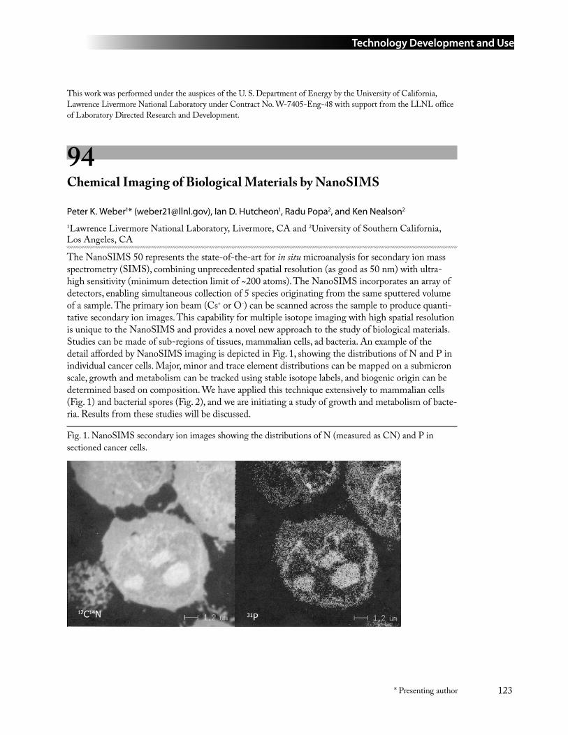
Technology Development and Use
123* Presenting author
This work was performed under the auspices of the U. S. Department of Energy by the University of California, Lawrence Livermore National Laboratory under Contract No. W-7405-Eng-48 with support from the LLNL office of Laboratory Directed Research and Development.
94 Chemical Imaging of Biological Materials by NanoSIMS
Peter K. Weber1* ([email protected]), Ian D. Hutcheon1, Radu Popa2, and Ken Nealson2
1Lawrence Livermore National Laboratory, Livermore, CA and 2University of Southern California, Los Angeles, CA
The NanoSIMS 50 represents the state-of-the-art for in situ microanalysis for secondary ion mass spectrometry (SIMS), combining unprecedented spatial resolution (as good as 50 nm) with ultra-high sensitivity (minimum detection limit of ~200 atoms). The NanoSIMS incorporates an array of detectors, enabling simultaneous collection of 5 species originating from the same sputtered volume of a sample. The primary ion beam (Cs+ or O-) can be scanned across the sample to produce quanti-tative secondary ion images. This capability for multiple isotope imaging with high spatial resolution is unique to the NanoSIMS and provides a novel new approach to the study of biological materials. Studies can be made of sub-regions of tissues, mammalian cells, ad bacteria. An example of the detail afforded by NanoSIMS imaging is depicted in Fig. 1, showing the distributions of N and P in individual cancer cells. Major, minor and trace element distributions can be mapped on a submicron scale, growth and metabolism can be tracked using stable isotope labels, and biogenic origin can be determined based on composition. We have applied this technique extensively to mammalian cells (Fig. 1) and bacterial spores (Fig. 2), and we are initiating a study of growth and metabolism of bacte-ria. Results from these studies will be discussed.
Fig. 1. NanoSIMS secondary ion images showing the distributions of N (measured as CN) and P in sectioned cancer cells.
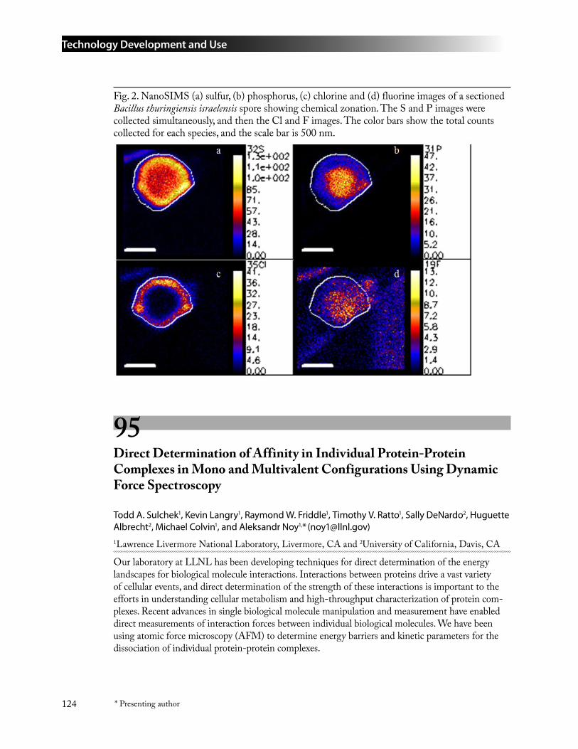
Technology Development and Use
124 * Presenting author
Fig. 2. NanoSIMS (a) sulfur, (b) phosphorus, (c) chlorine and (d) fluorine images of a sectioned Bacillus thuringiensis israelensis spore showing chemical zonation. The S and P images were collected simultaneously, and then the Cl and F images. The color bars show the total counts collected for each species, and the scale bar is 500 nm.
95 Direct Determination of Affinity in Individual Protein-Protein Complexes in Mono and Multivalent Configurations Using Dynamic Force Spectroscopy
Todd A. Sulchek1, Kevin Langry1, Raymond W. Friddle1, Timothy V. Ratto1, Sally DeNardo2, Huguette Albrecht2, Michael Colvin1, and Aleksandr Noy1,* ([email protected])1Lawrence Livermore National Laboratory, Livermore, CA and 2University of California, Davis, CA
Our laboratory at LLNL has been developing techniques for direct determination of the energy landscapes for biological molecule interactions. Interactions between proteins drive a vast variety of cellular events, and direct determination of the strength of these interactions is important to the efforts in understanding cellular metabolism and high-throughput characterization of protein com-plexes. Recent advances in single biological molecule manipulation and measurement have enabled direct measurements of interaction forces between individual biological molecules. We have been using atomic force microscopy (AFM) to determine energy barriers and kinetic parameters for the dissociation of individual protein-protein complexes.
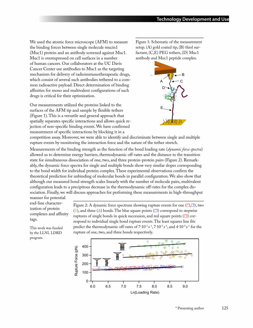
Technology Development and Use
125* Presenting author
We used the atomic force microscope (AFM) to measure the binding forces between single molecule mucin1 (Muc1) protein and an antibody screened against Muc1. Muc1 is overexpressed on cell surfaces in a number of human cancers. Our collaborators at the UC Davis Cancer Center use antibodies to Muc1 as the targeting mechanism for delivery of radioimmunotherapeutic drugs, which consist of several such antibodies tethered to a com-mon radioactive payload. Direct determination of binding affinities for mono and multivalent configurations of such drugs is critical for their optimization.
Our measurements utilized the proteins linked to the surfaces of the AFM tip and sample by flexible tethers (Figure 1). This is a versatile and general approach that spatially separates specific interactions and allows quick re-jection of non-specific binding events. We have confirmed measurement of specific interactions by blocking it in a competition assay. Moreover, we were able to identify and discriminate between single and multiple rupture events by monitoring the interaction force and the nature of the tether stretch. Measurements of the binding strength as the function of the bond loading rate (dynamic force spectra) allowed us to determine energy barriers, thermodynamic off-rates and the distance to the transition state for simultaneous dissociation of one, two, and three protein-protein pairs (Figure 2). Remark-ably, the dynamic force spectra for single and multiple bonds show very similar slopes corresponding to the bond width for individual protein complex. These experimental observations confirm the theoretical prediction for unbinding of molecular bonds in parallel configuration. We also show that although our measured bond strength scales linearly with the number of molecule pairs, multivalent configuration leads to a precipitous decrease in the thermodynamic off-rates for the complex dis-sociation. Finally, we will discuss approaches for performing these measurements in high-throughput manner for potential end-line character-ization of protein complexes and affinity tags.
This work was funded by the LLNL LDRD program.
Figure 1: Schematic of the measurement setup. (A) gold coated tip, (B) thiol sur-factant, (C,E) PEG tethers, (D) Muc1 antibody and Muc1 peptide complex.
E
B
C
D
A
Figure 2: A dynamic force spectrum showing rupture events for one (☐,☐), two (◊), and three (∆) bonds. The blue square points (☐) correspond to stepwise ruptures of single bonds in quick succession, and red square points (☐) cor-respond to individual single bond rupture events. The least squares line fits predict the thermodynamic off-rates of 7∙10-3 s-1, 7∙10-5 s-1, and 4∙10-9 s-1 for the rupture of one, two, and three bonds respectively.
400
300
200
100
0
Rupt
ure
Forc
e (p
N)
9.08.58.07.57.06.56.0Ln(Loading Rate)
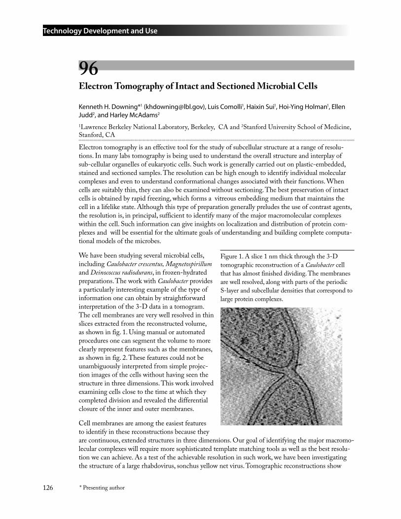
Technology Development and Use
126 * Presenting author
96 Electron Tomography of Intact and Sectioned Microbial Cells
Kenneth H. Downing*1 ([email protected]), Luis Comolli1, Haixin Sui1, Hoi-Ying Holman1, Ellen Judd2, and Harley McAdams2
1Lawrence Berkeley National Laboratory, Berkeley, CA and 2Stanford University School of Medicine, Stanford, CA
Electron tomography is an effective tool for the study of subcellular structure at a range of resolu-tions. In many labs tomography is being used to understand the overall structure and interplay of sub-cellular organelles of eukaryotic cells. Such work is generally carried out on plastic-embedded, stained and sectioned samples. The resolution can be high enough to identify individual molecular complexes and even to understand conformational changes associated with their functions. When cells are suitably thin, they can also be examined without sectioning. The best preservation of intact cells is obtained by rapid freezing, which forms a vitreous embedding medium that maintains the cell in a lifelike state. Although this type of preparation generally preludes the use of contrast agents, the resolution is, in principal, sufficient to identify many of the major macromolecular complexes within the cell. Such information can give insights on localization and distribution of protein com-plexes and will be essential for the ultimate goals of understanding and building complete computa-tional models of the microbes.
We have been studying several microbial cells, including Caulobacter crescentus, Magnetospirillum and Deinococcus radiodurans, in frozen-hydrated preparations. The work with Caulobacter provides a particularly interesting example of the type of information one can obtain by straightforward interpretation of the 3-D data in a tomogram. The cell membranes are very well resolved in thin slices extracted from the reconstructed volume, as shown in fig. 1. Using manual or automated procedures one can segment the volume to more clearly represent features such as the membranes, as shown in fig. 2. These features could not be unambiguously interpreted from simple projec-tion images of the cells without having seen the structure in three dimensions. This work involved examining cells close to the time at which they completed division and revealed the differential closure of the inner and outer membranes.
Cell membranes are among the easiest features to identify in these reconstructions because they are continuous, extended structures in three dimensions. Our goal of identifying the major macromo-lecular complexes will require more sophisticated template matching tools as well as the best resolu-tion we can achieve. As a test of the achievable resolution in such work, we have been investigating the structure of a large rhabdovirus, sonchus yellow net virus. Tomographic reconstructions show
Figure 1. A slice 1 nm thick through the 3-D tomographic reconstruction of a Caulobacter cell that has almost finished dividing. The membranes are well resolved, along with parts of the periodic S-layer and subcellular densities that correspond to large protein complexes.
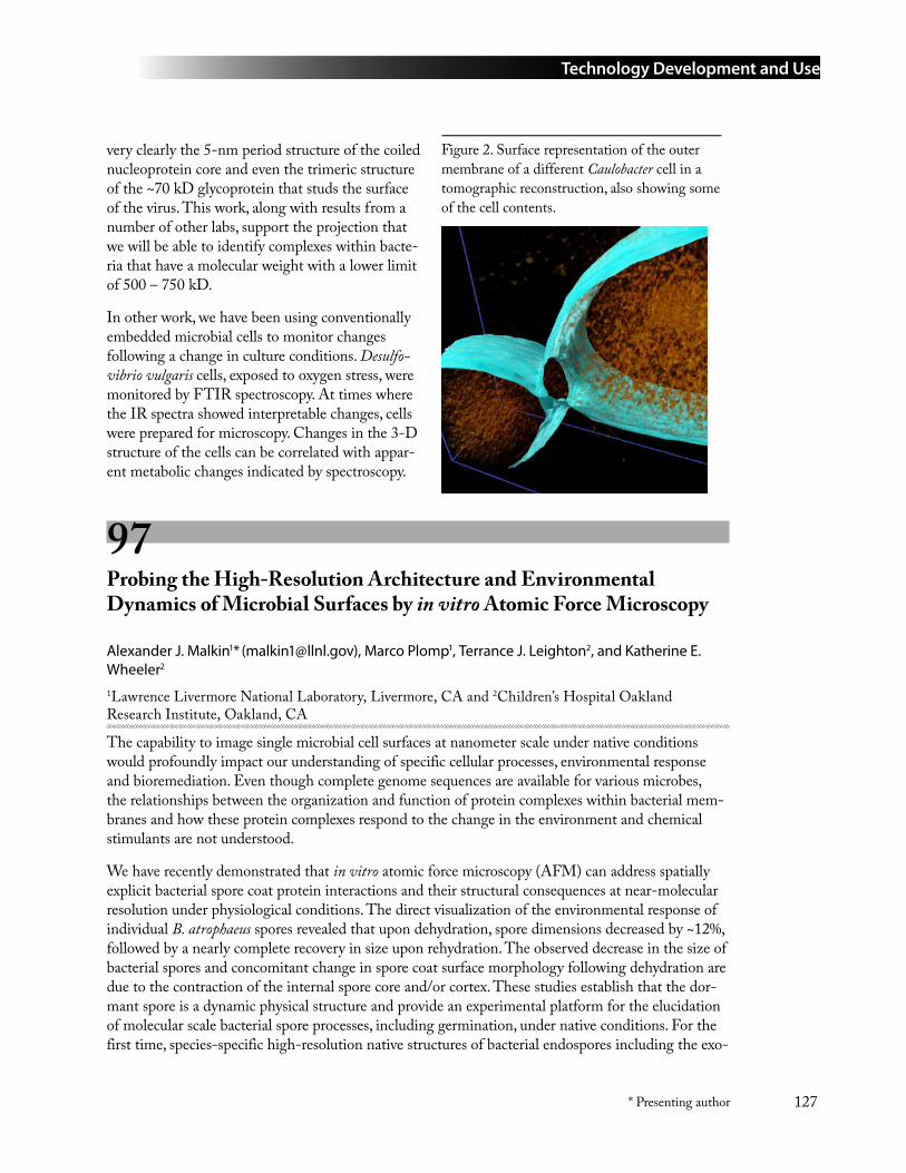
Technology Development and Use
127* Presenting author
very clearly the 5-nm period structure of the coiled nucleoprotein core and even the trimeric structure of the ~70 kD glycoprotein that studs the surface of the virus. This work, along with results from a number of other labs, support the projection that we will be able to identify complexes within bacte-ria that have a molecular weight with a lower limit of 500 – 750 kD.
In other work, we have been using conventionally embedded microbial cells to monitor changes following a change in culture conditions. Desulfo-vibrio vulgaris cells, exposed to oxygen stress, were monitored by FTIR spectroscopy. At times where the IR spectra showed interpretable changes, cells were prepared for microscopy. Changes in the 3-D structure of the cells can be correlated with appar-ent metabolic changes indicated by spectroscopy.
97 Probing the High-Resolution Architecture and Environmental Dynamics of Microbial Surfaces by in vitro Atomic Force Microscopy
Alexander J. Malkin1* ([email protected]), Marco Plomp1, Terrance J. Leighton2, and Katherine E. Wheeler2 1Lawrence Livermore National Laboratory, Livermore, CA and 2Children’s Hospital Oakland Research Institute, Oakland, CA
The capability to image single microbial cell surfaces at nanometer scale under native conditions would profoundly impact our understanding of specific cellular processes, environmental response and bioremediation. Even though complete genome sequences are available for various microbes, the relationships between the organization and function of protein complexes within bacterial mem-branes and how these protein complexes respond to the change in the environment and chemical stimulants are not understood.
We have recently demonstrated that in vitro atomic force microscopy (AFM) can address spatially explicit bacterial spore coat protein interactions and their structural consequences at near-molecular resolution under physiological conditions. The direct visualization of the environmental response of individual B. atrophaeus spores revealed that upon dehydration, spore dimensions decreased by ~12%, followed by a nearly complete recovery in size upon rehydration. The observed decrease in the size of bacterial spores and concomitant change in spore coat surface morphology following dehydration are due to the contraction of the internal spore core and/or cortex. These studies establish that the dor-mant spore is a dynamic physical structure and provide an experimental platform for the elucidation of molecular scale bacterial spore processes, including germination, under native conditions. For the first time, species-specific high-resolution native structures of bacterial endospores including the exo-
Figure 2. Surface representation of the outer membrane of a different Caulobacter cell in a tomographic reconstruction, also showing some of the cell contents.
Technology Development and Use
128 * Presenting author
sporium and crystalline layers of the spore coat of four Bacillus species were visualized in their natural environment, namely air and fluid. We found that strikingly different species-dependent structures of the spore coat appear to be a consequence of nucleation and crystallization mechanisms that regulate the assembly of the outer spore coat and proposed a unifying mechanism for outer spore coat surface self-assembly.
These studies establish in vitro AFM as a powerful tool capable of providing a direct insight into molecular architecture and structural variability of microbial surfaces as a function of spatial, tempo-ral, developmental and environmental organizational scales. We are currently developing approaches to utilize AFM for probing of the ultra-structure and environmental dynamics of several bacteria including Arthrobacter oxydans and Thiobacillus denitrificans.
M. Plomp, T.J. Leighton, K.E. Wheeler and A.J. Malkin (2005), Biophysical J., 88.
98 Real-Time Gene Expression Profiling of Single Live Cells of Shewanella oneidensis
X. Sunney Xie*, Jie Xiao, Ji Yu, Long Cai, Paul Choi*, Nir Friedman, Xiajia Ren, and Luying Xun*
Harvard University, Cambridge, MA
Our objective is to make real-time observations of gene expression in live Shewanella oneidensis MR1 cells with high sensitivity and high throughput. Available technology is sufficient for the detection of gene expression at high levels; whereas, new techniques need to be developed to study expression that produces only a few protein molecules. Our efforts are divided into three areas: Developing sensitive protein reporters, adapting and fine-tuning a practical cloning strategy for library construction, and testing automation techniques for high throughput measurements with single-molecule microscopes.
Modified β-Galactosidase and green fluorescence protein (GFP) are used for reporters. Each β-galactosidase molecule generates hundreds of fluorescent molecules per second, and the product formation can be monitored in real-time. On the flipside, it is hard to observe single GFP molecules in live cells. Among the available GFP reporters, Venus GFP was chosen because of its enhanced fluorescence and short maturation time. We have detected single Venus GFP in bacterial cells after the reporter protein is attached to relatively immobile cellular components (Fig. 1). This is a major step, enabling us to follow gene expression at low signal levels. N-Terminal fusion with ubiquitin (an eukaryotic tag and degradation system) or C-terminal fusion with SsrA (a bacterial tag and degrada-tion system) have been constructed to shorten the cellular lifetime of reporter proteins, so that cell cycle related gene expression can be monitored with single cells.
1.
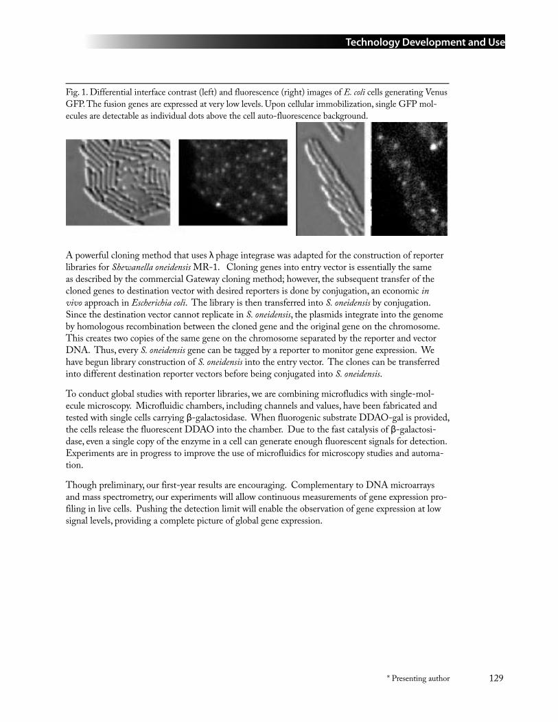
Technology Development and Use
129* Presenting author
Fig. 1. Differential interface contrast (left) and fluorescence (right) images of E. coli cells generating Venus GFP. The fusion genes are expressed at very low levels. Upon cellular immobilization, single GFP mol-ecules are detectable as individual dots above the cell auto-fluorescence background.
A powerful cloning method that uses λ phage integrase was adapted for the construction of reporter libraries for Shewanella oneidensis MR-1. Cloning genes into entry vector is essentially the same as described by the commercial Gateway cloning method; however, the subsequent transfer of the cloned genes to destination vector with desired reporters is done by conjugation, an economic in vivo approach in Escherichia coli. The library is then transferred into S. oneidensis by conjugation. Since the destination vector cannot replicate in S. oneidensis, the plasmids integrate into the genome by homologous recombination between the cloned gene and the original gene on the chromosome. This creates two copies of the same gene on the chromosome separated by the reporter and vector DNA. Thus, every S. oneidensis gene can be tagged by a reporter to monitor gene expression. We have begun library construction of S. oneidensis into the entry vector. The clones can be transferred into different destination reporter vectors before being conjugated into S. oneidensis.
To conduct global studies with reporter libraries, we are combining microfludics with single-mol-ecule microscopy. Microfluidic chambers, including channels and values, have been fabricated and tested with single cells carrying β-galactosidase. When fluorogenic substrate DDAO-gal is provided, the cells release the fluorescent DDAO into the chamber. Due to the fast catalysis of β-galactosi-dase, even a single copy of the enzyme in a cell can generate enough fluorescent signals for detection. Experiments are in progress to improve the use of microfluidics for microscopy studies and automa-tion.
Though preliminary, our first-year results are encouraging. Complementary to DNA microarrays and mass spectrometry, our experiments will allow continuous measurements of gene expression pro-filing in live cells. Pushing the detection limit will enable the observation of gene expression at low signal levels, providing a complete picture of global gene expression.
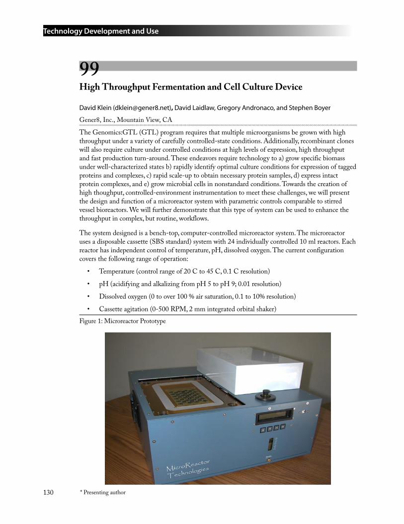
Technology Development and Use
130 * Presenting author
99 High Throughput Fermentation and Cell Culture Device
David Klein ([email protected]), David Laidlaw, Gregory Andronaco, and Stephen Boyer
Gener8, Inc., Mountain View, CA
The Genomics:GTL (GTL) program requires that multiple microorganisms be grown with high throughput under a variety of carefully controlled-state conditions. Additionally, recombinant clones will also require culture under controlled conditions at high levels of expression, high throughput and fast production turn-around. These endeavors require technology to a) grow specific biomass under well-characterized states b) rapidly identify optimal culture conditions for expression of tagged proteins and complexes, c) rapid scale-up to obtain necessary protein samples, d) express intact protein complexes, and e) grow microbial cells in nonstandard conditions. Towards the creation of high thoughput, controlled-environment instrumentation to meet these challenges, we will present the design and function of a microreactor system with parametric controls comparable to stirred vessel bioreactors. We will further demonstrate that this type of system can be used to enhance the throughput in complex, but routine, workflows.
The system designed is a bench-top, computer-controlled microreactor system. The microreactor uses a disposable cassette (SBS standard) system with 24 individually controlled 10 ml reactors. Each reactor has independent control of temperature, pH, dissolved oxygen. The current configuration covers the following range of operation:
Temperature (control range of 20 C to 45 C, 0.1 C resolution)
pH (acidifying and alkalizing from pH 5 to pH 9; 0.01 resolution)
Dissolved oxygen (0 to over 100 % air saturation, 0.1 to 10% resolution)
Cassette agitation (0-500 RPM, 2 mm integrated orbital shaker)
•
•
•
•Figure 1: Microreactor Prototype
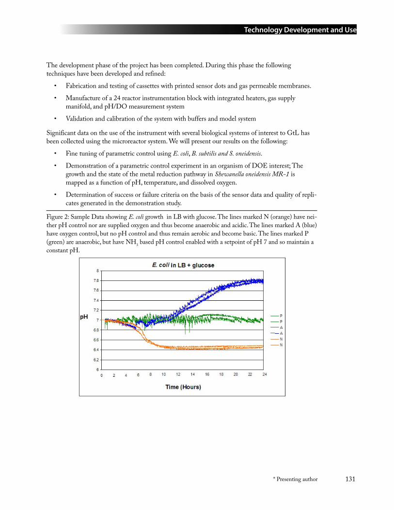
Technology Development and Use
131* Presenting author
The development phase of the project has been completed. During this phase the following techniques have been developed and refined:
Fabrication and testing of cassettes with printed sensor dots and gas permeable membranes.
Manufacture of a 24 reactor instrumentation block with integrated heaters, gas supply manifold, and pH/DO measurement system
Validation and calibration of the system with buffers and model system
Significant data on the use of the instrument with several biological systems of interest to GtL has been collected using the microreactor system. We will present our results on the following:
Fine tuning of parametric control using E. coli, B. subtilis and S. oneidensis.
Demonstration of a parametric control experiment in an organism of DOE interest; The growth and the state of the metal reduction pathway in Shewanella oneidensis MR-1 is mapped as a function of pH, temperature, and dissolved oxygen.
Determination of success or failure criteria on the basis of the sensor data and quality of repli-cates generated in the demonstration study.
•
•
•
•
•
•
Figure 2: Sample Data showing E. coli growth in LB with glucose. The lines marked N (orange) have nei-ther pH control nor are supplied oxygen and thus become anaerobic and acidic. The lines marked A (blue) have oxygen control, but no pH control and thus remain aerobic and become basic. The lines marked P (green) are anaerobic, but have NH3 based pH control enabled with a setpoint of pH 7 and so maintain a constant pH.
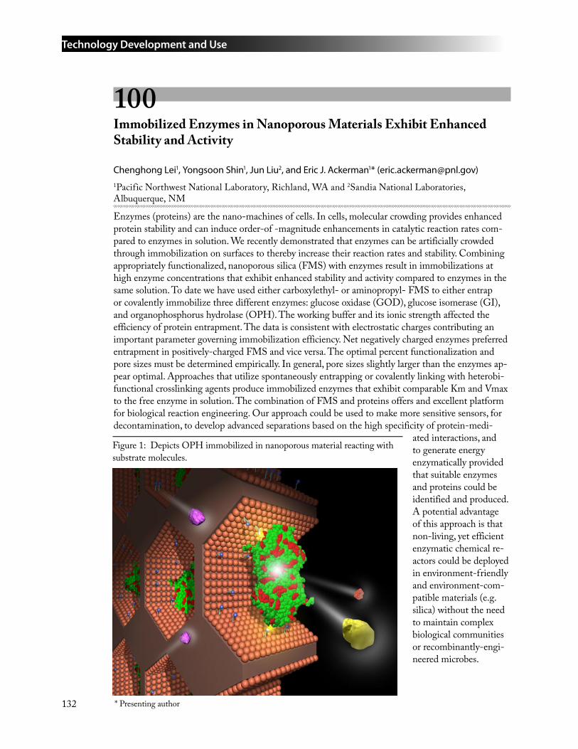
Technology Development and Use
132 * Presenting author
100 Immobilized Enzymes in Nanoporous Materials Exhibit Enhanced Stability and Activity
Chenghong Lei1, Yongsoon Shin1, Jun Liu2, and Eric J. Ackerman1* ([email protected])1Pacific Northwest National Laboratory, Richland, WA and 2Sandia National Laboratories, Albuquerque, NM
Enzymes (proteins) are the nano-machines of cells. In cells, molecular crowding provides enhanced protein stability and can induce order-of -magnitude enhancements in catalytic reaction rates com-pared to enzymes in solution. We recently demonstrated that enzymes can be artificially crowded through immobilization on surfaces to thereby increase their reaction rates and stability. Combining appropriately functionalized, nanoporous silica (FMS) with enzymes result in immobilizations at high enzyme concentrations that exhibit enhanced stability and activity compared to enzymes in the same solution. To date we have used either carboxylethyl- or aminopropyl- FMS to either entrap or covalently immobilize three different enzymes: glucose oxidase (GOD), glucose isomerase (GI), and organophosphorus hydrolase (OPH). The working buffer and its ionic strength affected the efficiency of protein entrapment. The data is consistent with electrostatic charges contributing an important parameter governing immobilization efficiency. Net negatively charged enzymes preferred entrapment in positively-charged FMS and vice versa. The optimal percent functionalization and pore sizes must be determined empirically. In general, pore sizes slightly larger than the enzymes ap-pear optimal. Approaches that utilize spontaneously entrapping or covalently linking with heterobi-functional crosslinking agents produce immobilized enzymes that exhibit comparable Km and Vmax to the free enzyme in solution. The combination of FMS and proteins offers and excellent platform for biological reaction engineering. Our approach could be used to make more sensitive sensors, for decontamination, to develop advanced separations based on the high specificity of protein-medi-
ated interactions, and to generate energy enzymatically provided that suitable enzymes and proteins could be identified and produced. A potential advantage of this approach is that non-living, yet efficient enzymatic chemical re-actors could be deployed in environment-friendly and environment-com-patible materials (e.g. silica) without the need to maintain complex biological communities or recombinantly-engi-neered microbes.
Figure 1: Depicts OPH immobilized in nanoporous material reacting with substrate molecules.
Technology Development and Use
133* Presenting author
Protein Production and Molecular Tags
101 Towards High Throughput Selection of Binding Ligands: Using Flow Cytometry
Peter Pavlik, Milan Ovecka, Nileena Velappan, and Andrew Bradbury* ([email protected])
Los Alamos National Laboratory, Los Alamos, NM
Phage display libraries represent a relatively easy way to generate binding ligands against a vast num-ber of different targets. Although in principle, phage display selection should be amenable to auto-mation, this has not yet been described and present selection protocols are far from high throughput. We have examined the selection process in a systematic approach and attempted to automate each individual step. Selection is carried out in the microtiter format using 24 targets as the individual selection lot size. Output is plated onto large assay trays, and a program to pick colonies in specific orders corresponding to the selection arrangement has been developed for the Qbot picking robot. This arrays clones according to the antigen they were selected against, and allows subsequent high density analysis using high density dot blots (up to 13,000 clones in the footprint of a microtiter plate). Although it proved possible to analyze such large numbers of clones using HD dot blots, it proved extremely difficult to quantify and digitize binding information. Furthermore, variations in expression levels led to non-specific binding artifacts – well-expressed clones gave binding signals which were often non-specific. Although this could be eliminated by also arraying clones on non-specific target filters, the integration of the information from the two filter types proved extremely difficult to quantify and analyze.
As an alternative we have examined the use of flow cytometry. In a model system, using bead based Luminex type assays, we have been able to carry out multiplex analyses, in which the reactivity of individual antibody clones for numerous different target parameters can be examined simultaneously. The analysis of each individual clone can be carried out in approximately 60 seconds, and all infor-mation is easily exportable to LIMS type systems, as well as being readily analyzed. In first experi-ments we were able to obtain information on the binding of individual antibody clones to specific and non-specific targets, as well as obtaining indications of expression levels. This was carried out by coupling different colored beads with: 1) specific antigen; 2) irrelevant antigen; 3) anti-tag antibodies (to determine expression level). Preliminary experiments with true selections will be presented.
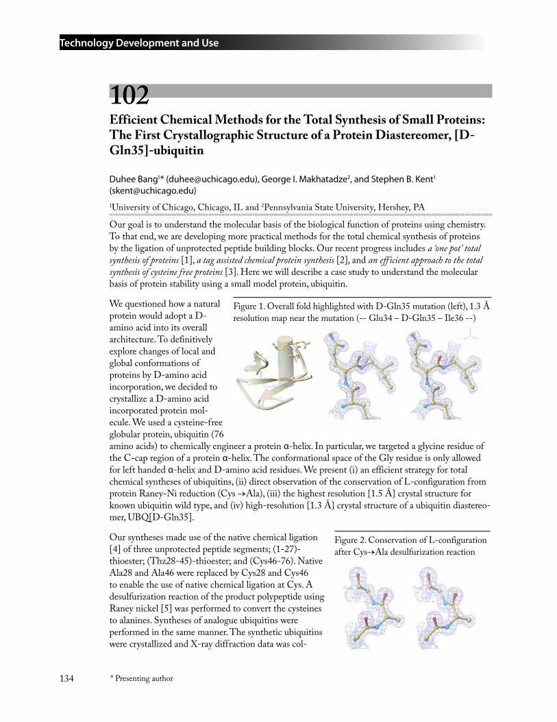
Technology Development and Use
134 * Presenting author
102 Efficient Chemical Methods for the Total Synthesis of Small Proteins: The First Crystallographic Structure of a Protein Diastereomer, [D-Gln35]-ubiquitin
Duhee Bang1* ([email protected]), George I. Makhatadze2, and Stephen B. Kent1 ([email protected])1University of Chicago, Chicago, IL and 2Pennsylvania State University, Hershey, PA
Our goal is to understand the molecular basis of the biological function of proteins using chemistry. To that end, we are developing more practical methods for the total chemical synthesis of proteins by the ligation of unprotected peptide building blocks. Our recent progress includes a ‘one pot’ total synthesis of proteins [1], a tag assisted chemical protein synthesis [2], and an efficient approach to the total synthesis of cysteine free proteins [3]. Here we will describe a case study to understand the molecular basis of protein stability using a small model protein, ubiquitin.
We questioned how a natural protein would adopt a D-amino acid into its overall architecture. To definitively explore changes of local and global conformations of proteins by D-amino acid incorporation, we decided to crystallize a D-amino acid incorporated protein mol-ecule. We used a cysteine-free globular protein, ubiquitin (76 amino acids) to chemically engineer a protein α-helix. In particular, we targeted a glycine residue of the C-cap region of a protein α-helix. The conformational space of the Gly residue is only allowed for left handed α-helix and D-amino acid residues. We present (i) an efficient strategy for total chemical syntheses of ubiquitins, (ii) direct observation of the conservation of L-configuration from protein Raney-Ni reduction (Cys →Ala), (iii) the highest resolution {1.5 Å} crystal structure for known ubiquitin wild type, and (iv) high-resolution {1.3 Å} crystal structure of a ubiquitin diastereo-mer, UBQ[D-Gln35].
Our syntheses made use of the native chemical ligation [4] of three unprotected peptide segments; (1-27)-thioester; (Thz28-45)-thioester; and (Cys46-76). Native Ala28 and Ala46 were replaced by Cys28 and Cys46 to enable the use of native chemical ligation at Cys. A desulfurization reaction of the product polypeptide using Raney nickel [5] was performed to convert the cysteines to alanines. Syntheses of analogue ubiquitins were performed in the same manner. The synthetic ubiquitins were crystallized and X-ray diffraction data was col-
Figure 1. Overall fold highlighted with D-Gln35 mutation (left), 1.3 Å resolution map near the mutation (-- Glu34 – D-Gln35 – Ile36 --)
Figure 2. Conservation of L-configuration after Cys→Ala desulfurization reaction
Technology Development and Use
135* Presenting author
lected using the advanced photon source at ANL. We are currently refining ubiquitin wild-type and its diastereomer structures (partially refined structures are shown in Figure). The structures and on-going efforts for the understanding of protein stability will highlight the power of the total chemical synthesis of proteins.
ReferencesBang, D. & Kent, S. B. (2004) Angew. Chem. Int. Ed. 43, 2534-2538
Bang, D. & Kent, S. B. PNAS Accepted.
Bang, D. G. I. Makhatadze, S. B. Kent Manuscript in preparation
P. E. Dawson, T. W. Muir, I. Clark-Lewis, S. B. Kent, Science 1994, 266, 776-779.
L. Z. Yan, P. E. Dawson, J. Am. Chem. Soc. 2001, 123, 526-533.
103 Development and Application of Multipurpose Affinity Probes to Isolate Intact Protein Complexes Associated with Metal Reduction from Shewanella oneidensis MR-1
Liang Shi*, Thomas C. Squier* ([email protected]), M. Uljana Mayer*, Haishi Cao, Baowei Chen, Yuri A. Gorby, David F. Lowry, Jeff Mclean, Seema Verma, and Ping Yan
Pacific Northwest National Laboratory, Richland, WA
Our long-term goal is to develop high-throughput methods for the rapid isolation of intact protein complexes and validation of these complexes in living cells. This methodology utilizes a small geneti-cally encoded protein tag with an 8 amino-acid sequence containing a tetracysteine motif, which can be captured using affinity reagents or labeled with fluorescent dyes in vivo to permit cellular validation of protein complexes. An important advantage of this strategy is that a single small and nonperturbing tag can be sequentially used to 1) isolate the intact protein complex for identification and structural analysis and 2) visualization of the location and abundance of the protein complex within cells (Chen et al., 2004; Mayer et al., 2005). Furthermore, by varying the architecture of the affinity reagent, multiple colors and photoactivatable cross-linkers can be incorporated into the design strategy to permit measurements of binding interactions within cells and the stabilization of transient interactions associated with signaling complexes.
Proof of principle for this approach has been achieved through the isolation of two protein complex-es (i.e., RNA polymerase and the metal reductase complex) from S. oneidensis MR-1, whose metabo-lism is important in understanding both microbial energy production and environmental remedia-tion. However, these strategies will be applicable to a wide range of microorganisms and will permit the identification of environmental conditions that affect the expression of critical proteins required for the formation of transient protein complexes that facilitate bacterial growth. Our hypothesis is that identifying dynamic changes in these adaptive protein complexes will provide important insights into the metabolic regulatory strategies used by these organisms to adapt to environmental changes.
1.
2.
3.
4.
5.
Technology Development and Use
136 * Presenting author
RNA polymerase is a well studied system, which contains a core complex containing RNA poly-merase alpha2betabeta’ subunits as well as regulatory proteins associated with the differential regula-tion of transcription. Following the expression of a tagged subunit of the RNA polymerase core complex in S. oneidensis MR-1, we have isolated this complex using the synthesized affinity reagent immobilized on a glass bead (Mayer et al., 2005). A critical advantage of this method is the ability to release the intact complex using a mild, one-step procedure with a competing dithiol. In addition to the identification of the core subunit complex, additional regulatory factors were identified, including the universal stress protein.
To investigate whether the current approach will also permit the identification of membrane protein complexes, we have tagged genes identified by the Shewanella Federation to be involved in metal reduction, and isolated members of this important protein complex. In one experiment, the metal reductase MtrC [a decaheme c-type cytochrome tentatively identified as an outer membrane protein whose activity is required for efficient reduction of Mn(IV) and Fe(III)] was genetically tagged and used to isolate two high-affinity heme-containing binding subunits in the complex that were not previously identified (i.e., OmcA and MtrA). The isolated MtrC complex maintained its activity to reduce Fe(III). This Fe(III)-reducing activity was enhanced by addition of purified MtrA, even though purified MtrA itself possessed no Fe(III)-reducing activity. Validation of this protein com-plex was achieved following purification of the individual proteins, using the affinity reagent dyes to measure the structural interactions between these proteins.
In summary, these multiuse affinity reagents have the advantage over other affinity tags for the high-throughput identification of protein binding partners, in that 1) the small tag can be rapidly cloned into the protein of interest and leads to minimal perturbations of binding interactions, 2) proteins are not denatured following elution permitting purification of the intact complex that can thus be further validated and studied by structural methods, and 3) the affinity reagents are cell permeable and can be used for imaging measurements to monitor protein-protein interactions in live cells.
ReferencesChen, B., M. U. Mayer, L. M. Markille, D. L. Stenoien, and T. C. Squier (2004) Dynamic motion of helix A in the amino-terminal domain of calmodulin is stabilized upon calcium activation. Biochem-istry, in press.
Mayer, M. U., L. Shi, and T. C. Squier (2005) One-step, non-denaturing isolation of an RNA Polymerase core enzyme complex using an improved multi-affinity probe resin. J. Am. Chem. Soc., submitted.
1.
2.
Technology Development and Use
137* Presenting author
104 A Combined Informatics and Experimental Strategy for Improving Protein Expression
Osnat Herzberg, John Moult* ([email protected]), Fred Schwarz, and Harold Smith
Center for Advanced Research in Biotechnology, Rockville, MD
Improved success rates for recombinant protein expression are critical to many aspects of the Ge-nomics:GTL program.
The project is focused on determining which factors determine whether or not soluble protein is produced in E. coli. We are investigating the role of protein family size, native expression level, protein stability and folding rate, and the response of the host cell to expression. The outcome of the project will be a set of informatics and experimental strategies. Informatics will provide a synopsis of all relevant information for a protein, ranking alternative strategies for optimization of produc-tion. Possible new strategies include the use of reporter fusions to monitor up or down regulation of known and newly discovered cell cellular response proteins; utilization of cellular response to control cell growth; protocols for the design of mutants to improve expression; inhibition of specific proteins shown to affect outcome; and co-expression of proteins found to enhance outcome.
In the first nine months of the project, a first scan of the different factors potentially affecting expres-sion outcome has begun. 10 proteins with representative expression properties have been prepared and submitted for micro-calorimetric investigation of their stability properties. Results are currently available for five. Messenger RNA content in E. coli has been investigated under conditions of over-expression of 10 proteins, five of which produce high amounts of soluble protein, and five which produce substantial amounts of insoluble material. Increased transcription of a number of genes, sev-eral of which have been implicated in stress response or protein folding, correlates strongly with the solubility status of the recombinant protein. Efforts are underway to increase or disrupt expression of those genes prior to protein induction and ascertain the effect on protein solubility.
This project is supported by Genomics:GTL award DE-FG02-04ER63787
Technology Development and Use
138 * Presenting author
105 High-Throughput Production and Analyses of Purified Proteins
F. William Studier1* ([email protected]), John C. Sutherland1,2, Lisa M. Miller1, and Lin Yang1
1Brookhaven National Laboratory, Upton, NY and 2East Carolina University, Greenville, NC
This work is aimed at improving the efficiency of high-throughput protein production from cloned coding sequences and high-throughput biophysical characterization of the proteins obtained. Vectors and protocols for high-throughput production of proteins in the T7 expression system in Escherichia coli are being developed and tested by expressing and purifying proteins of Ralstonia metallidurans, a bacterium that tolerates high concentrations of heavy metals and has potential for bioremediation. Auto-induction allows many clones in parallel to be screened for expression and solubility simply by inoculating the cultures and growing to saturation, without the need to monitor culture growth and add inducer to each culture at the proper time. Auto-induction protocols have been developed for both BL21(DE3), in which lactose induces T7 RNA polymerase and unblocks the T7lac promoter, and BL21-AI, in which arabinose induces T7 RNA polymerase and lactose unblocks the T7lac promoter. Progress is also being made in developing new vectors that allow inducible expression of proteins that are highly toxic to the host cells. The first set of 96 Ralstonia metallidurans proteins is being cloned and will be tested for expression and solubility in the new vectors.
Proteins produced from clones are often improperly folded or insoluble. Many such proteins can be solubilized and properly folded, whereas others appear soluble but remain aggregated or improperly folded. As high-throughput production of purified proteins becomes implemented in GTL projects and facilities, reliable analyses of the state of purified proteins will become increasingly important for quality assurance and to contribute functional information. Beam lines at the National Synchrotron Light Source analyze proteins by small-angle X-ray scattering (SAXS) to determine size and shape, X-ray fluorescence microprobe to identify bound metals, and Fourier transform infrared (FTIR), UV circular dichroism (CD), linear dichroism (LD) and fluorescence spectroscopy to assess secondary structure and possible intermolecular orientation. A flexible liquid-handling system for automated loading of samples from 96-well plates for analysis at each of these stations has been built and is being implemented with purified proteins. When fully functional, the system will be capable of high-throughput analyses of size, shape, secondary structure and metal content of purified proteins, which will complement analyses such as gel filtration, mass spectrometry and NMR.
This project is supported by the Office of Biological and Environmental Research of the Department of Energy. Work on auto-induction and vector development also receives support from the Protein Structure Initiative of the National Institute of General Medical Sciences of NIH, as part of the New York Structural Genomics Research Consortium.
Technology Development and Use
139* Presenting author
106 Development of Genome-Scale Expression Methods
Sarah Fey, Elizabeth Landorf, Yuri Londer, Terese Peppler, and Frank Collart* ([email protected])
Argonne National Laboratory, Argonne, IL
Protein diversity suggests multiple expression strategies will be required to insure production of the highest possible proportion of cellular proteins. We are developing novel cellular and cell-free technologies to optimize the expression of cytoplasmic, periplasmic/secreted proteins and protein domains. These molecular tools contain elements that enable localization to appropriate cellular or extracellular compartments coupled with regulatory elements to permit control and coordination of protein expression. They also incorporate specific fusion components that promote protein stability and solubility or that facilitate detection, purification and/or protein characterization. Specific focus areas for in vivo expression in E. coli are as follows:
Evaluation of various fusion tag cassettes to maximize the generation of soluble proteins or protein domains for downstream analysis.
Development of a periplasmic expression system compatible with current standard high throughput cytoplasmic cloning strategies. This process has been implemented in a 96-well plate format and is being used for analysis of expression and solubility for Shewanella and Geobacter proteins directed to the cytoplasm or periplasm.
Evaluation of a domain-based cloning and expression strategy for simple architecture membrane proteins. Proteins were analyzed for periplasmic signal sequences by sequence analysis using the signalP algorithm (1, 2) or for transmembrane regions by application of the TMHMM program (3). This approach is being applied to a set of two-component sensor and methyl accepting chemotaxis proteins from Shewanella and Geobacter.
Generation of several constructs intended to facilitate cloning and expression of genes coding for c-type cytochromes. These constructs and host strains are being evaluated for implemen-tation in a high throughput environment.
Our studies indicate a large fraction of proteins of highest interest are difficult to express using standard expression systems. Our novel expression methods extend the boundaries of current high throughput technology and provide strategies for expression of challenging proteins that can be implemented by the general scientific community. We are attempting to optimize distribution of purified proteins or clones that express soluble protein for characterization in detail and elucidation of biological function.
Nielsen, H., and Krogh, A. (1998) Prediction of signal peptides and signal anchors by a hidden Markov model. Proc Int Conf Intell Syst Mol Biol 6, 122-30.
Nielsen, H., Brunak, S., and von Heijne, G. (1999) Machine learning approaches for the predic-tion of signal peptides and other protein sorting signals. Protein Eng 12, 3-9.
Sonnhammer, E. L., von Heijne, G., and Krogh, A. (1998) A hidden Markov model for predict-ing transmembrane helices in protein sequences. Proc Int Conf Intell Syst Mol Biol 6, 175-82.
•
•
•
•
1.
2.
3.
Technology Development and Use
140 * Presenting author
107 Plate-Based Methods for Expression of Cytoplasmic Proteins from Shewanella oneidensis
Elizabeth Landorf1, Terese Peppler1, Sarah Fey1, Alexander Iakounine3, Eugene Kolker2, and Frank Collart1* ([email protected])1Argonne National Laboratory, Argonne, IL; 2 BIATECH, Bothell, WA ; and 3University of Toronto, Toronto, Canada
Transcriptomic and proteomic analyses by the Shewanella Federation suggest there are a large number (>500) of ORFs identified as hypothetical genes that are expressed in S. oneidensis cells both as mRNAs and proteins. We have expressed many of these “hypothetical” proteins using plate base methods to identify strategies appropriate for genome scale analysis. Expression was confirmed for 61% of the hypothetical target set by denaturing gel electrophoresis. This represents a lower bound since not all targets were analyzed for expression. For 21% of the targets, an obvious fusion protein was not observed after gel analysis. Soluble proteins were grouped into several relative categories based on qualitative assessment of band intensity (Commassie blue staining) after denaturing gel electrophoresis. Approximately two-thirds of clones expressing fusion products generated a protein that was soluble at an analytical scale. The solubility assignments at this analytical scale show a gen-eral correlation to the yield of soluble protein obtained in preparative scale cultures suggesting most of these samples would be suitable for functional interrogation. These expression results are consistent with the genomic expression analysis indicating the assigned ORFs produce an expression product and provide an opportunity for characterization of the samples expressed in soluble form. This analy-sis is essential as it will provide a protein resource for the larger scientific community that enables verification of functional hypothesis derived by the various functional prediction methods. Where possible, we are attempting to experimentally validate the functional assignment. In collaboration with the University of Toronto, we have submitted a number of purified protein samples for charac-terization using functional screen matrix for enzymatic activity. Functional assignments were validated for several proteins in the initial test set and new samples are being prepared for submission.
Technology Development and Use
141* Presenting author
108 Generating scFv and Protein Scaffolds to Protein Targets
Brian K. Kay* ([email protected]), Michael Scholle, Ushma Kriplani, John Kehoe, and Frank Collart
Argonne National Laboratory, Argonne, IL
We have recently used a library of human single-chain Fragments of variable regions (scFv) to generate antibodies to a collection of Geobacter, Shewanella, and Rhodobacter proteins. The proteins were overexpressed in E. coli, chemically biotinylated, and captured on streptavidin coated magnetic beads for screening a large library of human scFv molecules, which were displayed on the surface of bacteriophage M13. After three rounds of affinity selection with liquid handling robotic worksta-tions, the binding of isolates is confirmed by inserts are transferred en masse via ligation independent cloning into an alkaline phosphatase (AP) fusion vector for enzyme-linked binding assays (ELBA). The scFv-AP fusions can be used to check the specificity of the antibodies and probe western blots of cell lysates. In addition, soluble forms of the scFvs can be use to pull-down protein complexes from cell lysates and identify cellular machines. Finally, to overcome one potential limitation (i.e., presences of disulfides that may not form in reducing environments) of scFvs to perturb target func-tions intracellularly, we are currently exploring the use of FN3 monobodies and the villin headpiece as antibody-like scaffolds.
109 Cell Free Approaches for Protein Production
Gerald W. Becker2*, Pavel Shiyanov2, Yifei Wu2, Sarah Fey1, Elizabeth Landorf1, Terese Peppler1, and Frank Collart1 ([email protected])
1Argonne National Laboratory, Argonne, IL; and 2Roche Applied Science, Indianapolis, IN
Cell-free protein synthesis is an easily automated screening tool and is also a scalable process for the production of preparative amounts of protein. We developed a series of plate-based methods for protein expression and solubility screening using cell-free technology that can be applied to entire genomes. In a pilot study, we selected 384 ORFs from Shewanella oneidensis and amplified the coding regions with KOD polymerase. The same amplification products were used for parallel expression studies using cell-free technology and Escherichia coli as an in vivo expression system. Comparison of expression/solubility outcomes indicate a general correspondence of these parameters for the two expression strategies but there are a number of targets which were only expressed and/or soluble in one of the systems. These targets are being further evaluated to determine if specific classes of protein are more readily produced using the cell-free technology. These initial studies were extended to include a series of plate-based solubility screens that evaluated the effect of various additives such as detergents, cofactors, chaperones or auxiliary proteins. Application of this approach to a set of 24 samples indicates this is an effective strategy to improve expression/solubility outcomes for many challenging proteins. The solubility enhancement screen has been implemented in a 96-well plate format and the open nature of the cell-free system will facilitate the evaluation of additional additives in the transcription/translation reaction. These preliminary studies illustrate the many advantages of
Technology Development and Use
142 * Presenting author
cell-free expression systems such as the capability to support expression directly from PCR products without cloning, propagation and purification of plasmid DNA. This approach allows for an increase in the throughput capabilities for domain interrogation as well as enabling the screening for the suitability of different tags and fusion partners and the optimization of solubility by high throughput compatible methods.
110 Rapid Synthesis of Peptidic and Peptidomimetic Ligands for High-Throughput Protein Purification and Labeling
Jeffrey B.-H. Tok1* ([email protected]), Priscilla Chan1, David Smithson2, Ted Tarasow1, and Rod Balhorn1
1Lawrence Livermore National Laboratory, Livermore, CA and 2University of California, San Francisco, CA
The ability to rapidly generate protein-specific affinity reagents is clearly a desirable capability toward the goal of GTL program. My laboratory has utilized a combination of chemical and bio-chemical combinatorial approaches to generate such affinity tags against bacterial protein targets. Specifically, we have recently established an extremely versatile and useful approach to synthesize peptide libraries, in which several peptide libraries containing up to 2.5 million unique peptide li-gands had been synthesized in ~two weeks. These libraries had subsequently been utilized to success-fully identify novel affinity binders against the ganglioside-binding protein domain of the bacterium Clostridium tetanus. Calorimetric assays to enable rapid screening of the peptide library have also been demonstrated and have subsequently afforded “lead” sequences to enable a second generation of peptide ligands.
In addition, we have recently successfully applied the peptide affinity tags to purify the proteins through immobilizing the peptide affinity tags onto solid resins. Attempts to render this effort in a high-throughput fashion will be discussed.
Technology Development and Use
143* Presenting author
Proteomics and Metabolomics
111 Development and Application of New Technologies for Comprehensive and Quantitative High Throughput Microbial Proteomics
Richard D. Smith* ([email protected]), Mary S. Lipton, James K. Fredrickson, Matthew Monroe, Eric Livesay, Konstantinos Petritis, Joshua Adkins, Gordon A. Anderson, Kim Hixson, Ruihua Fang, Rui Zhao, Ronald J. Moore, and Yufeng Shen
Pacific Northwest National Laboratory, Richland, WA
With recent advances in whole genome sequencing for an increasing number of organisms, biological research is increasingly incorporating higher-level “systems” perspectives and approaches. Biology is transitioning from a largely qualitative descriptive science to a quantitative, ultimately predictive sci-ence. Key to supporting advances in microbial and other biological research at the heart of the DOE Genomics: GTL program is the ability to quantitatively measure the array of proteins (i.e., the pro-teome) in various biological systems under many different conditions. The challenges associated with making useful comprehensive proteomic measurements include identifying and quantifying large sets of proteins that have relative abundances spanning many orders of magnitude, which vary broadly in chemical and physical properties, have transient and low levels of modifications, and are subject to endogenous proteolytic processing. Ultimately, such measurements, and the resulting understandings of biochemical processes are expected to enable development of predictive computational models that could profoundly affect environmental clean-up and energy production by, for example, provid-ing a more solid basis for mitigating the impacts of energy-production-related activities on the environment and human health.
In FY 2001, a project was initiated to develop quantitative and high throughput global proteomic measurement capabilities for microbial systems. The platform is based on a combination of advanced separations and mass spectrometric instrumentation and supporting computational infrastructure. The scope has included the development of an ultra-high pressure nano-scale capillary liquid chro-matography platform combined with Fourier transform ion cyclotron resonance mass spectrometry and supporting data analysis and management capabilities. These developments provided the first high throughput mode “24/7” operation of such instrumentation, and resulted in its successful ap-plication to a set of microbial systems. The biological applications of this technology and associated activities are the subject of a separate, but interrelated project ( J. K. Fredrickson, PI) involving a number of microbial systems (e.g. Shewanella oneidensis MR1, Deinococcus radiodurans R1s) in col-laboration with leading experts on each organism. These studies have demonstrated the capability for automated high-confidence protein identifications, broad proteome coverage, and the capability for exploiting stable-isotope (e.g. 15N) labeling methods to obtain high precision relative protein abun-dance measurements from microbial cultures.
A present emphasis of this project is the need for higher throughput proteomics measurements. A “prototype high throughput production” lab was established in FY 2002 was an early step in this direction. Operations within this lab are distinct from technology development efforts, both in laboratory space and staffing. This step was instituted in recognition of the different staff “mind sets”
Technology Development and Use
144 * Presenting author
required for success in these different areas, as well as to allow “periodic upgrades” of the technology platform in a manner that does not significantly impact its production operation. The result has been faster implementation of technology advances and more robust automation of technologies that improve overall effectiveness.
Our efforts currently in progress aim to:
Significantly increase the overall data quality of global proteome measurements, and provide data that are quantitative and have statistically sound measures of quality.
Increase overall data production by more than an order of magnitude in conjunction with the improved data quality.
Provide the informatics tools and infrastructure required to support improved data quality and increased throughput and to efficiently manage, use, and disseminate large quantities of data generated by GTL “users.”
Develop the foundation for the further extension of proteomics measurements to enable more comprehensive coverage of protein modifications.
A significant challenge is the immense quantities of data that must be managed and effectively man-aged, analyzed, and communicated with associated measures of data quality in order to be useful. Thus, a key component of our program involves the development of the informatics tools necessary to make the data more broadly available and for extracting knowledge and new biological insights from large and complex data sets.
This research is supported by the Office of Biological and Environmental Research of the U.S. Department of Energy. Pacific Northwest National Laboratory is operated for the U.S. Department of Energy by Battelle Memorial Institute through Contract No. DE-AC06-76RLO 1830.
112 Characterization of Rhodobacter sphaeroides by High Resolution Proteomic Measurements
Mary S. Lipton*1 ([email protected]), Timothy Donohue*2 ([email protected]), Samuel Kaplan*3 ([email protected]), Stephen Callister1, Matthew E. Monroe1, Margie F. Romine1, Ruihua Fang1, Carrie D. Goddard1, Nikola Tolic1, Gordon A. Anderson1, Richard D. Smith1, Jim K. Fredrickson1, Miguel Dominguez2, Christine Tavano2, Xiaihua Zeng3, and Jung Hyeob Roh3
1Pacific Northwest National Laboratory, Richland, WA; 2University of Wisconsin, Madison, WI; and 3University of Texas Medical School, Houston, TX
Exploiting microbial function for purposes of bioremediation, energy production, carbon sequestra-tion and other missions important to the DOE requires an in-depth and systems level understanding of the molecular components of the cell that confer its function. Inherent to developing this systems level understanding is the ability to acquire global quantitative measurements of the proteome (i.e. the proteins expressed in the cell). We have applied out state of the art proteomics technologies based upon high-resolution separations combined with Fourier transform ion cyclotron resonance mass spectrometry to obtain quantitative and high throughput global proteomic measurements of
•
•
•
•
Technology Development and Use
145* Presenting author
the photosynthetic bacterium Rhodobacter sphaeroides. Significant progress has been made addressing biological questions using high resolution proteomic measurements of cells, and fractions thereof, cultivated under varying conditions.
Rhodobacter sphaeroides 2.4.1 is α-3 purple nonsulfur eubacterium with an extensive metabolic reper-toire. Under anaerobic conditions, it is able to grow by photosynthesis, respiration and fermentation. Aerobically it can grow by respiration as a chemoheterotroph. It can also be grown either photo- or chemo- lithotrophically on hydrogen and carbon dioxide. The organism can fix nitrogen under anaerobic conditions, and can use a wide diversity of terminal electron acceptors such as oxygen, metal oxides or oxyanions and an array of organic molecules as electron donors. When grown pho-tosynthetically, it uses wavelengths of light in the near infra-red and contains a reaction center that is the ancestor of plant photosystem II. R. sphaeroides has been shown to possess two chromosomes, the larger of approximately 3.0 Mbp and the smaller of approximately 1.0 Mbp and 5 plasmids that together encode some 4600 gene products.
The initial mass tag database consisted of global proteomic preparations from the organism cultured under both steady state aerobic and photosynthetic conditions. However, important in the physiology of the organism is not just the global expression of proteins but also the localization of these proteins. For example, the transition of the organism between an aerobic to a photosynthetic state is accompa-nied by a synthesis of the photosynthetic membrane imbedded with the photosynthetic apparatus. It is therefore important to determine the localization of the proteins with in the organism to achieve a clear view of the physiology. To this end, cellular fractions of these organisms cultured under both highly aerobic conditions where photosynthetic membrane synthesis is repressed (30% O2) and pho-tosynthetic cell states (low, 3W/m2, light intensity to maximize photosynthetic membrane synthesis) have been analyzed. Photosynthetic cells have been fractionated into 5 relatively discreet fractions (cytosol, periplasm, inner membrane, photosynthetic membrane and outer membrane) and the aero-bic cells have been fractionated into 4 relatively discreet fractions (cytosol, periplasm, inner mem-brane, and outer membrane) in an effort to determine protein localization in the cell. We will report on the identification of ~XXXX total proteins from aerobic and photosynthetically-grown cells as well as the localization of proteins associated with assembly, function or control of the photosynthetic apparatus to individual subcellular fractions from steady-state photosynthetically grown cells.
The true understanding of the transition between the steady states will be achieved by a temporal study of the protein expression patterns in aerobically grown R. sphaeroides cells shifting to photosyn-thetic conditions. We have applied quantitative proteomics measurements to cells taken from a time course experiment of these cells transitioning between the two states. Preliminary studies are focused on the synthesis and deposition of the photosynthetic apparatus into the cells, however, through clus-tering analysis we will be able to identify other proteins that are important in this transition as well.
Technology Development and Use
146 * Presenting author
113 Quantitative Metalloproteomics
Patrick G. Grant* ([email protected]), Sharon Shields, Magnus Palmblad, and Graham Bench
Lawrence Livermore National Laboratory, Livermore, CA
Numerous bacteria have unusual enzymatic capabilities especially extreomophiles or bacteria that thrive in environments with extreme conditions (heat, acid, cold, etc…). Enzymatic activity com-monly involves metal ions involved at the active site of the protein. To understand and utilize the enzymatic capability of these bacteria, enzymatic metalloproteins must be isolated, quantified, char-acterized, and identified as a function of the exposure to environmental cues(ions, pH, salt concentra-tion). We have developed a nondestructive quantitative method to measure the amount of an isolated protein, the elements within that protein and identify the same protein sample with MALDI-TOF/MS by peptide mass fingerprinting.
Proteins are a critical class of biomolecule and the study of proteins, proteomics, has been enhanced with the sequencing of the genes that define proteins. However, the sequencing a species genome or even the analysis of the expression of these genes does not define the quantity of a protein within a biological partition or what is the function of the protein. The expression or production of proteins is estimated to range 8 orders of magnitude. Well-defined or isolated protein samples often contain little material. Purification of larger samples can be impractical (time or resources) or impossible (single patient), and more sensitive quantitation of small amounts of proteins is the practical solution.
It is estimated that one third of all proteins in eukaryotic species contain metal atoms with similar numbers expected in prokaryotic species, and many proteins contain molecular and elemental modi-fications for function and activation. Quantitation of these components within proteins has pro-ceeded within individual protein studies instead of within groups of proteins or proteomes and is far from being completed. This neglects the relationships between the proteins and systematic changes. Accurate quantitation of proteins is also problematic because current methods utilize chemically-dependent spectroscopic analyses. The quantitative response from these analyses vary significantly even within a class of proteins. Quantitative responses are commonly linear only over limited ranges, requiring simultaneous calibrations using multiple internal or external standards. This is critical in proteomic studies of biological compartments, like a tissue, which can have up to 10000 different proteins expressed at once. We are utilizing nondestructive quantitation based on physical, rather than chemical, properties of the proteins to avoid these problems.
We utilize scanning transmission ion microscopy (STIM) to quantitate the sample, which is essen-tially dependent only on the amount of the analyte and is equally applicable to any isolated macro-molecular sample. This technique quantitates the amount of the analyte by measuring the energy loss of a three million electron Volt (MeV) proton beam as it passes through a sample. This measurement method is independent of chemical structure and produces absolute analysis independent of stan-dards. The sample rests on a thin, uniform substrate. The substrate allows further analysis of the same quantified sample for elemental content by particle induced X-ray emission (PIXE), identification of ligands by Time-of-flight secondary ion mass spectrometry (TOF-SIMS) or Hadamard Transform Time-of-flight mass spectrometry (HT-TOF-MS) and protein identification by Matrix Assisted Laser Desorption/ionization time-of-flight mass spectrometry (MALDI-TOF). Other surface based
Technology Development and Use
147* Presenting author
analytical techniques (RAMAN, UV/Vis, FTIR, etc…) and quantitation of isotopically labeled ligands with accelerator mass spectrometry (AMS) is also applicable to this same sample.
PIXE is an x-ray fluorescence technique that uses the same MeV proton beam to interrogate elemental composition within specimens. PIXE provides accurate quantitation, simultaneous multi-element detection for elements with atomic number greater than 12 and is capable of micron scale spatial resolution with 0.1 mg/kg elemental sensitivity.
We have developed methods for quantifying the mass of separated proteins at high femtomole levels, metal contents of the same protein sample to low femtomole sensitivity, identification to the fem-tomole level, and bound labeled ligands to the attomole level. Serial analysis of an individual sample allows the accurate determination of stoichiometric relationships of the protein to bound metals, many post-translational modifications, and bound ligands without the added error of duplicating samples for each analysis method. It is then critical that the protein that is quantitated and character-ized must be identified which is possible since STIM/PIXE is nondestructive.
These measurements can be coupled to capillary or nanoscale liquid separation methods such as chromatography or capillary electrophoresis through the use of fraction collection onto our sample surface and volatile buffers. We have successfully deposited proteins from chromatography systems in spots as small as few tens of micrometers. This increases the range of mass quantitation down to a few nanograms with STIM. This project was supported by Laboratory Directed Research and Development funds.
114 New Technologies for Metabolomics
Jay D. Keasling* ([email protected]), Carolyn Bertozzi, Julie Leary, Michael Marletta, and David Wemmer
Lawrence Berkeley National Laboratory, Berkeley, CA
Microorganisms have evolved complex metabolic pathways that enable them to mobilize nutrients from their local environment and detoxify those substances that are detrimental to their survival. Metals and actinides, both of which are toxic to microorganisms and are frequent contaminants at a number of DOE sites, can be immobilized and therefore detoxified by precipitation with cel-lular metabolites or by reduction using cellular respiration, both of which are highly dependent on cellular metabolism. Improvements in metal/actinide precipitation or reduction require a thorough understanding of cellular metabolism to identify limitations in metabolic pathways. Since the loca-tions of bottlenecks in metabolism may not be intuitively evident, it is important to have as complete a survey of cellular metabolism as possible. Unlike recent developments in transcript and protein profiling, there are no methods widely available to survey large numbers of cellular metabolites and their turnover rates simultaneously. The system-wide analysis of an organism’s metabolite profile, also known as “metabolomics”, is therefore an important goal for understanding how organisms respond to environmental stress and evolve to survive in new situations, in determining the fate of metals and actinides in the environment, and in engineering or stimulating microorganisms to immobilize these contaminants.
Technology Development and Use
148 * Presenting author
The goals of this project are to develop methods for profiling metabolites and metabolic fluxes in microorganisms and to develop strategies for perturbing metabolite levels and fluxes in order to study the influence of changes in metabolism on cellular function. We will focus our efforts on two microorganisms of interest to DOE, Shewanella oneidensis and Geobacter metallireducens, and the effect of various electron acceptors on growth and metabolism. Specifically, we will (1) develop new methods and use established methods to identify as many intracellular metabolites as possible and measure their levels in the presence of various electron acceptors; (2) develop new methods and use established methods to quantify fluxes through key metabolic pathways in the presence of various electron acceptors and in response to changes in electron acceptors; (3) perturb central metabolism by deleting key genes involved in respiration and control of metabolism or by the addition of polyam-ides to specifically inhibit expression of metabolic genes and then measure the effect on metabolite levels and fluxes using the methods developed above; and (4) integrate the metabolite and metabolic flux data with information from the annotated genome in order to better predict the effects environ-mental changes on metal and actinide reduction.
Recently, microorganisms have been explored for metal and actinide precipitation by secretion of cel-lular metabolites that will form strong complexes or by reduction of the metal/actinide. A complete survey of metabolism in organisms responsible for metal and actinide remediation, parallel to efforts currently underway to characterize the transcript and protein profiles in these microorganisms, would allow one to identify rate limiting steps and overcome bottlenecks that limit the rate of precipita-tion/reduction.
Not only will these methods be useful for bioremediation, they will also be useful for improving the conversion of plentiful renewable resources to fossil fuel replacements, a key DOE mission. For example, the conversion of cellulosic material to ethanol is limited by inefficient use of carbohydrates by the ethanol producer. Identification of limitations in cellulose metabolism and in products other than ethanol that are produced during carbohydrate oxidation could lead to more efficient organisms or routes for ethanol production – metabolomics is the key profile to identify these rate-limiting steps.
115 Characterization of Metal Reducing Microbial Systems by High Resolution Proteomic Measurements
Mary S. Lipton1* ([email protected]), Ruihua Fang1, Dwayne A. Elias1, Margie F. Romine1, Alex Beliaev1, Matthew E. Monroe1, Kim K. Hixson1, Yuri A. Gorby1, Ljiljana Pasa-Tolic1, Heather M. Mottaz1, Gordon A. Anderson1, Richard D. Smith1, Jim K. Fredrickson1, Derek Lovley2, and Yanhuai R. Ding2 1Pacific Northwest National Laboratory, Richland, WA and 2University of Massachusetts, Amherst, MA
Exploiting microbial function for purposes of bioremediation, energy production, carbon sequestra-tion and other missions important to the U.S. Department of Energy (DOE) requires an in-depth and systems level understanding of the molecular components of the cell that confer its function. Inherent to developing this systems-level understanding is the ability to acquire global quantitative
Technology Development and Use
149* Presenting author
measurements of the proteome (i.e., the proteins expressed in the cell). We have obtained these types of measurements in a high throughput manner for the metal reducing bacteria Shewanella oneidensis and Geobacter sulfurreducens by application of our state of the art proteomics technologies based upon high-resolution separations combined with Fourier transform ion cyclotron resonance mass spectrometry. S. oneidensis MR-1, a Gram-negative, facultative anaerobe and respiratory generalist, is of interest to the DOE because it can oxidize organic matter by using metals such as Fe(III) or Mn(III,IV) as electron acceptors. This bacterium can also reduce soluble U(VI) to the insoluble U(IV) form, which prevents further U mobility in groundwater and subsequent contamination of down-gradient water resources. Geobacter sulfurreducens also is a dissimilatory metal-reducing bacte-rium that can reduce soluble U(VI) to insoluble U(IV). Such microbial reduction shows significant promise for in situ bioremediation of subsurface environments contaminated with U, Tc, and other toxic metals such as chromium.
In collaboration with the Shewanella Federation, we have characterized global cellular responses to changes in electron acceptors in S. oneidensis MR-1 cultures from a broad range of growth condi-tions, specifically the presence and absence of oxygen. This in-depth characterization requires not only a qualitative survey of the protein expression patterns, but also an understanding of how the lev-els of expression change with culture condition. To this end, we have applied quantitative proteomics approaches to characterize protein expression profiles in Shewanella. Relative changes in protein abundance were determined by both 14N/15N and absolute peak intensity. Each method of protein quantitation was applied to cells grown aerobically and anaerobically to determine the proteins important for electron transport within the cell. Proteins involved in the electron transport chain, as well as Fe(III) reduction were observed to increase in expression under anaerobic conditions, while proteins involved in pyruvate and malate synthesis were observed to decrease in expression under anaerobic conditions. All hypothetical and conserved hypothetical proteins are under evaluation for expression under these conditions, as well.
Other projects under the DOE Microbial Genome Program have already sequenced the G. sulfurre-ducens genome and initiated a functional genomics study to elucidate genes of unknown function in this organism. Proteomic efforts with this microorganism that complement this work have focused on creating a database of characteristic peptide mass and elution time tags, which serve as a unique 2D markers for subsequent peptide identifications. Initial global protein expression determinations have shown protein expression in most functional categories as assigned by TIGR. Thus far, quanti-tative analyses of protein expression patterns in Geobacter have been derived only by the absolute peak intensity method. Characterization of the multiple cytochrome proteins in the organism has shown interesting changes in these proteins when they are exposed to different electron acceptors. Ad-ditionally, extension of these studies to the clustering of the protein expression patterns is revealing interesting trends in proteins expression as a result of the variation in solubility among the electron acceptor.
The accuracy and precision of making these proteomic measurements is intricately linked to the analytical instrumentation, as well as to the efficiency of the sample processing methods. Advances in automation of sample processing will reduce variation among digested samples. Additionally, improved methods for quantitation and the application of increasingly sophisticated bioinformatics tools for data analysis will greatly improve the types and quality of the proteomic data available in the future.
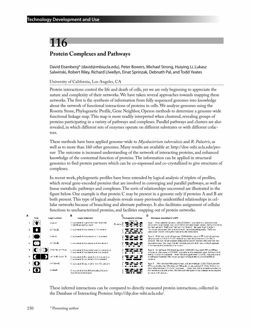
Technology Development and Use
150 * Presenting author
116 Protein Complexes and Pathways
David Eisenberg* ([email protected]), Peter Bowers, Michael Strong, Huiying Li, Lukasz Salwinski, Robert Riley, Richard Llwellyn, Einat Sprinzak, Debnath Pal, and Todd Yeates
University of California, Los Angeles, CA
Protein interactions control the life and death of cells, yet we are only beginning to appreciate the nature and complexity of their networks. We have taken several approaches towards mapping these networks. The first is the synthesis of information from fully sequenced genomes into knowledge about the network of functional interactions of proteins in cells. We analyze genomes using the Rosetta Stone, Phylogenetic Profile, Gene Neighbor, Operon methods to determine a genome-wide functional linkage map. This map is more readily interpreted when clustered, revealing groups of proteins participating in a variety of pathways and complexes. Parallel pathways and clusters are also revealed, in which different sets of enzymes operate on different substrates or with different cofac-tors.
These methods have been applied genome-wide to Mycobacterium tuberculosis and R. Palustris, as well as to more than 160 other genomes. Many results are available at: http://doe-mbi.ucla.edu/pro-nav The outcome is increased understanding of the network of interacting proteins, and enhanced knowledge of the contextual function of proteins. The information can be applied in structural genomics to find protein partners which can be co-expressed and co-crystallized to give structures of complexes.
In recent work, phylogenetic profiles have been extended by logical analysis of triplets of profiles, which reveal gene-encoded proteins that are involved in converging and parallel pathways, as well as linear metabolic pathways and complexes. The sorts of relationships uncovered are illustrated in the figure below. One example is that protein C may be present in a genome only if proteins A and B are both present. This type of logical analysis reveals many previously unidentified relationships in cel-lular networks because of branching and alternate pathways. It also facilitates assignment of cellular functions to uncharacterized proteins, and facilities mapping out of protein networks.
These inferred interactions can be compared to directly measured protein interactions, collected in the Database of Interacting Proteins: http://dip.doe-mbi.ucla.edu/.
Technology Development and Use
151* Presenting author
ReferencesVisualization and interpretation of protein networks in Mycobacterium tuberculosis based on hierar-chical clustering of genome-wide functional linkage maps. M. Strong, T.G. Graeber, M. Beeby, M. Pellegrini, M.J. Thompson, T.O. Yeates, & D. Eisenberg (2003). Nucleic Acids Research, 31, 7099-7109 (2003).
Prolinks: a database of protein functional linkages derived from coevolution. P.M. Bowers, M. Pel-legrini, M.J. Thompson, J. Fierro, T.O. Yeates, and D. Eisenberg (2004). Genome Biology, 5:R35.
Use of Logic Relationships to Decipher Protein Network Organization. P.M. Bowers, S.J. Cokus, D. Eisenberg & T.O. Yeates, Science, 306, 2246-2249 (2004).
117 Metabolomic Functional Analysis of Bacterial Genomes
Clifford J. Unkefer* ([email protected])
Los Alamos National Laboratory, Los Alamos, NM
Parallel with the terms genome, transcriptome and proteome, the combined profile of cellular metabolites is the metabolome. Examining changes in the metabolome is a potentially powerful approach to assessing gene function and contribution to phenotype. Achieving the GTL goal of obtaining a complete understanding of cellular function will require an integrated experimental and computational analysis of genome, transcriptome, proteome as well as the metabolome. Moreover, metabolites and their concentrations are a product of cellular regulatory processes, and thus the metabolome provides a clear window into the functioning of the genome and proteome. The profile of metabolites also reflects the response of biological systems to genetic or environmental changes. In addition, metabolites are the effectors that regulate gene expression and enzyme activity. The focus of this project is the elucidation of gene function by analysis of the metabolome. We will carry out functional studies using stable isotope labeling and Mass or NMR spectral analysis of low-molecular weight metabolites. Like the proteome, metabolic flux and metabolite concentrations change with the physiological state of the cell. Because metabolite flux and concentration are correlated with the physiological state, they can be used to probe regulatory networks. In prokaryotic organisms, the combination of functional information derived from metabolic flux analysis with gene and protein expression data being developed in other laboratories will provide a powerful approach in identifying gene function and regulatory networks. Our pilot studies will build upon our capability, demonstrate the scientific value, and establish a facility for isotope-enhanced high throughput metabolome analy-sis of sequenced environmental microbes.
The power of metabolome analysis will be greatly enhanced by applying the combination of stable isotope labeling and mutations. Stable Isotope labeling and NMR/Mass spectral analysis of metabo-lites will be used to assign metabolic function in three ways. First, we will apply specifically labeled compounds to establish precursor product relationships, and test if putative pathways identified from analysis of the genome are operational. Next, we will develop the capability for functional genomic analysis using comparative metabolomics to reveal the phenotype of a set of so-called silent mutations. This method combines null mutants constructed from the genome sequence by allelic exchange with metabolomic analysis to elucidate the function of unknown ORF’s. Finally, we will carry out a full metabolic flux analysis in steady state cultures. Flux analysis will provide input for a
1.
2.
3.
Technology Development and Use
152 * Presenting author
stoichiometric model. Many of the advantages of isotope labeling for metabolomics in autotrophs and methylotrophs will be demonstrated throughout this proposal. Once demonstrated, this capabil-ity will be even more powerfully applied to heterotrophic organisms growing on complex substrates. These studies will lay the foundation to take similar labeling and metabolomic strategies into the environment to study microbial communities.
118 Dynameomics: Mass Annotation of Protein Dynamics through Molecular Dynamics Simulations of Fold-Space Representatives
David A. C. Beck* ([email protected]), Ryan Day, Kathryn A. Scott, R. Dustin Schaeffer, Robert E. Steward, Amanda L. Jonsson, Darwin O. V. Alonso, and Valerie Daggett
University of Washington, Seattle, WA
The Protein Data Bank (PDB) has been a tremendously useful repository of experimentally derived, static protein structures that have stimulated many important scientific discoveries. While the util-ity of static physical representations of proteins is not in doubt, as these molecules are fluid in vivo, there is a larger universe of knowledge to be tapped regarding the dynamics of proteins. Thus, we are constructing a complementary database comprised of molecular dynamics (MD) simulation [1] derived structures for representatives of all protein folds. We are calling this effort ‘dynameomics.’ For each fold (derived from consensus between SCOP, CATH, and DALI [2]) a representative protein is simulated in its native (i.e., biologically relevant) state and along its complete unfolding pathway by MD, the time-dependent integration of the classical equations of motion for molecular systems. There are approximately 1130 known non-redundant folds, of which we have simulated the first 30 that represent about 50% of known proteins. With the data resulting from our large database of MD simulations, we are data-mining for patterns and general features of transition, intermediate and denatured states to improve not only our understanding of protein dynamics but structure prediction, protein-protein and protein-ligand docking algorithms. Structure prediction remains one of the most elusive goals of protein chemistry. It is necessary to successfully predict native states of proteins, in order to translate the current deluge of genomic information into a form appropriate for functional identification of proteins from their primary sequence and rapid structure / dynamics based drug design. While these specific aims represent our immediate scientific goal for the dynameomics data, we are constructing a web site (http://www.dynameomics.org) for publication of the trajectories’ coordinate data as well as in-depth analyses so that others may avail themselves of the resource and initiate areas of inquiry that we cannot even begin to anticipate.
ReferencesBeck, D. A. C., and Daggett, V. (2004) Methods for Molecular Dynamics Simulations of Protein Folding / Unfolding in Solution, Methods 34, 112-120.
Day, R., Beck, D. A. C., Armen, R. S., and Daggett, V. (2003) A consensus view of fold space: Combining SCOP, CATH, and the Dali Domain Dictionary, Protein Sci 12, 2150-2160.
1.
2.
153* Presenting author
Ethical, Legal, and Societal Issues
119 The DNA Files®
Bari Scott* ([email protected])
SoundVision Productions®, Berkeley, CA
SoundVision’s highly acclaimed series The DNA Files® has twice demonstrated that complex science could be made clear and exciting to listeners with no science background. The DNA Files 3 will con-tinue to show the general public the importance of cutting-edge science to their everyday lives and, at the same time, expand into a larger audience of minority and rural listeners. In addition, we plan to extend the impact of The DNA Files 3 beyond the airwaves into schools, museums, news outlets, and even the corner coffee shop through a new network of outreach services, media projects, and learning programs along with our rich and informative web site.
The DNA Files 3 will include five nationally distributed, hour-long public radio documentaries and five 5-minute features exploring the revolutionary new developments in systems biology, neurobiolo-gy, immunology and the interaction of the environment with our DNA. Program topics will include: Ethics Beyond the Genome: Systems Biology and Nanotechnology; Beyond DNA: The RNA World and Immunology; Individualizing the Genome: Toxicogenomics and Pharmacogenomics; Our Common Genes: Bugs, Mice and the Human Body; Depression, Addiction and Our Genes: Neurogenetics. In addition to the documentaries themselves, SoundVision has added components to The DNA Files® project that will promote science journalism by less-experienced science journalists and in the ethnic media; engage the public directly in workshops employing some of the science concepts covered; and contribute to science education and journalism across a broad spectrum of platforms. The project’s new outreach and education services, many of which are geared towards increasing minority and rural audiences, include:
• Media support and training
With the goal of expanding and enhancing coverage that makes science clearly relevant to a diverse population, The DNA Files 3 will make a variety of resources available for journalists and media outlets. This and related outreach will extend the documentaries’ impact by encouraging local and regional initiatives and motivating additional programs and news stories outside of SoundVision.
Materials and resources: SoundVision will provide talk show discussion topics targeted to specific ethnic communities as well as general audiences; lists of experts whom reporters can use as sources for programs and follow-up news reports, and highlights from our five documentaries. We will also develop The DNA Files Style Book, a handbook of best practices in science reporting on genetics and molecular biology geared to both minority-owned media and public radio as a whole. In a “push” element, SoundVision will send out short news alerts to reporters and editors to identify news stories related to The DNA Files 3 documentaries. And, working with two ethnic media consortia, we will provide targeted background material for ethnic print.
Ethical, Legal, and Societal Issues
154 * Presenting author
Funding and support: In addition to making the above resources generally available, SoundVision will work directly with up to 20 public radio stations. Technical support and grants (from another funding source) will be made available to stations on a competitive basis for local programming and projects related to The DNA Files 3. Stations will be encouraged to develop community outreach strategies and to produce programs ranging from feature reports and local documentaries to call-in programs. The impact is likely to be significant: If each of the 20 stations awarded outreach grants produced just one hour of programming, that alone would quadruple the amount of on-air content resulting from The DNA Files 3 funding. Our outreach consultants will help these stations develop community interest, build partnerships with ethnic and minority press, and work with local science and informal learning centers.
• Educational programs
The Exploratorium, the world-renowned museum of science, art, and human perception in San Francisco, has demonstrated its support for The DNA Files® by agreeing to create up to five hands-on workshops that can be produced at the Exploratorium and other science museums around the coun-try. The Exploratorium also will create a DNA Files Workshop Kit with materials to provide hands-on learning experiences. The kit will be designed for use by other museums, schools, churches, parks and recreation departments, as well as parents who home-school their children. It will be distributed to these outlets in tandem with the airing of The DNA Files 3 documentaries.
• Enhanced web site
Our information-packed multimedia web site will provide tool kits to help reporters, editors, muse-um directors, teachers, and home-schooling parents build articles and lesson plans around The DNA Files 3 programs. Materials on the site will include original in-depth articles related to each of the five documentaries, background information and research for editors and reporters, a library of links to related web sites, and The DNA Files® Style Book. The DNA Files 3’s improved web site will support public radio programming and museum and school programs that can stimulate public interest in science long after the series airs.
Evaluation
An independent firm specializing in educational media will evaluate The DNA Files 3. Its principals will conduct annual online user surveys and interview listeners to gage their understanding and retention of the project’s core themes. The evaluation also will include an evaluation of our relation-ship with outreach stations, plus pre- and post-production focus groups with multicultural audiences, African American audiences, and high school and junior college level biology teachers.
Project History
SoundVision, a 501(c)(3) nonprofit organization, has produced two previous The DNA Files® radio projects and a related multimedia web site supported by major grants from a variety of sources. The DNA Files® has won numerous awards, including the George Foster Peabody Award and the Alfred I. duPont-Columbia University Award. National Public Radio, which aired The DNA Files®, will continue to air The DNA Files 3, hosted by NBC Dateline reporter John Hockenberry, in its member stations.
Ethical, Legal, and Societal Issues
155* Presenting author
120 Science Literacy Training for Public Radio Journalists
Bari Scott* ([email protected])
SoundVision Productions®, Berkeley, California
In the genomic era, journalists bear a greater responsibility than ever to communicate science’s rapid advances and their societal implications to the public. Understanding the basics about DNA and human genetics, which most journalists still are learning, are no longer enough. Now the media must grasp concepts about the regulation of gene expression, the activity of proteins, the workings of RNA and other mechanics of the cell, both in humans and other forms of life such as microbes. Advances in these areas have profound implications, and journalists are obliged to provide clear and accurate information to the public.
SoundVision Productions®, creator and facilitator of workshops for public radio journalists, will develop a new series of three, week-long science literacy training workshops and related activities to help public radio reporters and producers meet this challenge. Through a competitive process, twelve mid-career public radio producers and reporters will be selected to attend these sessions. SoundVi-sion will select participants who represent stations and national or regional programs that reach broad audiences. We also place a high priority on including journalists from rural and minority-controlled stations and networks. The recruitment and selection process will be designed to ensure ethnic, racial, and gender diversity.
The SoundVision workshops incorporate three goals. They are designed to increase the number and the quality of science stories produced for radio; increase the number of reporters able to report competently on complex research processes, discoveries, and resulting societal implications; and ulti-mately, increase civic literacy and help minimize the widening knowledge gap between the scientific community and the public. We will explore the interactions of DNA, RNA, and proteins and the overall complexity of the machinery of the cell; teach reporters what scientists are discovering about the most basic elements of life; examine the characteristics of one or more nonpathogenic microbes of environmental importance; and delve into the interactions between the human genome and the environment and toxicogenomics. A key workshop focus will be ethics in the post-human-genome-project era, including new questions about the relationship between science and business, the impact of highly patented science on society, and the risks and responsibilities of attempting to manipulate life.
Each workshop will center around 15 to 20 presentations by scientists, science journalists, scientific researchers, and radio production professionals. Sessions will orient producers to basic science, focus on the craft and responsibility of science journalism, and explore techniques for presenting complex scientific content on radio. The focus on radio is particularly important given the specific production needs that distinguish radio from other media. Each workshop will include a field trip and several informal gatherings with scientists to develop relationships and learn more about their ideas and research.
In order to reach more producers, the week-long workshops will be held in key scientific and cultural centers throughout the country. The first will be held in Boston, co-hosted by WGBH-FM and The Whitehead Institute. The second will be held in San Francisco at KQED-FM, and the third will be in Austin, Texas, in cooperation with Latino USA/KUT-FM at the University of Texas.
Ethical, Legal, and Societal Issues
156 * Presenting author
SoundVision’s training methodology has had long-lasting positive effects on public radio journalists who have attended previous workshops. Even years after attending, participants from rural to large metropolitan stations report that the workshops, which were funded by the U.S. Department of En-ergy, still help them with their work. Producers and reporters continue to benefit from their familiar-ity with the basics of DNA research, an ability to identify stories that they wouldn’t previously have tackled, and better skills in getting behind press releases and scientific papers to create compelling public radio features. SoundVision’s innovative workshops boost participants’ confidence and their ability to communicate complex and emerging scientific research accurately. We believe that their collective work throughout the country will help lead to a better public understanding of current scientific research and its social implications.
The project also includes a website that provides transcripts and selected audio from the training ses-sions, “tip sheets” and online resources for participants and interested users. Follow-up teleconferenc-es will support participants in pursuing complex and rewarding science stories for their communities. In addition, if funds permit, a pilot interactive DVD with highlights of the workshop will be offered to rural and minority-controlled stations and networks. If successful, the pilot may be developed and used for other applications.
As in our previous workshop projects, a comprehensive evaluation will be conducted by Rockman et al, a well-established, San Francisco-based evaluation firm with expertise in evaluating media projects and assessing the impact of training on journalistic practice.
Although the project targets public radio producers and reporters, with slight modifications the workshop is applicable and adaptable for news directors and editors, live interview call-in hosts and their producers (since so much air time is dedicated to that format), and even television staff..
157
As of January 18, 2005
Carl Anderson Brookhaven National Laboratory [email protected]
Gordon Anderson Pacific Northwest National Laboratory [email protected]
Adam Arkin Lawrence Berkeley National Laboratory [email protected]
Holly Baden-Tillson J. Craig Venter Institute [email protected]
Ray Bair Argonne National Laboratory [email protected]
Kristin Balder-Froid Lawrence Berkeley National Laboratory [email protected]
Duhee Bang University of Chicago [email protected]
Gang Bao Georgia Institute of Technology / Emory University [email protected]
Christian Barnes J. Craig Venter Institute [email protected]
Paul Bayer U.S. Department of Energy [email protected]
Jane BearingerLawrence Livermore National [email protected]
Appendix 1: Attendees
David Beck University of Washington [email protected]
Alexander Beliaev Pacific Northwest National Laboratory [email protected]
Andrea BelgranoNational Center for Genome [email protected]
Harvey Bolton Pacific Northwest National Laboratory [email protected]
Jennifer Bownas Oak Ridge National Laboratory [email protected]
Timothy Boyle Sandia National Laboratories [email protected]
Andrew Bradbury Los Alamos National Laboratory [email protected]
James Brainard Los Alamos National Laboratory [email protected]
Joseph BreenNational Institutes of [email protected]
David Bruce DOE Joint Genome Institute / Los Alamos National Laboratory [email protected]
James Bruce Washington State University [email protected]
Attendees
158
Erick Butzlaff University of Wisconsin [email protected]
Denise Casey Oak Ridge National Laboratory [email protected]
Parag Chitnis National Science Foundation [email protected]
Paul Choi Harvard University [email protected]
Ray-Yuan Chuang J. Craig Venter Institute [email protected]
Bruce Church Gene Network Sciences [email protected]
George ChurchHarvard University / Massachusetts Institute of [email protected]
Dean ColeU.S. Department of [email protected]
Frank CollartArgonne National [email protected]
Michael ColvinUniversity of California, [email protected]
Sean ConlanWadsworth [email protected]
Maddalena CoppiUniversity of Massachusetts, [email protected]
Robert CoyneNational Science [email protected]
Paul CrozierSandia National [email protected]
Barbara CullitonGenome News [email protected]
Patrik D’haeseleerLawrence Livermore National [email protected]
Sacha De CarloUniversity of California, [email protected]
Anjali DhimanGene Network [email protected]
Timothy DonohueUniversity of Wisconsin, [email protected]
Norman DovichiUniversity of [email protected]
Kenneth DowningLawrence Berkeley National [email protected]
Daniel DrellU.S. Department of [email protected]
Leland EllisDepartment of Homeland [email protected]
Larry FelserGene Network [email protected]
Attendees
159
Matthew FieldsMiami [email protected]
Peg FoltaLawrence Livermore National [email protected]
Marvin FrazierJ. Craig Venter [email protected]
James K. FredricksonPacific Northwest National [email protected]
Haichun GaoOak Ridge National [email protected]
Timothy GardnerBoston [email protected]
George GarrityMichigan State [email protected]
Sara GaucherSandia National [email protected]
Al GeistOak Ridge National [email protected]
Damian GesslerNational Center for Genome [email protected]
Daniel GibsonJ. Craig Venter [email protected]
Carol GiomettiArgonne National [email protected]
Maria GiovanniNational Institutes of [email protected]
Stephen GiovannoniOregon State [email protected]
John GlassJ. Craig Venter [email protected]
Peter GoodNational Institutes of [email protected]
Yuri GorbyPacific Northwest National [email protected]
Andrey GorinOak Ridge National [email protected]
Patrick GrantLawrence Livermore National [email protected]
Chris [email protected]
David HaalandSandia National [email protected]
Masood HadiSandia National [email protected]
Qiang HeOak Ridge National [email protected]
Grant HeffelfingerSandia National [email protected]
Attendees
160
Robert HettichOak Ridge National [email protected]
Peter HighnamNational Institutes of [email protected]
Roland HirschU.S. Department of [email protected]
Dawn HolmesUniversity of Massachusetts, [email protected]
Norman HommesOregon State [email protected]
Brian HookerPacific Northwest National [email protected]
John HoughtonU.S. Department of [email protected]
Greg HurstOak Ridge National [email protected]
Thomas HuserLawrence Livermore National [email protected]
Clyde HutchisonJ. Craig Venter [email protected]
Prabha IyerJ. Craig Venter [email protected]
Eric JakobssonUniversity of Illinois, [email protected]
Barbara JasnyScience/[email protected]
Grant JensenCalifornia Institute of [email protected]
Matthew KaneNational Science [email protected]
Samuel KaplanUniversity of Texas Medical School, [email protected]
Arthur KatzU.S. Department of [email protected]
Brian KayArgonne National [email protected]
Jay KeaslingUniversity of California, [email protected]
Martin KellerDiversa [email protected]
Stephen KennelOak Ridge National [email protected]
Vladimir KeryPacific Northwest National [email protected]
Arnold KimUniversity of California, [email protected]
William KimmerlyPacific Northwest National [email protected]
Attendees
161
David KleinGener8, [email protected]
Michael KnotekConsultant to [email protected]
Eugene [email protected]
David KoppenaalPacific Northwest National [email protected]
Henrietta [email protected]
Stephen LaneLawrence Livermore National [email protected]
Jennie LarkinNational Institutes of [email protected]
Carole LartigueJ. Craig Venter [email protected]
Charles LawrenceBrown [email protected]
Philip LeDucCarnegie Mellon [email protected]
Adam LeeUniversity of [email protected]
Kyriacos LeptosHarvard Medical [email protected]
Huilin LiBrookhaven National [email protected]
Xiaoxia LinHarvard Medical [email protected]
Derek LovleyUniversity of [email protected]
William MacConnellMacConnell Research [email protected]
Lee MakowskiArgonne National [email protected]
Alexander MalkinLawrence Livermore National [email protected]
Reinhold MannOak Ridge National [email protected]
Betty MansfieldOak Ridge National [email protected]
Roummel MarciaUniversity of Wisconsin, [email protected]
Anthony MartinoSandia National [email protected]
Adam MartinyMassachusetts Institute of [email protected]
Mahir MarufJ. Craig Venter [email protected]
Attendees
162
M. Uljana Mayer-CumblidgePacific Northwest National [email protected]
Harley McAdamsStanford [email protected]
Lee Ann McCueWadsworth [email protected]
Gail McLeanU.S. Department of [email protected]
Barbara MetheThe Institute for Genomic [email protected]
Lisa MillerBrookhaven National [email protected]
Marissa MillsOak Ridge National [email protected]
Julie MitchellUniversity of Wisconsin, [email protected]
Jennifer Morrell-FalveyOak Ridge National [email protected]
Sue MorssArgonne National [email protected]
Vasantha [email protected]
Dave NelsonLawrence Livermore National [email protected]
Lee NewbergWadsworth [email protected]
Charles NgoweMichigan State [email protected]
Kim NylanderOak Ridge National [email protected]
Yasuhiro OdaUniversity of [email protected]
Susan OldNational Institutes of [email protected]
Paola [email protected]
Paula OlsiewskiAlfred P. Sloan [email protected]
Regina O’NeilUniversity of Massachusetts, [email protected]
Mary Oster-GraniteNational Institutes of [email protected]
Brian PalenikUniversity of California, San [email protected]
Aristides PatrinosU.S. Department of [email protected]
Dale PelletierOak Ridge National [email protected]
Attendees
163
Cynthia PfannkochJ. Craig Venter [email protected]
Anne PlantNSTC/[email protected]
Steve PlimptonSandia National [email protected]
Gemma RegueraUniversity of Massachusetts, [email protected]
Mark RintoulSandia National [email protected]
Margie RominePacific Northwest National [email protected]
Denise RussoNational Institutes of [email protected]
Herbert SauroKeck Graduate [email protected]
Luis Sayavedra-SotoOregon State [email protected]
David SchwartzUniversity of Wisconsin, [email protected]
Salvatore SechiNational Institutes of [email protected]
Michael SeibertNational Renewable Energy [email protected]
Margrethe SerresMarine Biological [email protected]
Liang ShiPacific Northwestern National [email protected]
Michael SinclairSandia National [email protected]
Dinah SingerNational Cancer [email protected]
Richard SmithPacific Northwest National [email protected]
Hamilton SmithJ. Craig Venter [email protected]
Harold SmithUniversity of [email protected]
Thomas SquierPacific Northwest National [email protected]
Ranjan SrivastavaUniversity of [email protected]
David A. StahlUniversity of [email protected]
Marvin StodolskyU.S. Department of [email protected]
F. William StudierBrookhaven National [email protected]
Attendees
164
John SutherlandBrookhaven National [email protected]
F. Robert TabitaOhio State [email protected]
Chad TalleyLawrence Livermore National [email protected]
Theodore TarasowLawrence Livermore National [email protected]
Michael TeresinskiU.S. Department of [email protected]
Michael ThelenLawrence Livermore National [email protected]
David ThomassenU.S. Department of [email protected]
Janelle ThompsonMassachusetts Institute of [email protected]
Joyce ThorpeJ. Craig Venter [email protected]
James TiedjeMichigan State [email protected]
Jerilyn TimlinSandia National [email protected]
Jeffrey TokLawrence Livermore National [email protected]
Emily TurnerUniversity of [email protected]
Ravishankar VallabhajosyulaKeck Graduate [email protected]
Sanjay VasheeJ. Craig Venter [email protected]
Wim VermaasArizona State [email protected]
Judy WallUniversity of Missouri, [email protected]
Sharlene WeatherwaxU.S. Department of [email protected]
Peter WeberLawrence Livermore National [email protected], [email protected]
Xueming WeiOregon State [email protected]
Raymond WildungU.S. Department of [email protected]
H Steven WileyPacific Northwest National [email protected]
David WilsonCornell [email protected]
X. Sunney XieHarvard [email protected]

Attendees
165
Ying XuUniversity of [email protected]
Qing XuJ. Craig Venter [email protected]
Luying XunHarvard [email protected]
Haw YangUniversity of California, [email protected]
Jane YeNational Institutes of [email protected]
Shibu YoosephJ. Craig Venter [email protected]
Karsten ZenglerDiversa [email protected]
Jizhong ZhouOak Ridge National [email protected]
Julie ZimmermanLouisiana State [email protected]
Jeremy ZuckerHarvard Medical [email protected]

Attendees
166
167
Appendix 2: Web Sites
Program Web SitesGenomics:GTL Web site: http://doegenomestolife.org/
This book: http://doegenomestolife.org/pubs/2005abstracts/
DOE Microbial Genome Program: http://microbialgenome.org/
Web Sites Listed in AbstractsBioWarehouse System: http://bioinformatics.ai.sri.com/biowarehouse/
Carbon Sequestration in Synechococcus sp.: From Molecular Machines to Hierarchical: Modeling http://www.genomes2life.org/
Center for Molecular and Cellular Systems: http://www.ornl.gov/GenomestoLife/
GelBank: http://gelbank.anl.gov/
Joint Genome Institute: http://www.jgi.doe.gov/
LAMMPS Molecular Dynamics Simulator: http://www.cs.sandia.gov/~sjplimp/lammps.html
Microbial Ecology, Proteogenomics & Computational Optima. DOE Genomes to Life Center at Harvard/MIT/BWH/MGH: http://arep.med.harvard.edu/DOEGTL/
ProteomeWeb: http://proteomeweb.anl.gov/
Synechococcus sp. WH8102 Knowledgebase: http://csbl.bmb.uga.edu/WH8102/
Synechocystis sp. PCC 6803: http://lsweb.la.asu.edu/synechocystis/
The SEED: An Annotation/Analysis Tool: http://theseed.uchicago.edu/FIG/index.cgi
Virtual Institute of Microbial Stress and Survival (VIMSS): http://vimss.org
•
•
•
•
•
•
•
•
•
•
•
•
•
•
•

Web Sites
168
169Indexed by page number.
Author Index
AAbulencia, Carl 8, 11, 88Acinas, Silvia G. 89Ackerman, Eric J. 132Adamson, Anne E. 65Adhikari, Devi 115Adkins, Joshua N. 16, 19, 143Ahrendt, Angela 58Albrecht, Huguette 124Allen, Eric 87Alm, Eric 6, 8Alonso, Darwin O. V. 152Alperovich, Nina 60Alton, Anita J. 65Andersen, Gary L. 8, 11, 107Anderson, Gordon A. 51, 114, 115, 143, 144, 148Anderson, Kenneth K. 18Andrews, Shirley H. 65Andronaco, Gregory 130Aravind, L. 83Arkin, Adam 6, 8Arnold, Nikita D. 22Arp, Daniel 93Assad-Garcia, Nacyra 60Atlas, Jordan C. 68Auberry, Deanna L. 16, 18, 19Auberry, Kenneth J. 19, 51
BBabnigg, Gyorgy 51, 58Baden-Tillson, Holly A. 61Bai, Yun 72Baker, Brett J. 87Bakkal, Suphan 44Balhorn, Rod 121, 142Banfield, Jill 87Bang, Duhee 134Bao, Gang 117Barnes, Christian 62Barns, Susan 88Barofsky, Douglas F. 95Battista, J. R. 96Bearinger, Jane 122Beatty, J. Thomas 105Beck, David A. C. 152Becker, Gerald W. 141Belgrano, Andrea 34Beliaev, Alex S. 51, 52, 148
Bench, Graham 122, 146Bender, Kelly 8Benoit, Jennifer 89Bertozzi, Carolyn 147Bibbs, Lisa 95Blake, Robert C., II 87Borglin, Sharon 8, 11Bowers, Peter 150Bownas, Jennifer L. 65Boyer, Stephen 130Bradbury, Andrew 133Brahamsha, Bianca 24Brandes, Aaron 3Brockman, Fred 88Broderick, Joan 99Broderick, William 99Brodie, Eoin L. 8, 11, 107Brown, Steven 14Brown, W. Michael 32Bruce, David 109Bruce, James E. 114, 115Buchanan, Michelle 15Burisch, Christian 38Butler, Jessica 46Butzlaff, Erick A. 74
CCai, Long 128Cai, Zhi 100Callister, Stephen 144Cannon, Bill 15Cantor, C.B. 56Cao, Haishi 135Carmack, C. Steven 72Casey, Denise K. 65Castellanos, Mariajose 68Cate, Jamie H. D. 119Chain, Patrick 109Chan, James W. 121Chan, Priscilla 142Chandramohan, Praveen 21Chang, Sherman 88Chaudhuri, Swades K. 46Chen, Baowei 135Chen, Shaolin 102Chen, X. 34Chhabra, Swapnil 8, 14Chin, Kuk-Jeong 44Chisholm, Sallie W. 4, 5, 90, 103
Index
170Indexed by page number.
Cho, Jang-Cheon 95Choi, Paul 128Chowdhury, Saiful 115Christian, Allen T. 121, 122Chuang, Ray-Yuan 62Church, Bruce 68Church, George M. 3, 4, 5Cole, James R. 72Coleman, Maureen 103Collart, Frank R. 16, 50, 51, 139, 140, 141Collins, James J. 50, 56Colvin, Michael 75, 124Comolli, Luis 126Conlan, Sean 70Coppi, Maddalena 44Corzett, Michele H. 121Cottarel, G. 56Cottrell, Matthew T. 112Crawford, Jackie 122Crozier, Paul S. 38
DDaggett, Valerie 152Daly, Don S. 18Daly, Michael J. 57Dam, P. 30, 34Dasika, Madhukar 84Davidson, Eric H. 91Davidson, George S. 28Davis, John 99Day, Robert M. 22Day, Ryan 152DeNardo, Sally 124Desiderio, Russell 95DeWitt, David 99Dhiman, Anjali 68DiDonato, Laurie 44DiDonato, Raymond 46Ding, Yanhuai R. 148Distel, Daniel L. 89Doktycz, Mitchel 15Dominguez, Miguel 144Donohue, Timothy J. 110, 144Downing, Kenneth H. 126Driscoll, M. E. 56Dubchak, Inna 6, 8Dugan, Lawrence 122
EEdlefsen, Paul 51Eisenberg, David 150Elias, Dwayne A. 51, 148Emo, Brett 14
Ensman, L. 76Entcheva-Dimitrov, Plamena 112Esposito, Anthony 121Estes, Sherry A. 65Esteve-Núñez, Abraham 44, 46
FFaith, J. 56Fan, J. 76Fang, Ruihua 143, 144, 148Faull, Kym 100Faulon, Jean-Loup 32Fey, Sarah 139, 140, 141Fields, Matthew 8, 11, 14Foote, Bob 15Foote, Linda J. 16Fredrickson, James K. 50, 51, 52, 58, 143, 144, 148Friddle, Raymond W. 124Fridman, Tema 22Friedman, Nir 128
GGalperin, Michael Y. 51Gao, Weimin 14Gardner, Timothy S. 50, 56Garrity, George M. 72Gassman, Natalie R. 55Gaucher, Sara P. 8, 14, 17Geist, Al 21, 37Gelfand, Mikhail 6Geller, Jil 8, 11Gerdes, Sveta 100Gessler, Damian 34Ghirardi, Maria L. 97Gibson, Janet L. 105Gilna, Paul 109Giometti, Carol S. 50, 51, 58Giovannoni, Stephen J. 95Givan, Scott 95Glass, John I. 60Glaven, Richard 44, 46Goddard, Carrie D. 144Gonzales, Arlene 31, 40Gorby, Yuri A. 51, 52, 135, 148Gorin, Andrey 22Grant, Patrick G. 146Green, Michelle L. 81, 82Grossman, Arthur R. 97
HHaaland, David M. 24, 33Hadi, Masood Z. 8, 14, 17
Index
171Indexed by page number.
Hainfeld, James 120Haller, Imke 88Hamad, Sawsan 33, 100Hanson, David 28Hara, Christine A. 121Harwood, Caroline S. 104, 105Harzman, Christina 99Havin, Victor 25Hazen, Terry C. 8, 11He, Qiang 8, 14He, Zhili 8, 14Heffelfinger, Grant S. 20Herman, Rob 24Herzberg, Osnat 137Hettich, Robert 15, 87Hiddessen, Amy 122Higdon, Roger 51Hill, Eric 52Hinz, Angela 122Hixson, Kim K. 143, 148Holland, Trevin 88Hollars, Christopher W. 121Holman, Hoi-Ying 8, 11, 126Holmes, Dawn E. 40, 46Holzman, Ted 51Hommes, Norman 93Hooker, Brian S. 15, 16Howard, Meredith 91Hoyt, Peter 16Hu, Ping 107Huang, Katherine 6, 8Huang, Rick 8, 11Hubbard, K. 76Hunt, Dana E. 89Hurst, Gregory B. 15, 16, 19Huser, Thomas 121, 122Hutcheon, Ian D. 123Hutchison, Clyde A., III 60, 61
IIakounine, Alexander 140Iqbal, Tahera 31Iyer, Gopal 55Iyer, Lakshminarayan 83Iyer, Prabha P. 64
JJacobsen, Janet 8, 11Jaffe, Jacob D. 4Jakobsson, Eric 83Jensen, Grant 117Jiang, T. 34Jonsson, Amanda L. 152
Joyner, Dominique 8, 11Judd, Ellen 126Jung, David 37
KKane, Sean 67Kang, Katherine 24, 25Kaplan, Samuel 144Karp, Peter D. 78, 81, 82Kay, Brian K. 141Kaye, Jonathan 40Keasling, Jay D. 8, 14, 147Kehoe, John 141Keller, Keith 8Keller, Martin 8, 11, 14, 88Kennel, Steve J. 15, 16Kent, Stephen B. 134Kery, Vladimir 15, 16, 18, 19Khare, Tripti 58Kim, Arnold 75Kim, Byoung-Chan 44Kim, Sang-Hoon 99Kim, Younggyu 55Kirchman, David L. 112Klappenbach, Joel 51Klein, David 130Klepac-Ceraj, Vanja 89Knorr, Andrea L. 69Kohanski, M. K. 56Kolker, Eugene 50, 51, 140Kolker, Natali 51Kong, Xiangxu 55Konstantinidis, Konstantinos T. 51Kriplani, Ushma 141Krishnamurthy, Ramya 21Krushkal, Julia 42, 44Kuske, Cheryl 88Kyrpides, Nikos 109
LLaidlaw, David 130Land, Miriam L. 51Landorf, Elizabeth 139, 140, 141Lane, Stephen M. 121Lane, Todd W. 31, 40Laney, Samuel 95Langry, Kevin 124Lankford, Trish K. 16Lanthier, Martin 40Lapidus, Alla 109Larimer, Frank W. 15, 18, 19, 50, 51, 104, 105, 109Lartigue, Carole 60Laub, Michael 108
Index
172Indexed by page number.
Laurence, Ted 121Lawrence, Charles E. 50, 51, 70, 72, 86Leang, Ching 44Leary, Julie 147Leavell, Michael D. 31LeDuc, Phil 117Lee, Pei-Yun 91Lee, Tom 78Lei, Chenghong 132Leighton, Terrance J. 127Leptos, Kyriacos C. 4Li, Huilin 120Li, Huiying 150Li, Ting 14Liao, James C. 105Light, Yooli K. 31Lightstone, Felice 75Lilburn, Timothy G. 72Lin, Chiann-Tso 16Lin, Xiaoxia 3Lindell, Debbie 4Lindsey, Susan D. 74Lipton, Mary S. 51, 57, 143, 144, 148Liu, Jun 132Liu, Yongqing 14Livesay, Eric A. 16, 143Llwellyn, Richard 150Londer, Yuri 139Lovley, Derek R. 40, 42, 44, 46, 48, 58, 148Lowry, David F. 135Lu, Tse-Yuan S. 16
MMahadevan, R. 42Maier, Klaus 79Makarova, Kira S. 51Makhatadze, George I. 134Malkin, Alexander J. 127Mansfield, Betty K. 65Mao, F. 30, 34Maranas, Costas D. 84Marcelino, Luisa A. 89Marcia, Roummel F. 74Marletta, Michael 147Marsh, Terence L. 99Martin, Shawn 28, 32Martin, Sheryl A. 65Martino, Anthony 31, 40Martino, Tony 25Martiny, Adam C. 5, 90Maruf, Mahir 60Mathur, Eric 95Mayer, M. Uljana 135
McAdams, Harley H. 107, 110, 126McCarthy, Kevin 46McClay, David 91McCue, Lee Ann 50, 51, 70, 72McDonald, W. Hayes 15, 19McKeown, Cathy K. 16McLean, Jeff 52, 135Mehta, Teena 46Mester, Tunde 46Methé, Barbara 44, 46, 48Miles, Robin 122Miller, Lisa M. 138Mills, Marissa D. 65Minokawa, Takuya 91Mitchell, Julie C. 74Monroe, Matthew E. 51, 143, 144, 148Moore, Priscilla A. 16Moore, Ronald J. 16, 143Morrell, Jenny 15Mottaz, Heather M. 148Moult, John 137Mukhopadhyay, Aindrila 8, 11, 14Munske, Gerhard 114, 115Mutatu, Washington 99
NNatarajan, Shreedhar 83Natarajan, Vijaya 6, 25Naughton, Thomas 25Nealson, Kenneth H. 50, 51, 123Nevin, Kelly 40, 44, 46, 48Newberg, Lee A. 72Ngowe, Michael 99Nie, Shuming 117Noordeweir, Mick 95Noy, Aleksandr 124Nunez, Cinthia 44Nylander, Kim 65
OO’Neil, Regina 40, 44Oda, Yasuhiro 104Oliveri, Paola 91Olman, V. 30, 34Omelchenko, Marina V. 57Ortoleva, P. J. 76Osterman, Andrei 100Ovecka, Milan 133Overbeek, Ross 100
Index
173Indexed by page number.
PPacocha, Sarah 89Pal, Debnath 150Paladugu, Sri 79Palenik, Brian 24, 25, 34Palmblad, Magnus 122, 146Pasa-Tolic, Ljiljana 51, 148Paulsen, Ian 24, 25Pavlik, Peter 133Pelletier, Dale A. 15, 16Peppler, Terese 139, 140, 141Petritis, Konstantinos 143Pfannkoch, Cynthia 60, 61Pharino, Chanathip 89Pharkya, Priti 84Phinney, Brett 99Picone, Alex F. 51Pinchuk, Grigoriy 51, 52Plimpton, Steven J. 38Plomp, Marco 127Podar, Mercha 95Podevels, Angela M. 110Polz, Martin F. 89Pomposiello, Pablo 44Popa, Radu 123Posewitz, Matthew C. 97Postier, Brad 40Pouliot, Yannick 78Price, Morgan 6, 8Purvine, Samuel 51
QQiu, Xiaoyun 51, 57
RRam, Rachna J. 87Rappé, Michael S. 95Ratto, Timothy V. 124Redding, Alyssa 14Reguera, Gemma 46Ren, Xiajia 128Reppas, Nikkos B. 5Richardson, Paul 109Riley, Monica 50, 51, 54Riley, Robert 150Ringbauer, Joseph A., Jr. 8, 14Rintoul, Mark D. 28, 32, 38Roberson, Robert 100Roh, Jung Hyeob 144Rohwer, Forest 103Romine, Margaret F. 51, 52, 144, 148Roth, Martin 67
Ruan, Jin 67Rubin, Eddy 109
SSaffarini, Daad A. 52Salwinski, Lukasz 150Samanta, Sudip K. 104Samatva, Nagiza 21Sapra, Rajat 31Sarma-Rupavtarm, Ramahi 89Sauro, Herbert M. 79Sayavedra-Soto, Luis 93Sayyed-Ahmad, A. 76Schaeffer, R. Dustin 152Schilling, Christophe H. 42, 67Schlitter, Jürgen 38Schmoyer, Denise D. 15, 18, 19Schmutz, Jeremy 109Scholle, Michael 141Schwarz, Fred 137Scott, Bari 153, 155Scott, Kathryn A. 152Segrè, Daniel 3Segura, D. 42Seibert, Michael 97Serres, Margrethe H. 51, 54Shah, Manesh B. 15, 18, 19, 87Shapiro, Lucy 110Shelbolina, Zhenya 40Shen, Yufeng 143Shi, Liang 135Shields, Sharon 146Shin, Yongsoon 132Shiyanov, Pavel 141Shoshani, Arie 25Shuler, Michael L. 68Sinclair, Michael B. 33Singh, Anup 8, 14Slepoy, Alex 38Smith, Hamilton O. 60, 61, 62, 63, 64Smith, Harold 137Smith, Joel 91Smith, Richard D. 51, 143, 144, 148Smithson, David 142Spormann, Alfred M. 112Sprinzak, Einat 150Squier, Thomas C. 135Srivastava, Ranjan 69Stadsklev, Kurt 67Stahl, David 8, 11Stanek, Dawn 14Stanley, F. 76Stapels, Martha D. 95
Index
174Indexed by page number.
Steward, Robert E. 152Stolyar, Sergey 8, 11Strauss, Charlie 32Strittmatter, Eric F. 19Strong, Michael 150Studier, F. William 138Su, Z. 30, 34Sui, Haixin 126Sulchek, Todd A. 124Sullivan, Matthew B. 103Sun, Jun 8, 11, 14Sun, Lianhong 14Sundin, George 57Sutherland, John C. 138
TTabita, F. Robert 105Tai, P. K. Amos 90Talley, Chad E. 121, 122Tang, Xiaoting 114, 115Tarasow, Ted 142Tasneem, Asba 83Tavano, Christine L. 110, 144Thakar, Rajendra 67Thelen, Michael P. 87Thomas, Ed 24Thompson, Anne W. 90Thompson, Dorothea 8, 11, 14Thompson, Janelle R. 89Tiedje, James M. 50, 51, 57, 99Timlin, Jerilyn A. 24, 25, 33Tok, Jeffrey B.-H. 142Tolic, Nikola 114, 115, 144Tollaksen, Sandra L. 58Tran, Hoa 44Travnik, Evelyn 67Tripp, James 95Tsang, Allen 44Tsapin, Sasha 51Tu, Qiang 91Tuncay, K. 76Tuominen, Mark T. 46Tyson, Gene W. 87
UUnkefer, Clifford J. 151
VVajrala, Neeraja 93Vallabhajosyula, Ravishankar R. 79van de Meene, Allison 100van Dien, Steve 8, 11, 67
Varala, K. 76Varma, Sameer 83Vashee, Sanjay 62Vasilenko, Alexander 57Velappan, Nileena 133Venter, J. Craig 60, 61, 62, 63, 64VerBerkmoes, Nathan 87Vergin, Kevin L. 95Verma, Seema 135Vermaas, Wim 33, 100Victry, Kristin D. 16
WWabb, Dave L. 19Wagner, Valerie 78Walcher, Marion 88Walker, Christopher 8Wall, Judy 8, 11, 14Wang, Bing 100Wang, Yue 6Weber, Peter K. 123Webster, Jennifer 48Wei, Jing 8, 14Wei, Xueming 93Weigele, Peter 103Weis, Robert 44Weiss, Shimon 50, 55Wemmer, David 147Werner-Washburne, Margaret 28Wheeler, Elizabeth 122Wheeler, Katherine E. 127White, Amanda M. 18Whitelegge, Julian 100Wiback, Sharon 67Wierzbowski, J. 56Wiley, Steven 15Wilson, David B. 102Winhold, Heiko 121Wolf, Denise 8Wu, H. 34Wu, Liyou 14, 57Wu, Yifei 141Wyborski, Denise 8, 11, 88Wyrick, Judy M. 65
XXiao, Jie 128Xie, X. Sunney 128Xu, Qing 63Xu, Ying 30, 34Xun, Luying 128
Index
175Indexed by page number.
YYan, Bin 42, 44Yan, Ping 135Yang, Haw 119Yang, Lin 138Yates, John R., III 51, 58Yeates, Todd 150Yen, Huei-Che 8, 11, 14Yooseph, Shibu 60, 63Young, Malin M. 17Yu, Ji 128Yu, Wen 14
ZZakrajsek, Brian A. 51Zane, Grant 8, 11, 14Zeng, Xiaihua 144Zengler, Karsten 88Zhang, Kun 5Zhang, Yuan 72Zhang, Zhaoduo 31, 40Zhao, Rui 143Zhou, Jizhong 8, 11, 14, 50, 51, 52, 57, 105Zhu, Harry 115Zhu, Wenhong 51, 58Zimmerman, J. M. 96Zinser, Eric 4Zucker, Jeremy 3Zuniga, Beto 8, 11

Index
176Indexed by page number.
Index
177Indexed by page number.
American Type Culture Collection 72Argonne National Laboratory 16, 50, 51, 58, 139,
140, 141Arizona State University 33, 100A & M College 96BIATECH 50, 51, 140Boston University 50, 56Brookhaven National Laboratory 120, 138Brown University 70, 72, 86California Institute of Technology 91, 117Carnegie Institution of Washington 97Carnegie Mellon University 117Center for Advanced Research in Biotechnology
137Children’s Hospital Oakland Research Institute 127Colorado School of Mines 97Columbus State University 99Cornell University 68, 102Diversa Corporation 8, 11, 14, 88, 95Duke University 91East Carolina University 138Emory University 117Fellowship for the Integration of Genomes (FIG)
100Gener8, Inc. 130Gene Network Sciences 68Genomatica, Inc. 42, 67Georgia Institute of Technology 117Harvard Medical School 3, 4, 5Harvard University 108, 128Hawaii Institute of Marine Biology 95Howard Hughes Medical Institute 6Indiana University, Bloomington 76J. Craig Venter Institute 60, 61, 62, 63, 64Joint Genome Institute 109Keck Graduate Institute 79Lawrence Berkeley National Laboratory 6, 8, 11,
14, 25, 107, 109, 119, 126, 147Lawrence Livermore National Laboratory 75, 109,
121, 122, 123, 124, 127, 142, 146Los Alamos National Laboratory 32, 88, 109, 133,
151Louisiana State University 96Marine Biological Laboratory 50, 51, 54Massachusetts Institute of Technology 4, 5, 89, 90,
103Miami University 8, 11Michigan State University 50, 57, 72, 99
National Center for Genome Resources 34National Institutes of Health 51, 57, 83National Renewable Energy Laboratory 97New England Biolabs 89Oak Ridge National Laboratory 8, 11, 14, 15, 16,
18, 19, 21, 22, 25, 30, 34, 37, 50, 51, 52, 57, 65, 87, 104, 105, 109
Ohio State University 105Oregon State University 93, 95Pacific Northwest National Laboratory 15, 16, 18,
19, 50, 51, 52, 57, 58, 88, 114, 115, 132, 135, 143, 144, 148
Pennsylvania State University 84, 134Rensselaer Polytechnic Institute 72Roche Applied Science 141Ruhr-Universität Bochum 38Sandia National Laboratories 8, 14, 17, 20, 24, 25,
28, 31, 32, 33, 38, 40, 132San Diego State University 103Scripps Institution of Oceanography 24, 25, 34Scripps Research Institute 51, 58SoundVision Productions 153, 155SRI International 78, 81, 82Stanford Human Genome Center 109Stanford University 112Stanford University School of Medicine 107, 110,
126The Institute for Genomic Research 24, 25, 44, 46,
48Tohoku University 91Uniformed Services University of the Health Sci-
ences 57University of British Columbia 105University of California, Berkeley 6, 87University of California, Davis 124University of California, Los Angeles 50, 55, 100,
105, 150University of California, Merced 75University of California, Riverside 34University of California, Santa Barbara 55University of California, San Diego 74University of California, San Francisco 142University of Chicago 134University of Connecticut, Storrs 69University of Delaware 112University of Georgia, Athens 30, 34University of Illinois, Urbana 83University of Iowa 104, 105
Institution Index
Index
178Indexed by page number.
University of Massachusetts, Amherst 40, 42, 44, 46, 48, 58, 148
University of Missouri, Columbia 8, 11, 14University of New Mexico, Albuquerque 28University of Southern California 50, 51, 123University of Stuttgart 79University of Tennessee, Knoxville 87University of Tennessee, Memphis 42University of Tennessee Health Science Center 44University of Texas Medical School 144University of Toronto 140University of Washington, Seattle 8, 11, 152University of Wisconsin, Madison 74, 110, 144University of Wisconsin, Milwaukee 52Wadsworth Center 50, 70, 72Washington State University 114, 115Waters Corporation 95Woods Hole Oceanographic Institution 89Xavier University 87

Addendum Abstracts received after January 24, 2005
Metagenome Analysis of Contaminated Sediments at the DOE Hanford Site
Natalia Maltsev1, Tanuja Bompada1, Banu Gopalan2 ([email protected]), Shu-mei Li3, Weiwen Zhang3, J. Chris Detter4, Paul Richardson4, Margie Romine3, and Fred Brockman3
1Bioinformatics Group, Argonne National Laboratory; 2Computational Biology and Bioinformatics, Pacific Northwest National Laboratory, Richland, WA; 3Microbiology Group, Pacific Northwest National Laboratory, Richland, WA; 4Genomics Division, Lawrence Berkeley National Laboratory, Berkeley, CA
Technologies are needed to make improved inferences of microbial community function from metagenome sequence. Most microbes have evolved multiple mechanisms (programs) to capture energy. The cells’ immediate biochemical and geochemical environment determines the regulatory networks that are engaged to run the best program to capture energy. Bulk extraction of RNA and analysis on microarrays largely destroys the spatial and functional linkages that are the key to understanding how communities interact. Therefore, a critical need for understanding the details of how multi-species microbial communities interact at the cellular level to generate a functional output, is the ability to interrogate the microscopic spatial organization of gene expression in these multi-species microbial communities.
The most likely methods to accomplish this objective are (1) robotic single (prokaryotic) cell “picking” by laser catapulting microscopy followed by RNA extraction, whole transcriptome amplification (if required), and probing and/or sequencing; or (2) mRNA-targeted non-PCR based fluorescence in situ hybridization (mRNA-FISH).
A second critical technology is the use of high-end computing to utilize massive amounts of metagenome sequence to design optimal phylogenetically-constrained function-oriented oligonucleotide probes for both of these approaches.
The goals of this newly funded project are to:
• Develop mRNA-targeted non-PCR based fluorescence in situ hybridization (mRNA-FISH) using soluble and nanoparticle near-infrared (low noise) probes coupled to advanced microscopy able to detect a very small number of photons
• Use grid-based computational tools to analyze community metagenome data to develop hypotheses regarding the functional processes and linkages occurring in the multi-species community, and for design of a suite of phylogenetically-constrained metabolic function “signature” probes.
The results presented in this poster relate to the second goal. As an initial exercise, we are analyzing metagenome sequence produced in a previous Microbial Genome Project. That project pooled enrichments from contaminated sediments beneath a leaking high-level radioactive waste tank at the DOE Hanford Site. Because biomass levels were very low (~104 cells per gram), a variety of enrichments were pooled in order to have adequate DNA to construct a clone library for sequencing. Most of the enrichments produced so little biomass that DNA concentrations were inadequate (in 2002) for constructing clone libraries. In 2003, a clone library was made from an enrichment pool that had the highest amount of DNA (only 750 nanograms). Community DNA’s were also extracted in 2002 from pools of enrichments derived from more contaminated sediments, and in light of recent technological advancements clone libraries are now being constructed from those DNA samples (5 - 50 nanogram amounts) by Lucigen Corporation (Madison, WI).
Here we report on the preliminary metagenome analysis of the clone library constructed in 2003. Although the initial analysis reported here is a very small amount of sequence, we plan a minimum of 100-fold higher amounts of metagenome sequence from each library currently under construction. A total of 2,887 bacterial clones were sequenced and yielded a total of 7,071 hits representing 489 EC classes and 113 KEGG maps. At least one gene was present for synthesis of 18 of the 20 amino acids. Pathways in which ten or more genes in the pathway were present include metabolism of purines, pyrimidines, aminoacyl-tRNA, glycolysis/gluconeogenesis, pyruvate, starch, glycerolipid, porphyrin, glycine/serine/threonine, arginine/proline; and phenylalanine/tyrosine/tryptophan.
Protein hits were largely consistent with amplified and sequenced 16S rDNA phylogenies from both pooled enrichments and sediments. The 16S data from pooled enrichments showed 10 genera from the Micrococcineae, Propionibacterineae, and Steptomycineae suborders within the Actinobacteria (high GC Gram positive) phylum; and one genera (Pseudomonas) within the gamma class of the Proteobacteria phylum). The protein hits were 68% Proteobacteria, 30% Actinobacteria, and 3% to the Bacilli and Clostridia classes of the Firmicutes (low GC Gram positive) phylum. The Pseudomonas species detected in the DNA and protein hits are nitrate-reducers and are rare or absent in pristine, deep subsurface sediments at the Hanford Site; however, their presence is consistent with nitrate being the predominant inorganic contaminant in the sediments.
A web site has been constructed displaying taxonomic analysis of the metagenome; views of each contig including CDS information, potential functions, and relevant metabolic pathways; metagenome metabolic reconstruction; metabolic pathways indexed by similarity to organisms; and list of KEGG map identifications linked to organisms. Visualizations of the data
using PNNL-developed Biological Data Fusion and OmniViz software will be shown. Interesting findings include (1) quite low and amino acid identities (and e-scores) for hits to the Actinobacteria and Firmicutes, suggesting a relatively novel component of this metagenome in comparison to current microbial and metagenome sequence from these phyla, and (2) a strong over-representation of transmembrane transport protein hits in the metagenome sequence. The relevance of these findings is being analyzed in detail. Future work includes metagenome sequence analysis of the very highly contaminated Hanford sediments and identification of phylogenetically-constrained metabolic function “signature” probes.
SAXS/WAXS Studies of σ54-Dependent AAA+ ATPases: Insights about Signal ransduction and Motor Function. T
B. Tracy Nixon1 ([email protected]), Baoyu Chen,1 Michaeleen Doucleff,2 David E.
emmer,2,3 Timothy R. Hoover,4 and Elena Kondrashkina.5 W 1) Biochemistry and Molecular Biology, Penn State University, University Park, PA 16802 USA; 2) Chemistry, University of California, Berkeley, CA USA; 3) Lawrence Berkeley National Lab, Berkeley, CA USA; 4) Microbiology, University of Georgia, Athens, GA USA; 5) BioCAT at APS/Argonne National Lab, Illinois Institute of Technology, 9700 South Cass Ave, Argonne, IL 60439, USA. AAA+ ATPases are molecular motors that provide important biological functions in all kingdoms of life. We are still learning how their actions are controlled and how they perform mechanical work. The most prevalent information processing system in bacteria, two-component signal transduction, is sometimes used to regulate the assembly AAA+ ATPase machines that regulate transcription by the σ54-form of RNA polymerase. Powerful and complementary microscope technologies are being developed under the GTL project ‘Microscopes of Molecular Machines (M3): Structural Dynamics of Gene Regulations in Bacteria’ (Carlos Bustamante, PI) to study the structure and changes that occur when multi-protein molecular machines such as AAA+ ATPases are formed. The technologies (cryo-electron microscopy, atomic force microscopy, optical tweezers, and single-molecule fluorescence microscopy) look at molecular machines from different but complementary perspectives. Cryo-electron microscopy can, for example, form visual images of an entire molecular complex, while single-molecule fluorescence can show real time formation of complexes as a result of fluorescence signals that can be seen as specific proteins come in contact with each other. Although static light scattering from molecules in solution occurs from molecules in all possible orientations, shape information is available to about 5 Angstroms resolution. The BioCAT beamline 18ID of the Advance Photon Source in Chicago is well suited for collecting such data, especially for large molecules for which very low Q data are desired. The research presented in this poster demonstrates how solution structures derived ab initio from small- and wide-angle X-ray scattering (SAXS/WAXS) data complement the microscopy approaches (comparable cryo-electron microscopy data will be presented separately by Sacha de
arlo and Eva Nogales). C We have collected scattering data for several proteins or protein fragments of DctD, NtrC, NtrC1 and PspF proteins, four such AAA+ ATPases. Solution structures determined from the scattering data give us insight into regulation and function of these molecular motors. In one case, a regulatory domain adopts two homo-dimeric forms, alternately repressing or derepressing motor assembly by adjacent ATPase domains; in another case, regulatory and ATPase domains cooperate to stabilize the assembled motor. Structures of ATPase in the presence of nucleotide analogs promise to reveal subdomain reorientations that are coupled with conformational changes in the ‘second region of homology’ and pore region of the ring shaped motors to mediate interaction with the target protein, σ54. Scattering data also yield preliminary models to explain how σ54 binds tightly to the activator in the transition state for ATP hydrolysis.

Use of the Advanced Photon Source was supported by the Department of Energy under contract W-31-109-ENG-38. BioCAT is a Research Center supported by National Institutes of Health, grant RR08630.
Cell-Free Protein Synthesis for High-Through-Put Proteomics MacConnell Research Corporporation Evan Dushman, Randal Sivila, and Jennifer Holmes and William P. MacConnell
The production of proteins from cloned DNA sequences is an important process for functional genomic studies and structural analysis, as well as many research applications including pharmaceutical drug discovery. We are developing new methodology and products for cell-free protein synthesis that allow production of up to 100 milligrams of protein using an inexpensive and highly stable wheat germ cell-free system. This system offers a tremendous advantage in simplicity and cost over in vivo protein expression methods in that it simplifies or eliminates: vector construction, cell transfection, and uncertainties of host cell synthesis. The method also allows the expression of multiple proteins in parallel, and can begin with PCR-generated DNA templates.
The overall objective of this work is to develop affordable in vitro protein synthesis reagents that will produce up to 100 milligrams of highly active protein using a universally applicable protocol. A simple processing instrument is also being developed to automate the protein synthesis reaction steps.
Results thus far demonstrate that our enhanced S30 wheat germ lysate can generate up to 30 milligrams of enzymatically active protein in one reaction. The process begins with double-stranded template DNA that is transcribed into mRNA (non-capped) using T7 RNA polymerase. A typical mRNA transcript is designed to contain the coding domain of the desired protein downstream from a strong ribosome binding sequence such as the TMV 5’UTR. The synthesized protein can be produced from either plasmid or PCR generated template DNA. In the case of PCR template, we begin with genomic DNA that was amplified with a first set of primers, then re-amplified with subsequent primer sets to add the T7, UTR and/or affinity tag sequences to the message or protein.
We have used the system to synthesize seven different proteins of varying sizes from bacterial and mammalian origin. Two of these proteins were successfully purified from the reaction mixture using a 6-his tag affinity purification method. The wheat germ system has been shown to be scaleable in trials where the energy generating reagents were added sequentially or when these components were diffused into the reaction through a permeable membrane. The in vitro method allows for the expression of toxic proteins that are impossible to produce by cellular expression methods. The system can also be used to generate protein from a predicted, but unknown, coding sequence or multiple variations of the same protein.
Several products will arise from this technology that can be sold directly by our company to laboratories throughout the world that perform genomic and proteomic research. We estimate that purified protein can be synthesized by this system for $13 per milligram. These products will save time and labor, improve the outcome of experiments and reduce the cost of small-scale protein production.
DOE Grant Number: DE-FG02-03ER83706