Embed Size (px)
Citation preview

Neural structures involved in the control of movement
Key take-home messages:
- Components of the basal ganglia
- Function of the basal ganglia
- Functional circuitry of the basal ganglia e.g., direct and indirect pathways, transmitters
- Circuitry involved in movement disorders discussed
Basal Ganglia
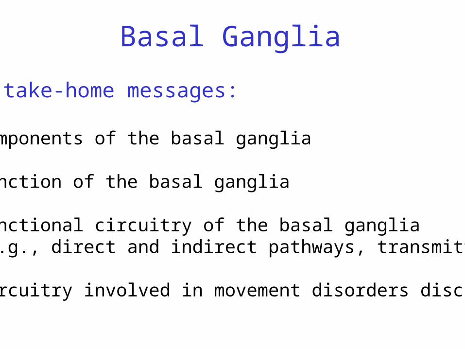
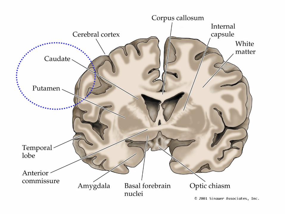
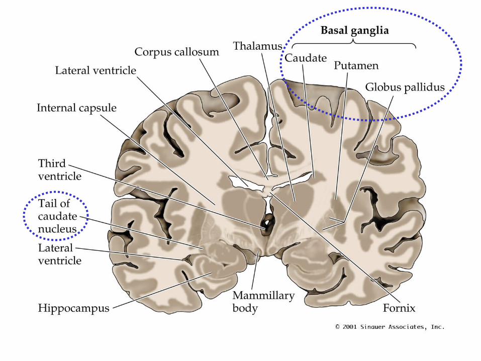
Basal Ganglia1. Neostriatum Caudate nucleus Putamen Ventral striatum (nucleus accumbens)
2. Paleostriatum Globus pallidus external segment (GPe)
Globus pallidus internal segment (GPi)
3. Substantia Nigra Pars compacta (SNc) Pars reticulata (SNr)
4. Subthalamic nucleus (STN)

What do the basal ganglia do?
Basal ganglia are involved in generation of goal-directed voluntary movements:
• Motor learning
• Motor pattern selection
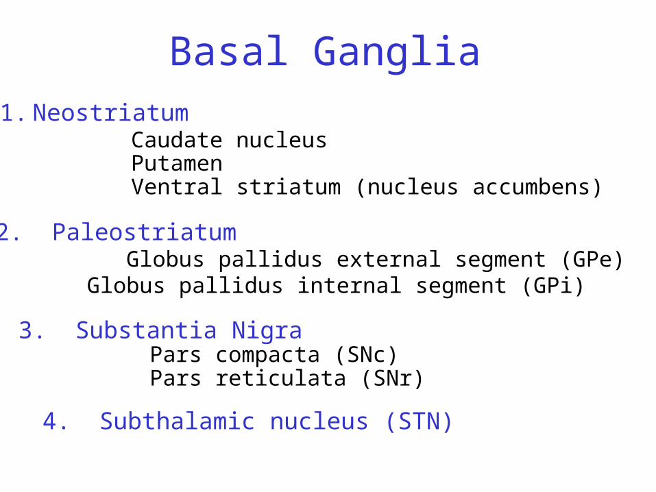
Location in human brain
From Neuroscience, Purves et al. eds., 2001
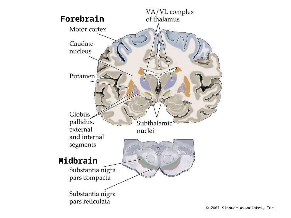
Forebrain
Midbrain
Forebrain
Midbrain
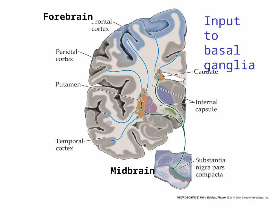
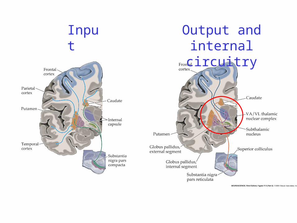
Input to basal ganglia
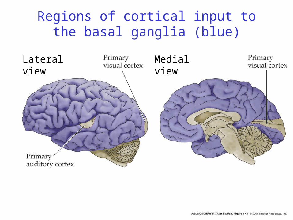
Lateral view Medial view
Regions of cortical input to the basal ganglia (blue)
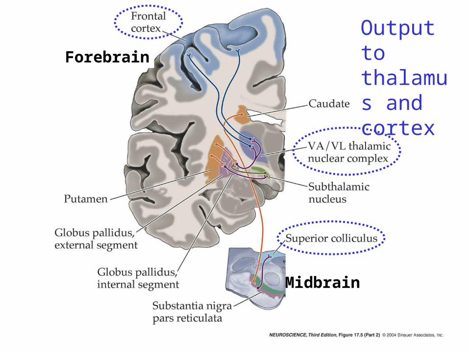
Output to thalamus and cortex
Forebrain
Midbrain
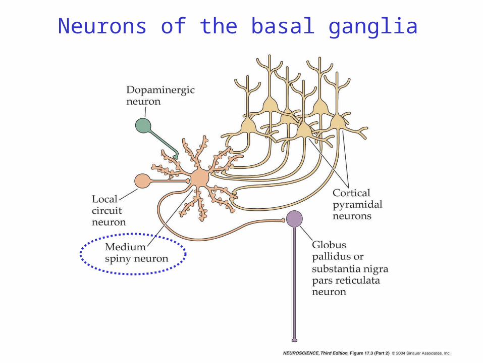
Neurons of the basal ganglia
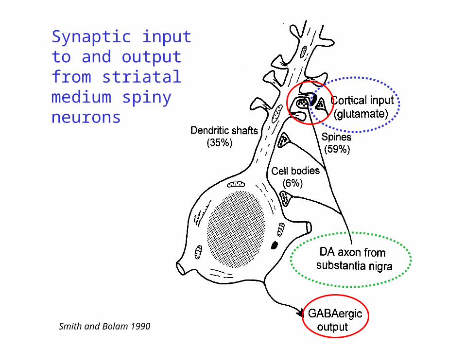
Smith and Bolam 1990
Synaptic input to and output from striatal medium spiny neurons
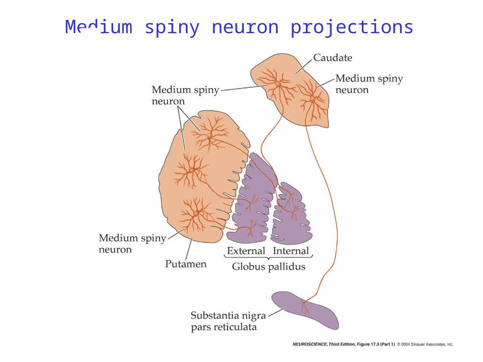
Medium spiny neuron projections
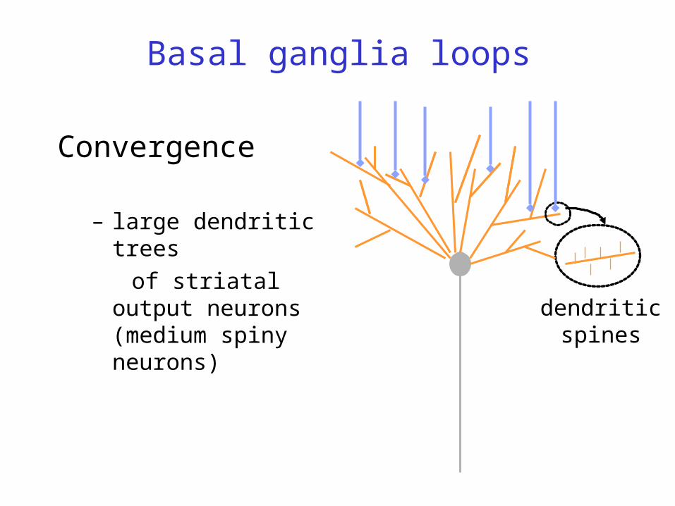
Convergence
– large dendritic trees of striatal output
neurons (medium spiny neurons) dendritic
spines
Basal ganglia loops
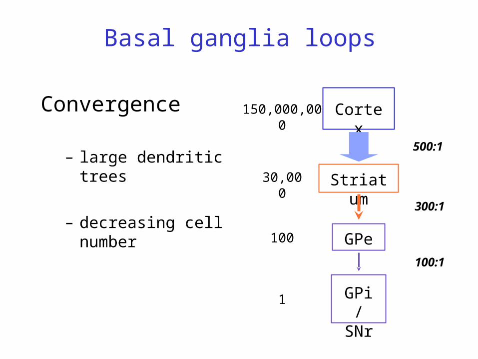
Convergence
– large dendritic trees
– decreasing cell number
Cortex
Striatum
GPe
GPi/SNr
500:1
150,000,000
30,000
100
1
100:1
300:1
Basal ganglia loops

Basal ganglia loops – motor and non-motor
Motor loopPrefrontal loop(Associative) Limbic loop
Input
Output and internal circuitry
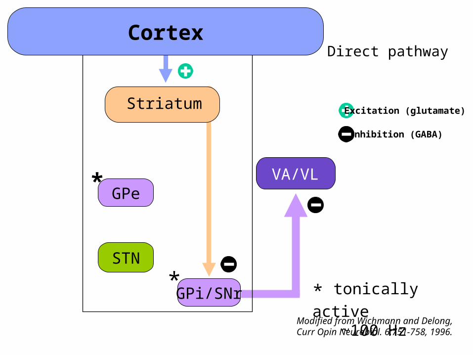
Cortex
VA/VL
GPi/SNr
Striatum
Modified from Wichmann and Delong, Curr Opin Neurobiol. 6:751-758, 1996.
* * tonically active ~100 Hz
GPe
STN
*
Direct pathway
Excitation (glutamate)
Inhibition (GABA)
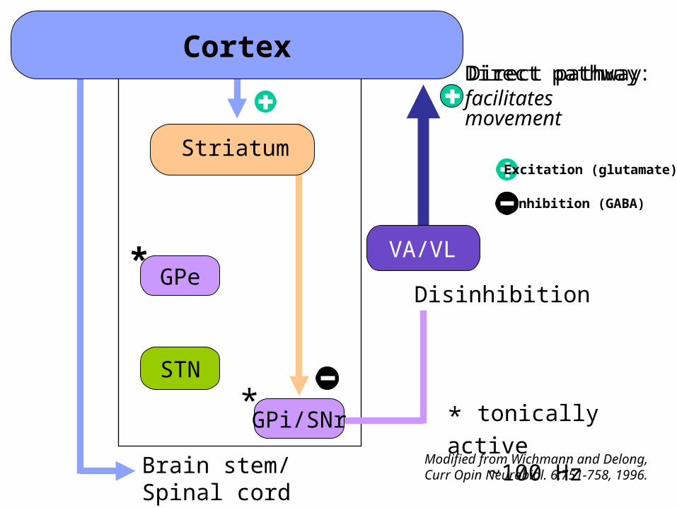
Direct pathway
Brain stem/Spinal cord
VA/VL
Striatum
Modified from Wichmann and Delong, Curr Opin Neurobiol. 6:751-758, 1996.
Direct pathway:facilitatesmovement
* * tonically active ~100 Hz
GPe
STN
*Disinhibition
Cortex
GPi/SNr
Excitation (glutamate)
Inhibition (GABA)
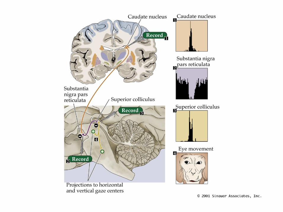
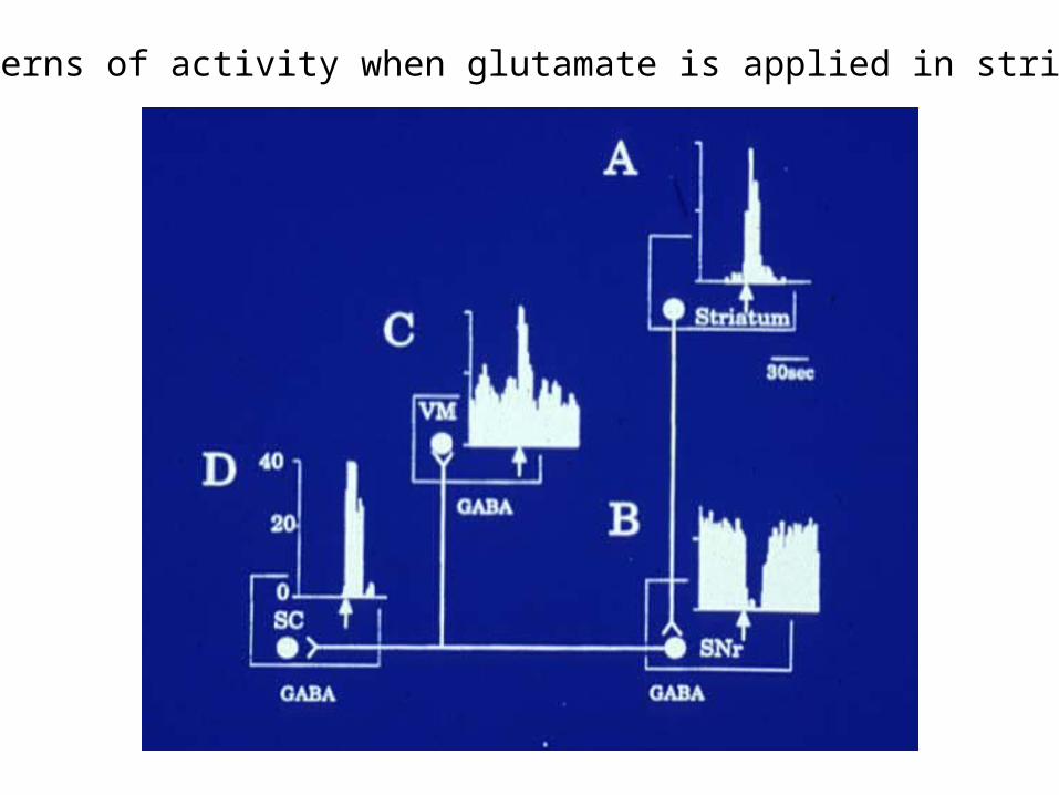
Patterns of activity when glutamate is applied in striatum

Patterns of activity during motor behavior
Disinhibition
Brain stem/Spinal cord
VA/VL
Striatum
Modified from Wichmann and Delong, Curr Opin Neurobiol. 6:751-758, 1996.
* * tonically active ~100 Hz
STN
*
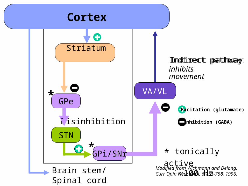
Indirect pathway:inhibitsmovement
Cortex
Indirect pathway
GPe
GPi/SNr
Excitation (glutamate)
Inhibition (GABA)

Cortex
Brain stem/Spinal cord
VA/VL
GPi/SNr
Striatum
Modified from Wichmann and Delong, Curr Opin Neurobiol. 6:751-758, 1996.
Direct pathway:facilitatesmovement
* * tonically active ~100 Hz
GPe
STN
*
Indirect pathway:inhibitsmovement
D1D2
SNc
Excitation (glutamate)
Inhibition (GABA)
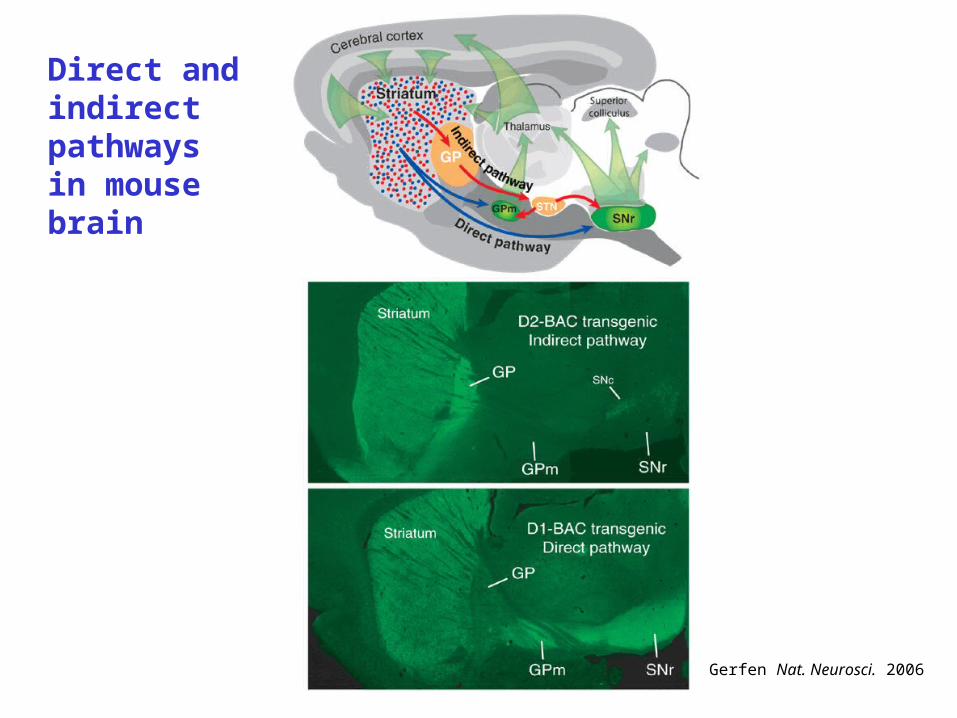
Gerfen Nat. Neurosci. 2006
Direct and indirect pathways in mouse brain
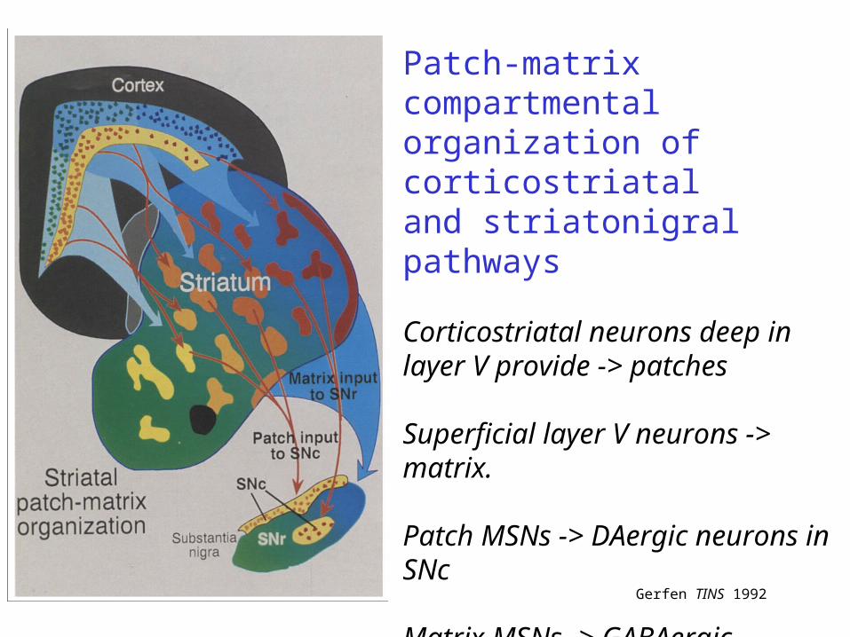
Patch-matrix compartmental organization of corticostriataland striatonigral pathways
Corticostriatal neurons deep in layer V provide -> patches
Superficial layer V neurons -> matrix.
Patch MSNs -> DAergic neurons in SNc
Matrix MSNs -> GABAergic neurons in SNr
Gerfen TINS 1992

Patch-matrix organization of corticostriatal and striatonigral pathways
Gerfen TINS 1992
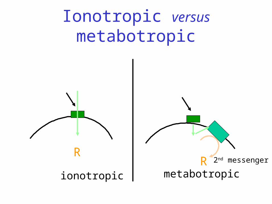
Ionotropic versus metabotropic
ionotropic metabotropic
RR 2nd messenger
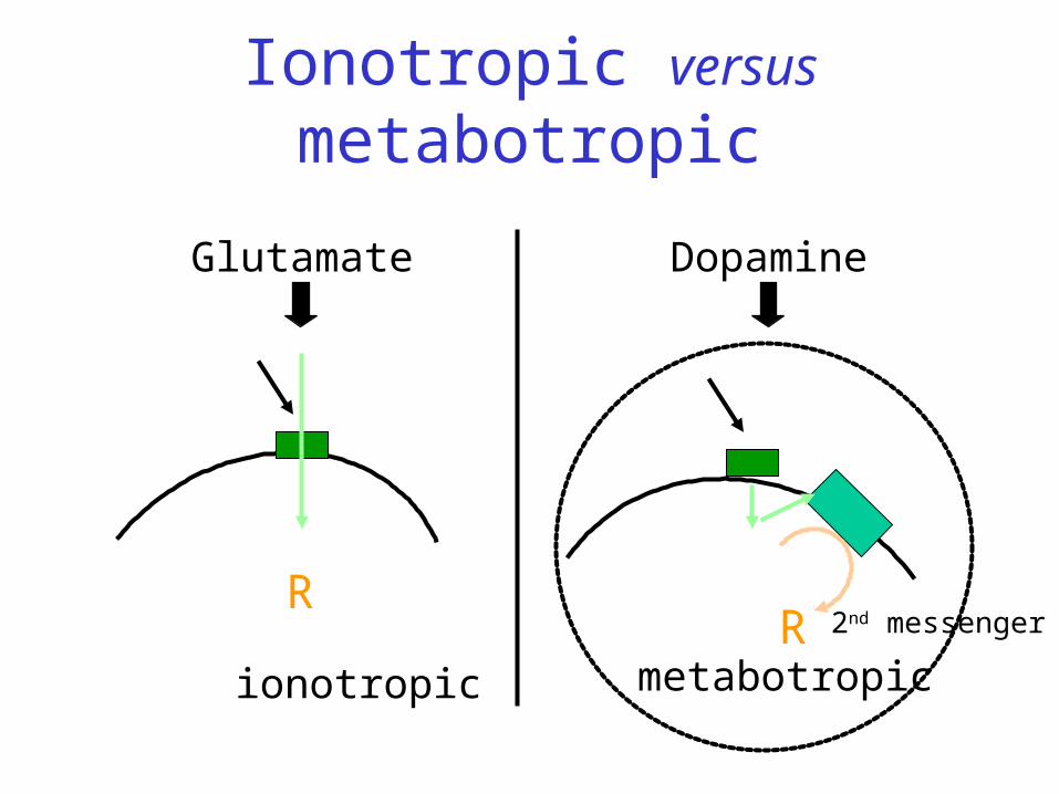
Ionotropic versus metabotropic
ionotropic metabotropic
RR
Dopamine
2nd messenger
Glutamate
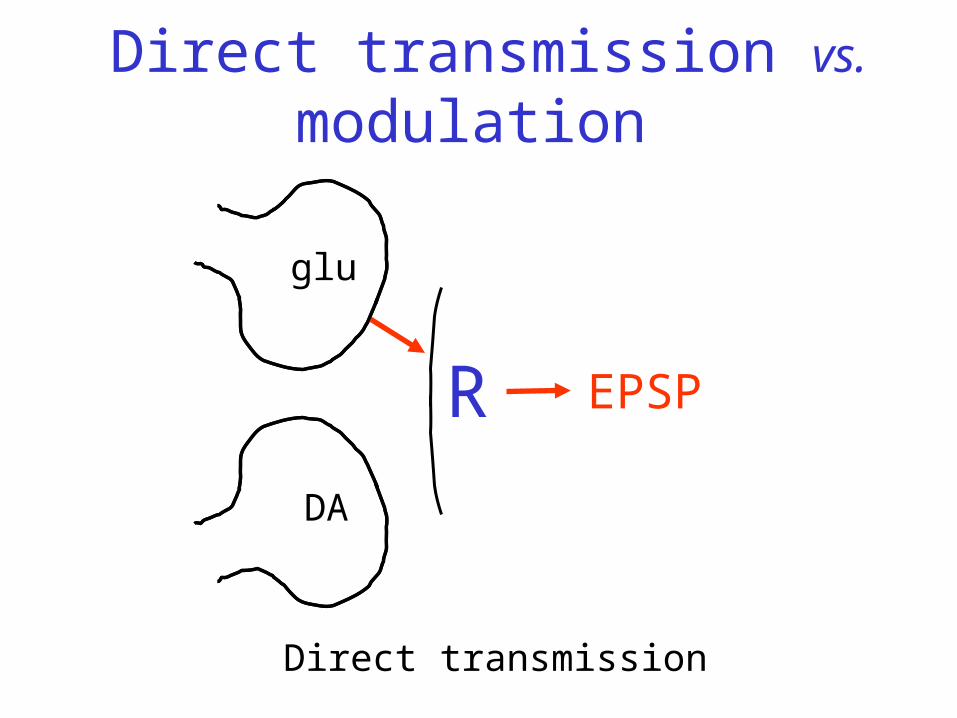
Direct transmission vs. modulation
R
glu
DA
Direct transmission
EPSP

glu
DA
No direct effect of DA
Direct transmission vs. modulation
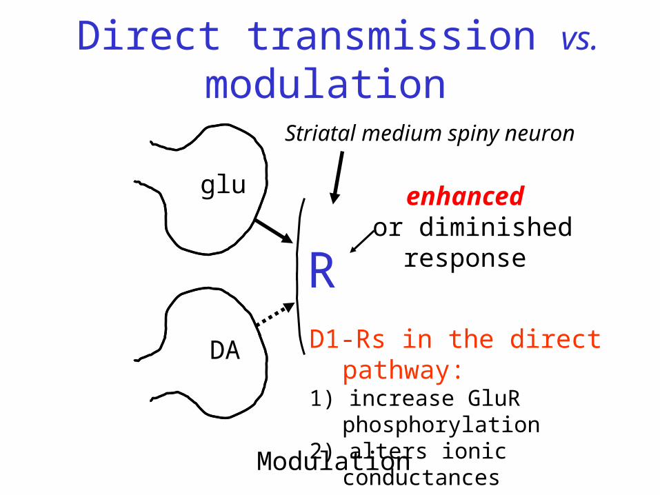
R
enhanced or diminished
response
Modulation
glu
DA D1-Rs in the direct pathway: 1) increase GluR phosphorylation2) alters ionic conductancesto amplify cortical input
Direct transmission vs. modulation
Striatal medium spiny neuron
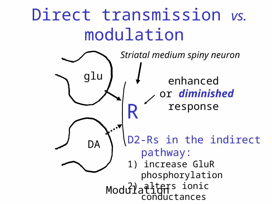
R
enhanced or diminished
response
Modulation
glu
DA
Direct transmission vs. modulation
D2-Rs in the indirect pathway: 1) increase GluR phosphorylation2) alters ionic conductancesto dampen cortical input
Striatal medium spiny neuron
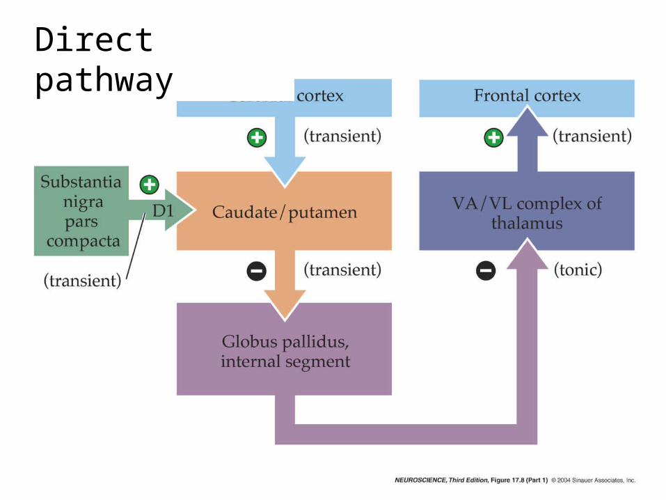
Direct pathway
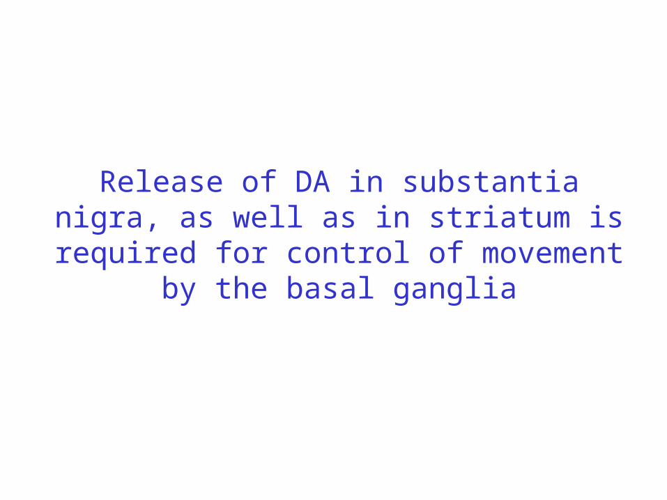
Release of DA in substantia nigra, as well as in striatum is required for control of
movement by the basal ganglia

Synaptic DA release in striatum
Smith and Bolam 1990
modified from Fallon et al. 1978
SNc DA cell
Somatic release(Jaffe et al. 1998)
Dendritic release(Geffen et al. 1976; Rice et al. 1994)
Somatodendritic DA release in SNc
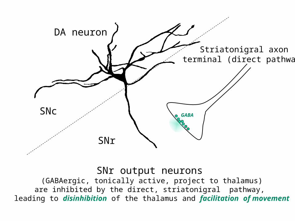
DA neuron
Striatonigral axonterminal (direct pathway)
GABA
SNr
SNc
SNr output neurons (GABAergic, tonically active, project to thalamus)are inhibited by the direct, striatonigral pathway,
leading to disinhibition of the thalamus and facilitation of movement
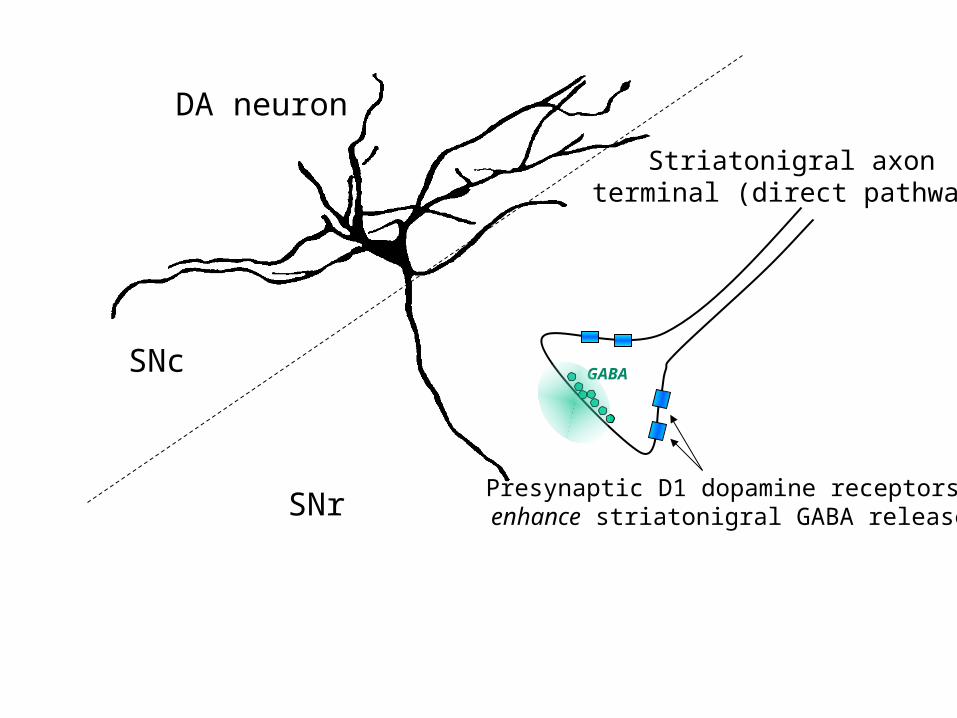
DA neuron
Presynaptic D1 dopamine receptors enhance striatonigral GABA release
Striatonigral axonterminal (direct pathway)
GABA
SNr
SNc
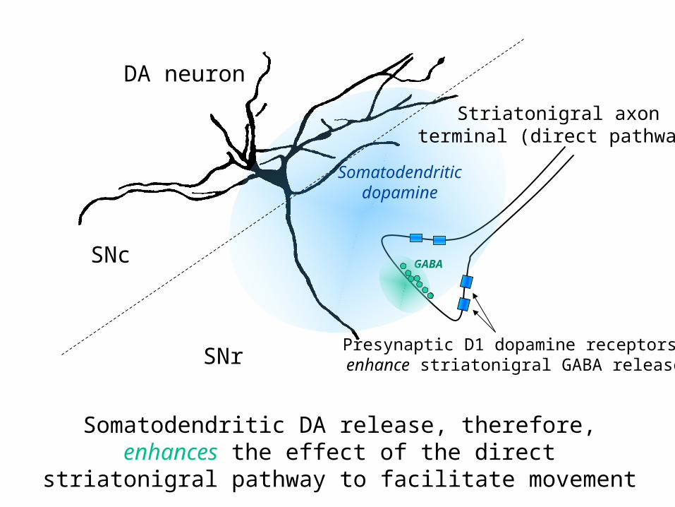
DA neuron
Presynaptic D1 dopamine receptors enhance striatonigral GABA release
Somatodendriticdopamine
Striatonigral axonterminal (direct pathway)
GABA
SNr
SNc
Somatodendritic DA release, therefore, enhances the effect of the direct striatonigral pathway to facilitate movement
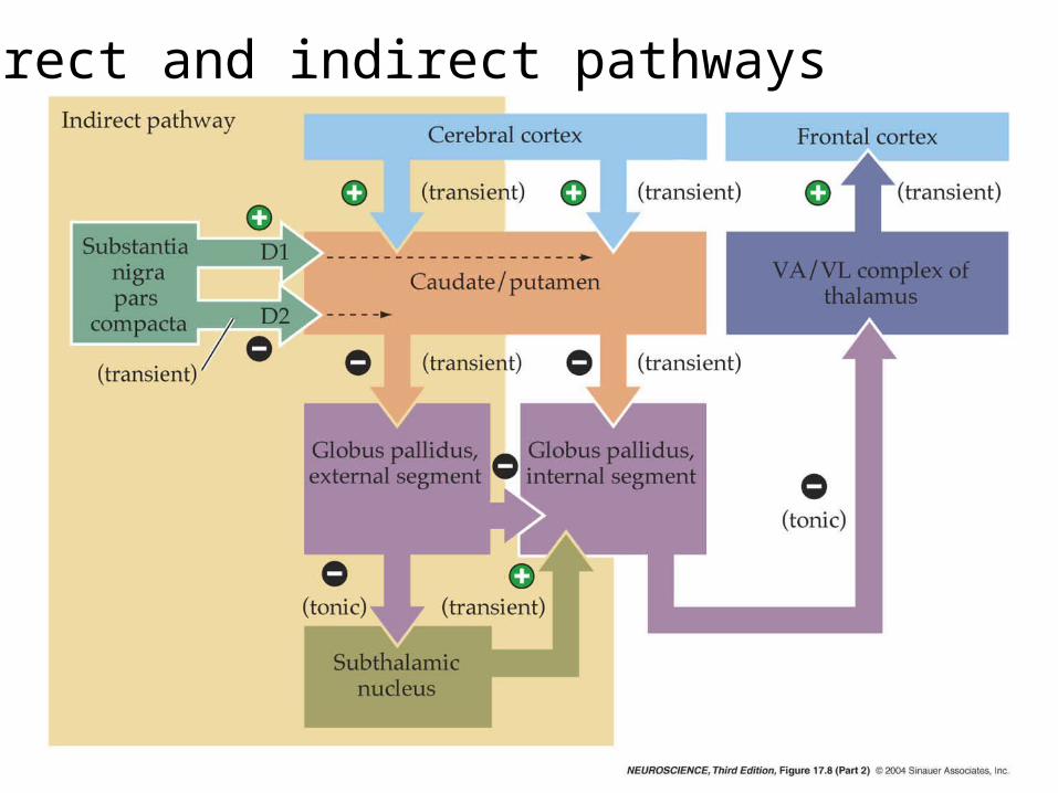
Direct and indirect pathways
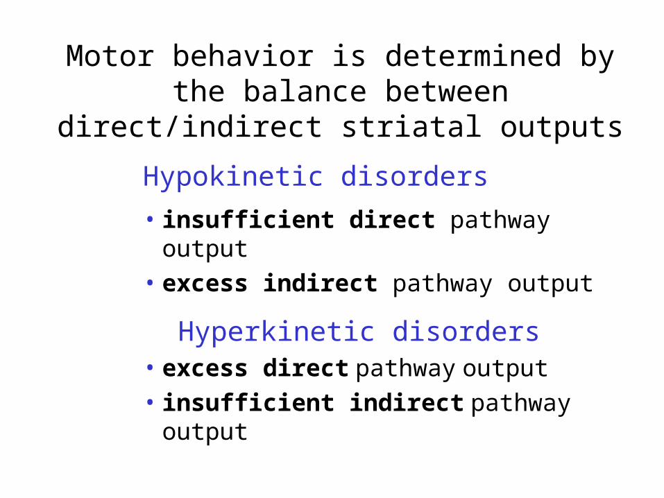
Hypokinetic disorders• insufficient direct pathway output• excess indirect pathway output
Hyperkinetic disorders• excess direct pathway output• insufficient indirect pathway output
Motor behavior is determined by the balance between direct/indirect striatal outputs
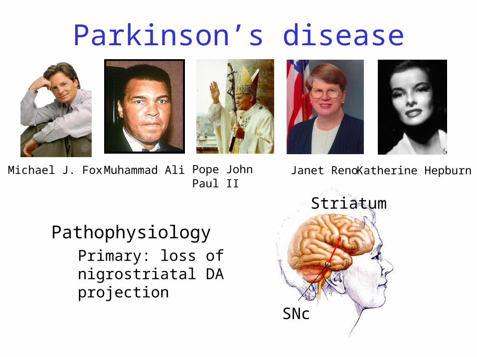
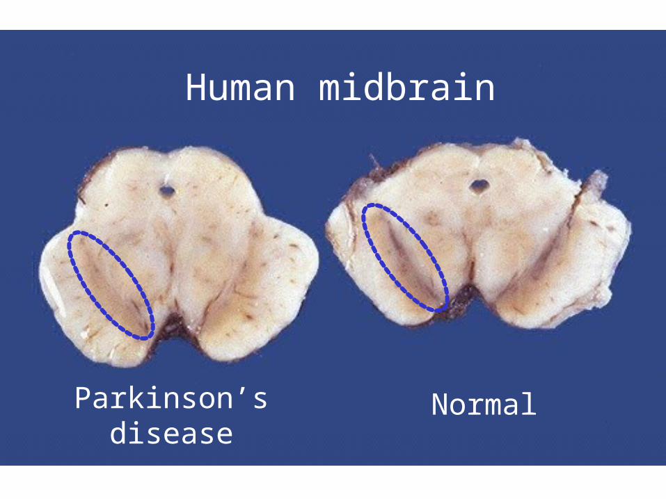
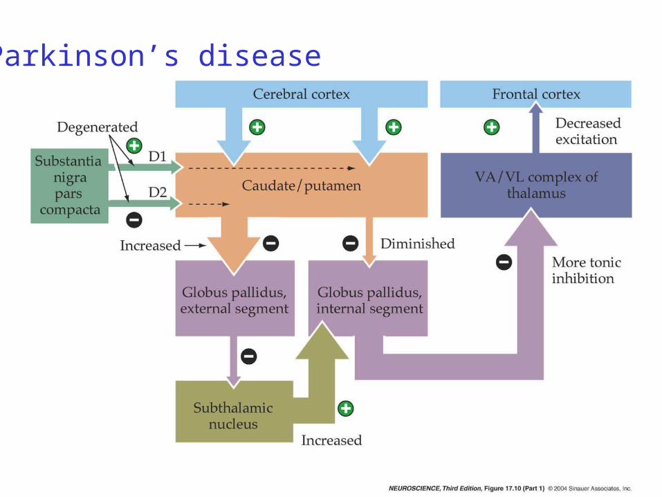
Parkinson’s disease
PathophysiologyPrimary: loss of nigrostriatal DA projection
SNc
Striatum
Michael J. Fox Muhammad Ali Pope John Paul II Janet Reno Katherine Hepburn
NormalParkinson’sdisease
Human midbrain
Parkinson’s disease
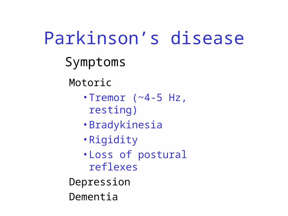
Motoric• Tremor (~4-5 Hz, resting)• Bradykinesia• Rigidity• Loss of postural reflexes
DepressionDementia
Parkinson’s diseaseSymptoms
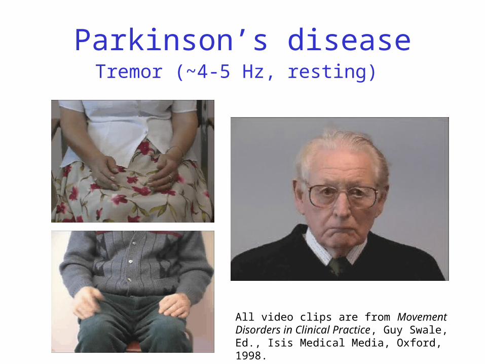
Parkinson’s diseaseTremor (~4-5 Hz, resting)
All video clips are from Movement Disorders in Clinical Practice, Guy Swale, Ed., Isis Medical Media, Oxford, 1998.

Parkinson’s diseaseBradykinesia
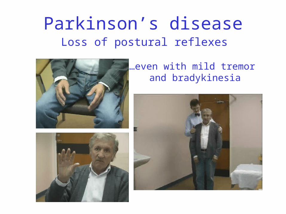
Parkinson’s diseaseLoss of postural reflexes
…even with mild tremor and bradykinesia
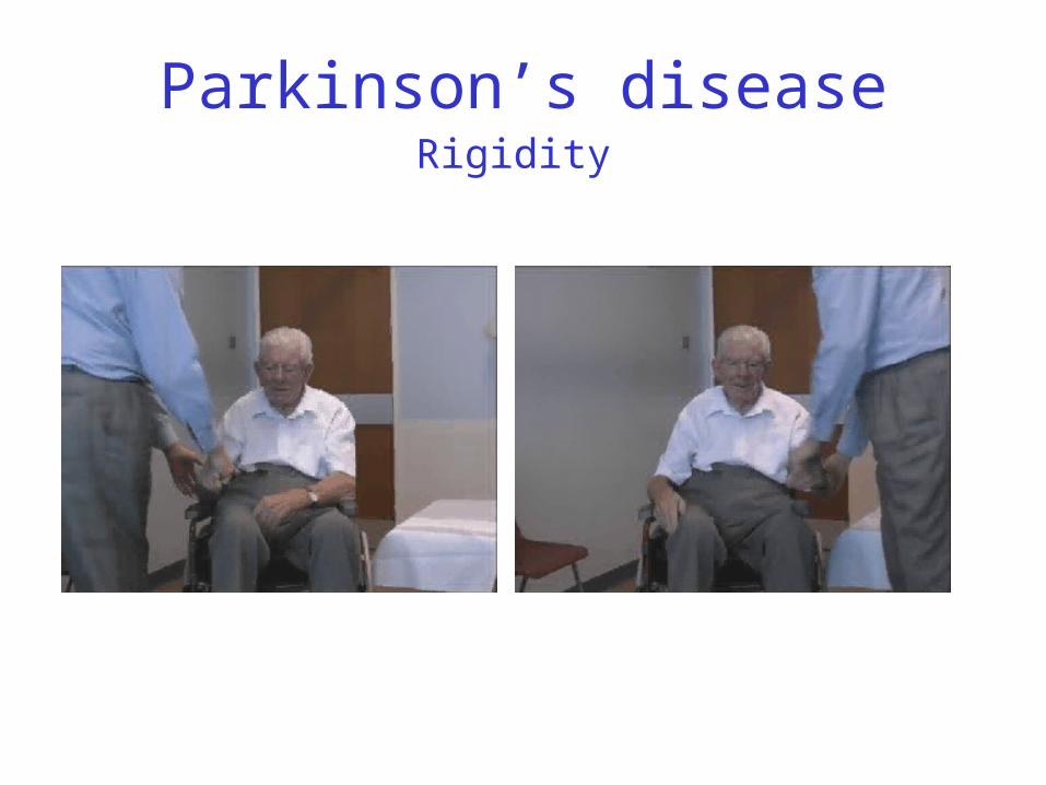
Parkinson’s diseaseRigidity
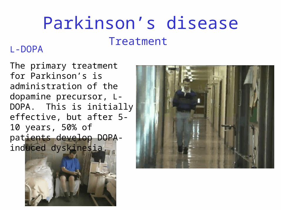
Parkinson’s diseaseTreatment
L-DOPA
The primary treatment for Parkinson’s is administration of the dopamine precursor, L-DOPA. This is initially effective, but after 5-10 years, 50% of patients develop DOPA-induced dyskinesia.
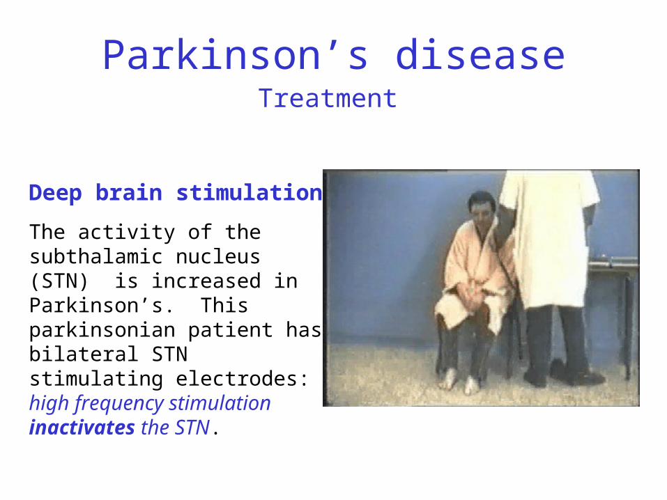
Parkinson’s diseaseTreatment
Deep brain stimulationThe activity of the subthalamic nucleus (STN) is increased in Parkinson’s. This parkinsonian patient has bilateral STN stimulating electrodes: high frequency stimulation inactivates the STN.

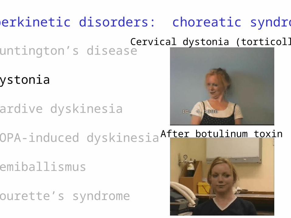
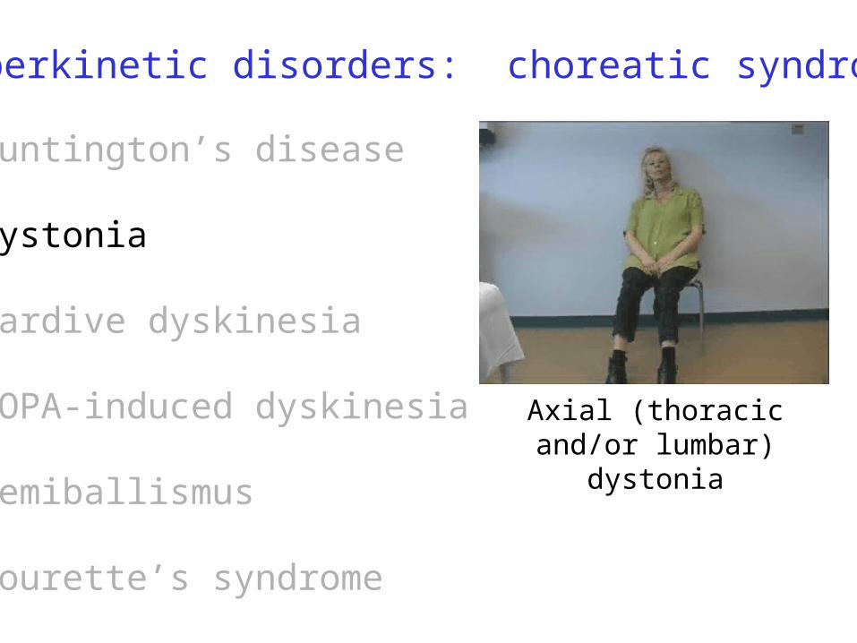
Hyperkinetic disorders: choreatic syndromes
1. Huntington’s chorea
2. Dystonia
3. Tardive dyskinesia
4. DOPA-induced dyskinesia
5. Hemiballismus
6. Tourette’s syndrome
Causes:Genetic (autosomal dominant)
Genetic or idiopathic
Chronic neuroleptic use
Parkinson’s therapy
Unilateral vascular accident, typically subthalamic nucleus
Excessive D2-subtypeDA receptor expression(?)
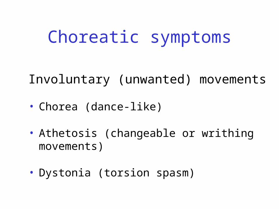
Choreatic symptoms
Involuntary (unwanted) movements
• Chorea (dance-like)
• Athetosis (changeable or writhing movements)
• Dystonia (torsion spasm)
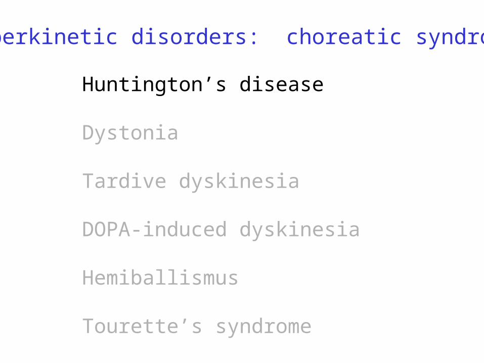
Hyperkinetic disorders: choreatic syndromes
Huntington’s disease
Dystonia
Tardive dyskinesia
DOPA-induced dyskinesia
Hemiballismus
Tourette’s syndrome
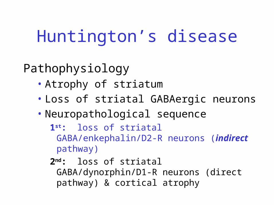
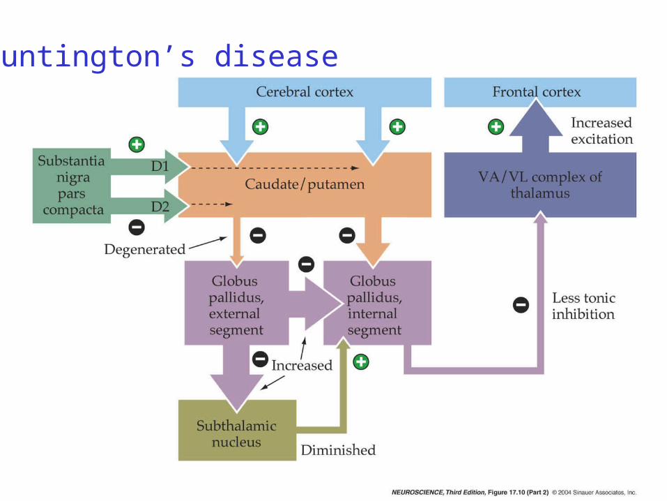
Huntington’s disease
Pathophysiology• Atrophy of striatum• Loss of striatal GABAergic neurons• Neuropathological sequence
1st: loss of striatal GABA/enkephalin/D2-R neurons (indirect pathway)
2nd: loss of striatal GABA/dynorphin/D1-R neurons (direct pathway) & cortical atrophy

Huntington’s disease pathology
Huntington’s
Normal
Huntington’s disease
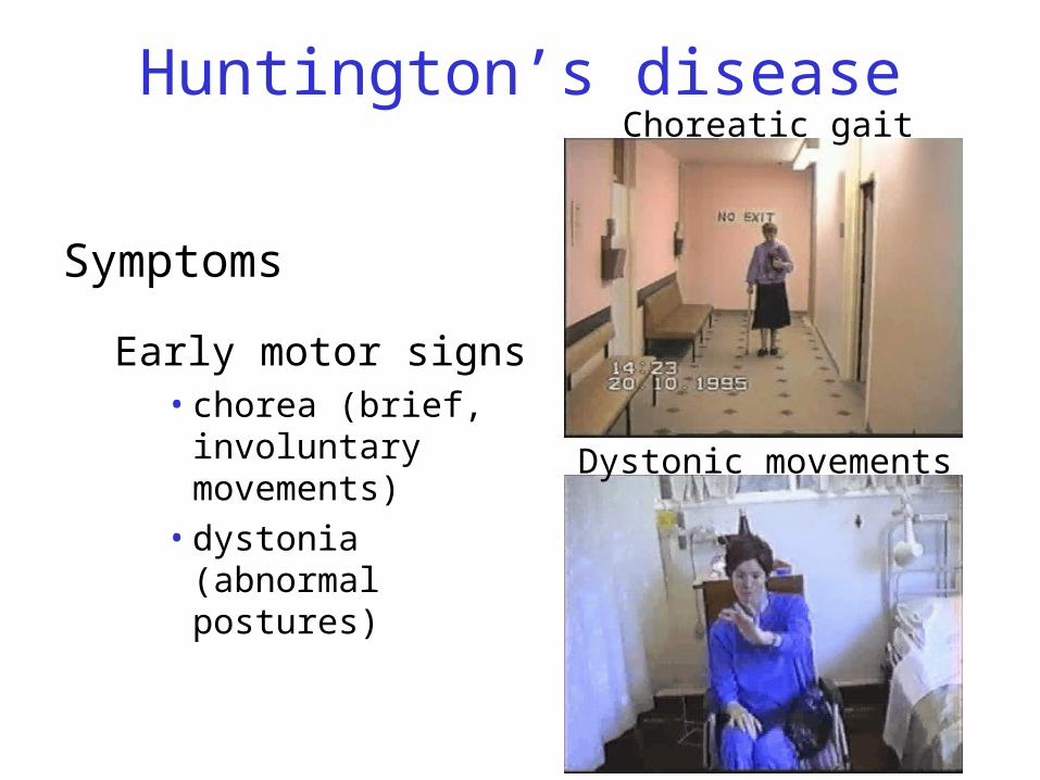
Symptoms
Early motor signs• chorea (brief,
involuntary movements)
• dystonia (abnormal postures)
Choreatic gait
Dystonic movements

Huntington’s diseaseCognitive abnormalities
• Executive function (complex tasks)• Recent and remote memory (poor retrieval)
Psychiatric changes• Depression• Psychosis
Later decline• Immobility• Weight loss• Death within 10-25 years (often from pneumonia)
Huntington’s disease
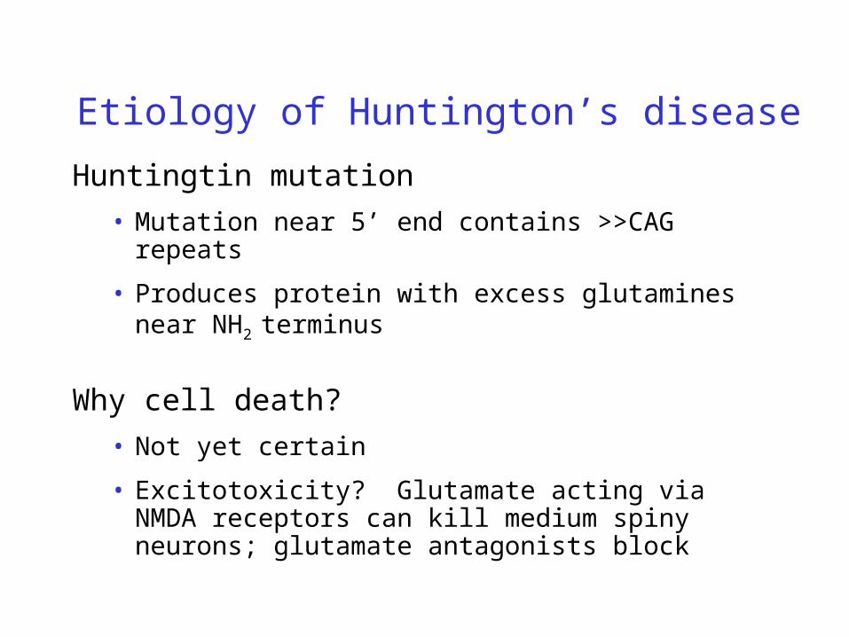
Etiology of Huntington’s disease
Huntingtin mutation• Mutation near 5’ end contains >>CAG repeats
• Produces protein with excess glutamines near NH2 terminus
Why cell death?
• Not yet certain
• Excitotoxicity? Glutamate acting via NMDA receptors can kill medium spiny neurons; glutamate antagonists block
Hyperkinetic disorders: choreatic syndromes
Huntington’s disease
Dystonia
Tardive dyskinesia
DOPA-induced dyskinesia
Hemiballismus
Tourette’s syndrome
Cervical dystonia (torticollis)
After botulinum toxin
Hyperkinetic disorders: choreatic syndromes
Huntington’s disease
Dystonia
Tardive dyskinesia
DOPA-induced dyskinesia
Hemiballismus
Tourette’s syndrome
Axial (thoracic and/or lumbar) dystonia
Hyperkinetic disorders: choreatic syndromes
Huntington’s disease
Dystonia
Tardive dyskinesia
DOPA-induced dyskinesia
Hemiballismus
Tourette’s syndrome

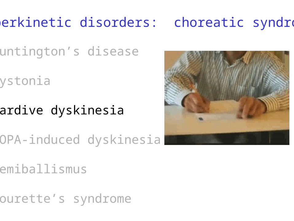
Hyperkinetic disorders: choreatic syndromes
Huntington’s disease
Dystonia
Tardive dyskinesia
*DOPA-induced dyskinesia
Hemiballismus
Tourette’s syndrome*50% of PD patients on L-DOPA will develop DOPA dyskinesia
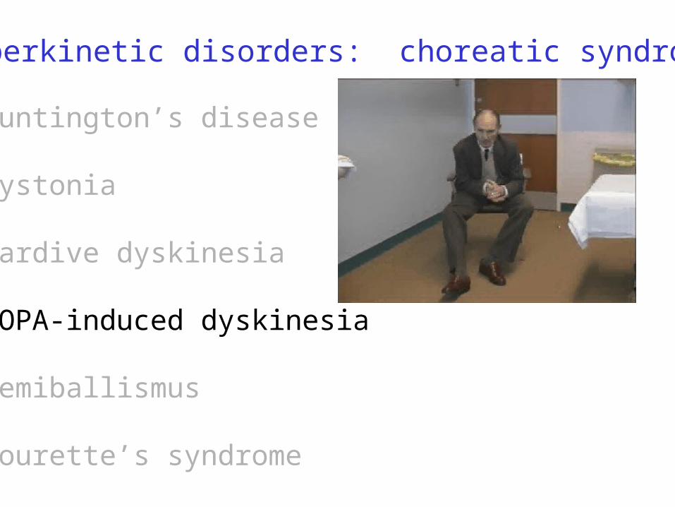
Hyperkinetic disorders: choreatic syndromes
Huntington’s disease
Dystonia
Tardive dyskinesia
DOPA-induced dyskinesia
Hemiballismus
Tourette’s syndrome
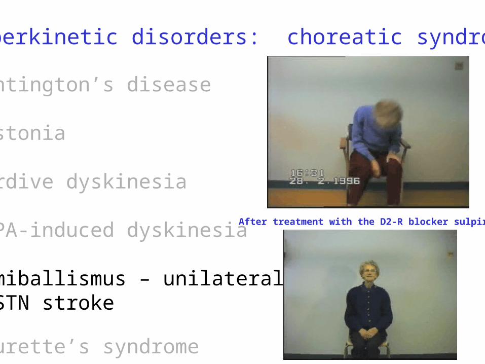
Hyperkinetic disorders: choreatic syndromes
Huntington’s disease
Dystonia
Tardive dyskinesia
DOPA-induced dyskinesia
Hemiballismus – unilateral STN stroke
Tourette’s syndrome
After treatment with the D2-R blocker sulpiride

Hyperkinetic disorders: choreatic syndromes
Huntington’s disease
Dystonia
Tardive dyskinesia
DOPA-induced dyskinesia
Hemiballismus
Tourette’s syndrome

![SeedNet India Portal · 11— 35. 36. 37. 38. 39. 41. 42. 43. 45. 47. 3(ii)] (2) Rice Rice Rice Rice Rice Rice Rice Green Gram Red Gram Taramira Täramira Rice Rice Maize](https://img.pdfslide.net/doc/110x75/5f97db6345fe5e455963d66a/seednet-india-portal-11a-35-36-37-38-39-41-42-43-45-47-3ii-2-rice.jpg)