Embed Size (px)
Citation preview

الرحمن الله بسمالرحيم

Introduction Parasites are organisms
adapts them selves to live in or on another organisms termed host .The relationship between the parasites and host occurs in 2 form:

Commensalism: one of two organisms (commensal) benefits while the other host is unaffected (no benefit, no harm), commensal organism referred as non pathogenic.
Parasitism : one of two organisms (parasite) benefits, gain shelter and nutrition on the expanse of the other (host). The host may suffer from wide range of functional and organic disturbances due to such association (pathogenic)

Types of parasites:
•Ectoparasite : lives outside on the surface of the body (infestation).
•Endoparasite: lives inside the body of host (infection).
•Temporary or intermittent parasite: visits the host from time to time for food.
•Permanent parasite : remains on or in the body of the host for its entire life.

• Facultative parasite : organism that can exist in a free living state or as a parasite.
• Obligatory parasite : can not survive without a host i.e. completely adapted for parasitic existence.
• Opportunistic parasite : produces disease only in immunodeficient or immuno-suppressed patients (AIDS). In immunocompetent individuals the organisms may exist in latent form producing no symptoms.
• Coprozoic or spurious parasite foreign organisms which have been swallowed merely pass along alimentary canal of man (without establishment) to be recovered in faeces.

Types of hosts:• a-Definitive or final host : host with
adult stage or sexually reproducing forms of the parasite.
• b-intermediate host : host with the larval stage or asexually reproducing forms of parasite.
• c-Reservoir host : animal that harbors the same species of parasite as man . such animal help to maintain the cycle of parasite in nature and act as a potential source for human infection with this parasite.
• d- Vector: an arthropod which carries the parasite from one host to another.

zoonosis •Disease transmitted from animals to man are known as zoonotic diseases
Mode of parasitic infections:• Congenital from mother to foetus.• Direct contact through the skin or sexually.• Ingestion of contaminated food and water
or undercooked meat in which the infective stage has developed.
• Penetration of the skin due to contact with infected soil or water stream.
• Inhalation of dust carrying the infective stage of parasite.
• Vectors: through the bite or faeces of infected vector or by swallowing the vector.
• Autoinfection: occurs when infective stages are carried from faeces to mouth of patient.
Important terminology• Prevalence = % of infection.• Intensity = number of parasite
in the host.• Incidence = % of new
infection .
Endemicity
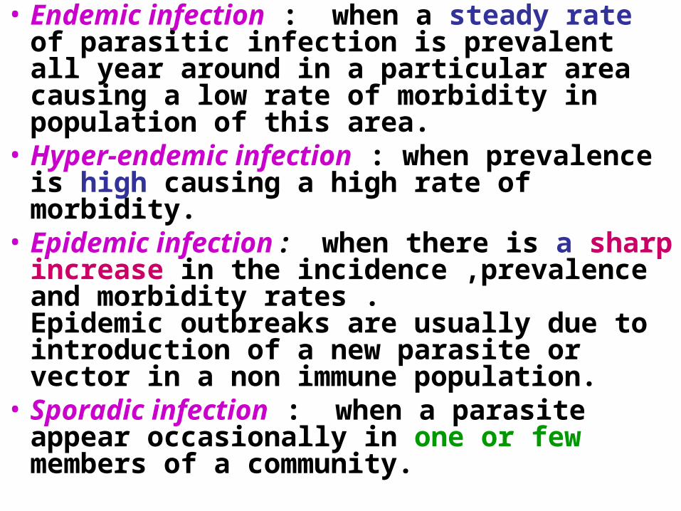
• Endemic infection : when a steady rate of parasitic infection is prevalent all year around in a particular area causing a low rate of morbidity in population of this area.
• Hyper-endemic infection : when prevalence is high causing a high rate of morbidity.
• Epidemic infection : when there is a sharp increase in the incidence ,prevalence and morbidity rates . Epidemic outbreaks are usually due to introduction of a new parasite or vector in a non immune population.
• Sporadic infection : when a parasite appear occasionally in one or few members of a community.

Source of parasitic infection•Food -meat ( T.saginata, T. spiralis)
-vegetables(Ascaris, E.histolytica)
•Water - protozoal cysts (E.histolytica, Cryptosporidium)
- cercaria (Schistosoms) - cyclops(D.medinensis)
•Soil contaminated with faeces (Ancylostoma ,Ascaris ,Strongyloides,
Giardia )
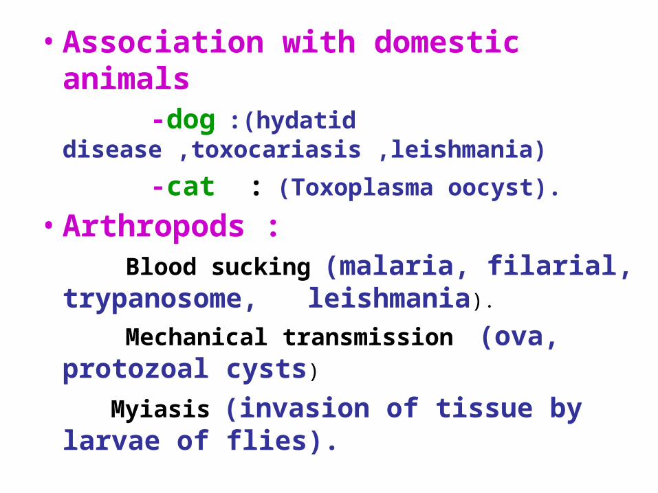
•Association with domestic animals
-dog :(hydatid disease ,toxocariasis ,leishmania)
-cat : (Toxoplasma oocyst).
•Arthropods :
Blood sucking (malaria, filarial, trypanosome, leishmania).
Mechanical transmission (ova, protozoal cysts)
Myiasis (invasion of tissue by larvae of flies).

•Blood transfusion : (erythrocytic stages of
Plasmodium).
•Congenital transplantation : (toxoplasma, Plasmodium).
•Sexual intercourse : (Trichomonas vaginalis, Phthirus pubis).
• Inhalation of dust :
(Enterobius ova).
Parasites -Helminthes
- Protozoa
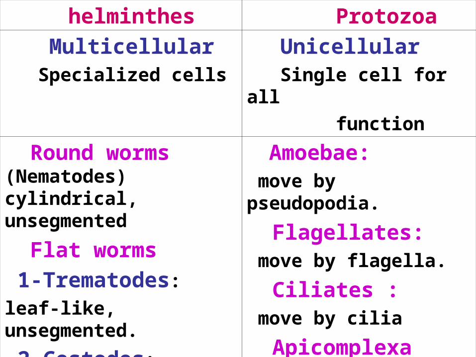
Protozoa helminthes Unicellular Single cell for all function
Multicellular Specialized cells
Amoebae: move by pseudopodia.
Flagellates: move by flagella.
Ciliates : move by cilia
Apicomplexa(sporozoa) Tissue parasites
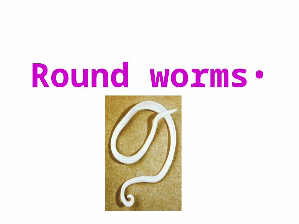
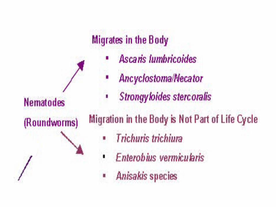
Round worms (Nematodes) cylindrical, unsegmented
Flat worms 1-Trematodes:
leaf-like, unsegmented.
2-Cestodes:
tape-like, segmented

HELMINTHES• Round worms (nematodes): Elongated, cylindrical,
unsegmented.• Flat worms: -Trematodes: leaf-like,
unsegmented. -Cestodes : tape-like, segmented
•Round worms

Common intestinal nematode infection:
•Enterobius (Oxyuris) vermicularis (Pinworm,seatworm,threadworm)
•Trichuris trichiura (whipworm)
•Ascaris lumbricoides (roundworm)
•Ancylostoma duodenale& Necator americanus (hookworm)
•Strongyloides stercoralis

Nematodes General features:
1. Elongated worm, cylindrical, unsegmented and tapering at both ends.
2. Variable in size, measure less than 5mm~as long as 100cm.
3. Sex separate and male is smaller than female

Location of Nematodes: Intestinal:• Small intestine: Ascaris, Ancylostoma, Strongyloides• Large intestine:
Enterobius Trichuris

In Tissue•W.Bancrofti : lymphatic
system•O.Volvulus & Loa loa
D.medinesis: subcutaneous tissue.
•Trichinella (larva): muscle, brain, lung.
•Toxocara canis : non human species larva carried in blood to liver, lung ,brain, eye….
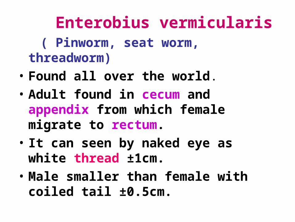
Enterobius vermicularis ( Pinworm, seat worm,
threadworm)• Found all over the world.• Adult found in cecum and
appendix from which female migrate to rectum.
• It can seen by naked eye as white thread ±1cm.
• Male smaller than female with coiled tail ±0.5cm.
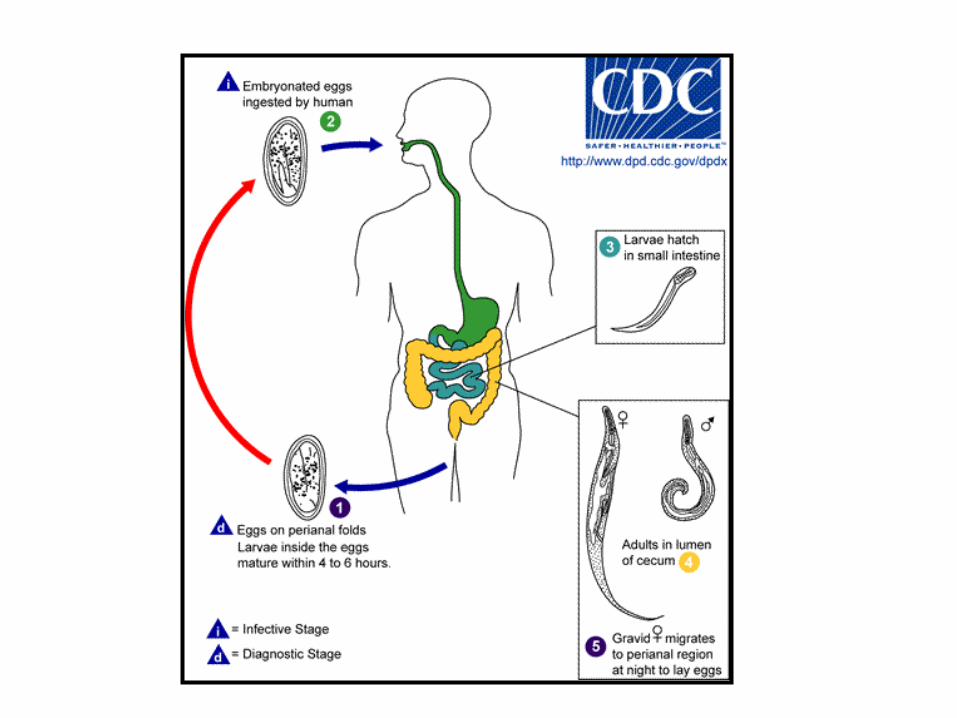

LIFE CYCLE


Pathology• Majority of infection is asymptomatic• Main clinical presentation pruritus ani
, perianal excoriation.• Many cases presented by
appendicitis.• Ectopic enterobiasis occurs in female
when invade valva and vagina result in valvovagintis
• Usually accompanied by insomnia, anorexia,loss of weight and concentration
Treatment: Albandazole for whole family.


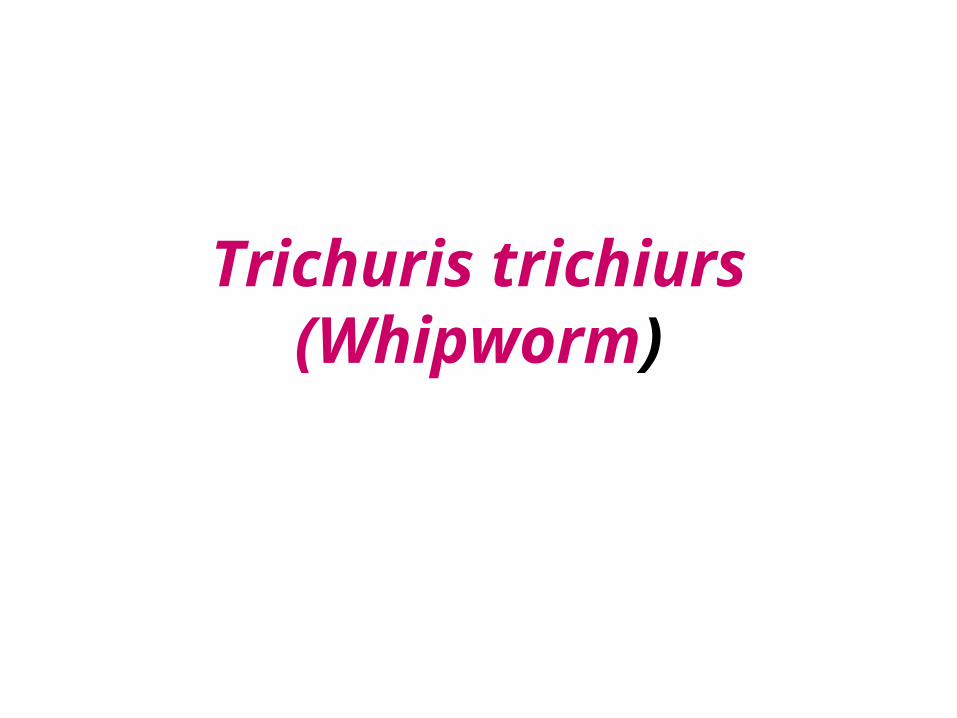
Trichuris trichiurs (Whipworm)

Trichuris trichiurs (Whipworm)
World wide ,common in poor sanitation.
• It coexists with Ascaris because of similar requirement.
• Adult live in large intestine especially caecum and appendix – in heavy infection the whole length of large intestine affected.
• Male and female worm have narrow anterior portion penetrate the intestinal mucosa
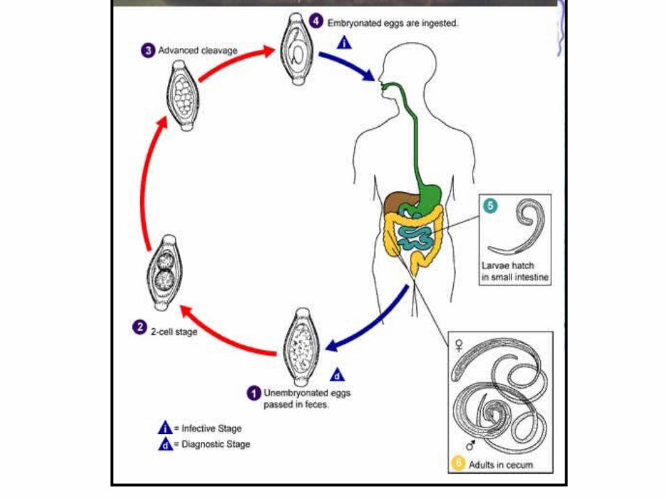

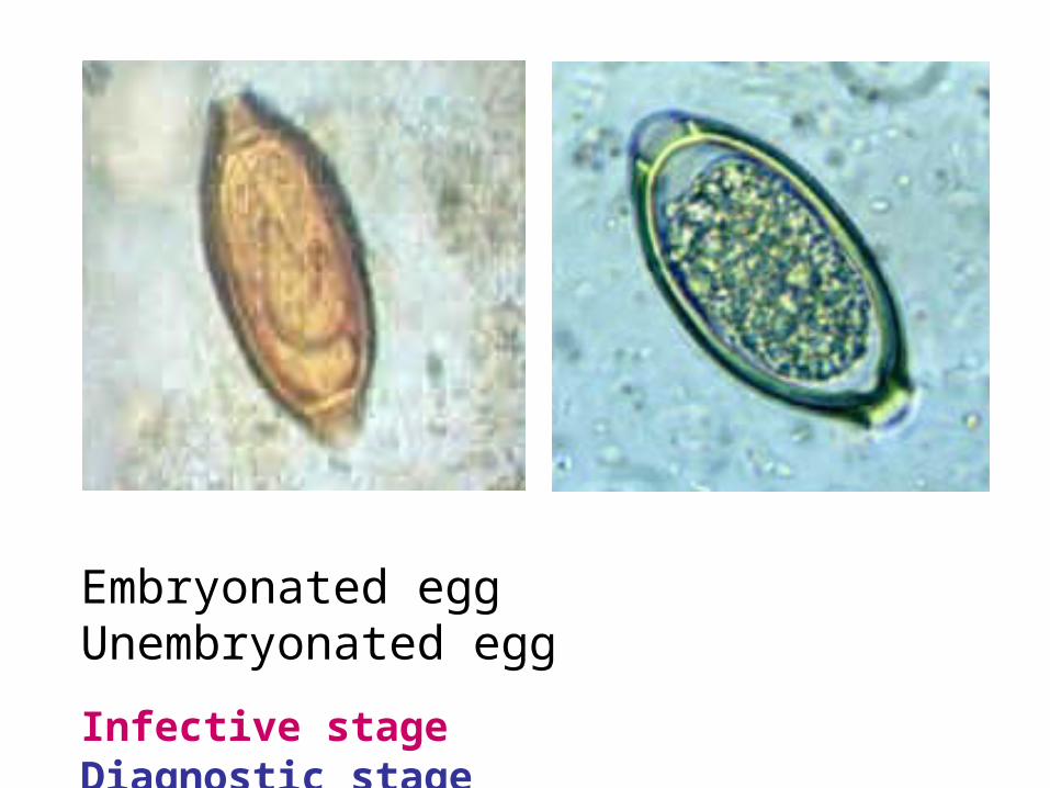
Embryonated egg Unembryonated egg
Infective stage Diagnostic stage
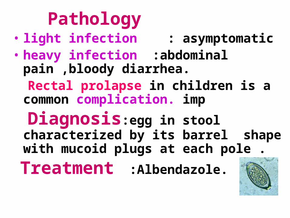
Pathology • light infection : asymptomatic• heavy infection :abdominal
pain ,bloody diarrhea. Rectal prolapse in children is a
common complication. imp
Diagnosis:egg in stool characterized by its barrel shape with mucoid plugs at each pole .
Treatment :Albendazole.
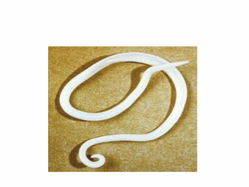
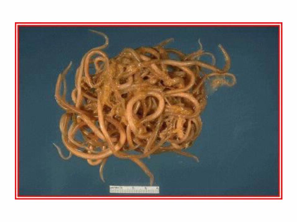
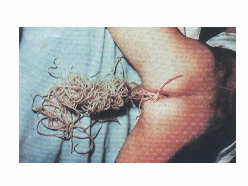
Ascaris lumbricoides
(roundworm)
Ascaris lumbricoides (roundworm)
•The commonest humen helminthes infection.
•Found in jejunum and upper part of ileum.
•Female ±20 cm longer than male ±10 cm
•Feed on semidigested food.

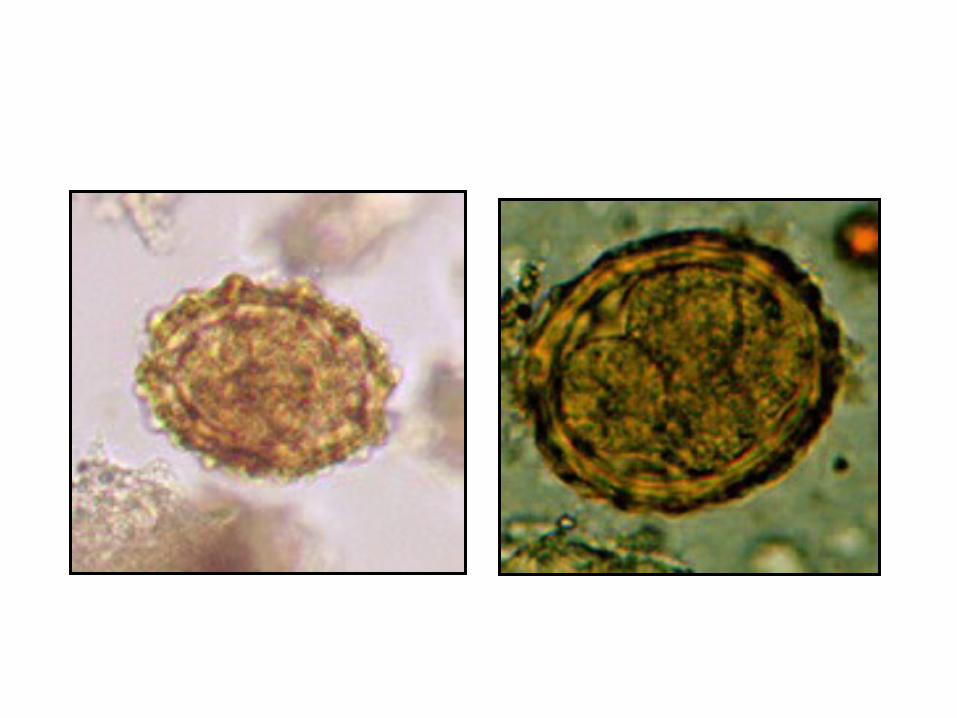
Infective stage diagnostic stage
embryonated egg Unembryonated

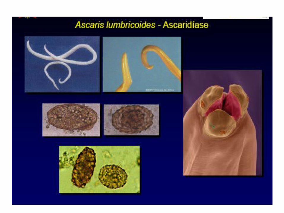

Pathology: 1-Adult worm • Light infection asymptomatic.• Heavy infection may cause intestinal obstruction • Migrating adult to bile duct may causing jaundice 2-Larvae: Loefflers syndrome : Pneumonia,cough with bloody
sputum Eosinophilia, urticaria


Loeffler`s syndrome: Larvae in lung
pneumonia, cough ,bloody sputum

Diagnosis: - eggs in stool - larvae in sputum.
Treatment: albendazol

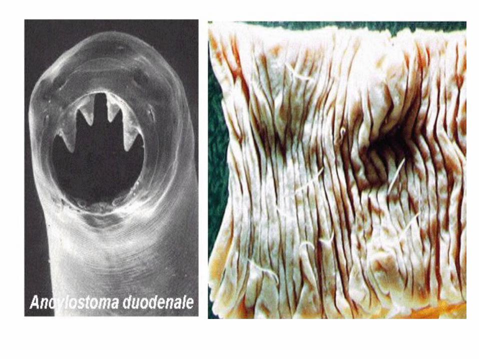
Hook warms Ancylostoma dudenale &
Necator americanus•The commonest cause of anemia.•Found in small intestine mainly
jejunum.• Its buccal capsule (mouth) lined
with hard hooks, triangular cutting plates and anticoagulant glands
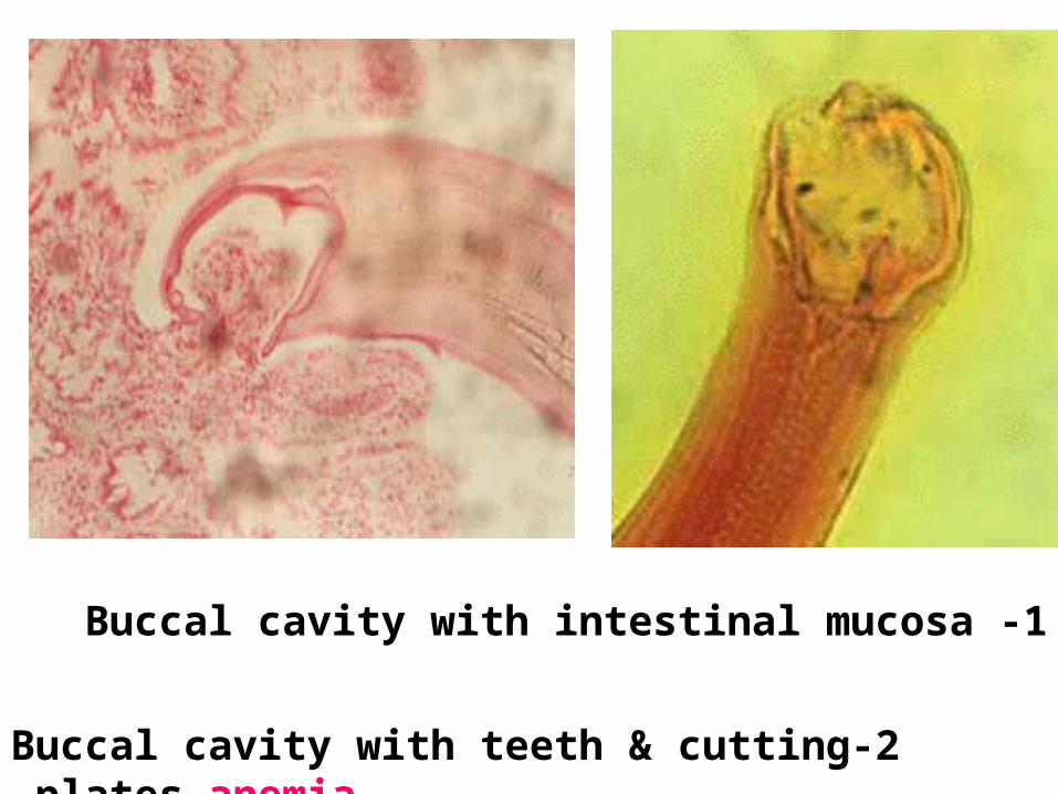
1- Buccal cavity with intestinal mucosa
2-Buccal cavity with teeth & cutting plates anemia


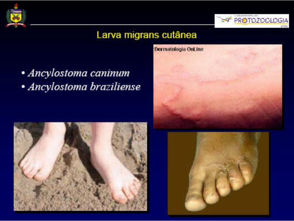
pathology& clinical picture i-At the site of entry of larvae ( ground itch).ii-Migration phase of larvae: cough with bloody sputum
pneumonia (loeffler`s syndrome) eosinophilia, urticaria. - Adult worm: • low worm burden: no symptoms.• Moderate to heavy burden:
epigastric pain vomiting ,simulating duodenal ulcer hemorrhagic enteritis.
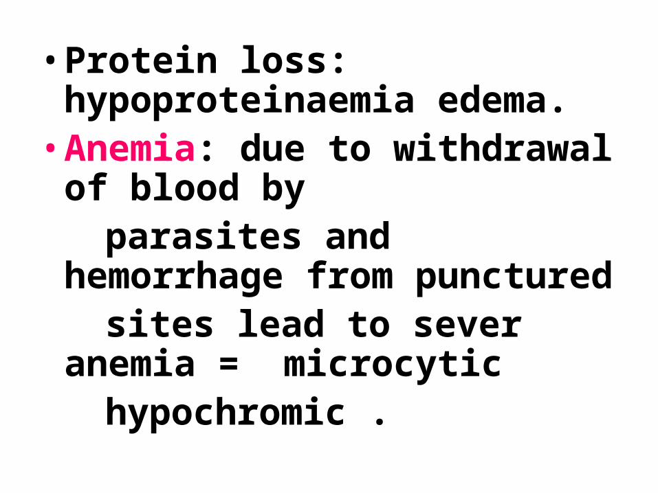
•Protein loss: hypoproteinaemia edema.
•Anemia: due to withdrawal of blood by
parasites and hemorrhage from punctured
sites lead to sever anemia = microcytic
hypochromic .
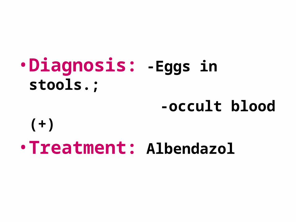
•Diagnosis: -Eggs in stools.;
-occult blood (+)
•Treatment: Albendazol
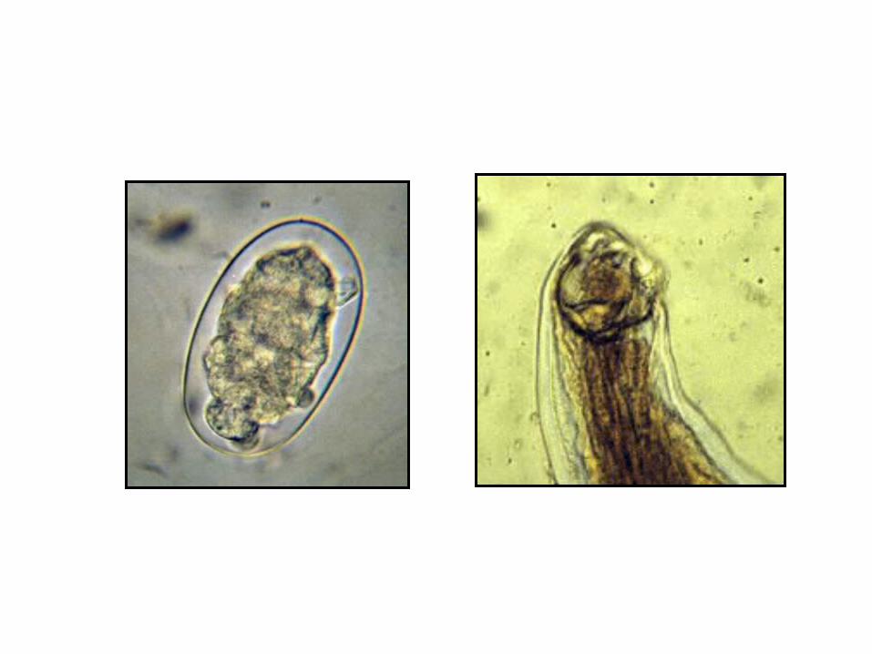
Strongyloides stercoralis
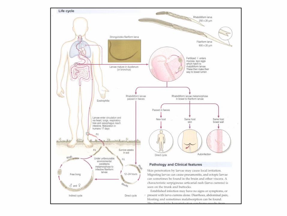

Strongyloides stercoralis• Widely distributed in tropical
region worldwide .• fetal opportunistic in immuno- compromised host.• it is smallest pathogenic
nematodes ±2.5mm.• adult live in mucous membrane
of duodenum, jejunum rarely m.m.of bronchus.

Pathology and clinical picture:
1-Cutaneous little reaction on penetration.
Sever dermatitis at perianal region in case of external autoinfection.
2- Migration :same as hook worms .
3- Intestinal: inflammation of upper intestinal mucosa, diarrhea, upper abdominal pain colicky in nature.

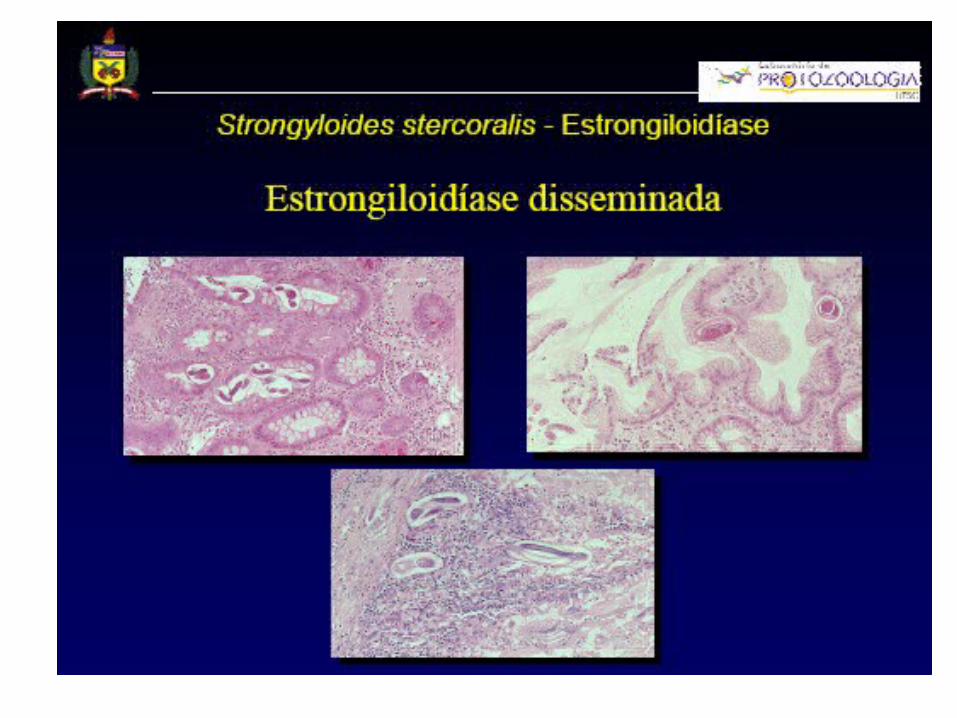
Disseminated strongyloidiasis
in patient with immuno-deficiency
-uncontrolled diarrhea – granulomatus changes – necrosis--perforation– peritonitis--death.


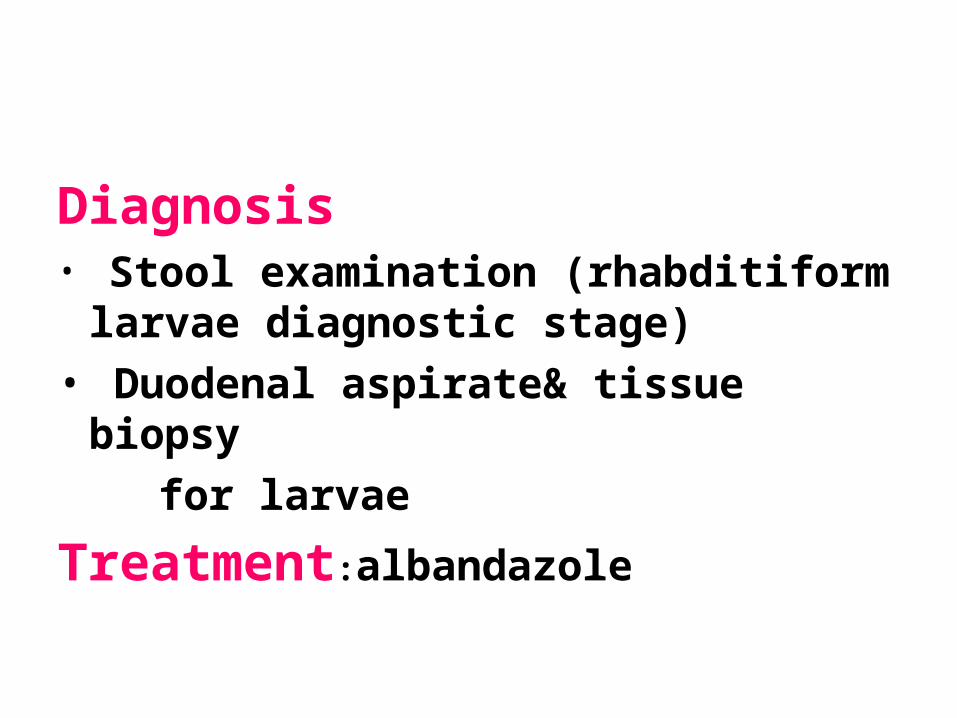
Diagnosis• Stool examination (rhabditiform
larvae diagnostic stage)• Duodenal aspirate& tissue
biopsy for larvae
Treatment:albandazole
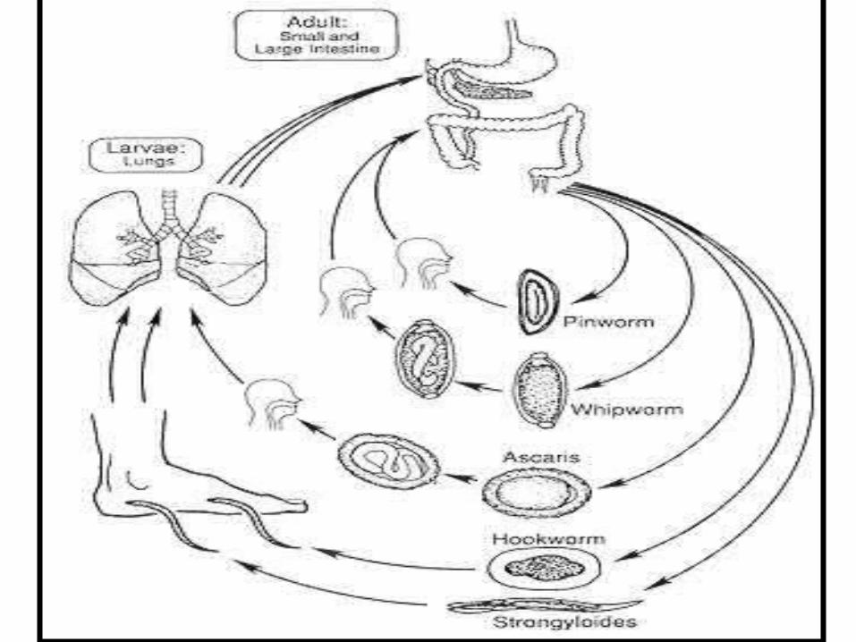
fig86_7[1].jpg

الحمد لله