Embed Size (px)
Citation preview

第 102 回⽇本病理学会総会 コンパニオンミーティング10(CM-10)
⽇本婦⼈科病理学会 『外陰・膣の病理』
座⻑︓ 笹島ゆう⼦(帝京⼤学医学部病理学講座) 佐藤勇⼀郎(宮崎⼤学医学部病理学 構造機能病態学分野)
講演1 外陰部扁平上⽪病変 ⼤⽯善丈 九州⼤学⼤学院医学研究院保健学部⾨検査技術科学
講演2 外陰部における⾊素細胞性疾患の病理診断 ABC 泉 美貴 東京医科⼤学 医学教育学講座
講演3 外陰・膣の軟部腫瘍 福永真治 東京慈恵会医科⼤学附属第三病院 病院病理部
平成 25 年 6 ⽉ 7 ⽇(⾦) 18︓40〜20︓40 R3 会場(ロイトン札幌 2 階リージェント)

Vulvar Squamous Neoplasia
Department of Anatomic Pathology
Kyushu University
Yoshihiro Ohishi
Content
1. Overview of vulvar squamous neoplasia
2. VIN (histology, IHC, malignant potential)
3. VSCC (histology, IHC, prognosis)
4. Special type (verrucous, BCC)
5. Condyloma vs papilloma
Histopathology 2013; 62; 161‐175
Vulvar Squamous Cell Carcinoma(VSCC)
• < 5% Gynecologic malignancies
• > 90% Vulvar malignancies
HPV‐related VSCC HPV‐independent VSCC(HPV+) (HPV‐)
basaloid/warty SCC Keratinizing SCC
Vulvar Squamous Cell Carcinoma(VSCC)
Basaloid SCC WHO, 2003Keratinizing SCC
Vulvar Intraepithelial Neoplasia(VIN, precursor of VSCC)
HPV‐related VSCC HPV‐independent VSCC(HPV+) (HPV‐)
usual VIN
(classic VIN)(bowenoid VIN)
differentiated VIN
(simplex VIN)
Vulvar Intraepithelial Neoplasia (VIN)
uVIN 1
uVIN 3, basaloid
uVIN 3, warty
Differentiated VIN WHO, 2003
1

Lichen Sclerosis
• Precursor of HPV‐indepependent VSCC
• Lichen sclerosis << differentiated VIN
Lichen Sclerosis
HPV in VSCCs
Histopathology 2013; 62; 161‐175
Authors Year n HPV typing test HPV (%) HPV16 (%)
Monk 1995 55 PCR L1 consensus primers 60 49
Kim 1996 18 PCR L1 consensus primers 39 71
Pinto 1999 16 PCR L1 consensus primers 50 –
Carter 2001 38 PGMY9/11 79 55
Riethdorf 2004 71 GP5+/GP6+ and p16INK4a 35 76
van der Nieuwenhof
2009 130 Short PCR fragment L1 35 44
Kowalewska 2010 46 Linear array HPV test 15 71
Alonso 2011 98 SPF10 and p16INK4a 19 74
Gargano 2012 121 PGMY9/11 69 81
Tsimplaki 2012 6 PapilloCheck HPV 50 100
HPV in VINs
Histopathology 2013; 62; 161‐175
Authors Year n HPV typing testType of
VINHPV (%) HPV16 (%)
Trimble 1996 54OmniprobeAssay (Digene)
NOS 89 –
Pinto 1999 16PCR L1 consensus primers
uVIN 67 –
Carter 2001 18 PGMY9/11 NOS 91 74.6
Riethdorf 2004 67GP5+/GP6+ and p16INK4a NOS 52 88
van der Avoort
2006 37 SPF10 uVIN 66 65
van der Nieuwenhorf
2009 13Short PCR fragment L1
uVIN 100 44
Garland 2009 62 PCR uVIN 84 42
Smith 2009 65 PGMY09/11 NOS 98 50
Alonso 2011 48 SPF10 uVIN 83 –
Gargano 2011 66 PGMY9/11 NOS 94 48
Tsimplaki 2012 28PapilloCheckHPV
uVIN 71 65
HPV in VSCCs & VINs
• 1/5 ~ 1/2 VSCCs ・・・・・・・ HPV+
• > 4/5 VINs ・・・・・・・・・・・ HPV+
Most VSCCs are HPV‐
Most VINs are HPV+
HPV in vulvar squamous lesion
• VSCC & VINs ・・・・・・ HPV16 >>>>>18,31,33,45
• Condyloma acuminatum ・・・・・ HPV6,11
• Verrucous carcinoma ・・・・・・・・ HPV6?
2

Epidemiology
HPV‐ squamous lesion
• Elderly
• Keratinizing SCC (common) >
• Differentiated VIN (rare) <
• Lichen sclerosis (+)
• Multifocal lesion (‐)
• p16‐; p53+
HPV+ squamous lesion
• Young
• Basaloid SCC (rare)
• Usual VIN (common)
• Lichen sclerosis (‐)
• Multifocal lesion (+)
• p16++; p53‐
Histological features of VINuVIN・・・・・similar to CIN/VAIN
• Thickened epidermis
• Hyperkeratosis/parakeratosis
• Loss of maturation
• N/C ratio↑
• Hyperchromasia
• Mitosis↑
Histological features of VIN
uVIN
Basaloid VIN
(undifferentiated)
• Flat
• Non‐papillomatous
Warty VIN
(condylomatous)
• Condylomatous
• papillomatous
Basaloid and warty VINs
Basaloid VIN 3 Warty VIN 3
Overlap between basaloid and wartyHistological feature of VIN
• Morphological overlaps ; basaloid & warty VINs
• Basaloid & warty VINs ; single category
usual VIN
3

Histological features of VIN
Blaustein’s textbook, 6th edition
VIN 1, warty VIN 2, warty VIN 3, warty
Histological features of VINWHO terminology
• VIN1
• VIN2
• VIN3
uVIN
(classic VIN) subjective
Histological features of VINISSVD recommendation
• VIN1
• VIN2
• VIN3
uVIN
Condyloma
Histological features of VINdifferentiated VIN (dVIN)
• Subtle morphological change
• Thick epidermis
• Elongated rete ridges
• Large vesicular nuclei, macronucleoli
• Abundant, brightly eosinophilic cytoplasm
• Intercellular bridge (++)
Differentiated VIN Differentiated VIN
4

Differentiated VIN
• Interobserver reproducibility↓
• dVIN alone ・・・・・ rare diagnosis
• Confused with benign lesion
• “differentiated” but “high‐grade”
Immunohistochemistry of VIN
• uVIN ・・・・・ p16 (+, diffuse staining), p53 (‐)
• dVIN ・・・・・ p16 (‐), p53 (+, suprabasal extension)
p16 staining in uVIN p53 staining in dVIN
Malignant potential of VIN
• uVIN ・・・・・ 9‐16% progress to SCC
• dVIN ・・・・・ higher% progress to SCC
(underdiagnosis, transient lesion)
Histological features of VSCCWHO classification
• Basaloid
• Warty
• Nonkeratinizing
• Keratinizing ・・・・・・・・ most common
5
Keratinizing SCC Nonkeratinizing SCC
Basaloid SCC Warty SCC
Histological features of VSCC
• Basaloid & warty HPV+
• Keratinizing HPV‐
Basaloid & keratinizing SCC
6
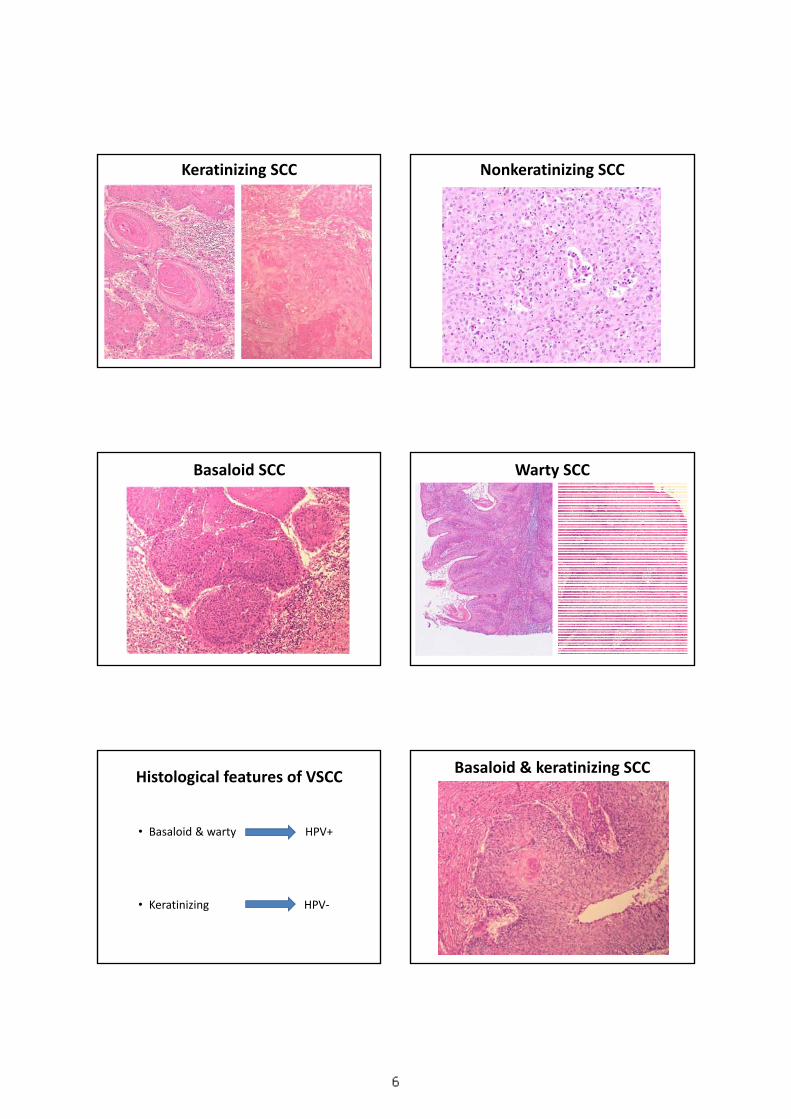
Keratinizing SCC, HPV+
Histopathology 2013; 62; 161‐175
Histological features of VSCC
• Basaloid & warty HPV+
• Keratinizing HPV‐
overlap+
HPV+ VSCC vs HPV‐ VSCC
• dVIN/Lichen sclerosis + ・・・・・ HPV‐
• uVIN+ ・・・・・・・・・・・・・・・・・・・ HPV+
HPV+ VSCC vs HPV‐ VSCCimmunohistochemistry (most reliable)
• HPV+ VSCC ・・・・・ p16 (++), p53 (‐)
• HPV‐ VSCC ・・・・・ p16 (‐), p53 (+, 50‐70%)
Keratinizing SCC + uVIN Keratinizing SCC + uVINp16 immunostaining
7

Keratinizing SCC
p16 IHC p53 IHC
Basaloid SCCp16 immunostaining
Prognosis of VSCC
Authors Year nOverall survival at 5 years
(%) P
HPV+ HPV-
Monk 1995 55 72 44 0.01
Pinto 2002 16 63 71 0.447
van derNieuwenhof
2009 130 80 78 0.646
Lindell 2010 75 85 40 0.03
Alonso 2011 98 67 71 0.789
Choschzic 2011 39 40 75 >0.05
Histopathology 2013; 62; 161‐175
Verrusous carcinoma
• 1‐2% of VSCC
• Undulating warty surface
• Hyperkeratosis
• Pushing border
• Minimal atypia
• HPV 6+?
• Favorable prognosis
Verrucous carcinoma Verrucous carcinoma
8

Basal cell carcinoma Condyloma acuminatum
Condyloma acuminatum Papilloma
Papilloma Condyloma vs papilloma
9
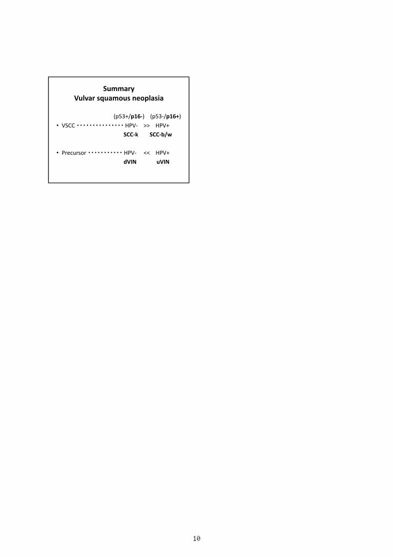
SummaryVulvar squamous neoplasia
(p53+/p16‐) (p53‐/p16+)
• VSCC ・・・・・・・・・・・・・・・ HPV‐ >> HPV+
SCC‐k SCC‐b/w
• Precursor ・・・・・・・・・・・ HPV‐ << HPV+
dVIN uVIN
10
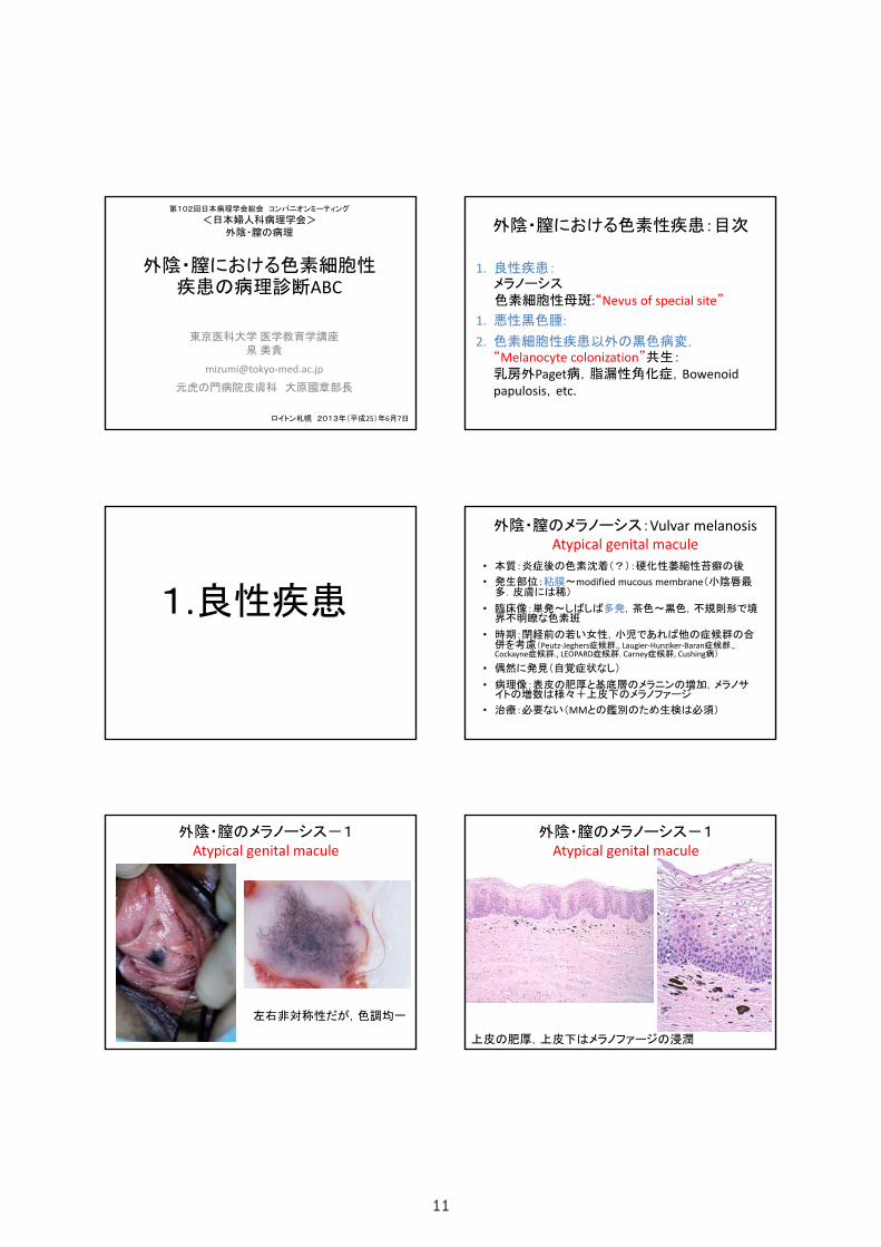
外陰・膣における色素細胞性疾患の病理診断ABC
東京医科大学 医学教育学講座泉 美貴
mizumi@tokyo‐med.ac.jp
元虎の門病院皮膚科 大原國章部長
ロイトン札幌 2013年(平成25)年6月7日
第102回日本病理学会総会 コンパニオンミーティング
<日本婦人科病理学会>外陰・膣の病理 外陰・膣における色素性疾患:目次
1. 良性疾患:メラノーシス色素細胞性母斑:“Nevus of special site”
1. 悪性黒色腫:
2. 色素細胞性疾患以外の黒色病変,“Melanocyte colonization”共生:乳房外Paget病,脂漏性角化症,Bowenoidpapulosis,etc.
1.良性疾患
外陰・膣のメラノーシス:Vulvar melanosisAtypical genital macule
• 本質:炎症後の色素沈着(?):硬化性萎縮性苔癬の後
• 発生部位:粘膜~modified mucous membrane(小陰唇最多.皮膚には稀)
• 臨床像:単発~しばしば多発,茶色~黒色,不規則形で境界不明瞭な色素班
• 時期:閉経前の若い女性,小児であれば他の症候群の合併を考慮(Peutz‐Jeghers症候群., Laugier‐Hunziker‐Baran症候群.,Cockayne症候群., LEOPARD症候群. Carney症候群, Cushing病)
• 偶然に発見(自覚症状なし)
• 病理像:表皮の肥厚と基底層のメラニンの増加,メラノサイトの増数は様々+上皮下のメラノファージ
• 治療:必要ない(MMとの鑑別のため生検は必須)
外陰・膣のメラノーシス-1Atypical genital macule
左右非対称性だが,色調均一
外陰・膣のメラノーシス-1Atypical genital macule
上皮の肥厚,上皮下はメラノファージの浸潤
11

外陰・膣のメラノーシス-2Atypical genital macule
外陰・膣の色素細胞性母斑左右非対象,異型性,ascentがある
1. 特別な部位
① “Nevus of special sites” (Milk line)
② 間擦部(腋窩,外陰部)
③ 粘膜:膣,口唇・口腔内,眼瞼,鼻腔
④ 掌蹠(手掌・足底)
2. 特別な母斑① 先天性母斑
② Spitz母斑
③ 青色母斑
3. 特別な時期:新生児・乳幼児,妊娠中
“Nevus of special sites”外陰・膣の色素細胞性母斑
• 病理所見が悪性様:
“Atypical melanocytic nevi of genital type”
“Nevi of site related atypia”
• 臨床的には良性所見:左右非対象でも境界明瞭,色調均一
• 年齢:閉経前の女性(平均23歳)
• 偶然に発見:妊娠・出産,子宮筋腫の手術時など
• 部位:皮膚に多い(通常の母斑は粘膜と皮膚両方),小児は小陰唇と陰核
外陰・膣の色素細胞性母斑“Atypical melanocytic nevi of genital type ”
“ Atypical genital nevi”
• 悪性と間違える:先天性母斑様(案外多い),異形成母斑様
– 大型: 10 mm~(先天性)– 不整形:胞巣が不整・多形・分布が不規則,炎症細胞,メ
ラニンの分布– 表皮の増生:(表皮突起の延長,癒合(DN),乳頭状,偽
癌性表皮過形成)– 付属器に沿う配列(先天性)– 細胞が大型(特に表皮内や真皮浅層),異型性,多形性
(先天性)– 明瞭な核小体– 多少のAscent– 高度の炎症細胞浸潤(DN)– Lamellar fibroplasia(層状で好酸性の線維化)(DN)
外陰・膣の色素細胞性母斑“Atypical melanocytic nevi of genital type ”
“ Atypical genital nevi”
• 良性と判断できる所見:– 潰瘍はない– 表皮内病変は真皮内病変を超えない
(shoulder lesionはない)– 境界が明瞭– 構造異型は目立つが細胞異型は乏しい– 多少のascentはあっても,Paget様進展はない– 壊死はない– 核分裂像はない(特に上皮下にはない)– 深部への成熟を示す– 細胞周囲の密な線維化やリンパ球浸潤はない
外陰・膣の色素細胞性母斑-1,境界型
メラノサイトがやや腫大∵上皮の延長,境界明瞭,ascentなし,明かな異型性
なし,連続するほどの増殖なし
12

外陰・膣の色素細胞性母斑-2 外陰・膣の色素細胞性母斑-3
異形成母斑様のlamellar fibroplasiaとリンパ球浸潤
外陰・膣の色素細胞性母斑-3
左右非対象性だが境界は明瞭,色調も均一
外陰・膣の色素細胞性母斑-3
表皮が肥厚娘結節
異型性はない
外陰・膣の色素細胞性母斑-4
5歳女児,陰核:典型例
外陰・膣の色素細胞性母斑-4
大型の胞巣,細胞接着が緩い
13

外陰・膣の色素細胞性母斑-5
臨床的には不規則,左右非対象,隆起あり,娘結節? ただし境界は明瞭,色調も均一
外陰・膣の色素細胞性母斑-5
左右非対称性
上皮直下は細胞が大型 ∵深部は成熟あり
外陰・膣の色素細胞性母斑-6
左右非対称性.境界は明瞭
高度の炎症細胞浸潤,
外陰・膣の色素細胞性母斑-6
表皮突起の不規則な延長∵母斑細胞に異型性はない.表皮の偽癌性過形成,著しいリンパ球浸潤
外陰・膣の色素細胞性母斑-7
若い女性の外陰部(有毛部)の境界明瞭な色素性病変:母斑の典型
外陰・膣の色素細胞性母斑-7大型,表皮が増生,付属器周囲の配列真皮浅層は大型だが成熟がある→先天性母斑
14

2.悪性黒色腫
外陰部悪性黒色腫の臨床
• 患者:閉経後の女性,平均68歳• 頻度:外陰部の悪性腫瘍でSCCに次ぎ5%,悪性黒色腫の,1~2.3%• 部位:Atypical NZNと異なり,無毛部の大陰唇,陰核• 臨床像:境界不明瞭,色調不均一(amelanotic melanomaは稀),しば
しば多発(>20%)• 大きさ:≧7mm• 生検,excisional biopsy,(shave biopsyは禁忌:深達度不明,取り残し
必至)
• 組織像:表皮は潰瘍~萎縮,明瞭な構造異型・細胞異型,境界不明瞭,ascent/Pagetoid進展,核分裂像
• Breslow thickness:平均3.2 mm,in situは極めて稀• 予後因子:年齢.Stage(潰瘍あり,深達度深い,amelanosis).LN転移
の有無,深達度は再発に関与しても生存率には無関係とも.• 予後:悪い(平均生存期間5.2年,1/5/10年生存率=85/50/30%)• 治療:切除・リンパ節郭清
膣の悪性黒色腫の臨床• 患者:閉経後(80%)の女性,平均60歳前後
• 頻度:非常に稀,報告300例余り,膣悪性腫瘍の<3%
• 発生母地:成人女性3%の膣にメラノサイト+
• 部位:膣外側1/3
• 症状:出血,結節
• 臨床像:潰瘍形成,易出血性,黒色調(amelanoticは稀),20%が多発
• 予後因子:①大きさ≧3cm 12ヶ月,<3cm 41ヶ月生存.②リンパ節転移がない.深達度は関与しないとも
• 予後:40%が局所再発,転移:肺,肝臓,骨
• 治療:切除(拡大手術と最小限のマージンでも予後に差は無い)+放治
• 予後:5年生存率 0~21%
外陰悪性黒色腫-1
臨床的に悪性
潰瘍,腫瘤の形成
外陰悪性黒色腫-1
表皮内病変あり
短紡錘形の異型細胞高いN/C比,クロマチンの濃縮
外陰悪性黒色腫-2
両側の小陰唇 辺縁にシミ出し
15

外陰悪性黒色腫-2
皮膚の結節型黒色腫と同様
外陰悪性黒色腫-2
外陰悪性黒色腫-3外陰悪性黒色腫-3
外陰悪性黒色腫-3
Atypical melanocytic hyperplasiaがmelanomaの周辺に広がる(14%)
外陰悪性黒色腫-3
Vulvectomy施行するも 左右の鼠徑リンパ節に転移
16
膣悪性黒色腫-1
膣壁複数の斑~小丘疹 浸潤部は紡錘形細胞
膣悪性黒色腫-2
膣壁の大型腫瘤,ヘルニア状 類円形異型細胞,びまん性に増殖
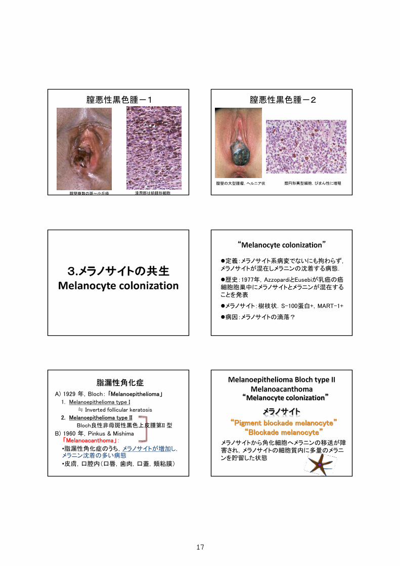
3.メラノサイトの共生Melanocyte colonization
定義:メラノサイト系病変でないにも拘わらず,メラノサイトが混在しメラニンの沈着する病態.
歴史:1977年,AzzopardiとEusebiが乳癌の癌細胞胞巣中にメラノサイトとメラニンが混在することを発表
メラノサイト:樹枝状.S-100蛋白+,MART-1+
病因:メラノサイトの滴落?
“Melanocyte colonization”
A) 1929 年,Bloch: 「Melanoepithelioma」
1. Melanoepithelioma type I
≒ Inverted follicular keratosis
2. Melanoepithelioma type II
Bloch良性非母斑性黒色上皮腫第II 型
B) 1960 年,Pinkus & Mishima「Melanoacanthoma」:
•脂漏性角化症のうち,メラノサイトが増加し,メラニン沈着の多い病態
•皮膚,口腔内(口唇,歯肉,口蓋,頬粘膜)
脂漏性角化症
“Melanocyte colonization”
“Pigment blockade melanocyte”“Blockade melanocyte”
メラノサイトから角化細胞へメラニンの移送が障害され,メラノサイトの細胞質内に多量のメラニンを貯留した状態
メラノサイト
Melanoepithelioma Bloch type IIMelanoacanthoma
17

メラノサイトは少数メラニンが豊富
色素型の脂漏性角化症“Pigmented type, seborrheic keratosis”
脂漏性角化症Melanoepithelioma, Bloch type II
Melanoacanthoma
“ボタンを置いたような”
メラノサイトが増加メラニンが豊富
樹枝状のメラノサイト,豊富なメラニン“Pigment blockade melanocyte”
“Blockade melanocyte”
“Melanocyte colonization”
<腫瘍性疾患>乳癌基底細胞上皮腫(癌)乳房外Paget病SCC(転移)ボーエン病(日光角化症)脂漏性角化症石灰化上皮腫汗孔腫, 汗孔癌Apocrine hidrocystoma神経線維腫(Pigmented)DFSP(Bednar tumor)
<非腫瘍性病変>
尋常性疣贅
尖圭コンジローマ
Bowenoid papulosis
Melanocyte colonizationを来す疾患
<腫瘍性疾患>乳癌基底細胞上皮腫(癌)乳房外Paget病SCC(転移)ボーエン病(日光角化症)脂漏性角化症石灰化上皮腫汗孔腫, 汗孔癌Apocrine hidrocystoma神経線維腫(Pigmented)DFSP(Bednar tumor)
<非腫瘍性病変>
尋常性疣贅
尖圭コンジローマ
Bowenoid papulosis
Melanocyte colonizationを来す疾患
湿疹様の紅色~一部白色調病変+褐色?!
乳房外Paget病
18

“Melanocyte colonization”
悪性黒色腫乳房外Paget病
乳房外Paget病 悪性黒色腫
特殊染色 DPAS + -
アルシアン青 + -
免疫染色 AE1/AE3 + -
CAM5.2 + -
サイトケラチン 7 + -
サイトケラチン 20 - -
CEA + -
MUC1 + -
GCDFP-15 (BRST-2) + -
c-erb B-2 (Her-2) + -
Androgen receptor (AR) + -
S-100蛋白 - +
MART-1/Melan-A - +
特殊染色・免疫染色による鑑別法
Condyloma acuminatum肌色,乳頭状集簇性
Bowenoid papulosis褐色局面~乳頭状,孤立性
Bowenoid papulosis(HPV)
<SCC in situ (Bowen病)>1. 核分裂像2. 個細胞壊死3. Clumping cells
<HPV 16感染>•コイロサイトーシス•錯角化を伴う過角化柱
•多顆粒細胞症Bowenoid papulosis ご清聴ありがとうございました
リレーションシップ ID rId3 のイメージ パーツがファイルにありませんでした。
19

Pathologic Diagnosis of Vulvovaginal Mesenchymal Tumors
Masaharu Fukunaga, MD
Department of Pathology
Jikei University Daisan Hospital, Tokyo, Japan
A. Site‐specific or characteristic vulvovaginal mesenchymal lesion
B. Miscellaneous mesenchymal lesions with diagnostic difficulties
Site‐specific or characteristic vulvovaginal mesenchymal lesion
• Angiomyofibroblastoma
• Aggressive angiomyxoma
• Superficial angiomyxoma
• Superficial (cervicovaginal)myofibroblastoma (of the lower uterinegenital tract)
• Cellular angiofibroma
• Fibroepithelial stromal polyp
• Prepubertal vulval fibroma
• (Vaginal tubulo‐squamous polyp)
A right vulva mass in a 46‐year‐old female. Clinical diagnosis: Bartholin gland cyst
20

Angiomyofibroblastoma
• Reproductive age women (and men)• Vulva (20%), vagina, inguinal area and scrotum• Typically small (<5cm), circumscribed, non‐recurring superficial soft tissue benign tumor.
• Preoperative diagnosis: Bartholin’s gland cyst.Immunohistochemistry• ER + (AR+ in men)• PR +• Desmin, actin, CD34:+/‐• S100 ‐
Aggressive angiomyxoma
• Subcutaneous or deep soft tissue• Large (>5cm), gelatinous or myxoid mass with infiltrative margins.
• Bland spindle cell, no nuclear atypia, low mitotic index
• Small to medium sized blood vessels with may be thick walled and hyalinized.
• Condensation of fibrillary collagenous material or bundles of smooth muscle around blood vessels.
21

Aggressive angiomyxoma
Superficial angiomyxoma
• Superficial, small (>5cm), circumscribed, with multilobulated pattern.
• Neutrophil infiltrates.
• Epithelial or adnexal component (1/3)
• Significant risk for local (nondestructive recurrence (up to 40%).
Vulva Superficialangiomyxoma
22

Cellular angiofibroma
• Rare, small (<5cm), superficial soft tissue tumor in middle age women (and men) in the vulvovaginal region and the inguinoscrotal or paratesticular region.
• Numerous small to medium sized vascular vessels with hyalinization.
• Uniform spindle cell proliferation
• Variable mitotically active.
• CD34 +
Superficial cervicovaginal myofibroblastoma
• Cervix, vagina, vulva
• Well‐demarcated polypoid or nodular benign lesion
• A well‐circumscribed but unencapsulated lesion. The presence of Grenz zone.
• Bland spindle cell proliferation in a fibrous or myxoid stroma
• Fascicular, lacelike, silve‐like patterns.
• Immunostaining: No‐characteristic: Vimentin, ER, PgR :+. CD34, aSMA: + some cases
• Fibroepithelial stromal polyp:same spectrum?
23

Fibroepithelial stromal polyp
• Small, superficial exophytic or polypoid mass in reproductive aged women.
• Vagina, vulva, cervix.
• No Grenz zone.
• Multinucleate cells, enlarged bizarre nuclei.
• May have high mitotic index (>10/10HPF)
• Pale, edematous and myxoid stroma.
• Variable vascular component with thick‐walled vessels in middle.
Fibroepithelial stromal polyp
Cellular pseudosarcomatous fibroepithelial stromal polyp
B. Miscellaneous Mesenchymal lesions withdiagnostic difficulties
• Reactive, proliferative lesions
• Epithelioid/Biphasic tumors
• Myxoid tumors
• Others
A 1.5cm vulva mass in a 28‐year‐old female Case: A 2.5cm vaginal mass in a 50‐year‐old female
24

Case: A 2.5cm vaginal mass in a 50‐year‐old female
Epithelioid leiomyoma
Vulva. epithelioid leiomyoma Vulva leiomyoma with myxohyaline change
Vulva. Leiomyosarcoma
Vulvovaginal smooth muscle tumors
• Premenopausal women
• Vagina more common than vulval
• Vagina usually benign, but recurrence seen when mitotic index>5/10HPF
• Vulval lesions recur locally and may metastasize to lungs (<25%)
• Main histologic patterns: spindles, myxohyaline and epithelioid.
• ER, PR:+
25

Vulval smooth muscle tumors
• Main histologic patterns: spindles, myxohyaline and epithelioid.
Vulvovaginal smooth muscle tumors:Criteria for malignancy
• Size>5cm*• Infiltrative margins*• Moderate to severe cytologic atypia*• Mitotic index>5/10HPF*• Tumor cell necrosis
*Tumors with 3 of these features are classified as malignant (leiomyosarcoma).Any single feature may indicate local recurrence and complete excision is needed.
A “vaginal” nodule in a 34‐year‐old “female” A “vaginal” nodule in a 34‐year‐old “female”
Case: A 34‐year‐old female (originally male) with a vaginal nodule
Homosexual, HIV (+)Vagina was formed by rectum three weeks
previouslyA nodule at the operative site
Diagnosis: Postoperative spindle cell nodule
Postoperative spindle cell nodule
• A non‐neoplastic localized lesion
• At the site of recent operation several weeks to several months postoperatively, especially in GU or GI areas.
• Closely packed proliferation of spindle cells and capillaries simulating a leiomyosarcoma. Inflammatory cell infiltrates.
• Diagnostic clue: History of a recent operation at the same site
26
A 33‐year‐old woman with a polypoid 2.5 cm mass in the posterior wall of the lower vagina
vagina.
CAM5.2, vimentin:+
A 3cm vulva mass in a 52‐year‐old female
27
Malignant myoepithelioma of the soft tissue(myoepithelial carcinoma)
Tumor with moderate or severe nuclear atypia
In soft tissue cases.
Hornick H, Fletcher CDM.
Am J Surg Pathol ;27:1183‐96.
Synovial sarcoma, proximal type
A 3cm vaginal tumor in 54‐year‐old femaleExtra GI GIST
A 31‐year‐old femalea 3cm mass in the uterovaginal area
A 31‐year‐ old femalea 3cm, mass tumor in uterovaginal
area
Sclerosing perivascular epitheliodcell tumor (PEComa)
• PEComa with extensive stromal hyalinization
• Women, middle aged ( mean, 49 yrs)
• Retroperitoneum, pelvic cavity, uterus
• Rarely recur or metastasize
28

Dermatofibrosarcoma protuberans,myxoid type A vulvar mass in a 78‐year‐old female
Myxoid liposarcoma
A 2cm vaginal mass in a three month old female
Embryonal rhabdomyosarcomaSarcoma botryoid ブドウ状肉腫
Miscellaneous mesenchymal lesionswith diagnostic difficulties (continued)
• Reactive, proliferative lesion: Nodular fasciitis. Postoperative spindle cell nodule, endometriosis
• Epithelioid/Biphasic tumor:
smooth muscle tumor
mixed tumor of vagina
myoepithelioma
synovial sarcoma
GIST
PEComa
(mesonephric adenocarcinoma with spindle
cell component)
Miscellaneous mesenchymal lesionswith diagnostic difficulties
• Myxoid tumor: smooth muscle tumor, myxoid DFSP, myxoid liposarcoma
• Other mesenchymal tumors : embryonal rhabdomyosarcoma, Ewing sarcoma, SFT.
• Mammary type tumors: fibroadenoma, phyllodes tumor, myofibroblastoma
Conclusions• If clinically benign , but not easily classified, consider “specialized (gonadal) stromal tumor”Immuostains are not always useful
• Exclude reactive and/or inflammatory processes.
• Take care a lot if smooth muscle tumor is diagnosed. The criteria of malignancy is not same as those in the uterus.
• Consider extension from retroperitoneum, pelvic, and mammary type lesions.
29