Embed Size (px)
Citation preview

t:: '
i" ~.
1 r~{
:'i' ,~
•
..
c
...... y •
STRAIN,SPECIFICITY AND- "SELF/NON-SELF" RECOGNITION IN
A TROPICAL MARINE DEMOSPONGE, Verongia longissima.
Heather R. Kaye
@
A thesis subm:i:tted to the Facul t Graduate Studies anij Research McGill Uni versi ty, in partial fulfil1ment of the requir~ments for the degree of
Masters of Science.
Marine Sciences Cent:re McGill University Montreal, 'Canada.
() -,
of
, f
August 1979.
.. . '
! 1 l-l t l 1 1 ,
\ l

, ~j
~ ~ ," i !l" ~, ,~
.~' ~ ri' ~
'v Il
k ,. ~, J L ~.
~, "
(
-) . . . • -~ -- '"'-. \
,. .. .. .. , -.'
r ,t: • ,
" ~ "
"'-'\ "'"'"' lt
- /
. ' / (ly This thesis i9 dedicated to my. friend and constant diving bUddy, Tina Ortiz, whose support, and wi.llingness to help.throughout this study contributed greatly to the complet~on of the work."
, .
• .. 11,
.. ~
\
J
" ,
. ,'" ~ '~J' , '
\ . ,\
t'
•
. 1 '
---1
1 , ) ,
, -) i
1 ,',
. . : ) , "

.. . ,~c
. , ,
, .
i , ... ~ ~.---.. ~---_ ...... _-.~, ........ -. . ,
... .
AB~'l!RACT
. ,~rafd.nq- experi~ents' and i~nologi~al analyse.s have
~a proviçied direct· evidence for the çccurrence of strain spec"'-
ificity in the marine Demosponge, Verongia .... longissima. _. All, . , autOgr~fts accepted, albx'enogra~ts .r,ejected, and th,e.re .was·
both acceptance and te je ct ion of allografts. Among . / (~ .. allqgrafts group.s of indiv~uals could easily be identified . ' .
. 'which' Sho~ed consistent acc~ptances and rej ections o.f other
indi vidua: s. ':~roups , 1 1
were designa ted as stràins.·
Results of imrnqnological
" ant~sera ,and sponge antigens 'supported this strain'
designation and reve'ale,Çi extensive antigenic"polyYnorphism,
among strains~of verongia longissi~a. These fponqes . . . p ~ •• ~
, demonstrate an immun?reco~nition a~ 'the' allOgen71é:: ~lev"~l,
similar ta the immunological.response of higper metazoans. " • J - ~
" The use of an immunofluorescen6~ test for strain . , iaentifidation proved .impossibl~ 'when the cel-ls of Verongia
j;I
longissima Wère found to possess ~utofluorescenc~. '
HistologicaJ" preparations of graft rejections showed
the aevelopment of a cuticle between the surfaces ~f the
..
. ,
. '" two sponges in contact. Results of radioautography
experiments have provided 'the basis for the formulation df .,..
\ an hypothesis concerning the formation and deposition of
q loi"
tllis cuticle. The role of speci'ficity in patterns of " .. interactions between sponges in situ has been discussed' \
• -- 1
frod'an ecolo~al perspective.
iii
1 f
i ! '\ "
·f
l "

y , ,
.'
" .'
. -
. , .
"
(
.. ô
O'
'1 ,
, , RESUME
",1-' ~ Des' expériènces de greffage et des é,tudes immunologi-
} . ,1 .' .
qu 1 s avec l' éponge mari~e Verongia lon~issima (Demospong,ia)
G>nt révél~ chez ce spongiaire une ~pécifici té dè souche.
Les homogreffes sont compatibles~. les xénogreffes sont' . .
:t;ejetées et les"allogreffes sont parf?is compatibles,
parfois rejetées. , Des groupes, d'individus ayant,' aux
. ,
. allogreffes, des réactions constantes de compatibilité' ou de
~ejet sont identifiables. Ces groupes sont app~lés souche. ./ \ ,
Les résultats d' a~alyse immunologiques u-tilisant i "ant1sérum
d~ iapin et des a~tigèn~s d'éponge confirme l'identification <:1
.. des souches e't opt démontré l'existance de 'polymofrphisme
.àntigénique des.souche~ de verongi~ longissima .
. ' L ~ identification des souches à l'aide de tests d' immuno-
'. fluorescence s' est· avérée impossible à cause de l' auto-
fluorescence des cellules de Verongia loagissima.
, . L'examen histologique de preparat10ns de tissus
provenant de g,reffefi'J FejJtées al révélé la formation d'une
cuticuie 'sur les' zones de contact entre les deux éponges. \ -
·Les résultats d' experiences de radioautographie ont permis '" "1 f
" 1 (élaboration d-' une hypo.thèse concernant la formation et ~--, ,
, , V}
, déposition de cette ~ cut.icule. Le r5le ~e la spécificité , ,
dàns les, iriteractions entre éponges in situ est discuté du
point de vue écologique.
..
iv )
1
i l ! !
j J i
~ Cr i
f
,
-/ /
, -ACKNOWLEpGËMENTS
The success of conducting field worl using 1CUBA
de~nds to a large degree on the co-operation an, Ilelp
a nu.ml5€r of people. l express sincere thanks to \Wendy 1
of
GoLdblatt, Julie LaRoche, Madeleine Roberts, Ray Lynch, and il
~ames Ortiz, who served as diving partners throughout the
duration of this study.
l would like to thank Dr. Peter Fieldes, of the' Queen ~ ),
Elizab~th Hospital, Bridgetown, Barbados, fo~ his help and
guidance with the immunization of the rabbits and
preliminary immunological analyses.
l wish to acknowledge the àid of Dr. Glenda Wright,
Dr. 'Beatrice Kopriwa, and Fernando Evaristo of the McGill
Anatomy DepartmenJ.. for their guidance and help u\ radio-
autographi~ analyses. •
l would like to express my s~~ thanks ~. Lorry Il
Schneider for Many stimula ting and supporti,ve discussions at /
different times during this study, and for his time in , critically reviewing this,ma~uscript. Thanks to Audrey
Filion for, time spent in the French translation of the
abstracto
Sincere'thanks to my father, who helped invaluably with
the preparation of the figures and tables and contributed !?
many constructive suggestions and gave support during,this 1
study.'·
v
1
1
i ( 1
j \ ,
î
1 . .,. ?
),

,
t
-, l'
l"~ ~ ,1
, : ~,
~ ,h
(
~I
t \ l
" K
()
-- -_..--_.- -...... -_ .... -... ,.",... ~.. ...-,
"
~ would like to express my deep a'ppr~ciation to Dr.
Harold Rode, and Eis Schotman of the Experimental Medical ,
Research Depa:~tmeI\t of McGi11 Univefsity, who introduced me
to the methods and techniques of immunology, and offered
invaluable information and guidance which contributed
grea tly to the ·completion ~f this study'- For the knowled'ge
l gained and the many hoU'I'S they spent encouraging antl'
1 enlightening me in this subj ect area, l am indebt,.ed. o
Lastly l would likec to express my d~epest gratitude to , •
~y advisor, Dr. Henry M. Reiswig, whos~ continued
encouragement, and invaluable advice. throughout this study
contributed grea tly ~o the completïo'n of this manuscript.
His lÏterest in the a~e,a of spo~ge biology and knowledge of
the subje~t matter seryed as valuable inèentiv~s. For the
knowledge l gained fram his çonstant supervision, and the (
many hours he spent in reviewing this ,manuscript, l am ~ )
o indeed grfUl. "
This study was supported by a National RJarch Council ~,
Operating Grant NO. A-9554 to Dr. Henry M. ~ei ig.
, ,
/
" '\
, vi
J
. f
1-

,
"
<.
, / ...
TABLE OF CONTENTS
ABSTRACTo 0 ................... . '" ACKNO~DGEMENTS. . . . . . . . . . . . . . . . . . . . . . . . 'LIST 'OF FIGURES".
'" . /' -- . . . . . . . . . . . . . . . .
Î
.......... . ...... .
1
LIST OF TABLES •••• · .... • •••••••••••••••••• ''''Je •••••
, -LIST OF PLATES •••• · .................. . -............. . INTRODUCTION •••••• · .... . ................ . · ......... .
(
MATE RIALS AND METHODS •• , GRAFTING EXPERIMENTS. . . . . . . IMMUNOLOGICAL STUDIES •••••••••
-,.
~
· . . , .
a) Preparation of antigens. · ......... . h) Immunization •••••.•• · .. c)
d)
Agglutination test~.
Cross-absorption tests ..•
· ..... . · .. .
e) Immunofluorescence ex~eriment. · .. • HISTOLOGICAL STUDy ••••••• . . . . . . . . . . . . . . . · ...
f' RADIOAUTOGRAPHIC STUDIES ••••••••• . . . . . . . ~. '.' . .....
a)
h)
c)
RESULTS
Incorporation of labe1!ed praline. t
Histologica1 processing .••• · .... Radioautographic proc~dure. ...
AND OBSERVATIONS. · ..... • 0 0 .......... · ..
· .... · ... · ........ · .. \
. Il
....
. .... '.
. . G~~NG EXPER~TS ••
l OLOGICAL STUDIES. · .... • 0 0 . .............. o •
HISTOLOGICAL STUDY •••••••• . . . . . . . . . . . . . . . . . . . . . . . fRADIOAUTOGRAPHIC
DISCUSSION.
STUDIES. . . . . . . · ................ . · ................... . . .. "
SUMMARY ••••••••••• . . . . . . . . . ..... . . . . . . . . . . . . . . . . . LITERATURE CITED. . . ~ ........... . · ................ .
'. vii
!
Page
iii
v .' ..
'11\1
\~
X 1
7
10
-- 13
15
15
16
19
19
20
21
21
22
22 , 24
24
36
48
,51
56
65
67
,
.....
~
,.
, i 1
\

J'
,
I~
l ,
<,
. 1r ) 9
~ f:
~ 1 i ,
()
..
FIGURE 1:
FIGURE 2:
FIGURE 3a:
. ,
" ,
LIST OF FIGURES , ~.
Location of study area on west coast of -Barbados ~
Representative bottom profil~1 through study site located onr west coast of Barbados
Grafting Site A \
FIGURE 3b: Grafting Site B
1
FIGURE 3e: Summary of grafting - Both sites ,,-FIGURE 4a: % Abundaiice of strains in Site r FIGURE 4b: % Abundanee of strains in Site B
FIGURE' 4c: Total % abundance of strains from both sites ~
fi
FIGURE Sa: Absorption of #71 with various spongte extracts
FIGURE Sb:
FIGURE 6:.
Absorpt~on of 112 with ~arious sponge extrp,cts
Radioautography Results
. ..
.J
. , . . ~" \
\
Page .....
S
(-Il
~
26
27 '.
28 . , '"' 30
30
31 1
) v J
42
43 ! 53 1
-1
-=- l ~, ,

-~ --~---. .
o TABLE 1:
..
) , r
,
o ---~---------
j
l~ j
\ 1
;
,
LIST OF TABLES ... \. .
rsUIDlJl!ry of Agg1utinatLion- Resul ts'
..
• •
, 0
• l."
(
J
Page
38
,
-

'(
)
"
o
J
1.
PLÀTE 1:
Pf.,ATE II-A:
PLATE II-B:
PLATE II-C:
PLATE III-A:
l PLATE III-B:
;
PLATE IIpe:
PLATE IV-A:'
1
PLATE IV-B:
PLATE IV-C:
PLATE V-A: \
,PLATE V-B:
PLATE V-C:
P~TE Y-C:
, ... ----.v"~--- ,-_. --, ,
,', ,
LIST OF PLATES
. '
In situ photograph of Veronsia IOngISsima
\
In situ photograph'~éf S'tudy site
In situ~otograph,of procedure ' \
o
corner of
grafting
o ,
C1ose-up photograph of the·tieing procedure
o
In si tu photograph of' gr~fts
Close-up photograph of a control autograft acceptance .
Close-up photograph o~two rejected"grafts 1
Photograph of a microti ~re kg,te showing macroscopic results of the agglutination reactions
p'hotomicrograph of a posi ti ve reaction in the agglutination test
, 1tJ
Photomicrograph of a negative reaction in the agglutina,!=-ion~' test
\ C1ose-up photograph of the \' external features of a typical \ acceptance
oClose-up photograph of the internaI features of the zone of contact of anoacceptance
Close-up photograph of the external features of a typica1 rejecti0!1
Close-up photograph o~ t~e internal features of the zone of contact of a rejection
•
r
., 9
12
12
12
• J 14
14
• 17
17
t •
35
35
35
,
o
o
PLATE VI-A:
PLATE VI-B:
PLATE VII':"A:
.,
, Photomicrograph of autof1uorescence of verongia longissima cells
Photomicrograph of the. f1uorescence emitted when f1uorescein was added to a mixture of antiserum and antigen .
Photomicrograph of a crosssection through the zone of contact of a rejected graft
PLATE VII-B: Photomicrograph of a crosssection through the zone of contact of allt accepted graft
(
/
}(; }
.......... _---------~._~--- -
-Page
47
47
49
49
, >
{ -
c
(
/ - -
INTRODUCTION
One of the most definitive ~preSSions o~viduality ition) in higher organisrns is the rejection of
grafts individuals. Graft rejection or acceptance
immune ,mechanisrns considered to be different
for each ind'ividual (Curtis, 1978). However, a much simpler .... (,,, .
expression of individuality is the capacity for an organism
to maintain its autonomy in nature despite contacts with
~ other individuals of the s~ species.
At the turn of the century Wilson pioneered sponge cell
reaggregation studies with his classical work on coalescence ., and,regeneration. When t~ells of two different species
of sponges were disaggregated and cultured under conditions .\.
favourable to reaggregation, Wilson (1907) observ~d that the
celis aggregated into ~pecific cellÀlar masses. They r-
appeared to be maintaining a separateness from one another,
a phenomenon described by Wilson as a~type of species
specificity. This was the first recprd ~f this concept in
the lower Metazoa, and;with the possibility of gaining
furthe~ insi~ht into the general prinCiP~ of specie~
specificity and the implication of it i~both lower and
higher organisms, further studies of sponge cell ,
reaggregation have been carried out by many workers
(Galstoff, 1925; Curtis, 1962; Humphreys, 1970; John, et al, Q ~
1971; McC1ay, 1971; 1974; MacLennan, 1974).
)
In the past, the acceptance by an invertebrate of
l
-"' .. ---- ... ~,
\
, \
Y
,. >
1
2
material from an,ther m~er of the same species was
considered ta be (~e rule (Burnet, 1971). Exceptions to ..
that rule are now appearing in the literature. In the last 6
decade histoincompatibility, or the capacity for an organisrn
to distinguish "self" from "non-self" has come to be a
common phenomenon among the lower invertebrates, including
gorgonians (Theodor, 1970), corals (Hildemann, et al, 1975;
1977), ascidians (Oka, 1970; Mukai and Watanabe, 1974), and
sponges (Humphreys, 1970; Van de Vyver, 1971 b)<. This
"self/non-self" recognition system could be a negative
reaction, where two individuals graft together unless a
specifie signal is exchanged to indicate incompatibility,
or, it could be a positive reaction where the two tend to •
regard each other as foreign until a positive signal occurs
enabling them to recognize each other as compatible (Burger1
et al, 1978). A review of the literature related to this
phenomenon reveals a lack of information of the underlying
mechanisms involved in almost aIl cases of irnmunorecognition
and the incompat~bility reactions whicn follow such
:E'ecognition.
The re)ection of tissue,transplanted from one , /
individual te anether individual of the same species, an
allogeneic graft, is one as~ect of an individual's ability
to form an adaptive immunity response ta foreign tissue
(Coffaro and Hinegardner, 1977'). This is a weIl known •
phenomenon among vertebrates (Warr and Marchalonis, 1978). . ,
The incompatibility responses noted in lower invertebrates
'i-l-,
1 1
t 1 ~ · · 1 l
<" 1 f ·

3
are mor~ commonly found when transplants between ~ specie,s,
xenogeneic grafts, are used. These incompatibility
responses have been described as nonimmunologica1 (Coffaro , --
and Hinegardner, 1977). However, immunorecognition at the r
a1logeneic level has been recently reported by Hildemann,
et al (1977) in corals, anft Hildemann, Johnson, and Jokiel
(1979) in sponges. These workers have found that a highly
"sensitive immune system demonstrating the essential
attributes of adaptive irnmunity is present at this lower
phylogenetic level. This could represent ~ origins of
the major histocompatibility comp1ex and cell-mediated
immunity found' in higher vertebrates (Hildemann, Johnson,
and Jokiel, 1979).
The first record of incompàtibility between individual 1
members of the same species (intraspecific incompatibility) J,
in the Porifera was reported by Van de Vyver (1970) in the
freshwater ~pecies, Ephydatia fluviatilis, and a marine
species, Crambe crambe. This study revealed that there are
a number of strain types in these two species, each strain
being defined by their incompatibility with aIl other
members of the species in contact zones, creating a discrete
border, or zone of non-coalescence, separating the
a110geneic individuals. Members of the same strains fused
compatibly. This strain specificity appears to be carried -
over into the sexual larvae of Ephydatia fluviati1is.
Larvae of the same 1 mother sponge fused .and gave rise' to a ,
single sponge, in contrast to larvae from different motbers
s
,1 1
(' 1
4
which did not fuse. It appears that th~/stability of a Ê '
strain is preseive~ through ear1y stag,~ of sexua1 , f'~ -, ' rep~duction, but both paternity of the'se 1arvae and the
\ -
time of expression of that genetic component in
c~mpatibility reacti~ns was unknown.
Van de Vyver also noted the existence of eight ù
different strains of a number of crossed individuals of
Ephydatia fluviatilis in five different ponds containing
large populations of this sponge, compared to ten different 1-
strains of the marine sponge Crambe crambe found on a single
rock surface in the Mediterranean Sea. From this study it
appears that thé marine system offers conditions more .
favourable to the evolution of a higher numb~r of strains
within a species. This is substantiated by the recent work
of Hildemann, Johnson, and Jokiel (1979) on the tropic~~
Indo-Pacific sponge Callyspongia diffusa. Isografts ~(self
grafts) of this species fused compatibly, but allografts
(sarne species grafts) were invariably incompatible for aIl
200 individuals'tested.
Most of our knowledge of strain specificity in sponges
has corné fr~m extensive investi~ations on the freshwater ~,
sponges (Van de Vyver, 1970; 1971 (b); 19.75; Curtis and
Van de Vyver, 1971; Van de Vyver and Willenz, 1975).
However, as a resu1t of the relative1y recent recognition
of the importance of the roles that. sponges play in a coral
reef system (Bergquist, 1978; Fry, 1970; Grassé, 1973;
! ;,
li

(~
(
, 5
Harrison and Cowden, 1970; H~an, 1970; Sarà, 1970; and~
V~celet, 1971), more studies are being carried out on the \~. \
marine sponges.
Immunological approaches to the study of o ~
"self/non-self" recognition in sponges have extended the
concept of species specificity to a more rigcirous
experimental arena. As a rfsult of this approach sorne
workers have postulated'an antigen-antibody forrn of
complementarity té account for specificity noted in 1
reaggregation studies (Kuh~s, et al, 1974). It appears that
an irnmunological approach to strain specificity in grafting
• experiments of'sponges is lacking in the literature to date.
Perhaps if studies were to take this direction,. more light -
could.be, shed on the mechanisms involved in histoinc~t-- ~
ibility reactions occurring in allageneip rejectaon, and id" ,
the sensitive immune system that these lawer invertebrates , , , - 1
> •
< l
posses~. J (.
) \ ~ Investigations carried out by Vacelet (l97l) on the . sponge species Verongia aerophoba (Schmidt) and Verongia
cavernicola (Vacelet) demonstrated the presence of a . cuticle
on the surface of these sponges. It was reported ta be '4
present when foreign substances came into contact with the
( " :
)' 1 't
surface, and under pa~ological conditions. Since the 1
process of "self/non-self" recognition involves the } / ~
recognition of, "foreigness", and this process has been
to occur in allogeneic grafting Î

'-
t
f ! ~
1
1 f ,
, .
6
Hildemann, Johnson, and Jokiel, 1979), it is expected that
-the cuticle formed by Verongia species is invol ved in graft
rejection of this genus.
I t was wi ~these co;siderations 'of the process of
"self/non-self" recognition that l chose to carry out the
investigations reported in thfs manuscript. $pecifically,
the objective of this study was to investigate whether , 1
strain specificity is expressed in the marine Demosponge,
Verongia longissima and the significan~e of this specificity
as demonstrated in in~~ grafting experiments,- irnmuno
logical analyses, ,hi{tological studies, and radioautographic 1\, analyses • .... ;i,?
!
N
, .
,-
" ,.
1 j .~ J .~

t. () : "-t
Y. • ~
!' 1
, 1 i
i > 1
l
L' 1 l 1
i' t: 1 ( !
j:
f ~ "1
'1-
..
MATERIALS AND METHODS ,
/
The field work was conducted from/May to September 1978 , "
and December 1978, at the Bellairs Reseàrch Institute of,
McGill University. The institute is located on the west , f,
coast of Barbados, West Indies (Figure 1). Preliminary 1
laboratary experiments were carried out at the institute and ~
final analyses were perfarmed at McGill University in
Montreal.
The study was focused principally on the marine
Demosponge, Verongia longissima (Carter) which is typically , '
• an irridescent blue-violet ramase sponge with branches ~bou~
1 èm in diameter (Plate I). It is a common_~ponge wide1~
distributed on the west coast of Barbados at depths ran'ging
from 5 to 25 metres. A closely related species, Verongia
cauliformis (carter)'was studied less intensiveIy. It i5 a
bright gqlde?-yellow ramase sponge,with branches a~out 1.5
cm in diameter. This species is usually found only in
sha1low water (approximately three metres) but was observed , in the study area at a depth of 13 metres. Another verongia
sp~cies, verongia fistularis (Pallas) was also observed and
used in several of the investigations. It is a golden
yellow tube spange, ranging from a few centimetres to more
than a metre in height, and is found in 5 to 25 metres of
water. AlI tbree species are common, widely distributed
members of general sponge fauna of Caribbean coral teefs.
1
7

; i
[
1
! Ci
q 'r,' w-
BARBADOS
~ BELLAIRS RESEARCH wu- INSTlTUrTE
CAR/88EAN SEA
• 1 HOLETOWN
.'
FIG. l,: LOCATION OF STUDY AREA ON WEST COAST OF BARBADOS
, \
/

L , ; F
t " "
1 1
i 1.
,
PLATE ]'
Verongia longissima. In-aitu photograph of t~s typically irridescent blue-viole~ramose sponge growing attached to a piece of dead coral. The branches are about l cm in diameter. It is a cornmon marine sponge widely distributed on the west coast of Barbados at depths ranging from 5 to 25 metres. Note the natural graf~ing occurring (indicated by the arrow) when branches of ~e individual come into contact, and join together.
~hot~graph by H. M. Reiswig.
1 li
..
-\

, '
( , ""> ),
I-
l ,
( \
- ~ ............... ~---------------------! ~.
GRAFTING EXPERlMENTS:
The grafting was carried out in situ using SCUBA --- . equipment. Two sites were selected at a depth of 13 metres
off the mid-west coast of the island in a coral rubble area f
of the "seaward slope of the fringing reef lJ (Lewis, 1960;
Figure 2). Each site covered ~n area of approximately 9 .
square metres and were one metre apart. .{)
Nyloft rope was used
to mark the boundaries 9f each site and ~
a moored buoy to ;
identify their position. The two sites wer4 selècted
because they each had a high concentration of Verongia
longissima individuals (Plate II-A).. A total of 91
specimens were numbered and tagged accord.i:ng ~' si te,' for
~ater identification.
Grafti~g experiments involved removing small pieces .
from-donors, attaching them in close contact to hosts, and r
scoring results of the ensuing interaction. Grafts were
removed from don~rs by cutting off a smal·l piece of a branch \\
'tip, approximately 1 cm in length, with a diving knife. The
g1afts were immed~ately tied to a host individual by a
tagged piece of nylon thread used ~o' identify the qonor
(Plate II-B, and, II-C). AlI grafts wer9i ex~ined in situ
'after three days, and after one month.i a portion of the
early grafts were re-examined after six months. Grafts
were scored as an acc~p~ance if:-
1) it could not be separl~ed from the host by
gef?tle pulling.
10
..
1 ) , \ , .'
~ ~
~
<,
!1
~ 'i , ,,' ~ :l
, , i' ~ y ~ , ; '.:~
t
.;

j
~ . -'.
[-1 ~
r 1
10
l'
1
1
f
1
l,
, c
,'" , ) ~.
do
o
100 ..
. Frinai", rH'
(b=': r"'l
i If i"
. -~--
.~
~
'.
FI~ 2 ....
r
...
.., ZOm
\ '
~
. , G
~
\. ~
"" /
REPRESENTATIVE BOTTOt.1 PROFlLE THROUGH STUOY srr-e:, LOCATE 0 ON WE ST· -COA Sr -OF BAR BAOOS ..
1 9
1 · ~ ~ .~
c --
,
./..
AI
... ... d.
~ ~
!
J , 1
(
or.
! .:
1
" \
1
\
\ '1 ' • HI.lln.mm']], 1 [t!*i~.~".....,..- ... ""~~"'~':".N,~,t--~"'''''-''''''···'''::'-'~'''~~î'''tfffil.lll'ifIî:( •• iaf'3t1b!tt.m: -wr"ssrtn C<IZ 17 MW" '1IIf!l'1!:l!7tntll
. .,
~
...

" "',--
. ,
, .
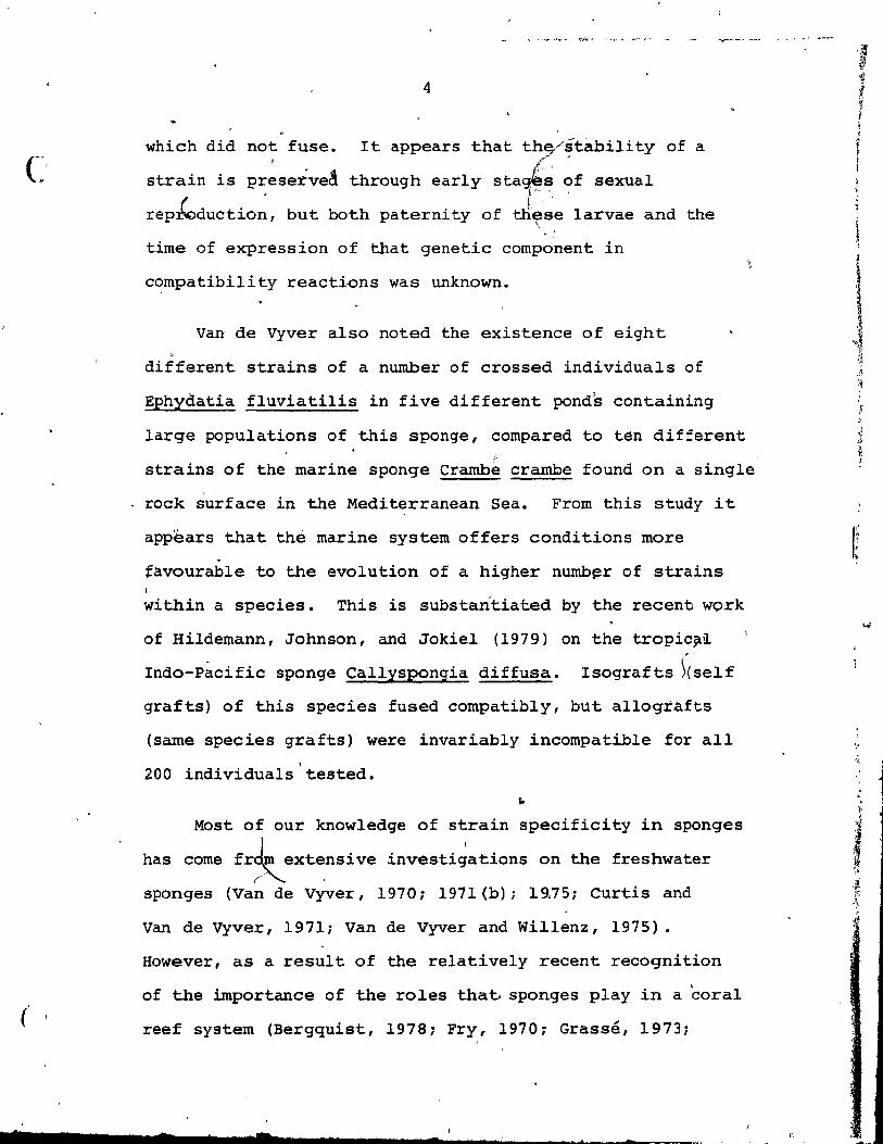
PLATE II
A. In situ p~otograph of a corner of one of the study sit~ The sites were 9 square metres in area and the boundaries were marked bl nylon rope. ( ..
c.
~
'-'"
B. In situ photograph of the grafting procedure •• ~small pieee of a braneh was eut off from a donor sponge and tied to a hast sponge by a tagged pieee of nylôn thread.
\
Close-up photograph of the tieing piece of nylon thread was used ta (approximately l cm in length) of to a host sponge (h).
\ ~,
l' • 0
> 1 procedure. A tagged tie a"small piece a donor sp~nge (g)
....
... ....
l i ,
~
o
..
12
, A
•
B !
c (
--

i l,
" ,
1
( ~ .'
and
13
2) it was joined to the hast by a continuous
superficial epi thelium - the pinacadterrn.
3) i t was in good heal th (i. e. colour, presence
" of <7 pinacoderm) .
Grafts were scored as a rejection if: r •
l) i t could be easily separated from the host
by touch.
2) there was a clear, evident gap between 'the o
host and graft.
and 3) a ridge had formed on each edge of the
sponges in contact (noted after l month) •
Plate III shows typical exarnples of acceptances and
rejections. Control autografts (self grafts) were made fior
aIl tagged individuals in both sites.
IMMUNOLOGICAL STUDIES:
Eighteen tissue samples were collected, from tagged
ind~viduals within the ttWo sites, for irnmunoiogieal studies.
Branches of the test specimens were eut from the donor and
transferred to plastic tlags underwater. The bags were
filled with seawater, tied, anç inunediately ~eturned to the
labora tory. Here the tag numbers of the specimens were"
recorded and the specimens were then used in the preparation
~f antigens, (sponge~cell suspensions) for immunization,
agglutination tests, and cross-absorptior! tests.
- - .... __ ,'f."'''"''
'P
, 1 ~ 1 1 ! ! 1 l ~
i
1 Il.
i F,
4
l l ~f ~1< J ~
~ '.
~~ r '.
r • lt
.' , , ,

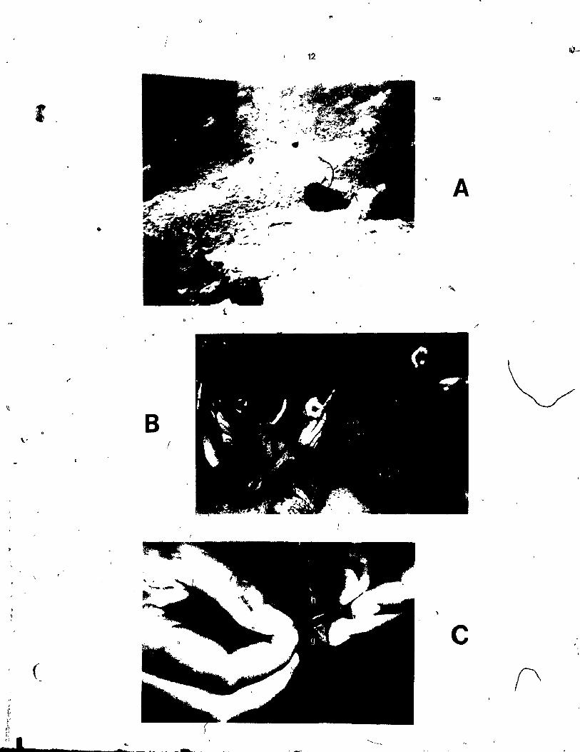
PLATE III
A. In situ photograph of a graft from Verongia cauITfurmis that has been aeeepted (a), and a graft from Verongia longissima that lias been rejeeted (r). The host (indieated by the arrow) is Verongia cauliformis. (a) represents an allograft, (b) represents a xenograft.
B. Close-ul? photograph of a control autograft accèptance of Verongia longissima. The graft was eut from the braneh of the sarne host individual. Tagged nylon thread identifies the graft. Scale is in millimetres.
('
C.. Close-up photograph of two rejected grafts. The upper graft (a) was taken from a Verongia eaul-iformis individual, and the lower graft (b) was taken from a Verongia Ion issima individual. The host (indieated by the arrow ~sOVerongia longissima. (a) represents a xenograft, (b) represents an allograft. Saale is in millimetres.
"
)

14
A
B
c , ,

, . :
(
15
a) Preparation of antigens:
The specimens were dissociated according to the
procedure of Humphreys, Humphreys, and Moscona (1960) with
slight modifications. Approximately 10 g of the specimens~
'were cut into small pieces (1 cm3) .a1nd placed in 100 ml of
calciurn-magnesium-free seawater (CMF-SW) containing 20 mM
ethylenediamine tetra-acetate (EDTA) "f/f l ho ur . These
preparations were then mechanically dis~ociated using a
'Waring blender. The resulting suspensions were pressed
through cheese cloth and the dissociated cells were
collected in JO ml of CMF-SW. Cell counts were taken for
aIl the suspensions u~ing a· Neubauer haemocytometer.
Herthiolate (0.3 ml of a 5% sOlution) was added to each of
the suspensions to retard bacterial growth. Six of the
suspensions were then divided into 0.1 ml aliquots to be
used for irnrnunization, and 10 ml aliq~ots to be used in the
agglutination and cross-absorption tests. The other 12
susJ;>ensions were divided into 10 ml aliquots ta be used
on1y in the agglutination and ~ross-absorption tests.
b) Irnrnunization:
Six young adult rabbits weighing 4 to 6 kg were used in
the production of antisera. The rabb~ts were injected
subcutaneously in the hind region with 0.1 ml of the sponge
antigen emulsified wi th 0.2 ml of ~reund' s (OmPlete
adjuvant. The injection was adrninistered using a 26 guage,
2.5 cm long hypoderrnic n~edle (C~rpenter, 1975). The
- ,
c
(
\
16 ..
following immunization ~ch~dule was used for the production
of sponge antisera in the rabbits:
lst day: Injection l 3rd day: Injection 2 5th day: Injection 3 7th day: Injection,. 4 9th day: Injection 5
~17th day: Inj ection 6 19th day: Injection 7 21s1: day: Injection 8 23rd 'day: Injection 9 31st day: Bleed from the heart
Blood (làO cm3) was collected via cardiac puncture. The "
blood was' allowed to clot for 30 min and then centrifuged
at 3,000 rpm for 5 minutes'. Serum (50 cm3) was removed, , 0
heated at 55 C for 30 min to inactivate complement
components present in the sera, and' then frozen in 5 ml
aliquots for later use in the agglutination and cross-
absorption tests. These tests were performed 2 to 6 months c
after the collection and frozen s~orage of the antisera and c,
antigens (sponge ceil suspensions)t'
c) Agglutination tests:
. Micfotitre plates (Plate ~V-A) containing U-wells were
" \ us.ed for the agglutination reactions. Each reaction weIl
cO,ntained 25 ~\ of di1uted antisera and 25 ~ of di1uted
antigen (sponge cell suspeQsion). The antisera in the wells
were seria1ly diluted by doubling dilutions, of CMF-SW and
antiserum, ranging from 1:1 to 1:256. The antigens were
di1uted 1:1 with CMF-SW. Co~tro1 wells c;ntained only 25.:\ 1 - ,
of the di1uted antigen and 25 À of CMF-SW. The plates were
incubated at room temperature for 8 hours and then
" l
r PLATE IV
A. Photograph of a microtitre plate showing macroscopic resul ts o-f the agglutination reactions. The far right hand column of wells (c) contain antigen only and dernonstrate the pellet formation of these controls. The second row from the top (2) shows a definite pellet (Yike that in the control) in aIl Il
f wells. This was Iscored às a negative reaction (no agglutination) . The first 6 wells- (from left to right) of the fourth row from the top (4) show a cloudy mixture. This was scored as a positive reaction (agglutination). The rernaining 5 welXs in this row show a pellet (like that of the control for this row). This was scored as a negative reaction (no agglutination).
.,. B. photomicrograph of,a positive
reaction 'in the agglutination test. The sponge cells aggregate ~gether in small clumps (indicated by arrowr. 2l0X
c. Photomicrograph of a negative reaction in the agglutination test. The sporige cells remain evenly distributed~similar to the control we'lls) and no clumping of cells oceurs. 2l0X
1-
0
\ )
" 9 ,l '{ ';
f~ Cl
1 1
1] j f
o

, 1 , .
..
•
1
17
te •• \. le ••••• :. te (e '. ~ ••••• ) .• ••••• ~ •• ~ •••••••••• e ••••••• •••••••••• _e.e ••••
\
••••••• t
A
c ;,

,
i r l-l',
i--',
."
18
macroscopic and microscopie observations of the contents of ,
the wells were recorded.
For macroscopic analyses a positive reaction
(agglutination) was scored if a w~ll containing the mixture
of antiserum and antigen was ccloudy and showed no 'signs of
defini te pellet formation such as occurred in the control
weIl containing the antigen only. A negative reaction
(no agglutination) was scored if a well containing the
mixture of antiserum and antigen showed a defini te pellet
like that of the control weIl (Plate IV-A) •
For microscopie analyses the contents of each weIl
were gently mixed and placed on a slide for observation
under the compound microscope at 200X magnification. A
c positive reaction' (agglutination) wa~ scored if the sponge
cells had formed clumps and ag-gregates which hard not been
disturbed during t~e gentle mixing, and were not observed
in the control wells (Plate IV-B). A negative reaction
(no agglutination) was scored if the sponge cells had not
clumped or aggréga ted togeth~r, and the mixture of
antiserurn and antigen appeared similar to that in the
control (Plate IV-C).
It should be noted here that macroscopic and micro
scopie observations agreed 90%'of the time, and when there
was a disagreement the microscopie observations were alwayS\
recorded. The re~iprocal.s/of the last dilutions of
antisera that showed agglutination were recorded.
•
\
\
19'
/ This procedure was carried out for each of the 6
d!fferent antisera against each of the 18 antigens (sponge
cell suspensions) prepared earlier.
d} Cross-absorption tests:
cross-absorption tests were performed on two of the
antisera to co~fir.m the agglutination tests. Varying cell
numbers of each of several antigens (sponge cell
suspensions) were spun down at 7,000 rpm for 20 min in an
RC-S Sorvall centrifuge. The supernatant was discarded and
the cells in the pellet were washed ànd resuspended once in
CMF-SW and again spun down at 7,000 rpm for 10 minutes. The
pellet was th en mixed with 0.3 ml of the antiserum and
)incubated at room temperature for l hour. Each mixture was
spun down at 7,000 rpm for 10 min and the sup~rnatant was
collected and used in the agglutination reaction to test for
the amount of antibodyabsorbed (removed). Agglut~nation
was performed as before (see IMMUNOLOGICAL STUDIES: c)
Agglutination tests} testing the absorbed antiserum ~
'(supernatant) against its homologous antigen.
-The reciprocals of the highest dilutions of. antisera
that showed agglutination were plotted against the various
cell numbers of each of the tested antigens.
e) Immunofluorescence experiment:
In this experiment, the rabbit antisera prepared
earlier wer~ utilized in conjunction with goat anti-rabbit
/ /
/
/

,
1 l j
~ i, 1
t (
20
immunoglObU~n fluoresce!n (FITCl conjugated antiserum which ~ ~"'-"----
was to act as a specifie label. The antibodies in the anti-
sera directed against the sponge cells bind ta their
homologous ·antigens and then thè goat anti-rabbit
immunoglobulin fluorescein <j.TCl b~nds ta the antibodies.
When observed under ultraviolet light the bound label will·
fluoresce.
A mixture of 0.1 ml of antiserum (diluted 1:10 with
CMF-SW) and 0.9 ml of antigen (diluted 1:10 with CMF-SN) was o
incubated for 30 min at 5 C, and spun down at 3,000 rpm.
The pellet was washed 3 times by resuspending in CMF-Sl'J and
spinning at 3,000 rpm for removal of excess antiserum. The o
pellet was then incubated for'30 min at 5 C with 0.1 ml of
the goat anti-rabbit immunoglobulin fluorescein (PITe)
con\jugated antiserurn diluted l: 4 with CMF-SW. After
incuba tion the mixtur e was s pun down a t 3, 000 rpm and 'f ,
washea 3 times to remove excess label. The cells were
examined microscopically at low and high power using phase
and fluorescence optics.
HISTOLOGICAL STUDY:
Sample specimens of graft acceptances and r.ej ections . '
were collected and returned to the laboratory where they
were preserved in small vials of Bouin's fixative until they
were processed :for histological examination two months
later. The samples were dehydrated, embedded in p~affin
and sectioned. The specimens were sectioned through the
...
)
, ,

C:
.n ",
!
21
zone of contact at a thickness of BJLm. The sections were
mounted on albumin-coated slides, deparaffinized, and
stained with Masson trichrome stain for differentiation
('UmaSOn,1962). The sections were examined under th~ compound microscope at 10wi (IQOX) and high (400X) power.
RADIOAUTOGRAPHIC STUDIES:
, The radioautographic studies w~re performed on a host
and donor, from witliin one of the study sites, that had been
grafted earlier and scored as a rejection.
â) Incorporation of label1ed proline:
... Two branchefs':, 3'0 cm long, were cut from one specimen
r
and each branch was tied by nylon thread at 2.5 cm intervals
to a branch of another specimen. This, procedure was carried . .
out in situ. One graft branch was left in contact in the
field for six hours and the other for nine hours. ·After
field incubation, the graf;ts were eut into 12 pieces
(2.5 cm) marking the' donor individual with a notchi six of
these grafts were placed in Mil1ipore filtered seawater
plus 3H-proline at a final èoncentration of 0.125 mg/l for
four hours~ the remai~i~g si~ piec~s were placed in a
controlAfluid bbntaining Millipore filtered seawater plus <Q.
unlabelled proline at a final concentra tian of 0 .• l:.25 mg/l
for four hours. After this four hour period aIl the graft
pieces w::e placed in individual, circular~ng chase
solutions of Millipore filtered seawater plus unlabelled

, &
, . " "-22 ~ .. 0
proline at.a final conpe~tration of 1.Z5 mg/le Control !: • .. .. ' , ~ J "-
graft piece~ and ~abelled qraft pieces for radioautography Q 0
~ere removed' from the 'chase'solution after 6, 12, 24,"36,
, .
48,' and '62 hours and fixed in Bouin's fixative.
b) Histolog~cal proce~sing:
After being fixed in Bôuin's for 24 hours, the graft
p;ieces wer~" dehydrated,' embedded in paraffin, and \tored , ,
untii sectioned. Th~ g~afts were sectioned 3 months after .
embedding. The sectioi1s'. were 'cut thfo~gh the zone of
contact at 8 r ' mounted on a~b'J.Illin:-.cQated slldes, dried
and processed for radioautogra~hy. ~he remaining paraffin .. block of each graft piece was trimmed, deparaffinized, and
embedded in Spurr epoxy. The grafts were again sectioned - , ,
through the zone of contact, at .O~5.f'm,. These sections .f'"
were mounted in a drop of distilled water on a slide, dried,
and processed for radioautography. 1
c) Radioautographie procedure:
The. procedures' of Kopriwa and Leblond (1962) were
followed apd were carried out in total darkness. The slides
were dipped into Kodak NTB-2 photographie emulsion for 3
seconds, drained and dried for 2 hours. Coated slides were o
stored in dry, light-tight containers at) 2 C and were
eXPosed for 2 ta 4 weeks. The ernulsion was developed in \
Kodak 0-19, and fixed. Slides with paraffin sections were
stained with haemoto~ylin-eosin, (Hurnason, 1962), and slides , •
...... _-------~~---~~ -
~
i 1 f f 1 ;
l 1
:$ 'J: .~ :~ .' -,
.'
'" -:
',f,
" " ; ,
}F
1
.~ ,
•
o
," , .
with epoxy sections were stained with 1%'toluidine blue ih
borax (Humason 1 196°2)" Sil ver grains were loca1ized a1;ld ~ ..
quantified under oil -immers~on. Features of contact , -
surfaces were 'èxâmined for deposition of cutic1e materia1
(Vace1et, 1971).
"
," " , . cl
o
\ -
. "
. -...... ' ..
a
- "
\ ,
4
-
. -
..
l "

C ! !
( "
RESULTS AND OBSERVATIONS
GRAFTING EXPERIMENTS:
Intraspecific acceptance and rejection of individual 1
'Verongia longi~si~, specimens were pbserved to occur
commonly in. nature. A natural acceptance was noted when two
branches had joined and totally fused ai:: the point of
contact, and a natural rejection was noted when each branch . ~
J
retained its separate integrity and, after extended contact, ,
had formed a ridge.on each of the surfaces in the zone of
contact. Acceptance was also observed when branches of one
individual came into contact with themselves (Plate I). The
branches had united and bec orne one at the point of contact. ,
Natural auto-rejection was never observed to occur in these
circumstances.
A total of 91 in9ividuals consisting of 29 specimens
of verongia longissirna, and 13 specimens of verongia
cauliformis found in site A, and 49 specimens of'verongia
.longissima found in site B were used for in sïtu grafting \
experiments. In order to test aIl combinations of grafts
betw~en individuals and (elf/self controls 8,372 (91 x 92)
operations ~ould be reqU~red. A random' grafting protocol
was initiated to allow provisional strain identification of
\eme individuals. These were then grafted to single rnembers . -"'-... ,
of othe~ strains to insure strain identification of aIl
individuals with a rnini~um number of required operations.
The re$ults of·the grafting within each site are presented
24

25 '
in Figures 3a and 3b. The results of aIl the 758 grafts
that were performed within each site and between sites are
presented in Figure 3c.
The results clearly show that aIl autografts (self '("'
grafts) of both species accepted, aIl xenografts (different
species grafts) rejected, and there was both acceptance and
rejection observed in allografts (sarne species grafts) of
verongfa-'longissima (Plate III:-A, and III-C)., Am0l1g 'the
allograrts groups of individuals could easily be identified
which always accepted grafts from individuals within their
group and always rejecte~ aIl other individuals.
groups were designated as strains (Figure 3c).
These ,
A preliminary investigative xenograft, not included in
the grafting results, was performed between Verongia
longissima and Verongia fistularis which was scored as a
rejection.
The consistent specificity of within and between strain "
grafts obserÇ~~ in the early st~ges of the study allowed
dèvelopment of assumptions concerning the acceptance or
rejection of one individual by another individual. For
example, if indiv~dual A rejeèted individual Band accepted 1
, individual C, and '~ndividual C accepted individual D and
rejected individu,l B, then it was assumed that individual A
would àcdePt ind!vidual 0 and individual 0 would reject " --
individual B. Individuals A, C, and D were asswmed to be of
the sarne 'strain, and çf a ditferent strain than individualB.
o ' ,
r, 1
j ,.II 1 .j
'l,.t
j t ~ /l

ST~Alt>I
2
1
!5
7
18
26
IGRAFT J
HOST J
Il'' v IL 1/ 1/ 1/1/ 1/ V "'IL" 1/ 1/ 1/1/ 1/
1/1/ 1/'" 1/ v ... ~ 1/ 1/1/ 1/ V
1/1/ V V I/V VI/' Vil'" 1/1/ V 1/ 1/ IL IL
VILI/ v v !LV 1/ 1/
• 1/. V V V 1/ 1/ V • 1/1/ 1/ '/ !/ Il'" • • 1/1/ V. V 1/ I/V V V • .V \;' ILlL IL IL • V 1/ V VV v v
1/1/ 1/1/ vI' ... IL IL IL VV v I/V V V V VI/' IL
{/ .v v !"IL ILlL !LV VV 1/1/ V VI/ V
IIv .1/ V 1/1/ 1/1/ ~~ Il 1.11/1/ vv 1/,. IL IL IL!L !LJ..I: !LV 1/1/ V VV V v IL if .{/ L'; V 1/ V 1/. 1/ I/V V V 1/ 1/ IL V
vl/ VV VII' IL IL it"1L' 1Lit" J..I:it"~
Lagand:
vv VV 1/ ...... V VI/ 1/1/ V 1/ ~ V VI/ 1/1/ 1/ IL 1/ {/ V
1/ !/ IL • Il~ ~~~ 1/ VV IL 1/ VV V V 1/ VV VI/ I/V 1/ 1/1/ VV
ILV V 1-v VI/ V 1-1/1/ 1/IL l-V 1/ 1/ V l-V V V V V V V 1/ V V 1/ VI/ V
FIG. :; a: GRAFTING SITE 1 A
Acceptance .- ---- - - .-. - --. ----- ----- • Assumed occeptonce on basis of actuol •• __ 0 acceptance of mert'bers of some stra in R · .' ~ ." eJectlon -----------.---- ------ ----~ Assumed rejection on bosis of actuol r-?t rejection of members of same strain ----~
V V IL V IL
Note· Results of ln situ grafting experiments in site A. Dates of testing, May"';" Sapt. 1978 , and Dec. 1978. Numbers 1- 29, Verong.ut l2.!:!.gi ssima. Numbers 30 - 42, Verong!.!!. cauliformis.
1/ IL V IL
1/
o

ISTRAIN
1
r
3
4
6
5
8 I!§ 10 ~
2 •
27
~RAFT HOST
1/ F. 1/ IL:
IL 1/ 1/ 1/ V 1/ V ".. .. 1/ ".. v v
./1/ V /I/~L /1/ v / /1/ V V /v L' v ./ V V " / V
./ ,;'.
v,;' IL • 1/ V 1/ 1/ ./ / V L 1 1/ V • / .. V 1./ V V 1/ V VV
vv .// // L'/ V/ 1LlL'/ / 1/ .IlL 1// IL V 1/ ./ V 1/ V./ 1/ 1/ V .. vv
1/ 1/ / II/V IL' IL iL / .1 ..... Î/ iI/./ v V V ./
1; 1/,..
1/ 1/ 1/ V ./ ,.. F V F ,.. j 1 LI/ V V IL L
~ ./
1/ V 1/ ./ ./ V V / ./ ..
./ V ./ 1/ ./
/ 1/1/ 1// // Vr V/ / // I/'/" 1/1/ L 1// IL Il" /
• ./ / V ./ ./ '/ V ./ ./ ./ 1/ IL'
1/1/ f' L V '/ /1/ 1/ IL 1/1/ 1/
1/ v ./ 1/ V L/ V ./ V IL /' .. F 1/ l/P'P'
LIgend:
FIG. 3 b: GRAFTING SITE B
Acceptonce --------- ------------. Assumed occeptonce on bosis of octu~I ____ 0 acceptonce of members of some straln Rejection ---- ----- --- -- - - - ---- - --~ Assumed rejection on bosis of actual 0 rejection of members of same strain .---
Note: Results of ln. situ grafting experiments ln site B. Dates of testing, May -Sept. 1978, and Dec. 1978. Ali individuals are Verongia longiuimo.
ft
"

,
FIG. 30: SUMMARY OF GRAFTING BOTH SITES
L. EGEND
Acceptance.------- --------.
Assumed acceptance on basis of actual acceptance---- --0 of members of sorne atrain.
Rejection .• - -- -- -- - - --- - ----~ Assumed rejection on basis of actuaf rejection of ---------·0 members of sorne strain
NOTE
Results of ail in llliI grafting experiments in both site A and site B. OatlS of tlsting t May - Sept. 1978, an d Oec.1978. Stro;"s ,- 17, ïeroog!s. ~9i •• ;ma Strat" lé Ytrooajg cgul"ffi:~mll.

28 >
-HOST 1
ISTR'dt.: Ir:RAI JJ_L
"
• -1 v
v v v
1 v v v v v
. 1
v v .. 1/
1/ L 1/
[/
1 v
• il'It'
2
Il ,1/1,11 1/ ..II/V II/V
:3
v
• 4 v V 1/ 1 v
1 v v 5 v
v V LI'
6 v
7 1.(] Fl
8
LJ
z
18
1/ 1/ V
.. / (Ji=" -

,.
~
28 ,
/ HOST
L... .il ~~ . --- - I~ .. PI
1/ /;
• [/
• .. 17 1/ /
V 7 ,)'
[/ [/ [7 1.1' 7
1/ 7 1/
1
• 1/ 17' 1/ "p 1/
1/ [/ 7
r/
7[7 V 1/
7 -,
/7 1/ 17 17
"}'} [7 777
• V/ ,.
/: 17 V
V
1/
~ V ~. V ..
~L/ V .iL 1/ 17
i/
L 7 V • L 1/ /7
V 'J
L 7 V 17 r?
'}
[77
V
7 /1 il":.,IV [/
il":7 1/
[/ V
1/17 1/
1/ V [7
V
i7' / [/ /
-,
1/1 ri 17
")
V [/ [/
V V 1/
[1
~/7 :/ V
.. V
17 1/ 1.11
J!;lI
/: V /:
7'1
1/ '/1 /1
V 1.1'~7
po [7
1/
-[/
r" 1/
/: r1
1/ / .l';
V
1.1'1/[717 1/
V
17
.. 17 1/'17 W~I v 1/
V /
v l/ V
7. 7
1/ V 1/ 17
V • 1/ V /! .
L 1/
L 1/ V V 7 ,.
171 :.JI: .1'1 ~
V 1/ V '~
L/ ~ v
!71}ti'
7 "}
.-'7 '1
V 1717 v Ll 1/1 1/:
lA/! 1/1.11 U! V l/
.l'Il/I /1 1.1'1 V/I /:
lI'I/ ,.., 1/1
A l/I lI'I~ 1/ 1.11
71
1 2 Df:L ï < i 1

29 \
A~l assumed strain identifications'~ich were randomly
chosen to be tested were proven to be correct when the "--
actual grafts were p~rformed. It is concluded then that
because of absolute specificity of strain identities, the
individual graft reactions assumed in Figure 3c would prove
correct should-the indicated grafts be carried out.
The grafting experiments showed Il strains among 29
individuals of Verongia l0ngissima, and 1 strain among 13
individuals of Verongia cauliformis in site A and 9 strains
among 49 individuals of Verongia longissima in site B. The
strains are observed as the blocks of acceptances and ,
assumed acceptances recorded in Figures 3a and 3b. Percent
abundance of strains in each site is presented in Figures 'If
4a and 4b.
The results of aIl the grafting experime?ts, (Figure 3c)
demonstrate the existence of 17 strains among 78
individuals of Verongia longissima, and one single strain
among aIl 13 individuals of Verongia cauliformis. Total
percent abundance of strains from both sites is presented -in
_~~e~, 4c. The incidence of strain occurrence, in the
~pecies Verongia longissima investigated in this study May
be used to calculate the probable number of strains that one
would expect to finq in other similar sites sampled: The
followlng statistical formulae have been applied (Bailey,
1959) :
-- ---""",-~
1
1 1

Fli. la:gIiM •• ~,l"""~",-,~">."_-~,;~_~~"",,,,~-~,,, '~~-. ..... , ............ l''~' ~ .... -
..-.... '-
" Veronajs lo~i"imo
FIG. 40: SITE A
:t!.mng!!t WM .. 'Y' .... iiJ c
.. "", -~ ,
~
Ylc.mgig, l2.n.g i Si jma
! 4 6 5 8 13 15 16
STRAINS
--- FIG. 4 b: SITE B
% ABUNDANCE OF ~AINS IN EACH SITE
7' -' -~~ ... ~J' - ............. '''''~.:r'"_. _~ ......... __ ......... .,. ....... _P<_ ."...,"",
~
w .... 0
~ --- -- ._-- - ... ~ ... ~ '- .~~~
/

'-FOSjiAaap;, J :qQt4iAJI "'ct.C ...... tJ ... • 11'~.I"!~~~""e ... 't"I~ ... "" .... ,~ .. ~~ .. ~~ .. !""""'"< ...... , •• ~ .... ""~_,~" .... _~,"' ....... _ r"-~ ... " .... "'l~,'$ ... {>.-.......... ~.. ~, ..
~
,,-.
Cfo ~ A B
'U N 0 A' N C E
~
Ytrmloio v ... " ... xl"":........,.
.. ' ~' ..
VerongÙl 1.Mgiss;mg
a ~ 4 e 6 7 8 9 la Il 12 13 14 1!5 16 17
FIG. 4 c :
..
STRAINS
TOTAL % ABUNDANCE OF STRAINS FROM BOTH SITES
/,"", \ '
J
CI) ....
If Ili' j n 7 !Tt OF ... ~,,--.- .. ;.,-""~",,J', ... ,'.:'>"" ... ~,).>~..-..d~~~.~~ .... e,.., ....... ' "-------" 4-._< ___ • '_ ....... ~
l
1 ~
! ~

- -- - - ~, .. -.~ ': ~f "
32 :t
" -< . j
~
1- MEAN = xl ~X2 Xl = number of strains in
(,1 ~~~ site A ~ \ = number of strains in
f n x 2 site B
~ in this stud~ n = number of sites ~-
Mean = '1
f· Il + 9' 1 l' f' ~;
2 \ \ !
Mean = 10 'j
~ANCE 1 ~
2 2 2 ~
2. (Xl t
= (xl + x 2 ) - + x2
) ,1
~. ,
n -r
n - l
, Variarlce in this study =
112 + 92 - 20
2
2
2 - + Variance ::. 2
3. VARIANCE OF MEAN = variance
n
varian~of mean in this study = 2
'1 Variance of mean = l
" 4. STATISTICAL ERROR = ri variance of the mean
Statistical error, in this study =
Jl
( Statistical. error = l
'.-

(
( )
1 _
! 33
In other sites we would expect to find 10 ± l strains, , .
or in 68% of future sites sampled,-one would expect to find
between 9 and Il strains.
It is understood that the statistical analyses
presented in this study have USàd~ minimum n~er of sarnple
sites, and for more accurate staternents of probability rnany
more sample sites should be employed. Nevertheless, it is
interesting to note the significance of strain occurrence in
Verongia longissima, and the probability of finding a large . '
number of strains within a single species of a marine
sponge.
w
It was noted earlier (MATERIALS AND METHODS - -GRAFTING
EXPERlMENTS) that the re~ults of aIl grafts were recorded 3
days after grafting, and aga in a month later. Graf'ts
checked 6 months after the initial grafting were, ~n aIl
cases, scored the same as they had been ,3 days after
~~afting. - During the initial observations of the graf~s •
subsequent observations it was noted that aIl grafts,
No distinctive features or characteristics readily
and
discernable in nature could be used to distinguish between
the strains in Verongia lon2issirna. , This species although -
typically an irridescent blue-violet colour, does occur in
other forms. 'Its colours range from a deep blue-violet, to
o
, i !
1 1
1
! ....... _-_._--_ .... __ ... _---_ .... _---_.:..._-------- ""~~,,~ .. ,_ ........... - ...
- 34
.. a "tannish" blue-violet. These differences in colour could"
occur within one individual or between individual~ of the
s~e strain. Another common feature of this species,that
-~~ ,~ar{ed from individual to individual was the external , .'
surface relief. 1
Sorne have a smooth, even surface \fhe,reas \ ,
others are r~ugh with many spinous protuberances but no
consistent pattern related this feature to strain
designation.
It should be noted that in the grafts tha,t have been
accepted by a hast, the external and i~ternal features of
both donor and hast eventually become continuous and
matrices and skeletons are shared (Plate V-A, and, V-B). In
g~afts that have been rejected by a hast, the external and
internal features of the original specimens remain separa te
and no intermingling or sharlng occurSi after a time, a
border ~r ridge forms on the two surfaces in contact
(Plate V-C, and v-Dl. This same border forms on an
individual in the zone of contact with the stri~g used to
tie the graft ta the host (Plate V-A) and ~e ether foreign
(non-self) abjects.
/
.'
l' i 1 f !
f
1 1
:; J
If , '
t • " >' .' ,
". \
1 ,
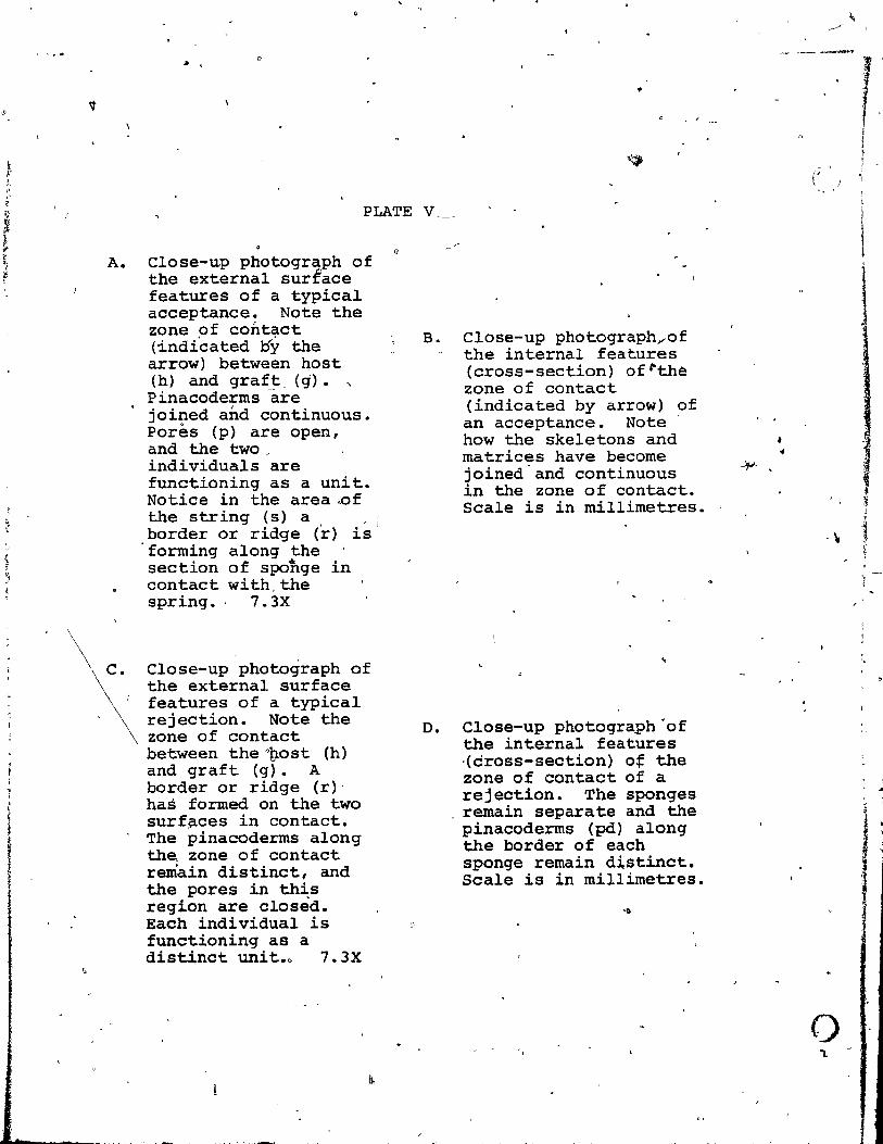
A.
PLATE V
Close-up photograph of the external surface features of a typical acceptance. Note the zone of contact (indicated Dy the arrow) between host (h) and graft, (g). " P inacoderms are joi~ed and continuous. Pores (p) are open, and the two individuals 'are functioning as a unit. Notice in the area..of the string (s) a '
,border or ridge (r) i~1 forrning along the section of spohge in contact with,the spring.· 7. 3X
the external surface features of a typical rejection. Note the zone of contact between theo~ost (h) and graft (g). A border or ridge (r)' has formed on the two surf~ces in contact. The pinacoderms along tha zone of contact remain distinct, and the pores in this region are closèd. Each individual is functioning as a dist~nct unit.~ 7.3X
B.
D.
Close-up photographrof the internaI features (cross-section) of~the zone of contact (indicated by arrow) of an acceptance. Note how the skeletons and matrices have become joined'and continuous in the zone of contact. Scale is in millimetres.
Close-up photograph'of the internal features ·(dross-section) of the zone of contact of a reje~tion. The sponges rema~n separate and the pinacoderms (pd) along the border of each sponge remain distinct. Scale is in millimetres.
- -----..,.
{' ,.'
- \
o
r ! }
i

/~\ -/
.. .. - h
• 1 c I~ .)
; .".. ., \
••• ~ . , . • . ,
.,'
0
i } (
o

IMMUNOLOGICAL STUDIES: ..
In the grafting experiments the sponges showed
recognition (fusion or rejection) at the levels of self
(specimen), strains within a species, and species. This
section of the study was performed to dete.rmine if these
recognition differences could be demonstrated using
immunological techniques. Rabbits were immunized against 6
different sponge individuals, and the antibodies produced f'
were then tested for their ability to react with varoÏ!ous
other sponge individ~aIs.
Antibod,ies specifie for an antigen are hypothesized to
possess a receptor si te wi th a structural configuration
c9mplementary to that of the antigen, and are capable of 1
reacting with the homologous (corresponding) antigen in a
lock and key type complex. This antigen-antibody reaction
will only occur when the antibody receptor si te conforms
closely enough to the determinant site of the antigen so
that a union can occur (Carpen.ter, 1975). There are a ~~
number of techniques that can De emp10yed to demonstrate
this specificity of an antibody for its homologous antigen.
In this study agglutination, cross-absorption, and 1
(~,
0"
immunofluorescence were the methods employed to exhibit
this specifie recognition system, and to substantiate - - .
further the occurrence of strairi speeificity in the marine
sponge Verongia longissima.
When a small amount of Ci serum was mixed with an
36 '
1
1
.)
,
<
t

; (.1 i ,-
f {
(
pa c .,'*
37
hornologous antigen (sponge cell in the wells of
a rnicrotitre plate agglutination observed. The cells
clumped and gradua,lly settled to the bottom of the fluide
Macroscopic examination revealed a clou~y liquid in the
well's of the plat:,e, 'compared to a def~nite pellet that
formed in the control wells (Plate IV-A). High power
microscopic ex~ination revealed that the particles of
antigen behaved as though they were sticky, and clung
together in small cl~ps or aggregates compared to the "
generally even distribution of cells in the controls
(Plate IV-B).
Quantitative analy~~~s to det;rmine the agglutinating
antibody content of the antisera were rna?-e by serially . diluting the antisera. The reciprocal of the last dilution
of antis'erum that showed agglutination is cal~ed the
antiserum titre. The titres for the antisera tested in
this study are presented in T'able 1. As an example 1 anti
serum 89 has a titre of 64 for antigen 89 and titres of 2
or less for the remaining' antigens. In the case of antigen
TA there is nQ titre recorded +or antiserum 89 because
agglutination was never observed when antisèrum 89 was
mixed with this antigen. It is assumed here that a high
titre indicates a process of recognition while a low titre'
indicàtes that 'little recognition has occurred (for further
discus&ion see'pagé 40).
The results indicate that' 5 of the 6 antisera (31, 2,
~ ~ ),
;§ 1 l
1 1
~ \
1
i ,t '1 i
'1~ ~~ ;'
.ft < ."
J 1

(~.
f
1
1
( ,
J ~.
38
TABLE 1: SUMMARY OF AGGLUTINATION RESULTS
CELL ANTISERA A NTI GEN COUNT ,
(x IOrml~ " 31 2 1 12 73 71
31 194.4 32 1\ 1 0 0
2 259.6 1 32 32 1 1
73 177.8 l' 2 2 32 32
-.
89
0
1
1
89 ~210.6 0 1 1 0 0 ~ 33 17--1.0 32 1 1 0 0
1 1 2~3.4 1 32) 32 J 1
71 160.0 1 fi 2 32 32 h
? 75 f'\-
162.4 ~ ~"'-l/2 2 32 32
35 \190.8 32 2 1 1 1
2'63.2 1
12 1 32 32 1 1
TA 281.8 1 - - - -20 168.2 2 2 1 0 0,
1
23 147.4 1 2 2 32 32
"" 19 164.4 1 2 1 1 1
67 149.8 0 1 1 0 0
7~ 213.2 0 0 0 0 0 ,
44 161.2 1 1 1 1 1
87 215.4 2 2 2 2 2 '1:
Note: Numbers ln antllera columns represent the hlghest dilution of that ontisertm that would still cause an agglutination reaction ta occur. Antigens 31,33 and 35 are from ~g.iA cgullform;s.
, Antlgln TA is from V,rong!!!. flstularis. Ail other antigens are from VeronQia lonoissima. Antiserum 31 il from Veronoia cauliformil. Ali other antisera are from Y!!.2.D.gia l.2n.gissima.
0
2
1
2
2
2
-O.
1
0
0
0
1
2
-

(.
,j
1
!
1 ! f
39
12, 73, and 71) appear to recognize more than one antigen,
noted by. the high titre values appearing in the antisera
colurnns. Antiserum .31 recognizes antigans 31, 33, and 35.
In the grafting experiments, it was noted that sponges 31,
33, and 35 were all of the 'sarne single strain of Verongia
cauliformis. Antiserum 2 recognizes antigens 2, Il, Fnd 12,
and interestingly enohgh antiserum 12 aiso recognizes these
same 3 antigens. Thus from these results antisera 2 and 12
recognize common antigens, and are therefore likely to have
antibodies of similar specificity. Recailing the results of 1
the grafting experirnents again, it was shown tha~ sponges 2,
Il, and 12 were of the sarne strain type 2. Antisera 73 and
71 are also considered to have the sarne specificities each
recognizing the sarne antigens, 73, 7l,~75, and 23. The
grafting resu1 ts showed sponges 73, 71, 75, and 23 aIl to be
of the same strain type 3.
Antiserum 89 appears to recogni~é only its homologous ..-'
antigen, antigen 89. In the grafting results sponge 89 ~as
the only sponge found to be Qf strain type 15.
These data ,clearly demonstrate specificity in antibody
formation.' Knowing that the different antigens which were
agglutinated in high titre -rith each antiserum are from the
same,strain and that sponges of the sarne strain elicit
antisera which show identical r~cognition patterns it is
clear that this spec~ficity is direct1y related to strain ,
designation based upon "grafting reSJ,llts. There are 3 strain
(
. .
l '~ II .r;. ,1 J~,.
:~
,1 .~
.' r\ l
;.
l. . ~ l, i
:' i' , l,
~ 'J " ~
~ r 1 !ri
~ 1 ,

l 1 !' i t f
1
(
40
types of Verongia longissima, and l single strain of
Verongia cauliformis expressed in the agglutination results,
and their expr~ssion of specifici ty concur precisely wi th \
the resul ts of the grafting experiments (Figure 3e) .
. A high agglutination titre was observed when an anti-
serum was-- reacted with its homologous antigen from a sponge
of the same strain. However a low titre was often observed
with heterologous (unrelated) antigens from different sponge
strains. Normally no agglutination would be expected if
antibodies against those different sponge strains were not
present •. ' There are 3 conditions ~hat eould explain these
low titre occurrences. Fir~t, the sponge cells could show
antigenic cross-reactions, second, the cells have surface
components common for aIl the strains but each strain having
different levels of each antigen, and third, the antiserum
could have antibodies against surface components for species
and sorne for strain.
The agglutination tést is somewhat insensitive, and a
more detailed analysis is required to elucidate the type of
recognition that is occurring at the low agglutination
titres using other immunological techniques. In the cross
absorption test antibodies are removed by adherence to their
specifie antigens, and it was hoped that this test would .
reveal more precisely the type of recognition occurring at
the low agglutinating titres of the anti.sera in this st~dy.
/
, '~
f j

(
The cross-absorption tests were performed on sponge
• 1
ant~sera 71 and 12, and the resu1ts are presented in Figures
Sa and Sb. These tests involved absorb'ing-out an antiserum -
by incubating various ce1..J. numbers of an antigen with _the
antiserum. If the antiserum was reacted with the antigen
that had elicited the antisèrum, an antigen-antibody
reaction 0ccurred and the antibodies united with the antigen
cel1s and remained in the pellet after centrifugation. t'lhen ~
~ the supernatant antiserum was tested again in an
agg,lutination reaction with the aritigen that had elicited
the antiserum, there was either no agglutination or .;.a low
titre agglutination reaction, depending on the concentration
of antigen cells originally incubated with the antiserum. 1
If a high number of cells -are incubated, a large portion of
the antibodies will be absorbed from the antiserum, leaving ?, ,
the supernatant with only a few antibodies to react in the
agglutination test (causing low titre agglutination) ;- and
if a small number of cells are incub~ted, more antibodies
will remain in the supernatant to react in the agglutination
test, (cau.!?i~g high titre agglutination). Therefore when the
antigen cell concentrations were plotted against~the anti-. serum ti tres, an absorption curve was obtained for the
antigen to which the antiserum was originally directe~ 1
(Figure Sa, #71, and Figure Sb, #12). This curve was used
to compare the efficiency of the other sponge cell
suspensions to absorb the antibodies.
'fihen the antisera were absorbed with other sponge cell

(j
• -- ----~_._-~--. -------- --- v
42
64
r-*TA
32 ... -':-' .... I.I-=:-~----"'---_. 1 t ..
4
/' 2
J
. • • .
• ···r*67 ••••••••••••
"
FIG-. 5a: ABSORPTION OF o<.#" 11 WITH VARIOUS SPONGE EXTRAC TS
Note: Results of agglutinati0'LJelt oft.r absorption of 0<.71 by vorioui .ponge extracts. Graph shows the dilut ion (T ITRE axi s) and concentration of ttlt extrocts (CELL COUNT axis) caullng an agglutination reaction to occur . • TA is Vl[ongig, fjstylgrjs. <
:1= 67 and *77 or. VtronalJl l.2.o9issima. *71 and #75 are the same stroin of ~gjg !2àgl"ima.

T 1 T
( R , E 1 ~ 4 i ~
2
Note:
()
----l'
43
80 100 150 200 250
CELL COUNT (X 106 lm! )
F.IG. Sb: ABSORPTION OF oc=ll=' 12 WITH ~ VARIOUS SPONGE EXTRACTS
Resulta of aogiutination test after absorption of oc =Ir 12 by varioul ,ponge extracts. Graph shows the dilution (TITRE axis) and concentration of test extracts (CELL COUNT axis) cauling an agglutination re action to oeeur. ~ 17·-is ~gjg J.2.n.gjulmg. :ft! 2 and # 12 are the same strain of yerongjg l.2.ngjujma.
L
f
..
& i ...
': ,
Il ,~
i -t / { •
1 i , l
<1
L ~ , 1
; ,:l
" "

C)
44
suspens ions, two type s of absorption occurred. ~~hen sponge
cells of the sarne strain as the antigen used in eliciting
the antiserum were used, absorption curves similar ta the
control were seen. For examp'1e, sponge #75 absorbed-out the
antibodies wi~h the sarne efficiency as sponge #71 on anti-
serum 71 (Figure Sa). , '1 °1 #12 d #2 b b d S~m~ ar y sponges an a sor e
antiserum 12 in_9n identical rnanner (Figure Sb).
However when antiserum was reacted with heterologous
antigens, even at high cell numbers, li ttle antibody was
absorbed-out as seen by the high'agglutination titres still
present. For exarnple, sponge #TA did not reduce the
agglutination titre antibodies in antiserurn 71, and sponge
#77 only gave-a- small reduction (Figure Sa). This indicates
~hat regardless of cell concentrations there was little or
, no positive r-ecognition as most of the antibodies rernained
behind in the supernatant and were available to unite with
the antigens ~~,~n el ici 'l7ing the antiserum.
. The results of the cross-absorption tests for sponge
antiserum 71 (Figure Sa) demonstrate that sponges 71 and 75
are similar. In the grafting experirnents these two sponges
were found to be of the sarne strain type 3. The results of "
the cross-absorption tests for sponge antiserum 12 (Figure
Sb) dernonstrate that spong~s 2' and 12 are similar. Agai~
this result agrees wit:.h those o-f the grafting experirnent .
which found sponge ~2 and 2 to be of the sarne strain type , /
2. These data, like those from the agglutination tests,
\
;'
;, ~Ij
"
'j
~
~ ( ~
)
i
,
45
exhibit antigenic $pecificities , and they are directly
related to strain designation based upon grafting results.
These cross-absorption tests demqnstrate the ability of sarne 1
strain antigens to cornpletely absorb the antiserum, sarne
species antigens to absorb very little antiserum, and --
different species antigens to absorb none of the antiserum.
It is apparent from the cross-absorption tests that the
antigens (sponge ~ell suspensions) have gualitative
differences. In other words, the type of recognition that
is 09curring at the low ag~lu~ination titres is more ~ikely
to be due to a species recognition and not the result of
cross reactivity or quantitative differences in the strains.
More precisely, the antisera recognize the strain
differences of the sponge cells. However the fact that a
small arnoufit of anti~ody was absorbed by heterologous sponge
cells may indicate that sorne antibodies were formed against
cornponents common to the species. The strain surface
components seern to play the dominant role-in e1iciting
antibody formation in Verongia longissima.
A limited supply of sponge cell suspensions and anti
sera restricted the extent to which,the agglutination
reaction tests and the cross-absorption tests could be
carried out. TO allow for immunological results to be
employed as conclusive evidence rega~ding strain occurrence
in Verongia lonqissima the agglutination and cross
abs~tion tests should ~e carried out for aIl the sponges
j j
,,'
;'
''r I~:
~
i 'f ~, ,\ ,
46 \ .
employed in the grafting ex~iments. However, sufficient
data has been presented to demonstrate the possibilities of
employing immunologicai methods to show the occurrence lof . , ,
strain specifiêity in verongia longissima.
,
,. 1
\,-" In attempts to employ~the .. fluor~cent-antibody label
of a sponge antiserum forGlhe to ~emonstrate the specificity
antigen used to elicit the antiserum, it was discovered that
cells of Verongi~longissima are autofluorescent (Plate VI-
A). It proved to be impossible tô utilize the fluorescent
antibody labelling technique in this study since there is
little difference in fluorescence when the ~abel is addad
to the mixture of antigen and antiserum (Plate VI-B).· The . ~
. \\ t autofluorescence has an absorption peak'in the range of
590 nrn (orange-red ~egion of the absorption spectrum).
.J
t -'

"
r ,
.. 1
!' t
/
1 PLATE VI , il
A. Photornicrograph of autofluorescence of Verongia longissirna cells clwmped together. The fluorescence has an absorption peak in the range of 590 run however other substances w~re also ernitting a fluorescence so the absbrption reading was not accurate. 320X
,-,
B. Photomicrograph of the fluorescence ami tted when goa t anti-rabbit immunoglobulin fluorescein (FITC) conjugated antiserurn was added to a mixture of antiserum directed against the spong~ cells and i ts, hornologous antigen. There is little difference between the fluorescence emitted by this aggregate of sponge cells and the auto fluorescence of an aggregate of sponge cells. 320K
, ,
"
•
,0

('
A
1
B·
" ,.
•
"

i ! i
of , 1
1
/' 1 f J l
'\
, ,
o
\
) 48
. HISTOLOGlCAL STUDY:
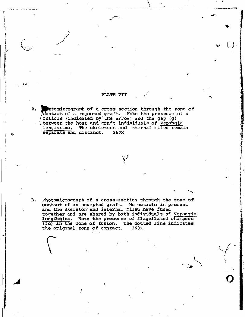
Histological sections thrbugh the zone of contact of
rej~cted grafts revealed the presençe of a cuticle between . the contact surfaces of the two individuals of Verongià
longissima (Plate VII-A). Tight junction~ and smail gaps
could be observed along the rejected interface, but no
fusion ôf tissues occurred along the con'tact surfaces. \
Distinct pinacoderms separated the two individuals. The "' c1.
cu ticle was also found to be present around werm tubes \ "
\
found in sections of this- sponge, and in zones of con.tact
wi th the thread used for tieing th.e grafts tG the hosts.
It would appear from these results that a cuticle is laid
down by Verongia longissima when any foreign substance,
inanimate or animate cornes into contact w~any part of
-the sponge. This subkantiates Vacelet' s findings of the
presence of a cuticle on the surface of verongiaQ
aerophoba
and Verongia cavernicola in contact wi th foreign"
substances, and extends i t to include another member of the
fam±ly Verongiidae.
Histological sections through the zone of contact of \
accepted grafts revealed fusion of the tissues of the two ~
individuals (Plate VII-B). T~e zope of~ fusion was marked by
a region of new tissue which still sho"wed signs f" j,mmaturi ty. There wer.e few flagellated cfioanocyrt,e chambers
(regions of sponge responsible for water circulation and
pumping) and the bacteria population in this region was very
?
, 1 p-----
C'
"J '1 ,~
~
1 I!
i ,
i i l ~ ~
~ • , , , "
~ ~
.~
.1 , ~ l , .' \
,f \
.f 1
~ 1 ~ \
S ,
~.-r;
~ . ,~
~ ,
\ -------~----------
PLATE VII
A. !ËtomicrograPh of a cross-section thr~ugh the zone of contact of a rejected graft. Note the presence of a cutic~e (indic:ated byo the arrow) and the gap (g) between the hast and graft individuals of Verohgia longissima. The skeletons and internaI mileu remain separate and distinct. 260X
B. Photomicrograph of a cross-section through the zone of contaet of an accepted graft. No cutic~e is present and the skeleton'and internal_ mileu ,have fused together and are shared- by bath indi viduals of Verong ia lon1iS!ima. Note the presence of flagellated chaffiQers (fc in the zone of fusion. The dotted line indicates the original zone of contact. 260X
J
-
V (J\
,
l ,
o

J.
A
-----
!
"" B
, , \
r '>, ,~
Jl:
1 >,
'.
..
~t!--~~
( -
~ f;J
'4j
1 1-
"f
'~ .t

..
, 50
reduced compared to that of older tissues. No distinct
features could be seen to mark the original point of
contact; the two individuals had grown together and were
sharing a common internal mil~u. 1
1
{ 1 ,
".'
1 1
1 f
-1
!

/
~.
( 1
1
51
RADIOAUTOGRAPHIC RESULTS:
The radioautography experiments were performed to
demonstrate the supposed collagenous nature of the cuticle
(VaceIet, 1971) formed at rejection surface's of Verongia
longissima and to identify the source and rate of
incorporation of cutic1e precursors. It was presumed that
the ce1Is along the contact surfaces would incorporate the
3H-proline directly from solution which would then'be
utilized in the formation of a collagenous cuticle in the
zone of contact between the two individuals. This would be
evidenced by the ~igh concentration of tritium labels in
the ce1ls along the contact surfaces. Depending on the
length of time the rejecting -spong~s were in contact in
situ, and the length of time in the chase solution one would
expect to find an increase in the nuIDber of labels in the
cells of the surfaces in contact over a period of time, and
once the amino acid had been incorporated, and the collagen
synthesized there would be a decrease in the nurnber of r-
labels in the cells.
, The results of these experiments show Many
unexp~ainab1e inconsistencies in the controls of the six
hour experiment and in the 48 hour time interval of the
nine' hour exper~ment. These are most likely due to
contamination and experimental error during the
incorporation ~f the labelled proli~e. There were 'also
inconsistencies between the paraffin and epon sections of ~
" ,
. '; -,
<'
.' ,.'
" '1

t
t
f /
()
52
~ll of the tirne intervals.~ The most likely source of error
here would be in the tissue transfer from paraffin ~to
epoxy. For this reason the results include only
observations of the paraffin sections.
r/ Labelled proline is taken up and incorporated into
~ells of the free non-contact surfaces of' the grafted
sponges and then slowly becomes lightly distributed through-
out the cells in the centre of the spo~e. Grain counts
made on internaI choanoderm sponge t~ssùe show an incréase
in grain density over the time intervals of both six and
nine hour experiments (Figure 6). The grai~ counts of the
tissue have been correc~ for the background activity of
the emulsion by subtracting the average number of silver
_grains occurring on areas of the slide free from tissue .... sections (0.5 - O. 8 grainS/I?O rm 2 ). The controls for each 1
of the time intervals in the nine houruexperiment aIl .. showed grain counts in the same r~nge as that of the back
ground radioactivity. As mentioned earlier the controls of
the six hour experiment had to be discarded due to
contamination.
/
It appears from these results and general observations
of tn~ labelled tissues that rroline can be taken up at the
free surfaces of the spongé but cannot be t~ken up by cells ,
at the graft contact surfaces. This may simply be due to
the physically tight contact of these joined
surfaces acting as a barrier to fluid in t.his '\

, ' ..
f '
(
N
e } Q
......... en z ct cr; C)
53
•
6 12 24 36 48 60 72
TIME IN CHASE SOlUT'lON (H ours)
FIG. 6: RADIOAUTOGRAPHY RESULTS
Not.: Six hour and nin. hour line. represent the time th. sponOls spent tlad in situ befor. bting placad in sH- praline -The dotted lin. indieatll a IInear inerease in in silver.groinl over 1ime.
'1
-' J,
If
"
.......... ,.".. '

1 f
1
1 (
1
region.~The experimental sponges did however show develop-
ment o~ a thin-cuticle 46 - 49 hours after initial contact
i(36 hours in crase + 6 or 9 hours in situ + ~ hours in
3H-proline) , and this remained unlabelled. Since the
labelled proline was not incorporated, the cuticle may be
either non-collagenous, or, if collagenous, formed from an
internaI pool of praline or preformed cOllagen. As can be
seen in Figure 6, the label in the control sponge tissue
was still in~reasing at this time (36 hours) in bath six
and nine hour experiments, and observations of these tissue
sections showed that no,ne of the label had been incorporated
into the cells of the contact edges of the sponges but was
still largely concentrated in the free non-contact surfaces.
Restricted lab6ratory conditions and the small pieces
of specimens used in the experiments almost certainly
adversely affected the activtties of the water pumping
system of the sponges. Under natural conditions this
highly active purnping system is responsible for the fapt and
efficient transport of materials across the surface of the />
sponge. When the activities of the purnping system are
reduced, the rates of transporting material across the
surface of the sponge are aIse reduced. In the present
study the r~duced pumping rates possibly resulted in lower
uptake of the labelled proline than expected. This may haye
centributed te the relatively long time required for the
movement of the label into the inner tissue of the spenge.

1 i
(
j
55
The lack of silver grains in the cells of the contact
f 62 h ft 3 1· b sur aces ours a er exposure to H-pro ~ne can e
accounted for by the long time required to move the ~abel ,
from the cells of the free surfaces to the cells in'the
inner tissue of the sponge, and by the fact that none of
the label is taken up by the cells of the surfaces in
contact.
If the experimen~ had been performed under ideal
conditions and the purnping activities of the sponges were
operating ail their normal 'efficiency, it is highly probable
that the cells of the contact surfaces would still have /.
been una~ to take up precursor rnaterial directly due to
the physical limits at th~ contact surfaces. These cells
would still be expected to rnobilize internaI "pools" of
collagen or precursors in these contact zones and deposit
new'collagen in the form of a cuticle between the two
surfaces in contact. Further investigations into the
physical, chemical, and structural nature of this cuticle,
and the processes involved in its formation and deposition
~ are required to confirm this hypothesis.
" ,
j 'f :

( L
..
DISCUSSION
• Sponges comprise a major portion of the animal biomass
of rnany benthic marine habitats. In spite of the importance
of species of Porifera in these cornrnunities their inter-
actions among thernselves and with other community mernbers
and t~e pr~cise ecological significance of these inter-
actions remain undefined.
When one looks at the results of related areas of \
recent~ponge research such as cell reaggregation (Burger,
et al, 1978; Humphreys, 1970; MacLennan and Dodd, 1967; and
McClay, 1974), spatial competition (Jackson and Buss, 1975; •
and Sarà, 1970), gr~fti~g (Curtis, 1978(a); Hildemann,
Johnson, and Jokiel, 1979; and Van de Vyver, 1970), and
immunological responses (Kuhns, et al, 1974) patterns of
inter- and intraspecific interactions begin to emerge. The
rnechanisms involved- in these patterns of specifie inter-
actions seems to be directed by the specificity of a "self/
non-self" recognition process which appears to be inherent
in these superficially simple organisms. What is the
mechanism operating in these specifie interactions that
allow for sponges to co-habit, over long periods at high
diversity with other sponges and benthic organisms in a.
space-limi~ed habitat like a coral, reef community? To
answer-~is question one must first understand the patterns
of specifie interactions involved and then try.to interpret
them from an ecological viewpoint.
56
" ,
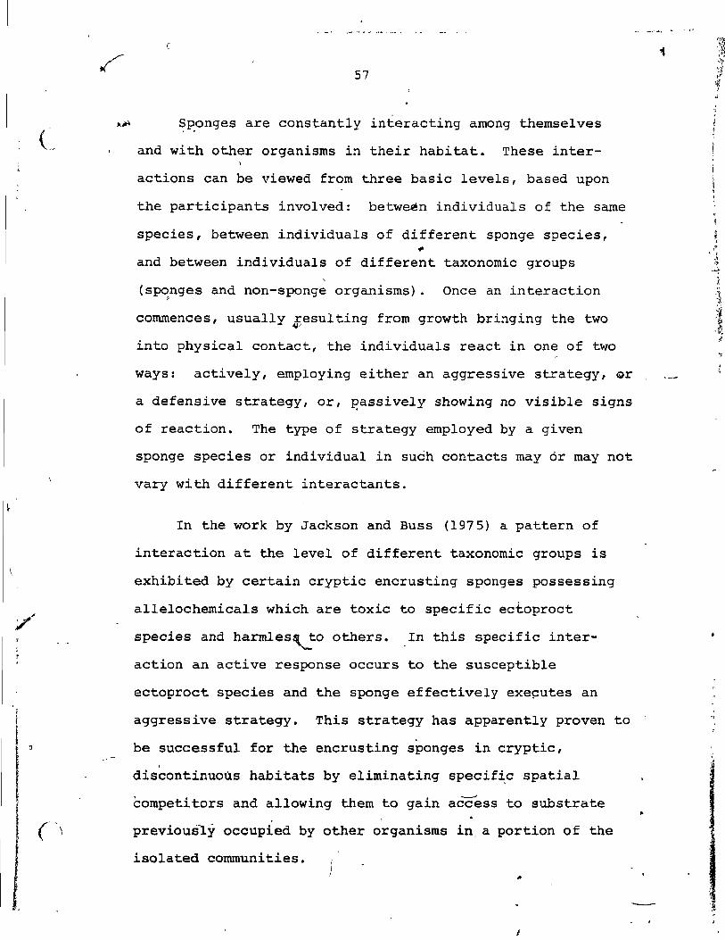
57
.~ ~~onges are constantly interacting among themseives
and with other organisms in their habitat. These inter-
actions can be viewed from three basic levels, based upon
the participants involved: betweèn individuals of the sarne
species, between individuais of different sponge species,
and between individuals of different taxonomie groups
(sponges and non-sponge organisms). Once an interaction , commences, usuaIIy ~esuiting from growth bringing the two
into physicai contact, the individuals react in one of two
ways: activeIy, employing either an aggressive strategy, Qr
a defensive strategy, or, p,assively showing no visible signs
of reaction. The type of strategy employed by a given
sponge species or individual in such contacts may or may not
vary with different interactants.
In the work by Jackson and Buss (1975) a pattern of
interaction at the levei of different taxonomie groups is
exhibited by certain cryptic encrusting sponges possessing
allelochemicals which are toxic to specifie ectoproct
species and har.mles~to others. In this specifie inter
action an active response occurs to the susceptible
ectoproct species and the sponge effectively exeçutes an
aggressive strategy. This strategy has apparently proven to
be successful for the encrusting sponges in cryptic, ,
discontinuous habitats by eliminating specif~c spatial
competitors and allowing them to gain acc:ess to substrate .
previous~y occupied by other organisms in, a portion of the
isolated communities. J
..

58
The interactions that occur in complex epizoie-
relationships involve primarily the levels of different
spo~e species and different taxonomi~ groups. These
interactions encompass the highly specifie symbiotic
relationships known to exist between sponges and other
organisl"(ls such as gastropods, bivalves, crabs" shrimps,
worms, zoant~ids, bacteria, and blue-green algae. These
syrnbiotic relationshiPs~~e not completely understood and
in most cases the role of the sponge remains undefined.
Generally the sponge is considered to be passive, or a non
rerctant, in ~~izoic assocations~ In these interactions the .
sponge may live attached -to another individual, or a~t as a . host for other organisms which ~hereby avoid competition for
l , __
'space by such strategy. Further investigations into "
syrnbiotic relationships and the role that sponges play in
these associations may reveal an active reaction hitherto
unknown.
The grafting experiments of this study and those of
other workers (Curtis, 1978(a); Hildemann, Johnson, and ..-----_/ \
Jokiel, 1979; and Van de Vyver, -1970) provide excellent
evidence of the range of specificity of the recognition
system ope~ating at the two levels of inter~çtioq within and \,
between different species of sponges, and the strategies
that have be~n evolved by s'ponges to deal with proETéms of
space utilization in benthic mari~e habitats. The inc~dence
of patterns in allograft acceptance and rejection, and ~ .
xenograft rejection discovered in these studies suggests
'.
1 J

59 t.
that these interactions are compatible with the concept of
strain and species specifie repognition systems in sponges. ~
The patterns of Jnteractioqp range from extremes of
c0nsistent allograft acceptance.and xenograft rejection to u
consistent rejection of both allografts and xenografts. In
~ other" words, sponges ~ay fuse with each individual of i~~ own species and reject aIl individuals of other species, or,
\
.. fuse with sorne individuals of its own species and reject aIl
other individuals o~ its own species and aIl individuals of
other species, or, reject aIl individuals of its own and
other species.
Interactions resulting in rejections are likewise
ultimately expressed in a variety of ways. In the present
study it was noted that in verongia longissima, rejection ,
was accompanied by the depo.sition of· a cuticle (Vacelet,
1971) between the two surfaces of individuals in contact.
The cuticle may serve to eftectively isolate tissues of each
member of the graft, essentially removing the living tissues
from direct interaction through development of an
" impermeable defensive barrier. Tissue ,damage is prevented
and space utilization by the species is ma~imized by such
strategy. It f~y be a result of the mechanisms involved in
the specific recognition system an~ the s~rategy employed
that has resul d~d in the expression of strain specifici ty in
Verongia longissima. In the, studies by Curtis (19,78a) 'and
Hildemann, Johnson, and Jokiel (1979) rejection of sponge
grafts involyed the eventual death and destruction of the
, -,
j 1

. >
1
1 t
..
(.
, 60
• graft individual shortly after the initial contact. Van de
vyver U970) observed a line or zone of non-coalescence
representing phys~cal retreat between rejected indi~iduals
0t the frespwater sponge Ephydatia fluviatilis. In these
interactions the evolution of.aggressive or derensive
(withdrawal) str~tegies dernonstrate a specifie recognition
. system.
-Studies on sponge cell reaggregation (Burger, ~ al,
1978; Hurnphreys, 1970; MacLennan and Dodd, 1967; and McClày,
1974}-llâve helped elucidate the biochemical na±ure of some
strain- or species-specific diff~seable surface-active
molecules which'operate to promote or prevent cell-cell
" adhesion. The specifie Il self/non-self" . recogni tion system·
which leads ta fusion or non-fusion in the grafting
experiments is almost. certainly modulated by the action.of
aggregation factors of the type identified in cell
-reaggregation studies. It seems likely that this type of ,
system is at the-'basi~ of the natural sponge-sponge inter-
actions occurring in all benthic communities including a .. "coral reef system. '\.
o
From an ecological v'iewpoint, the var-ious fatterns of
interactions and the strategies th~t haveJevolv~d- in various
sponge species appear ta be a means"of coping with the basic , "
problem of spatial competition by'f~rst gaining substrate,
, and then ~efendin~~~ In~a space-limited ha~itat l~ke a
coral reef communi ty·· this is o'f utmost ~mpoitance if high
• .J -.-,.-- __ 1_-'--'-----
.'
"J , '1
f 1 ! ! , . j
1 •
] , J' , , .
... ' .. é -

.)
fi
..
'61
diversity is to be maintained. However other niche /
diversification factors such a~ thfo flexibility in grow~h
form and, growth rate, reproductive pattern, etc. must be
partially effective as mechanisms of establishing and
maintaining high di~ersity of sponge populations in a space
limited habitat.
The immunological techniques employed in this study
have been focused on the strain specifi~ recog~ition system
of Verongia longissirna in hopes of providing sorne insig~t
into the compléxity of this sp~cific interàction., Why. do we "1 - . .
find ~ejection~ocourring between individu~ls of the ~ame o'
:s.i~ies, and what is inv:olved at this level of interaction?
Pernaps it is a form .of· t,he adaptive immun~ty response to " ..
fdr.eign tiss~e .w~11 known among the vertebra tes. l Hildem~iln, JOhnSO~,< and Jokiel (1979) have described it rs an immuno
recognition at the.allogeneic' level previousl~hoûght to
oocur only in high~r phylogenetic levels, namely the
vertebrates. These authors also expressed the idea that it"
'could represent the origins of the major histocompatibility
complex and cell-medi,fted immunity found in higher . .
vertebratès.
\,
There are three fundamental features of an immuno-• • , logi.cal response:.' memory, specifici ty, ,a,nÇi the recognition
- Q .. 1 \ 1
, of "non-self" (Roitt',' 197'7). . Thelse feature~ emQody the
ultimate SOPhistic:tion of an eXqUisiteTimmunoreco~nition , '. , " ~ --- ' .• ,~syst~. tA The resul t,s of 'the study by Hildemann, Johnson.' and·
1
"
. .,..
o 'Jo

~ f • t "
'.
,
1
/'
J 1 t 1
~
'.
, 62
f
Jokrel (1979) have finally demonstrated that sponges possess • ' 0 ,.
the thi' , fundamen tal .f ea ture , tha t 0 f memory. ",nowever, the
present ~tUdy did ~ot examine this fea~ and so there i.-s Î ' ,
no 'evidence here to support those findings. Thus it appears
from the resul ts of this' study and those of other worker.t(
that sponge~d~ possess an immunoreeognition.at the
allogeneic level and a form of adàptive immunity-~vhether " . • Y·'. this might ppssibly represent the origins of the m~or
histocompatibility complex and cell-mediated immunity found
--------
-
~ .,.--in higher vertebrates suggested by Hildemann, Johnson, and
Jokiel (1979) remains to ·be seen. However, these pioneering )
studies surely stimulate and invite fur~her confirmations
and extensions to provide insight' into the mechanisms
operating in' the specifie interactions occurring between
sponges in their habitat.
Most of our present knowledge on strain specificity in
sponges has come fr~m the extensive investigations on ~he
freshwater, sponges (Van de Vyver, 1970; 1971(b); 1975;
Curtis and Van'de Vyver, 1971; and Van de Vyver and Wi11enz,
1975). As mentioned ear1ier (INTRODUCTION) Van de Vyver \
. not!ed the existence of only a few' different strains of /'. ,
1 •
Ephydatia fluviatilis in a relatively. large 1rabitat space. ,
Recent investigations oh marine sponges by Curtis (1978a) on 1. ,(J •
HYt;neniacidon (~p.) in Engl,and, and H~1.demann, Johnson, and
JOkiel (1979) on Ca11ysfongia diffusa in Hawaii noteâ the
existençe of large numbers of different strains of these
species in a re1atively small habitat' space. These limited
.. n
J 1
f
1 1 l
1 . cl j
~ J : j

"c-" , ;. --
l
63
• results seem to imply that the marine system offers
.. condi~ons more favourable to the evolution of a higher
". number of strainS'~thin a species. This is substantia ted
in the present study by the occurrence of 17 different
'., strains of a single marine species, Verongia longissima, in ~
a relatively. small area. When 'the results were a~alyzed
statistically it was showI\ that_ip. 68% of future sites,
similar to those of the present study, one would expect to \;1'\.
find betw~en 9 and Il different strains of Verongia r
'longiss-ima. It is, understood, as .previously stated, that . ~ ,
the analyses were based on a minimum hUmber of-~ple sites
and .therefar~ :.li ttle empha,sis ~an be ,placed on: these
results.
" However, in contrast to these patterns of Atrain ,
" diversification, only one single strain of verongia .. -eauliformis was observed in the pr~sent study. This would
se~m to contradict the~earlie~ imp*ication~of. ~e rapid
~volution and maintenance of s~ains in marine spe~ies.
"
Verongia cauliforrnis is, however, ~imilar to Verongia
longissima i~ aIl morphological characters except
pigmentation'. _.1t i8 possible that Verongia cauliforrnis. may
prove'to be merely'a visqally distinctive strain of Verongia '., . "
longissima and not a ~eparat~ speci~~ Investigation of
reproductive rèlatiortships be;tween these' t.wo groups,
... ',presently recognized as separate species, ~ill be requi-red /1'1; •
. \ to resol v~, this problern. ! 1

< 1
~ t
1
1
1
1 1,
f _.~ -.....,..- -- - ~-
o
•
,
l
)
\
64
~n eoncl~sion, this study has dernonstrated the presence
of-a complex strain specifie recognition system in the
marine Qemosponge, Verongia Iongissima. Insight into ,the
basis of this specificfty has been provided through the
successful application of irnmunological techniques, and the
role that it plays in the patterns of specifie interactions
and strategies oecurring in a benthic marine conununity have
been diseussed. Further investigations of the cornplex
patterns of interactions that oecur between sponges and
~ther organisms in a benthic habitat may help to .define the
precise ecolQgical role that sponges play in the rnari~e
ecosys'tern and the major selective pressures which have been
operative in the evolution of relatively sophistipated
inununorecognition systems in these primitive organisms •
Such information-~y also provide greater insight into the
general evolutiona~~histOry of the exquisite i~uno-~ [1
recognition systems of higher metazoans.
.. ,
..
r·
\.
or
"
.Ii
J •
\
. ,
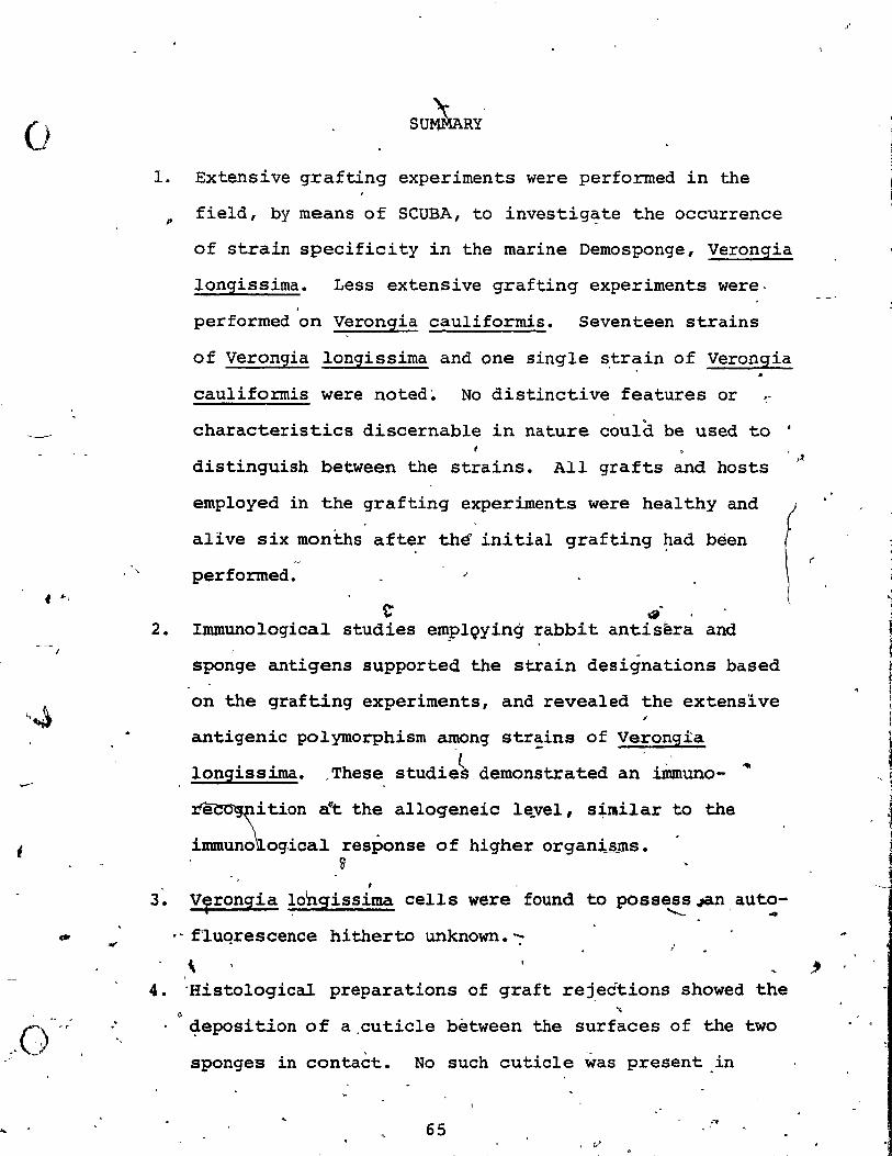
o 1.
p
. "
f ",
2. {
,.~
3 •
4.
"
Extensive grafting experiments were perforrned in the
field, by means of SCUBA, to investigate the occurrence
of strain specificity in the marine Demosponge, Verongia
longissima. Less extensive grafting experiments were· .
performed on Verongia cauliformis. Seventeen strains
of Verongia longissima and one single s,tr,:,-in of Verongia
cauliforrnis were noted~ No distinctive features or
characteristics discernable in nature could be used to •
distinguish between the strains. AlI grafts and hosts
employed in the grafting experiments were healthy and
alive six montlls after thé' initial grafting had bèen
performed. ~
t' ~.
lnununo logical studies em9lQying rabbit antisèra and
sponge antigens supported the strain designations based
on the grafting experiments, and revealed the extensive
antigenic polymorphism among strains of Verongia e -. . . ~ . ~
long1ss~a. ,These stud1e~ demonstrated an 1mmuno-
~ition a't the allogeneic le.vel, s~lIilar to the
immuno\ogical response of higher organi.§ms. ~
1
VTrongia IOhgissima cells were found to posse~s.;an, aut~-
. ' f'luqrescence hitherto unknown. "":'
\ 'Histological preparations of graft rej.edtions showed the
li
~eposition of a.cuticle bètween the surfaces of the two
sponges in contaèt. No such cuticle was present.in
65 ..•
. 1/
~ "
If
t'
~
·1 1
,

5.
6 •
•
•. ,..
(J
----_.-...,. 66 5
preparations of graft acceptanc~s. It appears that the
forma t:ljn of a collagenous cuticle is a common response
by Verongia species to a foreign solid surface . .. / "
It has been hypothesized from radioautography re~lts
that. preformed collagen is probably mobili~e4 from
"pools" within the sPQnge tissue and deposited between
the contact surfaces of two individuals that reject one 1
d" another • The first signs of a cuticle were observed 46 . hours' after two individuals of different strains of
. ~
verongia longissima had been brought into contact.
I~er~ and intraspecific acceptance and rejection, noted
in sponges, appear to be one of several patterns of ,
interactions ,that oceur between individuals in a space
timited habi~~.The ecological significance of.this
specificity can be seen when one considers the abundance
and diversity of thé Porlfera in benthic marine
communi ties •
"
r
1 " ;
-. . ,
LITERATURE CITED
~
Bailey, N. ~1959. Statistical methods in biology. John Wiley & Sons Inc., New York. 200 pp.
Bergquist, P. 1978. Sponges. Hutchinson University Library, London. 268 pp.
v Burger, M., W. Burkart, G. Neinbaum, and J. Jumblatt. 1978.
Cell-cell Recognition: Molecular aspects. Recogriition and its relation to morphogenetic processes in general. Pages 1-23 in A. Curtis, ed. Symposia of the Society for ExperImental Biology XXXII Cell-cell Recognition. Cambridge University Press, Cambridge.
\ Bur,net, F.
forms and inununity.
1971. .. ) 1 Self-recognition 1 in colonial marine flowering plants in relation to the evolution of Nature 232:230-238.
~ ~ rd Carpenter, P. l rn 5. Immunology and ser~y. 3 ed. W. B.
Saunders Co., Toronto. 346.pp.
Coffaro, K., and R. Hinegardner. 1977. Immune response in the Bea urchin Lytechinus ei:2us. Science 197~:389-1390.
Curtis, A. 1962. Pattern and echanism in the reaggregat~on of sponges. Nature 196:245-248.~
Curtis, A. 1978(a). Individualityand graft rejection in sponges: A cellular basis for individuality-in -sponges. (in press) in B. Rosen, ed. Biology and systematics of colonial organi~ms. Systematics Association.
Curtis, A., and G. Van de Vyver.- 1971. The control of cel1 adhesion in a morphogenetic system. J. Embryol. exp. Morph. 26:295-312. /~""
Fry, W. 1970. The sponge as a popu1~tionl: If biometric approach. Page.s 135-162 !!!. W. Fry, ed. The bio.logy of the 1?orifera, Zo01ogical Soc:Lety of London symposia 25 •
. Academic Press, London.
Galstoff, P. 1925. Regen~ralion after dissociation (An experimental study on sponges I). J. exp. Zool. 42:183-198.
Grassé, P., ed. 1973. Traité de Zoologie: Spongiaires, Tome III. Masson et Cie., Paris. 716 pp • •
---.- /!.,. ~ Harrison, F., and R. Cowden, eds. 1976. 'Aspects of sponge
bio1ogy. 'Academie Press, New York. 354 pp •
• 1')
67

,
f
K
1 f ,
- --_ .. _ ...... - ~-... - ~----_.-
68
Hildemann, W. , D. Linthicum, and D. Vanne 1975. Transplantation and immunoincornpatibility reactions arnong"reefbuilding corals. Immunogenetics ~:269-284.
Hildemann, W., et al. 1977. Tissue txansplantation immunity in corals. PrOë.~rd Inter. Coral Reef S~. pp. 537-543.
Hildemann, W., I. Johnson, and P. Jokiel. 1979. Irnmunocompetence in the lowest metazoan phylum: Transplantation immunity in sponges. Science 204:420-422.
Humason, G. 1962."Anirna1 tissue techniques. N. H. Freeman and Co., San Francisco. 468 pp. ,
Humphreys, T. 1970. Species-specific aggregation of dissociated sponge cells. Nature 228:685-686.
Humphreys, T., S. Humphreys, and A. Moscona. 1960. A procedure for obtaining cornpletely dissociated sponge cel1s. Biol. Bull. 119:294.
J
Hyman, L. 1940. The Invertebrates: Protozoa through Ctenophora. McGraw-Hil1, New York. 726 pp.
Jackson".r., and L. Buss. 1975. A11e~opathy and spatial -competition arnong coral reef invert~brates. Proc. Nat.
Acad. Sei. 72:5160-5163.
John, H., M. Campo, A. MacKenzie, and R. Kemp. 1971. Role of different sponge ce!l types in species-specific cell aggregation. Nature New Bio. 230:126-128.
) Kbpriwa, B., ancr-ê: LeBlond. 1962. Improvements in the coating techni~ue of radioautography. J. Histochem. Cytochem. 10:269-284.
Kuhns, W.,. et al. 1974. Sponge cel1 aggregation: A model for 'studiëi on cell-cel1 interactions. Ann. N. Y. Acad. Sei. 234:52-74.
Lewis, J. 1960. The coral reefs and coral communities of Barbados, W. I. Can. J. Zool. 38:1133-1145.
MacLennan, A. 1974. The chemica1 bases of taxon-specifie cellular reaggregation and 'self-not-self' recognition in sponges. Arch. Bio. (Bruxelles) ~: 53-90. .
1 •
MacLennan, A., and,R. Oodd. 1967. Promoting activity of extracellular materials on sponge cell reaggregation. J. Embryol~ exp. Morph. 17:473-480.
J , • 1
1
.'
; .. !r 1
~J ~ ï .'
f
1
1 , , !,
~ , ~,
1
~ f, --~

\ 1
'Ct 1 , , 1 f
1 \
)
• 1
-- --------- ------
69
McClay, D. 1971. An autoradiographie analysis of the species specificity during sponge cell reaggregation. Bio.
"Bull. 141:319-330.
McC1ay, D. 1974. Cell aggregation: Properties of ce11 ~ surface factors from five species of sponge. J. exp. Zool. 188: 89-101.
Mukai, H., and H. Watanabe. 1974. On the occurrence of colony specificity in sorne compound ascidians. Bio. Bull. 147:411-421.
Oka, H. 1970. CoYony specificity in compound ascidians. Pages 195-206 in H. Yukawa, ed. Profiles of Japanese Science and ScIëntists. '
Roitt, l. 1977. Essentia1 Immunology. 2nd ed. Blackwell Scientific Publications, London. 324 pp.
Sarà, M. 1970. Competition and co-operation in sponge populations. Pages 273-284 in ~-Fry, ed. The biology of the Porifera,' Zoologica1 Soë:rety of London Symposia 25. Academic Press, LondOn.
Theodor, J. 1970. Distinction between "self" and j'non-self" in lower invertebrates. Nature 227: 690-692.
#
Vace1et, J. 1971. L'ultrastructure de la cuticle d'Eponges Cornées du ~enre Verongia. J. Micro. 10:113-116.
# ...
Van de Vyver, G. 1970. Analyse de quelques phenqmenes d' histoincornpatibili té intraspécifique chez 01' Eponge d'eau douce. Ephydatia fluviatilis (Linné). Pa~es 55~6l in Reactions immunitaires chez les invertébres. .
Van de Vyver, G. 1970. La non-confluence intraspécifique chez les spongiaires et la notion d'individu. Ann. Embr. Morph. 1:251-262.
Van de Vyver, G. 1971(~)., En evidence d'un facteur . , hd' ~i; d'aggregat~on chez l'Eponge d eau douce. Ep y at1a •
f1uviati~is. Ann. Embr. Morph. !:373-381.
Van de Vyver, G. 1975. Phenomena of cellular ~ecognition' in sponges. Pages 123-140 in A. Moscona, and A. Monroy, eds. Current topies in deve1opmental" bio1ogy Vol. 4. Academic Press, New York. ,,,
Van de vy"ver, G., and~.' Wil1enz. 1975. An experimenta1 study of the ;J.ife-cy~le of the fresh-water sponge . Ephydatia f1uviati1is in its natura1 surroundinqs. Wi1h~lm. Roux' Archives 177:41-52. .
1
f < J . 1 f
'J !
1 ~
1 1

-.
~ ,
i f
(
1 Ci) ~ " h~-t...-, '~
~
Warr, G., and J. Marchalonis. 1978. Spectfic immune recognition by lymphocytes: An evolutionary perspective. Quart. Rev. Bio. 53:225-241.
Wilson, H. 1907. On some phenomena of coalescence and regeneration in sponges. J. exp. Zool. 5:245-258 •
. '
\
'~
0~? ~ l,
•
";.
\ X . ,
. \ , .
" , ~"(r,\ i , . , ,
...