Embed Size (px)
Citation preview

{
The Evaluation of Weakness in the Electromyography Lab
Anthony Chiodo, MD, MBAUniversity of Michigan Health SystemAAPMR Meeting, San Diego

38 year old right handed woman comes in with a chief complaint of weakness
Noticed it first in her inability to complete her usual circuit training program over the last month
Subsequently affected her aerobic exercise program and day to day home management activities
Case Study

She does not think there has been any numbness or tingling
She has not noticed it affecting one area of the body first but is uncertain
She has had no new pain, no fever or chills, no change in bowel or bladder control

Past medical history is remarkable for hypothyroidism for which she takes synthroid
Family history is significant for a maternal grandmother with rheumatoid arthritis, paternal grandparents with diabetes mellitus and heart disease, father with hypertension, mother with hypothyroidism

Deep tendon reflexes are 1+ Pin sensation and light touch is
symmetric Strength testing shows shoulder and hip
girdle muscles in the 3-4 range, hamstrings 3, elbows 4, hands and feet 4+ to 5
There is no tenderness and no pain with ROM
Physical Examination

{
Differential Diagnosis

{
Neuromuscular Junction Disorders
Anthony Chiodo, M.D.

Motor disorder Defect affecting the relationship between
the distal motor axon and muscle Defect can be pre-synaptic, synaptic, or
post-synaptic Acquired disorders are pre- or post-
synaptic
NMJ Disorders: Nature of the Abnormality

Normal distally and conduction
Sensory Nerve Conduction Studies

Post-synaptic: Normal Pre-synaptic: inability to achieve
transmission: decreased motor evoked amplitudes but normal latency and conduction
Motor Nerve Conduction Studies

Pre-synaptic: if no motor units are seen with activation, fibrillation potentials and positive waves are possible Botulism Otherwise, decreased amplitude motor units
of varying amplitude and rapid recruitment Post-synaptic: varying amplitude motor
units with rapid recruitment If myasthenia gravis: may see proximal
fibrillation potentials and positive waves
Routine Needle Examination

{Repetitive Nerve Stimulation
Physiology is Key to Understanding

Molecules = Quanta X # Released(p) Typical Quanta = several thousand
acetylcholine molecules, generate MEPP, amplitude 1 mV
P typically around 60 quanta released per nerve stimulus
Muscle action potentials at 7-20 mV
Presynaptic Acetylcholine Stores

Reserve: 300,000 quanta Mobilization: 10,000 quanta Immediate Release: 1,000 quanta
Types of Acetylcholine Stores

Mobilization store: 1500 msec Reserve: 3-4 minutes
Time Availability

Decreased quanta released per nerve stimulation
Synaptic vesicle fusion time(Ca++ dependent) is 100-200 msec
Maximal decrement at 2-3 Hz No decrement at 10 Hz Normal decrement < 8%
Concept of Decrement
1st stimulus: 60 quanta 2nd stimulus: 56 quanta 3rd stimulus: 53 quanta 4th stimulus: 55 quanta as reserve
quanta become available Accounts for decrease in EPP and results
in increase risk of failure (blocking) over four repetitive stimulations at 2 Hz
At 2 Hz Repetitive Stimulation

Other Disorders NMJ Transmission Myotonia Neurogenic Disorders with
Denervation/Reinnervation Rapidly Progressive ALS Polyneuropathy Mononeuropathy Radiculopathy
What Other Disorders Have a Decrement To Low Frequency Repetitive Nerve Stimulation?

Yes, temperature Increased decrement and blocking at
increased temperature due to increased acetylcholinesterase activity
May account for the fact that the effect is more pronounced in proximal muscles
Are there any physiological parameters that effect this finding?
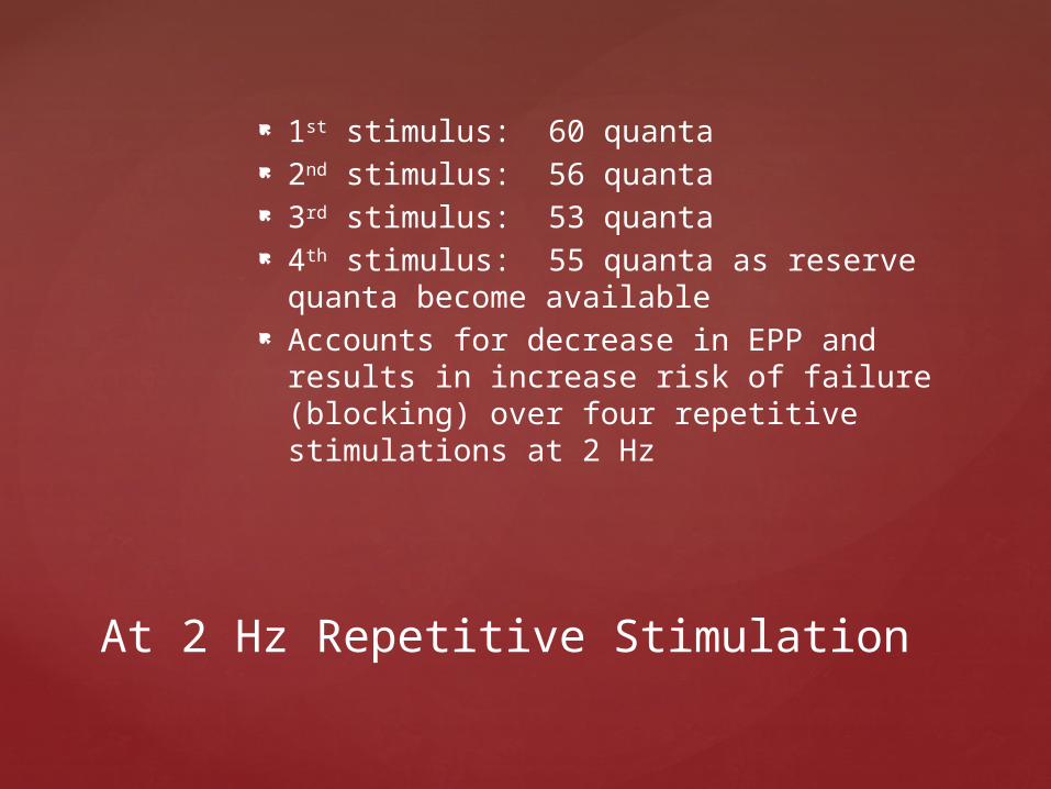
Acetylcholine Receptor Antibodies Normal number of MEPP’s MEPP amplitude decreased by 80% Post-activation facilitation Post-activation exhaustion
Myasthenia Gravis

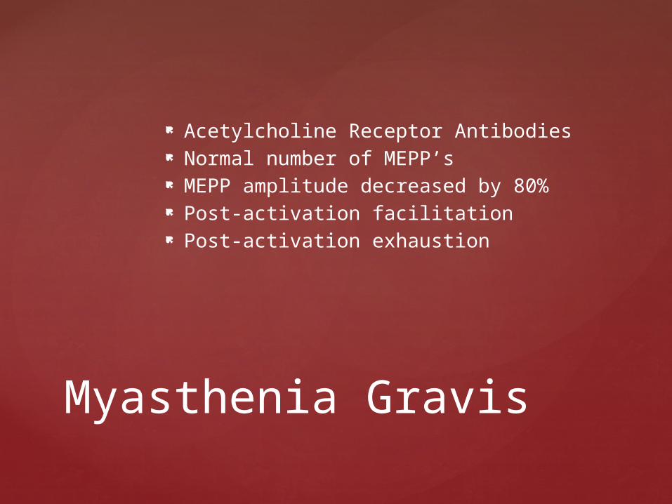
Increased calcium in endplate increases the quanta released
Decrement decreased, small increase in motor evoked amplitude
Post-activation Facilitation

Depletion of mobilization and immediate release stores, before reserve store becomes available
Decreased receptor excitability Characteristic of Myasthenia Gravis
Post-activation Exhaustion


Recording surface 25 mcm to pick up from single muscle fiber
Quantify the differences in time of onset of firing of two muscle fibers from the same motor unit
Jitter is the mean difference in this firing onset time
Blocking is the rate of failure of a muscle fiber from firing with it’s motor unit
SFEMG


Pre- and post- synaptic neuromuscular junction disorders
Ongoing neuropathic processes: motor neuron disease, neuropathy, radiculopathy
Diagnoses with Increased Jitter

Hallmark: Fluctuating weakness Diplopia Ophthalmoplegia Ptosis Facial Weakness Dysphagia Vocal cord weakness Respiratory muscle weakness Pelvic floor muscle weakness
Myasthenia Gravis Clinical Symptoms


I: Ocular IIA: Mild generalized IIB: Moderate generalized III: Acute severe with bulbar
symptoms IV: Late severe V: Muscle atrophy
Clinical Classifications of Myasthenia Gravis Severity: Osserman


Active: 5-7 years Inactive: 10 years Burned Out: Slow improvement seen
40-50% of ocular myasthenics will become generalized in the first 2 years
Clinical Course in Myasthenia Gravis
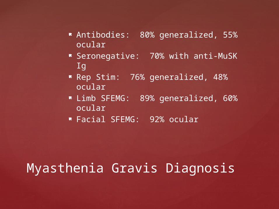
Antibodies: 80% generalized, 55% ocular
Seronegative: 70% with anti-MuSK Ig Rep Stim: 76% generalized, 48% ocular Limb SFEMG: 89% generalized, 60%
ocular Facial SFEMG: 92% ocular
Myasthenia Gravis Diagnosis

Evaluate/treat other autoimmune disorders: RA, thyroid, B-12
Pacing High K+ diet Avoid excessive heat>cold Watch for cyclic changes in women Avoid botox, quinamm,
aminoglycosides, tetracyclines, anesthetic agents, anticonvulsants
Preventing Exacerbations in Myasthenia Gravis

Mestinon: Acetylcholinesterase inhibitor Prednisone Imuran, Cytoxan, Cyclosporine, Mycophenolate Therapeutic Plasma Exchange Thymectomy AchR-based Immunoadsorbants Mucosal injection of AchR-recombinant
fragments IG to proinflammatory cytokine IL-18 and
costimulatory factor CD40L Create viral manipulated antigen presenting
cells that express AchR to present to AchR-specific T-cells and activate Fas ligand “guided missile”
Myasthenia Gravis Treatment

Creatine plus resistance exercise with normal treatment in mild MG shows improved strength and muscle mass
Isometric exercise effective in improving strength in mild MG
Exercise in Myasthenia Gravis

Increased acetylcholinesterase activity Decreased sensitivity of acetylcholine
receptors More rapid presynaptic acetylcholine
depletion Can explain proximal>distal weakness
Effect of Temperature in Myasthenia Gravis

Antibodies prevent pre-synaptic Ca++ influx prevents quanta release
Decreased number of MEPP’s of normal amplitude
Decrement to low frequency rep stim due to many muscle fibers activated near-threshold so decreased release is miniscule but significant
Post-exercise facilitation due to increased Ca++ in cell resulting in increased quanta release with next nerve stimulus
Myasthenic Syndrome

Immunogenic: responds to TPE, prednisone, and 2,4-DAP
Tumorogenic: responds to cancer therapy
Eaton-Lambert Types

Food, wound or infantile Markedly decreased pre-synaptic release
due to botulinum toxin binding to and entry into the nerve terminus membrane
Cleave proteins in synaptic vesicle membrane inhibiting release
Complete binding may result in no increment to exercise
Complete binding may lead to fibrillation potentials and positive waves
Botulism

LEMS: hallmark is marked incremental response with exercise
AIDP Critical illness myopathy
Botulism Differential Diagnosis

Does not include neonatal myasthenia gravis
Presynaptic: failed production, storage and mobilization of acetylcholine
Acetylcholine receptors: decreased number, decreased binding, prolonged binding/opening
Congenital absence of acetycholinesterase
Congenital Myasthenia Gravis

Thank you!