Embed Size (px)
Citation preview

-Tocopherol Reduces Lipid Accumulation in Niemann-Pick Type C1 and Wolman Cholesterol Storage Disorders
Miao Xu,1, 7* Ke Liu,1* Manju Swaroop,1 Forbes D. Porter,2 Rohini Sidhu,3 Sally Finkes,3 Daniel S. Ory,3 Juan J. Marugan,1 Jingbo Xiao,1 Noel Southall,1 William J. Pavan,4 Cristin Davidson,5 Steven U. Walkley,5 Alan T. Remaley,6 Ulrich Baxa,7 Wei Sun1, John C. McKew,1 Christopher P. Austin,1 Wei Zheng1† 1National Center for Advancing Translational Sciences, NIH, Bethesda, MD 20892; 2Program in
Developmental Endocrinology and Genetics, Eunice Kennedy Shriver National Institute of Child Health
and Human Development, NIH, Bethesda, MD 20892; 3Diabetic Cardiovascular Disease Center,
Washington University School of Medicine, St. Louis, MO 63110; 4Genetic Disease Research Branch,
National Human Genome Research Institute, NIH, Bethesda, MD 20892; 5Sidney Weisner Laboratory of
Genetic Neurological Disease, Rose F. Kennedy Center, Albert Einstein College of Medicine, Bronx, NY
10461; 6Laboratory of Lipoprotein Metabolism, National Heart, Lung and Blood Institute, Bethesda, MD
20892; 7Sir Run Run Shaw Hospital, Zhejiang University School of Medicine, Hangzhou, China; 8Electron Microscopy Laboratory, National Cancer
Institute, NIH, Bethesda, MD 20892.
*
These authors contributed equally to this work
†To whom correspondence should be addressed: Wei Zheng, Ph.D. National Center for Advancing Translational Sciences, NIH 9800 Medical Center Drive, MSC: 3370 Bethesda, MD 20892-3370 Email: [email protected] Tel.: (301)827-6727 Running title: Reducing storage in lysosomal diseases through exocytosis Keywords: -Tocopherol, Niemann Pick disease type C1, Wolman disease, Lysosomal storage diseases, lysosomal exocytosis. Background: Niemann Pick disease Type C and Wolman diseases are caused by mutations in genes responsible for intracellular cholesterol processing and trafficking. Results: -tocopherol reduces lysosomal accumulation of cholesterol and other lipids potentially through enhancement of lysosomal exocytosis. Conclusion: -tocopherol is a novel lead compound for drug development to treat lysosomal storage diseases. Significance: Lysosomal exocytosis may represent a new drug target broadly applicable to lysosomal storage diseases.
http://www.jbc.org/cgi/doi/10.1074/jbc.M112.357707The latest version is at JBC Papers in Press. Published on October 3, 2012 as Manuscript M112.357707
Copyright 2012 by The American Society for Biochemistry and Molecular Biology, Inc.
by guest on February 19, 2019http://w
ww
.jbc.org/D
ownloaded from
by guest on February 19, 2019
http://ww
w.jbc.org/
Dow
nloaded from
by guest on February 19, 2019http://w
ww
.jbc.org/D
ownloaded from
SUMMARY
Niemann-Pick disease type C (NPC) and
Wolman disease are two members of a family of
storage disorders caused by mutations of genes
encoding lysosomal proteins. Deficiency in
function of either the NPC1 or NPC2 protein in
NPC disease, or lysosomal acid lipase in
Wolman disease results in defective cellular
cholesterol trafficking. Lysosomal
accumulation of cholesterol and enlarged
lysosomes are shared phenotypic characteristics
of both NPC and Wolman cells. Utilizing a
phenotypic screen of an approved drug
collection, we found that -tocopherol
effectively reduced lysosomal cholesterol
accumulation, decreased lysosomal volume,
increased cholesterol efflux, and alleviated
pathological phenotypes in both NPC1 and
Wolman fibroblasts. Reduction of these
abnormalities may be mediated by a -
tocopherol-induced intracellular Ca2+ response
and subsequent enhancement of lysosomal
exocytosis. Consistent with a general
mechanism for reduction of lysosomal lipid
accumulation, we also found that -tocopherol
reduces pathological phenotypes in patient
fibroblasts from other lysosomal storage
diseases including NPC2, Batten (CLN2),
Fabry, Farber, Niemann-Pick disease type A,
Sanfilippo type B (MPS IIIB), and Tay-Sachs.
Our data suggest that regulated exocytosis may
represent a potential therapeutic target for
reduction of lysosomal storage in this class of
diseases.
INTRODUCTION
Niemann-Pick Disease type C (NPC) disease is
caused by mutations in either the NPC1 or NPC2
gene, encoding two distinct lysosomal cholesterol
binding proteins (1,2). The NPC cellular
phenotype is characterized by lysosomal
accumulation of unesterified cholesterol and other
lipids, resulting from impaired cholesterol export
from the late endosomal and lysosomal
compartments (2,3). Wolman disease is caused by
mutations in the gene encoding lysosomal acid
lipase (LAL). Deficiency of LAL function results
in two distinct disease phenotypes that accumulate
both cholesteryl ester and triglycerides. The early
onset form is known as Wolman disease and the
late onset form as cholesteryl ester storage disease
(3,4). Enlarged lysosomes are a common cellular
phenotype for both NPC and Wolman diseases.
Cholesterol is an essential component of cellular
membranes and is central to maintaining both
membrane integrity and fluidity, as well as
regulating membrane protein function, cell
signaling and ion permeability (5). In addition,
cholesterol is the precursor molecule for synthesis
of cellular components such as bile acids,
oxysterols, and steroid hormones. Cholesterol
homeostasis is tightly regulated at both the cellular
and whole-body levels (6). While the homeostasis
of serum and tissue cholesterol has been well
defined, many details of intracellular cholesterol
regulation and trafficking are still to be elucidated.
Esterified cholesterol contained in low-density
lipoprotein (LDL) enters a cell via endocytosis.
by guest on February 19, 2019http://w
ww
.jbc.org/D
ownloaded from

Upon LDL entry into the late endosome/lysosome,
LAL hydrolyzes fatty acyl chains from cholesteryl
esters to liberate unesterified cholesterol that is
transported out of the late endosome/lysosome
involving NPC1 and NPC2 proteins (6-8). The
precise mechanism and pathway of cholesterol
trafficking from the lysosome remains unclear,
though it has been established that unesterified
cholesterol moves to the endoplasmic reticulum
(ER) for re-esterification or utilization by the cell,
and to the trans-Golgi network and/or plasma
membrane for efflux from the cell (5,9).
Currently, there are no FDA-approved
therapies for either NPC disease or Wolman
disease. However, miglustat, originally approved
as a substrate reduction drug for Gaucher disease,
has been approved in the EU for the treatment of
NPC1 disease by European Agency for the
Evaluation of Medicinal Products (EMEA).
Therapeutic strategies that have been explored
include reduction of lysosomal lipid burden,
augmentation of mutant protein expression, or
replacement of mutant proteins in lysosome (10).
Enzyme replacement therapy (ERT), which has
been successfully used to treat selected lysosomal
storage diseases, was studied in the mouse model
of Wolman disease but is not clinically approved
(11). A protein replacement therapy, similar to
ERT, would in theory be possible for NPC2
disease, but is not feasible for NPC1 disease
because NPC1 is an integral membrane protein. A
phenotypic screen of a compound collection
recently identified compounds that reduced filipin
staining in NPC1 fibroblasts (12), and several
laboratories have reported lead compounds
including 2-hydroxypropyl–-cyclodextrin and
HDAC inhibitors that reduce cholesterol
accumulation (12-15). A recent report revealed
that the cyclodextrin’s mechanism of action to
reduce cholesterol accumulation in NPC1 mutant
cells is through induction of exocytosis (16). Here
we report the identification of -tocopherol (-T)
in a phenotypic screen of an approved drug library
that significantly reduces lysosomal cholesterol
accumulation in NPC1 fibroblasts. Following -T
treatment, the enlarged acidic
endosomal/lysosome compartment was
dramatically reduced, and the pathological
phenotype of NPC1 cells, as determined by
ultrastructural analysis, was alleviated. Similar
effects of -T were also observed in Wolman
fibroblasts. The mechanism of action for -T was
linked to increase in cytosolic Ca2+, correction of
the lysosomal Ca2+ deficiency, and enhancement
of lysosomal exocytosis. The effect of -T also
extended to patient-derived fibroblasts from other
lysosomal storage diseases, including Batten
(CLN2), Fabry, Farber, Niemann-Pick disease
type A (NPA), NPC2, Sanfilippo type B (MPS
IIIB), and Tay-Sachs. The results indicate that the
enhancement of lysosomal exocytosis may
represent a new therapeutic strategy for the drug
development that may be widely applicable to
lysosomal storage diseases.
EXPERIMENTAL PROCEDURES
Materials - Hoechst and LysoTracker Red dye
were purchased from Invitrogen (Carlsbad, CA).
3
by guest on February 19, 2019http://w
ww
.jbc.org/D
ownloaded from

Filipin dye was obtained from Sigma-Aldrich (St.
Louis, MO). α-Tocopherol and δ-tocopherol were
purchased from Sigma-Aldrich and purified by
HPLC to purity greater than 99%. β-Tocopherol
(racemic), α-tocotrienol, β- tocotrienol, and γ-
tocotrienol were obtained from Sigma-Aldrich in
analytical standard grade (purity ≥ 97%). γ-
Tocopherol and δ-tocotrienol were purchased from
ChromaDex (Irvine, CA) in vitamin reference
standard grade (purity > 96%). The 96-well, 384-
well and 1536-well plates were purchased from
Greiner Bio-One (Monroe, NC). Human skin
fibroblast cell lines (Table S1) were purchased
from the Coriell Cell Repository (Camden, NJ)
including control cell line (GM05659), NPC1
(GM03123), NPC2 (GM17910), Wolman
(GM11851), Batten (GM16485), Farber
(GM20015), Fabry (GM00107), Niemann-Pick
Disease type A (GM16195), Sanfilippo type B
(GM02552), and Tay-Sachs (GM00221). The
GM03123 fibroblasts were utilized mainly in the
NPC1 experiments unless specifically indicated.
The NPC25 (c.2979dupA, p.N701K) skin
fibroblast line was derived from a NPC1 patient
evaluated under an NICHD IRB approved clinical
protocol, and obtained with consent.
Cell culture - Both human fibroblasts and baby
hamster kidney (BHK) cells were cultured in
DMEM medium (Invitrogen, cat# 11995-040)
supplemented with 10% fetal bovine serum (FBS),
100 unit/ml penicillin and 100 g/ml streptomycin
in a humidified incubator with 5% CO2 at 37°C.
Approved drug collection - A collection of 2,816
compounds approved drugs that were dissolved in
DMSO solution as 10 mM stock solution. were set
up as described previously (17). The compounds
were serially diluted 1:5 in 384-well plates for
seven concentrations and formatted into 1,536-
well plates at 5 l/well as the compound source
plates. In the primary screen, 23 nl of compounds
in DMSO solution was transferred using a Pintool
station (Klaypsys, San Diego, CA) to 1536-well
assay plates containing 5 l/well cell culture
medium. The final concentrations of compounds
ranged from 1.32 nM to 46 M.
Amplex-Red cholesterol assay - Total
cholesterol in patient cells was measured by the
Amplex-Red Cholesterol Assay Kit (Invitrogen).
The unesterified cholesterol was determined using
the same kit without the enzyme acid lipase.
Esterified cholesterol was determined as the
difference between the total and unesterified
cholesterol values. The cells were seeded in black,
tissue culture-treated 1536-well plates at 800
cells/well in 5 l medium by a Multidrop Combi
dispenser (Thermo Scientific, Waltham, MA) and
cultured for 24 hr. The assay plates were added
with 23 nl/well of compound DMSO solution
using a Pintool station (Klaypsys, San Diego, CA)
and cultured for 3 days. The cells were washed
twice using a centrifugation method in which the
inverted plates were placed on a stack of paper
towel and centrifuged at 800 rpm for 1 min
followed by addition of 7 l/well PBS (added
gently with a 45 degree angled liquid dispenser
(Klaypsys)). The cholesterol assay mixture from
the kit was added at 2.5 l/well and incubated for
1 hr at 37°C. The resulted fluorescence intensity
4
by guest on February 19, 2019http://w
ww
.jbc.org/D
ownloaded from

was measured with excitation of 560 (±10) and
emission of 590 (±10) in a fluorescence plate
reader (Tecan, Durham, NC).
Cholesterol analysis by liquid
chromatography–mass spectrometry (LC-MS) -
Cell pellets were collected from 100-mm dishes
and washed once with 10 ml PBS. Proteins were
assayed using the BCA kit (Sigma-Aldrich) and
lipids were extracted by modified Bligh and Dyer
procedure (18) from the cells. Internal standards
were added based on protein concentration that
included N12:0 sphingomyelin, D7-cholesterol,
17:0 cholesteryl ester (Avanti Polar Lipids, Inc).
The lipid extract was derivatized to convert
cholesterol and its ester into acetate for facilitating
ionization using direct infusion mass spectrometric
assay. The derivatization was performed by
treating the dried crude lipid extract with solutions
of 1M acetic acid with1M DMAP in chloroform
and 1M EDC in chloroform at 50oC for 2 hours.
The derivatized product was extracted with hexane
and analyzed by direct infusion mass
spectrometric assay with 5 % ammonium
hydroxide for facilitating ionization.
A triple-quadrupole mass spectrometer
equipped with an electrospray was used for
analysis of lipids in positive mode.
Sphingomyelins, cholesterol and cholesteryl esters
were detected by neutral loss scan of 213
(collision energy: 50 V), neutral loss scan of 77
(collision energy: 14 V) and precursor ion scan of
m/z 369 (collision energy: 14 V) respectively. Data
processing of mass spectrometric analyses
including ion peak selection, data transferring,
peak intensity comparison and quantification was
conducted using self-programmed Microsoft Excel
macros (19).
Cytotoxicity assay - In an initial experiment, we
found that commercially-available vitamin E
isoforms were cytotoxic at the similar
concentrations required for their pharmacological
effect. The ATP content assay kit (Promega or
PerkinElmer) was applied to monitor the
compound cytotoxicity. Cells were seeded in
white solid 96-well or 1536-well plates and treated
with compounds as described above for the
cholesterol assay. On the experimental day, 3
l/well assay mixture (prepared according to the
manufacturer’s instruction) was added to the assay
plates followed by incubation at RT for 10 min.
The luminescence signal was determined in the
luminescence mode of a Tecan multifunction plate
reader.
Filipin staining - Cells were seeded at 1500
cells/well in 100 l medium in black/clear bottom,
tissue culture treated 96-well plates and cultured
for 24 hr. After addition of compound solution, the
plates were cultured for 3 days. The cells were
washed twice with PBS and fixed by 100 l/well
3.2% formaldehyde at room temperature (RT) for
30 min. After washing with PBS two times, the
cells were stained with 100 l/well 50 ng/ml
filipin (freshly-dissolved in DMSO at 10 mg/ml
and then diluted in PBS) at RT for 1 hr. The plates
were stored at 4°C after washing two times with
PBS before the imaging analysis. On the day of
imaging experiment, cells were stained with 100
l/well of 2 M of CellMask-Red dye (Invitrogen,
5
by guest on February 19, 2019http://w
ww
.jbc.org/D
ownloaded from

# H32711) in PBS at RT for 1 hour. The plates
were imaged using an Incell2000 automated
fluorescence plate imaging reader (GE Healthcare)
with a 20 or 40 objective. DAPI filter set (Ex
= 360 ± 20 nm and Em = 460 ± 20 nm) and
TRITC filter set (Ex = 545 ± 20 nm and Em = 593
± 20 nm) were used to visualize filipin and
CellMask staining, respectively.
Nile-red staining - The cells were cultured and
treated as described above. On the experimental
day, cells were washed two times with PBS and
live-stained with 1 M Nile-red dye solution
(prepared in cell culture medium) at 100 l/well
followed by an incubation at 37 C for 10 min.
After washing twice with PBS, the cells were
fixed in 3.2% paraformaldehyde in PBS at 100
l/well for 1 hr at RT. The nuclear staining was
carried out by an addition of 100 l/well 1 g/ml
Hoechst 33342 (Invitrogen) in PBS and incubation
at RT for 30 min. The plate was washed twice
with PBS and the images were measured in
Incell2000 imaging plate reader with a FITC filter
set (Ex = 480 ± 20 nm and Ex = 525 ± 36 nm) for
the neutral lipids (cholesteryl esters and
triglycerides) and a DAPI filter set for Hoechst
nuclear staining.
Bodipy-lactosylceramide (bodipy-LacCer)
staining - The cells in 96-well assay plate were
prepared as described above, incubated with 100
l/well bodipy-LacCer dye (Invitrogen, # B-
34402) in 1% FBS DMEM and incubated at 37C
for 1 hr followed by plate washing with HMEM
buffer (13.8 mM HEPES, 137 mM NaCl, 5.4 mM
KCl, 2 mM glutamine, 0.4 mM KH2PO4, 0.18
mM Na2HPO4, 1X MEM vitamins and 1X MEM
amino acids at pH 7.4). The cells were rinsed with
cold HMEM buffer containing 5% fatty acid-free
BSA and back exchanged at 11°C six times over
60 min as described previously (20). Images were
immediately taken in the InCell2000 imaging plate
reader with the DAPI filter set.
LysoTracker dye staining - The assay was
optimized to visualize the enlarged lysosomes by
applying appropriate concentration of LysoTracker
dye in which the control cells exhibited minimal
staining while the NPC1 or other disease cells
showed significant staining. The cells were
cultured and treated as described above. On the
experimental day, cells were live-stained with 100
l/well 50 nM LysoTracker-Red DND-99 dye
(Invitrogen, # L-7528) in medium at 37°C for 1 hr
followed by plate washing twice with PBS. The
plate was used for image analysis of live cell
staining or was fixed in 100 l/well 3.2%
formaldehyde for 1 hr and washed two times with
PBS. The nuclear staining was performed by an
addition of 100 l/well 1 g/ml Hoechst 33342
(Invitrogen) in PBS and incubation at RT for 30
min. After washing twice with PBS, the plates
were stored at 4°C until imaging analysis. DAPI
filter set and TRITC filter set in the Incell2000
imaging plate reader were used to visualize
Hoechst nuclear staining and LysoTracker
staining, respectively.
Electron Microscopy - Fibroblast cells were
seeded in 6-well plates in 5 ml medium at 150,000
cells/well and cultured for 1 day in the presence or
6
by guest on February 19, 2019http://w
ww
.jbc.org/D
ownloaded from
absence of compounds. Cells were fixed in 2%
glutaraldehyde, 0.1 M cacodylate buffer, pH 7.2
for 1 hour at room temperature and then stored at 4
°C until TEM analysis was performed. The cells
were post fixed in 1% osmium tetroxide in the
same buffer for 1 hour and en bloc stained with
0.5% uranyl acetate in 0.1 M acetate buffer, pH
4.2. The cells were then dehydrated in graded
ethanol solutions (35%, 50%, 70%, 95% and
100%) and infiltrated overnight in epoxy resin
(Poly/Bed 812, Polysciences). After adding fresh
pure resin the cell plates were cured for 72 h in 55
°C. After removing the polystyrene plates, suitable
areas for thin sectioning were selected, cut out
with a jewelry saw and glued onto empty resin
stubs. About 70 nm thin sections were cut on an
ultramicrotome (Leica EM UC6) and mounted on
naked copper grids. The thin sections were double
stained (uranyl acetate and lead citrate), examined
in a Hitachi H-7650 transmission electron
microscope, and images were taken using an AMT
CCD camera (21).
Western blot analysis - Cell lysates were
prepared and protein concentrations were
determined as described above. Samples were
heated at 65°C for 10 min before being resolved
by SDS-PAGE under reducing conditions.
Proteins were transferred to PVDF membranes
(Bio-Rad Laboratory) using either Criterion
Blotter (Bio-Rad Laboratory) or iBlot dry blotting
devices (Invitrogen). For NPC1 and HMG-CoA
reductase detection, rabbit polyclonal antibodies
against NPC1 (Abcam, # ab36983, 1:2000) or
HMG-CoA reductase (Millipore, # 07-457,
1:1000) were used, respectively. A peroxidase-
conjugated donkey anti-rabbit IgG (Santa Cruz)
was used as secondary antibody at a 1:500
dilution. Flotillin, an exosome marker, was
determined using a method described previously
(16). Briefly, the media from cells in 100 x 20 mm
dishes treated with 40 M -T or positive controls,
2% HPBCD and 1 M ionomycin for 1 hr at 37 C
were collected and centrifuged at 100,000 g for 1
hr for isolation of exosomes. The resulting
exosomes were resuspended in 20 mM Tris-HCl
buffer (pH 8.0) and sonicated in a bath sonicator to
yield the final samples for Western blot analysis as
described above. A mouse anti-flotillin 2 antibody
(BD Biosciences) was used for the detection of
flotillin in the exosomes. Cells were lysed and the
cell lysates were collected for the determination of
GAPDH used for normalization of proteins in each
sample.
3H-Cholesterol efflux assay - Efflux of
cholesterol from BHK cells was performed as
previously described (22). Briefly, BHK cells and
BHK cells stably transformed with ABCA1,
ABCG1 and SR-BI were radiolabeled with 3H-
cholesterol (1 Ci/mL) in medium for 24 hr. After
washing three times with DMEM, DMEM
medium containing 50 g/ml HDL was added and
cultured for 18 hr. The radioactivity in the HDL
containing medium and cell lysate were
determined. The percent of total radiolabeled
cholesterol effluxed into the media was calculated
by dividing by the sum of the radioactive counts
remaining in the cells after the efflux period plus
the counts in medium.
7
by guest on February 19, 2019http://w
ww
.jbc.org/D
ownloaded from

Intracellular and lysosomal Ca2+
measurements - Intracellular cytosolic Ca2+
concentration was measured fluorescently using a
Fluo-8 dye kit (ATT Bioquest, Sunnyvale, CA) as
described previously. Briefly, fibroblasts were
cultured at 2500 cell/well in 20 l medium in
black, clear bottom 384-well plates for 24 hr at
37C. The calcium dye mixture was added at 20
l/well and incubated at 37C for 30 min followed
by room temperature for 30 min. The plates were
then placed into a fluorescence kinetic plate reader
(Cell, Hamamatsu, Hamamatsu City, Japan). The
basal fluorescence intensity was recorded 10 times
at 1Hz for 10 seconds and the compound (-T or
-T) was then added at 20 l/well inside the
instrument followed by additional reading at 1Hz
for 5 min. The results were normalized to the
average basal fluorescence intensity in ratio and
the peak response (Max.) was used for the result
calculation. The lysosomal Ca2+ induced by Gly-
Phe β-naphthylamide (GPN) was measured
similarly as that for cytosolic Ca2+ except 200 nM
GPN was added instead of -T or -T after the
measurement of basal fluorescence intensity that
released lysosomal Ca2+.
Endocytosis and lysosomal pH measurement –
Endocytosis was measured using a fluorescence
labeled dextran dye (pHrodo™ dextran, catalog #
P10361, Invitrogen) that enters cell through
endocytosis. This dye emits fluorescent signal at
the acidic pH while exhibiting minimal
fluorescence signal at neutral pH. The fibroblasts
in 96-well plates were treated with 40 M -T or
80 M -T for 24 hrs and incubated with 20
mg/ml pHrodo dye for 16 hrs. The live cell
imaging was carried out after a cell wash with
PBS with an excitation of 560 nm and emission of
590 nm. Chlorpromazine, a known inhibitor of
endocytosis was used as a positive control (final
concentration of 5 g/ml). The lysosomal pH was
determined using a fluorescent LysoSensor
dextran dye (catalog # L-22460, Invitrogen) that
emits strongly at 530 nm in acidic pH environment
and at 450 in neutral pH. The fibroblasts in 96-
well plates were treated with 40 M -T for 24 hrs
and incubated with the pH indicator dye for 16 hrs.
The images were taken using Incell2000 (GE
healthcare) with an excitation of 360 nm,
emission-1 of 530 and emission-2 of 450 nm.
Measurement of -hexosaminidase (HEXB)
release - Fibroblasts were cultured in 24-well
plates at 30,000 cells/well in 0.4 ml medium for
one day at 37C. After being washed twice with
assay buffer (DMEM with 2mM D-mannose 6-
phosphate sodium salt), the cells with 0.4 ml/well
assay buffer were incubated at 37C with 0.2
ml/well compound in assay buffer. At 5, 10, 20, 30
and 40 min time points, 30 l of assay buffer from
each well in the 24-well plate were aliquoted into
a 96-well black plate. The rest of assay buffer in
the 24-well plate was discarded followed by
addition of 0.6 ml 1% Triton-X100 in dH2O to
lyse the cells. After incubation at 37C for 30 min,
6 l/well cell lysate were added to the 96-well
plate with 24 l assay buffer followed by 90
l/well 2.25 mM HEXB substrate, 4-
8
by guest on February 19, 2019http://w
ww
.jbc.org/D
ownloaded from
Methylumbelliferyl N-acetyl-β-D-glucosaminide
(Sigma-Aldrich, # M2133), in a 25 mM citric acid
buffer at pH 4.5. The 96-well plate was then
measured in the Tecan fluorescence plate reader
(Ex = 365 ± 20 nm and Em = 460 ± 20 nm) after 1
hr incubation at 37C and addition of 100 l/well
stop solution (1 M glycine and 1 M NaOH at pH
10.5).
Data analysis and statistical analysis - The
compound library screen data was analyzed using
software developed internally at NIH Chemical
Genomics Center. Concentration-response curves
were analyzed and EC50 values (mean ± SD)
calculated using Prism software (GraphPad, San
Diego, CA). Results in figures are expressed as
mean of triplicates ± SD. The statistical
significance of differences was determined by a
two-tailed single-factor analysis or variance
(ANOVA) or Student’s t test. A P value of 0.05
was considered significant.
RESULTS
High throughput screen and identification of
cholesterol reduction activity of -T - Skin
fibroblasts derived from NPC1 patients
demonstrate profound and reproducible cholesterol
accumulation in late endosomes and lysosomes
and, therefore, provide a robust cellular model of
NPC1 disease (12,16). Using a phenotypic screen
of 2,816 approved drugs with a biochemical assay
to measure unesterified cholesterol, -T was
identified as a lead compound that dramatically
reduced cellular cholesterol accumulation in NPC1
fibroblasts. -T is one of eight components in
natural vitamin E that consists of a mixture of four
tocopherols and four tocotrienols. All eight
isoforms reduced unesterified cholesterol
accumulation in a concentration dependent manner
with EC50 values ranging from 13.2 M for -T to
58.6 M for β-tocopherol (Fig. 1A). A similar
reduction of cholesterol accumulation was
observed in NPC2 fibroblasts (Fig. S1A).
Cholesterol accumulation in NPC1 fibroblasts was
not affected by lovastatin, an inhibitor of cellular
cholesterol synthesis, antioxidants Vitamin C and
N-Acetyl-Cysteine (NAC), or by vitamin K (Fig.
S1B). The cholesterol reducing effect of -T in
NPC1 cells was confirmed by liquid
chromatography-mass spectrometry (LC-MS)
analysis (Fig. 1B). In addition to the reduction of
unesterified cholesterol, cholesteryl esters were
also reduced in the NPC1 cells after the treatment
with -T (Fig. 1C). To assess the specificity of -T
to reduce cholesterol accumulation, we tested -T
in Wolman fibroblasts. Wolman fibroblasts have
significantly decreased levels of LAL activity and
accumulate cholesteryl ester in lysosomes. We
found that -T effectively reduced total
cholesterol, including cholesteryl ester. -T was
the most potent (EC50 = 9.0 M) one among the
vitamin E isoforms in the Wolman fibroblasts
(Fig. 1A). We also found that all the vitamin E
isoforms exhibited no cytotoxicity up to 80 M in
the treatment of control and patient cells for four
days except -tocotrienol and -tocotrienol, which
showed cytotoxicity at 80 M (Fig. S2).
9
by guest on February 19, 2019http://w
ww
.jbc.org/D
ownloaded from

-T reduces lysosomal size and corrects
ultrastructural pathology of NPC1 and Wolman
cells - Filipin is a fluorescent antibiotic that binds
to unesterified cholesterol and is commonly used
in the diagnosis of NPC1 disease (12,16,23).
Treatment of NPC1 fibroblasts with either 40 M
-T or 80 M tocopherol (-T) significantly
reduced unesterified cholesterol storage (Fig. 2A),
a cellular phenotypic hallmark of NPC1 disease,
thus confirming the biochemical findings.
Likewise, treatment of Wolman cells with these
two compounds significantly reduced staining
with Nile Red (Fig. 2B), a dye used to detect
nonpolar lipids including cholesteryl ester and
triglyceride (24), consistent with the observed
reduction of cholesteryl ester accumulation. In
addition to accumulation of unesterified
cholesterol, NPC1 fibroblasts exhibit lysosomal
storage of other lipids, including lactosylceramide
(20). Thus, we examined whether the altered
trafficking of a fluorescent lactosylceramide
analogue, bodipy-lactosylceramide (bodipy-
LacCer), could be corrected by -T treatment.
After treatment with 40 M -T, bodipy-LacCer
staining in the NPC1 fibroblasts was dramatically
reduced, approaching normal levels (Fig. 2C).
These results suggest that the reduction of
lysosomal cholesterol accumulation by -T may
also help to correct the defective trafficking of
other lipids in NPC1 cells.
The mixed lipid storage phenotype results in a
marked enlargement of lysosomes in the NPC1
and Wolman fibroblasts. Therefore, we next
determined whether the enlarged lysosomes in
these cells could be reduced by the treatment with
-T. LysoTracker, a probe which stains the
intracellular acidic compartment, has been used to
visualize the enlarged endolysosomal
compartment in NPC1 cells (20). We found
increased LysoTracker staining in both NPC1 and
Wolman fibroblasts, as expected. Treatment with
either 40 M -T or 80M-T significantly
reduced LysoTracker staining in both types of
fibroblasts (Fig. 3A). Both NPC1 and Wolman
fibroblasts have a distinct ultrastructural
phenotype that is evident by electron microscopy
(25,26). The electron microscopic images
exhibited enlarged lysosomes full of lamellated
membranes and dense osmiophilic material in
NPC1 cells and lipid droplet-like and cleft-like
lysosomes in Wolman cells (Fig. 3B). Treatment
with 40 M -T significantly reduced the
characteristic storage materials in lysosomes of
both cell types. Together, these findings
demonstrate that the -T-mediated cholesterol
reduction is associated with alleviation of the
disease phenotypes in NPC1 and Wolman cells.
-T increases 3H cholesterol efflux - To
investigate -T’s mechanism of action, we
analyzed its effect on the expression of the NPC1
protein and other proteins involved in cholesterol
homeostasis. We found that the protein levels of
NPC1, HMG-CoA reductase (the rate-limiting
enzyme in cholesterol synthesis), and ABCA1 (a
sterol transporter) were also not altered by -T
treatment in NPC1 fibroblasts (Fig. S4). Taken
together, these data indicate that the effect of-T
10
by guest on February 19, 2019http://w
ww
.jbc.org/D
ownloaded from

is likely not mediated by a change in either de
novo cholesterol synthesis or the major transporter
involved in cholesterol efflux.
We then examined the effect of tocopherol on 3H-cholesterol efflux in baby hamster kidney
(BHK) cells that express ABCA1 at basal level
and a BHK cell line with overexpression of
ABCA1. We found that 40 M -T or 80 M -T
increased the efflux of 3H-cholesterol in BHK cells
from 9.0 % to 32.1% and 27.6 %, respectively
(Fig. 4A). In the ABCA1 overexpressing cells, 3H-
cholesterol efflux at the basal level was much
higher than that in the parental cells. Treatment
with 40 M -T or 80 M -T was able to further
increase 3H-cholesterol efflux in the ABCA1
overexpressing cells (Fig. 4A). A similar increase
in 3H-cholesterol efflux was also observed in cells
overexpressing ABCG1 and SR-B1 (Fig. S5).
These results suggest that increased cholesterol
efflux by -T may not be mediated by activation
of a specific cholesterol efflux pathway, but rather
by increasing the general cholesterol efflux
through diverse cellular pathways (27).
-T increases intracellular Ca2+ concentration
and ameliorates lysosomal calcium deficiency in
NPC1 cells - Increase in the concentration of
intracellular Ca2+, an important second messenger,
triggers a variety of cellular responses including
lysosomal exocytosis. In NPC1 fibroblasts there is
a dysregulation of calcium homeostasis, as
evidenced by lysosomal Ca2+ deficiency (20).
Treatments that compensate for loss of lysosomal
Ca2+ (e.g., curcumin) have been reported to reduce
cholesterol storage in NPC1 cells (20). To explore
whether -T may similarly exert its effects through
changes in intracellular Ca2++, we measured
cytosolic calcium levels in both NPC1 and
Wolman cells following the treatment with -T.
We found that -T stimulated a transient increase
of cytosolic Ca2+ in both NPC1 and Wolman
fibroblasts, as well as in control fibroblasts (Fig.
4B, D and E). In addition, the intracellular Ca2+
response to -T was independent of extracellular
Ca2+ concentration (Fig. S6A), indicating that Ca2+
was released from intracellular storage sites such
as the ER in response to -T. We further studied
the effect of -T on lysosomal Ca2+ released by
Gly-Phe β-naphthylamide (GPN) in NPC1
fibroblasts. Consistent with an earlier report (20),
lysosomal Ca2+ was reduced in NPC1 cells
compared with that in control cells. Treatment of
NPC1 fibroblasts with 40 M -T for 24 hours
significantly increased lysosomal Ca2+ (Fig. 4C)
and this effect was not dependent on extracellular
Ca2+ (Fig. S6B).
To examine the potential effect of -T on the
endocytosis, we treated the fibroblasts with a
fluorescent dextran dye that enters the cells via
endocytosis and becomes fluorescent in acidic
compartments. We found that the treatment with
-T did not alter the fluorescence intensity of this
dye in the endocytic vesicles in both NPC1 and
Wolman cells (Fig. 4F), indicating that -T does
not affect the endocytosis. We also measured the
pH in late endosomes and lysosomes using a
fluorescent pH indicator dye. The treatment with
-T in the fibroblasts did not change the
11
by guest on February 19, 2019http://w
ww
.jbc.org/D
ownloaded from
fluorescence intensity of this pH dye staining in
acidic compartments (Fig. S3), suggesting that -T
does not affect the pH in late endosomes and
lysosomes.
-T stimulates lysosomal exocytosis in NPC1
and Wolman fibroblasts - 2-hydroxypropyl--
cyclodextrin has been reported to promote a
calcium-dependent lysosomal exocytosis, which
offers a potential mechanism for its cholesterol-
reducing effect in NPC1 fibroblasts (16). We
measured lysosomal exocytosis in -T-treated
NPC1 and Wolman fibroblasts by determining the
activity of -hexosaminidase (HEXB), a
lysosomal enzyme, in the extracellular medium.
We found that HEXB activity increased in culture
medium after 40 M -T treatment for 24 hours
compared with the vehicle treated cells (Fig. 5A, B
and C). The increased HEXB activity in culture
media stimulated by the 24 hr incubation with -T
exhibited different kinetics and extent of enzyme
release than that induced by the calcium ionophore
ionomycin (Fig. S7A-C). The basal levels of
HEXB activity in the culture media were also
higher in the NPC1 and Wolman fibroblasts
compared to that in the wild type cells (Fig. S7D).
We then determined the effect of -T on the
secretion of flotillin 2, an exosome marker, whose
secretion was increased by 2-hydroxypropyl--
cyclodextrin (HPBCD) and ionomycin in
fibroblasts (16). Treatment with -T stimulated the
secretion of flotillin 2 (Fig. 5D), indicative of
release of endolysosomal-derived vesicles. We
also observed in the electron microscopic images a
tendency for the stored materials (in endosomal
and lysosomal compartments) to be distributed
closer to the plasma membrane and more distant
from the nucleus in addition to a reduction in
amount of stored materials after treatment with 40
M -T (Fig. 5E). Taken together, these results
demonstrate that the pharmacological effect of -T
may be mediated by the increase of cytosolic Ca2+
and enhancement of lysosomal exocytosis.
-T reduces lipid storage and lysosomal
enlargement in fibroblasts from patients with other
lysosomal storage diseases - Lipid accumulation
in the lysosome and enlargement of lysosome size
in affected cells are common features of more than
50 lysosomal storage diseases caused by genetic
mutations of genes generally encoding lysosomal
proteins (28). Despite the heterogeneity in cellular
phenotypes among these diseases, lysosomal
exocytosis has recently emerged as a new drug
target for the potential treatment of these storage
disorders (29,30). Based on the data for both
NPC1 and Wolman cells, we hypothesized that the
pharmacological effect of -T on the intracellular
Ca2+ and lysosomal exocytosis is a general
mechanism for elimination of lysosomal storage.
To test this hypothesis we measured the ability of
-T to decrease acidic/lysosomal compartment
size as determined by LysoTracker staining in
fibroblasts derived from patients with six other
diseases. Lysosomal storage in these fibroblasts
consists of ceroid/lipofuscin in Batten (CLN2),
globotriaosylceramide in Fabry, ceramide in
Farber, sphingomyelin in NPA, partially degraded
heparan sulfate in Sanfilippo type B, and GM2
12
by guest on February 19, 2019http://w
ww
.jbc.org/D
ownloaded from
ganglioside in Tay-Sachs (Table S1). Whereas
untreated fibroblasts showed increased
LysoTracker staining, indicating the enlarged
lysosomes (Fig. 6A and B), treatment with 40 M
-T significantly reduced the LysoTracker staining
in all six fibroblast cell lines studied (Fig. 6A and
B). The reduction of acidic cellular compartments
by -T treatment is consistent with decreased
intracellular storage that was confirmed by
alleviation of the ultrastructural pathology (Fig.
6C and D). Thus, the amelioration of lysosomal
pathology by -T, initially demonstrated in NPC1
and Wolman cells, can be generalized to other
lysosomal storage diseases.
DISCUSSION
Exocytosis is a housekeeping function
responsible for the secretion of hormones,
cytokines, and neurotransmitter in secretory cells.
In response to a transient increase of cytosolic
Ca2+, secretory vesicles move towards the plasma
membrane, fuse with the membrane, and then
expel the luminal contents into the external
cellular environment. Exocytosis in nonsecretory
cells plays an important role in plasma membrane
repair (31), bone resorption (30), cycling/recycling
proteins to plasma membrane (32), pathogen
invasion (33), neurite outgrowth (34), and cellular
clearance (29,35). Lysosomes, which are the most
important exocytic organelle in nonsecretory cells,
release their contents in response to a transient rise
in cytosolic Ca2+ or ionomycin (29). A recent
paper reported that lysosomal exocytosis is
regulated by the bHLH-leucine zipper
transcription factor EB (TFEB), and
overexpression of TFEB reduces the storage
materials in cell based disease models (30).
Regulated exocytosis may be operative in vivo as
lysosomal contents have been found in
extracellular fluids, blood, and urine in certain
patients with lysosomal storage diseases (35).
Thus, targeting lysosomal exocytosis may be a
useful strategy to mitigate the lysosomal burden in
storage diseases.
In this study we show that -T, a minor vitamin
E species, appears to exert its effect in NPC1 and
Wolman fibroblasts through stimulation of
lysosomal exocytosis. Translating these cell-based
results to the in vivo use in animal models and
patients poses a challenge with respect to its
unfavorable pharmacokinetics. In comparison to
-T, which is present at 20-40 M in human
plasma – the highest among eight vitamin E
isoforms, the -T plasma concentration in human
is <1 M (36-39). Contributing to the low plasma
concentration of -T is its rapid oxidation by the
P450 enzyme CYP4F2 (40). The brain
concentration of -T in mice was under 1 M after
four-week treatment with 1.67 g/Kg diet per day
-T (unpublished data). By contrast, -T, unique
among the vitamin E isoforms, is stabilized in
plasma by alpha tocopherol transfer protein (41).
Nonetheless, -T is far less potent than -T in
reducing storage in NPC1 and Wolman
fibroblasts, and even with a dietary supplement,
may not reach the required plasma concentration
for this pharmacological effect (36,38). In the
13
by guest on February 19, 2019http://w
ww
.jbc.org/D
ownloaded from
Npc1-/-mouse model, treatment with -T provided
a functional benefit though it did not prolong
survival (42). This could be due to the insufficient
plasma and tissue concentration of -T in mouse
that is needed for its cholesterol reduction effect.
Therefore, the insufficient therapeutic
concentrations in plasma and brain limit the use of
-T and -T in the animal model study as well as
the clinic use to treat patients. To explore the
potential effect of -T in vivo, chemistry
optimization of its pharmacokinetic properties will
be required.
Although the antioxidant capacity of vitamin E
is frequently thought to be its primary biological
function, a number of non-antioxidant functions
have been proposed. These include regulation of
enzyme function, cell signaling, cell proliferation
and neuroprotection (43,44). Vitamin E contains a
chromanol ring with a hydroxyl group that is
responsible for its antioxidant effect and a 13-
carbon hydrophobic phytyl side chain that inserts
into the plasma membrane (45). -T, the major
vitamin E isoform in human plasma, prevents
apoptosis induced by 7-ketocholesterol, one of
cholesterol oxidation products in neuronal cells
that is markedly increased in NPC1 disease (46).
-T has also been shown to stimulate mast cell
degranulation, a secretory form of exocytosis (47).
This effect of -T was not mediated by an
upregulation of genes for proteins involved in
vesicular transport (e.g., Nsf, Cplx2, Snap23, and
Stx3), but through the direct interaction of -T
with the plasma membrane (47). The pretreatment
requirement of -T for these reported
pharmacological effects may be explained by the
time necessary for the incorporation of -T
molecules into plasma membrane and for the
subsequent transfer of incorporated -T molecules
to other cellular organelles and compartments.
A specific target protein for -T has not been
found or implicated. In lysosomal storage diseases,
the accumulation of lipids in lysosomes impairs
cellular lipid trafficking, which could directly
impact plasma membrane fluidity and dynamics. It
is possible that the incorporation of -T in the
plasma membrane and the subsequent alteration of
membrane physical status, such as lipid fluidity,
plays a critical role in -T’s effect on reduction of
cholesterol and other lipid in the lysosome. The
increase of intracellular Ca2+ and subsequent
lysosomal exocytosis by -T support a mechanism
of action mediated through the favorable change in
membrane physical status. Thus, further studies on
membrane biophysics and the function of
membrane protein as affected by -T are necessary
to elucidate the precise mechanism of action.
Based on the above data, we have proposed a
model for -T’s pharmacological effect on the
reduction of lipid accumulation in cells from
patients with lysosomal storage diseases. Under
normal conditions, cellular cholesterol derived
from either LDL endocytosis or de novo synthesis
is mobilized from lysosomes through a LAL-
NPC2-NPC1 dependent pathway for distribution
to the trans-Golgi and ER. We hypothesized that a
minor, low-flux cholesterol trafficking pathway,
14
by guest on February 19, 2019http://w
ww
.jbc.org/D
ownloaded from
15
such as lysosomal exocytosis, may also exist as
the cholesterol accumulation in NPC1 cells can be
corrected following culture in lipoprotein-deficient
serum for 5-6 days (48). Deficiency in function of
the NPC1 protein disrupts the LAL-NPC2-NPC1
dependent pathway for cholesterol trafficking,
resulting in impaired cholesterol efflux and
lysosomal accumulation of cholesterol and other
lipids. When the NPC1-deficient cells are treated
with a high concentration of -T, the enhanced
lysosomal exocytosis reduces lysosomal
accumulation of cholesterol and other lipids
although the defect in the LAL-NPC2-NPC1
dependent pathway is not corrected. However,
given the complexity of its mechanism of action,
other intracellular pathways may be involved in
the vitamin E’s function that merit further study.
In conclusion, we have demonstrated that -T
reduces cholesterol accumulation and alleviates
cellular phenotypes in NPC1 and Wolman cells.
Our data suggests that the pharmacological effect
of -T may be mediated by stimulating an increase
in cytosolic Ca2+ that enhances lysosomal
exocytosis. This mechanism appears to be
independent of either the mutant enzyme or the
storage material, as we found that -T alleviated
the lysosomal storage phenotype in other
fibroblasts derived from patients with Batten
(CLN2), Fabry, Farber, Niemann-Pick disease
type A, Sanfilippo type B (MPS IIIB), and Tay-
Sachs diseases. Although utilization of -T
directly as a therapeutic agent is at present limited
by its inability to achieve adequate plasma and
brain concentrations in both animal and human,
structure optimization to improve its
pharmacokinetic properties and increase its plasma
and tissue concentrations could lead to
development of a new class of drugs for the
treatment of lysosomal storage diseases.
SUPPLEMENTARY MATERIAL Supplementary Material is available at JBC online. ACKNOWLEDGEMENTS This work was supported by the Intramural Research Program of the Therapeutics for rare and neglected Diseases, National Center for Advancing Translational Sciences, by the Intramural Research Program of the National Human Genome Research Institute, by the Intramural Research Program of the Eunice Kennedy Shriver National Institute of Child Health and Human Development, National Institutes of Health, by a grant from Ara Parseghian Medical Research Foundation, and by federal funds from the National Cancer Institute, National Institutes of Health, under contract HHSN26120080001E. We thank Support of Accelerated Research for Niemann-Pick disease Type C (SOAR-NPC) for fostering a collaborative research environment and stimulating discussion. Mass spectrometry analysis was performed in the Washington University Metabolomics Facility and supported by P60 DK020579. We thank Dr. Yiannis A. Ioannou (The Mount Sinai School of Medicine) for technical assistance with the exocytosis assays and Ferri Soheilian and Christina Burks for assistance with electron microscopy.
by guest on February 19, 2019http://w
ww
.jbc.org/D
ownloaded from

Abbreviations used: -T, alpha-tocopherol; -T, delta-tocopherol; bodipy-LacCer, bodipy-lactosylceramide; CLN2, Ceroid Lipofuscinosis, neuronal 2; ER, endoplasmic reticulum; FDA, The Food and Drug Administration; GPN, Gly-Phe β-naphthylamide; HPBCD, 2-hydroxypropyl--cyclodextrin; HDAC, histone deacetylases; HDL, high-density lipoprotein; HMG-CoA reductase, 3-hydroxy-3-methyl-glutaryl-CoA reductase; LAL, lysosomal acid lipase; LDL, low-density lipoprotein; MPSIIIB, Mucopolysaccharidosis type IIIB; MSCs, mesenchymal stem cells; NPA, Niemann-Pick disease type A; NPC, Niemann-Pick disease type C; PBS, phosphate Buffer saline. FIGURE LEGENDS FIGURE 1. Chemical structures of eight isoforms of vitamin E and EC50 values for reduction of cholesterol accumulation in NPC1 and Wolman fibroblasts. (A) The difference between tocotrienols and tocopherols is that tocotrienols have three double bonds in the phytyl side chain while tocopherols do not. The EC50 values in NPC1 fibroblasts were measured for the reduction of cellular unesterified cholesterol, whereas the EC50 values in Wolman fibroblasts were determined for the reduction of total cellular cholesterol (unesterified cholesterol and cholesteryl ester). (B) Cellular unesterified cholesterol and (C) Cellular cholesteryl ester were decreased by 40 M -T or 80 M -T in two NPC1 cell lines (GM03123 and NPC25) measured by the LC-MS method. Results are showed as mean of three experiments with SD. FIGURE 2. -T and -T reduced cholesterol accumulation phenotypes in NPC1 and Wolman fibroblasts. (A) Filipin staining of unesterified cholesterol was reduced in NPC1 cells and (B) Nile Red staining of neutral lipids (cholesteryl ester and triglyceride) was decreased in Wolman fibroblasts by 40 M -T or 80 M -T. The filipin stained cells were co-stained with CellMask, a plasma membrane dye. (C) Mistrafficking of bodipy-LacCer in NPC1 fibroblasts was corrected by 40 M -T. FIGURE 3. -T and -T reduced cellular acidic compartments and decreased pathological lysosomal inclusions in NPC1 and Wolman cells. (A) The LysoTracker dye staining (for acidic compartments) was reduced in both NPC1 and Wolman cells by 40 M -T or 80 M -T. The LysoTracker-red staining images were merged with Hoechst nuclei dye staining. (B) Electron micrographs of thin sections of NPC1 and Wolman fibroblasts were compared with control fibroblasts. The inset in the untreated NPC1 fibroblast shows in detail the lamellated and osmophilic structures in lysosomes (bar = 1 m) which were significantly reduced by the treatment with 40 M -T. In the untreated Wolman fibroblasts, the white arrowheads mark the typical elongated and cleft-shaped lipid droplets observed in lysosomes and which were alleviated by the treatment with 40 M -T. FIGURE 4. -T increased cholesterol efflux and intracellular Ca2+ concentration. (A) 3H-cholesterol efflux carried out by addition of HDL in medium was enhanced by 40 M -T and 80 M -T in the BHK cells, respectively. The efflux of 3H-cholesterol was greatly augmented by the expression of ABCA1 transporters in BHK cells, which was further enhanced by -T and -T. (B) Intracellular Ca2+ increased in the presence of 75 M -T or 250 M -T in both NPC1 (see D for the calcium tracers) and Wolman (see E for the calcium tracers) fibroblasts. (C) -T reduced the impairment of lysosomal Ca2+
16
by guest on February 19, 2019http://w
ww
.jbc.org/D
ownloaded from
release in NPC1 cells. The NPC1 fibroblasts exhibited reduced lysosomal Ca2+ release stimulated by 200 nM Gly-Phe β-naphthylamide (GPN), which was partially corrected by a 24 hr pretreatment with 40 M-T. Data Results showed are mean of three experiments; error bar represent SD. (F) Images of endocytic vesicles indicated by a fluorescent pHrodo™ dextran dye in NPC1 and Wolman fibroblasts. The treatment with 40 M -T or 80 M-T did not alter the fluorescence intensity in the endocytic vesicles in these cells, indicating that both compounds do not affect the endocytosis in NPC1 and Wolman fibroblasts. FIGURE 5. Lysosomal exocytosis in -T and -T treated NPC1 and Wolman disease fibroblasts. Lysosomal exocytosis was enhanced by -T and -T in NPC1 (A) and Wolman (B) fibroblasts as well as in the control fibroblasts (C). After a 24 hr pretreatment with 40 M -T or 80 M -T, the activity of HEXB (a lysosomal enzyme) in extracellular medium significantly increased in all three cell types. The data shown are the mean of three experiments and the error bars represent SD. (D) Secretion of exosomes in δ-tocopherol treated cells measured by Western blot analysis of flotillin 2 in the culture media of control fibroblasts. The upper panel exhibits the bands detected by an anti-flotillin 2 antibody in exosome fractions incubated with buffer, 2% HPBCD (a positive control), 160 µM δ-tocopherol or 1 µM Ionomycin. Equal loading was normalized by GAPDH levels in the cell lysate (lower panel). (E) Electron micrographs of untreated NPC1 (top left) and Wolman (top right) cells versus treated NPC1 (bottom left) and Wolman (bottom right) cells show that the lysosomal storage materials in 40 M -T not only decreased but also moved closer to the plasma membrane in overall distribution in the cell (Bar = 5 µm). Insets show 5x magnified areas with vesicles close to the plasma membrane. FIGURE 6. Effect of -T on ultrastructural pathology in lysosomal storage disorder fibroblasts. Treatment with 40 M -T reduced lysosomal size and corrected lysosomal inclusions in patient fibroblasts of Batten (CLN2), Fabry, Farber, Niemann-Pick disease type A, Sanfilippo type B (MPS IIIB), and Tay-Sachs diseases. Images of LysoTracker staining in the absence of (A) or presence of (B) 40 M -T treatment. LysoTracker-red staining was significantly reduced after the treatment with -T, consistent with decrease of the acidic compartment size. Electron micrographs of patient fibroblasts in the absence of (C) or presence of -T (D) (bar = 2 m in main panels and = 500 nm in insets). Typical large osmiophilic, mono- and multinuclear multilamellar bodies are observed in Niemann-Pick Disease type A (NPA), Tay-Sachs, and Farber disease cells; similar but smaller and usually multinuclear multilamellar bodies are observed in Batten (CLN2), Sanfilippo type B (MPS IIIB), and Fabry disease cells (see insets for detail). The pathological phenotypes in ultrastructure were significantly alleviated after the treatment with 40 M -T in all these six types of patient fibroblasts. REFERENCES 1. Patterson, M. C., Vanier, M. T., Suzuki, K., Morris, J. A., Carstea, E. D., Neufeld, E. B.,
Blanchette-Mackie, J. E., and Pentchev, P. G. (2001) Niemann-Pick Disease Type C: A Lipid Trafficking Disorder. in The Metabolic and Molecular Bases of Inherited Disease (Scriver, C. R., Beaudet, A. L., Sly, W. S., and Valle, D. eds.), McGraw-Hill Medical Publishing Division, New York. pp 3611-3633
2. Vanier, M. T., and Millat, G. (2003) Clin Genet 64, 269-281 3. Schulze, H., and Sandhoff, K. (2011) Cold Spring Harb Perspect Biol 3 4. Wolman, M. (1995) Clin Pediatr (Phila) 34, 207-212
17
by guest on February 19, 2019http://w
ww
.jbc.org/D
ownloaded from
5. Ikonen, E. (2008) Nat Rev Mol Cell Biol 9, 125-138 6. Ory, D. S. (2004) Trends Cardiovasc Med 14, 66-72 7. Deffieu, M. S., and Pfeffer, S. R. (2011) Proc Natl Acad Sci U S A 108, 18932-18936 8. Kwon, H. J., Abi-Mosleh, L., Wang, M. L., Deisenhofer, J., Goldstein, J. L., Brown, M.
S., and Infante, R. E. (2009) Cell 137, 1213-1224 9. Fielding, C. J., and Fielding, P. E. (1997) J Lipid Res 38, 1503-1521 10. Perez-Poyato, M. S., and Pineda, M. (2011) Curr Pharm Biotechnol 12, 897-901 11. Du, H., Cameron, T. L., Garger, S. J., Pogue, G. P., Hamm, L. A., White, E., Hanley, K.
M., and Grabowski, G. A. (2008) J Lipid Res 49, 1646-1657 12. Rosenbaum, A. I., Rujoi, M., Huang, A. Y., Du, H., Grabowski, G. A., and Maxfield, F.
R. (2009) Biochim Biophys Acta 1791, 1155-1165 13. Davidson, C. D., Ali, N. F., Micsenyi, M. C., Stephney, G., Renault, S., Dobrenis, K.,
Ory, D. S., Vanier, M. T., and Walkley, S. U. (2009) PLoS One 4, e6951 14. Liu, B., Turley, S. D., Burns, D. K., Miller, A. M., Repa, J. J., and Dietschy, J. M. (2009)
Proc Natl Acad Sci U S A 106, 2377-2382 15. Pipalia, N. H., Cosner, C. C., Huang, A., Chatterjee, A., Bourbon, P., Farley, N.,
Helquist, P., Wiest, O., and Maxfield, F. R. (2011) Proc Natl Acad Sci U S A 108, 5620-5625
16. Chen, F. W., Li, C., and Ioannou, Y. A. (2010) PLoS One 5, e15054 17. Huang, R., Southall, N., Wang, Y., Yasgar, A., Shinn, P., Jadhav, A., Nguyen, D. T., and
Austin, C. P. (2011) Sci Transl Med 3, 80ps16 18. Bligh, E. G., and Dyer, W. J. (1959) Can J Biochem Physiol 37, 911-917 19. Han, X., Yang, J., Cheng, H., Ye, H., and Gross, R. W. (2004) Anal Biochem 330, 317-
331 20. Lloyd-Evans, E., Morgan, A. J., He, X., Smith, D. A., Elliot-Smith, E., Sillence, D. J.,
Churchill, G. C., Schuchman, E. H., Galione, A., and Platt, F. M. (2008) Nat Med 14, 1247-1255
21. Nagashima, K., Zheng, J., Parmiter, D., and Patri, A. K. (2011) Methods Mol Biol 697, 83-91
22. Sethi, A. A., Stonik, J. A., Thomas, F., Demosky, S. J., Amar, M., Neufeld, E., Brewer, H. B., Davidson, W. S., D'Souza, W., Sviridov, D., and Remaley, A. T. (2008) J Biol Chem 283, 32273-32282
23. Norman, A. W., Demel, R. A., de Kruyff, B., and van Deenen, L. L. (1972) J Biol Chem 247, 1918-1929
24. Pani, A., Dessi, S., Diaz, G., La Colla, P., Abete, C., Mulas, C., Angius, F., Cannas, M. D., Orru, C. D., Cocco, P. L., Mandas, A., Putzu, P., Laurenzana, A., Cellai, C., Costanza, A. M., Bavazzano, A., Mocali, A., and Paoletti, F. (2009) J Alzheimers Dis 18, 829-841
25. Boustany, R. N., Kaye, E., and Alroy, J. (1990) Pediatr Neurol 6, 177-183 26. Tietge, U. J., Sun, G., Czarnecki, S., Yu, Q., Lohse, P., Du, H., Grabowski, G. A., Glick,
J. M., and Rader, D. J. (2001) Hum Gene Ther 12, 279-289 27. Yancey, P. G., Bortnick, A. E., Kellner-Weibel, G., de la Llera-Moya, M., Phillips, M.
C., and Rothblat, G. H. (2003) Arterioscler Thromb Vasc Biol 23, 712-719 28. Schultz, M. L., Tecedor, L., Chang, M., and Davidson, B. L. (2011) Trends Neurosci 34,
401-410
18
by guest on February 19, 2019http://w
ww
.jbc.org/D
ownloaded from
19
29. Klein, D., Bussow, H., Fewou, S. N., and Gieselmann, V. (2005) Biochem Biophys Res Commun 327, 663-667
30. Medina, D. L., Fraldi, A., Bouche, V., Annunziata, F., Mansueto, G., Spampanato, C., Puri, C., Pignata, A., Martina, J. A., Sardiello, M., Palmieri, M., Polishchuk, R., Puertollano, R., and Ballabio, A. (2011) Dev Cell 21, 421-430
31. Reddy, A., Caler, E. V., and Andrews, N. W. (2001) Cell 106, 157-169 32. Qureshi, O. S., Paramasivam, A., Yu, J. C., and Murrell-Lagnado, R. D. (2007) J Cell Sci
120, 3838-3849 33. Martins, R. M., Alves, R. M., Macedo, S., and Yoshida, N. (2011) Cell Microbiol 13,
943-954 34. Arantes, R. M., and Andrews, N. W. (2006) J Neurosci 26, 4630-4637 35. Gieselmann, V. (1995) Biochim Biophys Acta 1270, 103-136 36. Eggermont, E. (2006) Eur J Pediatr 165, 429-434 37. Huang, H. Y., and Appel, L. J. (2003) J Nutr 133, 3137-3140 38. Ravaglia, G., Forti, P., Lucicesare, A., Pisacane, N., Rietti, E., Mangialasche, F.,
Cecchetti, R., Patterson, C., and Mecocci, P. (2008) Am J Clin Nutr 87, 1306-1313 39. Sontag, T. J., and Parker, R. S. (2002) J Biol Chem 277, 25290-25296 40. Sontag, T. J., and Parker, R. S. (2007) J Lipid Res 48, 1090-1098 41. Manor, D., and Morley, S. (2007) Vitam Horm 76, 45-65 42. Bascunan-Castillo, E. C., Erickson, R. P., Howison, C. M., Hunter, R. J., Heidenreich, R.
H., Hicks, C., Trouard, T. P., and Gillies, R. J. (2004) J Appl Genet 45, 461-467 43. Gohil, K., Vasu, V. T., and Cross, C. E. (2010) Mol Nutr Food Res 54, 693-709 44. Sen, C. K., Khanna, S., and Roy, S. (2006) Life Sci 78, 2088-2098 45. Atkinson, J., Harroun, T., Wassall, S. R., Stillwell, W., and Katsaras, J. (2010) Mol Nutr
Food Res 54, 641-651 46. Royer, M. C., Lemaire-Ewing, S., Desrumaux, C., Monier, S., Pais de Barros, J. P.,
Athias, A., Neel, D., and Lagrost, L. (2009) J Biol Chem 284, 15826-15834 47. Hemmerling, J., Nell, S., Kipp, A., Schumann, S., Deubel, S., Haack, M., and Brigelius-
Flohe, R. (2010) Mol Nutr Food Res 54, 652-660 48. Kruth, H. S., Comly, M. E., Butler, J. D., Vanier, M. T., Fink, J. K., Wenger, D. A., Patel,
S., and Pentchev, P. G. (1986) J Biol Chem 261, 16769-16774
by guest on February 19, 2019http://w
ww
.jbc.org/D
ownloaded from
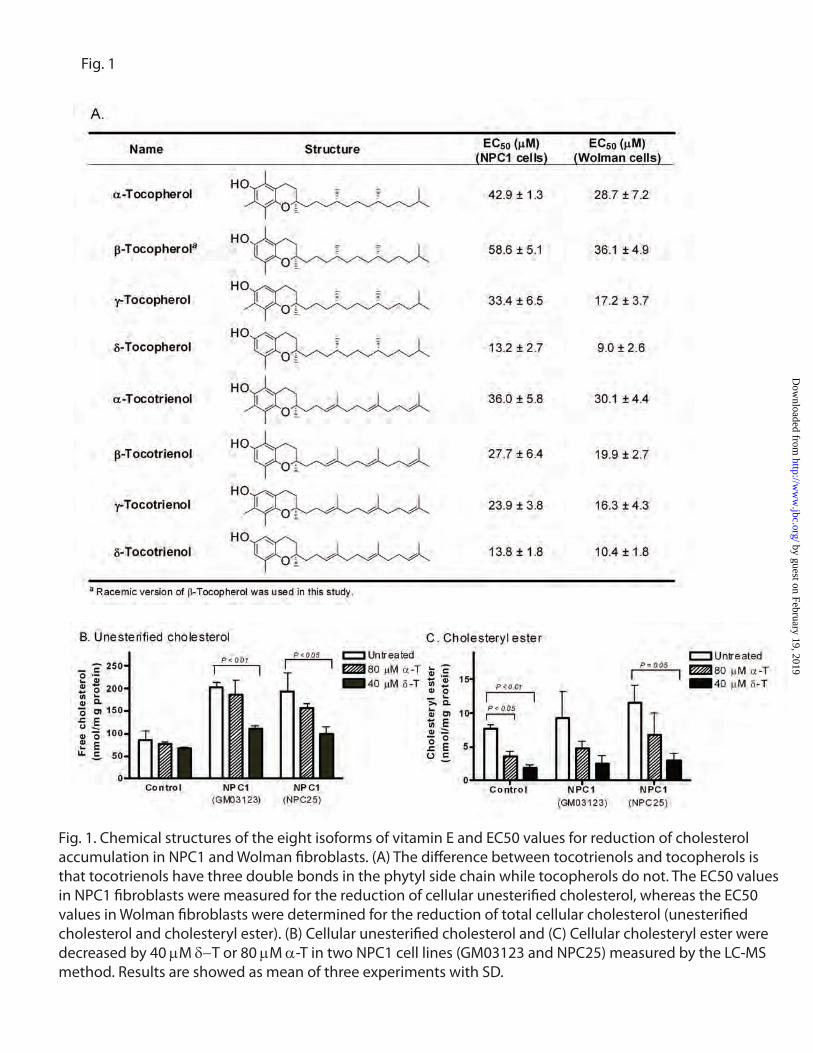
Fig. 1
Fig. 1. Chemical structures of the eight isoforms of vitamin E and EC50 values for reduction of cholesterol accumulation in NPC1 and Wolman �broblasts. (A) The di�erence between tocotrienols and tocopherols is that tocotrienols have three double bonds in the phytyl side chain while tocopherols do not. The EC50 values in NPC1 �broblasts were measured for the reduction of cellular unesteri�ed cholesterol, whereas the EC50 values in Wolman �broblasts were determined for the reduction of total cellular cholesterol (unesteri�ed cholesterol and cholesteryl ester). (B) Cellular unesteri�ed cholesterol and (C) Cellular cholesteryl ester were decreased by 40 µM δ−T or 80 µM α-T in two NPC1 cell lines (GM03123 and NPC25) measured by the LC-MS method. Results are showed as mean of three experiments with SD.
by guest on February 19, 2019http://w
ww
.jbc.org/D
ownloaded from

A. Filipin staining (red: filipin; green: CellMask)
C. Bodipy-LacCer staining
Control NPC1 NPC1 + 40µM δ-T
Control NPC1 NPC1 + 40µM δ-T NPC1 + 80µΜ α-T
B. Nile-red staining (cyan: nile-red; blue: Hoechst 33342) Control Wolman Wolman + 40µM δ-T Wolman + 80µM α-T
Fig. 2.
Fig. 2. δ-T and α-T reduced cholesterol accumulation phenotypes in NPC1 and Wolman �broblasts. (A) Filipin staining of unesteri�ed cholesterol was reduced in NPC1 cells and (B) Nile Red staining of neutral lipids (cholesteryl ester and triglyceride) was decreased in Wolman �broblasts by 40 µM δ-T or 80 µM α-T. The �lipin stained cells were co-stained with the CellMask dye that stained plasma mem-brane. (C) Mistra�cking of bodipy-LacCer in NPC1 �broblasts was corrected by 40 µM µ-T.
by guest on February 19, 2019http://w
ww
.jbc.org/D
ownloaded from

Control NPC1 NPC1 + 40µM δ-T NPC1 80µM + α-T
Wolman Wolman + 40µM δ-T Wolman + 80µM α-T
Control NPC1 NPC1 + 40µM δ-T NPC1 80µM + α-T
Wolman Wolman + 40µM δ-T Wolman + 80µM α-T
A. LysoTracker staining
B. Electron microscopic images
Fig. 3
FIGURE 3. δ-T and α-T reduced cellular acidic compartments and decreased pathological lysosomal inclusions in NPC1 and Wolman cells. (A) The LysoTracker dye staining (for acidic compartments) was reduced in both in both NPC1 and Wolman cells by 40 µM δ-T or 80 µM α-T. The LysoTracker-red staining images were merged with Hoechst nuclei dye staining. (B) Electron micrographs of thin sections of NPC1 and Wolman fibroblasts were compared with control fibroblasts. The inset in the untreated NPC1 fibroblast shows in detail the lamellated and osmophilic structures in lysosomes (bar = 1 µm) which were significantly reduced by the treatment with 40 µM δ-T. In the untreated Wolman fibroblasts, the white arrowheads mark the typical elongated and cleft-shaped lipid droplets observed in lysosomes and which were alleviated by the treatment with 40 µM δ-T.
by guest on February 19, 2019http://w
ww
.jbc.org/D
ownloaded from

NPC1 NPC1 + chlorp NPC1 + δ-T NPC1 + α-T
Wolman Wolman +chlorp Wolman + δ-T Wolman + α-T
NPC1 NPC1 + chlorp NPC1 + δ-T NPC1 + α-T
Wolman Wolman +chlorp Wolman + δ-T Wolman + α-T
FIGURE 4. δ-T increased cholesterol efflux and intracellular Ca2+ concentration. (A) 3H-cholesterol efflux carried out by addition of HDL in medium was enhanced by 40 µM δ-T and 80 µM α-T in the BHK cells, respectively. The efflux of 3H-cholesterol was greatly augmented by the expression of ABCA1 transporters in BHK cells, which was further enhanced by δ-T and α-T. (B) Intracellular Ca2+ increased in the presence of 75 µM δ-T or 250 µM δ-T in both NPC1 (see D for the calcium tracers) and Wolman (see E for the calcium tracers) fibroblasts. (C) δ-T reduced the impairment of lysosomal Ca2+ release in NPC1 cells. The NPC1 fibroblasts exhibited reduced lysosomal Ca2+ release stimulated by 200 nM Gly-Phe β-naphthylamide (GPN), which was partially corrected by a 24 hr pretreatment with 40 µM δ-T. Data Results showed are mean of three experiments; error bar represent SD. (F) Images of endocytic vesicles indicated by a fluorescent pHrodo™ dextran dye in NPC1 and Wolman fibroblasts. The treatment with δ-T or α-T did not alter the fluorescence intensity in the endocytic vesicles in these cells, indicating that both compounds do not affect the endocytosis in NPC1 and Wolman fibroblasts.
F. Endocytosis
by guest on February 19, 2019http://w
ww
.jbc.org/D
ownloaded from

E. Electron microscopic images
Fig. 5
D. Flotillin
Ionomycin
δ-tocopherol
HPBCD
Flotillin
GAPDH
Control
Ionomycin
δ-tocopherol
HPBCD
Flotillin
GAPDH
Control
NPC1 Wolman
FIGURE 5. Lysosomal exocytosis in δ-T and α-T treated NPC1 and Wolman disease fibroblasts. Lysosomal exocytosis was enhanced by δ-T and α-T in NPC1 (A) and Wolman (B) fibroblasts as well as in the control fibroblasts (C). After a 24 hr pretreatment with 40 µM δ-T or 80 µM α-T, the activity of HEXB (a lysosomal enzyme) in extracellular medium significantly increased in all three cell types. The data shown are the mean of three experiments and the error bars represent SD. (D) Secretion of exosomes in δ-tocopherol treated cells measured by Western blot analysis of flotillin 2 in the culture media of control fibroblasts. The upper panel exhibits the bands detected by an anti-flotillin 2 antibody in exosome fractions incubated with buffer, 2% HPBCD (a positive control), 160 µM δ-tocopherol or 1 µM Ionomycin. Equal loading was normalized by GAPDH levels in the cell lysate (lower panel). (E) Electron micrographs of untreated NPC1 (top left) and Wolman (top right) cells versus treated NPC1 (bottom left) and Wolman (bottom right) cells show that the lysosomal storage materials in 40 µM δ-T not only decreased but also moved closer to the plasma membrane in overall distribution in the cell (Bar = 5 µm). Insets show 5x magnified areas with vesicles close to the plasma membrane.
by guest on February 19, 2019http://w
ww
.jbc.org/D
ownloaded from

Fig. 6LysoTracker staining Electron microscopy
Fig. 6. E�ect of δ-T on ultrastructural pathology in lysosomal storage disorder �broblasts. Treatment with 40 µM δ-T reduced lysosomal size and corrected lysosomal inclusions in patient �broblasts of Batten (CLN2), Fabry, Farber, Niemann-Pick disease type A, San�lippo type B (MPS IIIB), and Tay-Sachs diseases. Images of LysoTracker staining in the absence of (A) or presence of (B) 40 µM δ-T treatment. LysoTracker-red staining was signi�cantly reduced after the treatment with δ-T, consistent with decrease of the acidic compartment size. Electron micro-graphs of patient �broblasts in the absence of (C) or presence of δ-T (D) (bar = 2 µm in main panels and = 500 nm in insets). Typical large osmiophilic, mono- and multinuclear multilamellar bodies are observed in Niemann-Pick Disease type A (NPA), Tay-Sachs, and Farber disease cells; similar but smaller and usually multi-nuclear multilamellar bodies are observed in Batten (CLN2), San�lippo type B (MPS IIIB), and Fabry disease cells (see insets for detail). The pathological phenotypes in ultrastructure were signi�cantly alleviated after the treatment with 40 µM δ-T in all these six types of patient �broblasts.
by guest on February 19, 2019http://w
ww
.jbc.org/D
ownloaded from

P. Austin and Wei ZhengSteven U. Walkley, Alan T. Remaley, Ulrich Baxa, Wei Sun, John C. McKew, Christopher
Ory, Juan J. Marugan, Jingbo Xiao, Noel Southall, William J. Pavan, Cristin Davidson, Miao Xu, Ke Liu, Manju Swaroop, Forbes D. Porter, Rohini Sidhu, Sally Firnkes, Daniel S.
Cholesterol Storage Disorders-Tocopherol Reduces Lipid Accumulation in Niemann-Pick Type C1 and Wolmanδ
published online October 3, 2012J. Biol. Chem.
10.1074/jbc.M112.357707Access the most updated version of this article at doi:
Alerts:
When a correction for this article is posted•
When this article is cited•
to choose from all of JBC's e-mail alertsClick here
Supplemental material:
http://www.jbc.org/content/suppl/2012/10/03/M112.357707.DC1
by guest on February 19, 2019http://w
ww
.jbc.org/D
ownloaded from

VOLUME 287 (2012) PAGES 39349 –39360DOI 10.1074/jbc.A112.357707
�-Tocopherol reduces lipid accumulation in Niemann-Picktype C1 and Wolman cholesterol storage disorders.Miao Xu, Ke Liu, Manju Swaroop, Forbes D. Porter, Rohini Sidhu, Sally Firnkes,Daniel S. Ory, Juan J. Marugan, Jingbo Xiao, Noel Southall, William J. Pavan,Cristin Davidson, Steven U. Walkley, Alan T. Remaley, Ulrich Baxa, Wei Sun,John C. McKew, Christopher P. Austin, and Wei Zheng
Dr. Firnkes name was misspelled. The correct spelling is shownabove.
THE JOURNAL OF BIOLOGICAL CHEMISTRY VOL. 288, NO. 1, p. 296, January 4, 2013© 2013 by The American Society for Biochemistry and Molecular Biology, Inc. Published in the U.S.A.
296 JOURNAL OF BIOLOGICAL CHEMISTRY VOLUME 288 • NUMBER 1 • JANUARY 4, 2013
ADDITIONS AND CORRECTIONS
Authors are urged to introduce these corrections into any reprints they distribute. Secondary (abstract) services are urged to carry notice ofthese corrections as prominently as they carried the original abstracts.


![GÖQ - tip.kocaeli.edu.trtip.kocaeli.edu.tr/.../NIEMANN-PICKTIPC.pdf · x Genetik inceleme Niemann Pick d] Z ofRfvf }R µo v fX. Niemann Pick , ofRf (NPH) Niemann Pick , ofRfV ol](https://img.pdfslide.net/doc/110x75/5c6c994f09d3f2fe088b4cea/goeq-tip-x-genetik-inceleme-niemann-pick-d-z-ofrfvf-r-o-v-fx-niemann.jpg)