Embed Size (px)
DESCRIPTION
jhg
Citation preview

7/18/2019 (2015) Lysosomal Physiology
http://slidepdf.com/reader/full/2015-lysosomal-physiology 1/27
Lysosomal Physiology Haoxing Xu1 and Dejian Ren2
1Department of Molecular, Cellular, and Developmental Biology, University of Michiga Ann Arbor, Michigan 48109; email: [email protected]
2Department of Biology, University of Pennsylvania, Philadelphia, Pennsylvania 19104;email: [email protected]
Annu. Rev. Physiol. 2015. 77:57–80
The Annual Review of Physiology is online at physiol.annualreviews.org
This article’s doi:10.1146/annurev-physiol-021014-071649
Copyright c 2015 by Annual Reviews. All rights reserved
Keywords
TRPML1, TPC1, TPC2, mTOR, TFEB, lysosomal exocytosis, lyso
storage disease
Abstract
Lysosomes are acidic compartments filled with more than 60 differen
of hydrolases. They mediate the degradation of extracellular particle
endocytosis and of intracellular components from autophagy. The di
products are transported out of the lysosome via specific catabolite exp
or via vesicular membrane trafficking. Lysosomes also contain more t
membrane proteins and are equipped with the machinery to sense nu
availability, which determines the distribution, number, size, and acti
lysosomestocontrolthespecificityofcargofluxandtiming(theinitiati
termination) of degradation. Defectsin degradation, export, or traffick
sult in lysosomal dysfunction and lysosomalstorage diseases (LSDs). Lmal channels and transporters mediate ion flux across perimeter mem
to regulate lysosomal ion homeostasis, membrane potential, catabol
port, membrane trafficking, and nutrient sensing. Dysregulation of l
mal channels underlies the pathogenesis of many LSDs and possibly
metabolic and common neurodegenerative diseases.
57
Click here for quick links to
Annual Reviews content online,
including:
• Other articles in this volume
• Top cited articles
• Top downloaded articles
• Our comprehensive search
FurtherANNUAL
REVIEWS

7/18/2019 (2015) Lysosomal Physiology
http://slidepdf.com/reader/full/2015-lysosomal-physiology 2/27
INTRODUCTION: LYSOSOMES AS THE CENTER FOR NUTRIENT SENSING AND RECYCLING
Lysosomes are the cell’s degradation center and are primarily responsible for the breakdow
of proteins, polysaccharides, and complex lipids into their respective building-block molecule
amino acids (AAs), monosaccharides, and free fatty acids (1, 2). Lysosomes are filled with mo
than 60 different types of hydrolases: lipases, proteases, and glycosidases for catabolic degradatio
(3). The products of degradation, lysosomal catabolites, are transported out of lysosomes v
specific exporters in the limited membrane (4) or via vesicular membrane trafficking for enerhomeostasis or reutilization in biosynthetic pathways (5).
Lysosome-mediated catabolic degradation is an adaptive process regulated by nutrient stat
and cellular signaling (6). Lysosomes receive extracellular or cell surface cargos via endocyt
sis and receive intracellular components via autophagy (1). Whereas increases in the endocyt
and autophagic fluxes stimulate lysosomal degradation (2, 3), accumulation of catabolites, e.g
AAs, in the lysosome terminates the degradation and autophagic flux (7, 8). To adapt to t
changing cellular environment, lysosomes contain nutrient-sensing machinery that consists
mechanistic/mammalian target of rapamycin (mTOR), the master regulator of growth, and
associated proteins (6, 9, 10). Nutrient starvation not only inhibits mTOR-mediated growth, b
also increases autophagosome (AP) formation (7). Furthermore, it activates TFEB, the lysos
mal biogenesis transcription factor, to facilitate lysosomal degradation by increasing lysosomfunction (acidification and delivery of hydrolases) and trafficking (AP-lysosome fusion) (6, 11).
Lysosomal adaptation to nutrient availability requires coordinated changes of multiple lysos
malparameters: distribution,number, andsize (7,11, 12). In fed cells, lysosomesareheterogeneo
in size (100–500 nm in diameter), morphology, and distribution (13). There are normally sever
hundred lysosomes in each mammalian cell. Upon nutrient starvation, lysosomal number is dr
matically reduced to <50 per cell, and the size of the lysosome is increased to 500–1,500 nm
a result of membrane fusion (7, 14). In addition, lysosomes are redistributed to the perinucle
region, where AP-lysosome fusion primarily takes place (12). After prolonged starvation, the com
pletion of autophagy triggers lysosomal reformation or biogenesis to restore lysosomal quanti
(7).
Lysosomal ion channels and transporters play essential roles in regulating lysosomal homeossis. First, lysosomal function requires the maintenance of the lumen homeostasis, especially ion
homeostasis and membrane potential (ψ , defined as V cytosol − V lumen; V lumen is set to 0 mV) (
For example, most lysosomal hydrolases require an acidic lumen to function (13). H+ pumping f
lysosomal acidification is also dependent on lysosomal ψ , which is estimated to be approximate
−20 to −40 mV (that is, 20 to 40 mV more negative in the cytosol than in the lumen) (15, 1
17, 18), as well as on the efflux of countercations and influx of counteranions (16, 17). Notab
due to the high density of various ion cotransporters and exchangers in the small-sized vesicl
(18), an increase in the permeability of one ion may alter the concentration gradients of oth
ions, hence indirectly affecting ion homeostasis. Second, many catabolite exporters are sensiti
to lysosomal ψ (19–21). Third, lysosomal trafficking is regulated by H+ homeostasis (22),
(23), or Ca2+
(24). Although H+
flux and ψ may also indirectly affect lysosomal Ca2+
relea(25), Ca2+ regulates most steps in lysosomal trafficking, including fusion of lysosomes with A
and late endosomes (LEs) (2, 26). LE-lysosome fusion mediates the delivery of most hydrolas
to the lysosome (5).
Defective degradation, catabolite export, or trafficking leads to lysosomal dysfunction an
lysosomal storage diseases (LSDs). Lysosomal channels directly or indirectly regulate all the
processes. Indeed, impaired function of lysosomal channels underlies multiple LSDs (22, 24, 27
58 Xu · Ren

7/18/2019 (2015) Lysosomal Physiology
http://slidepdf.com/reader/full/2015-lysosomal-physiology 3/27
LYSOSOMAL ION HOMEOSTASIS
Lysosomal degradation and the precise delivery of both hydrolases and cargos require the estab-
lishment of luminal ionic homeostasis, ionic gradients across the membrane, and ψ . In addition,
lysosomal ion homeostasis is also required for the timely removal of the digested products and for
the functionality of catabolite exporters. Below we discuss individual ions in the lumen. Given the
size of the lysosome, however, the luminal concentrations of one ion must be viewed in the context
of other ions and ion-dependent channels/transporters. In addition, lysosomal fusion events, espe-
cially the fusion of cytosol-derived, K +-loaded APs and Na+-loaded lysosomes (23), may rapidly change the ionic composition of the resulting organelle.
pH. The functions of most lysosomal hydrolases require an acidic (pH lumen ∼ 4.6) lumen;
V-ATPase establishes and maintains this pH (13). ClC transporters and Na+ /K + chan-
nels/transporters contribute to acidification by providing counterions (17, 22, 28). In addition
to assisting hydrolases, the established 100–1,000-fold H+ gradient is also important for lysoso-
mal trafficking, ψ generation, and content condensation during membrane fission (2, 18). For
example, the V-ATPase inhibitor Bafilomycin-A1 is commonly used to block AP-lysosome fusion
(11). Furthermore, the H+ gradient provides the driving force for many catabolite exporters (4).
Finally, V-ATPase is essential for the lysosome to sense luminal AA level (10).
Na+ /K +. The luminal Na+ and K + concentrations have been estimated in only a few cell types.
The values range from 20 to 140 mM for Na+ (higher than [Na+]cytosol of ∼12 mM) and from
2 to 50 mM for K + (lower than [K +]cytosol of ∼150 mM) (16, 23). The large variation in the
values presumably reflects ionic heterogeneity in lysosomes from different cells and variation in
the measurements. The mechanisms by which the ionic gradients between lysosomal lumen and
cytosol are established or maintained are largely unknown. Whereas endocytosis may provide
Na+ ions, endosomal Na+- or K +-coupled H+ transporters may regulate luminal Na+ /K +
concentrations (18). Lysosomal membranes are permeable to both Na+ and K + (see below).
Because of the higher [Na+]lumen than [Na+]cytosol, Na+ efflux from lysosomal lumen into cytosol
depolarizes organelle ψ toward the equilibrium (Nernst) potential of Na+ (ψ Na = +57 mV,
assuming a [Na+
]lumen of 100 mM and a [Na+
]cytosol of 12 mM). Likewise, K +
influx into thelysosome from cytosol hyperpolarizes ψ toward ψ K (−54 mV, assuming a [K +]lumen of 20 mM
and [K +]cytosol of 150 mM). The balance between the movements of Na + and K + across lysosomal
membranes may set resting lysosomal ψ (18, 29), which is determined by the membrane’s
permeability to each ion and the concentrations of the ions in the lumen and in the cytosol. ψ
is a critical determinant of the lysosomal acidification rate (17).
Ca2+. Ca2+ efflux from endosomes and lysosomes is thought to be important for signal transduc-
tion, organelle homeostasis, and organelle acidification (2, 18, 25, 30). [Ca2+]lumen is ∼0.5 mM,
approximately 5,000-fold higher than cytosolic [Ca2+] (∼100 nM) (31, 32). The Ca2+ gradient is
thought to be established by an unidentified Ca2+ /H+ exchanger or Ca2+ transporter (18). In cell-
free vesicle fusion assays, LE-lysosome fusion is inhibited by the Ca2+ chelator BAPTA, but not by
EGTA (2, 33). Although both BAPTA and EGTA have a high binding affinity for Ca2+, BAPTA
binds to Ca2+ approximately 100 times faster than EGTA does (25). Therefore, such distinct
BAPTA-versus-EGTA sensitivity suggests that the source of Ca2+ must be very close to the fusion
spot (25). In other words, lysosomesmost likely provide the Ca2+ required for lysosomalfusion (25,
33). Interestingly, preloading cells with a membrane-permeable ester form of BAPTA (BAPTA-
AM) blocks lysosomal exocytosis (34). Collectively, lysosomes may provide Ca2+ for membrane
fusion between lysosomes and other compartments, including LEs and the plasma membrane.
www.annualreviews.org • Physiology and Cell Biology of Lysosomal Ion Channels 59
7/18/2019 (2015) Lysosomal Physiology
http://slidepdf.com/reader/full/2015-lysosomal-physiology 4/27
The existence of multiple Ca2+ sensors may allow lysosomal Ca2+ release to regulate distin
steps of lysosomal trafficking. For lysosomal exocytosis, the C2 domain–containing synaptotagm
VII is likely the sensor (35). For LE-lysosome fusion, the candidate sensors include calmodul
(33) and ALG-2, a lysosome-targeted EF-hand protein (36). Ca2+-bound sensors may increa
their association with preassembled SNARE complexes to facilitate lipid bilayer mixing (37).
Fe2+ and Zn 2+. Lysosomal metals such as Fe3+, Fe2+, and Zn2+ ions are liberated via proteoly
of endocytosed or autophagocytosed metal-bound proteins (38). Lysosomes are the intracellulstores of Fe3+, Fe2+, Zn2+, and Cu2+ ions in micromolar concentrations (38, 39). Lysosom
export of Fe2+ and Zn2+ ions provides the cell with these trace metals as needed (38, 39). Where
lysosomal Fe2+ is required for the Fenton reaction and for ROS production, as well as for th
survival of lysosome-resident pathogens (38, 39), lysosomal Zn2+ may be required for the activ
of hydrolases such as acid sphingomyelinases (aSMases) (40). In addition, high lysosomal Zn
inhibits cathepsin activity (41). Whether heavy metal release regulates signal transduction
lysosomal trafficking remains unclear.
Cl−. [Cl−]lumen, estimated to be higher than 80 mM (22), may affect the function of cataboli
exporters,as shown for many plasma membrane transporters(42). [Cl−] influxregulates endosom
Ca2+
release (43) and is required for lysosomal acidification by providing counterions for Hpumping (17, 22), as recently reviewed in this journal (22, 28).
LYSOSOMAL FUNCTION
Lysosomal Trafficking
Lysosomal function and trafficking (fusion and fission) are interconnected. Lysosomal traffickin
supplies autophagic and endocytic substrates for degradation and hydrolases for lysosom
−−−−−−−−−−−−−−−−−−−−−−−−−−−−−−−−−−−−−−−−−−−−−−−−−−−−−−−−−−−−−−−−−−−−−−−→
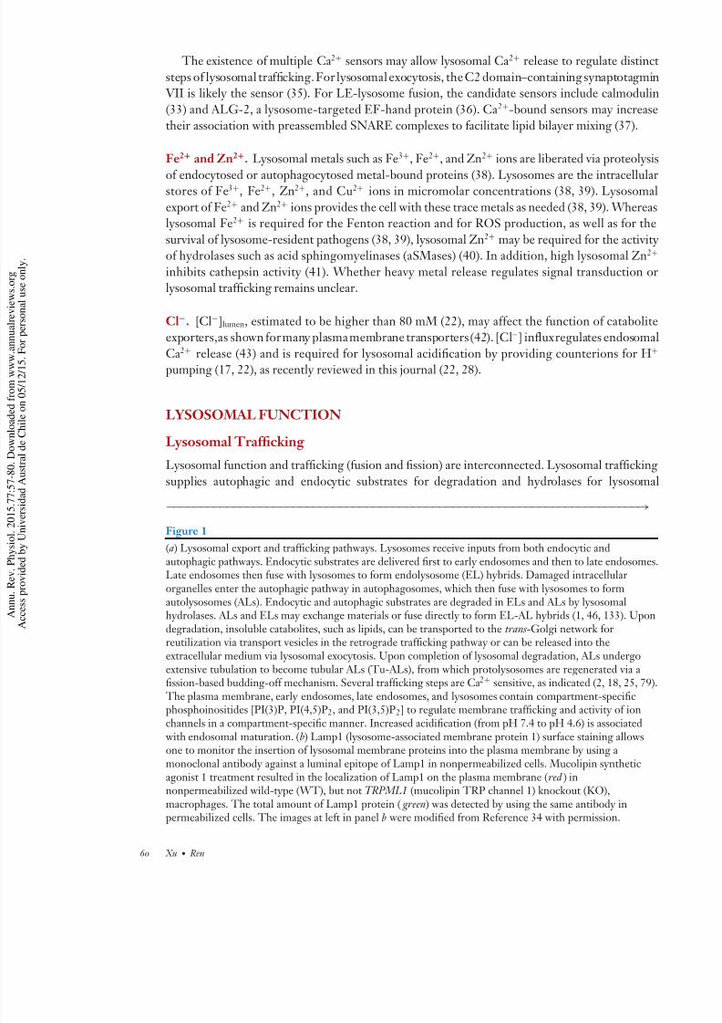
Figure 1(a) Lysosomal export and trafficking pathways. Lysosomes receive inputs from both endocytic andautophagic pathways. Endocytic substrates are delivered first to early endosomes and then to late endosomLate endosomes then fuse with lysosomes to form endolysosome (EL) hybrids. Damaged intracellularorganelles enter the autophagic pathway in autophagosomes, which then fuse with lysosomes to formautolysosomes (ALs). Endocytic and autophagic substrates are degraded in ELs and ALs by lysosomalhydrolases. ALs and ELs may exchange materials or fuse directly to form EL-AL hybrids (1, 46, 133). Upodegradation, insoluble catabolites, such as lipids, can be transported to the trans -Golgi network forreutilization via transport vesicles in the retrograde trafficking pathway or can be released into theextracellular medium via lysosomal exocytosis. Upon completion of lysosomal degradation, ALs undergoextensive tubulation to become tubular ALs (Tu-ALs), from which protolysosomes are regenerated via afission-based budding-off mechanism. Several trafficking steps are Ca2+ sensitive, as indicated (2, 18, 25, 7
The plasma membrane, early endosomes, late endosomes, and lysosomes contain compartment-specific
phosphoinositides [PI(3)P, PI(4,5)P2, and PI(3,5)P2] to regulate membrane trafficking and activity of ionchannels in a compartment-specific manner. Increased acidification (from pH 7.4 to pH 4.6) is associated with endosomal maturation. (b) Lamp1 (lysosome-associated membrane protein 1) surface staining allowsone to monitor the insertion of lysosomal membrane proteins into the plasma membrane by using amonoclonal antibody against a luminal epitope of Lamp1 in nonpermeabilized cells. Mucolipin syntheticagonist 1 treatment resulted in the localization of Lamp1 on the plasma membrane (red ) innonpermeabilized wild-type (WT), but not TRPML1 (mucolipin TRP channel 1) knockout (KO),macrophages. The total amount of Lamp1 protein ( green) was detected by using the same antibody inpermeabilized cells. The images at left in panel b were modified from Reference 34 with permission.
60 Xu · Ren
7/18/2019 (2015) Lysosomal Physiology
http://slidepdf.com/reader/full/2015-lysosomal-physiology 5/27
activation. In addition, lysosomal trafficking exports lipid catabolites and regulates the consump-
tion and biogenesis of lysosomes during lysosomal adaptation.
Input: endocytosis and autophagy. The biomaterials destined for degradation are delivered
to lysosomes through endocytic/phagocytic and autophagic pathways (Figure 1). Endocytosis or
phagocytosis of extracellular or cell surface cargo begins with the fission of the plasma membrane
to form endocytic vesicles, which then undergo a series of maturation processes to become early
endosomes and then LEs (reviewed in Reference 1) (Figure 1). In LEs, the destined-to-be-degraded endocytic cargos are sorted into intraluminal vesicles (1). Mature LEs, also termed
multivesicular bodies, fuse with lysosomes to become endolysosome (EL) hybrids to mediate the
bulk of the degradation (Figure 1). In addition, LE-lysosome fusion also delivers trans -Golgi
network (TGN)-derived hydrolases for lysosomal function (5).
pH = 7.2
pH = 4.6 L a m p 1 ( t o t a l )
L a m p 1 ( s u r f a c e ) WT TRPML1 KO
20 μm
Late endosomes(multivesicular bodies)
trans-Golginetwork
Autophagosome
Tu-AL
Transportvesicles Lysosome
EL/AL
Ca2+
Ca2+
Ca2+
Ca2+
Ca2+
Earlyendosomes
PI(3)P
PI(4,5)P2
PI(3,5)P2
a
b
Transportvesicle
www.annualreviews.org • Physiology and Cell Biology of Lysosomal Ion Channels 61

7/18/2019 (2015) Lysosomal Physiology
http://slidepdf.com/reader/full/2015-lysosomal-physiology 6/27
In a parallel pathway, worn-out intracellular organelles and protein aggregates are deliver
to lysosomes through autophagy (Figure 1). APs fuse directly, or indirectly through LEs, wi
lysosomes to form autolysosomes (ALs) (44, 45) in which the autophagic substrates are broke
down(Figure 1). Although lysosomes receivecargo from both autophagic andendocytic pathway
there are no clear boundaries between ALs and EL hybrids (Figure 1), as APs may merge direct
with LEs or even with early endosomes (46).
Output: reformation, retrograde trafficking, and exocytosis. There are at least three distinoutput pathways in lysosomal trafficking. The best-understood example is exocytosis of lysosom
contents, i.e., lysosomal exocytosis, which is present in all cell types (47, 48). Upon stimul
tion, kinesin-associated lysosomes translocate from the perinuclear region to the plasma mem
brane along microtubules (12, 14, 48, 49). After docking, vesicle-associated membrane protein
(VAMP7), which is locatedon thelysosomalsurface, forms a trans-SNAREcomplexwith syntaxi
4 and SNAP23 on the plasma membrane (47, 50). Upon localized Ca2+ increase, lysosomes fu
directly with the plasma membrane (48), which can be monitored by measuring Lamp1 surfa
staining (Figure 1), by measuring lysosomal enzyme release, or by using an electrophysiolog
based exocytosis assay (34, 47).
There are at least two membrane fission–based events in the lysosome. First, retrograde tra
ficking transports the free insoluble lipids out of late endosomes and lysosomes (LELs) to t TGN (Figure 1) for reutilization in biosynthetic pathways (5). Additionally, retrograde traffic
ing recycles the mannose-6-P receptor, which is required for TGN-to-LEL delivery of hydrolas
(5). Second, lysosomal reformation from the ALs (7) or EL hybrids (2) is essential for lysosom
biogenesis and homeostasis (Figure 1). Upon mTOR reactivation after prolonged starvation, A
undergo extensive tubulation (7). Lysosomes are then regenerated from these lysosomal tubul
via a poorly understood fission-based budding-off mechanism (7).
Catabolite Exporters
Insoluble lipid catabolites can be transported through vesicular trafficking. In contrast, solub
catabolites—for example, carbohydrates and AAs—are exported to the cytosol by specific tranporters on the perimeter membrane (4, 51) (see Figure 2).
Amino acid exporters. Only a few lysosomal AA transporters have been identified. Cys
nosin/CTNS is a lysosomal, H+-driven cysteine transporter (52). Mutations in CTNS cau
abnormal accumulation of the AA cysteine (cystinosis) and secondary lysosomal storage (52
PQLC2/LAAT-1 is a lysosomal, H+-dependent arginine/lysine exporter (21). Caenorhabditis
egans lacking LAAT-1 exhibit accumulation of arginine and lysine in enlarged lysosomes (5
Proton-assisted AA transporter 1 (PAT1)/SLC36A1, an intestinal AA transporter, is also localiz
in the lysosome and regulates AA export and sensing (54).
Sugar exporters. Sialin/SLC17A5, which is encoded by a gene that is mutated in sialic acid sto
age disorders, is a lysosomal, H+-dependent exporter of sialic acids and acidic monosaccharid
(4). Drosophila Spinster proteins are presumed to transport sugars that are produced upon lysos
mal degradation of glycolipids, glycoproteins, or autophagocytosed glycogens (8). Cells lackin
Spinster exhibit lysosomal dysfunction and storage (8).
Heavy metal exporters. Heavy metal ions are released into the lumen upon lysosomal degrad
tion of autophagocytosed, heavy metal–containing proteins, such as cytochrome c in mitochond
62 Xu · Ren

7/18/2019 (2015) Lysosomal Physiology
http://slidepdf.com/reader/full/2015-lysosomal-physiology 7/27
b
Nucleus
Patch clamp mCherry-TRPML1
EGFP-Lamp1
5 µm
a
pH 4.6
+ + + +
TPCs
TRPMLs
Exporters:AA, sugar, lipids
V-ATPase
Whole lysosome I (pA)
ψ = –40 to –20 mV
Ca2+: 0.5 mM
–100 +100
H+
Ca2+ transp
Na+
– –
– –
ψ = 0 mV
H+, K + I X
Cl–
Ca2+, Fe2+, Zn2+
(= V cytosol – V lumen)(mV)
TPC
TRPML
I K
I Cl
Δψ
Lysosome
hole lysosome (pA)
100 10
(= V ytosol – V lumen)(mV)
TPC
TRPML
I K
Cl
Δ
Figure 2
Ion channels and transporters in the lysosome. (a) An illustration of a whole-lysosome patch-clamp recording (23, 55). TRPMLdenotes mucolipin TRP channel 1. The right panel was modified from Reference 55 with permission. (b) ( Right ) The lysosome i
acidic compartment with relatively high [Ca2+]. These gradients are established by V-ATPases and putative Ca 2+ transporters. lysosomal membrane potential (ψ) is cytosolic-side negative, with ψ ranging from −40 to −20 mV. The products of lysosomdegradation, such as amino acids (AAs), sugars, and lipids, are transported out of the lysosome via specific exporters. Among theconductances that have been identified, two-pore channels (TPCs) encode I Na, and TRPMLs encode I Ca and I Fe; ClC-7 is presuencode I Cl. Several other conductances, such as I H and I K , have been electrophysiologically but not molecularly characterized. (
I -ψ curves of four lysosomal channels or currents (TPCs, TRPMLs, I Cl, and I K ) are shown.
(38). During mitophagy or degradation of the iron storage protein ferritin, Fe3+ is dissociated and
subsequently reduced to Fe2+ (38, 39). DMT1 and NRAPM1 are lysosomal Fe2+ exporters in
specialized cell types (39). The ubiquitously expressed mucolipin TRP channel 1 (TRPML1), the
principal Ca2+ channel in the lysosome, also conducts Fe2+ as well as Zn2+ and other heavy metal
ions (55). Loss of TRPML1 results in lysosomal Fe2+
(38, 39, 55) and Zn2+
(56, 57) overload.
Lipid exporters. Unlike other complex lipids in intraluminal vesicles, cholesterol is not degraded
by lysosomal hydrolases but is sorted and presumably exported via the cholesterol transporter
NPC1 (58). Mutations in NPC1 lead to lysosomal accumulation of cholesterol and sphingolipids,
resulting in an LSD termed Niemann-Pick type C (NPC) (31, 58).
www.annualreviews.org • Physiology and Cell Biology of Lysosomal Ion Channels 63

7/18/2019 (2015) Lysosomal Physiology
http://slidepdf.com/reader/full/2015-lysosomal-physiology 8/27
Lysosomal Storage Diseases: Defects in Degradation, Export, or Trafficking
Abnormal accumulation of lysosomal materials causes more than 50 rare, inherited metabo
disorders in humans, collectively termed LSDs (59). Most LSDs are caused by mutations
lysosomal hydrolases (60). However, defective catabolite export and membrane trafficking m
also lead to traffic jams and to secondary storage in the lysosome (61). Furthermore, prima
accumulation of undigested, insoluble lipids mayalso slow down membrane trafficking and sortin
thereby affecting the delivery of lysosomal hydrolases (2, 61). Hence, problems in degradatio
export, or trafficking negatively affect each other to cause lysosomal dysfunction and storag Mutations in lysosomal channels and transporters lead to defective ion homeostasis, which in tu
impairs lysosomal trafficking and degradation to result in LSDs (22, 28, 31).
Progressive accumulation of undigested materials leads to build-up of enlarged ( >1,000-nm
dysfunctional lysosomes (62). Increases in both vesicular content and osmolarity may cau
vesicle enlargement. First, an increase in trafficking input or a decrease in trafficking output m
also result in vesicle enlargement. Second, increased lysosomal osmolarity causes water influx
mechanically expand the vesicle (14). In normal physiology, a change in nutrient or cellular stat
results in a transient increase followed by a decrease in lysosomal size due to membrane fusio
and subsequent fission events (7). Hence, large (>500-nm), Lamp1-positive compartments, i.
ELs and ALs, are transient organelles or secondary lysosomes that, upon completion of lysosom
degradation, return to normal small-sized primary lysosomes via lysosomal reformation (2, 7However, in LSDs, when lysosomal reformation or AP-lysosome fusion is defective, EL/AL li
span increases. Subsequently, secondary lysosomes, i.e., ELs/ALs, are filled with incomplete
digested materials, which further enlarges ELs/ALs to >1,000 nm. This enlargement in tu
causes further impairment in the equilibrium between input and output. Hence, the enlarge
dysfunctional lysosomes seen in LSDs are prolonged ELs/ALs. In essence, LSDs are caused
an escalating disequilibrium that results in endocytic and autophagic block or arrest (44).
With the presence of many degradation-defective, enlarged ELs/ALs, the total number
lysosomes (estimated by the number of Lamp1-positive vesicles) may not be reduced in LSD
However, the overall lysosomal function within a cell is compromised, leading to a deficiency
building-block precursors for biosynthetic pathways and to cellular starvation (60). In an attem
to compensate for the reduced degradative capacity, most cells in LSD maladaptively increabasal autophagy and the expression levels of housekeeping lysosomal proteins such as Lamp1 (4
63). These compensatory changes may enable LSD cells to survive under normal conditions.
Lysosomes as Energy and Nutrient Sensors
In addition to the lysosome’s long-recognized function as a recycling center for nutrient gener
tion, lysosomal membranes were also recently found to directly monitor intracellular energy an
extracellular nutrient status (29, 64). Lysosomes fulfill two central functions in energy and nutr
ent sensing. First, these organelles provide a physical platform for several of the most importa
nutrient-sensitive signaling molecules, such as mTOR and TFEB (6, 65). In response to chang
in nutrient status, these proteins move onto the lysosomal surface, where they can be modifie
Intriguingly, most of their known downstream targets are not lysosome associated. For examp
the targets of TFEB are in nuclei, and those of mTOR are largely cytosolic. That the surface
the lysosome is the focal point for nutrient sensors may be related to its relative mobility with
cells. In addition, there may be advantages of stationing sensors around vacuoles that produce an
export recycled nutrients. For example, mTOR senses AA contents both outside and within th
organelle (10, 66).
64 Xu · Ren

7/18/2019 (2015) Lysosomal Physiology
http://slidepdf.com/reader/full/2015-lysosomal-physiology 9/27
Second, in a manner similar to that of plasma membranes that sense ATPwith an ATP-sensitive
K + channel (K ATP), lysosomal membranes also sense [ATP] with ATP-sensitive Na+ channels (29,
64). In addition, the Na+ channels’ ATP sensitivity is highly sensitive to extracellular nutrients,
linking energy and nutrient status to lysosomal ψ .
LYSOSOMAL ION CHANNELS AND CONDUCTANCES
Ion channels and transporters in the lysosome provide the ionic environment necessary for degra-dation, pathways for export, and signals for lysosomal trafficking. Mutations of the few known
lysosomal channel or transporter genes cause LSDs in humans or LSD-like phenotypes in mice
(22, 67). The lysosome requires H+ and Fe2+ channels/transporters to establish the ionic envi-
ronment for proper oxidative and lytic function. As H+ and Ca2+ ions are 1,000–5,000 times more
concentrated in the lysosomal lumen than in the cytosol, the pathways for H+ and Ca2+ flux, i.e.,
Ca2+- and H+-permeant channels, must be tightly regulated.
Lysosomal ionic conductances have been studied by using ionic fluxes; single-channel record-
ings with purified proteins reconstituted in lipid bilayers; and, more recently, whole-lysosomal
recordings from artificially enlarged lysosomes (18, 23, 55, 68). Whole-lysosome recordings
from enlarged vacuoles isolated from macrophages, neurons, fibroblasts, kidney cells, and cardiac
myocytes have revealed native K
+
, Na
+
, Ca
2+
, Cl
−
, and H
+
conductances (23, 29, 64, 68)(Figure 2 a).
K + conductances. In most vacuolin-enlarged lysosomes, a voltage-independent, K + leak–like
conductance has been recorded (64). The identity of this K + conductance is currently unknown.
The K + channel likely contributes to ψ because alterations in [K +]cytosol affect ψ (C. Cang &
D. Ren, unpublished data).
H+ conductances. Consistent with the observation that V-ATPase inhibition quickly leads to
lysosomal alkalization (32), there must be a proton leak conductance; H+-dependent catabolite
exporters are the leading candidates (8), but the electrogenic V-ATPase H+ pump may also con-
tribute (69). In addition, there is also a depolarization-activated H
+
conductance (64). However,this conductance is insensitive to Zn2+ and activates rapidly (C. Cang & D. Ren, unpublished
data), suggesting that it is not mediated by the voltage-activated H+ channel H V 1 (70, 71).
Cl− conductances. A depolarization-activated, fast-activating, outwardly rectifying Cl− con-
ductance can be recorded from lysosomes (64). The ClC-7 H+ /Cl− exchanger is localized to
lysosomes and is believed to permeate Cl− via a channel mechanism (72). ClC-7, coexpressed on
the plasma membrane with Ostm1 in HEK293 cells, generates an outwardly rectifying Cl − con-
ductance that activates much more slowly than the native, lysosomal membrane–associated Cl−
conductance (17, 64, 73). Further recordings from ClC-7/Ostm1-expressing lysosomes and from
ClC-7/Ostm1 knockout lysosomes are required to determine whether ClC-7 indeed encodes the
native Cl− conductance.
Na+ conductances. Like plasma membranes of excitable cells (74), the lysosome’s relative
permeability to Na+ and K + ( P Na / P K ) changes markedly (by ∼30-fold) depending on recording
conditions (64). Both PI(3,5)P2-dependent I Na (23) and PI(3,5)P2-insensitive I Na can be measured
(X. Zhang & H. Xu, unpublished data). In the presence of PI(3,5)P2, two major Na+ conductances
have been recorded (23, 29, 64); in most mammalian lysosomes, there is a voltage-independent
Na+ conductance, but in a subset of kidney and cardiac lysosomes, there is also a voltage-activated
www.annualreviews.org • Physiology and Cell Biology of Lysosomal Ion Channels 65

7/18/2019 (2015) Lysosomal Physiology
http://slidepdf.com/reader/full/2015-lysosomal-physiology 10/27
Na+ conductance (lysoNa V ). TPC2 forms the voltage-independent Na+ conductance, where
TPC1 is responsible for lysoNa V (see below) (64).
Ca2+ conductances. Ca2+-permeant, nonselective cation channels have been recorded in t
lysosomes of most mammalian cell types, including fibroblasts, macrophages, pancreatic cells, a
skeletal muscle cells (23, 24, 34, 68). TRPMLs encode the primary lysosomal channel conductin
Ca2+ across lysosomal membranes (see below).
Fe2+ and Zn 2+ conductances. Whereas overexpression of TRPMLs increases whole-lysosom
iron and zinc conductances (55), TRPML1-mutant organelles exhibited lysosomal Fe2+ /Fe3+ a
Zn2+ accumulation, suggesting that TRPMLs are channels that can conduct these metal ions fro
the lysosome lumen to cytosol (39, 75).
Other conductances. Endogenous conductances activated by sphingosine-1-phosphate a
NAADP have been proposed but have not been directly measured under voltage clamp (18
TPC overexpression in lysosomes increases NAADP-sensitive lysosomal Ca2+ release. NAAD
activated cation channels have been recorded from TPC2-overexpressing lysosomes in sever
reports (76–78).
In summary, major ionic conductances have now been recorded from whole lysosom(Figure 2). Because I Na, I Ca, I Fe, and I Cl have been functionally characterized and molecular
identified, their physiological functions have been studied by using both in vitro and in vi
assays. Significantly, molecular and genetic studies have provided definitive evidence for the exi
tence of these conductances, confirming their previously proposed cell biological functions. Th
ClC family of H+ /Cl− exchangers likely responsible for lysosomal Cl− conductances was recen
reviewed (17, 22). Below, we focus on the TRPML and TPC families.
TRPML CHANNELS
As the principal Ca2+ release channel in the lysosome, TRPML1 is a key regulator of mo
lysosomal trafficking processes (26, 79). Whereas human mutations of TRPML1
cause type I
mucolipidosis (ML-IV) (80, 81) and inhibiting TRPML1 leads to several other LSDs (24), TFE
overexpression (82) can induce cellular clearance (of lysosomal storage materials) in most LSD
except ML-IV (49), highlighting TRPML1’s unique role in basic cell biology.
An LEL-Localized TRP Channel
The mucolipin subfamily of TRP channels (TRPML1–3), like other TRPs, consists of six putati
transmembrane (TM)-spanning domains (S1–S6), with the N and C termini facing the cytos
(79). The channel pore of TRPML1 is formed by the pore-loop region between the S5 and S
domains, which are presumed to form the channel gate (79). Whereas TRPML1 is ubiquitous
expressed in every tissue and cell type, TRPML2 and TRPML3 are expressed only in special c
types (79, 83).
TRPML1–3 channels are predominantly (>75%) localized on LEL membranes, but he
erologously expressed TRPML1 proteins are also detected in the early endosomes and plasm
membranes (79, 83). Two dileucine motifs located separately in the N- and C-cytosolic tails a
responsible for the LEL localization through a direct, AP1/3-dependent, TGN-LEL traffic
ing pathway and through an indirect, AP2-dependent, TGN–plasma membrane–LEL pathw
(84–86). Mutations of both dileucine motifs result in significant TRPML1 surface expression (8
66 Xu · Ren
7/18/2019 (2015) Lysosomal Physiology
http://slidepdf.com/reader/full/2015-lysosomal-physiology 11/27
A Ca2+- and Fe2+ /Zn 2+-Permeable Channel
Due to the LEL localization of TRPML1, measuring the permeation and gating properties of
the channel is difficult. However, the recent development of the whole-lysosome patch-clamp
technique (43, 55) has enabled the direct study of TRPML1 on artificially enlarged lysosomes
(Figure 2 a). These lysosomes are induced by vacuolin-1, a small-molecule chemical compound
that selectively enlarges lysosomes (23, 88). TRPML1 is an inwardly rectifying channel (where
inward indicates cations moving out of the lumen) permeable to Ca2+, Fe2+, Zn2+, Na+, and K +
but impermeable to protons (55). Hence, upon activation, TRPML1 may mediate the release of Ca2+, Fe2+, and Zn2+ ions.
PI(3,5)P2 Activation
Phosphoinositides are important regulators of membrane trafficking (89). Among them, PI(3)P
and PI(3,5)P2 are localized on endosomes and lysosomes (90). Lysosomal trafficking is regulated
by PI(3,5)P2, whose synthesis requires the endosome-localized kinase PIKfyve in association with
the FIG4 lipid phosphatase and scaffolding Vac14 proteins (90). PI(3,5)P2 not only recruits a
variety of cytoplasmic effector proteins to facilitate lysosomal trafficking (90), but also regulates
the activity of lysosomal channels and transporters (90); such effects are similar to the effects of
PI(4,5)P2 on plasma membrane channels (91). Indeed, PI(3,5)P2 activates whole-lysosome I TRPML1in a physiologically relevant low-nanomolar range (68). TRPML1 contains a cluster of positively
charged AA residues that bind directly to PI(3,5)P2 in in vitro protein/lipid-binding assays (68).
Charge removal mutations abolished PI(3,5)P2 activation and, importantly, eliminated the effect
of TRPML1 on lysosomal trafficking (68).
A genetically encoded PI(3,5)P2 indicator based on the PI(3,5)P2-binding domain of TRPML1
was used to reveal that PI(3,5)P2 levels increase transiently prior to fusion of two Lamp1-positive
vesicles (92) and during phagocytic uptake of large particles (34). Therefore, under certain phys-
iological conditions, PI(3,5)P2 may play an instructive role in regulating lysosomal trafficking
through TRPML1 activation.
How other trafficking cues regulate TRPML1 is not yet known. However, several synthetic
small-molecule compounds were recently identified as PI(3,5)P2-independent TRPML agonists(24, 93). Of them, mucolipin synthetic agonist 1 (ML-SA1) robustly activates TRPML1 at low-
micromolar concentrations with a response comparable to that of PI(3,5)P 2 (24).
PI(4,5)P2 and Sphingomyelin Inhibition
Two plasma membrane–localized lipids, PI(4,5)P2 and sphingomyelin (SM), inhibit I TRPML1 (24,
87). Conversely, I TRPML1 is potentiated by aSMases and phospholipase C (24, 87). The inhibition
may be a mechanism to prevent lysosomal TRPML1 from being active in nonnative compartments
such as the plasma membrane. In addition, SMs accumulate in the lysosomes of aSMase-deficient
NPA and NPB cells and cholesterol-accumulated NPC cells (60). Hence, TRPML1 inhibition
may be an underlying pathogenic cause for certain LSDs.
Ca2+-Dependent Lysosomal Trafficking: Fusion-Based Input
The permeation and gating properties of TRPML1 suggest that the channel function of TRPML1
is to release Ca2+ from the LEL lumen in response to various cellular cues (79, 94), such as an
increase in lysosomal PI(3,5)P2. TRPML1 may regulate the LE maturation process involving
LE-lysosome fusion, as the lysosomal delivery of endocytosed proteins, such as plasma membrane
growth factor receptors, is delayed in TRPML1−/− cells (95, 96). Likewise, in TRPML1−/− mouse
www.annualreviews.org • Physiology and Cell Biology of Lysosomal Ion Channels 67

7/18/2019 (2015) Lysosomal Physiology
http://slidepdf.com/reader/full/2015-lysosomal-physiology 12/27
neurons, LC3-positive autophagosomal puncta were elevated (96, 97), suggestive of increas
AP formation and delayed AP-lysosome fusion (96, 97). Consistently, in trpml −/− fly neuron
AP-lysosome fusion was impaired (98). As increased AP formation is commonly observed in mo
LSDs (44), this process may also be a maladaptation to lysosomal dysfunction and basal TFE
activation in LSDs (6).
The fusion defects observed in TRPML1−/− cells, together with the Ca2+ dependence of L
lysosome fusion observed in in vitro vesicle-mixing assays (2), suggest that TRPML1 may media
lysosomal Ca2+
release to promote fusion. However, as most cellular assays do notdirectly measu vesicle fusion, these cellular defects may also be caused indirectly by chronic storage of lysosom
materials or defects in another cellular process such as membrane fission. To maintain lysosom
homeostasis, lysosomaltrafficking input andoutputmust be coordinated. An increase in input mu
accompany a corresponding increase in output. Conversely, defects in output may also slow dow
the input process. Hence, the observed fusion defects are likely caused by the fission defects, an
vice versa. To distinguish these possibilities, it may be necessary to perform super-resolution li
imaging to monitor fusion and fission events while acutely activating and inhibiting TRPML1
channel function.
Ca2+-Dependent Lysosomal Trafficking: Fission-Based Output
LEL-to-TGN retrograde trafficking is defective in ML-IV cells, as fluorescence-conjugated latosylceramide, a lipid that is normally localized in the TGN at steady state, accumulates in LEL
(99). This trafficking defect is also observed in other LSDs, including NPC (61), but can be re
cued by increasing TRPML1 expression and activity (24), suggesting a direct role of TRPML
in this specific trafficking step. On the basis of the observation that enlarged ELs accumulate
TRPML−/− C. elegans cells, it was proposed that lysosomal reformation (biogenesis) is defecti
in these cells (100). However, as other mechanisms may also account for lysosomal enlargeme
(see above), it is necessary to test whether acute activation and inhibition trigger and termina
lysosomal reformation, respectively.
Lysosomal Exocytosis
Multiple pieces of evidence suggest that TRPML1 regulates lysosomal exocytosis. First, HEK2
cells transfected with TRPML1V432P (a gain-of-function mutation) exhibit enhanced lysosom
exocytosis (101). In contrast, lysosomal exocytosis induced by TFEB overexpression requir
TRPML1 (49), and ML-IV fibroblasts exhibit impaired ionomycin-induced lysosomal exocyto
(49, 102). In primary macrophages, acute ML-SA1 treatment induced Lamp1 surface staining (s
Figure 1b) and lysosomal enzyme release (34). In contrast, ML-SA1-induced lysosomal exocyto
was dramatically attenuated by TRPML1 knockout or BAPTA-AM (34).
TRPML1-mediated lysosomal exocytosis is required for the phagocytic uptake of large pa
ticles in macrophages (34). Macrophages engulf large cellular particles, such as apoptotic cel
by forming pseudopods (103). Pseudopod formation consumes large amounts of membrane (3
derived from lysosomal membranes (35). TRPML1−/− macrophages exhibit uptake defects sim
lar to those of synaptotagmin VII– or VAMP7-deficient macrophages (34), suggesting a role
TRPML1-mediated lysosomal exocytosis in particle uptake. Consistently, a lysosome-targete
genetically encoded Ca2+ sensor, GCaMP3-TRPML1 (24), detects TRPML1-mediated Ca
release specifically at the site of particle uptake (34).
The repair of plasma membrane damage in skeletal muscle and other cell types requires
rapid Ca2+ increase to trigger the recruitment of intracellular vesicles that fuse with the plasm
68 Xu · Ren

7/18/2019 (2015) Lysosomal Physiology
http://slidepdf.com/reader/full/2015-lysosomal-physiology 13/27
membrane and replace the disrupted membranes (48, 104). Both TRPML1 and lysosomal Ca2+
release are required for this process. TRPML1 knockout mice exhibit muscle repair defects and
develop muscular dystrophy (105).
Metal Export
TRPML1−/− cells exhibit a cytosolic Fe2+ deficiency and concurrent lysosomal Fe2+ and Zn2+
overload, suggesting that TRPML1 is a lysosomal Fe2+
and Zn2+
exporter (55–57). ML-IV cellscontain a large amount of lipofuscin in the lysosome, which can be explained by increased oxidative
stress due to lysosomal Fe2+ overload (38, 39).
Lysosomal Diseases
More than 20 loss-of-function mutations in TRPML1 cause ML-IV, an LSD manifested by men-
tal retardation, retinal degeneration, and constitutive achlorhydria (27, 80, 81). As in ML-IV,
TRPML1 knockout mice display neurological, gastric, and ophthalmological abnormalities, grad-
ually developing hind-limb paralysis; mice typically die at 8–9 months (67). At the cellular level,
dense membranous storage bodies are observed in most TRPML1−/− cells (67).
TRPML1’s importance may also be extended to other LSDs, including NPA and NPC (24), in which TRPML1-mediated lysosomal Ca2+ release and lysosomal trafficking are partially blocked
(24). Likewise, in PI(3,5)P2-deficient cells, TRPML1 activity is also reduced, which may cause
lysosomal trafficking defects and storage (88). Furthermore, lysosomal trafficking is defective in
many common neurodegenerative diseases, such as Alzheimer’s, Parkinson’s, and Huntington’s
diseases, suggesting a potential involvement of TRPML1. Collectively, TRPML1 channel dys-
regulation may be a primary pathogenic mechanism that results in secondary lysosomal storage
in many lysosomal diseases.
TWO-PORE CHANNELS
TPC proteins form a unique branch of ion channels with a 2 × 6TM structure, which is thetransition state between the 6TM channels [such as voltage-gated K + channels (K V s), TRPs,
and sperm-specific Ca2+ channels (CatSpers)] and the 4 × 6TM channels [voltage-gated Ca2+
channels (Ca V s), voltage-gated Na+ channels (Na V s), and nonselective Na+ leak channel] (106).
The overall sequences of TPCs are similar to those of Ca V s and Na V s (107, 108). There are three
animal TPCs (TPC1–3). TPC3 is found only in some animals, such as cats, dogs, and chickens, but
not in humans or mice (107, 109). TPC1 and TPC2 are widely expressed, with the highest TPC1
expression found in the heart and kidney. Heterologously expressed TPC proteins are localized
in lysosomes (TPC1 and TPC2) and endosomes (TPC1) (110).
TPC Selectivity Both TPC1 and TPC2 are highly selective for Na+. The apparent P Na / P K estimated with whole-
lysosomal recordings is ∼80 for TPC1 (64) and ∼30 for TPC2 (23). Whole-lysosomal TPC Ca2+
current is minimal or undetectable (23). Given the apparently low Ca2+ permeability, whether the
fractional Ca2+ efflux directly from TPCs contributes to increases in cytosolic [Ca 2+] remains to
be determined.
www.annualreviews.org • Physiology and Cell Biology of Lysosomal Ion Channels 69

7/18/2019 (2015) Lysosomal Physiology
http://slidepdf.com/reader/full/2015-lysosomal-physiology 14/27
ATP ADP
[ATP]
I
pH
2 31
H+
pH 4.6
ATP ADPH+
ATP ADPH+
I Na I Na
Na+
pH 4.7 pH 5.5
Na+
Na+
Δψ
ψ
ψ Na
I TPC2 I TPC1 I TPC2
I TPC1
Figure 3
A model for nutrient sensing by two-pore channels (TPCs). (Stage 1) In nutrient-replete cells, TPCs areminimally open at resting lysosomal membrane potential ( ψ). (Stage 2) Upon a drop in [ATP], TPC2opens to promote Na+ efflux (into the cytosol) and to depolarize the membrane (the lumen becomes lesspositive). In TPC1-expressing organelles, a ψ above TPC1’s activation threshold also triggers TPC1opening. (Stage 3) Following lysosomal alkalinization, TPC1 lowers its activation threshold. Membranedepolarization and Na+ efflux potentiate H+ pumping by the V-ATPase and help stabilize pH. Strongpositive feedback between ψ and TPC1 can also lead to plateau potentials and membrane bistability. ψN
denotes Na+ Nernst potential. See References 29 and 64 for details.
TPC Regulation
TPCs are the targets of a converging regulatory network with many physiological inputs fro
inside and outside the organelles. These inputs include luminal H+, lysosomal ψ , membra
PI(3,5)P2, lysosome-attached protein kinases, Mg2+ ions, cytosolic ATP, and extracellular nutr
ents such as AAs (see Figure 3).
Membrane voltage. When recorded with symmetric ion concentrations in the bath and pipet
TPC2 has a largely linear I -ψ relationship, and the currents rise and fall instantaneously upo
voltage changes, without apparent activation and deactivation (23, 29, 64). These properties
TPC2 are similar to those of the native Na+ channel recorded from lysosomes. TPC2-mediat
lysosomal Na+ conductance is controlled by the availability of organelle PI(3,5)P2 (23), cytoso
ATP, and extracellular nutrients (111), which together presumably regulate lysosomal ψ .
TPC1 is voltage activated (64). At pH lumen 4.6, the channel opens at ψ > 0 mV. Compar
with the voltage dependences of the strongly voltage-dependent K V s, TPC1’s voltage dependen
of activation is much weaker (with a slope factor of ∼20–50 mV versus a few millivolts in K V(64). Like K V s, TPC1 has S1–S4 voltage-sensing domains (VSDs) whose S4s contain charg
residues (K/R) in an every-third-residue fashion. Mutating several of the charged residues th
are on TPC1 but not on TPC2 rendered TPC1 largely voltage independent, suggesting that th
organelles also use the S1–S4 VSDs to sense voltage changes.
70 Xu · Ren

7/18/2019 (2015) Lysosomal Physiology
http://slidepdf.com/reader/full/2015-lysosomal-physiology 15/27
Unlike any other voltage-gated Na+ channel currently known, TPC1 activates slowly (τ ∼
100 ms) (64). At a holding potential of −70 mV, the channel did not inactivate at any of the
depolarizing voltages (64). Upon PI(3,5)P2 activation, I TPC1 at negative voltages exhibited voltage-
dependent fast decay presumed to be due to channel inactivation (23). Because TPC1-encoded
Na V makes the organelle excitable (see below), the lack of inactivation and the slow deactivation
presumably help sustain depolarization. The slow activation may act similarly to a low-pass filter
in preventing noise-induced depolarization, which is especially useful for organelles with high
input resistance.
Luminal pH. TPC1 is highly sensitive to pHlumen. A one-unit pH increase from 4.6 to 5.6 shifts
TPC1’s conductance (G)-ψ relationship toward hyperpolarizationby 62 mV (64).For comparison,
a pure-H+ electrode has a voltage response of 61.5 mV/pH unit at 37◦C. How pH so markedly
affects the channel’s voltage sensing and/or coupling to channel opening is not known. Because of
the channel’s high pH sensitivity, a pH increase in the lysosome can also increase the basal Na +
conductance, which depolarizes the lysosomal membranes to allow for easier H + entry into the
lumen. Thus, a TPC1-mediated pH-ψ feedback loop may help maintain the stability of lysosomal
pH. Similarly, the high pH sensitivity of TPC1’s activation threshold may help set the resting
ψ during maturation of the organelles, a process associated with large pH changes from ∼6.0
to ∼4.5 (18).
PI(3,5)P2. In vacuolin-1-enlarged lysosomes expressing recombinant TPCs,largecurrents can be
recorded in the presence of exogenously applieddi-C8-PI(3,5)P2 [EC50 of ∼400 nM for TPC2 (23)
and ∼100 nM for TPC1 (64)]. In addition, native I TPC1 recorded from enlarged cardiac myocyte
lysosomes has similar PI(3,5)P2 sensitivity, suggesting that lipid sensitivity does not result from
channel overexpression (64). TPC’s PI(3,5)P2 sensitivity is specific, as PI(4,5)P2 and PI(3,4)P2 do
not activate TPC1 or TPC2 (23, 64). Unlike the mammalian TPCs, plant TPC1 is a voltage- and
Ca2+-sensitive, nonselective cation channel but is PI(3,5)P2 insensitive (112). In mammalian cells,
nutrient availability regulates lysosomal PI(3,5)P2 levels (113). Whether PI(3,5)P2 regulation of
TPCs plays a role in lysosomal nutrient sensing remains to be determined.
Mg 2+. At a physiological concentration of ∼0.5 mM, Mg2+ suppresses ∼50% of outward I TPC2,
presumably through a pore-blockade mechanism (78). The suppression of inward I TPC (the di-
rection under physiological conditions) by Mg2+ is much weaker. Whether a change in [Mg2+]
regulates TPC’s in vivo function requires further studies.
Cytosolic ATP. Lysosomal ATP-sensitive Na+ channels (lysoNa ATP) have been recorded from
several cell types, including macrophages, fibroblasts, hepatocytes, neurons, kidney cells, and
cardiac myocytes (29, 64). In lysosomes isolated from nutrient-replete HEK293 cells transfected
with TPC1 or TPC2, both I TPC1 and I TPC2 are inhibited by ATP at an IC50 of ∼0.1 mM (29). In
addition, lysoNa ATP
is absent in tpc knockout lysosomes (29). These findings suggest that TPCs
are the major ATP-sensitive channels in the organelle.
How ATP inhibits TPCs is not well understood. ATP binds and inhibits K ATP (114). Similar
direct inhibition does not explain TPC’s ATP sensitivity, as the inhibition is slow (∼1 min) and
requires protein kinases (29, 78). Because ATP sensitivity is preserved in a patch-clamp recording
configuration with isolated lysosomes, the kinases must be tightly tethered to the lysosomal sur-
faces. The best-studied lysosome-attached kinase is mTOR (115). Indeed, inhibiting mTOR with
either small-molecule chemicals or shRNA depletion disrupts TPC’s ATP sensitivity. In contrast,
www.annualreviews.org • Physiology and Cell Biology of Lysosomal Ion Channels 71

7/18/2019 (2015) Lysosomal Physiology
http://slidepdf.com/reader/full/2015-lysosomal-physiology 16/27
AMPK, a highly ATP/AMP-sensitive kinase, is not required for the channel’s ATP sensitivi
(29). Several other kinases, including p38 and JNK, also inhibit I TPC2 (78).
The ATP sensitivity of many protein kinases is at approximately micromolar range, too lo
to be a meaningful sensor for physiological [ATP], which is in the 1 mM range. Under certa
conditions, mTOR’s ATP sensitivity is within the millimolar range (116), making the kinase
feasible ATP sensor for the channel. TPC proteins associate with mTOR in coimmunoprecip
tation assays. In addition, mTOR’s kinase activity is required for the channel’s ATP activity, as
kinase-dead mTOR mutant is unable to support this sensitivity (29). However, there is no evidenthat TPC protein phosphorylation by mTOR is responsible for the ATP inhibition. The dire
kinase target important for TPC may be an as-yet-unidentified TPC-associated subunit.
Extracellular nutrients. TPCs are also highly sensitive to the availability of extracellular n
trients. Glucose can directly control I TPC through ATP generation by glycolysis. In contra
extracellular AAs indirectly regulate I TPC by controlling TPC’s ATP sensitivity and PI(3,5)
availability. On lysosomes isolated from cells starved for AAs for 60 min, TPC is little inhibit
by ATP, and such ATP sensitivity is quickly restored upon AA refeeding (29). Thus, either [AT
drop or insufficiency in extracellular AAs leads to TPC opening. Whether TPCs are also sensiti
to other nutrients such as circulating fatty acids remains to be determined.
Extracellular AAs control TPC’s ATP sensitivity through an mTOR-based mechanism (29). AA-fed cells, mTORC1 (mTOR complex 1) is recruited by the Ragulator-Rag GTPase complex
the lysosomal surface, where the kinase is activated by Rheb (a Ras-related GTP-binding protei
(66, 117). Upon AA starvation, Rag GTPases loosen the mTOR-lysosome association and recru
TSC2 (tuberous sclerosis complex 2, a GTPase-activating protein) onto the lysosome to inactiva
Rheb (65). Transfecting RagBGTP, a GTP-bound Rag mutant that keeps mTOR on the lysosom
renders TPC inhibited by ATP even during cell starvation. Conversely, RagB GDP, a GDP-boun
Rag mutant that prevents mTOR from being recruited to the lysosomal surfaces, makes th
channel insensitive to ATP in AA-fed cells. Rheb and TSC2 also receive inputs from many oth
physiological and pathophysiological stimuli such as growth factor stimulation, inflammation, a
hypoxia. ThemTOR network is extensively linked to many signaling pathways and diseases such
diabetes, cancer, neurodegeneration, and autism (115). It will be interesting to test whether thepathways and diseases also regulate TPCs to influence lysosomal function, which is implicated
some pathological conditions.
NAADP. TPCs were first functionally characterized as candidates for NAADP-activated Ca2
release channels (110). TPC proteins reconstituted in lipid bilayers led to NAADP-activate
Ca2+- or Ba2+-permeable single-channel activities (118). Under whole-organelle recordings wi
TPC2-expressing LELs, NAADP also activated a Ca2+ current in one study (76) and a sm
Na+ current (∼4% of the maximum in the absence of Mg2+) in another (78). In several oth
studies with physiological concentrations of Mg2+ in the whole-lysosomal recording solutio
no NAADP activation was observed (23, 29, 64). Reconciling the apparent discrepancies in th
channel activation and ion selectivity is difficult at present (119). Photoaffinity labeling sugges
that NAADP binds to proteins much smaller than TPCs (120). Therefore, a yet-to-be-isolat
subunit harboring the NAADP-binding site may be required for robust channel activation.
TPCs’ Cellular and Organismal Functions
TPCs’ in vivo functions have been studied by using knockout mice. TPCs are not required for t
animals’ viability under lab conditions, as mice with both tpc1 and tpc2 knocked out are fertile an
72 Xu · Ren

7/18/2019 (2015) Lysosomal Physiology
http://slidepdf.com/reader/full/2015-lysosomal-physiology 17/27
appear normal. The channels’ function becomes more pronounced under stress conditions such
as decreased availability of ATP and nutrients. Activation of TPCs helps to set lysosomal ψ , to
ensure lumen pH stability, to facilitate AA efflux from the lysosome, to provide circulating AAs
for energy generation, and to enhance animal’s physical endurance during food restriction.
Membrane potential regulation. Due to TPCs’ dominant contribution to Na+ conductances,
a major cellular function of the channel may be to set ψ , which in turn controls many other
responses. Conditions that lead to TPC opening drastically increase P Na / P K by ∼
30-fold anddepolarize ψ by ∼70 mV (64). In addition, as TPCs are the major lysosomal ATP-sensitive ion
channels and others, such as the K + channel, appear to be ATP insensitive (64; C. Cang & D. Ren,
unpublished data), decreases in [ATP]cyt lead to organelle depolarization in wild-type lysosomes,
but not in tpc knockout lysosomes (29).
Organelle excitability. LELs are electrically excitable. In a subset of LELs, a brief (200-ms)
depolarization stimulus leads to a TPC1-dependent, long-lasting depolarization spike, resulting
in ψ bistability. The function of the apparent organelle bistability is totally unknown. On the
plasma membrane, Na V -mediated depolarization activates Ca2+ influx through Ca V s. Whether
Ca V s are also functional on LEL membranes is unknown. Similarly, it is not clear whether there
is a feedback loop between ψ and chemical messages to generate an oscillating signaling network,similar to the excitable electrical-chemical behavior of mitochondria (121).
Lysosomal pH. For efficient acidification of lysosomes by the V-ATPase, Cl−, Na+, and K + are
believed to provide counterions by anion influx and/or cation efflux (16, 17). In nutrient-replete
tpc knockout macrophages, lysosomes are only slightly alkalinized by ∼0.1 pH units compared
with the wild-type pH. That TPC mutation does not have a major impact on pH is consistent
with the idea that TPC activity is minimal at resting in nutrient-replete cells. The large basal K +
conductance presumably provides the countercation (64). Upon nutrient starvation, however, tpc
knockout lysosomes are markedly alkalinized by ∼0.6 pH units, whereas wild-type pH is relatively
stable (29). The contribution of TPCs to pH stability is due to the fact that channel opening leads
to organelle depolarization (a less positive luminal voltage), a condition more favorable for the V-ATPase H+ pump.
Amino acid efflux. A somewhat unexpected role of TPC is in lysosomal AA efflux. In lysosomes
loaded with radiolabeled lysine, the AA efflux rate in the knockout is significantly lower than
that in the wild type when [ATP] is lowered to 0.1 mM (29). Conversely, overexpressing TPC
increases the efflux rate (C. Cang & D. Ren, unpublished data). How TPC opening speeds up
the efflux rate is unknown and cannot be explained simply by membrane depolarization, which
actually decreases the efflux driving force for the positively charged AA. Some of the transporters
may also be regulated by ψ (19), which is controlled by TPC-mediated Na+ conductances.
Other cellular functions. Overexpression, knockdown, or knockout experiments have revealed
that TPCs areimportantfor autophagy, neuronal differentiation, osteoclastogenesis, T cell killing,
receptor-stimulated smooth muscle contraction, cholesterol processing in hepatocytes, and the
acrosome reaction in sperm (122–128).
Function at the whole-organism level. No severe human disease has been found to be caused by
TPC mutations. TPC2 variation is associated with pigmentation determination, suggesting that
TPCs may function in the lysosome-related organelle melanosome (129). Under normal housing
www.annualreviews.org • Physiology and Cell Biology of Lysosomal Ion Channels 73

7/18/2019 (2015) Lysosomal Physiology
http://slidepdf.com/reader/full/2015-lysosomal-physiology 18/27
conditions, well-fed tpc1/tpc2 double-knockout mice with a mixed background are viable, ferti
and without obvious gross abnormality. Under fasting conditions, however, the mutant mice ha
severely reduced physical endurance. During fasting, the levels of several AAs in the circulatio
increase in the wild type, an adaptive process presumably involving the generation of AAs an
their export by lysosomes. Such an increase is absent in the knockout mice. These findings sugge
that under normal conditions, when animals are supplied with sufficient nutrients, TPC’s role
less significant because ATP suppresses the channel. Under environmental and cellular stre
TPC activity increases, and the channel’s function becomes apparent. Intriguingly, plant TPCfunctional importance becomes apparent only when the organism is under stress challenge, su
as exposure to excessive salt and wounding (130, 131). Hence TPCs may have evolved to expan
adaptive responses to stress.
TARGETING LYSOSOMAL CHANNELS TO TREAT LYSOSOMAL STORAGE DISEASES?
Mutations in hydrolases or exporters cause lysosomal storage, which in turn affects lysosom
degradation and trafficking to cause secondary storage, resulting in a vicious cycle. As TRPML1
a central regulator of lysosomal trafficking, deinhibition of TRPML1 may break this vicious cyc
Indeed, TRPML1 overexpression and small-molecule TRPML1 agonists can increase cholesterclearance in NPC cells (24). Additionally, TRPML1 activation may boost phagocytic clearan
of apoptotic debris in the brain (34, 132). Hence, manipulating the expression and activity
TRPML1 and other lysosomal channels may provide an exciting opportunity to clear lysosom
storage in NPC cells. As lysosomal trafficking defects are commonly seen in LSDs (61), th
approach could potentially provide a novel therapeutic approach for many other LSDs.
If increasing TRPML1 expression or activity promotes cellular clearance, cellular conditio
or manipulations that boost TRPML1 expression or activity may also enhance lysosomal functio
TFEB is a master regulator of lysosomal biogenesis and autophagy (6, 9, 82). When autopha
is triggered or when lysosomes are under stress conditions, TFEB proteins translocate fro
the cytoplasm to the nucleus, thereby inducing the expression of hundreds of autophagy- an
lysosome-related genes(9, 82). In multiple sulfatase deficiency and mucopolysaccharidosis-III, twglycosaminoglycan (GAG)-storage LSDs,TFEBoverexpressionwas sufficient to reduce lysosom
GAG accumulation (49). Similarly, cellular clearance was observed in the mouse models of Batte
neuronal ceroid lipofuscinoses, and Pompe’s diseases (49, 133). Strikingly, the beneficial effec
of TFEB on various LSDs depend on TRPML1 and lysosomal exocytosis (49, 133). Becau
TRPML1 is upregulated by TFEB overexpression (49, 134), the TFEB-TRPML1 interacti
may play a pivotal role in promoting lysosomal exocytosis for cellular clearance.
CONCLUSION
The lysosome is a highly dynamic organelle that integrates multiple metabolic pathways to mai
tain cellular homeostasis and to regulate basic cellular functions, including cell growth and deat
Lysosomal ion channels and transporters play a central role in lysosomal degradation, traffickin
catabolite export, nutrient sensing, and homeostasis. However, the molecular identities of mo
lysosomalchannelsareunknown. In addition,the regulation of lysosomalchannels by environme
factors and cellular cues is largely unexplored. High-resolution live-cell imaging will be necessa
to detect lysosomal dynamics under various physiological conditions and upon acute manipulati
of the activity of lysosomal channels. We hope that enhancing lysosomal trafficking may allevia
74 Xu · Ren

7/18/2019 (2015) Lysosomal Physiology
http://slidepdf.com/reader/full/2015-lysosomal-physiology 19/27
the pathological symptoms in most LSDs regardless of the primary deficiency. Whether lysosomal
channels can be common targets for the treatment of many LSDs awaits deeper understanding.
DISCLOSURE STATEMENT
The authors are not aware of any affiliations, memberships, funding, or financial holdings that
might be perceived as affecting the objectivity of this review.
ACKNOWLEDGMENTS
We apologize to colleagues whose works are not cited due to space limitations. The works in
the authors’ laboratories are supported by NIH grants (NS062792, MH096595, and AR060837
to H.X. and NS055293 and NS074257 to D.R.). We appreciate the encouragement and helpful
comments of members of the Xu and Ren laboratories.
LITERATURE CITED
1. Huotari J, Helenius A. 2011. Endosome maturation. EMBO J. 30:3481–500
2. Luzio JP, Pryor PR, Bright NA. 2007. Lysosomes: fusion and function. Nat. Rev. Mol. Cell Biol. 8:622–32
3. Kolter T, Sandhoff K. 2005. Principles of lysosomal membrane digestion: stimulation of sphingolipid
degradation by sphingolipid activator proteins and anionic lysosomal lipids. Annu. Rev. Cell Dev. Biol.
21:81–103
4. Ruivo R, Anne C, Sagne C, Gasnier B. 2009. Molecular and cellular basis of lysosomal transmembrane
protein dysfunction. Biochim. Biophys. Acta 1793:636–49
5. Saftig P, Klumperman J. 2009. Lysosome biogenesis and lysosomal membrane proteins: Trafficking
meets function. Nat. Rev. Mol. Cell Biol. 10:623–35
6. Settembre C, Fraldi A, Medina DL, Ballabio A. 2013. Signals from the lysosome: a control centre for
cellular clearance and energy metabolism. Nat. Rev. Mol. Cell Biol. 14:283–96
7. Yu L, McPhee CK, Zheng L, Mardones GA, Rong Y, et al. 2010. Termination of autophagy and
reformation of lysosomes regulated by mTOR. Nature 465:942–46
8. RongY, McPhee CK,Deng S,Huang L,ChenL, etal. 2011.Spinster isrequired forautophagiclysosome
reformation and mTOR reactivation following starvation. Proc. Natl. Acad. Sci. USA 108:7826–319. Settembre C, Di Malta C, Polito VA, Garcia Arencibia M, Vetrini F, et al. 2011. TFEB links autophagy
to lysosomal biogenesis. Science 332:1429–33
10. Zoncu R, Bar-Peled L, Efeyan A, Wang S, Sancak Y, Sabatini DM. 2011. mTORC1 senses lysosomal
amino acids throughan inside-out mechanism that requires thevacuolar H+-ATPase. Science 334:678–83
11. Zhou J, Tan SH, Nicolas V, Bauvy C, Yang ND, et al. 2013. Activation of lysosomal function in the
course of autophagy via mTORC1 suppression and autophagosome-lysosome fusion. Cell Res. 23:508–23
12. Korolchuk VI, Saiki S, Lichtenberg M, Siddiqi FH, Roberts EA, et al. 2011. Lysosomal positioning
coordinates cellular nutrient responses. Nat. Cell Biol. 13:453–60
13. Mellman I. 1989. Organelles observed: lysosomes. Science 244:853–54
14. Bandyopadhyay D, Cyphersmith A, Zapata JA, Kim YJ, Payne CK. 2014. Lysosome transport as a
function of lysosome diameter. PLOS ONE 9:e86847
15. Ohkuma S, Moriyama Y, Takano T. 1983. Electrogenic nature of lysosomal proton pump as revealed with a cyanine dye. J. Biochem. 94:1935–43
16. Steinberg BE, Huynh KK, Brodovitch A, Jabs S, Stauber T, et al. 2010. A cation counterflux supports
lysosomal acidification. J. Cell Biol. 189:1171–86
17. Ishida Y, Nayak S, Mindell JA, Grabe M. 2013. A model of lysosomal pH regulation. J. Gen. Physiol.
141:705–20
18. Morgan AJ, Platt FM, Lloyd-Evans E, Galione A. 2011. Molecular mechanisms of endolysosomal Ca2+
signalling in health and disease. Biochem. J. 439:349–74
www.annualreviews.org • Physiology and Cell Biology of Lysosomal Ion Channels 75

7/18/2019 (2015) Lysosomal Physiology
http://slidepdf.com/reader/full/2015-lysosomal-physiology 20/27
19. Pisoni RL, Thoene JG. 1991. The transport systems of mammalian lysosomes. Biochim. Biophys. A
1071:351–73
20. Dickson EJ, Duman JG, Moody MW, Chen L, Hille B. 2012. Orai-STIM-mediated Ca2+ release fro
secretory granules revealed by a targeted Ca2+ and pH probe. Proc. Natl. Acad. Sci. USA 109:E3539–
21. Jezegou A, Llinares E, Anne C, Kieffer-Jaquinod S, O’Regan S, et al. 2012. Heptahelical protein PQLC
is a lysosomal cationic amino acid exporter underlying the action of cysteamine in cystinosis therap
Proc. Natl. Acad. Sci. USA 109:E3434–43
22. Stauber T, Jentsch TJ. 2013. Chloride in vesicular trafficking and function. Annu. Rev. Physiol. 75:453–
23. Wang X, Zhang X, Dong XP, Samie M, Li X, et al. 2012. TPC proteins are phosphoinositide-activatsodium-selective ion channels in endosomes and lysosomes. Cell 151:372–83
24. Shen D, Wang X, Li X, Zhang X, Yao Z, et al. 2012. Lipid storage disorders block lysosomal trafficki
by inhibiting a TRP channel and lysosomal calcium release. Nat. Commun. 3:731
25. Luzio JP, Bright NA, Pryor PR. 2007. The role of calcium and other ions in sorting and delivery in t
late endocytic pathway. Biochem. Soc. Trans. 35:1088–91
26. Li X, Garrity AG, Xu H. 2013. Regulation of membrane trafficking by signalling on endosomal an
lysosomal membranes. J. Physiol. 591:4389–401
27. Slaugenhaupt SA. 2002. The molecular basis of mucolipidosis type IV. Curr. Mol. Med. 2:445–50
28. Mindell JA. 2012. Lysosomal acidification mechanisms. Annu. Rev. Physiol. 74:69–86
29. Cang C, Zhou Y, Navarro B, Seo YJ, Aranda K, et al. 2013. mTOR regulates lysosomal ATP-sensit
two-pore Na+ channels to adapt to metabolic state. Cell 152:778–90
30. Dong XP, Wang X, Xu H. 2010. TRP channels of intracellular membranes. J. Neurochem. 113:313–31. Lloyd-Evans E, Morgan AJ, He X, Smith DA, Elliot-Smith E, et al. 2008. Niemann-Pick disease ty
C1 is a sphingosine storage disease that causes deregulation of lysosomal calcium. Nat. Med. 14:1247–
32. Christensen KA, Myers JT, Swanson JA. 2002. pH-dependent regulation of lysosomal calcium
macrophages. J. Cell Sci. 115:599–607
33. Pryor PR, Mullock BM, Bright NA, Gray SR, Luzio JP. 2000. The role of intraorganellar Ca2+ in la
endosome-lysosome heterotypic fusion and in the reformation of lysosomes from hybrid organell
J. Cell Biol. 149:1053–62
34. Samie M, Wang X, Zhang X, Goschka A, Li X, et al. 2013. A TRP channel in the lysosome regula
large particle phagocytosis via focal exocytosis. Dev. Cell 26:511–24
35. Czibener C, Sherer NM, Becker SM, Pypaert M, Hui E, et al. 2006. Ca 2+ and synaptotagmin VI
dependent delivery of lysosomal membrane to nascent phagosomes. J. Cell Biol. 174:997–1007
36. Vergarajauregui S, Martina JA, Puertollano R. 2009. Identification of the penta-EF-hand protein ALGas a Ca2+-dependent interactor of mucolipin-1. J. Biol. Chem. 284:36357–66
37. Chapman ER. 2008. How does synaptotagmin trigger neurotransmitter release? Annu. Rev. Bioche
77:615–41
38. Kiselyov K, Colletti GA, Terwilliger A, Ketchum K, Lyons CW, et al. 2011. TRPML: transporters
metals in lysosomes essential for cell survival? Cell Calcium 50:288–94
39. Mills E, Dong XP, Wang F, Xu H. 2010. Mechanisms of brain iron transport: insight into neurodege
eration and CNS disorders. Future Med. Chem. 2:51
40. Schissel SL, Keesler GA, Schuchman EH, Williams KJ, Tabas I. 1998. The cellular trafficking and zi
dependence of secretory and lysosomal sphingomyelinase, two products of the acid sphingomyelin
gene. J. Biol. Chem. 273:18250–59
41. Lockwood TD. 2013. Lysosomal metal, redox and proton cycles influencing the CysHis catheps
reaction. Metallomics 5:110–2442. DeFelice LJ, Goswami T. 2007. Transporters as channels. Annu. Rev. Physiol. 69:87–112
43. Saito M, Hanson PI, Schlesinger P. 2007. Luminal chloride–dependent activation of endosome calciu
channels: patch clamp study of enlarged endosomes. J. Biol. Chem. 282:27327–33
44. Lieberman AP, Puertollano R, Raben N, Slaugenhaupt S, Walkley SU, Ballabio A. 2012. Autophagy
lysosomal storage disorders. Autophagy 8:719–30
45. Mizushima N, Levine B, Cuervo AM, Klionsky DJ. 2008. Autophagy fights disease through cellul
self-digestion. Nature 451:1069–75
76 Xu · Ren
7/18/2019 (2015) Lysosomal Physiology
http://slidepdf.com/reader/full/2015-lysosomal-physiology 21/27
46. Berg TO, Fengsrud M, Stromhaug PE, Berg T, Seglen PO. 1998. Isolation and characterization of rat
liver amphisomes. Evidence for fusion of autophagosomes with both early and late endosomes. J. Biol.
Chem. 273:21883–92
47. Samie MA, Xu H. 2014. Lysosomal exocytosis and lipid storage disorders. J. Lipid Res. 55:995–1009
48. Reddy A, Caler EV, Andrews NW. 2001. Plasma membrane repair is mediated by Ca2+-regulated
exocytosis of lysosomes. Cell 106:157–69
49. Medina DL, Fraldi A, Bouche V, Annunziata F, Mansueto G, et al. 2011. Transcriptional activation of
lysosomal exocytosis promotes cellular clearance. Dev. Cell 21:421–30
50. Rao SK, Huynh C, Proux-Gillardeaux V, Galli T, Andrews NW. 2004. Identification of SNAREsinvolved in synaptotagmin VII–regulated lysosomal exocytosis. J. Biol. Chem. 279:20471–79
51. Sagne C, Gasnier B. 2008. Molecular physiology and pathophysiology of lysosomal membrane trans-
porters. J. Inherit. Metab. Dis. 31:258–66
52. Kalatzis V, Cherqui S, Antignac C, Gasnier B. 2001. Cystinosin, the protein defective in cystinosis, is a
H+-driven lysosomal cystine transporter. EMBO J. 20:5940–49
53. Liu B, Du H, Rutkowski R, Gartner A, Wang X. 2012. LAAT-1 is the lysosomal lysine/arginine trans-
porter that maintains amino acid homeostasis. Science 337:351–54
54. Ogmundsdottir MH, Heublein S, Kazi S, Reynolds B, Visvalingam SM, et al. 2012. Proton-assisted
amino acid transporter PAT1 complexes with Rag GTPases and activates TORC1 on late endosomal
and lysosomal membranes. PLOS ONE 7:e36616
55. Dong XP, Cheng X, Mills E, Delling M, Wang F, et al. 2008. The type IV mucolipidosis-associated
protein TRPML1 is an endolysosomal iron release channel. Nature 455:992–9656. Kukic I, Lee JK, Coblentz J, Kelleher SL, Kiselyov K. 2013. Zinc-dependent lysosomal enlargement in
TRPML1-deficient cells involves MTF-1 transcription factor and ZnT4 (Slc30a4) transporter. Biochem.
J. 451:155–63
57. Eichelsdoerfer JL, Evans JA, Slaugenhaupt SA, Cuajungco MP. 2010. Zinc dyshomeostasis is linked with
the loss of mucolipidosis IV–associated TRPML1 ion channel. J. Biol. Chem. 285:34304–8
58. Infante RE, Wang ML, Radhakrishnan A, Kwon HJ, Brown MS, Goldstein JL. 2008. NPC2 facilitates
bidirectional transfer of cholesterol between NPC1 and lipid bilayers, a step in cholesterol egress from
lysosomes. Proc. Natl. Acad. Sci. USA 105:15287–92
59. Vitner EB, Platt FM, Futerman AH. 2010. Common and uncommon pathogenic cascades in lysosomal
storage diseases. J. Biol. Chem. 285:20423–27
60. Schulze H, Sandhoff K. 2011. Lysosomal lipid storagediseases. Cold Spring Harb. Perspect. Biol. 3:a004804
61. Parkinson-Lawrence EJ, Shandala T, Prodoehl M, Plew R, Borlace GN, Brooks DA. 2010. Lysosomal
storage disease: revealing lysosomal function and physiology. Physiology 25:102–15
62. Walkley SU, Vanier MT. 2009. Secondary lipid accumulation in lysosomal disease. Biochim. Biophys. Acta
1793:726–36
63. Ballabio A, Gieselmann V. 2009. Lysosomal disorders: from storage to cellular damage. Biochim. Biophys.
Acta 1793:684–96
64. Cang C, Bekele B, Ren D. 2014. The voltage-gated sodium channel TPC1 confers endolysosomal
excitability. Nat. Chem. Biol. 10:463–69
65. Benjamin D, Hall MN. 2014. mTORC1: Turning off is just as important as turning on. Cell 156:627–28
66. Bar-Peled L, Schweitzer LD, Zoncu R, Sabatini DM. 2012. Ragulator is a GEF for the rag GTPases
that signal amino acid levels to mTORC1. Cell 150:1196–208
67. Venugopal B, Browning MF, Curcio-Morelli C, Varro A, Michaud N, et al. 2007. Neurologic, gastric,
and opthalmologicpathologies in a murine model of mucolipidosistype IV. Am. J. Hum. Genet. 81:1070–
8368. Dong XP, Shen D, Wang X, Dawson T, Li X, et al. 2010. PI(3,5)P2 controls membrane trafficking by
direct activation of mucolipin Ca2+ release channels in the endolysosome. Nat. Commun. 1:38
69. Harikumar P, Reeves JP. 1983. The lysosomal proton pump is electrogenic. J. Biol. Chem. 258:10403–10
70. Sasaki M, Takagi M, Okamura Y. 2006. A voltage sensor-domain protein is a voltage-gated proton
channel. Science 312:589–92
71. Ramsey IS, Delling M, Clapham DE. 2006. An introduction to TRP channels. Annu. Rev. Physiol.
68:619–47
www.annualreviews.org • Physiology and Cell Biology of Lysosomal Ion Channels 77

7/18/2019 (2015) Lysosomal Physiology
http://slidepdf.com/reader/full/2015-lysosomal-physiology 22/27
72. Graves AR, Curran PK, Smith CL, Mindell JA. 2008. The Cl − /H+ antiporter ClC-7 is the prima
chloride permeation pathway in lysosomes. Nature 453:788–92
73. Leisle L, Ludwig CF, Wagner FA, Jentsch TJ, Stauber T. 2011. ClC-7 is a slowly voltage-gat
2Cl− /1H+-exchanger and requires Ostm1 for transport activity. EMBO J. 30:2140–52
74. Hodgkin AL. 1958. Ionic movements and electrical activity in giant nerve fibres. Proc. R. Soc. B 148:1–
75. Colletti GA, Kiselyov K. 2011. Trpml1. Adv. Exp. Med. Biol. 704:209–19
76. Schieder M, Rotzer K, Bruggemann A, Biel M, Wahl-Schott C. 2010. Planar patch clamp approach
characterize ionic currents from intact lysosomes. Sci. Signal. 3:pl3
77. Rybalchenko V, Ahuja M, Coblentz J, Churamani D, Patel S, et al. 2012. Membrane potential regulanicotinic acid adenine dinucleotide phosphate (NAADP) dependence of the pH- and Ca2+-sensit
organellar two-pore channel TPC1. J. Biol. Chem. 287:20407–16
78. Jha A, Ahuja M, Patel S, Brailoiu E, Muallem S. 2014. Convergent regulation of the lysosomal two-po
channel-2 by Mg2+, NAADP, PI(3,5)P2 and multiple protein kinases. EMBO J. 33:501–11
79. Cheng X, Shen D, Samie M, Xu H. 2010. Mucolipins: intracellular TRPML1–3 channels. FEBS Le
584:2013–21
80. Bargal R, Avidan N, Olender T, Ben Asher E, Zeigler M, et al. 2001. Mucolipidosis type IV: nov
MCOLN1 mutations in Jewish and non-Jewish patients and the frequency of the disease in the Ashken
Jewish population. Hum. Mutat. 17:397–402
81. Sun M, Goldin E, Stahl S, Falardeau JL, Kennedy JC, et al. 2000. Mucolipidosis type IV is caused
mutations in a gene encoding a novel transient receptor potential channel. Hum. Mol. Genet. 9:2471–
82. Sardiello M, Palmieri M, di Ronza A, Medina DL, Valenza M, et al. 2009. A gene network regulatilysosomal biogenesis and function. Science 325:473–77
83. Grimm C, Hassan S, Wahl-Schott C, Biel M. 2012. Role of TRPML and two-pore channels in e
dolysosomal cation homeostasis. J. Pharmacol. Exp. Ther. 342:236–44
84. Vergarajauregui S, Puertollano R. 2006. Two di-leucine motifs regulate trafficking of mucolipin-1
lysosomes. Traffic 7:337–53
85. Pryor PR, Reimann F, Gribble FM, Luzio JP. 2006. Mucolipin-1 is a lysosomal membrane prote
required for intracellular lactosylceramide traffic. Traffic 7:1388–98
86. Abe K, Puertollano R. 2011. Role of TRPchannels in the regulation of the endosomal pathway. Physiolo
26:14–22
87. Zhang X, Li X, Xu H. 2012. Phosphoinositide isoforms determine compartment-specific ion chann
activity. Proc. Natl. Acad. Sci. USA 109:11384–89
88. Dong XP, Shen D, Wang X, Dawson T, Li X, et al. 2010. PI(3,5)P2 controls membrane traffic by direactivation of mucolipin Ca release channels in the endolysosome. Nat. Commun. 1:38
89. Di Paolo G, De Camilli P. 2006. Phosphoinositides in cell regulation and membrane dynamics. Natu
443:651–57
90. McCartney AJ, Zhang Y, Weisman LS. 2014. Phosphatidylinositol 3,5-bisphosphate: low abundan
high significance. BioEssays 36:52–64
91. Falkenburger BH, Jensen JB, Dickson EJ, Suh BC, Hille B. 2010. Phosphoinositides: lipid regulators
membrane proteins. J. Physiol. 588:3179–85
92. Li X, Wang X, Zhang X, Zhao M, Tsang WL, et al. 2013. Genetically encoded fluorescent probe
visualize intracellular phosphatidylinositol 3,5-bisphosphate localization and dynamics. Proc. Natl. Ac
Sci. USA 110:21165–70
93. Grimm C, Jors S, Saldanha SA, Obukhov AG, Pan B, et al. 2010. Small molecule activators of TRPML
Chem. Biol. 17:135–4894. Shen D, Wang X, Xu H. 2011. Pairing phosphoinositides with calcium ions in endolysosomal dynami
Phosphoinositides control the direction and specificity of membrane trafficking by regulating the activ
of calcium channels in the endolysosomes. BioEssays 33:448–57
95. Thompson EG, Schaheen L, Dang H, Fares H. 2007. Lysosomal trafficking functions of mucolipin
in murine macrophages. BMC Cell Biol. 8:54
96. Vergarajauregui S, Connelly PS, Daniels MP, Puertollano R. 2008. Autophagic dysfunction in muc
lipidosis type IV patients. Hum. Mol. Genet. 17:2723–37
78 Xu · Ren
7/18/2019 (2015) Lysosomal Physiology
http://slidepdf.com/reader/full/2015-lysosomal-physiology 23/27
97. Curcio-Morelli C, Charles FA, Micsenyi MC, Cao Y, Venugopal B, et al. 2010. Macroautophagy is
defective in mucolipin-1-deficient mouse neurons. Neurobiol. Dis. 40:370–77
98. Wong CO, Li R, Montell C, Venkatachalam K. 2012. Drosophila TRPML is required for TORC1
activation. Curr. Biol. 22:1616–21
99. Chen CS, Bach G, Pagano RE. 1998. Abnormal transport along the lysosomal pathway in mucolipidosis,
type IV disease. Proc. Natl. Acad. Sci. USA 95:6373–78
100. Treusch S, Knuth S, Slaugenhaupt SA, Goldin E, Grant BD, Fares H. 2004. Caenorhabditis elegans
functional orthologue of human protein h-mucolipin-1 is required for lysosome biogenesis. Proc. Natl.
Acad. Sci. USA 101:4483–88101. Dong XP, Wang X, Shen D, Chen S, Liu M, et al. 2009. Activating mutations of the TRPML1 channel
revealed by proline-scanning mutagenesis. J. Biol. Chem. 284:32040–52
102. LaPlante JM, Sun M, Falardeau J, Dai D, Brown EM, et al. 2006. Lysosomal exocytosis is impaired in
mucolipidosis type IV. Mol. Genet. Metab. 89:339–48
103. Aderem A, Underhill DM. 1999. Mechanisms of phagocytosis in macrophages. Annu. Rev. Immunol.
17:593–623
104. McNeil PL, Kirchhausen T. 2005. An emergency response team for membrane repair. Nat. Rev. Mol.
Cell Biol. 6:499–505
105. Cheng X, Zhang X, Gao Q, Azar M, Tsang WL, et al. 2014. An intracellular Ca2+ channel MCOLN1
is required for sarcolemma repair to prevent muscular dystrophy. Nature Med. 20:1187–92
106. Babst M. 2011. MVB vesicle formation: ESCRT-dependent, ESCRT-independent and everything in
between. Curr. Opin. Cell Biol. 23:452–57107. Arredouani A, Evans AM, Ma J, Parrington J, Zhu MX, Galione A. 2010.An emerging role for NAADP-
mediated Ca2+ signaling in the pancreatic β-cell. Islets 2:323–30
108. Ishibashi K, Suzuki M, Imai M. 2000. Molecular cloning of a novel form (two-repeat) protein related to
voltage-gated sodium and calcium channels. Biochem. Biophys. Res. Commun. 270:370–76
109. Brailoiu E, Hooper R, CaiX, Brailoiu GC, Keebler MV, et al. 2010. An ancestral deuterostome family of
two-pore channels mediates nicotinic acid adenine dinucleotide phosphate–dependent calcium release
from acidic organelles. J. Biol. Chem. 285:2897–901
110. Calcraft PJ, Ruas M, Pan Z, Cheng X, Arredouani A, et al. 2009. NAADP mobilizes calcium from acidic
organelles through two-pore channels. Nature 459:596–600
111. Cang C, Zhou Y, Navarro B, Seo Y-J, Aranda K, et al. 2013. mTOR regulates lysosomal ATP-sensitive
two-pore Na+ channels to adapt to metabolic state. Cell 152:778–90
112. Boccaccio A, Scholz-Starke J, Hamamoto S, Larisch N, Festa M, et al. 2014. The phosphoinositidePI(3,5)P2 mediates activation of mammalian but not plant TPC proteins: functional expression of en-
dolysosomal channels in yeast and plant cells. Cell. Mol. Life Sci. 71:4275–83
113. BridgesD, Ma JT,Park S, Inoki K, Weisman LS,Saltiel AR.2012.Phosphatidylinositol 3,5-bisphosphate
plays a role in the activation and subcellular localization of mechanistic target of rapamycin 1. Mol. Biol.
Cell 23:2955–62
114. Nichols CG. 2006. KATP channels as molecular sensors of cellular metabolism. Nature 440:470–76
115. Laplante M, Sabatini DM. 2012. mTOR signaling in growth control and disease. Cell 149:274–93
116. Dennis PB, Jaeschke A, Saitoh M, Fowler B, Kozma SC, Thomas G. 2001. Mammalian TOR: a homeo-
static ATP sensor. Science 294:1102–5
117. Sancak Y, Bar-Peled L, Zoncu R, Markhard AL, Nada S, Sabatini DM. 2010. Ragulator-Rag complex
targets mTORC1 to the lysosomal surface and is necessary for its activation by amino acids. Cell 141:290–
303118. Pitt SJ, Funnell TM, Sitsapesan M, Venturi E, Rietdorf K, et al. 2010. TPC2 is a novelNAADP-sensitive
Ca2+ release channel, operating as a dual sensor of luminal pH and Ca2+. J. Biol. Chem. 285:35039–
46
119. Morgan AJ, Galione A. 2014. Two-pore channels (TPCs): current controversies. BioEssays 36:173–83
120. Lin-Moshier Y, Walseth TF, Churamani D, Davidson SM, Slama JT, et al. 2012. Photoaffinity labeling
of nicotinic acid adenine dinucleotide phosphate (NAADP) targets in mammalian cells. J. Biol. Chem.
287:2296–307
www.annualreviews.org • Physiology and Cell Biology of Lysosomal Ion Channels 79

7/18/2019 (2015) Lysosomal Physiology
http://slidepdf.com/reader/full/2015-lysosomal-physiology 24/27
121. Ichas F, Jouaville LS, Mazat JP. 1997. Mitochondria are excitable organelles capable of generating a
conveying electrical and calcium signals. Cell 89:1145–53
122. Davis LC, Morgan AJ, Chen JL, Snead CM, Bloor-Young D, et al. 2012. NAADP activates two-po
channels on T cell cytolytic granules to stimulate exocytosis and killing. Curr. Biol. 22:2331–37
123. Notomi T, Ezura Y, Noda M. 2012. Identification of two-pore channel 2 as a novel regulator of oste
clastogenesis. J. Biol. Chem. 287:35057–64
124. Bolton E, Bayliss R, Kalungia CA, Bloor-Young D, Ruas da Silva M, et al. 2013. The involvement
NAADP and two-pore Ca2+ channels in the cardiac β-adrenergic response. Presented at Biophys. Soc. Ann
Meet., 58th, Philadelphia, Feb. 2–6125. Pereira GJ, Hirata H, Fimia GM, do Carmo LG, Bincoletto C, et al. 2011. Nicotinic acid adeni
dinucleotide phosphate (NAADP) regulates autophagy in cultured astrocytes. J. Biol. Chem. 286:2787
81
126. Durlu-Kandilci NT, Ruas M, Chuang KT, Brading A, Parrington J, Galione A. 2010. TPC2 prote
mediate nicotinic acid adenine dinucleotide phosphate (NAADP)- and agonist-evoked contractions
smooth muscle. J. Biol. Chem. 285:24925–32
127. Grimm C, Holdt LM,Chen CC, Hassan S, Muller C, et al. 2014.Highsusceptibility to fatty liver disea
in two-pore channel 2 deficient mice. Nat. Commun. 5:4699
128. Arndt L, Castonguay J, Arlt E, Meyer D, Hassan S, et al. 2014. NAADP and the two-pore chann
protein 1 participate in the acrosome reaction in mammalian spermatozoa. Mol. Biol. Cell 25:948–64
129. Sulem P, Gudbjartsson DF, Stacey SN, Helgason A, Rafnar T, et al. 2008. Two newly identified gene
determinants of pigmentation in Europeans. Nat. Genet. 40:835–37130. ChoiWG, Toyota M, Kim SH, Hilleary R, Gilroy S. 2014. Salt stress–induced Ca2+ waves are associat
with rapid, long-distance root-to-shoot signaling in plants. Proc. Natl. Acad. Sci. USA 111:6497–502
131. Bonaventure G, Gfeller A, Proebsting WM, Hortensteiner S, Chetelat A, et al. 2007. A gain-of-functi
allele of TPC1 activates oxylipin biogenesis after leaf wounding in Arabidopsis . Plant J. 49:889–98
132. Venkatachalam K, Long AA, Elsaesser R, Nikolaeva D, Broadie K, Montell C. 2008. Motor deficit
a Drosophila model of mucolipidosis type IV due to defective clearance of apoptotic cells. Cell 135:83
51
133. Spampanato C, Feeney E, Li L, Cardone M, Lim JA, et al. 2013. Transcription factor EB (TFEB) i
new therapeutic target for Pompe disease. EMBO Mol. Med. 5:691–706
134. Sardiello M, Ballabio A. 2009. Lysosomal enhancement: a CLEAR answer to cellular degradative nee
Cell Cycle 8:4021–22
80 Xu · Ren

7/18/2019 (2015) Lysosomal Physiology
http://slidepdf.com/reader/full/2015-lysosomal-physiology 25/27
Annual Review
Physiology
Volume 77, 201
Contents
PERSPECTIVES, David Julius, Editor
A Conversation with Oliver Smithies
Oliver Smithies and Tom Coffman 1
CARDIOVASCULAR PHYSIOLOGY, Marlene Rabinovitch, Section Editor Exosomes: Vehicles of Intercellular Signaling, Biomarkers,
and Vectors of Cell Therapy
Stella Kourembanas 13
Mechanisms of Ventricular Arrhythmias: From Molecular Fluctuations
to Electrical Turbulence
Zhilin Qu and James N. Weiss 29
CELL PHYSIOLOGY, David E. Clapham, Section Editor
Lysosomal Physiology Haoxing Xu and Dejian Ren 57
Phosphoinositide Control of Membrane Protein Function: A Frontier
Led by Studies on Ion Channels
Diomedes E. Logothetis, Vasileios I. Petrou, Miao Zhang, Rahul Mahajan,
Xuan-Yu Meng, Scott K. Adney, Meng Cui, and Lia Baki 81
ENDOCRINOLOGY, Holly A. Ingraham, Section Editor
Hedgehog Signaling and Steroidogenesis
Isabella Finco, Christopher R. LaPensee, Kenneth T. Krill, and Gary D. Hammer 105
Hypothalamic Inflammation in the Control of Metabolic Function
Martin Valdearcos, Allison W. Xu, and Suneil K. Koliwad 131
Regulation of Body Fat in Caenorhabditis elegans
Supriya Srinivasan 161
vii

7/18/2019 (2015) Lysosomal Physiology
http://slidepdf.com/reader/full/2015-lysosomal-physiology 26/27
GASTROINTESTINAL PHYSIOLOGY, Linda Samuelson, Section Editor
Cellular Homeostasis and Repair in the Mammalian Liver
Ben Z. Stanger 17
Hippo Pathway Regulation of Gastrointestinal Tissues
Fa-Xing Yu, Zhipeng Meng, Steven W. Plouffe, and Kun-Liang Guan 20
Regeneration and Repair of the Exocrine Pancreas L. Charles Murtaugh and Matthew D. Keefe 22
NEUROPHYSIOLOGY, Roger Nicoll, Section Editor
Homeostatic Control of Presynaptic Neurotransmitter Release
Graeme W. Davis and Martin M¨ uller 25
Intrinsic and Extrinsic Mechanisms of Dendritic Morphogenesis
Xintong Dong, Kang Shen, and Hannes E. B ¨ ulow 27
RENAL AND ELECTROLYTE PHYSIOLOGY, Peter Aronson, Section Editor
Concurrent Activation of Multiple Vasoactive Signaling Pathways
in Vasoconstriction Caused by Tubuloglomerular Feedback:
A Quantitative Assessment
Jurgen Schnermann 30
The Molecular Physiology of Uric Acid Homeostasis
Asim K. Mandal and David B. Mount 32
Physiological Roles of Acid-Base Sensors
Lonnie R. Levin and Jochen Buck 34
The Role of Pendrin in Renal Physiology Susan M. Wall and Yoskaly Lazo-Fernandez 36
RESPIRATORY PHYSIOLOGY, Augustine M.K. Choi, Section Editor
Cilia Dysfunction in Lung Disease
Ann E. Tilley, Matthew S. Walters, Renat Shaykhiev, and Ronald G. Crystal 37
Dynamics of Lung Defense in Pneumonia: Resistance, Resilience,
and Remodeling
Lee J. Quinton and Joseph P. Mizgerd 40
Nitrogen Chemistry and Lung Physiology Nadzeya V. Marozkina and Benjamin Gaston 43
Unmasking the Lung Cancer Epigenome
Steven A. Belinsky 45
viii Contents

7/18/2019 (2015) Lysosomal Physiology
http://slidepdf.com/reader/full/2015-lysosomal-physiology 27/27
SPECIAL TOPIC: GENETIC AND MOLECULAR BASIS OF EPISODIC
DISORDERS, Louis J. Pt´ aˇ cek, Section Editor
Episodic Disorders: Channelopathies and Beyond
Louis J. Pt´ aˇ cek 475
Sodium Channel β Subunits: Emerging Targets in Channelopathies
Heather A. O’Malley and Lori L. Isom 481
Alternative Paradigms for Ion Channelopathies: Disorders of Ion
Channel Membrane Trafficking and Posttranslational Modification
Jerry Curran and Peter J. Mohler 505
Episodic and Electrical Nervous System Disorders Caused by
Nonchannel Genes
Hsien-yang Lee, Ying-Hui Fu, and Louis J. Pt´ aˇ cek 525
Indexes
Cumulative Index of Contributing Authors, Volumes 73–77
000
Cumulative Index of Article Titles, Volumes 73–77 000
Errata
An online log of corrections to Annual Review of Physiology articles may be found at
http://www.annualreviews.org/errata/physiol