Embed Size (px)
Citation preview
Cell Linkages: IntegrinsElizabeth A. Cowles, Ph.D.
Eastern Connecticut State University Willimantic, Connecticut
IntroductionCommunication is the name of the game, especially in cells; specialized receptors called integrins provide vital communication links between the interior and exterior of the cell. Integrins are transmembrane proteins that act as mechanotransducersand signal conductors, providing a physical link between the extracellular matrix (ECM) and the cell’s cytoskeleton. Although integrins do not have intrinsic enzymatic activity, they can interact with enzymes such as kinases that have specific signaling functions. Integrins are involved in many cellular processes, such as differentiation, migration, proliferation, ECM protein expression, activation of growth factors, apoptosis, and cell survival. Interestingly, integrins work from either direction: They can bind to extracellular ligands, thus triggering intracellular signal cascades, or they can be activated by factors from within the cell to influence the relationship of the cell with its environment.
Integrins are heterodimers, meaning that they are composed of two distinct subunits termed α and β. Humans produce 18 different alpha chains (the larger subunit weighing 120–180 kDa) and 8 different betas (smaller subunits weighing 90–110 kDa), which combine to form different integrins. So far, 24 human integrins have been identified, each named for their two-component subunits (α2β1, for example). Studies of invertebrate species such as Drosophila melanogaster and Caenorhabditis elegans have revealed the presence of integrins (though there are fewer varieties), and the basic heterodimer structure is highly conserved among all animals. Proteins very similar to integrins are found in plants, fungi, or prokaryotes; these proteins may be important in touch (thigmo) responses in these organisms
1
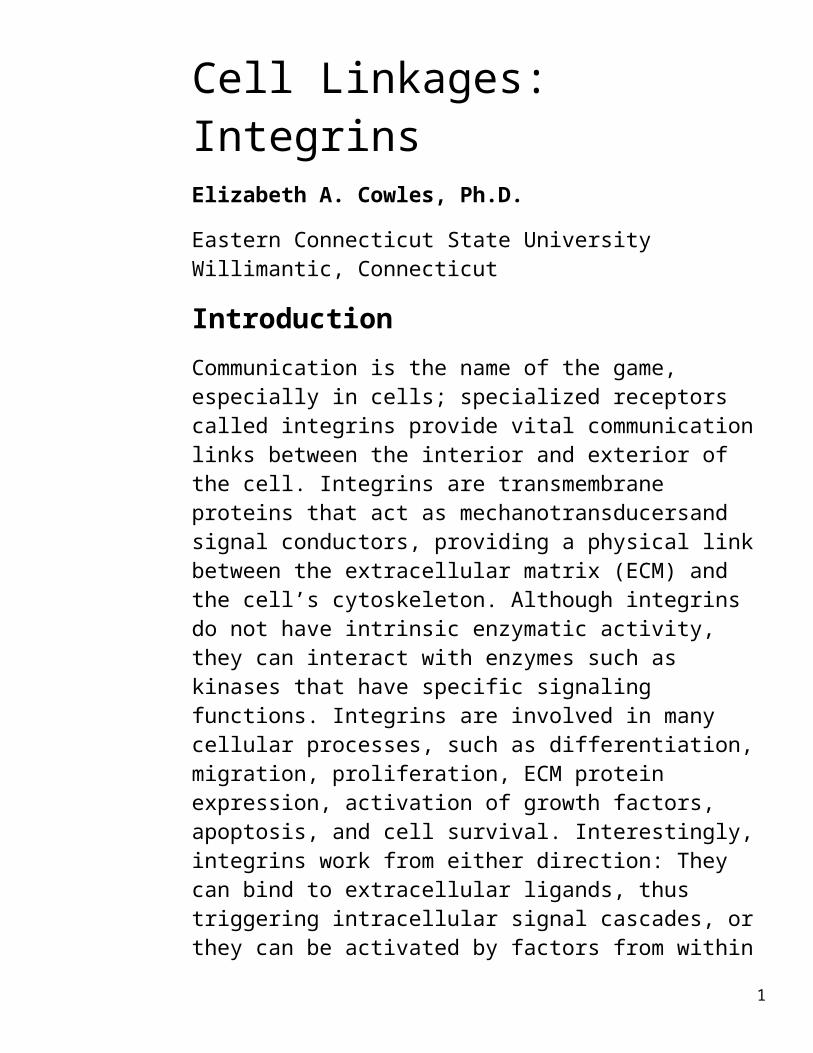
FIGURE 1
A model of integrin activation. Schematic representation of a heterodimeric integrin molecule in the bent, inactive conformation (left) and the upright, active conformation (right). The switch in conformation is triggered by the binding of a protein, in this case talin, to the small cytoplasmic domain of the β subunit. The binding of talin induces a separation of the two subunits and conversion to the active conformation. Activated integrins typically become clustered as the result of interactions of their cytoplasmic domains with the underlying cytoskeleton. The extracellular ligand, in this case a collagen fiber, binds between the two subunits in the head region of the activated integrin dimer.
Figures 1–4 from Cell and Molecular Biology: Concepts and Experiments by Gerald Karp. New York: John Wiley & Sons, Inc., 2007. Used with permission of John Wiley & Sons, Inc.
(Jaffe et al. 2001), in gravity perception in plants (Katembe et al. 1997), and in the binding of a pathogenic fungus to fibronectin (Kottom et al. 2008).
Integrins typically span the cell’s plasma membrane with the N- or amino- terminus of both subunits extending into the extracellular matrix, providing potential ligand binding sites. The subunits interact with each other and exist in either a low-affinity or a high-affinity conformation depending on external and internal signals (Humphries and Liddington 2002). The integrins are normally bent in the low-affinity state, but “open like a switchblade” upon activation via phosphorylation of the β subunit’s cytoplasmic end.
2
This physical change modulates both the integrin’s affinity for its ligand and also the stability of the attachment. The relatively small 40–70 amino acid subunit ends that extend into the cytoplasm allow interactions with intracellular proteins; the β4 cytoplasmic “tail” is so long, it can even bind to intermediate filaments in the cytoskeleton.
Fibronectin, which has important cell migration and wound healing functions, provides a good example of extracellular protein interaction with integrins. Fibronectin contains an RGD or arginine–glycine–aspartate sequence, which is recognized by the integrin α5β1. The integrin simultaneously interacts with talin, which provides a physical link to actin filaments, thus allowing cells to migrate along the fibronectin.
FIGURE 2
Focal adhesions are sites where cells adhere to their substratum. This drawing of a focal adhesion shows the interactions of integrin molecules with other proteins on both sides of the lipid bilayer. The binding of extracellular ligands, such as collagen and fibronectin, is thought to induce conformational changes in the cytoplasmic domains of the integrins that cause the integrins to become linked to actin filaments in the cytoskeleton. Linkages with the cytoskeleton are mediated by various actin- binding proteins, such as talin and a-actinin, that bind to the β subunit of the integrin. The cytoplasmic domains of integrins are also associated with protein kinases, such as FAK (focal adhesion kianse) and Src. The attachment of the integrin to an extracellular ligand can activate these protein kinases and start a chain reaction that transmits signals throughout the cell. The association of myosin molecules with the actin filaments can generate traction forces that are transmitted to sites of cell–substrate attachment.
3

Other proteins containing the RGD sequence include vitronectin, bone sialoprotein, von Willebrand factor, fibrillin, fibrinogen, thrombospondin, PECAM (platelet endothelial cell adhesion molecule), tenascin, and LAP-TGFβ (latency associated peptide transforming growth factor-β). The RGD motif is also used for interactions between ECM proteins and integrins αvβ1 and α8β1. Other integrins use a different tripeptide sequence, leucine-aspartate-valine (LDV), to recognize and bind proteins important for intercellular adhesion, such as VCAM (vascular cell adhesion molecule), ICAM (intercellular adhesion molecule), factor X, mucosal adhesion cell adhesion molecule (MadCAM), and E-cadherin. Integrins containing the α1 and α2 subunits have an A domain, similar to von Willebrand factor, a blood glycoprotein important in clotting. These subunits combine with β1 to form receptors for collagen, thrombospondin, and laminin.
Integrins and Cellular SignalingInactive integrins are dispersed over the cell surface. Upon binding to ECM proteins, integrins migrate within the cell membrane to cluster and form focal adhesion sites in a process called activation.
These sites may then include interactions with several additional proteins such as focal adhesion kinase (FAK), paxillin, talin, and tensin (Tadokoro et al. 2003; Lo 2006). Integrin interactions between talin, vinculin, α-actinin, and paxillin provide a physical linkage between the ECM and the actin cytoskeleton; these links are critical for cell anchorage and migration. Phosphorylation of the β tail, in the cytoplasm, disrupts the talin–integrin interaction, thus permitting cell movement.
Integrin activation via external factors can set off a cascade of events; thisis called outside-in signaling. These interactions can involve several proteins, in which the integrin has a critical mediating role. Outside-in signaling can resultin modification of the cytoskeleton, cause cellular proliferation or migration, and determine cell survival or apoptosis. One example is the MAPK/ERK (mitogen- activated protein kinase or extracellular signal-related kinase) signal pathway, which is turned on by
4

integrin-extracellular ligand interactions.
FIGURE 3
This pathway controls gene expression by increasing the stability of c-jun and increasing transcription of the protein c-fos. C-jun and c-fos combine to form AP-1 (activating protein-1), which binds to specific DNA promoters; therefore, integrins can control gene transcription via the MAPK/ ERK signaling kinases. In particular, AP-1 induces expression of integrin α2β1 and its ligand, collagen.
Another example of integrin-mediated control of transcription involves activation of the α5β1 integrin, which increases ERK; this ultimately up-regulates the transcription of the protein Bcl-2, which is an anti-apoptotic signal. High Bcl-2 levels prevent apoptosis, or programmed cell death. Temporary loss of integrin-substrate contact is necessary for migration; however, loss of adhesion may trigger apoptosis. This balance between integrin binding and apoptosis prevents inappropriate cell migration and adhesion; this equilibrium is often aberrant in cancer cells.
Signaling via integrins is not always outside-in, nor is it always unidirectional.
5

FIGURE 4
Blood clots form when platelets adhere to one another through fibrinogen bridges that bind to platelet integrins. The presence of synthetic RGD peptides can inhibit blood clot formation by competing with fibrinogen molecules for the RGD-binding sites on platelet αIIIβ3 integrins. Nonpeptide RDG analogs and anti-integrin antibodies can act in a similar way to prevent clot formation in high-risk patients.
Recent research has focused on the intracellular Rho family of GTPases. Rho coordinates cell functions suchas cell adhesion, cell migration, gene expression, and the cell cycle. Integrins activated by extracellular factors regulate Rho through the MAPK/ERK pathway.
Rho, however, increases integrin avidity (“stickiness”) and the numbers of stress fibers, which in turn regulates the formation of integrin focal adhesion sites (Schwartz and Shattil 2000; Lo 2006). Therefore, an internal molecule like Rho can modulate integrin–
6
ECM interactions by changing the integrin conformation to the active form.
The inside-out signaling is exemplified by the blood clotting system. Platelets have inactive integrins, which prevent inappropriate adhesion to vessel walls (Horowitz 1997). Damage to endothelial cells exposes thrombin, which activates platelets that come in direct contact by causing their αIIbβ3 integrin to become more adhesive and to bind fibrinogen. Platelets bind to the thrombin without the assistance of integrins; the thrombin–platelet interaction elicits intracellular signals, which change integrin adhesiveness.
FIGURE 5
From Biology, by Robert J. Brooker, Eric P. Widmaier, Linda Graham, and Peter Stiling. New York: McGraw-Hill Science, 2007. Used with permission of The McGraw-Hill Companies.
The now-activated integrins on the platelet surfaces capture circulatingvon Willebrand factor and fibrinogen, which form the blood clot. Glanzmann’s thrombasthenia is caused by a mutation in
7

either the αIIb or β3 integrin genes; the disorder is characterized by abnormal bruising and bleeding and is often mistaken for hemophilia. Antagonists (inhibitors) of αIIbβ3 integrin, such as abciximab and xemilofiban, are used to reduce clot formation in patients at high risk for stroke or heart attacks.
Leukocytes infiltrate damaged tissues, but only do so when their αMβ2 or αLβ2 integrins are activated by cytokines via inside-out signaling (Horowitz 1997). Cytokines are small, secreted proteins which regulate immunity and inflammation. The integrins mediate attachment to vascular endothelial ICAMs (intercellular adhesion molecules), and then the leukocyte migrates between the endothelial cells. Leukocyte adhesion deficiency (LAD), a very rare human disorder in which patients lack β2 or make defective β2, occurs when phagocytes cannot attach to endothelial cells. LAD patients often succumb to bacterial infections early in life.
Many integrin signaling events are coupled with growth factor responses; this is because cellular responses, such as migration and mitosis, require integrin-substrate interactions or anchorage (Giancotti and Ruoslahti 1999).
For example, the platelet-derived growth factor (PDGF) receptor responds maximally only when αVβ3 is bound to the ECM and PDGF increases the amount of αVβ3 in the plasma membrane. This synergistic activity between the PDGF receptor and the integrin increases cell migration and wound healing. The interactions between the growth factor and integrin signaling pathways fine-tune the cell’s response to its external environment and allow for coordinated regulation.
Integrins in Health and DiseaseAngiogenesis, or the production of new blood vessels, is critical not only during development but also during oncogenesis or cancer development; tumors need a ready blood supply for survival. αVβ3 integrin attachment to the ECM is regulated by the vascular endothelial growth factor (VEGF) receptor; impairing the VEGF receptor– integrin interaction reduces angiogenesis
8

(Mahabeleshwar et al. 2006). Drugs that interfere with integrin function during angiogenesis are in clinical trials; Vitaxin, an anti-αVβ3 antibody, shrinks tumors by decreasing blood vessels and Avastin, an anti- VEGF monoclonal antibody, is used for treating colorectal cancers.
Researchers have noted that certain breast cancers metastasized to bone. It appears that αVβ3 expression is elevated in breast cancer tissue, and this integrin recognizes bone sialoprotein, a major bone matrix constituent. Cancer cells migrating from breast tumors bind to bone tissue through αVβ3 and become established (Sloan et al. 2006). Scientists are investigating whether αVβ3 antagonists could prevent metastases (Zhao et al. 2007). Unpublished data from Barbara Susini’s laboratory (University of California, San Diego) indicate that an α4β1 antagonist inhibits lymph node blood vessel development; because breast cancer spreads via the lymphatic system, such an antagonist may prevent breast cancer metastases.
Viral and bacterial pathogens take full advantage of integrins. Bacterial pathogens use integrins to maintain contact and prevent removal from the host, and then gain entry (Scibelli et al. 2007). Binding allows the bacteria to be phagocytosed, to inject virulence factors, or to adhere indirectly through fibronectin. The α5β1 integrin that binds fibronectin is recognized by Shigella; several Shigella protein antigens mediate bacterial-directed endocytosis. Staphlococcus aureus and Streptococcus spp. express fibronectin-binding proteins, and indirectly attach to cells through the ECM.
Viruses also employ integrins to bind and gain entry into cells (Stewart and Nemerow 2007). Adenovirus is recognized by the αV integrin. The herpes virus outer envelope glycoprotein has a sequence that mimics a metalloprotease; this glycoprotein binds to β1 and αVβ3 integrins. Hantaviruses, which directly affect platelet and endothelial cell functions, have αIIbβ3 and αVβ3 as receptors. Binding not only allows the virus access to the cell’s synthetic machinery but also to its signaling pathways. Novel vaccines, therapeutic strategies, and antiviral drugs may be based upon integrin antagonists.
9

A new area of integrin research is in nanoscale technology and material science (Stevens and George 2005). Implants often fail because cells do not attach and reproduce on the foreign surface. Titanium implants are commonly used in dentaland joint replacements, but do not readily support osteogenesis. Implants coated with matrices of calcium carbonate (hydroxylapatite) and RGD-containing proteins, such as fibronectin, collagen or related peptides, bond bone-forming osteoblasts better than uncoated material (Reyes et al. 2007). These coatings promoted α2β1 binding, which triggered the expression of osteoblast-specific genes, which resulted in increased cell differentiation and bone production.
Integrins are involved in many cellular and organismal processes, including tissue remodeling, immunology, and proliferation. These biological activities require coordination between the internal and external cellular environments; integrins provide some of the critical communication links. Insights into these fascinating cell surface receptors will help us design new therapies for cancer, inflammation, and pathogen-related diseases.
BibliographyGiancotti, F. G., and E. Ruoslahti. “Integrin Signaling.” Science 285 (1999): 1028–1032.
Horowitz, A. F. “Integrins and Health.” Scientific American, May 1997; 68–75.
Humphries, M. J., and R. C. Liddington. “Molecular Basis of Integrin-Dependent Cell Adhesion in Protein-Protein Recognition,” In Frontiers in Molecular Biology: Protein-Protein Recognition, edited by C. Kleanthous, 102–125. New York: Oxford University Press, 2000.
Hynes, R. O. “Integrins: Bidirectional, Allosteric Signaling Machines.” Cell 110 (2002): 673–687.
Jaffe, M. J., A. C. Leopold, and R. C. Staples. “Thigmo Responses in Plants and Fungi.” American Journal of Botany 89 (2002): 375–382.
10

Katembe, W. J., L. J. Swatzell, C. A. Markaroff, and J. Z. Kiss. “Immunolocalization of Integrin-Like Proteins in Arabidopsis and Char.” Physiologia Plantarum 99(1) (1997), 7–14.
Kottom, T. J., C. C. Kennedy, and A. H. Limper. “Pneumocystis PCINT1, a Molecule with Integrin-Like Features That Mediates Organism Adhesion to Fibronectin.” Molecular Microbiology (online early articles) (2008), 67: 747–761.
Lo, S. H. “Focal Adhesions: What’s New Inside.” Developmental Biology 294 (2006): 280–291.
Mahabeleshwar, G. H., W. Feng, D. R. Phillips, and T. V. Boyzova. “Integrin Signaling Is Critical for Pathological Angiogenesis.” Journal of Experimental Medicine. 203(11) (2006): 2,495–2,507.
Reyes, C. D., T. A. Petrie, K. L. Burns, Z. Schwartz, and A. J. Garcia. “Biomolecular Surface Coating to Enhance Orthopaedic Tissue Healing and Integration. Biomaterials 28(21) (2007): 3,228–3,238.
Schwartz, M. A., and A. J. Shattil. “Signaling Networks Linking Integrins and Rho Family GTPases.” Trends in Biochemical Sciences 25 (2000): 388–391.
Scibelli, A., S. Roperto, L. Manna, L. M. Pavone, S. Tafuri, R. D. Morte, and N. Staiano. “Engagement of Integrins as a Route of Invasion by Bacterial Pathogens.” Veterinary Journal 173 (2007): 482–491.
Sloan, E. K., N. Pouliot, K. L. Stanley, J. Chia, J. M. Mosley, D. K. Hards, and R. L. Anderson. “Tumor-Specific Expression of αVβ3 Integrin Promotes Spontaneous Metastasis of Breast Cancer to Bone.” Breast Cancer Research 8 (2006): R20.
Stevens, M. M., and J. H. George. “Exploring and Engineering the Cell Surface Interface.” Science 310 (2005): 1135–1138.
Stewart, P. L., and G. R. Nemerow. “Cell Integrins: Commonly Used Receptors for Viral Pathogens.” Trends in Microbiology
11

15(11) (2007): 500–507.
Tadokoro, S., S. J. Shattil, K. Eto, V. Tai, R. C. Liddington, J. M. de Pereda, M. H. Ginsberg, and D.A. Calderwood. “Talin Binding to Integrin β Tails: A Final Common Step in Integrin Activation.” Science 302 (2003): 103–106.
Zhao, Y., R. Bachelier, I. Treilleux, P. Pujuquet, O. Peyuchaud, R. Baron, P. Clément- Lacroix, and P. Clézardin. “Tumor αVβ3 Integrin is a Therapeutic Target for Breast Cancer Metastases. Cancer Research 67(12) (2007): 5,821–5,830.
12