Embed Size (px)
Citation preview

Structural basis of DNA recognitionby the heterodimeric cellcycle transcription factor E2F–DPNing Zheng,1,4 Ernest Fraenkel,2,3,4 Carl O. Pabo,2 and Nikola P. Pavletich1,5
1Howard Hughes Medical Institute, Cellular Biochemistry and Biophysics Program, Memorial Sloan-Kettering CancerCenter, New York, New York 10021 USA; 2Howard Hughes Medical Institute, Department of Biology, MassachusettsInstitute of Technology, Cambridge, Massachusetts 02139 USA
The E2F and DP protein families form heterodimeric transcription factors that play a central role in theexpression of cell cycle-regulated genes. The crystal structure of an E2F4–DP2–DNA complex shows that theDNA-binding domains of the E2F and DP proteins both have a fold related to the winged-helix DNA-bindingmotif. Recognition of the central c/gGCGCg/c sequence of the consensus DNA-binding site is symmetric, andamino acids that contact these bases are conserved among all known E2F and DP proteins. The asymmetry inthe extended binding site TTTc/gGCGCc/g is associated with an amino-terminal extension of E2F4, in whichan arginine binds in the minor groove near the TTT stretch. This arginine is invariant among E2Fs but notpresent in DPs. E2F4 and DP2 interact through an extensive protein–protein interface, and structural featuresof this interface suggest it contributes to the preference for heterodimers over homodimers in DNA binding.
[Key Words: E2F; DP; Winged-helix, DNA-binding domain; transcription factor; cell cycle]
Received January 12, 1999; revised version accepted January 22, 1999.
Progression through the G1 and S phases of the eukary-otic cell cycle is tightly coupled to the transcriptionalcontrol of genes involved in growth and in DNA repli-cation (Dynlacht 1997). In mammalian and many othereukaryotic cells, this temporal control of gene expres-sion is carried out primarily by the E2F family of tran-scription factors (for review, see Slansky and Farnham1996; Helin 1998). E2F-responsive genes include c-myc(Hiebert et al. 1989; Thalmeier et al. 1989), cyclin A (Sch-ulze et al. 1995) and cdc2 (Dalton 1992; Furukawa et al.1994), growth-regulatory proteins, and dihydrofolate re-ductase (Blake and Azizkhan 1989; Mudryj et al. 1990),thymidine kinase (Dou et al. 1992) and DNA polymerasea (Pearson et al. 1991; DeGregori et al. 1995) proteinsneeded for DNA synthesis. E2F activity is controlled bythe cell cycle machinery primarily through the bindingof the retinoblastoma (Rb) family of pocket proteins(Chellappan et al. 1991; Chittenden et al. 1991; Cao et al.1992; Shirodkar et al. 1992; Cobrinik et al. 1993; for re-view, see Dyson 1998), and through phosphorylation bythe cyclin-dependent kinases (Dynlacht et al. 1994; Kreket al. 1994).
E2F proteins can, depending on whether they are as-sociated with Rb, act either as repressors or as activatorsof transcription (Hiebert et al. 1992; Weintraub et al.1992, 1995; Bremner et al. 1995). The Rb–E2F complexes,which predominate in quiescent or early G1 cells (Bagchiet al. 1991; Cao et al. 1992), act as repressors of transcrip-tion as Rb masks the E2F transactivation domain (Helinet al. 1993a) and can also recruit a histone deacetylase atcertain promoters (Brehm et al. 1998; Luo et al. 1998;Magnaghi-Jaulin et al. 1998). Free E2F transcription fac-tors function as activators of transcription, and these arereleased following the phosphorylation of Rb at the G1–Stransition. Deletion of E2F1 in mice leads to atrophy ofsome tissues, and to displasia and tumors in other tis-sues, consistent with E2F1’s dual role as an activator anda repressor of transcription in different contexts (Field etal. 1996; Yamasaki et al. 1996).
The E2F proteins form heterodimers with members ofthe DP family, which are distantly related to the E2Ffamily. E2F proteins can also form homodimers, but het-erodimerization with DPs enhances their DNA-binding,transactivation, and Rb-binding activities (Bandara et al.1993; Helin et al. 1993b; Krek et al. 1993), and it appearsthat DP proteins are a component of all E2F activity inthe cell (Wu et al. 1996). In humans, six E2F and two DPproteins have been isolated to date. E2Fs one throughfive share 20%–55% identity and have a similar organi-zation of functional domains. E2F6 is divergent as it
3Present address: Department of Molecular and Cellular Biology, Har-vard University, Cambridge, Massachusetts 02138 USA.4These authors contributed equally to this work.5Corresponding author.E-MAIL [email protected]; FAX (212) 717-3135.
666 GENES & DEVELOPMENT 13:666–674 © 1999 by Cold Spring Harbor Laboratory Press ISSN 0890-9369/99 $5.00; www.genesdev.org
Cold Spring Harbor Laboratory Press on June 10, 2018 - Published by genesdev.cshlp.orgDownloaded from

lacks the transactivation domain and has been proposedto function as a repressor of E2F-dependent transcription(Morkel et al. 1997; Cartwright et al. 1998; Trimarchi etal. 1998). The two DP proteins are highly homologous(70%), and each can form functional heterodimers withany E2F family member (Wu et al. 1995).
Different E2F proteins are differentially regulated bymembers of the Rb family of pocket proteins. E2F1,E2F2, and E2F3 are bound and regulated by Rb (Lees et al.1993), E2F5 binds to p130 only (Hijmans et al. 1995),whereas E2F4 associates with all three pocket proteins(Ikeda et al. 1996; Moberg et al. 1996). E2F proteins canalso differ in the regulation of their subcellular localiza-tion. E2F1, E2F2, and E2F3 contain nuclear localizationsignals and are found exclusively in the nucleus. E2F4and E2F5 appear to require other nuclear factors, such astheir DP partners or the pocket proteins, to promotetheir nuclear localization (Muller et al. 1997; Verona etal. 1997).
Beside differences in their regulation, it is not yet en-tirely clear whether different E2F proteins differ in theirDNA sequence specificity and in their preference for pro-moters of different E2F-responsive genes. On one hand,all E2F–DP combinations can bind to and transactivateat the consensus TTTc/gGCGCc/g E2F site (Lees et al.1993; Buck et al. 1995; Zhang and Chellappan 1995). Onthe other hand, in vitro binding-site selection experi-ments have suggested that E2F-1 and E2F-4 may havedifferences in their specificity for variants of the E2Fconsensus site (Tao et al. 1997).
The E2F proteins contain an ∼70-residue domain re-sponsible for DNA binding (Kaelin et al. 1992; Cress etal. 1993; Ivey-Hoyle et al. 1993; O’Connor and Hearing1994). DP proteins need a larger, 90-residue region forbinding to DNA. The E2F and DP DNA-binding domainsare followed by a hydrophobic heptad repeat involved inhomo- and heterodimerization (Helin et al. 1993b). TheE2F family also contains regions involved in transacti-vation and Rb binding (Kaelin et al. 1992; Cress et al.1993). E2F and DP proteins share sequence homology inthe last 30 residues of their DNA-binding domains andwithin their hydrophobic heptad repeat (Girling et al.1993).
The hydrophobic heptad repeat is the primary dimer-ization domain, being necessary for the formation of E2Fhomodimers and E2F–DP heterodimers in the absence ofDNA (Helin et al. 1993b). But several lines of evidencehave suggested that the DNA-binding domains contrib-ute to dimerization. The isolated DNA-binding domainof E2F1 binds DNA weakly (Krek et al. 1993; Jordan et al.1994) and the corresponding region of DP1 does not binddetectably, but mixing the two DNA-binding domainsgreatly enhances DNA binding (Bandara et al. 1993; Frae-nkel 1998). This has suggested that the DNA-bindingdomains may interact on the DNA and that this maycontribute to dimerization and preference for DNA bind-ing as heterodimers.
Here we report the 2.6-Å crystal structure of a complexcontaining the DNA-binding domains of E2F4 and DP2bound to a DNA site from the adenovirus E2 promoter.
The structure reveals that the DNA-binding domains ofE2F4 and DP2 adopt the winged-helix fold (Clark et al.1993) and use residues invariant within their respectivefamilies to contact the bases of the DNA. This indicatesthat the winged-helix domains of other E2F–DP combi-nations will have very similar DNA sequence specific-ity. The structure also shows that E2F4 and DP2 form anextensive protein–protein interface, and structural fea-tures of this interface suggest that the winged-helix do-mains contribute to the preference of E2F and DP pro-teins to bind to DNA as heterodimers.
Results
Structures of the DNA-binding domains of E2F4and DP2
The boundaries of the DNA-binding domains of E2F4(residues 11–86) and DP2 (residues 60–154) used in thecrystallization were identified by the proteolytic diges-tion of an E2F–DP heterodimer-DNA complex (Fig. 1;Fraenkel 1998). The heterodimer of the E2F4 and DP2
Figure 1. The DNA-binding domains of E2Fs and DPs havelimited across-family homology but share the same fold. (A)Sequence alignment of known E2F and DP family memberswith the E2F4 and DP2 polypeptides used in the crystallization.Residues conserved throughout the E2F and DP families arehighlighted in yellow. The RRXYD DNA recognition motif isunderlined in the E2F4 and DP2 sequences. (B) Sequence of theDNA duplex used in the cocrystallization, with the E2F site atthe adenovirus E2 promoter underlined.
Structure of an E2F4–DP2–DNA complex
GENES & DEVELOPMENT 667
Cold Spring Harbor Laboratory Press on June 10, 2018 - Published by genesdev.cshlp.orgDownloaded from

DNA-binding domains gives half-maximal DNA bindingwhen each protein is at a concentration of 50 nM, com-pared with ∼500 nM and >2000 nM for homodimers ofE2F4 and DP2, respectively (data not shown).
The crystal structure, shown in Figure 2, reveals thatthe E2F DNA-binding domain has a structure related tothe winged-helix DNA-binding motif, and not the basichelix–loop–helix (bHLH) motif that has been suggested(Kaelin et al. 1992; Cress et al. 1993). The winged-helixmotif occurs in several eukaryotic transcription factors(for review, see Kaufmann and Knochel 1996), includingHNF-3g (Clark et al. 1993), ETS1 (Kodandapani et al.1996), and HSF (Harrison et al. 1994), as well as in thehistone H5 globular domain (Ramakrishnan et al. 1993)(Fig. 3). This motif consists of three a helices and a bsheet, each contributing to a compact hydrophobic core.DP2 has the same overall structure as E2F4, except thatthe a2 and a3 helices of DP2 are longer by about twoturns each, and E2F4 has an amino-terminal helical ex-tension (aN; Figs. 1A and 3A ) that is not present in DP2.The remaining regions of E2F4 and DP2 superimposequite well, with a 1.4-Å root mean square deviation(rmsd) for 59 residues (Fig. 3A). The structure-basedalignment of E2F4 and DP2 shows that the regions cor-responding to the a1 and a2 helices have only 8% se-quence identity (Fig. 1A), and this, coupled to the greaterlength of the a2 and a3 helices of DP2, presumably com-plicated previous efforts to align the two families. The30-residue region of clear homology between the E2F andDP families, termed the DEF box (Girling et al. 1993),coincides with the a3 helix and the b2 and b3 strands(Fig. 1A).
Compared with the winged-helix domain of HNF-3g(Clark et al. 1993), the E2F4 and DP2 domains do notcontain the carboxy-terminal wing extension. DP2 andHNF-3g can be superimposed with an rmsd of 1.7 Å inthe Ca positions of 36 residues. E2F4 is more divergent,superimposing on HNF-3g with a 2.1-Å rmsd for 36 resi-dues.
DNA sequence recognition
The structure shows that the a3 helices of E2F4 and DP2bind in the major groove of the DNA and make criticalcontacts to the edges of the bases. In both cases, theamino-termini of the a1 helices and portions of the bsheets contact the phosphodiester backbone of the DNA(Fig. 2). This overall DNA-binding arrangement for eachprotein is analogous to the other winged-helix proteins.However, winged-helix proteins typically bind DNA asmonomers (Clark et al. 1993; Harrison et al. 1994; Ko-dandapani et al. 1996), whereas E2F4 and DP2 form anextensive hetero-oligomerization interface and present acontinuous protein surface to the DNA (Fig. 2).
The E2F4–DP2 heterodimer binds to and recognizesthe palindromic CGCGCG sequence of the binding sitein an essentially symmetric arrangement (Fig. 4A,B). Inthis region, all of the DNA base contacts and most of thephosphate contacts are made by residues conservedthroughout the E2F and DP families (Fig. 1A). E2F4 andDP2 each contact half of the palindromic sequence(CGCGCG and CGCGCG, top strand in Fig. 1B) by useof a conserved Arg-Arg-Xxx-Tyr-Asp sequence on theira3 helices (RRXYD motif, underlined in Fig. 1A). Thebinding of the E2F4 and DP2 a3 helices in the majorgroove is highly analogous, but a detailed comparisonreveals that their positioning relative to the bases theycontact differs by about 1 Å along the DNA axis.
The two arginines of the RRXYD motif each make apair of hydrogen bonds with a guanine. These guaninesoccur in neighboring basepairs and are on oppositestrands of the half site (CGC). The aspartic acid of theRRXYD motif makes charged hydrogen bonds to botharginines and appears to stabilize this arrangement (Fig.4C). This pattern of hydrogen bonds is identical in thetwo halves of the heterodimer, except that DP2 has anadditional residue, Asn118, helping to stabilize the ar-ginines. The DP2 Asn118 side chain bridges the phos-phodiester backbone of the DNA with the Arg122 gua-nidinium group (RRXYD, Fig. 4A,B). Several of the re-ported E2F-binding sites contain a half-site variant thathas the c/gGC sequence of the consensus half site re-
Figure 2. Structure of the E2F4-DP2 heterodimer DNA com-plex. (A) Schematic view looking down the approximate axis oftwofold pseudo symmetry in the heterodimer. The DNA axis isvertical in this view. (B) View of the complex looking down theDNA axis. Figures were prepared with the programs MOL-SCRIPT (Kraulis 1991) and RASTER3D (Merritt and Bacon1997).
Figure 3. The E2F4 and DP2 DNA-binding domains consist ofthe winged-helix motif. (A) Superposition of the E2F4 and DP2winged-helix DNA-binding domains. (B) The winged-helix do-main of the HNF-3g transcription factor (Clark et al. 1993) in anorientation obtained by aligning it with the DP2 winged-helixdomain in A.
Zheng et al.
668 GENES & DEVELOPMENT
Cold Spring Harbor Laboratory Press on June 10, 2018 - Published by genesdev.cshlp.orgDownloaded from

placed with c/gCC (Slansky and Farnham 1996). Accord-ingly, we find that the mixture of the E2F4 and DP2winged-helix domains has an affinity for a TTTCCC-GCG site that is comparable with the affinity for theTTTCGCGCG site used in the crystallization (data notshown). The crystal structure suggests that the arginine(RRXYD) that contacts this position could, in principle,reach to a guanine on either strand (Fig. 4C).
The outer position of the 3-bp half site (CGC) is rec-ognized by the tyrosine of the conserved RRXYD motif(Fig. 4A,B). The tyrosine phenyl group makes multiplevan der Waals contacts to the C5 and C6 atoms of the
cytosine, whereas the side-chain hydroxyl group con-tacts phosphate and sugar groups. The close contacts be-tween the tyrosine and the cytosine, which are verysimilar in the E2F and DP half sites, are consistent withsequence discrimination at this position suggested bythe c/gGC consensus.
The consensus-binding site is not completely symmet-ric, having a T-rich portion at one end (TTTc/gGCGCg/c). In the crystal structure, this is associated with anE2F4 amino-terminal extension (residues 16–19) that isconserved in the E2F family but not in the DP family(Fig. 1A). This region forms the short aN helix and in-serts an arginine side chain (Arg17) deep inside the minorgroove of the DNA near the T-rich portion (Fig. 4A,B).The Arg-17 side chain contacts the O2 group of Thy4 andthe O2 and sugar groups of Cyt5, and these contacts areassociated with a compression of the minor groove inthis region. On the basis of the electron density maps,Arg-17 is not as rigid as some of the other side-chainbasepair contacts in the complex. However, the argininecontacts appear to be important because this residue isinvariant among E2F family members (Fig. 1A) and itsdeletion abolishes DNA binding (Jordan et al. 1994). Thecorresponding region of DP2 (residues 60–68), whichlacks the arginine, is disordered in our crystal structureand does not appear to be making direct DNA contacts.Similar arginine-minor groove contacts, associated withan A/T rich sequence and with a compressed minorgroove have been observed in other protein–DNA com-plexes (Aggarwal et al. 1988; Clark et al. 1993; Cho et al.1994).
The heterodimerization interface
The structure of the complex reveals an extensive pro-tein–protein interface between the E2F4 and DP2winged-helix domains (Figs. 2 and 5). This heterodimerinterface is predominantly hydrophobic and buries a to-tal of 1160 Å2 surface area. The interface contains the a1and a3 helices from both E2F4 and DP2. These helicespack in triplets. The a3 helix of E2F4 packs in betweenthe a1 and a3 helices of DP2 at ∼45° and ∼90°, respec-tively, and the a3 helix of DP2 packs in a reciprocalfashion with the a1 and a3 helices of DP2. The arrange-ment of the helices at the interface is thus approximatelysymmetrical, explaining how the DNA-binding domainof E2F can also form homodimers (Huber et al. 1993).The E2F4 and DP2 residues at the interface are well con-served within their separate families, being 75% and100% identical in the respective families. This suggeststhat other E2F and DP combinations will have similarheterodimer interfaces (Fig. 1A).
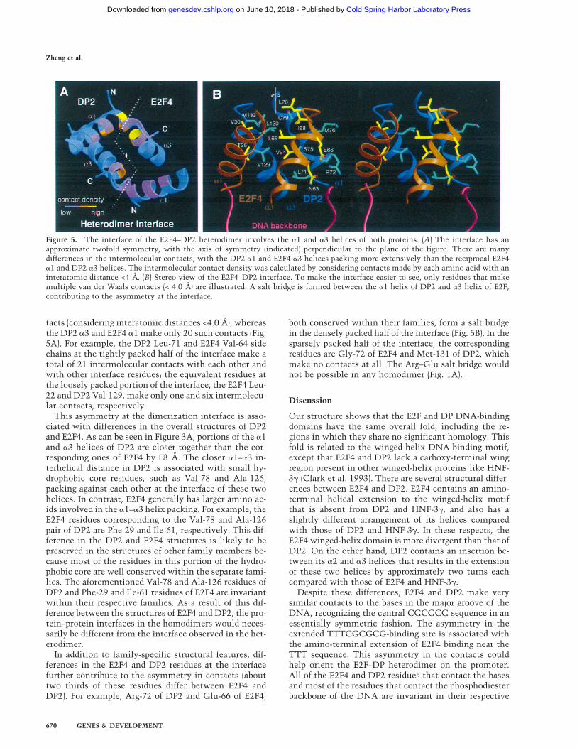
The symmetry at the heterodimer interface is less pre-cise than that at the protein–DNA interface, owing tomodest differences in the intermolecular packing of theE2F4 and DP2 structural elements (Fig. 5). In particular,contacts between the E2F4 a3 helix and the DP2 a1 helixare more extensive than the reciprocal contacts betweenthe DP2 a3 and E2F4 a1 helices. The E2F4 a3 and DP2a1 helices make 70 intermolecular van der Waals con-
Figure 4. Recognition of the core DNA site by the E2F4–DP2heterodimer is overall symmetric. (A) Figure shows the E2F4and DP2 residues that contact the DNA bases; for clarity, onlypart of the a3 helices of each subunit and the amino-terminalhelix of E2F4 are shown. (B) Sketch summarizes the DNA con-tacts made by the E2F4–DP2 heterodimer. (C) Close-up view ofthe interactions between the two arginines of the RRXXD motifand the two neighboring guanines within the half site.
Structure of an E2F4–DP2–DNA complex
GENES & DEVELOPMENT 669
Cold Spring Harbor Laboratory Press on June 10, 2018 - Published by genesdev.cshlp.orgDownloaded from
tacts (considering interatomic distances <4.0 Å), whereasthe DP2 a3 and E2F4 a1 make only 20 such contacts (Fig.5A). For example, the DP2 Leu-71 and E2F4 Val-64 sidechains at the tightly packed half of the interface make atotal of 21 intermolecular contacts with each other andwith other interface residues; the equivalent residues atthe loosely packed portion of the interface, the E2F4 Leu-22 and DP2 Val-129, make only one and six intermolecu-lar contacts, respectively.
This asymmetry at the dimerization interface is asso-ciated with differences in the overall structures of DP2and E2F4. As can be seen in Figure 3A, portions of the a1and a3 helices of DP2 are closer together than the cor-responding ones of E2F4 by ∼3 Å. The closer a1–a3 in-terhelical distance in DP2 is associated with small hy-drophobic core residues, such as Val-78 and Ala-126,packing against each other at the interface of these twohelices. In contrast, E2F4 generally has larger amino ac-ids involved in the a1–a3 helix packing. For example, theE2F4 residues corresponding to the Val-78 and Ala-126pair of DP2 are Phe-29 and Ile-61, respectively. This dif-ference in the DP2 and E2F4 structures is likely to bepreserved in the structures of other family members be-cause most of the residues in this portion of the hydro-phobic core are well conserved within the separate fami-lies. The aforementioned Val-78 and Ala-126 residues ofDP2 and Phe-29 and Ile-61 residues of E2F4 are invariantwithin their respective families. As a result of this dif-ference between the structures of E2F4 and DP2, the pro-tein–protein interfaces in the homodimers would neces-sarily be different from the interface observed in the het-erodimer.
In addition to family-specific structural features, dif-ferences in the E2F4 and DP2 residues at the interfacefurther contribute to the asymmetry in contacts (abouttwo thirds of these residues differ between E2F4 andDP2). For example, Arg-72 of DP2 and Glu-66 of E2F4,
both conserved within their families, form a salt bridgein the densely packed half of the interface (Fig. 5B). In thesparsely packed half of the interface, the correspondingresidues are Gly-72 of E2F4 and Met-131 of DP2, whichmake no contacts at all. The Arg–Glu salt bridge wouldnot be possible in any homodimer (Fig. 1A).
Discussion
Our structure shows that the E2F and DP DNA-bindingdomains have the same overall fold, including the re-gions in which they share no significant homology. Thisfold is related to the winged-helix DNA-binding motif,except that E2F4 and DP2 lack a carboxy-terminal wingregion present in other winged-helix proteins like HNF-3g (Clark et al. 1993). There are several structural differ-ences between E2F4 and DP2. E2F4 contains an amino-terminal helical extension to the winged-helix motifthat is absent from DP2 and HNF-3g, and also has aslightly different arrangement of its helices comparedwith those of DP2 and HNF-3g. In these respects, theE2F4 winged-helix domain is more divergent than that ofDP2. On the other hand, DP2 contains an insertion be-tween its a2 and a3 helices that results in the extensionof these two helices by approximately two turns eachcompared with those of E2F4 and HNF-3g.
Despite these differences, E2F4 and DP2 make verysimilar contacts to the bases in the major groove of theDNA, recognizing the central CGCGCG sequence in anessentially symmetric fashion. The asymmetry in theextended TTTCGCGCG-binding site is associated withthe amino-terminal extension of E2F4 binding near theTTT sequence. This asymmetry in the contacts couldhelp orient the E2F–DP heterodimer on the promoter.All of the E2F4 and DP2 residues that contact the basesand most of the residues that contact the phosphodiesterbackbone of the DNA are invariant in their respective
Figure 5. The interface of the E2F4–DP2 heterodimer involves the a1 and a3 helices of both proteins. (A) The interface has anapproximate twofold symmetry, with the axis of symmetry (indicated) perpendicular to the plane of the figure. There are manydifferences in the intermolecular contacts, with the DP2 a1 and E2F4 a3 helices packing more extensively than the reciprocal E2F4a1 and DP2 a3 helices. The intermolecular contact density was calculated by considering contacts made by each amino acid with aninteratomic distance <4 Å. (B) Stereo view of the E2F4–DP2 interface. To make the interface easier to see, only residues that makemultiple van der Waals contacts (< 4.0 Å) are illustrated. A salt bridge is formed between the a1 helix of DP2 and a3 helix of E2F,contributing to the asymmetry at the interface.
Zheng et al.
670 GENES & DEVELOPMENT
Cold Spring Harbor Laboratory Press on June 10, 2018 - Published by genesdev.cshlp.orgDownloaded from

families. This suggests that other combinations of E2Fand DP winged-helix domains would make very similarcontacts to the 9-bp binding site. It is conceivable, how-ever, that the sequence specificity of intact E2F–DP het-erodimers may be modulated by residues outside of thewinged-helix domains, or by other proteins bound to theE2F–DP complex making contacts to bases peripheral tothe core binding site.
The E2F4 and DP2 winged-helix domains do not in-clude the hydrophobic heptad repeat, which is necces-sary for homo and heterodimerization in the absence ofDNA (Helin et al. 1993b). Nevertheless, they show thepreference seen with the intact proteins to bind DNA asheterodimers instead of homodimers (Bandara et al.1993; Helin et al. 1993b). The structure shows that theE2F4 and DP2 winged-helix domains form an extensiveprotein–protein interface. The arrangement of E2F4 andDP2 structural elements and their interactions at thisinterface have significant asymmetry, and this wouldcontribute to the preference to bind DNA as a heterodi-mer.
Materials and methods
Protein purification
The human E2F4 (residues 11–86) and DP2 (residues 60–154)polypeptides were overexpressed as GST fusion proteins inEscherichia coli and isolated from the soluble cell lysate byglutathione affinity chromatography. Following cleavage fromthe GST by thrombin, they were purified by cation exchangeand gel filtration chromatography and were concentrated by ul-trafiltration. The purified E2F4 (0.6 mM) and DP2 (0.7 mM) poly-peptides were first mixed together in 20 mM bis-Tris-propane-HCl (BTP), 150 mM NaCl, 5 mM dithiothreitol (DTT) at pH 7.5,and were then added to a solution of the DNA duplex and am-
monium acetate. The final solution contained 0.3–0.6 mM of theternary complex in a buffer of 20 mM BTP, 100 mM NaCl, 5 mM
DTT, and 300 mM ammonium acetate.
Crystallization and data collection
Crystals were grown at 4°C by the hanging drop vapor diffusionmethod by mixing the complex with an equal volume of reser-voir solution containing 27%–32% polyethylene glycol (PEG)400, 50 mM NaCl, 100 mM 2-(N-Morpholino) ethanesulfonicacid (MES), 20 mM DTT at pH 6.0. The crystals form in spacegroup P3121, with a = b = 101.3, c = 73.5 Å. Heavy atom soakswere performed in crystallization buffer lacking DTT with 10.0mM (Au-1 in Table 1) or 1.0 mM (Au-2) of K2Au(CN)4. Additionalderivatives were obtained by growing crystals with DNA thathad 5-iodouracil or 5-iodocytosine substitutions. Diffractiondata were collected at −170°C with crystals flash frozen in crys-tallization buffer containing 35% PEG400. The derivative datawere collected with an R-AXIS IV imaging plate detectormounted on a Rigaku 200HB generator, and the native data setwas collected at the F1 beamline of the Cornell High EnergySynchrotron Source. Data were processed with the programsDENZO and SCALEPACK.
Structure determination and refinement
The structure was determined by the multiple isomorphous re-placement (MIR) method (Table 1). Initial MIR phases, calcu-lated with the program MLPHARE (Collaborative Computa-tional Project 4 1994), had a mean figure of merit of 0.73–3.5 Åand were improved by solvent flattening with the program DM(Collaborative Computational Project 4 1994). The MIR mapshad continuous electron density for most of the E2F4 and DP2polypeptides and the DNA. A model of the complex was builtinto the MIR electron density maps with the program O (Joneset al. 1991) and was refined by simulated annealing with theprogram CNS (Brunger et al. 1998). Restrained temperature fac-tor refinement and solvent correction was done with CNS. The
Table 1. Statistics from the crystallographic analysis
Data set Native Au-1 Au-2 IdU(1) IdU(2) IdU(3) IdC(118)
Resolution (Å) 2.6 4.7 3.0 3.1 3.5 3.1 3.1Observations 41452 7078 31983 33211 18682 37245 30606Unique reflections 12901 2092 8526 7837 5361 8443 7783Data coverage (%) 96.5 88.9 96.4 98.9 96.5 97.7 97.8Rsym (%) 5.4 8.6 5.8 8.9 8.4 7.4 7.5MIR analysis (20.0–3.5 Å)
phasing power — 2.2 1.2 1.3 1.8 1.2 1.1Rcullis — 0.60 0.81 0.77 0.67 0.78 0.81
Refinement statistics
Resolution(Å)
Reflections(|F| > 1s)
Totalatoms
Wateratoms
R-factor(%)
R-free(%)
rmsd
bonds(Å)
angles(°)
B-factor(Å2)
15–2.6 12057 1887 87 22.9 28.3 0.012 1.75 2.9
(Rsym) ShSi |Ih−Ih|/ShSi Ih,i for the intensity (I) of i observations of reflection h. Phasing power-⟨Fli⟩/E, where ⟨Fli⟩ is the root meansquare heavy atom structure factor and E is the residual lack of closure error.Rcullis is the mean residual lack of closure error divided by the dispersive difference. R-factor-S|Fobs − Fcalc|S|Fobs|, where Fobs and Fcalc
are the observed and calculated structure factors, respectively. (R-free) R-factor calculated using 5% of the reflection data chosenrandomly and omitted from the start of refinement. (rmsd) Root mean square deviations from ideal geometry and root mean squarevariation in the B-factor of bonded atoms.
Structure of an E2F4–DP2–DNA complex
GENES & DEVELOPMENT 671
Cold Spring Harbor Laboratory Press on June 10, 2018 - Published by genesdev.cshlp.orgDownloaded from

final model consists of residues 15–83 of E2F4, residues 68–147of DP2, the central 15 bp of the DNA duplex, and 87 watermolecules. The single base overhangs of the DNA, residues 11–14 and residues 84–86 at the ends of E2F4, and residues 60–66and residues 148–154 of DP2 have no electron density and aredisordered in the crystals.
Electrophoretic mobility shift assay
DNA gel mobility shift assays were performed with 1 nM aradiolabeled 50-bp DNA fragment containing the binding site,in the presence of 5 mg/ml of nonspecific competitor DNA(sheared calf thymus DNA) and 75 mM NaCl. The apparentdissociation constants of the complexes were typically obtainedfrom a seven-point titration of protein concentration.
Acknowledgments
We thank S. Geromanos and H. Erdjument-Bromage of theSloan-Kettering Microchemistry Facility for amino-terminal se-quence and mass spectrometry analyses; the staff of the CornellHigh Energy Synchrotron Source MacChess for help with datacollection; David King for mass spectrometry analyses; and C.Murray for administrative help. This work was supported by theHoward Hughes Medical Institute, the National Institutes ofHealth, the Dewitt Wallace Foundation, and the Samuel andMay Rudin Foundation. E.F. was a Howard Hughes MedicalInstitute predoctoral fellow. We thank S.C. Harrison for provid-ing support to E.F. during the final stages of this project. Coor-dinates have been deposited with the Brookhaven Protein DataBank.
The publication costs of this article were defrayed in part bypayment of page charges. This article must therefore be herebymarked ‘advertisement’ in accordance with 18 USC section1734 solely to indicate this fact.
References
Aggarwal, A., D. Rodgers, M. Drottar, M. Ptashne, and S. Har-rison. 1988. Recognition of a DNA operator by the repressorof phase 434: A view at high resolution. Science 242: 899–907.
Bagchi, S., R. Weinmann, and P. Raychaudhuri. 1991. The ret-inoblastoma protein copurifies with E2F-I, an E1A-regulatedinhibitor of the transcription factor E2F. Cell 65: 1063–1072.
Bandara, L.R., V.M. Buck, M. Zamanian, L.H. Johnston, andT.N.B. La. 1993. Functional synergy between DP-1 andE2F-1 in the cell cycle-regulating transcription factorDRTF1/E2F. EMBO J. 12: 4317–4324.
Blake, M.C. and J.C. Azizkhan. 1989. Transcription factor E2F isrequired for efficient expression of the hamster dihydrofolatereductase gene in vitro and in vivo. Mol. Cell. Biol. 9: 4994–5002.
Brehm, A., E.A. Miska, D.J. McCance, J.L. Reid, A.J. Bannister,and T. Kouzarides. 1998. Retinoblastoma protein recruitshistone deacetylase to repress transcription. Nature 391: 597–601.
Bremner, R., B.L. Cohen, M. Sopta, P.A. Hamel, C.J. Ingles, B.L.Gallie, and R.A. Phillips. 1995. Direct transcriptional repres-sion by pRB and its reversal by specific cyclins. Mol. Cell.Biol. 15: 3256–3265.
Brunger, A.T., P.D. Adams, G.M. Clore, W.L. Delano, P. Gros,R.W. Grosse-Kunstleve, J.-S. Jiang, J. Kuszewski, M. Nilges,
N.S. Pannu, R.J. Read, L.M. Rice, T. Simonson, and G.L.Warren. 1998. Crystallography & NMR System: A new soft-ware suite for macromolecular structure determination.Acta Crystallogr. D54: 905–921.
Buck, V., K.E. Allen, T. Sorensen, A. Bybee, E.M. Hijmans, P.M.Voorhoeve, R. Bernards, and T.N.B. La. 1995. Molecular andfunctional characterisation of E2F-5, a new member of theE2F family. Oncogene 11: 31–38.
Cao, L., B. Faha, M. Dembski, L.H. Tsai, E. Harlow, and N.Dyson. 1992. Independent binding of the retinoblastomaprotein and p107 to the transcription factor E2F. Nature355: 176–179.
Cartwright, P., H. Muller, C. Wagener, K. Holm, and K. Helin.1998. E2F-6: A novel member of the E2F family is an inhibi-tor of E2F-dependent transcription. Oncogene 17: 611–623.
Chellappan, S.P., S. Hiebert, M. Mudryj, J.M. Horowitz, and J.R.Nevins. 1991. The E2F transcription factor is a cellular targetfor the RB protein. Cell 65: 1053–1061.
Chittenden, T., D.M. Livingston, and W.G. Kaelin Jr. 1991. TheT/E1A-binding domain of the retinoblastoma product caninteract selectively with a sequence-specific DNA-bindingprotein. Cell 65: 1073–1082.
Cho, Y., S. Gorina, P. Jeffrey, and N. Pavletich. 1994. Crystalstructure of a p53 tumor suppressor-DNA complex: Under-standing tumorigenic mutations. Science 265: 346–355.
Clark, K., E. Halay, E. Lai, and S. Burley. 1993. Co-crystal struc-ture of the HNF-3/fork head DNA-recognition motif re-sembles histone H5. Nature 364: 412–420.
Cobrinik, D., P. Whyte, D.S. Peeper, T. Jacks, and R.A. Wein-berg. 1993. Cell cycle-specific association of E2F with thep130 E1A-binding protein. Genes & Dev. 7: 2392–2404.
Collaborative Computational Project. 1994. The CCP4 suite:Programs for protein crystallography. Acta Crystallogr. D50.
Cress, W.D., D.G. Johnson, and J.R. Nevins. 1993. A geneticanalysis of the E2F1 gene distinguishes regulation by Rb,p107, and adenovirus E4. Mol. Cell. Biol. 13: 6314–6325.
Dalton, S. 1992. Cell cycle regulation of the human cdc2 gene.EMBO J. 11: 1797–1804.
DeGregori, J., T. Kowalik, and J.R. Nevins. 1995. Cellular tar-gets for activation by the E2F1 transcription factor includeDNA synthesis- and G1/S-regulatory genes. Mol. Cell. Biol.15: 4215–4124.
Dou, Q.P., P.J. Markell, and A.B. Pardee. 1992. Thymidine ki-nase transcription is regulated at G1/S phase by a complexthat contains retinoblastoma-like protein and a cdc2 kinase.Proc. Natl. Acad. Sci. 89: 3256–3260.
Dynlacht, B. 1997. Regulation of transcription by proteins thatcontrol the cell cycle. Nature 389: 149–152.
Dynlacht, B.D., O. Flores, J.A. Lees, and E. Harlow. 1994. Dif-ferential regulation of E2F transactivation by cyclin/cdk2complexes. Genes & Dev. 8: 1772–1786.
Dyson, N. 1998. The regulation of E2F by pRB-family proteins.Genes & Dev. 12: 2245–2262.
Field, S.J., F.Y. Tsai, F. Kuo, A.M. Zubiaga, W.G. Kaelin Jr., D.M.Livingston, S.H. Orkin, and M.E. Greenberg. 1996. E2F-1functions in mice to promote apoptosis and suppress prolif-eration. Cell 85: 549–561.
Fraenkel, E. 1998. “Side chain mobility and hydration in ho-meodomain-DNA recognition.” Ph.D. thesis, MassachusettsInstitute of Technology, Boston, MA.
Furukawa, Y., Y. Terui, K. Sakoe, M. Ohta, and M. Saito. 1994.The role of cellular transcription factor E2F in the regulationof cdc2 mRNA expression and cell cycle control of humanhematopoietic cells. J. Biol. Chem. 269: 26249–26258.
Girling, R., J.F. Partridge, L.R. Bandara, N. Burden, N.F. Totty,J.J. Hsuan, and T.N.B. La. 1993. A new component of the
Zheng et al.
672 GENES & DEVELOPMENT
Cold Spring Harbor Laboratory Press on June 10, 2018 - Published by genesdev.cshlp.orgDownloaded from

transcription factor DRTF1/E2F. Nature 365: 468.Harrison, C., A. Bohm, and H. Nelson. 1994. Crystal structure
of the DNA binding domain of the heat shock transcriptionfactor. Science 263: 224–227.
Helin, K. 1998. Regulation of cell proliferation by the E2F tran-scription factors. Curr. Opin. Genet. Dev. 8: 28–35.
Helin, K., E. Harlow, and A. Fattaey. 1993a. Inhibition of E2F-1transactivation by direct binding of the retinoblastoma pro-tein. Mol. Cell. Biol. 13: 6501–6508.
Helin, K., C.L. Wu, A.R. Fattaey, J.A. Lees, B.D. Dynlacht, C.Ngwu, and E. Harlow. 1993b. Heterodimerization of thetranscription factors E2F-1 and DP-1 leads to cooperativetrans-activation. Genes & Dev. 7: 1850–1861.
Hiebert, S.W., M. Lipp, and J.R. Nevins. 1989. E1A-dependenttrans-activation of the human MYC promoter is mediated bythe E2F factor. Proc. Natl. Acad. Sci. 86: 3594–3598.
Hiebert, S.W., S.P. Chellappan, J.M. Horowitz, and J.R. Nevins.1992. The interaction of RB with E2F coincides with an in-hibition of the transcriptional activity of E2F. Genes & Dev.6: 177–185.
Hijmans, E.M., P.M. Voorhoeve, R.L. Beijersbergen, t.V.L.J. van,and R. Bernards. 1995. E2F-5, a new E2F family member thatinteracts with p130 in vivo. Mol. Cell. Biol. 15: 3082–3089.
Huber, H.E., G. Edwards, P.J. Goodhart, D.R. Patrick, P.S.Huang, M. Ivey-Hoyle, S.F. Barnett, A. Oliff, and D.C. Heim-brook. 1993. Transcription factor E2F binds DNA as a het-erodimer. Proc. Natl. Acad. Sci. 90: 3525–3529.
Ikeda, M.A., L. Jakoi, and J.R. Nevins. 1996. A unique role forthe Rb protein in controlling E2F accumulation during cellgrowth and differentiation. Proc. Natl. Acad. Sci. 93: 3215–3220.
Ivey-Hoyle, M., R. Conroy, H.E. Huber, P.J. Goodhart, A. Oliff,and D.C. Heimbrook. 1993. Cloning and characterization ofE2F-2, a novel protein with the biochemical properties oftranscription factor E2F. Mol. Cell. Biol. 13: 7802–7812.
Jones, T., J. Zou, S. Cowan, and M. Kjeldgaard. 1991. Improvedmethods for building protein models in electron densitymaps and the location of errors in these models. Acta Crys-tallogr. A47: 110–119.
Jordan, K., A. Haas, T. Logan, and D. Hall. 1994. Detailed analy-sis of the basic domain of the E2F1 transcription factor in-dicate that it is unique among bHLH proteins. Oncogene9: 1177–1185.
Kaelin, W.G., Jr., W. Krek, W.R. Sellers, J.A. DeCaprio, F.Ajchenbaum, C.S. Fuchs, T. Chittenden, Y. Li, P.J. Farnham,M.A. Blanar et al. 1992. Expression cloning of a cDNA en-coding a retinoblastoma-binding protein with E2F-like prop-erties. Cell 70: 351–364.
Kaufmann, E. and W. Knochel. 1996. Five years on the wings offork head. Mech. Dev. 57: 3–20.
Kodandapani, R., F. Pio, C. Ni, G. Piccially, M. Klemsz, S. Mc-Kercher, R. Maki, and K. Ely. 1996. A new pattern for helix-turn-helix recognition revealed by the PU.1 ETS-domain-DNA complex. Nature 380: 456–460.
Kraulis, P. 1991. Molscript: A program to produce both detailedand schematic plots of protein structures. J. Appl. Crystal-logr. 24: 946–950.
Krek, W., D.M. Livingston, and S. Shirodkar. 1993. Binding toDNA and the retinoblastoma gene product promoted bycomplex formation of different E2F family members. Science262: 1557–1560.
Krek, W., M.E. Ewen, S. Shirodkar, Z. Arany, W.G. Kaelin Jr.,and D.M. Livingston. 1994. Negative regulation of thegrowth-promoting transcription factor E2F-1 by a stablybound cyclin A-dependent protein kinase. Cell 78: 161–172.
Lees, J.A., M. Saito, M. Vidal, M. Valentine, T. Look, E. Harlow,
N. Dyson, and K. Helin. 1993. The retinoblastoma proteinbinds to a family of E2F transcription factors. Mol. Cell. Biol.13: 7813–7125.
Luo, R.X., A.A. Postigo, and D.C. Dean. 1998. Rb interacts withhistone deacetylase to repress transcription. Cell 92: 463–473.
Magnaghi-Jaulin, L., R. Groisman, I. Naguibneva, P. Robin, S.Lorain, J. Le Villain, F. Troalen, D. Trouche, and A. Harel-Bellan. 1998. Retinoblastoma protein represses transcriptionby recruiting a histone deacetylase. Nature 391: 601–605.
Merritt, E.A. and D.J. Bacon. 1997. Raster3D—Photorealisticmolecular graphics. Methods Enzymol. 277: 505–524.
Moberg, K., M.A. Starz, and J.A. Lees. 1996. E2F-4 switches fromp130 to p107 and pRB in response to cell cycle reentry. Mol.Cell. Biol. 16: 1436–1449.
Morkel, M., J. Wenkel, A.J. Bannister, T. Kouzarides, and C.Hagemeier. 1997. An E2F-like repressor of transcription. Na-ture 390: 567–568.
Mudryj, M., S.W. Hiebert, and J.R. Nevins. 1990. A role for theadenovirus inducible E2F transcription factor in a prolifera-tion dependent signal transduction pathway. EMBO J.9: 2179–2184.
Muller, H., M.C. Moroni, E. Vigo, B.O. Petersen, J. Bartek, andK. Helin. 1997. Induction of S-phase entry by E2F transcrip-tion factors depends on their nuclear localization. Mol. Cell.Biol. 17: 5508–5520.
O’Connor, R.J. and P. Hearing. 1994. Mutually exclusive inter-action of the adenovirus E4-6/7 protein and the retinoblas-toma gene product with internal domains of E2F-1 and DP-1.J. Virol. 68: 6848–6862.
Pearson, B.E., H.P. Nasheuer, and T.S. Wang. 1991. HumanDNA polymerase alpha gene: Sequences controlling expres-sion in cycling and serum-stimulated cells. Mol. Cell. Biol.11: 2081–2095.
Ramakrishnan, V., J. Finch, V. Graziano, P. Lee, and R. Sweet.1993. Crystal structure of globular domain of histone H5 andits implications for nucleosome binding. Nature 362: 219–223.
Schulze, A., K. Zerfass, D. Spitkovsky, S. Middendorp, J. Berges,K. Helin, P. Jansen-Durr, and B. Henglein. 1995. Cell cycleregulation of the cyclin A gene promoter is mediated by avariant E2F site. Proc. Natl. Acad. Sci. 92: 11264–11268.
Shirodkar, S., M. Ewen, J.A. DeCaprio, J. Morgan, D.M. Living-ston, and T. Chittenden. 1992. The transcription factor E2Finteracts with the retinoblastoma product and a p107-cyclinA complex in a cell cycle-regulated manner. Cell 68: 157–166.
Slansky, J.E. and P.J. Farnham. 1996. Introduction to the E2Ffamily: Protein structure and gene regulation. Curr. Top. Mi-crobiol. Immunol. 208: 1–30.
Tao, Y., R.F. Kassatly, W.D. Cress, and J.M. Horowitz. 1997.Subunit composition determines E2F DNA-binding sitespecificity. Mol. Cell. Biol. 17: 6994–7007.
Thalmeier, K., H. Synovzik, R. Mertz, E.L. Winnacker, and M.Lipp. 1989. Nuclear factor E2F mediates basic transcriptionand trans-activation by E1a of the human MYC promoter.Genes & Dev. 3: 527–536.
Trimarchi, J.M., B. Fairchild, R. Verona, K. Moberg, N. Andon,and J.A. Lees. 1998. E2F-6, a member of the E2F family thatcan behave as a transcriptional repressor. Proc. Natl. Acad.Sci. 95: 2850–2855.
Verona, R., K. Moberg, S. Estes, M. Starz, J.P. Vernon, and J.A.Lees. 1997. E2F activity is regulated by cell cycle-dependentchanges in subcellular localization. Mol. Cell. Biol.17: 7268–7282.
Weintraub, S.J., C.A. Prater, and D.C. Dean. 1992. Retinoblas-
Structure of an E2F4–DP2–DNA complex
GENES & DEVELOPMENT 673
Cold Spring Harbor Laboratory Press on June 10, 2018 - Published by genesdev.cshlp.orgDownloaded from

toma protein switches the E2F site from positive to negativeelement. Nature 358: 259–261.
Weintraub, S.J., K.N. Chow, R.X. Luo, S.H. Zhang, S. He, andD.C. Dean. 1995. Mechanism of active transcriptional re-pression by the retinoblastoma protein. Nature375: 812–815.
Wu, C.L., L.R. Zukerberg, C. Ngwu, E. Harlow, and J.A. Lees.1995. In vivo association of E2F and DP family proteins. Mol.Cell. Biol. 15: 2536–2546.
Wu, C.L., M. Classon, N. Dyson, and E. Harlow. 1996. Expres-sion of dominant-negative mutant DP-1 blocks cell cycleprogression in G1. Mol. Cell. Biol. 16: 3698–3706.
Yamasaki, L., T. Jacks, R. Bronson, E. Goillot, E. Harlow, andN.J. Dyson. 1996. Tumor induction and tissue atrophy inmice lacking E2F-1. Cell 85: 537–548.
Zhang, Y. and S.P. Chellappan. 1995. Cloning and characteriza-tion of human DP2, a novel dimerization partner of E2F.Oncogene 10: 2085–2093.
Zheng et al.
674 GENES & DEVELOPMENT
Cold Spring Harbor Laboratory Press on June 10, 2018 - Published by genesdev.cshlp.orgDownloaded from

13:1999, Genes Dev. Ning Zheng, Ernest Fraenkel, Carl O. Pabo, et al.
DP−transcription factor E2F Structural basis of DNA recognition by the heterodimeric cell cycle
References
http://genesdev.cshlp.org/content/13/6/666.full.html#ref-list-1
This article cites 67 articles, 33 of which can be accessed free at:
License
ServiceEmail Alerting
click here.right corner of the article or
Receive free email alerts when new articles cite this article - sign up in the box at the top
Cold Spring Harbor Laboratory Press
Cold Spring Harbor Laboratory Press on June 10, 2018 - Published by genesdev.cshlp.orgDownloaded from