Embed Size (px)
Citation preview

ANTIMICROBIAL SUSCEPTIBILITY PATTERNS OF BACTERIA ISOLATED FROM
STERILE SITES: CEREBRAL SPINAL FLUID, BLOOD, PERITONEAL
FLUID, PLEURAL FLUID AND SYNOVIAL FLUID AT
KENYATTA NATIONAL HOSPITAL.
A Project submitted in Partial Fulfillment for the Award of Master of Science Degree in Tropical
and Infectious Diseases from University of Nairobi, Institute of Tropical and Infectious Diseases
UNITID.

INVESTIGATOR: Dr Lavinia Bwisa
MBChB (University Of Nairobi)
W64/68981/2011.
Signature: ……………………………. Date: …………………………
SUPERVISOR:
1. Ms Susan Odera,
Bsc. Biomedical Sciences; Msc Medical Microbiology
Medical Microbiology Department,
University of Nairobi.
Signature: ……………………… Date: ………………………….
2. Dr Peter Mwathi
Head, Medical Microbiology Laboratory, Kenyatta National Hospital.
MBChB, MSc. (Medical Microbiology), PgD (Biomedical Research)
Signature: ……………………… Date: ………………………….
ii

DEDICATION
To my family which encouraged me and gave tremendous support through the duration of this
project.
iii
ACKNOWLEDGEMENT
I sincerely thank and acknowledge the following:
Almighty God, for His continuous blessings, favour, good health and strength.
My supervisors; Dr Mwathi and Ms Odera for their constant help and supervision in writing
this dissertation.
All lecturers in UNITID for their dedication and commitment to academics and research.
Microbiology Laboratory staff especially Mr Kuria for assistance, guidance and clarification.
Statisticians for their assistance in data analysis and interpretation.
My TID classmates of 2011, for the time and knowledge shared through this enriching journey.
iv

ABBREVIATIONS
AFB- Acid Fast Bacilli
AIDS- Acquired Immunodeficiency Syndrome
AMR- Antimicrobial Resistance
AST- Antimicrobial Susceptibility Testing
BA- Blood Agar
CBA-Chocolate Blood Agar
CNS-Central Nervous System
CSF- Cerebrospinal Fluid
ERC- Ethics Review Committee
ESKAPE (Enterobacter, S.aureus, K.Pneumonia, A.baumanni, P.aeroginosa, E.faecium)
HIV- Human Immunodeficiency Virus
KNH- Kenyatta National Hospital
NNISS - National Nosocomial Infections Surveillance System
UON- University of Nairobi
Contents
v
ABSTRACT.............................................................................................................................................viii
CHAPTER 1................................................................................................................................................1
1.0 BACKGROUND...................................................................................................................................1
CHAPTER 2................................................................................................................................................3
2.0 LITERATURE REVIEW......................................................................................................................3
2.1 STERILE BODY SITES, PATHOGENS AND CONTAMINANTS.................................................................3
2.1.1 Cerebral Spinal Fluid:.................................................................................................................3
2.1.2 Blood..........................................................................................................................................4
2.1.3 Peritoneal Fluid...........................................................................................................................6
2.1.4 Pleural Fluid...............................................................................................................................7
2.1.5 Synovial Fluid.............................................................................................................................9
2.2 CLINICAL IMPORTANCE AND IMPLICATIONS OF CURRENT PRACTICES OF ANTIMICROBIAL USE.......9
2.3 JUSTIFICATION...........................................................................................................................11
2.4 RESEARCH QUESTION......................................................................................................................12
2.5 OBJECTIVES......................................................................................................................................12
2.5.1 Broad Objective........................................................................................................................12
2.5.2 Specific Objectives...................................................................................................................12
CHAPTER 3.................................................................................................................................................13
3.0 STUDY DESIGN AND METHODOLOGY.......................................................................................13
3.1 Study design....................................................................................................................................13
3.2 Study area........................................................................................................................................13
3.3 Study Population.............................................................................................................................13
3.4 Sampling.........................................................................................................................................14
3.5 Data collection, entry and validation..............................................................................................14
3.6 Procedures......................................................................................................................................14
3.6.1 Data Collection Form................................................................................................................15
3.6.2 Laboratory Procedures..............................................................................................................15
3.7 Ethical Issues....................................................................................................................................15
3.8 Data Management and Analysis......................................................................................................16
CHAPTER FOUR.....................................................................................................................................19
4.1 RESULTS...........................................................................................................................................19
4.2 DISCUSSION.....................................................................................................................................27
vi
4.2.1 CSF...........................................................................................................................................27
4.2.2 Blood cultures...........................................................................................................................27
4.2.3 Ascitic fluid..............................................................................................................................28
4.2.4 Pleural fluid..............................................................................................................................29
4.2.5 Antibiotic Susceptibility Patterns..............................................................................................29
4.3 CONCLUSION...................................................................................................................................31
CHAPTER 5..............................................................................................................................................33
5.1 TIMELINE.........................................................................................................................................33
5.2 BUDGET..........................................................................................................................................34
5.3 REFERENCES.....................................................................................................................................35
5.4 APPENDIX........................................................................................................................................41
5.4.1 Data Collection Form................................................................................................................41
5.4.2 Laboratory Procedures..............................................................................................................45
5.4.3 Vitek 2.....................................................................................................................................49
ABSTRACT
Background. Antimicrobial resistance is dramatically increasing worldwide. Much of it due to
inappropriate overuse and is causing significant morbidity and mortality.
vii
Diagnosis of sterile site infections is based on culture of properly collected and processed
samples. Since definitive diagnosis is based on quantitative cultures, the course of antibiotic
therapy should be determined after the culture results have been confirmed. Unfortunately in
most instances empiric treatment is commenced because it is not possible to wait for culture
reports or laboratory facilities are unavailable.
Infections caused by drug resistant organisms are difficult to eradicate because of limited
therapeutic options. With growing antimicrobial resistance in Kenya, reliance on international
guidelines is insufficient and hence a study such as this one is needed to get our local patterns to
help formulate local policies.
Objectives. The general objective of this study was to determine the bacterial isolates identified
from sterile body sites and their antibiotic susceptibility patterns from both inpatients and
outpatients at the Kenyatta National Hospital (KNH) microbiology laboratory, in the period
January to December 2013.
Study design and Methodology. This was a retrospective descriptive study done over three
months using previously available data from the patients’ laboratory files.
After obtaining ethical approval from the KNH/UON- ERC, abstraction of data of samples
collected from sterile sites was done from the existing laboratory database using a coded form,
which was then recorded on a tally sheet .The outcomes that were considered were bacterial
isolates from the respective sterile sites i.e. Cerebral Spinal Fluid (CSF), blood, peritoneal,
pleural and synovial fluid; and their antibiotic susceptibility patterns. Demographic
characteristics such as age and sex were also looked at. Data was then analyzed using Statistical
Package for Social Sciences Programme (SPSS) version 17.0 by univariate and bivariate
analysis.
viii
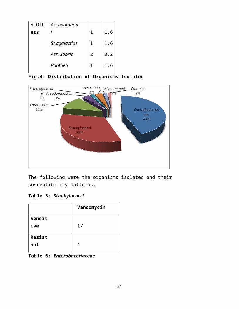
Results. A total of 63 organisms identified from the various sites and included Staphylococci-21
(33.3%), Enterobacteriaceae-28 (44.4%), Enterococci-7 (11%), Pseudomonas-2 (3.2%), Aer.
sobria-2 (3.2%) and one isolate each of Aci. baumanni, Strep. agalactiae and Pantoea (1.6%).
Among Staphylococci, 81% were sensitive to vancomycin which was the only drug that was
tested in VITEK 2. Enterobacteriaceae showed sensitivity against piperacillin/tazobactam,
cefoxitin, cefepime, amikacin and meropenem, and resistance against ampicillin and cefuroxime.
Pseudomonas isolates both showed sensitivity towards ceftazidime and amikacin with resistance
to ampicillin, piperacillin/tazobactam, cefuroxime, cefoxitin. St. agalactiae had sensitivity
towards both ampicillin and vancomycin. Aer. sobria was sensitive towards cefepime and
amikacin. Pantoea showed sensitivity towards cefepime and meropenem and resistance against
ceftazidime and piperacillin/tazobactam. Aci.baumanni was resistant to all drugs tested against
it: ampicillin, piperacillin/tazobactam, ceftazidime, cefuroxime and cefoxitin.
Conclusion and Recommendations. Emerging ESKAPE (Enterobacter, S.aureus, K.Pneumonia,
A.baumanni, P.aeroginosa, E.faecium) organisms have been isolated in this study and remain
important pathogens as far as infections in sterile sites are concerned. Commonest organisms
from blood were Staphylococci and Klebsiella, and from ascitic fluid were Enterobacteriaeceae.
Surveillance especially for the emerging pathogens needs to be carried out judiciously to help
develop rational antimicrobial guidelines alongside continuous medical education. Rational use
of antibiotics is advised to help curb this trend of increasing antibiotic resistance.
ix

CHAPTER 1
1.0 BACKGROUND
An antimicrobial is an agent that kills microorganisms such as bacteria, viruses or fungi, or
suppresses their multiplication or growth. Antibiotic susceptibility is the inhibition of growth or
killing of bacteria by use of antibiotics.
Acquisition of Antimicrobial resistance (AMR) is resistance of a microorganism to an
antimicrobial agent to which it was originally sensitive. Resistant organisms are able to
withstand attack by antimicrobial medicines, such as antibiotics, antifungals and antivirals
such that standard treatments become ineffective and infections persist.
Resistant factors can be exchanged between certain types of bacteria when microorganisms
are exposed to antimicrobial drugs, causing them to evolve naturally into resistant strains.
The resistance rate to antibiotics has been on the increase due to factors such as inappropriate
use of antimicrobial medicines; either misuse or overuse such as treating viral infections with
antibiotics and poor infection prevention and control practices. On the contrary, underuse of
antibiotics also contributes to resistance through inadequate dosing and poor compliance by
patients hence according the micro-organisms opportunities to multiply and continue
spreading (WHO, 2011).
Additionally, weak or absent antimicrobial resistance surveillance systems makes it difficult
to acquire the necessary data needed to assess and improve antibiotic use.
According to Centre for Disease Control Morbidity and Mortality Weekly Report (2013), data
emerging from different parts of the world have suggested that strains of highly multidrug
1
resistant organisms have quadrupled in the past decade. This emergence can not be ignored as
the risk of morbidity and mortality is heightened when they afflict vulnerable individuals.
As AMR is a complex multifaceted problem, single isolated interventions have little impact in
curbing it and coordinated actions between different stakeholders starting from the patient to
the healthcare providers and national governments are required to effectively tackle it.
2

CHAPTER 2
2.0 LITERATURE REVIEW
2.1 STERILE BODY SITES, PATHOGENS AND CONTAMINANTS
Sterile body sites are those in which no bacteria or microbes exist as commensals when in a
healthy state. This can either be pathological agents or contaminants from skin or laboratory
processes and include blood, CSF, peritoneal fluid, pleural fluid and synovial fluid. Non-sterile
samples are those obtained from sites considered not sterile and there may be colonizing
microbial agents. The significance of the isolates obtained is through the density of growth, for
example in urine >105 colony-forming units (CFU) of bacteria per milliliter of urine, formed
colonies and in skin Bacillus growth versus S.epidermidis.
2.1.1 Cerebral Spinal Fluid:
CSF is a clear colorless bodily fluid found in the brain and spine whose primary function is to
cushion the brain within the skull and serve as a shock absorber for the central nervous system.
CSF also circulates nutrients and chemicals filtered from the blood and removes waste products
from the brain. It occupies the subarachnoid space (the space between the arachnoid and the pia
mater) and the ventricular system around and inside the brain and spinal cord (Wikipedia).
Various studies from India revealed that meningitis is caused by various pathogens depending on
the patient's age group. In neonates, Group B (49%) and non-Group B Streptococcus species,
Escherichia coli (18%), and Listeria monocytogenes (7%) are the most common causative
3
organisms. Children and infants acquire meningitis from infection with Haemophilus
influenzae (40-60%), Neisseria meningitidis (25-40%), and Streptococcus pneumoniae (10-
20%). The sources of adult meningitis include S. pneumoniae (30-50%), N. meningitidis (10-
35%), Staphylococcus (5-15%), other Streptococcus species, H. influenzae (1-3%), Gram-
negative bacilli (1-10%), and L. monocytogenes (Chandramukhi, 1989; Chinchankar, 2002;
Sonavane, 2008).
Unlike the community acquired bacterial meningitis, gram-negative bacilli (40-60%) and
staphylococci, mainly coagulase negative (30-50%), are the most common causative agents of
nosocomial meningitis (Krcmery,2000).
Latex Agglutination Test is an adjunct to conventional techniques in the diagnosis of pyogenic
bacterial meningitis, where the latter tests fail. It is used for detection of the antigens of
Streptococcus pneumoniae, Group B Streptococci, Escherichia coli, Neisseria meningitidis and
Haemophilus influenzae type b. It was originally designed to be used in patients who
demonstrated laboratory and clinical findings consistent with meningitis. However it has been
used much too often as screening tool in cases of suspected meningitis in patients whose CSF
specimens have normal chemistries and cell counts (Kiska, 1995).
2.1.2 Blood
Blood culture is required when bacteraemia (the presence of bacteria in the blood) or septicaemia
is suspected. It usually occurs when pathogens enter the bloodstream from abscesses, infected
wounds or burns, or from areas of localized disease as in pneumococcal pneumonia, meningitis,
pyelonephritis, osteomyelitis amongst others.
4

Septicaemia occurs when multiplying bacteria release toxins into the blood stream and trigger
the production of cytokines, causing fever, chills, toxicity, tissue anoxia, reduced blood pressure
and collapse and can complicate as septic shock.
Bloodstream infections cause significant morbidity and mortality worldwide and are among the
most common healthcare-associated infections, with mortality rates approaching 45% in
bacteremia due to gram negative organisms (Blot, 2002).
Diekema (2003) compared community-onset and nosocomial bloodstream infections and found
that Gram-positive pathogens caused the majority of both with Staphylococcus aureus being the
most common pathogen overall. Specifically, Escherichia coli was the most common cause of
community-onset bloodstream infection, whereas S. aureus caused similar proportions of both
community-onset (18%) and nosocomial (21%) bloodstream infections.
A study done by Deverick (2014) showed that healthcare exposure preceded the onset of blood
stream infections in almost 3 of every 4 patients in their cohort, as evidenced by the fact that
majority of the patients in that cohort had central venous lines and had invasive devices present
at the time of infection.
However, findings by Weinstein (1997) showed that only 50% of all positive blood cultures
represent true bloodstream infection.
Blood Contaminants:
A significant proportion of cases have been found to be contaminated with certain organisms
which include Coagulase Negative Staphylococci (most common), Corynebacterium species,
Bacillus species other than Bacillus anthracis, Propionibacterium acnes, Micrococcus species,
Viridians group streptococci, enterococci, and Clostridium perfringens (Weinstein, 2003,1997) .
5

According to National Nosocomial Infections Surveillance (NNIS) System (1991), coagulase-
negative bacteremia is often the result of long-term use of indwelling central and peripheral
catheters as well as other prosthetic devices, the ubiquity of these bacteria as normal skin flora,
and the ability of these relatively avirulent organisms to adhere to the surface of biomaterials .
With particular regard to Coagulase Negative Staphylococci, growth of 2-5% is considered as
contamination; >5% shows poor infection control or swabbing practices and <2% shows a high
risk of laboratory overprocessing. However, it is crucial to recognize that each of these
organisms can also represent true bacteremias with devastating consequences, particularly if
untreated due to misinterpretation as contaminants.
BODY CAVITIES
An effusion is fluid which collects in a body cavity or joint. Fluid which collects due to an
inflammatory process such as infections or malignancy is referred to as an exudate and that
which forms due to a non-inflammatory condition is referred to as a transudate.
2.1.3 Peritoneal Fluid
Ascitic (peritoneal) fluid is from the peritoneal (abdominal) cavity. Peritonitis means
inflammation of the peritoneum, which is the serous membrane that lines the peritoneal cavity.
It can be caused by the rupture of an abdominal organ, or as a complication of bacteraemia or
can be spontaneous. Peritoneal dialysis is also associated with a high risk of infection of the
peritoneum, subcutaneous tunnel and catheter exit site.
6
The causative organisms in peritoneal dialysis peritonitis are generally different to those causing
‘surgical peritonitis’. In surgical cases, infections are usually poly-microbial consisting of both
anaerobic and aerobic bacteria. In contrast, a single micro-organism, usually a skin-colonising
Gram-positive bacteria, is the common cause of peritoneal dialysis peritonitis; Staphylococcus
aureus, Staphylococcus epidermidis and Streptococcus spp. account for 60–80% of cases.
(Brook, 2004)
However, it must be kept in mind that a significant proportion of the infections are culture
negative - about 20% to 32.5% (Lobo, 2010) and so appropriate samples should be obtained be
obtained prior to commencing treatment. It is recommended that evaluation of culture-negative
Peritoneal dialysis-related infections be done for rapidly growing nontuberculous mycobacteria
infections especially in the clinical setting of non-resolving peritonitis after prior exposure to
antibiotics (Renaud, 2011).
Unfortunately, the prolonged turnaround time of 1 to 2 days of culture limits its utility for
directing antibiotic selection in acute care settings.
2.1.4 Pleural Fluid
This is fluid from the pleural cavity i.e. space between the lungs and the inner chest wall. Pleural
effusion is used to describe a nonpurulent serous effusion which sometimes forms in pneumonia,
tuberculosis, malignant disease, or pulmonary infarction (embolism), Systemic Lupus
Erythematosus, lymphoma, rheumatoid disease, or amoebic liver abscess.
Common bacterial pathogens include Streptococcus milleri group species, Streptococcus
pneumoniae, Methicillin sensitive Staphylococcus aureus (MSSA) and
the Enterobacteriaceae group (Foster, 2007; Meyer, 2011)
7
S. aureus is more commonly seen in the older, hospitalized patient with co-morbidities and is
associated with cavitation and abscess formation, with empyema present in 1-25% of adult cases.
(Lindstrom,1999). Anaerobic bacteria however contribute significantly to pleural infection,
being identified as the sole or co-pathogen in 25-76% of pediatric cases (Micek, 2005).
The causative microorganism is, however, only identified in approximately 50% of cases.
Empyema is used to describe a purulent pleural effusion when pus is found in the pleural space.
It can occur with pneumonia, tuberculosis, infection of a haemothorax (blood in the pleural
cavity), or rupture of an abscess through the diaphragm. Common organisms associated with
empyema include: Staphylococcus aureus, Haemophilus influenza, Streptococcus pneumonia,
Bacteroides, Streptococcus pyogenes, Pseudomonas aeruginosa, Actinomycetes, Klebsiella
strains, Mycobacterium tuberculosis. Organisms such as Methicillin Resistant Staphylococcus
aureus, Enterobacteriae and anaerobes are more prevalent in nosocomial empyema (Schultz,
2004)
Worldwide, Mycobacterium tuberculosis is one of the most important causes of pleural infection.
In immunocompetent patients Acid Fast Bacilli smear in pleural effusion is rarely positive and in
HIV patients it’s positive in 20%. Pleural fluid culture is positive approximately 40% of
patients. In order to achieve a definitive diagnosis of tuberculous pleurisy, Mycobacterium
tuberculosis must be isolated from the culture of pleural fluid or tissue; the presence of
granulomas in pleural tissue is suggestive. (Valdes, 2003)
Choice of antibiotic should be informed by the results of blood and pleural fluid cultures and
sensitivities; empirical anaerobic antibiotic cover should be considered as anaerobes frequently
coexist but are difficult to isolate.
8

2.1.5 Synovial Fluid
Synovial fluid is the thick colourless lubricating fluid that surrounds a joint and fills a tendon
sheath. It’s secreted by the synovial membrane which lines the joint capsule, and when inflamed
this is known as synovitis. Causes can be due to bacteria, rheumatic disorder, or injury. Infective
synovitis is usually secondary to bacteraemia.
Organisms involved in both synovitis and arthritis include Staphylococcus aureus , Neisseria
gonorrhoeae, Streptococcus pyogenes, Neisseria meningitides, Streptococcus pneumoniae ,
Haemophilus influenza, Anaerobic streptococci Brucella species, Actinomycetes, Salmonella
serovars, Escherichia coli, Pseudomonas aeruginosa, Proteus, Bacteroides and Mycobacterium
tuberculosis.
While S. aureus, group B streptococci and Gram-negative organisms are isolated in newborn
infants, in older infants Hemophilus influenzae becomes a prominent pathogen (Sequira, 1985).
In those over 2 years of age, staphylococci, streptococci, H.influenzae and Neisseria
gonorrhoeae are predominant organisms (Barton, 1987). S.aureus was found to be the
commonest cause of septic arthritis in children by Wang (2003), however in other studies it was
not confined to any age group (Barton, 1987; Welkon, 1986). The gold-standard test for
diagnosis of septic arthritis is synovial fluid culture which is positive in 80 % of cases according
to Wang (2003).
2.2 CLINICAL IMPORTANCE AND IMPLICATIONS OF CURRENT PRACTICES OF
ANTIMICROBIAL USE.
There is dramatic increase of antimicrobial resistance worldwide in response to antibiotic use,
and is causing significant morbidity and mortality. It has been estimated that antimicrobial
9

resistance costs the health-care system in excess of US$ 20 billion in the USA annually and
generates more than 8million additional hospital days (Roberts, 2009).
A recent situation analysis by Kariuki (2011) in Kenya showed that three main factors
contribute greatly to Antimicrobial resistance which include:
1. Burden of Infectious disease :
The top five killers in Kenya are Infectious diseases with Acute respiratory infections being the
second leading cause with pneumonia as the largest contributor to the burden of disease among
children living in ‘urban informal settings, followed by diarrhoeal diseases.
2. Healthcare Environment and Behaviour
Antibiotics are also misused, their effectiveness wasted in patients with conditions that cannot be
cured by antibiotics. Possible reasons for this include: lack of microbiology facilities and
diagnostic capacity; fear of negative outcomes if antibiotics are withheld, particularly with
malaria patients and limited access to formal healthcare services and the prevalence of self-
medication.
3. Antibiotic Use in Animals
Evidence on antibiotic use in farm animals indicates that these medicines are used primarily
(90%) for therapeutic applications. There’s no regulation of antibiotic use in Kenya and no
surveillance is done for effectiveness of these drugs. This will greatly impact on the
susceptibility of most pathogens even those causing human disease. It’s been postulated that not
only clonal spread of resistant strains occurs, but also transfer of resistance genes occurs between
human and animal bacteria.
10
Ecological factors:
Antimicrobial resistant bacteria, like antibiotic-susceptible bacteria can spread, from person to
person to the environment, and then back to humans. In addition, the genes that encode
antimicrobial resistance are often readily transferable from resistant to susceptible
microorganisms, which can then multiply, spread and act as a source of further transfer of
resistance genes. Infection prevention and control activities such as proper hygiene and sewage
disposal to limit the spread of resistant bacteria are therefore crucial.
2.3 JUSTIFICATION
Sterile sites are those in which no bacteria or microbes exist as commensals when in a healthy
state, such that any growth is considered significant and can either be pathogenic micro-
organisms or contaminants. On the contrary, for non-sterile sites, not all isolated organisms are
significant as they can be normal flora.
Diagnosis of sterile site infections is based on culture of properly collected and processed
samples. Preliminary reports such as gram staining, biochemistry and cytology may be helpful in
providing immediate information to support the diagnosis and justify initiation of antibiotic
treatment. However since definitive diagnosis is based on quantitative cultures, the course of
antibiotic therapy should be determined after the culture results have been confirmed.
Unfortunately in most instances it is either not possible to wait for culture reports hence empiric
treatment is commenced, or laboratory facilities are unavailable or unreliable.
11

Infections caused by drug resistant organisms are difficult to treat hence limiting the therapeutic
options for treatment. With growing antimicrobial resistance in Kenya, reliance on international
guidelines is insufficient. There is need of research such as this one to get antibiotic
susceptibility testing profiles for pathogens from sterile site infections in our local setting, to
enable us formulate local guidelines for treatment.
2.4 RESEARCH QUESTION
What is the prevalence and antibiotic susceptibility patterns of bacterial isolates from sterile
body sites from both inpatients and outpatients at the KNH microbiology laboratory in the period
January to December 2013?
2.5 OBJECTIVES
2.5.1 Broad ObjectiveTo describe the bacterial isolates identified from sterile body sites and their antibiotic
susceptibility patterns from both inpatients and outpatients at the KNH microbiology laboratory,
in the period January to December 2013.
2.5.2 Specific Objectives
1. To describe bacterial isolates identified in cultures and their distribution in body fluids
collected from sterile sites: CSF, blood, pleural, ascitic and synovial fluids.
2. To describe the antibiotic susceptibility patterns of isolated bacteria to various antibiotic
agents.
12

CHAPTER 3
3.0 STUDY DESIGN AND METHODOLOGY
3.1 Study design
This was a retrospective descriptive study using previously available data from the KNH
patients’ laboratory files.
3.2 Study area
The study was based in Kenyatta National Hospital Medical Microbiology Laboratory. KNH is
located in the Kenyan capital city of Nairobi. It’s the largest teaching and referral for East and
Central Africa and provides services to patients within the catchment area. It serves as one of the
only two tertiary referral centers in the country and is also a training institute for doctors housing
the University of Nairobi, College of Health Sciences.
3.3 Study Population
Data was abstracted from the KNH Microbiology laboratory records of both inpatients and
outpatients of samples collected from sterile sites.
Inclusion criteria:
Laboratory records of all patients from whom bacterial isolates were cultured from sterile body
sites during the period January to December 2013.
Exclusion Criteria:
Laboratory records of samples with no growth obtained.
13

3.4 Sampling
There was no sampling the number of sterile site samples in the period January to December
2013. The whole population was included in the study. The whole population was eligible for the
study. Initially 74 samples were thought to be eligible but 11 ended up being contaminants
bringing the population size to 63.
This was done in the Microbiology Laboratory archives, where materials were archived for each
sample collected. Information on sterile sample sites specimen was picked; then each was run
through manually to check the ones positive for growth. Thereafter sequential data extraction of
isolates from sterile body sites was done from January to December 2013, through file reports
and registers and also summary of Vitek 2 backup data which is a computerized software that
analyses and stores data for epidemiological review (Appendix 5.4.3).
3.5 Data collection, entry and validation
Abstraction of data using a coded form was done from the existing laboratory database which
was recorded on a tally sheet for the time period January to December 2013. All the data
collection tools were reviewed by the principle investigator to ensure proper data management
was done. Data collected was entered into a database in a password protected computer and a
backup.
3.6 Procedures
The procedures that were used in the study were Data Collection form and Laboratory
Procedures.
14
3.6.1 Data Collection FormThis is a tool that was used to gather information on variables of interest, in a systematic fashion
that would enable us to answer the stated research questions, test hypotheses, and evaluate
outcomes. It was adapted from a previous study done by Dr Simon Njiru in Institute of Tropical
and Infectious Diseases (Appendix 5.4.1).
3.6.2 Laboratory ProceduresThe procedures that were used in the laboratory to isolate and identify isolates and the
antimicrobial susceptibility patterns during the period January to December 2013 have been
elaborated further in Appendix 5.4.2.
3.7 Ethical Issues
Ethical approval prior to conducting the research was obtained from KNH/UON Ethics and
Research Committee. Permission to extract data from the hospital registers and records was
obtained from the KNH Head of Laboratory Medicine through KNH Research Office.
The study was a minimal risk study as there was no direct patient involvement but a retrospective
review of the records.
For confidentiality, the patient’s files were used within the confines of the KNH microbiology
Laboratory and only the investigator with the assistance of research assistants and laboratory
personnel would access the files for purposes of this study.
Patient identifying information such as the name and patient numbers were not included in the
data collection forms. The report registers when not in use were kept under lock and key in the
Microbiology Laboratory archives.
15

3.8 Data Management and Analysis
Data downloaded from Vitek database was transferred to a code-secured MS Excel spreadsheet.
It was then cleaned continually as the study progressed.
Variables that were studied included:
Dependent: - Isolates from the respective sterile sites i.e. CSF, blood, peritoneal and pleural
fluid.
- Antibiotic susceptibility patterns of these isolates.
Independent: - Age and sex of the patients.
For analysis, MS Excel spreadsheet was imported to Statistical Package for Social Sciences
Programme (SPSS) version17.0. The data was then analyzed for demographic characteristics,
isolate outcomes and antibiotic susceptibility patterns.
This was carried out in two stages:
i) Univariate analysis
The Univariate analysis involved summarization and graphical presentation of both categorical
and continuous variables. Categorical variables were summarized using frequency distributions
and presented using bar charts and frequency distribution tables.
ii) Bivariate analysis
16

Bivariate analysis was used to investigate any association between variables. The χ2 test was
used to test association between 2 variables if they were categorical and satisfied all the
conditions. If some χ2 conditions were not be met, Fisher’s exact test was used instead.
The raw data was then stored in a secured MS-Excel spreadsheet in a safe locker in UNITID till
after publication.
STUDY LIMITATIONS
1. Missing data of some of the computerized records of relevant information such as the
wards and sample type necessitated going back to the archives for confirmation.
2. Analysis of synovial fluid could not be carried out as there was no growth obtained from
any of the samples.
17
18
CHAPTER FOUR.
4.1 RESULTS
During the twelve month study period, majority of the general population from whom samples
were obtained were female at 63.5% in comparison to male who were 36.5%.
Table 1: Demographic Characteristics
Characteristics N %
Gender
Male 23 36.5
Female 40 63.5
63 100
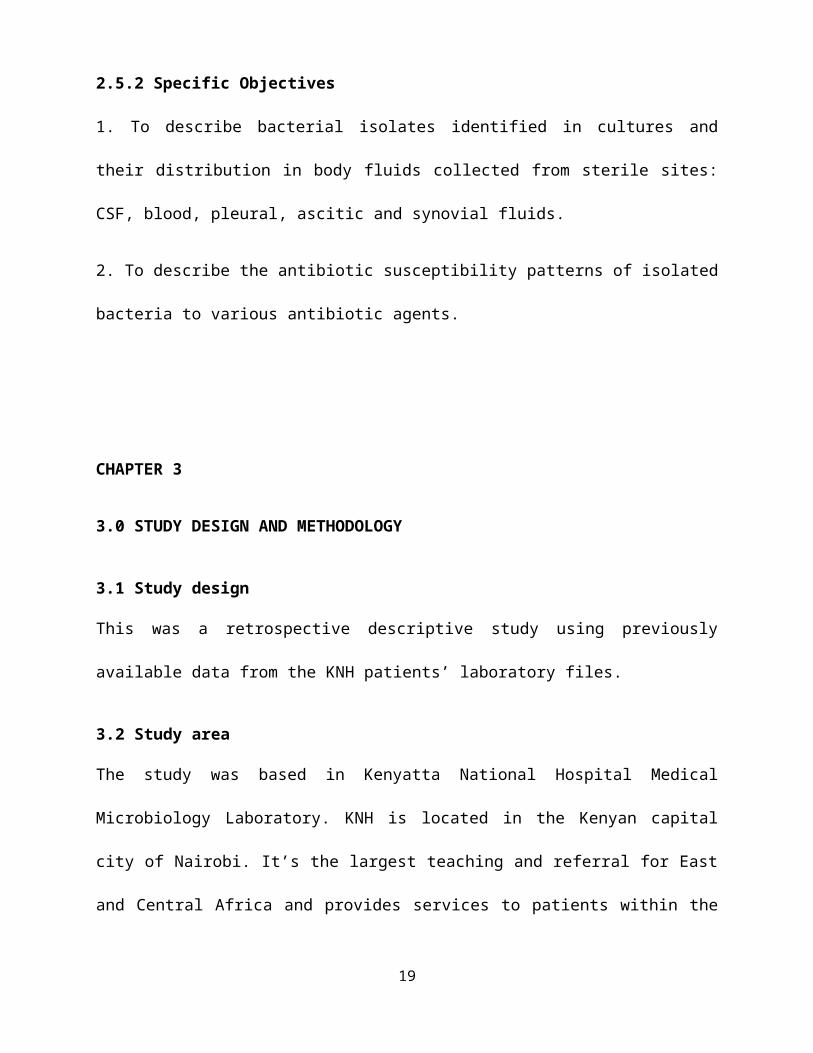
Sterile site samples obtained were mainly from blood culture at 73% (46/63), followed by ascitic fluid 11.1% (7/63) then pleural fluid and CSF both at 7.9% (5/63) each. No isolate was obtained from synovial fluid.
Figure 1: Sample source distribution
19
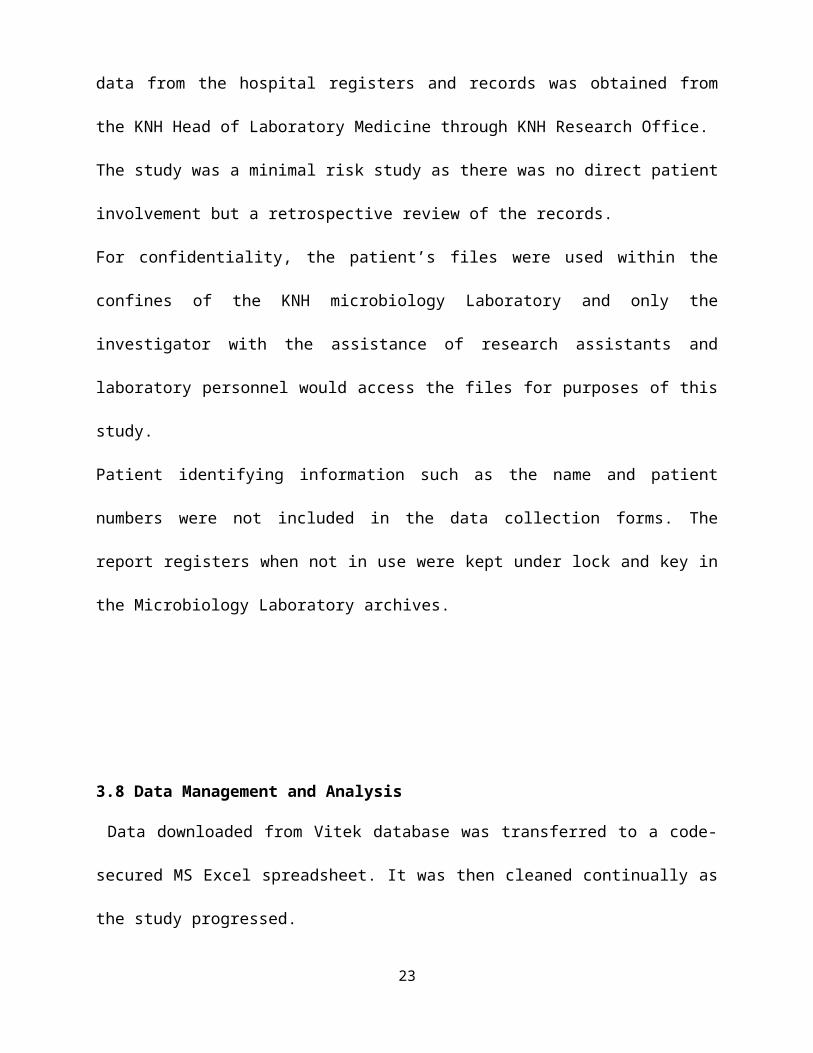
According to ward location, Newborn unit seemed to dominate with 54% of isolates arising from there, followed by the general adult wards (23.8%), followed by paediatrics ward (15.9%) with a small percentage from renal unit (3.2%) , radiology and Intensive care unit (1.6% each).
Figure 2: Ward distribution
Table 2: Isolation rate
The isolation rate for each of the sterile samples collected over the year 2013 were as follows:
SampleTotal collected Total cultured Isolation rate (%)
CSF 2,088 5 0.24
Blood 2,852 46 1.6
Ascitic fluid 263 7 2.66
Pleural fluid 262 5 1.91
Synovial fluid 49 0 0
20
The month with the highest number of isolates was September with 33 isolates whereas from February to June no isolates were identified, and the lowest being 2 in July, August and December.
Fig. 3: Isolation by Months
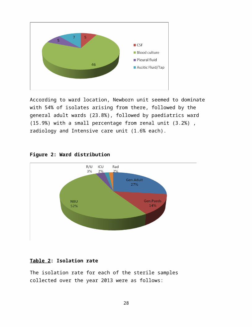
Organisms isolated
The following organisms were identified:
Table 3
Organisms N %
1. Staphylococci 21 33.3
Staph.aureus 3 4.8
Other Staph 18 28.6
2. Enterobacteriaceae 28 44.4
E.coli 6 9.5
Klebsiella 19 30
E.cloacae 2 3.2
S.marcescens 1 1.6
3. Pseudomonas 2 3.2
21
4. Enterococci 7 11
E.faecalis 4 6.4
E.faecium 2 3.2
E.casseliflavus 1 1.6
5.Others Aci.baumanni 1 1.6
St.agalactiae 1 1.6
Aer. Sobria 2 3.2
Pantoea 1 1.6
Fig.4: Distribution of Organisms Isolated
The following were the organisms isolated and their susceptibility patterns.
Table 5: Staphylococci
Vancomycin
Sensitive 17
Resistant 4
22

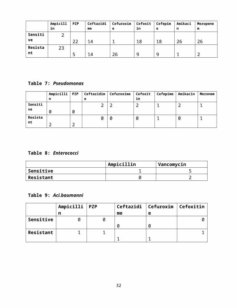
Table 6: Enterobaceriaceae
Ampicillin PZP Ceftazidime Cefuroxime Cefoxitin Cefepime Amikacin Meropenem
Sensitive 2 22 14 1 18 18 26 26
Resistant 23 5 14 26 9 9 1 2
Table 7: Pseudomonas
Ampicillin PZP Ceftazidime Cefuroxime Cefoxitin Cefepime Amikacin Meronem
Sensitive 0 0 2 2 2 1 2 1
Resistant 2 2 0 0 0 1 0 1
Table 8: Enterococci
Ampicillin VancomycinSensitive 1 5Resistant 0 2
Table 9: Aci.baumanni
Ampicillin PZP Ceftazidime Cefuroxime CefoxitinSensitive 0 0 0 0 0Resistant 1 1 1 1 1
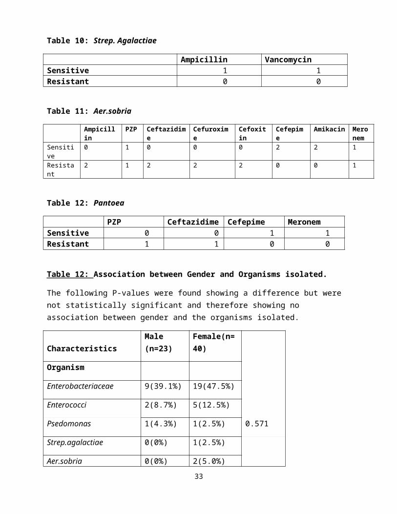
Table 10: Strep. Agalactiae
Ampicillin VancomycinSensitive 1 1Resistant 0 0
23

Table 11: Aer.sobria
Ampicillin PZP Ceftazidime Cefuroxime Cefoxitin Cefepime Amikacin Meronem
Sensitive 0 1 0 0 0 2 2 1
Resistant 2 1 2 2 2 0 0 1
Table 12: Pantoea
PZP Ceftazidime Cefepime MeronemSensitive 0 0 1 1Resistant 1 1 0 0
Table 12: Association between Gender and Organisms isolated.
The following P-values were found showing a difference but were not statistically significant and therefore showing no association between gender and the organisms isolated.
Characteristics Male (n=23)Female(n=40)
0.571
Organism
Enterobacteriaceae 9(39.1%) 19(47.5%)
Enterococci 2(8.7%) 5(12.5%)
Psedomonas 1(4.3%) 1(2.5%)
Strep.agalactiae 0(0%) 1(2.5%)
Aer.sobria 0(0%) 2(5.0%)
Aci.baumanni 0(0%) 1(2.5%)
Pantoea 1(4.3%) 0(0%)
Staph 10(43.5%) 11(27.5%)
24

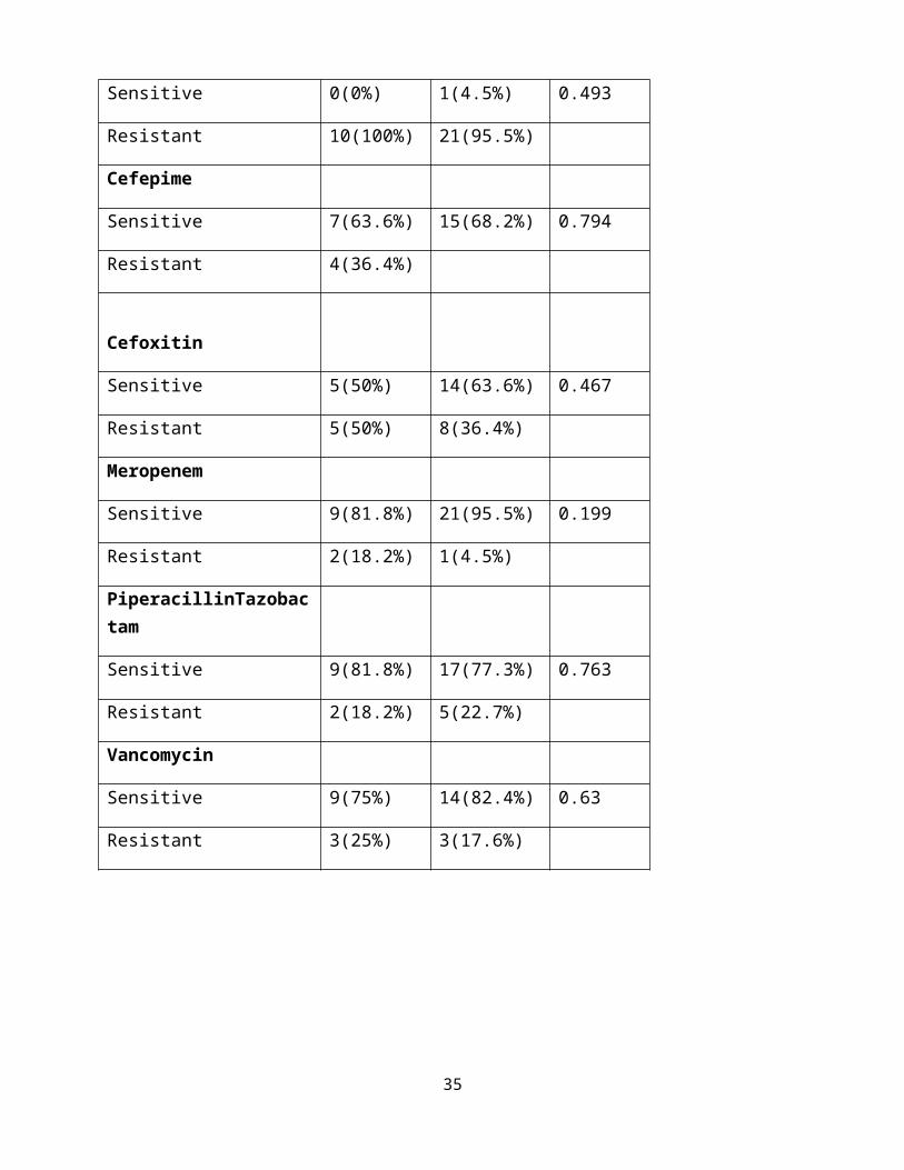
Table 12: Association between Gender and Antibiotic Susceptibility Patterns.
The following P-values were found showing a difference but were not statistically significant and therefore showing no association between gender and antibiotic susceptibility patterns.
Males(n=23)
Female(n=40)
Ampicillin
Sensitive 1(10.0%) 4(16.0%) 0.647
Resistant 9(90.0%) 21(84.0%)
Amikacin
Sensitive 10(100%) 20(95.2%) 0.483
Resistant 0(0%) 1(4.8%)
Ceftazidime
Sensitive 5(45.5%) 11(47.8%) 0.897
Resistant 6(54.5%) 12(52.2%)
Cefuroxime
Sensitive 0(0%) 1(4.5%) 0.493
Resistant 10(100%) 21(95.5%)
Cefepime
Sensitive 7(63.6%) 15(68.2%) 0.794
Resistant 4(36.4%)
25
Cefoxitin
Sensitive 5(50%) 14(63.6%) 0.467
Resistant 5(50%) 8(36.4%)
Meropenem
Sensitive 9(81.8%) 21(95.5%) 0.199
Resistant 2(18.2%) 1(4.5%)
PiperacillinTazobactam
Sensitive 9(81.8%) 17(77.3%) 0.763
Resistant 2(18.2%) 5(22.7%)
Vancomycin
Sensitive 9(75%) 14(82.4%) 0.63
Resistant 3(25%) 3(17.6%)
4.2 DISCUSSION
26

The aim of this study was to identify bacterial pathogens identified from samples collected in
sterile sites and their antibiotic susceptibility patterns.
4.2.1 CSF
In the present study, an isolate each of E.coli and Staphylococci were isolated from both adults
and children. This was partly in keeping with previous studies by Chandramukhi (1989),
Chinchankar (2002), and Sonavane(2008) though the main organisms they found ie
N.meningitidis, S.pneumoniae and H.influenzae in infants and children were not isolated. This
could be because of the increased uptake of childhood vaccines. No isolates were obtained from
the neonatal group. P.aeruginosa was isolated from the paediatric age group with the sample
having been obtained from an infected ventriculoperitoneal (VP) shunt in the surgical ward.
These findings can also be attributed to usage of antibiotics prior to collection of CSF samples in
peripheral clinics before referral.
P.aeruginosa is one of the most common nosocomial pathogens causing iatrogenic meningitis
( Talbot) and has greater ability to develop resistance to virtually any antibiotic it’s exposed to
because of multiple resistance mechanisms that can be present concurrently within the pathogen
(Livermore DM, 2001). In this study an incidence of 16% was found compared to that of 66% in
two different studies by Navageni (2011) and Jones (1997). Although lower, this incidence is
still high as this organism is mainly found in critical patients whose immunity is low.
4.2.2 Blood culturesBlood cultures had the highest number of samples collected over the year 2013 and also yielded
the highest number of isolated pathogens in comparison to other samples collected from sterile
sites. A majority of the samples were from the newborn unit.
27

In this study, the majority of isolates were Klebsiella species (37%) with 82% being from the
newborn unit, followed by Coagulase negative Staphylococci (30.4%), then Enterococci
(10.9%). This is in contrast to a previous study done by Deverick (2014) whose findings were
S.aureus (28%) followed by E.coli (24%) then Coagulase Negative Staphylococcci (10%). The
observed emergence of K.oxytoca which is an opportunistic pathogen associated with
nosocomial infections may be due to poor infection control measures in the newborn unit,
overcrowding and also the fact that newborns themselves are susceptible to infections due to
immature immune systems. In addition, lack of standard facilities with controlled air flow, entry
and exit points and proper sterilization of all gowns and equipment are also contributory. The
high rate of growth of CoNS may also be due to poor infection control or swabbing practices.
4.2.3 Ascitic fluidAll positive cultures of ascitic fluid were from adult patients from medical wards with none from
the surgical wards. This could be attributed to misclassification of ascitic taps under pus aspirates
or surgical site infections. The main organisms found in this study were Enterobacteriaceae
(57%) and Enterococci (29%). 50% (2/4) of Enterobacteriaceae were from peritoneal dialysis
fluid in the renal unit. This was in keeping with a study done by Montravers (2009) to identify
microbiological profiles of intra-abdominal infections found E.coli to be the highest (27%),
followed by Anaerobes (23%) and Streptococci(12%) . On the contrary, findings by Reuken
(2012) which isolated Enterococci as 50% of gram positive bacteria in spontaneous bacterial
peritonitis and 28% in secondary peritonitis .Only one patient was found to have S.epidermidis
which is usually peritoneal dialysis related primarily due to touch contamination (Finkelstein,
2002) but in this case we were not able to identify whether this patient had had dialysis before or
not.
28

4.2.4 Pleural fluidPleural fluid isolates found were Klebsiella and Staphylococcus species, in keeping with
previous studies by Foster (2007) and Schultz (2004). An interesting finding is that of
Aci. Baumanni isolated from a child in the general paediatric ward. This is an organism that is
becoming increasingly nosocomial and has been identified by Rice (2008) as an ESKAPE
pathogen (Enterobacter, S.aureus, K.Pneumonia, A.baumanni, P.aeroginosa, E.faecium) whose
drug of choice for treatment is carbapenems (Talbot, 2006).
4.2.5 Antibiotic Susceptibility Patterns.
Among Staphylococci, 81% were sensitive to vancomycin which was the only drug that was
tested in VITEK 2 at KNH. Other antibiotic susceptibility tests were done using antibiotic disk
inhibition method. Out of the 21 that were tested, 4 (19%) showed resistance against vancomycin
and only 25% of these were S.aureus. These findings are contrary to a study done in neonates by
Qu (2010) whereby most coagulase-negative staphylococcal isolates were resistant to penicillin
G (100%), gentamicin (83.3%) and oxacillin (91.7%) and susceptible to vancomycin (100%). A
different study in Benin by Sina (2013) looking at 136 isolates of S.aureus strains from
furuncles, pyomyositis, abscesses, Buruli ulcers, and osteomyelitis, from hospital admissions and
out-patients found all strains to be resistant to benzyl penicillin, while 25% were resistant to
methicillin, and all showed sensitivity to vancomycin. Further local studies are necessary to
establish staphylococcal susceptibility patterns to vancomycin to ascertain whether it can still be
a drug of choice considering the upward trend of antimicrobial resistance.
The Enterobacteriaceae showed sensitivity against Piperacillin/Tazobactam, cefoxitin, cefepime,
amikacin and meropenem, and resistance against ampicillin and cefuroxime. Against ceftazidime
they had 50% sensitivity and 50% resistance. These findings are consistent with a local study by
Kebira (2012) where all isolates of E.coli (100%) were susceptible to Ticarcillin,
29

Piperacillin/Tazobactam, Amikacin, Ofloxacin; and 80% of the isolates were susceptible to
Gentamycin, Norfloxacin, and Ceftazidime.
Isolates of K.oxytoca were susceptible to ampicillin-sulbactam, imipenem, aminoglycosides and
fluoroquinolones in a different study (Lin,1997), showing a similar pattern to the same family of
antibiotic group but different molecular structure.
Pseudomonas isolates both showed sensitivity towards ceftazidime and amikacin as well.
Against meropenem and cefepime 50% sensitivity and 50% resistance was found. Resistance
was noted against ampicillin, piperacillin/tazobactam, cefuroxime and cefoxitin. In 2002, 14 %
multridrug resistant isolates were found by Obritsch where isolates were resistant to 3 out of 4
drugs: ceftazidime, ciprofloxacin, tobramycin and imipenem. Further studies are necessary to
identify drugs that can curb this trend.
Enterococci, most of which were isolated from blood, were found to be sensitive towards
ampicillin but majorly towards vancomycin. On the contrary previous studies seem to contradict
our findings. One study by Reuken showed 63% resistance to ampicillin and 13% to vancomycin
and a different study has shown resistance of vancomycin to be on the increase with a rate of
∼60% among E. faecium isolates (Wisplinghoff, 2004).
Findings by Landman (2002) revealed that many isolates of Aci.baumanni are now resistant to
all aminoglycosides, cephalosporins, and fluoroquinolones, similar to findings in this study
where the only isolate was resistant to all drugs tested against it: ampicillin,
piperacillin/tazobactam, ceftazidime, cefuroxime and cefoxitin. There’s need for further
development of drugs against it.
St. agalactiae was sensitive towards both ampicillin and vancomycin, in keeping with findings
from a study by Simoes (2004).
30
Aer. sobria was sensitive towards cefepime and amikacin. An intermediary pattern towards
piperacillin/tazobactam and meropenem with 50% sensitivity and resistance was shown.
Resistance against ampicillin, ceftazidime, cefuroxime and cefoxitin were noted. This was in
contrast to a Taiwanese study which looked at Aeromonas though primarily, A. hydrophila. The
strains were found to be susceptible to moxalactam, ceftazidime, cefepime, aztreonam,
imipenem, amikacin, and fluoroquinolones, but they were more resistant to tetracycline,
trimethoprim-sulfamethoxazole, some extended-spectrum cephalosporins, and aminoglycosides
than strains from the United States and Australia.
Pantoea showed sensitivity towards cefepime and meropenem and resistance against ceftazidime
and piperacillin/tazobactam. A Texas Children’s Hospital retrospective study (January 2000 to
December 2006) reviewed culture-positive P.agglomerans records. This organism was identified
in 88 patient cultures. For the 53 children whose sterile-site cultures grew P.agglomerans, the
isolates were uniformly susceptible to amikacin, gentamicin, meropenem and trimethoprim-
sulfamethoxazole. In addition, 92.5% of isolates were susceptible to broad-spectrum
cephalosporins and semisynthetic penicillins, 62.3% to extended-spectrum cephalosporins, and
47.2% to ampicillin. Our findings are similar to those of this study and the two drugs can be
recommended for treatment of Pantoea.
4.3 CONCLUSION
Emerging ESKAPE (Enterobacter, S.aureus, K.Pneumonia, A.baumanni, P.aeroginosa,
E.faecium) organisms have been isolated in this study and remain important pathogens as far as
infections in sterile sites are concerned. Commonest organisms from blood were Staphylococci
and Klebsiella, and from ascitic fluid were Enterobacteriaeceae.
31

Among Staphylococci, 81% were sensitive to vancomycin which was the only drug that was
tested.
The Enterobacteriaceae showed sensitivity against Piperacillin/Tazobactam, cefoxitin, cefepime,
amikacin and meropenem, and resistance against ampicillin. These drugs can still be used with
confidence in patients with suspected enterobacteriaceae infections.
Pseudomonas isolates both showed sensitivity towards ceftazidime and amikacin and multidrug
resistance against ampicillin, piperacillin/tazobactam, cefuroxime and cefoxitin. Further studies
are necessary to identify drugs that can curb this trend.
Further studies need to be done especially concerning novel antibiotics to establish the best drugs
that can be used to tackle these bacteria.
Recommendations:
1. Surveillance especially for the emerging pathogens needs to be carried out judiciously to
help develop rational antimicrobial guidelines alongside continuous medical education .
2. Rational use of antibiotics is advised to help curb this trend of increasing antibiotic
resistance.
3. Infection prevention and control interventions in the hospital such as promotion of proper
personal hygiene and controlled environment eg. in newborn units is necessary to help
control nosocomial infections.
4. Subsidizing the cost of some of the very expensive antibiotics like vancomycin will help
with the treatment especially of the emerging organisms.
32
CHAPTER 5
5.1 TIMELINE
2014
Apri
l
May June July Aug Sept Oct Nov
Proposal submission and
Ethical approval
X X X
Data Abstraction X X
Data analysis and Report
writing
X X
Project Defense X
33
5.2 BUDGET
ITEM COST ( Kshs)
1. Ethics fees
2. Statistician
3. Operating expenses: stationery,
Printing, photocopying etc
4. Miscellaneous
5. Contingency
2,000
30,000
35,000
10,000
7,700
TOTAL 84,700
34
5.3 REFERENCES
1. Al Anazi KA, Al Jasser AM, Al Zahrani HA et al, 2008. Klebsiella oxytoca bactereamia
causing septic shock in recepients of haematopoetic stem transplant: Two case reports.
Cases Journal ;1:160.
2. Barton LL, Dunkle LM, Habib FH, 1987. Septic arthritis in childhood. A 13-year
review. The American Journal of Diseases of Children; 141: 898–900; 149: 53–40.
3. Blot S, Vandewoude K, De Bacquer D et al, 2002. Nosocomial bacteremia caused by
antibiotic-resistant gram-negative bacteria in critically ill patients: clinical outcome and
length of hospitalization. Clinical Infectious Diseases; 34:1600–1606.
4. Brook NR, White SA, Waller JR et al, 2004. The surgical management of peritoneal
dialysis catheters. Annals of the Royal College of Surgeons of England ; 86: 190-195
5. Centers for Disease Control and Prevention, 2013. “Vital signs: carbapenem-resistant
enterobacteriaceae,” Morbidity andMortality Weekly Report (MMWR).
6. Chandramukhi A,1989. Neuromicrobiology. In: Neurosciences in India: Retrospect and
Prospect. The Neurological Society of India, Trivandrum. New Delhi. Council of Scientific and
Industrial Research ; 361-95.
7. Chinchankar N, Mane M, Bhave S et al, 2002. Diagnosis and outcome of acute bacterial
meningitis in childhood. Indian Padiatrics ; 39: 914-21.
8. Cruz A, Cazacu A, Allen C, 2007. Pantoea agglomerans, a Plant Pathogen Causing
Human Disease. Journal Of Clinical Microbiology,45(6): 1989–1992
35
9.Dagnachew M, Yitayih W, Getachew T et al, 2014 .Bacterial isolates and their antibiotic
susceptibility patterns among patients with pus and/or wound discharge at Gondar
university hospital. ; 7:619
10. Deverick JA, Rebekah WM, Richard S et al ,2014 Bloodstream Infections in Community
Hospitals in the 21st Century: A Multicenter Cohort Study PLos ONE ; 9 (3)
11. Diekema DJ, Beekmann SE, Chapin KC et al, 2003: Epidemiology and outcome of
nosocomial and community-onset bloodstream infection. Journal of Clinical Microbiology,
41:3655-3660.
12. Finkelstein ES, Jekel J, Troidle L et al, 2002. Patterns of infection in patients maintained
on long-term peritoneal dialysis therapy with multiple episodes of peritonitis . American
Journal of Kidney Diseases; 39: 1278-86.
13. Foster S, Maskell N, 2007. Bacteriology of complicated parapneumonic
effusions. Current Opinion in Pulmonary Medicine; 13:319–23.
14. Jones RN, Marshall SA, Pfaller MA et al, 1997. Nosocomial enterococcal blood stream
infections in the SCOPE Program: antimicrobial resistance, species occurrence, molecular
testing results and laboratory testing accuracy. Diagnostic Microbiology and Infectious
Disease; 29:95-102.
15. Kariuki S, Gichia M, Kakai R et al, 2011 Situation Analysis, Antibiotic use and
Resistance in Kenya. Global Antibiotic Resistance Partnership .
16. Kebira A.N, Ochola, P. and Khamadi, S.A. Isolation and antimicrobial susceptibility
testing of Escherichia coli causing urinary tract infections. Journal of Applied Biosciences
22: 1320 - 1325
36

17. Kiska DL, Jones MC, Mangum ME et al,1995. Quality assurance study of bacterial
antigen testing of cerebrospinal fluid. Journal of Clinical Microbiology; 33:1141–4.
18. Krcmery V, Paradisi F, 2000. Nosocomial bacterial and fungal meningitis in children:
An eight year national survey reporting 101 cases. Pediatric Nosocomial Meningitis Study
Group. International Journal of Antimicrobial Agents ; 15:143-7
19. Landman D, Quale JM, Mayorga D et al, 2002. Citywide clonal outbreak of multresistant
Acinetobacter baumannii and Pseudomonas aeruginosa in Brooklyn, NY: the preantibiotic
era has returned. Arch Intern Med;162:1515-20.
20. Lin RD, Hsuel PR, Chang SC et al, 1997. Bactereamia secondary to Klebsiella oxytoca:
Clinical features of patients and antimicrobial susceptibility of isolates. Clinical Infectious
Diseases. 24 (6): 1217-22
21. Lindstrom ST, Kolbe J, 1999. Community acquired parapneumonic thoracic
empyema: predictors of outcome. Respirology ; 4:173-9.
22. Livermore DM, 2001. Of Pseudomonas, prorins, pumps and carbapenems. Journal of
Antimicrobial Chemotherapy. 47 (3); 247-250
23. Lobo JV, Villar KR, de Andrade Júnior MP et al,2010 .Predictor factors of peritoneal
dialysis-related peritonitis. Jornal Brasileiro de Nefrologia; 32: 156-164
24. Marilee D, Douglas N, Robert M.et al, 2004. National Surveillance of Resistance in
Pseudomonas aeruginosa Isolates Obtained from Intensive Care Unit Patients from 1993-
2002. Antimicrobial Agents and Chemotherapy; 48:4606-10
37
25. Meyer CN, Rosenlund S, Nielsen J, Friis-Møller A, 2011. Bacteriological aetiology and
antimicrobial treatment of pleural empyema. Scandinavian Journal of Infectious
Diseases. 43(3):165-9.
26. Micek ST, Dunne M, Kollef MH, 2005. Pleuropulmonary complications of Panton-
Valentine leukocidin-positive community-acquired methicillinresistant Staphylococcus
aureus: importance of treatment with antimicrobials inhibiting exotoxin production. Chest
Journal ; 128:2732-8.
27. Montravers P, Lepape A, Dubreuil L et al, 2009. Clinical and microbiological profiles of
community-acquired and nosocomial intra-abdominal infections: results of the French
prospective, observational EBIIA study .Journal of Antimicrobial Chemotherapy;63(4):785-
94.
28. Nagaveni S, Rajeshwari H, Oli KA et al, 2011. Widespread Emergence of Multidrug
Resistant Pseudomonas aeruginosa Isolated from CSF Samples. Indian Journal of
Microbiology; 51(1):2-7
29. National Nosocomial Infections Surveillance (NNIS) System, 1991. Nosocomial infection
rates for interhospital comparison: limitations and possible solutions. Infection Control and
Hospital Epidemiology; 12:609-621.
30. Obritsch MD, Fish DN, Mac Laren R, 2004. National Surveillance of Antimicrobial
Resistance of Pseudomonas aeruginosa isolates from Intensive care unit from 1993-
2002.Antimicrobial Agents and Chemotherapy; 48:4606-10
31. Qu Y, Daley A, Istivan T et al , 2010. Antibiotic susceptibility of coagulase-negative
staphylococci isolated from very low birth weight babies: comprehensive comparisons of
38

bacteria at different stages of biofilm formation. Annals of Clinical Microbiology and
Antimicrobials, 9:16
32. Renaud CJ, Subramanian S, Tambyah PA et al, 2011. The clinical course of rapidly
growing nontuberculous mycobacterial peritoneal dialysis infections in Asians: A case
series and literature review. Nephrology (Carlton); 16: 174-17
33. Reuken P, ,Pletz M, M. Baier et al , 2012. Emergence of spontaneous bacterial peritonitis
due to enterococci – risk factors and outcome in a 12-year retrospective study. Alimentary
Pharmacology & Therapeutics ;35(10): 1199–1208.
34.Rice LB, 2008. Federal Funding of the study of antimicrobial resistance in nosocomial
pathogens : no ESKAPE. Journal of Infectious Diseases. 197 (8): 1079-81
35. Roberts RR, Hota B, Ahmad I et al, 2009. Hospital and Societal Costs of antimicrobial-
resistant infections in a Chicago teaching Hospital: Implications for Antibiotic
Stewardship. Clinical Infectious Diseases; 49:1175-84
36. Schultz KD, Fan LL, Pinsky J et al, 2004. The changing face of pleural empyemas in
children: epidemiology and management. Pediatrics ;113:1735-40
37. Sequeira W, Swedler WI, Skosey JL, 1985. Septic arthritis in childhood. Annals of
Emergency Medicine ; 14(12): 1185–7.
38. Simoes J, Aroutcheva A, Heimler I et al, 2004. Antibiotic Resistance Patterns of Group B
Streptococcal Clinical Isolates. Infectious Diseases in Obstetrics and Gynecology. 12 (1): 1-8
39

39.Sina H, Ahoyo T,Moussaoui W et al , 2013. Variability of antibiotic susceptibility and
toxin production of Staphylococcus aureus strains isolated from skin, soft tissue, and bone
related infections. BMC Microbiology 13:188
40. Sonavane A, Baradkar VP, Mathur M, 2008. Bacteriological profile of pyogenic meningitis
in adults. Bombay Hosp Journal; 50: 452-55.
41.Talbot GH, Bradley J, Edwards S et al, 2006. Bad bugs need drugs: an update on
development pipeline from Antimicrobial Availability Task force of Infectious disease
society of America. Clinical Infectious Diseases. 42: 657-668.
42. Valdes L, Pose A, San Jose E, et al, 2003. Tuberculous pleural effusions. European
Journal of Internal Medicine, 14: 77–88
43. Wang CL, Wang SM, Yang YJ, et al, 2003. Septic arthritis in children: relationship of
causative pathogens, complications, and outcome. Journal of Microbiology, Immunology and
Infection ; 36 (1):41-6
44. Weinstein MP, Towns ML, Quartey SM, et al 1997: The clinical significance of positive
blood cultures in the 1990s: a prospective comprehensive evaluation of the microbiology,
epidemiology, and outcome of bacteremia and fungemia in adults. Clinical Infectious
Diseases , 24:584-602
45. Wen Chien Ko, Kwok Woon Yu, Cheng Yi Liu et al, 1995. Increasing Antibiotic
Resistance in Clinical Isolates of Aeromonas Strains in Taiwan. Antimicrobial Agents And
Chemotherapy; 40(5): 1260–1262
40
46. Wisplinghoff H, Bischoff T, Tallent SM et al, 2004. Nosocomial bloodstream infections in
US hospitals: analysis of 24,179 cases from a prospective nationwide surveillance study.
Clinical Infectious Diseases;39:309-17.
5.4 APPENDIX
5.4.1 Data Collection Form
41
ANTIMICROBIAL SUSCEPTIBILITY PATTERNS OF BACTERIA ISOLATED FROM
STERILE SITES: CEREBRAL SPINAL FLUID, BLOOD, PERITONEAL
FLUID, PLEURAL FLUID AND SYNOVIAL FLUID AT KENYATTA NATIONAL
HOSPITAL.
Sample No:
Socio-Demographic characteristics
Age of Patient :…………Yrs
Sex Male
Female
Sample: CSF Blood Culture Peritoneal fluid
Pleural Fluid Synovial Fluid
Month in which organisms were isolated.
January February
March April
May June
July August
September October
November December
Area from which specimen was obtained
ICU Paeds Gen Ward
Renal Unit Burns OPD
42
Others
Organisms Isolated: ……………………………………..
Antibiotic Susceptibility Patterns
Drug Sensitive Resistant
Ampicillin
Amoxicillin/Clavulanic Acid
Amoxicillin
Amikacin
Beta-Lactamase
Chloramphenicol
Ceftazidime
Cefalotin
Ciprofloxacin
Clindamycin
Cefpodoxime
Ceftriaxone
Cefotaxime
Cefuroxime
Erythromycin
Fusidic Acid
Cefepime
43
Fosfomycin
Cefoxitin
Nitrofurantoin
Gentamicin
Imipenem
Levofloxacin
Linezolid
Meropenem
Mupirocin
Moxifloxacin
Norfloxacin
Ofloxacin
Oxacillin
Benzylpenicillin
Piperacillin
Quinupristin/Dalfopristin
Rifampicin
Ampicillin/Sulbactam
Trimethoprim/
Sulfamethoxazole
Tetracycline
Teicoplanin
Telithromycin
44

Tobramycin
Piperacillin/Tazobactam
Vancomycin
5.4.2 Laboratory Procedures
I. SAMPLE COLLECTION
a)CSF :
Before aspirating the specimen the skin is disinfected then the sample is collected in a sterile
screw-cap tube. Rapid testing such as Gram stain and cryptococcal antigen should be considered.
For direct examination, best sensitivity for Gram staining is achieved after centrifugation.
It is transported immediately to the lab and can be stored for 6 hours at room temperature before
processing. Primary plating media is Blood Agar (BA) and Chocolate Blood Agar (CBA).
b)Blood culture:
The venipuncture site is disinfected with 70%alcohol and disinfectant such as betadine. Blood
should be drawn at the time of the febrile episode and if possible before antimicrobial treatment
has started. Two sets are drawn from right and left arms, not more than three sets should be
drawn in a 24hour period. For adults draw > 20ml/set and for children 1-20mls/set depending on
patient’s weight and blood culture bottles’ manufacturer’s instructions. It should be transported
to the laboratory within 2hours of collection, and should be incubated at 37oc.
c)Effusions:
45
The skin is disinfected before aspirating the specimen using a needle. The sample’s collected in a
universal sterile, screw-cap tube or anaerobic transporter and should be transported immediately
to the laboratory, with a maximum of 6hours for ascitic fluid. Plating should be done as soon as
received using both aerobic and anaerobic blood culture bottle set for body fluids. Gram staining
is then done. One may need to concentrate by centrifugation or filtration then stain and culture
the sediment.
II. SAMPLE PROCESSING
a) CSF
Day 1: Report appearance by describing whether it’s clear, slightly turbid, cloudy or purulent
and whether it contains blood or clots.
Testing c.s.f.:
- Purulent or cloudy c.s.f.: Suspect pyogenic bacterial meningitis and perform a Gram smear then
report on the number of pus cells and on bacteria.
-Slightly turbid or clear c.s.f.: Perform cell count and note whether there are pus cells or
lymphocytes. If pus cells then do a gram smear and then culture. If lymphocytes measure protein
and glucose and do a Ziehl-Nielsen stain for Acid Fast Bacilli (AFB) and Indian Ink for
encapsulated yeasts.
b) Blood culture
Using an aseptic technique, dispense 10–12 ml blood into Columbia agar diphasic medium and
mix. Incubate up to 7 days (4 weeks when brucellosis is suspected). When growth occurs it is
subcultured on BA and CBA for identification. When anaerobic infection is suspected, dispense
5 ml blood into thioglycollate broth and mix. Incubate up to 14 days.
The blood broth is allowed to run over the slope by tipping the bottle at regular intervals.
Microbial activity can be seen by growth on the slope (beginning at the broth-agar interface).
46

3.Effusions:
Day 1 - Describe colour and whether the specimen’s clear, cloudy, or purulent; blood stained or
contains clots (non-citrated sample).
If pus: Do a gram smear and microscopically examine for pus cells and bacteria, and Ziehl-
Nielsen smear when tuberculosis or M. ulcerans disease is suspected.
- Potassium Hydroxide (KOH) preparation: when a fungal or actinomycete infection is
suspected.
-Giemsa or Wayson’s smear: when bubonic plague is suspected.
-Polychrome methylene blue: when cutaneous anthrax is suspected.
-Dark-field microscopy: To detect treponemes when yaws or pinta is suspected.
If blood stained: proceed to culture.
Examine by estimating cell numbers and report % of cells that are neutrophils or lymphocytes.
For protein: report total protein in g/l.
- Transudate: Clear unclotted fluid with few cells and protein below 30 g/l: No need to test
further.
- Exudate: When cloudy fluid with more than a few cells and protein over 30 g/l do culture and
examine microscopically.
- Gram smear - microscopically look for pus cells and Ziehl-Nielsen look for AFB.
III SAMPLE CULTURE AND IDENTIFICATION
a)CSF
Day 2 and onwards, culture CSF.
On BA and CBA. Incubate in CO2
47

– If neonate: also MacConkey agar. Incubate aerobically.
On BA and CBA look particularly for:
. Neisseria meningitidis (growing on chocolate agar and blood agar, oxidase positive)
. Streptococcus pneumoniae (sensitive to optochin)
. Haemophilus influenzae (growing only on chocolate agar)
. Cryptococcus neoformans (Gram stain the colonies)
On MacConkey agar culture look especially for bacteria that cause neonatal meningitis.
b) Blood culture
Day 2 and onwards examine and report cultures. After overnight incubation examine diphasic
culture. Subculture (even when no growth is seen at end of 7 days for aerobic incubation or 14
days for anaerobic incubation) on BA and incubate aerobically or anaerobically. For CBA
incubate in CO2.
Examine thioglycollate culture. Subculture and examine microscopically. Incubate subculture
anaerobically.
Note: When there is no growth, wash slope of diphasic culture. Reincubate cultures. Subculture
as indicated.
Examine subcultures for likely pathogens.
c)Effusions
Culture specimen on BA and incubate aerobically. For CBA incubate in CO2 and for
MacConkey which is rarely used incubate aerobically.
Culture for M. tuberculosis requires facilities of a Tuberculosis Reference Lab on Lowenstein
Jensen Medium and examined for growth after 6weeks.
Examine and report cultures: BA, CBA, MacConkey.
48

Look particularly for: S. aureus, S. pyogenes, S. pneumonia, H. influenza, Neisseria species,
Enterobacteria, P. aeruginosa.
IV ANTIMICROBIAL SUSCEPTIBILITY TESTING (AST)
This is done for all the above samples with growth after subculturing on Muller-Hinton Agar.
Disc diffusion techniques are used by most laboratories to test routinely for antimicrobial
susceptibility. A disc of blotting paper is impregnated with a known volume and appropriate
concentration of an antimicrobial, and this is placed on the plate of susceptibility testing agar
uniformly inoculated with the test organism. The antimicrobial diffuses from the disc into the
medium and the growth of the test organism is inhibited at a distance from the disc that is related
(among other factors) to the susceptibility of the organism. Strains susceptible to the
antimicrobial are inhibited at a distance from the disc whereas resistant strains have smaller
zones of inhibition or grow up to edge of the disc.
In VITEK 2, the AST is done automatically with appropriate cassettes and results produced
automatically and where in doubt, conventional methods used to confirm identification and AST
results.
5.4.3 Vitek 2The Vitek 2 System (bioMérieux) is highly automated and uses very compact plastic reagent
cards (credit card size) that contain microliter quantities of antibiotics and test media in a 64-well
format. Each card has a pre-inserted transfer tube used for inoculation . Cards have bar codes
that contain information on product type and a unique identifier that can be linked to the sample
either before or after loading the card onto the system.
49

There are currently four reagent cards available for the identification of different organism
classes :
1.GN - Gram-negative fermenting and non-fermenting bacilli
2. GP - Gram-positive cocci and non-spore-forming bacilli
3. YST - yeasts and yeast-like organisms
4. BCL - Gram-positive spore-forming bacilli
Suspension Preparation
A sterile swab or applicator stick is used to transfer a sufficient number of colonies of a pure
culture and to suspend the microorganism in 3.0 mL of sterile saline in a 12 x 75 mm clear
plastic (polystyrene) test tube.
Inoculation
Identification cards are inoculated with microorganism suspensions using an integrated vacuum
apparatus. A test tube containing the microorganism suspension is placed into a special rack
(cassette) and the identification card is placed in the neighboring slot while inserting the transfer
tube into the corresponding suspension tube. The filled cassette is transported automatically into
a vacuum chamber station. After the vacuum is applied and air is re-introduced into the station,
the organism suspension is forced through the transfer tube into micro-channels that fill all the
test wells.
Card Sealing and Incubation
Inoculated cards are passed by a mechanism, which cuts off the transfer tube and seals the card
prior to loading into the carousel incubator. Each card is removed from the carousel incubator
once every 15 minutes, transported to the optical system for reaction readings, and then returned
to the incubator until the next read time. Data are collected at 15-minute intervals during the
entire incubation period.
50
Optical System
A transmittance optical system allows interpretation of test reactions using different wavelengths
in the visible spectrum. During incubation, each test reaction is read every 15 minutes to measure
either turbidity or colored products of substrate metabolism. In addition, a special algorithm is
used to eliminate false readings due to small bubbles that may be present.
Database Development
The databases of the VITEK 2 identification products are constructed with large strain sets of
well-characterized microorganisms tested under various culture conditions. These strains are
derived from a variety of clinical and industrial sources as well as from public (e.g., ATCC) and
university culture collections.
Analytical Techniques
Test data from an unknown organism are compared to the respective database to determine a
quantitative value for proximity to each of the database taxa. Each of the composite values is
compared to the others to determine if the data are sufficiently unique or close to one or more of
the other database taxa. If a unique identification pattern is not recognized, a list of possible
organisms is given, or the strain is determined to be outside the scope of the database.
( Microbial Identification Using The Biomérieux Vitek® 2 System , David H. Pincus ,
bioMérieux, Inc. USA)
51