Embed Size (px)
Citation preview
9/6/2016
1
The Nervous SystemBiology 30
General Outcome:
� Explain how the Nervous Systemcontrols physiological processes
The Nervous SystemA system of the body that coordinates & regulatesthe activities of the body
Control System
� Maintains homeostasis
� homeo/stasis (same/changing)
◦ changes in order to keep a balance
5 major components
Stimulus receptor
modulator/regulator
effector
Action
Sensory Pathway
Motor Pathway
-highly specific-receive stimuli
-Selects appropriate Response (spinal cord or brain)
-carries out the response(muscle or gland)
The Nervous System & Homeostasis
◦Organization of the Nervous System
◦ Structure of a neuron
◦Action Potential
◦ Synaptic Transmission
◦ Structure of the Brain
◦ Senses: The Eye & Ear
9/6/2016
2
Nervous System Organizational Tree!
Peripheral Nervous
System (PNS)
Feeds into & out of CNS
Central Nervous System
(CNS)
Decision maker
Nervous System
Somatic Pathway
(Voluntary)
under conscious control
Examples?
Autonomic Pathway
(Involuntary)
unconscious control
Examples?
Parasympathetic
(Restores to normal)
Restores balance!
Restores Homeostasis!
Sympathetic
(Stimulatory)
Speeds you up!
Excites you!
Brain & Spinal CordMotor PathwaySensory Pathway
NS Overview
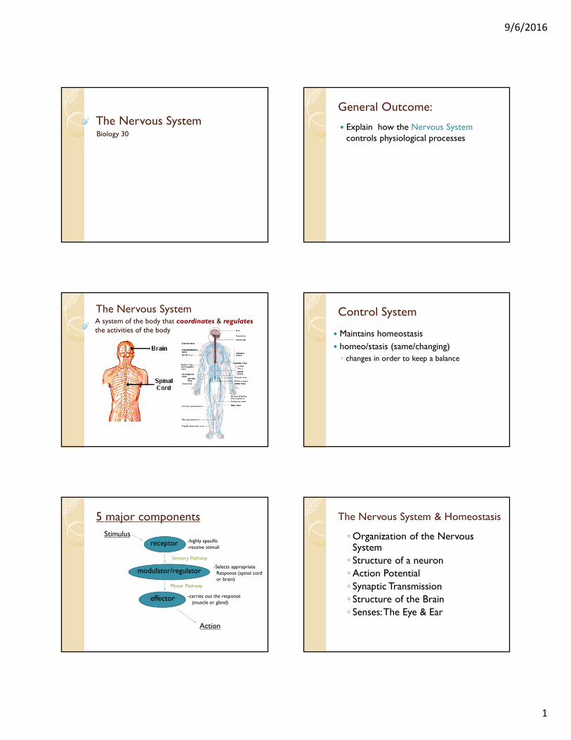
Anatomy of a Nerve Cell
� Two different types of cells are found in the nervous system:
◦ Glial Cells: non-conducting; important for support and metabolism of nerve cells
◦ Neurons: functional units of the nervous system (conduct nerve impulses)
The Neuron: Wires within a nerve!
See diagram on p.410 of text
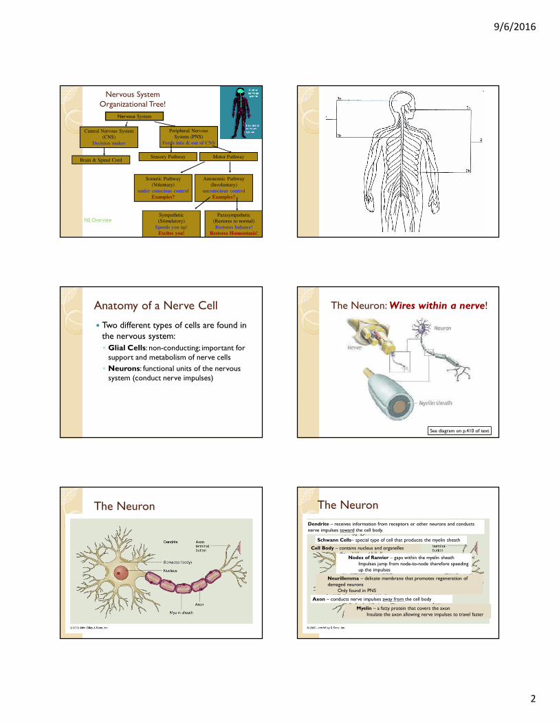
The Neuron The Neuron
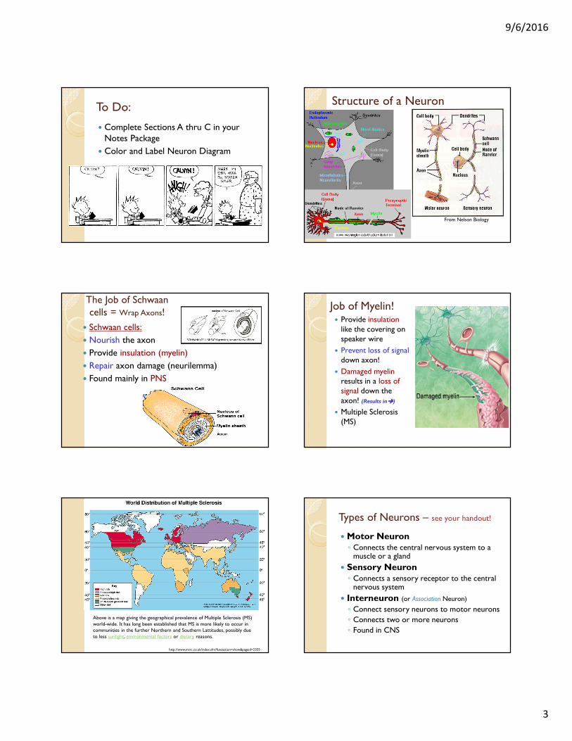
Myelin – a fatty protein that covers the axonInsulate the axon allowing nerve impulses to travel faster
Nodes of Ranvier – gaps within the myelin sheathImpulses jump from node-to-node therefore speeding up the impulses
Neurillemma – delicate membrane that promotes regeneration of damaged neurons
Only found in PNS
Schwann Cells– special type of cell that produces the myelin sheath
Dendrite – receives information from receptors or other neurons and conducts nerve impulses toward the cell body.
Cell Body – contains nucleus and organelles
Axon – conducts nerve impulses away from the cell body
9/6/2016
3
To Do:
� Complete Sections A thru C in your Notes Package
� Color and Label Neuron Diagram
Structure of a Neuron
From Nelson Biology
The Job of Schwaan cells = Wrap Axons!
� Schwaan cells:
� Nourish the axon
� Provide insulation (myelin)
� Repair axon damage (neurilemma)
� Found mainly in PNS
Job of Myelin!� Provide insulation
like the covering on speaker wire
� Prevent loss of signaldown axon!
� Damaged myelinresults in a loss of signal down the axon! (Results in����)
� Multiple Sclerosis (MS)
Above is a map giving the geographical prevalence of Multiple Sclerosis (MS) world-wide. It has long been established that MS is more likely to occur in communities in the further Northern and Southern Lattitudes, possibly due to less sunlight, environmental factors or dietary reasons.
http://www.msrc.co.uk/index.cfm?fuseaction=show&pageid=2325
Types of Neurons – see your handout!
� Motor Neuron◦ Connects the central nervous system to a
muscle or a gland
� Sensory Neuron◦ Connects a sensory receptor to the central
nervous system
� Interneuron (or Association Neuron)
◦ Connect sensory neurons to motor neurons
◦ Connects two or more neurons
◦ Found in CNS

9/6/2016
4
Types of NeuronsSensory Neurons (Afferent Neurons) –conducts nerve impulse from sense organs to the brain and spinal cord (CNS)
Motor Neuron (Efferent Neurons) –conducts nerve impulses from CNS to muscle fiber or glands (effectors)
Interneuron (Association Neuron) –found within the CNS
No myelinationIntergrates and interprets sensory information and relays information to outgoing neurons
To Do:� Table comparing neurons
� Finish workbook up to pg. 7
� Remember:
◦ Purchase Key Booklet by Friday February 12
◦ Parent letter – if I don’t already have it
9/6/2016
5
Returning involuntary body functions to normal after a period of stress is the function of which division of the nervous system?
A. Central
B. Somatic
C. Sympathetic
D. Parasympathetic
� Syphilis is a STI that affects the central nervous system. The neurons damaged by syphilis are
A. interneurons
B. sensory neurons
C. somatic motor neurons
D. autonomic motor neuron
FORMATIVE ASSESSMENT #1Common Reflexes
� Knee jerk reflex (patellar):◦ a tap on the patellar tendon causes top leg muscles to contract & lower leg muscles to
relax simultaneously
� Achilles Reflex:◦ Tap on the achelles tendon causes an ankle jerk
� Babinski reflex: ◦ a tickle of a babies foot causes the toes to curl
� Pupillary reflex:◦ iris diameter changes in response to light conditions
� Cross extensor reflex:◦ in a standing position, fatigue of one leg causes withdrawal of weight onto the other
leg
� Stretch reflex:◦ -the body senses the muscles shortening so the tendency is to stretch them out in
order to perform motor functions properly
� Withdrawal reflex:◦ defensive strategy in response to a painful stimuli like heat & cold or a cut or pinch of
some kind
� Others include: Vomiting, coughing, defecation, & milk release some of which are voluntary reflexes.
Reflexes:
� are well established neural circuits that are pre-programmed to allow motor responses to certain stimuli
� some may be instinctual as in newborns
� some are learned as in repeated motor tasks involved in sport
� some are defensive to enhance survival
� and other reflexes help your eye to move as you read this page.
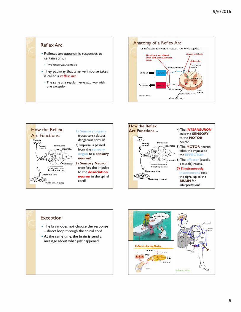
Don’t need to write yet… Reflex Arc� A reflex does not
require the brain
� Reflexes may be innate or acquired
9/6/2016
6
Reflex Arc
� Reflexes are autonomic responses to certain stimuli
◦ Involuntary/automatic
� They pathway that a nerve impulse takes is called a reflex arc
◦ The same as a regular nerve pathway with one exception
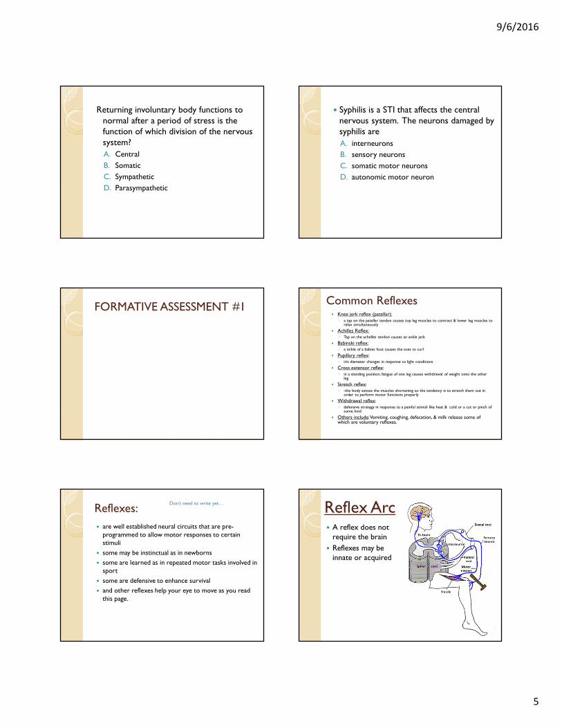
Anatomy of a Reflex Arc
How the Reflex Arc Functions:
1) Sensory organs(receptors) detect dangerous stimuli!
2) Impulse is passed from the sensory organ to a sensory neuron!
3) Sensory Neurontransfers the impulse to the Association neuron in the spinal cord!
4) The INTERNEURONlinks the SENSORYto the MOTORneuron!
5) The MOTOR neuron takes the impulse to the EFFECTOR!
6) The effector (usually a muscle) reacts.
7) Simultaneously, interneurons send the signal up to the BRAIN for interpretation!
How the Reflex Arc Functions…
Exception:
� The brain does not choose the response – direct loop through the spinal cord
� At the same time, the brain is send a message about what just happened.
Reflex Arc Video
9/6/2016
7
Learner Outcome:
� Describe the composition and function of reflex arcs
� Design and perform an experiment to investigate the physiology of reflex arcs
To Do:� Reflex Lab
� Section E in your notes package
◦ P. 414 Questions # 2, 3, 5, 6
***Key Booklets – pay $18 at office, due next Monday
You are innocently walking down the street when aliens zap away the sensory neurons in your legs. What happens?
a) Your walking movements show no significant change
b) You can no longer walk
c) You can walk, but the pace changes
d) You can walk, but clumsily
Action Potentialhow do nerves work???
� In 1900 Bernstein hypothesized that nerve impulses where electrochemical in nature.
◦ Future experimentation proved this.
� Giant Squid Experiment:
◦ Cole and Curtis placed two tiny electrodes –one inside the large axon of a squid and the second across from the first outside the axon.
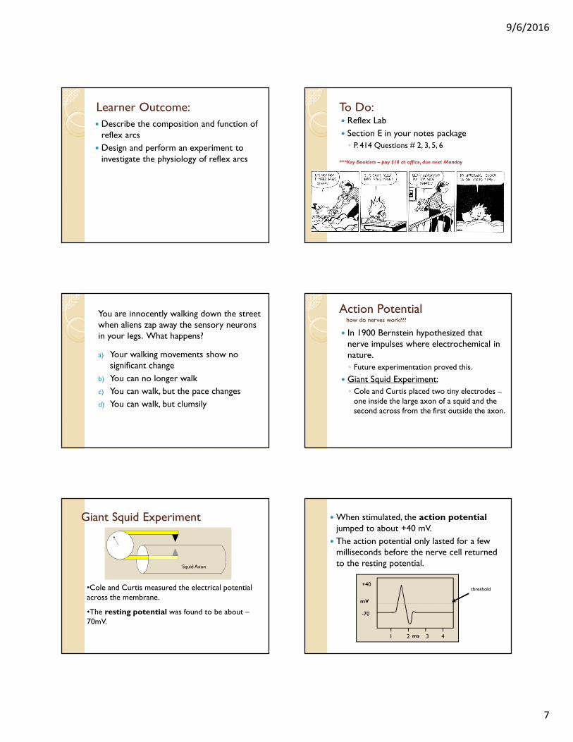
•Cole and Curtis measured the electrical potential across the membrane.
•The resting potential was found to be about –70mV.
Squid Axon
Giant Squid Experiment � When stimulated, the action potentialjumped to about +40 mV.
� The action potential only lasted for a few milliseconds before the nerve cell returned to the resting potential.
-70
+40
mV
1 2 3 4ms
threshold
9/6/2016
8
Definitions:
� Action Potential:
◦ the voltage difference across a nerve cell membrane when the nerve is excited (~40 mV)
� Resting Potential:
◦ Voltage difference across a nerve membrane when it is NOT transmitting a nerve impulse (almost always -70 mV)
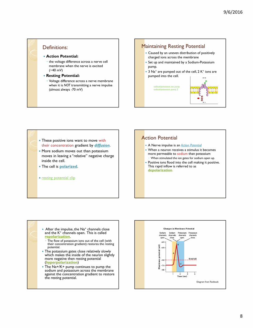
� Caused by an uneven distribution of positively charged ions across the membrane
� Set up and maintained by a Sodium-Potassium pump.
� 3 Na+ are pumped out of the cell, 2 K+ ions are pumped into the cell.
Maintaining Resting Potential
sodium/potassium ion pump
sodium/potassium pump 2
� These positive ions want to move with their concentration gradient by diffusion.
� More sodium moves out than potassium moves in leaving a “relative” negative charge inside the cell.
� The cell is polarized.
� resting potential clip
� A Nerve impulse is an Action Potential
� When a neuron receives a stimulus it becomes more permeable to sodium than potassium◦ When stimulated the ion gates for sodium open up.
� Positive ions flood into the cell making it positive. This rapid inflow is referred to as depolarization.
Action Potential
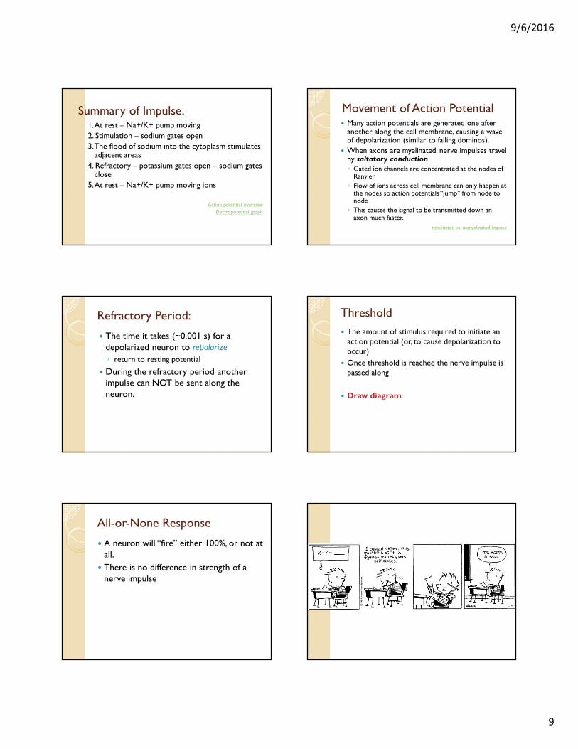
� After the impulse, the Na+ channels close and the K+ channels open. This is called repolarization.◦ The flow of potassium ions out of the cell (with
their concentration gradient) restores the resting potential.
� The potassium gates close relatively slowly which makes the inside of the neuron slightly more negative then resting potential (hyperpolarization)
� The Na+/K+ pump continues to pump the sodium and potassium across the membrane against the concentration gradient to restore the resting potential.
Diagram from Textbook
9/6/2016
9
Summary of Impulse.1. At rest – Na+/K+ pump moving
2. Stimulation – sodium gates open
3. The flood of sodium into the cytoplasm stimulates adjacent areas
4. Refractory – potassium gates open – sodium gates close
5. At rest – Na+/K+ pump moving ions
Action potential overview
Electropotential graph
Movement of Action Potential� Many action potentials are generated one after
another along the cell membrane, causing a wave of depolarization (similar to falling dominos).
� When axons are myelinated, nerve impulses travel by saltatory conduction◦ Gated ion channels are concentrated at the nodes of
Ranvier
◦ Flow of ions across cell membrane can only happen at the nodes so action potentials “jump” from node to node
◦ This causes the signal to be transmitted down an axon much faster.
myelinated vs. unmyelinated impuse
Refractory Period:
� The time it takes (~0.001 s) for a depolarized neuron to repolarize
◦ return to resting potential
� During the refractory period another impulse can NOT be sent along the neuron.
Threshold
� The amount of stimulus required to initiate an action potential (or, to cause depolarization to occur)
� Once threshold is reached the nerve impulse is passed along
� Draw diagram
All-or-None Response
� A neuron will “fire” either 100%, or not at all.
� There is no difference in strength of a nerve impulse
9/6/2016
10
To Do:
� Read pages 415 – 419 in your textbook
� Complete Section E in your notes package
� Schwann Cell & Action Potential� http://www.mcgrawhill.ca/school/applets/abbio/ch11/actionpotential_action.swf
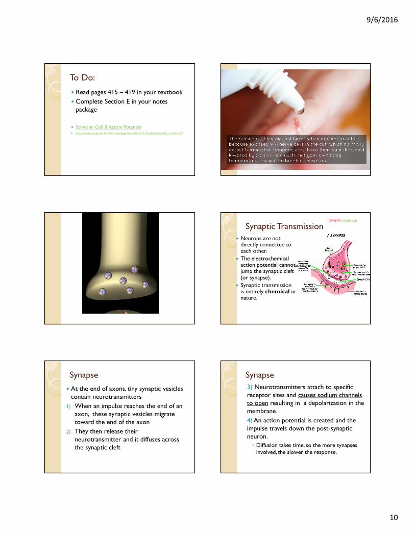
Synaptic Transmission
� Neurons are not directly connected to each other.
� The electrochemical action potential cannot jump the synaptic cleft (or synapse).
� Synaptic transmission is entirely chemical in nature.
Synapse movie clip
Synapse
� At the end of axons, tiny synaptic vesicles contain neurotransmitters
1) When an impulse reaches the end of an axon, these synaptic vesicles migrate toward the end of the axon
2) They then release their neurotransmitter and it diffuses across the synaptic cleft
Synapse
3) Neurotransmitters attach to specific receptor sites and causes sodium channels to open resulting in a depolarization in the membrane.
4) An action potential is created and the impulse travels down the post-synaptic neuron.
◦ Diffusion takes time, so the more synapses involved, the slower the response.
9/6/2016
11
Synapse – direction of message
� Synaptic transmission can only occur in one direction.
� Since only presynaptic neurons contain synaptic vesicles, and only post synaptic neurons have receptor sites for them, the messages cant be sent in the other direction
� This explains why impulses can only travel from sensory neuron to interneuron to motor neuron and never in the other direction
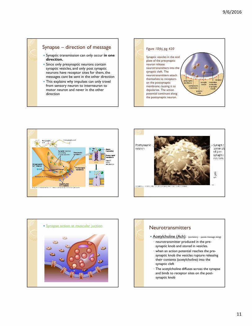
Figure 10(b), pg. 420
Synaptic vesicles in the end plate of the presynaptic neuron release neurotransmitters into the synaptic cleft. The neurotransmitters attach themselves to receptors on the postsynaptic membrane, causing it to depolarize. The action potential continues along the postsynaptic neuron.
� Synapse action at muscular juction Neurotransmitters
� Acetylcholine (Ach): (excitatory – passes message along)
◦ neurotransmitter produced in the pre-synaptic knob and stored in vesicles.
◦ when an action potential reaches the pre-synaptic knob the vesicles rupture releasing their contents (acetylcholine) into the synaptic cleft
◦ The acetylcholine diffuses across the synapse and binds to receptor sites on the post-synaptic knob

9/6/2016
12
Neurotransmitters
� How do we stop the message?
◦ Before another message can cross the cleft, it must be cleaned (remove the neurotransmitter)
◦ The enzyme acetyl cholinesterase removes acetylcholine from the receptor sites and breaks it into acetic acid & choline
◦ the acetic acid & choline are reabsorbed into the presynaptic knob to be reused
Neurotransmitters
� Neurotransmitters can be:◦ excitatory - passes along message to the next
neuron, or
◦ Inhibitory – binds to the next neuron and inhibits the message from being passed on
See figure 11 p. 422
• Not all neurons cause depolarization in the post synaptic membrane. Some neurons are inhibitory.
Neurotransmitters
� Often it takes more than one neuron releasing its neurotransmitter into the synaptic cleft to elicit a response in the post synaptic neuron.
◦ This is referred to as SUMMATION
Figure 11, pg. 422
Action potentials must occur simultaneously in A and B to reach the threshold in D.
Other Types of Neurotransmitters:
� Serotonin
� Dopamine
� Norepinephrine
� GABA
� You will fill in some information regarding these in your notes package but do NOT have to memorize them. If used on a diploma you will be told what you need to know about them
9/6/2016
13
13.2 Summary Electrochemical Impulse
• Nerves conduct electrochemical impulses from
the dendrites along the axon to the end plates of
the neuron.
• Active transport and diffusion of sodium and
potassium ions establish a polarized membrane.
• An action potential is caused by the inflow of
sodium ions.
• Nerve cells exhibit an all-or-none response.
• Neurotransmitters allow the nerve message to
move across synapses.
To Do:
� Complete section F in your notes package
� Mouse Party on Drugs