Embed Size (px)
DESCRIPTION
Strategies and Outcomes WithPharmacologic Vitreolysis:Perspectives
Citation preview

Strategies and Outcomes With Pharmacologic Vitreolysis:
Perspectives From Clinical
Practice
A roundtable discussion with Pravin U. Dugel, MD; Carl C. Awh, MD; R.V. Paul Chan, MD;
Dean Eliott, MD; and Elias Reichel, MD
Supported by an unrestricted educational grant from
Supplement to September 2013

2 SUPPlEMEnt to REtInA toDAY SEPtEMbER 2013
Strategies and Outcomes With Pharmacologic Vitreolysis:
Perspectives From Clinical Practice

SEPtEMbER 2013 SUPPlEMEnt to REtInA toDAY 3
ContentsIntroduction . . . . . . . . . . . . . . . . . . . . . . . . . . . . . . . . . . . . . . . . . . . . . . . . . . . . . . . . . . . . . . . . . . . . 4
Patient Selection for Ocriplasmin . . . . . . . . . . . . . . . . . . . . . . . . . . . . . . . . . . . . . . . . . . . . . . . 5
Preparing Patients for Injection With Ocriplasmin . . . . . . . . . . . . . . . . . . . . . . . . . . . . . . 5
Case Reports . . . . . . . . . . . . . . . . . . . . . . . . . . . . . . . . . . . . . . . . . . . . . . . . . . . . . . . . . . . . . . . . . . . 6
Additional Considerations . . . . . . . . . . . . . . . . . . . . . . . . . . . . . . . . . . . . . . . . . . . . . . . . . . . . .12
Future Uses of Ocriplasmin . . . . . . . . . . . . . . . . . . . . . . . . . . . . . . . . . . . . . . . . . . . . . . . . . . . .13
Physician Biographies and Financial Disclosures . . . . . . . . . . . . . . . . . . . . . . . . . . . . . . .14
Ocriplasmin: Summary of the Phase 3 Data . . . . . . . . . . . . . . . . . . . . . . . . . . . . . . . . . . . .15
4 SUPPlEMEnt to REtInA toDAY SEPtEMbER 2013
Perspectives From Clinical Practice
Strategies and outcomes With Pharmacologic Vitreolysis:
Perspectives From Clinical PracticeA roundtable discussion with Pravin U. Dugel, MD; Carl C. Awh, MD; R.V. Paul Chan, MD; Dean Eliott, MD; and Elias Reichel, MD
IntroductIonOcriplasmin (JETREA, ThromboGenics) is a truncated
form of human plasmin. It is made with recombinant DNA technology and targets degraded extracellular mol-ecules, such as fibronectin, laminin, and collagen, that comprise the macromolecular vitreomacular attachment complex. The mechanism of action of ocriplasmin is to enhance vitreous liquefaction and to facilitate separation of the vitreous cortex from the internal limiting mem-brane of the retina.1
The phase 3 studies of ocriplasmin, known as the MIVI-TRUST program, consisted of 2 separate trials—MIVI-006, which was conducted in the United States, and MIVI-007, which was conducted in the United States and European Union.2 The trials were prospective, randomized, double-masked, placebo-controlled, and evaluated the efficacy and safety of a single intravitreal injection of 125 µg ocri-plasmin vs placebo for the treatment of patients with symptomatic vitreomacular adhesion (VMA).
The primary endpoint was pharmacologic resolution of VMA at 28 days as determined by optical coherence tomography (OCT). Secondary endpoints included total posterior vitreous detachment (PVD) at day 28, non-surgical closure of full-thickness macular hole (FTMH), visual acuity improvement of 2 lines or greater, need for vitrectomy, and patient-reported assessment of visual function (with the National Eye Institute Visual Functioning Questionnaire-25). The study duration was 6 months.
Patients in these trials fell into 3 nonexclusive catego-ries: vitreomacular traction (VMT) without FTMH or epiretinal membrane (ERM) at baseline; FTMH with or without ERM at baseline; and patients with ERM at base-line (note that ERM was not a target of treatment). All patients had OCT-confirmed VMA. Ocriplasmin met the
studies’ primary endpoint of pharmacologic resolution of VMA at day 28. In the integrated data analysis of the 2 phase 3 studies, 26.5% of patients in the ocriplasmin group achieved VMA resolution at day 28 compared with 10.1% in the placebo group, a difference that was statistically significant.
The results for FTMH closure were impressive. Nonsurgical closure of FTMH was achieved by 40% of patients in the ocriplasmin group at day 28 through month 6. Subgroup analysis showed that patients with small FTMH (≤250 µm) had a higher rate of closure (60%) compared with patients with medium to large FTMH (>250 µm, 25%). Ocriplasmin did not appear to work well in cases where ERM was present.
It is not rare for a drug post-market to have different characteristics compared with the controlled premarket clinical trials (eg, phase 3 data). The reasons for this are varied and include complex study designs that are diffi-cult to mimic in a real-world setting, deviation from trial protocol, and patient selection criteria.
Although these results were statistically significant, when applied to clinical practice, they may be disap-pointing. I tell people to consider the MIVI-TRUST studies in proper historical context. These studies were conceived in approximately 2004, prior to the wide-spread use of OCT, and therefore, patients who would otherwise have been excluded were included, suggesting that more careful patient selection might have increased the success rates. Subsequent to the phase 3 data, there was a retropective analysis done to guide patient selec-tion and the predictors of response.
Here, we are going to discuss ocriplasmin and specific considerations that will help to maximize outcomes with this agent in clinical practice.
—Pravin U. Dugel, MD
SEPtEMbER 2013 SUPPlEMEnt to REtInA toDAY 5
Perspectives From Clinical Practice
PatIent SelectIon for ocrIPlaSmInDr. Dugel: Based on the clinical trial data, what is
your opinion regarding selecting appropriate candi-dates with symptomatic VMA and FTMH for injection with ocriplasmin?
Elias Reichel, MD: It would appear from the subanal-ysis and clinical experience that FTMH smaller than 250 µm, adhesions less than 1500 µm, and absence of ERM are important suggestors of success. Subanalysis also suggested age and phakic lens status were important, but I have my doubts about this. Cataracts are associ-ated with age, and so the younger a patient is, the more likely he or she is to be phakic. If a patient has already undergone cataract surgery and there is no PVD, there will be some increased adherence of the hyaloid to the fovea. More than 80% of patients who have cataract surgery will develop PVD if they have not had 1 already. Cataract surgery, itself, may increase fibrosis and inflam-mation and, therefore, you would have continued attachment to the fovea.
The absence of ERM is important, as it is a predictor of failure with ocriplasmin. The smaller hole size and smaller area of adhesion are good predictors of success with ocri-plasmin.
Carl C. Awh, MD: I agree that these are appropriate guidelines for patient selection, but it is also important to remember that patients with smaller areas of adhe-sion and no ERM are those who are most likely to experi-ence spontaneous resolution of VMA. I wish that ocri-plasmin worked better for eyes with ERM because it is very common in the setting of VMA. As with most new treatments, however, I am sure that with further clinical experience we will discover situations where patients who do not fit within these guidelines benefit from the use of ocriplasmin.
Dean Eliott, MD: Most stage II holes are fairly small in diameter. If the anatomy of the vitreous is going to be used to define the stages of macular holes, and the vitre-ous is focally adherent to a small flap of retina with a can opener-type appearance, then the vast majority of these stage II holes are not going to be larger than 250 µm.
R.V. Paul Chan, MD: An important question is, what does ocriplasmin do for visual function?
Dr. Dugel: This is a good point. What are the predic-tors for how patients will respond with ocriplasmin?
Dr. Eliott: The clinical trials only included patients with VMA less than 1500 µm and stage II macular holes. If you believe in the study results, I think it would be safe to follow these inclusion criteria, as these patients
respond favorably to ocriplasmin. As Dr. Reichel noted, I think that factors such as age and lens status are some-what irrelevant.
Dr. Dugel: Are there instances where you might con-sider going outside the boundaries of the clinical trial experience? For example, if a patient presented with a 400 µm FTMH but expressed that he or she did not want surgery and the subsequent gas or positioning, would you consider ocriplasmin? The current procedural tech-nology (CPT) code allows for holes up to 400 µm in the setting of VMA.
Dr. Awh: If avoiding a trip to the OR is highly impor-tant to the patient or doctor, for instance, because of the patient’s medical status, this changes the risk-benefit analysis for that patient. Because the success rate is lower, but not 0, for this size of macular hole and the risks associated with ocriplasmin are low I think that it is a reasonable option in this setting.
Dr. Reichel: We are currently in the infancy stage in terms of using ocriplasmin, and how we classify macu-lar holes and VMA is evolving. I would not say that it is incorrect to use ocriplasmin in this setting, but I think that it will require gathering the collective clinical expe-rience with the drug to know whether it will be useful outside the parameters from the clinical trials. The sub-group analyses include small numbers of patients.
Dr. Chan: I agree with Dr. Awh in that it is a patient selection issue. If a patient is wary of undergoing surgery, then I would discuss ocriplasmin. If the patient under-stands the risks and benefits and has given informed consent, in my opinion, it is a reasonable option.
Dr. Eliott: It is important to keep in mind that retina specialists do not routinely measure FTMH diameter. OCT scans do not typically have automated measurement soft-ware, so there is no record of the exact size; although, one can attempt to estimate hole diameter using calipers. In the clinical trials, favorable results were obtained for stage II macular holes less than 250 µm in diameter. It is unlikely that a patient with an FTMH of 380 µm would be con-sidered a candidate for ocriplasmin while another patient with an FTMH of 420 µm would be excluded solely on the basis of hole diameter. Both of these hypothetical patients have holes that are outside the parameter of the trials, and I think it is reasonable to simply tell patients that larger holes are less likely to close with this agent.
PreParIng PatIentS for InjectIon WIth ocrIPlaSmIn
Dr. Dugel: Do you prepare patients any differently for ocriplasmin than you would for aflibercept (Eylea,
6 SUPPlEMEnt to REtInA toDAY SEPtEMbER 2013
Perspectives From Clinical Practice
Regeneron), ranibizumab (Lucentis, Genentech), or beva-cizumab (Avastin, Genentech)?
Dr. Reichel: The anesthesia and the antisepsis proce-dure is exactly the same. The preparation of ocriplas-min is what is different. First, because it is an unstable drug, it must be kept frozen at -20° C prior to injection. My nursing and technician staff thaw and prepare the drug for injection, which includes adding 2/10 of 0.9 cc saline to the solution. The dilution is crucial. If the con-centration of ocriplasmin is too high there are potential risks of lens subluxation that were clearly evaluated in the preclinical studies.
Dr. Dugel: It is important to note that ocriplasmin has a very narrow therapeutic window. I agree that the dilu-tion is crucial. In the preclinical studies, lens subluxation occurred when ocriplasmin was given at 1.4 times the approved concentration.
I would advise that those who are new to using ocri-plasmin prepare the drug themselves, rather than having staff perform this task. Once the drug is thawed and pre-pared, it should be injected as soon as possible. The label states that it should be injected within 30 minutes.
Dr. Reichel: Once the drug is mixed, you have to swirl it around in the vial to ensure that there is no particulate matter, after which 1/10 of 0.9 cc is drawn out.
Dr. Awh: Can you add saline at the moment it is removed from the freezer, or does the drug have to be at room temperature?
Dr. Reichel: The drug must be thawed first. It thaws quickly, within 10 or 15 minutes.
Dr. Dugel: This hits on an important point. Unlike patients with AMD who are receiving ranibizumab, afliber-cept, or bevacizumab, for whom patient flow is rather sim-ple and only 1 room for injection is required, ocriplasmin injection requires a separate room for the mixing process and another for the injection. It is similar to the days of photodynamic therapy, when timing was of the essence.
Dr. Reichel: I agree. Ocriplasmin injections fall into a completely different flow. Some of this has to do with insurance, and our patient flow fits more into the cat-egory of an OR procedure.
caSe rePortSCase No . 1
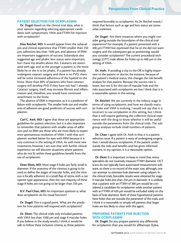
Dr. Dugel: A nurse, 70 years of age, presented with visual acuity loss for 4 months from a macular hole (Figure 1). When she presented to me, her visual acuity was 20/100. I injected her with ocriplasmin and within
hours after the injection, she called the office with com-plaints of flashes and floaters. Her vision had fallen to 20/400 the same day.
On OCT (Figure 2) it appeared that the hyaloid was straightening out, which demonstrates how quickly this drug works. Figure 3 shows her OCT at 2 weeks.
Dr. Chan: It appears that there is still some focal adhe-sion centrally.
Dr. Eliott: It is interesting, however, that the hole appears to be closed but some adhesion remains and it looks like there is a serous detachment.
Dr. Dugel: This is exactly what I thought. Despite adhesion, the macular hole appears to be closing from the inside out, like a zipper.
She was no longer bothered by the side effects of ocriplasmin even though her visual acuity had only improved to 20/200. Would you be confident that this patient would continue to improve?
Dr. Eliott: I would be confident that she would improve.
Figure 1 . A woman, 70 years of age, with visual acuity loss for
4 months from a macular hole and visual acuity of 20/100 .
Figure 2 . Same day post-injection . Visual acuity 20/400 .
SEPtEMbER 2013 SUPPlEMEnt to REtInA toDAY 7
Perspectives From Clinical Practice
Dr. Dugel: One month later, her visual acuity had improved to 20/40 and the outer retinal separation was visible on OCT (Figure 4). The patient was happy with her vision improvement, but she wanted it to be even better.
Dr. Eliott: I think that the serous fluid will improve, but I am not as certain about the outer retinal defect.
Dr. Reichel: We are seeing things after ocriplasmin injection that we could not have possibly observed after surgery because the gas bubble would obstruct our view on OCT. Postoperatively, we might see defects 2 to 3 months out, but they are improved due to time and often the patients have excellent vision.
Dr. Dugel: I agree that the serous fluid should resolve, but it is hard to say whether the defect will improve or whether it will have an effect on her vision in the long term. Figure 5 shows the patient’s improvement over time.
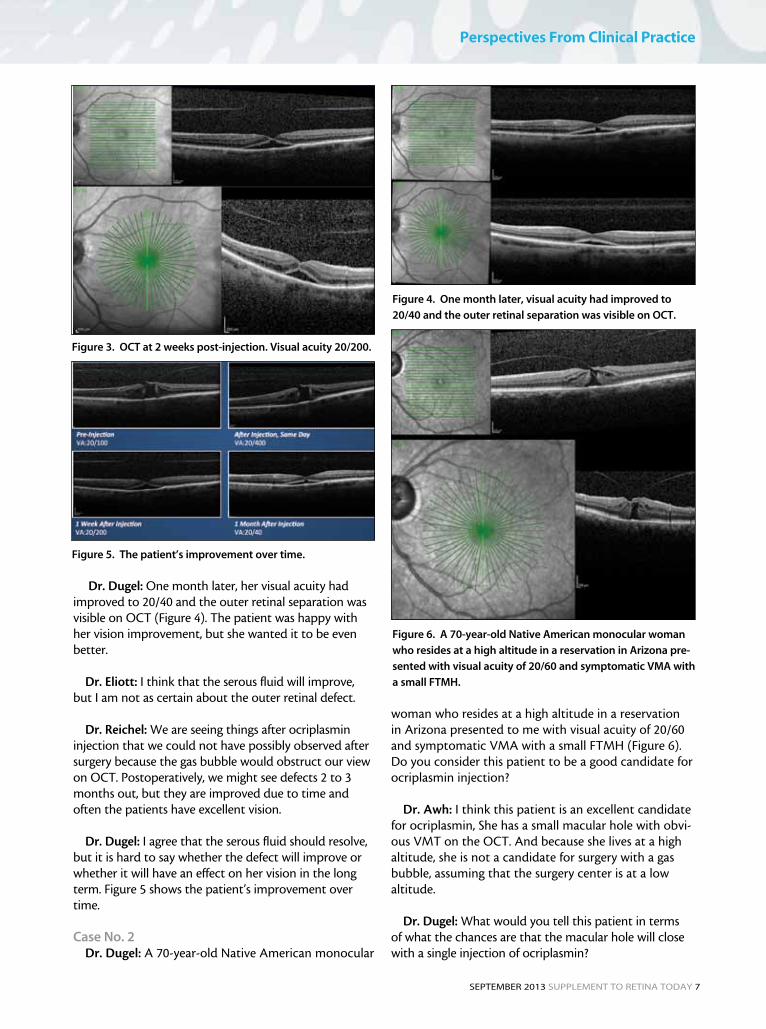
Case No . 2Dr. Dugel: A 70-year-old Native American monocular
woman who resides at a high altitude in a reservation in Arizona presented to me with visual acuity of 20/60 and symptomatic VMA with a small FTMH (Figure 6). Do you consider this patient to be a good candidate for ocriplasmin injection?
Dr. Awh: I think this patient is an excellent candidate for ocriplasmin, She has a small macular hole with obvi-ous VMT on the OCT. And because she lives at a high altitude, she is not a candidate for surgery with a gas bubble, assuming that the surgery center is at a low altitude.
Dr. Dugel: What would you tell this patient in terms
of what the chances are that the macular hole will close with a single injection of ocriplasmin?
Figure 3 . OCT at 2 weeks post-injection . Visual acuity 20/200 .
Figure 5 . The patient’s improvement over time .
Figure 4 . One month later, visual acuity had improved to
20/40 and the outer retinal separation was visible on OCT .
Figure 6 . A 70-year-old Native American monocular woman
who resides at a high altitude in a reservation in Arizona pre-
sented with visual acuity of 20/60 and symptomatic VMA with
a small FTMH .
8 SUPPlEMEnt to REtInA toDAY SEPtEMbER 2013
Perspectives From Clinical Practice
Dr. Reichel: The VMA is very focal and the area is small, so I would tell the patient that there is a 50% to 60% chance of success, but that if the injection does not work, we may have to operate. I find that some patients, particularly women in the age range of 60 to 70 years, are gun-shy regarding surgery. This is even more pronounced when a patient’s vision is like that of the woman in your case. At 20/60, she does not have as profound of vision loss as a patient with visual acuity of 20/400, so she may question the need for surgery. That said, my plan would be to inject this patient but make sure she understands that if, in 1 month, the hole has not closed, I will sched-ule her for surgery.
Dr. Chan: You could wait 1 month before giving her an ocriplasmin injection. Even though she is monocular, her vision is relatively good and there is a chance for sponta-neous release of the vitreomacular adhesion. Before we had this drug available to us, watch-and-wait was a pos-sible strategy for such a case.
Dr. Dugel: Dr. Awh, do you think there is chance of spontaneous resolution for this case?
Dr. Awh: Yes, but I think that the chance is small, relative to the fact that the patient is monocular. Assuming that the patient is symptomatic and has noticed a decline in her vision, I most likely would not hold out for that. If, on the other hand, the patient were 20/20 in her fellow eye, I might watch and wait. However, I cannot recall any macular holes in my clinical experience that have spontaneously resolved.
Dr. Dugel: I agree that it is rare. When I do observe, it is for a fairly short period of time. For this patient, I did not see any disadvantage to waiting for 1 month. After this period of observation, I gave her an ocriplasmin injection.
Dr. Reichel, how do you prepare a patient for injec-tion? Do you say or do anything different than you would with an injection of anti-VEGF?
Dr. Reichel: Patient consent for ocriplasmin injection is crucial because PVD is a dynamic process, during which a patient will be symptomatic. In my practice, we have modified our consent forms to reflect this. We are most concerned with the increase of floaters and the temporary decrease in visual acuity that can result with PVD. We also include the potential retinal tear and detachment, which we do not have on our consent forms for patients with age-related macular degeneration and diabetic macular edema.
I explain to patients that we are inducing the process of vitreous separation that occurs naturally as the eye ages and that, in doing so, may release collagen and lead to the development of floaters. I also explain the risk of retinal tears and detachments and tell patients to watch out for changes in peripheral or central vision, advising
them to come into the office if these occur. I see patients 1 week after injection.
Dr. Eliott: In preparing a patient for injections, I tell them in layman’s terms that there is something unique about their vitreomacular interface that has resulted in traction, and that PVD is a natural process.
Dr. Chan: I agree that patients should understand the increased risk of floaters and significant temporary vision loss after the injection.
I would see these patients the next day, as I do with my postoperative patients. I also think it’s very important that the patient understand what the risks are and what symp-toms they may have after ocriplasmin injection.
Dr. Dugel: Patient preparation is incredibly important for ocriplasmin, because it simulates surgery. I agree with Dr. Chan that follow-up should be the same as for postop-erative patients.
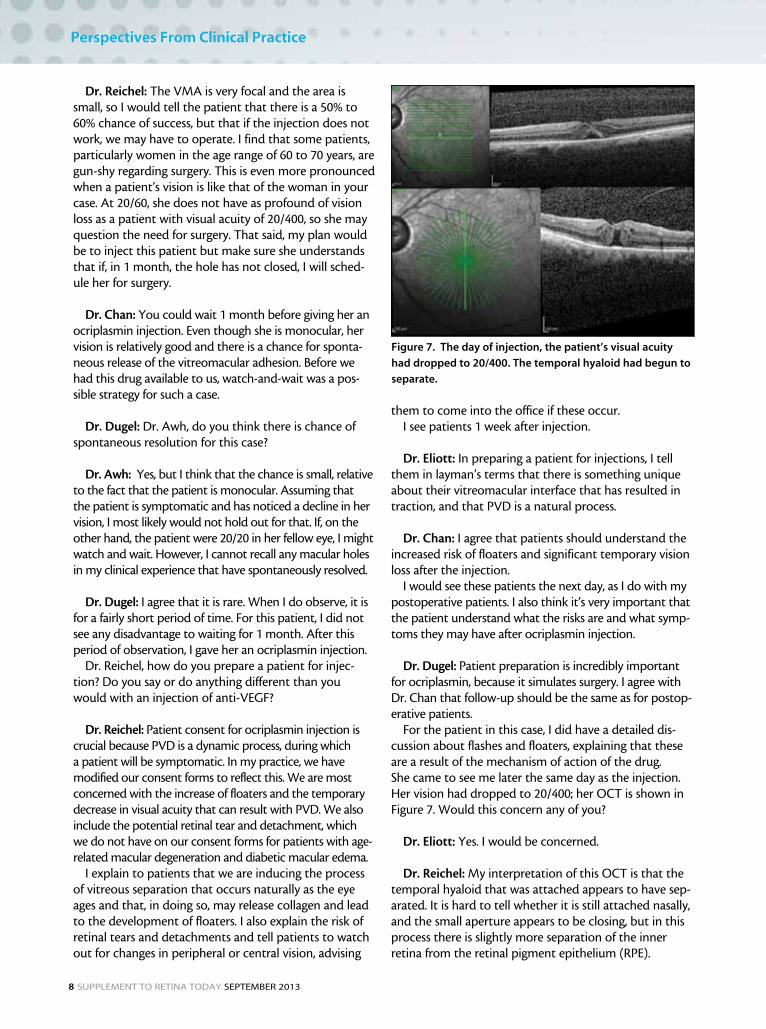
For the patient in this case, I did have a detailed dis-cussion about flashes and floaters, explaining that these are a result of the mechanism of action of the drug. She came to see me later the same day as the injection. Her vision had dropped to 20/400; her OCT is shown in Figure 7. Would this concern any of you?
Dr. Eliott: Yes. I would be concerned.
Dr. Reichel: My interpretation of this OCT is that the temporal hyaloid that was attached appears to have sep-arated. It is hard to tell whether it is still attached nasally, and the small aperture appears to be closing, but in this process there is slightly more separation of the inner retina from the retinal pigment epithelium (RPE).
Figure 7 . The day of injection, the patient’s visual acuity
had dropped to 20/400 . The temporal hyaloid had begun to
separate .
SEPtEMbER 2013 SUPPlEMEnt to REtInA toDAY 9
Perspectives From Clinical Practice
Dr. Dugel: I also thought that the temporal hyaloid had begun to separate, and although I initially thought this was quite rapid, ocriplasmin works quickly and is a highly unstable nonspecific serine protease.
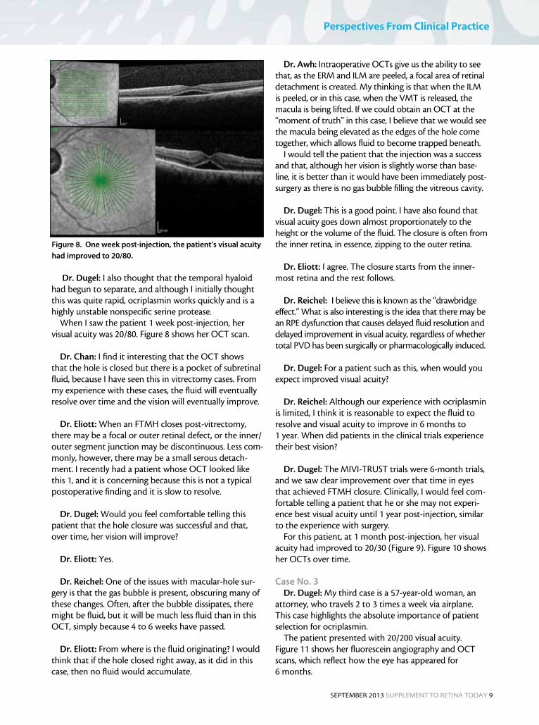
When I saw the patient 1 week post-injection, her visual acuity was 20/80. Figure 8 shows her OCT scan.
Dr. Chan: I find it interesting that the OCT shows that the hole is closed but there is a pocket of subretinal fluid, because I have seen this in vitrectomy cases. From my experience with these cases, the fluid will eventually resolve over time and the vision will eventually improve.
Dr. Eliott: When an FTMH closes post-vitrectomy, there may be a focal or outer retinal defect, or the inner/outer segment junction may be discontinuous. Less com-monly, however, there may be a small serous detach-ment. I recently had a patient whose OCT looked like this 1, and it is concerning because this is not a typical postoperative finding and it is slow to resolve.
Dr. Dugel: Would you feel comfortable telling this patient that the hole closure was successful and that, over time, her vision will improve?
Dr. Eliott: Yes.
Dr. Reichel: One of the issues with macular-hole sur-gery is that the gas bubble is present, obscuring many of these changes. Often, after the bubble dissipates, there might be fluid, but it will be much less fluid than in this OCT, simply because 4 to 6 weeks have passed.
Dr. Eliott: From where is the fluid originating? I would think that if the hole closed right away, as it did in this case, then no fluid would accumulate.
Dr. Awh: Intraoperative OCTs give us the ability to see that, as the ERM and ILM are peeled, a focal area of retinal detachment is created. My thinking is that when the ILM is peeled, or in this case, when the VMT is released, the macula is being lifted. If we could obtain an OCT at the “moment of truth” in this case, I believe that we would see the macula being elevated as the edges of the hole come together, which allows fluid to become trapped beneath.
I would tell the patient that the injection was a success and that, although her vision is slightly worse than base-line, it is better than it would have been immediately post-surgery as there is no gas bubble filling the vitreous cavity.
Dr. Dugel: This is a good point. I have also found that visual acuity goes down almost proportionately to the height or the volume of the fluid. The closure is often from the inner retina, in essence, zipping to the outer retina.
Dr. Eliott: I agree. The closure starts from the inner-most retina and the rest follows.
Dr. Reichel: I believe this is known as the “drawbridge effect.” What is also interesting is the idea that there may be an RPE dysfunction that causes delayed fluid resolution and delayed improvement in visual acuity, regardless of whether total PVD has been surgically or pharmacologically induced.
Dr. Dugel: For a patient such as this, when would you expect improved visual acuity?
Dr. Reichel: Although our experience with ocriplasmin is limited, I think it is reasonable to expect the fluid to resolve and visual acuity to improve in 6 months to 1 year. When did patients in the clinical trials experience their best vision?
Dr. Dugel: The MIVI-TRUST trials were 6-month trials, and we saw clear improvement over that time in eyes that achieved FTMH closure. Clinically, I would feel com-fortable telling a patient that he or she may not experi-ence best visual acuity until 1 year post-injection, similar to the experience with surgery.
For this patient, at 1 month post-injection, her visual acuity had improved to 20/30 (Figure 9). Figure 10 shows her OCTs over time.
Case No . 3Dr. Dugel: My third case is a 57-year-old woman, an
attorney, who travels 2 to 3 times a week via airplane. This case highlights the absolute importance of patient selection for ocriplasmin.
The patient presented with 20/200 visual acuity. Figure 11 shows her fluorescein angiography and OCT scans, which reflect how the eye has appeared for 6 months.
Figure 8 . One week post-injection, the patient’s visual acuity
had improved to 20/80 .
10 SUPPlEMEnt to REtInA toDAY SEPtEMbER 2013
Perspectives From Clinical Practice
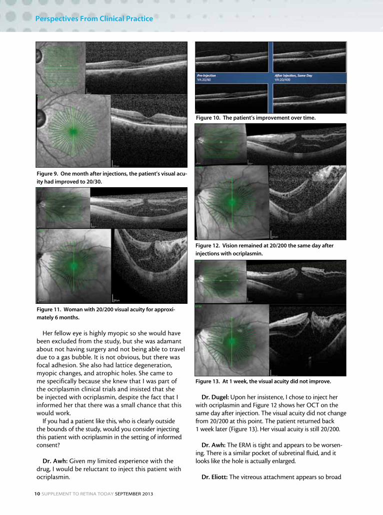
Her fellow eye is highly myopic so she would have been excluded from the study, but she was adamant about not having surgery and not being able to travel due to a gas bubble. It is not obvious, but there was focal adhesion. She also had lattice degeneration, myopic changes, and atrophic holes. She came to me specifically because she knew that I was part of the ocriplasmin clinical trials and insisted that she be injected with ocriplasmin, despite the fact that I informed her that there was a small chance that this would work.
If you had a patient like this, who is clearly outside the bounds of the study, would you consider injecting this patient with ocriplasmin in the setting of informed consent?
Dr. Awh: Given my limited experience with the drug, I would be reluctant to inject this patient with ocriplasmin.
Dr. Dugel: Upon her insistence, I chose to inject her with ocriplasmin and Figure 12 shows her OCT on the same day after injection. The visual acuity did not change from 20/200 at this point. The patient returned back 1 week later (Figure 13). Her visual acuity is still 20/200.
Dr. Awh: The ERM is tight and appears to be worsen-ing. There is a similar pocket of subretinal fluid, and it looks like the hole is actually enlarged.
Dr. Eliott: The vitreous attachment appears so broad
Figure 9 . One month after injections, the patient’s visual acu-
ity had improved to 20/30 .
Figure 11 . Woman with 20/200 visual acuity for approxi-
mately 6 months .
Figure 12 . Vision remained at 20/200 the same day after
injections with ocriplasmin .
Figure 13 . At 1 week, the visual acuity did not improve .
Figure 10 . The patient’s improvement over time .
SEPtEMbER 2013 SUPPlEMEnt to REtInA toDAY 11
Perspectives From Clinical Practice
in Figure 11 that its release may allow the ERM to create more tangential traction on the macula, having a worse effect on the patient’s vision.
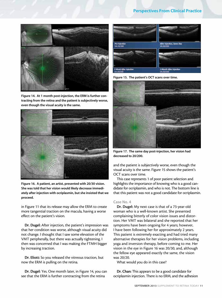
Dr. Dugel: After injection, the patient’s impression was that her condition was worse, although visual acuity did not change. I thought that I saw some elevation of the VMT peripherally, but there was actually tightening. I then was concerned that I was making the FTMH bigger by increasing traction.
Dr. Eliott: So you released the vitreous traction, but now the ERM is pulling on the retina.
Dr. Dugel: Yes. One month later, in Figure 14, you can see that the ERM is further contracting from the retina
and the patient is subjectively worse, even though the visual acuity is the same. Figure 15 shows the patient’s OCT scans over time.
This case represents 1 of poor patient selection and highlights the importance of knowing who is a good can-didate for ocriplasmin, and who is not. The bottom line is that this patient was not a good candidate for ocriplasmin.
Case No . 4Dr. Dugel: My next case is that of a 73-year-old
woman who is a well-known artist. She presented complaining bitterly of color vision issues and distor-tion. Her VMT was bilateral and she reported that her symptoms have been ongoing for 4 years; however, I have been following her for approximately 2 years. This patient is extremely exacting and had tried many alternative therapies for her vision problems, including yoga and inversion therapy, before coming to me. Her vision in the eye in Figure 16 was 20/30, and, although the fellow eye appeared exactly the same, the vision was 20/20.
What would you do in this case?
Dr. Chan: This appears to be a good candidate for ocriplasmin injection. There is no ERM, and the adhesion
Figure 14 . At 1 month post-injection, the ERM is further con-
tracting from the retina and the patient is subjectively worse,
even though the visual acuity is the same .
Figure 16 . A patient, an artist, presented with 20/30 vision .
She was told that her vision would likely decrease immedi-
ately after injection with ocriplasmin, but she insisted that we
proceed .
Figure 15 . The patient’s OCT scans over time .
Figure 17 . The same day post-injection, her vision had
decreased to 20/200 .

12 SUPPlEMEnt to REtInA toDAY SEPtEMbER 2013
Perspectives From Clinical Practice
is focal. Although the vision is relatively good, she is symp-tomatic, so, after a discussion about the risk and benefits of ocriplasmin, I would inject.
Dr. Eliott: I agree that this patient is a good candidate for ocriplasmin. My only concern is that artists in par-ticular are highly aware of any changes in vision.
Dr. Dugel: I informed the patient that her vision would likely decrease after the injection, but we decided to go ahead with ocriplasmin. Figure 17 shows her scans the same day post-injection. Her vision had decreased to 20/200. What would you tell this patient?
Dr. Eliott: I would tell her to hang in there and that we will wait and see what happens.
Dr. Dugel: How long would you wait before you schedule this patient for surgery?
Dr. Eliott: I would wait at least 1 month before operating.
Dr. Reichel: I would wait longer than 1 month.
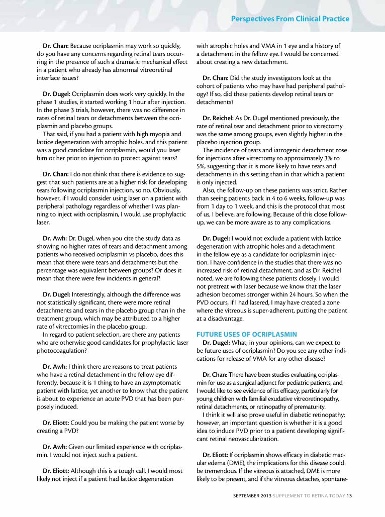
Dr. Dugel: In this case, I waited. Figure 18 shows her scans at 1 week, showing little improvement.
One month later, she called me from London, hys-terical. She said that she was flying directly home and needed to come to my office. Initially, I thought that she had a detachment or something worse.
When I saw the patient and looked at her OCT, there was resolution of the VMT (Figure 19). Her visual acu-ity is now 20/20, and the patient was thrilled with her results. I can only guess that the adhesion popped off so quickly that it had alarmed her. Figure 20 shows the patient’s OCT scans over time.
addItIonal conSIderatIonSDr. Reichel: Suppose we have a patient with visual
acuity of 20/100 and a pocket of subretinal fluid. Two months after injections, the vision is still at 20/100 and although the macular hole is closed, subretinal fluid pocket remains. What would you do in this case? Would you inject a gas bubble?
Dr. Awh: There is no evidence that gas has a mechani-cal effect to displace the pocket of fluid. The problem is most likely that the RPE is not pumping the fluid out as quickly as it should, so in my opinion, pressure from a gas bubble is not necessarily going to alter the course of what will occur naturally, given more time.
Dr. Dugel: Previously, with hyaluronidase or chondroi-tinase, which are both agents that cause liquefaction, gas was used to complete the vitreolysis process with sepa-ration. However, I would not inject gas without more scientific data.
Figure 18 . The patient showed little improvement at 1 week
after an injection with ocriplasmin .
Figure 19 . One month later, the patient experienced a com-
plete PVD, and on OCT her retina was completely flat, with
complete resolution of the VMT . Her final visual acuity is
20/20 .
Figure 20 . The patient’s improvement over time .
SEPtEMbER 2013 SUPPlEMEnt to REtInA toDAY 13
Perspectives From Clinical Practice
Dr. Chan: Because ocriplasmin may work so quickly, do you have any concerns regarding retinal tears occur-ring in the presence of such a dramatic mechanical effect in a patient who already has abnormal vitreoretinal interface issues?
Dr. Dugel: Ocriplasmin does work very quickly. In the phase 1 studies, it started working 1 hour after injection. In the phase 3 trials, however, there was no difference in rates of retinal tears or detachments between the ocri-plasmin and placebo groups.
That said, if you had a patient with high myopia and lattice degeneration with atrophic holes, and this patient was a good candidate for ocriplasmin, would you laser him or her prior to injection to protect against tears?
Dr. Chan: I do not think that there is evidence to sug-gest that such patients are at a higher risk for developing tears following ocriplasmin injection, so no. Obviously, however, if I would consider using laser on a patient with peripheral pathology regardless of whether I was plan-ning to inject with ocriplasmin, I would use prophylactic laser.
Dr. Awh: Dr. Dugel, when you cite the study data as showing no higher rates of tears and detachment among patients who received ocriplasmin vs placebo, does this mean that there were tears and detachments but the percentage was equivalent between groups? Or does it mean that there were few incidents in general?
Dr. Dugel: Interestingly, although the difference was not statistically significant, there were more retinal detachments and tears in the placebo group than in the treatment group, which may be attributed to a higher rate of vitrectomies in the placebo group.
In regard to patient selection, are there any patients who are otherwise good candidates for prophylactic laser photocoagulation?
Dr. Awh: I think there are reasons to treat patients
who have a retinal detachment in the fellow eye dif-ferently, because it is 1 thing to have an asymptomatic patient with lattice, yet another to know that the patient is about to experience an acute PVD that has been pur-posely induced.
Dr. Eliott: Could you be making the patient worse by creating a PVD?
Dr. Awh: Given our limited experience with ocriplas-min. I would not inject such a patient.
Dr. Eliott: Although this is a tough call, I would most likely not inject if a patient had lattice degeneration
with atrophic holes and VMA in 1 eye and a history of a detachment in the fellow eye. I would be concerned about creating a new detachment.
Dr. Chan: Did the study investigators look at the
cohort of patients who may have had peripheral pathol-ogy? If so, did these patients develop retinal tears or detachments?
Dr. Reichel: As Dr. Dugel mentioned previously, the rate of retinal tear and detachment prior to vitrectomy was the same among groups, even slightly higher in the placebo injection group.
The incidence of tears and iatrogenic detachment rose for injections after vitrectomy to approximately 3% to 5%, suggesting that it is more likely to have tears and detachments in this setting than in that which a patient is only injected.
Also, the follow-up on these patients was strict. Rather than seeing patients back in 4 to 6 weeks, follow-up was from 1 day to 1 week, and this is the protocol that most of us, I believe, are following. Because of this close follow-up, we can be more aware as to any complications.
Dr. Dugel: I would not exclude a patient with lattice degeneration with atrophic holes and a detachment in the fellow eye as a candidate for ocriplasmin injec-tion. I have confidence in the studies that there was no increased risk of retinal detachment, and as Dr. Reichel noted, we are following these patients closely. I would not pretreat with laser because we know that the laser adhesion becomes stronger within 24 hours. So when the PVD occurs, if I had lasered, I may have created a zone where the vitreous is super-adherent, putting the patient at a disadvantage.
future uSeS of ocrIPlaSmInDr. Dugel: What, in your opinions, can we expect to
be future uses of ocriplasmin? Do you see any other indi-cations for release of VMA for any other disease?
Dr. Chan: There have been studies evaluating ocriplas-min for use as a surgical adjunct for pediatric patients, and I would like to see evidence of its efficacy, particularly for young children with familial exudative vitreoretinopathy, retinal detachments, or retinopathy of prematurity.
I think it will also prove useful in diabetic retinopathy; however, an important question is whether it is a good idea to induce PVD prior to a patient developing signifi-cant retinal neovascularization.
Dr. Eliott: If ocriplasmin shows efficacy in diabetic mac-ular edema (DME), the implications for this disease could be tremendous. If the vitreous is attached, DME is more likely to be present, and if the vitreous detaches, spontane-

14 SUPPlEMEnt to REtInA toDAY SEPtEMbER 2013
Perspectives From Clinical Practice
ous resolution of DME may occur. It is important to note, however, that the vitreoretinal interface of a patient with DME is different than that of a typical patient.
Dr. Reichel: The vitreoretinal interface is likely differ-ent, not only in DME, but also in AMD. There are many inflammatory factors, cytokines, and complement issues in eyes with DME and AMD that play a role in the dis-ease states. I look forward to more data in these areas.
Dr. Awh: I agree. It is important to not equate the mere presence of VMA with a pathologic role in AMD or DME. I am also eager to see some trial results.
Dr. Dugel: I also agree that any future uses of ocriplas-min must be data driven. As Dr. Reichel stated, VMA may vary among disease states and, as such, the utility of ocriplasmin may also vary. However, if the data show that ocriplasmin is effective for DME, AMD, or pediatric surgical cases, this will have a large impact on our daily practice and our patient outcomes. n
Carl C. Awh, MD, practices at Tennessee Retina in Nashville. He is a Retina Today Editorial Board member. Dr. Awh states that he is a consultant to ArcticDx; Bausch + Lomb; Genentech; Katalyst; Notal Vision; Regeneron; Synergetics; ThromboGenics; and Volk Optical. He may be reached at [email protected].
R.V. Paul Chan, MD, is an Associate Professor of Ophthalmology, the St. Giles Associate Professor of Pediatric Retina, and Director of the Retina Service at Weill Cornell Medical College. He is a Retina Today Editorial Board member. Dr. Chan states that he has no financial relationships to disclose. He may be reached at [email protected].
Pravin U. Dugel, MD, is Managing Partner of Retinal Consultants of Arizona in Phoenix; Clinical Associate Professor of Ophthalmology, Doheny Eye Institute, Keck School of Medicine at the University of Southern California, Los Angeles; and Founding Member of the Spectra Eye Institute in Sun City, AZ. He is a Retina Today Editorial Board member. Dr. Dugel states that he is a consultant for Alcon; AMO; ArcticDx; Ora; Regeneron; and ThromboGenics. He may be reached at [email protected].
Dean Eliott, MD, is Associate Director of the Retina Service, Massachusetts Eye and Ear Infirmary, Harvard Medical School, and is a Retina Today Editorial Board member. He states that he is an ad hoc consultant for Acucela, Alimera Sciences, Allergan Inc., Arctic, Genentech, Glaukos, Regeneron Pharmaceuticals, and ThromboGenics. He may be reached by phone: +1 617 573-3736; fax: +1 617 573 3698; or via email at [email protected].
Elias Reichel, MD, is Vice Chair for Research and Education, Department of Ophthalmology, at New England Eye Center and Professor of Ophthalmology at Tufts University School of Medicine in Boston. Dr. Reichel is a Retina Today Editorial Board Member. Dr. Reichel states that he is a consultant to Akorn; Ocular Instruments; Ophthotech; NewGen Biopharma; Hemera Biosciences; Regeneron; ThromboGenics; EMD Serono; Valeant; and Genentech. He may be reached at [email protected].
1. Gandorfer A, Rohleder M, Sethi C, et al. Posterior vitreous detachment induced by microplasmin. Invest Ophthal-mol Vis Sci. 2004;45(2):641-647.2. Stalmans P, Benz MS, Gandorfer A, et al; MIVI-TRUST Study Group. Enzymatic vitreolysis with ocriplasmin for vitreomacular traction and macular holes. N Engl J Med. 2012;367(7):606-615.
SEPtEMbER 2013 SUPPlEMEnt to REtInA toDAY 15
Perspectives From Clinical Practice
The MIVI-TRUST program, the 2 phase 3 trials evalu-ating ocriplasmin (JETREA, ThromboGenics), random-ized 652 patients to either a single injection of 125 µg ocriplasmin (n=464) or placebo (n=188).1 The saline that was injected in the eyes of those patients in the placebo group was equal in volume to that in the ocri-plasmin group to delineate volumetric effect on vitre-ous separation.
The primary endpoint was resolution of vitreomacular adhesion (VMA) at day 28. The secondary endpoints of the study were complete posterior vitreous detachment (PVD) at day 28, nonsurgical closure of full-thickness macular holes (FTMHs), change in visual acuity, the need for vitrectomy, and responses on a visual function questionnaire.
Enrollment criteria included symptomatic VMA con-firmed by a central reading center with baseline visual acuity of 20/25 or worse. Patients with epiretinal mem-branes (ERMs) were also included in the study. Exclusion criteria included high myopia, prior vitrectomy, and macular holes larger than 400 µm.
Patients in these trials fell into 3 nonexclusive categories: vitreomacular traction (VMT) without FTMH or epiretinal membrane (ERM) at baseline; FTMH with or without ERM at baseline; and patients with ERM at baseline (note that ERM was not a target of treatment). All patients had OCT-confirmed VMA. Ocriplasmin met the studies’ primary endpoint of pharmacologic resolution of VMA at day 28. In the integrated data analysis of the 2 phase 3 studies, 26.5% of patients in the ocriplasmin group achieved VMA resolution at day 28 compared with 10.1% in the placebo group, a difference that was statistically significant.
reSultS VMA resolved in 26.5% of ocriplasmin-injected eyes
and in 10.1% of placebo-injected eyes (P < .001). Total PVC was more prevalent among the eyes treated with ocriplasmin than among those injected with placebo (13.4% vs 3.7%, P < .001). Resolution of VMA was higher in eyes with adhesion areas smaller than 1500 µm. Approximately 41% of ocriplasmin-treated VMT patients who achieved VMA resolution gained 2 or more lines of visual acuity at 6 months. Nonsurgical closure of FTMH was achieved in 40.6% of ocriplasmin-injected eyes, compared with 10.6% of placebo-injected eyes (P < .001). Patients with ERM did not appear to benefit significantly with ocriplasmin injection.
ocular adverSe eventSOcular adverse events included vitreous floaters, pho-
topsia, injection-related eye pain, and conjunctival hem-orrhage and occurred in 68.4% of ocriplasmin-injected eyes and in 53.5% of placebo-injected eyes (P < .001). Most of these events were transient, resolving within 1 week of injection.
The incidence of serious ocular adverse events was similar between the 2 groups (P = .26).
SummaryA single injection of ocriplasmin was shown to result
in higher rates of PVD and VMA and FTMH resolution, improving visual acuity in most patients.
1. Stalmans P, Benz MS, Gandorfer A, et al. Enzymatic vitreolysis with ocriplasmin for vitreomacular traction and macular holes. N Engl J Med. 2012;367(7):606-615.
Ocriplasmin: summary Of the phase 3 Data