Embed Size (px)
Citation preview

1© 2015, Elsevier Inc., Heymann, Bone Cancer, Second Edition
Chapter 45
DIAGNOSIS OF BONE METASTASES IN UROLOGICAL MALIGNANCIES - AN UPDATE

2© 2015, Elsevier Inc., Heymann, Bone Cancer, Second Edition
FIGURE 45.1 Forty-eight-year-old male diagnosed with multiple myeloma. (A) Skull showing multiple lesions “pepper pot” skull appear-ance. (B) Distal right femur with multiple lytic lesions.

3© 2015, Elsevier Inc., Heymann, Bone Cancer, Second Edition
FIGURE 45.2 Eighty-two-year-old male was diagnosed with metastatic prostate cancer. (A) and (B) Plain radiographs of the right hip and pelvis show multiple sclerotic lesions.

4© 2015, Elsevier Inc., Heymann, Bone Cancer, Second Edition
FIGURE 45.3 Conventional CT imaging of spinal column with the presence of bony metastases is seen in (A). Bony windows in (B) shows greater detail in the in the bone structure and the lesions are seen with more defi nition in comparison to the conventional window.

5© 2015, Elsevier Inc., Heymann, Bone Cancer, Second Edition
FIGURE 45.4 A staging bone scintigraphy was performed on a 77-year-old male with prostate cancer. Metastases are seen in the left 6th rib posteriorly, the right 5th and 6th ribs laterally, T6, spinous process of L2, scarum and both ilia adjacent to sacroiliac joints and in the right superior acetabulum.
6© 2015, Elsevier Inc., Heymann, Bone Cancer, Second Edition
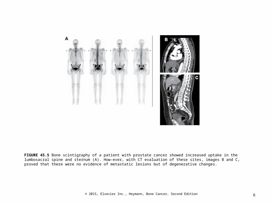
FIGURE 45.5 Bone scintigraphy of a patient with prostate cancer showed increased uptake in the lumbosacral spine and sternum (A). How-ever, with CT evaluation of these sites, images B and C, proved that there were no evidence of metastatic lesions but of degenerative changes.

7© 2015, Elsevier Inc., Heymann, Bone Cancer, Second Edition
FIGURE 45.6 Bone scintigraphy in a patient with prostate cancer shows the presence of bone metastases in L4 vertebra (A). However, further imaging with MRI shows the presence of other bony lesions in T8 as seen in image B and also in T3, L1 and L2.

8© 2015, Elsevier Inc., Heymann, Bone Cancer, Second Edition
FIGURE 45.7 A 61-year-old male with right sided RCC presented with lower back pain. Reverse bone scintigraphy (A) shows a bony lesion at the L4 level. The group of images in B were performed with PET, which shows increased activity at the L4 level. Note increased activity is seen in the upper pole of the right kidney, depicting the RCC.
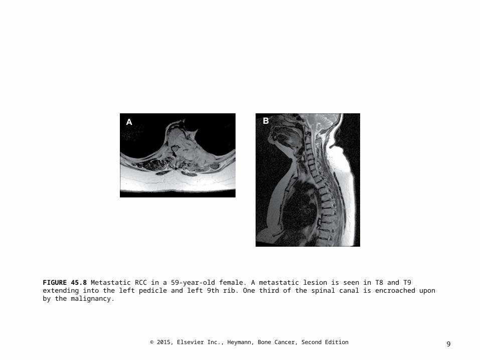
9© 2015, Elsevier Inc., Heymann, Bone Cancer, Second Edition
FIGURE 45.8 Metastatic RCC in a 59-year-old female. A metastatic lesion is seen in T8 and T9 extending into the left pedicle and left 9th rib. One third of the spinal canal is encroached upon by the malignancy.

10© 2015, Elsevier Inc., Heymann, Bone Cancer, Second Edition
FIGURE 45.9 Standard bone scintigraphy (A) in a 66-year-old with prostate cancer failed to diagnose a metastatic deposit. However, fluorocholine PET-CT diagnosed a rib metastasis in the second rib (B) as well as a nodal metastases (C), both seen easily on CT alone.

11© 2015, Elsevier Inc., Heymann, Bone Cancer, Second Edition
FIGURE 45.10 The formation of a PET/CT image is by the combination of a PET image with a CT scan. Image (A) shows the PET of the patient seen above with metastatic RCC. The CT image in (B) is able to show the anatomical location of the lesion in L4. The final product, image (C), shows PET/CT depicting the L4 lesion. It shows the metabolic activity as well as the anatomical location of the lesion.

12© 2015, Elsevier Inc., Heymann, Bone Cancer, Second Edition
FIGURE 45.11 A 66-year-old female diagnosed with metastatic breast cancer. Image (A) shows widespread disease involving. Mixed lytic and sclerotic lesions measuring up to 1 cm in the lumbar vertebrae are seen on CT using bony windows (B and C).

13© 2015, Elsevier Inc., Heymann, Bone Cancer, Second Edition
FIGURE 45.12 A patient diagnosed with hormone refractory prostate cancer shown here to have widespread osseous metastatic disease involving the axial and appendicular skeleton (A). The images B and C show multiple sclerotic lesions in the lumbar spine.

14© 2015, Elsevier Inc., Heymann, Bone Cancer, Second Edition
FIGURE 45.13 Fifty-seven-year-old female with NSCLC noted to have increased tracer uptake in multiple regions on bone scintigraphy (A). Tracer uptake is seen in left mandible, right humeral head, sternum, spinal column and right sacroiliac joint. An OPG and CT (B and C respectively) show a lytic lesion in the mandible highlighting the presence of mixed lesions occurring in lung cancer.